fMRI responses to words repeated in a congruous semantic context are abnormal in mild Alzheimer's...
12
Neuropsychologia 48 (2010) 2476–2487 Contents lists available at ScienceDirect Neuropsychologia journal homepage: www.elsevier.com/locate/neuropsychologia fMRI responses to words repeated in a congruous semantic context are abnormal in mild Alzheimer’s disease John M. Olichney a,∗ , Jason R. Taylor b , Shiaohui Chan a,c , Jin-Chen Yang a , Andrew Stringfellow d , Dieter G. Hillert e , Amanda L. Simmons f , David P. Salmon e , Vicente Iragui-Madoz e , Marta Kutas g a Department of Neurology, University of California, Davis, Davis, CA, USA b Medical Research Council, Cognition and Brain Sciences Unit, Cambridge, UK c Department of English, National Taiwan Normal University, Taipei, Taiwan d University of California, San Diego, CA, USA e Department of Neurosciences, University of California, San Diego, CA, USA f Department of Psychology, California State University, Sacramento, CA, USA g Department of Cognitive Science, University of California, San Diego, CA, USA article info Article history: Received 12 August 2009 Received in revised form 18 April 2010 Accepted 20 April 2010 Available online 28 April 2010 Keywords: Memory Semantic Language Medial temporal lobe Cingulate abstract Background: We adapted an event-related brain potential word repetition paradigm, sensitive to early Alzheimer’s disease (AD), for functional MRI (fMRI). We hypothesized that AD would be associated with reduced differential response to New/Old congruous words. Methods: Fifteen mild AD patients (mean age = 72.9) and 15 normal elderly underwent 1.5T fMRI during a semantic category decision task. Results: We found robust between-groups differences in BOLD response to congruous words. In con- trols, the New > Old contrast demonstrated larger responses in much of the left-hemisphere (including putative P600 generators: parahippocampal, cingulate, fusiform, perirhinal, middle temporal (MTG) and inferior frontal gyri (IFG)); the Old > New contrast showed modest activation, mainly in right parietal and prefrontal cortex. By contrast, there were relatively few regions of significant New > Old responses in AD patients, mainly in the right-hemisphere, and their Old > New contrast did not demonstrate a right-hemisphere predominance. Across subjects, the spatial extent of New > Old responses in left medial temporal lobe (MTL) correlated with subsequent recall and recognition (r’s ≥ 0.60). In controls, the mag- nitude of New–Old response in left MTL, fusiform, IFG, MTG, superior temporal and cingulate gyrus correlated with subsequent cued recall and/or recognition (0.51 ≤ r’s ≤ 0.78). Conclusions: A distributed network of mostly left-hemisphere structures, which are putative P600 gener- ators, appears important for successful verbal encoding (with New > Old responses to congruous words in normal elderly). This network appears dysfunctional in mild AD patients, as reflected in decreased word repetition effects particularly in left association cortex, paralimbic and MTL structures. © 2010 Elsevier Ltd. All rights reserved. 1. Introduction Most prior fMRI studies of memory encoding in Alzheimer’s disease (AD) have found abnormal medial temporal lobe (MTL) acti- vation, across paradigms using verbal (Remy, Mirrashed, Campbell, & Richter, 2005), picture (Machulda et al., 2003), and face-name associative (Sperling et al., 2003) encoding tasks. In contrast, using word repetition “priming” paradigms, some fMRI studies of ver- bal memory in AD have found neocortical regions with relatively ∗ Corresponding author. University of California, Davis School of Medicine, Center for Mind and Brain, 267 Cousteau Place, Davis, CA 95618-5412, USA. Tel.: +1 530 297 4429; fax: +1 530 297 4400. E-mail address: [email protected] (J.M. Olichney). normal New–Old word differences, e.g. selected portions of left inferior frontal gyrus (IFG) and middle frontal gyrus (MFG) (BA 45/47 and 44/46) (Lustig & Buckner, 2004). This pattern of results is as expected if the MTL is critical for the normal function of explicit/declarative memory circuitry, but the frontal neocortex is either mainly involved in implicit verbal memory processes (e.g. phonological or semantic priming) or its activation is insufficient for normal encoding and explicit learning to occur. We have adapted an event-related brain potential (ERP) inci- dental learning paradigm (with cross-modal category-target word associations), shown by our prior ERP studies to be very sensitive to early AD (Olichney et al., 2008; Olichney et al., 2006; Olichney et al., 2002), even at the MCI stage, for functional MRI (fMRI). This paradigm normally produces robust incidental learning of the semantically congruous category-exemplar words, but not of the 0028-3932/$ – see front matter © 2010 Elsevier Ltd. All rights reserved. doi:10.1016/j.neuropsychologia.2010.04.021
Transcript of fMRI responses to words repeated in a congruous semantic context are abnormal in mild Alzheimer's...
fi
JDa
b
c
d
e
f
g
a
ARRAA
KMSLMC
1
dv&awb
fT
0d
Neuropsychologia 48 (2010) 2476–2487
Contents lists available at ScienceDirect
Neuropsychologia
journa l homepage: www.e lsev ier .com/ locate /neuropsychologia
MRI responses to words repeated in a congruous semantic context are abnormaln mild Alzheimer’s disease
ohn M. Olichneya,∗, Jason R. Taylorb, Shiaohui Chana,c, Jin-Chen Yanga, Andrew Stringfellowd,ieter G. Hillerte, Amanda L. Simmonsf, David P. Salmone, Vicente Iragui-Madoze, Marta Kutasg
Department of Neurology, University of California, Davis, Davis, CA, USAMedical Research Council, Cognition and Brain Sciences Unit, Cambridge, UKDepartment of English, National Taiwan Normal University, Taipei, TaiwanUniversity of California, San Diego, CA, USADepartment of Neurosciences, University of California, San Diego, CA, USADepartment of Psychology, California State University, Sacramento, CA, USADepartment of Cognitive Science, University of California, San Diego, CA, USA
r t i c l e i n f o
rticle history:eceived 12 August 2009eceived in revised form 18 April 2010ccepted 20 April 2010vailable online 28 April 2010
eywords:emory
emanticanguageedial temporal lobe
ingulate
a b s t r a c t
Background: We adapted an event-related brain potential word repetition paradigm, sensitive to earlyAlzheimer’s disease (AD), for functional MRI (fMRI). We hypothesized that AD would be associated withreduced differential response to New/Old congruous words.Methods: Fifteen mild AD patients (mean age = 72.9) and 15 normal elderly underwent 1.5T fMRI duringa semantic category decision task.Results: We found robust between-groups differences in BOLD response to congruous words. In con-trols, the New > Old contrast demonstrated larger responses in much of the left-hemisphere (includingputative P600 generators: parahippocampal, cingulate, fusiform, perirhinal, middle temporal (MTG) andinferior frontal gyri (IFG)); the Old > New contrast showed modest activation, mainly in right parietaland prefrontal cortex. By contrast, there were relatively few regions of significant New > Old responsesin AD patients, mainly in the right-hemisphere, and their Old > New contrast did not demonstrate aright-hemisphere predominance. Across subjects, the spatial extent of New > Old responses in left medial
temporal lobe (MTL) correlated with subsequent recall and recognition (r’s ≥ 0.60). In controls, the mag-nitude of New–Old response in left MTL, fusiform, IFG, MTG, superior temporal and cingulate gyruscorrelated with subsequent cued recall and/or recognition (0.51 ≤ r’s ≤ 0.78).Conclusions: A distributed network of mostly left-hemisphere structures, which are putative P600 gener-ators, appears important for successful verbal encoding (with New > Old responses to congruous wordsin normal elderly). This network appears dysfunctional in mild AD patients, as reflected in decreasedarticu
word repetition effects p. Introduction
Most prior fMRI studies of memory encoding in Alzheimer’sisease (AD) have found abnormal medial temporal lobe (MTL) acti-ation, across paradigms using verbal (Remy, Mirrashed, Campbell,
Richter, 2005), picture (Machulda et al., 2003), and face-namessociative (Sperling et al., 2003) encoding tasks. In contrast, usingord repetition “priming” paradigms, some fMRI studies of ver-
al memory in AD have found neocortical regions with relatively
∗ Corresponding author. University of California, Davis School of Medicine, Centeror Mind and Brain, 267 Cousteau Place, Davis, CA 95618-5412, USA.el.: +1 530 297 4429; fax: +1 530 297 4400.
E-mail address: [email protected] (J.M. Olichney).
028-3932/$ – see front matter © 2010 Elsevier Ltd. All rights reserved.oi:10.1016/j.neuropsychologia.2010.04.021
larly in left association cortex, paralimbic and MTL structures.© 2010 Elsevier Ltd. All rights reserved.
normal New–Old word differences, e.g. selected portions of leftinferior frontal gyrus (IFG) and middle frontal gyrus (MFG) (BA45/47 and 44/46) (Lustig & Buckner, 2004). This pattern of resultsis as expected if the MTL is critical for the normal function ofexplicit/declarative memory circuitry, but the frontal neocortex iseither mainly involved in implicit verbal memory processes (e.g.phonological or semantic priming) or its activation is insufficientfor normal encoding and explicit learning to occur.
We have adapted an event-related brain potential (ERP) inci-dental learning paradigm (with cross-modal category-target word
associations), shown by our prior ERP studies to be very sensitiveto early AD (Olichney et al., 2008; Olichney et al., 2006; Olichneyet al., 2002), even at the MCI stage, for functional MRI (fMRI).This paradigm normally produces robust incidental learning of thesemantically congruous category-exemplar words, but not of the
sychologia 48 (2010) 2476–2487 2477
itomoavAhl(IrHonSSni&
aotlantem2
stalNRmowhlgetaFfroGtc(
2
2
yaaasc
Table 1Demographic, behavioral and subsequent memory performance data (mean ± SD).
Mild AD(n = 15)
Normal(n = 15)
AD vs. Normal(p-value)
DemographicsAge 72.9 ± 8.6 68.7 ± 12.1 .28Education 14.7 ± 2.3 15.5 ± 2.4 .41Gender 10M,5F 9M,6F .71
Accuracy rate (%)Congruous 88.5 ± 8.0 96.6 ± 3.2 .001*
Incongruous 87.7 ± 12 98.8 ± 1.4 .001*
RT (ms)Congruous-New 1181 ± 266 1149 ± 287 .75Congruous-Old 986 ± 238 742 ± 221 .007*
Incongruous-New 1499 ± 600 1243 ± 275 .15Incongruous-Old 1273 ± 479 995 ± 268 .06
RT Priming (New – Old, ms)Congruous 195 ± 187a 407 ± 171c .003*
Incongruous 226 ± 170b 248 ± 147d .71
Subsequent memory scoresFree Recall (Total count) 0.5 ± 0.9 13.9 ± 5.3 <.0001*
Cued Recall – Cong (%) 17 ± 14 85 ± 11 <.0001*
Cued Recall – Incong (%) 0.0 ± 0.0 1.7 ± 4.4 .16Recognition – Cong (%) 39 ± 17 94 ± 6.7 <.0001*
Recognition – Incong (%) 0.9 ± 3.3 63 ± 30 <.0001*
RT: Response time; Cong: Congruous; Incong: Incongrous.* p < 0.05 (t-tests or Chi-square); Paired t-tests (within-group, 2-tailed).
J.M. Olichney et al. / Neurop
ncongruous target words (Olichney et al., 2000). P600 word repe-ition effects (with larger P600s to New than Old congruous words)n this paradigm have correlated positively with superior verbalemory abilities. The P600 word repetition effect is either absent
r reduced (amplitudes < 2.5 �V) in 83% of patients with chronicmnesia (Olichney et al., 2000), 81% of MCI patients who later con-ert to AD (Olichney et al., 2008), and 91% of patients with mildD (Olichney et al., 2006). Invasive electrophysiological studiesave identified many candidate P600 neural generators, including
imbic (hippocampus, parahippocampal gyrus (PHG), paralimbiccingulate, temporal pole) and association neocortical (fusiform,FG, middle temporal gyrus (MTG), superior temporal gyrus (STG))egions (Fernández et al., 1999; Guillem, Rougier & Claverie, 1999;algren et al., 1994). Functional imaging studies have shown manyf these regions are important for memory retrieval and/or recog-ition processes (Kahn et al., 2005; Kirchhoff, Wagner, Maril, &tern, 2000; Wagner, Desmond, Glover, & Gabrieli, 1998; Wagner,chacter, et al., 1998; Wagner, 1999). In addition, the associationeocortical regions noted above have each been implicated as being
nvolved in semantic processing (Bookheimer, 2002; Chao, HaxbyMartin, 1999).Our prior ERP studies have shown that when incongruous words
re repeated in this paradigm, there is no significant modulationf the P600, but instead a decrement of an earlier component,he N400, which likely reflects a diminished semantic processingoad (Chwilla, Brown & Hagoort, 1995; Olichney et al., 2000). Themplitude of this distinct word repetition effect has consistentlyot correlated with memory abilities or subsequent memory forhe stimuli (Olichney et al., 2008; Olichney et al., 2002; Olichneyt al., 2000). This effect has been interpreted as reflecting implicitemory processes, most likely semantic “priming” (Olichney et al.,
000; Taylor & Olichney, 2007).The main study objective was to define the neuroanatomical
tructures which mediate the congruous repetition effect (relatedo learning efficiency and explicit memory) in normal elderly (NE)nd how these are impacted by mild AD. We hypothesized, inine with our prior ERP studies, that AD would have diminishedew > Old congruous word repetition effects on this fMRI paradigm.egions of significant inter-group differences in BOLD responseay provide potentially useful markers for early AD and mem-
ry failure. While the whole brain analyses of the incongruousord repetition effects are beyond the scope of this paper, weave included analyses of these effects within the medial temporal
obe in order to contrast these effects with those elicited by con-ruous words (which are much more robustly learned by normallderly). The use of multi-modal stimuli and a semantic judgmentask may be advantageous in producing activation of higher associ-tion cortex, a predilection site for AD pathology (Arnold, Hyman,lory, Damasio, & van Hoesen, 1991). Also, because some recentMRI studies have suggested that delays in the hemodynamic BOLDesponse may precede the loss or reduction of BOLD responsesn cognitive activation paradigms in early-stage AD (Rombouts,oekoop, Stam, Barkhof, & Scheltens, 2005), we sought to charac-
erize the time-course of the BOLD response to New and Repeatedongruous words in the MTL and other regions of interest (ROIs)i.e. putative P600 generators) of mild AD, compared to NE.
. Methods
.1. Subjects
Fifteen patients with mild Probable AD (McKhann et al., 1984) (mean age: 72.9
ears; education: 14.7 years; mean MMSE = 24.4, range 20–28) and 15 NE (meange: 68.7 years; education: 15.5 years) were recruited from the Shiley-Marcos ADRCnd the San Diego community. There were no significant intergroup differences inge, education or gender (Table 1). Subjects were all right-handed English nativepeakers. Exclusion criteria included history of other neurological (CNS) or psy-hiatric disorders; cardiac, respiratory, renal, or hepatic failure; and severe loss ofa Congruous New vs. Congruous Old in AD: t = 4.0, p = .001.b Incongruous New vs. Incongruous Old in AD: t = 5.0, p = .0003.c Congruous New vs. Congruous Old in Normal: t = 9.3, p < .0001.d Incongruous New vs. Incongruous Old in Normal: t = 6.5, p < .0001.
hearing (e.g. use of hearing aid, difficulty hearing conversational speech) or vision(corrected distant visual acuity poorer than 20/50). All subjects gave informed con-sent prior to their participation. The research protocol was approved by the UCSDHuman Research Protection Program and performed in accordance with the 1964Declaration of Helsinki.
2.2. Materials and procedure
A set of 72 stimuli was constructed (144 trials total, including repetitions),each consisting of a unique short auditory category statement followed by a visualtarget word (noun), half of which were semantically congruous (e.g. “Part of theface–CHEEKS”) and half of which were incongruous (e.g. “A citrus fruit–PORT”).
Subjects were briefly trained on a semantic category decision task outside ofthe scanner until reliable performance was demonstrated. The task was to indicatewhether a visual target word belonged to an auditorily stated category. Auditorycategory statements were presented via noise-attenuating headphones, and pro-jected visual stimuli were viewed through a mirror (visual angle ∼0.5◦). Responseswere made with a two-button mouse placed in the dominant/right hand andresponse time (RT) data recorded. On each trial, a fixation crosshair and an audi-tory category statement (total duration = 3 s, including inter-stimulus interval) werepresented together, followed by a visual target word (duration = 500 ms). Variableinter-trial intervals (5, 10, 12.5 and 15 s) were used (see Supplemental Fig. 1 foran illustration of single trial timing). Stimuli were presented in 6 runs of 24 tri-als (12 new and 12 repeated items; all repetitions occurred within runs), eachrun lasting 265 s (106 TRs). The lag between repetition of items was, on average,93 s (range: 15–178 s). Runs 1, 3, and 5 consisted of 5/6 congruous items and 1/6incongruous items. Runs 2, 4, and 6 consisted of 5/6 incongruous items and 1/6congruous items. Therefore, across runs, 50% of trials were congruous and 50% wereincongruous. Immediately following the MRI session, participants were given unan-ticipated tests of free recall, cued recall, and multiple-choice recognition, in thatorder (Olichney et al., in press). In the cued-recall task, participants were given a listof category statements and asked to fill in the associated target words seen earlier(regardless of congruity). The multiple-choice recognition task consisted of categorystatements, each with six possible completions (four congruous, two incongruous;chance performance = 16.7%). The cued-recall and multiple-choice questionnaireswere weighted towards congruous trials (35 congruous and 8 incongruous items;maximum score = 43).
2.3. Imaging methods and analysis
2.3.1. Image acquisitionImaging was performed on a 1.5 T Siemens MRI scanner. High-resolution
(1 mm × 1 mm × 1 mm) T1-weighted anatomical images of the entire brainwere acquired (180 sagittal slices, 1 mm thickness, TR = 11.4 ms, TE = 4.4 ms,
2 sycho
fl(waflph
2
fOms2evhlal2dpIotNrOsa(pHw(r
2
waptdcps
Fap
478 J.M. Olichney et al. / Neurop
ip angle = 10◦ , FOV = 256 mm). This sequence provided high-resolution1 mm × 1 mm × 1 mm) T1-weighted images of the entire brain. BOLD responseas assessed with T2*-weighted gradient-echo planar imaging (EPI) sequences (29
xial slices, 4 mm thickness, 4 × 4 mm in-plane resolution, TR = 2.5 s, TE = 32 ms,ip angle = 90◦ , FOV = 256 mm). For each functional run, 106 repetitions wereerformed which resulted in time series fMRI data for the entire bilateral cerebralemispheres, most of the cerebellum and brainstem.
.3.2. Individual subject data analysesThe functional and structural MRI data processing and analyses were per-
ormed primarily with the AFNI software package (Cox, 1996), for details, seelichney et al. (in press). Functional image runs were analyzed in an event-relatedanner. Timepoints with large head movements not correctable or containing
canner artifacts were censored from the analyses (19.5% of timepoints in NE,2.7% in AD; t = 1.19, p = 0.24). Small head movements were corrected for inach functional run, with AFNI program “3dVolreg”, which registers all brainolumes to a reference volume, chosen to minimize the total correction (Cox,ttp://afni.nimh.nih.gov/pub/dist/doc/program help/3dvolreg.html). The average
inear displacement (x, y, z) and rotation (pitch, roll, yaw) were estimated at 0.16 mmnd 0.15◦ relative to the reference volume in the non-censored time points. Multipleinear regression analysis with stick-function (square wave with duration = 1 TR or.5 s) regressors was performed on the motion-corrected concatenated time seriesata, with BOLD signal intensity as the dependent variable, predicted by the inde-endent effects of four experimental conditions (Congruous-New, Congruous-Old,
ncongruous-New, Incongruous-Old) and by the residual motion estimates in sixrthogonal planes (3 linear, 3 rotational). This analysis produced functional activa-ion maps for all four conditions as well as for two contrasts (New vs. Old Congruous;ew vs. Old Incongruous) for each timepoint. In this paper, we primarily report the
esults for the congruous trials (New and Old trials separately, and the New vs.ld contrast), focusing on the congruous word repetition effects and their relation-
hip to behavior, especially declarative memory. Further analyses were conductedt 3-TR (7.5 s) and 4-TR (10 s) after the onset of the trial to capture the rising/peak∼4.5 s after visual target word onset) and falling (∼7 s after target word onset)hases, respectively, of the BOLD response to the visual target word onset. Theemodynamic Response Function (HRF) for each voxel by experimental conditionas also estimated using stick-function references at timepoints 0- through 5-TR
0–12.5 s post-trial onset). HRFs shown here are averaged across subjects within aegion-of-interest (ROI).
.3.3. Group analysesIndividual subject maps were smoothed (isotropic Gaussian kernel, full-
idth half-maximum (FWHM) = 4 mm) and spatially transformed into standardizednatomical coordinates (Talairach & Tournoux, 1988). Data were analyzed for the
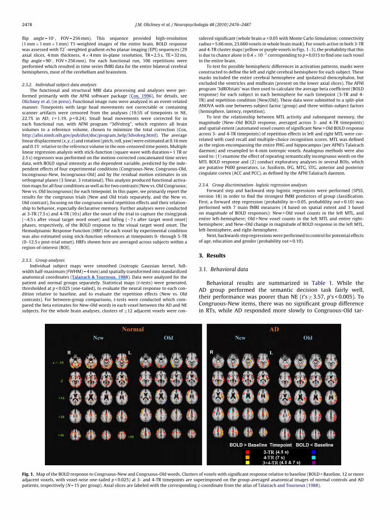
atient and normal groups separately. Statistical maps (t-tests) were generated,hresholded at p < 0.025 (one-tailed), to evaluate the neural response to each con-ition relative to baseline, and to evaluate the repetition effects (New vs. Oldontrasts). For between-group comparisons, t-tests were conducted which com-ared the beta estimates for New-Old words in each voxel between the AD and NEubjects. For the whole brain analyses, clusters of ≥12 adjacent voxels were con-ig. 1. Map of the BOLD response to Congruous-New and Congruous-Old words. Clustersdjacent voxels, with voxel-wise one-tailed p < 0.025) at 3- and 4-TR timepoints are supatients, respectively (N = 15 per group). Axial slices are labeled with the corresponding z
logia 48 (2010) 2476–2487
sidered significant (whole brain ˛ < 0.05 with Monte Carlo Simulation; connectivityradius = 5.66 mm, 23,660 voxels in whole brain mask). For voxels active in both 3-TRand 4-TR cluster maps (yellow or purple voxels in Figs. 1–3), the probability that thisis due to chance alone is 0.4 × 10⁻⁷ corresponding to p = 0.015 to find one such voxelin the entire brain.
To test for possible hemispheric differences in activation patterns, masks wereconstructed to define the left and right cerebral hemisphere for each subject. Thesemasks included the entire cerebral hemisphere and ipsilateral diencephalon, butexcluded the ventricles and midbrain (present on the lower axial slices). The AFNIprogram ‘3dROIstats’ was then used to calculate the average beta coefficient (BOLDresponse) for each subject in each hemisphere for each timepoint (3-TR and 4-TR) and repetition condition (New/Old). These data were submitted to a split-plotANOVA with one between-subject factor (group) and three within-subject factors(hemisphere, latency, repetition).
To test the relationship between MTL activity and subsequent memory, themagnitude (New–Old BOLD response, averaged across 3- and 4-TR timepoints)and spatial extent (automated voxel counts of significant New > Old BOLD responseacross 3- and 4-TR timepoints) of repetition effects in left and right MTL were cor-related with cued recall and multiple-choice recognition scores. MTL was definedas the region encompassing the entire PHG and hippocampus (per AFNI’s Talairachdaemon) and resampled to 4-mm isotropic voxels. Analogous methods were alsoused to: (1) examine the effect of repeating semantically incongruous words on theMTL BOLD response and (2) conduct exploratory analyses in several ROIs, whichare putative P600 generators, i.e. fusiform, IFG, MTG, STG, anterior and posteriorcingulate cortex (ACC and PCC), as defined by the AFNI Talairach daemon.
2.3.4. Group discrimination- logistic regression analysesForward step and backward step logistic regressions were performed (SPSS,
version 18) in order to find the strongest fMRI predictors of group classification.First, a forward step regression (probability in = 0.05, probablility out = 0.10) wasperformed with 7 main fMRI measures (4 based on spatial extent and 3 basedon magnitude of BOLD responses): New > Old voxel counts in the left MTL, andentire left-hemisphere; Old > New voxel counts in the left MTL and entire right-hemisphere; and New–Old change in magnitude of BOLD response in the left MTL,left-hemisphere, and right-hemisphere.
Next, backwards step regressions were performed to control for potential effectsof age, education and gender (probability out = 0.10).
3. Results
3.1. Behavioral data
Behavioral results are summarized in Table 1. While theAD group performed the semantic decision task fairly well,their performance was poorer than NE (t’s ≥ 3.57, p’s < 0.005). ToCongruous-New items, there was no significant group differencein RTs, while AD responded more slowly to Congruous-Old tar-
of voxels with significant response relative to baseline (BOLD > Baseline, 12 or moreerimposed on the group-averaged anatomical images of normal controls and AD-coordinate from the atlas of Talairach and Tournoux (1988).
J.M. Olichney et al. / Neuropsychologia 48 (2010) 2476–2487 2479
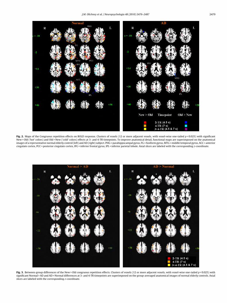
Fig. 2. Maps of the Congruous repetition effects on BOLD response. Clusters of voxels (12 or more adjacent voxels, with voxel-wise one-tailed p < 0.025) with significantNew > Old (‘hot’ colors) and Old > New (‘cold’ colors) effects at 3- and 4-TR timepoints. To improve anatomical detail, functional maps are superimposed on the anatomicalimages of a representative normal elderly control (left) and AD (right) subject. PHG = parahippocampal gyrus, FG = fusiform gyrus, MTG = middle temporal gyrus, ACC = anteriorcingulate cortex, PCC = posterior cingulate cortex, IFG = inferior frontal gyrus, IPL = inferior parietal lobule. Axial slices are labeled with the corresponding z-coordinate.
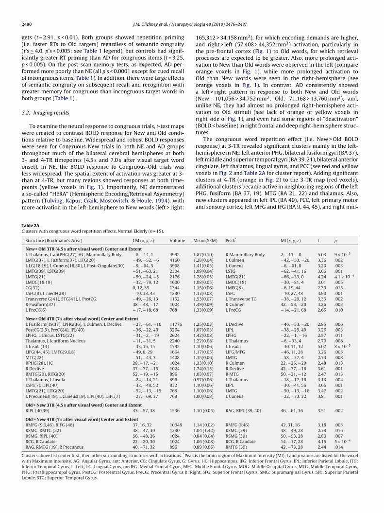
Fig. 3. Between-group differences of the New > Old congruous repetition effects. Clusters of voxels (12 or more adjacent voxels, with voxel-wise one-tailed p < 0.025) withsignificant Normal > AD and AD > Normal differences at 3- and 4-TR timepoints are superimposed on the group-averaged anatomical images of normal elderly controls. Axialslices are labeled with the corresponding z-coordinate.
2 sycho
g((ipfoogb
3
wtwt3oltpapm
TC
CwIPL
480 J.M. Olichney et al. / Neurop
ets (t = 2.91, p < 0.01). Both groups showed repetition primingi.e. faster RTs to Old targets) regardless of semantic congruityt’s ≥ 4.0, p’s < 0.005; see Table 1 legend), but controls had signif-cantly greater RT priming than AD for congruous items (t = 3.25,< 0.005). On the post-scan memory tests, as expected, AD per-
ormed more poorly than NE (all p’s < 0.0001 except for cued recallf incongruous items, Table 1). In addition, there were large effectsf semantic congruity on subsequent recall and recognition withreater memory for congruous than incongruous target words inoth groups (Table 1).
.2. Imaging results
To examine the neural response to congruous trials, t-test mapsere created to contrast BOLD response for New and Old condi-
ions relative to baseline. Widespread and robust BOLD responsesere seen for Congruous-New trials in both NE and AD groups
hroughout much of the bilateral cerebral hemispheres at both- and 4-TR timepoints (4.5 s and 7.0 s after visual target wordnset). In NE, the BOLD response to Congruous-Old trials wasess widespread. The spatial extent of activation was greater at 3-
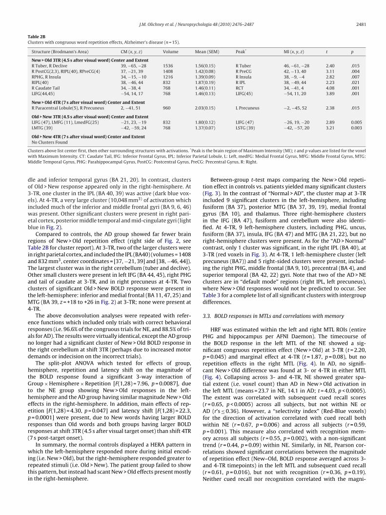
han at 4-TR, but many regions showed responses at both time-oints (yellow voxels in Fig. 1). Importantly, NE demonstratedso-called “HERA” (Hemispheric Encoding/Retrieval Asymmetry)attern (Tulving, Kapur, Craik, Moscovitch, & Houle, 1994), withore activation in the left-hemisphere to New words (left > right:able 2Alusters with congruous word repetition effects, Normal Elderly (n = 15).
Structure (Brodmann’s Area) CM (x, y, z) Volume M
New > Old 3TR (4.5 s after visual word) Center and ExtentL Thalamus, L antPHG(27), HC, Mammillary Body −8, −14, 1 4992 1LMTG(37), L Fusiform(37), LITG(20) −49, −52, −6 4160 1L LG(18,19), L Cuneus(18,30), L Post. Cingulate(30) −9, −64, 5 3968 1LMTG(39), LSTG(39) −51, −63, 21 2304 1LMTG(21) −59, −24, −5 2176 1LMOG(18,19) −32, −79, 12 1600 1CG(32) 0, 12, 39 1344 1LSFG(8), L medFG(8) −10, 33, 43 1280 1Transverse G(41), STG(41), L PostCG, −49, −26, 13 1152 1R Fusiform(37) 38, −48, −17 1024 1L PreCG(6) −17, −18, 68 768 1
New > Old 4TR (7 s after visual word) Center and ExtentL Fusiform(19,37), LPHG(36), L Culmen, L Declive −27, −61, −10 11776 1PostCG(2,3), PreCG(4), IPL(40) −36, −22, 40 3264 1LPHG, L Uncus, LSTG(22) −31, −2, −19 2624 1Thalamus, L lentiform Nucleus −11, −31, 5 2240 1L Insula(13) −33, 15, 15 1792 1LIFG(44, 45), LMFG(9,6,8) −49, 8, 29 1664 1MTG(22) −51, −44, 3 1408 1RPHG(28), HC 28, −17, −21 1024 1R Declive 37, −77, −15 1024 1RMTG(20), RITG(20) 52, −19, −15 896 1L Thalamus, L Insula −24, −14, 21 896 0LSPL(7), LIPL(40) −32, −48, 52 832 1LMTG(21), LITG(20) −52, −13, −15 768 1L Precuneus(19), L Cuneus(19), LIPL(40), LSPL(7) −27, −69, 37 768 1
Old > New 3TR (4.5 s after visual word) Center and ExtentRIPL (40,39) 43, −57, 38 1536 1
Old > New 4TR (7 s after visual word) Center and ExtentRMFG (9,6,46), RIFG (46) 37, 16, 32 10048 1RSMG, RMTG (22) 38, −47, 30 1280 1RSMG, RIPL (40) 56, −48, 26 1024 0RCG, R Caudate 22, −20, 30 1024 1RAG, RMTG (19), R Precuneus 40, −71, 32 896 0
lusters above list center first, then other surrounding structures with activations. *Peak iith Maximum Intensity. AG: Angular Gyrus, ant: Anterior, CG: Cingulate Gyrus, G: Gyru
nferior Temporal Gyrus, L: Left„ LG: Lingual Gyrus, medFG: Medial Frontal Gyrus, MFG: MHG: Parahippocampal Gyrus, PostCG: Postcentral Gyrus, PreCG: Precentral Gyrus R: Rigobule, STG: Superior Temporal Gyrus.
logia 48 (2010) 2476–2487
165,312 > 34,158 mm3), for which encoding demands are higher,and right > left (57,408 > 44,352 mm3) activation, particularly inthe pre-frontal cortex (Fig. 1) to Old words, for which retrievalprocesses are expected to be greater. Also, more prolonged acti-vation to New than Old words were observed in the left (compareorange voxels in Fig. 1), while more prolonged activation toOld than New words were seen in the right-hemisphere (seeorange voxels in Fig. 1). In contrast, AD consistently showeda left > right pattern in response to both New and Old words(New: 101,056 > 34,752 mm3; Old: 71,168 > 13,760 mm3), and,unlike NE, they had almost no prolonged right-hemisphere acti-vation to Old stimuli (see lack of orange or yellow voxels inright side of Fig. 1), and even had some regions of “deactivation”(BOLD < baseline) in right frontal and deep right-hemisphere struc-tures.
The congruous word repetition effect (i.e. New > Old BOLDresponse) at 3-TR revealed significant clusters mainly in the left-hemisphere in NE: left anterior PHG, bilateral fusiform gyri (BA 37),left middle and superior temporal gyri (BA 39, 21), bilateral anteriorcingulate, left thalamus, lingual gyrus, and PCC (see red and yellowvoxels in Fig. 2 and Table 2A for cluster report). Adding significant
clusters at 4-TR (orange in Fig. 2) to the 3-TR map (red voxels),additional clusters became active in neighboring regions of the leftPHG, fusiform (BA 37, 19), MTG (BA 21, 22) and thalamus. Also,new clusters appeared in left IPL (BA 40), PCC, left primary motorand sensory cortex, left MFG and IFG (BA 9, 44, 45), and right mid-ean (SEM) Peak* MI (x, y, z) t p
.87(0.10) R Mammillary Body 2, −13, −8 5.03 9 × 10⁻⁵
.28(0.04) L Culmen −42, −53, −20 3.36 .002
.41(0.05) L Cuneus −6, −61, 8 3.20 .003
.09(0.04) LSTG −62, −41, 16 3.66 .001
.28(0.05) LMTG(21) −66, −33, 0 4.24 4.1 × 10⁻⁴
.08(0.05) LMOG(18) −30, −81, 4 3.01 .005
.15(0.06) LMFG(8) −6, 19, 44 2.39 .015
.33(0.08) LSFG −10, 27, 48 3.90 .001
.53(0.07) L Transverse TG −38, −29, 12 3.35 .002
.49(0.09) R Culmen 42, −53, −20 3.26 .003
.33(0.09) L PreCG −14, −21, 68 2.65 .010
.25(0.03) L Declive −46, −53, −20 2.85 .006
.07(0.03) LIPL −38, −29, 40 3.26 .003
.42(0.08) LPHG −22, −1, −16 2.57 .011
.22(0.08) L Thalamus −6, −33, 4 2.70 .008
.10(0.06) L Insula −30, 11, 12 5.07 8 × 10⁻⁵
.17(0.05) LIFG/MFG −46, 11, 28 3.26 .003
.15(0.06) LMTG −58, −37, 4 2.73 .008
.33(0.10) R Culmen 22, −25, −20 2.48 .013
.74(0.15) R Declive 42, −77, −16 3.61 .001
.03(0.07) R MTG 50, −21, −12 2.47 .013
.97(0.06) L Thalamus −18, −17, 16 3.13 .004
.10(0.06) LIPL −30, −41, 56 3.66 .001
.10(0.06) LMTG −50, −13, −16 3.47 .002
.00(0.08) L Cuneus −22, −73, 32 3.81 .001
.10 (0.05) RAG, RIPL (39, 40) 46, −61, 36 3.51 .002
.14 (0.02) RMFG (R46) 42, 31, 16 3.18 .003
.04 (1.42) RSMG (39) 38, −49, 28 2.38 .016
.84 (0.04) RSMG (39) 50, −53, 28 2.80 .007
.06 (0.08) RCG, R Caudate 14, −17, 28 4.15 5 × 10⁻⁴
.89 (0.06) RMTG (39) 42, −73, 28 2.44 .014
s the brain region of Maximum Intensity (MI); t and p values are listed for the voxels, HC: Hippocampus, IFG: Inferior Frontal Gyrus, IPL: Inferior Parietal Lobule, ITG:iddle Frontal Gyrus, MOG: Middle Occipital Gyrus, MTG: Middle Temporal Gyrus,
ht, SFG: Superior Frontal Gyrus, SMG: Supramarginal Gyrus, SPL: Superior Parietal
J.M. Olichney et al. / Neuropsychologia 48 (2010) 2476–2487 2481
Table 2BClusters with congruous word repetition effects, Alzheimer’s disease (n = 15).
Structure (Brodmann’s Area) CM (x, y, z) Volume Mean (SEM) Peak* MI (x, y, z) t p
New > Old 3TR (4.5 s after visual word) Center and ExtentR Tuber, R Declive 39, −65, −28 1536 1.56(0.15) R Tuber 46, −61, −28 2.40 .015R PostCG(2,3), RIPL(40), RPreCG(4) 37, −21, 39 1408 1.42(0.08) R PreCG 42, −13, 40 3.11 .004RPHG, R Insula 34, −15, −10 1216 1.39(0.09) R Insula 38, −9, −4 2.82 .007RIPL(40) 38, −46, 44 832 1.87(0.19) R IPL 38, −49, 44 2.23 .021R Caudate Tail 34, −38, 4 768 1.46(0.11) RCT 34, −41, 4 4.08 .001LIFG(44,45) −54, 14, 17 768 1.46(0.13) LIFG(45) −54, 11, 20 3.89 .001
New > Old 4TR (7 s after visual word) Center and ExtentR Paracentral Lobule(5), R Precuneus 2, −41, 51 960 2.03(0.15) L Precuneus −2, −45, 52 2.38 .015
Old > New 3TR (4.5 s after visual word) Center and ExtentLIFG (47), LMFG (11), LmedFG(25) −21, 23, −19 832 1.80(0.12) LIFG (47) −26, 19, −20 2.89 0.005LMTG (39) −42, −59, 24 768 1.37(0.07) LSTG (39) −42, −57, 20 3.21 0.003
Old > New 4TR (7 s after visual word) Center and ExtentNo Clusters Found
C Peak iw ParietM PreCG
do3eiweb
rTiaTOactM4
erantd
htGtheeprr(
wirti
lusters above list center first, then other surrounding structures with activations. *
ith Maximum Intensity. CT: Caudate Tail, IFG: Inferior Frontal Gyrus, IPL: Inferioriddle Temporal Gyrus, PHG: Parahippocampal Gyrus, PostCG: Postcentral Gyrus,
le and inferior temporal gyrus (BA 21, 20). In contrast, clustersf Old > New response appeared only in the right-hemisphere. At-TR, one cluster in the IPL (BA 40, 39) was active (dark blue vox-ls). At 4-TR, a very large cluster (10,048 mm3) of activation whichncluded much of the inferior and middle frontal gyri (BA 9, 6, 46)
as present. Other significant clusters were present in right pari-tal cortex, posterior middle temporal and mid-cingulate gyri (lightlue in Fig. 2).
Compared to controls, the AD group showed far fewer brainegions of New > Old repetition effect (right side of Fig. 2, seeable 2B for cluster report). At 3-TR, two of the larger clusters weren right parietal cortex, and included the IPL (BA40) (volumes = 1408nd 832 mm3, center coordinates = [37, −21, 39] and [38, −46, 44]).he largest cluster was in the right cerebellum (tuber and declive).ther small clusters were present in left IFG (BA 44, 45), right PHGnd tail of caudate at 3-TR, and in right precuneus at 4-TR. Twolusters of significant Old > New BOLD response were present inhe left-hemisphere: inferior and medial frontal (BA 11, 47, 25) and
TG (BA 39, z = +18 to +26 in Fig. 2) at 3-TR; none were present at-TR.
The above deconvolution analyses were repeated with refer-nce functions which included only trials with correct behavioralesponses (i.e. 96.6% of the congruous trials for NE, and 88.5% of tri-ls for AD). The results were virtually identical, except the AD groupo longer had a significant cluster of New > Old BOLD response inhe right cerebellum at shift 3TR (perhaps due to increased motoremands or indecision on the incorrect trials).
The split-plot ANOVA which tested for effects of group,emisphere, repetition and latency shift on the magnitude ofhe BOLD response found a significant 3-way interaction ofroup × Hemisphere × Repetition [F(1,28) = 7.96, p = 0.0087], due
o the NE group showing New > Old responses in the left-emisphere and the AD group having similar magnitude New > Oldffects in the right-hemisphere. In addition, main effects of rep-tition [F(1,28) = 4.30, p = 0.047] and latency shift [F(1,28) = 22.3,= 0.0001] were present, due to New words having larger BOLD
esponses than Old words and both groups having larger BOLDesponses at shift 3TR (4.5 s after visual target onset) than shift 4TR7 s post-target onset).
In summary, the normal controls displayed a HERA pattern in
hich the left-hemisphere responded more during initial encod-ng (i.e. New > Old), but the right-hemisphere responded greater toepeated stimuli (i.e. Old > New). The patient group failed to showhis pattern, but instead had scant New > Old effects present mostlyn the right-hemisphere.
s the brain region of Maximum Intensity (MI); t and p values are listed for the voxelal Lobule, L: Left, medFG: Medial Frontal Gyrus, MFG: Middle Frontal Gyrus, MTG:
: Precentral Gyrus, R: Right.
Between-group t-test maps comparing the New > Old repeti-tion effect in controls vs. patients yielded many significant clusters(Fig. 3). In the contrast of “Normal > AD”, the cluster map at 3-TRincluded 9 significant clusters in the left-hemisphere, includingfusiform (BA 37), posterior MTG (BA 37, 39, 19), medial frontalgyrus (BA 10), and thalamus. Three right-hemisphere clustersin the IFG (BA 47), fusiform and cerebellum were also identi-fied. At 4-TR, 9 left-hemisphere clusters, including PHG, uncus,fusiform (BA 37), insula, IFG (BA 47) and MTG (BA 21, 22), but noright-hemisphere clusters were present. As for the “AD > Normal”contrast, only 1 cluster was significant, in the right IPL (BA 40), at3-TR (red voxels in Fig. 3). At 4-TR, 1 left-hemisphere cluster (leftprecuneus (BA7)) and 5 right-sided clusters were present, includ-ing the right PHG, middle frontal (BA 9, 10), precentral (BA 4), andsuperior temporal (BA 42, 22) gyri. Note that two of the AD > NEclusters are in “default mode” regions (right IPL, left precuneus),where New > Old responses would not be predicted to occur. SeeTable 3 for a complete list of all significant clusters with intergroupdifferences.
3.3. BOLD responses in MTLs and correlations with memory
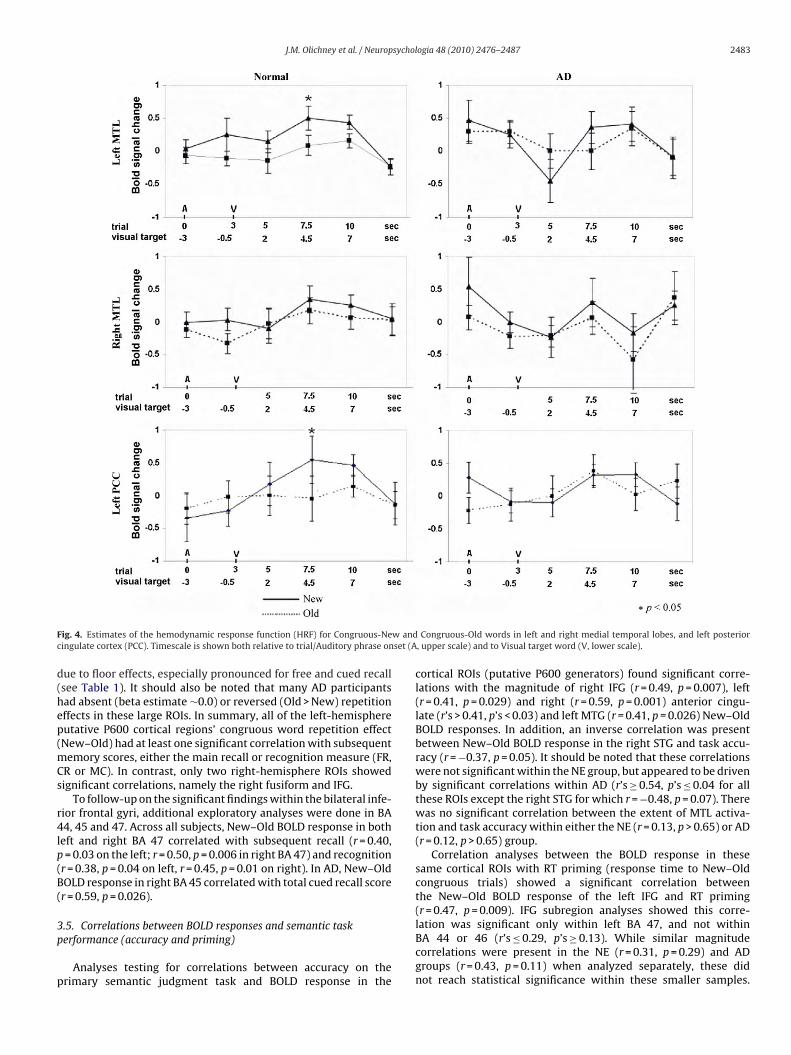
HRF was estimated within the left and right MTL ROIs (entirePHG and hippocampus per AFNI Daemon). The timecourse ofthe BOLD response in the left MTL of the NE showed a sig-nificant congruous repetition effect (New > Old) at 3-TR (t = 2.20,p = 0.045) and marginal effect at 4-TR (t = 1.87, p = 0.08), but norepetition effects in the right MTL (Fig. 4). In AD, no signifi-cant New > Old difference was found at 3- or 4-TR in either MTL(Fig. 4). Collapsing across 3- and 4-TR, NE showed greater spa-tial extent (i.e. voxel count) than AD in New > Old activation inthe left MTL (means = 23.7 in NE, 14.1 in AD; t = 4.03, p < 0.0005).The extent was correlated with subsequent cued recall scores(r = 0.65, p < 0.0005) across all subjects, but not within NE orAD (r’s ≤ 0.36). However, a “selectivity index” (Red-Blue voxels)for the direction of activation correlated with cued recall bothwithin NE (r = 0.67, p = 0.006) and across all subjects (r = 0.59,p = 0.001). This measure also correlated with recognition mem-ory across all subjects (r = 0.55, p = 0.002), with a non-significanttrend (r = 0.44, p = 0.09) within NE. Similarly, in NE, Pearson cor-
relations showed significant correlations between the magnitudeof repetition effect (New–Old, BOLD response averaged across 3-and 4-TR timepoints) in the left MTL and subsequent cued recall(r = 0.61, p = 0.016), but not with recognition (r = 0.36, p = 0.19).Neither cued recall nor recognition correlated with the magni-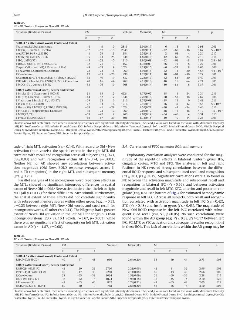
2482 J.M. Olichney et al. / Neuropsychologia 48 (2010) 2476–2487
Table 3ANE > AD Clusters, Congruous New–Old Words.
Structure (Brodmann’s area) CM Volume Mean (SE) MI t p
x y z x y z
3-TR (4.5 s after visual word), Center and ExtentThalamus, L Subthalamic nuc. −4 −9 0 2816 3.01(0.17) 6 −13 −8 2.98 .003L FG(37), L Culmen, L Declive −32 −57 −19 2048 2.09(0.11) −22 −65 −16 3.67 5 × 10⁻⁴
medFG(10, 9)(R > L), R SFG −1 59 13 1984 2.54(0.11) −2 63 4 2.24 .017L MTG(39), LSTG(22), L SG −35 −63 25 1344 1.85(0.10) −42 −65 24 2.18 .018L ITG, L MTG(37) −45 −52 −5 1216 1.86(0.08) −42 −61 −8 3.89 2.8 × 10⁻⁴
L OLG, L IOG(18, 19), L MOG, L FG −32 −71 −3 1152 1.78(0.09) −26 −77 −8 3.27 .001Corpus Callosum(L > R), L Pulvinar, L PHG −8 −33 9 1024 2.28(0.15) −6 −37 8 2.65 .006L Thalamus, L Claustrum, L Caudate −22 −23 16 960 1.91(0.16) −22 −13 20 4.58 4.4 × 10⁻⁵
R Cerebellum 17 −63 −20 896 1.75(0.11) 10 −61 −16 3.27 .001R Culmen, R FG(37), R Declive, R Tuber, R ITG(20) 38 −49 −19 832 2.28(0.17) 42 −53 −20 3.49 .001R IFG(47), R Insula(13), R STG(38, 22), R Claustrum 40 16 −4 768 2.15(0.10) 46 15 −4 2.74 .005L MOG(19), L Cuneus, L MTG −33 −76 10 768 1.94(0.14) −30 −81 8 3.37 .001
4TR (7 s after visual word), Center and ExtentL Insula(13), L Claustrum, L IFG(45) −31 13 15 4224 1.77(0.05) −18 −1 24 2.24 .016L FG (37), L Declive, L Culmen, L ITG −44 −52 −17 1344 2.20(0.16) −50 −53 −16 2.67 .006L Claustrum, L Insula (13), L IFG(47) −29 22 0 1216 1.79(0.08) −38 19 0 2.42 .011L Insula (13), L Caudate −27 −34 16 1216 2.10(0.10) −26 −37 12 3.55 5 × 10⁻⁴
L Uncus(28), L MTG(21), L STG, L PHG(38) −33 −1 −28 1024 2.55(0.27) −30 −1 −24 2.46 .010L PHG(36), L Hippocampus, L Caudate −38 −23 −12 960 2.01(0.12) −38 −17 −16 4.12 1.5 × 10⁻⁴
L MTG(22, 21) −53 −47 2 960 2.02(0.14) −54 −45 4 3.31 .015L PreCG(4), L PostCG(3) −35 −17 39 896 1.72(0.15) −30 −9 44 3.26 .001
Clusters above list center first, then other surrounding structures with significant intensity differences. The t and p values are listed for the voxel with Maximum Intensity( G: InfG ampaF
tacpNta(
teMtwpieite
TA
C(P
MI). FG: Fusiform Gyrus, IFG: Inferior Frontal Gyrus, IOG: Inferior Occipital Gyrus, ITyrus, MTG: Middle Temporal Gyrus, OLG: Occipital Lingual Gyrus, PHG: Parahippocrontal Gyrus, SG: Superior Gyrus, STG: Superior Temporal Gyrus.
ude of right MTL activation (r’s ≤ 0.14). With regard to Old > Newctivation (blue voxels), the spatial extent in the right MTL didorrelate with recall and recognition across all subjects (r’s ≥ 0.41,’s ≤ 0.03) and with recognition within AD (r = 0.74, p = 0.003).either NE nor AD showed any correlations between activa-
ion magnitude (Old–New, BOLD response averaged across 3-nd 4-TR timepoints) in the right MTL and subsequent memory|r’s| ≤ 0.25).
Parallel analyses of the incongruous word repetition effects inhe MTLs showed no significant intergroup differences in spatialxtent of New > Old or Old > New activation in either the left or rightTL (all p’s > 0.17) for these difficult to learn stimuli. Furthermore,
he spatial extent of these effects did not correlate significantlyith subsequent memory scores within either group (e.g. r = 0.33,= 0.23 between right MTL New > Old words and cued recall for
ncongruous words, all other r’s < 0.33). The NE group had a greaterxtent of New > Old activation in the left MTL for congruous thanncongruous items (23.7 vs. 16.1 voxels, t = 3.67, p = 0.003), whilehere was no significant effect of congruity on left MTL activationxtent in AD (t = −1.87, p = 0.08).
able 3BD > NE Clusters, Congruous New–Old Words.
Structure (Brodmann’s area) CM Volume
x y z
3-TR (4.5 s after visual word), Center and ExtentR IPL(40), R SPL(7) 40 −47 43 960
4TR (7 s after visual word), Center and ExtentR MFG(9, 46), R IFG 41 20 29 5760PreCG(4), R PostCG(3, 2) 46 −17 38 2240R Cerebellum 28 −65 −39 1024R LG(19), R FG(37) 32 −52 −5 1024L Precuneus(7) −2 −63 40 832R STG(42, 22), R TTG(41) 60 −20 9 768
lusters above list center first, then other surrounding structures with significant intensMI). FG: Fusiform Gyrus, IFG: Inferior Frontal Gyrus, IPL: Inferior Parietal Lobule, L: Left, LGostcentral Gyrus, PreCG: Precentral Gyrus, R: Right,: Superior Parietal Lobule, STG: Supe
erior Temporal Gyrus, L: Left, medFG: Medial Frontal Gyrus, MOG: Middle Occipitall Gyrus, PostCG: Postcentral Gyrus, PreCG: Precentral Gyrus, R: Right, SFG: Superior
3.4. Correlations of P600 generator ROIs with memory
Exploratory correlation analyses were conducted for the mag-nitude of the repetition effects in bilateral fusiform gyrus, IFG,cingulate cortex, MTG and STG. The analyses in left and rightfusiform in NE revealed strong correlations between the differ-ential BOLD response and subsequent cued recall and recognition(r’s ≥ 0.61, p’s ≤ 0.015). Significant correlations were also found inNE between the activation magnitude and subsequent recall andrecognition in bilateral IFG (r’s ≥ 0.56), and between activationmagnitude and recall in left MTG, STG, anterior and posterior cin-gulate (r’s ≥ 0.51, see bottom of Fig. 4 for estimated hemodynamicresponse in left PCC). Across all subjects, both recall and recogni-tion correlated with activation magnitude in left IFG (r’s ≥ 0.42),STG (r’s ≥ 0.48) and fusiform gyrus (r’s ≥ 0.43). The magnitude of
New–Old BOLD response in the left PCC correlated with subse-quent cued recall (r = 0.51, p = 0.05). No such correlations werefound within the AD group (e.g. r’s ≤ 0.38, p’s > 0.17 between leftIFG, FG, MTG or STG activation and subsequent recall or recognition)in these ROIs. This lack of correlations within the AD group may beMean (SE) MI t p
x Y z
2.64(0.20) 38 −49 44 2.73 .005
2.28(0.06) 42 11 36 2.96 .0032.11(0.08) 46 −13 40 2.66 .0061.51(0.09) 26 −69 −36 2.28 .0151.95(0.18) 30 −45 −4 2.10 .0222.74(0.21) −2 −65 44 2.05 .0242.63(0.20) 58 −25 8 3.10 .002
ity differences. The t and p values are listed for the voxel with Maximum Intensity: Lingual Gyrus, MFG: Middle Frontal Gyrus, PHG: Parahippocampal Gyrus, PostCG:
rior Temporal Gyrus, TTG: Transverse Temporal Gyrus.
J.M. Olichney et al. / Neuropsychologia 48 (2010) 2476–2487 2483
F w andc set (A
d(hep(mCs
r4lp(B(
3p
p
ig. 4. Estimates of the hemodynamic response function (HRF) for Congruous-Neingulate cortex (PCC). Timescale is shown both relative to trial/Auditory phrase on
ue to floor effects, especially pronounced for free and cued recallsee Table 1). It should also be noted that many AD participantsad absent (beta estimate ∼0.0) or reversed (Old > New) repetitionffects in these large ROIs. In summary, all of the left-hemisphereutative P600 cortical regions’ congruous word repetition effectNew–Old) had at least one significant correlation with subsequent
emory scores, either the main recall or recognition measure (FR,R or MC). In contrast, only two right-hemisphere ROIs showedignificant correlations, namely the right fusiform and IFG.
To follow-up on the significant findings within the bilateral infe-ior frontal gyri, additional exploratory analyses were done in BA4, 45 and 47. Across all subjects, New–Old BOLD response in both
eft and right BA 47 correlated with subsequent recall (r = 0.40,= 0.03 on the left; r = 0.50, p = 0.006 in right BA 47) and recognition
r = 0.38, p = 0.04 on left, r = 0.45, p = 0.01 on right). In AD, New–OldOLD response in right BA 45 correlated with total cued recall scorer = 0.59, p = 0.026).
.5. Correlations between BOLD responses and semantic taskerformance (accuracy and priming)
Analyses testing for correlations between accuracy on therimary semantic judgment task and BOLD response in the
Congruous-Old words in left and right medial temporal lobes, and left posterior, upper scale) and to Visual target word (V, lower scale).
cortical ROIs (putative P600 generators) found significant corre-lations with the magnitude of right IFG (r = 0.49, p = 0.007), left(r = 0.41, p = 0.029) and right (r = 0.59, p = 0.001) anterior cingu-late (r’s > 0.41, p’s < 0.03) and left MTG (r = 0.41, p = 0.026) New–OldBOLD responses. In addition, an inverse correlation was presentbetween New–Old BOLD response in the right STG and task accu-racy (r = −0.37, p = 0.05). It should be noted that these correlationswere not significant within the NE group, but appeared to be drivenby significant correlations within AD (r’s ≥ 0.54, p’s ≤ 0.04 for allthese ROIs except the right STG for which r = −0.48, p = 0.07). Therewas no significant correlation between the extent of MTL activa-tion and task accuracy within either the NE (r = 0.13, p > 0.65) or AD(r = 0.12, p > 0.65) group.
Correlation analyses between the BOLD response in thesesame cortical ROIs with RT priming (response time to New–Oldcongruous trials) showed a significant correlation betweenthe New–Old BOLD response of the left IFG and RT priming(r = 0.47, p = 0.009). IFG subregion analyses showed this corre-
lation was significant only within left BA 47, and not withinBA 44 or 46 (r’s ≤ 0.29, p’s ≥ 0.13). While similar magnitudecorrelations were present in the NE (r = 0.31, p = 0.29) and ADgroups (r = 0.43, p = 0.11) when analyzed separately, these didnot reach statistical significance within these smaller samples.
2484 J.M. Olichney et al. / Neuropsycho
Table 4Logistic regression model to discriminate AD and NE groups.
B SE Wald Sig Exp (B)
L. HemisphereNew > Old voxels
−0.23 0.011 4.714 0.03 .977
L. MTL New > Oldvoxels
−0.242 0.135 3.212 0.073 .785
R. Hemisphere BOLDresponse (New–Old)
2.709 1.332 4.133 0.042 15.008
CW
Np
3
edv((((st
4
wobdpieoMa
ai(mrcee(afttatsaeMoe(i
Constant 16.798 6.854 6.007 0.014 1.973 × 10⁷
utoff for group classification: 0.500 (1 = AD, 0 = NE). B = Slope. SE = Standard Error.ald: Wald statistic. Exp (B) = Odds ratio.
one of the other ROIs showed significant correlations with RTriming.
.6. Group discrimination
The forward step and backward step logistic regression mod-ls converged on a very highly significant model (chi-square = 29.2,f = 3, p < 0.0001) in which lower left-hemisphere New > Oldoxel counts (B = −.023), lower left MTL New > Old voxel countsB = −.242) and larger right-hemisphere New–Old BOLD responseB = 2.71) were associated with increased likelihood of having ADsee Table 4). This model classified 93.3% of subjects correctly14/15 in each group). Adding the demographic variables of age,ex and education did not produce any significant improvementso this model.
. Discussion
During a semantic judgment task, normal elderly showedidespread New > Old BOLD response in the left-hemisphere. Many
f these clusters were in regions known to produce P600-likerain potentials (left PHG, cingulate, MTG, IFG). Converging evi-ence suggests that this circuit of interconnected P600 generators isarticularly important for successful encoding and “memory bind-
ng” (Kahn et al., 2005; Kirchhoff et al., 2000; Wagner, Desmond,t al., 1998; Wagner, 1999). The magnitude and spatial extentf New > Old BOLD responses in several of these regions (leftTL/PHG, PCC, IFG, fusiform) correlated with subsequent recall
nd/or recognition.The main abnormal findings in AD were: (1) MTL failure, with
severe loss of New > Old BOLD response in the left MTL, whichs the more relevant hemisphere for learning verbal materials;2) Widespread left-hemisphere dysfunction, not an overall decre-
ent of BOLD response in AD, but a selective loss of New > Oldepetition effects in this hemisphere. Since these same effectsorrelated strongly with verbal declarative memory in normallderly, it is likely that this loss of left-hemisphere New > Oldffects in AD may account for their dense verbal memory deficits;3) Some evidence of right-hemisphere dysfunction as well, with
loss of the Old > New effects seen in right parietal and pre-rontal regions of the normal elderly. It should also be notedhat a few right-hemispheric ROIs, (i.e. fusiform and BA 47 inhe inferior frontal gyrus) likely related to semantic processing,lso showed New > Old effects in the normal elderly and thathese effects also correlated with memory for the experimentaltimuli. Our logistic regression analyses achieved excellent sep-ration (93.3% sensitivity and specificity) of the AD and normallderly groups, using fMRI variables which quantify the main
TL and hemispheric responses noted above. This is in line withur prior ERP studies of mild AD with this paradigm (Olichneyt al., 2006), which also achieved excellent group discrimination100% sensitivity, 82% specificity). This range of discriminabil-ty is consistent with a potentially useful biomarker for AD (The
logia 48 (2010) 2476–2487
Ronald and Nancy Reagan Research Institute & NIA Working Group,1998).
This fMRI word repetition paradigm involves attention, per-ceptual, conceptual and episodic memory processes and requiresa motor response. As such, it produced widespread activation ofthe cerebral cortex (sensory, association, paralimbic, limbic andmotor cortex), which was generally more pronounced for the novelstimuli (Fig. 1). One disadvantage of using a complex cognitivetask such as category judgment, along with cross-modal stimuli,is that it is difficult to isolate the specific cognitive processes per-formed by a given anatomical structure. However, when New vs.Old congruous words contrasts were made at timepoints chosento model the BOLD response to the visual target words, a neuralcircuit of interconnected structures thought important for verbalmemory emerged in the NE activation maps, as discussed in detailin Results above. As hypothesized, the magnitude of the BOLDresponse to New–Old congruous words in several ROIs (putativeP600 generators) correlated with declarative memory (both recalland recognition) for the verbal stimuli.
In contrast, the degree of activation (New vs. Old BOLD response)in the MTLs when semantically incongruous words were repeateddid not correlate with any of our memory measures (neither withtotal recall or recognition scores for incongruous or all words).
In contrast to normal elderly, the AD group showed little evi-dence of New > Old BOLD responses in the left-hemisphere and ageneralized decrease in fMRI repetition effects. Our results resem-ble those of Golby et al. (2005), who found reduced activation, fornovel vs. repeated visual scenes (color photographs), along the ven-tral visual stream, with most marked decrements in the MTL andfusiform regions. It should be noted that our results were not dueto a general failure of cognitive activation, or an inadequate signal-to-noise ratio in AD, but rather a relatively selective decrementin New/Old effects. The spatial extent of left-hemisphere activa-tion was similar in AD and NE when collapsed across New and Oldwords, but as hypothesized, the AD group showed a selective lossof New > Old effects in this hemisphere. Old words elicited simi-lar spatial extents of activation in both groups but with differenthemispheric patterns (right > left in NE, left > right in AD), as will bediscussed further below.
Relatively few prior fMRI studies of AD have used purely verbalstimuli (Lustig & Buckner, 2004) and we are not aware of any priorpublished studies which used cross-modal audio–visual stimuli toprobe incidental learning in AD. The use of multi-modal stimuliwith integrative tasks such as semantic judgment may be advanta-geous in producing activation of higher association cortex, whichis a predilection site for AD pathology relative to primary sensoryand motor areas (Arnold, Hyman, Flory, Damasio, & van Hoesen,1991; Braak & Braak, 1991). Remy et al. (2005), in a block designfMRI study of verbal encoding and recognition, found activation ofthe left hippocampus, fusiform, IPL and MFG in normal elderly buta complete lack of activation during encoding in AD (relative to areading condition with more rapidly presented words). Lustig andBuckner (2004), using visually presented word lists and a seman-tic (living/nonliving) judgment, found relative sparing of New/Oldword effects in the inferior and middle frontal gyrus of early-stageAD patients. The magnitude of the fMRI effects in left BA 45/47showed moderate correlations with repetition-priming, and wasinterpreted as evidence of relatively preserved priming in AD. Ourresults resemble Lustig and Buckner (2004) in that both AD andnormal elderly groups had significant New > Old BOLD responseswhich included portions of BA 44 and 45 in the inferior frontal
gyrus (IFG). Similarly, we found that New > Old BOLD response inthe left IFG was associated with greater RT priming (r = .47, p < 0.01)across our entire sample, which did not reach statistical significancewithin AD. We believe this is due to limited power to detect moder-ate correlations in smaller sample sizes. Within AD, the correlation
sychol
ctaRoalcD&
rathpfr1jifrNs“mi(tfberm
prntadqtetioirie2magaocnIAtviw
J.M. Olichney et al. / Neurop
oefficient was of similar strength in our study (r = .43, p = 0.11) andheir prior report (r = .39, p = 0.07, with n = 24 AD participants). Asfurther refinement to Lustig and Buckner’s study, we found thatT priming on our task correlated significantly with the magnitudef New–Old BOLD responses in BA 47, but not BA 45. The formerrea has been shown to be concerned with semantic aspects ofanguage processing, while the latter is thought primarily to be con-erned with syntactic or phonemic processing (Bookheimer, 2002;apretto & Bookheimer, 1999; Wagner, Koutstaal, Maril, Schacter,Buckner, 2000).In the present study, we found different patterns of BOLD
esponse across the two hemispheres in AD vs. NE. The NE showed“HERA” pattern (with initial encoding preferentially activating
he left, and repeat presentations resulting in increased right-emisphere responses), which may represent memory recognitionrocesses. Some prior PET studies have suggested that right pre-rontal activity is more closely related to memory retrieval effort,ather than a reliable marker of retrieval success (Kapur et al.,995). Thus, the Old > New BOLD responses in many of our NE sub-
ects may reflect greater retrieval effort, rather than success, ass supported by the lack of significant correlations between rightrontal Old > New activation and memory performance. In fact, oneegion of the right anterior inferior frontal gyrus (BA 47) showedew > Old BOLD responses were associated with higher recall
cores. This finding is more consistent with Cabeza’s proposedHAROLD” (Hemisphere Asymmetry Reduction in Older Adults)odel in which the right pre-frontal cortex is more likely to partic-
pate in verbal encoding processes in Older than Younger personsCabeza, 2002). This age-related reduction is asymmetry is thoughto have a compensatory function, which may help with decliningrontal lobe function. One study limitation is that retrieval was noteing systematically manipulated or demanded by our semanticncoding paradigm, but some investigators believe that this is notequired for making meaningful applications or testing of the HERAodel (Babiloni et al., 2006; Habib, Nyberg, & Tulving, 2003).We acknowledge that not all New > Old responses on this
aradigm are due to memory encoding and not all Old > Newesponses are due to memory retrieval processes. The complexature of the task which involves attention, perceptual, concep-ual and episodic memory processes along with motor preparationnd response has been acknowledged above. We attempted toeal with this complexity by examining correlations with subse-uent memory, RT priming and accuracy on the primary semanticask within selected ROIs, which mostly have established roles inpisodic memory or semantic memory. Another possible interpre-ation of the increased right-hemisphere response to Old wordsn normal older participants is that it may indicate resumptionf ‘default mode’ activity, as much of this Old > New activity wasn the right IPL, a region considered central to this network. Theelationship of impaired default network function and memorympairment is currently an area of intensive investigation (Bucknert al., 2005; Celone et al., 2006; Greicius, Srivastava, Reiss, & Menon,004; Rombouts, Barkhof, Goekoop, Stam, & Scheltens, 2005) whichay fundamentally advance our understanding of how attention
nd short-term memory interact. The precuneus and posterior cin-ulate are two of the earliest regions to show severe atrophy andmyloid deposition in AD (Buckner et al., 2005). The connectivityf the PCC makes it a likely “crossroad” between limbic structuresritical for memory and neocortical regions supporting exoge-ous attention (Vincent et al., 2006; Vogt, Finch, & Olson, 1992).
n particular, its reciprocal connections with the PHG (Suzuki &
maral, 1994) may be relevant to why both of these structures (inhe left-hemisphere) show New > Old effects during this incidentalerbal learning paradigm. On our cross-modal incidental learn-ng paradigm, NE showed significant activation in left PCC to New
ords which attenuated with word repetition while AD patients
ogia 48 (2010) 2476–2487 2485
had only modest BOLD event-related responses in the PCC, similarfor New and Old words. This adds to the growing literature showingabnormal cingulate function or responses in early AD (Del Sole et al.,2008; Greicius et al., 2004; Johnson et al., 2006; Lustig et al., 2003).Celone et al. (2006), using independent component analysis, foundthat patients with AD or advanced MCI had the least task-relatedhippocampal activity and the least task-related ‘deactivation’ in leftcingulate and bilateral parietal cortex during face-name encoding.Our cross-modal semantic task, in contrast, strongly suggests thatleft PCC can also be in relative synchrony with its ipsilateral MTLconnections, as a sign of successful associative encoding.
These fMRI results agree well with our ERP studies whereinnormal controls have shown large P600 repetition effects with aleft-hemisphere bias, while mild AD patients have severely attenu-ated repetition effects with a right-central peak (Olichney et al.,2006), intriguingly near the two right parietal New > Old clus-ters found in AD. Pariente et al. (2005) previously reported leftIPL activation in mild AD (hyperactivation compared to healthyelderly) during the successful encoding of name-face associations.Their face encoding task also produced hyperactivation of theright parietal and frontal cortex, which they interpreted as reflect-ing compensating strategies for memory impairment. Hemisphericabnormalities were also reported in AD, with excessive right-sidedactivation during encoding and a left-sided emphasis during recog-nition, which could be interpreted as a “reversed HERA” pattern,although some investigators reserve this term for changes in pre-frontal cortex activity only (Habib et al., 2003). Our present studyhas a somewhat similar pattern of results in which AD patientsshowed a loss of the normal HERA effect with increased continuedleft-hemisphere activation to repeated words in conjunction withdecreased right-hemisphere responses. We interpret this pattern asreflecting ongoing deep semantic encoding, but possibly impairedrecognition processes for the repeated words. For example, recog-nition memory (but not recall) in AD was indexed by the extentof Old > New BOLD response in the right medial temporal lobe.Exploratory subregion analyses of the IFG also found that largerNew–Old BOLD response in right BA 45 was associated with higherrecall (but not recognition) scores in our mild AD group, suggestingthis region might help compensate for left-hemisphere dysfunc-tion (Thompson et al., 2003) and the verbal and semantic memoryencoding deficits which comprise an established cardinal featureof AD (Granholm & Butters, 1988; Martin, Brouwers, Cox & Fedio,1985).
Our correlation analyses showed that New > Old BOLD responsesin the left MTL and PCC correlated with subsequent recall within NE.In our participants, the strongest correlations of successful recallwere the extent of left MTL activation (New > Old) and the magni-tude of left STG New–Old activation, suggesting that the efficiencyof semantic encoding, and dominant temporal lobe function, is wellindexed by this paradigm. Another study limitation is that the use ofa standardized template in Talairach space to measure the MTL andits BOLD responses. Greater MTL atrophy in the AD group may haveled to a greater percentage of noisy voxels, and thus handicap ourability to find significant activation. On the other hand, the use of astandard uniform ROI of a fixed dimension does not bias the chanceof finding false positive voxels (i.e. due to chance alone) betweengroups and between subjects. Manually corrected MTL voxel countshave also been done (with method described in Olichney et al., inpress ePub) which are very highly correlated (r = 0.88–0.92 rangeacross measures) with the automated counts and led to essentiallythe same results.
We also found significant correlations between memory per-formance and bilateral fusiform and IFG response, both putativeP600 generators (Halgren et al., 1994). Both BA 47 and fusiformcortex have been shown to be sensitive to semantic processing(Bookheimer, 2002; McCarthy, Nobre, Bentin, & Spencer, 1995;
2 sycho
WrsImeg(bttqapaMttta“
toacCwcacarj
epBteld&pBdnm“m“AptBphwina1iBlt
486 J.M. Olichney et al. / Neurop
agner et al., 2000). Thus, it seems likely that New > Old BOLDesponse in these areas is a sign of “deeper” more elaborativeemantic encoding within our NE cohort. As noted above, bilateralFG and BA 47 activation would be predicted by Cabeza’s HAROLD
odel, rather than unilateral left frontal activation, in our normallderly. Prior fMRI studies of memory encoding have shown thatreater left fusiform activity is associated with subsequent recallDickerson et al., 2007; Wagner, Schacter, et al., 1998), and moreilateral fusiform activity is associated with subsequent recogni-ion (Garoff, Slotnick, & Schacter, 2005). Interestingly, the magni-ude of bilateral IFG New–Old activation did correlate with subse-uent memory in NE, but not in AD (who had significant left IFGctivation in BA 44 and 45). Previous literature has suggested therefrontal regions have a compensatory role for medial temporalctivity declines in encoding and recognition in older adults (Grady,cIntosh, & Craik, 2005; Gutchess et al., 2005). Outside of the IFG,
he AD group showed a lack of any other significant New–Old clus-ers in their entire left-hemisphere. Perhaps, New–Old effects needo be also present across several key left-hemisphere regions (e.g.lso MTL, FG, lateral temporal cortex) in order for effective memorybinding” with enduring associations to occur.
Regarding which brain regions appeared to predict success onhe primary semantic judgment task, we found that the magnitudef New > Old activation in the bilateral anterior cingulate, right IFGnd left MTG were all correlated with task accuracy. The ACC islosely related to conflict monitoring and decision making (Jones,ho, Nystrom, Cohen & Braver, 2002) and therefore correlationsith task accuracy might be predicted. We interpret the behavioral
orrelations with right IFG and left MTG activation as providingdditional evidence that these regions participate in semantic pro-esses and semantic judgments. However, independent replicationnd caution is advised before concluding that activation of theseegions are necessarily a sign of high performance on semanticudgment tasks.
Similar to Rombouts, Goekoop, et al. (2005) fMRI study of facencoding, the intergroup fMRI differences were affected by therecise time period analyzed. Analyses designed to examine peakOLD response (shift 3-TR) showed more pervasive differenceshan analyses of the sustained BOLD response (shift 4-TR). How-ver, it should be kept in mind that apparent differences in theatency of a “significant” BOLD response can be confounded byifferences in BOLD signal intensity (Henson, Price, Rugg, Turner,Friston, 2002). Thus, some of the voxels activated at 4-TR (7 s
ost-visual target word), may have been smaller, rather than later,OLD responses. Our use of deconvolution analyses with indepen-ent estimates of the BOLD response at consecutive time points,onetheless, remains the generally accepted best method for esti-ating the shape of the hemodynamic response, while canonical
gamma” functions (Cohen, 1997) are generally preferred for esti-ating the amplitude of response (Birn, Cox, & Bandettini, 2002).
Peak” (3-TR) BOLD responses showed only one cluster where theD group had more New > Old responses, and this was in the rightarietal cortex, a ‘default-mode’ region expected to normally deac-ivate during memory encoding. The model for “sustained” (4-TR)OLD response showed clear differences in hemispheric responseatterns: Nearly all the Normal > AD clusters were in the left-emisphere (e.g. PHG, IFG), while nearly all AD > Normal clustersere on the right, including decrements in Old > New activation
n right prefrontal regions thought important for memory recog-ition (e.g. BA 9, 46) (Tulving, Kapur, Markowitsch, et al., 1994)nd stimulus familiarity (Henson, Rugg, Shallice, Josephs, & Dolan,
999). The increase in right-hemisphere New > Old BOLD responsen AD appears to have been largely driven by an attenuation ofOLD response to the Old words, with some “deactivation” at the
ater timepoints, rather than a delay in the peak BOLD responseo New words. Thus, our study, along with Rombouts, Goekoop,
logia 48 (2010) 2476–2487
et al.’s (2005) results, supports the notion that mild AD patientshave abnormal brain dynamics when performing various memoryencoding tasks. But, unlike Rombouts and colleagues, we did notfind significant delays in the modeled peak BOLD response of ourAD group, relative to NE.
In conclusion, our results suggest that a distributed left-hemisphere network of putative P600 generators (e.g. parahip-pocampal, inferior frontal and fusiform gyri, hippocampus,cingulate cortex), which normally has New > Old BOLD responsesto congruous words, is important for successful verbal encod-ing. This network appears highly dysfunctional in mild AD whoshow decreased congruous word repetition effects, particularly inleft association cortex, paralimbic, and MTL structures. In addi-tion, the right ventrolateral prefrontal cortex (especially BA 47)and fusiform gyrus appeared to participate in successful verbalencoding in our normal elderly, perhaps by mediating elabo-rative semantic encoding. The differences in medial temporallobe and cortical hemispheric response patterns allowed excel-lent group discrimination when the main fMRI measures wereused in logistic regression models. Thus, this word repetition fMRIparadigm has the potential to be a clinically useful marker ofearly Alzheimer’s disease and also has provided some new insightsinto the spatio-temporal mechanisms which underlie their verbalencoding deficits.
Acknowledgments
Special thanks to the Center for Mind and Brain, Laboratory ofCognitive Imaging (LOCI), VA San Diego Healthcare System, the UCDavis Alzheimer’s Disease Center (ADC), and the Shiley-MarcosAlzheimer’s Disease Research Center (ADRC). We would like tothank Gregory Brown, Terry Jernigan, Eric Wong, Simion Kreimer,Shaunna Morris, and the late Leon Thal for advising the adaptationof this paradigm for fMRI. We thank Jeremy Smith and AlexanderBressler for technical support, and Lannah Lua and Lillian Chi formanuscript preparation.
Funding: Supported by NIH grants #R01-AG18442, R01-AG08313, P30-AG10129 and P50-AG05131 and the CaliforniaDepartment of Health’s Alzheimer’s Disease Program.
Appendix A. Supplementary data
Supplementary data associated with this article can be found, inthe online version, at doi:10.1016/j.neuropsychologia.2010.04.021.
References
Arnold, S. E., Hyman, B. T., Flory, J., Damasio, A. R., & van Hoesen, G. W. (1991).The topographical and neuroanatomical distribution of neurofibrillary tanglesand neuritic plaques in the cerebral cortex of patients with Alzheimer’s disease.Cerebral Cortex, 1(1), 103–116.
Babiloni, C., Vecchio, F., Cappa, S., Pasqualetti, P., Rossi, S., Miniussi, C., et al. (2006).Functional frontoparietal connectivity during encoding and retrieval processesfollows HERA model. A high-resolution study. Brain Research Bulletin, 68(4),203–212.
Birn, R. M., Cox, R. W., & Bandettini, P. A. (2002). Detection versus estimation inevent-related fMRI: Choosing the optimal stimulus timing. Neuroimage, 15(1),252–264.
Bookheimer, S. (2002). Functional MRI of language: New approaches to under-standing the cortical organization of semantic processing. Annual Review ofNeuroscience, 25, 151–188.
Braak, H., & Braak, E. (1991). Neuropathological stageing of Alzheimer-relatedchanges. Acta Neuropathologica, 82(4), 239–259.
Buckner, R. L., Snyder, A. Z., Shannon, B. J., LaRossa, G., Sachs, R., Fotenos, A. F., et al.(2005). Molecular, structural, and functional characterization of Alzheimer’s dis-
ease: Evidence for a relationship between default activity, amyloid, and memory.Journal of Neuroscience, 25(34), 7709–7717.Cabeza, R. (2002). Hemispheric asymmetry reduction in older adults: the HAROLDmodel. Psychology and Aging, 17(1), 85–100.
Celone, K. A., Calhoun, V. D., Dickerson, B. C., Atri, A., Chua, E. F., Miller, S. L., etal. (2006). Alterations in memory networks in mild cognitive impairment and
sychol
C
C
C
C
D
D
D
F
G
G
G
G
G
G
G
H
H
H
H
J
J
K
K
K
L
L
J.M. Olichney et al. / Neurop
Alzheimer’s disease: An independent component analysis. Journal of Neuro-science, 26(40), 10222–10231.
hao, L. L., Haxby, J. V., & Martin, A. (1999). Attribute-based neural substrates intemporal cortex for perceiving and knowing about objects. Nature Neuroscience,2(10), 913–919.
hwilla, D. J., Brown, C. M., & Hagoort, P. (1995). The N400 as a function of the levelof processing. Psychophysiology, 32(3), 274–285.
ohen, M. S. (1997). Parametric analysis of fMRI data using linear systems methods.Neuroimage, 6(2), 93–103.
ox, R. W. (1996). AFNI: Software for analysis and visualization of functionalmagnetic resonance neuroimages. Computers and Biomedical Research, 29(3),162–173.
el Sole, A., Clerici, F., Chiti, A., Lecchi, M., Mariani, C., Maggiore, L., et al. (2008).Individual cerebral metabolic deficits in Alzheimer’s disease and amnestic mildcognitive impairment: An FDG PET study. European Journal of Nuclear Medicineand Molecular Imaging, 35(7), 1357–1366.
apretto, M., & Bookheimer, S. Y. (1999). Form and content: Dissociating syntax andsemantics in sentence comprehension. Neuron, 24(2), 427–432.
ickerson, B. C., Miller, S. L., Greve, D. N., Dale, A. M., Albert, M. S., Schacter,D. L., et al. (2007). Prefrontal-hippocampal-fusiform activity during encod-ing predicts intraindividual differences in free recall ability: An event-relatedfunctional-anatomic MRI study. Hippocampus, 17(11), 1060–1070.
ernández, G., Effern, A., Grunwald, T., Pezer, N., Lehnertz, K., Dümpelmann, M., etal. (1999). Real-time tracking of memory formation in the human rhinal cortexand hippocampus. Science, 285(5433), 15082–21585.
aroff, R. J., Slotnick, S. D., & Schacter, D. L. (2005). The neural origins of specificand general memory: The role of the fusiform cortex. Neuropsychologia, 43(6),847–859.
olby, A., Silverberg, G., Race, E., Gabrieli, S., O’Shea, J., Knierim, K., et al. (2005).Memory encoding in Alzheimer’s disease: an fMRI study of explicit and implicitmemory. Brain, 128(4), 773–787.
rady, C. L., McIntosh, A. R., & Craik, F. I. (2005). Task-related activity in prefrontalcortex and its relation to recognition memory performance in young and oldadults. Neuropsychologia, 43(10), 1466–1481.
ranholm, E., & Butters, N. (1988). Associative encoding and retrieval in Alzheimer’sand Huntington’s disease. Brain and Cognition, 7(3), 335–347.
reicius, M. D., Srivastava, G., Reiss, A. L., & Menon, V. (2004). Default-mode networkactivity distinguishes Alzheimer’s disease from healthy aging: Evidence fromfunctional MRI. Proceedings of the National Academy of Science of the United Statesof America, 101(13), 4637–4642.
uillem, F., Rougier, A., & Claverie, B. (1999). Short- and long-delay intracranial ERPrepetition effects dissociate memory systems in the human brain. Journal ofCognitive Neuroscience, 11(4), 437–458.
utchess, A. H., Welsh, R. C., Hedden, T., Bangert, A., Minear, M., Liu, L. L., et al. (2005).Aging and the neural correlates of successful picture encoding: Frontal activa-tions for decreased medial temporal activity. Journal of Cognitive Neuroscience,17(1), 84–96.
abib, R., Nyberg, L., & Tulving, E. (2003). Hemispheric asymmetries of memory: TheHERA model revisited. Trends Cognitive Science, 7(6), 241–245.
algren, E., Baudena, P., Heit, G., Clarke, J. M., Marinkovic, K., & Clarke, M. (1994).Spatio-temporal stages in face and word processing. Depth recorded potentialsin the human occipital, temporal and parietal lobes. Journal of Physiology, 88(1),1–50.
enson, R. N., Rugg, M. D., Shallice, T., Josephs, O., & Dolan, R. J. (1999). Recollectionand familiarity in recognition memory: An event-related functional magneticresonance imaging study. Journal of Neuroscience, 19(10), 3962–3972.
enson, R. N., Price, C. J., Rugg, M. D., Turner, R., & Friston, K. J. (2002). Detectinglatency differences in event-related BOLD responses: Application to words ver-sus non-words and initial versus repeated face presentations. Neuroimage, 15,83–97.
ohnson, S. C., Schmitz, T. W., Moritz, C. H., Meyerand, M. E., Rowley, H. A., Alexan-der, A. L., et al. (2006). Activation of brain regions vulnerable to Alzheimer’sdisease: The effect of mild cognitive impairment. Neurobiology of Aging, 27(11),1604–1612.
ones, A. D., Cho, R. Y., Nystrom, L. E., Cohen, J. D., & Braver, T. S. (2002). A compu-tational model of anterior cingulate function in speeded response tasks: Effectsof frequency, sequence, and conflict. Cognitive, Affective, and Behavioral Neuro-science, 2(4), 300–317.
ahn, I., Pascual-Leone, A., Theoret, H., Fregni, F., Clark, D., & Wagner, A. D. (2005).Transient disruption of ventrolateral prefrontal cortex during verbal encod-ing affects subsequent memory performance. Journal of Neurophysiology, 94(1),688–698.
apur, S., Craik, F. I., Jones, C., Brown, G. M., Houle, S., & Tulving, E. (1995). Functionalrole of the prefrontal cortex in retrieval of memories: A PET study. Neuroreport,6(14), 1880–1884.
irchhoff, B. A., Wagner, A. D., Maril, A., & Stern, C. E. (2000). Prefrontal-temporalcircuitry for episodic encoding and subsequent memory. Journal of Neuroscience,20(16), 6173–6180.
ustig, C., Snyder, A. Z., Bhakta, M., O’Brien, K. C., McAvoy, M., Raichle, M. E., etal. (2003). Functional deactivations: change with age and dementia of theAlzheimer type. Proceedings of the National Academy of Sciences of the UnitedStates of America, 100(24), 14504–14509.
ustig, C., & Buckner, R. L. (2004). Preserved neural correlates of priming in old ageand dementia. Neuron, 42(5), 865–875.
ogia 48 (2010) 2476–2487 2487
Machulda, M. M., Ward, H. A., Borowski, B., Gunter, J. L., Cha, R. H., O’Brien, P. C.,et al. (2003). Comparison of memory fMRI response among normal, MCI, andAlzheimer’s patients. Neurology, 61(4), 500–506.
Martin, A., Brouwers, P., Cox, C., & Fedio, P. (1985). On the nature of the verbalmemory deficit in Alzheimer’s disease. Brain and Language, 25(2), 323–341.
McCarthy, G., Nobre, A. C., Bentin, S., & Spencer, D. D. (1995). Language-related fieldpotentials in the anterior-medial temporal lobe: I. Intracranial distribution andneural generators. Journal of Neuroscience, 15(2), 1080–1089.
McKhann, G., Drachman, D., Folstein, M., Katzman, R., Price, D., & Stadlan, E. M.(1984). Clinical diagnosis of Alzheimer’s disease: Report of the NINCDS-ADRDAWork Group under the auspices of Department of Health and Human ServicesTask Force on Alzheimer’s Disease. Neurology, 34(7), 939–944.
Olichney, J. M., Gatherwright, J., Taylor, J. R., Salmon, D. P., Kutas, M., & Iragui, V. J.(2008). Patients with MCI and N400 or P600 abnormalities are at very high riskfor conversion to dementia. Neurology, 70(19 Pt 2), 1763–1770.
Olichney, J. M., Iragui, V. J., Salmon, D. P., Riggins, B. R., Morris, S. K., & Kutas, M. (2006).Absent event-related potential (ERP) word repetition effects in mild Alzheimer’sdisease. Clinical Neurophysiology, 117(6), 1319–1330.
Olichney, J. M., Morris, S. K., Ochoa, C., Salmon, D. P., Thal, L. J., Kutas, M., & Iragui,V. J. (2002). Abnormal verbal event-related potentials in mild cognitive impair-ment and incipient AD. Journal Neurology, Neurosurgery, and Psychiatry, 73, 377–384.
Olichney, J.M., Taylor, J.R., Hillert, D.G., Chan, S., Salmon, D.P., Gatherwright,J., Iragui, V.J., & Kutas, M. (in-press). fMRI congruous word repetitioneffects reflect memory variability in normal elderly. Neurobiology of Aging,doi:10.1016/j.neurobiolaging.2008.10.010.
Olichney, J. M., Van Petten, C., Paller, K., Salmon, D. P., Iragui, V. J., & Kutas, M.(2000). Word repetition in amnesia: Electrophysiological measures of impairedand spared memory. Brain, 123(Pt 9), 1948–1963.
Pariente, J., Cole, S., Henson, R., Clare, L., Kennedy, A., Rossor, M., et al. (2005).Alzheimer’s patients engage an alternative network during a memory task.Annals of Neurology, 58(6), 870–879.
Remy, F., Mirrashed, F., Campbell, B., & Richter, W. (2005). Verbal episodic memoryimpairment in Alzheimer’s disease: A combined structural and functional MRIstudy. Neuroimage, 25(1), 253–266.
Rombouts, S. A., Goekoop, R., Stam, C. J., Barkhof, F., & Scheltens, P. (2005). Delayedrather than decreased BOLD response as a marker for early Alzheimer’s disease.Neuroimage, 26(4), 1078–1085.
Rombouts, S. A., Barkhof, F., Goekoop, R., Stam, C. J., & Scheltens, P. (2005). Alteredresting state networks in mild cognitive impairment and mild Alzheimer’s dis-ease: An fMRI study. Human Brain Mapping, 26(4), 231–239.
Ronald and Nancy Reagan Research Institute of the Alzheimer’s Association andthe National Institute on Aging Working Group (1998). Consensus Report ofthe Working Group on: “Molecular and Biochemical Markers of Alzheimer’sDisease”. Neurobiology of Aging, 19, 109–116.
Sperling, R., Chua, E., Cocchiarella, A., Rand-Giovannetti, E., Poldrack, R., Schacter,D. L., et al. (2003). Putting names to faces: Successful encoding of associativememories activates the anterior hippocampal formation. Neuroimage, 20(2),1400–1410.
Suzuki, W. A., & Amaral, D. G. (1994). Perirhinal and parahippocampal cortices of themacaque monkey: Cortical afferents. Journal of Comparative Neurology, 350(4),497–533.
Talairach, J., & Tournoux, P. (1988). Co-planar stereotaxic atlas of the human brain.New York: Thieme Medical Publishers.
Taylor, J. R., & Olichney, J. M. (2007). From amnesia to dementia: ERP studies ofmemory and language. Clin EEG Neurosci, 38(1), 8–17.
Tulving, E., Kapur, S., Craik, F. I., Moscovitch, M., & Houle, S. (1994). Hemisphericencoding/retrieval asymmetry in episodic memory: Positron emission tomog-raphy findings. Proceedings of the National Academy of Sciences of the United Statesof America, 91(6), 2016–2020.
Tulving, E., Kapur, S., Markowitsch, H. J., Craik, F. I., Habib, R., & Houle, S. (1994).Neuroanatomical correlates of retrieval in episodic memory: Auditory sentencerecognition. Proceedings of the National Academy of Sciences of the United StatesAmerica, 91(6), 2012–2015.
Thompson, P. M., Hayashi, K. M., de Zubicaray, G., Janke, A. L., Rose, S. E., Semple,J., et al. (2003). Dynamics of gray matter loss in Alzheimer’s disease. Journal ofNeuroscience, 23(3), 994–1005.
Vincent, J. L., Snyder, A. Z., Fox, M. D., Shannon, B. J., Andrews, J. R., Raichle, M. E.,et al. (2006). Coherent spontaneous activity identifies a hippocampal-parietalmemory network. Journal of Neurophysiology, 96(6), 3517–3531.
Vogt, B. A., Finch, D. M., & Olson, C. R. (1992). Functional heterogeneity in cingulatecortex: The anterior executive and posterior evaluative regions. Cerebral Cortex,2(6), 435–443.
Wagner, A. D., Schacter, D. L., Rotte, M., Koutstaal, W., Maril, A., Dale, A. M., et al.(1998). Building memories: Remembering and forgetting of verbal experiencesas predicted by brain activity. Science, 281(5380), 1188–1191.
Wagner, A. D., Desmond, J. E., Glover, G. H., & Gabrieli, J. D. (1998). Prefrontal cor-tex and recognition memory. Functional-MRI evidence for context-dependent
retrieval processes. Brain, 121(Pt 10), 1985–2002.Wagner, A. D. (1999). Working memory contributions to human learning andremembering. Neuron, 22(1), 19–22.
Wagner, A. D., Koutstaal, W., Maril, A., Schacter, D. L., & Buckner, R. L. (2000).Task-specific repetition priming in left inferior prefrontal cortex. Cerebral Cortex,10(12), 1176–1184.