Floral Ontogeny in Scirpus, Eriophorum and Dulichium (Cyperaceae), with Special Reference to the...
11
Floral Ontogeny in Scirpus, Eriophorum and Dulichium (Cyperaceae), with Special Reference to the Perianth A. VRIJDAGHS 1, *, P. CARIS 1 , P. GOETGHEBEUR 2 and E. SMETS 1 1 Laboratory of Plant Systematics, Institute of Botany and Microbiology, K.U. Leuven, Kasteelpark Arenberg 31, B-3001 Leuven, Belgium and 2 Research Group Spermatophytes, Department of Biology, Ghent University, K.L. Ledeganckstraat 35, B-9000 Gent, Belgium Received: 9 July 2004 Returned for revision: 28 January 2005 Accepted: 16 February 2005 Published electronically: 23 March 2005 Background and Aims Based on molecular phylogenetic analysis, it has been suggested recently that the Cyperaceae comprises only two subfamilies: the Mapanioideae and the Cyperoideae. In most flowers of the Cyperoideae, the whorl of inner stamens is reduced, resulting in tetracyclic flowers. In the more primitive (scirpoid) genera within the Cyperoideae, the perianth consists of two polysymmetric whorls, whereas the perianth parts in the more derived genera have been subject to modifications and/or reduction. Comparative studies of the many silky hairs of Eriophorum and of the eight bristles of Dulichium have given rise to much discussion about their homology. Methods The spikelet and floral ontogeny in freshly collected inflorescences was investigated using scanning electron microscopy. Key Results Complete floral ontogenies are presented for Scirpus sylvaticus L., Eriophorum latifolium Hoppe and Dulichium arundinaceum (L.) Britton, with special reference to the perianth. The results in S. sylvaticus confirm the trimerous monocot-like organization of the flower. It is used as a model for floral development in Cyperoideae. In the early developmental stages, the androecium of E. latifolium is surrounded by a massive perigonial primordium, from which the many hair-like bristles originate. Consequently, the stamens develop among the hair primordia, more or less simultaneously. The hairs are arranged in whorls, which develop centripetally. The development of the perianth in D. arundinaceum starts with the formation of three initial perianth primordia opposite the stamens. Subsequently, two more abaxial bristle primordia, alternating with the stamens, originate simultaneously with the appearance of three adaxial bristle primordia in the zone where an adaxial inner perianth primordium is expected. Conclusions The floral development in E. latifolium and D. arundinaceum can be considered as variations upon the scirpoid floral ontogenetic theme. Key words: Dulichium arundinaceum (L.) Britton, Eriophorum latifolium Hoppe, floral ontogeny, perianth, scirpoid flower, Scirpus sylvaticus L, scanning electron microscopy. INTRODUCTION Simpson et al. (2005) made the suggestion, based on molecular phylogenetic analyses, that the Cyperaceae com- prises only two subfamilies, the Mapanioideae (with the tribes Hypolytreae and Chrysitricheae) and the Cyperoideae (all the other tribes). Within the Cyperoideae, they recog- nized a Cariceae–Dulichieae–Scirpeae complex. The pre- sent study focuses on the genera Scirpus L., Eriophorum L. and Dulichium L.C.Rich., which all belong to this Cariceae– Dulichieae–Scirpeae complex. A typical monocot flower can be described as actino- morphic, pentacyclic and trimerous with a superior and syncarpous ovary (e.g. Rudall and Bateman, 2004). Most Scirpus-like flowers, however, are tetracyclic with two alternating whorls of three bristles, three stamens opposite the outer bristles and three carpels opposite the inner bris- tles, surrounding a central ovule. In the other two genera studied, variation on this general floral bauplan was observed. Eriophorum has many hair-like bristles in sev- eral perigonial whorls, and they are deciduous with the fruit (Mora-Osejo, 1987; Goetghebeur, 1998). Dulichium, which is traditionally placed in the Dulichieae together with Blysmus Panzer ex Schultes and Sumatroscirpus Oteng-Yeb. (e.g. Goetghebeur, 1998), possesses bisexual flowers with mostly eight bristles. As Bruhl (1991, 1995) stated earlier, there are still many questions about the interpretation of partial inflor- escences and flowers in the Cyperaceae. Some authors have considered cyperaceous flowers to be synanthia, res- ulting from the reduction of partial inflorescences with a terminal female flower and numerous male flowers. According to the synanthium (Mattfeld, 1938) and the anthocorm (Meeuse, 1975a, b) hypotheses, tepals (bristles) were defined in the past as bracts or as ‘glumellae’ (e.g. Meert and Goetghebeur, 1979). The perianth in Dulichium, with its eight bristles, has been used to argue in favour of both views (e.g. Mattfeld, 1938; Blaser, 1941; Kern, 1962). In this study, the floral ontogenies of Scirpus sylvaticus L., Eriophorum latifolium Hoppe and Dulichium arundin- aceum (L.) Britton are presented. The organization and floral ontogeny of the flowers in S. sylvaticus as a model for Cyperoideae flowers is used. Eriophorum has tradi- tionally been considered to be a scirpoid genus (e.g. Goetghebeur, 1998), though its flowers have a very unusual perianth. Dulichium was selected for this study because of its perianth with eight bristles. The floral ontogeny of Scirpoides holoschoenus (L.) Soja `k, a genus without * For correspondence. E-mail [email protected] Annals of Botany 95: 1199–1209, 2005 doi:10.1093/aob/mci132, available online at www.aob.oupjournals.org ª The Author 2005. Published by Oxford University Press on behalf of the Annals of Botany Company. All rights reserved. For Permissions, please email: [email protected] by guest on August 20, 2014 http://aob.oxfordjournals.org/ Downloaded from
-
Upload
independent -
Category
Documents
-
view
1 -
download
0
Transcript of Floral Ontogeny in Scirpus, Eriophorum and Dulichium (Cyperaceae), with Special Reference to the...
Floral Ontogeny in Scirpus, Eriophorum and Dulichium (Cyperaceae),with Special Reference to the Perianth
A. VRIJDAGHS1,*, P . CARIS1, P . GOETGHEBEUR2 and E. SMETS1
1Laboratory of Plant Systematics, Institute of Botany and Microbiology, K.U. Leuven, Kasteelpark Arenberg 31,
B-3001 Leuven, Belgium and 2Research Group Spermatophytes, Department of Biology, Ghent University,
K.L. Ledeganckstraat 35, B-9000 Gent, Belgium
Received: 9 July 2004 Returned for revision: 28 January 2005 Accepted: 16 February 2005 Published electronically: 23 March 2005
� Background and Aims Based on molecular phylogenetic analysis, it has been suggested recently that theCyperaceae comprises only two subfamilies: the Mapanioideae and the Cyperoideae. In most flowers of theCyperoideae, the whorl of inner stamens is reduced, resulting in tetracyclic flowers. In the more primitive (scirpoid)genera within the Cyperoideae, the perianth consists of two polysymmetric whorls, whereas the perianth parts in themore derived genera have been subject to modifications and/or reduction. Comparative studies of the many silkyhairs of Eriophorum and of the eight bristles of Dulichium have given rise to much discussion about their homology.� Methods The spikelet and floral ontogeny in freshly collected inflorescences was investigated using scanningelectron microscopy.� Key Results Complete floral ontogenies are presented for Scirpus sylvaticus L., Eriophorum latifolium Hoppe andDulichium arundinaceum (L.) Britton, with special reference to the perianth. The results in S. sylvaticus confirm thetrimerous monocot-like organization of the flower. It is used as a model for floral development in Cyperoideae. In theearly developmental stages, the androecium of E. latifolium is surrounded by a massive perigonial primordium, fromwhich the many hair-like bristles originate. Consequently, the stamens develop among the hair primordia, more orless simultaneously. The hairs are arranged in whorls, which develop centripetally. The development of the perianthin D. arundinaceum starts with the formation of three initial perianth primordia opposite the stamens. Subsequently,two more abaxial bristle primordia, alternating with the stamens, originate simultaneously with the appearance ofthree adaxial bristle primordia in the zone where an adaxial inner perianth primordium is expected.� Conclusions The floral development in E. latifolium and D. arundinaceum can be considered as variations upon thescirpoid floral ontogenetic theme.
Keywords: Dulichium arundinaceum (L.) Britton,Eriophorum latifoliumHoppe, floral ontogeny, perianth, scirpoid flower,Scirpus sylvaticus L, scanning electron microscopy.
INTRODUCTION
Simpson et al. (2005) made the suggestion, based onmolecular phylogenetic analyses, that the Cyperaceae com-prises only two subfamilies, the Mapanioideae (with thetribes Hypolytreae and Chrysitricheae) and the Cyperoideae(all the other tribes). Within the Cyperoideae, they recog-nized a Cariceae–Dulichieae–Scirpeae complex. The pre-sent study focuses on the genera Scirpus L., Eriophorum L.and Dulichium L.C.Rich., which all belong to this Cariceae–Dulichieae–Scirpeae complex.
A typical monocot flower can be described as actino-morphic, pentacyclic and trimerous with a superior andsyncarpous ovary (e.g. Rudall and Bateman, 2004). MostScirpus-like flowers, however, are tetracyclic with twoalternating whorls of three bristles, three stamens oppositethe outer bristles and three carpels opposite the inner bris-tles, surrounding a central ovule. In the other two generastudied, variation on this general floral bauplan wasobserved. Eriophorum has many hair-like bristles in sev-eral perigonial whorls, and they are deciduous with thefruit (Mora-Osejo, 1987; Goetghebeur, 1998). Dulichium,which is traditionally placed in the Dulichieae togetherwith Blysmus Panzer ex Schultes and Sumatroscirpus
Oteng-Yeb. (e.g. Goetghebeur, 1998), possesses bisexualflowers with mostly eight bristles.
As Bruhl (1991, 1995) stated earlier, there are stillmany questions about the interpretation of partial inflor-escences and flowers in the Cyperaceae. Some authorshave considered cyperaceous flowers to be synanthia, res-ulting from the reduction of partial inflorescences with aterminal female flower and numerous male flowers.According to the synanthium (Mattfeld, 1938) and theanthocorm (Meeuse, 1975a, b) hypotheses, tepals (bristles)were defined in the past as bracts or as ‘glumellae’(e.g. Meert and Goetghebeur, 1979). The perianth inDulichium, with its eight bristles, has been used toargue in favour of both views (e.g. Mattfeld, 1938; Blaser,1941; Kern, 1962).
In this study, the floral ontogenies of Scirpus sylvaticusL., Eriophorum latifolium Hoppe and Dulichium arundin-aceum (L.) Britton are presented. The organization andfloral ontogeny of the flowers in S. sylvaticus as a modelfor Cyperoideae flowers is used. Eriophorum has tradi-tionally been considered to be a scirpoid genus (e.g.Goetghebeur, 1998), though its flowers have a very unusualperianth. Dulichium was selected for this study becauseof its perianth with eight bristles. The floral ontogenyof Scirpoides holoschoenus (L.) Sojak, a genus without* For correspondence. E-mail [email protected]
Annals of Botany 95: 1199–1209, 2005
doi:10.1093/aob/mci132, available online at www.aob.oupjournals.org
ª The Author 2005. Published by Oxford University Press on behalf of the Annals of Botany Company. All rights reserved.
For Permissions, please email: [email protected]
by guest on August 20, 2014
http://aob.oxfordjournals.org/D
ownloaded from

perianth parts, is also compared with the floral ontogeneticpattern in Scirpus sylvaticus.
MATERIALS AND METHODS
Young spikelets of three species were collected with priorpermission from several botanic gardens (Table 1) and pre-served in FAA (70 % ethanol, acetic acid, 40 % formalde-hyde, 90 : 5 : 5). Floral buds were dissected in 70 % ethanolunder a Wild M3 (Leica Microsystems AG, Wetzlar,Germany) stereo-microscope equipped with a cold-lightsource (Schott KL1500; Schott-Fostec LLC, Auburn, NY,USA). The material was washed twice with 70 % ethanol for5 min and then placed in a mixture (1 : 1) of 70 % ethanoland DMM (dimethoxymethane) for 5 min. Subsequently,the material was transferred to 100 % DMM for 20 min,before it was dried using a CPD 030 critical point dryer(BAL-TEC AG, Balzers, Liechtenstein). The dried sampleswere mounted on aluminium stubs using Leit-C and coatedwith gold with a SPI-ModuleTM Sputter Coater (SPISupplies, West-Chester, PA, USA). Images were obtainedusing a JEOL JSM-5800 LV (JEOL, Tokyo) scanning elec-tron microscope at the National Botanic Garden of Belgiumin Meise and on a Jeol JSM-6360 (JEOL) at the Laboratoryof Plant Systematics (K.U. Leuven).
RESULTS
Scirpus sylvaticus L.
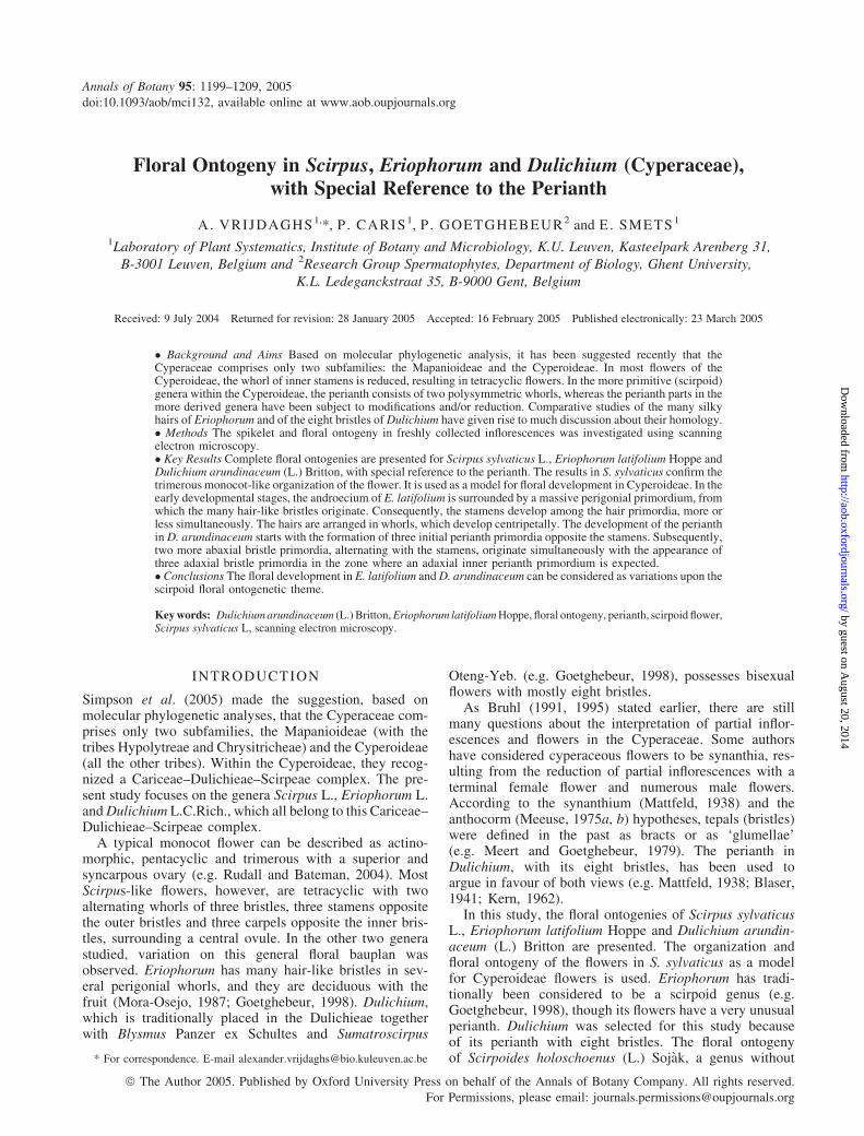
The spikelet of S. sylvaticus is indeterminate and consists ofmany spirally arranged glumes each subtending a bisexualflower (Figs 1A, B and 2D). Under the apex, a new glumebecomes apparent as a rim (Fig. 1A and B). Subsequently,a flower primordium originates in its axil (Fig. 1A, B). Theflower primordium then develops two lateral primordia(Fig. 1C) and, slightly later, one abaxial stamen primor-dium (Fig. 1D–F). Subsequently, the gynoecial primordium(Fig. 1G) and the inner as well as the outer tepal primor-dia (Fig. 1H, arrowed) appear. Meanwhile, the subtendingglume increases in size and protrudes along its midrib(Fig. 1A–E). The gynoecium primordium is formed as adisc-like structure on the top of the flower primordium(Fig. 1F and G), and it differentiates into an annularovary primordium surrounding a central ovule primordium(Fig. 1H and I). Subsequently, two adaxial and one abaxialstigma primordia (Fig. 1J) become visible on the annular
ovary primordium, opposite the three stamens. The twowhorls of tepal primordia differentiate more or less simul-taneously. Opposite the outer whorl of three tepals are thestamens. The three inner tepals alternate with the stamens(Fig. 1H–J). The tepal primordia develop slowly into longbristles (Figs 1H–K and 2A–C), becoming scabrid inappearance (Fig. 2F) at the end of anthesis, when the acheneis ripening (Fig. 2E). The bristles are deciduous with thefruit (Fig. 2E). When the stigma primordia become appar-ent, the three stamens differentiate into anthers andfilaments (Figs 1K and 2A–C). The ovary wall rises form-ing a single style without thickened or distinct style base(Fig. 2B). Meanwhile, the three stigma primordia differen-tiate into three stigmas with a papillose surface (Fig. 2C).After the differentiation of the anthers (Fig. 1K), thestamens grow rapidly and rise above the gynoecium(Fig. 2A–C).
Eriophorum latifolium Hoppe
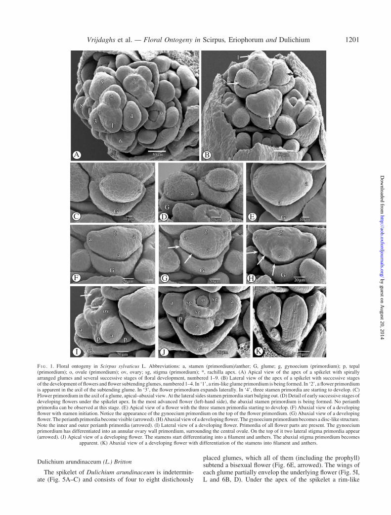
The spikelet of E. latifolium is indeterminate and com-posed of many spirally arranged glumes, each subtending abisexual flower (Fig. 3A). Immediately below the apex ofthe spikelet, a rim-like glume primordium is formed(Fig. 3A). Subsequently, a flower primordium appears inits axil (Fig. 3A, B). Two lateral and one abaxial stamenprimordia are formed (Fig. 3A, C). Simultaneous with thedevelopment of the stamens and the appearance of the gyn-oecium primordium, a massive, dome-shaped perigonialzone is differentiated (Fig. 3D) at the base of the flowerprimordium. The development of the third, abaxial stamenprimordium is delayed (Fig. 3E, F).
Numerous ‘hair’ primordia originate on the perigonialzone shortly after the appearance of the stamen primordia(Fig. 3G–K). The initial development of the hairs at theadaxial side of the flower is less prominent (Figs 3G,I and 4A). The hair primordia are arranged in several whorlsarising centripetally (Fig. 3J, K). The hairs opposite thelateral stamens tend to grow faster than the other ones(Figs 3I, K and 4A). There are no intrastaminal hair prim-ordia (Fig. 3F–J). The growth of the hairs is relatively slowuntil the stamens are well developed and reaching out abovethe gynoecium (Fig. 4A, B). Afterwards, the longest hairsreach until the base of the anthers, at the adaxial as well asat the abaxial side (Fig. 4D, E). Only later, after anthesis,the hairs elongate and develop into the well-known typicalsilky ‘hairs’ (Fig. 4F), which are deciduous with the fruit(Fig. 4C).
The disc-like gynoecium primordium differentiates intoan annular primordium, surrounding a central ovule prim-ordium. On the top of it two lateral stigma primordiabecome apparent (Fig. 3D, E) and, with some delay,a third abaxial one (Fig. 3F, G). The annular gynoeciumprimordium grows up from the base, forming the ovary wall(Fig. 3F–H), and at a later stage a single style withoutdistinct transition between style and ovary (Figs 3I and4A). On the top of the rising ovary wall the stigma primordiagrow out into long papillose stigmas (Figs 3I, J and 4B, D).At this stage, the three stamens develop fast, growing outabove the gynoecium (Fig. 4D, E).
TABLE 1. Species of Cyperaceae studied
Taxa Voucher no. (provenance)
Dulichium arundinaceum(L.) Britton
PG9914 (Botanical Garden of theUniversity of Gent, Gent, Belgium)
Eriophorum latifoliumHoppe
AV04 (Botanical Garden of the Cityof Leuven, Leuven, Belgium)
Scirpoides holoschoenus(L.) Sojak
AV03 (Botanical Garden of the Cityof Leuven, Leuven, Belgium)
Scirpus sylvaticus L. AV02 (Botanical Garden of the ‘Instituutvoor Plantkunde en Microbiologie KUL’,Leuven, Belgium)
1200 Vrijdaghs et al. — Floral Ontogeny in Scirpus, Eriophorum and Dulichium
by guest on August 20, 2014
http://aob.oxfordjournals.org/D
ownloaded from
Dulichium arundinaceum (L.) Britton
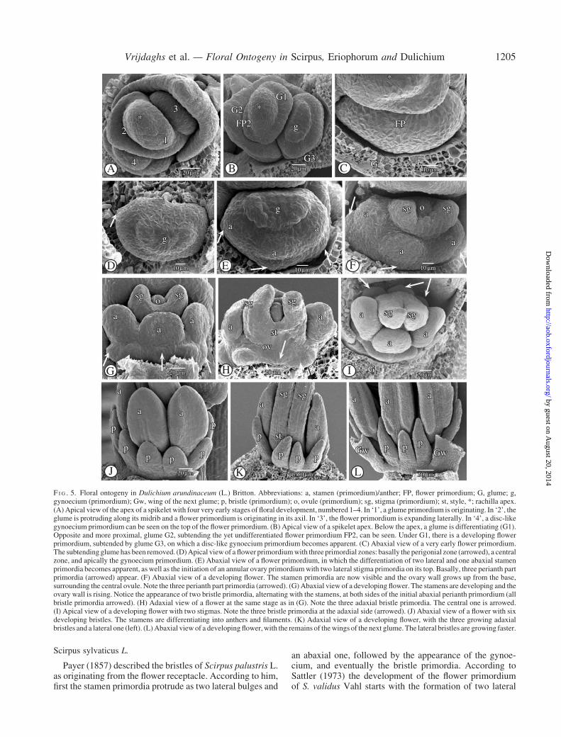
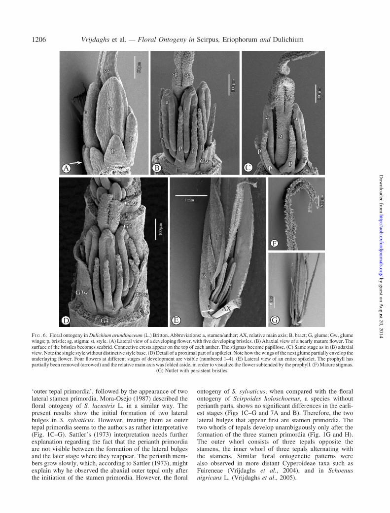
The spikelet of Dulichium arundinaceum is indetermin-ate (Fig. 5A–C) and consists of four to eight distichously
placed glumes, which all of them (including the prophyll)subtend a bisexual flower (Fig. 6E, arrowed). The wings ofeach glume partially envelop the underlying flower (Fig. 5I,L and 6B, D). Under the apex of the spikelet a rim-like
*
13
2
4
6
5 7
*
aa
G
aa
a
aa
G
G
a
a
a a
a
a
G G
aa
a
g
G
1
2
4
G
aa
pp
ovo
oa
a
a
G
p p
p
p
aa
asg
sg
sg
89
g g
20 µm
50 µm
10 µm 20 µm 10 µm
20 µm 20 µm 20 µm
20 µm 20 µm 50 µm
A B
C ED
F G H
I J K
*
13
2
4
6
5 7
*
aa
G
aa
a
aa
G
G
a
a
a a
a
a
G G
aa
a
g
G
1
2
4
3
G
aa
a
pp
ovo
oa
a
a
G
p p
p
p
aa
asg
sg
sg
89
g g
50 µm 50 µm
10 µm 20 µm 10 µm
20 µm 20 µm 20 µm
20 µm 20 µm 50 µm
50 µm
F I G . 1. Floral ontogeny in Scirpus sylvaticus L. Abbreviations: a, stamen (primordium)/anther; G, glume; g, gynoecium (primordium); p, tepal(primordium); o, ovule (primordium); ov, ovary; sg, stigma (primordium); *, rachilla apex. (A) Apical view of the apex of a spikelet with spirallyarranged glumes and several successive stages of floral development, numbered 1–9. (B) Lateral view of the apex of a spikelet with successive stagesof the development of flowers and flower subtending glumes, numbered 1–4. In ‘1’, a rim-like glume primordium is being formed. In ‘2’, a flower primordiumis apparent in the axil of the subtending glume. In ‘3’, the flower primordium expands laterally. In ‘4’, three stamen primordia are starting to develop. (C)Flower primordium in the axil of a glume, apical–abaxial view. At the lateral sides stamen primordia start bulging out. (D) Detail of early successive stages ofdeveloping flowers under the spikelet apex. In the most advanced flower (left-hand side), the abaxial stamen primordium is being formed. No perianthprimordia can be observed at this stage. (E) Apical view of a flower with the three stamen primordia starting to develop. (F) Abaxial view of a developingflower with stamen initiation. Notice the appearance of the gynoecium primordium on the top of the flower primordium. (G) Abaxial view of a developingflower. The perianth primordia becomevisible (arrowed). (H)Abaxial viewof a developingflower. The gynoeciumprimordiumbecomes a disc-like structure.Note the inner and outer perianth primordia (arrowed). (I) Lateral view of a developing flower. Primordia of all flower parts are present. The gynoeciumprimordium has differentiated into an annular ovary wall primordium, surrounding the central ovule. On the top of it two lateral stigma primordia appear(arrowed). (J) Apical view of a developing flower. The stamens start differentiating into a filament and anthers. The abaxial stigma primordium becomes
apparent. (K) Abaxial view of a developing flower with differentiation of the stamens into filament and anthers.
Vrijdaghs et al. — Floral Ontogeny in Scirpus, Eriophorum and Dulichium 1201
by guest on August 20, 2014
http://aob.oxfordjournals.org/D
ownloaded from

glume primordium appears (Fig. 5A–C). While the glume isincreasing in size, its midrib protrudes and a flower prim-ordium appears in its axil (Fig. 5A, B). Subsequently, on theflower primordium, three zones can be recognized; a basalperigonial primordium, a central primordial tissue fromwhich the three staminal primordia originate, and an apicalgynoecium primordium (Fig. 5D–F). The perigonial prim-ordium can be distinguished as a separate dome-like masswith two lateral and one abaxial, congenitally fused, peri-anth part primordia (Fig. 5E, F, arrowed). On the centralzone of the flower primordium two lateral, and a third,abaxial stamen primordium appear (Fig. 5E). Subsequently,at both sides of the initial abaxial perianth part primordium,and alternating with the stamens, an extra bristle primor-dium appears (Fig. 5G). Simultaneously, three adaxialbristle primordia become apparent (Fig. 5H, I). The threeabaxial, the two lateral, and the three adaxial bristle prim-ordia develop into eight separate bristles (Fig. 5J–L and6A–D), which, at maturity, have a scabrid surface(Fig. 6G) and are longer than the nutlet. They are deciduouswith the fruit (Fig. 6G). The apical, disc-like gynoeciumprimordium (Fig. 5D) differentiates into an annular ovary
primordium (Fig. 5E). On the top of the annular ovaryprimordium two lateral stigma primordia are formed(Fig. 5E, F). The ovary primordium grows up from thebase, surrounding the central ovule primordium, and form-ing an ovary wall (Fig. 5F–H). Meanwhile, the three initialstaminal bulges develop into stamens (Fig. 5F–L). Therising ovary wall forms a single style (Fig. 5H) withouttransition between style and ovary (Fig. 6C). The twostigma primordia elongate and differentiate into two longstigmas (Figs 5K and 6A–C, E), which become papillose(Fig. 6E, F). At maturity, they rise high above the stamens(Fig. 6E).
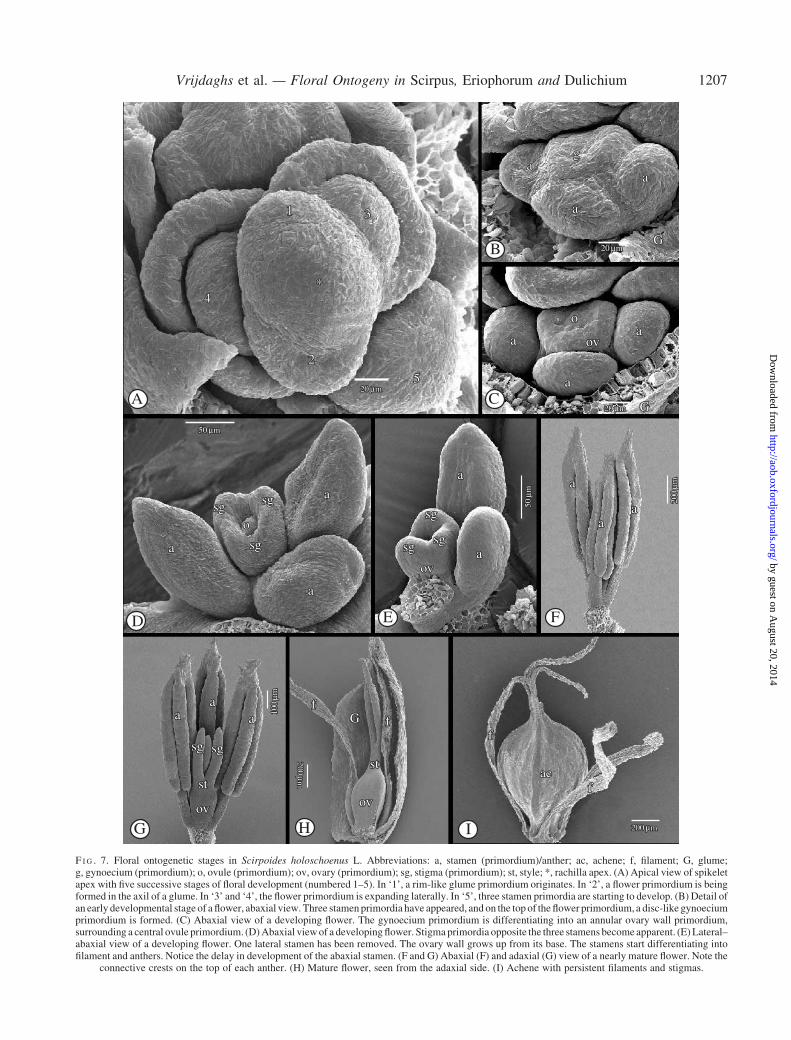
Scirpoides holoschoenus (L.) Sojak
The spikelet is indeterminate and the glumes are spirallyarranged (Fig. 7A). Under the apex, a new glume becomesapparent as a rim (Fig. 7A). Subsequently, a flower prim-ordium is formed in its axil (Fig. 7A). Soon, the flowerprimordium expands laterally, forming two lateral stamenprimordia, more or less simultaneously with the formationof a third, abaxial stamen primordium (Fig. 7A and B).
a
a a
aa
a
a
G
sg
p pp
p pp
p
pp
p
sg
stsg
sg
sg
st
G G
p
p
pp
p
50µm
50 µm100 µm
200
µm 200
µm
50µm
A B C
D E F
a
a a
aa
a
a
G
sg
p pp
p pp
p
pp
p
sg
stsg
sg
sg
st
G G
p
p
pp
p
50µm
50 µm100 µm
200
µm 200
µm
50µm
F I G . 2. Floral ontogeny in Scirpus sylvaticus L. Abbreviations: a, stamen (primordium)/anther; G, glume; p, tepal; sg, stigma (primordium); st, style. (A)Lateral–abaxial view of a developing flower. The bristles are growing out. (B) Adaxial view. Same stage of development as in (A). The ovarywall has formeda single style and the three stigmas are growing. (C)Adaxial viewof a developingflower. The surface of the stigmas becomes papillose, and simultaneously onthe top of the anthers connective crests are formed. (D) Lateral view of a complete spikelet as a typical example of a racemose partial inflorescence with
acropetally developing flowers. (E) Achene with persistent bristles. Note the beak at the top of the nutlet. (F) Detail of a mature bristle.
1202 Vrijdaghs et al. — Floral Ontogeny in Scirpus, Eriophorum and Dulichium
by guest on August 20, 2014
http://aob.oxfordjournals.org/D
ownloaded from
*12 3
4
P
a
a
sg sg
sg sg
sg
a a
a
o
G
sgsg
sg
a a
a G
a
P
aa
sg sg
sg
P
a
a a st
sgsgsg
a
a
a
a
FP1
a a
sg sg
st
*
FP2
G1
G2
oo
aa
g
G
o
ov
sg
sg
P
ov
P
50 µm
50µm
20 µm 20 µm 20 µm
20 µm 20 µm
50 µm
20 µm
50 µm 50 µm
A
*12 3
4
P
a
a
sg sg
G
F
sg sg
sg
a a
a
o
G H
sgsg
sg
a a
a G
a
P
G
aa
sg sg
sg
P
K
a
a a st
sgsg sg
a
a
a
a
FP1
D E
I J
a a
sg sg
st
*
FP2
G1
G2
oo
C
aa
g
G
o
ov
sg
sg
P
ov
P
B50 µm
50µm
20 µm 20 µm 20 µm
20 µm 20 µm
50 µm
20 µm
50 µm 50 µm
F I G . 3. Floral ontogeny in Eriophorum latifolium Hoppe. Abbreviations: a, stamen (primordium)/anther; FP, flower primordium; G, glume; g, gynoecium(primordium); P, perianth; o, ovule (primordium); sg, stigma (primordium); st, style; *, rachilla apex. (A) Spikelet apex, lateral view, with distichouslypositioned glumes, each subtending a flower at successive stages of floral development (numbered 1–4). In ‘1’, a glume is being formed. In ‘2’, a flowerprimordiumoriginates in the axil of the subtendingglume. In ‘3’, three stamenprimordia are developing. In ‘4’,most of the developingflower is hiddenbehindthe subtending glume. Notice the torsion in the apical part of the spikelet. (B) Apical view of a spikelet apexwith two flower primordia at a very early stage ofdevelopment. The most advanced flower primordium (FP2) is expanding laterally. (C) Apical view of a flower primordium in which two lateral stamenprimordia are starting to develop. (D) Differentiation of the gynoecium primordium into an annular ovary wall primordium, with lateral stigma primordia onits top. Notice themassive perigonial primordium at the base of the flower primordium. (E) Lateral–abaxial view of a developing flower. The development ofthe abaxial stamen primordium is delayed (arrowed). (F) Abaxial view of a developing flower. An abaxial stigma primordium appears. (G) Adaxial view.Same stage as in (F). (H) Centripetal initiation of whorls of hairs (arrowed). Note the delay of the development of the abaxial stamen with respect to the twolateral stamens. (I) Adaxial view of a developing flower. The development of the adaxial hairs is delayed. (J) Apical view of a developing flower. The threestigmas become papillose. (K) Lateral–adaxial view of a developing flower. The adaxial hairs start developing. Note the advanced development of the hairs
opposite the lateral stamen.
Vrijdaghs et al. — Floral Ontogeny in Scirpus, Eriophorum and Dulichium 1203
by guest on August 20, 2014
http://aob.oxfordjournals.org/D
ownloaded from

Meanwhile, a gynoecium primordium appears (Fig. 7B)on the top of the flower primordium. It differentiates intoan annular ovary primordium surrounding a central ovule(Fig. 7C). At a later stage, the ovary primordium grows upfrom the base, forming an ovary wall (Fig. 7D). On the topof the ovary wall, three stigma primordia appear (Fig. 7D).Meanwhile, the stamens start differentiating into filamentsand anthers (Fig. 7D). At an intermediary stage, the stamenshave grown out above the gynoecium, and the apical partof the connective becomes spiny (Fig. 7E). The stigmaprimordia develop into three stigmas (Fig. 7F).
DISCUSSION
Much of the controversy relating to floral and inflorescencemorphology in Cyperaceae arises from the interpretation ofa flower according to either the ‘euanthial’ or ‘synanthial’hypotheses. Scirpoid flowers have been described as con-ventional flowers with a typical trimerous monocotyledon-ous arrangement. Alternatively, they have been consideredto be composed of floral structures derived from a partialinflorescence with several floral units. According to Sattler(1973), the existing terminology to describe floral organo-genesis is often interpretative. For example, based on
vasculature patterns, Blaser (1941) named the outer whorlof cyperaceous perianth parts ‘calyx’ and the inner ‘corolla’.Sattler (1973) used the word ‘tepals’ for perianth membersin Cyperaceae. Mora-Osejo (1987) considered the innerperianth parts to be ‘petals’. However, perianth partswere also named ‘bracts’ (Mattfeld, 1938; Kern, 1962) or‘glumellae’, both terms suggesting leaf-like structures thateach axillate a stamen (e.g. Meert and Goetghebeur, 1979;Goetghebeur, 1986). Actually, most authors designate thecyperaceous bristle- or scale-like perianth structures as‘perianth parts’, ‘perianth bristles’, ‘bristles’ or ‘scales’.In an attempt to describe objectively the floral ontogenyin cyperoid flowers, the term ‘tepals’ is used when all peri-anth parts are similar. If there are morphological differencesbetween the outer and inner perianth parts, the outer peri-anth parts may be named ‘sepals’ and the inner ones ‘petals’(e.g. Leins, 2000). In the Cyperoideae morphological dif-ferences between the inner and outer whorls of the perianthparts occur in some genera, e.g. Fuirena Rottb. (Forbes,1997; Vrijdaghs et al., 2004), and the number of perianthparts may also be greater than six (e.g. Dulichium). Inthese cases, the general term ‘perianth part’ is used. InEriophorum, with its many hair-like bristles, the word‘hairs’ is also used to indicate the perianth parts.
a
ov
st
a
a a
P
a aa
P
sg sg sg
a
a a
a
sgsg
sg
st
50 µm
100
µm
500 µm
100
µm
200
µm
50µm
a
ov
A
D E
st
a
a a
P
a aa
P
B C
F
sg sg sg
a
a a
a
sg sg
sg
st
50 µm
100
µm
500 µm
100
µm
200
µm
50µm
F I G . 4. Floral ontogeny in Eriophorum latifoliumHoppe. Abbreviations: a, stamen/anther; P, perianth; ov, ovary; sg, stigma; st, style. (A) Adaxial view of adeveloping flower. Notice the centripetal development of the perigonium, and the advanced development of the bristles opposite the lateral stamens. (B)Apical–abaxial view of a developing flower. The surface of the three stigmas is becoming papillose. (C) Achene with persistent hairs. (D–E) Adaxial (D) andabaxial (E) view of the same flower at nearly mature stage. The stamens are growing up above the gynoecium, and the hairs are developing rapidly. Note the
advancement of the hairs opposite the lateral stamens. (F) Detail of a hair.
1204 Vrijdaghs et al. — Floral Ontogeny in Scirpus, Eriophorum and Dulichium
by guest on August 20, 2014
http://aob.oxfordjournals.org/D
ownloaded from
Scirpus sylvaticus L.
Payer (1857) described the bristles of Scirpus palustris L.as originating from the flower receptacle. According to him,first the stamen primordia protrude as two lateral bulges and
an abaxial one, followed by the appearance of the gynoe-cium, and eventually the bristle primordia. According toSattler (1973) the development of the flower primordiumof S. validus Vahl starts with the formation of two lateral
*
12
3
4
g
a
a
a
a
aa
sg sgo
a
aa
sg sgo
a a
a
p
pp p
p
pp
p pGw Gw
st
a
a
sg sg a aa
p p p
*G2
g
G1
FP2
G3G
FP
*
a
aa
sg
G
sga
a
sg sg
st
ov
g
p
20 µm20 µm 10 µm
10 µm 10 µm 10 µm
20 µm 20 µm 20 µm
20 µm 50 µm
A
J K L
*
12
3
4
E
g
a
a
a
F
a
aa
sg sgo
G
a
aa
sg sgo
a a
a
p
p p p
p
p p
p pGw Gw
st
a
a
sg sg a aa
p p p
B
*G2
g
G1
FP2
G3C G
FP
*
I
a
aa
sg
G
sga
a
sg sg
st
H
ov
D
g
p
20 µm20 µm 10 µm
10 µm 10 µm 10 µm
20 µm 20 µm 20 µm
20 µm 50 µm 50 µm50 µm
F I G . 5. Floral ontogeny in Dulichium arundinaceum (L.) Britton. Abbreviations: a, stamen (primordium)/anther; FP, flower primordium; G, glume; g,gynoecium (primordium); Gw, wing of the next glume; p, bristle (primordium); o, ovule (primordium); sg, stigma (primordium); st, style, *; rachilla apex.(A)Apical view of the apex of a spikelet with four very early stages of floral development, numbered 1–4. In ‘1’, a glume primordium is originating. In ‘2’, theglume is protruding along its midrib and a flower primordium is originating in its axil. In ‘3’, the flower primordium is expanding laterally. In ‘4’, a disc-likegynoecium primordium can be seen on the top of the flower primordium. (B) Apical view of a spikelet apex. Below the apex, a glume is differentiating (G1).Opposite and more proximal, glume G2, subtending the yet undifferentiated flower primordium FP2, can be seen. Under G1, there is a developing flowerprimordium, subtended by glume G3, on which a disc-like gynoecium primordium becomes apparent. (C) Abaxial view of a very early flower primordium.The subtendingglumehas been removed. (D)Apical viewof a flower primordiumwith three primordial zones: basally the perigonial zone (arrowed), a centralzone, and apically the gynoecium primordium. (E) Abaxial view of a flower primordium, in which the differentiation of two lateral and one abaxial stamenprimordia becomes apparent, as well as the initiation of an annular ovary primordiumwith two lateral stigma primordia on its top. Basally, three perianth partprimordia (arrowed) appear. (F) Abaxial view of a developing flower. The stamen primordia are now visible and the ovary wall grows up from the base,surrounding the central ovule. Note the three perianth part primordia (arrowed). (G)Abaxial view of a developing flower. The stamens are developing and theovary wall is rising. Notice the appearance of two bristle primordia, alternating with the stamens, at both sides of the initial abaxial perianth primordium (allbristle primordia arrowed). (H) Adaxial view of a flower at the same stage as in (G). Note the three adaxial bristle primordia. The central one is arrowed.(I) Apical view of a developing flower with two stigmas. Note the three bristle primordia at the adaxial side (arrowed). (J) Abaxial view of a flower with sixdeveloping bristles. The stamens are differentiating into anthers and filaments. (K) Adaxial view of a developing flower, with the three growing adaxialbristles and a lateral one (left). (L)Abaxial view of a developing flower,with the remains of thewings of the next glume.The lateral bristles are growing faster.
Vrijdaghs et al. — Floral Ontogeny in Scirpus, Eriophorum and Dulichium 1205
by guest on August 20, 2014
http://aob.oxfordjournals.org/D
ownloaded from
‘outer tepal primordia’, followed by the appearance of twolateral stamen primordia. Mora-Osejo (1987) described thefloral ontogeny of S. lacustris L. in a similar way. Thepresent results show the initial formation of two lateralbulges in S. sylvaticus. However, treating them as outertepal primordia seems to the authors as rather interpretative(Fig. 1C–G). Sattler’s (1973) interpretation needs furtherexplanation regarding the fact that the perianth primordiaare not visible between the formation of the lateral bulgesand the later stage where they reappear. The perianth mem-bers grow slowly, which, according to Sattler (1973), mightexplain why he observed the abaxial outer tepal only afterthe initiation of the stamen primordia. However, the floral
ontogeny of S. sylvaticus, when compared with the floralontogeny of Scirpoides holoschoenus, a species withoutperianth parts, shows no significant differences in the earli-est stages (Figs 1C–G and 7A and B). Therefore, the twolateral bulges that appear first are stamen primordia. Thetwo whorls of tepals develop unambiguously only after theformation of the three stamen primordia (Fig. 1G and H).The outer whorl consists of three tepals opposite thestamens, the inner whorl of three tepals alternating withthe stamens. Similar floral ontogenetic patterns werealso observed in more distant Cyperoideae taxa such asFuireneae (Vrijdaghs et al., 2004), and in Schoenusnigricans L. (Vrijdaghs et al., 2005).
p
p
a a
sg sg sg
aa
a
pp p p
p
Gw pp
pp
a a
st
sg sg
G3w
G4
G2w
G2
G1w
B
50µm
100
µm
100
µm
100
µm
1 mm
200
µm
A B C
D
F
p
p
a a
sg sg sg
aa
a
pp p p
p
Gw pp
pp
a a
st
sg sg
G3w
G4
G2w
G2
G1w
GE
AX
B
50µm
100
µm
100
µm
100
µm
1 mm
200
µm
500500µ
m500µ
m
F I G . 6. Floral ontogeny in Dulichium arundinaceum (L.) Britton. Abbreviations: a, stamen/anther; AX, relative main axis; B, bract; G, glume; Gw, glumewings; p, bristle; sg, stigma; st, style. (A) Lateral view of a developing flower, with five developing bristles. (B) Abaxial view of a nearly mature flower. Thesurface of the bristles becomes scabrid. Connective crests appear on the top of each anther. The stigmas become papillose. (C) Same stage as in (B) adaxialview.Note the single stylewithout distinctive style base. (D)Detail of a proximal part of a spikelet. Note how thewings of the next glume partially envelop theunderlaying flower. Four flowers at different stages of development are visible (numbered 1–4). (E) Lateral view of an entire spikelet. The prophyll haspartially been removed (arrowed) and the relative main axis was folded aside, in order to visualize the flower subtended by the prophyll. (F) Mature stigmas.
(G) Nutlet with persistent bristles.
1206 Vrijdaghs et al. — Floral Ontogeny in Scirpus, Eriophorum and Dulichium
by guest on August 20, 2014
http://aob.oxfordjournals.org/D
ownloaded from
a
a
a
G
ov
o
sgsg
a
a
a
aa
a
a
a
a
G
g
2
1
4
3
5
*
sg
aa
a
st
sg sg
ov
a
a
sg
sgsg
ov
o
ov
st
Gf
ff
fac
20 µm
20 µm
50µm
50 µm
200
µm
100
µm
200µm
200 µm
C
a
a
a
G
ov
o
D
sgsg
a
a
a
F
aa
a
B
a
a
a
G
g
A
2
1
4
3
5
*
sg
G
aa
a
st
sg sg
ov
E
a
a
sg
sgsg
ov
o
ov
st
Gf
ff
fac
H I
20 µm
20 µm
20 µm20 µm
50µm
50 µm
200
µm
100
µm
200µm
200 µm
F I G . 7. Floral ontogenetic stages in Scirpoides holoschoenus L. Abbreviations: a, stamen (primordium)/anther; ac, achene; f, filament; G, glume;g, gynoecium (primordium); o, ovule (primordium); ov, ovary (primordium); sg, stigma (primordium); st, style; *, rachilla apex. (A) Apical view of spikeletapex with five successive stages of floral development (numbered 1–5). In ‘1’, a rim-like glume primordium originates. In ‘2’, a flower primordium is beingformed in the axil of a glume. In ‘3’ and ‘4’, the flower primordium is expanding laterally. In ‘5’, three stamen primordia are starting to develop. (B) Detail ofan early developmental stage of a flower, abaxial view. Three stamen primordia have appeared, and on the top of the flower primordium, a disc-like gynoeciumprimordium is formed. (C) Abaxial view of a developing flower. The gynoecium primordium is differentiating into an annular ovary wall primordium,surrounding a central ovule primordium. (D) Abaxial view of a developing flower. Stigma primordia opposite the three stamens become apparent. (E) Lateral–abaxial view of a developing flower. One lateral stamen has been removed. The ovary wall grows up from its base. The stamens start differentiating intofilament and anthers. Notice the delay in development of the abaxial stamen. (F and G) Abaxial (F) and adaxial (G) view of a nearly mature flower. Note the
connective crests on the top of each anther. (H) Mature flower, seen from the adaxial side. (I) Achene with persistent filaments and stigmas.
Vrijdaghs et al. — Floral Ontogeny in Scirpus, Eriophorum and Dulichium 1207
by guest on August 20, 2014
http://aob.oxfordjournals.org/D
ownloaded from

Eriophorum latifolium Hoppe
Eriophorum, with its many spirally arranged glumes eachsubtending a bisexual flower, is considered to be very closeto Scirpus. The rather artificial separation of both generais based on the number of perianth parts (Goetghebeur,1998). Species with six or less perianth parts belong toScirpus, while species with ten or more perianth partsbelong to Eriophorum. However, the perigonial structureof Eriophorum is morphologically different from the tri-merous scirpoid perianth. Mora-Osejo (1987) interpretedthe ‘perianth parts’ of flowers in Eriophorum as ‘ . . .estructuras sui generis, que se originan ‘‘de novo’’ a partirde la protuberancia meristematica semicircular, resultantedel agradamiento y fusion de los primordios perigonalesindividuales’ (Mora-Osejo, 1987, p. 23). This seems toagree with Blaser (1941), who stated that the hair-like bris-tles of E. angustifolium grow faster and appear to be largerwhere perianth parts could be expected. The present SEMobservations in E. latifolium confirm that the hairs originatein several whorls, though without any particular relation innumber (nor placement) to the staminal or gynoecial prim-ordia. Fields of hairs corresponding to the positions whereperianth parts are to be expected (opposite the stamens andopposite the sides of the ovary) were not observed, thoughthe hairs in the zones opposite the lateral stamens tend togrow faster and longer (Figs 3K and 4A). On the other hand,the authors agree with Mora-Osejo’s (1987) interpretationthat the perigonial primordium is a structure resulting fromcongenital fusion of individual bristle primordia. They con-sider it to be homologous with a scirpoid perianth because(a) of its position in the flower and (b) all other flower partsin E. latifolium develop according to the the scirpoidpattern. In E. latifolium the apical part of the flower prim-ordium expands laterally, before the initiation of the peri-gonial primordium (Fig. 3A). If the floral development inEriophorum follows the same sequence as Sattler (1973)observed in Scirpus, the lateral bulges should logicallybe considered to be perigonial primordia. However, sincethe early floral ontogeny in Eriophorum does not differessentially from the early floral ontogeny in Scirpoidesholoschoenus (L.) Sojak, the authors consider the initialtwo lateral bulges, as they did in Scirpus sylvaticus, to bestaminal primordia.
Dulichium arundinaceum (L.) Britt.
Mattfeld (1938) used Dulichium as an argument in favourof his synanthium hypothesis. He considered the five bris-tles at the abaxial side to be fused, and interpreted them to bethe remaining veins of one simple bract, which subtendedthe three stamens. In the same way he interpreted the threeremaining adaxial bristles as one intrastaminal bract, sub-tending the gynoecium, being the female flower. Hence,Mattfeld (1938) concluded that bisexual flowers are derivedfrom the reduction of partial inflorescences consisting ofunisexual flowers. However, based on anatomical studies,Blaser (1941) did not agree with Mattfeld’s (1938) inter-pretation of the bristles of Dulichium as the veins of twodifferent bracts. According to Blaser (1941), sepals can bemodified by splitting up along the midvein and the two
lateral veins, resulting in three separate bristles. The lossof parts of such tri-partite ‘sepals’ occurs so that one‘sepal’ can be reduced to two bristles or even one. ‘Petals’are never increased in number, though they might partiallysplit, or be absent. He considered that the eight bristles inD. arundinaceum result from the one bristle-like abaxial‘sepal’, consisting of the median vein of the ancestralsepal, the two lateral ‘sepals’ each reduced to a bristleconsisting of the median vein and a bristle consisting ofone lateral vein, and the three reduced, bristle-like ‘petals’.
Mora-Osejo (1987), on the basis of a floral ontogeneticstudy, stated that all perianth parts in D. arundinaceumare positioned extrastaminally. Moreover, he concludedthat the floral ontogenies in D. arundinaceum and in Scirpuslacustris L. are similar. The present results support thisconclusion. During floral ontogeny, three perianth primor-dia homologous with the perianth parts of the outer whorlare formed first. At a later stage, three bristle primordiaappear at the adaxial side of the ovary, in a zone wherethe adaxial inner perianth part primordium is expected. Theauthors consider these three adaxial bristles to be homolog-ous with the inner adaxial perianth member as observed inScirpus sylvaticus L. More or less simultaneously, at bothsides of the original abaxial bristle primordium and altern-ating with the stamens, an extra bristle primordium becomesapparent (Fig. 5F), which the authors consider to be homo-logous with the inner tepals in Scirpus. Therefore, theyconclude that the floral ontogeny of D. arundinaceumfollows a scirpoid pattern.
CONCLUSIONS
Floral structure and development in Scirpus sylvaticus mayserve as a model for all scirpoid flowers, even flowers withmore than six perianth parts, as found in Dulichium orEriophorum. A typical scirpoid floral ontogeny startswith the formation of a floral primordium in the axil of asubtending glume. Subsequently, two lateral stamen prim-ordia are formed, usually followed by a third, abaxial sta-men primordium. Only then do the perianth primordiaappear, more or less simultaneously. Three outer perianthparts are positioned opposite the stamens, and three innerperianth parts alternate with the stamens.
In Eriophorum latifolium, the perianth originates fromone circular perigonial primordium. The hair-like bristlesdevelop centripetally, arising in several whorls. No bristlesare found intrastaminally. The perigonial primordium ofE. latifolium is interpreted as resulting from a congenitalfusion of individual perianth primordia and it is consideredto be homologous with the perianth primordia in Scirpus.
In Dulichium arundinaceum three initial perianth prim-ordia are formed opposite the stamens, forming the outerwhorl of perianth parts. Subsequently, three adaxial(homologous with the scirpoid adaxial inner tepal) andtwo extra abaxial bristle primordia (homologous with thescirpoid abaxial inner tepals), which alternate with thestamens, originate. It is concluded that floral developmentin E. latifolium and D. arundinaceum can be considered asvariations upon the scirpoid floral ontogenetic theme.
1208 Vrijdaghs et al. — Floral Ontogeny in Scirpus, Eriophorum and Dulichium
by guest on August 20, 2014
http://aob.oxfordjournals.org/D
ownloaded from
ACKNOWLEDGEMENTS
We thank Marcel Verhaegen, for assistance with SEMobservations at the National Botanic Garden of Belgiumin Meise. This work was supported by research grantsof the K.U. Leuven (0T/01/25) and the Fund forScientific Research-Flanders (FWO-Vlaanderen, Belgium,G.0104.01, G.0268.04; 1.5.069.02 and 1.5.061.03).
LITERATURE CITED
Blaser HW. 1941. Studies in the morphology of the Cyperaceae. I.Morphology of flowers. A. Scirpoid genera. American Journal ofBotany 28: 542–551.
Bruhl JJ. 1991. Comparative development of some taxonomically criticalfloral/inflorescence features in Cyperaceae. Australian Journal ofBotany 9: 119–127.
Bruhl JJ. 1995. Sedge genera of the World: relationships and a new clas-sification of the Cyperaceae.Australian Journal of Botany 8: 125–305.
Forbes PL. 1997. Studies in Cyperaceae in southern Africa. 34. Fuirenacoerulescens, a polymorphic species. South African Journal of Botany63: 514–520.
Goetghebeur P. 1986. Genera Cyperacearum. Een bijdrage tot de kennisvan de morfologie, systematiek en fylogenese van de Cyperaceae-genera. PhD Thesis, Groep Plantkunde, Rijksuniversiteit Gent, Gent,Belgium.
Goetghebeur P. 1998. Cyperaceae. In: Kubitzki K. ed. The families andgenera of vascular plants, Vol. 4. Berlin/Heidelberg: Springer-Verlag,141–190.
Kern JH. 1962. New look at some Cyperaceae mainly from the tropicalstandpoint. Advances in Science 19: 141–148.
Leins P. 2000. Bl€uute und Frucht. Morphologie, Entwicklungsgeschichte,Phylogenie, Funktion, Okologie. Stuttgart, Germany: E. Schweizer-bart’sche Verlagsbuchhandlung (Nagele u. Obermiller).
Mattfeld J. 1938. Das morphologische Wesen und die phylogenetischeBedeutung der Blumenblatter. Berichte der Deutschen BotanischenGesellschaft 56: 86–116.
Meeuse ADJ. 1975a. Changing floral concepts: anthocorms, flowers, andanthoids. Acta Botanica Neerlandicum 24: 23–36.
MeeuseADJ. 1975b. Interpretative floralmorphology of the Cyperaceae onthe basis of the anthoid concept. Acta Botanica Neerlandicum 24:291–304.
Meert M, Goetghebeur P. 1979. Comparative floral morphology ofBisboeckelereae andCariceae (Cyperaceae) on the basis of the anthoidconcept.Bulletin de la Societe Royale Botanique de Belgique–Bulletinvan de Koninklyke Belgische Botanische Vereniging 112: 128–143.
Mora-Osejo LE. 1987.Consideraciones sobre la naturalezamorfologica delasflores de algunesgenerosde lasCyperaceae.Revista de la AcademiaColombiana de Ciencias Exactas, Fisicas y Naturales 16: 23–35.
Payer JB. 1857. Traite d’organogenie comparee de la fleur. Masson: Paris.Rudall PJ, Bateman RM. 2004. Evolution of zygomorphy in monocot
flowers: iterative patterns and developmental constraints. NewPhytologist 162: 25–44.
Sattler R. 1973. Organogenesis of flowers. Toronto, Canada: University ofToronto Press.
Simpson DA,Muasya AM, AlvesM et al. 2005. Phylogeny of Cyperaceaebased on DNA sequence data—a new rbcL analysis. In: Aliso(Monocots III), in press.
Vrijdaghs A, Goetghebeur P, Smets E, Caris P. 2004. The nature ofthe perianth inFuirena (Cyperaceae). South African Journal of Botany70: 587–594.
Vrijdaghs A, Goetghebeur P, Smets E, Caris P. 2005. The Schoenusspikelet a rhipidium?A floral ontogenetic answer. In:Aliso (MonocotsIII), in press.
Vrijdaghs et al. — Floral Ontogeny in Scirpus, Eriophorum and Dulichium 1209
by guest on August 20, 2014
http://aob.oxfordjournals.org/D
ownloaded from