The Floral Scales in Hellmuthia (Cyperaceae, Cyperoideae) and Paramapania (Cyperaceae,...
12
The Floral Scales in Hellmuthia (Cyperaceae, Cyperoideae) and Paramapania (Cyperaceae, Mapanioideae): An Ontogenetic Study A. VRIJDAGHS 1, *, P. GOETGHEBEUR 2 , E. SMETS 3 and A. M. MUASYA 4 1 Laboratory of Plant Systematics, Institute of Botany and Microbiology, K.U.Leuven, Kasteelpark Arenberg 31, B-3001 Leuven, Belgium, 2 Research Group Spermatophytes, Department of Biology, Ghent University, K.L. Ledeganckstraat 35, B-9000 Gent, Belgium, 3 National Herbarium of The Netherlands, Leiden University Branch, PO Box 9514, NL-2300 RA Leiden, The Netherlands and 4 Botany Department, University of Cape Town, 7701 Rondebosch, South Africa Received: 12 April 2006 Returned for revision: 9 May 2006 Accepted: 19 May 2006 Published electronically: 28 June 2006 Background and Aims In 1976 the monotypic genus Hellmuthia was placed in the Hypolytreae s.l., but was subsequently ascribed to the Mapanioideae, tribe Chrysitricheae, mainly because of the presence in Hellmuthia of two lateral, mapanioid-like floral scales with ciliated keels, the anatomy of the nutlet, the embryo and the inflorescence. Recently, based on cladistic analyses and supported by pollen ontogenetic evidence, Hellmuthia was transferred to a Cyperaceae, tribe Cypereae, clade mainly consisting of Ficinia and Isolepis. In this study, the floral ontogeny in Hellmuthia was investigated and compared with the floral ontogeny in Paramapania, with special attention for the floral scales. Methods Freshly collected inflorescences of Hellmuthia membranacea and Paramapania parvibractea were investigated using scanning electron and light microscopy. Key Results In the conical ‘spikelet’ in Hellmuthia, proximal bracts occur, each axillating an axis with empty glume-like structures, or a reduced spikelet. Hence, it is a reduced partial inflorescence. In Hellmuthia, the stamen primordia originate before the primordia of the perianth–gynoecium appear. Moreover, a third adaxially positioned ‘floral scale’ was observed for the first time. The position and relative time of appearance of the floral scales in Hellmuthia are typical for perianth parts in Cyperoideae. The basal position of Hellmuthia within a clade of species with usually perianthless flowers, allows the presence of rudiments of a perianth in Hellmuthia to be interpreted as a primitive state. Development of the lateral ‘scales’ in Paramapania follows a different pattern. Therefore, it was decided that the lateral ‘scales’ in Paramapania are different from the lateral perianth parts in Hellmuthia. The pollen grains in Hellmuthia are cyperoid, with one polar and five lateral apertures, of which the membrane is covered with sexinous bodies. The pollen surface is granulate and perforate with microspines. Conclusions The floral ontogeny in Hellmuthia occurs according to the general cyperoid pattern. The lateral scales in Hellmuthia are perianth parts, and they are not homologous to the lateral ‘scales’ in Paramapania. Key words: Floral scales, Paramapania, floral ontogeny, Cyperaceae, Hellmuthia, SEM, homology. INTRODUCTION According to the molecular phylogenetic studies of Simpson et al. (2006; see Table 1), the Cyperaceae can be subdivided into two subfamilies, the Mapanioideae and the Cyperoideae (Table 1). Morphologically, the Mapanioideae are characterized by a particular ‘flower’, also called ‘spicoids’. The term ‘spicoid’ refers to a synanthial interpretation of the inflorescence unit, opposite to the use of the term ‘flower’. A typical ‘spicoid’ (e.g. Kukkonen, 1984) or ‘flower’ (e.g. Goetghebeur, 1998) is subtended by a large glume-like bract, which envelops most of the reproductive unit. The ‘flower’ consists of a very short and contracted central axis, several spirally placed scales subtending a stamen (or not), and a terminal pistil. The two proximal scales are keeled, opposite to each other and larger than the other scales, each subtending or opposite to a stamen (or not) (Kukkonen, 1984; Goetghebeur, 1986). In the Cyper- oideae, a flower is trimerously built, tetracyclic with two whorls of each three perianth parts, one whorl of three stamens, and a trimerous gynoecium. In many genera, there is a tendency to reduce the number of floral parts or modify them (Vrijdaghs et al., 2004, 2005a, b). The taxon, initally described as Scirpus membranaceus by Thunberg (1794), has been treated as a member of a number of several distinct genera depending on the interpretation of the floral characters. The species was transferred to Isolepis (Nees von Esenbeck, 1835), Ficinia (Kunth, 1837), and independently, the same plant was placed by Steudel (1855) in a new genus named after his son, Hellmuthia: ‘genus in nomine filii Hellmuth Steudel dixi’. The genus Hellmuthia consisted of one species H. restoides, because of its ressembance with the Restionaceae (Steudel, 1855). In 1898, Clarke used the name Scirpus membranaceus Thunb. for the current Hellmuthia membranacea, arguing that it belongs to Scirpus: ‘I regard the squamellae here present as analogous to the broad scale-like hypogynous setae sometimes developed in Scirpus littoralis Schrad . . . the species seems to me a Eu-Scirpus, near S. littoralis Schrad., rather than an Isolepis.’ (see Fig. 8A, after Clarke, 1909). Scho ¨nland (1922) mentioned that the * For correspondence. E-mail [email protected] Annals of Botany 98: 619–630, 2006 doi:10.1093/aob/mcl138, available online at www.aob.oxfordjournals.org Ó The Author 2006. Published by Oxford University Press on behalf of the Annals of Botany Company. All rights reserved. For Permissions, please email: [email protected] by guest on August 20, 2014 http://aob.oxfordjournals.org/ Downloaded from
-
Upload
independent -
Category
Documents
-
view
2 -
download
0
Transcript of The Floral Scales in Hellmuthia (Cyperaceae, Cyperoideae) and Paramapania (Cyperaceae,...

The Floral Scales in Hellmuthia (Cyperaceae, Cyperoideae) and Paramapania(Cyperaceae, Mapanioideae): An Ontogenetic Study
A. VRIJDAGHS1,*, P . GOETGHEBEUR2, E. SMETS3 and A. M. MUASYA4
1Laboratory of Plant Systematics, Institute of Botany and Microbiology, K.U.Leuven, Kasteelpark Arenberg 31,
B-3001 Leuven, Belgium, 2Research Group Spermatophytes, Department of Biology, Ghent University,
K.L. Ledeganckstraat 35, B-9000 Gent, Belgium, 3National Herbarium of The Netherlands,
Leiden University Branch, PO Box 9514, NL-2300 RA Leiden, The Netherlands and 4Botany Department,
University of Cape Town, 7701 Rondebosch, South Africa
Received: 12 April 2006 Returned for revision: 9 May 2006 Accepted: 19 May 2006 Published electronically: 28 June 2006
� Background and Aims In 1976 the monotypic genus Hellmuthia was placed in the Hypolytreae s.l., but wassubsequently ascribed to the Mapanioideae, tribe Chrysitricheae, mainly because of the presence in Hellmuthia oftwo lateral, mapanioid-like floral scales with ciliated keels, the anatomy of the nutlet, the embryo and theinflorescence. Recently, based on cladistic analyses and supported by pollen ontogenetic evidence, Hellmuthiawas transferred to a Cyperaceae, tribe Cypereae, clade mainly consisting of Ficinia and Isolepis. In this study, thefloral ontogeny in Hellmuthia was investigated and compared with the floral ontogeny in Paramapania, with specialattention for the floral scales.� Methods Freshly collected inflorescences of Hellmuthia membranacea and Paramapania parvibractea wereinvestigated using scanning electron and light microscopy.� Key Results In the conical ‘spikelet’ in Hellmuthia, proximal bracts occur, each axillating an axis with emptyglume-like structures, or a reduced spikelet. Hence, it is a reduced partial inflorescence. In Hellmuthia, the stamenprimordia originate before the primordia of the perianth–gynoecium appear. Moreover, a third adaxially positioned‘floral scale’ was observed for the first time. The position and relative time of appearance of the floral scales inHellmuthia are typical for perianth parts in Cyperoideae. The basal position of Hellmuthia within a clade of specieswith usually perianthless flowers, allows the presence of rudiments of a perianth in Hellmuthia to be interpreted asa primitive state. Development of the lateral ‘scales’ in Paramapania follows a different pattern. Therefore, it wasdecided that the lateral ‘scales’ in Paramapania are different from the lateral perianth parts in Hellmuthia. Thepollen grains inHellmuthia are cyperoid, with one polar and five lateral apertures, of which the membrane is coveredwith sexinous bodies. The pollen surface is granulate and perforate with microspines.� Conclusions The floral ontogeny inHellmuthia occurs according to the general cyperoid pattern. The lateral scalesin Hellmuthia are perianth parts, and they are not homologous to the lateral ‘scales’ in Paramapania.
Key words: Floral scales, Paramapania, floral ontogeny, Cyperaceae, Hellmuthia, SEM, homology.
INTRODUCTION
According to the molecular phylogenetic studies ofSimpson et al. (2006; see Table 1), the Cyperaceae canbe subdivided into two subfamilies, the Mapanioideaeand the Cyperoideae (Table 1). Morphologically, theMapanioideae are characterized by a particular ‘flower’,also called ‘spicoids’. The term ‘spicoid’ refers to asynanthial interpretation of the inflorescence unit, oppositeto the use of the term ‘flower’. A typical ‘spicoid’(e.g. Kukkonen, 1984) or ‘flower’ (e.g. Goetghebeur,1998) is subtended by a large glume-like bract, whichenvelops most of the reproductive unit. The ‘flower’consists of a very short and contracted central axis, severalspirally placed scales subtending a stamen (or not), anda terminal pistil. The two proximal scales are keeled,opposite to each other and larger than the other scales,each subtending or opposite to a stamen (or not)(Kukkonen, 1984; Goetghebeur, 1986). In the Cyper-oideae, a flower is trimerously built, tetracyclic with twowhorls of each three perianth parts, one whorl of three
stamens, and a trimerous gynoecium. In many genera,there is a tendency to reduce the number of floral parts ormodify them (Vrijdaghs et al., 2004, 2005a, b).
The taxon, initally described as Scirpus membranaceusby Thunberg (1794), has been treated as a member ofa number of several distinct genera depending on theinterpretation of the floral characters. The species wastransferred to Isolepis (Nees von Esenbeck, 1835), Ficinia(Kunth, 1837), and independently, the same plant wasplaced by Steudel (1855) in a new genus named afterhis son, Hellmuthia: ‘genus in nomine filii HellmuthSteudel dixi’. The genus Hellmuthia consisted of onespecies H. restoides, because of its ressembance with theRestionaceae (Steudel, 1855).
In 1898, Clarke used the name Scirpus membranaceusThunb. for the current Hellmuthia membranacea, arguingthat it belongs to Scirpus: ‘I regard the squamellae herepresent as analogous to the broad scale-like hypogynoussetae sometimes developed in Scirpus littoralis Schrad . . .the species seems to me a Eu-Scirpus, near S. littoralisSchrad., rather than an Isolepis.’ (see Fig. 8A, afterClarke, 1909). Schonland (1922) mentioned that the
* For correspondence. E-mail [email protected]
Annals of Botany 98: 619–630, 2006
doi:10.1093/aob/mcl138, available online at www.aob.oxfordjournals.org
� The Author 2006. Published by Oxford University Press on behalf of the Annals of Botany Company. All rights reserved.
For Permissions, please email: [email protected]
by guest on August 20, 2014
http://aob.oxfordjournals.org/D
ownloaded from

lateral scales (Clarke’s squamellae) were often called‘prophylls’, though not fully approving this view: ‘. . . theview is gaining ground that all lateral cyperaceous flowersare without prophylls, whereas these are a constant featurein Graminaceae. Structures which most botanists wouldconsider as prophylls, . . . , are also occasionally found inScirpus membranaceus Thunb. and Ficinia ixioides Nees.’(Schonland, 1922, p. 12).
In 1976, Haines and Lye changed Scirpusmembranaceus into Hellmuthia membranacea, belongingto the monotypic genus Hellmuthia, and they transferredHellmuthia to the Hypolytreae s.l., tribe Mapaniineae.The spikelet and flower in Hellmuthia was described asa cone with a wide prophyll at its base and numerousimbricate membranous scales, each subtending a single,bisexual flower. Distinction was made between ‘lower’and ‘upper flowers’, the lower ones having a membra-nous ‘perianth’. Haines and Lye (1976) did not describea perianth, but a drawing of a flower and a floraldiagram showed two lateral scales enclosing the twolateral-adaxial stamens (Haines and Lye, 1976, fig. 1Gand I, p. 62). In the captions of the figure, the ‘flower’ iscalled spikelet, though in the description the term ‘flower’is used. Goetghebeur (1998) used unambiguously the term‘flower’, and ‘floral scales’, and he classified Hellmuthiawithin his subfamily Mapanioideae, tribe Chrysitricheae(Table 1), mentioning that the taxonomic position ofthe genus is controversial. About the lateral scales, Hainesand Lye (1976, p. 66) wrote: ‘. . . They bear noresemblance to the true perianth members found in otherspecies of Scirpus s. lat. They can, however, be matchedprecisely, particularly as regards the ciliation of thekeels, in the inflorescences of the Mapanieae.’ Accordingto Haines and Lye (1976), as well as Goetghebeur(1986), only the more distally positioned flowers withoutlateral scales form fruits. The lateral scales with ciliatedkeels within the flower, as well as the anatomy of thefruit, were for Haines and Lye (1976) decisive argu-
ments to revalidate the genus Hellmuthia and classifyit in the Mapanioideae. Hellmuthia was used by Holttum(1948) and Schultze-Motel (1959) as an argument infavour of their synanthial hypothesis. More recently,however, based on a combined morphological and mole-cular analysis, Muasya et al. (2000) suggested Hellmuthiato be phylogenetically closer to Ficinia, Isolepis andScirpoides. Eventually, Hellmuthia has been classified inthe Cyperoideae tribe Cypereae, in the Ficinia–Isolepisclade (Simpson et al., 2003, 2006; Muasya et al., 2006).Table 1 summarizes the suprageneric phylogenetichypotheses of Goetghebeur (1998) and Simpson et al.(in press), which are used in this study. The global floralontogenetic pattern in Hellmuthia was examined,comparing it with the ontogenetic pattern in thereproductive unit of Paramapania, and with the recentlypublished (Richards et al., 2006) floral ontogenetic data inExocarya (Mapanioideae), with special attention for thetwo lateral ‘floral scales’. In addition, some pollenmorphological data in Hellmuthia are presented.
MATERIALS AND METHODS
Partial inflorescences of Hellmuthia membranacea werecollected in South Africa by A. M. Muasya (voucher2792, KUL) and Byetebier (voucher 2645, STELL), inJune–July 2005 and fixed in FAA (70% ethanol, aceticacid, 40% formaldehyde; 90 : 5 : 5). Partial inflorescencesof Paramapania parvibractea (Clarke) Uittien werecollected by Reynders and Sabulao (voucher 32, UG) onSamar Island, Philippines in January 2006. Floral budswere dissected in 70% ethanol under a Wild M3 stereomicroscope (Leica Microsystems AG, Wetzlar, Germany)equipped with a cold-light source (Schott KL1500; Schott-Fostec LLC, Auburn, NY, USA).
The material was washed twice with 70% ethanol for5min and then placed in a mixture (1 : 1) of 70% ethanoland DMM (dimethoxymethane) for 5min. Subsequently,the material was transferred to 100% DMM for20min, before it was critical point dried using liquidCO2 with a CPD 030 critical point dryer (BAL-TEC AG,Balzers, Liechtenstein). The dried samples were mountedon aluminium stubs using Leit-C and coated with goldwith a SPI-Module� Sputter Coater (SPI Supplies,West-Chester, PA, USA). Scanning electron microscope(SEM) images were obtained with a JEOL JSM-6360(JEOL Ltd, Tokyo) at the Laboratory of Plant Systematics(K.U.Leuven).
For light microscopic (LM) observations, semi-maturespikelets of Hellmuthia membranacea were dehydratedthrough a t-butyl alcohol series to be embedded inparaffin. Transverse and longitudinal serial sections(Microm HM360) were cut at 12mm and stained withsaffranin in 70% ethanol and anilin blue in an automaticstaining machine Varistain 24-3 (Shandon, Runcorn, UK)and were mounted with Eukitt. LM images were observedwith a Leitz Dialux 20 microscope (Germany) and digitalphotographs were made with an Olympus DP50 camera(Germany).
TABLE 1. Suprageneric classification of Cyperaceae
Goetghebeur (1998) Simpson et al. (2006)
Subfamily Mapanioideae Subfamily Mapanioideae1. Hypolytreae 1. Hypolytreae2. Chrysitricheae (+ Hellmuthia) 2. Chrysitricheae
Subfamily Cyperoideae Subfamily Cyperoideae1. Cypereae 1. Cypereae (+ Hellmuthia)2. Fuireneae 2. Fuireneae3. Eleocharideae 3. Eleocharideae4. Abildgaardieae 4. Abildgaardieae5. Scirpeae 5. Scirpeae6. Dulichieae 6. Dulichieae7. Schoeneae 7. Schoeneae
Subfamily Sclerioideae 8. Cryptangieae1. Cryptangieae 9. Trilepideae2. Trilepideae 10. Sclerieae3. Sclerieae 11. Bisboeckelereae4. Bisboeckelereae 12. Cariceae
Subfamily Caricoideae1. Cariceae
620 Vrijdaghs et al. — The Floral Scales in Hellmuthia and Paramapania
by guest on August 20, 2014
http://aob.oxfordjournals.org/D
ownloaded from

RESULTS
The conical ‘spikelet’ in Hellmuthia is indeterminate, withspirally placed glumes, each subtending a bisexual flower(Fig. 1A). Proximally, bracts axillating lateral axes with
empty glume-like structures, or rudimentary spikelets withglumes axillating floral primordia (Fig. 4E), occur. Atearly stages, glumes subtending neighbouring flowers maybe fused (Fig. 1B). In the axil of the glume, a floral
A B
C D E
F G
H I JF I G . 1. Floral ontogeny inHellmuthia membranacea. (A) Apical view of the rachilla apex and flower primordia in successive stages of development (1–6).The glumes of stages 4 and 6 are fused. (B) Lateral view of the rachilla apex with two flower primordia with fused glumes (marked). (C) Glume primordiumwith the floral primordiumappearing in its axil. (D) Early flower primordium. (E) Twofloral primordia at successive stages of development. The oldest flowerprimordium (right-hand side) expands laterally. (F) Flower primordium with two lateral stamen primordia and developing flower apex (arrowed). (G, H)The flower apex differentiates into a ovary primordium surrounding a central ovule. In (G), the abaxial stamen primordium is appearing (arrowed). (I)Two lateral and one abaxial stigma primordia originate on the top of the ovary wall. (J) Lateral-abaxial view of a developing flower. The abaxial and oppositelateral stamen have been removed. Anther and filament become visible. The ovary wall encloses the central ovule, and the stigma primordia are growing out.*, rachilla apex. Abbreviations: a, anther; f, filament; F, flower primordium; G, glume (primordium); o, ovule (primordium); ov, ovary (primordium);
s, stamen (primordium); sg, stigma.
Vrijdaghs et al. — The Floral Scales in Hellmuthia and Paramapania 621
by guest on August 20, 2014
http://aob.oxfordjournals.org/D
ownloaded from

primordium originates (Fig. 1C, D), which expandslaterally, forming two stamen primordia (Fig. 1E–G).Meanwhile, a floral apex becomes apparent as a bulge.Next, the floral apex differentiates into an annular ovaryprimordium surrounding a central ovule primordium(Fig. 1H). Simultaneously, the abaxial stamen pri-mordium originates (Fig. 1H). The ovary primordiumrises, and on the top of it two lateral and one abaxialstigma primordium are formed (Fig. 1I). Subsequently,the ovary wall envelops the central ovule, and thestigma primordia grow out (Figs 1 J and 2A, B).Simultaneously, each stamen primordium develops intoa filament and a basifixed and introrse anther (Figs 1 J,2A–E and 5A). In proximal flowers, opposite the lateralstamens, a scale-like structure is formed (Fig. 2C–G),which appears only after the formation of thestamens (Fig. 2C). At later stages, the scale-like structuresobtain ciliated margins and keels, each one envelopinga filament (Fig. 2F, G). Meanwhile, a short single styleand three papillose stigma branches are formed (Figs 2Hand 3C, D, G, H). At semi-maturity, the relatively shortstyle shows spiny protuberances, consisting of four cells,one more elongated cell, two spherical cells, and anantrorsely protruding spine (Fig. 3I). In the stamens, firstthe anthers elongate. Antrorse spine-like cells protrudefrom the distal part of the anther and a spiny apiculushas been formed (Figs 2H and 4B). Next, the filamentselongate and become flattened (Fig. 3E–G), continuingin a broad connective (Figs 3E and 4A). The base of eachpollen sac is prolonged proximally (Fig. 4A), and on thetop of each anther a spiny apiculus is formed (Figs 2Dand 4B). In the proximal half of the spikelet, in betweenthe larger glumes, small, membranous glumes arepresent (Fig. 3A), subtending (or not) each a reducedflower (Figs 3B and 4C). The margin of such a smallglume is partially ciliated (Figs 3A, B and 4C). The largeglumes are very wide (Figs 3C, D and 4D) with lateralwings that at later stages fold back (Figs 3C and 4D).At maturity, the lateral wings might separate from themain body of the glume (Fig. 3G). The margins ofthe wings and the apex of the mature glume are ciliated(Fig. 4D). In many proximally situated, (semi-)matureflowers, the ovary appears to protrude out of the conical‘rachilla’, having a curved shape (Figs 2F–H and 5D).Lateral scales do not occur in more distally situatedflowers (Fig. 5A, B), whereas in the proximal part of thespikelet flowers with lateral scales are present (Fig. 5C, D).In a more basal transverse section of flowers with lateralscales, a third, adaxial scale can also be present (Fig. 5E,F). Hellmuthia pollen grains have six apertures, fiveequatorial, and a polar one (Fig. 6A–D). Critical point-dried (partially collapsed) pollen grains are bullet-shapedwith an average length of 37mm and diameter of 20 mm(Fig. 6C). The surface is granulate with microspines, andperforate (Fig. 6E). Each aperture membrane is coveredwith sexinous bodies (Fig. 5D, G) showing the sameornamentation and microspines as the sexine. The tapetumwall is covered with irregularly shaped aggregated (orfree) orbicules with connecting threads (Fig. 6F). The
orbicules tend to have the same ornamentation as thesexinous bodies (Fig. 6E–G).
In Paramapania parvibractea, the two lateral scalesoriginate and develop before other parts of the reproduc-tion unit become visible (Fig. 7A). The scales elongate,enveloping the rest of the floral unit (Fig. 7B, C), andobtain the typical margins with spiny outgrows. Atmaturity, the lateral scales envelop the filaments of thestamens, and protect the inner part of the floral unit whichconsists of three inner scales and the gynoecium (Fig. 7D).Only the style and three short stigma branches protrudeabove the inner scales (Fig. 7D). The lateral scales arefixed at the floral apex neatly below the point ofattachment of the filaments (Fig. 7E). The nutlet is terete,and projected on a plane, it shows an obovate shape. Thesurface is smooth to slightly wrinkled. The style base ispersistent with the fruit (Fig. 7F).
DISCUSSION
The conical ‘spikelet’ in Hellmuthia is considered to bea reduced partial inflorescence, because of the presence ofproximal bracts subtending reduced spikelets. Thesespikelets are sterile, or glumes subtending floral primordia(Fig. 4E) do occur. The floral ontogeny in Hellmuthia(Figs 1–5) follows the general floral ontogenetic patternas it was observed in all Cyperoideae studied (Vrijdaghset al., 2004, 2005a –c). There are, however, some par-ticular features in its floral development and morphology,such as (a) fused glumes (Fig. 1A, B) at early develop-mental stages, (b) the membranous and partially ciliatedsmall glumes subtending reduced flowers (Figs 3A, Band 4C), and (c) the extremely wide glumes with folding,ciliated wings at (semi-)mature stage (Figs 3C, D, Gand 4D). These glume features might originate frominitially fused glumes of which one part develops intoa usual wide, winged glume subtending a well-developingflower, and the other part into a small membranous glumesubtending a underdeveloped flower.
Most controversy on Hellmuthia was related to thepresence of two lateral floral scales, which occur only inthe proximal flowers (Figs 2F, G and 5C–F). Hainesand Lye (1976) mentioned that the lateral scales mightjoin adaxially below. The present observations showthe existence of a third adaxial scale (Fig. 5F), which isbasally connate with the two lateral scales (Fig. 5D, E).Scale-like perianth parts also occur in Fuirena, a genusin which a wide diversity of scale morphology andseveral reduction trends in the number of perianth partscan be observed (Muasya, 1998; Vrijdaghs et al., 2004;Muasya et al., 2006). In Fuirena, however, laminarperianth parts always belong to the inner whorl of theperianth. The lateral scale-like structures observed inHellmuthia are positioned exactly where an outherperianth part could be expected (Fig. 2C, E), at the baseof the filaments. In some flowers, situated in the proximalzone of the conical ‘spikelet’, a third, adaxial (and inner)perianth part occurs (Fig. 5F). Schonland (1922, p. 44)
622 Vrijdaghs et al. — The Floral Scales in Hellmuthia and Paramapania
by guest on August 20, 2014
http://aob.oxfordjournals.org/D
ownloaded from

described a similar adaxial structure in Ficinia ixoidesNees: ‘Its most extraordinary character is the associationwith each flower of an adaxial, linear, obtuse, (oftenobtusely red) scale’. The adaxial scale-like structure, inH. membranacea (as well as in F. ixoides as illustrated by
Schonland; Fig. 8B) has the position of an adaxial, innerperianth part (Fig. 5F). Basal fusion of perianth parts isnot uncommon (Vrijdaghs et al., 2004, 2005a). Theposition and the relative time of appearance of the scale-like structures in Hellmuthia, after the formation and
A B
C D E
F G HF I G . 2. Floral ontogeny in Hellmuthia membranacea. (A) Adaxial view of a developing (distally positioned) flower, with the anthers appearing, the ovarywall enveloping the central ovule, and the stigmaprimordia starting to growout. (B)Adaxial viewof a successive developmental stage of a (distally positionedin the spikelet) flower. (C) Lateral-abaxial view of a developing (proximally positioned in the ‘spikelet’) flower. Connective crests appear on the tops of theanthers. At the base of the lateral stamen, a primordium of a lateral perianth part originates (arrowed). (D) Abaxial-lateral view of a developing flower witha lateral stamen.Opposite it, a scale-shaped perianth part is developing (arrowed). (E)Detail of a lateral perianth part. (F)Adaxial viewof a developingflower.The anthers rise high above the still underdeveloped gynoecium. The lateral perianth parts (‘scales’) show their typical morphology with ciliated keels.(G) Adaxial view of a developing flower with well-developed lateral perianth parts. At the adaxial base of the gynoecium, a rudiment of an adaxial perianthpart is visible (arrowed). (H) Semi-mature gynoeciumand stamen. The stigmabranches are becomingpapillose.Antrorse spine-like cells protrude at the distal
part of the anther. Abbreviations: a, anther; ap, apiculus; f, filament; o, ovule (primordium); ov, ovary; pp, perianth part; sg, stigma; st, style.
Vrijdaghs et al. — The Floral Scales in Hellmuthia and Paramapania 623
by guest on August 20, 2014
http://aob.oxfordjournals.org/D
ownloaded from
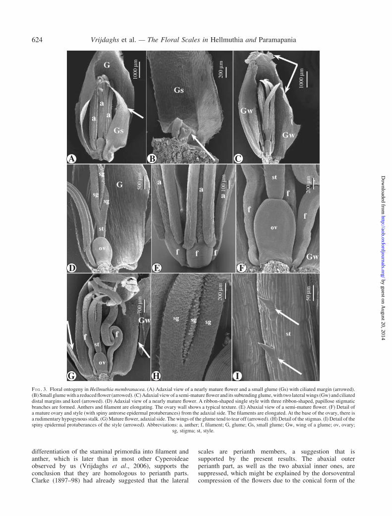
differentiation of the staminal primordia into filament andanther, which is later than in most other Cyperoideaeobserved by us (Vrijdaghs et al., 2006), supports theconclusion that they are homologous to perianth parts.Clarke (1897–98) had already suggested that the lateral
scales are perianth members, a suggestion that issupported by the present results. The abaxial outerperianth part, as well as the two abaxial inner ones, aresuppressed, which might be explained by the dorsoventralcompression of the flowers due to the conical form of the
A B C
D E F
G H I
1000
µm
1000
µm
200
µm
500
µm70
0 µm 20
0 µm
50 µ
m
100
µm
200
µm
F I G . 3. Floral ontogeny in Hellmuthia membranacea. (A) Adaxial view of a nearly mature flower and a small glume (Gs) with ciliated margin (arrowed).(B) Small glumewith a reducedflower (arrowed). (C)Adaxial viewof a semi-mature flower and its subtendingglume,with two lateralwings (Gw) and ciliateddistal margins and keel (arrowed). (D) Adaxial view of a nearly mature flower. A ribbon-shaped single style with three ribbon-shaped, papillose stigmaticbranches are formed. Anthers and filament are elongating. The ovary wall shows a typical texture. (E) Abaxial view of a semi-mature flower. (F) Detail ofa mature ovary and style (with spiny antrorse epidermal protuberances) from the adaxial side. The filaments are elongated. At the base of the ovary, there isa rudimentary hypogynous stalk. (G)Mature flower, adaxial side. Thewings of the glume tend to tear off (arrowed). (H) Detail of the stigmas. (I) Detail of thespiny epidermal protuberances of the style (arrowed). Abbreviations: a, anther; f, filament; G, glume; Gs, small glume; Gw, wing of a glume; ov, ovary;
sg, stigma; st, style.
624 Vrijdaghs et al. — The Floral Scales in Hellmuthia and Paramapania
by guest on August 20, 2014
http://aob.oxfordjournals.org/D
ownloaded from

‘spikelet’ axis. The early development of the lateral scalesresembles the ontogeny of the inner perianth parts inFuirena.
The detailed floral ontogenetic results in Exocaryapublished by Richards et al. (2006), show a similardevelopmental sequence for the lateral scales as inParamapania. Figures 14–21 of this publication showthat the lateral scales in Exocarya are actually the veryfirst ‘floral’ primordia to appear, followed by the lateralstamen primordia. The point of attachment of the lateralscales in Paramapania, neatly below the point ofattachment of the filaments, suggests that the lateralscales do not belong to the reproductive unit. Thedeveloping lateral scales seem to envelop and protect the‘floral apex’, without being part of it. The abaxial andadaxial scales in Exocarya, as shown by Richards et al.(2006), originate apparently at the same positions andrelative time as perianth parts in a cyperoid flower. InParamapania, the lateral scales also originate and develop
before the appearance of any other structure on the ‘floralapex’. The two lateral scale primordia develop fast,enveloping the ‘floral apex’ (floral primordium?), onwhich the first (lateral) stamen primordia appear onlywhen the lateral scales have already been well developed.This is a different floral ontogenetic sequence from thegeneral scirpoid floral ontogenetic pattern observed byVrijdaghs et al. 2006. Therefore, the lateral scales inParamapania are not considered to be homologous withthe lateral perianth parts in Hellmuthia.
The presence of antrorse, spiny cells on the style(Fig. 3F, H, I) and anthers (Fig. 2H) is peculiar. Sincethe style is deciduous (Haines and Lye, 1976, fig. 3A,p. 64; Goetghebeur, 1998), these hook-shaped epidermalprotuberances directed distally cannot play a role in thedistribution of the fruit. They might protect the plantinflorescence against herbivores. Pollen grains inHellmuthia are medium sized compared with otherCyperoideae (Fig. 6A–D). They have a typical cyperoid
100
µm
50 µ
m
500
µm
1000
µm
100 µm
A B C
D EF I G . 4. Floral ontogeny in Hellmuthia membranacea. (A) Detail of the base of an anther in a developing stamen. The base of each pollen sac is prolongedproximally (arrowed). A broad connective can be observed in between the pollen sacs. The filament is wide and ribbon-shaped. (B) Detail of the connectivecrest (apiculus) on the top of a developing anther. (C) Small glume (Gs) with ciliated margin, subtending a reduced flower (arrowed). (D) Flower subtendingglume with ciliated distal parts of the margins and glume apex, and wings that fold back (arrowed). (E) ‘Spikelet’ with developing early floral primordia(arrowed), removed from its subtending glume-like bract in the proximal part of a conical ‘spikelet’. Abbreviations: Ap, piculus; co, connective; f, filament;
Gs, small glume; ps, pollen sac.
Vrijdaghs et al. — The Floral Scales in Hellmuthia and Paramapania 625
by guest on August 20, 2014
http://aob.oxfordjournals.org/D
ownloaded from

200 µm
200 µm
200 µm
200
µm20
0 µm
100 µmA B
C D
E FF I G . 5. Images of transverse LM sections through the ‘spikelet’ of Hellmuthia membranacea. (A) Section through a flower at the more distal part ofa ‘spikelet’. (B) Section through the distal part of a ‘spikelet’. In ‘1’ and ‘2’ a transverse section through the basal part of the floral primordium shows threestamen primordia around the floral axis. Floral primordium ‘2’ is older and the section through it is higher. The tranverse section through flower ‘3’ is made atthe transition between the just formed filament and anther. In the centre of the flower the ovary wall surrounding the central ovule primordium is visible. Theglumes (G1–G3) are imbricate. Glume ‘G1’ encloses a developing flower primordiumaswell as an undifferentiated primordial zone (arrowed). (C) Section atthe height of the style of a developing flower and through the central part of the rachilla, with numerous, circulary positioned vessels (v). Opposite each lateralstamen there is a flat and plied perianth part (arrowed). (D) Section through the central part of the ovary of a developing flower, and through the filaments (f) ofthe lateral stamens and the anther of the abaxial stamen (a). The perianth part (pp) at the left-hand side is fusedwith a tissue (primordial adaxial perianthpart) inbetween the adaxial side of the ovary and rachilla (arrowed). (E) Section through a developing flower, with two free lateral perianth parts and an adaxialperianth part fused with the more basal central ovary wall (arrowed). (F) Section at the height of the transition between style and stigma branches througha developing flower. Two lateral and a free adaxial perianth part are visible (arrowed). Abbreviations: a, anther; g, gynoecium; f, filament; G, glume;
pp, perianth part; v, vessel.
626 Vrijdaghs et al. — The Floral Scales in Hellmuthia and Paramapania
by guest on August 20, 2014
http://aob.oxfordjournals.org/D
ownloaded from

morphology, mostly described as triangular to pear-shaped(e.g. Haines and Lye, 1983) or cuneiform (e.g.Goetghebeur, 1998; Wichelen et al., 1999) with sixapertures. Many authors mention four apertures for pollengrains within the Cyperoideae and monoporate pollenwithin the Hypolytreae tribe (e.g. the Carex-like type andthe Mapania type pollen in Haines and Lye, 1983). Pollengrains, however, differ in number of apertures as well asmorphologically within the Cyperoideae and within the
Mapanioideae sensu Simpson (2006) (e.g. Erdtman, 1966;Koyama, 1969; Fernandez, 1987; Bruhl, 1995). Theauthors’ observations (unpublished) confirm that, withinCyperoideae, the number of apertures varies. In Hainesand Lye (1983, p. 20), an SEM photograph of pollen inFuirena is shown, with four equatorial and one polaraperture out of probably six apertures. The pollen surfacein Hellmuthia is similar to a ‘Carex-type’ pollen surface.Simpson et al. (2003) showed that pollen grains in
10 µm 5 µm
5 µm 5 µm
1 µm
2 µm
2 µm
A B
C D
E F GF I G . 6. SEM images of critical point-dried pollen grains in Hellmuthia membranacea. (A) View of a group of pollen grains, seen from the polar side, eachwith a polar aperture (pa). (B) Lateral view of a pollen grain, with three lateral (la) and the polar apertures visible. (C) Lateral view of a pollen grain with twolateral apertures and the zone of the previous adhesion (arrowed)with the three deteriorated pollen grains. (D) Polar viewof a pollen grain. (E)Detail of the thegranulate, perforate pollen surface withmicrospines. (F) Tapetum coveredwith orbicules. (G)Detail of an aperturewith sexinous bodies withmicrospines on
its membrane. Abbreviations: pa, polar aperture; la, lateral (equatorial) aperture.
Vrijdaghs et al. — The Floral Scales in Hellmuthia and Paramapania 627
by guest on August 20, 2014
http://aob.oxfordjournals.org/D
ownloaded from

Hellmuthia are pseudomonads, like most pollen grains inCyperoideae. Opposite the polar aperture (Fig. 6A) is thezone (Fig. 6B, C) where the three deteriorated cells of theformer tetrad are present (Simpson, 2003). FollowingSimpson et al. (2003), the present observations confirmthat the pollen morphology in Hellmuthia is cyperoid(Wichelen et al., 1999) and hence the conclusion iscorroborated that Hellmuthia is better placed away fromMapanioideae, Chrysitriceae.
Muasya et al. (1998, 2000) suggested that Hellmuthiashould not be maintained in the Chrysitricheae sensuGoetghebeur (1998) and that it should be placed closerto Ficinia, Isolepis and Scirpoides. The mature nutletof Hellmuthia membranacea, as illustrated with a SEMimage by Haines and Lye (1976, p. 64, fig. 3A), hasa hypogynous stalk and its morphology resembles thetypical Isolepis-type nutlet (Muasya and Simpson, 2002;Vrijdaghs et al., 2005c). Eventually, as a result ofa cladistic analysis based on plastid DNA sequences, andpollen developmental evidence, Hellmuthia membranaceawas placed in a Cypereae clade, sister to Ficiniagracilis (Muasya et al., 2006; Simpson et al., 2006).
Cypereae have always been defined based on, amongothers, perianthless flowers (e.g. Goetghebeur, 1998).There are, however, indications that flowers withperianth parts do occur within the Cypereae (Muasyaet al., 2006). The basal position of Hellmuthia in theFicinia–Isolepis complex suggests that the tendency inthis clade to reduce the number of perianth parts to zerohas not yet been fully established. As has already beenillustrated in Fuirena (Vrijdaghs et al., 2004), the absenceor presence of perianth parts can no longer be used asa decisive character for the delimitation of genera or tribes.
It is concluded that (a) the ‘spikelet’ in Hellmuthia isactually a reduced partial inflorescence, (b) the third,adaxial ‘floral scale’ and the two lateral ones in someproximal flowers in ‘spikelets’ in Hellmuthia are perianthmembers, (c) the overall floral ontogeny in Hellmuthiaoccurs according to the cyperoid general floral ontogeneticmodel, (d) the lateral ‘floral scales’ in Hellmuthia are nothomologous to the lateral ‘floral scales’ in Paramapania,(e) the present morphological results support the transfer,based on molecular data, of Hellmuthia to a Cypereaeclade.
50 µm 100 µm
100
µm
200
µm
200
µm
200
µm
A B C
D E FF I G . 7. Floral ontogenetic stages inParamapania parvibractea. (A)Apical view of a reproductive unit hidden by two lateral scales at an early developmentalstage. (B) Adaxial view of a subtending glume-like bract with developing lateral scales, which, at this stage, envelop the entire reproductive unit.(C) Successive developmental stage. The distal part of the margins of the lateral scales becomes ciliated. (D) Mature flower with the two massive lateralscales, abaxial view. (E) Mature flower, the lateral scales have been removed. The lateral scales are attached below the point of attachment of the filaments
(arrowed). (F) Beaked nutlet. Abbreviations: f, filament; GB, glume-like bract; isc, inner scale; lsc, lateral scale; sg, stigma; st, style.
628 Vrijdaghs et al. — The Floral Scales in Hellmuthia and Paramapania
by guest on August 20, 2014
http://aob.oxfordjournals.org/D
ownloaded from

ACKNOWLEDGEMENTS
We thank Marc Reynders (Research Group Spermato-phytes, University of Gent) for kindly providing Para-mapania material and DENR-PAWB region 8, Taclobancity, Philippines for providing collection permits, BennyByetebier (Stellenbosch University) for collectingHellmuthia material, Anja Vandeperre for her most pro-fessional help with the LM preparations and drawings, andpalynologists Dr Stefan Vinckier and Dr Suzy Huysmans(Laboratory of Plant Systematics, K.U.Leuven). This workwas supported financially by research grants of theK.U.Leuven (0T/05/35) and the Fund for ScientificResearch-Flanders (FWO-Vlaanderen, Belgium, G.0268�04).A. Muthama Muasya was a visiting postdoctoral fellowof the Fund for Scientific Research-Flanders (FWO-Vlaanderen, Belgium) and of the Research Fund of theK.U.Leuven.
LITERATURE CITEDBruhl JJ. 1995. Sedge genera of the world: relationships and a new
classification of the Cyperaceae. Australian Journal of Botany 8:125–305.
Clarke CB. 1897–98. Cyperaceae. In: Thiselton-Dyer WT, ed. FloraCapensis, Vol. 7. London: Reeve, 266–524.
Clarke CB. 1909. Illustrations of Cyperaceae. London: Williams &Norgate.
Erdtman G. 1966. Pollen morphology and plant taxonomy. Angiosperms.New York, NY: Hafner.
Fernandez I. 1987. Contribucion al conocimiento palinologico deCyperaceae. Acta Botanica Malacitana 12: 173–182.
Goebel K. 1888. Uber den Bau der Archen und Bluten einigerJavanischer Cyperaceen. Annales du Jardin Botanique de Buitenzorg7: 120–140.
Goetghebeur P. 1986. Genera Cyperacearum. Een bijdrage tot dekennis van de morfologie, systematiek en fylogenese van deCyperaceae-genera. PhD Thesis, Groep Plantkunde, Rijksuniver-siteit Gent, Gent.
Goetghebeur P. 1998. Cyperaceae. In: Kubitzki K. ed. The families andgenera of vascular plants. Vol. 4. Berlin/Heidelberg: Springer-Verlag, 141–190.
Haines RW, Lye KA. 1976. Studies in African Cyperaceae. XIV. Thegenus Hellmuthia Steud. Botaniska Notiser 129: 61–68.
Haines RW, Lye KA. 1983. The sedges and rushes of East Africa.Nairobi, Kenya: East African National History Society.
Holttum RE. 1948. The spikelet in Cyperaceae. Botanical Review 14:525–541.
Koyama T. 1969. Delimitation and classification of the Cyperaceae-Mapanioideae. In: Gunkel JE, ed. Current topics in plant science.New York: Academic Press, 201–228.
A B
C
F I G . 8. Reproductions of diagrams. (A) Drawing of a flower (adaxial view) of Hellmuthia membranacea, reproduced from Clarke (1909), plate XLVII,12.The lateral perianth parts are arrowed. (B) Drawing of a flower of Ficinia ixoides, reproduced from Schonland (1922), plate 44). The adaxial perianth part is
arrowed. (C) Floral diagram of Mapania, reproduced from Goebel (1888), plate XIV, 14. The lateral ‘scales’ are arrowed.
Vrijdaghs et al. — The Floral Scales in Hellmuthia and Paramapania 629
by guest on August 20, 2014
http://aob.oxfordjournals.org/D
ownloaded from

Kukkonen I. 1984. On the inflorescence structure in the familyCyperaceae. Annales Botanici Fennici 21: 257–264.
Kunth CS. 1837. Enumeratio plantarum. 2. Cyperographia synoptica.Stutgart: Colla.
Muasya MA. 1998. A synopsis of Fuirena (Cyperaceae) for the flora ofTropical East Africa. Kew Bulletin 53: 187–202.
Muasya AM, Simpson DA. 2002. A monograph of the genus Isolepis R.Br. (Cyperaceae). Kew Bulletin 57: 257–362.
Muasya AM, Simpson DA, Culham A, Chase MW. 1998. Anassessment of suprageneric phylogeny in Cyperaceae usingrbcL DNA sequences. Plant Systematics and Evolution 211:257–271.
Muasya AM, Bruhl JJ, Simpson DA, Culham A, Chase MW. 2000.Suprageneric phylogeny of Cyperaceae: a combined analysis. In:Wilson KL, Morrison DA, eds.Monocots: systematics and evolution.Melbourne, Victoria, Australia: CSIRO, 593–601.
Muasya AM, Vrijdaghs A, Simpson DA, Chase MW, Goetghebeur P,Smets E. 2006. What is a genus in Cypereae: phylogeny, chareacterhomology assessment and generic circumscription. Botanical Review(in press).
Nees von Esenbeck CG. 1835. Uebersicht der Cyperaceengattungen.Linnaea 9: 273–306.
Richards JH, Bruhl JJ, Wilson KL. 2006. Flower or spikelet?—Understanding the morphology and development of reproductivestructures in Exocarya (Cyperaceae, Mapanioideae, Chrysitricheae).American Journal of Botany (in press).
Schonland S. 1922. South African Cyperaceae. Memoirs of the BotanicalSurvey of South Africa 3.
Schultze-Motel W. 1959. Entwicklungsgeschichtliche und vergleichend-morphologische Untersuchungen im Blutenbereich der Cyperaceae.Englers Botanische Jahrbuch der Systematischen Pflanzengeschichteund Pflanzengeographie 78: 129–170.
Simpson DA, Furness CA, Hodkinson TR, Muasya AM, Chase MW.2003. Phylogenetic relationships in Cyperaceae subfamilyMapanioideae inferred from pollen and plastid DNA sequence data.American Journal of Botany 90: 1071–1086.
Simpson DA, Muasya AM, Alves M, Bruhl JJ, Dhooge S, Chase MW,et al. 2006. Phylogeny of Cyperaceae based on DNA sequencedata—a new rbcL analysis. In: Monocots III/Grasses IV. Claremont,CA: ALISO, Rancho Santa Ana Botanic Garden (in press).
Steudel EG. 1855. Synopsis plantarum glumacearum 2. Cyperographiasynoptica. Stuttgart: Metzler.
Thunberg PC. 1794. Prodromus Plantarum Capensis. Uppsala.Vrijdaghs A, Goetghebeur P, Muasya AM, Smets E, Caris P. 2004.
The nature of the perianth in Fuirena (Cyperaceae). South AfricanJournal of Botany 70: 587–594.
Vrijdaghs A, Caris P, Goetghebeur P, Smets E. 2005a. Floral Ontogenyin Scirpus, Eriophorum, and Dulichium (Cyperaceae), with specialreference to the perianth. Annals of Botany 95: 1199–1209.
Vrijdaghs A, Goethgebeur P, Smets E. 2005b. Homologyproblems in cyperoid flowers: a floral ontogenetic approach.In: XVII International Botanical Congress, Abstracts. Vienna,Austria, 219.
Vrijdaghs A, Goetghebeur P, Muasya AM, Caris P, Smets E. 2005c.Floral ontogeny in Ficinia Schrad. and Isolepis R.Br. (Cyperaceae),with focus on the nature and origin of the gynophore. Annals ofBotany 96: 1247–1264.
Vrijdaghs A, Muasya AM, Goetghebeur P, Caris P, Nagels A, Smets E.2006. A floral ontogenetic approach to homology questions withinthe Cyperoideae (Cyperaceae). Botanical Review (in press).
Wichelen J, Camelbeke K, Chaerle P, Goetghebeur P, Huysmnans S.1999. Comparison of different treatments of LM and SEM studiesand systematic value of pollen grains in Cyperaceae. Grana 38:50–58.
630 Vrijdaghs et al. — The Floral Scales in Hellmuthia and Paramapania
by guest on August 20, 2014
http://aob.oxfordjournals.org/D
ownloaded from