THE FLORAL STRUCTURE OF THREE WEEDY SPECIES OF SIDA (MALVACEAE)
11
THE FLORAL STRUCTURE OF THREE WEEDY SPECIES OF SIDA (MALVACEAE) Junior Cezar Muneratto Luiz Antonio de Souza Universidade Estadual de Maringá Universidade Estadual de Maringá Departamento de Biologia Departamento de Biologia Av. Colombo, 5790 Av. Colombo, 5790 (87020-900) Maringá, PR, BRAZIL (87020-900) Maringá, PR, BRAZIL [email protected] Odair José Garcia de Almeida Instituto de Biociências UNESP – Univ. Estadual Paulista Programa de Pós-Graduação em Biologia Vegetal Av. 24 A, 1515, (13506-900) Rio Claro, SP, BRAZIL [email protected] ABSTRACT Malvaceae sensu lato is monophyletic and is characterized by the presence of nectaries of glandular trichomes, located internally at the base of the calyx. The genus Sida has been heterogeneous since its origin; however, there has been a reduction in the size of the genus with the removal of many species to other genera, giving a more natural and definable residual group subdivided into 11 sections. The aims of this work were to describe and compare the floral structure of three weedy species Sida rhombifolia, S. urens, and S. regnellii, using a traditional anatomical approach, along with scanning electron microscopy analysis. The comparative study of the three Sida species allows us to sug- gest that some structural characters are shared, i.e., the hypodermis in the sepal and ovary wall, trichomatous calyx nectary, perianth with homogeneous mesophyll, and anther with a middle layer and plasmodial tapetum type, and they may reinforce the natural residual group of Sida. On the other hand, other floral characters have some diagnostic value for classification at the species level, such as the style structure being solid or rift in the center. KEY WORDS: Floral anatomy, calyx nectary, trichomes, weedy species RESUMO Malvaceae sensu lato é monofilética e se caracteriza pela presença de nectários que consistem de tricomas glandulares localizados interna- mente na base do cálice. O gênero Sida é considerado como heterogêneo na sua concepção inicial, mas ultimamente ele tem sido reduzido de tamanho com a remoção de várias espécies para outros gêneros, tornando-o um grupo natural residual subdividido em 11 seções. O objetivo deste trabalho foi descrever e comparar a estrutura floral de três espécies invasoras, Sida rhombifolia, S. urens e S. regnellii, utilizando-se de estudo anatômico tradicional e análise em microscopia eletrônica de varredura. O estudo comparativo das três espécies de Sida permite sugerir que alguns caracteres estruturais são comuns às espécies, como presença de hipoderme na sépala e parede do ovário, nectário cali- cino tricomatoso, perianto com mesofilo homogêneo, e antera com uma camada média e tapete plasmodial, o que reforça o gênero Sida como um grupo natural residual. Por outro lado, outros caracteres florais têm algum valor diagnóstico para separação das espécies, como, por exemplo, a estrutura do estilete que pode ser sólido ou apresentar fenda na região central. INTRODUCTION When circumscribed broadly to include four families (i.e., Tiliaceae, Sterculiaceae, Bombacaceae, and Malva- ceae – sensu stricto), Malvaceae sensu lato is monophyletic (Judd et al. 2002). It is characterized by the pres- ence of nectaries of glandular trichomes, located internally at the base of the calyx or, with less frequency, on the petals or on the androgynophore (Judd & Manchester 1997). This circumscription is also supported by adnation of the androecium to the corolla and usually the presence of unilocular anthers, although Tiliaceae and Sterculiaceae often retain numerous stamens with 2-locular anthers (Judd et al. 2002). More recently, Malvaceae sensu lato has been divided into nine subfamilies (as discussed in Tate et al. 2005): Bombacoideae (formerly Bombacaceae, in part), Brownlowioideae, Byttnerioideae, Dombeyoideae, Grewioideae, Helicteroi- deae, Malvoideae (formerly Malvaceae), Sterculioideae (formerly Sterculiaceae, in part), and Tilioideae (for- merly Tiliaceae, in part). The Malvoideae are sometimes called the “core” Malvaceae s.str., and some authors J. Bot. Res. Inst. Texas 8(1): 127 – 137. 2014
Transcript of THE FLORAL STRUCTURE OF THREE WEEDY SPECIES OF SIDA (MALVACEAE)

THE FLORAL STRUCTURE OF THREE WEEDY SPECIES OF SIDA (MALVACEAE)
Junior Cezar Muneratto Luiz Antonio de Souza Universidade Estadual de Maringá Universidade Estadual de Maringá Departamento de Biologia Departamento de Biologia Av. Colombo, 5790 Av. Colombo, 5790 (87020-900) Maringá, PR, BRAZIL (87020-900) Maringá, PR, BRAZIL [email protected]
Odair José Garcia de AlmeidaInstituto de Biociências
UNESP – Univ. Estadual PaulistaPrograma de Pós-Graduação em Biologia Vegetal
Av. 24 A, 1515, (13506-900) Rio Claro, SP, [email protected]
absTracT
Malvaceae sensu lato is monophyletic and is characterized by the presence of nectaries of glandular trichomes, located internally at the base
of the calyx. The genus Sida has been heterogeneous since its origin; however, there has been a reduction in the size of the genus with the
removal of many species to other genera, giving a more natural and definable residual group subdivided into 11 sections. The aims of this
work were to describe and compare the floral structure of three weedy species Sida rhombifolia, S. urens, and S. regnellii, using a traditional
anatomical approach, along with scanning electron microscopy analysis. The comparative study of the three Sida species allows us to sug-
gest that some structural characters are shared, i.e., the hypodermis in the sepal and ovary wall, trichomatous calyx nectary, perianth with
homogeneous mesophyll, and anther with a middle layer and plasmodial tapetum type, and they may reinforce the natural residual group of
Sida. On the other hand, other floral characters have some diagnostic value for classification at the species level, such as the style structure
being solid or rift in the center.
key words: Floral anatomy, calyx nectary, trichomes, weedy species
resuMo
Malvaceae sensu lato é monofilética e se caracteriza pela presença de nectários que consistem de tricomas glandulares localizados interna-
mente na base do cálice. O gênero Sida é considerado como heterogêneo na sua concepção inicial, mas ultimamente ele tem sido reduzido de
tamanho com a remoção de várias espécies para outros gêneros, tornando-o um grupo natural residual subdividido em 11 seções. O objetivo
deste trabalho foi descrever e comparar a estrutura floral de três espécies invasoras, Sida rhombifolia, S. urens e S. regnellii, utilizando-se de
estudo anatômico tradicional e análise em microscopia eletrônica de varredura. O estudo comparativo das três espécies de Sida permite
sugerir que alguns caracteres estruturais são comuns às espécies, como presença de hipoderme na sépala e parede do ovário, nectário cali-
cino tricomatoso, perianto com mesofilo homogêneo, e antera com uma camada média e tapete plasmodial, o que reforça o gênero Sida como
um grupo natural residual. Por outro lado, outros caracteres florais têm algum valor diagnóstico para separação das espécies, como, por
exemplo, a estrutura do estilete que pode ser sólido ou apresentar fenda na região central.
inTroducTion
When circumscribed broadly to include four families (i.e., Tiliaceae, Sterculiaceae, Bombacaceae, and Malva-ceae – sensu stricto), Malvaceae sensu lato is monophyletic (Judd et al. 2002). It is characterized by the pres-ence of nectaries of glandular trichomes, located internally at the base of the calyx or, with less frequency, on the petals or on the androgynophore (Judd & Manchester 1997). This circumscription is also supported by adnation of the androecium to the corolla and usually the presence of unilocular anthers, although Tiliaceae and Sterculiaceae often retain numerous stamens with 2-locular anthers (Judd et al. 2002). More recently, Malvaceae sensu lato has been divided into nine subfamilies (as discussed in Tate et al. 2005): Bombacoideae (formerly Bombacaceae, in part), Brownlowioideae, Byttnerioideae, Dombeyoideae, Grewioideae, Helicteroi-deae, Malvoideae (formerly Malvaceae), Sterculioideae (formerly Sterculiaceae, in part), and Tilioideae (for-merly Tiliaceae, in part). The Malvoideae are sometimes called the “core” Malvaceae s.str., and some authors
J. Bot. Res. Inst. Texas 8(1): 127 – 137. 2014

128 Journal of the Botanical Research Institute of Texas 8(1)
continue to retain this family name for this core group alone. Prior to that, Bayer and Kubitzki (2003) divided the subfamily Malvoideae into four tribes: Gossypieae, Hibisceae, Kydieae, and Malveae. Traditionally, mem-bers of the Malveae have been characterized by a combination of several morphological characters: schizocar-pic fruits (sometimes a capsule), three to over 20 mericarps with an equal number of free styles, antheriferous apex of the staminal column and the absence of lysigenous cavities (“gossypol glands”) (Fryxell 1988; Bayer & Kubitzki 2003; Tate et al. 2005). According to Fryxell (1985), the genus Sida has been heterogeneous since its origin as botanists tended to put into Sida any member of the Malvaceae that are uniovulate and without an involucel. However, there has been a reduction in the size of the genus with the removal of many species to other genera, giving a more natu-ral and definable residual group. Fryxell (1985) subdivided this residual group into 11 sections. The aims of this study were to describe and compare the floral structure of three weedy species of Sida, namely S. rhombifolia L., S. urens L., and S. regnellii R.E. Fr. Economically, the Malvaceae s.l. are important ei-ther ornamentally (for instance, the genera Alcea, Hibiscus, and Malvaviscus) or in the textile industry (e.g. Gossypium and Urena), or as weedy plants, such as Sida, a genus that can be very detrimental to the agricul-tural economy (Bovini et al. 2001).
MaTerial and MeThods
Plant materialFloral buds and flowers of the three weedy species were collected in the city of Maringá, Brazil (state of Paraná): S. rhombifolia at 506 m altitude, 23°24'13.3" latitude and 51°56'21.17" longitude; S. regnellii at 559 m altitude, 23°24'43.0" latitude and 51°56'29.6" longitude; and S. urens at 518 m altitude, 23°24'13.1" latitude and 51°56'20.4" longitude. Voucher materials were identified by Massimo Giuseppe Bovini (RB) and deposited at HUEM (Herbarium of the Universidade Estadual de Maringá) with these accession numbers: S. urens:19909 (J. Muneratto 001); S. rhombifolia:19910 (J. Muneratto 002); and S. regnellii:19911 (J. Muneratto 003).
Anatomical analysisThe material was fixed in glutaraldehyde (1% in 0.1M phosphate buffer, pH 7.2) and then conserved in 70% ethanol (Johansen 1940). The fixed material was embedded in historesin (Gerrits 1991), sectioned (cross- and longitudinally) in a rotation microtome, and stained in toluidine blue (O’Brien et al. 1964). Specific micro-chemical tests were done for lipid substances (Sudan IV and Sudan Black), tannins (ferric chloride), starches (iodine-potassium iodide test), lignins (phloroglucin test) and calcium crystals (sulfuric acid) (Sass 1951; Rawlins & Takahashi 1952; Ruzin 1999). Images were taken using Leica ICC50 and Olympus BX50 optical microscopes with digital camera attachments.
Scanning Electronic Microscopy analysisMicromorphological analysis of the floral buds and flowers was done with material fixed in Karnovsky solu-tion (Karnovsky 1965). Samples were processed and then mounted on aluminum stubs, gold coated, examined using scanning electron microscopy (Shimadzu SS-550 Superscan), and digitally photographed.
resulTs
PerianthThe flowers (Fig. 1a–c) in all three of the Sida species investigated are actinomorphic, hermaphrodite, with yellowish (S. urens and S. rhombifolia - Fig. 1a,b) and whitish (S. regnellii - Fig. 1c) petals, distinct from the green calyx (Table 1). The five sepals are green in color, connate in the basal half, and with free triangular lobes. The uniseriate adaxial epidermis is made of cuboid or tabular cells with outer and inner periclinal walls thicker than the an-ticlinal ones (Fig. 2a,a’). The abaxial sepal epidermis is uniseriate with trichomes and stomata in all species and has cells with sinuous anticlinal walls in S. rhombifolia and S. urens. The stomata found in the sepals of the species are anisocytic and paracytic in S. regnellii and S. rhombifolia and anisocytic in S. urens (Table 1). The trichomes are non-glandular and glandular (Fig. 1d,f,g–i). Unicellular non-glandular trichomes occur only in

Muneratto et al., Floral structure of Sida species 129
Fig. 1. General morphology of the flower in frontal view of Sida rhombifolia (a), S. urens (b), and S. regnellii (c); scanning electron micrographs (SEM) of the calyx surface showing trichomes in S. regnellii (d–g), d. Abaxial face, apical region. e. Nectary (trichomes). f–g. Abaxial face. Sida urens (h–i). h. Adaxial surface. i. Abaxial face. Arrowheads indicate unicellular trichomes, long arrow shows stellate trichomes, and short and interrupted arrows indicate glandular trichomes with a small and long stalk, respectively. Scale bars: 0.5 cm (a,c), 1.5 cm (b), 50 µm (d,f,g), 100 µm (e,h,i).
Table 1. Flower characters which have diagnostic value for separation of Sida rhombifolia, Sida urens, and Sida regnellii.
Characters Sida rhombifolia Sida urens Sida regnellii
Flowers Yellowish Yellowish WhitishSepal epidermis abaxial Sinuous anticlinal walls Sinuous anticlinal walls Straight anticlinal wallsSepal stomata Anisocytic/paracytic Anisocytic Anisocytic/paracyticUnicellular nonglandular trichomes in sepals Absent Present AbsentSepal spongy parenchyma 2–3 cell layers 2–6 cell layers 4–5 cell layersStalk base/glandular trichomes (nectary) 3–4 layers 3–4 layers 1–2 layersPetal shape Asymmetric Obcordate AsymmetricPetal fasciculate trichome Present Absent PresentOvary carpels 9–11 5 5Ventral vascular bundles of the ovary 9–11 5 5Style Hollow/solid in the base Solid Solid with reduced rift
S. urens (Fig. 1i), and this species has epidermal cells completely surrounding the base of the trichome. Stellate, multicellular, non-glandular trichomes occur on the sepals of all three species (Fig. 1g). Also occurring in these three species are multicellular glandular trichomes (Fig. 1g–i), consisting of a small stalk and a head of several secretory cells or a long stalk and a unicellular secretory head. In the adaxial surface of the Sida sepals are found crystalliferous cells (druses) in the hypodermis, which have a U-shaped wall thickness (Fig. 2a,a’).

130 Journal of the Botanical Research Institute of Texas 8(1)
The S. rhombifolia has a lesser developed hypodermis in the abaxial surface (Fig. 2a,a’). The mesophyll (Fig. 2a) has spongy parenchyma with mucilaginous cells and cavities, exhibiting two or three cell layers in S. rhombi-folia, four or five in S. regnellii, and two to six in S. urens. The nectary (Fig. 1e; 2b–c) occurs close to the base of the calyx (Fig. 1e) and is a carpet-like tissue made of multicellular clavate glandular trichomes and a subtending reduced secretory parenchyma, which is sup-plied by xylem and a greater proportion of phloem elements. The glandular trichomes have a uniseriate or pluriseriate stalk and a unicellular head (Fig. 2c). The trichomes are longer in S. rhombifolia than in the other species; the stalk base of this S. rhombifolia and S. urens has a thickness of three or four cell layers, while S. regnellii has a base with one or two layers (Table 1). The corolla shows adnation to the androecium, and the petal shape varies among the Sida species. Sida urens has symmetrical obcordate petals (Fig. 1b; 2f), while in S. rhombifolia (Fig. 1a) and S. regnellii the petals are obcordate but very asymmetrically so. The petal epidermis of S. rhombifolia and S. regnellii (Fig. 2d,e, re-spectively) is uniseriate with mucilage idioblasts and glandular and non-glandular trichomes. The mesophyll (Fig. 2d,e) is composed of a homogeneous parenchyma with mucilage cells and cavities. The petal vasculariza-tion (Fig. 2d,e) consists of a central bundle and six to nine minor bundles.
AndroeciumThe androecium consists of filaments joined to form a tube (filament tube) (Figs. 3b; 5e) that surrounds the style (Figs. 4a,c; 5b–f). The anthers (Fig. 3a) are dorsifixed, bisporangiate, longicidal, and septate monothecal. The filament tube is composed of internal and external uniseriate epidermis of cuboid or elongated cells and capitate glandular trichomes; the inner parenchyma has mucilage cells (Fig. 4a,c), and the vascular system consists of 10 collateral vascular bundles arranged in pairs. The wall of the young anther (Fig. 3c) has epidermis, endothecium with thin-walled cells, a middle layer of elongated cells and a tapetum made of a layer of cells. Later, the cell walls of the tapetum undergo disintegra-tion, and the protoplasts of the tapetum cells remain available among the pollen grains during their develop-ment (Fig. 3d). In the developing anther, the endothecium cells acquire thickenings in the anticlinal walls and in the inner periclinal wall, the middle layer disintegrates, and the tapetum protoplast is absorbed (Fig. 3e, f). The connective consists of papillose epidermis, parenchyma with druse idioblasts and one vascular bundle.
GynoeciumThe structure of the style differs among the three species (Table 1) when seen in cross section. In S. rhombifolia the style is hollow for the most part (Fig. 4b), being solid only in the base; in S. regnellii the style has a reduced rift (Fig. 4c); and S. urens, in turn, has an entirely solid style (Fig. 4d). The style is composed of uniseriate epi-dermis, parenchyma and strands of transmitting tissue, one for each carpel, with 11 in S. rhombifolia (Fig. 4b) and five in the other two species (Fig. 4d). The stigma consists of papillate epidermis (Fig. 4e,f). The ovary presents many carpels and locules (Fig. 5a–e) (five in S. regnellii and S. urens, and nine to 11 in S. rhombifolia) (Table 1). There is one ovule in each locule, and the placentation is axial (Fig. 5b–f). The style consists of a single column (solid or rifted, depending of the species, according to the description above) (Fig. 5b–f). Close to the apex the style starts to lose the fused shape and separates into free branches, which corre-spond to each stigma lobe, in which the number of stigma lobes and locules is the same. The ovary wall (Fig. 6a–f) has glabrous uniseriate outer epidermis with cuboid cells in S. regnellii and S. urens, and cuboid to cylin-dric in S. rhombifolia. The ovary mesophyll (Fig. 6a–f) is parenchymatous, spongy and homogeneous with two or three cell layers in the middle region; the remainder (base and apex) of the ovary consists of five cell layers. A crystalliferous hypodermis (druse cells) is present in the wall ovary (Fig. 6b), although some cells lack crys-tals in S. urens. The inner epidermis undergoes cell division periclinally to the ovary surface resulting in a tis-sue with two or three cell layers (Fig. 6b), the precursor of the endocarp. In the ovary, all species studied here had septa (Fig. 6a,c,e) made up of an epidermis of one or two layers of cells and bi- to multilayered spongy parenchyma tissue with scattered druse idioblasts. The ovary vascula-ture is made of a median dorsal bundle (Fig. 6b,d), one or two lateral bundles, and a ventral or placental bundle. The number of ventral bundles differs among the species, with nine to 11 bundles in S. rhombifolia (Fig. 6e) and

Muneratto et al., Floral structure of Sida species 131
Fig. 2. Perianth structure of S. rhombifolia (a,a’,d), S. regnellii (c,e), and S. urens (b,f), in cross-section and scanning electron micrograph (SEM). a. Calyx. a’. Details of the epidermis. b. Nectary in SEM. c. Nectary longitudinal section. d–e. Petals. f. Petal base in SEM. Arrow indicates hypodermis cells with U-shaped wall thickness. Scale bars: 100 µm (a,c,e,f), 30 µm (a’), 50 µm (b,d).
five in S. regnellii (Fig. 6c) and S. urens, which corresponds to the same number as the carpels and styles, and the same numbers of mericarps in the developed fruit (Muneratto & Souza 2013). In the parenchyma close to the ventral bundles (Fig. 6a,c,e), there are mucilaginous and druse cells. The ovules (Fig. 6a–f) are bitegmic, crassinucellate, with an outer integument made up of two layers of cell, and multiseriate inner integument.
discussion
The trichomatous calyx nectary of the Sida species analyzed here is one of the features that characterize the core Malvales (Malvaceae s.l. including Bombacaceae, Malvaceae, Sterculiaceae, and Tiliaceae) (Vogel 2000;

132 Journal of the Botanical Research Institute of Texas 8(1)
Leitão et al. 2005). According to Vogel (2000), the trichomatous calyx nectary may have evolved from hyda-thodes, which provide moisture to protect floral primordia from desiccation, being active in the bud stage. As discussed by Bernardello (2007), trichomes are by far the most common epidermal nectaries in Angiosperms, and the nectariferous trichomes may have taxonomic importance in defining related plants groups, such as Bombacoideae and Malvoideae.
Fig. 3. Anther structure and filament tube of S. regnellii (a,b,f); anther structure of S. urens (c) and S. rhombifolia (d,e). a. Mature anthers - SEM. b. Apex of the filament tube with glandular trichomes - SEM. c. Immature anther in cross-section. d. Anther detail visualizing plasmodial tapetum among microspores. e. Immature anther in cross-section without middle layer and tapetum. f. Mature anther in cross-section with septum. * indicates the stigma branches/lobes. White arrow shows septum of the anther. Black arrow indicates the tapetum protoplast among microspores in development. (en = endothecium; ml = middle layer; ta = tapetum). Scale bars: 200 µm (a), 50 µm (b–f).
Muneratto et al., Floral structure of Sida species 133
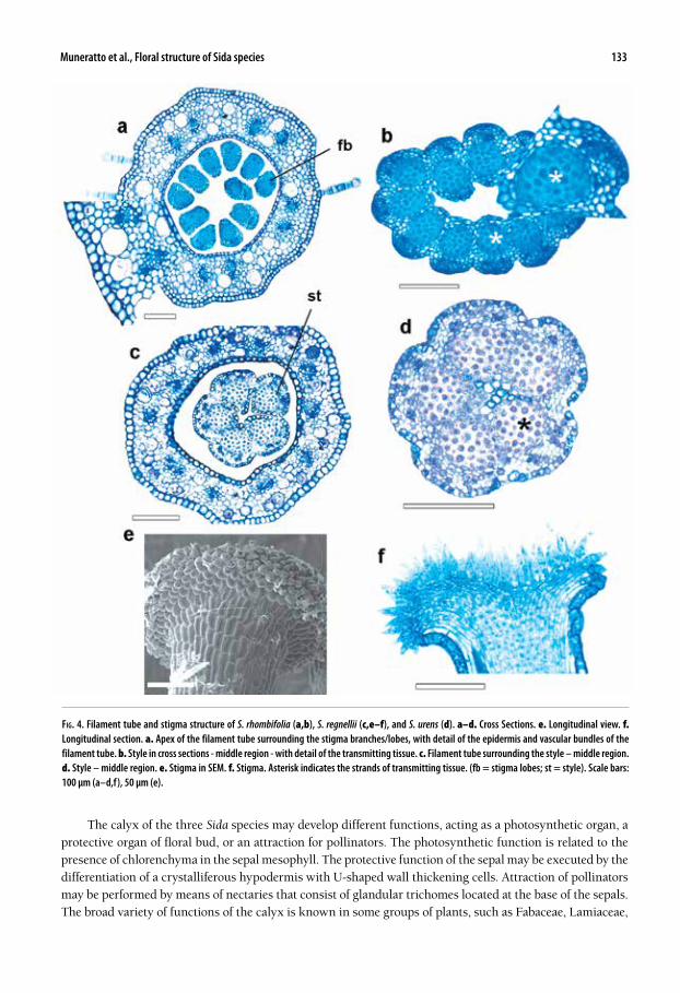
The calyx of the three Sida species may develop different functions, acting as a photosynthetic organ, a protective organ of floral bud, or an attraction for pollinators. The photosynthetic function is related to the presence of chlorenchyma in the sepal mesophyll. The protective function of the sepal may be executed by the differentiation of a crystalliferous hypodermis with U-shaped wall thickening cells. Attraction of pollinators may be performed by means of nectaries that consist of glandular trichomes located at the base of the sepals. The broad variety of functions of the calyx is known in some groups of plants, such as Fabaceae, Lamiaceae,
Fig. 4. Filament tube and stigma structure of S. rhombifolia (a,b), S. regnellii (c,e–f), and S. urens (d). a–d. Cross Sections. e. Longitudinal view. f. Longitudinal section. a. Apex of the filament tube surrounding the stigma branches/lobes, with detail of the epidermis and vascular bundles of the filament tube. b. Style in cross sections - middle region - with detail of the transmitting tissue. c. Filament tube surrounding the style – middle region. d. Style – middle region. e. Stigma in SEM. f. Stigma. Asterisk indicates the strands of transmitting tissue. (fb = stigma lobes; st = style). Scale bars: 100 µm (a–d,f), 50 µm (e).

134 Journal of the Botanical Research Institute of Texas 8(1)
Malpighiaceae, and Ranunculaceae (Roth 1977; Weberling 1992; Endress 1994). Unfortunately we do not have supplementary data concerning the time of activity about the trichomatous calyx nectary in the Sida species nor information concerning the visitors of these flowers, in order to ascertain the role of these nectaries in the pollination system of these species. However, we can assume a nuptial function (i.e., they are involved in pol-lination events, sensu Almeida et al. 2013) of the carpet-like nectaries on the sepals of Sida species, since the flowers of Malvaceae s.l. (Vogel 2000) do not have an annular nectary (nectariferous disk) to attract the pollina-
Fig. 5. Morphology of the ovary of S. rhombifolia (a,b), S. urens (c,d), and S. regnellii (e,f). a, c, e. Ovary/gynoecium in SEM. b, d, f. Flower in longitudinal section showing ovary and other floral organs. (an = filament tube; ne = nectary; pe = petal; se = sepal; st = style). Scale bars: 500 µm (a,e) 250 µm (b,c,d,f).

Muneratto et al., Floral structure of Sida species 135
tors, and these nectaries can be accessed via the gaps at the base of the corolla lobes in the region of adnation between petals and the filament tube (Vogel 2000; Bernardello 2007). Dahlgren (1991) introduced a classification based on Davis (1966) of the different types of anther wall formations, in which four types were presented: basic, dicotyledonous, monocotyledonous, and reduced. He reported the basic type of anther wall formation, characterized by two middle layers, for some taxa of Malvales. In contrast, the Sida species studied here has only one single middle layer in the anther wall, placing it clearly
Fig. 6. Ovary and ovule structure of S. urens (a,b,f), S. regnellii (c, d), and S. rhombifolia (e) in cross-sections (a–e) and longitudinal section (f). (ca = calyx; co = corolla; oi = outer integument; ov = ovule; ow = ovary wall). Asterisk indicates the vascular bundle, and the black arrow shows the septum. Scale bars: 50 µm.

136 Journal of the Botanical Research Institute of Texas 8(1)
in the dicotyledoneous type. Other species of Malvaceae also have only one middle layer in the anther wall, such as Sida cordifolia L. (Rao 1954) and Modiolastrum malvifolium (Griseb.) K. Schum. (Galati et al. 2007), in-dicating the necessity for new studies concerning the anther development in this group of plants. In all Sida species investigated here, the tapetum is of the plasmodial type, although fusion between pro-toplasts of the microspores was not observed. Embryological studies in Malvaceae (Rao 1954) showed that as the microspores enlarge, the protoplasts of the tapetum cells become smaller and smaller until they are ab-sorbed; in addition, according to Rao (1954), they may disappear or their remnants may persist until the pollen grains have become 2-nucleate and nearly mature, as shown in Sida carpinifolia Mill. The style may be hollow or solid, depending on the degree of closure of the fused or free carpels (Fahn 1990). The analyzed styles of the Sida flowers are quite distinct. The solid type is restricted to S. urens, and hol-low type with solid base occurs in S. rhombifolia. Unlike these two species, S. regnellii has a narrow single canal occupying the center of all styles. The style structure might be a reasonable alternative to the identification of the Sida species. In fact, there has been little research dealing with the style structure. Nevertheless, Souza et al. (2001) found similar results in three species of Trichilia P. Browne (Meliaceae); that is, T. elegans A. Juss. has a solid style, T. catigua A. Juss. a hollow style, while T. pallida Sw. has a narrow canal in its style. This variation in the style structure among species of the same genus may occur in other genera from the clade Eurosids II. Our results showed mucilaginous cells and cavities in the flowers of the three species, which usually oc-curs in the Malvaceae (Metcalfe & Chalk 1957; Esau 1974; Fahn 1979). However, there are differing opinions concerning the development of cavities in some taxa, as pointed out by Evert (2006). The cavities present in the flowers of Malvaceae have been reported as both schizogenous and lysigenous (Fahn 1979 and references therein). Metcalfe and Chalk (1957) recorded lysigenous secretory glands (cavities) in the leaves of Malvaceae species. Even though for Evert (2006) the concept of lysigenous cavity development is questionable, here, we consider it as lysigenous since they are formed by the disintegration of mucilaginous cells and surrounding parenchymatous cells. The comparative study of these three Sida species allows us to suggest that some structural characters are shared, i.e., the hypodermis in the sepal and ovary wall, trichomatous calyx nectary, perianth with homoge-neous mesophyll, anther with a middle layer and plasmodial tapetum type. These characters may reinforce the natural residual group of Sida as proposed by Fryxell (1985). On the other hand, Table 1 shows some floral characters which may have some diagnostic value for classification at the species level, such as the style struc-ture being solid or rift in the center.
acknowledgments
We thank CAPES (Coordenação de Aperfeiçoamento de Pessoal de Nível Superior, Brazil) and CNPq (Con-selho Nacional de Desenvolvimento Científico e Tecnológico, Brazil) for the support granted to realize this study. The authors are grateful to Denver Falconer, Steven R. Hill, and an anonymous reviewer for critical re-view of early versions of the manuscript. We also would like to thank Massimo Giuseppe Bovini for the identi-fication of the voucher materials.
references
AlmeidA, O.J.G., A.A.S. PAOli, & H. COtA-SánCHez. 2013. The systematic significance of floral morphology, nectaries, and nec-tar concentration in epiphytic cacti of tribes Hylocereeae and Rhipsalideae (Cactaceae). Perspect. Pl. Ecol. Evol. Syst. 15:255–268. http://dx.doi.org/10.1016/j.ppees.2013.08.001.
BAyer, C. & K. KuBitzKi. 2003. Malvaceae. In: K. Kubitzki and C. Bayer, eds. Flowering plants, dicotyledons: Malvales, Cap-parales, and nonbetalain Caryophyllales. Springer-Verlag, Berlin, Germany. Pp. 225–311.
BernArdellO, G. 2007. A systematic survey of floral nectaries. In: S.W. Nicolson, M. Nepi, and E. Pacini, eds. Nectaries and nectar. Springer, Netherlands. Pp. 19–128.
BOvini, M.G. 2001. Malvaceae A. Juss. no Parque Estadual do Rio Doce Minas Gerais, Brasil. Rodriguésia 52:17–47.dAHlGren, G. 1991. Steps toward a natural system of the Dicotyledons: Embryological characters. Aliso 13:107–165.dAviS, G.L. 1966. Systematic embryology of the Angiosperms. John Wiley, New York, U.S.A.

Muneratto et al., Floral structure of Sida species 137
endreSS, P.K. 1994. Diversity and evolutionary biology of tropical flowers. Cambridge University Press, Cambridge, UK.evert, R.F. 2006. Esau’s plant anatomy (Meristems, cells, and tissues of the plant body: Their structure, function, and
development). John Wiley & Sons, Hoboken, New Jersey, U.S.A.eSAu, K. 1974. Anatomia das plantas com sementes. Edgard Blücher, São Paulo, Brazil.FAHn, A. 1979. Secretory tissues in plants. Academic Press, London, UK.FAHn, A. 1990. Plant anatomy. IV ed. Pergamon Press, Oxford, UK.Fryxell, P.A. 1985: Sidus sidarum. V. The North and Central American species of Sida. Sida 11(1):62–91.Fryxell, P.A. 1988. Malvaceae of Mexico. Syst. Bot. Monogr. 1–522.GAlAti, B.G., F. mOnACCi, m.m. GOtelli, & S. rOSenFeldt. 2007. Pollen, tapetum and orbicule development in Modiolastrum
malvifolium (Malvaceae). Ann. Bot. 99:755–763.GerritS, P.O. 1991. The application of glycol methacrylate in histotechnology; some fundamental principles. University
Groningen, Netherlands.JOHAnSen, D.A. 1940. Plant microtechnique. McGraw-Hill Book Company, New York, U.S.A.Judd, W.S., C.S. CAmPBell, e.A. KellOGG, P.F. StevenS, & m.J. dOnOGHue. 2002. Plant systematics: A phylogenetic approach, 2nd
edition. Sinauer Associates, Sunderland, Massachusetts. U.S.A.Judd, W.S. & S.r. mAnCHeSter. 1997. Circumscription of Malvaceae (Malvales) as determined by a preliminary cladistic
analysis of morphological, anatomical, palynological, and chemical characters. Brittonia 49:384–405.KArnOvSKy, M.J.A. 1965. Formaldehyde-glutaraldehyde fixative of high osmolality for use in electron microscopy. J. Cell
Biol. 27:137–138.leitãO, C.A.e., r.m.S.A. meirA, A.A. AzevedO, J.m. ArAúJO, K.l.F. SilvA, & r.G. COllevAtti. 2005. Anatomy of the floral, bract, and
foliar nectaries of Triumfetta semitriloba (Tiliaceae). Canad. J. Bot. 83:279–286.metCAlFe, C.r. & l. CHAlK. 1957. Anatomy of the Dicotyledons, V. I and II. Clarendon Press, Oxford, UK.munerAttO, J.C. & l.A. SOuzA. 2013. Fruit (pericarp and seed) ontogeny of Sida species. Gayana Bot. 70:44–56. http://
dx.doi.org/10.4067/S0717-66432013000100006.nyFFeler, r., C. BAyer, W.S. AlverSOn, A. yen, B.A. WHitlOCK, m.W. CHASe, & d.A. BAum. 2005. Phylogenetic analysis of the Malva-
dendrina clade (Malvaceae s.l.) based on plastid DNA sequences. Organisms Diversity Evol. 5:109–123.O’Brien, t.P., n. Feder, & m.e. mCCully. 1964. Polychromatic staining of plant cell walls by Toluidine Blue O. Protoplasma
59:368–373.rAO, V.C. 1954. Embryological studies in Malvaceae I: Development of gametophytes. Proc. Natl. Inst. Sci. India, B.
20:127–150.rAWlinS, t.e. & W.n. tAKAHASHi. 1952. Techniques of plant histochemistry and virology. The National Press, Millbrae, Cali-
fornia, U.S.A.rOtH, I. 1977. Fruits of angiosperms. In: K. Linsbauer, F.G. Tischler, and A. Pascher, eds. Encyclopedia of plant anatomy.
Gebrüder Borntraeger, Berlin, Germany. Pp. 258–291.ruzin, S.E. 1999. Plant microtechnique and microscopy. Oxford University Press, New York, U.S.A.SASS, J.E. 1951. Botanical microtechnique. Iowa State College Press, Iowa, U.S.A.SCHmid, R. 1988. Reproductive versus extra-reproductive nectaries: Historical perspective and terminological recom-
mendations. Bot. Rev. 54:179–232.SOuzA, l.A., i.S. mOSCHetA, K.S.m. mOurãO, & A. SilvériO. 2001. Morphology and anatomy of the flowers of Trichilia catigua A.
Juss., T. elegans A. Juss. and T. pallida Sw. (Meliaceae). Brazil. Arch. Biol. Technol. 44:383–394.tAte, J.A., J.F. AGuiAr, S.t. WAGStAFF, C.A. duCKe, t.A.B. SlOttA, & B.B. SimPSOn. 2005. Phylogenetic relationships within the tribe
Malveae (Malvaceae, subfamily Malvoideae) as inferred from its sequence data. Amer. J. Bot. 92:584–602.vOGel, S. 2000. The floral nectaries of Malvaceae sensu lato. A conspectus. Kurtziana 28:155–171.WeBerlinG, F. 1992. Morphology of flowers and inflorescences. Cambridge University Press, Cambridge, UK.


![[Livre] Homosexualités au temps du sida : tensions sociales et identitaires](https://static.fdokumen.com/doc/165x107/63126f9e5cba183dbf06a6b9/livre-homosexualites-au-temps-du-sida-tensions-sociales-et-identitaires.jpg)