Flexible Electronics and Devices as Human‐Machine ...
110
-
Upload
khangminh22 -
Category
Documents
-
view
2 -
download
0
Transcript of Flexible Electronics and Devices as Human‐Machine ...

This article has been accepted for publication and undergone full peer review but has not been
through the copyediting, typesetting, pagination and proofreading process, which may lead to
differences between this version and the Version of Record. Please cite this article as doi:
10.1002/adma.202107902.
This article is protected by copyright. All rights reserved.
Flexible Electronics and Devices as Human-Machine Interfaces for Medical Robotics
Wenzheng Heng,† Samuel Solomon,† Wei Gao*
Wenzheng Heng, Samuel Solomon, Wei Gao
Andrew and Peggy Cherng Department of Medical Engineering, California Institute of Technology,
Pasadena, California, 91125, USA.
E-mail: [email protected]
Keywords: flexible electronics, medical robotics, machine learning, human-machine interaction,
prosthetics, rehabilitation
Medical robots are invaluable players in non-pharmaceutical treatment of disabilities. Particularly,
using prosthetic and rehabilitation devices with human-machine interfaces can greatly improve the
quality of life for impaired patients. In recent years, flexible electronic interfaces and soft robotics have
attracted tremendous attention in this field due to their high biocompatibility, functionality,
conformability, and low-cost. Flexible human-machine interfaces on soft robotics would make a
promising alternative to conventional rigid devices, which could potentially revolutionize the paradigm
and future direction of medical robotics in terms of rehabilitation feedback and user experience. In this
review, the fundamental components of the materials, structures, and mechanisms in flexible human-
machine interfaces are summarized by recent and renown applications in five primary areas: physical
and chemical sensing, physiological recording, information processing and communication, soft robotic
actuation, and feedback stimulation. This review further concludes by discussing the outlook and
current challenges of these technologies as a human-machine interface in medical robotics.
This article is protected by copyright. All rights reserved.
2
1. Introduction
With a growing population and the subsequent rise of age-associated diseases, the development and
integration of novel sensors and materials into medical robotics have the potential to prolong life
expectancy and enrich the quality of life for the next generation.[1–5] The motivation for the
advancement of medical robotics stems from the United Nations’ (UN) report that 46% of seniors
(defined as 60 years and older) have some form of disability in comparison to 15% of the general
public.[6] The higher ratio of disabled persons in the senior population is due to the greater
frequency of age-associated diseases such as stroke, Parkinson’s, and Alzheimer’s disease that can
lead to physical and cognitive impairment.[3,5,7] According to the Center for Disease Control (CDC), of
disabled Americans, 13.7% of the population has a mobility impairment and 10.8% has a cognitive
disability that can be alleviated with medical-assisted robotics and novel therapeutics.[3,8,9]
Despite the prevalence of cognitive and physical disabilities, the application of robotics for medical
therapeutics only coincided with the paramount shift in medical ideology in the early 1980s on the
neuroplasticity of the brain after injury.[10–13] In the late 20th century, researchers found that over
long periods of time guided exercises had a significant improvement in restoring lost brain function
and mobility.[14] These remedial motions were integrated into a wheelchair-adaptable therapeutic
for stroke patients in 1989 with MANUS: the first medical robot designed specifically for
rehabilitation.[3,12,15,16] Later in the 1990s, the KAIST KARES wheelchair further integrated torque
sensors and vision-based servoing to allow for guided user navigation.[17,18] The field has continued
to grow well into the 21rst century, with the keyword “rehabilitation robotics” in PubMed’s annual
academic articles skyrocketing from 191 to 772 papers in 2010 and 2020 respectively.
The progression of feedback sensors, modern designs, and novel materials similarly generated
broader user-acceptance of medical prosthetics.[19,20] Up until the sixteenth century, prosthetic
appendages were mainly used as a form of aesthetic with minimal added functionality past holding
and gripping tightly onto objects.[21] It was not until the early 1500s – when people could fabricate
prosthetic limbs using complex moveable metal (iron) designs to replace or augment the fixed fabric,
copper, and wooden schemes – that mechanical prosthetics were given broader mobility, stability,
and functionally.[22] The innovation in prosthetic fabrication in the 21rst century has further advanced
through the development of three dimensional (3D) printable soft materials and electronic skin
sensors for flexible lightweight designs that enhance user-communication with their environment.

This article is protected by copyright. All rights reserved.
3
Despite the recent improvements to the material, design, and functionality of medical robotics, as
well a $6.39 billion dollars market,[23] up to 40% of limb impairment patients still option out of using
artificial replacements.[19] The common issues cited are the unnatural feel, poor functionality, and
heavy weight of the device.[19] At its core, the inherent limitation of wearable robotics is that the
electronics are not recognized as an extension of the human body, resulting in a high cognitive effort
for the user to control. Additional shortcomings include their limited environmental feedback, power
supply, and failure to autonomously respond appropriately to external (possibly dangerous)
stimuli.[24] For many prostheses, the same sensory information can be acquired through the users’
stump.[19] Recent advancements in soft robotics and electronic skin (e-skin) may offer a way to
bridge this communication gap, allowing the user to have broader functionality in their medical
device for an enhanced user-experience.[25]
Initially proposed in science fiction movies, the first tangible application of e-skin for prosthetics
occurred in 1974 with minor sensory feedback incorporated onto a robotic arm.[26] Now the base of
many wearable devices, e-skin has been shown to outperformed human skin in capturing thermal,
humidity, physiological, various chemical, and tactile sensations while maintaining a high
spatiotemporal resolution under varying degrees of strain. Unfortunately, recording these
multimodal chemical, temperature, and pressure signals can drain power and electrically interfere
with the accuracy of sensor readings. It is therefore imperative to reduce the sensor density and
improve the efficiency of data collection by maximizing the environmental information extracted
through novel signal processing and machine learning (ML) techniques.[27] To make the replacement
from current rigid electronics, e-skin requires a low mechanical modulus (high stretchability),[28–30]
aesthetic appearance,[31] low-cost,[32] lightweight design,[29] large-scale fabrication techniques,[32]
self-healing properties,[30] and be thermally stable.[29] With these functions, e-skin can provide the
user with a more accurate understanding of their dynamic environment as well as update medical
staff on the progress of their patient.
The combination of soft robotics with e-skin can additionally provide the user with a safe, light, and
low complexity mode of actuation without the high cognitive strain associated with its rigid and
heavy counterpart.[33–35] In particular, when e-skin tactile sensing communicates harmful external
stimuli to the user, soft robotics can utilize its flexibility and high degree of freedom to efficiently
move away and adapt to the scenario.[19,36] Together, e-skin and soft robotics can therefore alleviate
problems such as limited functionality,[37] heavy weight,[19] and poor aesthetic design.[31]
Unfortunately, the tradeoff to such mobility is structural degradation of the robot over long periods
This article is protected by copyright. All rights reserved.
4
of duress. To replace the current metal designs, soft robotic materials therefore require the ability to
withstand rapid exposure to extreme temperatures and toxic chemicals as well as continuous
deformations and elongations.[38] Through dual sensory communication and flexible robotic
actuation, e-skin and soft robotics devices can restore the user’s lost sensory awareness in the
extremity, allowing the patient to see the limb as an extension of their body rather than a lifeless
mechanical appendage.
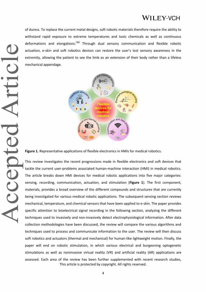
Figure 1. Representative applications of flexible electronics in HMIs for medical robotics.
This review investigates the recent progressions made in flexible electronics and soft devices that
tackle the current user-problems associated human-machine interaction (HMI) in medical robotics.
The article breaks down HMI devices for medical robotic applications into five major categories:
sensing, recording, communication, actuation, and stimulation (Figure 1). The first component,
materials, provides a broad overview of the different compounds and structures that are currently
being investigated for various medical robotic applications. The subsequent sensing section reviews
mechanical, temperature, and chemical sensors that have been applied to e-skin. The paper provides
specific attention to bioelectrical signal recording in the following section, analyzing the different
techniques used to invasively and non-invasively detect electrophysiological information. After data
collection methodologies have been discussed, the review will compare the various algorithms and
techniques used to process and communicate information to the user. The review will then discuss
soft robotics and actuators (thermal and mechanical) for human-like lightweight motion. Finally, the
paper will end on robotic stimulation, in which various electrical and burgeoning optogenetic
stimulations as well as noninvasive virtual reality (VR) and artificial reality (AR) applications are
assessed. Each area of the review has been further supplemented with recent research studies,

This article is protected by copyright. All rights reserved.
5
indicating the stage and future application of each methodology. In the final section, the challenges
of flexible electronics and devices are summarized, noting their potential future directions within the
scientific community.
2. Materials and Structures
The incorporated materials (Figure 2) and structural designs (Figure 3) in flexible devices directly
impact their various properties. One of the most important properties for medical applications is
biocompatibility, which includes the safety, comfort, and normal functionality of the device in vivo or
on the skin. In the following subsection, various flexible materials and structural designs in HMI
devices are introduced, alongside their desired properties, with specific attention given to their
biocompatibility and functionality in various medical robotics.
2.1 Mechanical biocompatibility
From a mechanical perspective, biocompatible devices for medical applications must be flexible,
elastic, and compliant enough to adapt to any target tissues’ complex morphology. Proper
conformability can reduce damage to the device and body during tissue displacement and motion
artifacts while also improving patient comfort and outcome.[39] HMI materials can be broadly
classified into two categories: inherently elastic and intrinsically non-stretchable components. The
former, such as liquid metals[40] (Figure 2A and 2B) and conductive or semiconductive polymers,[41,42]
directly rely on their mechanical properties for flexibility without further modification (Figure 2G and
2H). The latter (rigid materials), on the other hand, gain elasticity through flexible structural designs.
A common method for incorporating rigid components into flexible devices is to use thin strips of
the material. Bending strains decrease linearly with thickness, making inherently inelastic materials
flexible when fabricated into ultrathin films or wires (on the nanometers/micrometers scale). Typical
examples are metallic films on flexible circuits,[43] or nanomesh electrodes (Figure 2C,D) in flexible
sensors.[44] Another way to obtain flexibility is through structural designs, like serpentine (Figure 3A
and 3B),[45] kirigami,[46] fabric (Figure 3I and 3J),[47] and waves (Figure 3K and 3L).[48] Such structures
can not only endow a degree of flexibility and stretchability to rigid devices, but also further enhance
the elasticity of inherently soft devices.[49] Upon gaining flexibility through materials and structures,
HMI devices must be further processed into the appropriate biocompatible morphology suitable for
different tissues (similar material to tissues).
This article is protected by copyright. All rights reserved.
6
2.2 Chemical biocompatibility
The chemical composition of flexible devices should be adaptable to various biochemical
environments (biofluids like sweat and interstitial fluid) to ensure user safety and prevent device
failure, especially in biological fluids containing various erosive ions and attacking immune cells.[50]
For implantable devices, noble metals such as platinum (Pt) and gold (Au) play an important role in
neural interface electrodes due to their chemical stability in physiological environments.[51] Another
commonly used biocompatible material is hydrogels (see Figure 2E and 2F). As a material whose
features resemble water-rich tissue in the human body, hydrogels can greatly reduce the
inflammatory response of foreign objects, regulate cell or protein attachment, and prevent device
failure in vivo,[52] which is important for implants.[53,54] Coating hydrogels on electronics[55] or mixing
conductive materials such as carbon nanotubes (CNTs),[56] ionic liquids,[57] or conductive polymers[58]
into the gel can help maintain biocompatibility while also preserving the electronic properties. For
devices that are not chemically biocompatible, the last commonly used technique is to completely
seal the device in biocompatible material, like polyimide (PI) or polydimethylsiloxane (PDMS).[59]
2.3 Functionality and performance
The functionality and performance of many medical robotics can be evaluated by the device’s
sensitivity, electrode interface impedance, actuation capability, or stimulator charge injection
capability, all of which are dependent on the electrical properties of the material and structural
designs. As an example, conductive polymers and hydrogels have considerable charge storage and
injection capabilities. Because of this, they are commonly used in electrostimulation electrodes,[58]
electrophysiological recording electrodes, and nano-percolation networks that maintain statistical
stability of impedance under deformation conditions.[44,60] In terms of structural design, such as
mechanical sensor microstructure, to improve the functionality and sensitivity of the device (Figure
3C and 3D),[61] the combination of electroactive polymer (EAP) actuator and hydraulic structure can
improve actuation capability[62,63] as the structural design of a pneumatic chamber will achieve the
pre-defined actuating motion (Figure 2I and 2J).[64–66] In addition, self-healing can be achieved by
leveraging the bonding properties of the relevant materials,[67] while biodegradability is mainly
achieved by using inherently unstable materials.[68,69] Notably, 3D flexible electronics maintain
conductivity while gaining an extra dimension and thus more sophisticated functions that 2D
electronics do not offer (Figure 3E and 3F).[70–72]

This article is protected by copyright. All rights reserved.
7
2.4 Comfort and convenience
For wearable devices, biocompatibility also includes comfort and convenience, as the user needs to
wear the device for long periods of time. Generally, porous structures have a high stretchability and
breathability (See Figure 3G and 3H), which can facilitate conformal and comfortable attachment to
the skin.[73] Moreover, textiles, the most common structure in our clothes, are also naturally porous
due to the space between the fibers and yarns.[74] Due to their breathability, these aforementioned
2D porous materials facilitate the outflow and evaporation of sweat and are not only comfortable to
wear but also do not cause much skin irritation.[75] In addition to breathability, lightweight is also an
attractive feature of flexible devices. Unlike rigid devices based on bulky metal and silicon materials,
flexible devices made of plastic and rubber are usually lighter. For example, using flexible lightweight
materials, a prosthetic system weighing 300 grams was developed that mimics a commercially
available prosthetic device that weighs more than 400 grams.[76] The benefit of lightweight devices is
that they provide a more labor-saving feeling for better user comfort. Furthermore, the
transparency based on polymers, or nanomesh materials, also contributes to greater aesthetic
properties of flexible devices.[77,78]

This article is protected by copyright. All rights reserved.
8
Figure 2. Materials for soft electronic devices. A) Scanning electron microscopy (SEM) image of the
surface on a biphasic gallium–indium (bGaIn) alloy film. B) Photograph of a multilayer LED display
based on the bGaIn interconnects. A,B) Reproduced with permission.[40] Copyright 2021, Springer
Nature. C) Cross-sectional SEM image of a gold nanomesh-based pressure sensor. D) The ultrathin
pressure sensor attached conformally to the index finger. C,D) Reproduced with permission.[44]
Copyright 2020, American Association for the Advancement of Science (AAAS). E) SEM image of
interconnected poly(3,4-ethylenedioxythiophene) (PEDOT) polymer networks in the electrically
conductive hydrogels (ECH). F) A micropatterned ECH elastic electrode array. E,F) Reproduced with
permission.[58] Copyright 2019, Springer Nature. G) Atomic force microscope (AFM) phase image of a
semi-conductive polymer under 100% strain. G) Reproduced with permission.[41] Copyright 2017,
AAAS. H) A large-scale array of intrinsically stretchable transistors using the polymer as the
semiconductor layer. H) Reproduced with permission.[42] Copyright 2018, Springer Nature. I) A hyper-
elastic light-emitting capacitor encapsulated in insulating polymer (Ecoflex 00-30) under uniaxial
stretching. I) Reproduced with permission.[66] Copyright 2016, American Association for the
Advancement of Science. J) McKibben-type artificial muscles (soft polymer composite material inside
braided mesh sleeving) and corresponding actuation. J) Reproduced under the terms of the CC-BY
Creative Commons Attribution 4.0 International license.[65] Copyright 2017, The Authors, published
by Springer Nature.

This article is protected by copyright. All rights reserved.
9
Figure 3. Structures of soft electronic devices. A) Angled-view SEM of serpentine bridging
interconnect networks. B) Optical image of the island-bridge device at equal-biaxial 50% strains. A,B)
Reproduced with permission.[43] Copyright 2014, AAAS. C) SEM image of a dense array of reversible
interlocking of Pt-coated polymer nanofibers. D) Photograph showing a nanofibers-based strain
gauge with interlocking structure. C,D) Reproduced with permission.[61] Copyright 2012, Springer
Nature. E) Papercutting inspired 3D microelectronic devices formed of SU8+Silicon in micrometers
scale. F) 3D microelectronic devices in tens of micrometers scale using silicon. E,F) Reproduced with
permission.[70] Copyright 2018, Springer Nature. G) SEM image of a nanomesh-based conductor with
porous structure. H) A photo of a conductor attached conformally to a fingertip. G,H) Reproduced
with permission.[73] Copyright 2017, AAAS. I) Photograph of a soft multicolor display in form of
textile. J) The display under complex deformations, including bending and twisting. I,J) Reproduced
with permission.[47] Copyright 2021, Springer Nature. K) SEM image of ultrathin transistors on an
elastomer with waves formed by pre-stretching. L) Stretchable transistors on the elastomer with
waves. K,L) Reproduced with permission.[48] Copyright 2013, Springer Nature.
2.5 Discussion
Collectively, research into materials and structures is fundamentally driving the growth of flexible
electronics. It is important to note that most of the materials and structures discussed are versatile
and can have multiple roles in prosthetics and rehabilitation robotics such as sensing and recording,
computing and storage, as well as actuation and stimulation. The current challenges and potential
future directions of materials and structures in flexible electronics include: 1) Although most studies
mention durability and functionality, for many devices this is just a theoretical prediction or proof of
concept. 2) Compared to rigid devices, the manufacturing precision and integration density of
flexible devices are still in their infancy. This places a high requirement on the machinability of
materials and structures to ensure flexibility. 3) The manufacturing cost of materials and structures
is the key in determining the device’s price. Many materials and structures are already costly for
proof of concept in the lab. For mass production, low-cost solutions need to be continuously
explored.
3. Sensing
In the human body, the skin protects internal tissue from damage[79] while transmitting abundant
external information to the brain through various high-density subcutaneous receptors.[80] In this
way, the skin acts as a platform for the embodiment and extraction of internal and external

This article is protected by copyright. All rights reserved.
10
sensations, such as physical (pressure and stretching),[81,82] chemical (perspiration),[83] and
physiological (respiration and pulse)[84,85] information.
In medical robotics, e-skin presents itself as an optimal replacement for human skin to collect
external and internal information from a patient. In the past decade, various flexible e-skin sensors
that mimic the functions and features of human skin have already been integrated onto medical
robotics to sense external information[86] for rehabilitation applications.[87] In rehabilitation, the
absence of supervision may lead to incorrect patient posture, which reduces the efficiency of
rehabilitation and can even aggravate a disability.[88] Collecting information from the patient during
rehabilitation, specifically strain[89] and pressure,[90] can inform the therapist in real-time about the
accuracy of the movement as well as the physiological status. At the current stage, e-skin can mimic
or surpass human skin performance in mechanical and thermal sensing (as shown in Table 1) and
functionalities in terms of various chemical,[91] physiological, and proximity sensing[92] to deliver
accurate and diversified information to users. In such scenarios, e-skin sensing can achieve
autonomous electronic rehabilitation by monitoring the patient in real-time, providing a promising
solution for the development of personalized rehabilitation science.[93,94]
Table 1. Comparison of key sensing performance of natural skin and artificial skin.
Limit of
detection
Sensitivity(
pressure)
Spatial
resolution
Response
time
Sensitivity
(temperature)
Mechanical
property Refs.
1 mN[86]
0.078–0.018 kPa[95]
1 mm[96]
15 ms[86]
20 mk[97]
Stretchable
~30%[95]
Human skin
0.08 Pa >220 kPa−1
50 μm 9 ms NA Flexible Bai et al.[98]
NA 0.01 kPa−1
0.1 mm 15 ms NA Flexible Yan et al.[99]
1 ug ~ 1.25 Pa 4.4 kPa−1
5 mm NA NA Flexible Wu et al.[100]
0.3 Pa >5000 kPa−1
1000 DPIa <1 ms NA Flexible Lee et al.
[101]
<0.5 Pa 192 kPa-1
0.8 cm 10 ms NA Flexible Zang et al.[102]
NA NA 5 mm NA 20 mk Flexible Yokota et al.[103]
7.3±1.2 Pa >1.25 MPa−1
2 mm NA 2410 ppm °C-1
Stretchable
~800%
Hua et al.[104]
10 Pa ~1.78×10⁻³
kPa−1
318 CPIb 32 ms 3×10
−4 °C
-1 Flexible An et al.
[105]
NA 0.41% kPa−1
or
0.075% kPa−1
0.5 mm NA ~0.01 °C -1
Stretchable
~20%
Kim et al.[106]
100 Pa 28.9 kPa-1
1 mm 40 ms <0.1 K Flexible Zhang et al.[107]
aDPI, dots per inch; bCPI, capacitors per inch.
This article is protected by copyright. All rights reserved.
11
3.1 Pressure sensing
There are many e-skin pressure sensing mechanisms that have been widely and reliably adopted in
medical robotics.[108–110] Several of these exemplary principles (such as piezoresistive,
piezocapacitive, piezoelectric, piezoionic, and more) are discussed below alongside their applications
in prosthetic sensing and rehabilitation health monitoring.
3.1.1 Pressure sensing mechanisms
Conventional static force-sensing mechanisms in e-skins mainly consist of resistive and capacitive
sensing. Due to its simple mechanism and convenient data collection strategy, resistive pressure
sensing has been extensively applied using various recording mechanisms.[111] Bulky piezoresistive e-
skins, like sponge-based sensors, rely primarily on pressure-induced changes in the number of
conductive pathways[112] or in the shape of the sensing material.[113] Meanwhile, another resistive
sensor may analyze the changes in the contact resistance, like the quantum tunneling effect (Figure
4A).[114,115] This type is much more sensitive and thinner than the bulky option. An e-skin based on
bilayer microdome arrays can use microstructures to maximize the changes in surface contact
resistance based on the tunneling effect (Figure 4B).[115] Nevertheless, some drawbacks, like large
hysteresis,[116] large confounding temperature sensitivity,[117] and varying pressure sensitivity[118]
have limited the performance of resistive sensors. Compared with resistive sensors, capacitive
sensors have excellent linearity with lower power consumption.[119] For piezocapacitive e-skins,
capacitance changes are mainly based on the deformation of the dielectric layer. Normal pressure
and tangential strain can be measured through a capacitive sensor as the dielectric layer can be
deformed under tensile and external pressure (Figure 4C). As demonstrated in Figure 4D,[120] a
capacitive pressure sensing array with cross-arranged electrodes was fabricated using CNTs as the
electrode material and PDMS as the dielectric layer.
Piezoelectric and triboelectric have their unique advantages in dynamic force measurements, i.e.
high-frequency measurement, and self-power supply.[121–123] As an example of piezoelectric sensors,
a dual-gated ZnO-based thin-film transistor (TFT) was fabricated (Figure 4E)[124] where external
pressure causes noticeable voltage changes in the flat-band of the device (Figure 4F). In practice, this
sensing mechanism is well-suited for HMI’s high-frequency environment, such as the detection of
surface textures by robotic hands (high-frequency vibrations) and [125] wearable pulse monitors.[126] A
representative example is shown in Figure 4G and 4H, where an all-textile triboelectric generator
(TENG)-based machine washable sensor array was woven into different parts of a vest to enable

This article is protected by copyright. All rights reserved.
12
measurement of pulse and respiratory waves.[127] Nonetheless, the major drawbacks of these two
sensing mechanisms are the unreliable static sensing performances and drift in long-term
measurements.[108]
Achieving a high integration density of sensing arrays is dependent on the fabrication process,
materials, and the data acquisition method.[128] Meanwhile, the high integration density of the
electronics easily leads to crosstalk and interference of the signals. An attractive approach to address
this issue is to have transistors that maintain the sensing unit state integrated around the device to
control and amplify the signal, namely the “active matrix”.[129] The previously mentioned resistive,
capacitive, and piezoelectric cells are all capable of being integrated into active matrices. As shown
in Figure 4I, capacitive integration uses primarily pressure-sensitive capacitors directly as gate
capacitors, where the change in capacitance is translated into a variation in drain current in the field-
effect transistor (FET)-based device.[102,130–132] On the other hand, piezoresistive sensors can be
connected to the source or drain of the FET, where the drain current of the FET varies with the
applied pressure (Figure 4J).[48,77,133–135]
Other pressure sensing mechanisms, like optical,[136–138] magnetic,[99,139] and piezoionic,[140–142] are
emerging and accelerating the growth of the pressure-sensitive e-skin field. Optical pressure sensors
rely on the differences in optical signal between the electromagnetic emitter and receiver, where an
external force can deform the light guide (transmission medium) and alter the optical signal received
(Figure 4K). By exploiting this principle, Figure 4L demonstrates a stretchable distributed fiber-optic
sensor that can simultaneously monitor real-time deformations such as bending and pressing.[136] In
addition to optical methods, magnetic sensors are also suitable for high-resolution pressure
perception.[143] A pressure sensor was recently developed using soft materials that contain magnetic
particles, where the external force applied changes the uniformly distributed magnetic particles and
changes the internal magnetic field in the device.[99] As another example, Figure 4M and 4N illustrate
a GMI-based pressure sensor with a high sensitivity to force stimuli.[100] Some innovative e-skins
based on the piezoionic mechanism have been proposed.[140,144] The mechanical deformation of the
e-skin can be indicated by a change in voltage or current due to the redistribution of ions with
different mobility (Figure 4O). As demonstrated in Figure 4P, e-skins can utilize the piezoionic
principle to measure the applied force by detecting the current between the nanopore
membrane.[145]

This article is protected by copyright. All rights reserved.
13
Figure 4. Sensing mechanisms and examples of flexible pressure sensors. A) A piezoresistive
mechanism. B) A giant tunneling effect-based piezoresistive pressure sensor. A,B) Reproduced with
permission.[115] Copyright 2014, American Chemical Society. C) Piezocapacitive mechanism. D)
Transparent piezocapacitive sensors. C,D) Reproduced with permission.[120] Copyright 2011, Springer
Nature. E) A piezoelectric mechanism. F) Scalable tactile sensor arrays with piezoelectric materials in
active matrixes. E,F) Adapted with permission.[124] Copyright 2020, AAAS. Reprinted/adapted from
ref.[124]. Distributed under a Creative Commons Attribution NonCommercial License 4.0 (CC BY-NC).
G) Triboelectric sensing mechanism. H) Triboelectric mechanism-based textile for pressure sensing.
G,H) Adapted with permission.[127] Copyright 2020, American Association for the Advancement of
Science (AAAS). Reprinted/adapted from ref.[127]. Distributed under a Creative Commons Attribution
NonCommercial License 4.0 (CC BY-NC). I) Piezocapacitive active matrix. Reproduced under the
terms of the CC-BY Creative Commons Attribution 4.0 International license.[102] Copyright 2015, The
Authors, published by Springer Nature. J) Piezoresistive active matrix. Reproduced with
permission.[48] Copyright 2013, Springer Nature. K) Optic pressure sensing mechanism. L) A
This article is protected by copyright. All rights reserved.
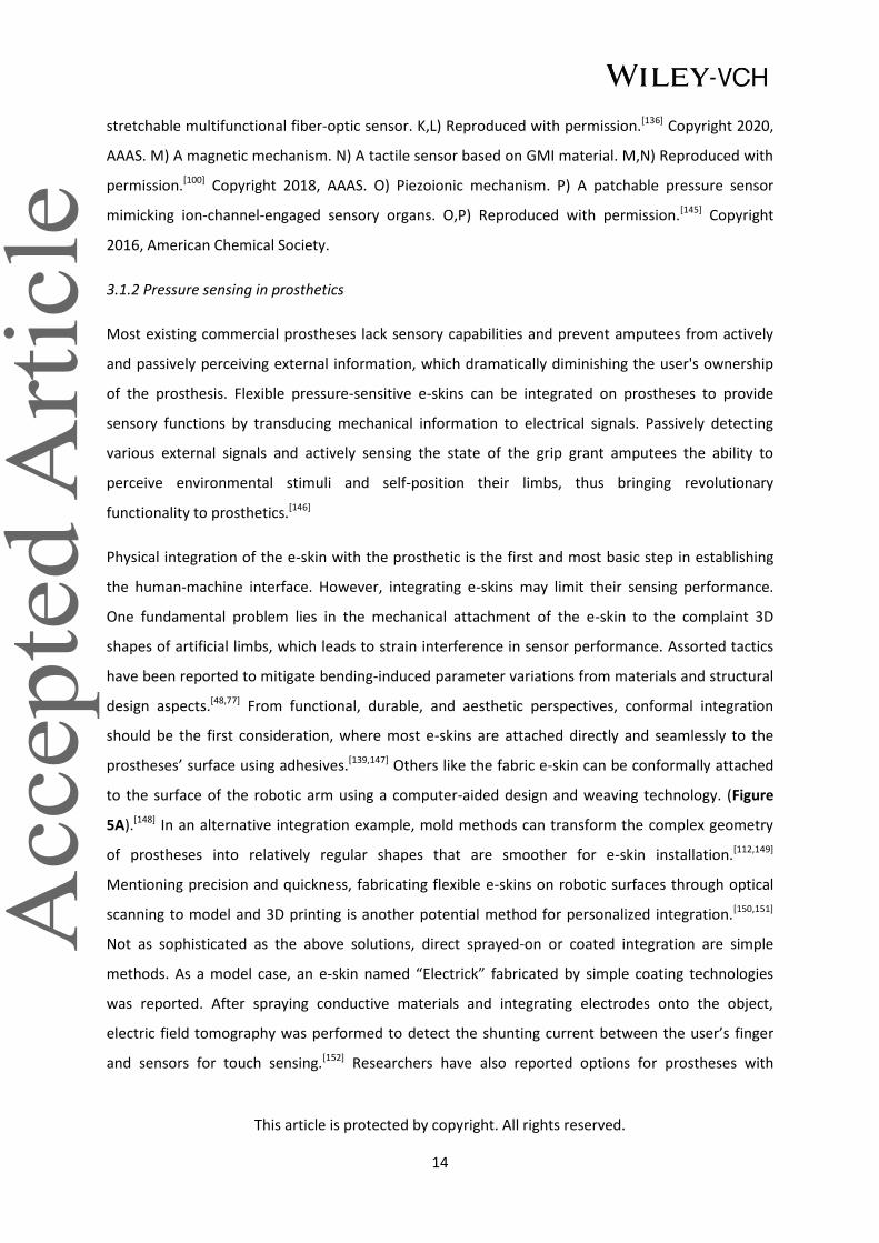
14
stretchable multifunctional fiber-optic sensor. K,L) Reproduced with permission.[136] Copyright 2020,
AAAS. M) A magnetic mechanism. N) A tactile sensor based on GMI material. M,N) Reproduced with
permission.[100] Copyright 2018, AAAS. O) Piezoionic mechanism. P) A patchable pressure sensor
mimicking ion-channel-engaged sensory organs. O,P) Reproduced with permission.[145] Copyright
2016, American Chemical Society.
3.1.2 Pressure sensing in prosthetics
Most existing commercial prostheses lack sensory capabilities and prevent amputees from actively
and passively perceiving external information, which dramatically diminishing the user's ownership
of the prosthesis. Flexible pressure-sensitive e-skins can be integrated on prostheses to provide
sensory functions by transducing mechanical information to electrical signals. Passively detecting
various external signals and actively sensing the state of the grip grant amputees the ability to
perceive environmental stimuli and self-position their limbs, thus bringing revolutionary
functionality to prosthetics.[146]
Physical integration of the e-skin with the prosthetic is the first and most basic step in establishing
the human-machine interface. However, integrating e-skins may limit their sensing performance.
One fundamental problem lies in the mechanical attachment of the e-skin to the complaint 3D
shapes of artificial limbs, which leads to strain interference in sensor performance. Assorted tactics
have been reported to mitigate bending-induced parameter variations from materials and structural
design aspects.[48,77] From functional, durable, and aesthetic perspectives, conformal integration
should be the first consideration, where most e-skins are attached directly and seamlessly to the
prostheses’ surface using adhesives.[139,147] Others like the fabric e-skin can be conformally attached
to the surface of the robotic arm using a computer-aided design and weaving technology. (Figure
5A).[148] In an alternative integration example, mold methods can transform the complex geometry
of prostheses into relatively regular shapes that are smoother for e-skin installation.[112,149]
Mentioning precision and quickness, fabricating flexible e-skins on robotic surfaces through optical
scanning to model and 3D printing is another potential method for personalized integration.[150,151]
Not as sophisticated as the above solutions, direct sprayed-on or coated integration are simple
methods. As a model case, an e-skin named “Electrick” fabricated by simple coating technologies
was reported. After spraying conductive materials and integrating electrodes onto the object,
electric field tomography was performed to detect the shunting current between the user’s finger
and sensors for touch sensing.[152] Researchers have also reported options for prostheses with

This article is protected by copyright. All rights reserved.
15
flexible sensors embedded in the manufacturing process,[153,154] which solves the problem of surface
mounting, but also presents a replacement challenge.
Daily interactions between amputees and the environment in the form of touch and collisions
require passive pressure sensing. For example, coverage of piezoresistive textiles on a robotic arm
can provide a spatial map of pressure distributions (Figure 5B).[148] Compared with passive
perception, active sensing refers to amputees detecting unstructured environments with prosthetics
subjectively, such as identifying the stiffnesses and texture of objects, user-interaction with people,
and grasping.[155] As an example, parallel ridges were fabricated on the surface of a poly(vinylidene
difluoride) (PVDF)-based piezoelectric sensor to detect texture-induced vibrations.[156] Furthermore,
adding e-skins on prosthetics creates a closed-loop sensory feedback control, which allows for a
more accurate, facile, and compliant touch and grasp. In an example, sensory feedback control was
demonstrated by pulling the stem from a cherry (without crushing it) using an artificial limb, which
requires facile control through relayed feedback of the grasping force (Figure 5C and 5D).[157]
Collectively, with the assistance of artificial skin, active and passive interactions make up the
prosthetic pressure perception.
3.1.3 Pressure sensing in rehabilitation
Pressure monitoring is essential for rehabilitation, especially for extremities recovery and small
pressure capture in health monitoring (heartbeats, pulse, etc.). Eg. fingertip and palm pressure
feedback can inform the user about their gripping force for stroke patients to regain accurate
grasping ability.[44,158–161] A liquid metal-based pressure-sensitive glove was used to train a subject to
relearn a specific manual skill by providing real-time pressure monitoring (Figure 5E).[162] In another
application, the medical importance of plantar pressure monitoring in patients with lower extremity
disabilities are tremendous. Through analyzing the pressure concentration points on the foot, the
gait phase can be identified for real-time correction of improper movements in rehabilitation.[163]
Additionally, the imitation of human gait also facilitates the adaptability and compliance of bionic
prosthesis to the human body.[164,165] In one example, a pressure-sensitive insole with an inkjet
printing circuit and sensing units made of CNT embedded in PDMS was fabricated for detecting
plantar pressure. These pressure data can be used to train ML algorithms to classify human gait
phases (Figure 5F).[163] In rehabilitation, micro-vibrational physiological signals, such as a
heartbeat[166,167] and pulse,[168,169] can provide information on the body's status and load, allowing for
the immediate adjustment of the rehabilitation intensity and planning. Moreover, pressure
This article is protected by copyright. All rights reserved.
16
monitoring of bedridden patients or prosthetic sockets can also effectively prevent skin ulceration
due to prolonged pressure.[170,171]
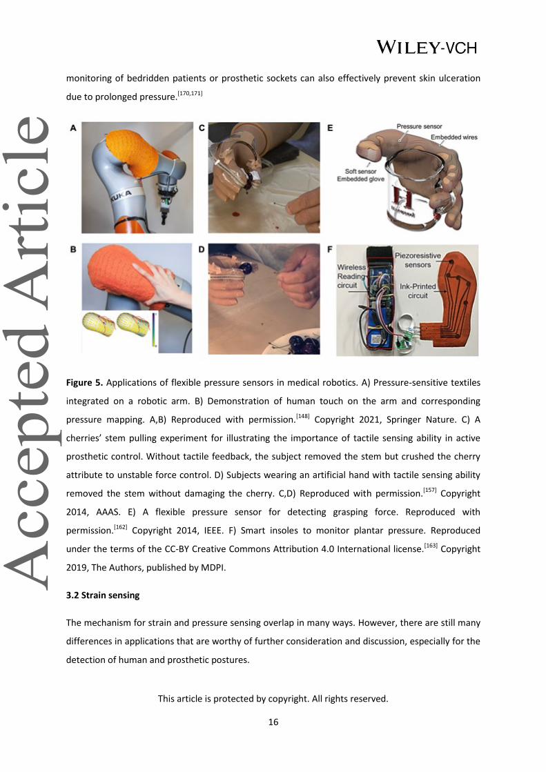
Figure 5. Applications of flexible pressure sensors in medical robotics. A) Pressure-sensitive textiles
integrated on a robotic arm. B) Demonstration of human touch on the arm and corresponding
pressure mapping. A,B) Reproduced with permission.[148] Copyright 2021, Springer Nature. C) A
cherries’ stem pulling experiment for illustrating the importance of tactile sensing ability in active
prosthetic control. Without tactile feedback, the subject removed the stem but crushed the cherry
attribute to unstable force control. D) Subjects wearing an artificial hand with tactile sensing ability
removed the stem without damaging the cherry. C,D) Reproduced with permission.[157] Copyright
2014, AAAS. E) A flexible pressure sensor for detecting grasping force. Reproduced with
permission.[162] Copyright 2014, IEEE. F) Smart insoles to monitor plantar pressure. Reproduced
under the terms of the CC-BY Creative Commons Attribution 4.0 International license.[163] Copyright
2019, The Authors, published by MDPI.
3.2 Strain sensing
The mechanism for strain and pressure sensing overlap in many ways. However, there are still many
differences in applications that are worthy of further consideration and discussion, especially for the
detection of human and prosthetic postures.
This article is protected by copyright. All rights reserved.
17
3.2.1 Strain sensing mechanisms
The mechanisms of pressure sensors mentioned above, like resistive, capacitive, and optical, etc.,[172]
can also be applied to flexible strain sensors. It is worth noting that some microscopic mechanisms in
strain sensing, like misplace,[173,174] disconnection,[175] and crack propagation[168,176] are not available
in pressure sensing. The most widely accepted capacitive structure for strain sensors is the
“sandwich” structure. Inherent stretchable polymers are attractive materials for dielectric layers due
to their stretchability and permittivity. Nanomaterials[177,178] and liquid metals[179] can be used as
electrodes owing to their stable conductivity under tensile strain. Piezoelectric and triboelectric
strain sensors are two other major types of stretchable strain sensors.[180–182] Piezoelectric materials,
like BaTiO3,[183] zinc oxide (ZnO),[184] PVDF,[185,186] can be utilized to fabricate strain sensors by
converting strain readings into voltage signals. Similar to the mechanism behind pressure sensing,
flexible waveguides[136,153] and magnetic materials[187,188] have also been investigated for wearable
strain sensing applications.
3.2.2 Strain sensing in prosthetics
Flexible strain gauges are commonly used in prosthetic control and proprioception. Prosthetics can
be controlled by neural electrophysiological signals (will be further discussed in section 4) and
mechanical signals such as gestures[189,190] or body movements.[191] By using simple and accurate
signal gathered from non-invasive and visible sensors, gesture control strategy has become an
intuitive and reliable control method for prosthetics.[146] Flexible strain sensors prove to be
promising alternatives to monitor these mechanical signals owing to their stretchability, durability,
and lightweight nature. As shown in Figure 6A, a biofuel powered strain sensor was integrated onto
a human elbow to capture strain signals. By utilizing the strain signal, the subject successfully
controlled the prosthetic to assist walking in real-world environments (Figure 6B).[192] Tiny stretching
of the skin, such as muscle contractions and relaxation (i.e. mechanomyography) can also be
detected by flexible strain gauges[193–195] or pressure-sensitive units[196] for prosthetic control.
Besides pressure sensing, proprioception is another crucial perception for prosthetics, which allows
the amputee to directly feel the posture of the device. In particular, flexible strain sensors can sense
the stretching and relaxation of prosthetics like the human body's mechanoreceptors located on the
muscles, skin, and tendons.[197] Commercial prosthetics are mostly driven by motors, where the
angle encoder inside the device can accurately reflect the movements. Soft actuators lack this ability
because of their continuous mode of motion, making the application of flexible strain sensors on soft
This article is protected by copyright. All rights reserved.
18
actuators extremely attractive.[37,198,199] A noteworthy example to detect bending angles, an essential
part of its self-perception, is flexible capacitive sensors alongside soft actuators. This hybrid robotic
skin can turn inanimate objects into soft robots by estimating their current position. As shown in
Figure 6C, to demonstrate the proprioceptive ability, the bend angle of a foam integrated with the
robotic skin was calculated from the dimension of the device and the electrical signals.[200] Therefore,
the combination of soft actuators and flexible sensors is a promising direction for future soft robotic
systems.
3.2.3 Strain sensing in rehabilitation
For rehabilitation, the rotation of the joints, the contraction and relaxation of the muscles, and the
natural breath cause strain on the human skin and are essential information to record. A healthy
hand allows for dexterous manipulation. In terms of rehabilitation, the angle of the hand joints
monitored by the sensors can be used as a basis for measuring movements, which provides rich data
for hand rehabilitation. As shown in Figure 6D, a stable flexible strain gauge based on the “island and
gap” structure of aligned CNT thin films was recently demonstrated.[175] Using this strain gauge for
hand rehabilitation, a data glove can be fabricated to accurately detect the motion of each finger
individually. In gait rehabilitation, ankle angles are essential physical information for orthotic devices
to address pathological gaits. Figure 6E presents an EGaIn based-strain sensor that can be applied on
top of the ankle to measure the joint’s angle to provide feedback on the foot’s motion during
rehabilitation.[201] Unlike large strain measurements of joint angles, measuring the strain from small
muscle deformations requires a high sensitivity. Figure 6F demonstrates an ultra-sensitive and
resilient strain-mediated contact in anisotropically resistive structures based on a compliant strain
gauge. To verify the effectiveness of this sensor, researchers fabricated a textile-based sensor that
was integrated into the sleeve to detect small muscle deformations and classify hand and wrist
movements.[194] On the skin, respiratory waves and pulses can also contribute to slight stretches,
which can be captured by sensitive strainers for health monitoring during rehabilitation.[94,202,203]
In addition to monitoring external strain on the human skin, recently in vivo strain sensing has been
investigated to monitor inner tissue, e.g. tendons and muscle recovery, by continuously providing
real-time and long-term information for rehabilitation surveillance.[179,204] As shown in Figure 6G, an
implantable capacitive multifunctional sensor was designed to measure pressure and strain signals
under the skin. After being implanted on the back of a rat (Figure 6H), the in vivo sensor can
accurately and stably detect strain and pressure signals applied on the implanted region (Figure 6I).
This device was fabricated with biodegradable materials, which avoids the need for surgical
This article is protected by copyright. All rights reserved.
19
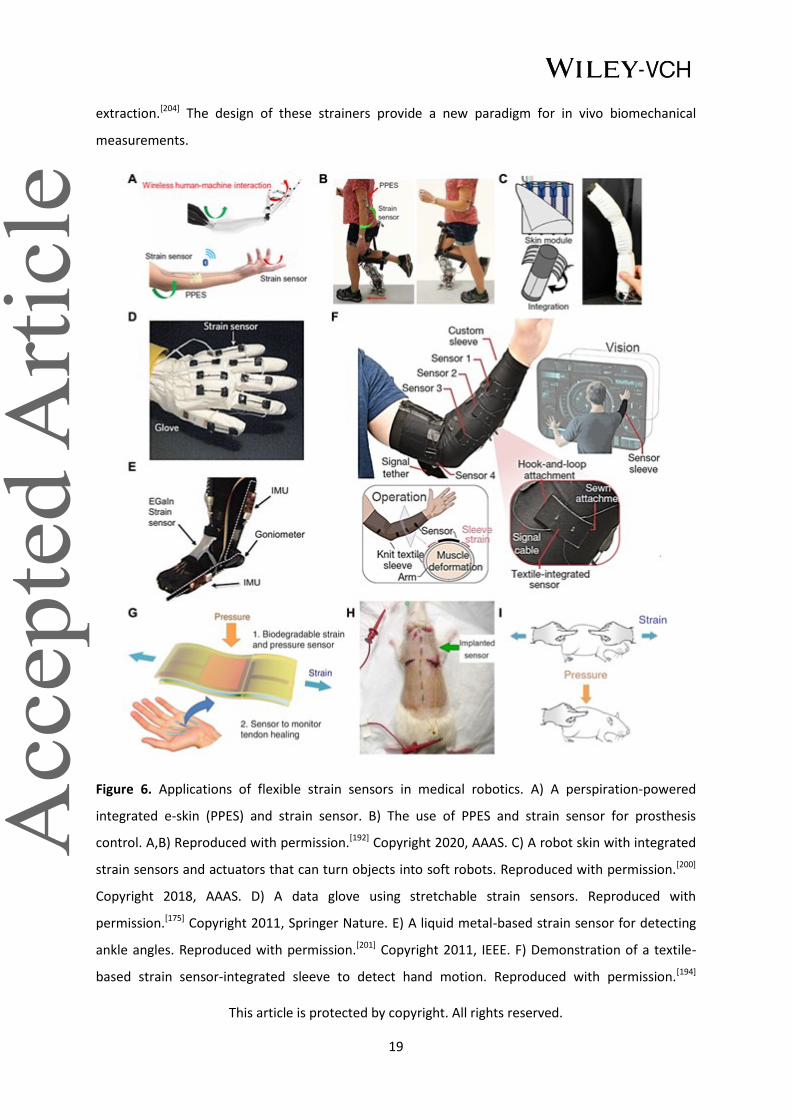
extraction.[204] The design of these strainers provide a new paradigm for in vivo biomechanical
measurements.
Figure 6. Applications of flexible strain sensors in medical robotics. A) A perspiration-powered
integrated e-skin (PPES) and strain sensor. B) The use of PPES and strain sensor for prosthesis
control. A,B) Reproduced with permission.[192] Copyright 2020, AAAS. C) A robot skin with integrated
strain sensors and actuators that can turn objects into soft robots. Reproduced with permission.[200]
Copyright 2018, AAAS. D) A data glove using stretchable strain sensors. Reproduced with
permission.[175] Copyright 2011, Springer Nature. E) A liquid metal-based strain sensor for detecting
ankle angles. Reproduced with permission.[201] Copyright 2011, IEEE. F) Demonstration of a textile-
based strain sensor-integrated sleeve to detect hand motion. Reproduced with permission.[194]
This article is protected by copyright. All rights reserved.
20
Copyright 2020, Springer Nature. G) An implantable strain sensor can be attached to the tendon for
real-time healing assessment. H) The strain sensor implanted subcutaneously on the back of a rat. I)
External strain and pressure applied on the sensor. G-I) Reproduced with permission.[204] Copyright
2018, Springer Nature.
3.3 Thermal sensing
As temperature is another dimension of physical information beyond mechanical perception,
mimicking thermoreceptors on the human skin is vital for the sensory function of prostheses.
Currently, some temperature-sensitive e-skins can outperform the human skin in terms of
sensitivity, accuracy, and detection range.[205,206]
3.3.1 Thermal sensing mechanisms
Resistive metallic temperature sensors[207] and thermistors[208] are the most commonly used sensors
in flexible thermal electronics. The mechanism of resistive metallic temperature detectors relies on
the linearity between resistance and temperature found in metals.[209] Thin metal films, like Au,[210]
Pt[211] are widely employed in constructing flexible temperature detectors due to their good linearity
and proven success in their microfabrication technologies. Unfortunately, the sensitivity of many
metal materials can be a significant drawback for temperature sensing applications. Thermistors are
another common class of resistive temperature sensor and can be categorized into nonlinear
positive temperature coefficient (PTC) and negative temperature coefficient (NTC) sensors. In the
PTC type, fluctuations in the temperature alter the specific volume of the material, which in turn
affect its resistance and current measured. Conductive nanomaterial-filled polymers[212,213] are a
promising candidate for flexible PTC temperature sensors, where the volume changes as the
material progresses through the melting point of crystalline regions, thus affecting the resistance.[214]
For measuring spatial temperature gradients, an organic active matrix with polymer PTC sensor pads
was fabricated on a PI substrate, which exhibits a bending insensitivity and high spatial resolution
(Figure 7A and 7B).[103] The negative temperature coefficient (NTC) thermistor has a clear advantage
over other temperature sensors in that it exhibits a much simpler structure while presenting
equivalently high temperature sensitivity.[215]
In addition to resistive temperature sensors, thermocouples and PN junction sensors are also two
common thermosensing mechanisms. Thermocouples can generate a potential difference under
different temperature between different materials based on the thermoelectric (Seebeck)
phenomenon.[216] Whereas the forward voltage in PN junction temperature sensors can vary with
This article is protected by copyright. All rights reserved.
21
temperature, converting the heat into electrical signals in the diodes and transistors. The advantage
of this method is its small size, fast response time, and good linearity.[106,217] Moreover, some optical
temperature sensors use infrared (IR) thermography[218] and colorimetric techniques with
thermochromic liquid crystals that can be used to record the temperature more intuitively[219] and
have already been applied to measure the temperature of the human body. Some remarkable
designs based on biomaterials and structures have superior temperature response properties,
offering another method for thermal perception.[97]
3.3.2 Thermal sensing applications
Thermal perception for prosthetics can complement pressure sensing for extracting more tactile
information from the environment, which not only informs the amputee about the environment
thermographically, but can also prevents the user from danger or potential device failure due to
extreme heat or cool.[220,221] Figure 7C shows an intrinsically stretchable rubbery electronic, which
can be used to fabricate thermistors.[222] Researchers demonstrated the sensor’s functions by using a
robotic hand equipped with e-skin to grab and measure the temperature of different cups. (Figure
7D) One important lesson learned from studying the natural perception of human skin is that the
identification of materials is inseparable from accurate temperature perception. As a heat source
and sensor, the human hand can effectively perceive the heat dissipation ability for fluid flow rate
sensing[223] and identifying the thermal characteristics of surface materials.[224] One such example
used an artificial fingertip with e-skin to acquire information about the thermal properties and
surface texture of different materials. Using machine learning, the combination of vibrational and
thermal information was used to identify the group (e.g. wood, metal, plastic) and type (e.g.
aluminum, copper, pine, etc.) of the material.[224] In medical rehabilitation, temperature sensing is
commonly used to monitor the body, which can be a reference for health.[225,226]
In the field of medical rehabilitation, monitoring body temperature at different locations can reflect
the body's state (both physiological and psychological) in real time. For example, the potential
fatigue and psychological tension during the rehabilitation process can cause temperature variations
due to sweat evaporation.[226] In addition, some disease-induced disabilities have distinct thermal
characteristics (either high or low) at the extremities due to abnormal blood circulation and
metabolism state. For example, the foot temperature of some diabetic patients can be about 5
degrees Celsius higher than that of healthy people. Long-term monitoring of body temperature by
using flexible temperature sensors is convenient and can effectively prevent medical abnormalities
such as foot ulcers.[227]

This article is protected by copyright. All rights reserved.
22
3.3.3 Multifunctional simultaneous sensing
Simultaneous detection of thermal and mechanical signals is challenging due to the coupling
problem; however, it is possible. Five mainstream existing multisensory (mechanical and thermal)
detection modes have been developed. The most conventional multisensory model is for
temperature and pressure sensors to be placed on different parts of a prosthesis (Figure 7C and
7D).[222] Integrating the two sensors into one substrate would provide a higher spatial
resolution.[104,105] Nonetheless, both types mentioned above cannot intrinsically measure two signals
simultaneously at one point, resulting in a waste of valuable space at key locations (like fingertips).
The development of novel dual-parameter sensors that transduce different stimuli into separate
signals can minimize signal interference, allowing the detection of both temperature and pressure at
a single point without decoupling analysis.[107,228,229] In Figure 7E, by taking advantage of independent
thermoelectric and piezoresistive effects, a dual parameter device can simultaneously transduces
temperature and pressure stimuli into separate electrical signals.[107] Another potential solution is to
measure pressure and temperature in a single parameter, which is more efficient data collection.
The first stretchable e-skin that decoupled temperature and strain in a single parameter is shown in
Figure 7F.[230] The e-skin has a simple electrode-electrolyte-electrode structure, where two variables
(temperature and strain) are derived from analyzing the ion relaxation dynamics: the charge
relaxation time of the capacitance as a strain-insensitive feature to measure absolute temperature,
and the normalized capacitance as a temperature-insensitive extrinsic feature to measure strain. In
the demonstration, it can provide real-time thermal information, force directions, and strain
graphics in various tactile motions (shear, pinch, spread, torsion; Figure 7G). Another promising
multimodal detection method based on machine learning is the “cross-reactive” sensor matrix.
Instead of responding to certain specific stimuli like conventional “lock and key” sensors, this sensor
has the ability to respond to a wide range of stimuli. Utilizing machine learning methods to directly
analyze the coupled multimodal data, these devices can achieve a certain degree of decoupling. This
approach greatly reduces the complexity of the sensor mechanism and structure.[231] These dual-
monitoring devices greatly enriches the external information that can be acquired by prosthetic
haptics.

This article is protected by copyright. All rights reserved.
23
Figure 7. Temperature sensors and applications in medical robotics. A) A PTC temperature sensing
array. B) Temperature mapping after touching the sensing sheet. A,B) Reproduced with
permission.[103] Copyright 2015, The Authors, published by National Academy of Sciences. C) A
prosthetic hand integrated with pressure and temperature sensors on different regions. D) The
robotic hand touching and identifying hot and cold cups. C,D) Adapted with permission.[222]
Copyright 2017, AAAS. Reprinted/adapted from ref. [222]. The Authors, some rights reserved;
exclusive licensee AAAS. Distributed under a Creative Commons Attribution NonCommercial License
4.0 (CC BY-NC). E) An e-finger assembled with flexible dual-parameter temperature–pressure sensors
touching an ice cube and corresponding photograph temperature and pressure mappings. E)
Reproduced under the terms of the CC-BY Creative Commons Attribution 4.0 International
license.[107] Copyright 2015, The Authors, published by Springer Nature. F) A multimodal ion-
electronic skin attached to a dummy hand and its temperature/strain sensor responses under a

This article is protected by copyright. All rights reserved.
24
weak unidirectional shear. G) Photo of finger press and corresponding schematic of temperature and
strain variation. F,G) Reproduced with permission.[230] Copyright 2020, AAAS.
3.4 Chemical sensing
Although physical sensing still dominates the field, the past decade has seen an exponential growth
in the exploration of molecular detection, which provides another dimension for human
understanding of their own health condition and external environment.
3.4.1 Chemical sensing mechanisms
For chemical detection, the mainstream detection routes are electrochemical and optical detection.
Electrochemical methods are attractive due to their high sensitivity, low response time, and long-
term stability.[232] Electrochemistry has several main regimes for chemical detection: the
amperometric approach (including voltammetry and chronoamperometry)[91] based on redox
reactions, the potentiometric approach (based on the Nernst equation),[91,233] and electrochemical
impedance spectroscopy (EIS) based on analyzing surface properties changes caused by affinity
binding.[234] In addition to conventional electrochemical biosensing techniques, the transistor-based
approach, including field-effect transistors (FET) and organic electrochemical transistors (OECT) also
show great promise owing to their ability for in situ amplification of the detected signal.[235,236] For
optical methods there are also two prevailing techniques. One is based on the colorimetric method,
whose principle and structure are simple, but more difficult to reach high sensitivity. In recent years,
with the popularity of smartphone cameras, the quantitative measurement of color is highly
attractive for in-home monitoring of optical sensors.[237] The last method is the fluorescence
approach, where the wavelength of the light emitted is larger than the wavelength received, which
is more sensitive and suitable for trace substance measurement.[238]
3.4.2 Chemical sensing applications
Chemical sensors for prosthetic and robotic applications have been less investigated than
mechanical and thermal sensors. From a bionic perspective, human skin does not have accurate
chemical sensing capabilities, but rather uses mechanoreceptors to sense chemical contact and
nociceptors to perceive chemical damage. However, there are enormous quantities and types of
chemicals in the environment (e.g., gases, food, toxins, etc.), where rapid and accurate identification
of these substances could provide the user with essential information about the environment. Thus,
adding chemical sensing capability to prostheses can prevent users from being exposed to harmful

This article is protected by copyright. All rights reserved.
25
chemicals, such as organophosphate pesticide residues in agricultural products, and subsequently
reduce the potential risk of harm.[239] Moreover, some researchers have also proposed the
employment of chemosensing in daily diets.[91] Furthermore, the chemical and biomolecular
information collected by the e-skin from the human body could greatly benefit the design of next-
generation HMI toward personalized robotic rehabilitation.
Although chemical sensing is not sufficiently developed for prosthesis, in the field of health
monitoring, sweat chemical sensing (including electrochemical and optical modalities) is well-studied
and highly relevant to rehabilitation exercises.[240] Sweat detection can respond in real-time to
changes in the volume and rate of sweat loss,[241,242] as well as the concentration of various ions and
metabolites in the body.[45,243–245] In this regard, sweat can potentially communicate to the user
about health and rehabilitation progress, informing the user when to rehydrate and replenish
electrolytes. Moreover, some diseases of the locomotor system provoke abnormal elevations of
metabolites and biomarkers, such as L-dopa in Parkinson’s disease[246–248] uric acid in gout,[45] which
are very important evaluation factors. In addition to monitoring patient fatigue, sweat sensing can
also be used for psychological applications by monitoring the level of stress hormones such as
cortisol and norepinephrine.[249,250] In this promising field, many in-situ, multi-channel devices are
emerging.[251–253] Recently, a fully integrated wearable sensor array for multiplexed in-situ
perspiration analysis was reported, where the sodium, potassium, lactate, and glucose content of
sweat can be measured simultaneously by electrochemical methods. Conventional commercially
available integrated-circuit components (more than ten chips) can be applied on a FPCB to serve as
data processing and transmission.[83] Integrating in-situ sensing, on-site processing, and data
transmission together, chemical sensing platforms can achieve the goal of continuous, real-time
sensing of ions and metabolites, which is crucial to obtain more comprehensive knowledge about a
wearer’s well-being.
3.5 Other Sensing Techniques
While the skin has receptors for sensing mechanical and thermal information, there are no specific
receptors that sense humidity. Rather, the human brain is able to "perceive" wetness indirectly by
analyzing mechanical and thermal information (normal pressure and tangential adhesion between
the liquid and the skin as well as heat conduction of the liquid).[254,255] Such mechanisms, which may
require intelligent cross-sensor algorithms, are not available in the current e-skin. Existing flexible
humidity sensors are mainly fabricated by accommodating different transducing materials, such as
graphene, CNT, and MoS2, on flexible substrates. The change in the resistive or dielectric properties

This article is protected by copyright. All rights reserved.
26
of these materials as they absorb moisture is used to reflect the humidity in the air.[256–258] Due to
new sensing mechanisms, e-skin can exhibit greater functionality beyond the human skin, such as
proximity[259,260] and magnetic field sensing,[261,262] which can be seen as complements to the natural
skin capabilities, allowing HMI devices to obtain richer information about the external environment.
Other mechanisms of flexible health monitoring in rehabilitation, like optoelectronic[263,264] and
ultrasound[265,266] devices, can also be applied to heartbeat, pulse oximetry, blood pressure, and
other on-body measurements. As an example, organic flexible LEDs and photodiodes are driving the
application of photoplethysmogram (PPG) in the wearable sector. PPG uses optical differences
between the vascular and other tissues or oxygenated and deoxygenated hemoglobin to measure
heart rate,[263] blood oxygen,[267] and other in-depth cardiovascular information.[268]
3.6. Discussion
In this section, the mechanisms and applications behind pressure, strain, temperature, and chemical
sensors are discussed for human-machine interfaces in medical robotics. In many cases, the sensing
capabilities of each have surpassed human skin in terms of function and performance, which is
crucial for the development of artificial limbs with the same or better sensory function as human
limbs. This allows the users to better perceive the information around them. Providing real-time
monitoring of human motions and physiological signatures provides rich data for the field of
disability rehabilitation, which is conducive to the formulation of personalized rehabilitation
strategies to achieve better rehabilitation results.
Despite the rapid expansion of all these flexible sensors in the last decade, there are still many
challenges to be investigated. First, flexible sensors attached to prostheses and human skin generally
require compliant flexible characteristics, which places a limit on the thickness of e-skins. Normally,
ultra-thin e-skins have better mechanical compliance, but their mechanical strength decreases
significantly with their thickness. How to endow flexible sensors ultra-low thickness and high
strength through material selection and structural design will directly determine the performance
and durability of these interfaces. Secondly, fabrication of large area e-skins is challenging. Most of
the existing research on flexible sensors are miniaturized proof-of-concept. Large-area fabrication
must still comply with conformal design from 2D to 3D, signal crosstalk handling, the self-healing
requirement, as well as the low preparation costs (time and money). Thirdly, the principles of bionics
have not been fully developed and applied to electronic skin, like moisture detection. Lastly, further
sensing capabilities beyond human skin still need to be explored, especially in chemical detection, as

This article is protected by copyright. All rights reserved.
27
reliable chemical sensing opens up another dimension for the human to perceive the external
environment.
4. Electrophysiological recording
Electrophysiological signals carry a wealth of information about the human body. One prominent
parameter carried through the nervous system is movement intention. Motor intention originates
from the primary motor cortex of the brain and is transmitted from the central nervous system to
the peripheral nervous system in the form of electrophysiological signals, which are translated into
mechanical contractions of the muscles.[269] Such bioelectrical signals propagating through the
nervous system can be detected both in vivo and on the skin’s surface (using electrodes).
Subcutaneous invasive electrophysiological signals can be derived through brain interfaces,[270–272]
using electrocorticography (ECoG)[273,274] on the cortical surface as well as local field potentials
(LFP)[275,276] extracted from electrodes inserted into the cortex. Invasive recording techniques
routinely use peripheral in vivo tissues with electroneurography (ENG)[277,278] and electromyography
(EMG),[279,280] which can record peripheral nerve activity and muscle electrical signals respectively.
Non-invasive electrophysiological recording is also widely accepted and can be extracted on the
skin’s surface. Common wearable techniques include electroencephalography (EEG),[281,282] surface
electromyography (sEMG),[283,284] and electrooculography (EOG),[285] which capture the electrical
activity of the brain, muscles, and eye movements respectively. Utilizing these electrophysiological
signals to capture and decode the movement intention can provide a natural way to control
prosthetics. Thus, the vast number of studies on different interface locations and various probing
devices to record electrophysiological signals have established the foundation of user-
communication with prosthetics.[24,286,287] Another popular detection technique for
electrophysiological signals captured via invasive and wearable devices is electrocardiogram
(ECG),[288,289] which cannot be used for prosthetic control, but does provide auxiliary information for
disability rehabilitation monitoring.
There are two broad categories for electrophysiology recording - implantable (also known as sensing
neural interfaces) and non-invasive (also known as wearable electrodes) recording.[290–292]. Of the
two, non-invasive sensors are more extensively researched and is generally preferred by many
patients. Compared to commercially available rigid electrodes based on silicon wafers and rigid
packaging, flexible electrodes are also more biocompatible (details described in section 2) and can
adapt to the complex tissue geometries of the human body,[293–295] such as sulci in the cerebral
cortex,[296,297] bundled peripheral nerves,[298] and stretching motions on the skin.[299] In this section,

This article is protected by copyright. All rights reserved.
28
we will mainly review flexible implantable and epidermal electrophysiological recorders for medical
robotics.
4.1 Invasive modalities
Implantable electrodes that are embedded under the skin or inserted into the target tissue,
especially around deep or delicate tissues, are more accurate than non-invasive electrodes as they
obtain substantially more biological signals. Various electrodes, with different materials, shapes, and
characteristics, can be places on or in the human brain, peripheral nerves, and muscles to record
ECoG,[300] LFP,[301] ENG[302] and EMG[303] respectively.
4.1.1 Sensing neural interfaces
Biocompatibility is the determining factor for the safety and stability of implants in long-term
chronic operation (see section 2).[304] Apart from the biocompatibility, the requirements of
implantable sensing neural interfaces for good performance lie primarily in the device impedance,
and hence conductivity and capacitance. As impedance adds noise to the signal, lower impedance
electrodes are expected to have a higher signal-to-noise ratio (SNR) overall. Moreover, low electrode
impedance combined with the distributed capacitance between the electrodes and the recording
amplifier will enhance the high-frequency response performance of the electrode.[305]
Materials underlying the biocompatibility and electrical properties of the implantable devices are
constantly developing. Traditional commercial implantable neural interfaces (e.g., Utah
electrodes[306] and Michigan electrodes[307]) typically contain noble metals[308] (high conductivity and
chemical stability), and silicon-based materials[309] (ideally suited to existing microfabrication
techniques) as electrodes. With further evolution of processing technology, these materials can be
fabricated into ultra-thin and ultra-fine forms, thus enabling high-density integration, as well as
endowing some flexibility.[310,311] Recent advancements in soft and nanomaterials have opened up
more options for flexible recording electrodes, like conductive polymers (e.g., PEDOT)[302,312] and
nanomaterial composites (metal-based, CNT, graphene)[313–317]. Apart from the conductive functional
materials, the insulative packaging materials are also a critical part of sensing recorders. Many
popular insulating soft materials have been used for packaging sensing electrodes,[318] such as
PI,[319,320] PDMS,[321,322] hydrogel,[323,324] etc., as they have suitable mechanical, dielectric and
biological properties.
Recently, there has been a focus on wireless transmission of data and energy for flexible implants,
which can reduce messy wires and improve user mobility and social interaction.[325] Moreover, given

This article is protected by copyright. All rights reserved.
29
the damage of the surgery to the human body, fully implantable non-removable devices should be
operational for a long period of time (ideally lasting a full human lifetime) to avoid the surgery
associated with frequent battery replacement. Recent advances in wireless charging and self-
powered technologies have been established to limit the number of times a user must undergo
replacement surgery.[326] Specifically, recent studies have demonstrated the feasibility of
piezoelectric,[327] near field communication (NFC),[328] and ultrasonic technologies[329] in power supply
and data transmission.
4.1.2 ECoG and LFP
As invasive brain monitoring electrodes, ECoG and LFPs play an important role in examining motor
intention and elucidating the fundamental pathogenesis of various neurological disorders, such as
epilepsy and Parkinson’s disease.[330,331] ECoG and LFPs can be delineated depending if they are
inserted into the cerebral cortex. LFP recorders mostly use microelectrodes to penetrate directly
into the cerebral cortex, which capture more accurate and deeper brain signals. In contrast, ECoG
electrodes are generally placed on the surface of the cerebral cortex and can collect signals without
penetrating the tissue, with a larger recording area with relatively less damage to the brain.[270]
Harvesting these brain signals as an information source bring three distinct advantages to HMI.
Firstly, the brain acts as the source of motor intention, which is essential to reduce the delay time
between conscious action and robotic actuation.[332] Moreover, the high spatial and temporal
resolution of ECoG and LFPs can provide finer and more accurate control signals for prostheses as
the neuronal areas can be recorded independently at a higher density.[333,334] Beyond these, as the
recorders directly interface with the brain, they circumvent the communication and control channels
in peripheral nerves and muscles, which is of great significance for patients with damaged peripheral
nervous systems or other severe spinal cord injuries.[335]
From the perspective of information richness, it is apparent that a one-channel recording neural
interface is not the best choice, especially for brain-machine interfaces (BMIs). Thus, neural
interfaces are rapidly evolving in two conceptually different,[304] but complementary,[336] directions
(high integration[337] and flexibility[290]). Flexible electrode array is an attractive approach to combine
high density and flexibility, increasing data collection. Conventional BMIs with probe-like
morphology for LFP recordings are generally small and rigid.[338] As materials and manufacturing
processes evolve, more micro-wires are becoming flexible alternatives to microneedle arrays, which
are able to remain relatively stationary as the brain moves, improving signal stability while reducing
tissue damage.[339–342] An exemplary case is a flexible filamentary bioinspired neuron-like electronic,

This article is protected by copyright. All rights reserved.
30
which consists of a polymer-metal-polymer structures. The bending stiffness of this implant is
comparable to that of a neuron’s axon, enabling biocompatible high-resolution LFP recording.[310]
Referring to another brain (ECoG) recording, ultrathin film electrode arrays are a promising and
widely adopted option for electrodes used on the surface of the cortex, owing to its high recording
density in a non-penetrating fashion.[296,343–346] With this format, a multiplexed neural interface with
capacitive electrodes incorporates high spatial resolution with long-term temporal mapping
capabilities on a thin PI substrate (Figure 8A). In this device, the thermally grown silicon dioxide (t-
SiO2) serves as a biofluid barrier as well as a dielectric medium, providing both encapsulation and
capacitive functions (Figure 8B). To verify the feasibility of the device, the arrays were implanted
over sensorimotor cortices in monkeys, which presents a stable long-term recording. (Figure 8C).[347]
As another representative type of flexible structures, the mesh structure also distinguishes it for
both inserted and superficial neural implants owing to its stretchability and adaptability.[301,348–350]
4.1.3 ENG
Electrical activity recorded from efferent axons in peripheral nerves provide another alternative for
monitoring motor intention. In contrast to the neurons hidden in the cortex, peripheral neurons
have a cable-like morphology. Various adapted peripheral electrodes have been proposed, such as
implants around the nerve (cuff electrodes[351] and flat interface nerve electrodes (FINE)[352]),
longitudinally through the nerves (wire[353] and longitudinal interfascicular electrodes (LIFE)[354]), and
through the nerves via shafts (such as transverse interfascicular multichannel electrodes (TIME)[355]).
To accommodate peripheral nerve movement and deformation, most of the above electrodes are
moderately flexible. For these cable-like peripheral nerves, the flexible cuff electrodes (both
spiral[356,357] and helical[358,359] shaped) are the most common ones that must be mentioned.[360] In
this type, spiral means that the ability to circumscribe the nerve, which somewhat limits the
variation in nerve diameter (nerve growth), unless the electrodes are stretchable spiral cuffs.[302,361]
For helical electrodes, self-morphology is an attractive property.[362] As shown in Figure 8D, an
electrode inspired by climbing twining plants for neural recording and stimulation was fabricated.[358]
This device mainly consists of an array of serpentine electrodes (Au, 200 nm thick) and PI strips (2
μm thick) on matching substrates of shape memory polymer (SMP; ~100 μm thick; Figure 8E) that
enabling devices wrapped on nerves under body temperature. In vivo Vagus nerve stimulation was
carried out on a rabbit to demonstrate the validity of the device (Figure 8F). Nevertheless, ENG
signals have a low signal-to-noise ratio and limited stability.[363]

This article is protected by copyright. All rights reserved.
31
4.1.4 EMG
The exploration of electromyography for prosthesis control is based on the assumption that the
user’s intentions can be extracted from the activation of the remnant muscles.[364,365] In contrast to
surface EMG, implantable EMG has many advantages, such as higher signal quality, less movement
artifacts, and the ability to record small and deep muscle activity.[364] Generally, two methods,
penetrating or surficial (those between the epimysium and the skin), undertake the EMG
recording.[366] The penetrating electrodes are mostly rigid enough to maintain their structural
integrity in the muscle.[367,368] In contrast, surface electrodes generally are more flexible,[364,369]
whose morphology in the limited space between this epimysium and the skin is either 1D or 2D thin
electrode layers[370,371] or very tiny electrodes (millimeter or even sub-millimeter level).[369,372] As a
representative case of tiny wireless electrodes, millimeter neural interface (“neuron dust”) provides
a promising and effective solution to record electrical activities of various neural tissue.[54,373,374] In
one representative example, an ultrasonic backscattering concept-based neural dust demonstrated
stable wireless ENG and EMG recording from the sciatic nerve and the gastrocnemius muscle
respectively (Figure 8G). In the neural dust, a piezoelectric crystal receiving ultrasonic pulses acts as
a wireless power supply and data transmitter, a single custom transistor serves as a data transducer,
and a pair of recording electrodes can be integrated on a PI substrate of size 0.8×3×1 mm (Figure 8H
and 8I).[372] The microscopic size of these “dust” also opens up the possibility of injectable
implantation, which greatly reduces the difficulty and expense of implantation.
4.1.5 ECG
Studies toward ECG recording, an important physiological indicator for patient health, also facilitate
the development of rehabilitation. In fact, the heartbeat is an autonomic response without intend
control, thus, ECG signals cannot be applied to control prosthetics. In the disability rehabilitation,
monitoring ECG signals still provides useful information for analyzing the relationship between
rehabilitation intensity and body load. Also, ECG monitoring provides immediate alerts such as
arrhythmia, bradycardia, tachycardia, heart failure, etc.[288] Unlike static neural implants mentioned
above, in vivo ECG recording equipment on the surface of the heart is subjected to dynamic
movement.[375–378] Consequently, leveraging this biomechanical beating to energize the implant is a
unique feature of some ECG monitors compared to the other aforementioned electrophysiological
recorders.[379] As implants, flexible self-powered ECG devices have been explored through
piezoelectric,[380,381] triboelectric[382,383], and photovoltaic[384] methods. For example, Figure 8J
demonstrates a TENG-powered ECG recording system conformed around a pig heart. Owing to the

This article is protected by copyright. All rights reserved.
32
triboelectric material, which couples the contact electrification and electrostatic induction
mechanism in the heartbeat (Figure 8K), and the structure, the device successfully performed real-
time wireless cardiac monitoring (Figure 8L).[382] This self-powered approach holds the promise of
true freedom of movement for rehabilitators. However, both invasive and wearable ECG
measurements require electrodes to be placed directly on the surface of human tissues, which can
cause discomfort to the user. In recent years, the rapid emergence of PPG is a potential alternative
monitoring solution for ECG, as it can obtain superficial vascular information through infrared optical
signals, while being placed comfortably around the finger, ear, or forehead in a non-contact way.[268]
Figure 8. Implantable electrophysiological recorders. A) A neural interface was implanted epidurally
over rat’s cortex to record μECoG. B) Exploded view of the high-definition neural interface for μECoG
This article is protected by copyright. All rights reserved.
33
mapping. C) Corresponding recorded μECoG signals. A-C) Reproduced with permission.[347] Copyright
2020, American Association for the Advancement of Science. D) Bipolar twining electrodes
integrated on the sciatic nerve for ENG recording. E) Layout of the twining electrode. F) Recorded
ENG signal evoked by the shaking of the anesthetized rabbit’s leg. D-F) Reproduced under the terms
of the CC-BY Creative Commons Attribution 4.0 International license.[358] Copyright 2019, The
Authors, published by AAAS. G) Optical image of an ultrasonic neural dust. H) Exploded view of the
key components of the EMG recording system. I) EMG signals recorded by the neural dust. G-I)
Reproduced with permission.[372] Copyright 2016, Elsevier. J) Photographs of a TENG-powered ECG
recording system conformed around a pig heart. K) Exploded view of the ECG recording system. L)
The filtered ECG recorded in vivo. J-L) Reproduced with permission.[382] Copyright 2016, American
Chemical Society.
4.2 Non-invasive methods
In recent years, the emergence of designs and technologies resembling ultra-thin epidermal
electrodes,[299,385,386] multichannel large-area electrodes,[387,388] and flexible hybrid integrated systems
in the field of flexible electronics[389,390] has brought unlimited possibilities for non-invasive
physiological electrical recording in the field of medical robotics.
4.2.1 Skin surface electrophysiology
Non-invasive electrophysiological recording techniques are more common than invasive procedures
as their convenience and safety can relieve users from the risk of surgery. Compared with neural
implants, epidermal electrodes are suitable for large area and long-term recording. Vast non-
invasive electrophysiological signals are widely investigated and used for prosthetic controlling,
including EEG,[43,287,391] sEMG,[392–395] and EOG,[396–398], in academic laboratories and commercial
products. As the most common brain signal in clinical applications, EEG has been widely utilized in
prosthetics and other HMI applications.[285,399] The richness of EEG signal is an advantage, but it also
brings the complexity of decoding. In the context of non-invasive interfaces for controlling
mechanical hands, a concrete possibility arises from forearm surface EMG: a technique by which
muscle activation potentials are gathered by electrodes placed on the patient’s forearm skin. These
potentials can be used to track which muscles the patient is willing to activate and with what force.
Surface EMG is therefore, in principle, a low-cost and specific way of detecting what the patient
wants the prosthetic to do.[400,401] EOG is a technique that measures the resting potential of the
retina by using two electrodes placed on the subject's face. In this case, eye movements can be
detected by calibrating the potentials in a simple and stable way.[402,403] Lastly, extracorporeal
This article is protected by copyright. All rights reserved.
34
monitoring is the most popular form of ECG monitoring, which provides a real-time response and
long-term monitoring to rehabilitative exercise load and assesses exercise capacity.[404]
4.2.2 Epidermal electrodes
Modern commercial clinical electrodes mainly rely on bulky metals (Ag/AgCl), interfaced with
conductive gels, which are uncomfortable to wear, irritates the skin, and tends to dry out. This
makes them unsuitable for daily, simple, long-term recording. In contrast, flexible dry epidermal
electrodes based on conductive materials and stretchable structures overcame these drawbacks.
Due to its low Young's modulus and excellent stretchability, the flexible electrode can form a
relatively stable combination with the skin, which can effectively reduce the contact impedance in
measurement and increase the SNR (by reducing movement artifacts).[93] Moreover, by virtue of the
lightweight and ultrathin structure, the flexible epidermal electrodes can achieve aesthetics,
comfortability, even imperceptibility for daily long-term electrophysiological recordings.
The need to provide mechanical compliance on the human skin, combined with the demand on low
electrical impedance, has brought forth a wave in development of materials and structures for
epidermal electrodes. The stretchability and conductivity of epidermal electrodes often comes from
metal[405,406] or intrinsically stretchable conductive materials (like conductive polymers,[386] liquid
metal[407] or percolation networks of graphene[408] and nanomaterials[409]) electrodes with delicate
structures (e.g., serpentine flexure[299,410]). In addition to mechanical and electrical performance, two
noteworthy challenges remain for the comfort and longevity of epidermal electrodes: adhesion and
permeability. From the perspective of adhesion, interface microstructures, glue and chemical
treatment of the interface,[411,412] have been proposed for intimate attachment on skin. In
permeability issue, the accumulation of sweat can lead to adhesion off delamination increasing
motion artifacts and contact impedance, irritating the skin, or directly causing device failure. Gas
permeability enables sweat to evaporate with the flow of air, while liquid permeability enables
sweat to be discharged directly outside the device. To improve permeability, a number of structures
have been explored, including textiles, meshes, and porous architectures.[73,75,413]
Researchers have developed various versatile epidermal electrodes with high performance that can
universally measure EEG, EMG, EOG, and ECG through placing electrodes on different areas of the
skin.[25,414] One of the advantages of flexible over rigid electrodes is that they are suitable for
mounting to complicated morphologies on the skin surfaces where space is limited. For example, in
the case of EEG measurements, the auricle and mastoid (around the ear) can serve as effective
points for the flexible reference/grounding electrode and the recording electrode respectively
This article is protected by copyright. All rights reserved.
35
because the auricle is electrically isolated from the scalp and the mastoid is on the scalp (Figure 9A).
To install the electrodes on the complex topography of the auricle, an epidermal electrode,
consisting of filamentary serpentine traces (300-nm-thick and 30-μm-wide patterns of Au with 1.2-
μm-thick layers of PI above and below) and elastomer (Ecoflex) was designed (Figure 9B and 9C).[391]
The feasibility of epidermal electrodes with single-channel recording electrophysiological signals has
been proved in many studies. The spatial mapping ability limited in a small region and single signal
channel greatly hinders the information density of these devices. Therefore, multichannel, large
area, even body-scale epidermal systems are of interest to researchers.[415] As demonstrated in
Figure 9D, a high-density epidermal sensing array consisting of 17 mesh electrodes is integrated on
the residual limbs to record EMG signals for prosthetic applications. The fractal mesh-shaped
electrodes are made of Au/Cr by conventional microfabrication techniques. The PI-coated
encapsulated interconnects provide electrical insulation and mechanical strain isolation, and
additionally the microporous soft silicone layer provides a highly permeable interface with the skin
(Figures 9E and 9F). This large area electrode has demonstrated the feasibility of mounting on the
head, back, and stump for different electrophysiological recordings.[388] In recording system, signal
processing and transmission can bridge raw data from epidermal electrophysiological recording
systems with user-friendly output devices (cellphones, smartwatches). In this signal loop, on-site
signal processing is beneficial to reduce the loss of sensing information and filter noise as well as
improve the transmission efficiency and enable the system to be fully portable.[416] In order to
ensure the connection stability of electrical signals and the computing ability of processing circuits,
commercial silicon-based chips, FPCB technologies,[417] and stretchable hybrid circuits are exploited
to incorporate the critical signal conditioning, processing, and wireless transmission functionalities
using readily available integrated-circuit components. A high integrated 3D stretchable
electrophysiological recording system can be attached on the side of the face to record EOG (Figure
9G). As shown in Figure 9H and 9I, benefitting from the serpentine structure of Cu interconnections
and stretchable substrates, the device can be mounted on human skin conformally and maintain
stable electrical performance under stretching. Moreover, the multilayers structures greatly
miniaturize the devices, which enhances space utilization.[418] Although such flexible hybrid
electrophysiological systems have been reported, their comfortability to practical healthcare and
medicine is limited due to the rigid components. Recently, a fully ultra-flexible organic differential
amplifier that can record weak human physiological potentials with high signal integrity was
reported (Figure 9J). By the design of the OFTF structure (Figure 9K) and circuit strategy for a

This article is protected by copyright. All rights reserved.
36
differential amplifier, the device successfully realizes the acquisition and processing of ECG signal
with fully flexible circuit (Figure 9L).[419]
Figure 9. Non-invasive electrophysiological recorders. A) Optical image of an integrated set of
electrodes on the auricle and mastoid to record EEG. B) Schematic illustration of the flexible EEG
electrodes. C) Typical raw EEG output signals (Including eye closure at 30 seconds). A-C) Reproduced
with permission.[391] Copyright 2015, The Authors, published by National Academy of Sciences. D)
Photographs of multichannel epidermal electrodes on residue limbs for surface EMG recording. E)
Exploded view of the large-area epidermal electrodes. F) EMG recordings results of different
channels. D-F) Adapted with permission.[388] Copyright 2019, Springer Nature. G) Camera image of a
stretchable three-dimensional integrated system on the side head of subjects to record EOG signal.
This article is protected by copyright. All rights reserved.
37
G) Exploded view of the key components of the EOG recording system. I) Corresponding recorded
EOG signals versus different gazing angles. G-I) Reproduced with permission.[418] Copyright 2018,
Springer Nature. J) Picture of a fully flexible electrophysical-recording system attached to the human
chest to measure ECG. K) Cross-sectional diagram of an ultrathin differential amplifier circuit. L) The
differential output ECG signal amplified by the ultra-flexible circuit. J-L) Reproduced with
permission.[419] Copyright 2019, Springer Nature.
4.3 Discussion
In this section, the classification of electrophysiological signals, and medical robotic applications are
the focus of the description. Different electrophysiological signals have different application
scenarios and unique advantages in HMI systems. In order to obtain these physiological signals, the
development of flexible implantable and epidermal electrodes has been instrumental, where the
choice of materials and the design of stretchable structures are the priorities of research for both.
However, implantable electrodes are more specialized and require special structures to adapt to in
vivo tissues, while in contrast, electrical recordings from epidermal electrodes tend to be universal.
Both implantable and epithelial electrodes are on the fast track and they face both shared and
specific challenges waiting to be addressed.
A common feature of implants and epidermis is the pursuit of ideal materials (including functional
materials, encapsulation materials). In terms of functional materials, the key issue is how to achieve
the most fidelity electrical measurements according to the equivalent circuit between tissue and
electrode, which is closely related to the device interface impedance. Another key is the
consideration of encapsulation. The focus of encapsulation materials for implantable devices is
biocompatibility and hermeticity. A proper encapsulation ensures, on the one hand, that rejection by
the body is minimized and, on the other hand, that the body fluid environment does not erode the
device. In this regard, natural biomaterials may be a breakthrough. For epidermal electronic
packaging is hoped that it has high strength, high elasticity, lightweight, and breathability. The bond
of flexible electronics on internal and external human tissues is another key problem. The floating
movement of the device in the body environment will directly lead to the failure of the record, and
even cause infection, resulting in life danger. How to stabilize the device to the surface of the tissue
without affecting the electrical function requires highly on the design of interface materials and
structures.[420] At the same time, the fixation of the device should also consider the replacement
problem. The adhesion of epidermal electrons also needs consideration of irritation to the skin and
the effect on impedance to the interface.
This article is protected by copyright. All rights reserved.
38
5. Communication
The primary function of the somatosensory nervous system is to act as a communication pathway
between environmental bodies and our perception of the world.[421] Specifically, the ability to record
and distinguish each sensation allows people to assess possible dangers as well as experience the
space they are inside.[421] This process begins with signal transduction of sensory information,
discrimination of important signals (neural decoding), and comprehension of the event (neural
encoding).[422] The given transduction pathway occurs via triggered mechanoreceptors, nociceptors,
thermoreceptors, and proprioceptors, which respectively provide the sensation of touch, pain,
temperature, and motion.[423,424] Upon receiving their respective stimuli, these receptors destabilize
afferent neurons, which at a certain voltage threshold can convert the physical sensation into an
electrical pulse (a spike). The spikes then collectively travel through the nervous system and carry
information up into the brain where it is subsequently processed and interpreted for conscious
thought.[423]
The most prominent communication pathway in our body, by size alone, is the skin.[421] The
sensation of touch on the skin provides users the ability to distinguish pain, hold onto objects, and
discriminate different materials.[421] Inside the palm, the perception of touch is specifically received
via tactile afferent neurons located in the glabrous portion of the hand, primed with millisecond-
level discrimination of objects.[425] The time it takes to recognize an event is dependent on the
processing speed from each contributing neuronal cell as well as the density of cells present in the
contact area.[425] The signals received by the brain can thereby be processed based on the amplitude,
number, frequency, location, and type of receptors triggered.
5.1 Sensory relay in medical robotics
Unfortunately, the transduction of external signals into the brain using prosthetics and rehabilitation
robotics cannot follow the same communication pathway due to the lack of interface between the
mechanical parts and the human body. While in vitro and on-body artificial sensory neurons can
communicate to batches of afferent cells using optical methods, they currently lack the ability to
selectively connect with individual neurons (Figure 10A).[426,427] Despite the biological benefit of
direct neuron communication, without single cell precision, artificial sensory neurons cannot
communicate an accurate spatiotemporal relay of each signal to the user, which in turn makes it
harder to discriminate environmental stimuli in many cases. The problem with inaccurate
communication channels in medical robotics is that it goes against visual cues from the user leading
This article is protected by copyright. All rights reserved.
39
to cognitive confusion,[428] which is the number one reason why 40% of amputees reject wearing a
prosthetic device.[19]
The alternative to installing a direct (invasive) communication line into the nervous system is to
redirect the signals onto active tissue on the surface of the skin based on the type of sensory
information provided. While this technique is simpler to perform experimentally than invasive
methods, there are nuances in adapting the stimulation modality to the user while minimizing
cognitive confusion.[19,428] Specifically, users prefer a modality-matched feedback system where
vibrations are felt as vibrations and temperature is felt as heat.[19] The most common indirect
communication methods currently on the market are electrocutaneous, vibrotactile, skin stretching,
and auditory cues.[429] For auditory stimulation, researchers found a reduced cognitive effort in
interpreting different hand gestures using sound cues in contrast to relying on vision alone.[428] Hand
amputees have been shown to differentiate painful (noxious) and safe (innocuous) tactile sensations
using pulse width and frequency modulated transcutaneous electrical nerve stimulation (a shock) on
their residual limb (Figure 10B).[430]
There are many tradeoffs to investigate when developing and utilizing these wearable feedback
electrodes as a communication interface. In electrocutaneous stimulation for grip intensity,
amplitude modulation is the most important parameter for accurate association and comprehension
of the received stimuli; however, amplitude modulation is also known to be susceptible to sensation
adaptations.[431] In contrast, while pulse frequency modulation is harder for users to associate with
grip intensity, it has been shown to be reliably discriminated over a longer period of time.[431] Long-
term comprehension of a signal must therefore be evaluated separately from short term accuracy.
Additional electrode parameters to optimize are the surface area, material, and adhesion of the
stimulation device. While a larger contact area with the user’s skin allows for a higher signal
discrimination, in practice most devices are limited by the available surface area on the extremity
(most notably when on a finger).[429] Therefore, it is important to optimize these design tradeoffs not
only based on the initial signal being relayed, but also on the personal situation of the patient. The
continual advancement of novel relay modalities provides an interface for transmitting different
external signals to the user, allowing them to interact with their physical reality.
5.2 Time delay in feedback sensors
Regardless of the feedback modality chosen, signals should be transmitted to the user relatively fast
for the patient to have enough time to decode, recognize, and respond to the sensation. In abled-
This article is protected by copyright. All rights reserved.
40
bodied patients, the latency period for tactile sensations to reach the cuneate nucleus is 14–28
milliseconds. Any delay beyond 300 milliseconds will further result in a user-perceived lag.[432]
Unfortunately, for multiple signals, this poses a problem as electronics are limited by the amount of
data they can process simultaneously. Furthermore, processing multiple signals in parallel can result
in electrical interference between the sensors due to a small current induced from an adjacent
wire’s magnetic field. Transistor switches can minimize the magnetic overlap by sending signals
asynchronously, but this additionally adds to the processing delay time. Recently, an asynchronously
coded e-skin (ACES) was developed that can simultaneously transmit information from 10,000
sensors using only a single wire with a latency period of 1 millisecond (Figure 10C and 10D).[433]
Further research into processing data without electrical interference while minimizing the time delay
can help maximize the amount of data that medical robots can extract from their environment.
Adding onto the time delay in signal processing, most electronics use von Neumann architectures
(first introduced in 1945), which separates the memory and central processing units (CPU; executes
commands) in a device, allowing only a small bandwidth of communication between the two
components. Practically, this means that every time the computer looks up or stores variables, data,
and files, the program being executed by the CPU’s thread will have to stop and wait as the memory
is being accessed. The limited communication line between the two units leads to the well-known
von Neumann bottleneck: the inherent idle time in receiving information from computer memory
due to the small transfer rate between the CPU and stored memory.[434] Researchers have been
trying to increase this bandwidth (the pathway between the two systems) by increasing the number
of transistors on a chip (following Moore’s law); however, the ability to compact more transistors
together without losing functionality due to massive heat generation is beginning to slow down as
processing nodes decrease past 2-3 nm.[435–437]
Another approach researchers have taken to mitigate the von Neumann bottleneck is to safely
parallelizing data flow between the CPU and memory – like how the brain accesses information
using multiple neurons at the same time without any interference.[438] By parallelizing the CPU’s data
access, one can transfer more information (using the same transfer rate per bandwidth) in the same
amount of time. Recent studies have investigated this neuromorphic model using artificial
intelligence to create non-von Neumann architectures.[439] One such promising artificial intelligent
model is the Spiking Neural Network (SNN) design. In SNNs, data is only sent when a given node
receives an input voltage (a spike) above some threshold, allowing multiple nodes to be activated at
the same time.[439] In literature, SNNs have been used in dynamic neuromorphic asynchronous

This article is protected by copyright. All rights reserved.
41
processors with an insignificant time delay, outperforming other modern von Neumann
architectures in accessing stored data.[439] Further research into the integration of ML in computer
architectures can enhance data utilization and parallelization, fundamentally changing the speed at
which computers transmit information.
Figure 10. Informational transform in HMI. A) A power-efficient skin-inspired mechanoreceptor
transducing pressure signals into digital frequency signals directly for optogenetic stimulation.
Reproduced with permission.[427] Copyright 2015, AAAS. B) A neuromorphic interface producing
receptor-like spiking neural activity for tactile stimuli feedback. Reproduced with permission.[430]

This article is protected by copyright. All rights reserved.
42
Copyright 2018, AAAS. C) Illustration of artificial receptors asynchronously transducing tactile events
into pulse signatures. D) Artificial receptors generate tactile events with spatiotemporal structures
(dashed lines) that encode the stimulation sequence. C,D) Reproduced with permission.[433]
Copyright 2019, AAAS.
5.3 Artificial sensory neurons
Another promising neuromorphic design for communication is artificial sensory neurons, which have
already been applied to measure tactile, noxious, and mechanical stimuli.[440] The general schematic
for an artificial sensory neuron consists of three portions: an initial sensor that receives
environmental stimuli (such as a pressure sensor), a transducer that converts the signal into a
voltage pulse (like in neurons), and a transistor that integrates and transmits the signals to the user.
Artificial sensory neurons are commonly used for muscle stimulation, and were recently applied to
create a reflex arc inside a cockroach’s prosthetic limb.[441] Artificial sensory neurons have been used
to enhance user performance while reading braille by producing auditory cues when different letters
are recognized.[441] Integrating artificial intelligence into these neurons, researchers further
developed a neuromorphic tactile processing system (NeuTap) that acquires tactile sensory
information with a 0.4% accuracy for spatiotemporal features.[442]
By mimicking their biological counterpart, artificial sensory neurons act as a natural interface for
communicating external stimuli into the body. Non-invasive communication in artificial sensory
neurons generally involves optical stimulation of the cells. The advantage of optical transduction is
that the light is low energy (visible or infrared) and the device is robust and flexible. In the future,
the neuromorphic, flexible, and biocompatible design of artificial sensory neurons will allow
researchers to possibly integrate the sensors directly into the central nervous system, opening the
door for possible single neuron communication.
5.4 Artificial intelligence in medical robotics
Artificial intelligence (AI), specifically machine learning (ML), in data communication can enhance the
accuracy and reduce the volume of sensory information needed, uncovering hidden – possibly
overlapping – features present in the data. In machine learning, there are three general
subcategories of algorithms used to model a new system: supervised learning, unsupervised
learning, and reinforcement learning. For the purposes of stimuli identification and feature analysis

This article is protected by copyright. All rights reserved.
43
in HMI devices, one should train the algorithm with real data, limiting the number of mistakes made
in the process by performing supervised learning. This section will therefore only focus on
supervised learning modules, the most common being convolutional neural networks (CNN), support
vector machines (SVM), k-nearest neighbors (KNN), and artificial neural networks (ANN).
Convolutional neural networks are used for extracting information from photos by interpreting
(classifying) different features inside an image.[443] For sensory data in medicine, a common
application of CNNs is to diagnose patients from an EEG, EMG, or ECG recording. During
classification, CNNs extract quantifiable information from these images (which are represented by a
matrix of numbers) by first downsizing or sectioning off key areas of the photo into smaller
matrices,[443] where each color pixel is represented by three integer RGB values ranging from 0 to
255. Down-sampling is achieved by convolving (sliding) different matrix filters across the photo and
remapping the data into a new array that can be used to identify different features inside the
image.[443] As a simple example, the convolution of a vertical edge detection filter is shown below:
[ 1 1 1 0 0 01 1 1 0 0 01 1 1 0 0 01 1 1 0 0 01 1 1 0 0 0]
∗ [1 0 −11 0 −11 0 −1
] = [0 3 3 00 3 3 00 3 3 0
]
By searching for the non-zero values, it is now clear from the new array on the right that there is a
vertical line feature in the middle of the image matrix on the left, which can be further sent off for
processing and labeling inside the neural network. In practice, CNNs use multiple filters to extract
different key features within a photo. For medical robotics, CNNs can be applied to textile-based
tactile sensors to classify human poses, motions, and other user interactions (Figure 11A–
11C).[160,444] CNNs have also been widely used to analyze and identify tactile maps generated from
grabbing onto objects as well as quantifying their weight (Figure 11D and 11E).[148] CNNs have
additionally been shown to detect Alzheimer’s disease in MRI images.[445] It is worth noting that
similar analysis can be performed using artificial neural networks, just by extracting the key features
first (without using a matrix filter). By first preprocessing EMG data, ANNs were shown to classify 17
hand gestures from 6 EMG electrodes with an accuracy of 83%,[446] as well as 5 hand gestures from 8
EMG electrodes with an accuracy of 98.7%.[447] The benefit of CNNs over other ML algorithms (such
as ANNs), however, is that there is little preprocessing required on (most) imaging data as the
information will inevitably be compressed later inside the network.

This article is protected by copyright. All rights reserved.
44
Unlike convolutional neural networks, the support vector machine and k-nearest neighbor
algorithms classify data by first grouping the points into different subcategories. Support vector
machines segment the space by defining a boundary plane to maximally separate the greatest
number of similar points (specifically focusing on the edge points at the boundary). Meanwhile, the
k-nearest neighbors algorithm classifies incoming data by minimizing the (weighted) distance
between each testing group’s points (i.e., each point is classified with the closest group’s label while
minimizing the total error). If the input points (the features) cannot be well separated into different
groups, one can further apply a kernel function to remap the data into a higher dimension where it is
easier for boundary lines to section off the information. In medical robotics, the KNN algorithm has
been shown to outperform other ML techniques (including SVMs) for proprioception sensing when
training a soft robotic hand to recognize its bending and twisting angles.[199] Meanwhile, SVMs have
been successfully used by soft robotics in tactile sensing to identify objects with an accuracy of
98.1%.[448] The drawback to using these algorithms is that the input features to the ML models might
not be readily available. For example, EEG, EMG, and ECG data outputs a semi-continuous flow of
points, most of which are not relevant or hidden by noise. Much of the data needs preprocessing to
first extract useful features in the correct format. In practice, the final accuracy of these models is
highly dependent on the feature extraction and preprocessing methods chosen, as clean data (good
features) yields a high accuracy. Therefore, research into different pre-processing techniques for ML
applications is imperative to demonstrate the success and applicability of SVM and KNN algorithms
in medical robotics.
With an abundance of ML techniques available, it can be daunting picking which algorithm will
benefit a given dataset the most. Unfortunately, there is no formal proof that any ML algorithm will
enhance the classification accuracy of medical data above randomly guessing. It is rather in practice
that one sees the learning capabilities that ML offers. The inability to formally postulate whether ML
will enhance the accuracy in a dataset beforehand partly stems from the possibility of bad and
misused data present in the input samples. Furthermore, human sweat, slight deformations in
sensors positions, and electronic degradation can adjust on-body readings that no longer match the
feature-space mapped out by the model. This results in the natural decline of the ML accuracy over
time.

This article is protected by copyright. All rights reserved.
45

This article is protected by copyright. All rights reserved.
46
Figure 11. Machine learning for tactile sensing in HMI. A) The flow figure of a scalable tactile glove as
a platform to learn from human grasps. B) The design of the tactile glove architecture and the
calibration results of the force sensors. C) Optimization of the ML algorithm for the recognition of
commonly grabbed objects. A-C) Reproduced with permission.[160] Copyright 2019, Springer Nature.
D) Example images and tactile frames of some words pressed on a pressure-sensitive vest. E) The
relationship between classification accuracy and sensor resolution in confusion matrix of ML
algorithm. D,E) Reproduced with permission.[148] Copyright 2021, Springer Nature.
Another issue commonly overlooked in ML applications is sampling enough data points across all
gender, race, age, and sexuality.[449] One recent and prominent example of misapplying ML across a
population is Amazon’s facial recognition software (Rekognition) that was launched in 2016 and sold
to several government agencies. Despite performing well in training groups, in practice, Rekognition
was found to preferentially label innocent people in minority groups as potential criminals.[450] Such
prejudice is actually quite common in many other AI-based facial recognition platforms used in
industry, despite racial bias in ML being well-documented in the literature. In practice, when trained
across the whole population, ML models preferentially mislabel people from minority groups, but
when applied to each individual subgroup the models maintains their high accuracy.[451] Recognizing
that there is a racial, gender, sexuality, and age component in medical data reaffirms the need to
account for such factors in ML applications in medical robotics to prevent the algorithm from finding
a local optimum that overfits the majority of the population while underperforming on minority
groups.[449,451]
5.5 Discussion
Communicating external sensory information to a patient is an important step in establishing a
connection between the user and their phantom limb. There are two main routes researchers can
take in relaying sensory feedback information: invasive contact with the nervous system and
external stimulating the user’s limb. When stimulating the user in response to an external impulse,
matching the feedback modality of the stimulus (when possible) results in a lower cognitive effort
for the user to recognize and react to the event. When multiple signals are provided to the patient at
once, minimizing the idle time between data processing and user relay is important to maintain
spatiotemporal information about the signal as well as match the expectation from visual cues. ML
and other neuromorphic architectures offer a way to speed up this processing time, which is
currently limited by electronic interference and the von Neumann bottleneck. Through advancing
communication channels in rehabilitation robotics and prosthetics, medical robotics can be fully
This article is protected by copyright. All rights reserved.
47
integrated into the user’s daily routine, allowing for higher user acceptance of many therapeutic
technologies.
6. Actuation
The inherently soft and stretchable skeletal muscles, including the muscle bellies and tendons, are
the natural mechanical actuators in the human body.[452] Through the leverage of joints, changes in
length and tension of the muscles are translated into rotational movements and forces.[453] Take the
human hand for example. The human hand can achieve various dexterous gestures with almost 23
degrees of freedoms (DOFs).[454] In contrast, most commercial prostheses have only about 4
DOFs[455,456] due to the complexity of computing control signals for multiple rigid motors. Although
motor-driven prostheses are precise and adaptable, their low mechanical compliance, high stiffness,
and heavy weight are substantially different to natural human limbs, which may lead to physiological
and psychological rejection of prostheses by amputees.[457] Despite these issues, prosthetic actuation
has repeatedly been shown to enhance rehabilitation recovery through assisted motion
actuation[458] and heat therapy[203] after paralysis, muscle injury, or loss of limb.[459]
Soft robotics allow for flexible artificial actuation and can act as the base for flexible sensing
electronics (as discussed above) for prosthetic and rehabilitation applications. Currently, various soft
mechanical[76,460] and thermal actuators[106,461] have been extensively adopted as artificial muscles
and heat sources in prosthetic actuation. The most attractive features of soft actuators over rigid
prosthetics are their high biocompatibility and biomimicry[38] (on top of their mechanical compliance,
lightweight, silence, high power-to-weight ratio, and safety), which can naturally conform to and
imitate the human muscle. The main issue with the current rigid and metal-based medical devices is
that they carry a risk of causing irritation or damage to the human body, which can negatively affect
rehabilitation. Soft-robotic systems therefore show great potential in medical and wearable
applications.[462–464] In this section, soft mechanical and thermal actuators alongside their HMI
applications are discussed in detail.
6.1 Motion actuators
Developing artificial muscles that have a high-power density and fast response times, like natural
muscles, have been a hot area of actuator research. Researchers have focused tremendous efforts
on the development of soft actuators on human scales, such as pneumatics, tendon-like, and EAP
soft actuators, which are expected to revolutionize the potential directions and applications of
robotics,[64,465] prosthetics,[466,467] and rehabilitation.[38,468]

This article is protected by copyright. All rights reserved.
48
6.1.1 Pneumatic actuators
Soft pneumatic actuators can be deformed in a controlled manner through inflation or evacuation of
special structures[469] or external binding modes (fiber shells).[470] Broadly speaking, there are two
types of pneumatic actuators: PAM-pneumatic artificial muscles (known as McKibben actuators) for
linear actuation[471–473] and FEA- fluidic elastomer actuators, which are composed of low durometer
rubber and driven by relatively low air pressure (3–8 psi) for bending.[474–476] In contrast to the rigid
robotic hands with limited DOFs, pneumatic soft hands can achieve continual deformation with a
simple input signal, resulting in self-adaptation that is comparable to the high DOFs in human
hands.[477,478] As a representative case, Figure 12A illustrates a fiber-reinforced FEA robotic hand with
optical multifunctional sensors embedded inside for strain and pressure sensing. In the functional
demonstration, the soft prosthetic can not only perform dexterous operations in a self-adaptive way
by controlling the inner air pressure, but it also achieves a variety of tactile sensing functions
through simple artificial nerve innervation. Due to its sensing and actuating abilities, the soft hand
has been successfully applied in grasping and HMI scenarios (Figure 12B and 12C).[153] Bionic
elements can also be added to the pneumatic hand design to achieve a more realistic gripping effect,
such as flexible joints and hard finger bones, which can be achieved through FEA and plastic
respectively.[76] Unfortunately, the softness of the pneumatic prosthesis brings uncertainty in
modeling and control. In addition, issues like slow actuation also limit pneumatic applications.
6.1.2 Tendon driven actuators
Tendon driven actuators have been used in robotics for a long time. The name is a generic term,
because the “tendon” can be powered by many sources, such as shape memory alloys (SMAs),[479]
SMPs,[480] and thermo-responsive polymers[481]. Each power source has its own advantages, but,
unfortunately, they are all too slow for prosthetic actuation. In this section, we will therefore only
discuss actuators that can be applied to medical robotics, exclusively to the most widely
implemented motor-based tendon driven devices. In these devices, cables in the forms of variable
length tendons are embedded in soft or rigid skeletons to provide a controlled deformation.[482,483]
Compared with pneumatic actuating methods, tendon driven grippers have a higher grasping speed
and strength.[484,485] As an example, Figure 12D shows a low-cost, 3D-printed tendon-driven
prosthetic hand, which optimizes the relationship between grasping speed and force by using a
continuously variable transmission (CVT) system. In no-load, CVTs have a large radius and fast spool
drive (Figure 12E), but also apply a small total force because the moment arm is large. At high loads,
the exact opposite is true (Figure 12F), where the soft hand achieves an ideal self-adaptability for

This article is protected by copyright. All rights reserved.
49
grasping (there is a trade-off between strength and dexterity).[460] It is worth noting, most single
tendon drive does not have the ability to actuate bidirectionally (no "push" and self-recoverability),
so multiple cables are required to recover structures,[486,487] similar to how human agonist and
antagonistic muscles move.
Figure 12. Soft actuators simulating human limbs. A) Schematic of an optoelectronically innervated
soft pneumatic prosthetic hand. B) The soft pneumatic hand holding a coffee mug. C) A hand shaking
using the soft pneumatic hand. A-C) Reproduced with permission.[153] Copyright 2016, AAAS. D)
Schematic of a tendon-driven prosthetic hand. E) Time-lapse image of the tendon-driven hand
catching a thrown ball. F) Demonstration of the tendon-driven hand crushing an aluminum can. D-F)
Reproduced with permission.[460] Copyright 2018, AAAS. G) Schematic of an EAP based artificial
muscle. H) Lifting heavy objects by using the artificial muscle. I) A self-sensing planar HASEL actuator
powering a robotic arm. G-I) Reproduced with permission.[62] Copyright 2018, AAAS.
This article is protected by copyright. All rights reserved.
50
6.1.3 EAP actuators
In contrast to soft pneumatic actuators that demand gas supplements and tendon driven actuators
that require motors, EAP actuators are activated directly through electrical signals.[488,489] However,
the actuation stress, strain, and efficiency of EAPs (aforementioned in section 2.1.4) are problematic
in applications to prosthetics, especially for ionic polymer actuators.[490] Likewise, dielectric
elastomer actuators (DEAs) at human scale can only generate relatively low force (below 100 mN)
but require a high working voltage (around 1000 kV) to operate.[63] In order to amplify the force
output, a lot of studies have focused on the structural design of EAPs, like parallel connections[491]
and hydraulic amplification.[62,492] For example, a muscle-mimetic soft EAP transducer is shown in
Figure 12G. Combining the actuating ability of dielectric elastomer actuators and all–soft matter
hydraulic architectures, the output force of hydraulically amplified self-healing electrostatic (HASEL)
actuators can be greatly amplified. To verify the linear actuating ability of a single-unit planar HASEL
actuator, a 4kg object lifting experiment at 69% linear actuation strain was exhibited (Figure 12H). In
this design, the combination of high actuation strain and the ability to scale up to a large actuation
force is critical for the development of high-performance actuators for human-scale artificial
muscles. The combination of two planes of HASEL actuators with simultaneous capacitance
measurements can be used to reflect the actuation state of the robotic arm (Figure 12I).[62] The
actuation of the EAP is silent, which is more analogous to human muscles than other actuation
modalities.
6.1.4 Motion therapy
The field of soft wearable robotics offers an opportunity for rehabilitation, especially in motion
therapy. Although various soft actuators have been proven to be effective as artificial muscles, some
of their features constrain their deployment in wearable rehabilitation applications, such as their
heavy weight, slow actuation, dangers associated with high operating voltages, and thermal
irritation of thermally activated polymers. Because of this, pneumatic and tendon-actuation are
currently the most suitable forms of wearable rehabilitation in soft robots. These two actuation
methods can be applied to gloves, exoskeletons, and foot orthoses, providing a new paradigm for
rehabilitation devices.[459]
A soft robot's ability to adapt to curved and irregular surfaces is crucial in hand rehabilitation for
people with grasping pathologies. For example, an elastomeric bladder-based robotic FEA glove for
safely distributing forces along the length of the finger can provide active flexion and passive
extension of the fingers. To produce specific bending, anisotropic fiber reinforcements are applied to

This article is protected by copyright. All rights reserved.
51
these bladders (Figure 13A).[394] Partial hand paralysis is one of the most common complications in
stroke patients, and mirror therapy is a promising method for hand rehabilitation in this situation.
Figure 13B introduces a pair of gloves, i.e., a sensory glove and a motor glove, which are used to
measure the gripping force and bending angle in a normal hand and guide the affected hand with
enough driving force to perform training tasks in the same way, respectively.[158] Upper and lower
limb rehabilitation is a systematic work. Cable-like actuators are more suitable to mimic natural
muscles in this complex scenario owing to their simple structures. For instance, a soft bionic
ergonomic exoskeleton robot with 7 DOFs is shown in Figure 13C to assist upper-body motions and
to limit stroke complications in a natural way.[493] Some diseases, like stroke, poliomyelitis, will
induce hemiparetic gait and inhibit movement. Recent years have seen the development of powered
exoskeletal devices designed to enable walking. As an example, a lightweight, soft wearable robot
(exosuit) that interfaces to the paretic limb of a stroke patient via garment-like, functional textile
anchors were reported. Exosuits produce gait-restorative joint torques by transmitting mechanical
power from waist-mounted body-worn or off-board actuators to the wearer through the interaction
of textile anchors and cable-based transmission (Figure 13D).[494] PAM can also serve as tendon-like
actuators for rehabilitation devices. Figure 13E shows the design and control of a wearable robotic
device powered by pneumatic artificial muscles in ankle-foot rehabilitation which provides active
assistance without restricting natural DOFs at the ankle joint.[495] Dedicated tendon driven actuating
systems are suitable for bi-directional actuation in foot rehabilitation. For example, a soft wearable
3D printed robotic ankle-foot orthosis with a bi-directional tendon-driven actuator was proposed.
Through the integration of the device with the gait sensing module, the system has the potential to
improve hemiplegic gait after stroke (Figure 13F).[496] While these rehabilitation strategies based on
soft devices look promising, the portability issue still overshadows their development. Pneumatic
devices are complicated by air tubes, pumps, and cable actuators with motors as the power source.
All of these challenges are also faced by heavy and rigid devices, which are the current challenges for
fully integrated wearable designs.

This article is protected by copyright. All rights reserved.
52
Figure 13. Soft actuators for disability rehabilitation. A) A pneumatic soft actuating glove for hand
rehabilitation. Reproduced with permission.[394] Copyright 2016, IEEE. B) Motor glove with animation
of tendon design for hand rehabilitation. Reproduced with permission.[158] Copyright 2021, IEEE. C)
Bio-inspired soft exoskeleton for upper limb rehabilitation to reduce stroke-induced complications.
Reproduced with permission.[493] Copyright 2021, IOP Publishing. D) Soft robotic exosuit for lower
limb rehabilitation improves walking in patients after stroke. Reproduced with permission.[494]
Copyright 2017, AAAS. E) Pneumatic soft wearable robotic device for ankle–foot rehabilitation.
Reproduced with permission.[495] Copyright 2014, IOP Publishing. F) Soft wearable tendon-driven
robotic ankle-foot-orthosis for post-stroke patients. Reproduced with permission.[496] Copyright
2019, IEEE.

This article is protected by copyright. All rights reserved.
53
6.2 Thermal actuators
Intimate conformal contact with the skin promoting heat transfer between heaters and skin. In
comparison, flexible heaters perform better than rigid heaters, mainly due to the difference in
conformability between the two and the skin contact interface. Most flexible thermoactuators are
based on the Joule effect.[413,497,498] Recently, the Peltier cooling effect has also been used for the
thermal cooling in flexible devices (also known as semiconductors coolers).[499–501] Thermal actuators
provide an additional dimension beyond mechanical and electronic for HMI. Specifically, these skin-
like thermal actuators can be used for prosthetic applications,[106,502] AR/VR feedback thermal
stimulators,[191,503,504] and thermal therapy in rehabilitation.[505–507]
6.2.1 Thermal principle
Thermodynamics are no more than heating and cooling, and the electrothermal mechanism of these
two modes are almost constant, i.e., the Joule thermal effect and the Peltier cooling effect. The Joule
heating is the thermal effect of electric current that is commonly found in resistors. Various soft
conductive materials such as thin metal,[106] conductive polymers,[508] nanomaterials,[509] etc. can be
thermally actuated in the form of resistors. Under a constant voltage, materials with high electrical
and thermal conductivity tend to achieve a fast-thermal response. For cooling, the Peltier effect
means that when there is a current through a circuit composed of different conductors, heat
absorption and exothermic phenomena occur at the junctions of different conductors with different
current directions, which are usually used in n-p semiconductors. Taking advantage of this heat
absorption phenomenon, the cooling surface temperature can be effectively reduced. However,
compared to rigid thermoelectric coolers, the performance of all flexible Peltier cooling devices is far
from adequate for HMI use.[510] Most "flexible" devices that have been developed are partially
deformable by using small-sized rigid materials embedded in soft substrates.[511]
6.2.2 Thermal applications in HMI
The human skin not only has the function of sensory perception, but also has the thermal actuating
capability.[512] Endowing e-skins with "temperature" can help amputees interact with others and
improve people's acceptance of prostheses in social touch situations.[502] As shown in Figure 14A, an
e-skin with stretchable metal-based heaters is warmed to ~36.5°C to mimic body temperature on
artificial prosthetics. Various sensors and Au electroresistive heaters are connected with serpentine
networks and encapsulated in stretchable silicone to mimic the human skin functions. (Figure 14B).
After touching a baby doll using a robotic hand with the e-skin, the heat transfer to the baby doll is
then captured with an infrared camera, which is similar to that of human natural hand (Figure
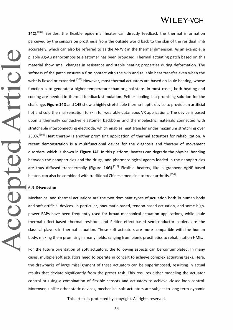
This article is protected by copyright. All rights reserved.
54
14C).[106] Besides, the flexible epidermal heater can directly feedback the thermal information
perceived by the sensors on prosthesis from the outside world back to the skin of the residual limb
accurately, which can also be referred to as the AR/VR in the thermal dimension. As an example, a
pliable Ag-Au nanocomposite elastomer has been proposed. Thermal actuating patch based on this
material show small changes in resistance and stable heating properties during deformation. The
softness of the patch ensures a firm contact with the skin and reliable heat transfer even when the
wrist is flexed or extended.[503] However, most thermal actuators are based on Joule heating, whose
function is to generate a higher temperature than original state. In most cases, both heating and
cooling are needed in thermal feedback stimulation. Peltier cooling is a promising solution for the
challenge. Figure 14D and 14E show a highly stretchable thermo-haptic device to provide an artificial
hot and cold thermal sensation to skin for wearable cutaneous VR applications. The device is based
upon a thermally conductive elastomer backbone and thermoelectric materials connected with
stretchable interconnecting electrode, which enables heat transfer under maximum stretching over
230%.[501] Heat therapy is another promising application of thermal actuators for rehabilitation. A
recent demonstration is a multifunctional device for the diagnosis and therapy of movement
disorders, which is shown in Figure 14F. In this platform, heaters can degrade the physical bonding
between the nanoparticles and the drugs, and pharmacological agents loaded in the nanoparticles
are thus diffused transdermally (Figure 14G).[513] Flexible heaters, like a graphene-AgNP-based
heater, can also be combined with traditional Chinese medicine to treat arthritis.[514]
6.3 Discussion
Mechanical and thermal actuations are the two dominant types of actuation both in human body
and soft artificial devices. In particular, pneumatic-based, tendon-based actuation, and some high-
power EAPs have been frequently used for broad mechanical actuation applications, while Joule
thermal effect-based thermal resistors and Peltier effect-based semiconductor coolers are the
classical players in thermal actuation. These soft actuators are more compatible with the human
body, making them promising in many fields, ranging from bionic prosthetics to rehabilitation HMIs.
For the future orientation of soft actuators, the following aspects can be contemplated. In many
cases, multiple soft actuators need to operate in concert to achieve complex actuating tasks. Here,
the drawbacks of large misalignment of these actuators can be superimposed, resulting in actual
results that deviate significantly from the preset task. This requires either modeling the actuator
control or using a combination of flexible sensors and actuators to achieve closed-loop control.
Moreover, unlike other static devices, mechanical soft actuators are subject to long-term dynamic

This article is protected by copyright. All rights reserved.
55
changes that are highly susceptible to fatigue failure of the device. Moreover, most soft materials
are prone to oxidation, which reduces durability of soft actuators. Therefore, Research on the
durability of device materials needs to be explored in depth. In the next challenge, although, the
response speed of thermal actuators can be improved by material, structure, power, etc. However,
the recovery from high to low temperature is usually a slow process. Such thermal inertia is a matter
of concern. Last but not least, the intimate combination of soft actuators and flexible sensors is also
a new direction for future soft system development, or the development of smart materials with
both actuation and sensing characteristics can also eliminate the heterogeneous problems in
integration.
Figure 14. Thermal actuators for medical robotics. A) Photograph of a smart artificial skin integrated
with sensors and actuators covering a prosthetic hand. B) An exploded view of the artificial skin
comprised of six stacked layers and microscopic image of electroresistive heater. C) Images of the
prosthetic limb caring a baby doll and IR camera images of temperature mapping in this condition. A-
C) Reproduced under the terms of the CC-BY Creative Commons Attribution 4.0 International
license.[106] Copyright 2014, The Authors, published by Springer Nature. D) Simplified illustration and
exploded view of a skin-like thermo-haptic device. E) Localized cooling and heating of hand-shaped
PDMS with the device placed beneath PDMS. D,E) Reproduced with permission.[501] Copyright 2020,

This article is protected by copyright. All rights reserved.
56
Wiley-VCH. F) Image of a multifunctional wearable devices for diagnosis and heat therapy of
movement disorders. G) Temperature distribution measurement of the heater on the skin patch.
F,G) Reproduced with permission.[513] Copyright 2014, Springer Nature.
7. Stimulation
Stimulation holds tremendous promise for prosthetic information feedback[86,515] and
rehabilitation.[329,516,517] From the perspective of haptic restoration, stimulation is the user-input
portion of the bidirectional information interface between the human body and prosthesis.
Specifically, stimulation modalities can transmit biopotential electrical, optical, or mechanical signals
to the body for sensation. Many studies have shown that feedback stimulation has the potential to
not only regain tactile and thermal sensation in amputees,[106,518] but also improve their accuracy and
confidence in manipulating the prosthesis, such as grasping objects.[157,455] Moreover, some subjects
wearing prostheses with sensory feedback reported that stimulation eliminates phantom pains in
the artificial limbs.[157,519] In rehabilitation therapy, stimulations have also been prescribed to treat
disabilities caused by neurological disorders through neural modulation, such as deep brain
stimulation for paralysis and Parkinson's disease.[305,520] Collectively, tactile and proprioceptive
restoration as well as motor rehabilitation can be achieved by stimulating the corresponding sensory
and motor nerves.
Existing literature has documented several types of stimulation methods, such as electrostimulation,
optogenetic stimulation, and vibrostimulation. These flexible stimulators are highly biocompatible,
lightweight, safe, and portable in the form of implants or wearable devices. Thus, they have
garnered the attention of academia, industry, and users. In this section, we primarily review these
flexible implantable and non-invasive stimulators for prosthetic and rehabilitation applications.
7.1 Implantable stimulators
In recent years, profiting from the academic interest and deep exploration in neuroscience, flexible
implantable stimulators with various mechanisms (electrical,[521–523] optogenetic,[524,525]
ultrasonic,[526,527] magnetic[528]) are being developed as non-pharmacological treatment options.
These flexible stimulators function by releasing (electrical and optical) signals into the brain,[272]
spinal cord,[522] and periphery nerves[529] to achieve neural modulation for associated sensory
perception and motor rehabilitation.

This article is protected by copyright. All rights reserved.
57
7.1.1 Electrical stimulators
Electrical signals are the predominant form of nerve excitation propagating in the human body.
Therefore, implantable electrical stimulators are widely deployed because of signal compatibility.
Compared to non-invasive electrical stimulators, implantable electrodes can be precisely inserted or
attached to specific sensory afferent and motor efferent nerves, allowing sensory feedback and
modulation therapy to be more natural and effective.
As a basic architecture, soft implants for electrical stimulation typically consist of flexible substrates
and active electrodes. The former is mechanically stable and compatible with the neural tissue and
serves to carry and protect the electrode. Common substrate materials include PI and hydrogel,
which can remain chemically inert in vivo while also providing sufficient dielectric strength to
prevent discharge breakdown. The latter is embedded in the substrate as a pivotal component of
electrical stimulation, delivering current to the target tissue.[515] As a general rule, electrical
stimulators can be classified by their mechanism of operation as either capacitive or faradic. The
capacitive type relies mainly on the double-layer charge at the electrode-electrolyte interfaces to
inject charge, while the faradic type is based on the redox reactions and ions movement at faradaic
electrodes which provides high levels of charge for stimulation.[305]
Electrode material is a decisive factor in the electrical performance of implantable stimulation
electrodes. Traditionally, capacitive and Faraday electrical stimulation occurs simultaneously on the
surface of metal stimulating electrodes (Au and Pt).[530,531] Generally, nitrides and oxides of titanium
(TiN and TiO2) are popular options for capacitive electrical stimulators due to their chemical
inertness and high electrochemical safety limitation. Faradic electrodes based on oxides of iridium
(IrOx) tend to have a higher charge capacity compared to titanium-derived electrodes due to their
excellent electrochemical properties.[532,533] In the recent decade, various nanomaterials[301,345,534,535]
and conductive polymers[536–538] have also made their way into implantable stimulation electrodes,
which have higher charge injection capacity (1~15 mC cm−2) compared to conventional materials (Pt
0.05~0.15 mC cm−2, TiN 0.9 mC cm-2).[39] Nanomaterials can constitute percolation networks that can
adapt to the deformation of neural tissue while maintaining high electrical performance. On the
other hand, the considerable surface area of nanomaterials can enhance charge injection
capabilities. The intrinsic mechanical elasticity, favorable charge storage and injection capacity of
conductive polymers provide an unparalleled option for faradic electrical stimulators. In addition,
the combination of hydrogels and conductive polymers is a hot area of research since the former has
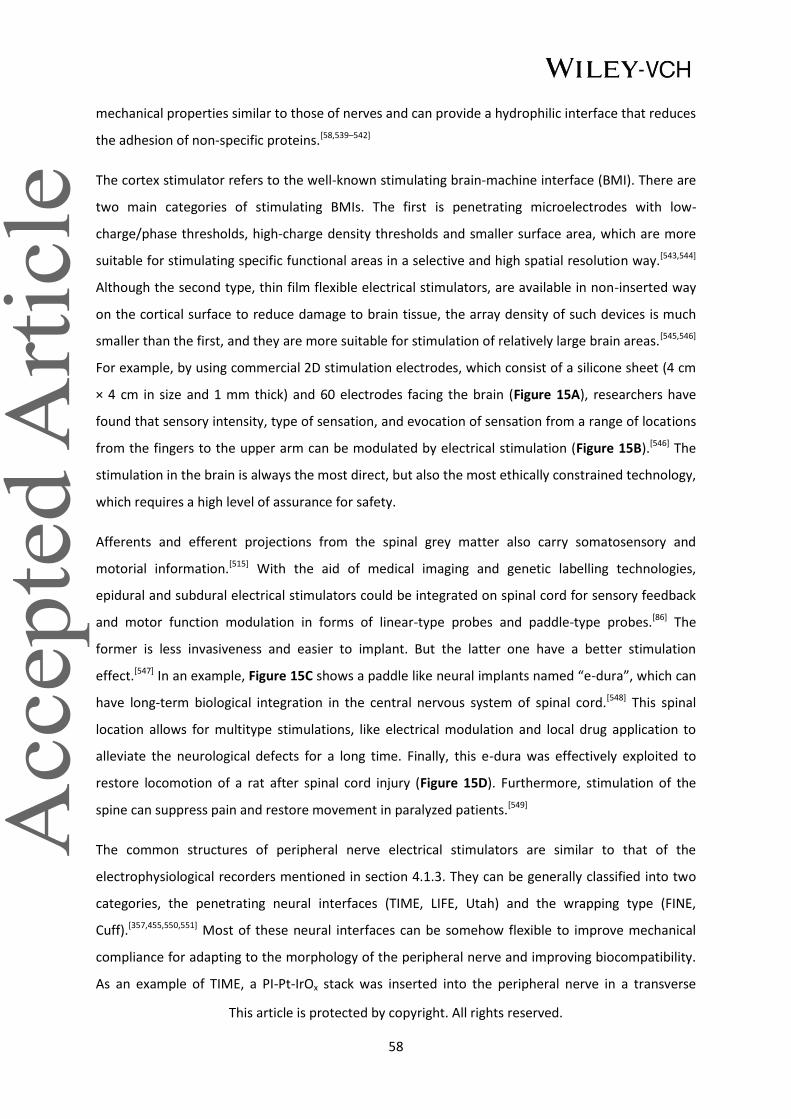
This article is protected by copyright. All rights reserved.
58
mechanical properties similar to those of nerves and can provide a hydrophilic interface that reduces
the adhesion of non-specific proteins.[58,539–542]
The cortex stimulator refers to the well-known stimulating brain-machine interface (BMI). There are
two main categories of stimulating BMIs. The first is penetrating microelectrodes with low-
charge/phase thresholds, high-charge density thresholds and smaller surface area, which are more
suitable for stimulating specific functional areas in a selective and high spatial resolution way.[543,544]
Although the second type, thin film flexible electrical stimulators, are available in non-inserted way
on the cortical surface to reduce damage to brain tissue, the array density of such devices is much
smaller than the first, and they are more suitable for stimulation of relatively large brain areas.[545,546]
For example, by using commercial 2D stimulation electrodes, which consist of a silicone sheet (4 cm
× 4 cm in size and 1 mm thick) and 60 electrodes facing the brain (Figure 15A), researchers have
found that sensory intensity, type of sensation, and evocation of sensation from a range of locations
from the fingers to the upper arm can be modulated by electrical stimulation (Figure 15B).[546] The
stimulation in the brain is always the most direct, but also the most ethically constrained technology,
which requires a high level of assurance for safety.
Afferents and efferent projections from the spinal grey matter also carry somatosensory and
motorial information.[515] With the aid of medical imaging and genetic labelling technologies,
epidural and subdural electrical stimulators could be integrated on spinal cord for sensory feedback
and motor function modulation in forms of linear-type probes and paddle-type probes.[86] The
former is less invasiveness and easier to implant. But the latter one have a better stimulation
effect.[547] In an example, Figure 15C shows a paddle like neural implants named “e-dura”, which can
have long-term biological integration in the central nervous system of spinal cord.[548] This spinal
location allows for multitype stimulations, like electrical modulation and local drug application to
alleviate the neurological defects for a long time. Finally, this e-dura was effectively exploited to
restore locomotion of a rat after spinal cord injury (Figure 15D). Furthermore, stimulation of the
spine can suppress pain and restore movement in paralyzed patients.[549]
The common structures of peripheral nerve electrical stimulators are similar to that of the
electrophysiological recorders mentioned in section 4.1.3. They can be generally classified into two
categories, the penetrating neural interfaces (TIME, LIFE, Utah) and the wrapping type (FINE,
Cuff).[357,455,550,551] Most of these neural interfaces can be somehow flexible to improve mechanical
compliance for adapting to the morphology of the peripheral nerve and improving biocompatibility.
As an example of TIME, a PI-Pt-IrOx stack was inserted into the peripheral nerve in a transverse
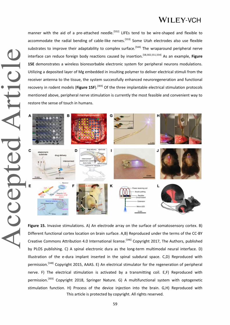
This article is protected by copyright. All rights reserved.
59
manner with the aid of a pre-attached needle.[552] LIFEs tend to be wire-shaped and flexible to
accommodate the radial bending of cable-like nerves.[553] Some Utah electrodes also use flexible
substrates to improve their adaptability to complex surface.[544] The wraparound peripheral nerve
interface can reduce foreign body reactions caused by insertion.[58,302,551,554] As an example, Figure
15E demonstrates a wireless bioresorbable electronic system for peripheral neurons modulations.
Utilizing a deposited layer of Mg embedded in insulting polymer to deliver electrical stimuli from the
receiver antenna to the tissue, the system successfully enhanced neuroregeneration and functional
recovery in rodent models (Figure 15F).[303] Of the three implantable electrical stimulation protocols
mentioned above, peripheral nerve stimulation is currently the most feasible and convenient way to
restore the sense of touch in humans.
Figure 15. Invasive stimulations. A) An electrode array on the surface of somatosensory cortex. B)
Different functional cortex location on brain surface. A,B) Reproduced under the terms of the CC-BY
Creative Commons Attribution 4.0 International license.[546] Copyright 2017, The Authors, published
by PLOS publishing. C) A spinal electronic dura as the long-term multimodal neural interface. D)
Illustration of the e-dura implant inserted in the spinal subdural space. C,D) Reproduced with
permission.[548] Copyright 2015, AAAS. E) An electrical stimulator for the regeneration of peripheral
nerve. F) The electrical stimulation is activated by a transmitting coil. E,F) Reproduced with
permission.[303] Copyright 2018, Springer Nature. G) A multifunctional system with optogenetic
stimulation function. H) Process of the device injection into the brain. G,H) Reproduced with

This article is protected by copyright. All rights reserved.
60
permission.[566] Copyright 2013, AAAS. I) A wireless optoelectronic system. J) The anatomy of the
system on the spinal cord. I,J) Reproduced with permission.[569] Copyright 2017, Wolters Kluwer
Health. K) Light delivery using wirelessly powered and fully internal implants. L) The implant allows
for wireless optogenetic stimulation of peripheral nerve endings. K,L) Reproduced with
permission.[574] Copyright 2015, Springer Nature.
7.1.2 Optogenetic stimulators
Optogenetic stimulation is based on opsins that are sensitive to light signals and only expressed in
certain types of cells. By genetically engineering these proteins into target neurons and controlling
the light around them, these neurons can be activated or inhibited, which is particularly important
for sensory and motor recovery and rehabilitation.[555] Fortunately, optogenetic stimulation does not
lead to muscle fatigue as prolonged and irregular electrical stimulation does. Furthermore,
optogenetic stimulation can excite nerve cells and inhibit them, whereas electrical stimulation only
excites nerves.[556] Many studies have delved into the relationship between optogenetic stimulation
and neurological disorders and control, such as Parkinson's,[557] Alzheimer's,[558] spinal cord
injuries,[559] and somatosensory nociception,[560,561] which have indistinguishable relationship with
prosthetic control, feedback, and human movement rehabilitation. In spite of its appealing
advantages and functions, optogenetics is still mainly exist in theoretical and animal experiments,
without few practical applications to humans, currently.
Light delivery has proven to be a vexing challenge in optogenetic applications, which typically
require implantable optical stimulators. With the advances of materials science and manufacturing
technologies, flexible light-emitting diodes (LED) [562] and microscale, inorganic LED (μ-ILED)[563] with
biocompatibility have been reported to facilitate the progress in optogenetic stimulation for central
and peripheral nervous system. For instance, flexible deep brain needles or wires offers the most
widely adopted carrier for optogenetic localization of cellular-scale stimulation in brain.[342,563–568]
Figure 15G shows a multifunctional system consisting of μ-ILEDs and photodetector for optogenetic
stimulation. This radio frequency-based optogenetics device and its successful operation for up to
four weeks represent a novel stride into closed-loop wireless optogenetic modulation for expediting
the adoption of optogenetics technologies in clinical applications (Figure 15H).[566] Optogenetics was
initially applied in rodent models to control neural circuits in the brain, but recent research efforts
have shown great promise in modulating the activity of the spinal cord and peripheral nervous
system.[556] However, the application of optogenetics to the spinal cord is somewhat limited by the
complex structure, dynamic mechanical characteristics, anchorless nature and low redundancy of

This article is protected by copyright. All rights reserved.
61
the spinal cord that render stable insertion of luminescent sites to target regions of the spinal cord
almost impossible. Thus, optical stimulators for spinal cord are generally flexible enough to be
wraparound or wire-like.[556,569–571] For example, a fully implantable, battery-free wireless
optoelectronic devices for spinal optogenetics is illustrated in Figure 15I. [569] This device design
avoids the tethered operation in traditional optic fiber implants. Based on NFC data and power
transmission, the device could be implanted over the mouse spinal cord in a minimally invasive way
without impeding movement of the mouse (Figure 15J). Additionally, for fully implantable
optogenetic stimulators, the mass and size of the devices have a nonnegligible effect on the animal’s
freedom of movement and behavior by preventing animals from entering small enclosures or
engaging in normal social interactions with others. Therefore, miniaturization or cuff shape is still the
mainstream design of peripheral nerve optical stimulators[572–575]. Figure 15K and 15L demonstrate a
wireless optogenetic devices weigh 20–50 mg, which is two orders of magnitude smaller and lighter
than previously reported wireless optogenetic systems.[574] In addition to the above mentioned
types, there are alternative stimulation modalities in optical stimulation, such as array-based optical
brain stimulators[576] and transcutaneous stimulation,[577] yet some of these are relatively restricted
in their selectivity for specific nerves.
7.2 Non-invasive stimulators
Emerging noninvasive tactile stimulation methods, like transdermal electrical, mechanical
stimulation, thermal feedback (introduced in section 6.2.2), provide alternative solutions to
stimulate the human body for prosthetic and rehabilitation applications. These methods also can
serve as multidimensional, multisensory complements to VR/AR in HMI for a more immersive and
realistic experience.
Compared with implantable stimulators, noninvasive stimulators needn’t any surgical intervention,
which greatly enhances the convenience and safety of stimulation. By mimicking the mechanical
properties (modulus, thickness) of human skin, most flexible wearable devices are becoming more
comfortable and conformable than rigid stimulators to attach on human skin directly and evoke the
sensory and motor function.
7.2.1 Mechanical stimulators
Perception of the residual limb can be elicited by mechanical stimulation, such as vibration and static
force stimulation. From a physiological point of view, the type of perception evoked by the feedback
depends mainly on the different mechanical stimulation parameters. E.g. for force stimuli, the

This article is protected by copyright. All rights reserved.
62
location of the stimulus within the array, the magnitude of the force, and even the direction of the
stimulus force are all relevant factors. Whereas, the frequency and amplitude of the vibration are
keys to generate different types of sensory information in the vibration stimulus.[360] For arraying
stimulators, the feedback stimulation can be achieved with continuous temporal and spatial
variation.[578]
Several mechanisms of micromechanical stimulation have been reported, common ones such as
forces generated by fluids (hydraulic, pneumatic),[579,580] heat-induced deformation,[581–583] etc. These
mechanisms are able to output significantly larger forces and deformation for static force
stimulation. The study in feedback of arraying vectoral forces with these mechanisms is an area of
interest. For example, a matrix composed of 15 SMA-based plasters can be attached to human skin,
and the user can perceive different touch patterns through the tangential and shear forces
generated by SMAs and changes in the location of working units.[584] Nonetheless, the low response
speed and hysteresis of fluidic and heat-induced deformation stimulators limit both mechanisms to
stimulate the skin in the form of high frequency vibrations.[585]
When it comes to the high bandwidth vibro-stimulation, it is necessary to mention DEAs,[586–591]
which naturally have the advantages like an extremely wide range of stimulation frequencies and
easy integration at high spatial density. However, there are still some drawbacks that hinder its
adoption, such as limited actuation force and amplitude, and the need for high operating voltage
(more 1kV). To solve the issue of insufficient stimulation force, multi-layer stacking structure,[592] or
hydraulic pressure to amplify the force have been proposed.[63,593,594] In this high frequency vibration
stimulation, piezoelectric stimulators based on the inverse piezoelectric effect are also a good
alternative.[595–597] In addition to the stimulation principles mentioned above, electromagnetic
stimulator can balance the frequency range and the magnitude of the stimulation force, whose
mechanism is the based on the force applied to the conductor in a magnetic field. For example,
Figure 16A shows a coin-sized (18 mm) electromagnetic exciter with a milliampere operating
current, a Young's modulus of 130 kPa, close to the skin modulus, and an output amplitude of 300
µm (Figure 16B and 16C).[598] However, the need for coils and permanent magnets, which are usually
rigid, always makes such stimulators occupy a large volume.
7.2.2 Electrical stimulators
Flexible epidermal electrotactile display is another haptic interface that provides stimulation by
applying an electric current through the skin surface.[430] It has the potential to replace mechanical
haptic displays owing to several advantages, such as small and thin size, lightweight and rapid

This article is protected by copyright. All rights reserved.
63
responsiveness. Electrical stimulation roughly includes two types, one with voltage control, which is
capable of reducing skin burns feeling during stimulation, and the other with current control, which
is relatively insensitive to differences in the feature of the skin interface.[360,599,600] In addition to
haptic feedback, electrical stimulation is also attractive pain relief and sensory-motor control for
prosthetics and orthotics.[599,601]
Figure 16. Non-invasive feedback stimulation. A) Exploded-view schematic of wireless mechanical
haptic AR/VR device. B) Prosthetics application of the device. C) The device produces a haptic
pattern of sensation. A-C) Reproduced with permission.[598] Copyright 2019, Springer Nature. D)
Schematic of a self-powered electro-tactile system based on transdermal electrical stimulation. E)
HMI application of the TENG-powered electrical stimulator. F) The generated random array and the
identification recurrence results of the subjects. D-F) Adapted with permission.[604] Copyright 2021,
AAAS. Reprinted/adapted from ref.[604]. The Authors, some rights reserved; exclusive licensee AAAS.
Distributed under a Creative Commons Attribution NonCommercial License 4.0 (CC BY-NC).
Integrating epidermal electrodes, which provide opportunities in intimate, long-term interfaces to
the human body,[602] on amputees’ remaining extremities has been tested in some studies.[603] A
representative example is that subjects wearing a PI/Au-based stimulating and sensing hybrid
platform attempted to control a robot hand to grip a bottle both with and without electro-tactile
stimulation. The result illustrates that subjects with force feedback were able to stop grasping at any
desired level of gentle touch, while those without force feedback cannot. Moreover, in this

This article is protected by copyright. All rights reserved.
64
demonstration, the facts that the parameters of the applied current on the skin determines the
tactile stimulation comfort, and stimulation with a large current will induce muscle contractions
were also verified.[599] Conventional electrical stimulators cannot work without bulky power sources,
which greatly confined their portability. TENGs provide a potential solution to achieve self-powered,
wireless electrical stimulation. Figure 16D presents a noninvasive electrical stimulation system
consisting of a TENG array for power supply and a ball-shaped electrode array for current simulation.
Subjects wearing the system can identify different virtual spatial patterns by spatio-temporal varying
electrical stimulation (Figure 16E and 16F).[604] Nevertheless, the variability and over time drift of
skin impedance also present perplexing challenges for electrotactile stimulation at the same
time.[598]
7.3 Discussion
This subsection focuses on the process of transferring external information into the human body in
the form of various stimulus in HMI, and summarizes the prevailing implantable electrostimulation,
optogenetic stimulation devices and wearable electrostimulation along with mechanical stimulation
devices in this field. These flexible devices serve as interfaces that establish a solid foundation for
the human body to obtain external information or to receive therapeutic treatment.
Nonetheless there are still some distinct challenges in this area that are waiting to be addressed. The
first problem is energy supply. One of the differences between stimulators and sensors is that
stimulators consume much more energy than sensors. Currently well-studied energy supply schemes
are ultrasound, NFC, piezoelectric, etc. Improving the efficiency of these existing energy supply
methods and exploring new ones will facilitate the promotion of stimulators. Next challenge is
stimulation density, due to structural, material, or fabrication limitations, the spatial density of many
stimulation arrays is limited, for example, high-density implantable electrical stimulators can easily
exceed cell safety limits while achieving stimulation margins. Multifunctional integration should also
be attached attention. Various implantable or wearable stimulators have unique advantages, and
multimodal stimulation is a potentially advantageous pooling option. Finally, the potential
development of flexible devices for many stimulation mechanisms is still not receiving community
attention, such as acoustic stimulation, and these flexible devices can also potentially be explored as
interfaces for HMI.

This article is protected by copyright. All rights reserved.
65
8. Systematic integration
Sensors, recorders, artificial neurons, actuators, and stimulators are all devices that are based on
soft technologies that acquire and process information from external environments and the human
body. Each of these systems is thereby subjected to human movement, requiring dexterity as the
devices change shape. In a system, they can work independently, but, ideally, each part can form a
closed-loop system that is interlinked to perform more precise and complex tasks.[76,153] Currently,
some integrated work can enhance space utilization,[599] reduce manufacturing costs,[155] and achieve
in-situ information sensing, processing, and actuating simultaneously for more accurate
therapy.[548,605]
A flexible prosthetic system has a representative flow of information with various integrated
components. For example, the electrophysiological signals recorded by a flexible recorder are
processed by machine learning algorithms to decode movement intentions, which can be used to
control a soft robotic hand. With the aid of soft robotic hand motions, flexible e-skins can then
acquire various signals from the exterior environment. Such stimuli can be further converted into
electrophysiological signals through artificial synapses, which are then fed back into the body
through flexible stimulators. All components must have good flexibility, biocompatibility, and
biosimilarity. As an exemplary case, a soft EMG controlled pneumatic neuroprosthetic with ionic
capacitive sensors and flexible stimulators was developed to reduce manufacturing costs and
achieve lightweight actuation. The on-body test of this system showed excellent results: after 15
minutes of training, the amputee was able to use the flexible prosthetic system to perform a range
of precise gripping and social tasks, demonstrating tremendous potential for future applications.[76]
For medical rehabilitation, system integration is also critical to achieve precise treatment and assess
rehabilitation efficacy. For example, implantable systems for motion-related epilepsy rely on real-
time acquisition and analysis of pre-seizure brain electrical signals and perform compensatory
electrical stimulation to eliminate abnormal brain waves and stop the epilepsy midway.[606] In
addition to the neurological aspects of motor therapy, flexible system integration can also be applied
to the limb, such as " mirror" therapy for the rehabilitation of stroke patients, which is based on
monitoring the healthy limb and mirroring the actuation to the hemiplegic limb. This combination
therapy can achieve independent rehabilitation without a guide.[158] The evaluation of optogenetic
stimulation relies mainly on real-time recording of the in situ electrophysiological signals.
Consequently, many integrated flexible stimulation systems can also perform electrophysiological
recording.[52]

This article is protected by copyright. All rights reserved.
66
9. Prospects and Outlooks
In today’s digital era, numerous man-made digital mechatronics products are beginning to permeate
into natural analogous human life, and the boundaries between the two are gradually blurring.
However, there is no denying the legitimate fact that the interaction between them should always
be human-centered, that is to say, to seek the maximum functionality and performance of the
device under the premise of biocompatibility. Because of favorable biocompatibility, high bionic,
low-cost, and safety features, soft electronics and devices, a field that has emerged in recent
decades, can gradually shoulder the role of bridging the gap between the human and machines. This
is especially true in medical robotics for people with disabilities, such as prosthetic engineering,
rehabilitation science and other related fields. Soft devices involve extremely comprehensive
technologies, including flexible sensing, soft actuation, brain-machine interface and other relevant
domains. These devices based on new materials and structures integrate the information flow from
the human body with that from the external environment, and use ML methods for deep processing
to control machines for achieving target actuation effects or feedback to humans themselves in a
biocompatible form. Human expect machines in a broad term to be friendly for coexisting with
human, and to make a difference for a better living. Although the attributes of these devices are
very diverse, they also have common problems with corresponding solutions as human-machine
interfaces, which are proposed and discussed in the following.
Biocompatibility always comes first in human-machine interfaces for medical robotics. Assuring that
materials are not toxic to humans is the most elemental principle of biocompatibility. While many
conceptual devices based on various materials have been validated in academic laboratories.
However, the long-term impact of these materials on the human body remains an uncertainty
limiting the actual long-term commercial application of the devices in HMI. An example is the
discussion of the safety of nanomaterials.[607,608] Long-term validation experiments in animals with
toxicological analysis are potential toxicity assessment solutions. In addition, biocompatibility
encompasses the fields of biomechanics, biochemistry, and immunology, which require materials
that are mechanically compliant, chemically inert, etc. Therefore, in-depth exploration of materials
and elastic structures is a major direction to improve biocompatibility.
Performance is the criterion for device functionality. In sensors, the balance and optimization of
sensitivity, linearity, response time, limit of detection, measuring range, hysteresis and other
properties are constantly pursued by the academic community. While for neural interfaces,
impedance in recording and charge injection capability in stimulation are key properties. Actuation
This article is protected by copyright. All rights reserved.
67
range, amplitude, frequency, response time are the criteria that determine the performance of soft
actuators. The multiple performance of the device often has an implicated relationship, the
optimization of one performance may bring another performance limitation (such as sensitivity vs.
range,[112] accuracy vs. softness[35] etc.), how to balance and optimize the device parameters
according to the performance requirements of the application is a point of consideration for
personalized device design.
Interface issues for signal relay are a major challenge in HMI robotics, specifically for mechanical and
electrical components. There are two main types of interfaces: within the device itself and between
biological and mechanical components. For both of these examples, interface problems mainly arise
in heterogeneous integration of different materials, such as for material bonding and embedding,
which can be addressed by micromechanics, chemical bonding links, etc.[609] Specifically for human-
device interfaces, there are also macro interface problems one must consider. For example, the
large conformal attachment of prosthetic skin,[149] sweat trap between the wearable sensor and the
skin,[75] and fixation of neural implants.[420] By addressing these key issues, one can affirm proper
device function.
Sensor design should be forward-thinking, using intelligent algorithms to inform which sensors are
required for proper data analysis. The reason why humans are specialized in perception and
movement is not only due to the excellent performance of receptors and musculoskeletons, but also
because the brain is skilled in processing this raw information. There have been many outstanding
examples of exploiting machine learning in the field of sensors and actuators, such as extracting
motion intent from electrophysiological signals,[287,610] using haptics to identify object
categories,[160,611] decoupling multimodal signals from cross-reactive sensor arrays,[231,612] and
controlling soft robots[613,614]. However, most of the hardware research is still delinked from
intelligent algorithms. Machine learning enhanced hardware design can help limit the number of
sensors and electronics needed to collect data.
Maintaining proper power supply is another important issue in HMI devices. Traditional tethered
and battery power suffer from motion restrictions and sophisticated recharging processes
respectively, which are obstacles to the application of both wearable and implantable devices.
Researchers are currently investigating wireless-powered and self-powered devices, which is of
upmost important for invasive devices that currently require surgery for battery replacement. For
wireless energy, NFCs[568] and ultrasonic[372] are good powering methods, but still need the energy
transmitter, as well as receiving energy at a certain range; biofuel cells,[615] piezoelectric,[122]
triboelectric,[127] solar[616] and other self-powered methods are prominent representatives of power
supply for low-energy devices, truly enabling the user to move freely. However, for soft devices with
This article is protected by copyright. All rights reserved.
68
high energy consumption, such as soft prostheses, the energy supply method still needs to be
further explored.
The fabrication of many flexible devices and electronics is still based on manual work, which
increases the uncertainty of the device performance as well as the difficulty of mass production. To
eliminate these issues, some potential mass-processing technologies are emerging, such as laser-
based processing,[249,617] roll-to-roll printing,[618,619] inkjet printing,[620] and other technologies for
flexible electronics that offer excellent and consistent performance while ensuring fast, low-cost
manufacturing. Therefore, tailoring the fabrication technique to such mass-producible platform or
exploring new processing technologies can boost the market application of these low-cost soft
devices.
System integration is the ultimate goal for biomimetic prosthetics and is a closed loop where all the
above-mentioned device types are connected.[621] Specifically, flexible electrophysiological recorders
capture the movement intent from human body, decodes it, and forms a prosthetic control signal.
Under the guidance of the signal, the soft robots (prosthesis) moves and perceives external
information using the e-skin mounted on the actuator. In turn, this information is stimulated back to
the body in the form of biocompatible signals through the flexible stimulator, constituting a
complete closed-loop. Among these, the physical integration of sensors and actuators, the
communication and transformation of in vivo signal-electrical signal-physical information are
potentially challenges for the deepening of scientific research. Promisingly, some prospective
systematic works have been reported.[76,153]
Acknowledgements
Wenzheng Heng and Samuel Solomon contributed equally to this work. This project was supported
by the National Institutes of Health grant R01HL155815, Office of Naval Research grant N00014-21-
1-2483 and N00014-21-1-2845, the Translational Research Institute for Space Health through NASA
NNX16AO69A, the Tobacco-Related Disease Research Program grant R01RG3746, and Carver Mead
New Adventures Fund at California Institute of Technology.
Conflict of Interest
The authors declare no conflict of interest.
Received: ((will be filled in by the editorial staff))
Revised: ((will be filled in by the editorial staff))
Published online: ((will be filled in by the editorial staff))

This article is protected by copyright. All rights reserved.
69
References
[1] E. Broadbent, R. Stafford, B. MacDonald, Int. J. Soc. Robot. 2009, 1, 319.
[2] P. Flandorfer, J Popul Res (Canberra) 2012, 2012.
[3] H. Krebs, L. Dipietro, S. Levy-Tzedek, S. Fasoli, A. Rykman-Berland, J. Zipse, J. Fawcett,
J. Stein, H. Poizner, A. Lo, B. Volpe, N. Hogan, IEEE Eng. Med. Biol. Mag. 2008, 27, 61.
[4] L. M. Tijsen, E. W. Derksen, W. P. Achterberg, B. I. Buijck, Clin Interv Aging 2019, 14,
1451.
[5] E. Jaul, J. Barron, Front. Public Health 2017, 5, 335.
[6] United Nations, Ageing and disability,
https://www.un.org/development/desa/disabilities/disability-and-ageing.html,
accessed: 2016.
[7] D. L. Coco, G. Lopez, S. Corrao, Vasc Health Risk Manag 2016, 12, 105.
[8] J. Laut, M. Porfiri, P. Raghavan, Curr Phys Med Rehabil Rep 2016, 4, 312.
[9] CDC, Disability Impacts All of Us,
https://www.cdc.gov/ncbddd/disabilityandhealth/infographic-disability-impacts-
all.html, accessed: 2019.
[10] I. R. Winship, T. H. Murphy, Neuroscientist 2009, 15, 507.
[11] W. M. Jenkins, M. M. Merzenich, in Progress in Brain Research (Eds.: F.J. Seil, E.
Herbert, B.M. Carlson), Elsevier, 1987, pp. 249–266.
[12] H. I. Krebs, B. T. Volpe, in Handbook of Clinical Neurology (Eds.: M.P. Barnes, D.C.
Good), Elsevier, 2013, pp. 283–294.
[13] J. C. Rothwell, Folia Phoniatr Logop 2010, 62, 153.
[14] M. J. Ashley, Cerebrum (N.Y.N.Y., Online) 2012, 2012, 8.
[15] N. Hogan, H. I. Krebs, J. Charnnarong, P. Srikrishna, A. Sharon, in Telemanipulator
Technology, International Society for Optics and Photonics, 1993, pp. 28–34.
[16] P. van Vliet, A. M. Wing, Phys Ther 1991, 71, 39.
[17] H. F. M. Van der Loos, D. J. Reinkensmeyer, E. Guglielmelli, in Springer Handbook of
Robotics (Eds.: B. Siciliano, O. Khatib), Springer International Publishing, Cham, 2016,

This article is protected by copyright. All rights reserved.
70
pp. 1685–1728.
[18] W.-K. Song, H.-Y. Lee, J.-S. Kim, Y.-S. Yoon, Z. Bien, in Proceedings of the 20th Annual
International Conference of the IEEE Engineering in Medicine and Biology Society. Vol.
20 Biomedical Engineering Towards the Year 2000 and Beyond (Cat. No. 98CH36286),
IEEE, 1998, pp. 2682–2685.
[19] B. Stephens-Fripp, G. Alici, R. Mutlu, IEEE Access 2018, 6, 6878.
[20] T. J. Bates, J. R. Fergason, S. N. Pierrie, Curr. Rev. Musculoskelet. Med. 2020, 13, 485.
[21] M. E. Ring, Plast. Reconstr. Surg. 1991, 87, 174.
[22] K. J. Zuo, J. L. Olson, Plast Surg (Oakv) 2014, 22, 44.
[23] Grand View Research “Prosthetics & Orthotics Market Size Report, 2021-2028”,
https://www.grandviewresearch.com/industry-analysis/prosthetics-orthotics-market,
accessed: 2020.
[24] M. R. Tucker, J. Olivier, A. Pagel, H. Bleuler, M. Bouri, O. Lambercy, J. del R. Millán, R.
Riener, H. Vallery, R. Gassert, J. Neuroeng. Rehabilitation 2015, 12, 1.
[25] J. C. Yang, J. Mun, S. Y. Kwon, S. Park, Z. Bao, S. Park, Adv. Mater. 2019, 31, 1904765.
[26] F. W. Clippinger, R. Avery, B. R. Titus, Bull. prosthet. res. 1974, 10, 247.
[27] D. C. Mohr, M. Zhang, S. M. Schueller, Annu Rev Clin Psychol 2017, 13, 23.
[28] R. Herbert, J.-H. Kim, Y. Kim, H. Lee, W.-H. Yeo, Materials 2018, 11, 187.
[29] T. Q. Trung, N.-E. Lee, Adv. Mater. 2017, 29, 1603167.
[30] S. J. Benight, C. Wang, J. B. H. Tok, Z. Bao, Prog. Polym. Sci. 2013, 38, 1961.
[31] R. L. J. Cruz, M. T. Ross, S. K. Powell, M. A. Woodruff, Front. Bioeng. Biotechnol. 2020,
8, 121.
[32] M. Wang, Y. Luo, T. Wang, C. Wan, L. Pan, S. Pan, K. He, A. Neo, X. Chen, Adv. Mater.
2021, 33, 2003014.
[33] S. Kim, C. Laschi, B. Trimmer, Trends Biotechnol. 2013, 31, 287.
[34] G. M. Whitesides, Angew. Chem. Int. Ed. 2018, 57, 4258.
[35] D. Rus, M. T. Tolley, Nature 2015, 521, 467.
[36] R. Dahiya, N. Yogeswaran, F. Liu, L. Manjakkal, E. Burdet, V. Hayward, H. Jorntell, Proc

This article is protected by copyright. All rights reserved.
71
IEEE Inst Electr Electron Eng 2019, 107, 2016.
[37] B. Shih, D. Shah, J. Li, T. G. Thuruthel, Y.-L. Park, F. Iida, Z. Bao, R. Kramer-Bottiglio, M.
T. Tolley, Sci. Robot. 2020, 5, eaaz9239.
[38] M. Cianchetti, C. Laschi, A. Menciassi, P. Dario, Nat. Rev. Mater. 2018, 3, 143.
[39] R. Chen, A. Canales, P. Anikeeva, Nat. Rev. Mater. 2017, 2, 1.
[40] S. Liu, D. S. Shah, R. Kramer-Bottiglio, Nat. Mater. 2021, 20, 851.
[41] J. Xu, S. Wang, G.-J. N. Wang, C. Zhu, S. Luo, L. Jin, X. Gu, S. Chen, V. R. Feig, J. W. F. To,
S. Rondeau-Gagné, J. Park, B. C. Schroeder, C. Lu, J. Y. Oh, Y. Wang, Y.-H. Kim, H. Yan, R.
Sinclair, D. Zhou, G. Xue, B. Murmann, C. Linder, W. Cai, J. B.-H. Tok, J. W. Chung, Z.
Bao, Science 2017, 355, 59.
[42] S. Wang, J. Xu, W. Wang, G.-J. N. Wang, R. Rastak, F. Molina-Lopez, J. W. Chung, S. Niu,
V. R. Feig, J. Lopez, T. Lei, S.-K. Kwon, Y. Kim, A. M. Foudeh, A. Ehrlich, A. Gasperini, Y.
Yun, B. Murmann, J. B.-H. Tok, Z. Bao, Nature 2018, 555, 83.
[43] S. Xu, Y. Zhang, L. Jia, K. E. Mathewson, K.-I. Jang, J. Kim, H. Fu, X. Huang, P. Chava, R.
Wang, S. Bhole, L. Wang, Y. J. Na, Y. Guan, M. Flavin, Z. Han, Y. Huang, J. A. Rogers,
Science 2014, 344, 70.
[44] S. Lee, S. Franklin, F. A. Hassani, T. Yokota, M. O. G. Nayeem, Y. Wang, R. Leib, G.
Cheng, D. W. Franklin, T. Someya, Science 2020, 370, 966.
[45] Y. Yang, Y. Song, X. Bo, J. Min, O. S. Pak, L. Zhu, M. Wang, J. Tu, A. Kogan, H. Zhang, T. K.
Hsiai, Z. Li, W. Gao, Nat. Biotechnol. 2020, 38, 217.
[46] K. Xu, Y. Lu, S. Honda, T. Arie, S. Akita, K. Takei, J. Mater. Chem. C 2019, 7, 9609.
[47] X. Shi, Y. Zuo, P. Zhai, J. Shen, Y. Yang, Z. Gao, M. Liao, J. Wu, J. Wang, X. Xu, Q. Tong, B.
Zhang, B. Wang, X. Sun, L. Zhang, Q. Pei, D. Jin, P. Chen, H. Peng, Nature 2021, 591,
240.
[48] M. Kaltenbrunner, T. Sekitani, J. Reeder, T. Yokota, K. Kuribara, T. Tokuhara, M. Drack,
R. Schwödiauer, I. Graz, S. Bauer-Gogonea, S. Bauer, T. Someya, Nature 2013, 499,
458.
[49] C. Wang, C. Wang, Z. Huang, S. Xu, Adv. Mater. 2018, 30, 1801368.
[50] R. Feiner, T. Dvir, Nat. Rev. Mater. 2018, 3, 17076.
[51] S. M. Won, E. Song, J. Zhao, J. Li, J. Rivnay, J. A. Rogers, Adv. Mater. 2018, 30, 1800534.

This article is protected by copyright. All rights reserved.
72
[52] S. Park, H. Yuk, R. Zhao, Y. S. Yim, E. W. Woldeghebriel, J. Kang, A. Canales, Y. Fink, G. B.
Choi, X. Zhao, P. Anikeeva, Nat. Commun. 2021, 12, 3435.
[53] S. Lin, H. Yuk, T. Zhang, G. A. Parada, H. Koo, C. Yu, X. Zhao, Adv. Mater. 2016, 28, 4497.
[54] J. Rivnay, H. Wang, L. Fenno, K. Deisseroth, G. G. Malliaras, Sci. Adv. 2017, 3,
e1601649.
[55] D. Wirthl, R. Pichler, M. Drack, G. Kettlguber, R. Moser, R. Gerstmayr, F. Hartmann, E.
Bradt, R. Kaltseis, C. M. Siket, S. E. Schausberger, S. Hild, S. Bauer, M. Kaltenbrunner,
Sci. Adv. 2017, 3, e1700053.
[56] S. R. Shin, S. M. Jung, M. Zalabany, K. Kim, P. Zorlutuna, S. bok Kim, M. Nikkhah, M.
Khabiry, M. Azize, J. Kong, K. Wan, T. Palacios, M. R. Dokmeci, H. Bae, X. (Shirley) Tang,
A. Khademhosseini, ACS Nano 2013, 7, 2369.
[57] C. Keplinger, J.-Y. Sun, C. C. Foo, P. Rothemund, G. M. Whitesides, Z. Suo, Science 2013,
341, 984.
[58] Y. Liu, J. Liu, S. Chen, T. Lei, Y. Kim, S. Niu, H. Wang, X. Wang, A. M. Foudeh, J. B.-H. Tok,
Z. Bao, Nat. Biomed. Eng. 2019, 3, 58.
[59] E. Song, J. Li, S. M. Won, W. Bai, J. A. Rogers, Nat. Mater. 2020, 19, 590.
[60] W. Zhou, S. Yao, H. Wang, Q. Du, Y. Ma, Y. Zhu, ACS Nano 2020, 14, 5798.
[61] C. Pang, G.-Y. Lee, T. Kim, S. M. Kim, H. N. Kim, S.-H. Ahn, K.-Y. Suh, Nat. Mater. 2012,
11, 795.
[62] E. Acome, S. K. Mitchell, T. G. Morrissey, M. B. Emmett, C. Benjamin, M. King, M.
Radakovitz, C. Keplinger, Science 2018, 359, 61.
[63] E. Leroy, R. Hinchet, H. Shea, Adv. Mater. 2020, 32, 2002564.
[64] Z. Jiao, C. Zhang, W. Wang, M. Pan, H. Yang, J. Zou, Adv. Sci. 2019, 6, 1901371.
[65] A. Miriyev, K. Stack, H. Lipson, Nat. Commun. 2017, 8, 596.
[66] C. Larson, B. Peele, S. Li, S. Robinson, M. Totaro, L. Beccai, B. Mazzolai, R. Shepherd,
Science 2016, 351, 1071.
[67] J. Kang, J. B.-H. Tok, Z. Bao, Nat. Electron. 2019, 2, 144.
[68] S.-K. Kang, R. K. Murphy, S.-W. Hwang, S. M. Lee, D. V. Harburg, N. A. Krueger, J. Shin,
P. Gamble, H. Cheng, S. Yu, Nature 2016, 530, 71.
[69] X. Wu, J. Zhou, J. Huang, Macromol Rapid Commun 2018, 39, 1800084.
This article is protected by copyright. All rights reserved.
73
[70] H. Fu, K. Nan, W. Bai, W. Huang, K. Bai, L. Lu, C. Zhou, Y. Liu, F. Liu, J. Wang, M. Han, Z.
Yan, H. Luan, Y. Zhang, Y. Zhang, J. Zhao, X. Cheng, M. Li, J. W. Lee, Y. Liu, D. Fang, X. Li,
Y. Huang, Y. Zhang, J. A. Rogers, Nat. Mater. 2018, 17, 268.
[71] B. H. Kim, K. Li, J.-T. Kim, Y. Park, H. Jang, X. Wang, Z. Xie, S. M. Won, H.-J. Yoon, G. Lee,
Nature 2021, 597, 503.
[72] L. Miao, Y. Song, Z. Ren, C. Xu, J. Wan, H. Wang, H. Guo, Z. Xiang, M. Han, H. Zhang,
Adv. Mater. 2021, 33, 2102691.
[73] A. Miyamoto, S. Lee, N. F. Cooray, S. Lee, M. Mori, N. Matsuhisa, H. Jin, L. Yoda, T.
Yokota, A. Itoh, M. Sekino, H. Kawasaki, T. Ebihara, M. Amagai, T. Someya, Nat.
Nanotech. 2017, 12, 907.
[74] Z. Li, M. Zhu, J. Shen, Q. Qiu, J. Yu, B. Ding, Adv. Funct. Mater. 2020, 30, 1908411.
[75] H. Yeon, H. Lee, Y. Kim, D. Lee, Y. Lee, J.-S. Lee, J. Shin, C. Choi, J.-H. Kang, J. M. Suh, H.
Kim, H. S. Kum, J. Lee, D. Kim, K. Ko, B. S. Ma, P. Lin, S. Han, S. Kim, S.-H. Bae, T.-S. Kim,
M.-C. Park, Y.-C. Joo, E. Kim, J. Han, J. Kim, Sci. Adv. 2021, 7, eabg8459.
[76] G. Gu, N. Zhang, H. Xu, S. Lin, Y. Yu, G. Chai, L. Ge, H. Yang, Q. Shao, X. Sheng, Nat.
Biomed. Eng. 2021, 1.
[77] S. Lee, A. Reuveny, J. Reeder, S. Lee, H. Jin, Q. Liu, T. Yokota, T. Sekitani, T. Isoyama, Y.
Abe, Z. Suo, T. Someya, Nat. Nanotech. 2016, 11, 472.
[78] L. Shi, T. Zhu, G. Gao, X. Zhang, W. Wei, W. Liu, S. Ding, Nat. Commun. 2018, 9, 2630.
[79] G. Pang, G. Yang, W. Heng, Z. Ye, X. Huang, H.-Y. Yang, Z. Pang, IEEE Trans. Ind.
Electron. 2021, 68, 3303.
[80] E. A. Lumpkin, M. J. Caterina, Nature 2007, 445, 858.
[81] P. Zhu, H. Du, X. Hou, P. Lu, L. Wang, J. Huang, N. Bai, Z. Wu, N. X. Fang, C. F. Guo, Nat.
Commun. 2021, 12, 4731.
[82] Y. Wang, S. Lee, T. Yokota, H. Wang, Z. Jiang, J. Wang, M. Koizumi, T. Someya, Sci. Adv.
2020, 6, eabb7043.
[83] W. Gao, S. Emaminejad, H. Y. Y. Nyein, S. Challa, K. Chen, A. Peck, H. M. Fahad, H. Ota,
H. Shiraki, D. Kiriya, D.-H. Lien, G. A. Brooks, R. W. Davis, A. Javey, Nature 2016, 529,
509.
[84] K. Xu, Y. Fujita, Y. Lu, S. Honda, M. Shiomi, T. Arie, S. Akita, K. Takei, Adv. Mater. 2021,
33, 2008701.
This article is protected by copyright. All rights reserved.
74
[85] A. Petritz, E. Karner-Petritz, T. Uemura, P. Schäffner, T. Araki, B. Stadlober, T. Sekitani,
Nat. Commun. 2021, 12, 2399.
[86] A. Chortos, J. Liu, Z. Bao, Nat. Mater. 2016, 15, 937.
[87] Y. Ma, Y. Zhang, S. Cai, Z. Han, X. Liu, F. Wang, Y. Cao, Z. Wang, H. Li, Y. Chen, X. Feng,
Adv. Mater. 2020, 32, 1902062.
[88] A. Hadjidj, A. Bouabdallah, Y. Challal, in 2011 IEEE International Symposium on a World
of Wireless, Mobile and Multimedia Networks, 2011, pp. 1–3.
[89] T. Giorgino, P. Tormene, G. Maggioni, C. Pistarini, S. Quaglini, IEEE trans. inf. technol.
biomed. 2009, 13, 1012.
[90] I. P. I. Pappas, T. Keller, S. Mangold, M. Popovic, V. Dietz, M. Morari, IEEE Sens. J. 2004,
4, 268.
[91] B. Ciui, A. Martin, R. K. Mishra, T. Nakagawa, T. J. Dawkins, M. Lyu, C. Cristea, R.
Sandulescu, J. Wang, ACS Sens. 2018, 3, 2375.
[92] S. Yao, Y. Zhu, Nanoscale 2014, 6, 2345.
[93] M. Ha, S. Lim, H. Ko, J. Mater. Chem. B 2018, 6, 4043.
[94] S. Niu, N. Matsuhisa, L. Beker, J. Li, S. Wang, J. Wang, Y. Jiang, X. Yan, Y. Yun, W.
Burnett, A. S. Y. Poon, J. B.-H. Tok, X. Chen, Z. Bao, Nat. Electron. 2019, 2, 361.
[95] A. Chortos, Z. Bao, Mater. Today 2014, 17, 321.
[96] R. W. Van Boven, K. O. Johnson, Neurology 1994, 44, 2361.
[97] R. Di Giacomo, L. Bonanomi, V. Costanza, B. Maresca, C. Daraio, Sci. Robot. 2017, 2,
eaai9251.
[98] N. Bai, L. Wang, Q. Wang, J. Deng, Y. Wang, P. Lu, J. Huang, G. Li, Y. Zhang, J. Yang, K.
Xie, X. Zhao, C. F. Guo, Nat. Commun. 2020, 11, 209.
[99] Y. Yan, Z. Hu, Z. Yang, W. Yuan, C. Song, J. Pan, Y. Shen, Sci. Robot. 2021, 6, eabc8801.
[100] Y. Wu, Y. Liu, Y. Zhou, Q. Man, C. Hu, W. Asghar, F. Li, Z. Yu, J. Shang, G. Liu, Sci. Robot.
2018, 3, eaat0429.
[101] B. Lee, J.-Y. Oh, H. Cho, C. W. Joo, H. Yoon, S. Jeong, E. Oh, J. Byun, H. Kim, S. Lee, J.
Seo, C. W. Park, S. Choi, N.-M. Park, S.-Y. Kang, C.-S. Hwang, S.-D. Ahn, J.-I. Lee, Y.
Hong, Nat. Commun. 2020, 11, 663.
[102] Y. Zang, F. Zhang, D. Huang, X. Gao, C. Di, D. Zhu, Nat. Commun. 2015, 6, 6269.
This article is protected by copyright. All rights reserved.
75
[103] T. Yokota, Y. Inoue, Y. Terakawa, J. Reeder, M. Kaltenbrunner, T. Ware, K. Yang, K.
Mabuchi, T. Murakawa, M. Sekino, W. Voit, T. Sekitani, T. Someya, Proc. Natl. Acad. Sci.
U.S.A. 2015, 112, 14533.
[104] Q. Hua, J. Sun, H. Liu, R. Bao, R. Yu, J. Zhai, C. Pan, Z. L. Wang, Nat. Commun. 2018, 9,
244.
[105] B. W. An, S. Heo, S. Ji, F. Bien, J.-U. Park, Nat. Commun. 2018, 9, 2458.
[106] J. Kim, M. Lee, H. J. Shim, R. Ghaffari, H. R. Cho, D. Son, Y. H. Jung, M. Soh, C. Choi, S.
Jung, K. Chu, D. Jeon, S.-T. Lee, J. H. Kim, S. H. Choi, T. Hyeon, D.-H. Kim, Nat. Commun.
2014, 5, 5747.
[107] F. Zhang, Y. Zang, D. Huang, C. Di, D. Zhu, Nat. Commun. 2015, 6, 8356.
[108] M. L. Hammock, A. Chortos, B. C.-K. Tee, J. B.-H. Tok, Z. Bao, Adv. Mater. 2013, 25,
5997.
[109] S. Chun, W. Son, H. Kim, S. K. Lim, C. Pang, C. Choi, Nano Lett. 2019, 19, 3305.
[110] S. Chun, J.-S. Kim, Y. Yoo, Y. Choi, S. J. Jung, D. Jang, G. Lee, K.-I. Song, K. S. Nam, I.
Youn, D. Son, C. Pang, Y. Jeong, H. Jung, Y.-J. Kim, B.-D. Choi, J. Kim, S.-P. Kim, W. Park,
S. Park, Nat Electron 2021, 4, 429.
[111] L. Pan, A. Chortos, G. Yu, Y. Wang, S. Isaacson, R. Allen, Y. Shi, R. Dauskardt, Z. Bao,
Nat. Commun. 2014, 5, 3002.
[112] W. Heng, G. Yang, G. Pang, Z. Ye, H. Lv, J. Du, G. Zhao, Z. Pang, Adv. Intell. Syst. 2021, 3,
2000038.
[113] Y. Gao, H. Ota, E. W. Schaler, K. Chen, A. Zhao, W. Gao, H. M. Fahad, Y. Leng, A. Zheng,
F. Xiong, C. Zhang, L.-C. Tai, P. Zhao, R. S. Fearing, A. Javey, Adv. Mater. 2017, 29,
1701985.
[114] L. Shi, Z. Li, M. Chen, Y. Qin, Y. Jiang, L. Wu, Nat. Commun. 2020, 11, 3529.
[115] J. Park, Y. Lee, J. Hong, M. Ha, Y.-D. Jung, H. Lim, S. Y. Kim, H. Ko, ACS Nano 2014, 8,
4689.
[116] A. Chiolerio, I. Roppolo, K. Bejtka, A. Asvarov, C. F. Pirri, RSC Adv. 2016, 6, 56661.
[117] L. Wang, R. Zhu, G. Li, ACS Appl. Mater. Interfaces 2020, 12, 1953.
[118] J. Jia, G. Huang, J. Deng, K. Pan, Nanoscale 2019, 11, 4258.
[119] J. Shintake, E. Piskarev, S. H. Jeong, D. Floreano, Adv. Mater. Technol. 2018, 3,

This article is protected by copyright. All rights reserved.
76
1700284.
[120] D. J. Lipomi, M. Vosgueritchian, B. C.-K. Tee, S. L. Hellstrom, J. A. Lee, C. H. Fox, Z. Bao,
Nat. Nanotech. 2011, 6, 788.
[121] F.-R. Fan, Z.-Q. Tian, Z. Lin Wang, Nano Energy 2012, 1, 328.
[122] Z. L. Wang, J. Song, Science 2006, 312, 242.
[123] H. Wang, M. Han, Y. Song, H. Zhang, Nano Energy 2021, 81, 105627.
[124] H. Oh, G.-C. Yi, M. Yip, S. A. Dayeh, Sci. Adv. 2020, 6, eabd7795.
[125] H. Yao, W. Yang, W. Cheng, Y. J. Tan, H. H. See, S. Li, H. P. A. Ali, B. Z. H. Lim, Z. Liu, B. C.
K. Tee, Proc. Natl. Acad. Sci. U.S.A. 2020, 117, 25352.
[126] C. Dagdeviren, Y. Su, P. Joe, R. Yona, Y. Liu, Y.-S. Kim, Y. Huang, A. R. Damadoran, J. Xia,
L. W. Martin, Y. Huang, J. A. Rogers, Nat. Commun. 2014, 5, 4496.
[127] W. Fan, Q. He, K. Meng, X. Tan, Z. Zhou, G. Zhang, J. Yang, Z. L. Wang, Sci. Adv. 2020, 6,
eaay2840.
[128] Z. A. Lamport, M. R. Cavallari, K. A. Kam, C. K. McGinn, C. Yu, I. Kymissis, Adv. Funct.
Mater. 2020, 30, 2004700.
[129] Y. Dai, H. Hu, M. Wang, J. Xu, S. Wang, Nat. Electron. 2021, 4, 17.
[130] G. Schwartz, B. C.-K. Tee, J. Mei, A. L. Appleton, D. H. Kim, H. Wang, Z. Bao, Nat.
Commun. 2013, 4, 1859.
[131] S.-H. Shin, S. Ji, S. Choi, K.-H. Pyo, B. Wan An, J. Park, J. Kim, J.-Y. Kim, K.-S. Lee, S.-Y.
Kwon, J. Heo, B.-G. Park, J.-U. Park, Nat. Commun. 2017, 8, 14950.
[132] Y.-C. Huang, Y. Liu, C. Ma, H.-C. Cheng, Q. He, H. Wu, C. Wang, C.-Y. Lin, Y. Huang, X.
Duan, Nat. Electron. 2020, 3, 59.
[133] T. Sekitani, U. Zschieschang, H. Klauk, T. Someya, Nat. Mater. 2010, 9, 1015.
[134] Z. Wang, S. Guo, H. Li, B. Wang, Y. Sun, Z. Xu, X. Chen, K. Wu, X. Zhang, F. Xing, L. Li, W.
Hu, Adv. Mater. 2019, 31, 1805630.
[135] C. Wang, D. Hwang, Z. Yu, K. Takei, J. Park, T. Chen, B. Ma, A. Javey, Nat. Mater. 2013,
12, 899.
[136] H. Bai, S. Li, J. Barreiros, Y. Tu, C. R. Pollock, R. F. Shepherd, Science 2020, 370, 848.
[137] T. Kim, S. Lee, T. Hong, G. Shin, T. Kim, Y.-L. Park, Sci. Robot. 2020, 5, eabc6878.

This article is protected by copyright. All rights reserved.
77
[138] M. Ramuz, B. C.-K. Tee, J. B.-H. Tok, Z. Bao, Adv. Mater. 2012, 24, 3223.
[139] J. Ge, X. Wang, M. Drack, O. Volkov, M. Liang, G. S. Cañón Bermúdez, R. Illing, C. Wang,
S. Zhou, J. Fassbender, M. Kaltenbrunner, D. Makarov, Nat. Commun. 2019, 10, 4405.
[140] X. Wu, M. Ahmed, Y. Khan, M. E. Payne, J. Zhu, C. Lu, J. W. Evans, A. C. Arias, Sci. Adv.
2020, 6, eaba1062.
[141] V. Amoli, J. S. Kim, E. Jee, Y. S. Chung, S. Y. Kim, J. Koo, H. Choi, Y. Kim, D. H. Kim, Nat.
Commun. 2019, 10, 4019.
[142] J.-Y. Sun, C. Keplinger, G. M. Whitesides, Z. Suo, Adv. Mater. 2014, 26, 7608.
[143] P. Piacenza, S. Sherman, M. Ciocarlie, IEEE Robot. Autom. Lett. 2018, 3, 1434.
[144] Y. R. Lee, T. Q. Trung, B.-U. Hwang, N.-E. Lee, Nat. Commun. 2020, 11, 2753.
[145] K.-Y. Chun, Y. J. Son, C.-S. Han, ACS Nano 2016, 10, 4550.
[146] D. Farina, I. Vujaklija, R. Brånemark, A. M. Bull, H. Dietl, B. Graimann, L. J. Hargrove, K.-
P. Hoffmann, H. H. Huang, T. Ingvarsson, Nat. Biomed. Eng. 2021, 1.
[147] C. M. Boutry, M. Negre, M. Jorda, O. Vardoulis, A. Chortos, O. Khatib, Z. Bao, Sci.
Robot. 2018, 3, eaau6914.
[148] Y. Luo, Y. Li, P. Sharma, W. Shou, K. Wu, M. Foshey, B. Li, T. Palacios, A. Torralba, W.
Matusik, Nat. Electron. 2021, 4, 193.
[149] G. Cheng, E. Dean-Leon, F. Bergner, J. Rogelio Guadarrama Olvera, Q. Leboutet, P.
Mittendorfer, Proc IEEE Inst Electr Electron Eng 2019, 107, 2034.
[150] Z. Zhu, H. S. Park, M. C. McAlpine, Sci. Adv. 2020, 6, eaba5575.
[151] R. Hensleigh, H. Cui, Z. Xu, J. Massman, D. Yao, J. Berrigan, X. Zheng, Nat. Electron.
2020, 3, 216.
[152] Y. Zhang, G. Laput, C. Harrison, in Proceedings of the 2017 CHI Conference on Human
Factors in Computing Systems, ACM, Denver Colorado USA, 2017, pp. 1–14.
[153] H. Zhao, K. O’Brien, S. Li, R. F. Shepherd, Sci. Robot. 2016, 1, eaai7529.
[154] A. Tomar, Y. Tadesse, in Electroactive Polymer Actuators and Devices (EAPAD) 2016,
International Society for Optics and Photonics, 2016, p. 979809.
[155] Z. Shen, X. Zhu, C. Majidi, G. Gu, Adv. Mater. 2021, 2102069.
[156] J. Park, M. Kim, Y. Lee, H. S. Lee, H. Ko, Sci. Adv. 2015, 1, e1500661.

This article is protected by copyright. All rights reserved.
78
[157] D. W. Tan, M. A. Schiefer, M. W. Keith, J. R. Anderson, J. Tyler, D. J. Tyler, Sci Transl Med
2014, 6, 257ra138.
[158] X. Chen, L. Gong, L. Wei, S.-C. Yeh, L. Da Xu, L. Zheng, Z. Zou, IEEE Trans Industr Inform
2021, 17, 943.
[159] J. Hughes, A. Spielberg, M. Chounlakone, G. Chang, W. Matusik, D. Rus, Adv. Intell.
Syst. 2020, 2, 2000002.
[160] S. Sundaram, P. Kellnhofer, Y. Li, J.-Y. Zhu, A. Torralba, W. Matusik, Nature 2019, 569,
698.
[161] M. Zhu, Z. Sun, Z. Zhang, Q. Shi, T. He, H. Liu, T. Chen, C. Lee, Sci. Adv. 2020, 6,
eaaz8693.
[162] F. L. Hammond, Y. Mengüç, R. J. Wood, in 2014 IEEE/RSJ International Conference on
Intelligent Robots and Systems, 2014, pp. 4000–4007.
[163] W. Heng, G. Pang, F. Xu, X. Huang, Z. Pang, G. Yang, Sensors 2019, 19, 5197.
[164] F. M. Petrini, G. Valle, M. Bumbasirevic, F. Barberi, D. Bortolotti, P. Cvancara, A.
Hiairrassary, P. Mijovic, A. Ö. Sverrisson, A. Pedrocchi, J.-L. Divoux, I. Popovic, K.
Lechler, B. Mijovic, D. Guiraud, T. Stieglitz, A. Alexandersson, S. Micera, A. Lesic, S.
Raspopovic, Sci Transl Med 2019, 11, eaav8939.
[165] G. Yang, G. Pang, Z. Pang, Y. Gu, M. Mäntysalo, H. Yang, IEEE Rev Biomed Eng 2019, 12,
34.
[166] C. Wang, Y. Yu, Y. Yuan, C. Ren, Q. Liao, J. Wang, Z. Chai, Q. Li, Z. Li, Matter 2020, 2,
181.
[167] Z. Li, Z. L. Wang, Adv. Mater. 2011, 23, 84.
[168] D. Kang, P. V. Pikhitsa, Y. W. Choi, C. Lee, S. S. Shin, L. Piao, B. Park, K.-Y. Suh, T. Kim, M.
Choi, Nature 2014, 516, 222.
[169] X. Peng, K. Dong, C. Ye, Y. Jiang, S. Zhai, R. Cheng, D. Liu, X. Gao, J. Wang, Z. L. Wang,
Sci. Adv. 2020, 6, eaba9624.
[170] J. W. Kwak, M. Han, Z. Xie, H. U. Chung, J. Y. Lee, R. Avila, J. Yohay, X. Chen, C. Liang, M.
Patel, I. Jung, J. Kim, M. Namkoong, K. Kwon, X. Guo, C. Ogle, D. Grande, D. Ryu, D. H.
Kim, S. Madhvapathy, C. Liu, D. S. Yang, Y. Park, R. Caldwell, A. Banks, S. Xu, Y. Huang,
S. Fatone, J. A. Rogers, Sci Transl Med 2020, 12, eabc4327.
[171] Y. S. Oh, J.-H. Kim, Z. Xie, S. Cho, H. Han, S. W. Jeon, M. Park, M. Namkoong, R. Avila, Z.
This article is protected by copyright. All rights reserved.
79
Song, S.-U. Lee, K. Ko, J. Lee, J.-S. Lee, W. G. Min, B.-J. Lee, M. Choi, H. U. Chung, J.
Kim, M. Han, J. Koo, Y. S. Choi, S. S. Kwak, S. B. Kim, J. Kim, J. Choi, C.-M. Kang, J. U.
Kim, K. Kwon, S. M. Won, J. M. Baek, Y. Lee, S. Y. Kim, W. Lu, A. Vazquez-Guardado, H.
Jeong, H. Ryu, G. Lee, K. Kim, S. Kim, M. S. Kim, J. Choi, D. Y. Choi, Q. Yang, H. Zhao, W.
Bai, H. Jang, Y. Yu, J. Lim, X. Guo, B. H. Kim, S. Jeon, C. Davies, A. Banks, H. J. Sung, Y.
Huang, I. Park, J. A. Rogers, Nat. Commun. 2021, 12, 5008.
[172] C. Zhang, H. Dong, C. Zhang, Y. Fan, J. Yao, Y. S. Zhao, Sci. Adv. 2021, 7, eabh3530.
[173] C. Cho, P. Kang, A. Taqieddin, Y. Jing, K. Yong, J. M. Kim, M. F. Haque, N. R. Aluru, S.
Nam, Nat. Electron. 2021, 4, 126.
[174] Y. Ma, N. Liu, L. Li, X. Hu, Z. Zou, J. Wang, S. Luo, Y. Gao, Nat. Commun. 2017, 8, 1207.
[175] T. Yamada, Y. Hayamizu, Y. Yamamoto, Y. Yomogida, A. Izadi-Najafabadi, D. N. Futaba,
K. Hata, Nat. Nanotech. 2011, 6, 296.
[176] Z. Han, L. Liu, J. Zhang, Q. Han, K. Wang, H. Song, Z. Wang, Z. Jiao, S. Niu, L. Ren,
Nanoscale 2018, 10, 15178.
[177] D. J. Cohen, D. Mitra, K. Peterson, M. M. Maharbiz, Nano Lett. 2012, 12, 1821.
[178] U.-H. Shin, D.-W. Jeong, S.-M. Park, S.-H. Kim, H. W. Lee, J.-M. Kim, Carbon 2014, 80,
396.
[179] J. Lee, S. J. Ihle, G. S. Pellegrino, H. Kim, J. Yea, C.-Y. Jeon, H.-C. Son, C. Jin, D. Eberli, F.
Schmid, B. L. Zambrano, A. F. Renz, C. Forró, H. Choi, K.-I. Jang, R. Küng, J. Vörös, Nat.
Electron. 2021, 4, 291.
[180] S. L. Zhang, Y.-C. Lai, X. He, R. Liu, Y. Zi, Z. L. Wang, Adv. Funct. Mater. 2017, 27,
1606695.
[181] C. Cui, X. Wang, Z. Yi, B. Yang, X. Wang, X. Chen, J. Liu, C. Yang, ACS Appl. Mater.
Interfaces 2018, 10, 3652.
[182] Z. Su, H. Wu, H. Chen, H. Guo, X. Cheng, Y. Song, X. Chen, H. Zhang, Nano Energy 2017,
42, 129.
[183] G. Dong, S. Li, M. Yao, Z. Zhou, Y.-Q. Zhang, X. Han, Z. Luo, J. Yao, B. Peng, Z. Hu, H.
Huang, T. Jia, J. Li, W. Ren, Z.-G. Ye, X. Ding, J. Sun, C.-W. Nan, L.-Q. Chen, J. Li, M. Liu,
Science 2019, 366, 475.
[184] C. Dagdeviren, S.-W. Hwang, Y. Su, S. Kim, H. Cheng, O. Gur, R. Haney, F. G. Omenetto,
Y. Huang, J. A. Rogers, Small 2013, 9, 3398.
This article is protected by copyright. All rights reserved.
80
[185] G.-F. Yu, X. Yan, M. Yu, M.-Y. Jia, W. Pan, X.-X. He, W.-P. Han, Z.-M. Zhang, L.-M. Yu, Y.-Z.
Long, Nanoscale 2016, 8, 2944.
[186] H. Guo, J. Wan, H. Wu, H. Wang, L. Miao, Y. Song, H. Chen, M. Han, H. Zhang, ACS Appl.
Mater. Interfaces 2020, 12, 22357.
[187] L. M. Loong, W. Lee, X. Qiu, P. Yang, H. Kawai, M. Saeys, J.-H. Ahn, H. Yang, Adv. Mater.
2016, 28, 4983.
[188] S. Ota, A. Ando, D. Chiba, Nat. Electron. 2018, 1, 124.
[189] H. Zhang, W. Han, K. Xu, Y. Zhang, Y. Lu, Z. Nie, Y. Du, J. Zhu, W. Huang, Nano Lett.
2020, 20, 3449.
[190] L. Tang, J. Shang, X. Jiang, Sci. Adv. 2021, 7, eabe3778.
[191] K. Sim, Z. Rao, Z. Zou, F. Ershad, J. Lei, A. Thukral, J. Chen, Q.-A. Huang, J. Xiao, C. Yu,
Sci. Adv. 2019, 5, eaav9653.
[192] Y. Yu, J. Nassar, C. Xu, J. Min, Y. Yang, A. Dai, R. Doshi, A. Huang, Y. Song, R. Gehlhar, A.
D. Ames, W. Gao, Sci. Robot. 2020, 5, eaaz7946.
[193] C.-T. Pan, C.-C. Chang, Y.-S. Yang, C.-K. Yen, Y.-H. Kao, Y.-L. Shiue, Sens. Actuator A Phys.
2020, 301, 111708.
[194] O. A. Araromi, M. A. Graule, K. L. Dorsey, S. Castellanos, J. R. Foster, W.-H. Hsu, A. E.
Passy, J. J. Vlassak, J. C. Weaver, C. J. Walsh, R. J. Wood, Nature 2020, 587, 219.
[195] P. Cai, C. Wan, L. Pan, N. Matsuhisa, K. He, Z. Cui, W. Zhang, C. Li, J. Wang, J. Yu, M.
Wang, Y. Jiang, G. Chen, X. Chen, Nat. Commun. 2020, 11, 2183.
[196] P.-G. Jung, G. Lim, S. Kim, K. Kong, IEEE Trans Industr Inform 2015, 11, 485.
[197] U. Proske, S. C. Gandevia, Physiol. Rev. 2012, 92, 1651.
[198] R. Deimel, O. Brock, Int J Rob Res 2016, 35, 161.
[199] I. M. Van Meerbeek, C. M. De Sa, R. F. Shepherd, Sci. Robot. 2018, 3, eaau2489.
[200] J. W. Booth, D. Shah, J. C. Case, E. L. White, M. C. Yuen, O. Cyr-Choiniere, R. Kramer-
Bottiglio, Sci. Robot. 2018, 3, eaat1853.
[201] Y.-L. Park, B. Chen, D. Young, L. Stirling, R. J. Wood, E. Goldfield, R. Nagpal, in 2011
IEEE/RSJ International Conference on Intelligent Robots and Systems, 2011, pp. 4488–
4495.
[202] Z. Yang, Y. Pang, X. Han, Y. Yang, J. Ling, M. Jian, Y. Zhang, Y. Yang, T.-L. Ren, ACS Nano
This article is protected by copyright. All rights reserved.
81
2018, 12, 9134.
[203] Y.-Q. Li, W.-B. Zhu, X.-G. Yu, P. Huang, S.-Y. Fu, N. Hu, K. Liao, ACS Appl. Mater.
Interfaces 2016, 8, 33189.
[204] C. M. Boutry, Y. Kaizawa, B. C. Schroeder, A. Chortos, A. Legrand, Z. Wang, J. Chang, P.
Fox, Z. Bao, Nat. Electron. 2018, 1, 314.
[205] V. S. Turkani, D. Maddipatla, B. B. Narakathu, B. J. Bazuin, M. Z. Atashbar, Sens.
Actuator A Phys. 2018, 279, 1.
[206] W. E. Mahmoud, S. A. Al-Bluwi, Sens. Actuator A Phys. 2020, 312, 112101.
[207] L. Xu, S. R. Gutbrod, A. P. Bonifas, Y. Su, M. S. Sulkin, N. Lu, H.-J. Chung, K.-I. Jang, Z.
Liu, M. Ying, C. Lu, R. C. Webb, J.-S. Kim, J. I. Laughner, H. Cheng, Y. Liu, A. Ameen, J.-
W. Jeong, G.-T. Kim, Y. Huang, I. R. Efimov, J. A. Rogers, Nat. Commun. 2014, 5, 3329.
[208] I.-J. Park, C.-Y. Jeong, I.-T. Cho, J.-H. Lee, E.-S. Cho, S. J. Kwon, B. Kim, W.-S. Cheong, S.-
H. Song, H.-I. Kwon, Semicond Sci Technol 2012, 27, 105019.
[209] T. Q. Trung, N.-E. Lee, Adv. Mater. 2016, 28, 4338.
[210] R. C. Webb, A. P. Bonifas, A. Behnaz, Y. Zhang, K. J. Yu, H. Cheng, M. Shi, Z. Bian, Z. Liu,
Y.-S. Kim, W.-H. Yeo, J. S. Park, J. Song, Y. Li, Y. Huang, A. M. Gorbach, J. A. Rogers, Nat.
Mater. 2013, 12, 938.
[211] D.-H. Kim, S. Wang, H. Keum, R. Ghaffari, Y.-S. Kim, H. Tao, B. Panilaitis, M. Li, Z. Kang,
F. Omenetto, Y. Huang, J. A. Rogers, Small 2012, 8, 3263.
[212] T. Q. Trung, S. Ramasundaram, S. W. Hong, N.-E. Lee, Adv. Funct. Mater. 2014, 24,
3438.
[213] S. Harada, K. Kanao, Y. Yamamoto, T. Arie, S. Akita, K. Takei, ACS Nano 2014, 8, 12851.
[214] Y. Luo, G. Wang, B. Zhang, Z. Zhang, Eur. Polym. J. 1998, 34, 1221.
[215] J. Shin, B. Jeong, J. Kim, V. B. Nam, Y. Yoon, J. Jung, S. Hong, H. Lee, H. Eom, J. Yeo, J.
Choi, D. Lee, S. H. Ko, Adv. Mater. 2020, 32, 1905527.
[216] D. Assumpcao, S. Kumar, V. Narasimhan, J. Lee, H. Choo, Sci. Rep. 2018, 8, 13725.
[217] T. Q. Trung, S. Ramasundaram, B.-U. Hwang, N.-E. Lee, Adv. Mater. 2016, 28, 502.
[218] H. Ota, M. Chao, Y. Gao, E. Wu, L.-C. Tai, K. Chen, Y. Matsuoka, K. Iwai, H. M. Fahad, W.
Gao, H. Y. Y. Nyein, L. Lin, A. Javey, ACS Sens. 2017, 2, 990.
[219] J. Choi, A. J. Bandodkar, J. T. Reeder, T. R. Ray, A. Turnquist, S. B. Kim, N. Nyberg, A.
This article is protected by copyright. All rights reserved.
82
Hourlier-Fargette, J. B. Model, A. J. Aranyosi, S. Xu, R. Ghaffari, J. A. Rogers, ACS Sens.
2019, 4, 379.
[220] S. Zhao, R. Zhu, Adv. Mater. 2017, 29, 1606151.
[221] K. Xu, Y. Lu, T. Yamaguchi, T. Arie, S. Akita, K. Takei, ACS Nano 2019, 13, 14348.
[222] H.-J. Kim, K. Sim, A. Thukral, C. Yu, Sci. Adv. 2017, 3, e1701114.
[223] H. Sturm, W. Lang, Sens. Actuator A Phys. 2013, 195, 113.
[224] E. Kerr, T. M. McGinnity, S. Coleman, Expert Syst. Appl. 2018, 94, 94.
[225] R. Lin, H.-J. Kim, S. Achavananthadith, S. A. Kurt, S. C. C. Tan, H. Yao, B. C. K. Tee, J. K.
W. Lee, J. S. Ho, Nat. Commun. 2020, 11, 444.
[226] Y. Lu, Y. Fujita, S. Honda, S. Yang, Y. Xuan, K. Xu, T. Arie, S. Akita, K. Takei, Adv. Healthc.
Mater. 2021, 2100103.
[227] P. Salvo, N. Calisi, B. Melai, V. Dini, C. Paoletti, T. Lomonaco, A. Pucci, F. Di Francesco,
A. Piaggesi, M. Romanelli, Int. J. Nanomedicine 2017, 12, 949.
[228] Y. Wang, H. Wu, L. Xu, H. Zhang, Y. Yang, Z. L. Wang, Sci. Adv. 2020, 6, eabb9083.
[229] G. Li, S. Liu, L. Wang, R. Zhu, Sci. Robot. 2020, 5, eabc8134.
[230] I. You, D. G. Mackanic, N. Matsuhisa, J. Kang, J. Kwon, L. Beker, J. Mun, W. Suh, T. Y.
Kim, J. B.-H. Tok, Z. Bao, U. Jeong, Science 2020, 370, 961.
[231] J. H. Lee, J. S. Heo, Y.-J. Kim, J. Eom, H. J. Jung, J.-W. Kim, I. Kim, H.-H. Park, H. S. Mo, Y.-
H. Kim, S. K. Park, Adv. Mater. 2020, 32, 2000969.
[232] Y. Yang, W. Gao, Chem. Soc. Rev. 2019, 48, 1465.
[233] T. Kim, M. Kaur, W. S. Kim, Adv. Mater. Technol. 2019, 4, 1900570.
[234] D. Kinnamon, R. Ghanta, K.-C. Lin, S. Muthukumar, S. Prasad, Sci. Rep. 2017, 7, 13312.
[235] A. Yang, F. Yan, ACS Appl. Electron. Mater. 2020, 3, 53.
[236] S. H. Kim, K. Hong, W. Xie, K. H. Lee, S. Zhang, T. P. Lodge, C. D. Frisbie, Adv. Mater.
2013, 25, 1822.
[237] A. J. Bandodkar, P. Gutruf, J. Choi, K. Lee, Y. Sekine, J. T. Reeder, W. J. Jeang, A. J.
Aranyosi, S. P. Lee, J. B. Model, R. Ghaffari, C.-J. Su, J. P. Leshock, T. Ray, A. Verrillo, K.
Thomas, V. Krishnamurthi, S. Han, J. Kim, S. Krishnan, T. Hang, J. A. Rogers, Sci. Adv.
2019, 5, eaav3294.
This article is protected by copyright. All rights reserved.
83
[238] Y. Sekine, S. B. Kim, Y. Zhang, A. J. Bandodkar, S. Xu, J. Choi, M. Irie, T. R. Ray, P. Kohli,
N. Kozai, T. Sugita, Y. Wu, K. Lee, K.-T. Lee, R. Ghaffari, J. A. Rogers, Lab Chip 2018, 18,
2178.
[239] M. Amit, R. K. Mishra, Q. Hoang, A. M. Galan, J. Wang, T. N. Ng, Mater. Horizons 2019,
6, 604.
[240] M. Bariya, H. Y. Y. Nyein, A. Javey, Nat. Electron. 2018, 1, 160.
[241] J. T. Reeder, J. Choi, Y. Xue, P. Gutruf, J. Hanson, M. Liu, T. Ray, A. J. Bandodkar, R. Avila,
W. Xia, S. Krishnan, S. Xu, K. Barnes, M. Pahnke, R. Ghaffari, Y. Huang, J. A. Rogers, Sci.
Adv. 2019, 5, eaau6356.
[242] K. Kwon, J. U. Kim, Y. Deng, S. R. Krishnan, J. Choi, H. Jang, K. Lee, C.-J. Su, I. Yoo, Y. Wu,
L. Lipschultz, J.-H. Kim, T. S. Chung, D. Wu, Y. Park, T. Kim, R. Ghaffari, S. Lee, Y. Huang,
J. A. Rogers, Nat. Electron. 2021, 4, 302.
[243] Y. Song, J. Min, Y. Yu, H. Wang, Y. Yang, H. Zhang, W. Gao, Sci. Adv. 2020, 6, eaay9842.
[244] L. B. Baker, J. B. Model, K. A. Barnes, M. L. Anderson, S. P. Lee, K. A. Lee, S. D. Brown,
A. J. Reimel, T. J. Roberts, R. P. Nuccio, J. L. Bonsignore, C. T. Ungaro, J. M. Carter, W. Li,
M. S. Seib, J. T. Reeder, A. J. Aranyosi, J. A. Rogers, R. Ghaffari, Sci. Adv. 2020, 6,
eabe3929.
[245] J. R. Sempionatto, M. Lin, L. Yin, K. Pei, T. Sonsa-ard, A. N. de Loyola Silva, A. A.
Khorshed, F. Zhang, N. Tostado, S. Xu, Nat. Biomed. Eng. 2021, 1.
[246] L. Lonini, A. Dai, N. Shawen, T. Simuni, C. Poon, L. Shimanovich, M. Daeschler, R.
Ghaffari, J. A. Rogers, A. Jayaraman, NPJ Digit. Med. 2018, 1, 64.
[247] L.-C. Tai, T. S. Liaw, Y. Lin, H. Y. Y. Nyein, M. Bariya, W. Ji, M. Hettick, C. Zhao, J. Zhao, L.
Hou, Z. Yuan, Z. Fan, A. Javey, Nano Lett. 2019, 19, 6346.
[248] J.-M. Moon, H. Teymourian, E. De la Paz, J. R. Sempionatto, K. Mahato, T. Sonsa-ard, N.
Huang, K. Longardner, I. Litvan, J. Wang, Angew. Chem. Int. Ed. 2021, 60, 19074.
[249] R. M. Torrente-Rodríguez, J. Tu, Y. Yang, J. Min, M. Wang, Y. Song, Y. Yu, C. Xu, C. Ye, W.
W. IsHak, W. Gao, Matter 2020, 2, 921.
[250] W. Tang, L. Yin, J. R. Sempionatto, J.-M. Moon, H. Teymourian, J. Wang, Adv. Mater.
2021, 33, 2008465.
[251] Y. Yu, H. Y. Y. Nyein, W. Gao, A. Javey, Adv. Mater. 2020, 32, 1902083.
[252] M. Bariya, L. Li, R. Ghattamaneni, C. H. Ahn, H. Y. Y. Nyein, L.-C. Tai, A. Javey, Sci. Adv.

This article is protected by copyright. All rights reserved.
84
2020, 6, eabb8308.
[253] S. Emaminejad, W. Gao, E. Wu, Z. A. Davies, H. Yin Yin Nyein, S. Challa, S. P. Ryan, H. M.
Fahad, K. Chen, Z. Shahpar, S. Talebi, C. Milla, A. Javey, R. W. Davis, Proc. Natl. Acad.
Sci. U.S.A. 2017, 114, 4625.
[254] J. Russell, A. G. Vidal-Gadea, A. Makay, C. Lanam, J. T. Pierce-Shimomura, Proc. Natl.
Acad. Sci. U.S.A. 2014, 111, 8269.
[255] W. M. B. Tiest, N. D. Kosters, A. M. Kappers, H. A. Daanen, Acta Psychol (Amst) 2012,
141, 159.
[256] A. Rivadeneyra, J. Fernández-Salmerón, M. Agudo, J. A. López-Villanueva, L. F. Capitan-
Vallvey, A. J. Palma, Sens. Actuators B Chem. 2014, 195, 123.
[257] D.-I. Lim, J.-R. Cha, M.-S. Gong, Sens. Actuators B Chem. 2013, 183, 574.
[258] L. Miao, J. Wan, Y. Song, H. Guo, H. Chen, X. Cheng, H. Zhang, ACS Appl. Mater.
Interfaces 2019, 11, 39219.
[259] M. S. Sarwar, Y. Dobashi, C. Preston, J. K. M. Wyss, S. Mirabbasi, J. D. W. Madden, Sci.
Adv. 2017, 3, e1602200.
[260] H. Guo, H. Wu, Y. Song, L. Miao, X. Chen, H. Chen, Z. Su, M. Han, H. Zhang, Nano
Energy 2019, 58, 121.
[261] G. S. C. Bermúdez, D. D. Karnaushenko, D. Karnaushenko, A. Lebanov, L. Bischoff, M.
Kaltenbrunner, J. Fassbender, O. G. Schmidt, D. Makarov, Sci. Adv. 2018, 4, eaao2623.
[262] A. S. Almansouri, N. A. Alsharif, M. A. Khan, L. Swanepoel, A. Kaidarova, K. N. Salama,
J. Kosel, Adv. Mater. Technol. 2019, 4, 1900493.
[263] H. Jinno, T. Yokota, M. Koizumi, W. Yukita, M. Saito, I. Osaka, K. Fukuda, T. Someya,
Nat. Commun. 2021, 12, 2234.
[264] T. Yokota, P. Zalar, M. Kaltenbrunner, H. Jinno, N. Matsuhisa, H. Kitanosako, Y.
Tachibana, W. Yukita, M. Koizumi, T. Someya, Sci. Adv. 2016, 2, e1501856.
[265] C. Wang, B. Qi, M. Lin, Z. Zhang, M. Makihata, B. Liu, S. Zhou, Y. Huang, H. Hu, Y. Gu, Y.
Chen, Y. Lei, T. Lee, S. Chien, K.-I. Jang, E. B. Kistler, S. Xu, Nat. Biomed. Eng. 2021, 5,
749.
[266] C. Wang, X. Li, H. Hu, L. Zhang, Z. Huang, M. Lin, Z. Zhang, Z. Yin, B. Huang, H. Gong, S.
Bhaskaran, Y. Gu, M. Makihata, Y. Guo, Y. Lei, Y. Chen, C. Wang, Y. Li, T. Zhang, Z. Chen,
A. P. Pisano, L. Zhang, Q. Zhou, S. Xu, Nat. Biomed. Eng. 2018, 2, 687.
This article is protected by copyright. All rights reserved.
85
[267] Y. Khan, D. Han, A. Pierre, J. Ting, X. Wang, C. M. Lochner, G. Bovo, N. Yaacobi-Gross, C.
Newsome, R. Wilson, A. C. Arias, Proc. Natl. Acad. Sci. U.S.A. 2018, 115, E11015.
[268] M. Ghamari, Int. j. biosens. bioelectron. 2018, 4, DOI 10.15406/ijbsbe.2018.04.00125.
[269] J. E. O’Doherty, M. A. Lebedev, P. J. Ifft, K. Z. Zhuang, S. Shokur, H. Bleuler, M. A. L.
Nicolelis, Nature 2011, 479, 228.
[270] G. Buzsáki, C. A. Anastassiou, C. Koch, Nat. Rev. Neurosci. 2012, 13, 407.
[271] N. V. Thakor, Sci Transl Med 2013, 5, 210ps17.
[272] S. J. Bensmaia, L. E. Miller, Nat. Rev. Neurosci. 2014, 15, 313.
[273] M. M. Shanechi, Nat. Neurosci. 2019, 22, 1554.
[274] T. Yanagisawa, M. Hirata, Y. Saitoh, T. Goto, H. Kishima, R. Fukuma, H. Yokoi, Y.
Kamitani, T. Yoshimine, J. Neurosurg. 2011, 114, 1715.
[275] L. R. Hochberg, D. Bacher, B. Jarosiewicz, N. Y. Masse, J. D. Simeral, J. Vogel, S.
Haddadin, J. Liu, S. S. Cash, P. van der Smagt, J. P. Donoghue, Nature 2012, 485, 372.
[276] L. R. Hochberg, M. D. Serruya, G. M. Friehs, J. A. Mukand, M. Saleh, A. H. Caplan, A.
Branner, D. Chen, R. D. Penn, J. P. Donoghue, Nature 2006, 442, 164.
[277] Z. T. Irwin, K. E. Schroeder, P. P. Vu, D. M. Tat, A. J. Bullard, S. L. Woo, I. C. Sando, M. G.
Urbanchek, P. S. Cederna, C. A. Chestek, J Neural Eng 2016, 13, 046007.
[278] M. Ortiz-Catalan, Nat. Biomed. Eng. 2017, 1, 1.
[279] D. Farina, I. Vujaklija, M. Sartori, T. Kapelner, F. Negro, N. Jiang, K. Bergmeister, A.
Andalib, J. Principe, O. C. Aszmann, Nat. Biomed. Eng. 2017, 1, 0025.
[280] O. C. Aszmann, A. D. Roche, S. Salminger, T. Paternostro-Sluga, M. Herceg, A. Sturma,
C. Hofer, D. Farina, Lancet 2015, 385, 2183.
[281] B. J. Edelman, J. Meng, D. Suma, C. Zurn, E. Nagarajan, B. S. Baxter, C. C. Cline, B. He,
Sci. Robot. 2019, 4, eaaw6844.
[282] C. I. Penaloza, S. Nishio, Sci. Robot. 2018, 3, eaat1228.
[283] A. Furui, S. Eto, K. Nakagaki, K. Shimada, G. Nakamura, A. Masuda, T. Chin, T. Tsuji, Sci.
Robot. 2019, 4, 31.
[284] J. M. Hahne, M. A. Schweisfurth, M. Koppe, D. Farina, Sci. Robot. 2018, 3, 19.
[285] S. R. Soekadar, M. Witkowski, C. Gómez, E. Opisso, J. Medina, M. Cortese, M. Cempini,

This article is protected by copyright. All rights reserved.
86
M. C. Carrozza, L. G. Cohen, N. Birbaumer, N. Vitiello, Sci. Robot. 2016, 1, eaag3296.
[286] K.-S. Hong, N. Aziz, U. Ghafoor, J Neural Eng 2018, 15, 031004.
[287] M. Mahmood, D. Mzurikwao, Y.-S. Kim, Y. Lee, S. Mishra, R. Herbert, A. Duarte, C. S.
Ang, W.-H. Yeo, Nat. Mach. Intell. 2019, 1, 412.
[288] E. Guasch, L. Mont, Nat. Rev. Cardiol. 2017, 14, 88.
[289] N. Sheikh, S. Sharma, Nat. Rev. Cardiol. 2014, 11, 198.
[290] J.-W. Jeong, G. Shin, S. I. Park, K. J. Yu, L. Xu, J. A. Rogers, Neuron 2015, 86, 175.
[291] Y. Liu, M. Pharr, G. A. Salvatore, ACS Nano 2017, 11, 9614.
[292] N. Obidin, F. Tasnim, C. Dagdeviren, Adv. Mater. 2020, 32, 1901482.
[293] P. A. Cody, J. R. Eles, C. F. Lagenaur, T. D. Y. Kozai, X. T. Cui, Biomaterials 2018, 161, 117.
[294] Engineering Acoustics, Inc., C-2 vibrotactile tactor,
https://www.eaiinfo.com/product/c2/, accessed: 2021.
[295] D. Afanasenkau, D. Kalinina, V. Lyakhovetskii, C. Tondera, O. Gorsky, S. Moosavi, N.
Pavlova, N. Merkulyeva, A. V. Kalueff, I. R. Minev, P. Musienko, Nat. Biomed. Eng. 2020,
4, 1010.
[296] J. Viventi, D.-H. Kim, L. Vigeland, E. S. Frechette, J. A. Blanco, Y.-S. Kim, A. E. Avrin, V. R.
Tiruvadi, S.-W. Hwang, A. C. Vanleer, D. F. Wulsin, K. Davis, C. E. Gelber, L. Palmer, J.
Van der Spiegel, J. Wu, J. Xiao, Y. Huang, D. Contreras, J. A. Rogers, B. Litt, Nat.
Neurosci. 2011, 14, 1599.
[297] S. Guan, J. Wang, X. Gu, Y. Zhao, R. Hou, H. Fan, L. Zou, L. Gao, M. Du, C. Li, Y. Fang, Sci.
Adv. 2019, 5, eaav2842.
[298] T. M. Otchy, C. Michas, B. Lee, K. Gopalan, V. Nerurkar, J. Gleick, D. Semu, L. Darkwa, B.
J. Holinski, D. J. Chew, A. E. White, T. J. Gardner, Nat. Commun. 2020, 11, 4191.
[299] D.-H. Kim, N. Lu, R. Ma, Y.-S. Kim, R.-H. Kim, S. Wang, J. Wu, S. M. Won, H. Tao, A.
Islam, Science 2011, 333, 838.
[300] K. J. Yu, D. Kuzum, S.-W. Hwang, B. H. Kim, H. Juul, N. H. Kim, S. M. Won, K. Chiang, M.
Trumpis, A. G. Richardson, H. Cheng, H. Fang, M. Thompson, H. Bink, D. Talos, K. J.
Seo, H. N. Lee, S.-K. Kang, J.-H. Kim, J. Y. Lee, Y. Huang, F. E. Jensen, M. A. Dichter, T. H.
Lucas, J. Viventi, B. Litt, J. A. Rogers, Nat. Mater. 2016, 15, 782.
[301] C. Xie, J. Liu, T.-M. Fu, X. Dai, W. Zhou, C. M. Lieber, Nat. Mater. 2015, 14, 1286.

This article is protected by copyright. All rights reserved.
87
[302] Y. Liu, J. Li, S. Song, J. Kang, Y. Tsao, S. Chen, V. Mottini, K. McConnell, W. Xu, Y.-Q.
Zheng, J. B.-H. Tok, P. M. George, Z. Bao, Nat. Biotechnol. 2020, 38, 1031.
[303] J. Koo, M. R. MacEwan, S.-K. Kang, S. M. Won, M. Stephen, P. Gamble, Z. Xie, Y. Yan, Y.-
Y. Chen, J. Shin, N. Birenbaum, S. Chung, S. B. Kim, J. Khalifeh, D. V. Harburg, K. Bean,
M. Paskett, J. Kim, Z. S. Zohny, S. M. Lee, R. Zhang, K. Luo, B. Ji, A. Banks, H. M. Lee, Y.
Huang, W. Z. Ray, J. A. Rogers, Nat. Med. 2018, 24, 1830.
[304] S. M. Wellman, J. R. Eles, K. A. Ludwig, J. P. Seymour, N. J. Michelson, W. E. McFadden,
A. L. Vazquez, T. D. Y. Kozai, Adv. Funct. Mater. 2018, 28, 1701269.
[305] S. F. Cogan, Annu Rev Biomed Eng 2008, 10, 275.
[306] E. M. Maynard, C. T. Nordhausen, R. A. Normann, Electroencephalogr. clin.
neurophysiol. 1997, 102, 228.
[307] K. D. Wise, IEEE eng. med. biol. mag. 2005, 24, 22.
[308] M. S. Malagodi, K. W. Horch, A. A. Schoenberg, Ann Biomed Eng 1989, 17, 397.
[309] K. Najafi, J. Ji, K. D. Wise, IEEE. Trans. Biomed. Eng. 1990, 37, 1.
[310] X. Yang, T. Zhou, T. J. Zwang, G. Hong, Y. Zhao, R. D. Viveros, T.-M. Fu, T. Gao, C. M.
Lieber, Nat. Mater. 2019, 18, 510.
[311] J. Li, E. Song, C.-H. Chiang, K. J. Yu, J. Koo, H. Du, Y. Zhong, M. Hill, C. Wang, J. Zhang, Y.
Chen, L. Tian, Y. Zhong, G. Fang, J. Viventi, J. A. Rogers, Proc. Natl. Acad. Sci. U.S.A.
2018, 115, E9542.
[312] T. D. Y. Kozai, N. B. Langhals, P. R. Patel, X. Deng, H. Zhang, K. L. Smith, J. Lahann, N. A.
Kotov, D. R. Kipke, Nat. Mater. 2012, 11, 1065.
[313] B. Tian, J. Liu, T. Dvir, L. Jin, J. H. Tsui, Q. Qing, Z. Suo, R. Langer, D. S. Kohane, C. M.
Lieber, Nat. Mater. 2012, 11, 986.
[314] S. Lienemann, J. Zötterman, S. Farnebo, K. Tybrandt, J Neural Eng 2021, 18, 045007.
[315] L. Lu, X. Fu, Y. Liew, Y. Zhang, S. Zhao, Z. Xu, J. Zhao, D. Li, Q. Li, G. B. Stanley, Nano
letters 2019, 19, 1577.
[316] D.-W. Park, A. A. Schendel, S. Mikael, S. K. Brodnick, T. J. Richner, J. P. Ness, M. R.
Hayat, F. Atry, S. T. Frye, R. Pashaie, Nat. Commun. 2014, 5, 1.
[317] D. Kuzum, H. Takano, E. Shim, J. C. Reed, H. Juul, A. G. Richardson, J. de Vries, H. Bink,
M. A. Dichter, T. H. Lucas, D. A. Coulter, E. Cubukcu, B. Litt, Nat. Commun. 2014, 5,
5259.

This article is protected by copyright. All rights reserved.
88
[318] A. J. Teo, A. Mishra, I. Park, Y.-J. Kim, W.-T. Park, Y.-J. Yoon, ACS Biomater. Sci. Eng.
2016, 2, 454.
[319] R. J. van Daal, J.-J. Sun, F. Ceyssens, F. Michon, M. Kraft, R. Puers, F. Kloosterman, J
Neural Eng 2020, 17, 016046.
[320] P. J. Rousche, D. S. Pellinen, D. P. Pivin, J. C. Williams, R. J. Vetter, D. R. Kipke, IEEE.
Trans. Biomed. Eng. 2001, 48, 361.
[321] E. Delivopoulos, D. J. Chew, I. R. Minev, J. W. Fawcett, S. P. Lacour, Lab Chip 2012, 12,
2540.
[322] L. Guo, G. S. Guvanasen, X. Liu, C. Tuthill, T. R. Nichols, S. P. DeWeerth, IEEE Trans
Biomed Circuits Syst 2012, 7, 1.
[323] Y. Lu, D. Wang, T. Li, X. Zhao, Y. Cao, H. Yang, Y. Y. Duan, Biomaterials 2009, 30, 4143.
[324] W.-C. Huang, X. C. Ong, I. S. Kwon, C. Gopinath, L. E. Fisher, H. Wu, G. K. Fedder, R. A.
Gaunt, C. J. Bettinger, Adv. Funct. Mater. 2018, 28, 1801059.
[325] S. M. Won, L. Cai, P. Gutruf, J. A. Rogers, Nat. Biomed. Eng. 2021, 1.
[326] Y. Song, D. Mukasa, H. Zhang, W. Gao, Acc. Mater. Res. 2021, 2, 184.
[327] C. Dagdeviren, B. D. Yang, Y. Su, P. L. Tran, P. Joe, E. Anderson, J. Xia, V. Doraiswamy, B.
Dehdashti, X. Feng, Proc. Natl. Acad. Sci. U.S.A. 2014, 111, 1927.
[328] S.-K. Kang, R. K. J. Murphy, S.-W. Hwang, S. M. Lee, D. V. Harburg, N. A. Krueger, J. Shin,
P. Gamble, H. Cheng, S. Yu, Z. Liu, J. G. McCall, M. Stephen, H. Ying, J. Kim, G. Park, R.
C. Webb, C. H. Lee, S. Chung, D. S. Wie, A. D. Gujar, B. Vemulapalli, A. H. Kim, K.-M.
Lee, J. Cheng, Y. Huang, S. H. Lee, P. V. Braun, W. Z. Ray, J. A. Rogers, Nature 2016, 530,
71.
[329] D. K. Piech, B. C. Johnson, K. Shen, M. M. Ghanbari, K. Y. Li, R. M. Neely, J. E. Kay, J. M.
Carmena, M. M. Maharbiz, R. Muller, Nat. Biomed. Eng. 2020, 4, 207.
[330] K. J. Miller, G. Schalk, E. E. Fetz, M. den Nijs, J. G. Ojemann, R. P. N. Rao, Proc. Natl.
Acad. Sci. U.S.A. 2010, 107, 4430.
[331] S. Little, R. Perrone, R. Wilt, C. de Hemptinne, M. S. Yaroshinsky, C. A. Racine, S. S.
Wang, J. L. Ostrem, P. S. Larson, D. D. Wang, Nat. Biotechnol. 2021, 1.
[332] A. B. Schwartz, X. T. Cui, D. J. Weber, D. W. Moran, Neuron 2006, 52, 205.
[333] G. Hotson, D. P. McMullen, M. S. Fifer, M. S. Johannes, K. D. Katyal, M. P. Para, R.
Armiger, W. S. Anderson, N. V. Thakor, B. A. Wester, N. E. Crone, J Neural Eng 2016, 13,
This article is protected by copyright. All rights reserved.
89
026017.
[334] M. S. Fifer, S. Acharya, H. L. Benz, M. Mollazadeh, N. E. Crone, N. V. Thakor, IEEE Pulse
2012, 3, 38.
[335] T. Yanagisawa, M. Hirata, Y. Saitoh, H. Kishima, K. Matsushita, T. Goto, R. Fukuma, H.
Yokoi, Y. Kamitani, T. Yoshimine, Ann. Neurol. 2012, 71, 353.
[336] D. Khodagholy, J. N. Gelinas, Z. Zhao, M. Yeh, M. Long, J. D. Greenlee, W. Doyle, O.
Devinsky, G. Buzsáki, Sci. Adv. 2016, 2, e1601027.
[337] J. J. Jun, N. A. Steinmetz, J. H. Siegle, D. J. Denman, M. Bauza, B. Barbarits, A. K. Lee, C.
A. Anastassiou, A. Andrei, Ç. Aydın, M. Barbic, T. J. Blanche, V. Bonin, J. Couto, B.
Dutta, S. L. Gratiy, D. A. Gutnisky, M. Häusser, B. Karsh, P. Ledochowitsch, C. M. Lopez,
C. Mitelut, S. Musa, M. Okun, M. Pachitariu, J. Putzeys, P. D. Rich, C. Rossant, W. Sun,
K. Svoboda, M. Carandini, K. D. Harris, C. Koch, J. O’Keefe, T. D. Harris, Nature 2017,
551, 232.
[338] P. J. Rousche, R. A. Normann, J. Neurosci. Methods 1998, 82, 1.
[339] T. L. Massey, S. R. Santacruz, J. F. Hou, K. S. J. Pister, J. M. Carmena, M. M. Maharbiz, J
Neural Eng 2019, 16, 016024.
[340] M. A. L. Nicolelis, D. Dimitrov, J. M. Carmena, R. Crist, G. Lehew, J. D. Kralik, S. P.
Wise, Proc. Natl. Acad. Sci. U.S.A. 2003, 100, 11041.
[341] E. Musk, J. Medical Internet Res. 2019, 21, e16194.
[342] A. Canales, X. Jia, U. P. Froriep, R. A. Koppes, C. M. Tringides, J. Selvidge, C. Lu, C. Hou,
L. Wei, Y. Fink, P. Anikeeva, Nat. Biotechnol. 2015, 33, 277.
[343] D. Khodagholy, J. N. Gelinas, T. Thesen, W. Doyle, O. Devinsky, G. G. Malliaras, G.
Buzsáki, Nat. Neurosci. 2015, 18, 310.
[344] C.-H. Chiang, S. M. Won, A. L. Orsborn, K. J. Yu, M. Trumpis, B. Bent, C. Wang, Y. Xue, S.
Min, V. Woods, C. Yu, B. H. Kim, S. B. Kim, R. Huq, J. Li, K. J. Seo, F. Vitale, A.
Richardson, H. Fang, Y. Huang, K. Shepard, B. Pesaran, J. A. Rogers, J. Viventi, Sci Transl
Med 2020, 12, eaay4682.
[345] K. Tybrandt, D. Khodagholy, B. Dielacher, F. Stauffer, A. F. Renz, G. Buzsáki, J. Vörös,
Adv. Mater. 2018, 30, 1706520.
[346] M. Thunemann, Y. Lu, X. Liu, K. Kılıç, M. Desjardins, M. Vandenberghe, S. Sadegh, P. A.
Saisan, Q. Cheng, K. L. Weldy, H. Lyu, S. Djurovic, O. A. Andreassen, A. M. Dale, A.
Devor, D. Kuzum, Nat. Commun. 2018, 9, 2035.
This article is protected by copyright. All rights reserved.
90
[347] HansE. Anderson, RichardF. ff. Weir, Neural Regen Res 2019, 14, 425.
[348] D.-H. Kim, J. Viventi, J. J. Amsden, J. Xiao, L. Vigeland, Y.-S. Kim, J. A. Blanco, B.
Panilaitis, E. S. Frechette, D. Contreras, D. L. Kaplan, F. G. Omenetto, Y. Huang, K.-C.
Hwang, M. R. Zakin, B. Litt, J. A. Rogers, Nat. Mater. 2010, 9, 511.
[349] T. Zhou, G. Hong, T.-M. Fu, X. Yang, T. G. Schuhmann, R. D. Viveros, C. M. Lieber, Proc.
Natl. Acad. Sci. U.S.A. 2017, 114, 5894.
[350] J. Liu, T.-M. Fu, Z. Cheng, G. Hong, T. Zhou, L. Jin, M. Duvvuri, Z. Jiang, P. Kruskal, C. Xie,
Z. Suo, Y. Fang, C. M. Lieber, Nat. Nanotech. 2015, 10, 629.
[351] H. Yu, W. Xiong, H. Zhang, W. Wang, Z. Li, J Microelectromech Syst 2014, 23, 1025.
[352] Y. M. Dweiri, M. A. Stone, D. J. Tyler, G. A. McCallum, D. M. Durand, J. Vis. Exp. 2016,
54388.
[353] T. G. McNaughton, K. W. Horch, J. Neurosci. Methods 1996, 70, 103.
[354] W. Poppendieck, S. Muceli, J. Dideriksen, E. Rocon, J. L. Pons, D. Farina, K.-P.
Hoffmann, in 2015 37th Annual International Conference of the IEEE Engineering in
Medicine and Biology Society (EMBC), 2015, pp. 7135–7138.
[355] T. Boretius, K. Yoshida, J. Badia, K. Harreby, A. Kundu, X. Navarro, W. Jensen, T.
Stieglitz, in 2012 4th IEEE RAS EMBS International Conference on Biomedical Robotics
and Biomechatronics (BioRob), 2012, pp. 282–287.
[356] L. Wang, C. Lu, S. Yang, P. Sun, Y. Wang, Y. Guan, S. Liu, D. Cheng, H. Meng, Q. Wang, J.
He, H. Hou, H. Li, W. Lu, Y. Zhao, J. Wang, Y. Zhu, Y. Li, D. Luo, T. Li, H. Chen, S. Wang, X.
Sheng, W. Xiong, X. Wang, J. Peng, L. Yin, Sci. Adv. 2020, 6, eabc6686.
[357] K.-I. Song, H. Seo, D. Seong, S. Kim, K. J. Yu, Y.-C. Kim, J. Kim, S. J. Kwon, H.-S. Han, I.
Youn, H. Lee, D. Son, Nat. Commun. 2020, 11, 4195.
[358] Y. Zhang, N. Zheng, Y. Cao, F. Wang, P. Wang, Y. Ma, B. Lu, G. Hou, Z. Fang, Z. Liang, M.
Yue, Y. Li, Y. Chen, J. Fu, J. Wu, T. Xie, X. Feng, Sci. Adv. 2019, 5, eaaw1066.
[359] Z. Xiang, S.-C. Yen, S. Sheshadri, J. Wang, S. Lee, Y.-H. Liu, L.-D. Liao, N. V. Thakor, C.
Lee, Adv. Mater. 2016, 28, 4472.
[360] U. Ghafoor, S. Kim, K.-S. Hong, Front. Neurorobot. 2017, 11, 59.
[361] J. Maeng, R. T. Rihani, M. Javed, J. S. Rajput, H. Kim, I. G. Bouton, T. A. Criss, J. J.
Pancrazio, B. J. Black, T. H. Ware, J. Mater. Chem. B 2020, 8, 6286.
[362] M. Zhang, R. Guo, K. Chen, Y. Wang, J. Niu, Y. Guo, Y. Zhang, Z. Yin, K. Xia, B. Zhou, H.

This article is protected by copyright. All rights reserved.
91
Wang, W. He, J. Liu, M. Sitti, Y. Zhang, Proc. Natl. Acad. Sci. U.S.A. 2020, 117, 14667.
[363] S. Micera, P. M. Rossini, J. Rigosa, L. Citi, J. Carpaneto, S. Raspopovic, M. Tombini, C.
Cipriani, G. Assenza, M. C. Carrozza, K.-P. Hoffmann, K. Yoshida, X. Navarro, P. Dario, J.
Neuroeng. Rehabilitation 2011, 8, 53.
[364] S. Lewis, M. Russold, H. Dietl, R. Ruff, J. M. C. Audi, K.-P. Hoffmann, L. Abu-Saleh, D.
Schroeder, W. H. Krautschneider, S. Westendorff, A. Gail, T. Meiners, E. Kaniusas, IEEE
Trans Instrum Meas 2013, 62, 1972.
[365] S. Salminger, A. Sturma, C. Hofer, M. Evangelista, M. Perrin, K. D. Bergmeister, A. D.
Roche, T. Hasenoehrl, H. Dietl, D. Farina, O. C. Aszmann, Sci. Robot. 2019, 4,
eaaw6306.
[366] Dario Farina, Ivan Vujaklija, Rickard Brånemark, Anthony MJ Bull, Hans Dietl,
Bernhard Graimann, Levi J Hargrove, Klaus-Peter Hoffmann, He Helen Huang,
Thorvaldur Ingvarsson, Hilmar Bragi Janusson, Kristleifur Kristjánsson, Todd Kuiken,
Silvestro Micera, Thomas Stieglitz, Agnes Sturma, Dustin Tyler, Oskar C Aszmann Nat.
Biomed. Eng. 2021, 1-13.
[367] J. J. Baker, E. Scheme, K. Englehart, D. T. Hutchinson, B. Greger, IEEE Trans. Neural Syst.
Rehabilitation Eng. 2010, 18, 424.
[368] D. R. Merrill, J. Lockhart, P. R. Troyk, R. F. Weir, D. L. Hankin, Artif Organs 2011, 35,
249.
[369] M. McAvoy, J. K. Tsosie, K. N. Vyas, O. F. Khan, K. Sadtler, R. Langer, D. G. Anderson,
Theranostics 2019, 9, 7099.
[370] S. Muceli, K. D. Bergmeister, K.-P. Hoffmann, M. Aman, I. Vukajlija, O. C. Aszmann, D.
Farina, J Neural Eng 2018, 16, 016010.
[371] M. Ortiz-Catalan, B. Håkansson, R. Brånemark, Sci Transl Med 2014, 6, 257re6.
[372] D. Seo, R. M. Neely, K. Shen, U. Singhal, E. Alon, J. M. Rabaey, J. M. Carmena, M. M.
Maharbiz, Neuron 2016, 91, 529.
[373] D. Seo, J. M. Carmena, J. M. Rabaey, M. M. Maharbiz, E. Alon, J. Neurosci. Methods
2015, 244, 114.
[374] K. Patch, Nat. Biotechnol. 2021, 39, 255.
[375] K. Sim, F. Ershad, Y. Zhang, P. Yang, H. Shim, Z. Rao, Y. Lu, A. Thukral, A. Elgalad, Y. Xi, B.
Tian, D. A. Taylor, C. Yu, Nat. Electron. 2020, 3, 775.

This article is protected by copyright. All rights reserved.
92
[376] Y. Fang, A. Prominski, M. Y. Rotenberg, L. Meng, H. Acarón Ledesma, Y. Lv, J. Yue, E.
Schaumann, J. Jeong, N. Yamamoto, Y. Jiang, B. Elbaz, W. Wei, B. Tian, Nat.
Nanotechnol. 2021, 16, 206.
[377] H. Fang, K. J. Yu, C. Gloschat, Z. Yang, E. Song, C.-H. Chiang, J. Zhao, S. M. Won, S. Xu,
M. Trumpis, Y. Zhong, S. W. Han, Y. Xue, D. Xu, S. W. Choi, G. Cauwenberghs, M. Kay, Y.
Huang, J. Viventi, I. R. Efimov, J. A. Rogers, Nat. Biomed. Eng. 2017, 1, 0038.
[378] C. M. Tringides, N. Vachicouras, I. de Lázaro, H. Wang, A. Trouillet, B. R. Seo, A.
Elosegui-Artola, F. Fallegger, Y. Shin, C. Casiraghi, Nat. Nanotechnol. 2021, 1.
[379] Q. Zheng, Q. Tang, Z. L. Wang, Z. Li, Nat. Rev. Cardiol. 2021, 18, 7.
[380] B. Lu, Y. Chen, D. Ou, H. Chen, L. Diao, W. Zhang, J. Zheng, W. Ma, L. Sun, X. Feng, Sci.
Rep. 2015, 5, 16065.
[381] C. Dagdeviren, B. D. Yang, Y. Su, P. L. Tran, P. Joe, E. Anderson, J. Xia, V. Doraiswamy, B.
Dehdashti, X. Feng, Proc. Natl. Acad. Sci. U.S.A. 2014, 111, 1927.
[382] Q. Zheng, H. Zhang, B. Shi, X. Xue, Z. Liu, Y. Jin, Y. Ma, Y. Zou, X. Wang, Z. An, W. Tang,
W. Zhang, F. Yang, Y. Liu, X. Lang, Z. Xu, Z. Li, Z. L. Wang, ACS Nano 2016, 10, 6510.
[383] H. Ouyang, Z. Liu, N. Li, B. Shi, Y. Zou, F. Xie, Y. Ma, Z. Li, H. Li, Q. Zheng, X. Qu, Y. Fan, Z.
L. Wang, H. Zhang, Z. Li, Nat. Commun. 2019, 10, 1821.
[384] S. Park, S. W. Heo, W. Lee, D. Inoue, Z. Jiang, K. Yu, H. Jinno, D. Hashizume, M. Sekino,
T. Yokota, K. Fukuda, K. Tajima, T. Someya, Nature 2018, 561, 516.
[385] Y. Zhao, S. Zhang, T. Yu, Y. Zhang, G. Ye, H. Cui, C. He, W. Jiang, Y. Zhai, C. Lu, X. Gu, N.
Liu, Nat. Commun. 2021, 12, 4880.
[386] L. Zhang, K. S. Kumar, H. He, C. J. Cai, X. He, H. Gao, S. Yue, C. Li, R. C.-S. Seet, H. Ren, J.
Ouyang, Nat. Commun. 2020, 11, 4683.
[387] Y. Wang, L. Yin, Y. Bai, S. Liu, L. Wang, Y. Zhou, C. Hou, Z. Yang, H. Wu, J. Ma, Y. Shen, P.
Deng, S. Zhang, T. Duan, Z. Li, J. Ren, L. Xiao, Z. Yin, N. Lu, Y. Huang, Sci. Adv. 2020, 6,
eabd0996.
[388] L. Tian, B. Zimmerman, A. Akhtar, K. J. Yu, M. Moore, J. Wu, R. J. Larsen, J. W. Lee, J. Li,
Y. Liu, B. Metzger, S. Qu, X. Guo, K. E. Mathewson, J. A. Fan, J. Cornman, M. Fatina, Z.
Xie, Y. Ma, J. Zhang, Y. Zhang, F. Dolcos, M. Fabiani, G. Gratton, T. Bretl, L. J. Hargrove,
P. V. Braun, Y. Huang, J. A. Rogers, Nat. Biomed. Eng. 2019, 3, 194.
[389] Y. Yamamoto, S. Harada, D. Yamamoto, W. Honda, T. Arie, S. Akita, K. Takei, Sci. Adv.
2016, 2, e1601473.

This article is protected by copyright. All rights reserved.
93
[390] Y. Liu, J. J. S. Norton, R. Qazi, Z. Zou, K. R. Ammann, H. Liu, L. Yan, P. L. Tran, K.-I. Jang, J.
W. Lee, D. Zhang, K. A. Kilian, S. H. Jung, T. Bretl, J. Xiao, M. J. Slepian, Y. Huang, J.-W.
Jeong, J. A. Rogers, Sci. Adv. 2016, 2, e1601185.
[391] J. J. S. Norton, D. S. Lee, J. W. Lee, W. Lee, O. Kwon, P. Won, S.-Y. Jung, H. Cheng, J.-W.
Jeong, A. Akce, S. Umunna, I. Na, Y. H. Kwon, X.-Q. Wang, Z. Liu, U. Paik, Y. Huang, T.
Bretl, W.-H. Yeo, J. A. Rogers, Proc. Natl. Acad. Sci. U.S.A. 2015, 112, 3920.
[392] D. Kireev, S. K. Ameri, A. Nederveld, J. Kampfe, H. Jang, N. Lu, D. Akinwande, Nat.
Protoc. 2021, 16, 2395.
[393] G. Yang, J. Deng, G. Pang, H. Zhang, J. Li, B. Deng, Z. Pang, J. Xu, M. Jiang, P. Liljeberg,
H. Xie, H. Yang, IEEE J. Transl. Eng. Health Med. 2018, 6, 1.
[394] M. A. Delph, S. A. Fischer, P. W. Gauthier, C. H. M. Luna, E. A. Clancy, G. S. Fischer, in
2013 IEEE 13th International Conference on Rehabilitation Robotics (ICORR), IEEE,
Seattle, WA, 2013, pp. 1–7.
[395] P. Won, J. J. Park, T. Lee, I. Ha, S. Han, M. Choi, J. Lee, S. Hong, K.-J. Cho, S. H. Ko, Nano
Lett. 2019, 19, 6087.
[396] X. Pu, H. Guo, J. Chen, X. Wang, Y. Xi, C. Hu, Z. L. Wang, Sci. Adv. 2017, 3, e1700694.
[397] J. Li, Y. Wang, L. Liu, S. Xu, Y. Liu, J. Leng, S. Cai, Adv. Funct. Mater. 2019, 29, 1903762.
[398] S. Mishra, Y.-S. Kim, J. Intarasirisawat, Y.-T. Kwon, Y. Lee, M. Mahmood, H.-R. Lim, R.
Herbert, K. J. Yu, C. S. Ang, W.-H. Yeo, Sci. Adv. 2020, 6, eaay1729.
[399] R. Leeb, L. Tonin, M. Rohm, L. Desideri, T. Carlson, J. del R. Millán, Proc IEEE Inst Electr
Electron Eng 2015, 103, 969.
[400] C. Castellini, P. van der Smagt, Biol Cybern 2009, 100, 35.
[401] C. Ahmadizadeh, L.-K. Merhi, B. Pousett, S. Sangha, C. Menon, IEEE Robot Autom Mag
2017, 24, 102.
[402] S. K. Ameri, M. Kim, I. A. Kuang, W. K. Perera, M. Alshiekh, H. Jeong, U. Topcu, D.
Akinwande, N. Lu, NPJ 2D Mater. Appl. 2018, 2, 19.
[403] M. Witkowski, M. Cortese, M. Cempini, J. Mellinger, N. Vitiello, S. R. Soekadar, J.
Neuroeng. Rehabilitation 2014, 11, 165.
[404] K. Bayoumy, M. Gaber, A. Elshafeey, O. Mhaimeed, E. H. Dineen, F. A. Marvel, S. S.
Martin, E. D. Muse, M. P. Turakhia, K. G. Tarakji, M. B. Elshazly, Nat. Rev. Cardiol. 2021,
18, 581.
This article is protected by copyright. All rights reserved.
94
[405] K. He, Z. Liu, C. Wan, Y. Jiang, T. Wang, M. Wang, F. Zhang, Y. Liu, L. Pan, M. Xiao, H.
Yang, X. Chen, Adv. Mater. 2020, 32, 2001130.
[406] J. Jeong, M. K. Kim, H. Cheng, W. Yeo, X. Huang, Y. Liu, Y. Zhang, Y. Huang, J. A. Rogers,
Adv. Healthc. Mater. 2014, 3, 642.
[407] C. Guo, Y. Yu, J. Liu, J. Mater. Chem. B 2014, 2, 5739.
[408] Z. Li, W. Guo, Y. Huang, K. Zhu, H. Yi, H. Wu, Carbon 2020, 164, 164.
[409] Y. Fang, Y. Li, X. Wang, Z. Zhou, K. Zhang, J. Zhou, B. Hu, Small 2020, 16, 2000450.
[410] S. Yang, Y.-C. Chen, L. Nicolini, P. Pasupathy, J. Sacks, B. Su, R. Yang, D. Sanchez, Y.-F.
Chang, P. Wang, D. Schnyer, D. Neikirk, N. Lu, Adv. Mater. 2015, 27, 6423.
[411] J. Tang, J. Li, J. J. Vlassak, Z. Suo, Soft Matter 2016, 12, 1093.
[412] C.-M. Chen, C.-L. Chiang, C.-L. Lai, T. Xie, S. Yang, Adv. Funct. Mater. 2013, 23, 3813.
[413] B. Sun, R. N. McCay, S. Goswami, Y. Xu, C. Zhang, Y. Ling, J. Lin, Z. Yan, Adv. Mater.
2018, 30, 1804327.
[414] H. Wu, G. Yang, K. Zhu, S. Liu, W. Guo, Z. Jiang, Z. Li, Adv. Sci. 2021, 8, 2001938.
[415] W. Guo, P. Zheng, X. Huang, H. Zhuo, Y. Wu, Z. Yin, Z. Li, H. Wu, ACS Appl. Mater.
Interfaces 2019, 11, 8567.
[416] H. U. Chung, B. H. Kim, J. Y. Lee, J. Lee, Z. Xie, E. M. Ibler, K. Lee, A. Banks, J. Y. Jeong, J.
Kim, C. Ogle, D. Grande, Y. Yu, H. Jang, P. Assem, D. Ryu, J. W. Kwak, M. Namkoong, J.
B. Park, Y. Lee, D. H. Kim, A. Ryu, J. Jeong, K. You, B. Ji, Z. Liu, Q. Huo, X. Feng, Y. Deng,
Y. Xu, K.-I. Jang, J. Kim, Y. Zhang, R. Ghaffari, C. M. Rand, M. Schau, A. Hamvas, D. E.
Weese-Mayer, Y. Huang, S. M. Lee, C. H. Lee, N. R. Shanbhag, A. S. Paller, S. Xu, J. A.
Rogers, Science 2019, 363, eaau0780.
[417] Y. Khan, M. Garg, Q. Gui, M. Schadt, A. Gaikwad, D. Han, N. A. D. Yamamoto, P. Hart, R.
Welte, W. Wilson, S. Czarnecki, M. Poliks, Z. Jin, K. Ghose, F. Egitto, J. Turner, A. C.
Arias, Adv. Funct. Mater. 2016, 26, 8764.
[418] Z. Huang, Y. Hao, Y. Li, H. Hu, C. Wang, A. Nomoto, T. Pan, Y. Gu, Y. Chen, T. Zhang, W.
Li, Y. Lei, N. Kim, C. Wang, L. Zhang, J. W. Ward, A. Maralani, X. Li, M. F. Durstock, A.
Pisano, Y. Lin, S. Xu, Nat. Electron. 2018, 1, 473.
[419] M. Sugiyama, T. Uemura, M. Kondo, M. Akiyama, N. Namba, S. Yoshimoto, Y. Noda, T.
Araki, T. Sekitani, Nat. Electron. 2019, 2, 351.
[420] Q. Yang, T. Wei, R. T. Yin, M. Wu, Y. Xu, J. Koo, Y. S. Choi, Z. Xie, S. W. Chen, I. Kandela,
This article is protected by copyright. All rights reserved.
95
S. Yao, Y. Deng, R. Avila, T.-L. Liu, W. Bai, Y. Yang, M. Han, Q. Zhang, C. R. Haney, K.
Benjamin Lee, K. Aras, T. Wang, M.-H. Seo, H. Luan, S. M. Lee, A. Brikha, N. Ghoreishi-
Haack, L. Tran, I. Stepien, F. Aird, E. A. Waters, X. Yu, A. Banks, G. D. Trachiotis, J. M.
Torkelson, Y. Huang, Y. Kozorovitskiy, I. R. Efimov, J. A. Rogers, Nat. Mater. 2021, DOI
10.1038/s41563-021-01051-x.
[421] G. Dussor, H. R. Koerber, A. L. Oaklander, F. L. Rice, D. C. Molliver, Brains res. rev. 2009,
60, 24.
[422] A. G. Dimitrov, J. P. Miller, Network (N Y) 2001, 12, 441.
[423] J. Hao, C. Bonnet, M. Amsalem, J. Ruel, P. Delmas, Pflugers Arch. 2015, 467, 109.
[424] J. C. Arezzo, H. H. Schaumburg, P. S. Spencer, Environ. Health Perspect. 1982, 44, 23.
[425] R. Ackerley, I. Carlsson, H. Wester, H. Olausson, H. Backlund Wasling, Front. Behav.
Neurosci. 2014, 8, 54.
[426] Y. Mosbacher, F. Khoyratee, M. Goldin, S. Kanner, Y. Malakai, M. Silva, F. Grassia, Y. B.
Simon, J. Cortes, A. Barzilai, T. Levi, P. Bonifazi, Sci. Rep. 2020, 10, 7512.
[427] B. C.-K. Tee, A. Chortos, A. Berndt, A. K. Nguyen, A. Tom, A. McGuire, Z. C. Lin, K. Tien,
W.-G. Bae, H. Wang, P. Mei, H.-H. Chou, B. Cui, K. Deisseroth, T. N. Ng, Z. Bao, Science
2015, 350, 313.
[428] J. Gonzalez, H. Soma, M. Sekine, W. Yu, J. Neuroeng. Rehabilitation 2012, 9, 33.
[429] C. Antfolk, M. D’Alonzo, B. Rosén, G. Lundborg, F. Sebelius, C. Cipriani, Expert Rev Med
Devices 2013, 10, 45.
[430] L. E. Osborn, A. Dragomir, J. L. Betthauser, C. L. Hunt, H. H. Nguyen, R. R. Kaliki, N. V.
Thakor, Sci. Robot. 2018, 3, eaat3818.
[431] K. A. Kaczmarek, J. G. Webster, P. Bach-y-Rita, W. J. Tompkins, IEEE Trans. Biomed. Eng.
1991, 38, 1.
[432] B. Hudgins, P. Parker, R. N. Scott, IEEE Trans. Biomed. Eng. 1993, 40, 82.
[433] W. W. Lee, Y. J. Tan, H. Yao, S. Li, H. H. See, M. Hon, K. A. Ng, B. Xiong, J. S. Ho, B. C. K.
Tee, Sci. Robot. 2019, 4, eaax2198.
[434] J. von Neumann, IEEE Ann. Hist. Comput. 1993, 15, 27.
[435] W. Chen, L. Cai, K. Wang, X. Zhang, X. Liu, G. Du, Microelectron Reliab 2019, 98, 63.
[436] J. Ajayan, D. Nirmal, S. Tayal, S. Bhattacharya, L. Arivazhagan, A. A. Fletcher, P.

This article is protected by copyright. All rights reserved.
96
Murugapandiyan, D. Ajitha, Microelectronics J 2021, 105141.
[437] C. Arenas, G. Herrera, E. Muñoz, R. C. Munoz, Mater. Res. Express. 2021, 8, 015026.
[438] H. Li, B. Gao, Z. Chen, Y. Zhao, P. Huang, H. Ye, L. Liu, X. Liu, J. Kang, Sci. Rep. 2015, 5, 1.
[439] S. Moradi, N. Qiao, F. Stefanini, G. Indiveri, IEEE Trans Biomed Circuits Syst 2018, 12,
106.
[440] C. Wan, P. Cai, M. Wang, Y. Qian, W. Huang, X. Chen, Adv. Mater. 2020, 32, 1902434.
[441] Y. Kim, A. Chortos, W. Xu, Y. Liu, J. Y. Oh, D. Son, J. Kang, A. M. Foudeh, C. Zhu, Y. Lee, S.
Niu, J. Liu, R. Pfattner, Z. Bao, T.-W. Lee, Science 2018, 360, 998.
[442] C. Wan, G. Chen, Y. Fu, M. Wang, N. Matsuhisa, S. Pan, L. Pan, H. Yang, Q. Wan, L. Zhu,
X. Chen, Adv. Mater. 2018, 30, 1801291.
[443] L. Yiyue, F. Yu, C. Xianjun, J Phys Conf Ser 2021, 1848, 012038.
[444] M. Patel, N. Kalani, in IOP Conference Series: Materials Science and Engineering, IOP
Publishing, 2021, p. 012008.
[445] A. Ebrahimi, S. Luo, A. D. N. Initiative, J Med Imaging (Bellingham) 2021, 8, 024503.
[446] T. Jiralerspong, E. Nakanishi, C. Liu, J. Ishikawa, Appl. Sci. 2017, 7, 1163.
[447] Z. Zhang, K. Yang, J. Qian, L. Zhang, Sensors 2019, 19, 3170.
[448] T. Jin, Z. Sun, L. Li, Q. Zhang, M. Zhu, Z. Zhang, G. Yuan, T. Chen, Y. Tian, X. Hou, C. Lee,
Nat. Commun. 2020, 11, 5381.
[449] M. A. Gianfrancesco, S. Tamang, J. Yazdany, G. Schmajuk, JAMA Intern. Med. 2018,
178, 1544.
[450] M. Melton, “Amazon Rekognition Falsely Matches 26 Lawmakers to Mugshots As
California Bill To Block Moves Forward,”
https://www.forbes.com/sites/monicamelton/2019/08/13/amazon-rekognition-
falsely-matches-26-lawmakers-to-mugshots-as-california-bill-to-block-moves-
forward/, accessed: 2021.
[451] B. F. Klare, M. J. Burge, J. C. Klontz, R. W. V. Bruegge, A. K. Jain, IEEE Trans. Inf.
Forensics Secur. 2012, 7, 1789.
[452] R. M. Enoka, Neuromechanics of Human Movement, Human Kinetics, 2008.
[453] J. M. Winters, L. Stark, IEEE. Trans. Biomed. Eng. 1985, BME-32, 826.
This article is protected by copyright. All rights reserved.
97
[454] T. L. Baldi, S. Scheggi, L. Meli, M. Mohammadi, D. Prattichizzo, IEEE Trans Hum Mach
Syst 2017, 47, 1066.
[455] J. A. George, D. T. Kluger, T. S. Davis, S. M. Wendelken, E. V. Okorokova, Q. He, C. C.
Duncan, D. T. Hutchinson, Z. C. Thumser, D. T. Beckler, P. D. Marasco, S. J. Bensmaia, G.
A. Clark, Sci. Robot. 2019, 4, eaax2352.
[456] P. Lukyanenko, H. A. Dewald, J. Lambrecht, R. F. Kirsch, D. J. Tyler, M. R. Williams, J.
Neuroeng. Rehabilitation 2021, 18, 1.
[457] L. Resnik, M. R. Meucci, S. Lieberman-Klinger, C. Fantini, D. L. Kelty, R. Disla, N. Sasson,
Arch Phys Med Rehabil 2012, 93, 710.
[458] H. K. Yap, Jeong Hoon Lim, F. Nasrallah, J. C. H. Goh, R. C. H. Yeow, in 2015 IEEE
International Conference on Robotics and Automation (ICRA), IEEE, Seattle, WA, USA,
2015, pp. 4967–4972.
[459] C. Walsh, Nat. Rev. Mater. 2018, 3, 78.
[460] K. W. O’Brien, P. A. Xu, D. J. Levine, C. A. Aubin, H.-J. Yang, M. F. Xiao, L. W. Wiesner, R.
F. Shepherd, Sci. Robot. 2018, 3, eaau5543.
[461] A. H. Y. Lau, G. K. K. Chik, Z. Zhang, T. K. W. Leung, P. K. L. Chan, Adv. Intell. Syst. 2020,
2, 2000005.
[462] C. Tefertiller, B. Pharo, N. Evans, P. Winchester, J Rehabil Res Dev 2011, 48, 387.
[463] P. Maeder-York, T. Clites, E. Boggs, R. Neff, P. Polygerinos, D. Holland, L. Stirling, K.
Galloway, C. Wee, C. Walsh, J Med Device 2014, 8, 020933.
[464] A. T. Asbeck, S. M. M. De Rossi, K. G. Holt, C. J. Walsh, Int J Rob Res 2015, 34, 744.
[465] E. J. Markvicka, M. D. Bartlett, X. Huang, C. Majidi, Nat. Mater. 2018, 17, 618.
[466] H. Zhao, J. Jalving, R. Huang, R. Knepper, A. Ruina, R. Shepherd, IEEE Robot Autom
Mag 2016, 23, 55.
[467] A. Mohammadi, J. Lavranos, H. Zhou, R. Mutlu, G. Alici, Y. Tan, P. Choong, D. Oetomo,
PLoS One 2020, 15, e0232766.
[468] K. E. Gordon, G. S. Sawicki, D. P. Ferris, J Biomech 2006, 39, 1832.
[469] W. Hu, G. Alici, Soft Robot. 2020, 7, 267.
[470] F. Connolly, C. J. Walsh, K. Bertoldi, Proc. Natl. Acad. Sci. U.S.A. 2017, 114, 51.
[471] B. Tondu, P. Lopez, IEEE Control Syst 2000, 20, 15.
This article is protected by copyright. All rights reserved.
98
[472] C.-P. Chou, B. Hannaford, IEEE trans. robot. autom. 1996, 12, 90.
[473] G. K. Klute, J. M. Czerniecki, B. Hannaford, in 1999 IEEE/ASME International
Conference on Advanced Intelligent Mechatronics (Cat. No.99TH8399), 1999, pp. 221–
226.
[474] A. D. Marchese, R. K. Katzschmann, D. Rus, Soft Robot. 2015, 2, 7.
[475] A. D. Marchese, K. Komorowski, C. D. Onal, D. Rus, in 2014 IEEE International
Conference on Robotics and Automation (ICRA), 2014, pp. 2189–2196.
[476] A. D. Marchese, D. Rus, Int J Rob Res 2016, 35, 840.
[477] J. Morrow, H.-S. Shin, C. Phillips-Grafflin, S.-H. Jang, J. Torrey, R. Larkins, S. Dang, Y.-L.
Park, D. Berenson, in 2016 IEEE International Conference on Robotics and Automation
(ICRA), IEEE, Stockholm, Sweden, 2016, pp. 5024–5031.
[478] Y.-L. Park, J. Santos, K. G. Galloway, E. C. Goldfield, R. J. Wood, in 2014 IEEE
International Conference on Robotics and Automation (ICRA), 2014, pp. 4805–4810.
[479] H. Rodrigue, W. Wang, M.-W. Han, T. J. Y. Kim, S.-H. Ahn, Soft Robot. 2017, 4, 3.
[480] J. Fan, G. Li, RSC Adv. 2017, 7, 1127.
[481] M. Kanik, S. Orguc, G. Varnavides, J. Kim, T. Benavides, D. Gonzalez, T. Akintilo, C. C.
Tasan, A. P. Chandrakasan, Y. Fink, P. Anikeeva, Science 2019, 365, 145.
[482] S. Seok, C. D. Onal, K.-J. Cho, R. J. Wood, D. Rus, S. Kim, IEEE ASME Trans Mechatron
2013, 18, 1485.
[483] M. Calisti, M. Giorelli, G. Levy, B. Mazzolai, B. Hochner, C. Laschi, P. Dario, Bioinspir
Biomim 2011, 6, 036002.
[484] S. A. Dalley, T. E. Wiste, T. J. Withrow, M. Goldfarb, IEEE ASME Trans Mechatron 2009,
14, 699.
[485] A. Arjun, L. Saharan, Y. Tadesse, in 2016 IEEE International Conference on Automation
Science and Engineering (CASE), 2016, pp. 910–915.
[486] A. A. Mohd Faudzi, J. Ooga, T. Goto, M. Takeichi, K. Suzumori, IEEE Robot. Autom. Lett.
2018, 3, 92.
[487] H. In, B. B. Kang, M. Sin, K.-J. Cho, IEEE Robot Automat Mag 2015, 22, 97.
[488] M. Mahato, R. Tabassian, V. H. Nguyen, S. Oh, S. Nam, W.-J. Hwang, I.-K. Oh, Nat.
Commun. 2020, 11, 5358.
This article is protected by copyright. All rights reserved.
99
[489] L.-J. Yin, Y. Zhao, J. Zhu, M. Yang, H. Zhao, J.-Y. Pei, S.-L. Zhong, Z.-M. Dang, Nat.
Commun. 2021, 12, 4517.
[490] R. Tiwari, E. Garcia, Smart Mater Struct 2011, 20, 083001.
[491] W. Tang, Y. Lin, C. Zhang, Y. Liang, J. Wang, W. Wang, C. Ji, M. Zhou, H. Yang, J. Zou, Sci.
Adv. 2021, 7, eabf8080.
[492] A. H. Zamanian, D. A. Porter, P. Krueger, E. Richer, American Society Of Mechanical
Engineers Digital Collection, 2018.
[493] N. Li, T. Yang, P. Yu, J. Chang, L. Zhao, X. Zhao, I. H. Elhajj, N. Xi, L. Liu, Bioinspir Biomim
2018, 13, 066001.
[494] L. N. Awad, J. Bae, K. O’Donnell, S. M. M. De Rossi, K. Hendron, L. H. Sloot, P. Kudzia, S.
Allen, K. G. Holt, T. D. Ellis, C. J. Walsh, Sci Transl Med 2017, 9, eaai9084.
[495] Y.-L. Park, B. Chen, N. O. Pérez-Arancibia, D. Young, L. Stirling, R. J. Wood, E. C.
Goldfield, R. Nagpal, Bioinspir Biomim 2014, 9, 016007.
[496] J. Kwon, J.-H. Park, S. Ku, Y. Jeong, N.-J. Paik, Y.-L. Park, IEEE Robot. Autom. Lett. 2019,
4, 2547.
[497] S. Yao, J. Cui, Z. Cui, Y. Zhu, Nanoscale 2017, 9, 3797.
[498] H. Lee, T. K. Choi, Y. B. Lee, H. R. Cho, R. Ghaffari, L. Wang, H. J. Choi, T. D. Chung, N.
Lu, T. Hyeon, S. H. Choi, D.-H. Kim, Nat. Nanotechnol. 2016, 11, 566.
[499] J. Ding, W. Zhao, W. Jin, C. Di, D. Zhu, Adv. Funct. Mater. 2021, 31, 2010695.
[500] X. Hu, X. Gong, M. Zhang, H. Lu, Z. Xue, Y. Mei, P. K. Chu, Z. An, Z. Di, Small 2020, 16,
1907170.
[501] J. Lee, H. Sul, W. Lee, K. R. Pyun, I. Ha, D. Kim, H. Park, H. Eom, Y. Yoon, J. Jung, D. Lee,
S. H. Ko, Adv. Funct. Mater. 2020, 30, 1909171.
[502] J.-J. Cabibihan, R. Jegadeesan, S. Salehi, S. S. Ge, in Social Robotics (Eds.: S.S. Ge, H. Li,
J.-J. Cabibihan, Y.K. Tan), Springer, Berlin, Heidelberg, 2010, pp. 362–371.
[503] S. Choi, S. I. Han, D. Jung, H. J. Hwang, C. Lim, S. Bae, O. K. Park, C. M. Tschabrunn, M.
Lee, S. Y. Bae, J. W. Yu, J. H. Ryu, S.-W. Lee, K. Park, P. M. Kang, W. B. Lee, R. Nezafat, T.
Hyeon, D.-H. Kim, Nat. Nanotechnol. 2018, 13, 1048.
[504] R. C. Webb, R. M. Pielak, P. Bastien, J. Ayers, J. Niittynen, J. Kurniawan, M. Manco, A.
Lin, N. H. Cho, V. Malyrchuk, G. Balooch, J. A. Rogers, PLoS One 2015, 10, e0118131.
This article is protected by copyright. All rights reserved.
100
[505] M. K. Choi, I. Park, D. C. Kim, E. Joh, O. K. Park, J. Kim, M. Kim, C. Choi, J. Yang, K. W.
Cho, J.-H. Hwang, J.-M. Nam, T. Hyeon, J. H. Kim, D.-H. Kim, Adv. Funct. Mater. 2015,
25, 7109.
[506] N.-S. Jang, K.-H. Kim, S.-H. Ha, S.-H. Jung, H. M. Lee, J.-M. Kim, ACS Appl. Mater.
Interfaces 2017, 9, 19612.
[507] S. Choi, J. Park, W. Hyun, J. Kim, J. Kim, Y. B. Lee, C. Song, H. J. Hwang, J. H. Kim, T.
Hyeon, D.-H. Kim, ACS Nano 2015, 9, 6626.
[508] X. Xu, J. Zhou, J. Chen, Adv. Funct. Mater. 2020, 30, 1904704.
[509] W. A. D. M. Jayathilaka, K. Qi, Y. Qin, A. Chinnappan, W. Serrano-García, C. Baskar, H.
Wang, J. He, S. Cui, S. W. Thomas, S. Ramakrishna, Adv. Mater. 2019, 31, 1805921.
[510] G.-H. Kim, L. Shao, K. Zhang, K. P. Pipe, Nat. Mater. 2013, 12, 719.
[511] S. Hong, Y. Gu, J. K. Seo, J. Wang, P. Liu, Y. S. Meng, S. Xu, R. Chen, Sci. Adv. 2019, 5,
eaaw0536.
[512] T.-H. Chang, K. Li, H. Yang, P.-Y. Chen, Adv. Mater. 2018, 30, 1802418.
[513] D. Son, J. Lee, S. Qiao, R. Ghaffari, J. Kim, J. E. Lee, C. Song, S. J. Kim, D. J. Lee, S. W.
Jun, S. Yang, M. Park, J. Shin, K. Do, M. Lee, K. Kang, C. S. Hwang, N. Lu, T. Hyeon, D.-H.
Kim, Nat. Nanotechnol. 2014, 9, 397.
[514] S.-Y. Lin, T.-Y. Zhang, Q. Lu, D.-Y. Wang, Y. Yang, X.-M. Wu, T.-L. Ren, RSC Adv. 2017, 7,
27001.
[515] S. Raspopovic, G. Valle, F. M. Petrini, Nat. Mater. 2021, 20, 925.
[516] H. Zheng, Z. Zhang, S. Jiang, B. Yan, X. Shi, Y. Xie, X. Huang, Z. Yu, H. Liu, S. Weng, A.
Nurmikko, Y. Zhang, H. Peng, W. Xu, J. Zhang, Nat. Commun. 2019, 10, 2790.
[517] S. S. Srinivasan, B. E. Maimon, M. Diaz, H. Song, H. M. Herr, Nat. Commun. 2018, 9,
5303.
[518] F. M. Petrini, G. Valle, I. Strauss, G. Granata, R. D. Iorio, E. D’Anna, P. Čvančara, M.
Mueller, J. Carpaneto, F. Clemente, M. Controzzi, L. Bisoni, C. Carboni, M. Barbaro, F.
Iodice, D. Andreu, A. Hiairrassary, J.-L. Divoux, C. Cipriani, D. Guiraud, L. Raffo, E.
Fernandez, T. Stieglitz, S. Raspopovic, P. M. Rossini, S. Micera, Ann. Neurol. 2019, 85,
137.
[519] F. M. Petrini, M. Bumbasirevic, G. Valle, V. Ilic, P. Mijović, P. Čvančara, F. Barberi, N.
Katic, D. Bortolotti, D. Andreu, K. Lechler, A. Lesic, S. Mazic, B. Mijović, D. Guiraud, T.

This article is protected by copyright. All rights reserved.
101
Stieglitz, A. Alexandersson, S. Micera, S. Raspopovic, Nat. Med. 2019, 25, 1356.
[520] M. L. Kringelbach, N. Jenkinson, S. L. F. Owen, T. Z. Aziz, Nat. Rev. Neurosci. 2007, 8,
623.
[521] J. J. FitzGerald, N. Lago, S. Benmerah, J. Serra, C. P. Watling, R. E. Cameron, E. Tarte, S.
P. Lacour, S. B. McMahon, J. W. Fawcett, J Neural Eng 2012, 9, 016010.
[522] R. B. North, Proc IEEE Inst Electr Electron Eng 2008, 96, 1108.
[523] S. Lee, S. Sheshadri, Z. Xiang, I. Delgado-Martinez, N. Xue, T. Sun, N. V. Thakor, S.-C.
Yen, C. Lee, Sens. Actuators B Chem. 2017, 242, 1165.
[524] J. W. Reddy, I. Kimukin, L. T. Stewart, Z. Ahmed, A. L. Barth, E. Towe, M. Chamanzar,
Front. Neurosci. 2019, 13, 745.
[525] X. Bi, T. Xie, B. Fan, W. Khan, Y. Guo, W. Li, IEEE Trans Biomed Circuits Syst 2016, 10,
972.
[526] V. Pashaei, P. Dehghanzadeh, G. Enwia, M. Bayat, S. J. A. Majerus, S. Mandal, IEEE
Trans Biomed Circuits Syst 2020, 14, 305.
[527] J.-H. Lee, I.-J. Cho, K. Ko, E.-S. Yoon, H.-H. Park, T. S. Kim, Microsyst. Technol. 2017, 23,
2321.
[528] M. Liu, P. H. Tuovinen, Y. Kawasaki, M. A. Yedeas, Y. Saitoh, M. Sekino, AIP Adv. 2019, 9,
035335.
[529] C. J. Bettinger, Bioelectron. med. 2018, 4, 6.
[530] H. Cui, X. Xie, S. Xu, L. L. H. Chan, Y. Hu, Biomed. Eng. OnLine 2019, 18, 86.
[531] S. B. Brummer, M. J. Turner, IEEE. Trans. Biomed. Eng. 1977, 436.
[532] S. F. Cogan, P. R. Troyk, J. Ehrlich, T. D. Plante, D. E. Detlefsen, IEEE. Trans. Biomed. Eng.
2006, 53, 327.
[533] J. D. Weiland, D. J. Anderson, M. S. Humayun, IEEE. Trans. Biomed. Eng. 2002, 49,
1574.
[534] L. Hu, W. Yuan, P. Brochu, G. Gruner, Q. Pei, Appl. Phys. Lett. 2009, 94, 161108.
[535] G. A. McCallum, X. Sui, C. Qiu, J. Marmerstein, Y. Zheng, T. E. Eggers, C. Hu, L. Dai, D.
M. Durand, Sci. Rep. 2017, 7, 11723.
[536] S. Venkatraman, J. Hendricks, Z. A. King, A. J. Sereno, S. Richardson-Burns, D. Martin, J.
M. Carmena, IEEE Trans. Neural Syst. Rehabilitation Eng. 2011, 19, 307.
This article is protected by copyright. All rights reserved.
102
[537] L. Guo, M. Ma, N. Zhang, R. Langer, D. G. Anderson, Adv. Mater. 2014, 26, 1427.
[538] X. Liu, Z. Yue, M. J. Higgins, G. G. Wallace, Biomaterials 2011, 32, 7309.
[539] S. Sekine, Y. Ido, T. Miyake, K. Nagamine, M. Nishizawa, J. Am. Chem. Soc. 2010, 132,
13174.
[540] A. Guiseppi-Elie, Biomaterials 2010, 31, 2701.
[541] M. Sasaki, B. C. Karikkineth, K. Nagamine, H. Kaji, K. Torimitsu, M. Nishizawa, Adv.
Healthc. Mater. 2014, 3, 1919.
[542] V. R. Feig, H. Tran, M. Lee, Z. Bao, Nat. Commun. 2018, 9, 2740.
[543] W. F. Gillis, C. A. Lissandrello, J. Shen, B. W. Pearre, A. Mertiri, F. Deku, S. Cogan, B. J.
Holinski, D. J. Chew, A. E. White, T. M. Otchy, T. J. Gardner, J Neural Eng 2018, 15,
016010.
[544] D. Byun, S. J. Cho, S. Kim, J Micromech Microeng 2013, 23, 125010.
[545] Y. Lu, H. Lyu, A. G. Richardson, T. H. Lucas, D. Kuzum, Sci. Rep. 2016, 6, 33526.
[546] S. V. Hiremath, E. C. Tyler-Kabara, J. J. Wheeler, D. W. Moran, R. A. Gaunt, J. L.
Collinger, S. T. Foldes, D. J. Weber, W. Chen, M. L. Boninger, W. Wang, PLoS One 2017,
12, e0176020.
[547] B. J. Woodington, V. F. Curto, Y.-L. Yu, H. Martínez-Domínguez, L. Coles, G. G. Malliaras,
C. M. Proctor, D. G. Barone, Sci. Adv. 2021, 7, eabg7833.
[548] I. R. Minev, P. Musienko, A. Hirsch, Q. Barraud, N. Wenger, E. M. Moraud, J. Gandar, M.
Capogrosso, T. Milekovic, L. Asboth, R. F. Torres, N. Vachicouras, Q. Liu, N. Pavlova, S.
Duis, A. Larmagnac, J. Voros, S. Micera, Z. Suo, G. Courtine, S. P. Lacour, Science 2015,
347, 159.
[549] M. Zhang, Z. Tang, X. Liu, J. Van der Spiegel, Nat. Electron. 2020, 3, 191.
[550] L. R. Sheffler, J. Chae, Muscle Nerve 2007, 35, 562.
[551] Y. S. Choi, Y.-Y. Hsueh, J. Koo, Q. Yang, R. Avila, B. Hu, Z. Xie, G. Lee, Z. Ning, C. Liu, Y.
Xu, Y. J. Lee, W. Zhao, J. Fang, Y. Deng, S. M. Lee, A. Vázquez-Guardado, I. Stepien, Y.
Yan, J. W. Song, C. Haney, Y. S. Oh, W. Liu, H.-J. Yoon, A. Banks, M. R. MacEwan, G. A.
Ameer, W. Z. Ray, Y. Huang, T. Xie, C. K. Franz, S. Li, J. A. Rogers, Nat. Commun. 2020,
11, 5990.
[552] T. Boretius, J. Badia, A. Pascual-Font, M. Schuettler, X. Navarro, K. Yoshida, T. Stieglitz,
Biosens. Bioelectron. 2010, 26, 62.

This article is protected by copyright. All rights reserved.
103
[553] N. Lago, K. Yoshida, K. P. Koch, X. Navarro, IEEE Trans. Biomed. Eng. 2007, 54, 281.
[554] S. Lienemann, J. Zötterman, S. Farnebo, K. Tybrandt, J Neural Eng 2021, 18, 045007.
[555] P. J. Ward, S. L. Clanton, A. W. English, Eur. J. Neurosci. 2018, 47, 294.
[556] K. L. Montgomery, S. M. Iyer, A. J. Christensen, K. Deisseroth, S. L. Delp, Sci Transl Med
2016, 8, 337rv5.
[557] F. Darlot, C. Moro, N. E. Massri, C. Chabrol, D. M. Johnstone, F. Reinhart, D. Agay, N.
Torres, D. Bekha, V. Auboiroux, T. Costecalde, C. L. Peoples, H. D. T. Anastascio, V. E.
Shaw, J. Stone, J. Mitrofanis, A.-L. Benabid, Ann. Neurol. 2016, 79, 59.
[558] H. F. Iaccarino, A. C. Singer, A. J. Martorell, A. Rudenko, F. Gao, T. Z. Gillingham, H.
Mathys, J. Seo, O. Kritskiy, F. Abdurrob, C. Adaikkan, R. G. Canter, R. Rueda, E. N.
Brown, E. S. Boyden, L.-H. Tsai, Nature 2016, 540, 230.
[559] W. J. Alilain, X. Li, K. P. Horn, R. Dhingra, T. E. Dick, S. Herlitze, J. Silver, J. Neurosci.
2008, 28, 11862.
[560] I. Daou, A. H. Tuttle, G. Longo, J. S. Wieskopf, R. P. Bonin, A. R. Ase, J. N. Wood, Y. De
Koninck, A. Ribeiro-da-Silva, J. S. Mogil, P. Seguela, J. Neurosci. 2013, 33, 18631.
[561] S. A. Prescott, Q. Ma, Y. De Koninck, Nat. Neurosci. 2014, 17, 183.
[562] D. Kim, T. Yokota, T. Suzuki, S. Lee, T. Woo, W. Yukita, M. Koizumi, Y. Tachibana, H.
Yawo, H. Onodera, M. Sekino, T. Someya, Proc. Natl. Acad. Sci. U.S.A. 2020, 117,
21138.
[563] J.-W. Jeong, J. G. McCall, G. Shin, Y. Zhang, R. Al-Hasani, M. Kim, S. Li, J. Y. Sim, K.-I.
Jang, Y. Shi, D. Y. Hong, Y. Liu, G. P. Schmitz, L. Xia, Z. He, P. Gamble, W. Z. Ray, Y. Huang,
M. R. Bruchas, J. A. Rogers, Cell 2015, 162, 662.
[564] C. Y. Kim, M. J. Ku, R. Qazi, H. J. Nam, J. W. Park, K. S. Nam, S. Oh, I. Kang, J.-H. Jang, W.
Y. Kim, J.-H. Kim, J.-W. Jeong, Nat. Commun. 2021, 12, 535.
[565] S. I. Park, G. Shin, J. G. McCall, R. Al-Hasani, A. Norris, L. Xia, D. S. Brenner, K. N. Noh,
S. Y. Bang, D. L. Bhatti, K.-I. Jang, S.-K. Kang, A. D. Mickle, G. Dussor, T. J. Price, R. W.
Gereau, M. R. Bruchas, J. A. Rogers, Proc. Natl. Acad. Sci. U.S.A. 2016, 113, E8169.
[566] T. -i. Kim, J. G. McCall, Y. H. Jung, X. Huang, E. R. Siuda, Y. Li, J. Song, Y. M. Song, H. A.
Pao, R.-H. Kim, C. Lu, S. D. Lee, I.-S. Song, G. Shin, R. Al-Hasani, S. Kim, M. P. Tan, Y.
Huang, F. G. Omenetto, J. A. Rogers, M. R. Bruchas, Science 2013, 340, 211.
[567] P. Gutruf, V. Krishnamurthi, A. Vázquez-Guardado, Z. Xie, A. Banks, C.-J. Su, Y. Xu, C. R.
This article is protected by copyright. All rights reserved.
104
Haney, E. A. Waters, I. Kandela, S. R. Krishnan, T. Ray, J. P. Leshock, Y. Huang, D.
Chanda, J. A. Rogers, Nat. Electron. 2018, 1, 652.
[568] G. Shin, A. M. Gomez, R. Al-Hasani, Y. R. Jeong, J. Kim, Z. Xie, A. Banks, S. M. Lee, S. Y.
Han, C. J. Yoo, J.-L. Lee, S. H. Lee, J. Kurniawan, J. Tureb, Z. Guo, J. Yoon, S.-I. Park, S. Y.
Bang, Y. Nam, M. C. Walicki, V. K. Samineni, A. D. Mickle, K. Lee, S. Y. Heo, J. G. McCall,
T. Pan, L. Wang, X. Feng, T. Kim, J. K. Kim, Y. Li, Y. Huang, R. W. Gereau, J. S. Ha, M. R.
Bruchas, J. A. Rogers, Neuron 2017, 93, 509.
[569] V. K. Samineni, J. Yoon, K. E. Crawford, Y. R. Jeong, K. C. McKenzie, G. Shin, Z. Xie, S. S.
Sundaram, Y. Li, M. Y. Yang, J. Kim, D. Wu, Y. Xue, X. Feng, Y. Huang, A. D. Mickle, A.
Banks, J. S. Ha, J. P. Golden, J. A. Rogers, R. W. Gereau, Pain 2017, 158, 2108.
[570] Y. Wang, K. Xie, H. Yue, X. Chen, X. Luo, Q. Liao, M. Liu, F. Wang, P. Shi, Nanoscale
2020, 12, 2406.
[571] C. Lu, U. P. Froriep, R. A. Koppes, A. Canales, V. Caggiano, J. Selvidge, E. Bizzi, P.
Anikeeva, Adv. Funct. Mater. 2014, 24, 6594.
[572] Y. Zhang, A. D. Mickle, P. Gutruf, L. A. McIlvried, H. Guo, Y. Wu, J. P. Golden, Y. Xue, J. G.
Grajales-Reyes, X. Wang, S. Krishnan, Y. Xie, D. Peng, C.-J. Su, F. Zhang, J. T. Reeder, S.
K. Vogt, Y. Huang, J. A. Rogers, R. W. Gereau, Sci. Adv. 2019, 5, eaaw5296.
[573] S. I. Park, D. S. Brenner, G. Shin, C. D. Morgan, B. A. Copits, H. U. Chung, M. Y. Pullen, K.
N. Noh, S. Davidson, S. J. Oh, J. Yoon, K.-I. Jang, V. K. Samineni, M. Norman, J. G.
Grajales-Reyes, S. K. Vogt, S. S. Sundaram, K. M. Wilson, J. S. Ha, R. Xu, T. Pan, T. Kim, Y.
Huang, M. C. Montana, J. P. Golden, M. R. Bruchas, R. W. Gereau, J. A. Rogers, Nat.
Biotechnol. 2015, 33, 1280.
[574] K. L. Montgomery, A. J. Yeh, J. S. Ho, V. Tsao, S. Mohan Iyer, L. Grosenick, E. A. Ferenczi,
Y. Tanabe, K. Deisseroth, S. L. Delp, A. S. Y. Poon, Nat. Methods 2015, 12, 969.
[575] C. Towne, K. L. Montgomery, S. M. Iyer, K. Deisseroth, S. L. Delp, PLoS One 2013, 8,
e72691.
[576] K. Y. Kwon, B. Sirowatka, A. Weber, W. Li, IEEE Trans Biomed Circuits Syst 2013, 7, 593.
[577] B. E. Maimon, A. N. Zorzos, R. Bendell, A. Harding, M. Fahmi, S. Srinivasan, P. Calvaresi,
H. M. Herr, J Neural Eng 2017, 14, 034002.
[578] S. D. Novich, D. M. Eagleman, Exp Brain Res 2015, 233, 2777.
[579] K. Song, S. H. Kim, S. Jin, S. Kim, S. Lee, J.-S. Kim, J.-M. Park, Y. Cha, Sci. Rep. 2019, 9,
8988.

This article is protected by copyright. All rights reserved.
105
[580] H. A. Sonar, J. Paik, Front. Robot. AI 2016, 2, 38.
[581] A. Richter, G. Paschew, Adv. Mater. 2009, 21, 979.
[582] N. Besse, S. Rosset, J. J. Zarate, H. Shea, Adv. Mater. Technol. 2017, 2, 1700102.
[583] J.-B. Chossat, D. K. Y. Chen, Y.-L. Park, P. B. Shull, IEEE Trans Haptics 2019, 12, 521.
[584] S. Muthukumarana, D. S. Elvitigala, J. P. Forero Cortes, D. J. Matthies, S. Nanayakkara,
in Proceedings of the 2020 CHI Conference on Human Factors in Computing Systems,
2020, pp. 1–12.
[585] T.-H. Yang, J. R. Kim, H. Jin, H. Gil, J.-H. Koo, H. J. Kim, Adv. Funct. Mater. 2021,
2008831.
[586] R. Zhu, U. Wallrabe, M. C. Wapler, P. Woias, U. Mescheder, Procedia Engineering 2016,
168, 1537.
[587] Ig Mo Koo, Kwangmok Jung, Ja Choon Koo, Jae-Do Nam, Young Kwan Lee, Hyouk Ryeol
Choi, IEEE Trans. Robot. 2008, 24, 549.
[588] A. Marette, A. Poulin, N. Besse, S. Rosset, D. Briand, H. Shea, Adv. Mater. 2017, 29,
1700880.
[589] S. Mun, S. Yun, S. Nam, S. K. Park, S. Park, B. J. Park, J. M. Lim, K.-U. Kyung, IEEE Trans
Haptics 2018, 11, 15.
[590] R. Hinchet, H. Shea, Adv. Mater. Technol. 2020, 5, 1900895.
[591] J. Mullenbach, M. Peshkin, J. E. Colgate, IEEE Trans Haptics 2017, 10, 358.
[592] H. Zhao, A. M. Hussain, M. Duduta, D. M. Vogt, R. J. Wood, D. R. Clarke, Adv. Funct.
Mater. 2018, 28, 1804328.
[593] G.-T. Go, Y. Lee, D.-G. Seo, M. Pei, W. Lee, H. Yang, T.-W. Lee, Adv. Intell. Syst. 2020, 2,
2000012.
[594] A. K. Han, S. Ji, D. Wang, M. R. Cutkosky, IEEE Robot. Autom. Lett. 2020, 5, 4021.
[595] C. Pacchierotti, S. Sinclair, M. Solazzi, A. Frisoli, V. Hayward, D. Prattichizzo, IEEE Trans
Haptics 2017, 10, 580.
[596] Q. Van Duong, V. P. Nguyen, A. T. Luu, S. T. Choi, Sci. Rep. 2019, 9, 13290.
[597] C. Dagdeviren, Y. Shi, P. Joe, R. Ghaffari, G. Balooch, K. Usgaonkar, O. Gur, P. L. Tran, J.
R. Crosby, M. Meyer, Y. Su, R. Chad Webb, A. S. Tedesco, M. J. Slepian, Y. Huang, J. A.
Rogers, Nat.Mater. 2015, 14, 728.
This article is protected by copyright. All rights reserved.
106
[598] X. Yu, Z. Xie, Y. Yu, J. Lee, A. Vazquez-Guardado, H. Luan, J. Ruban, X. Ning, A. Akhtar, D.
Li, B. Ji, Y. Liu, R. Sun, J. Cao, Q. Huo, Y. Zhong, C. Lee, S. Kim, P. Gutruf, C. Zhang, Y.
Xue, Q. Guo, A. Chempakasseril, P. Tian, W. Lu, J. Jeong, Y. Yu, J. Cornman, C. Tan, B.
Kim, K. Lee, X. Feng, Y. Huang, J. A. Rogers, Nature 2019, 575, 473.
[599] B. Xu, A. Akhtar, Y. Liu, H. Chen, W.-H. Yeo, S. I. Park, B. Boyce, H. Kim, J. Yu, H.-Y. Lai, S.
Jung, Y. Zhou, J. Kim, S. Cho, Y. Huang, T. Bretl, J. A. Rogers, Adv. Mater. 2016, 28, 4462.
[600] A. Withana, D. Groeger, J. Steimle, in Proceedings of the 31st Annual ACM Symposium
on User Interface Software and Technology, Association for Computing Machinery,
New York, NY, USA, 2018, pp. 365–378.
[601] R. A. Deyo, N. E. Walsh, D. C. Martin, L. S. Schoenfeld, S. Ramamurthy, N. Engl. J. Med.
1990, 322, 1627.
[602] B. Stephens-Fripp, V. Sencadas, R. Mutlu, G. Alici, Front. Bioeng. Biotechnol. 2018, 6,
179.
[603] T. Weiss, Front. Neurol. 2018, 9, 10.
[604] Y. Shi, F. Wang, J. Tian, S. Li, E. Fu, J. Nie, R. Lei, Y. Ding, X. Chen, Z. L. Wang, Sci. Adv.
2021, 7, eabe2943.
[605] L. Zou, H. Tian, S. Guan, J. Ding, L. Gao, J. Wang, Y. Fang, Nat. Commun. 2021, 12, 1.
[606] K. Vonck, P. Boon, Nat. Rev. Neurol. 2015, 11, 252.
[607] S. Sharifi, S. Behzadi, S. Laurent, M. L. Forrest, P. Stroeve, M. Mahmoudi, Chem. Soc.
Rev. 2012, 41, 2323.
[608] E. Valsami-Jones, I. Lynch, Science 2015, 350, 388.
[609] D. Jung, C. Lim, H. J. Shim, Y. Kim, C. Park, J. Jung, S. I. Han, S.-H. Sunwoo, K. W. Cho, G.
D. Cha, D. C. Kim, J. H. Koo, J. H. Kim, T. Hyeon, D.-H. Kim, Science 2021, 373, 1022.
[610] Y.-T. Kwon, Y.-S. Kim, S. Kwon, M. Mahmood, H.-R. Lim, S.-W. Park, S.-O. Kang, J. J. Choi,
R. Herbert, Y. C. Jang, Y.-H. Choa, W.-H. Yeo, Nat. Commun. 2020, 11, 3450.
[611] H. Yin, A. Varava, D. Kragic, Sci. Robot. 2021, 6, eabd8803.
[612] B. Shan, Y. Y. Broza, W. Li, Y. Wang, S. Wu, Z. Liu, J. Wang, S. Gui, L. Wang, Z. Zhang, W.
Liu, S. Zhou, W. Jin, Q. Zhang, D. Hu, L. Lin, Q. Zhang, W. Li, J. Wang, H. Liu, Y. Pan, H.
Haick, ACS Nano 2020, 14, 12125.
[613] J. M. Bern, Y. Schnider, P. Banzet, N. Kumar, S. Coros, in 2020 3rd IEEE International
Conference on Soft Robotics (RoboSoft), 2020, pp. 417–423.

This article is protected by copyright. All rights reserved.
107
[614] K. Chin, T. Hellebrekers, C. Majidi, Adv. Intell. Syst. 2020, 2, 1900171.
[615] L. Yin, J.-M. Moon, J. R. Sempionatto, M. Lin, M. Cao, A. Trifonov, F. Zhang, Z. Lou, J.-M.
Jeong, S.-J. Lee, S. Xu, J. Wang, Joule 2021, 5, 1888.
[616] H. Jinno, K. Fukuda, X. Xu, S. Park, Y. Suzuki, M. Koizumi, T. Yokota, I. Osaka, K.
Takimiya, T. Someya, Nat. Energy 2017, 2, 780.
[617] M. Wang, Y. Yang, W. Gao, Trends in Chemistry 2021.
[618] X. Cheng, Y. Song, M. Han, B. Meng, Z. Su, L. Miao, H. Zhang, Sens. Actuator A Phys.
2016, 247, 206.
[619] M. Bariya, Z. Shahpar, H. Park, J. Sun, Y. Jung, W. Gao, H. Y. Y. Nyein, T. S. Liaw, L.-C. Tai,
Q. P. Ngo, M. Chao, Y. Zhao, M. Hettick, G. Cho, A. Javey, ACS Nano 2018, 12, 6978.
[620] L. Nayak, S. Mohanty, S. K. Nayak, A. Ramadoss, J. Mater. Chem. C 2019, 7, 8771.
[621] G. Cheng, S. K. Ehrlich, M. Lebedev, M. A. L. Nicolelis, Sci. Robot. 2020, 5, eabd1911.
Wenzheng Heng received his B.S. degree in mechatronics engineering from Zhejiang University,
Hangzhou, China, in 2020. He joined Dr. Wei Gao's research group in 2020 and is currently pursuing
his Ph.D. degree in medical engineering at Caltech. His research interests include wearable devices
for medical and robotic applications, flexible electronics, and human-machine interfaces.

This article is protected by copyright. All rights reserved.
108
Samuel Solomon received his BS in chemistry-biology and physics from the Massachusetts Institute
of Technology. Currently, he is pursuing his PhD in medical engineering under the supervision of Dr.
Wei Gao at the California Institute of Technology. His main research interests include wearable
electronics, machine learning, and aeromedical technology.
Wei Gao is currently an assistant professor of medical engineering at the California Institute of
Technology. He received his Ph.D. in chemical engineering from the University of California, San
Diego in 2014. He worked as a postdoctoral fellow in electrical engineering and computer sciences at
the University of California, Berkeley between 2014 and 2017. His current research interests include
wearable biosensors, robotics, flexible electronics, and nanomedicine.

This article is protected by copyright. All rights reserved.
109
Wenzheng Heng, Samuel Solomon, Wei Gao*
Flexible Electronics and Devices as a Human-Machine Interface for Medical Robotics
ToC figure
Flexible electronics and devices could potentially revolutionize the paradigm and future direction of
medical robotics. Herein, the materials, structures, and mechanisms in flexible human-machine
interfaces used in prosthetic and rehabilitation robots are summarized in five primary areas: sensing,
recording, communication, actuation, and stimulation. The current challenges and outlook of these
technologies in medical robotics are discussed.