Role of grooming in reducing tick load in wild baboons (< i> Papio cynocephalus)
Upload
independentCategory
view
1download
0
R
Fo
CC1
h
����
a
ARRAA
KIHESNO
1
mdtopthmdms
0h
Behavioural Brain Research 234 (2012) 69– 75
Contents lists available at SciVerse ScienceDirect
Behavioural Brain Research
j ourna l ho mepage: www.elsev ier .com/ locate /bbr
esearch report
irst evidence of population-level oro-facial asymmetries during the productionf distress calls by macaque (Macaca mulatta) and baboon (Papio anubis) infants
atherine Wallez ∗, Jacques Vauclairenter for Research in the Psychology of Cognition, Language & Emotion, Department of Psychology, Aix-Marseille University, 29 Ave. Robert Schuman,3621 Aix-en-Provence Cedex 1, France
i g h l i g h t s
Oro-facial asymmetry measures during the production of crying in infant monkeys.Precocious hemispheric patterns for distress calls in macaques and baboons.Sex and specific vocalizations effects on hemispheric specialization.Review of the comparative literature on lateralization of emotions in primates.
r t i c l e i n f o
rticle history:eceived 23 April 2012eceived in revised form 3 June 2012ccepted 5 June 2012vailable online xxx
eywords:
a b s t r a c t
Infant distress calls are vocal communicative signals present in most animals. In nonhuman primates, theycorrespond to critical vocalizations for caregiving and contribute to the socio-emotional developmentof the individual. To our knowledge, no systematic study on the development of oro-facial hemisphericspecialization in nonhuman primates infants is available. Thus, we proposed to assess to what extendemotional behaviors underlying distress calls in macaques and in baboons younger than 1 year of agemay express lateralization. For the first time, a population-level cerebral lateralization was found for
nfant distress callsemispheric specializationmotionsocio-emotional developmentonhuman primatesntogeny
screaming and cooing calls in macaques and for the moaning call in baboons. However, differences inpatterns of lateralization were found between the two vocalizations produced by macaques (for cooing,the left-side of the mouth opened widest than the right one and for screaming, a preference towardthe right side of the mouth was noticed) as well as a sex effect for cooing. Our findings are discussedwithin the comparative literature in order to comprehend the ontogenetic and phylogenetic changes ofhemispheric specialization for emotions in the primate order.
. Introduction
An evolutionary comparative perspective with nonhuman pri-ates can be very productive in order to fully understand human
evelopmental processes. Numerous observations and experimen-al studies have been carried out mostly with chimpanzees becausef the phylogenetic proximity and the behaviors shared in earlyostnatal period with humans [for a review, see 1]. Infant dis-ress calls are the primary means of communication in infants andas been widely reported in the primate order as well as in otherammalians [for reviews, see 2,3] which reinforces the idea that
istress calls need to be considered by researchers. Indeed, New-an [4] reported a convergent developmental course of acoustic
tructures for infant distress calls between distant species (i.e.,
∗ Corresponding author. Tel.: +33 4 42 27 42 82; fax: +33 4 42 38 91 70.E-mail address: [email protected] (C. Wallez).
166-4328/$ – see front matter © 2012 Elsevier B.V. All rights reserved.ttp://dx.doi.org/10.1016/j.bbr.2012.06.004
© 2012 Elsevier B.V. All rights reserved.
macaques and marmosets) which suggests that they emerged earlyin primate evolution and has remained little changed. Evidencethat the same neural lateralization patterns underlie negative emo-tions like distress calls between human and nonhuman primateswould reinforce this hypothesis. Furthermore, studies in the humanchild reveal that efficient brain lateralization development for vocaland facial productions processing during dyadic affective commu-nications is critical for an adapted infant mental health [5]. Thisfacilitates the expansion of the child’s coping capacities to a rapidlychanging environment and response to stress [5]. To elucidate cere-bral lateralization of emotions in human and nonhuman, someauthors have used a suitable non-invasive index by assessing oro-facial asymmetries during facial expressions and vocal productionsof emotional behaviors [e.g., in human: 6, in nonhuman primate: 7].This measure is particularly reliable to infer cerebral specialization
during expression of emotions because each side of the lower partof the face is more innevarted by its contralateral hemisphere [8].Regarding the human infant literature on oro-facial asymme-tries during the production of emotions, contradictory results can

7 ral Br
bhirtasmsnciibreonClph
reanventsaimtnmomFhwdapltrtan
opaltoioahicH
0 C. Wallez, J. Vauclair / Behaviou
e highlighted. Earlier studies revealed a right-sided bias (i.e. leftemisphere) in infants (age-range from 6.5 to 13.5 months) dur-
ng the expression of positive and negative emotions [9,10]. Moreecently, Schuetze and Reid [11] found a left hemiface bias duringhe expression of negative emotions (age-range: 12–24 months)nd Holowka and Petitto [12] noticed a difference of hemisphericpecialization according to functional activity, namely a right hemi-outh asymmetry during babbling and a leftward one during
miling (age-range: 5–12 months). Collectively, these findings doot provide sufficient clues for understanding the developmentalourse of the neural structures underlying emotional processingn childhood. Furthermore, in adult humans, the cerebral special-zation for emotions is considered achieved, though discrepanciesetween studies results remain [for a review, see 13]. Indeed, someesearchers found that the right hemisphere is predominant formotions processing either positive or negative [e.g., 6,14], whereasthers demonstrated that the right hemisphere is implicated foregative emotions and the left hemisphere for positive ones [15].onsidering their phylogenetic proximity with humans, evidence of
ateralization for emotional behaviors in nonhuman primates mayrovide some clues to how hemispheric specialization evolved inumans [for reviews, see 16–18].
Historically, cerebral and behavioral asymmetries have beenegarded as exclusive features of human beings [e.g., 19]. How-ver, this latter view has been challenged by evidence of behavioralsymmetries in a wide range of vertebrates’ species [20] includingonhuman primates [16]. By contrast, the cerebral lateralization inery young nonhuman primates is subject for debate. MacNeilaget al. [21] argued, in a review of the literature on hand preference inonhuman primates, that researches integrating young subjects inheir sample skewed the results. Indeed, behavioral asymmetriestudies on infant and juvenile nonhuman primates showed eithern absence or a lesser degree of lateralization than that reportedn adults. Such findings led some authors to hypothesize that it
ay reflect an incompletely developed nervous system for the con-rol of functional processes [22–24]. Nonetheless evaluations ofeurobehavioral integrity using the Neonatal Behavioral Assess-ent Scale [NBAS; 25] in infant chimpanzees revealed the existence
f lateral biases for head orientation, hand-to-hand and hand-to-outh behaviors, defensive grasps and stepping reflexes [26–29].
urthermore, Hopkins and Bard [27] reported that asymmetricaland-to-mouth behaviors were associated with neonatal arousalhich highlighted the importance of self-calming behaviors for theevelopment of motor lateralization processing. Emotional vocalnd facial productions, such as infant distress calls in nonhumanrimates are believed to be under the control of the hypotha-
amic and interrelated limbic system [30], portions of the brainhat are concerned with emotional functions in humans [for aeview, see 31]. Thus, investigations about hemispheric specializa-ion for vocal and facial productions in infant nonhuman primatesre indispensable to determinate the lateralization of the particulareuropsychological mechanisms involved in these phenomena.
To our knowledge, only two studies have addressed the issuef vocal and facial productions asymmetries in infants nonhumanrimates [32,33]. However, their results reported no oro-facialsymmetries or only an hemimouth preference at the individualevel [32,34]. Regardless of these sparse studies and the weakness ofhe results, no robust conclusions can be drawn about the existencef an early hemispheric specialization for emotional processing innfant nonhuman primates. By contrast, studies that have examinedro-facial movements in adult nonhuman primates during vocalnd facial productions showed a prominent activation of the right
emisphere [18,35,36]. Thus, a left hemimouth asymmetry dur-ng oro-facial emotional expressions has been mostly reported inhimpanzees [37–39], in macaques [32,34], and in baboons [40].owever, Hook-Costigan and Rogers [7] noticed a discrepancy of
ain Research 234 (2012) 69– 75
hemispheric specialization according to the valence of the vocal-ization in a population of marmosets. A left hemimouth asymmetrywas found for fear vocalizations (negative emotion) and a rightone (i.e. left hemisphere) for a contact call production (positiveemotion).
The gathered evidence about oro-facial asymmetries duringvocal and facial productions in human and nonhuman primatesremains too disparate to shed lights on the major steps of theevolution of hemispheric specialization for emotions in the pri-mate order. New investigations seem necessary to elucidate theontogenetic as well as the phylogenetic changes of hemisphericlateralization for emotional processing in human and nonhumanprimates. The aim of this study was to examine oro-facial asymme-try during infant distress calls in macaque and in baboon infants inorder to determinate if a lateralized cerebral pathway is involvedfor the production of this behavior. Hemispheric specialization ofinfant baboons and infant macaques have never been examinedspecifically, whereas adult baboons demonstrated a clear righthemispheric dominance for processing negative emotions [40].Therefore, we propose to determinate to what extend the neuralmechanisms are efficient for the treatment of emotional facial andvocal productions (i.e., infant distress calls) in the first year of life inour two samples. In addition, if a cerebral lateralization appeared,we expect to find a right hemisphere involvement (i.e., left hemi-mouth asymmetry) during the production of infant distress callsbecause of their negative valence.
2. Method
2.1. Subjects and housing
The study was conducted at the field station of the CNRS Primatology Center inRousset sur Arc (France) from November 2010 to February 2011. Facial and vocalexpressions were recorded in two species of nonhuman primates: 14 macaques(Macaca mulatta) and 17 baboons (Papio anubis). We selected individuals aged 1year old or younger. Data were collected from 17 baboon infants, 9 females and 8males (age-range: 2–10 months; mean = 6.71; S.E. = 0.64). Baboons were housed insocial groups in parks or in large cages connected to indoor quarters (15–650 m2).Regarding the macaques’ sample, data were obtained from 14 infants, 10 femalesand 4 males (age-range: 5–12 months; mean = 8.86; S.E. = 0.55). Macaques were liv-ing in social groups divided into six large compounds of outdoor and indoor areas(respectively: 18 m2 and 11 m2). The two species had access to water ad libitumand had the same diet (monkey chow, fruits and vegetables twice a day). All thevideotapes were recorded before the feeding periods. Observations were made inaccordance with CNRS guidelines on animal care.
2.2. Behaviors of interest
We focused on the two predominant species-specific emotional vocalizationsemitted by infants and juveniles’ baboons and macaques. These two emotionalbehaviors occurred mostly during weaning, i.e., the period during which the motherstarts to gradually reject the infant in order to attainment its complete self-sufficiency. The associated vocalizations are described as “infant distress calls” andare characterized as “weaning tantrums” [41,42]. The first vocalization is called “coo”in the macaque [42–44] and “moan” in the baboon [41,45]. The “coo” and the “moan”calls correspond to a negative behavior. To produce it, the infant makes a tonalpursed-lip vocalization with several harmonics displayed at a very low rate. Whenthe infant is rejected from the mother, it attempts to get closer to her or to anotherindividual in the group. The production of the vocalization also serves to reassurethe infant. The second behavior observed is named “geck” in the baboon [41,45]and “screams” calls in the macaques [43,46]. The geck and the screams calls arenegative behaviors produced immediately after a mother rebuff. A high repetitivepitch vocalization is emitted and then followed by a moan or a coo call accordingto the species. In that circumstance, the infant runs in an incoherent manner (mostfrequently noticed in baboons) with several stops in ventral positioning againstthe ground during the moving. In our comparative analysis of these vocalizationsbetween the two species, each one occurred in the same context corresponding tothe same communicative signal and have a comparable baseline acoustical structure.
2.3. Data sampling and procedure
Infants’ distress calls are vocalizations proper to infancy and can be dis-played in several contexts (e.g., when infants are isolated, in potential danger,in pain, etc.). Thus, we focused (video-recording) on the set of the infants’population that emitted the calls after their mother had just rejected them.

ral Br
ATtvfsiif1l9btc
ewbotCvitpsdamhomtdTrotcbtitcl
omeswpt
2
ifaif−jomhna
3
3
st
C. Wallez, J. Vauclair / Behaviou
ll video sequences were recorded in an opportunistic and naturalistic way.he observer never instigated a stressful situation in order to increase poten-ially rejection’s behaviors from the mothers. Then, the video sequences wereiewed in order to select the image of the greatest exaggeration of theacial emotional expression with Avidemux 2.5.1 video editor. A total of 230till pictures providing from 31 infants were analyzed. For baboons, 85 stillmages of distress calls produced by 17 subjects were videotaped. For moan-ng, we recorded 29 still pictures from 11 infants. For gecking, 56 picturesrom 12 individuals were collected. Furthermore, a total of 145 pictures of4 macaques were extracted from videotapes. For cooing behavior, we col-
ected 77 images from 11 individuals and for screaming calls 68 pictures from individuals. In order to increase the number of individuals per emotionalehavior in the sample size, we decided to incorporate the individuals’ still pic-ures of which we only had one (3%) or two (2%) images in an expressionategory.
Behavioral sequences were recorded on a SANYO Xacti VPC-FH1 dual cam-ra (60 fps, HD). Still images of full expressions were captured from the videosith Avidemux 2.5.1 video editor. The observer viewed the preselected emotional
ehavior sequences frame-by-frame in order to extract the emotional pick of eachro-facial expression that was produced frontally with respect to the camera. Then,he still images were manipulated following the procedure developed by Hook-ostigan and Rogers [7]. Adobe Photoshop CS2 was used to rotate the face into aertical position. A line was drawn between the inner corners of the eyes. Accord-ng to a horizontal grid, the face was rotated until the line was perfectly parallel tohis reference point. The measurements of the oro-facial asymmetries were com-uted with Image J 1.43u (Wayne Rasband, National Institutes of Health, USA). Aecond line perpendicular to the first one and passing through it midpoint wasrawn to bisect the face. Then, lines were traced between the outer eyes cornersnd mouth corners to the midline and measured in pixels. The surface of the hemi-outh was also calculated in pixels by drawing around the inner side of each
emimouth with a freehand tool. In order to determinate oro-facial lateralizationf each individual, two Facial Asymmetry Indexes (FAIs) were calculated for theouth opening: the length (length-FAI) and the area (area-FAI). To obtain the FAI,
he left hemimouth measure is subtracted from the right hemimouth measure andivided by the sum of the right and left measures (FAI = (right − left)/(right + left)).he FAIs’ results lay on a continuum of −1.0 to 1.0. Positive values indicate aight asymmetry and negative values a left asymmetry while the absolute valuesf FAIs (ABSs) indicate the strength of hemiface asymmetries. Finally, accordingo previous studies on oro-facial asymmetry [7,38,39], a FAI for eyes (eFAI) wasomputed and was subtracted from length and mouth FAIs to adjust for possi-le asymmetries in the images due to the orientation of subject’s face relativeo the camera (see Fig. 1). The assessment of oro-facial asymmetries was real-zed three times for each still picture of each individual. Then, a mean of thehree measures was computed for each picture and if more than one image wasollected for an individual, a mean of the measures for each picture was also calcu-ated.
Finally, an interrater agreement was calculated. A rater, blind to the theoriesf hemispheric specialization for emotions, was trained to use the two software toanipulate and analyze still images. Ten percent of the data sets was selected for
ach expression category, and half the pictures were flipped horizontally. A Pear-on product-moment correlation was computed between the two raters. The scoresere significant and positive (r(29) = 0.61, p < .001 for area-FAI and r(29) = 0.75,
< .001 for length FAI) indicating an acceptable agreement in the measures betweenhe two raters.
.4. Data analysis
Firstly, the direction of hemiface lateralization was determined for each subjectn order to classify an individual with left, right or no hemispherical specializationor each behavior. A cut point value named asymmetry quotient (AQ), traditionallypplied in the neuroanatomical literature for humans and apes [47] was used. Its based on the same formula used in the literature dealing with emotional oro-acial asymmetries. Individuals with an AQ score greater than .025 or less than.025 showed respectively right- or left-hemimouth asymmetry. All other sub-
ects were categorized as exhibiting no bias. Second, a FAI indicating the degreef oro-facial lateralization for a given individual was computed based on the for-ula (R − L)/(R + L) where the R and the L were the values of the Right and Left
emimouth area measures. FAI values varied on a continuum from −.1 to 1, whereegative values correspond to a left asymmetry and positive values to a rightsymmetry.
. Results
.1. Oro-facial asymmetries
First, a one-sample t-test was computed on the eFAIs mea-ures to determinate if there is a facial orientation bias towardhe observer’s camera. No exposure bias of one side of the face
ain Research 234 (2012) 69– 75 71
from the other was found (t(43) = −1.64, p = .10)) indicating that thestill faces were head-on the camera. However, eFAIs computed ineach still face were taken into account when analyzing the hemi-mouth asymmetries of each focal subject. Regarding moaning ininfant baboons, no asymmetry was noticed (area-FAI: t(10) = −1.63,p = .11; length-FAI: t(10) = −1.91, p = .07). According to the indi-vidual AQ, 4 individuals showed a right-, 6 a left-hemimouthasymmetry and 1 individual did not show a bias according to thearea-FAI measures. Based on the length-FAI measures, 1 individ-ual demonstrated a right-bias and 7, a left-bias. Three subjectsshowed no bias. According to the area and length-FAI measures thedistributions of the population did not differed significantly fromchance based on a chi-square goodness-of-fit test, respectively:�2(2, 10) = 3.45, p = .18 and �2(2, 10) = 5.09, p = .08. For geckingbehavior in infant baboons, 2 subjects showed a right-hemimouthasymmetry, 8 individuals a left-bias and 2 infants showed no biasaccording to the area-FAI measures. This distribution differed sig-nificantly from chance based on a chi-square goodness-of-fit test,�2(2, 11) = 6, p = .05. A one-sample t-test on area-FAI indicated a sig-nificant left bias (t(11) = −2.14, p = .04). No asymmetry was foundfor length FAI (t(11) = −1.49, p = .15) and the distribution did notdiffer significantly from chance according to a chi-square goodness-of-fit (Nright = 2, Nleft, = 7, Nno bias = 3; �2(2, 11) = 3.5, p = .17). In infantmacaques, a significant left hemimouth asymmetry was foundfor cooing (area-FAI: t(10) = −2.9, p = .008; length-FAI: t(10) = −2.8,p = .01). According to the area-FAI, the distribution of oro-facialpreferences in the sample shows that was 2 individuals with aright- and 8 with a left-hemimouth asymmetry. Only 1 subjectdid not show oro-facial bias. This distribution differed significantlyfrom chance (�2(2, 10) = 7.82, p = .02)). Regarding the length-FAI,the distribution of oro-facial preferences in the population dif-fered significantly from chance, �2(2, 10) = 7.82, p = .02. One subjectshowed a right-side bias, 8 a left one and 2 individuals did notshow asymmetry. For screaming in infant macaques, a right hemifa-cial lateralization was observed (area FAI: t(8) = 2.35, p = .03; lengthFAI: t(8) = 2.14, p < .05). However, the distribution of oro-facial pref-erences did not differed significantly from chance according to achi-square goodness-of-fit (area-FAI = �2(2, 8) = 4.67, p = .1; length-FAI = �2(2, 8) = 4.67, =.1). The results of the area and length-FAIs arereported in Figs. 1 and 2.
3.2. Comparisons of intra-species measures
We analyzed the effect of the emotional categories by perform-ing intra-species comparisons on FAIs. An ANOVA was performedwith FAIs measures as dependent variables, behaviors as fixedfactors and individuals as random factors. Concerning the FAIsmeasures, no effect of neither moaning nor gecking was foundin baboons (area-FAI: F(1, 5) = 0.7, p = 0.44; length-FAI: F(1, 5) = 0.02, p = 0.9). For macaques, no effect of cooing and gecking wasfound concerning oro-facial asymmetries for area- and length-FAIs(respectively: F(1, 5) = 3.82, p = 0.11; F(1, 5) = 1.51, p = 0.27).
3.3. Comparisons of inter-species measures
Because of the similarities of the communicative signals andthe acoustic structures of the vocalizations, we decided to pro-cess an inter-species comparison of the infant distress calls: thecoo versus the moan and the geck versus the scream. An ANOVAwith FAIs measures as dependent variables and the moaning inbaboons and cooing in macaques as factors indicated neither effectof expressions, nor for the area-FAI (F(1, 20) = 0.80, p = 0.38) nor
for the length-FAI (F(1, 20) = 0.22, p = 0.64). By contrast, the com-parison between gecking in baboons and screaming in macaquesrevealed a significant effect of the vocalizations according to thespecies on the area-FAIs measures (F(1, 19) = 5.22, p = .03). The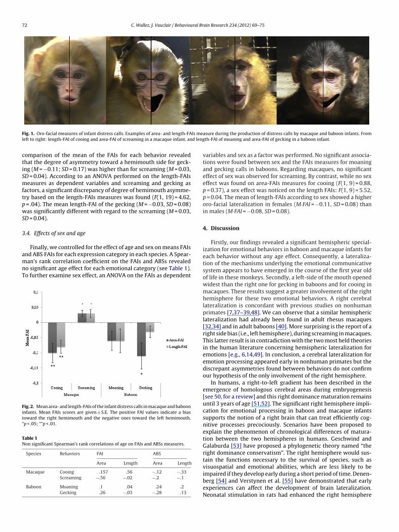
72 C. Wallez, J. Vauclair / Behavioural Brain Research 234 (2012) 69– 75
F Is mel d leng
ctiSmftpwS
3
amnT
Fit*
TN
ig. 1. Oro-facial measures of infant distress calls. Examples of area- and length-FAeft to right: length-FAI of cooing and area-FAI of screaming in a macaque infant, an
omparison of the mean of the FAIs for each behavior revealedhat the degree of asymmetry toward a hemimouth side for geck-ng (M = −0.11; SD = 0.17) was higher than for screaming (M = 0.03,D = 0.04). According to an ANOVA performed on the length-FAIseasures as dependent variables and screaming and gecking as
actors, a significant discrepancy of degree of hemimouth asymme-ry based on the length-FAIs measures was found (F(1, 19) = 4.62,
= .04). The mean length-FAI of the gecking (M = −0.03, SD = 0.08)as significantly different with regard to the screaming (M = 0.03,
D = 0.04).
.4. Effects of sex and age
Finally, we controlled for the effect of age and sex on means FAIs
nd ABS·FAIs for each expression category in each species. A Spear-an’s rank correlation coefficient on the FAIs and ABSs revealedo significant age effect for each emotional category (see Table 1).o further examine sex effect, an ANOVA on the FAIs as dependent
ig. 2. Mean area- and length-FAIs of the infant distress calls in macaque and baboonnfants. Mean FAIs scores are given ± S.E. The positive FAI values indicate a biasoward the right hemimouth and the negative ones toward the left hemimouth.p < .05; **p < .01.
able 1on significant Spearman’s rank correlations of age on FAIs and ABSs measures.
Species Behaviors FAI ABS
Area Length Area Length
Macaque Cooing .157 .56 −.12 −.33Screaming −.56 −.02 −.2 −.1
Baboon Moaning .1 .04 .24 .2Gecking .26 −.03 −.28 .13
asure during the production of distress calls by macaque and baboon infants. Fromth-FAI of moaning and area-FAI of gecking in a baboon infant.
variables and sex as a factor was performed. No significant associa-tions were found between sex and the FAIs measures for moaningand gecking calls in baboons. Regarding macaques, no significanteffect of sex was observed for screaming. By contrast, while no sexeffect was found on area-FAIs measures for cooing (F(1, 9) = 0.88,p = 0.37), a sex effect was noticed on the length FAIs: F(1, 9) = 5.52,p = 0.04. The mean of length-FAIs according to sex showed a higheroro-facial lateralization in females (M·FAI = −0.11, SD = 0.08) thanin males (M·FAI = −0.08, SD = 0.08).
4. Discussion
Firstly, our findings revealed a significant hemispheric special-ization for emotional behaviors in baboon and macaque infants foreach behavior without any age effect. Consequently, a lateraliza-tion of the mechanisms underlying the emotional communicativesystem appears to have emerged in the course of the first year oldof life in these monkeys. Secondly, a left-side of the mouth openedwidest than the right one for gecking in baboons and for cooing inmacaques. These results suggest a greater involvement of the righthemisphere for these two emotional behaviors. A right cerebrallateralization is concordant with previous studies on nonhumanprimates [7,37–39,48]. We can observe that a similar hemisphericlateralization had already been found in adult rhesus macaques[32,34] and in adult baboons [40]. More surprising is the report of aright side bias (i.e., left hemisphere), during screaming in macaques.This latter result is in contradiction with the two most held theoriesin the human literature concerning hemispheric lateralization foremotions [e.g., 6,14,49]. In conclusion, a cerebral lateralization foremotion processing appeared early in nonhuman primates but thediscrepant asymmetries found between behaviors do not confirmour hypothesis of the only involvement of the right hemisphere.
In humans, a right-to-left gradient has been described in theemergence of homologous cerebral areas during embryogenesis[see 50, for a review] and this right dominance maturation remainsuntil 3 years of age [51,52]. The significant right hemisphere impli-cation for emotional processing in baboon and macaque infantssupports the notion of a right brain that can treat efficiently cog-nitive processes precociously. Scenarios have been proposed toexplain the phenomenon of chronological differences of matura-tion between the two hemispheres in humans. Geschwind andGalaburda [53] have proposed a phylogenetic theory named “theright dominance conservatism”. The right hemisphere would sus-tain the functions necessary to the survival of species, such asvisuospatial and emotional abilities, which are less likely to be
impaired if they develop early during a short period of time. Denen-berg [54] and Verstynen et al. [55] have demonstrated that earlyexperiences can affect the development of brain lateralization.Neonatal stimulation in rats had enhanced the right hemisphere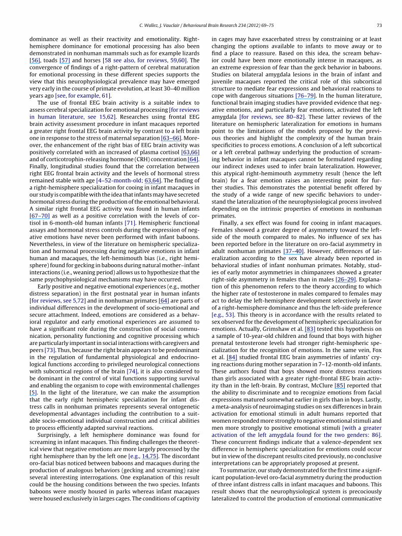
ral Br
dhd[cfvvy
aibaoopaFrraohA[taaNthsis
d[isihnapilwba[ttdat
siropscbw
C. Wallez, J. Vauclair / Behaviou
ominance as well as their reactivity and emotionality. Right-emisphere dominance for emotional processing has also beenemonstrated in nonhuman mammals such as for example lizards56], toads [57] and horses [58 see also, for reviews, 59,60]. Theonvergence of findings of a right-pattern of cerebral maturationor emotional processing in these different species supports theiew that this neurophysiological prevalence may have emergedery early in the course of primate evolution, at least 30–40 millionears ago [see, for example, 61].
The use of frontal EEG brain activity is a suitable index tossess cerebral specialization for emotional processing [for reviewsn human literature, see 15,62]. Researches using frontal EEGrain activity assessment procedure in infant macaques reported
greater right frontal EEG brain activity by contrast to a left brainne in response to the stress of maternal separation [63–66]. More-ver, the enhancement of the right bias of EEG brain activity wasositively correlated with an increased of plasma cortisol [63,66]nd of corticotrophin-releasing hormone (CRH) concentration [64].inally, longitudinal studies found that the correlation betweenight EEG frontal brain activity and the levels of hormonal stressemained stable with age [4–52-month-old; 63,64]. The finding of
right-hemisphere specialization for cooing in infant macaques inur study is compatible with the idea that infants may have secretedormonal stress during the production of the emotional behavioral.
similar right frontal EEG activity was found in human infants67–70] as well as a positive correlation with the levels of cor-isol in 6-month-old human infants [71]. Hemispheric functionalssays and hormonal stress controls during the expression of neg-tive emotions have never been performed with infant baboons.evertheless, in view of the literature on hemispheric specializa-
ion and hormonal processing during negative emotions in infantuman and macaques, the left-hemimouth bias (i.e., right hemi-phere) found for gecking in baboons during natural mother–infantnteractions (i.e., weaning period) allows us to hypothesize that theame psychophysiological mechanisms may have occurred.
Early positive and negative emotional experiences (e.g., motheristress separation) in the first postnatal year in human infantsfor reviews, see 5,72] and in nonhuman primates [64] are parts ofndividual differences in the development of socio-emotional andecure attachment. Indeed, emotions are considered as a behav-oral regulator and early emotional experiences are assumed toave a significant role during the construction of social commu-ication, personality functioning and cognitive processing whichre particularly important in social interactions with caregivers andeers [73]. Thus, because the right brain appears to be predominant
n the regulation of fundamental physiological and endocrino-ogical functions according to privileged neurological connections
ith subcortical regions of the brain [74], it is also considered toe dominant in the control of vital functions supporting survivalnd enabling the organism to cope with environmental challenges5]. In the light of the literature, we can make the assumptionhat the early right hemispheric specialization for infant dis-ress calls in nonhuman primates represents several ontogeneticevelopmental advantages including the contribution to a suit-ble socio-emotional individual construction and critical abilitieso process efficiently adapted survival reactions.
Surprisingly, a left hemisphere dominance was found forcreaming in infant macaques. This finding challenges the theoret-cal view that negative emotions are more largely processed by theight hemisphere than by the left one [e.g., 14,75]. The discordantro-facial bias noticed between baboons and macaques during theroduction of analogous behaviors (gecking and screaming) raise
everal interesting interrogations. One explanation of this resultould be the housing conditions between the two species. Infantsaboons were mostly housed in parks whereas infant macaquesere housed exclusively in larges cages. The conditions of captivityain Research 234 (2012) 69– 75 73
in cages may have exacerbated stress by constraining or at leastchanging the options available to infants to move away or tofind a place to reassure. Based on this idea, the scream behav-ior could have been more emotionally intense in macaques, asan extreme expression of fear than the geck behavior in baboons.Studies on bilateral amygdala lesions in the brain of infant andjuvenile macaques reported the critical role of this subcorticalstructure to mediate fear expressions and behavioral reactions tocope with dangerous situations [76–79]. In the human literature,functional brain imaging studies have provided evidence that neg-ative emotions, and particularly fear emotions, activated the leftamygdala [for reviews, see 80–82]. These latter reviews of theliterature on hemispheric lateralization for emotions in humanspoint to the limitations of the models proposed by the previ-ous theories and highlight the complexity of the human brainspecificities to process emotions. A conclusion of a left subcorticalor a left cerebral pathway underlying the production of scream-ing behavior in infant macaques cannot be formulated regardingour indirect indexes used to infer brain lateralization. However,this atypical right-hemimouth asymmetry result (hence the leftbrain) for a fear emotion raises an interesting point for fur-ther studies. This demonstrates the potential benefit offered bythe study of a wide range of new specific behaviors to under-stand the lateralization of the neurophysiological process involveddepending on the intrinsic properties of emotions in nonhumanprimates.
Finally, a sex effect was found for cooing in infant macaques.Females showed a greater degree of asymmetry toward the left-side of the mouth compared to males. No influence of sex hasbeen reported before in the literature on oro-facial asymmetry inadult nonhuman primates [37–40]. However, differences of lat-eralization according to the sex have already been reported inbehavioral studies of infant nonhuman primates. Notably, stud-ies of early motor asymmetries in chimpanzees showed a greaterright-side asymmetry in females than in males [26–29]. Explana-tion of this phenomenon refers to the theory according to whichthe higher rate of testosterone in males compared to females mayact to delay the left-hemisphere development selectively in favorof a right-hemisphere dominance and thus the left-side preference[e.g., 53]. This theory is in accordance with the results related tosex observed for the development of hemispheric specialization foremotions. Actually, Grimshaw et al. [83] tested this hypothesis ona sample of 10-year-old children and found that boys with higherprenatal testosterone levels had stronger right-hemispheric spe-cialization for the recognition of emotions. In the same vein, Foxet al. [84] studied frontal EEG brain asymmetries of infants’ cry-ing reactions during mother separation in 7–12-month-old infants.These authors found that boys showed more distress reactionsthan girls associated with a greater right-frontal EEG brain activ-ity than in the left-brain. By contrast, McClure [85] reported thatthe ability to discriminate and to recognize emotions from facialexpressions matured somewhat earlier in girls than in boys. Lastly,a meta-analysis of neuroimaging studies on sex differences in brainactivation for emotional stimuli in adult humans reported thatwomen responded more strongly to negative emotional stimuli andmen more strongly to positive emotional stimuli [with a greateractivation of the left amygdala found for the two genders: 86].These concurrent findings indicate that a valence-dependent sexdifference in hemispheric specialization for emotions could occurbut in view of the discrepant results cited previously, no conclusiveinterpretations can be appropriately proposed at present.
To summarize, our study demonstrated for the first time a signif-
icant population-level oro-facial asymmetry during the productionof three infant distress calls in infant macaques and baboons. Thisresult shows that the neurophysiological system is precociouslylateralized to control the production of emotional communicative
7 ral Br
sal(mt
A
CtosRTt
R
[
[
[
[
[
[
[
[
[
[
[
[
[
[
[
[
[
[
[
[
[
[
[
[
[
[
[
[
[
[
[
[
[
[
[
[
[
[
[
[
[
[
[
[
[
4 C. Wallez, J. Vauclair / Behaviou
ignals in nonhuman primates’ infants. The findings that macaquend baboon infants demonstrated some specific cerebral patterns ofateralization somewhat analogous to those displayed by humanssuch as sex or intensity effects), enhance the value of the infant
onkeys as a nonhuman primate model for studying the roots ofhe cerebral lateralization of emotional processing.
cknowledgments
The authors would like to thank Geoffrey Montes, Marion Azaïs,laire Janicot, Lucie Rigaill and Emeline Verna for their helpful par-icipation in videotaping the infant’s behaviors and the assessmentf the still pictures, and the caretakers at the CNRS primate fieldtation in Rousset. This study was supported by a French Nationalesearch Agency (ANR) grant reference ANR-08-BLAN-0011 01.he writing of this paper was made possible by a fellowship fromhe Medical Research Foundation to C.W.
eferences
[1] Bard KA. Emotions in chimpanzee infants: the value of a comparative develop-mental approach to understand the evolutionary bases of emotions. In: Nadel J,Muir D, editors. Emotional development. Oxford, UK: Oxford University Press;2005. p. 31–60.
[2] Maestripieri D, Call J. Mother–infant communication in primates. Advances inthe Study of Behavior 1996;25:613–42.
[3] Newman JD. Neural circuits underlying crying and cry responding in mammals.Behavioural Brain Research 2007;182:155–65.
[4] Newman JD. Vocal ontogeny in macaques and marmosets: convergent anddivergent lines of development. In: Zimmerman E, editor. Current topics inprimate vocal communication. New York: Plenum Press; 1995. p. 73–97.
[5] Schore AN. Effects of a secure attachment relationship on right brain develop-ment, affect regulation, and infant mental health. Infant Mental Health Journal2001;22:7–66.
[6] Borod JC, Haywood CS, Koff E. Neuropsychological aspects of facial asymmetryduring emotional expression: a review of the normal adult literature. Neu-ropsychology Review 1997;7:41–60.
[7] Hook-Costigan M, Rogers L. Lateralized use of the mouth in production of vocal-izations by marmosets. Neuropsychologia 1998;36:1265–73.
[8] Rinn WE. The neuropsychology of facial expression: a review of the neurologicaland psychological mechanisms for producing facial expressions. PsychologicalBulletin 1984;95:52–77.
[9] Best CT, Queen HF. Baby, it’s in your smile: right hemiface bias in infant emo-tional expressions. Developmental Psychology 1989;25:264–76.
10] Rothbart MK, Taylor SB, Tucker DM. Right-sided facial asymmetry in infantemotional expression. Neuropsychologia 1989;27:675–87.
11] Schuetze P, Reid H. Emotional lateralisation in the second year of life: evidencefrom oral asymmetries. Laterality 2005;10:207–17.
12] Holowka S, Petitto LA. Left hemisphere cerebral specialization for babies whilebabbling. Science 2002;297:1515.
13] Demaree HA, Everhart DE, Youngstrom EA, Harrison DW. Brain lateralization ofemotional processing: historical roots and a future incorporating dominance.Behavioral and Cognitive Neuroscience Reviews 2005;4:3–20.
14] Gainotti G. Unconscious processing of emotions and the right hemisphere.Neuropsychologia 2012;50:205–18.
15] Davidson RJ. Cerebral asymmetry, emotion, and affective style. In: Hugdahl K,editor. Brain asymmetry. Cambridge, MA: MIT Press; 1995. p. 361–87.
16] Hopkins WD. The evolution of hemispheric specialization in primates. London:Elsevier/Academic Press; 2007.
17] Vauclair J, Fagot J, Dépy D. Nonhuman primates as models of hemispheric spe-cialization. In: Haug M, Whalen RE, editors. Animal models of human emotionand cognition. New York: APA Books; 1999. p. 247–56.
18] Hopkins WD, Vauclair J. Evolution of behavioral and brain asymmetries in pri-mates. In: Tallerman M, Gibson K, editors. Handbook of language evolution.Oxford: Oxford University Press; 2012. p. 184–97.
19] Crow T. Directional asymmetry is the key to the origin of modern Homo sapiens(the Broca-Annett axiom): a reply to Rogers’ review of The Speciation of ModernHomo Sapiens. Laterality 2004;9:233–42.
20] Rogers L, Andrew R. Comparative vertebrate lateralization. Cambridge: Cam-bridge University Press; 2002.
21] MacNeilage PF, Studdert-Kennedy MG, Lindblom B. Primate handedness recon-sidered. Behavioral and Brain Sciences 1987;10:247–63.
22] Hopkins WD, Fernández-Carriba S. The effect of situational factors on hand
preferences for feeding in 177 captive chimpanzees (Pan troglodytes). Neu-ropsychologia 2000;38:403–9.23] Ward JP, Milliken GW, Dodson DL, Stafford DK, Wallace M. Handedness as afunction of sex and age in a large population of Lemur. Journal of ComparativePsychology 1990;104:167–73.
[
[
ain Research 234 (2012) 69– 75
24] Hook MA, Rogers LJ. Development of hand preferences in marmosets (Cal-lithrix jacchus) and effects of aging. Journal of Comparative Psychology2000;114:263–71.
25] Brazelton T. Neonatal behavioral assessment scale. 2nd ed. Philadelphia: Spas-tics Internal Medical Publications; 1984.
26] Hopkins WD, Bard KA. Asymmetries in spontaneous head orientation in infantchimpanzees (Pan troglodytes). Behavioral Neuroscience 1995;109:808–12.
27] Hopkins WD, Bard KA. Hemispheric specialization in infant chimpanzees (Pantroglodytes): evidence for a relation with gender and arousal. DevelopmentalPsychobiology 1993;26:219–35.
28] Hopkins WD, Bard KA. The ontogeny of lateralized behavior in nonhumanprimates with special reference to chimpanzees (Pan troglodytes). In: WardJP, Hopkins WD, editors. Primate laterality – current behavioral evidence ofprimate asymmetries. New York: Springer; 1993.
29] Bard KA, Hopkins WD, Fort CL. Lateral bias in infant chimpanzees (Pantroglodytes). Journal of Comparative Psychology 1990;104:309–21.
30] Panksepp J, Siviy SM, Normansell LA. Brain opioids and social emotions. In:Reite M, Fields T, editors. The psychobiology of attachment and separation.New York: Academic Press; 1985. p. 3–49.
31] Jürgens U. Neural pathways underlying vocal control. Neuroscience and Biobe-havioral Reviews 2002;26:235–58.
32] Hauser MD, Akre K. Asymmetries in the timing of facial and vocal expres-sions by rhesus monkeys: implications for hemispheric specialization. AnimalBehaviour 2001;61:391–400.
33] Hauser M. The evolution of a lopsided brain: asymmetries underlying facial andvocal expressions in nonhuman primates. In: Hauser MD, Konishi M, editors.The design of animal communication. Cambridge, Massachussetts: MIT Press;1999. p. 597–628.
34] Hauser MD. Right hemisphere dominance for the production of facial expres-sion in monkeys. Science 1993;261:475–7.
35] Taglialatela JP. Functional and structural asymmetries for auditory perceptionand vocal production in nonhuman primates. In: Hopkins WD, editor. The evo-lution of hemispheric specialization in primates. London: Elsiever/AcademicPress; 2007. p. 121–45.
36] Hook M. The evolution of lateralized motor functions. In: Rogers LJ, KaplanG, editors. Comparative vertebrate cognition: are primates superio to non-primates? New York: Plenum Publishers, New York; 2004. p. 325–70.
37] Fernández-Carriba S, Loeches A, Morcillo A, Hopkins WD. Functional asym-metry of emotions in primates: new findings in chimpanzees. Brain ResearchBulletin 2002;57:561–4.
38] Fernández-Carriba S, Loeches Á, Morcillo A, Hopkins WD. Asymmetry infacial expression of emotions by chimpanzees. Neuropsychologia 2002;40:1523–33.
39] Losin EA, Russell J, Freeman H, Meguerditchian A, Hopkins WD, Fitch T. Lefthemisphere specialization for oro-facial movements of learned vocal signalsby captive chimpanzees. PLoS ONE 2008;3:e2529.
40] Wallez C, Vauclair J. Right hemisphere dominance for emotion processing inbaboons. Brain and Cognition 2011;75:164–9.
41] Nash LT. The development of the mother–infant relationship in wild baboons(Papio anubis). Animal Behaviour 1978;26:746–59.
42] Partan SR. Single and multichannel signal composition: facial expres-sions and vocalizations of rhesus macaques (Macaca mulatta). Behaviour2002;139:993–1027.
43] Maestripieri D, Jovanovic T, Gouzoules H. Crying and infant abuse in rhesusmonkeys. Child Development 2000;71:301–9.
44] Kalin NH, Shelton SE, Snowdon CT. Affiliative vocalizations in infantrhesus macaques (Macaca mulatta). Journal of Comparative Psychology1992;106:254–61.
45] Byrne RW. Distance vocalisations of guinea baboons (Papio papio) in Senegal:an analysis of function. Behaviour 1981;78:283–313.
46] Jovanovic T, Gouzoules H. Effects of nonmaternal restraint on the vocalizationsof infant rhesus monkeys (Macaca mulatta). American Journal of Primatology2001;53:33–45.
47] Cantalupo C, Pilcher DL, Hopkins WD. Are planum temporale and sylvian fissureasymmetries directly related? A MRI study in great apes. Neuropsychologia2003;41:1975–81.
48] Fernández-Carriba S, Loeches Á, Morcillo A, Washburn DA, Hopkins WD.Human assessment of chimpanzee facial asymmetry. Laterality 2004;9:1–17.
49] Davidson RJ, Shackman AJ, Maxwell JS. Asymmetries in face and brain relatedto emotion. Trends in Cognitive Sciences 2004;8:389–91.
50] Hellige JB. Hemispheric asymmetry: what’s right and what’s left. Cambridge,MA: Harvard University Press; 1993.
51] Chiron C, Jambaque I, Nabbout R, Lounes R, Syrota A, Dulac O. The right brainhemisphere is dominant in human infants. Brain 1997;120:1057–65.
52] Schore A. Affect regulation and the organization of self: the neurobiology ofemotional development. Mahweh, New Jersey: Erlbaum; 1994.
53] Geschwind N, Galaburda AM. Cerebral lateralization: biological mechanisms,associations, and pathology: I. A hypothesis and a program for research.Archives of Neurology 1985;42:428–59.
54] Denenberg VH. Hemispheric laterality in animals and the effects of early expe-rience. Behavioral and Brain Sciences 1981;4:1–21.
55] Verstynen T, Tierney R, Urbanski T, Tang A. Neonatal novelty exposuremodulates hippocampal volumetric asymmetry in the rat. Neuroreport2001;12:3019–22.
56] Deckel AW. Laterality of aggressive responses in Anolis. Journal of ExperimentalZoology 1995;272:194–200.
ral Br
[
[
[
[
[[
[
[
[
[
[
[
[
[
[
[
[
[
[
[
[
[
[
[
[
[
[
[
[
C. Wallez, J. Vauclair / Behaviou
57] Lippolis G, Bisazza A, Rogers LJ, Vallortigara G. Lateralisation of predator avoid-ance responses in three species of toads. Laterality 2002;7:163–83.
58] Austin N, Rogers PL. Asymmetry of flight and escape turning responses inhorses. Laterality 2007;12:464–74.
59] Rogers LJ. Relevance of brain and behavioural lateralization to animal welfare.Applied Animal Behaviour Science 2010;127:1–11.
60] MacNeilage PF, Rogers LJ, Vallortigara G. Origins of the left & right brain. Scien-tific American 2009;301:60–7.
61] Boyd R, Silk JB. How humans evolved. New York: WW Norton; 1997.62] Coan JA, Allen JJB. Frontal EEG asymmetry as a moderator and mediator of
emotion. Biological Psychology 2004;67:7–50.63] Kalin NH, Larson C, Shelton SE, Davidson RJ. Asymmetric frontal brain activity,
cortisol, and behavior associated with fearful temperament in rhesus monkeys.Behavioral Neuroscience 1998;112:286–92.
64] Kalin NH, Shelton SE, Davidson RJ. Cerebrospinal fluid corticotropin-releasinghormone levels are elevated in monkeys with patterns of brain activity associ-ated with fearful temperament. Biological Psychiatry 2000;47:579–85.
65] Kalin NH, Shelton SE, Davidson RJ, Kelley AE. The primate amygdala mediatesacute fear but not the behavioral and physiological components of anxioustemperament. Journal of Neuroscience 2001;21:2067–74.
66] Rilling JK, Winslow JT, O’Brien D, Gutman DA, Hoffman JM, Kilts CD. Neu-ral correlates of maternal separation in rhesus monkeys. Biological Psychiatry2001;49:146–57.
67] Bell MA, Fox NA. Brain development over the first year of life: relations betweenEEG frequency and coherence and cognitive and affective behaviors. In: DawsonC, Fischer KW, editors. Human behavior and the developing brain. New York:Guilford; 1994. p. 314–45.
68] Davidson RJ, Fox NA. Frontal brain asymmetry predicts infants’ response tomaternal separation. Journal of Abnormal Psychology 1989;98:127–31.
69] Fox NA. If it’s not left, it’s right: electroencephalograph asymmetry and thedevelopment of emotion. American Psychologist 1991;46:863–72.
70] Field T, Diego M, Hernandez-Reif M, Schanberg S, Kuhn C. Relative right versusleft frontal EEG in neonates. Developmental Psychobiology 2002;41:147–55.
71] Buss KA, Schumacher JRM, Dolski I, Kalin NH, Goldsmith HH, Davidson RJ. Right
frontal brain activity, cortisol, and withdrawal behavior in 6-month-old infants.Behavioral Neuroscience 2003;117:11–20.72] Panksepp J. The long-term psychobiological consequences of infant emo-tions: Prescriptions for the twenty-first century. Infant Mental Health Journal2001;22:132–73.
[
ain Research 234 (2012) 69– 75 75
73] Thompson RA. Emotion and self-regulation. In: Thompson RA, editor. Socioe-motional development: Nebraska symposium on motivation 1988. Lincoln, NJ:University of Nebraska Press; 1990. p. 367–467.
74] Tucker DM. Developing emotions and cortical networks. In: Gunnar MR, NelsonCA, editors. Developemental behavioral neuroscience: Minnesota symposiumon child psychology. Hillsdale, NJ: Erlbaum; 1992. p. 75–128.
75] Adolphs R, Jansari A, Tranel D. Hemispheric perception of emotional valencefrom facial expressions. Neuropsychology 2001;15:516–24.
76] Bliss-Moreau E, Toscano JE, Bauman MD, Mason WA, Amaral DG. Neonatalamygdala lesions alter responsiveness to objects in juvenile macaques. Neu-roscience 2011;178:123–32.
77] Prather MD, Lavenex P, Mauldin-Jourdain ML, Mason WA, Capitanio JP, Men-doza SP, et al. Increased social fear and decreased fear of objects in monkeyswith neonatal amygdala lesions. Neuroscience 2001;106:653–8.
78] Mason WA, Capitanio JP, Machado CJ, Mendoza SP, Amaral DG. Amygdalectomyand responsiveness to novelty in rhesus monkeys (Macaca mulatta): generalityand individual consistency of effects. Emotion 2006;6:73–81.
79] Machado CJ, Kazama AM, Bachevalier J. Impact of amygdala, orbital frontal, orhippocampal lesions on threat avoidance and emotional reactivity in nonhu-man primates. Emotion 2009;9:147–63.
80] Fusar-Poli P, Placentino A, Carletti F, Allen P, Landi P, Abbamonte M, et al. Lat-erality effect on emotional faces processing: ALE meta-analysis of evidence.Neuroscience Letters 2009;452:262–7.
81] Wager TD, Phan KL, Liberzon I, Taylor SF. Valence, gender, and lateralizationof functional brain anatomy in emotion: a meta-analysis of findings from neu-roimaging. Neuroimage 2003;19:513–31.
82] Adolphs R. Neural systems for recognizing emotion. Current Opinion in Neu-robiology 2002;12:169–77.
83] Grimshaw GM, Bryden MP, Finegan JAK. Relations between prenatal testos-terone and cerebral lateralization in children. Neuropsychology 1995;9:68–79.
84] Fox NA, Bell MA, Jones NA. Individual differences in response to stress andcerebral asymmetry. Developmental Neuropsychology 1992;8:161–84.
85] McClure EB. A meta-analytic review of sex differences in facial expression
processing and their development in infants, children, and adolescents. Psy-chological Bulletin 2000;126:424–53.86] Stevens JS, Hamann S. Sex differences in brain activation to emotional stim-uli: a meta-analysis of neuroimaging studies. Neuropsychologia 2012;50:1578–93.
Copyright © 2022 FDOKUMEN


![Mapping muscarinic receptors in human and baboon brain using [N-11C-methyl]-benztropine](https://static.fdokumen.com/doc/165x107/6344f35df474639c9b049d90/mapping-muscarinic-receptors-in-human-and-baboon-brain-using-n-11c-methyl-benztropine.jpg)