Fine structure of the photogenous areas in the bioluminescent ophiuroid Amphipholis squamata...
10
&p.1:Abstract Amphipholis squamata is a small biolumines- cent ophiuroid whose arms are the only body part to pro- duce light. The morphology of the arms was described paying particular attention to the spinal ganglia, viz the areas of most intense luminescence. Spinal ganglia con- sist of five different cell types (A–E) which were studied at different stages of the photogenous reaction. Type D cells have numerous irregularlyshaped vacuoles, wide- spread Golgi apparatus and well-developed rough endo- plasmic reticulum (RER) that show obvious ultrastruc- tural changes after luminescence. Type D cells appear, therefore, to be the best photocyte candidate. Type B and C cells were frequently observed in the nervous system outside spinal ganglia. Type A and E cells have not been described before. Type A cells are ciliated cells and type E cells extend long processes which are intimately asso- ciated with type D cells and epidermal ciliated cells. Both type A and type E cells could take part to the stim- ulation pathway that triggers luminescence.&bdy: A. Introduction That some echinoderm species are able to produce light has been known for almost two centuries (Viviani 1805; Harvey 1952). Yet, light production by echinoderms has been poorly studied although bioluminescent representa- tives occur in every living higher taxa except the Echino- ida (Herring 1974, 1978, 1995). Investigations were mostly done on ophiuroids, notably on the small intertid- al to sublittoral cosmopolitan species, Amphipholis squamata Delle Chiaje, 1828. According to Brehm and Morin (1977) luminescence in this species originates from the basal part of the arm spines. Working on anoth- er luminescent ophiuroid species (Ophiopsila californica Clark, 1921) these authors reported that photogenic cells are close to the nervous tissue. This and the general assumption that bioluminescence in most metazoans is under nervous control suggest that photogenic cells in A. squamata are intimately associated with the nervous tissue of the basal part of the spine. Photocyte identification was tried several times (see Mil- lot 1966, for review) and light emission was often asso- ciated with a mucus secreted by glandular cells from ei- ther the brachial spines (Reichensperger 1908; Trojan 1909) or the podial tips (Sterzinger 1907). Only Man- gold (1907, 1910) reported that the outline of the lumi- nous parts of A. squamata remains perfectly constant, which suggests that there is no discharge of a luminous substance. He speculated that the emitted light is intra- cellular and that photocytes are subepidermal cells. With the exception of Brehm and Morin (1977), no author has worked on the morphology of A. squamata luminescence since Mangold’s contributions. The present work aims to precisely localize the photogenic areas in the species A. squamata, to describe their fine structure and to tenta- tively identify the photogenic cells. B. Materials and methods Individuals of A. squamata (Fig. 1) were collected intertidally at Langrune-sur-Mer (Normandy, France) from September 1993 to June 1994. They were transported to the marine biology laborato- ry, Brussels University, and kept alive in a closed-circuit marine aquarium (12°C, 32‰ salinity) until required. In order to localize the photogenic areas, light emission was triggered by putting entire individuals or dissected body parts (viz disc or arms) in a v/v sea-water-KCl 400 mM solution according to the technique of Mallefet et al. (1989). When necessary, indi- viduals were anaesthetized in a 3.5% MgCl 2 solution in artificial sea-water. For transmission electron microscopy (TEM), dissected body parts from individuals which either had or had not emitted D. Deheyn ( ✉ ) · V. Alva · M. Jangoux Laboratoire de Biologie Marine CP 160/15, Université Libre de Bruxelles, 50 Avenue FD Roosevelt, B-1050 Bruxelles, Belgium Tel. +32 2 650 22 34; Fax +32 2 650 27 96 e-mail [email protected] V. Alva Institut de Ciènces del mar (CSIC), Passeig Joan de Borbò s/n, E-08039, Barcelona, Spain M. Jangoux Laboratoire de Biologie Marine, Université de Mons-Hainaut, 19 Avenue Maistriau, B-7000 Mons, Belgium&/fn-block: Zoomorphology (1996) 116:195–204 © Springer-Verlag 1996 ORIGINAL ARTICLE &roles:D. Deheyn · V. Alva · M. Jangoux Fine structure of the photogenous areas in the bioluminescent ophiuroid Amphipholis squamata (Echinodermata, Ophiuridea) &misc:Accepted: 1 September 1996
-
Upload
independent -
Category
Documents
-
view
3 -
download
0
Transcript of Fine structure of the photogenous areas in the bioluminescent ophiuroid Amphipholis squamata...
&p.1:Abstract Amphipholis squamatais a small biolumines-cent ophiuroid whose arms are the only body part to pro-duce light. The morphology of the arms was describedpaying particular attention to the spinal ganglia, viz theareas of most intense luminescence. Spinal ganglia con-sist of five different cell types (A–E) which were studiedat different stages of the photogenous reaction. Type Dcells have numerous irregularlyshaped vacuoles, wide-spread Golgi apparatus and well-developed rough endo-plasmic reticulum (RER) that show obvious ultrastruc-tural changes after luminescence. Type D cells appear,therefore, to be the best photocyte candidate. Type B andC cells were frequently observed in the nervous systemoutside spinal ganglia. Type A and E cells have not beendescribed before. Type A cells are ciliated cells and typeE cells extend long processes which are intimately asso-ciated with type D cells and epidermal ciliated cells.Both type A and type E cells could take part to the stim-ulation pathway that triggers luminescence.&bdy:
A. Introduction
That some echinoderm species are able to produce lighthas been known for almost two centuries (Viviani 1805;Harvey 1952). Yet, light production by echinoderms hasbeen poorly studied although bioluminescent representa-tives occur in every living higher taxa except the Echino-ida (Herring 1974, 1978, 1995). Investigations were
mostly done on ophiuroids, notably on the small intertid-al to sublittoral cosmopolitan species, AmphipholissquamataDelle Chiaje, 1828. According to Brehm andMorin (1977) luminescence in this species originatesfrom the basal part of the arm spines. Working on anoth-er luminescent ophiuroid species (Ophiopsila californicaClark, 1921) these authors reported that photogenic cellsare close to the nervous tissue.
This and the general assumption that bioluminescencein most metazoans is under nervous control suggest thatphotogenic cells in A. squamataare intimately associatedwith the nervous tissue of the basal part of the spine.Photocyte identification was tried several times (see Mil-lot 1966, for review) and light emission was often asso-ciated with a mucus secreted by glandular cells from ei-ther the brachial spines (Reichensperger 1908; Trojan1909) or the podial tips (Sterzinger 1907). Only Man-gold (1907, 1910) reported that the outline of the lumi-nous parts of A. squamataremains perfectly constant,which suggests that there is no discharge of a luminoussubstance. He speculated that the emitted light is intra-cellular and that photocytes are subepidermal cells. Withthe exception of Brehm and Morin (1977), no author hasworked on the morphology of A. squamataluminescencesince Mangold’s contributions. The present work aims toprecisely localize the photogenic areas in the species A.squamata, to describe their fine structure and to tenta-tively identify the photogenic cells.
B. Materials and methodsIndividuals of A. squamata(Fig. 1) were collected intertidally atLangrune-sur-Mer (Normandy, France) from September 1993 toJune 1994. They were transported to the marine biology laborato-ry, Brussels University, and kept alive in a closed-circuit marineaquarium (12°C, 32‰ salinity) until required.
In order to localize the photogenic areas, light emission wastriggered by putting entire individuals or dissected body parts (vizdisc or arms) in a v/v sea-water-KCl 400 mM solution accordingto the technique of Mallefet et al. (1989). When necessary, indi-viduals were anaesthetized in a 3.5% MgCl2 solution in artificialsea-water. For transmission electron microscopy (TEM), dissectedbody parts from individuals which either had or had not emitted
D. Deheyn (✉) · V. Alva · M. JangouxLaboratoire de Biologie Marine CP 160/15, Université Libre de Bruxelles, 50 Avenue FD Roosevelt, B-1050 Bruxelles, BelgiumTel. +32 2 650 22 34; Fax +32 2 650 27 96e-mail [email protected]
V. AlvaInstitut de Ciènces del mar (CSIC), Passeig Joan de Borbò s/n, E-08039, Barcelona, Spain
M. JangouxLaboratoire de Biologie Marine, Université de Mons-Hainaut, 19 Avenue Maistriau, B-7000 Mons, Belgium&/fn-block:
Zoomorphology (1996) 116:195–204 © Springer-Verlag 1996
O R I G I N A L A RT I C L E
&roles:D. Deheyn · V. Alva · M. Jangoux
Fine structure of the photogenous areas in the bioluminescent ophiuroid Amphipholis squamata(Echinodermata, Ophiuridea)
&misc:Accepted: 1 September 1996
light were fixed for 3 h at 4°C in 3% glutaraldehyde in cacodylatebuffer, then rinsed in the buffer and postfixed for 30 min in 1% os-mium tetroxide in the same buffer. After a final buffer wash, piec-es were decalcified in the dark for 12–15 h at 4°C in 2% ascorbicacid in 0.3 M NaCl (Dietrich and Fontaine 1975). Decalcifiedpieces were dehydrated in graded ethanol and embedded in Spurr.Semithin sections (0.3µm) were cut on a Reichert OM2 ultrami-crotome and stained with a 1:1 methylene blue-azur II mixture(Richardson et al. 1960). Ultrathin sections (40–50 nm) were cuton an LKB V ultramicrotome, contrasted with uranyl acetate andlead citrate (Reynolds 1963), and examined with a JEOL JEM2000 FX II transmission electron microscope.
For scanning electron microscopy (SEM) entire individualswere fixed, decalcified and dehydrated as described for TEM prep-arations. They were then dried by the critical-point method (usingCO2 as transition fluid), coated with gold in a sputter coater andobserved with a JEOL JSM 6100 scanning electron microscope.
C. Results
I. Survey of luminescent areas
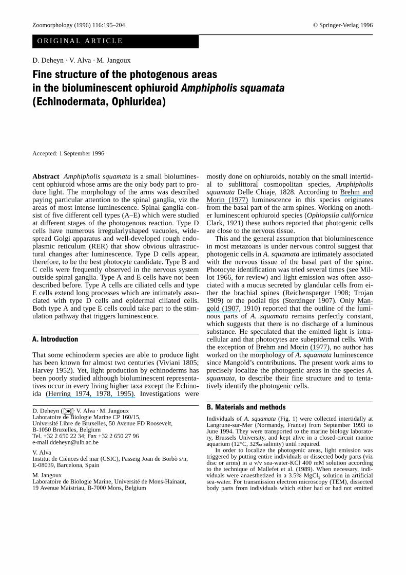
Luminescent areas were detected by triggering light pro-duction in previously anaesthetized or unanaesthetizedindividuals using the technique of Mallefet et al. (1989)that was applied to either entire individuals, dissecteddisc or arms, or arm fragments. The disc never emittedlight while the arms always produced it over all theirlength whether they were attached to the disc, dissectedfrom it or split into fragments. From repeated observa-tions of flashing individuals or arms it could be seen thatlight originated from areas located under the basal partof the brachial spines, thus confirming the previous ob-servation by Brehm and Morin (1977) (Figs. 2, 3).
II. Anatomy of the brachial nervous system
The arm of an adult A. squamata(disc diameter ca2.2 mm) measures ca 12 mm long. It consists of about 35successive segments whose shape and size depend on
196
Figs. 1–5 Amphipholis squamata. ax axon, cb cell bodies, cu cuti-cle, m muscle, osossicle, scsubcuticular region, sgspinal ganglion&/fig.c:
Fig. 1 Aboral view of an individual (SEM)&/fig.c:
Fig. 2 Luminescent areas of the arm as seen using an epifluores-cent technique&/fig.c:
Fig. 4 Epidermal indentation (➤) contacting a spinal ganglion(sg) (parasagittal section)&/fig.c:
Fig. 5 Section through a spinal ganglion&/fig.c:

their position along the arm, the distalmost being thesmallest (Fig. 1). As in other ophiuroid species (see, forexample, Cobb 1987), the brachial nervous system of A.squamatais made up of two nerve plexuses, the epineu-ral and the hyponeural, which are superimposed on oneanother in the oral region of the arm (the epineural plex-us is under the hyponeural plexus when going from theaboral to the oral side of the arm). The epineural plexusforms the bulk of the radial nerve cord that travels thelength of the arm and of the paired podial ganglia thatoccur in each segment (Fig. 6). Whilst also contributingto the radial nerve cord and the podial ganglia, the hypo-
neural plexus forms, mostly on its own, the paired nervesinnervating the intervertebral muscles (Fig. 6). As for theleft and right lateral nerves, these are epineural and inter-connect, respectively, with a left and right group of threesuccessive spinal ganglia from which distinctive spinalnerves originate. (There are three spines and, thus, threespinal ganglia and three spinal nerves on each side of abrachial segment of A. squamata.) (Figs. 3–6).
III. Anatomy of the spinal ganglia
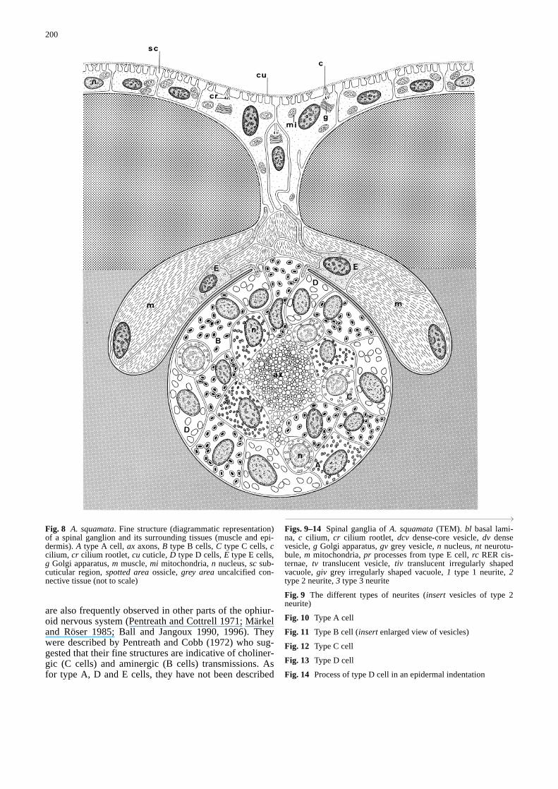
Information gathered on the luminescent areas of A.squamatastrongly supports the idea that light is pro-duced by cells associated with the spinal ganglia of eacharm segment. Spinal ganglia are grossly spherical inshape (ca 30µm in diameter) (Figs. 4, 5). They are part-ly located in slight depressions of the internal surface ofthe lateral skeletal plates and surrounded elsewhere byan uncalcified connective tissue (Fig. 8). Each ganglionis lined by a well-marked basal lamina that is continuouswith those surrounding the lateral and the spinal nerves(Fig. 6). That basal lamina is also continuous with theone lining the epidermal indentations (arrowhead) thatcontact by place the spinal ganglia, and with the laminasurrounding the cells of the spine muscle system (these
197
Fig. 3 Distribution of luminescent areas along the arm (light greybrachial spines, medium greyarm aboral surface, dark greylumi-nescent areas) (not to scale)&/fig.c:
Fig. 6 A. squamata. Transverse section at midpoint of arm seg-ment. ACC ambulacral and coelomic cavities, CT connective tis-sue, Epi epineural nerve plexus, Hyp hyponeural nerve plexus,IVM intervertebral muscles, OSossicle, a ambulacral cavities, apaboral plate, b coelomic cavities, es epineural sinus, ln lateralnerve, lp lateral plate, op oral plate, p podium, pg podial ganglion,sg spinal ganglia, sn spinal nerve, sp spine, v vertebra (not toscale)&/fig.c:

cells are mostly developed at the distal part of ganglia)(Figs. 4, 7, 8).
IV. Fine structure of spinal ganglia in luminescence- non-stimulated individuals
Spinal ganglia consist of a central bundle of neuritesaround which cell bodies of different cell types occur(Fig. 5). Table 1 summarizes the major characteristics ofeach of the ganglionic cell types. Among these celltypes, three (i.e. the internal cells) are distributed every-where in the ganglia and they are, thus, sometimes closeto the bundle of neurites, while the two others (i.e. theexternal cells) always have a strictly peripheral location.
Neurites in spinal ganglia are of three different typesaccording to their cytoplasmic content, the only commonfeature being the presence of small scattered mitochon-dria in their peripheral parts. Type 1 neurites (Fig. 9)measure from 0.3 to 0.6µm in diameter. They are elec-tron-dense, contain dense bundles of microtubules whichrun parallel to each other and to the neurite longitudinalaxis, and have scattered translucent and dense vesiclesmeasuring ca 100 and 130 nm in diameter, respectively.Type 2 neurites (Fig. 9) range from 0.6 to 1.6µm in di-ameter. They are rather translucent, contain a few shortand isolated microtubules and have dense-core vesiclesfrom 200 to 400 nm in diameter. Type 3 neurites (Fig. 9)measure from 0.6 to 1.4µm in diameter. They are rathertranslucent, do not contain microtubules and have trans-lucent vesicles of ca 70 nm in diameter.
Internal cells are grossly ovoid in shape. Whatevertheir type they measure from 4 to 8µm in length, have acircular to egg-shaped nucleus with patches of varingdensity of euchromatin and contain many mitochondriain their cytoplasm. Type A cells (Fig. 10) contain numer-ous dense vesicles (from 120 to 140 nm in diameter) thatare dispersed all over the cytoplasm except in its perinu-clear part. These cells also have a widespread and well-developed rough endoplasmic reticulum (RER) andsometimes include a rather inconspicuous Golgi appara-tus. Type B cells (Fig. 11) have many vesicles, measur-ing from 200 to 300 nm in diameter, that either contain adense central core or a grey finely granular material(one-third of the observed vesicles). These are monocili-ated cells (their cilium measures up to 4µm in length)whose cytoplasm contains highly developed RER cister-nae as well as a rather conspicuous Golgi apparatus oftenlocated close to the cilium basal rootlet (Fig. 11). Thecytoplasm of type C cells (Fig. 12) is often reduced to athin and rather translucent perinuclear layer without anyRER cisternae or Golgi apparatus. From serial TEM sec-tions of spinal ganglia it could be established that allneurites observed so far originate from one of the threeaforementioned cell types, with type 1 neurites belong-ing to type A cells, type 2 neurites to type B cells andtype 3 neurites to type C cells.
The two types of external cells are branched in shape.They most often show an ovoid nucleus with the euchro-matin density depending on the cell type. Type D cellsoccur at the periphery of spinal ganglia (Figs. 13, 14).They have numerous mitochondria, lipid vesicles (from400 to 700 nm in diameter) and many irregularlyshapedvacuoles (from 300 to 700 nm in length) that are oftentranslucent though some are grey and full of a fibrillar tofinely granular material (Fig. 13). The cytoplasm con-tains short tubules (length ca 500µm) in its peripheralpart and widespread Golgi apparatus and conspicuousRER in its central part (RER cisternae are often filledwith fibrillar material). Type E cells (Figs. 15, 16) have adense granular cytoplasm in which many translucent orgreyish vesicles (from 70 to 100 nm in diameter) and afew mitochondria occur. Type E cells are stronglybranched and send out several long processes. Their cell
198
Fig. 7A–F A. squamata. Diagrammatic representations showingthe relationships between a spinal ganglion, its corresponding spi-nal nerve and muscles, and the epidermal indentations occurring inthe area (relationships with lateral nerves are not shown). Thedrawing illustrates successive sections that are transverse (A–Cfrom proximal to distal) and parasagittal (D–F from outside to in-side). ep epidermal indentation, sg spinal ganglion, sn spinalnerve, sp spine, grey areaconnective tissue, spotted areaspinalmusculature&/fig.c:

bodies are located close to but outside the ganglia, oftenbeing situated in the most internal part of epidermal in-dentations and/or close to the muscle cells that surroundthe most distal part of the spinal ganglia. As for their cellprocesses, some penetrate the ganglion, contacting inti-mately the membrane of type D cells (Figs. 13, 14),while others travel through the epidermal indentationscontacting the basal part of monociliated (subcuticularcilium) epidermal cells (Fig. 8).
V. Location of ganglionic cell types
Additional TEM investigations were performed to ascer-tain whether the cells (i.e. cell bodies and/or neurites)found in spinal ganglia occur in other epineural areas,namely in the radial nerve cord, the podial ganglia, thelateral nerves (interganglionic junctions) and the spinalnerves. Type A and type C cells are found equally inthese four areas, with the only particularity being thatneurites from type A cells (neurites I) appear to be ratherscarce in spinal nerves. Apart from the spinal ganglia,type B cells were observed only in lateral and spinalnerves. Cell bodies of type D cells are confined in thespinal ganglia and send out long processes that penetratelateral and aboral indentations. These processes are sur-rounded by a basal lamina that is continuous with that ofthe ganglion (Figs. 13, 14). Type E cell processes mostlyoccur in epidermal indentations where they are often in-timately associated with processes originating from typeD cells (Figs. 13, 14). Also, a few type E cell processeswere found within the spinal nerves.
VI. Fine structure of spinal ganglia in luminescence-stimulated individuals
Additional observations were performed on arms afterthe emission of light. They showed that important ultra-structural changes occur in type D cells whose cytoplasmbecomes full of large irregularly shaped vacuoles (maxi-mal length from 0.7 to 2.5µm) which contain fibrillarmaterial in various amounts (Fig. 18). RER cisternae arenumerous and of various shapes and sizes (longitudinalaxis from 300 to 900 nm). They are filled with homoge-neous fibrillar material similar to that filling the vacuoles(Figs. 17, 18). Golgi apparatus are well developed andoften surround vacuoles (Fig. 19). Cytoplasmic process-es of type D cells contain tubules and vesicles(40–90 nm in diameter) of varing electron density(Fig. 20). Bundles of fibrils (Fig. 19) and lysosomal vesi-cles (diameter from 0.3 to 1µm) are other common fea-tures found in these cells after the emission of light. Mi-tochondria and lipid vesicles remain seemingly unaffect-ed by the photogenous reaction.
D. Discussion
Various earlier investigators tried to identify and describelight-producing cells in different ophiuroid species (see,e.g. Mangold 1907; Reichensperger 1908; Sokolow1909). The usual way to characterize photocytes was torecognize cells whose distribution matched that of the lu-minous areas observed in vivo. Although many attemptswere made, Byrne (1994) reported that “cells responsiblefor light production in ophiuroids have not been positive-ly identified”.
Among the five different cell types observed in thespinal ganglia of A. squamata, two, viz types B and C,
199
Table 1 Main characteristics and location of type A to type E ganglionic cells&/tbl.c:&tbl.b:
Cell Cell typescharacteristics
Internal cells External cells
A B C D E
Nucleus Euchromatin +++ +++ + ++ ++++density
Cytoplasm Vesicles Dense Dense-core Translucent Translucent Translucentor grey or grey or grey
Vesicle size 120–140 200–300 60–70 300–700 70–100(nm)RER Developed Developed Undeveloped Developed UndevelopedGolgi Inconspicuous Conspicuous Inconspicuous Conspicuous Inconspicuous
Location All over the Restricted to All over the Mostly in the outer Mostly innervous the spinal nervous part of spinal epidermalsystem ganglia and the system ganglia with some indentations
spinal and extremities going with some lateral nerves to the base of extremities
epidermal entering the indentations spinal ganglia
(+ translucent, ++ slightly dense, +++ dense, ++++ very dense)&/tbl.b:
are also frequently observed in other parts of the ophiur-oid nervous system (Pentreath and Cottrell 1971; Märkeland Röser 1985; Ball and Jangoux 1990, 1996). Theywere described by Pentreath and Cobb (1972) who sug-gested that their fine structures are indicative of choliner-gic (C cells) and aminergic (B cells) transmissions. Asfor type A, D and E cells, they have not been described
200
Fig. 8 A. squamata. Fine structure (diagrammatic representation)of a spinal ganglion and its surrounding tissues (muscle and epi-dermis). A type A cell, ax axons, B type B cells, C type C cells, ccilium, cr cilium rootlet, cu cuticle, D type D cells, E type E cells,g Golgi apparatus, m muscle, mi mitochondria, n nucleus, sc sub-cuticular region, spotted areaossicle, grey areauncalcified con-nective tissue (not to scale)&/fig.c:
Figs. 9–14 Spinal ganglia of A. squamata(TEM). bl basal lami-na, c cilium, cr cilium rootlet, dcv dense-core vesicle, dv densevesicle, g Golgi apparatus, gv grey vesicle, n nucleus, nt neurotu-bule, m mitochondria, pr processes from type E cell, rc RER cis-ternae, tv translucent vesicle, tiv translucent irregularly shapedvacuole, giv grey irregularly shaped vacuole, 1 type 1 neurite, 2type 2 neurite, 3 type 3 neurite&/fig.c:
Fig. 9 The different types of neurites (insert vesicles of type 2neurite)&/fig.c:
Fig. 10 Type A cell&/fig.c:
Fig. 11 Type B cell (insertenlarged view of vesicles)&/fig.c:
Fig. 12 Type C cell&/fig.c:
Fig. 13 Type D cell&/fig.c:
Fig. 14 Process of type D cell in an epidermal indentation&/fig.c:

201

202
Figs. 15, 16Spinal ganglia of A. squamata(TEM). bl basal lami-na, f bundle of fibrils, g Golgi apparatus, gv grey vesicles, lv lipidvacuole, m mitochondria, n nucleus, rc RER cisternae, t tubule, tvtranslucent vesicle, v vesicle, va vacuole&/fig.c:
Fig. 15 Type E cell&/fig.c:
Fig. 16 Cell process of type E cell&/fig.c:
Figs. 17–20 View of type D cell after production of luminescence(TEM) &/fig.c:Fig. 17 General view&/fig.c:Fig. 18 RER cisternae&/fig.c:Fig. 19 Fibrils and Golgi apparatus surrounding a vacuole&/fig.c:Fig. 20 Tubules and vesicles in a cytoplasmic process&/fig.c:

before. Type A and E cells may not be associated withluminescent activity in view of the non-agreement be-tween their distribution in the ophiuroid arm and the dis-tribution of areas of luminescence. Type D cells seem tobe the best photocyte candidate because: (1) their distri-bution, established by TEM, matches the distribution ofthe luminescence on light-producing arms (see alsoBrehm and Morin 1977) and (2) they are the only celltype showing noticeable ultrastructural changes oncelight production has occurred. The latter point is indica-tive of photogenic activity since ultrastructural changeswere observed after light production in luminescent or-ganisms from different taxa, changes being important forthe photocytes and much more discrete for the surround-ing nerve cells (Anctil 1979a,b; Swales et al. 1986).
Light production is usually under nervous control inmetazoans (Herring 1990) and this was accepted forophiuroids regarding the results of various physiologicalinvestigations on Ophiopsila californicaand A. squama-ta (respectively Brehm 1977; Brehm and Morin 1977;Mallefet et al. 1992; De Bremaeker et al. 1994). Lumi-nescence is almost never spontanous in ophiuroids, thelight being mostly in response to external stimuli (Her-ring 1990). Working on luminescent Ophiopsilaspecies,Basch (1988) and Grober (1988a,b) described two pat-terns of luminous reaction in response to mechanicalstimuli which depended on the magnitude, duration andfrequency of the stimulus. Weak, short and isolated stim-uli only trigger light emission of photocytes close to thearea of stimulation (this corresponds to the local lumi-nous reflex defined by Reichensperger 1908). Strong,long and repeated stimuli trigger light emission thatpropagates throughout all the ophiuroid photogenic tis-sue. This indicates that sensory inputs are integrated bythe receptors covering the area of epidermis stimulatedbefore being conducted either to nearby photocytes or tothe whole brachial photogenous tissue, presumably bythe epineural nervous system, depending on the intensityof the stimulation.
Cobb (1988) drew a general pathway of the ophiuroidneural behaviour. According to him, when stimuli occuron brachial segments, sensory inputs derive from recep-tors of different modalities that cover the epithelia (sen-sory perception). The input information is then driven tobasic morphological units of integration, ganglia (prima-ry integration), before being spread throughout all thebrachial nervous system (secondary integration) to final-ly reach motor tissues (command behaviour). In the pres-ent paper, it has been shown that type E cells from spinalganglia develop long processes, some of them being lo-cated very close to epidermal ciliated cells and some oth-ers being closely pressed against type D cells (which arethe putative photocytes). This suggests that the type Ecell could play a basic role in starting the luminous re-sponse by transferring the information from the ciliatedepidermal cells (sensory perception) to the putative pho-tocytes (primary integration and command behaviour)and trigger the local luminous reflex. Depending onstimuli, sensory information may also pass from the spi-
nal ganglia (primary integration) to the epineural nervoussystem (secondary integration) which propagates (com-mand behaviour) stimulation towards the photocytes ofsuccessive brachial segments. Yet, this functionalscheme remains hypothetical since the photocytic natureof type D cells, while being very likely, has still to bedemonstrated. It would, therefore be necessary to adaptthe method of Germain and Anctil (1988) to A. squama-ta, a method that would allow the isolation of fluorescentphotocytes from a suspension of dissociated cells usingdensity gradients, and the comparison of their ultrastruc-ture with that of the presumed photocyte of the spinalganglia.
&p.2:Acknowledgements We thank N. De Bremaeker for field assis-tance, E. Bricourt for technical assistance and M. Klinkert fordrawing assistance. We are grateful to Professor J. Avoine for pro-viding facilities at the marine laboratory of Luc-sur-Mer (France).The research was supported by an IRSIA grant to D. Deheyn andby an FRFC convention (ref. 2.4544.92). Contribution of the“Centre Interuniversitaire de Biologie Marine” (CIBIM). Figure 2was produced by J. Mallefet (UCL) with equipment financed bythe National Fund for Scientific Research (Belgium) (refs. 1-5.130.91F and 6-220-17).
References
Anctil M (1979a) Ultrastructural correlates of luminescence inPorichthysphotophores. I. Effects of spinal cord stimulationand exogenous noradrenaline. Rev Can Biol 38 (2):67–80
Anctil M (1979b) Ultrastructural correlates of luminescence inPorichthys photophores. II. Effects of metabolic inhibitors.Rev Can Biol 38 (2):81–96
Ball B, Jangoux M (1990) Ultrastructure of the tube foot sensory-secretory complex in Ophiocomina nigra (Echinodermata,Ophiuridea). Zoomorphology 109:201–209
Ball B, Jangoux M (1996) The secretory system of the spines ofOphiocomina nigra(Echinodermata, Ophiuroidea). J Mar BiolAssoc UK 76:451–466
Basch LV (1988) Bioluminescent anti-predator defense in a subtidalophiuroid. In: Burke RD, Mladenov PV, Lambert P, Parsley RL(eds) Echinoderm biology. Balkema, Rotterdam, pp 565–573
Brehm P (1977) Electrophysiology and luminescence of an op-hiuroid radial nerve. J Exp Biol 71:213–227
Brehm P, Morin JG (1977) Localization and characterization of lu-minescent cells in Ophiopsila californica and Amphipholissquamata(Echinodermata: Ophiuroidea). Biol Bull 152:12–25
Byrne M (1994) Ophiuroidea. In: Harrison FW, Chia FS (eds) Mi-croscopic anatomy of invertabrates, vol 14 Echinodermata.Wiley-Liss, New York, pp 247–343
Cobb JLS (1987) Neurobiology of the Echinodermata. In: Ali MA(ed) Nervous systems in invertebrates, NATO ASI series, vol141. Plenum Press, New York London, pp 483–527
Cobb JLS (1988) A preliminary hypothesis to account for the neu-ral basis of behaviour in echinoderms. In: Burke RD,Mladenov PV, Lambert P, Parsley RL (eds) Echinoderm biolo-gy. Balkema, Rotterdam, pp 565–573
De Bremaeker N, Mallefet J, Baguet F (1994) Luminescence con-trol of Amphipholis squamata(Ophiuroidea): Nature of cho-linergic receptors. In: David B, Guille A, Féral J-P, Roux M(eds) Echinoderms through time, Proc Eight Internat Echino-derm Conference Balkema, Rotterdam, pp 405–407
Dietrich HF, Fontaine AR (1975) A decalcification method for ul-trastructure of echinoderm tissues. Stain Technol 50:351–354
Germain G, Anctil M (1988) Luminescent activity and ultrastruc-tural characterization of photocytes dissociated from the coel-enterate Renilla Köllikeri. Tissue Cell 20:701–720
203

Grober MS (1988a) Responses of tropical reef fauna to brittle-starluminescence (Echinodermata: Ophiuroidea). J Exp Mar BiolEcol 115:157–168
Grober MS (1988b) Brittle-star bioluminescence functions as anaposematic signal to deter crustacean predators. Anim Behav36:493–501
Harvey EN (1952) Echinodermata. In: Bioluminescence. Academ-ic Press, NewYork, pp 472–479
Herring PJ (1974) New observations on the bioluminescence ofechinoderms. J Zool Lond 172:401–418
Herring PJ (1978) Bioluminescence of invertebrates other than in-sects. In: Herring PJ (ed) Bioluminescence in action. Academ-ic Press, London, pp 199–240
Herring PJ (1990) Bioluminescent communication in the sea. In:Herring PJ, Campbell AK, Whitfield M, Maddock L (eds)Light and life in the sea. Cambridge University Press, Cam-bridge, pp 245–264
Herring PJ (1995) Bioluminescent echinoderms: Unity of functionin diversity of expression? In: Emson RH, Smith AB, Camp-bell AC (eds) Echinoderm research. Balkema, Rotterdam, pp9–17
Mallefet J, Baguet F, Jangoux M (1989) Origin of the light emis-sion of Amphipholis squamata(Echinodermata) to potassiumchloride. Arch Int Physiol Biochim 97:37
Mallefet J, Vanhoutte P, Baguet F (1992) Study of Amphipholissquamataluminescence. In: Scalera-Liaci L, Canicatti C (eds)Echinoderm research. Balkema, Rotterdam, pp 125–130
Mangold E (1907) Leuchtende Schlangensterne und die Flimm-erbewegung bei Ophiopsila. Pflügers Arch Gesamte Physiol118:613–640
Mangold E (1910) Die Produktion von Licht. In: Winterstein H(ed) Handbuch der vergleichenden Physiologie (3). Jena, Fi-scher, pp 225–392
Märkel K, Röser U (1985) Comparative morphology of echino-derm calcified tissues: Histology and ultrastructure of ophiur-oid scales (Echinodermata, Ophiuroidea). Zoomorphology105:197–207
Millot N (1966) Light Production. In: Boolootian RA (ed) Physi-ology of Echinodermata. Intersciences Publ, New York, pp487–501
Pentreath VW, Cobb JLS (1972) Neurobiology of Echinodermata.Biol Rev 47:363–392
Pentreath VW, Cottrell GA (1971) “Giant” neurons and neurose-cretion in the hyponeural tissue of Ophiotrix fragilis. J ExpMar Biol Ecol 6:249–264
Reichensperger A (1908) Die Drüsengebilde der Ophiuren. Z WissZool 91:304–350
Reynolds E (1963) The use of lead citrate at high pH as an elec-tron-opaque stain in electron microscopy. J Cell Biol17:208–213
Richardson KC, Jarret L, Finke SEH (1960) Embedding in epoxyresins for ultrathin sectioning in electron microscopy. StainTechnol 35:313–323
Sokolow VI (1909) Zur Frage über das Leuchten und die Drüsen-gebilde der Ophiuren. Biol Zbl 29:637–648
Sterzinger I (1907) Über das Leuchtvermögen von AmphiurasquamataSars. Z Wiss Zool 88:358–384
Swales LS, Herring PJ, Lane NJ (1986) Unusual membranousstructures in the bioluminescent cells of the deep sea crusta-cean Scina. Biol Cell 57:53–62
Trojan E (1909) Die Lichtentwicklung bei Amphiura squamataSars. Zool Anz 34:776–781
Viviani D (1805) Phosphorescencia maris quattuordecim luce-scentium animalculorum novis speciebus illustrata. Genova17 pp
204