The skin ulceration disease in cultivated juveniles of Holothuria scabra (Holothuroidea,...
18
The skin ulceration disease in cultivated juveniles of Holothuria scabra (Holothuroidea, Echinodermata) P. Becker a, * , D. Gillan b , D. Lanterbecq a , M. Jangoux a,b,c , R. Rasolofonirina b,c , J. Rakotovao d , I. Eeckhaut a,c a Laboratoire de Biologie Marine, Universite ´ de Mons-Hainaut, 6 ave. du Champs de Mars, B-7000 Mons, Belgium b Laboratoire de Biologie Marine (CP160/15), Universite ´ Libre de Bruxelles, 50 ave. F.D.Roosevelt, B-1050 Brussels, Belgium c Laboratoire Aqua-Lab, c/o Institut Halieutique et des Sciences Marines (IH.SM), Universite ´ de Tule ´ar, 601 Tule ´ar, Madagascar d Laboratoire de microbiologie, Universite ´ de Tule ´ar, 601 Tule ´ar, Madagascar Received 7 July 2003; received in revised form 10 November 2003; accepted 20 November 2003 Abstract It is frequently reported that cultivated holothuroids can suffer from a disease affecting their integument. We report here on a disease of juvenile Holothuria scabra, the widely marketed edible sea cucumber, reared in the Aqua-Lab hatchery of Toliara, Madagascar. This disease, which has been called skin ulceration disease, is very contagious and results from a severe bacterial infection that causes death within 3 days. The first sign of the infection is a white spot that appears on the integument of individuals, close to the cloacal aperture. The spot extends quickly onto the whole integument leading to the death of individuals. Microscopic (histology, scanning and transmission electron microscopies) and biomolecular (denaturing gradient gel electrophoresis (DGGE) and sequencing) techniques have been used to describe the lesions and to investigate the infecting microbial communities. The lesions consist in a zone where the epidermis is totally destroyed and where collagen fibres and ossicles are exposed to the external medium. This zone is surrounded by a border line where degrading epidermis is mixed with the connective tissue. Lesions include three bacterial morphotypes: rod-shaped bacteria, rough ovoid bacteria, and smooth ovoid bacteria. The last morphotype is the only one found on the ossicles and is assumed to be responsible for their degradation. Three species of bacteria have been put 0044-8486/$ - see front matter D 2003 Elsevier B.V. All rights reserved. doi:10.1016/j.aquaculture.2003.11.018 * Corresponding author. Tel.: +32-65-37-34-69; fax: +32-65-37-34-34. E-mail address: [email protected] (P. Becker). www.elsevier.com/locate/aqua-online Aquaculture 242 (2004) 13 – 30
Transcript of The skin ulceration disease in cultivated juveniles of Holothuria scabra (Holothuroidea,...

The skin ulceration disease in cultivated juvenilesof Holothuria scabra
(Holothuroidea, Echinodermata)
P. Beckera,*, D. Gillanb, D. Lanterbecqa, M. Jangouxa,b,c,R. Rasolofonirinab,c, J. Rakotovaod, I. Eeckhauta,c
aLaboratoire de Biologie Marine, Universite de Mons-Hainaut, 6 ave. du Champs de Mars,
B-7000 Mons, BelgiumbLaboratoire de Biologie Marine (CP160/15), Universite Libre de Bruxelles, 50 ave. F.D.Roosevelt,
B-1050 Brussels, BelgiumcLaboratoire Aqua-Lab, c/o Institut Halieutique et des Sciences Marines (IH.SM), Universite de Tulear,
601 Tulear, MadagascardLaboratoire de microbiologie, Universite de Tulear, 601 Tulear, Madagascar
Received 7 July 2003; received in revised form 10 November 2003; accepted 20 November 2003
Abstract
It is frequently reported that cultivated holothuroids can suffer from a disease affecting theirintegument. We report here on a disease of juvenile Holothuria scabra, the widely marketededible sea cucumber, reared in the Aqua-Lab hatchery of Toliara, Madagascar. This disease,which has been called skin ulceration disease, is very contagious and results from a severebacterial infection that causes death within 3 days. The first sign of the infection is a white spotthat appears on the integument of individuals, close to the cloacal aperture. The spot extendsquickly onto the whole integument leading to the death of individuals. Microscopic (histology,scanning and transmission electron microscopies) and biomolecular (denaturing gradient gelelectrophoresis (DGGE) and sequencing) techniques have been used to describe the lesions andto investigate the infecting microbial communities. The lesions consist in a zone where theepidermis is totally destroyed and where collagen fibres and ossicles are exposed to the externalmedium. This zone is surrounded by a border line where degrading epidermis is mixed with theconnective tissue. Lesions include three bacterial morphotypes: rod-shaped bacteria, rough ovoidbacteria, and smooth ovoid bacteria. The last morphotype is the only one found on the ossiclesand is assumed to be responsible for their degradation. Three species of bacteria have been put
0044-8486/$ - see front matter D 2003 Elsevier B.V. All rights reserved.
doi:10.1016/j.aquaculture.2003.11.018
* Corresponding author. Tel.: +32-65-37-34-69; fax: +32-65-37-34-34.
E-mail address: [email protected] (P. Becker).
www.elsevier.com/locate/aqua-onlineAquaculture 242 (2004) 13–30

in evidence in the lesions thanks to biomolecular analyses: Vibrio sp., Bacteroides sp., and ana-Proteobacterium. Infection assays of healthy holothuroids have been performed from lesionsand from bacterial cultures but the causative agent has not been identified. It is suggested thatcombined events or agents, including bacteria, are required to induce the disease.D 2003 Elsevier B.V. All rights reserved.
Keywords: Disease; Bacteria; Holothuroidea; DGGE
1. Introduction
Bacterial diseases in echinoderms are known in echinoids, holothuroids andasteroids (see Jangoux, 1990 for review). In the latter, only the species Asterinagibbosa has been reported to suffer from a bacterial infection (Delavault and Leclerc,1969). The disease has only been observed in aquarium-reared asteroids andconsisted in epidermal necroses that progressively extended and caused the deathof individuals (Delavault and Leclerc, 1969). Much more documented are sea urchinbacterial diseases among which are the bald sea urchin disease and the black seaurchin plague, both occurring in the field and affecting regular echinoid populationswhere they produce mass mortality (Jangoux, 1990; Liddell and Ohlhorst, 1988).Infections cause the loss of spines and test lesions resulting in the death ofindividuals (Maes and Jangoux, 1984; Hughes et al., 1985). Vibrio anguillarumand Aeromonas salmonicida, two well known pathogenic marine bacteria, were ableto initiate bald sea urchin lesions (Gilles and Pearse, 1986). The agents of the blacksea urchin plague are supposed to be Clostridium perfringens and Clostridiumsordelli (Bauer and Argerter, 1987). The plague disease has been especially intensein Diadema antillarum populations in the Caribbean during the years 1983 and 1984when nearly 100% of individuals died in an epizootie that spread over three and ahalf million km2 (Lessios et al., 1984). The Diadema die-off provoked the expansionof algae that started to colonise coral reefs (Liddell and Ohlhorst, 1988).
Almost nothing is known about bacterial diseases in holothuroids. There is asingle note reporting that a broodstock of Holothuria scabra from Bribie Island(Australia) suffered from a bacterial disease (Morgan, 2000). The infection involvedthe loss of epidermal pigmentation associated with the presence of huge amount ofviscous mucus. The lesions first arose around the mouth and/or cloacal openings ofanimals and then spread on both sides of the individuals before possibly encom-passing the whole body. If infected animals were not eliminated from the brood-stock, up to 95% of the reared individuals died (Morgan, 2000). According to thisauthor, Vibrio harveyi was the predominant bacterium in the lesions of affectedholothuroids. We report here on a disease affecting juvenile H. scabra reared in thehatchery of Toliara, Madagascar. Microscopic and biomolecular techniques (denatur-ing gradient gel electrophoresis and sequencing) together with bacterial cultures andinfection assays were used to characterise the disease and to investigate themicrobial communities of the lesions.
P. Becker et al. / Aquaculture 242 (2004) 13–3014

2. Materials and methods
Diseased and healthy juveniles of H. scabra Jaeger, 1833 (1 to 3 cm in length) weresampled from the aquaria of the Aqua-Lab hatchery of Toliara (Madagascar) in February2001, May 2001 and February 2002.
2.1. Microscopic techniques
For light microscopy, diseased individuals were fixed for 8 h in Bouin’s fluid (withoutacetic acid) and decalcified overnight in a 1:1 mixture of a 2% ascorbic acid solution andNaCl 0.3 M at 4 jC (Dietrich and Fontaine, 1975). Specimens were then dehydratedthrough a graded series of ethanol (50%, 70%, 90% and 100%), embedded in paraplastand cut into 7 Am thick sections that were stained with a Masson trichrome (Ganter andJolles, 1969–1970).
For transmission electron microscopy (TEM), diseased individuals were fixed for 3to 24 h in a 3% glutaraldehyde solution in 0.1 M cacodylate buffer (pH 7.8) at 4 jC.Pieces of the body wall were washed in the buffer before post fixation in 1% osmiumtetroxide solution in the same buffer at 4 jC for 1 h. Pieces were then decalcified for1 week in a 1:1 mixture of a 2% ascorbic acid solution and NaCl 0.3 M at 4 jC(Dietrich and Fontaine, 1975). Decalcified samples were dehydrated through a gradedseries of ethanol (50%, 70%, 90% and 100%), transferred into Spuur resin at roomtemperature for 15 h follow by another 15 h at 60 jC and cut with a Leica ultracutUCT. Sections were stained with lead citrate and uranyl acetate and observed with aZeiss LEO 906E electron microscope.
For scanning electron microscopy (SEM), diseased individuals were fixed for 8 h inBouin’s fluid (without acetic acid) or for 3 to 24 h in a 3% glutaraldehyde solution in0.1 M cacodylate buffer (pH 7.8) at 4 jC, dehydrated through a graded series ofethanol (50%, 70%, 90% and 100%), critical-point dried, mounted on stubs and coatedwith gold. Diseased and healthy parts of the body wall were also immersed overnightinto a 2 mg ml! 1 proteinase K solution in MilliQ water at 37 jC. Once all the tissueswere digested, the remaining ossicles were placed on stubs and coated with gold. Allsamples were examined with a Jeol JSM-6100 electron microscope.
2.2. Biomolecular techniques
The number of phylotypes (i.e., nucleotide sequences obtained from an environmentalsample and having phylogenetic affiliation with sequences from known species; Muyzerand de Waal, 1994) occurring in the lesions, was determined by denaturing gradient gelelectrophoresis (DGGE hereafter; Muyzer et al., 1993; Muyzer and de Waal, 1994). Earlylesions from a single 1-day-diseased individual, late lesions from a single 3-days-diseasedindividual, a mixture of early and late lesions from several individuals, and, as a control, ahealthy part of the body wall of a single unaffected individual were taken as samples. TotalDNAwas extracted from the samples with a DNeasy (Qiagen) extraction kit. A 16S rDNAgene fragment was specifically amplified by PCR using the bacterial-specific primersGM5F-GC clamp and DS907R (Muyzer et al., 1993; Teske et al., 1996). These primers
P. Becker et al. / Aquaculture 242 (2004) 13–30 15

amplify a 550 bp fragment of the 16S rDNA suitable for subsequent DGGE analysis andsequencing (Table 1).
Touchdown-PCR amplifications were performed with the kit Ready-To-Go PCRBeads (Amersham Pharmacia) in a Thermal iCycler (Bio-Rad). After a denaturationstep of 4 min at 95 jC, the annealing temperature was decreased from 65 to 55 jCwithin 20 cycles. The cycles consisted of a 30 s denaturing step at 95 jC, a 30 sannealing step (from 65 to 55 jC) and a 30 s elongation step at 72 jC. After reachingthe temperature of 55 jC, 10 additional cycles were performed under identicalconditions (annealing temperature of 55 jC). The amplification results were checkedon a 1% agarose gel stained with ethidium bromide (0.5 mg l! 1).
DGGE were performed with a Bio-Rad Protean II system and 8% (w/v) poly-acrylamide gels in a 0.5" TAE buffer (20 mM Tris–acetate [pH 7.4], 10 mM acetate,0.5 mM disodium EDTA) with a denaturing gradient ranging from 25% to 75% ofdenaturant (100% corresponds to 7 M urea and 40% [v/v] formamide). The gradientwas performed using a gradient-maker (Bio-Rad) and a Gilson Minipuls II pump.Electrophoresis was performed for 16h at a constant 75 V and a temperature of 60 jC(Muyzer et al., 1993). After electrophoresis, the gels were incubated for 30 min inMilliQ water containing ethidium bromide (0.5 mg l! 1), photographed and analysedwith the Gel Doc System 1000/2000 of Bio Rad. The number of bands per lane wasdetermined with the Quantity One 4.1 program. This program allows the user to selecta level of sensitivity (based on the optical density) to determine the number of bandsper lane and to compare the lanes with more objectivity. Bands taken into accountwere those for which the corresponding pike has an optical density of at least 10 unitsmore than the average count of the whole lane.
DGGE bands were excised from the gels for sequencing and identification. Theacrylamide with the DNA was crushed in Ependorff tubes containing 300 Al of Tris–EDTA. After a night at 4 jC, the tubes were centrifuged and the DNA, present in thesupernatant, was precipitated with ethanol. DNA obtained after precipitation was usedfor a new touchdown-PCR amplification using the same primers as previously. After adenaturation step of 2 min at 94 jC, the annealing temperature was decreased from 70to 60 jC within 10 cycles. The cycles consisted of a 30 s denaturation step at 94jC, a40 s annealing step (from 70 to 60 jC) and a 30 s elongation step at 72 jC. After theannealing temperature reached 60 jC, an additional 15 cycles were performed underidentical conditions (annealing temperature of 60 jC). The amplified products werethen purified with a QIAQuick Purification kit (Qiagen) and ready to be sequenced.Sequences were obtained with the dRhodamine Terminator Cycle Sequencing ReadyReaction kit (Perkin Elmer) in an automated sequencer 377 (ABI). The cycle
Table 1
Sequences of the primers used for PCR amplification of the 16S rDNA fragment
Primers Positions Sequences
DS907R 907–927 5V-ccgtcaattccttragttt-3VGM5F 341–357 5V-cctacgggaggcagcag-3GC-clamp 5V-cgcccgccgcgcgcggcgggcggggcgggggcacgggggg-3VGM5F-GC clamp 5V-cctacgggaggcagcagcgcccgccgcgcgcggcgggcggggcgggggcacgggggg-3V
P. Becker et al. / Aquaculture 242 (2004) 13–3016

sequencing reaction consisted of 25 cycles with a 30 s denaturation step at 95 jC, a15 s annealing step at 50 jC and a 4 min elongation step at 60 jC. The primers usedwere the same as previously. The sequences determined for this study have beendeposited at the EMBL database under accession numbers AJ609636 (alpha-Proteo-bacterium), AJ609637 (Bacteroides sp.) and AJ609638 (Vibrio sp.).
The sequences obtained were checked against the BLAST database (http://www.ncbi.nlm.nih.gov/BLAST) in order to find related species (Altschul et al., 1990). The 16SrDNA of 44 related species (Table 2) were taken from the GenBank databasewww.ncbi.nlm.nih.gov/GenBank) and aligned with the sequences obtained in this studyusing ClustalX (Thompson et al., 1994). Phylogenetic analyses were performed withPaup* (Swofford, 1998). Maximum parsimony analysis (MP hereafter) was used with thetree-bisection reconnection (TBR) heuristic algorithm and neighbour-joining analysis (NJhereafter) with the Jukes and Cantor distance (Jukes and Cantor, 1969). Reliability ofvarious inferred phylogenetic nodes were estimated by bootstrapping (1000 replicates)(Felsenstein, 1985).
2.3. Bacterial cultures and infection assays
Lesions of a few micrometers in diameter were isolated from diseased juveniles andcultivated on Plate Count Agar (PCA) media (DIFCO Laboratories) with 30x NaCl.Bacterial population were stained with a Gram coloration or fixed in non-aceticBouin’s fluid for SEM observations or in 100% ethanol for sequencing. Fourexperiments were performed in order to better understand the transmission of the
Table 2
List of the 44 related species (and their accession number), taken from the GenBank database, with 16S rDNA
sequences close to the ones of the bacteria found in the lesions of Holothuria scabra
Names Accession no Names Accession no Names Accession no
Bacteroides F. litoralis M58784 R. euryhalium D16426
B. caccae X83951 F. polymorpus M58786 R. strictum D16419
B. fragilis X83943 F. roseolus M58787 R. sulfidophilum D16422
B. merdae X83854 F. ruber M58788 Roseobacter
B. uniformis L16486 F. sanci M58795 R. gallaeciensis Y13244
Chlorobium F. tractuosus M58789 R. litoralis X78312
C. ferrooxidants Y18253 Jannaschia Spirochaeta
C. limicola Y10640 J. helgolandensis AJ534224 S. smaragdinae U80597
C. vibrioforme M62791 Marinilabilia Tenacibaculum
Cytophaga M. salmonicolor D12672 T. maritimum D14023
C. fermentants M58766 Planctomyces Vibrio
Flavobacterium P. brasiliensis X85247 V. alginolyticus X56576
F. aquatile M62797 P. limnophilus X62911 V. campbellii X74692
F. ferrugineum M62798 P. maris X62910 V. harveyi X74693
F. johnsoniae M33886 Rhodobacter V. natriegens X74714
Flexibacter R. blasticus D16429 V. parahaemolyticus X74721
F. canadensis M62793 R. capsulatus D16428 V. proteolyticus X74723
F. echinicida AY006470 R. sphaeroides D16424 Zobellia
F. elegans M58782 R. veldkampii D16421 Z. uliginosa M62799
F. flexilis AB078050 Rhodovulum
P. Becker et al. / Aquaculture 242 (2004) 13–30 17
disease. In each experiment, 5 to 10 healthy juveniles of H. scabra were used astreated groups and placed in 2 l aerated aquaria filled with 0.2 Am filtered sea water.The treatments consisted in placing the holothuroids of the groups (1) with five sickjuveniles or (2) in a suspension of bacteria diluted in filtered sea water (concentrationof bacteria: 90" 106 ml! 1); in painting the integument of juveniles (3) with lesionsfreshly extracted from diseased individuals or (4) with a fragment of a bacterialculture. Treatments 2, 3 and 4 were performed with holothuroids slightly woundedwith a razor blade. Juveniles of the treated groups were compared during 12 days tocontrols that consisted in healthy holothuroids placed in aquaria filled with 0.2 Amfiltered sea water.
3. Results
3.1. Morphological observations
The skin ulceration disease, that affects the body wall of juvenile H. scabra, appearedfor the first time in September 2000 and then in February 2001, May 2001, February 2002and July 2002. The infection extended very quickly in the aquaria, being highlycontagious: in February 2001, the skin ulceration disease reached a maximum infestationfrequency by affecting two thirds of the juveniles 2 days after its appearance. The diseaseis also highly virulent causing death 3 days after detection of the first symptoms. Most ofthe time, diseased holothuroids were found lying on the sand while those that werecompletely buried were not infected.
The body wall of healthy juvenile holothuroids is ca. 800 Am thick and consists in anepidermis with cuticle, a sub-epidermal connective tissue layer, a layer of circularmusculature and a mesothelium (Fig. 1E). Five pairs of longitudinal muscles are alsoobserved on a transverse section of holothuroids (Fig. 1E,F). The first obvious signs of thedisease was the appearance of little, white, rounded spots of ca. 1 mm in diametercorresponding to areas where the epidermis was destroyed (Fig. 1A). The white colour ofthe lesions is due to the exposure of the connective tissue that follows the destruction of thecuticle, the epidermis and the upper part of the connective tissue. Yet, the mesothelium, themuscles and the internal organs remained unaffected (Fig. 1F). First lesions always appearedclose to the cloacal opening and this was quickly followed by the outbreak of other lesionssimilar in size, shape and colour in the posterior half of individuals. The rest of the bodyremained healthy and the behaviour of diseased and healthy holothuroids was unchangedexcept that the diseased holothuroids do not seemed to burrow anymore. Twenty-four hourslater, lesions spread and joined together to form a large, posterior lesion which covered abouta fifth of the body surface (Fig. 1B). Podia inside this lesion were totally destroyed (Fig. 1B).Other spots then appeared on the anterior part of the body and ossicles became visibleexternally, some of them separating from the integument. Forty-eight hours later, more than ahalf of the body surface was affected (Fig. 1C). Juveniles were much less active and becamealmost translucent, their internal organs being seen through the body wall. The latter wasreduced to a thin layer of no more than 200 Am thick (Fig. 1F) and could be easily disruptedwith forceps. Three days after the beginning of the disease, the whole body surface was
P. Becker et al. / Aquaculture 242 (2004) 13–3018

affected (Fig. 1D). Dead individuals were reduced to pellets of tissues heavily infested bymicroorganisms (Fig. 1D).
As the epidermis is rapidly destroyed during the infection, only the connective tissue isvisible on TEM sections of the lesions. No intact connective cells have been observed but
Fig. 1. Juveniles of Holothuria scabra affected by the skin ulceration disease. (A) Juvenile at the beginning of the
disease when a white spot (surrounded) appear near the cloacal opening. (B) A large white lesion covers the
posterior half of the dorsal part of the juvenile. (C) More than a half of the integument is affected. (D) The whole
body surface is destroyed. (E) and (F) Cross sections of a 2 days-diseased juvenile showing an unaffected part (E)
and an affected part (F) of the body wall. (G) TEM section of a lesion showing vacuoled cytoplasmic remnants
and bacteria (arrowheads). Scale bars= 3.5 mm for A-D, 150 Am for E-F and 3 Am for G. AZ: affected zone, C:
connective tissue, CM: circular muscles, CR: connective remnants, E: epidermis, HZ: healthy zone, LM:
longitudinal muscles, M: mouth, Mt: mesothelium.
P. Becker et al. / Aquaculture 242 (2004) 13–30 19

vacuoled cytoplasmic remnants and disorganised collagen fibres. Between them, liebacteria of 2 Am long that show an electron clear central part surrounded by an electrondense periphery (Fig. 1G). No signs of a viral infection have been detected within theaffected tissues.
With the SEM, two different zones of the body surface may be observed in thelesions of diseased holothuroids: (1) the affected zone, colonised by microorganisms,where the epidermis and cuticle was totally destroyed and where a disorganisedconnective tissue was exposed to the medium (Fig. 2C,D) and (2) a border line of a
Fig. 2. Bacteria from the lesions of juveniles of Holothuria scabra (SEM observations). (A) Rod-shaped bacteria
(r) in the border line. (B) The three bacterial morphotypes, observed in the border line: smooth ovoid bacteria (so),
rod-shaped bacteria (r) and rough ovoid bacteria (ro). (C) Smooth ovoid bacteria (so), single (arrow) or in division
(arrowhead), lying on collagen fibres in the affected zone. (D) Degraded ossicle in the affected zone. (E) Smooth
ovoid bacteria, single (arrow) or in division (arrowhead), on an ossicle. (F) Bacterial culture showing smooth
ovoid (so), rod-shaped (r) and rough ovoid (ro) bacteria. Scale bars= 10 Am for (D), 2 Am for (E) and 1 Am for
other pictures.
P. Becker et al. / Aquaculture 242 (2004) 13–3020

few tens of micrometers wide where the surface was colonised by microorganisms andwhere patches of degrading epidermis were mixed with degrading, exposed connectivetissue (Figs 2A,B). In the affected zone, collagen fibres ran in all directions, breakingoff from each other (Fig. 2C) and ossicles, some of them being highly degraded, wereexposed to the external medium (Fig. 2D). Three bacterial morphotypes were observedin these two zones: rod-shaped bacteria, rough ovoid bacteria and smooth ovoidbacteria. Rod-shaped bacteria (Fig. 2A,B), of 2 to 3 Am long, were abundant in theborder line where they usually lay in groups of more than five individuals. They werenever observed at the surface of the healthy integument nor on ossicles or on collagenfibres. Rough ovoid bacteria (Fig. 2B), of 300 to 800 nm in diameter, wereoccasionally found on the healthy part of the integument. They were, however, muchmore numerous in the border line and in the affected zone. Beside their small size,they were also characterised by their rough surface and by the fact that they laywithin bacterial filaments (Fig. 2B). Smooth ovoid bacteria (Fig. 2B,C,E), of 1 to 2Am long, were occasional on the healthy integument but were frequently observed inthe border line and in the affected zone. The surface of these bacteria was smooth.Some of them divided in two, giving them a twisted rod shaped aspect (Fig. 2C). Inthe affected zone, they lay on collagen fibres (Fig. 2C) but also on ossicles wherethey were the only bacteria that have been observed (Fig. 2E). Degradation of theossicles inside the lesions was very dramatic (Fig 2D). It led us to isolate them fromaffected tissues in order to compare them to intact ossicles and to follow the course of
Fig. 3. Button ossicles isolated from the integument of healthy juveniles ofHolothuria scabra (A) and from lesions
of diseased juveniles (B to E). Figs. B, D and E show a succession of ossicles more and more degraded. Fig. C is a
high magnification of a zone of degradation (SEM observations). Scale bars= 10 Am except for (C)= 1 Am.
P. Becker et al. / Aquaculture 242 (2004) 13–30 21

their destruction. The surface of healthy ossicles was sleek (Fig. 3A) while the one ofrecently attacked ossicles contained holes of a few tens of micrometers long (Fig.3B,C). The first sign of the degradation was the appearance of these holes mainly atthe periphery of the ossicles (Fig. 3B,C). The peripheral trabeculae were often initiallyeroded (Fig. 3D) giving rise to ossicles with a central trabeculae and small lateralprojections (Fig. 3E) before disappearing totally.
3.2. Observations of denaturing gradient gels and sequencing
Fig. 4 illustrates a gel obtained after electrophoresis of the four samples studied (A:mixture of early and late lesions, B: early lesion, C: late lesion, D: part of the integumentof a healthy juvenile). Two other replicate gels were made and their band pattern wassimilar except for small intensity differences. At the level of detection chosen (seeMaterials and methods), three bands (bands 1, 2 and 3) were revealed in lane A. Thesethree bands were also present in lane C which showed an additional band below band 3(named band 5). No band was detected in lane B; however a few areas darker than thebackground (including areas at the level of bands 1 and 2) were very faint. Two bands
Fig. 4. Gel profile obtained after DGGE of the 16S rDNA fragments of bacteria observed in diseased and healthy
juveniles of Holothuria scabra. Lane A: mixture of early and late lesions; lane B: early lesion; lane C: late lesion;
lane D: healthy part of the body wall of the juveniles. Continuous line: bands only observed in lesions samples;
dashed line: bands observed in lesions and in healthy integument samples; dotted line: bands only observed in
healthy integument samples.
P. Becker et al. / Aquaculture 242 (2004) 13–3022
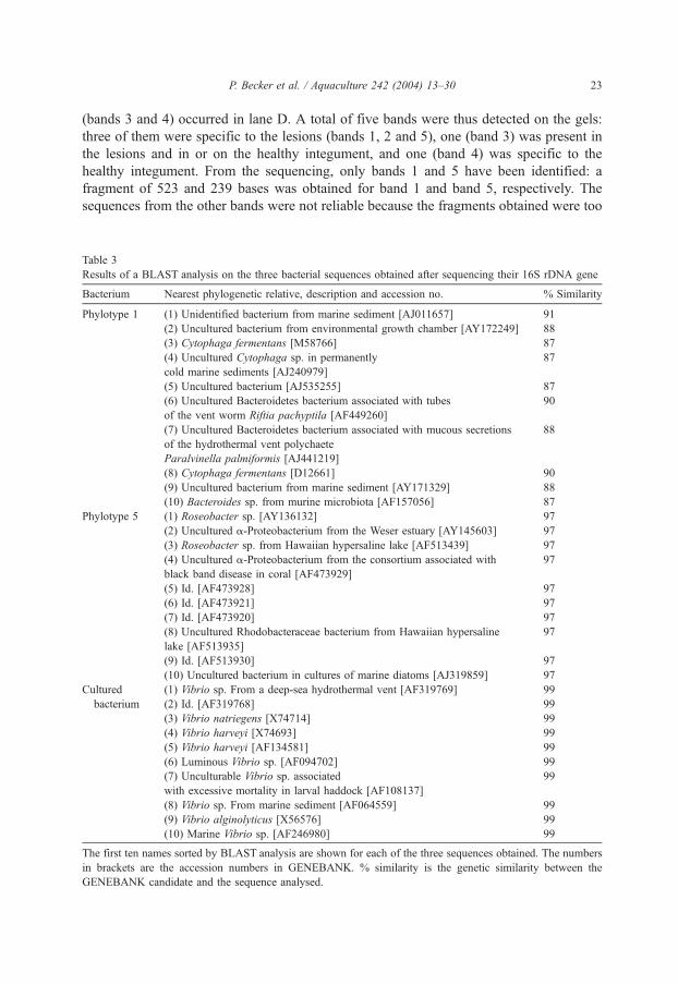
(bands 3 and 4) occurred in lane D. A total of five bands were thus detected on the gels:three of them were specific to the lesions (bands 1, 2 and 5), one (band 3) was present inthe lesions and in or on the healthy integument, and one (band 4) was specific to thehealthy integument. From the sequencing, only bands 1 and 5 have been identified: afragment of 523 and 239 bases was obtained for band 1 and band 5, respectively. Thesequences from the other bands were not reliable because the fragments obtained were too
Table 3
Results of a BLAST analysis on the three bacterial sequences obtained after sequencing their 16S rDNA gene
Bacterium Nearest phylogenetic relative, description and accession no. % Similarity
Phylotype 1 (1) Unidentified bacterium from marine sediment [AJ011657] 91
(2) Uncultured bacterium from environmental growth chamber [AY172249] 88
(3) Cytophaga fermentans [M58766] 87
(4) Uncultured Cytophaga sp. in permanently
cold marine sediments [AJ240979]
87
(5) Uncultured bacterium [AJ535255] 87
(6) Uncultured Bacteroidetes bacterium associated with tubes
of the vent worm Riftia pachyptila [AF449260]
90
(7) Uncultured Bacteroidetes bacterium associated with mucous secretions
of the hydrothermal vent polychaete
Paralvinella palmiformis [AJ441219]
88
(8) Cytophaga fermentans [D12661] 90
(9) Uncultured bacterium from marine sediment [AY171329] 88
(10) Bacteroides sp. from murine microbiota [AF157056] 87
Phylotype 5 (1) Roseobacter sp. [AY136132] 97
(2) Uncultured a-Proteobacterium from the Weser estuary [AY145603] 97
(3) Roseobacter sp. from Hawaiian hypersaline lake [AF513439] 97
(4) Uncultured a-Proteobacterium from the consortium associated with
black band disease in coral [AF473929]
97
(5) Id. [AF473928] 97
(6) Id. [AF473921] 97
(7) Id. [AF473920] 97
(8) Uncultured Rhodobacteraceae bacterium from Hawaiian hypersaline
lake [AF513935]
97
(9) Id. [AF513930] 97
(10) Uncultured bacterium in cultures of marine diatoms [AJ319859] 97
Cultured (1) Vibrio sp. From a deep-sea hydrothermal vent [AF319769] 99
bacterium (2) Id. [AF319768] 99
(3) Vibrio natriegens [X74714] 99
(4) Vibrio harveyi [X74693] 99
(5) Vibrio harveyi [AF134581] 99
(6) Luminous Vibrio sp. [AF094702] 99
(7) Unculturable Vibrio sp. associated
with excessive mortality in larval haddock [AF108137]
99
(8) Vibrio sp. From marine sediment [AF064559] 99
(9) Vibrio alginolyticus [X56576] 99
(10) Marine Vibrio sp. [AF246980] 99
The first ten names sorted by BLAST analysis are shown for each of the three sequences obtained. The numbers
in brackets are the accession numbers in GENEBANK. % similarity is the genetic similarity between the
GENEBANK candidate and the sequence analysed.
P. Becker et al. / Aquaculture 242 (2004) 13–30 23

short. Table 3 gives, for each of the two fragments, the names of the 10 species for whichthe sequences sorted during a BLAST search matched the fragments best. The first namesappearing from the list for band 1 were Cytophaga fermentans, Cytophaga sp., Bacter-oidetes bacteria and Bacteroides sp. indicating that it was a bacterium from the CFB group(Cytophaga–Flavobacterium–Bacteroides). Band 2 was close to a Roseobacter sp., anuncultured a-Proteobacteria and a Rhodobacteriaceae indicating that it was an a-Proteobacterium. The unrooted phylogram in Fig. 5 illustrates the results obtained duringa NJ analysis made on the data matrix that included the sequences of the two bands and
Fig. 5. Unrooted phylogram obtained during a neighbour_joining analysis on 16S rDNA fragments of the cultured
bacterium, of the two DGGE phylotypes and of their closest relatives. Bootstrap support (expressed in %)
obtained during MP and NJ analyses (1000 replicates) are indicated at the root of the relevant clades (MP/NJ).
NS: not supported. The scale bar indicates 0.1 substitutions per nucleotide position. B: Bacteroides, (C¯):
Chlorobium, C: Cytophaga, (F¯): Flavobacterium, F: Flexibacter, J: Jannaschia, M: Marinilabilia, P: Planctomyces,
R: Rhodobacter, (R¯): Rhodovulum, R: Roseobacter, S: Spirochaeta, T: Tenacibaculum, V: Vibrio.
P. Becker et al. / Aquaculture 242 (2004) 13–3024

their closest relatives. Phylotype 1 fits at the base of a clade joining all the Bacteroidesspecies. The clade and the basal position of phylotype 1 are well supported by both MPand NJ bootstrap values. Phylotype 5 clusters with Jannaschia helgolandensis and twoRoseobacter species, R. littoralis and R. gallaeciensis. The clade of the four species is wellsupported by MP and NJ bootstrap values but the position of phylotype 5 within the cladevaries. It is best supported when it clusters with J. helgolandensis.
3.3. Bacterial cultures and infection assays
Six cultures were obtained from the lesions of six diseased juveniles. All thebacterial populations in these cultures were Gram-negative. SEM analysis revealed thatorganisms from the cultures resembled the three bacteria already observed in juvenile’slesions: the rod-shaped bacteria of 2 to 3 Am long, the rough ovoid bacteria of 300 to800 nm long and the smooth ovoid bacteria of 1 to 2 Am long (Fig. 2F). Theproportion of these bacteria varied from a culture to another but on average theproportion was 1:60:500 for rod-shaped, smooth ovoid, and rough ovoid bacteria,respectively. One of the six cultures was an almost totally pure culture of rough ovoidbacterium. Each bacterial culture was sampled for sequencing. The six sequencesobtained varied in length from 451 to 485 bp. Even though the sample was a mix ofthree species, all the sequences were those of a Vibrio species, in the a-proteobacteriasubclass. The first names sorted from a blast search were Vibrio sp., V. natriegens, V.harveyi and Vibrio alginolyticus (Table 3). A neighbour-joining analysis clustered thecultured bacterium within the clade grouping all the Vibrio species with a highbootstrap support (both with MP and NJ) (Fig. 5). The position of the culturedbacterium within the group was unstable and was best supported when it clusteredwith V. alginolyticus.
None of the infection experiments done induced the skin ulceration disease. Twelvedays after the beginning of the assays, all the treated juveniles were healthy except thewounded holothuroids that were painted with a fragment of a bacterial culture. Thesejuveniles were diseased 5 days after the treatment. The disease however did not appearas a skin ulceration. The treated juveniles were much less active than those of thecontrol. They became totally yellow and their podia stayed contracted until deathoccurred.
4. Discussion
The recent ‘‘Advances in Sea Cucumber Aquaculture and Management’’ (ASCAM)workshop held in China (14–18 October 2003) has shown that the skin ulcerationdisease or very closely related diseases occur worldwide and on different species: thedisease has also been recorded on Apostichopus japonicus in China, on Isostichopusfuscus in Equator and on H. scabra in Australia and in New Caledonia. In all, theskin ulceration disease begins with the appearance of a white lesion. In juvenile H.scabra from Madagascar, the white lesion appears near the cloacal orifice and extendsover the whole body surface. The disease is highly contagious and expands quickly in
P. Becker et al. / Aquaculture 242 (2004) 13–30 25

the reared populations. It destroys first the cuticle and the epidermis, then theconnective tissue and ossicles. The disease is due to a bacterial infection but theagent that initiates the skin ulcerations has not been identified yet as none of theinfection assays performed in this work was successful. This suggests that eitherbacteria are not the etiological agent of the disease or, if they are, their action must becombined to other factors to induce it properly. In the first case, non-bacterial agentssuch as viruses or chemicals cannot be excluded. However, the transmission electronmicroscopy observations did not reveal any viral infection and a chemical does notprobably initiate the disease which would have also spread over genitors of H. scabrathat stand in the same water than juveniles. Combined events or agents, includingbacteria, are probably required to induce the disease. The intervention of copepods, forexample, that sometimes swarm at the surface of holothuroids, might cause appropriatewounds that would be colonized by pathogen bacteria. Whatever its origin, one agentof the disease is certainly carried by water currents because most buried holothuroidsare not infected. Moreover, the appearance of the first lesion close to the cloacalopening is probably a consequence of the breathing behaviour of juveniles that, whenthey are buried, expose their posterior end to breathe.
The morphological techniques used suggested that at least three microorganisms areimplicated in the skin ulceration disease. They are rod-shaped bacteria, rough ovoidbacteria and smooth ovoid bacteria. These morphotypes are all three present on thedegrading epidermis and could thus initiate the lesions or be opportunist bacteria.After the destruction of the cuticle and the epidermis, the connective tissue seems tobe invaded mainly by the smooth ovoid bacteria. These bacteria are the onlyorganisms that have been observed on ossicles and they are probably responsible oftheir lysis as they lie at the level of the holes of degraded ossicles. To our knowledge,this is the first time that bacteria are observed degrading ossicles of holothuroids.
The biomolecular techniques identified three species of bacteria in the lesions:Vibrio sp., isolated in the cultures, and Bacteroides sp. with an a-Proteobacteriumdetected by the DGGE. Vibrio sp., obtained from bacterial cultures, was not put inevidence by DGGE probably because it corresponds to one of the faint bandsundetected by the computer program or to the bands 2 and 3 that were not sequencedsuccessfully. Vibrio sp. is close to V. harveyi and V. alginolyticus, two well-knownpathogenic bacteria. V. harveyi is responsible of numerous infections to both verte-brates and invertebrates. It causes enteritis (Soffientino et al., 1999), ocular infections(Kraxberger-Beatty and MacGarey, 1990), exophthalmia (Alvarez et al., 1998; Iwa-moto et al., 1995), melanosis and haemorrhages (Alvarez et al., 1998; Iwamoto et al.,1995) in fishes. The brown shark, Carcharhinus plumbeus, held in captivity, sufferedof meningitis, encephalitis and vascular infection of its internal organs caused by V.harveyi (Grimes et al., 1984). V. harveyi also affects adults and larvae of severalcultured crustacean species including lobsters (Diggles et al., 2000) and penaidsshrimps (Robertson et al., 1998; Liu et al., 1996; Alvarez et al., 1998). Principallythe hepatopancreas and the body surface which are attacked. Vibrio alginolyticusshows a high pathogenecity towards vertebrates, as to the sea bream, Sparus aurata(Balebona et al., 1998), and to invertebrates. Shrimps are again the most studiedvictims of this Vibrio that has been isolated from the hæmolymph of Penaeus
P. Becker et al. / Aquaculture 242 (2004) 13–3026

monodon (Lee et al., 1996a), and from the hepathopancreas of Penaeus japonicus (Leeet al., 1996b) and Litopenaeus vannamei (Esteve and Herrera, 2000). V. alginolyticuswas identified as the etiological agent of a disease affecting two spatangoid seaurchins, Paleopneustes cristatus and Archaeopneustes hystrix (Bauer and Young,2000). The bacterium provoked epidermal lesions, initiated by small mechanicaldamages, that would provide a way for infectious organisms which would enter thecoelom and provoke death (Bauer and Young, 2000). Vibriosis is one of the majorthreats in fish and shellfish marine aquaculture. It is very tempting to identify Vibriosp. as the etiological agent but the infection assays have not established a direct linkbetween this Vibrio and the disease.
This is only the second time that DGGE has been performed on bacteria infectingdiseased marine invertebrates. This molecular tool was previously used by Cooney etal. (2002) on the bacterial consortium of the black band disease affecting corals.DGGE analysis performed on diseased juvenile H. scabra revealed that threephylotypes (1, 2 and 5; 1 and 5 were identified) were exclusively present in lesions.Phylotype 1 forms the most intense band of the DGGE analysis and also appearsfaintly in the samples of the early lesions. Sequencing and phylogenetic analyses ofthe 16S rDNA show that it is a member of the CFB group (100% bootstrap value)closer to the Bacteroides genus than to the Cytophaga and the Flavobacterium genera.Bacteroides species are Gram-negative bacteria that live in the colon and faeces ofseveral mammals including man (Moore and Holdeman, 1974). While the Bacteroidesoccupy a significant position in the normal intestinal flora, they are also opportunisticpathogens, primarily in infections of the peritoneal cavity. Bacteroides fragilis is themost notable pathogen: although it makes up only 12% of the normal intestinal flora,it is the Bacteroides species isolated from 81% of anaerobic clinical infections(Werner, 1974). Bacteroides species are however not known to induce diseases innon-mammalian animals and the Bacteroides sp. identified here probably derives fromlocal contamination by sewage.
Phylotypes 2 and 5 form less intense bands on gels than phylotype 1. They appearonly in late lesions, the corresponding bacteria are probably therefore secondaryinvaders rather than etiological agents. Only, the sequence of phylotype 5 has beenobtained. In the phylogenetic analyses, it clusters with several species of the Rose-obacter genus in a highly supported clade. Representatives of this genus are able totransform organic and inorganic sulfur compounds (Gonzalez et al., 1999; Zubkov etal., 2001) or to degrade aromatic compounds (Buchan et al., 2000). Others areassociated with algae: Roseobacter algicola has been isolated from a culture of thetoxin-producing dinoflagellate Prorocentrum lima (Lafay et al., 1995) and severalspecies of the marine red algal genus Prionitis bear bacterial galls due to aRoseobacter parasite (Ashen and Goff, 2000). Roseobacter species have also beenfound in the accessory nidamental glands of the Sepioid Sepia officinalis, togetherwith other a-Proteobacteria and Gram-positive bacteria but their symbiotic role has notbeen established yet (Grigioni et al., 2000). One species of Roseobacter was found tobe a true pathogen and has been described as being the etiological agent of a juvenileoyster disease affecting Crassostrea virginica in a US hatchery (Boettcher et al.,2000). The species was isolated from affected individuals and could induce the disease
P. Becker et al. / Aquaculture 242 (2004) 13–30 27

to healthy oyster after experimental infection (Boettcher et al., 2000). At the opposite,a Roseobacter showed an inhibitor effect to Vibrio species on agar plate and extractsof this bacterium has a probiotic effect on scallop larvae, enhancing their survival(Ruiz-Ponte et al., 1999). Phylotype 5 clusters best with J. helgolandensis, a newspecies belonging to the Roseobacter–Sulfitobacter–Silicibacter group within the a-subclass of the Proteobacteria. It is a Gram-negative, strictly aerobic and heterotrophicbacterium isolated from sea water samples of the North Sea (Wagner-Dobler et al.,2003).
Acknowledgements
We thank Drs. E. Mara and M.W. Rabenevanana of the IH.SM of Toliara for providingfacilities and Dr. M. Milinkovitch for opening the doors of his Evolutionary GeneticsLaboratory to make sequencing. This work was supported by a FRFC grant and a ‘‘ProjetInteruniversitaire Cible’’ (PIC) financed by the Belgian Cooperation Universitaire auDeveloppement. Contribution of the ‘‘Centre Interuniversitaire de Biologie Marine’’(CIBIM).
References
Altschul, S.F., Gish, W., Miller, W., Myers, E.W., Lipman, D.J., 1990. Basic local alignment search tool. J. Mol.
Biol. 215, 403–410.
Alvarez, J.D., Austin, B., Alvarez, A.M., Reyers, H., 1998. Vibrio harveyi: a pathogen of penaeid shrimps and
fish in Venezuela. J. Fish Dis. 21, 313–316.
Ashen, J.B., Goff, L.J., 2000. Molecular and ecological evidence for species specificity and coevolution in a
group of marine algal–bacterial symbioses. Appl. Environ. Microbiol. 66 (7), 3024–3030.
Balebona, M.C., Andreu, M.J., Bordas, M.A., Zorrilla, I., Morinigo, M.A., Borrego, J.J., 1998. Pathogenicity of
Vibrio alginolyticus for cultured gilt-head sea bream (Sparus aurata L.). Appl. Environ. Microbiol. 64 (11),
4269–4275.
Bauer, J.C., Argerter, C.J., 1987. Isolation of bacteria pathogenic for the sea urchin Diadema antillarum (Echi-
nodermata: Echinoidea). Bull. Mar. Sci. 40, 161–165.
Bauer, J.C., Young, C.M., 2000. Epidermal lesions and mortality caused by vibriosis in deep-sea Bahamian
echinoids: a laboratory study. Dis. Aquat. Org. 39 (3), 193–199.
Boettcher, K.J., Barber, B.J., Singer, J.T., 2000. Additional evidence that juvenile oyster disease is caused by a
member of the Roseobacter group and colonization of nonaffected animals by Stappia stellulata-like strains.
Appl. Environ. Microbiol. 66 (9), 3924–3930.
Buchan, A., Collier, L.S., Neidle, E.L., Moran, M.A., 2000. Key aromatic-ring-cleaving enzyme, protocatechuate
3,4-dioxygenase, in the ecologically important marine Roseobacter lineage. Appl. Environ. Microbiol. 66
(11), 4662–4672.
Cooney, R.P., Pantos, O., Le Tissier, M.D.A., Barer, M.R., O’Donnell, A.G., Bythell, J.C., 2002. Characterization
of the bacterial consortium associated with black band disease in coral using molecular microbiological
techniques. Environ. Microbiol. 4 (7), 401–413.
Delavault, R., Leclerc, M., 1969. Bacteries pathogenes decouvertes chez Asterina gibbosa Penn. (Echinoderme,
Asteride). C. R. Hebd. Seances Acad. Sci., Paris 268, 2380–2381.
Dietrich, H.F., Fontaine, A.R., 1975. A decalcification method for ultrastructure of echinoderm tissues. Stain
Technol. 50, 351–354.
P. Becker et al. / Aquaculture 242 (2004) 13–3028

Diggles, B.K., Moss, G.A., Carson, J., Anderson, C.D., 2000. Luminous vibriosis in rock lobster Jasus verreauxi
(Decapoda: Palinuridae) phyllosoma larvae associated with infection by Vibrio harveyi. Dis. Aquat. Org. 43
(2), 127–137.
Esteve, M., Herrera, F.C., 2000. Hepatopancreatic alterations in Litopenaeus vannamei (Boone, 1939) (Crustacea:
Decapoda: Penaeidae) experimentally infected with a Vibrio alginolyticus strain. J. Invertebr. Pathol. 76 (1),
1–5.
Felsenstein, J., 1985. Confidence limits on phylogenies: an approach using the bootstrap. Evolution 39,
783–791.
Ganter, P., Jolles, G., 1969–1970. Histochimie normale et pathologique, vol. 2. Gauthier-Villars, Paris. 1904 pp.
Gilles, K.W., Pearse, J.S., 1986. Disease in sea urchins Strongylocentrotus purpuratus: experimental infection
and bacterial virulence. Dis. Aquat. Org. 1, 105–114.
Gonzalez, J.M., Kiene, R.P., Moran, M.A., 1999. Transformation of sulfur compounds by an abundant
lineage of marine bacteria in the alpha-subclass of the class Proteobacteria. Appl. Environ. Microbiol.
65 (9), 3810–3819.
Grigioni, S., Boucher-Rodoni, R., Demarta, A., Tonolla, M., Peduzzi, R., 2000. Phylogenetic characterisation of
bacterial symbionts in the accessory nidamental glands of the sepioid Sepia officinalis (Cephalopoda: Decap-
oda). Mar. Biol. 136, 217–222.
Grimes, D.J., Stemmler, J., Hada, H., May, E.B., Maneval, D., Hetrick, F.M., Jones, R.T., Stoskopf, M.,
Colwell, R.R., 1984. Vibrio species associated with mortality of sharks held in captivity. Microb. Ecol.
10, 271–282.
Hughes, T.P., Keller, B.D., Jackson, J.B.C., Boyle, M.J., 1985. Mass mortality of the echinoid Diadema anti-
llarum Philippi in Jamaica. Bull. Mar. Sci. 36, 377–384.
Iwamoto, Y., Suzuki, Y., Kurita, A., Watanabe, Y., Shimizu, T., Ohgami, H., Yanagihara, Y., 1995. Vibrio
trachuri sp. nov., a new species isolated from diseased Japanese horse mackerel. Microbiol. Immunol. 39
(11), 831–837.
Jangoux, M., 1990. Iseases of echinodermata. In: Kinne, O. (Ed.), Diseases of Marine Animals, vol. 3. Biologi-
sche Anstalt Helgoland, Hamburg, Germany, pp. 439–567.
Jukes, T.H., Cantor, C.R., 1969. Evolution of protein molecules. In: Munro, H.N. (Ed.), Mammalian Protein
Metabolism. Academic Press, New York, USA, pp. 21–132.
Kraxberger-Beatty, T., MacGarey, D.J., 1990. Vibrio harveyi, an opportunistic pathogen of common snook,
Centropomus undecimalis (Bloch), held in captivity. J. Fish Dis. 13, 557–560.
Lafay, B., Ruimy, R., De Traubenberg, C.R., Breittmayer, V., Gauthier, M.J., Christen, R., 1995. Roseobacter
algicola sp. Nov., a new marine bacterium isolated from the phycosphere of a toxin-producing dinoflagellate
Prorocentrum lima. Int. J. Syst. Bacteriol. 45 (2), 290–296.
Lee, K.K., Yu, S.R., Chen, F.R., Yang, T.I., Liu, P.C., 1996a. Virulence of Vibrio alginolyticus isolated from
diseased tiger prawn Penaeus monodon. Curr. Microbiol. 32 (4), 229–231.
Lee, K.K., Yu, S.R., Yang, T.I., Liu, P.C., Chen, F.R., 1996b. Isolation and characterization of Vibrio alginolyticus
isolated from diseased kuruma prawn Penaeus japonicus. Lett. Appl. Microbiol. 22 (2), 111–114.
Lessios, H.A., Robertson, D.R., Cubit, J.D., 1984. Spread of Diadema mass mortality through the Caribbean.
Science 226, 335–337.
Liddell, W.D., Ohlhorst, S.L., 1988. Changes in the benthic community composition following the mass mortality
of Diadema at Jamaica. J. Exp. Mar. Biol. Ecol. 95, 271–278.
Liu, P.C., Lee, K.K., Yii, K.C., Kou, G.H., Chen, S.N., 1996. Isolation of Vibrio harveyi from diseased kuruma
prawns Penaeus japonicus. Curr. Microbiol. 33, 129–132.
Maes, P., Jangoux, M., 1984. The bald-sea-urchin: a biopathological approach. Helgol. Meeresunters. 37,
217–224.
Moore, W.E.C., Holdeman, L.V., 1974. Human fecal flora: the normal flora of 20 Japanese–Hawaiians. Appl.
Microbiol. 27, 961–979.
Morgan, A.D., 2000. Aspects de la gestion des stocks geniteurs d’holothuries de sable (Echinoderme: Holothur-
ides). La Beche-de-mer Bulletin d’information, vol. 13. Secretariat general de la Communaute du Pacifique,
pp. 2–8.
Muyzer, G., de Waal, E.C., 1994. Determination of the genetic diversity of microbial communities using DGGE
analysis of PCR-amplified 16S rDNA. NATO Adv. Sci. Inst. Ser., Ser. G 35, 207–214.
P. Becker et al. / Aquaculture 242 (2004) 13–30 29

Muyzer, G., de Waal, E.C., Uiterlinden, A.G., 1993. Profiling of complex microbial populations by denaturing
gradient gel electrophoresis analysis of polymerase chain reaction-amplified genes coding for 16S rRNA.
Appl. Environ. Microbiol. 59, 695–700.
Robertson, P.A.W., Calderon, J., Carrera, L., Stark, J.R., Zherdmant, M., Austin, B., 1998. Experimental Vibrio
harveyi infections in Penaeus vannamei larvae. Dis. Aquat. Org. 32, 151–155.
Ruiz-Ponte, C., Samain, J.F., Sanchez, J.L., Nicolas, J.L., 1999. The benefit of a Roseobacter species on the
survival of scallop larvae. Mar. Biotechnol. 1 (1), 52–59.
Soffientino, B., Gwaltney, T., Nelson, D.R., Specker, J.L., Mauel, M., Gomez-Chiarri, M., 1999. Infectious
necrotizing enteritis and mortality caused by Vibrio carchariae in summer flounder Paralichthys dentatus
during intensive culture. Dis. Aquat. Org. 38 (3), 201–210.
Swofford, D., 1998. PAUP*. Phylogenetics Analysis Using Parsimony (*and others methods). Version 4.0b10.
Sinauer Associates, Sunderland, Massachussetts.
Teske, A., Wawer, C., Muyzer, G., Ramsing, N.B., 1996. Distribution of sulfate-reducing bacteria in a stratified
fjord (Mariager Fjord, Denmark) as evaluated by most-probable-number counts and denaturing gradient gel
electrophoresis of PCR-amplified ribosomal DNA fragments. Appl. Environ. Microbiol. 62, 1405–1415.
Thompson, J.D., Higgins, D.G., Gibson, T.J., 1994. Clustal W: Improving the sensitivity of progressive multiple
sequence alignment through sequence weighting, positions-specific gap penalties and weight matrix choice.
Nucleic Acids Res. 22, 4673–4680.
Wagner-Dobler, I., Rheims, H., Felske, A., Pukall, R., Tindall, B.J., 2003. Jannaschia helgolandensis, gen. nov.,
sp. nov., a novel abundant member of the marine Roseobacter clade from the North Sea. Int. J. Syst. Evol.
Microbiol. 53, 731–738.
Werner, H., 1974. Differentiation and medical importance of sacharolytic intestinal anaerobes. Arzneim.-Forsch.
(Drug Res.) 24, 340–343.
Zubkov, M.V., Fuchs, B.M., Archer, S.D., Kiene, R.P., Amann, R., Burkill, P.H., 2001. Linking the composition
of bacterioplankton to rapid turnover of dissolved dimethylsulphoniopropionate in an algal bloom in the
North Sea. Environ. Microbiol. 3 (5), 304–311.
P. Becker et al. / Aquaculture 242 (2004) 13–3030