
Geometries of doleritic intrusions in central Spitsbergen ...
Upload
independentCategory
view
1download
0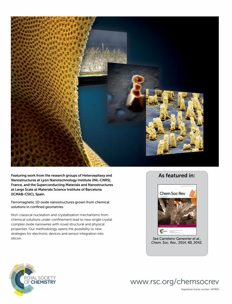
Registered charity number: 207890
Featuring work from the research groups of Heteroepitaxy and
Nanostructures at Lyon Nanotechnology Institute (INL-CNRS),
France, and the Superconducting Materials and Nanostructures
at Large Scale at Materials Science Institute of Barcelona
(ICMAB-CSIC), Spain.
Ferromagnetic 1D oxide nanostructures grown from chemical
solutions in confi ned geometries
Non-classical nucleation and crystallization mechanisms from
chemical solutions under confi nement lead to new single crystal
complex oxide nanowires with novel structural and physical
properties. Our methodology opens the possibility to new
strategies for electronic devices and sensor integration into
silicon.
www.rsc.org/chemsocrev
As featured in:
See Carretero-Genevrier et al.,Chem. Soc. Rev., 2014, 43, 2042.
This journal is c The Royal Society of Chemistry 2013 Chem. Soc. Rev.
Cite this: DOI: 10.1039/c3cs60288e
Ferromagnetic 1D oxide nanostructures grown fromchemical solutions in confined geometries
A. Carretero-Genevrier,*abc T. Puig,b X. Obradorsb and N. Mestres*b
This review summarizes the capabilities and recent developments of nanoporous polymeric template
systems directly supported on different substrates for the confined growth of epitaxial ferromagnetic
complex oxide 1D nanostructures. In particular, we describe the versatility and potentiality of chemical
solutions combined with track-etched polymers to synthesize (i) vertical polycrystalline La0.7Sr0.3MnO3
nanorods on top of single crystal perovskites, (ii) single crystalline manganese based octahedral
molecular sieve (OMS) nanowires on silicon substrates, and (iii) the epitaxial directional single crystal
OMS nanowires on top of fluorite-type substrates. The influence of the distinct growth parameters on
the nanostructural evolution of the resulting nanostructures and their magnetic properties is further
discussed in detail.
Key learning points(1) Complex oxide nanostructure growth based on chemical solutions and track-etched polymer templates as nanoreactors.(2) Confinement in high aspect ratio nanopores favors the nucleation of intermediate precursor phases.(3) Different crystalline structures and morphologies are stabilized thanks to the epitaxial templating of different substrates.(4) New polymorphs of known functional oxides under nanoscale confinement with enhanced magnetic properties.(5) Crystalline structure and magnetic properties determined by complementary macroscopic and nanoscopic characterization techniques.
1 Introduction
Selective synthesis of integrated nanomaterials with controllablemorphology and composition represents an emerging researcharea in nanoscience and nanotechnology because the intrinsicproperties of nanostructures are generally phase-, shape-, andsize-dependent.1–4 Additionally, confinement within nanometricpores can significantly modify crystallization pathways and giverise to new crystal polymorphs, particularly when the poredimensions and the critical size of an emergent nucleus aresimilar. Under these circumstances, a delicate balance existsamong interface, surface and volume free energies and polymorphstability selection may differ from the one in the bulk.5 Currentinvestigations in the field have demonstrated that confined
crystallization can be exploited to control polymorphism, forinstance in food and pharmaceuticals, and, importantly, tomodify the crystallization temperature of polymers.6–8 In theparticular case of production sectors such as food, pharmaceuticals,agricultural chemicals, and fragrances, understanding andcontrolling the occurrence of polymorphism within synthesisprocesses become critical since specific activity is often determinedby a unique polymorph.
In the field of electronics, growing interest is devoted totransition metal oxides, a class of electronic materials exhibiting awide range of electronic, magnetic and optical properties: i.e. hightemperature superconductivity, ferroelectricity, piezoelectricity,ferromagnetism, multiferroicity, colossal magnetoresistance,excellent catalytic properties and non-linear optical effects indifferent morphologies.9–11 Specifically, research on manganeseoxides has been a hot topic among studies on transition metaloxides since they exhibit a broad range of interesting physicaland chemical properties, which have large and potential appli-cations in different areas including gas sensors, energy storage,rechargeable lithium ion batteries, catalysis, molecular adsorptionand magnetism.12–17 Mixed valence manganese based perovskiteswith a general formula R1�xAxMnO3 (where R is a trivalent rare
a Laboratoire Chimie de la Matiere Condensee, UMR UPMC-College de France-
CNRS 7574, College de France, 11 place Marcelin Berthelot, 75231 Paris, France.
E-mail: [email protected] Institut de Ciencia de Materials de Barcelona ICMAB, Consejo Superior de
Investigaciones Cientıficas CSIC, Campus UAB 08193 Bellaterra, Catalonia, Spain.
E-mail: [email protected] Institut des Nanotechnologies de Lyon (INL)CNRS - Ecole Centrale de Lyon,
36 avenue Guy de Collongue, 69134 Ecully, France
Received 2nd August 2013
DOI: 10.1039/c3cs60288e
www.rsc.org/csr
Chem Soc Rev
TUTORIAL REVIEW
Publ
ishe
d on
03
Oct
ober
201
3. D
ownl
oade
d by
Uni
vers
ite P
ierr
e et
Mar
ie C
urie
on
03/1
0/20
13 1
4:50
:35.
View Article OnlineView Journal

Chem. Soc. Rev. This journal is c The Royal Society of Chemistry 2013
earth cation and A is a divalent alkaline earth cation) haveattracted significant research attention owing to their unusualelectronic and magnetic properties. This class of perovskites isrich in functional properties exhibiting numerous fundamentallynew phenomena like colossal magnetoresistance and charge andorbital ordering.12 Further downsizing of these systems to a fewnanometers, allowing incorporating various finite size effects, willyield several promising properties with great potential for deviceapplications. The subtle balance existing between electronic,magnetic and lattice degrees of freedom in these materials can
be further unbalanced when working at the nanoscale, thusproviding new outstanding properties.18,19
Another important group of functional materials based onmanganese oxides have the general formula AxMnO2. Thesederivative rutile-like structure materials, displaying porousstructures, open tunnels, and high surface areas, also haveMn ions in the mixed-valence state which indeed determinesthe final material properties. The building blocks of thesemanganese oxides are MnO6 octahedra that share cornersand edges to form channel-like slabs that cross-link to
A. Carretero-Genevrier
Adrian Carretero Genevrier iscurrently a Permanent CNRSresearcher at the Institute ofNanotechnology (INL) in Lyon(France). He got his PhD (2010)in ‘‘materials science’’ from theUniversitat Autonoma deBarcelona for work performed atICMAB-CSIC, and then moved forthree postdoctoral years atCollege de France in Paris. Hisresearch is aimed atunderstanding the growthmechanisms of new complex
oxides nanostructures synthesized by soft chemistry and therelationship between crystal structure and physical properties.Thus, his scientific interests span over a wide range of fieldsincluding soft chemistry routes to grow 1D functional complexoxides, crystal engineering, nanofabrication, template-assistedsynthesis of inorganic gels, mesoporous coating, crystallinenanostructured oxide coatings, and mesoporous materials forcatalysis.
T. Puig
Teresa Puig received a B.S. inPhysics (1989) and a Ph.D. inPhysics (1994) from UniversitatAutonoma de Barcelona. Afterpre- and postdoctoral positionsat Katholieke UniversiteitLeuven, Royal Institute ofTechnology of Stockholm, TrinityCollege Dublin and UnivesitatRegensburg, she became tenuredscientist at Barcelona MaterialsScience Institute (ICMAB-CSIC)in 2000, and in 2010 she wasappointed Full Professor. Since
2008 she is Head of the Superconducting Materials and largescale nanostructures Department. Her research interests includegrowth and functional properties of complex oxide nanostructuresand heteroepitaxial films, physics of nanofabricatedsuperconducting structures and superconducting tapes.
X. Obradors
Xavier Obradors received a Ph.D.in Physics from Universitat deBarcelona in 1982 and a Ph.D.in Materials Science fromUniversite Scientifique etMedicale de Grenoble in 1983.After postdoctoral positions atthe University of Barcelona,Argonne National Laboratoryand University of California atSan Diego, he became AssociateProfessor, in Universitat deBarcelona in 1985. He moved tothe Institute of Materials Science
of Barcelona (ICMAB), National High Research Council (CSIC) in1989. He became Deputy Director and Director of ICMAB-CSIC in2002 and 2008, respectively. His current research interests includecrystal growth and properties of functional oxide nanostructuresand thin films and preparation and properties ofsuperconducting tapes.
N. Mestres
Narcıs Mestres received a B.S. inPhysics from the UniversitatAutonoma de Barcelona (Spain)in 1983 and a Ph.D. in Physicsfrom Stuttgart University(Germany) in 1986 for workperformed at the Max-Planck-Institut fur Festkorperforschung.After a postdoctoral position atthe University of Michigan(USA), he held an associateprofessor position at theUniversidad Autonoma deMadrid (Spain). He moved to
the Materials Science Institut of the Spanish Research Council(ICMAB-CSIC) in Barcelona in 1993, where he holds a scientificresearcher position. His recent research focuses on chemicalsolution synthesis of functional oxides nanostructures for energyand environmental science, and the physics of self-assembly.
Tutorial Review Chem Soc Rev
Publ
ishe
d on
03
Oct
ober
201
3. D
ownl
oade
d by
Uni
vers
ite P
ierr
e et
Mar
ie C
urie
on
03/1
0/20
13 1
4:50
:35.
View Article Online
This journal is c The Royal Society of Chemistry 2013 Chem. Soc. Rev.
ultimately build one-dimensional (1-D) tunnels. Interestingly,these MnO2 derivative compounds have the capability to adaptA-cations within the channel structure, therefore providingcharge balance and support to the tunnel framework. Thisfamily of materials is also known as manganese oxide octahedralmolecular sieves (OMS).20 Recently, much effort has been devotedto synthesize novel nanoscale manganese oxide OMS materialsin order to modify their physical and chemical properties, andthus improve their performance as electrodes for batteries andsupercapacitors, and as redox catalysts.21–23
In the last few decades, 1D systems such as nanowires(NWs), nanotubes, and nanorods have attracted great attentiondue to their anisotropic optical, electrical, and magnetic properties,which are of major importance for a variety of applications(from sensing, to photonics, electronics and energy conversionand storage). As a consequence, active research has focused onthe development of tailored strategies for the synthesis of 1Dmaterials.24–26 Although the epitaxial growth of semiconductorNWs has been successfully accomplished through vapour–liquid–solid (VLS) processes using metal catalysts,25,27 thecontrolled synthesis of ternary and quaternary metal oxide1D-nanostructures is notoriously more problematic due to thedifficulty in controlling the precursor reactions and finalhomogeneous stoichiometry. Therefore, only limited successhas been achieved on few materials and to date the synthesis ofnanowires of multicomponent oxides has been a challenge.
The most commonly adopted techniques towards the synthesisof 1D nanostructures of complex metal oxides are ‘bottom-up’routes28 such as hydro/solvothermal synthesis,29,30 molten-saltsynthesis,31 solution based metal–organic decomposition,32,33
and electrospinning.34 In this context, the combination ofchemical solutions and template-inspired methodologies that werepioneered by Martin in the early 1990s represents a convenientand versatile method for generating 1D nanostructures.35
Originally, template assisted methods take advantage of templatesas physical scaffolds to shape the morphology of the desirednanomaterials. Thus, synthesis of nanostructures based ontemplates has been developed independently in different fieldsof nanotechnology.36 For instance, electrodeposition onto nano-porous templates is a very efficient method to grow metallicnanowire field emitters37 and magnetic multilayer nanowireswith enhanced magnetoresistance.38 In this framework, poroustemplates used so far include anodic aluminium oxide (AAO),anodic titanium oxide (ATO), porous silicon (PS), and track-etched (TE) polymers. One of the main drawbacks of usingtemplate assisted methods for the synthesis of 1D nanostructuresrelates to the ultimate template removal, which is a requirementfor many technological applications. However, polymer templatesdisplay low decomposition temperatures, which enable theireasy elimination during growth processes when suitable thermalconditions are chosen, as we will further discuss.
In this review, we present an overview of our work concerningthe growth of 1D manganese based oxide nanostructuresthrough the deposition of sol–gel precursor solutions combinedwith a novel version of a track-etched polymer nanoporoustemplate that buffers the substrates. We will prove that polymer
template nanopores can be used not only as mere physicalspatial constraints, thus defining the shape and size of nano-structures, but also as nanoreactors in which confined nuclea-tion favours the stabilization of particular metastable seednanostructures. Further epitaxial stabilization of new verticalpolymorphs with enhanced ferromagnetic properties can thenbe achieved by using different substrates, therefore supportingthe validity and generality of our methodology. We present thestudies conducted on specific ferromagnetic oxides as modelsystems, even though this methodology can be further appliedto other different oxide materials. Indeed, the possibility ofgenerating vertical 1D nanostructures by taking advantage ofthe good epitaxial relationship of similar crystallographic struc-tures during the growth process makes it possible to extend thisprocedure to other vertical functional oxide nanostructures.
In particular, using silicon substrates in the presence of alkalineearth cations in the precursor solution is key to promote SiO2
devitrification into a-quartz during thermal annealing at relativelylow temperatures.39 The generated a-quartz nanocrystals act astemplates for the epitaxial growth of single crystalline oxidenanowires of different compositions and structures, and moreimportantly allow the direct integration on silicon substrates.40
Thus, our methodology exhibits great potential and offers newroutes to design novel mixed valence oxide compounds by chemicalroutes with unique optical, electric, or magnetic properties.
2. Track-etched polymer template basedmethodology
In this review we take advantage of combining chemicalsolution deposition and the use of nanoporous polymer templatesto provide a general methodology capable of producing broad,versatile complex oxide nanostructured systems. Fig. 1 schematicallydisplays an overview of the confined growth process of differentkinds of manganese oxide based nanostructures. Common to allsystems, the first step is based on the use of track etched polymertemplates prepared by irradiation of polyimide (PI) or polycarbonate(PC) through heavy ions and posterior chemical development asdescribed elsewhere.41 Then, precursor solutions, which can betuned according to the different compositions, are used to fill thepolymer template nanopores using capillary force. Moreover, thechoice of the precursor solution was made by taking into accountseveral issues including the optimal control of the stoichiometry,the solubility, availability and purity of materials. Thus, metallicnitrates, which are completely soluble in water and possess long-term stability, are the precursors of choice in most cases for thepreparation of different chemical solutions as described else-where.40,41 In addition, we found that the capability of the precursorsolution to infiltrate into the template’s nanopores depends on therheological parameters such as viscosity or pH. These parametersare fundamental to the synthesis of complex oxide nanostructuresassisted by nanoporous templates. Thus ethylene glycol (EG) heatedabove 100 1C can be used to promote polymerization of EG in orderto reach the optimum viscosity and pH value of the precursorsolution required for filling the template’s pores. Moreover it is
Chem Soc Rev Tutorial Review
Publ
ishe
d on
03
Oct
ober
201
3. D
ownl
oade
d by
Uni
vers
ite P
ierr
e et
Mar
ie C
urie
on
03/1
0/20
13 1
4:50
:35.
View Article Online

Chem. Soc. Rev. This journal is c The Royal Society of Chemistry 2013
possible to include ethanol in the initial precursor solutionwith the objective to enhance the template filling and furtheroptimal evaporation of the solvent.
Different thermal treatments under an oxygen rich atmo-sphere are further applied in a tubular oven for the ultimatephase formation and crystallization. The thickness of templatescan be tuned between 1 mm and 8 mm. Note that for the thickertemplates (>5 mm), the mechanical stability of polymer foilsallows their direct deposition on top of the substrate surfaceand the subsequent filling of nanopores with the precursorsolution. In the case of thinner templates, PC and PI thin films(1–2 mm), these can be obtained by spin coating followed by ionirradiation and chemical development. However, the versatilityof the process allows obtaining vertical 1D nanostructuresirrespective of the polymer thickness. Therefore, here we provideexamples of crystalline OMS NWs on silicon substrates,42,43
obtained from thicker templates, and OMS NWs grown on topof yttria stabilized zirconia (YSZ) substrates44 and polycrystallineperovskite La0.7Sr0.3MnO3 (LSMO) nanorods grown on perovskiteSrTiO3 (STO) and LaAlO3 (LAO) substrates,45 obtained fromthinner polymer films.
3. Influence of the substrate on polymorphstabilization
Once the polymer template is decomposed during thermaltreatment, the atomic structure of the substrate will act as asecond template for epitaxial growth. Thus, in this section wewill discuss the fundamental role of the substrate as a determinantof the geometry and orientation of the grown nanostructured
oxides. In addition, surface quality of substrates is also a crucialrequirement for the deposition of the polymer template. There-fore, good adhesion and wettability of the polymer template arerequired in order to minimize the infiltration of the precursorsolution during the nanopore filling process.
3.1 Vertical polycrystalline LSMO nanorods on STO andLAO substrates
Vertical polycrystalline LSMO nanorods are synthesized usingPC and PI nanoporous templates assisted by chemical solutiondeposition on STO and LAO substrates.45 Here, we take advantageof the similar perovskite structures of LSMO nanorods andsubstrates to maintain the verticality of synthesized nanorods athigh temperatures. The difference in the lattice parametersbetween LSMO and the substrates is minimal, yielding a nominaltensile lattice mismatch of e B 0.9% for STO and a larger butcompressive magnitude of e B �2% for LAO substrates. Conse-quently, filling the nanoporous track-etched polymer (B1 mmthick) supported on STO and LAO substrates with the precursorsolution and performing a thermal treatment at 800 1C will resultin a final nanostructured perovskite substrate with verticalnanorods of 1 mm high as summarized in Fig. 2.
Fig. 2 displays schematics of the different stages during theformation process of La0.7Sr0.3MnO3 vertical nanorods on STOor LAO substrates. First, nanopores are filled by capillary forceusing a sol–gel based polymer precursor of LSMO (step (1) inFig. 2). Then, at a temperature between 500 1C and 600 1C thepolymer template is eliminated (see the corresponding thermo-gravimetric analysis plots for PC and PI in the inset), givingrise to vertically oriented amorphous nanorods according tothe composition of the precursor oxides (step (2) in Fig. 2).
Fig. 1 Schematics of the general methodology applied for growing 1D manganese based oxide nanostructures through the deposition of sol–gel precursor solutionscombined with a novel version of nanoporous track-etched polymer templates that buffer the substrates. (1) Nanostructuration of 1D perovskite oxide nanorods ontop of single crystal perovskite substrates. (2) Direct integration of single crystalline manganese based OMS nanowires with tunable composition and micro-porous sizeon silicon substrates. (3) Self-assembly of epitaxial directional single crystal OMS nanowires on top of fluorite-type substrates.
Tutorial Review Chem Soc Rev
Publ
ishe
d on
03
Oct
ober
201
3. D
ownl
oade
d by
Uni
vers
ite P
ierr
e et
Mar
ie C
urie
on
03/1
0/20
13 1
4:50
:35.
View Article Online
This journal is c The Royal Society of Chemistry 2013 Chem. Soc. Rev.
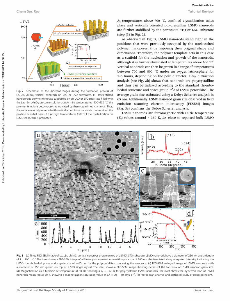
At temperatures above 700 1C, confined crystallization takesplace and vertically oriented polycrystalline LSMO nanorodsare further stabilized by the perovskite STO or LAO substrate(step (3) in Fig. 2).
As observed in Fig. 3, LSMO nanorods stand right in thepositions that were previously occupied by the track-etchedpolymer nanopores, thus imposing their original shape anddimensions. Therefore, the polymer template acts in this caseas a scaffold for the nucleation and growth of the nanorods,although it is further eliminated at temperatures above 600 1C.Vertical nanorods can then be grown in a range of temperaturesbetween 700 and 800 1C under an oxygen atmosphere for1–5 hours, depending on the pore diameter. X-ray diffractionanalysis (see Fig. 3b) shows that nanorods are polycrystallineand thus can be indexed according to the standard rhombo-hedral structure and space group R%3c of LSMO perovskite. Theaverage grain size estimated using a Debye Scherrer analysis is65 nm. Additionally, LSMO nanorod grain size observed in fieldemission scanning electron microscopy (FESEM) images(Fig. 3c) confirms the Debye Scherrer analysis.
LSMO nanorods are ferromagnetic with Curie temperature(Tc) values around B360 K, i.e. close to reported bulk LSMO
Fig. 2 Schematics of the different stages during the formation process ofLa0.7Sr0.3MnO3 vertical nanorods on STO or LAO substrates. (1) Track-etchednanoporous polymer template supported on an LAO or STO substrate filled withthe La0.7Sr0.3MnO3 precursor solution. (2) At mild temperatures (500–600 1C) thepolymer template decomposes as indicated by thermogravimetric analysis. Thus,the surface was fully covered with vertical amorphous nanorods that retained theposition of initial pores. (3) At high temperatures (800 1C) the crystallization onLSMO nanorods is promoted.
Fig. 3 (a) Tilted FEG-SEM image of La0.7Sr0.3MnO3 vertical nanorods grown on top of a (100)-STO substrate. LSMO nanorods have a diameter of 250 nm and a densityof 1 � 108 cm�2. The inset shows a FEG-SEM image of a PI nanoporous membrane with a pore size of 300 nm. (b) Associated X-ray integrated intensity, indicating theLMSO rhombohedral phase and a grain size of B65 nm for the polycrystallites composing the nanorods. (c) FEG-SEM enlarged image of LSMO nanorods witha diameter of 250 nm grown on top of a STO single crystal. The inset shows a FEG-SEM image showing details of the top view of LSMO nanorod grain size.(d) Magnetization as a function of temperature at 50 Oe showing a Tc B 360 K for polycrystalline LSMO nanorods. The inset shows the hysteresis loop of LSMOnanorods measured at 50 K, showing a magnetization saturation value of Ms = 90 � 10 emu g�1. (e) Profile scan analysis and statistical study of nanorod height.
Chem Soc Rev Tutorial Review
Publ
ishe
d on
03
Oct
ober
201
3. D
ownl
oade
d by
Uni
vers
ite P
ierr
e et
Mar
ie C
urie
on
03/1
0/20
13 1
4:50
:35.
View Article Online

Chem. Soc. Rev. This journal is c The Royal Society of Chemistry 2013
values (see Fig. 3d). All these results prove the capability of thechemical solution deposition method combined with track-etchpolymer templates for generating homogeneous nanostructuredsurfaces with vertical LSMO nanorods displaying interestingmagnetic properties.
In addition, post annealing treatments can be further carriedout to investigate the nanostructural evolution of the grownnanorods with temperature and time (see Fig. 4).
3.2 Structural evolution of vertical LSMO nanorods withtemperature
Strong evolution behaviour of LSMO polycrystalline nanorodsis observed after thermal activation at high temperatures, assummarized in Fig. 4. Upon thermal treatment at 1000 1C,vertical polycrystalline nanorods that have a high interfacialenergy due to their polycrystalline character re-crystallize into acompletely different nanostructure of lower energy (schematicallyshown in Fig. 4a). Fig. 4b reveals that the polycrystalline nanorodsdisappeared and instead, faceted nanopyramids with square basesare formed at the same place where the LSMO nanorods wereinitially located. The inset in Fig. 4c shows that the nanopyramidsare formed at the expense of the top polycrystalline material, andthat at this evolution stage some remaining polycrystallinematerial lies on top of the nanopyramids.
During the diffusive transformation of LSMO nanorods intofaceted nanopyramids, a wetting layer expands underneath thenanopyramids, thus covering the single crystalline substrate. Highresolution transmission electron microscopy (TEM) analyses canbe carried out to identify the nature of the wetting layer formedduring the nanostructure transformation (see Fig. 4b).
As observed in Fig. 4b, the 2D wetting film is a fully strainedLSMO layer with a rhombohedral structure (indexed by thepseudocubic structure), which grows cube on cube on the STO(or LAO) single crystals, (001)LSMO[100]//(001)STO[100]. Thethickness of the LSMO wetting layer varies depending on thedistance from the nearest nanopyramid, going from 20 to 5 nm.All together, this proves that the material used to form thewetting layer derives indeed from the original polycrystallinenanorods.
High resolution TEM images of the nanopyramids reveal acharacteristic layered atomic structure with a spacing of0.87 nm. This crystallographic plane is identical to that of the(La,Sr)xOy oxide phase found from spontaneous outcrop inLSMO thin films subjected to extended high temperatureoxygenation periods.46 Therefore, this is a new example of thecomplex chemistry behind LSMO layers at high temperatures(1000 1C), where LSMO epitaxial layers lose their stability andpreferentially segregate (La,Sr)xOy oxide nanostructures. All thisconfirms that the La0.7Sr0.3MnO3 polycrystalline nanorods arethermodynamically metastable nanostructures of high energy(large interfacial grain boundary energy) that, under strongthermal activation, kinetically transform into Mn-free oxideepitaxial nanopyramids on top of a La0.7Sr0.3MnO3 epitaxialwetting layer of variable thickness. Indeed, this system demon-strates that, at the nanoscale, metastable structures can easilyevolve towards the equilibrium state when the mass transportimplicated in the process is small enough.
Magnetic characterization of LSMO epitaxial layers withnon-magnetic oxide nanopyramids displays a Curie temperatureof 350 K, which is characteristic of the LSMO rhombohedralphase. Moreover the magnetic moment of 5 � 10�5 emuobtained is within the expected range if one considers the bulkmagnetization of LSMO at 10 K in a layer thickness of 10 nm.Additionally, magnetization loops at 100 K under in-planeand out-of-plane configurations revealed an in-plane easy axismagnetization of the epitaxial (100)-LSMO film (see Fig. 4d).These results confirm that the segregated nanopyramids do notapparently modify the macroscopic functional properties of theLSMO film. We have, therefore, achieved a nanostructuredsystem composed of an epitaxial LSMO wetting layer withepitaxial vertical nanopyramids grown on top and distributedaccording to the nanopores of the polymeric template.
The composite system presented here, which is formed by ametallic and magnetic LSMO layer with insulating and non-magnetic oxides, can be further tested as a template for theulterior growth of nanostructured superconducting YBa2Cu3O7�x
films using chemical solutions.47 In this way, pinning forces canbe importantly enhanced by a factor of 2 and 4 at 3 T and 5 T,respectively, at 77 K.
3.3 Direct integration of single crystalline OMS nanowires onsilicon substrates
Radically different nucleation and subsequent phase evolutionbehaviour is appreciated when silicon substrates are usedfor the synthesis of 1D nanostructures following our generalmethodology described above (see Fig. 1). In this case, vertical
Fig. 4 (a) Schematics of the structural and morphological evolution of LSMOpolycrystalline nanorods as a function of temperature. The figure shows thecrystallization process at 1000 1C for 5 h for single crystalline (La,Sr)xOy nano-pyramids on a LAO or STO substrate. (b) HRTEM images of an epitaxial (La,Sr)xOy
nanopyramid and the interface between the STO substrate and the LSMO film.The insets show the FFT of both phases. (c) FEG-SEM image of a large area ofnanopyramids grown on a LAO substrate. The inset shows a FEG-SEM imageduring single crystal nanopyramid formation, the remaining polycrystallinematerial is still lying on top of the nanopyramid. (d) Magnetization loops at100 K under in-plane and out-of-plane configurations revealed an in-plane easyaxis magnetization of the epitaxial (100) LSMO film.
Tutorial Review Chem Soc Rev
Publ
ishe
d on
03
Oct
ober
201
3. D
ownl
oade
d by
Uni
vers
ite P
ierr
e et
Mar
ie C
urie
on
03/1
0/20
13 1
4:50
:35.
View Article Online
This journal is c The Royal Society of Chemistry 2013 Chem. Soc. Rev.
single crystalline OMS nanowires of different pore sizes andcompositions can be directly integrated on silicon substrates.Note that silicon is the substrate of choice of the micro-electronics industry, and thus great efforts are currentlydevoted to combine the functionality of oxides with the perfor-mances of semiconductor platforms for the development ofnovel and more efficient device applications. However, furtherincorporation of functional oxide nanostructures as activematerials in electronics critically depends on the ability tointegrate crystalline metal oxides into silicon structures. Here,we take advantage of the recent development of soft chemistrybased routes to integrate a-quartz thin films on silicon substrates,39
which allows stabilizing and crystallizing OMS nanowires.a-Quartz thin films can be obtained after thermally activated
devitrification of the native amorphous silica surface layerassisted by the heterogeneous catalysis driven by alkaline earthcations present in the precursor solution.40 Our innovativegrowth method has allowed the synthesis of manganateLaSr-2 � 4 OMS monoclinic nanowires with ordered arrange-ment of the La3+ and Sr2+ cations inside the 1D-channels for thefirst time.42,43
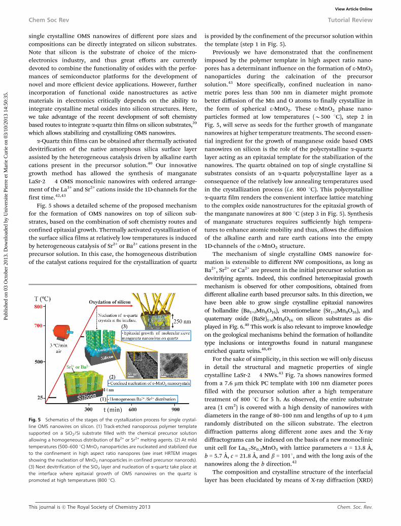
Fig. 5 shows a detailed scheme of the proposed mechanismfor the formation of OMS nanowires on top of silicon sub-strates, based on the combination of soft chemistry routes andconfined epitaxial growth. Thermally activated crystallization ofthe surface silica films at relatively low temperatures is inducedby heterogeneous catalysis of Sr2+ or Ba2+ cations present in theprecursor solution. In this case, the homogeneous distributionof the catalyst cations required for the crystallization of quartz
is provided by the confinement of the precursor solution withinthe template (step 1 in Fig. 5).
Previously we have demonstrated that the confinementimposed by the polymer template in high aspect ratio nano-pores has a determinant influence on the formation of e-MnO2
nanoparticles during the calcination of the precursorsolution.43 More specifically, confined nucleation in nano-metric pores less than 500 nm in diameter might promotebetter diffusion of the Mn and O atoms to finally crystallize inthe form of spherical e-MnO2. These e-MnO2 phase nano-particles formed at low temperatures (B500 1C), step 2 inFig. 5, will serve as seeds for the further growth of manganatenanowires at higher temperature treatments. The second essen-tial ingredient for the growth of manganese oxide based OMSnanowires on silicon is the role of the polycrystalline a-quartzlayer acting as an epitaxial template for the stabilization of thenanowires. The quartz obtained on top of single crystalline Sisubstrates consists of an a-quartz polycrystalline layer as aconsequence of the relatively low annealing temperatures usedin the crystallization process (i.e. 800 1C). This polycrystallinea-quartz film renders the convenient interface lattice matchingto the complex oxide nanostructures for the epitaxial growth ofthe manganate nanowires at 800 1C (step 3 in Fig. 5). Synthesisof manganate structures requires sufficiently high tempera-tures to enhance atomic mobility and thus, allows the diffusionof the alkaline earth and rare earth cations into the empty1D-channels of the e-MnO2 structure.
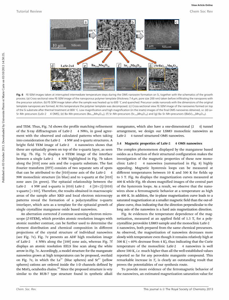
The mechanism of single crystalline OMS nanowire for-mation is extensible to different NW compositions, as long asBa2+, Sr2+ or Ca2+ are present in the initial precursor solution asdevitrifying agents. Indeed, this confined heteroepitaxial growthmechanism is observed for other compositions, obtained fromdifferent alkaline earth based precursor salts. In this direction, wehave been able to grow single crystalline epitaxial nanowiresof hollandite (Ba1+dMn8O16), strontiomelane (Sr1+dMn8O16), andquaternary oxide (BaSr)1+dMn8O16 on silicon substrates as dis-played in Fig. 6.40 This work is also relevant to improve knowledgeon the geological mechanisms behind the formation of hollanditetype inclusions or intergrowths found in natural manganeseenriched quartz veins.48,49
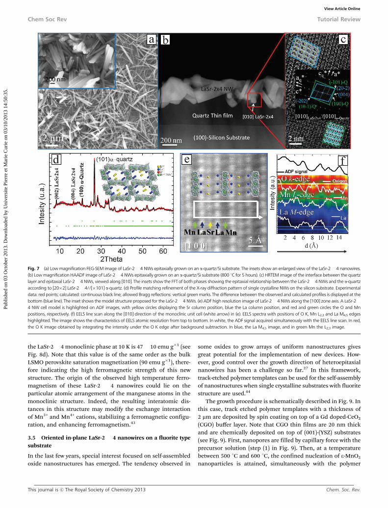
For the sake of simplicity, in this section we will only discussin detail the structural and magnetic properties of singlecrystalline LaSr-2 � 4 NWs.43 Fig. 7a shows nanowires formedfrom a 7.6 mm thick PC template with 100 nm diameter poresfilled with the precursor solution after a high temperaturetreatment of 800 1C for 5 h. As observed, the entire substratearea (1 cm2) is covered with a high density of nanowires withdiameters in the range of 80–100 nm and lengths of up to 4 mmrandomly distributed on the silicon substrate. The electrondiffraction patterns along different zone axes and the X-raydiffractograms can be indexed on the basis of a new monoclinicunit cell for La0.7Sr0.3MnO3 with lattice parameters a = 13.8 Å,b = 5.7 Å, c = 21.8 Å, and b = 1011, and with the long axis of thenanowires along the b direction.42
The composition and crystalline structure of the interfaciallayer has been elucidated by means of X-ray diffraction (XRD)
Fig. 5 Schematics of the stages of the crystallization process for single crystal-line OMS nanowires on silicon. (1) Track-etched nanoporous polymer templatesupported on a SiO2/Si substrate filled with the chemical precursor solutionallowing a homogeneous distribution of Ba2+ or Sr2+ melting agents. (2) At mildtemperatures (500–600 1C) MnO2 nanoparticles are nucleated and stabilized dueto the confinement in high aspect ratio nanopores (see inset HRTEM imagesshowing the nucleation of MnO2 nanoparticles in confined precursor nanorods).(3) Next devitrification of the SiO2 layer and nucleation of a-quartz take place atthe interface where epitaxial growth of OMS nanowires on the quartz ispromoted at high temperatures (800 1C).
Chem Soc Rev Tutorial Review
Publ
ishe
d on
03
Oct
ober
201
3. D
ownl
oade
d by
Uni
vers
ite P
ierr
e et
Mar
ie C
urie
on
03/1
0/20
13 1
4:50
:35.
View Article Online
Chem. Soc. Rev. This journal is c The Royal Society of Chemistry 2013
and TEM. Thus, Fig. 7d shows the profile matching refinementof the X-ray diffractogram of LaSr-2 � 4 NWs, in good agree-ment with the observed and calculated patterns when takinginto consideration the LaSr-2� 4 NW and a-quartz structures. Abright field TEM image of LaSr-2 � 4 nanowires shows thatthese are epitaxially grown on top of the a-quartz layer, as seenin Fig. 7b. Fig. 7c displays a STEM image of the interfacebetween a single LaSr-2 � 4 NW highlighted in Fig. 7b takenalong the [010] zone axis and the a-quartz substrate. The fastFourier transform (FFT) consists of two separate sets of spotsthat can be attributed to the [010]-zone axis of the LaSr-2 � 4NW monoclinic structure (in blue) and to a-quartz at the [010]zone axes (in green). The epitaxial relationship between theLaSr-2 � 4 NW and a-quartz is (010) LaSr-2 � 4 [20�2]//(010)a-quartz [�101]. Therefore, the results obtained in macroscopicareas of the sample after XRD and local electron microscopypatterns reveal the formation of a polycrystalline a-quartzinterlayer, which acts as a template for the epitaxial growth ofsingle crystalline manganese oxide based nanowires.
An aberration corrected Z contrast scanning electron micro-scope (Z-STEM), which provides atomic resolution images withatomic number contrast, can be further used to determine theelement distribution and chemical composition in differentprojections of the crystal structure of individual nanowires(see Fig. 7e). Fig. 7e presents an ADF high resolution imageof LaSr-2 � 4 NWs along the [100] zone axis, whereas Fig. 7fdisplays an atomic resolution EELS line scan along the whitearrow in Fig. 7e. Accordingly, a model structure for the manganatenanowires grown at high temperatures can be proposed, overlaidon Fig. 7e, in which the La3+ (blue spheres) and Sr2+ (yellowspheres) cations are ordered inside the 1-D channels defined bythe MnO6 octahedra chains.43 Since the proposed structure is verysimilar to the RUB-7 type structure found in synthetic alkali
manganates, which also have a one-dimensional (2 � 4) tunnelarrangement, we design our LSMO monoclinic nanowires asLaSr-2 � 4 tunnel structured OMS nanowires.
3.4 Magnetic properties of LaSr-2 � 4 OMS nanowires
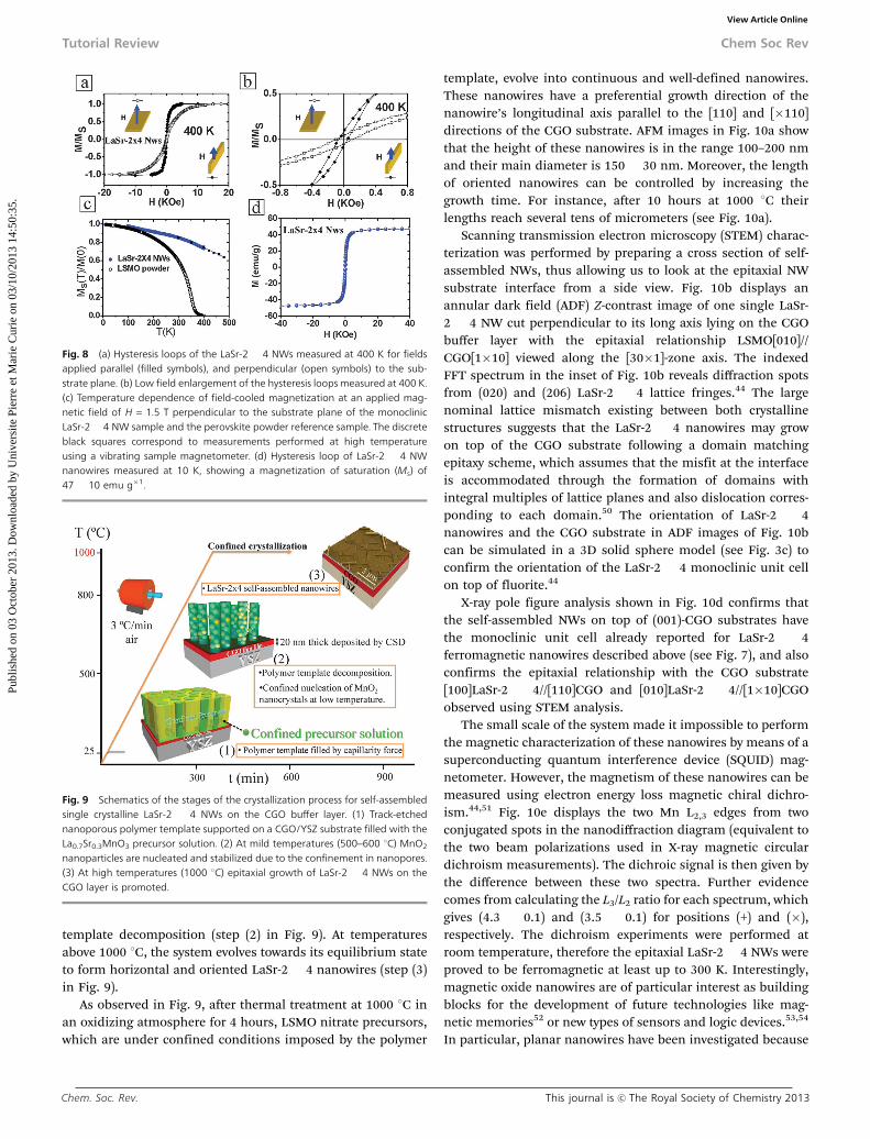
The complex phenomenon displayed by the manganese basedoxides as a function of their structural configuration makes theinvestigation of the magnetic properties of these new mono-clinic LaSr-2 � 4 nanowires (summarized in Fig. 8) highlyappealing. Magnetic hysteresis loops can be measured atdifferent temperatures between 10 K and 500 K for fields upto 5 T. Fig. 8a displays the magnetization curves measured at400 K while Fig. 8b shows magnified images of the central partof the hysteresis loops. As a result, we observe that the nano-wires show a ferromagnetic behavior at a temperature as highas 400 K. In addition, the in-plane magnetization curve leads to asaturated magnetization at a smaller magnetic field than the out-of-plane curve, thus indicating that the direction perpendicular to thelong axis of the nanowires is a hard axis magnetization direction.
Fig. 8c evidences the temperature dependence of the mag-netization, measured at an applied field of 1.5 T, for a poly-crystalline perovskite LSMO sample and the monoclinic LaSr-2�4 nanowires, both prepared from the same chemical precursors.As observed, the magnetization of nanowires decreases moreslowly with temperature even though it remains relatively high at500 K (B40% decrease from 4 K), thus indicating that the Curietemperature of the monoclinic LaSr-2 � 4 nanowires is wellabove 500 K, i.e. much higher than all the well-established valuesreported so far for any perovskite manganite compound. Thisremarkable increase in Tc is clearly an outstanding result thatproves the potentialities of our synthesis route.
To provide more evidence of the ferromagnetic behavior ofthe nanowires, an estimated magnetization saturation value for
Fig. 6 FE-SEM images taken at interrupted intermediate temperature steps during the OMS nanowire formation on Si, together with the schematics of the growthprocess. (a) Cross-sectional view FE-SEM image of the nanoporous polymer template (thickness 7.4 mm, pore size 200 nm) taken before infiltrating the nanopores withthe precursor solution. (b) FE-SEM image taken after the sample was heated up to 600 1C and quenched. Precursor oxide nanorods with the dimensions of the originaltemplate nanopores are formed. At this temperature the polymer template was decomposed. (c) Cross-sectional view FE-SEM image of the nanowires formed on topof the Si substrate after thermal treatment at 800 1C. Low magnification and high magnification (in the insets) images of the final OMS nanowires obtained, i.e. (d) La–Sr–Mn precursors (LaSr-2 � 4 OMS), (e) Ba–Mn-precursors (Ba1+dMn8O16), (f) Sr–Mn-precursors (Sr1+dMn8O16) and (g) Ba–Sr–Mn-precursors ((BaSr)1+dMn8O16).
Tutorial Review Chem Soc Rev
Publ
ishe
d on
03
Oct
ober
201
3. D
ownl
oade
d by
Uni
vers
ite P
ierr
e et
Mar
ie C
urie
on
03/1
0/20
13 1
4:50
:35.
View Article Online
This journal is c The Royal Society of Chemistry 2013 Chem. Soc. Rev.
the LaSr-2 � 4 monoclinic phase at 10 K is 47 � 10 emu g�1 (seeFig. 8d). Note that this value is of the same order as the bulkLSMO perovskite saturation magnetization (90 emu g�1), there-fore indicating the high ferromagnetic strength of this newstructure. The origin of the observed high temperature ferro-magnetism of these LaSr-2 � 4 nanowires could lie on theparticular atomic arrangement of the manganese atoms in themonoclinic structure. Indeed, the resulting interatomic dis-tances in this structure may modify the exchange interactionof Mn3+ and Mn4+ cations, stabilizing a ferromagnetic configu-ration, and enhancing ferromagnetism.43
3.5 Oriented in-plane LaSr-2 � 4 nanowires on a fluorite typesubstrate
In the last few years, special interest focused on self-assembledoxide nanostructures has emerged. The tendency observed in
some oxides to grow arrays of uniform nanostructures givesgreat potential for the implementation of new devices. How-ever, good control over the growth direction of heteroepitaxialnanowires has been a challenge so far.27 In this framework,track-etched polymer templates can be used for the self-assemblyof nanostructures when single crystalline substrates with fluoritestructure are used.44
The growth procedure is schematically described in Fig. 9. Inthis case, track etched polymer templates with a thickness of2 mm are deposited by spin coating on top of a Gd doped-CeO2
(CGO) buffer layer. Note that CGO thin films are 20 nm thickand are chemically deposited on top of (001)-(YSZ) substrates(see Fig. 9). First, nanopores are filled by capillary force with theprecursor solution (step (1) in Fig. 9). Then, at a temperaturebetween 500 1C and 600 1C, the confined nucleation of e-MnO2
nanoparticles is attained, simultaneously with the polymer
Fig. 7 (a) Low magnification FEG-SEM image of LaSr-2� 4 NWs epitaxially grown on an a-quartz/Si substrate. The insets show an enlarged view of the LaSr-2� 4 nanowires.(b) Low magnification HAADF image of LaSr-2� 4 NWs epitaxially grown on an a-quartz/Si substrate (800 1C for 5 hours). (c) HRTEM image of the interface between the quartzlayer and epitaxial LaSr-2� 4 NWs, viewed along [010]. The insets show the FFT of both phases showing the epitaxial relationship between the LaSr-2� 4 NWs and the a-quartzaccording to [20�2] LaSr-2� 4//[�101] a-quartz. (d) Profile matching refinement of the X-ray diffraction pattern of single crystalline NWs on the silicon substrate. Experimentaldata: red points; calculated: continuous black line; allowed Bragg reflections: vertical green marks. The difference between the observed and calculated profiles is displayed at thebottom (blue line). The inset shows the model structure proposed for the LaSr-2� 4 NWs. (e) ADF high resolution image of LaSr-2� 4 NWs along the [100] zone axis. A LaSr-2�4 NW cell model is highlighted on ADF images, with yellow circles displaying the Sr column position, blue the La column position, and red and green circles the O and Mnpositions, respectively. (f) EELS line scan along the [010] direction of the monoclinic unit cell (white arrow) in (e). EELS spectra with positions of O K, Mn L2,3 and La M4,5 edgeshighlighted. The image shows the characteristics of EELS atomic resolution from top to bottom. In white, the ADF signal acquired simultaneously with the EELS line scan. In red,the O K image obtained by integrating the intensity under the O K edge after background subtraction. In blue, the La M4,5 image, and in green Mn the L2,3 image.
Chem Soc Rev Tutorial Review
Publ
ishe
d on
03
Oct
ober
201
3. D
ownl
oade
d by
Uni
vers
ite P
ierr
e et
Mar
ie C
urie
on
03/1
0/20
13 1
4:50
:35.
View Article Online
Chem. Soc. Rev. This journal is c The Royal Society of Chemistry 2013
template decomposition (step (2) in Fig. 9). At temperaturesabove 1000 1C, the system evolves towards its equilibrium stateto form horizontal and oriented LaSr-2 � 4 nanowires (step (3)in Fig. 9).
As observed in Fig. 9, after thermal treatment at 1000 1C inan oxidizing atmosphere for 4 hours, LSMO nitrate precursors,which are under confined conditions imposed by the polymer
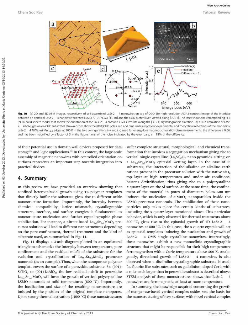
template, evolve into continuous and well-defined nanowires.These nanowires have a preferential growth direction of thenanowire’s longitudinal axis parallel to the [110] and [�110]directions of the CGO substrate. AFM images in Fig. 10a showthat the height of these nanowires is in the range 100–200 nmand their main diameter is 150 � 30 nm. Moreover, the lengthof oriented nanowires can be controlled by increasing thegrowth time. For instance, after 10 hours at 1000 1C theirlengths reach several tens of micrometers (see Fig. 10a).
Scanning transmission electron microscopy (STEM) charac-terization was performed by preparing a cross section of self-assembled NWs, thus allowing us to look at the epitaxial NWsubstrate interface from a side view. Fig. 10b displays anannular dark field (ADF) Z-contrast image of one single LaSr-2 � 4 NW cut perpendicular to its long axis lying on the CGObuffer layer with the epitaxial relationship LSMO[010]//CGO[1�10] viewed along the [30�1]-zone axis. The indexedFFT spectrum in the inset of Fig. 10b reveals diffraction spotsfrom (020) and (206) LaSr-2 � 4 lattice fringes.44 The largenominal lattice mismatch existing between both crystallinestructures suggests that the LaSr-2 � 4 nanowires may growon top of the CGO substrate following a domain matchingepitaxy scheme, which assumes that the misfit at the interfaceis accommodated through the formation of domains withintegral multiples of lattice planes and also dislocation corres-ponding to each domain.50 The orientation of LaSr-2 � 4nanowires and the CGO substrate in ADF images of Fig. 10bcan be simulated in a 3D solid sphere model (see Fig. 3c) toconfirm the orientation of the LaSr-2 � 4 monoclinic unit cellon top of fluorite.44
X-ray pole figure analysis shown in Fig. 10d confirms thatthe self-assembled NWs on top of (001)-CGO substrates havethe monoclinic unit cell already reported for LaSr-2 � 4ferromagnetic nanowires described above (see Fig. 7), and alsoconfirms the epitaxial relationship with the CGO substrate[100]LaSr-2 � 4//[110]CGO and [010]LaSr-2 � 4//[1�10]CGOobserved using STEM analysis.
The small scale of the system made it impossible to performthe magnetic characterization of these nanowires by means of asuperconducting quantum interference device (SQUID) mag-netometer. However, the magnetism of these nanowires can bemeasured using electron energy loss magnetic chiral dichro-ism.44,51 Fig. 10e displays the two Mn L2,3 edges from twoconjugated spots in the nanodiffraction diagram (equivalent tothe two beam polarizations used in X-ray magnetic circulardichroism measurements). The dichroic signal is then given bythe difference between these two spectra. Further evidencecomes from calculating the L3/L2 ratio for each spectrum, whichgives (4.3 � 0.1) and (3.5 � 0.1) for positions (+) and (�),respectively. The dichroism experiments were performed atroom temperature, therefore the epitaxial LaSr-2 � 4 NWs wereproved to be ferromagnetic at least up to 300 K. Interestingly,magnetic oxide nanowires are of particular interest as buildingblocks for the development of future technologies like mag-netic memories52 or new types of sensors and logic devices.53,54
In particular, planar nanowires have been investigated because
Fig. 8 (a) Hysteresis loops of the LaSr-2 � 4 NWs measured at 400 K for fieldsapplied parallel (filled symbols), and perpendicular (open symbols) to the sub-strate plane. (b) Low field enlargement of the hysteresis loops measured at 400 K.(c) Temperature dependence of field-cooled magnetization at an applied mag-netic field of H = 1.5 T perpendicular to the substrate plane of the monoclinicLaSr-2 � 4 NW sample and the perovskite powder reference sample. The discreteblack squares correspond to measurements performed at high temperatureusing a vibrating sample magnetometer. (d) Hysteresis loop of LaSr-2 � 4 NWnanowires measured at 10 K, showing a magnetization of saturation (Ms) of47 � 10 emu g�1.
Fig. 9 Schematics of the stages of the crystallization process for self-assembledsingle crystalline LaSr-2 � 4 NWs on the CGO buffer layer. (1) Track-etchednanoporous polymer template supported on a CGO/YSZ substrate filled with theLa0.7Sr0.3MnO3 precursor solution. (2) At mild temperatures (500–600 1C) MnO2
nanoparticles are nucleated and stabilized due to the confinement in nanopores.(3) At high temperatures (1000 1C) epitaxial growth of LaSr-2 � 4 NWs on theCGO layer is promoted.
Tutorial Review Chem Soc Rev
Publ
ishe
d on
03
Oct
ober
201
3. D
ownl
oade
d by
Uni
vers
ite P
ierr
e et
Mar
ie C
urie
on
03/1
0/20
13 1
4:50
:35.
View Article Online
This journal is c The Royal Society of Chemistry 2013 Chem. Soc. Rev.
of their potential use in domain wall devices proposed for datastorage52 and logic applications.55 In this context, the large-scaleassembly of magnetic nanowires with controlled orientation onsurfaces represents an important step towards integration intopractical devices.
4. Summary
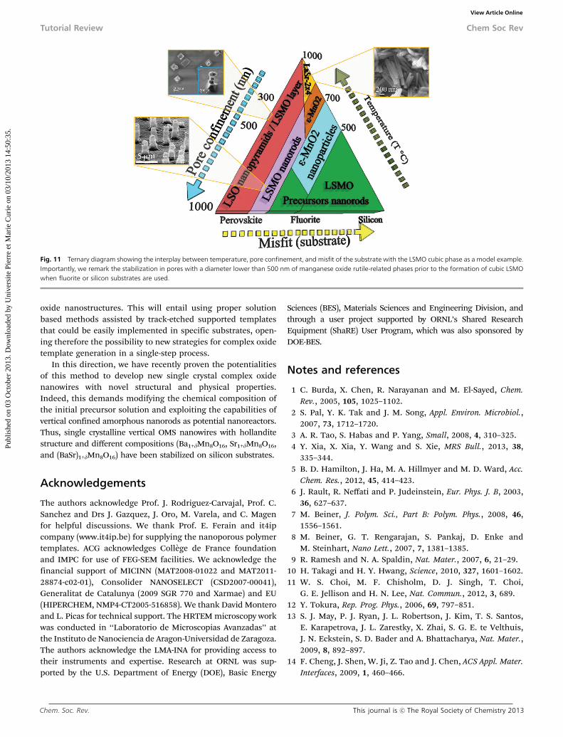
In this review we have provided an overview showing thatconfined heteroepitaxial growth using TE polymer templatesthat directly buffer the substrate gives rise to different oxidenanostructure formation. Importantly, the interplay betweenchemical compatibility, lattice mismatch, crystallographicstructure, interface, and surface energies is fundamental tonanostructure nucleation and further crystallographic phasestabilization. For instance, a nitrate based La0.7Sr0.3MnO3 pre-cursor solution will lead to different nanostructures dependingon the pore confinement, thermal treatment and the kind ofsubstrate used, as summarized in Fig. 11.
Fig. 11 displays a 3-axis diagram plotted in an equilateraltriangle to schematize the interplay between temperature, poreconfinement and the residual misfit of the substrate for theevolution and crystallization of La0.7Sr0.3MnO3 precursornanorods (as an example). Thus, when the nanoporous polymertemplate covers the surface of a perovskite substrate, i.e. (001)-SrTiO3 or (001)-LaAlO3, the low residual misfit to perovskiteLa0.7Sr0.3MnO3 will favor the growth of vertical polycrystallineLSMO nanorods at mild temperatures (800 1C). Importantly,the localization and size of the resulting nanostructure areinduced by the position of the original template nanopores.Upon strong thermal activation (1000 1C) these nanostructures
suffer complete structural, morphological, and chemical trans-formation that involves a segregation mechanism giving rise tovertical single-crystalline (La,Sr)xOy nano-pyramids sitting ona La0.7Sr0.3MnO3 epitaxial wetting layer. In the case of Sisubstrates, the interaction of the alkaline or alkaline earthcations present in the precursor solution with the native SiO2
top layer at high temperatures and under air conditions,hastens devitrification, thus giving rise to a polycrystallinea-quartz layer on the Si surface. At the same time, the confine-ment of the material in pores of diameters below 500 nminduces the nucleation of e-MnO2 nanoparticles inside theLSMO precursor nanorods. The stabilization of these nano-particles only takes place for certain kinds of substrates,including the a-quartz layer mentioned above. This particularbehavior, which is only observed for thermal treatments above500 1C, gives rise to the epitaxial growth of 1D LaSr-2 � 4nanowires at 800 1C. In this case, the a-quartz crystals will actas epitaxial templates inducing the nucleation and growth ofLaSr-2 � 4 OMS single crystalline nanowires. Interestingly,these nanowires exhibit a new monoclinic crystallographicstructure that might be responsible for their high temperatureferromagnetism with a Curie temperature above 500 K. Analo-gously, directional growth of LaSr-2 � 4 nanowires is alsoobserved when a dissimilar crystallographic substrate is used,i.e. fluorite like substrates such as gadolinium doped Ceria witha mismatch larger than in perovskite substrates described above.STEM analysis of these nanostructures shows that LaSr-2 � 4nanowires are ferromagnetic, at least at room temperature.
In summary, the knowledge acquired concerning the growthof manganese-based vertical complex oxides sets the basis forthe nanostructuring of new surfaces with novel vertical complex
Fig. 10 (a) 2D and 3D AFM images, respectively, of self-assembled LaSr-2 � 4 nanowires on top of CGO. (b) High resolution ADF Z-contrast image of the interfacebetween an epitaxial LaSr-2 � 4 nanowire oriented LSMO [010]//CGO [1�10] and the CGO buffer layer, viewed along [30�1]. The inset shows the corresponding FFT.(c) 3D solid sphere model that shows the orientation of the LaSr-2 � 4 NW and CGO substrate along the [30�1] crystallographic direction. (d) XRD2 simulation of LaSr-2 � 4 NWs grown on CGO substrates. Brown circles show the {001}CGO poles, red and blue circles represent experimental and theoretical reflections of the monoclinicLaSr-2 � 4 NWs. (e) Mn L2,3 edges at 300 K in the two configurations (+) and (�) used for energy-loss magnetic chiral dichroism measurements, the difference is 0.06,and has been magnified by a factor of 3 in the figure. r.m.s. of the noise, indicated by the error bars, is �15% of the difference.
Chem Soc Rev Tutorial Review
Publ
ishe
d on
03
Oct
ober
201
3. D
ownl
oade
d by
Uni
vers
ite P
ierr
e et
Mar
ie C
urie
on
03/1
0/20
13 1
4:50
:35.
View Article Online
Chem. Soc. Rev. This journal is c The Royal Society of Chemistry 2013
oxide nanostructures. This will entail using proper solutionbased methods assisted by track-etched supported templatesthat could be easily implemented in specific substrates, open-ing therefore the possibility to new strategies for complex oxidetemplate generation in a single-step process.
In this direction, we have recently proven the potentialitiesof this method to develop new single crystal complex oxidenanowires with novel structural and physical properties.Indeed, this demands modifying the chemical composition ofthe initial precursor solution and exploiting the capabilities ofvertical confined amorphous nanorods as potential nanoreactors.Thus, single crystalline vertical OMS nanowires with hollanditestructure and different compositions (Ba1+dMn8O16, Sr1+dMn8O16,and (BaSr)1+dMn8O16) have been stabilized on silicon substrates.
Acknowledgements
The authors acknowledge Prof. J. Rodriguez-Carvajal, Prof. C.Sanchez and Drs J. Gazquez, J. Oro, M. Varela, and C. Magenfor helpful discussions. We thank Prof. E. Ferain and it4ipcompany (www.it4ip.be) for supplying the nanoporous polymertemplates. ACG acknowledges College de France foundationand IMPC for use of FEG-SEM facilities. We acknowledge thefinancial support of MICINN (MAT2008-01022 and MAT2011-28874-c02-01), Consolider NANOSELECT (CSD2007-00041),Generalitat de Catalunya (2009 SGR 770 and Xarmae) and EU(HIPERCHEM, NMP4-CT2005-516858). We thank David Monteroand L. Picas for technical support. The HRTEM microscopy workwas conducted in ‘‘Laboratorio de Microscopias Avanzadas’’ atthe Instituto de Nanociencia de Aragon-Universidad de Zaragoza.The authors acknowledge the LMA-INA for providing access totheir instruments and expertise. Research at ORNL was sup-ported by the U.S. Department of Energy (DOE), Basic Energy
Sciences (BES), Materials Sciences and Engineering Division, andthrough a user project supported by ORNL’s Shared ResearchEquipment (ShaRE) User Program, which was also sponsored byDOE-BES.
Notes and references
1 C. Burda, X. Chen, R. Narayanan and M. El-Sayed, Chem.Rev., 2005, 105, 1025–1102.
2 S. Pal, Y. K. Tak and J. M. Song, Appl. Environ. Microbiol.,2007, 73, 1712–1720.
3 A. R. Tao, S. Habas and P. Yang, Small, 2008, 4, 310–325.4 Y. Xia, X. Xia, Y. Wang and S. Xie, MRS Bull., 2013, 38,
335–344.5 B. D. Hamilton, J. Ha, M. A. Hillmyer and M. D. Ward, Acc.
Chem. Res., 2012, 45, 414–423.6 J. Rault, R. Neffati and P. Judeinstein, Eur. Phys. J. B, 2003,
36, 627–637.7 M. Beiner, J. Polym. Sci., Part B: Polym. Phys., 2008, 46,
1556–1561.8 M. Beiner, G. T. Rengarajan, S. Pankaj, D. Enke and
M. Steinhart, Nano Lett., 2007, 7, 1381–1385.9 R. Ramesh and N. A. Spaldin, Nat. Mater., 2007, 6, 21–29.
10 H. Takagi and H. Y. Hwang, Science, 2010, 327, 1601–1602.11 W. S. Choi, M. F. Chisholm, D. J. Singh, T. Choi,
G. E. Jellison and H. N. Lee, Nat. Commun., 2012, 3, 689.12 Y. Tokura, Rep. Prog. Phys., 2006, 69, 797–851.13 S. J. May, P. J. Ryan, J. L. Robertson, J. Kim, T. S. Santos,
E. Karapetrova, J. L. Zarestky, X. Zhai, S. G. E. te Velthuis,J. N. Eckstein, S. D. Bader and A. Bhattacharya, Nat. Mater.,2009, 8, 892–897.
14 F. Cheng, J. Shen, W. Ji, Z. Tao and J. Chen, ACS Appl. Mater.Interfaces, 2009, 1, 460–466.
Fig. 11 Ternary diagram showing the interplay between temperature, pore confinement, and misfit of the substrate with the LSMO cubic phase as a model example.Importantly, we remark the stabilization in pores with a diameter lower than 500 nm of manganese oxide rutile-related phases prior to the formation of cubic LSMOwhen fluorite or silicon substrates are used.
Tutorial Review Chem Soc Rev
Publ
ishe
d on
03
Oct
ober
201
3. D
ownl
oade
d by
Uni
vers
ite P
ierr
e et
Mar
ie C
urie
on
03/1
0/20
13 1
4:50
:35.
View Article Online

This journal is c The Royal Society of Chemistry 2013 Chem. Soc. Rev.
15 Z. Chen, Z. Jiao, D. Pan, Z. Li, M. Wu, C. Shek, C. M. L. Wuand J. K. L. Lai, Chem. Rev., 2012, 112, 3833–3855.
16 F. Borgatti, C. Park, A. Herpers, F. Offi, R. Egoavil,Y. Yamashita, A. Yang, M. Kobata, K. Kobayashi,J. Verbeeck, G. Panaccione and R. Dittmann, Nanoscale,2013, 5, 3954–3960.
17 R. J. Choudhary, A. S. Ogale, S. R. Shinde, S. Hullavarad,S. B. Ogale, T. Venkatesan, R. N. Bathe, S. I. Patil andR. Kumar, Appl. Phys. Lett., 2004, 84, 3846–3848.
18 S. Dong, F. Gao, Z. Q. Wang, J. M. Liu and Z. F. Ren, Appl.Phys. Lett., 2007, 90, 082508.
19 S. Singh and S. B. Krupanidhi, Dalton Trans., 2008, 4708–4710.20 S. L. Suib, Acc. Chem. Res., 2008, 41, 479–487.21 F. Cheng, J. Chen, X. Gou and P. Shen, Adv. Mater., 2005, 17,
2753–2756.22 O. Ghodbane, J. Pascal and F. Favier, ACS Appl. Mater.
Interfaces, 2009, 1, 1130–1139.23 W. N. Li, J. Yuan, X. Shen, S. Gomez-Mower, L. Xu,
S. Sithambaram, M. Aindow and S. Suib, Adv. Funct. Mater.,2006, 16, 1247–1253.
24 Y. Xia, P. Yang, Y. Sun, Y. Wu, B. Mayers, B. Gates, Y. Yin,F. Kim and H. Yan, Adv. Mater., 2003, 15, 353–389.
25 C. M. Lieber and Z. L. Wang, MRS Bull., 2007, 32, 99–108.26 C. N. R. Rao, F. L. Deepak, G. Gundiah and A. Govindaraj,
Prog. Solid State Chem., 2003, 31, 5–147.27 S. A. Fortuna and X. Li, Semicond. Sci. Technol., 2010, 25, 024005.28 A. L. Tiano, C. Koenigsmann, A. C. Santulli and S. S. Wong,
Chem. Commun., 2010, 46, 8093–8130.29 Y. Zhu, L. Zhang, T. Natsuki, Y. Fu and Q. Ni, ACS Appl.
Mater. Interfaces, 2012, 4, 2101–2106.30 J. H. Jung, M. Lee, J. Hong, Y. Ding, C. Chen, L. Chou and
Z. L. Wang, ACS Nano, 2011, 5, 10041–10046.31 Y. Mao, S. Banerjee and S. S. Wong, J. Am. Chem. Soc., 2003,
125, 15718–15719.32 J. J. Urban, W. S. Yun, Q. Gu and H. Park, J. Am. Chem. Soc.,
2002, 124, 1186–1187.33 J. J. Urban, J. E. Spanier, L. Ouyang, W. S. Yun and H. Park,
Adv. Mater., 2003, 15, 423–426.34 H. Li, H. Wu, D. Lin and W. Pan, J. Am. Ceram. Soc., 2009,
92, 2162–2164.35 C. R. Martin, Science, 1994, 266, 1961–1966.36 Y. J. Lee, H. Yi, W. Kim, K. Kang, D. S. Yun, M. S. Strano,
G. Ceder and A. M. Belcher, Science, 2009, 324, 1051–1055.37 L. Vila, P. Vincent, L. Dauginet-De Pra, G. Pirio, E. Minoux,
L. Gangloff, S. Demoustier-Champagne, N. Sarazin,E. Ferain, R. Legras, L. Piraux and P. Legagneux, Nano Lett.,2004, 4, 521–524.
38 L. Piraux, J. M. George, J. F. Despres, C. Leroy, E. Ferain,R. Legras, K. Ounadjela and A. Fert, Appl. Phys. Lett., 1994,65, 2484–2486.
39 A. Carretero-Genevrier, M. Gich, L. Picas, J. Gazquez,G. L. Drisko, C. Boissiere, D. Grosso, J. Rodriguez-Carvajaland C. Sanchez, Science, 2013, 340, 827–831.
40 A. Carretero-Genevrier, J. Oro, J. Gazquez, J. Rodriguez-Carvajal, C. Magen, L. Miranda, E. Ferain, T. Puig,X. Obradors, C. Sanchez and N. Mestres, 2013, submittedfor publication.
41 E. Ferain and R. Legras, Nucl. Instrum. Methods Phys. Res.,Sect. B, 2003, 208, 115–122.
42 A. Carretero-Genevrier, N. Mestres, T. Puig, A. Hassini,J. Oro, A. Pomar, F. Sandiumenge, X. Obradors andE. Ferain, Adv. Mater., 2008, 20, 3672–3677.
43 A. Carretero-Genevrier, J. Gazquez, J. C. Idrobo, J. Oro,J. Arbiol, M. Varela, E. Ferain, J. Rodrıguez-Carvajal,T. Puig, N. Mestres and X. Obradors, J. Am. Chem. Soc.,2011, 133, 4053–4061.
44 A. Carretero-Genevrier, J. Gazquez, C. Magen, M. Varela,E. Ferain, T. Puig, N. Mestres and X. Obradors, Chem.Commun., 2012, 48, 6223–6225.
45 A. Carretero-Genevrier, J. Gazquez, T. Puig, N. Mestres,F. Sandiumenge, X. Obradors and E. Ferain, Adv. Funct.Mater., 2010, 20, 2139–2146.
46 C. Moreno, P. Abellan, A. Hassini, A. Ruyter, A. P. d. Pino,F. Sandiumenge, M. Casanove, J. Santiso, T. Puig andX. Obradors, Adv. Funct. Mater., 2009, 19, 3672–3677.
47 J. Gutierrez, A. Llordes, J. Gazquez, M. Gibert, N. Roma,S. Ricart, A. Pomar, F. Sandiumenge, N. Mestres, T. Puig andX. Obradors, Nat. Mater., 2007, 6, 367–373.
48 N. Meisser, E. A. Perseil, J. Brugger and P.-J. Chiappero, Can.Mineral., 1999, 37, 673.
49 W. Schreyer, A. M. Fransolet and H. J. Bernhardt, Contrib.Mineral. Petrol., 2001, 141, 560.
50 J. Narayan and B. C. Larson, J. Appl. Phys., 2003, 93, 278–285.51 P. Schattschneider, S. Rubino, C. Hebert, J. Rusz, J. Kunes,
P. Novak, E. Carlino, M. Fabrizioli, G. Panaccione andG. Rossi, Nature, 2006, 441, 486–488.
52 M. Hayashi, L. Thomas, R. Moriya, C. Rettner andS. S. P. Parkin, Science, 2008, 320, 209–211.
53 C. H. Kim, Y. Myung, Y. J. Cho, H. S. Kim, S. Park, J. Park,J. Kim and B. Kim, J. Phys. Chem. C, 2009, 113, 7085–7090.
54 K. Goto, H. Tanaka and T. Kawai, Nano Lett., 2009, 9,1962–1966.
55 D. A. Allwood, G. Xiong, C. C. Faulkner, D. Atkinson, D. Petitand R. P. Cowburn, Science, 2005, 309, 1688–1692.
Chem Soc Rev Tutorial Review
Publ
ishe
d on
03
Oct
ober
201
3. D
ownl
oade
d by
Uni
vers
ite P
ierr
e et
Mar
ie C
urie
on
03/1
0/20
13 1
4:50
:35.
View Article Online
Copyright © 2022 FDOKUMEN