Fauna de hidrozoos del Mediterráneo
453
-
Upload
unisalento -
Category
Documents
-
view
0 -
download
0
Transcript of Fauna de hidrozoos del Mediterráneo


FAUNA OF THE MEDITERRANEAN HYDROZOA
J. Bouillon, M.D. Medel, F. Pagès, J.M. Gili, F. Boero
and C. Gravili
Scientia Marina, 68 (Suppl. 2)Institut de Ciències del Mar, CMIMA-CSIC, Barcelona, Spain
sm68s2Aintro 14/10/04 15:19 Página 1

This book is dedicated to the memory of Fabio Cicogna (1926-2004) Fabio was among the founders of the Hydrozoan Society in 1985 and he was its President from 1985 to 1995
MINISTERIODE EDUCACIÓNY CIENCIA
Comisión Interministerial de Ciencia y Tecnología
With the support of:
Consejo Superior de Investigaciones Científicas
sm68s2Aintro 14/10/04 15:19 Página 2

CONTENTS
Foreword . . . . . . . . . . . . . . . . . . . . . . . . . . . . . . . . . . . . . . . . . . . . . . . . . . . . . . . . . . . . . 4
Boillon, J, M.D. Medel, F. Pagès, J.M. Gili, F. Boero and C. Gravili. – Fauna of theMediterranean Hydrozoa . . . . . . . . . . . . . . . . . . . . . . . . . . . . . . . . . . . . . . . . . . . . . . 5-438
Introduction . . . . . . . . . . . . . . . . . . . . . . . . . . . . . . . . . . . . . . . . . . . . . . . . . . . . . . . . 5-15General classification. . . . . . . . . . . . . . . . . . . . . . . . . . . . . . . . . . . . . . . . . . . . . . . . . 15-18General morphology of hidroids, medusae and siphonophores . . . . . . . . . . . . . . . . 18-26Development . . . . . . . . . . . . . . . . . . . . . . . . . . . . . . . . . . . . . . . . . . . . . . . . . . . . . . . 26-29The stinging cells. . . . . . . . . . . . . . . . . . . . . . . . . . . . . . . . . . . . . . . . . . . . . . . . . . . . 29-32Diagnostic characters. . . . . . . . . . . . . . . . . . . . . . . . . . . . . . . . . . . . . . . . . . . . . . . . . 33-36Simplified keys for Hydrozoa subclasses identification . . . . . . . . . . . . . . . . . . . . . . 37Outline classification . . . . . . . . . . . . . . . . . . . . . . . . . . . . . . . . . . . . . . . . . . . . . . . . . 38-39Systematics . . . . . . . . . . . . . . . . . . . . . . . . . . . . . . . . . . . . . . . . . . . . . . . . . . . . . . . . 39-246
Class Hydroidomedusa. . . . . . . . . . . . . . . . . . . . . . . . . . . . . . . . . . . . . . . . . . . . . 39-245Subclass Anthomedusae. . . . . . . . . . . . . . . . . . . . . . . . . . . . . . . . . . . . . . . . . . . 39-114Subclass Laingiomedusae . . . . . . . . . . . . . . . . . . . . . . . . . . . . . . . . . . . . . . . . . 114-115Subclass Leptomedusae . . . . . . . . . . . . . . . . . . . . . . . . . . . . . . . . . . . . . . . . . . . 115-202Subclass Limnomedusae . . . . . . . . . . . . . . . . . . . . . . . . . . . . . . . . . . . . . . . . . . 202-207Subclass Siphonophorae . . . . . . . . . . . . . . . . . . . . . . . . . . . . . . . . . . . . . . . . . . 207-231Subclass Actinulidae . . . . . . . . . . . . . . . . . . . . . . . . . . . . . . . . . . . . . . . . . . . . . 231-232Subclass Narcomedusae. . . . . . . . . . . . . . . . . . . . . . . . . . . . . . . . . . . . . . . . . . . 232-237Subclass Trachymedusae . . . . . . . . . . . . . . . . . . . . . . . . . . . . . . . . . . . . . . . . . . 237-245
Class Polypodiozae . . . . . . . . . . . . . . . . . . . . . . . . . . . . . . . . . . . . . . . . . . . . . . . 245-246Collection of material . . . . . . . . . . . . . . . . . . . . . . . . . . . . . . . . . . . . . . . . . . . . . . . . 246Techniques . . . . . . . . . . . . . . . . . . . . . . . . . . . . . . . . . . . . . . . . . . . . . . . . . . . . . . . . . 247-248Glossary . . . . . . . . . . . . . . . . . . . . . . . . . . . . . . . . . . . . . . . . . . . . . . . . . . . . . . . . . . . 249-264Acknowledgements . . . . . . . . . . . . . . . . . . . . . . . . . . . . . . . . . . . . . . . . . . . . . . . . . . 264References . . . . . . . . . . . . . . . . . . . . . . . . . . . . . . . . . . . . . . . . . . . . . . . . . . . . . . . . . 265-282Figures . . . . . . . . . . . . . . . . . . . . . . . . . . . . . . . . . . . . . . . . . . . . . . . . . . . . . . . . . . . . 283-438
Taxonomic index. . . . . . . . . . . . . . . . . . . . . . . . . . . . . . . . . . . . . . . . . . . . . . . . . . . . . . . 439-449
sm68s2Aintro 14/10/04 15:19 Página 3

Foreword
“The battle delivered against taxonomy has obtained a victory; nowadays the classificationof an organism has become a difficulty.” Ramón Margalef wrote this statement in 1968 in hisbook Principles of Ecological Theory. Since then, the status of taxonomy in the scientific worldhas fallen increasingly. However, it is surprising to observe that many scientific articles are pub-lished—such as those published in emblematic magazines like Science or Nature—and TV doc-umentaries warn us of the importance of biodiversity and how it is seriously threatened by insa-tiable human greed. And it is even more surprising to see that scientific community, totally awareof such threats, ignores or undervalues men of science who are trying to find out how manyspecies live in ecosystems and how to identify and distinguish them correctly. The biased usedof the index of impact is leading the Natural Sciences towards a cul-de-sac: the number of tax-onomists is decreasing at a dramatic rate and as a logical consequence so are the monographs onflora and fauna.
Can you imagine a language that does not update its dictionaries? It will soon be considereda dead language. It seems that ecology is moving in this direction. Our “dictionaries” are becom-ing obsolete and this will ultimately bring us to an ecology with deficits, founded on a taxonomicbasis obtained decades ago.
In this gloomy panorama the work of Bouillon et al. on the Mediterranean Hydrozoans ischarmingly anachronistic in the best sense of the word. It is a splendid work that helps to palli-ate the ignorance about an important but difficult taxonomic group due to the lack of works likethe present monograph. There is no doubt that this work fully accomplishes the difficult missionof being the tiger shot for future studies on the ecology and biology of these interesting organ-isms. This is a thorough and complete work that updates all the taxonomic knowledge of thisbroad group of cnidarians.
Having this work published in the journal Scientia Marina is a great satisfaction for me andmakes me feel very proud. The work follows the journal’s tradition of publishing basic studieson marine fauna and flora, which was initiated with the case Crustáceos Decápodos Ibéricos(1968) by Ricardo Zariquiey-Alvarez and has achieved worldwide recognition. It is a great sat-isfaction to have the work of Bouillon et al. in our hands. Maybe the victory of the battle of tax-onomy is not such a victory.
Prof. Enrique MacphersonCentro de Estudios Avanzados de Blanes (CSIC)
sm68s2Aintro 14/10/04 15:19 Página 4

FAUNA OF THE MEDITERRANEAN HYDROZOA 5
INTRODUCTION
The Mediterranean framework
The Mediterranean Sea is located betweenEurope, Asia, and Africa. Excluding the Black Sea,
it covers 2.542.000 Km2, with an average depthclose to 1.500 m and a maximal depth of 5.121 m inthe Ionian Sea. The Mediterranean Sea is an almostclosed basin, connected with the Atlantic Ocean viathe Strait of Gibraltar, with the Red Sea via the SuezCanal, and with the Black Sea via the Bosphorousand the Dardanelles Straits. Surface temperaturesrange between 11-13°C (with extremes of 4-5°C in
SCI. MAR., 68 (Suppl. 2): 5-438 SCIENTIA MARINA 2004
Fauna of the Mediterranean Hydrozoa*
JEAN BOUILLON1, MARIA DOLORES MEDEL2, FRANCESC PAGÈS3, JOSEP-MARIA GILI3, FERDINANDO BOERO4 and CINZIA GRAVILI4
1 Laboratoire de Biologie Marine, Université Libre de Bruxelles, 50 Ave F. D. Roosevelt, 1050 Bruxelles, Belgium.2 Departamento de Fisiología y Zoología, Facultad de Biologia, Universidad de Sevilla, Reina Mercedes 6,
410121 Sevilla, Spain.3 Institut de Ciències del Mar (CSIC) Passeig Marítim de la Barceloneta 37-49, 08003 Barcelona, Catalonia, Spain.4 Dipartimento di Scienze e Tecnologie Biologiche ed Ambientali, Stazione di Biologia Marina, Università di Lecce,
73100, Lecce, Italy.
SUMMARY: This study provides a systematic account of the hydrozoan species collected up to now in the MediterraneanSea. All species are described, illustrated and information on morphology and distribution is given for all of them. This workis the most complete fauna of hydrozoans made in the Mediterranean. The fauna includes planktonic hydromedusae, benth-ic polyps stages and the siphonophores. The Hydrozoa are taken as an example of inconspicuous taxa whose knowledge hasgreatly progressed in the last decades due to the scientific research of some specialists in the Mediterranean area. The num-ber of species recorded in the Mediterranean almost doubled in the last thirty years and the number of new records is stillincreasing. The 457 species recorded in this study represents the 12% of the world known species. The fauna is completedwith classification keys and a glossary of terms with the main purpose of facilitating the identification of all Meditrraneanhydrozoan species.
Key words: Hydrozoa, hydromedusae, hydropolyps, siphonophores, taxonomy, systematics, fauna, Mediterranean.
RESUMEN: FAUNA DE HIDROZOOS DEL MEDITERRÁNEO. – Este estudio proporciona una relación sistemática de las especiesde hidrozoos conocidas hasta la actualidad en el mar Mediterráneo. Se describen e ilustran todas las especies de las cualesse ofrece información sobre su morfología y distribución. Este trabajo es la fauna mas completa de hidrozoos hecha en elMediterráneo. Esta fauna incluye las hidromedusas planctónicas, los estadios bentónicos de pólipos y los sifonóforos. LosHydrozoa se han tomado como un ejemplo de taxones inconspicuos cuyo conocimiento ha progresado enormemente durantelas últimas décadas gracias a la investigación de algunos especialistas en el area mediterránea. El número de especies citadasen el Mediterráneo se ha casi doblado en los últimos treinta años y el número de nuevos registros sigue incrementándose.Las 457 especies referenciadas en este estudio representan el 12% del total mundial de especies. La fauna se complementacon claves de clasificación y un glosario de términos con el propósito de facilitar la identificación de todas las especies dehidrozoos del Mediterráneo.
Palabras clave: Hydrozoa, hidromedusas, hidropólipos, sifonóforos, taxonomía, sistemática, fauna, Mediterráneo.
*Received June 24, 2004. Accepted July 15, 2004.
sm68s2Aintro 14/10/04 15:19 Página 5

the Gulf of Trieste) in winter and 25-30°C in sum-mer, determining cold temperate to warm-temperateconditions in the cold season, and tropical condi-tions in the warm one. Deep-water temperature isabout 13°C and, normally, this is also the surfacetemperature in winter, when the basin becomeshomoeothermic. Summer thermoclines divide thevariable surface waters from the more stable, deepwaters. In the Mediterranean, evaporation exceedsboth direct rainfall and the water input by rivers that,in general, drain rather arid countries. This balanceexplains the main properties of the sea. Water of rel-atively high salinity, an average of 37 psu, is formedby evaporative concentration and, being moredense, sinks to the bottom and falls across theGibraltar sill into the Atlantic, at a level whereAtlantic water has a lower density. The Mediter-ranean water pouring into the Atlantic sinks until itfinds equal density, travelling northwards, along thePortuguese coast. The loss of deep Mediterraneanwater is over-compensated by a surface Atlantic cur-rent entering through Gibraltar Strait. These waterexchange sums about 1,900 Km3 per year, whereasthe losses by evaporation could be of 1,300 Km3 peryear. This Mediterranean-Atlantic balance avoidsexcessive eutrophication (Margalef, 1985): theMediterranean loses deep water, relatively rich withmineralised or recycled nutrients, and receives low-nutrient surface Atlantic water.
The present-day situation is the result of a com-plex history. The Mediterranean Sea is a remnant ofthe once extensive Tethys Sea, a wedge-shaped,eastward-open equatorial water-body that wasindenting Pangaea during the Triassic (Maldonado,1985). In the Cretaceous, the Atlantic Ocean wasformed and the Tethys connected it to the Indopacif-ic Ocean, harbouring a highly diverse warm-waterbiota. In the Oligocene, a reduction in the surface ofthe Tethys Sea and a decrease of its warming influ-ence on the world oceans caused cold water condi-tions elsewhere. Simultaneously, at the beginning ofthe Miocene (10 Ma), a biota extinction occurredoutside the Tethys (Bianchi and Morri, 2000). Dur-ing the Messinian crisis, about 5.5 Ma, the Mediter-ranean Sea became closed and evaporated almostcompletely, with just a few “pools” of water thatallowed the survival of some paleoendemics, where-as the rest of the local biota went through a massextinction. After the crisis, the present-day biotaoriginated from a contingent that entered in the basalPliocene (5.3 Ma) from the Atlantic Ocean throughthe newly opened Strait of Gibraltar. In spite of
some relicts of the Tethys Sea (the Indopacific-Atlantic connection of ancient times), most Mediter-ranean species are of Atlantic origin, having enteredthrough Gibraltar from either the African or theEuropean portion of the Atlantic. This made theMediterranean biota an Atlantic province from abiogeographic point of view (Briggs, 1974). Thealteration of the ice ages and warm interglacial peri-ods during the Quaternary resulted in differentimmigration waves of Atlantic fauna of boreal orsubtropical origin respectively (Bianchi and Morri,2000). Considering the long and complex Mediter-ranean history, the present high species richness isprobably due to both its evolutionary period throughthe Tertiary and the post-Pliocene diversity pumpfrom the Atlantic Ocean.
Historical reasons are, however, just one side ofthe explanation of the rich Mediterranean biodiver-sity, the other side being strictly ecological: season-ality is the key for the coexistence of a high numberof species (Coma et al. 2000). The same physicalspace, in fact, can widen its potential for life inanother dimension, with the alternation of species intime.
The availability of proper conditions for bothtropical and temperate species makes the Mediter-ranean a perfect sea for biological invasions (Boero,2002). Species of cold affinity are thus pre-adaptedto deep-waters and/or winter temperature condi-tions, whereas species of temperate affinity are pre-adapted to surface and/or summer conditions. MostMediterranean life, in fact, is characterised by asharp seasonality in its rhythms of activity, either interms of actual presence (many species spend theadverse season as dormant or encysted stages) or interms of sexual reproduction (Boero, 1994; Boero etal., 1996; Marcus and Boero, 1998). Biodiversity isnot equally distributed throughout the basin (Arvan-itridis et al., 2002). The Western Mediterranean isricher in species than the Eastern basin. Many expla-nations have been proposed for this pattern, includ-ing a low effort in biodiversity research. An histori-cal-ecological reason might be that the Eastern basinis of greater tropical affinity that the Western basinand that the tropical species entering from Gibraltarfound a temperate barrier that prevented them fromreaching the portion of the basin with proper condi-tions for their establishment. The opening of theSuez Canal, in fact, allowed the entrance of Red Seaspecies that thrive now in the easternmost part(warmer in the winter than the rest of the basin) andcan also widen their geographic range under partic-
6 J. BOUILLON et al.
sm68s2Aintro 14/10/04 15:19 Página 6
ularly favourable conditions. Very few Mediter-ranean species entered the Red Sea, since the morestable physical conditions have selected there a spe-cialised biota that easily out compete the lessfocused Mediterranean species. The establishmentof so many Lessepsian species in the EasternMediterranean led Por (1989) to propose a new bio-geographical province: the Lessepsian Province.This establishment of many new species of tropicalaffinity suggests that the Eastern Mediterranean,after the opening of the Suez Canal, is fulfilling itspotential of being inhabited by tropical species.
In addition to the entrance of new species fromboth Suez and Gibraltar (the Atlantic flow neverstopped), the Mediterranean is experiencing also thetransport of exotic species (Zaitsev and Öztürk,2001) by ships, both in hulls’ fouling and in the bal-last waters of big cargoes. These release their ballastin the harbour of destination, and sail away with aload of goods. This pattern is particularly evident inthe Black Sea, with the traffic of oil tankers thatbrought in ctenophores that impaired the yield offisheries by feeding on fish larvae and on their food(CIESM, 2001, 2002). Another source of biologicalinvasions is aquaculture. In addition, the growingnumber of allochtonous species recorded from thebasin (e.g. the algal Caulerpa spp pool) can be sure-ly due to unwanted (but very effective) human-mediated spreading. The recent natural history ofthe Mediterranean Sea is full of biological inva-sions, many poorly documented but inferable fromthe present observed distribution ranges of manyspecies. After the waters of the Atlantic, however,that colonization process has always been regardedas a natural mechanism within the species’ colo-nization strategies (Gili, 2000). For the past 200years, the introduction of new species can no longerbe regarded as natural, with mankind playing amajor role. This historical process of migrations,colonisations, and invasions has been changing themarine ecosystem in the Mediterranean and hasgiven rise to an underwater landscape that can belikened to a puzzle due to its high degree of hetero-geneity.
A further impulse to the presence of alien speciesof tropical affinity is the undeniable temperatureincrease that characterises the Mediterranean sur-face waters since several years and that is probablydue to a tendency towards global warming. The bio-logical response to this trend is dramatic. Southernspecies are widening their geographical range; trop-ical species that enter the basin establish successful
populations, like the Caulerpa spp., medusae likeRhopilema nomadica (Kideys and Gücü, 1995;Lotan et al., 1992; Avsar, 1999) and many fishspecies. Species of cold water affinity, furthermore,are affected by the deepening of surface thermoclineand warm waters in the summer (Francour et al.,1994). Mass mortalities of gorgonians in the Liguri-an Sea are most probably due to such impacts (Cer-rano et al., 2000).
From all the above, it is evident that the Mediter-ranean biota are at the opposite extreme of Indo-Malayan ones, characterised by an extremely longstability that yielded a very rich biodiversity that, inits turn, was exported to both nearby and distantzones. If seen in the framework of source and sinkecology (Pulliam, 1988), the Indo-Malayan region isa source and the Mediterranean is a sink.
As stressed by Maurer (1999), the extremely richbiodiversity of a given place might be an epi-phe-nomenon due to the immigration of species comingfrom different source populations. Applied to theMediterranean, the problem is: are the newly formedpopulations liable of undergoing a separate evolu-tion in respect to the source ones? Following theallopatric model of speciation, genetic bottlenecksand founder effects should affect the propagulesoriginating new populations, leading to evolutionarynovelties (or to dramatic failures) (Boero, 2002).Actually, the number of Mediterranean endemismsis very high (the most outstanding example is Posi-donia oceanica, the trademark of the MediterraneanSea) and accounts for the originality of the Mediter-ranean biota.
The natural experiment (enhanced by humanactivities) of species immigration is leading to anextraordinary blend of species that makes of theMediterranean the ecological crossroad of the worldocean, with the fastest evolving biota in terms ofboth ecological and evolutionary time.
It is evident, from all this, that the Mediterraneanhas always been changing (both physically and bio-logically) and that the change of today is not the firstrevolution that the basin passed through. It is impor-tant, however, to distinguish human-generatedchanges from changes due to natural (or more glob-al) forces.
The example of Caulerpa is paradigmatic. Itmight be possible, in fact, that C. taxifolia had beenintroduced in the basin by the careless managementof some human enterprise (Meinesz, 1999), but theinvasion by other species of the same genus, andtheir extraordinary success, calls for reconsideration
FAUNA OF THE MEDITERRANEAN HYDROZOA 7
sm68s2Aintro 14/10/04 15:19 Página 7

of the causes of this phenomenon. Exotic Caulerpaspecies coul reach the Mediterranean many times inthe past, but did not find proper conditions fordevelopment, whereas the present situation is con-ducive to their establishment.
The Hydrozoa as an example of inconspicuousgroup
The bulk of biodiversity, in terms of speciesnumbers, is made of poorly known and inconspicu-ous species that, usually, can be noticed only by spe-cialised taxonomists. Taxonomy, however, is disap-pearing from most scientific communities, and ourappreciation of biodiversity is biased towards con-spicuous groups. The Hydrozoa are an inconspicu-ous group whose knowledge greatly progressed inthe last decades due to the presence of some spe-cialists in the Mediterranean area. The number ofspecies recorded from the Mediterranean almostdoubled in thirty years and the number of newrecords is still increasing (Boero et al., 1997). The457 species recorded in this fauna represent 12% ofthe world species described (Bouillon et al., inpress). The ecological role of these animals, espe-cially those represented also by a medusa stage, canbe great due to their general ability to feed upon fisheggs and larvae and/or on the plankton that fish lar-vae feed upon, so acting as potential predatorsand/or competitors of commercial species. Theymight even be keystone predators, depressing poten-tially monopolising fish species, so leaving spacefor less competitive species (Piraino et al., 2002).Neglecting this apparently minor component of bio-diversity might lead to ecological misunderstand-ings that, in their turn, might lead to misleadinginterpretations of the causes affecting the yield offisheries. A worth-asking question is: are conspicu-ous groups sufficient to appreciate marine biodiver-sity and understand its functioning?.
The knowledge of the biogeography of Mediter-ranean Hydrozoa is far from being complete, notonly due to the continuous recording of new speciesin the basin, but also due to insufficient or geo-graphically too concentrated research efforts, soleading to inefficient coverage of distribution areas.
The case of the Hydrozoa shows that we are stillvery far from a proper evaluation of the structure ofbiodiversity, in terms of species presence and distri-bution, and that the understanding of biodiversityfunction, via the identification of species roles, iseven less developed. The lack of specialists in
almost every marine group will rapidly lead to theimpossibility of pursuing this kind of studies, withthe paradox that, in the era of biodiversity, biodiver-sity experts are disappearing, at least from Europe(Boero, 2001).
The Hydrozoa of the Mediterranean Sea
The study of Mediterranean Hydrozoa dates backto the dawn of modern zoology. Several Linnaeanspecies thrive also in the Mediterranean and, in1785, Cavolini reported on Eudendrium racemo-sum, probably the first species described as new toscience from this Sea. Since the 19th Century,oceanographic vessels explored seas and oceans faraway from Europe and few expeditions surveyed theMediterranean. The establishment of the ZoologicalStation of Naples in the same century, however,signed also the dawn of modern marine biology:people from all over the world came to the Mediter-ranean to study marine biology on a residentialbasis. Biological oceanography and marine biologystemmed from completely different approaches tomarine science (oceanographic vessels Vs marinestations) but had the same original aim: the explo-ration of marine biodiversity. Oceanographersfocused on distant parts of the world and/or to deepwaters, whereas marine biologists focused oncoastal areas that were particularly rich in biodiver-sity. Along with Naples, another possible locationfor a marine station was Messina, due to the strongcurrents of its Strait, which brought to the coast theunusual animals of deep waters. Metchnikoff(1886a, b) and Kölliker (1853a, b) worked exten-sively at Messina and gave important contributionsto the knowledge of the Hydrozoa. The founders ofHydrozoan Zoology, with very few exceptions, werefrom Northern European Countries characterised byhostile climates and relatively poor faunas. Theysoon started to move towards the warmer and richerMediterranean to perform their studies on livingorganisms. The pattern of activity of these non-resi-dential researchers was dictated by climate and bytheir academic obligations. They usually worked intheir home countries during the winter and movedtowards the Mediterranean (mainly Naples) duringthe summer. This tendency marked sharply the typeof Hydrozoan fauna that was going to be describedbecause, as we have seen, the Mediterranean is asharply seasonal sea. Other places where researchon marine life became prominent were located in theAustro-Hungarian Empire at Trieste, Rovinj, Split,
8 J. BOUILLON et al.
sm68s2Aintro 14/10/04 15:19 Página 8

and in France at Villefranche-sur-Mer, Endoumeand Banyuls.
A very particular student of marine biology wasRupert Riedl. He went to Naples, and then to manyother parts of the Mediterranean Sea, at the begin-ning of the Fifties and started the exploration ofmarine caves with SCUBA diving techniques. Anew era of marine biology began, with the entranceof the scientist in the environment that was theobject of his studies. Riedl (1959) studied thehydroids of marine caves and of rocky coasts ingeneral, producing one of the first ecologicalpapers on the group.
The new frontier of Hydrozoan studies originat-ed in the first half of the 20th Century, when theBritish school of Hydrozoan zoology, led by F.S.Russell and W.J. Rees, started to work at the build-ing of a single classification for both hydroids andmedusae. They opened a research project that is stillfar from being accomplished: reconstruct the lifecycle of all species so to treat them as a single bio-logical and taxonomical unit. In the Mediterranean,at the Naples Zoological Station, this project was thebasis for a great enterprise that was expected to leadto the Hydrozoan Fauna of the Mediterranean.Bouillon, Brinckmann-Voss, Haeckel, Petersen, Ste-chow, Tardent, Vannucci, Uchida, Yamada and oth-ers amongst the most prominent researchers ofHydrozoa gathered at Naples and worked there formore than a decade. Unfortunately, the project wasaccomplished only in part and Brinckmann-Voss(1970) published just the first of a series of mono-graphs that should have covered the Hydrozoa of theMediterranean Sea. Another great contribution tothe knowledge of Mediterranean Hydrozoa camefrom the work of Picard and a group of his pupilswho, at the Endoume Marine Station, bridged tax-onomy, phylogenetics and ecology. At the Zoologi-cal Station of Villefranche, a long series of papersmainly by Claude Carré and Danièle Carré gave agreat contribution to the knowledge of theSiphonophora and of the Hydromedusae.
In 1985, at the Laboratory of Benthic Ecology ofthe Zoological Station of Naples, at Ischia, theHydrozoan Society was founded, gathering most ofthe active students of the Hydrozoa from all over theworld (Bouillon et. al. 1987). This gave new impulseto the study of the Hydrozoa and set the basis for ahost of collaborations that has been increased withjoint work and publications in following meetings ofthe Hydrozoan Society (Bouillon et al., 1992;Piraino et al., 1996; Mills et al., 2000).
Origins of Mediterranean Hydromedusae: thecase of the fauna of marine canyons
The present Mediterranean hydrozoan fauna iscomposed of species referable to several biogeo-graphic categories (Boero and Bouillon, 1993):
- Circumtropical species form the most abundantgroup with a temperate Atlantic-Mediterraneanbackground.
- Cosmopolitan species, including panoceanicspecies occurring from the Polar seas to the Equator;
- Boreal Atlantic species, remnants of the ice-ages, especially of the Würm glacial.
- Tropical-Atlantic species which include inter-glacial remnants especially from the Tyrrhenianstage.
- Mediterranean-Atlantic species with easternAtlantic migrants, specially into the Alborán Sea.
- Indo-Pacific species. - Endemic species comprising both paleoen-
demics, of Tethyan origin, and neoendemics, main-ly of Pliocene origin.
The origins, and the originality, of the faunainhabiting the depths of the Mediterranean Sea aredebated. The temperature there is constantly 13°C,thus higher than that of the Atlantic, from where mostof the Mediterranean fauna originated. The Gibraltarsill, moreover, is too shallow to permit the entrance ofbathypelagic species in the Mediterranean after theMessinian crisis. Recent studies on Mediterraneansubmarine canyons provide important clues aboutsome aspects of the origin of deep-sea biodiversity inthe basin. Marine canyons are channels for the trans-port of large quantities of organic matter from thecoastal region to the deep-sea via downwelling cur-rents (Vetter and Dayton, 1998). They are systemswith high production rates and hence locations capa-ble of attaining high faunal richness. Submarinecanyons, moreover, may strongly contribute to thebiological production processes in many coastalregions via their upwelling currents (Della Tommasaet al., 2000). Furthermore, they may also play animportant role in speciation processes affecting thedeep-water fauna, as observed in the Foix, Lacaze-Duthiers, and Planier canyons, located in the westernMediterranean, where medusa species richness ishigh, with seven new species being found recently(38.8% of the total) (Gili et al., 1998; Gili et al.,1999). This discovery raised the level of endemism inthe deep-sea Mediterranean medusan fauna to 24%.The species were collected by sediment traps locatedabove the seabed at the 1000 m isobath in the
FAUNA OF THE MEDITERRANEAN HYDROZOA 9
sm68s2Aintro 14/10/04 15:19 Página 9
canyons, a region inaccessible to conventional sam-pling methods.
The most important aspect of the discovery is therelationship between the number of individuals ofendemic species and the ecological features of thesubmarine canyons. In the canyons, the number ofindividuals increased progressively as the total flux oforganic matter reaching the interior of the canyonsincreased. In addition, canyons with close-togetherwalls might increase isolation while attenuatinghydrodynamic processes and are thus home to largernumbers of individuals. Furthermore, almost all thenew species were endemic to one of the canyons, sug-gesting a high level of isolation inside. These canyonshad ancestor paleocourses in the Messinian period (5Ma ago), when the Mediterranean dried up nearlycompletely. Still, some areas, such as the deepest partof these canyons, were not completely dried up sinceMessinian evaporated deposits accumulated there(Hsü et al., 1978). This indicates that the deepest partof these canyons remained submerged during theMessinian times. The two new species of the genusFoersteria described in the Mediterranean (F. antoni-ae in the Lacaze-Duthiers Canyon and F. araiae in theFoix Canyon) (Gili et al., 2000) have two congenericspecies in the Pacific (F. purpurea) and Indian Oceans(F. bruuni). Probably, the common ancestor of boththe Mediterranean species and the oceanic onesinhabited the Tethys Sea. That ancestral speciesunderwent a process of allopatric speciation bothworldwide, when the drying up of the Tethys Sea sep-arated the Atlantic and the Pacific basins, and at alocal scale, when the parallel canyons became sepa-rated as well. Indeed, the ecological differencesbetween the canyons are probably responsible forgenerating the allopatric speciation found there.
The discovery of this singular medusan fauna isa key to investigate the origin of the Mediterraneandeep-sea fauna. It also calls for the protection ofmarine biodiversity. Mediterranean submarinecanyons incise the continental shelf and approachvery close inshore. Mediterranean coastal regionsare overpopulated and cause heavy environmentalimpacts that may be channelled through the subma-rine canyons and, consequently, that fauna is threat-ened and could disappear entirely even before itbecomes fully known.
Lists of Mediterranean Hydrozoa
Picard (1958b) made a complete list of Mediter-ranean Antho- and Leptomedusae, summing up 191
species, to set the basis for a never to be made Faunede France volume on the group. Boero and Bouillon(1993) updated the work of Picard, treating all theHydrozoa besides the Siphonophores, bringing thelist to 346 records. Boero et al. (1997) further updat-ed the list to 379 species. To these, the Siphonopho-ra have to be added. Boero and Bouillon (1993)treated the life cycle patterns of the Hydrozoa andtheir bearing on the distribution of these organisms.They also defined zoogeographical regions havingthe Mediterranean as their centre, and made ananalysis of the affinities of the fauna. These topicswill not be treated further here.
A complete bibliographic database on the Hydro-zoa is on line, and can be found at the followingaddress: http://siba2.unile.it/ctle/hydro/index.php3.
The records
All species reported at least once from theMediterranean are part of the list. Some records areold and need reconfirmation, but they were kept tohelp future workers that might find them again. Asit happens for all groups, also in the Hydrozoa thereare common and rare species. The history ofrecords, however, indicates that the species contin-gent of the Mediterranean is going through greatchanges and that species that were not recorded inthe past, presumably due to their rarity, are nowcommon, whereas species that have been found inthe past are not being reported anymore. Speciesrecords can be divided into the following categories:
a) “Evident” common species. These species areknown since a long time and can be found in almostevery sample from a proper environmental setting.Their list comprises popular species such as Euden-drium racemosum, E. glomeratum, Aglaopheniaspp., Clytia hemisphaerica, Obelia dichotoma, O.geniculata, Dynamena disticha, Sertularella gau-dichaudi and S. crassicaulis. The set of speciesinhabiting Posidonia leaves are part of this group,even though one of them, a Sertularella, is still tobe described formally. Some common species, evenof large size, can have been misidentified in thepast, so that their names might be rather obscure.The most striking case is possibly Eudendriumarmatum, a large species that can be invariablyfound on the roofs at the entrance of caves andcrevices. Its general aspect resembles that ofEudendrium rameum and is possibly often reportedwith this name. Riedl (1959), in his monograph onthe hydroids of Mediterranean caves, did not men-
10 J. BOUILLON et al.
sm68s2Aintro 14/10/04 15:19 Página 10

tion it, but recorded repeatedly E. rameum, evenfiguring it in his popular guides of the Adriatic andthe Mediterranean flora and fauna. Confirmedrecords of Eudendrium rameum are very rare,whereas E. armatum is extremely common. Speciesvery common in plankton communities alongcoastal waters mainly in spring-summer are themedusae Aglaura hemistoma and Rhopalonemavelatum, and the siphonophore Muggiaea atlantica(e.g. Gili et al., 1988). Species more common inopen waters, throughout the water column includ-ing deep layers are Solmissus albescens, Sol-mundella bitentaculata and the siphonophores Che-lophyes appendiculata, Lensia conoidea and Aby-lopsis tetragona (Mills et al., 1996).
b) Cryptic species now collected more frequent-ly due to increased sampling efficiency. Halocoryneepizoica, a species described by Hadzi in 1917 fromthe Adriatic Sea, was not found again until Picardreported it from the Ligurian Sea, fifty years later(see Piraino et al., 1992). The species was anywayso “rare” that Brinckmann-Voss (1970) could notfind it while working at her monograph on capitatehydroids and medusae. Halocoryne epizoica isstrictly symbiotic with the bryozoan Schizoporellasanguinea, a very abundant species in sea urchinbarrens, at low depth. This type of environment iseasily reachable by SCUBA diving techniques, butis not easy to sample from the surface with grabsand dredges. The yield of samplings, thus, can bebiased by the employed techniques. This means thatH. epizoica might have been common even in thepast but that it passed unnoticed due to sampling dif-ficulties. The same is true for Cytaeis schneideri(formerly referred to as Perarella schneideri),another specialist for symbiosis with bryozoans.Another example is the medusa Ptychogastria aster-oides, previously found in two places in theMediterranean but nowadays caught frequently bysediment traps located near the bottom of submarinecanyons (Gili et al., 1999). The first casual recordsprobably could indicate intrusion of waters from thedeep sea to the continental shelf.
c) Species that are alternately rare and common.Paracoryne huvei became frequent and abundant fora relatively narrow time window. It cannot bemissed or mistaken with anything else, since it livesin the infralittoral zone and is the only knownMediterranean hydroid that settles higher above themean sea level. It forms large pink patches on blackmussels and on bare rocks that can be seen whilewalking along the shore. Its polymorphic colonies
are so different from those of any other species thatPicard (1957), describing it as a new species,ascribed it to a new genus and a new family! It isalmost impossible that such a species passed unno-ticed to all the people who worked earlier onMediterranean hydroids, if not for its extreme rarity.In the Seventies and Eighties Paracoryne huveibecame common along the northern coasts of theMediterranean and was repeatedly reported. Then itdisappeared again and went back to rarity. The pres-ence of resting stages in its cycle can be the reasonwhy the species is able to disappear and reappear.Extra long diapause might be rather widespread inmost species since all hydroids can become dormantwhile regressing to resting hydrorhizae. This patternof presence makes it possible to be active during thefavourable season, but might be also the way theseanimals can disappear for decades and be suddenlyback again. This pattern is even more evident fortheir medusae! The second example refers to aspecies that was formerly very abundant and is nowapparently rarer: Ectopleura crocea (=Tubulariacrocea). Pierre Tardent worked extensively on pop-ulations of this species living in the Gulf of Naples.He chose it as experimental animal due to the easeof finding specimens in the harbour on every season.He went back to Naples after thirty years, consider-ing the opportunity of performing more work buthad to give up his project because he could not finda single specimen (personal communication to F.B.).It is possible that the period of commonness is ratherlong, as that of rarity. It is part of the ecology of allgelatinous plankton (and of its benthic counterpart)to be present with massive blooms or even outbreaksand to be absent even for decades thereafter. This ispossible mainly due to the presence of benthicstages that can become encysted, so escaping mostsampling efforts. Some holoplanktonic species fol-lows high inter-annual bloom variation mainly incoastal waters. Over recent decades, man’s expand-ing influence on the oceans has begun to cause realchange and there is reason to think that, in someregions, new blooms of jellyfish are occurring inresponse to some of the cumulative effects of theseimpacts. Beyond that basic life cycle-driven season-al change in numbers, several other kinds of eventsappear to be increasing the numbers of jellies pre-sent in some ecosystems (Mills, 2001). In theMediterranean, the frequency of jellyfish bloomsincreased during the last decades causing importantproblems in the people that visit beaches duringsummer time. There are several hydrozoan species
FAUNA OF THE MEDITERRANEAN HYDROZOA 11
sm68s2Aintro 14/10/04 15:19 Página 11

generating dense blooms that can be observed nearthe coast (e.g. Lizzia blondina, Velella velella).
d) Species that newly entered the basin. Thestudy of Lessepsian migration from the Red Sea tothe Mediterranean through the Suez Canal originat-ed a wealth of information about Indo-Pacificspecies entering the basin (Por, 1978; Spanier andGalil, 1991). No such records regarding the Hydro-zoa were noticed until recent times. Clytia gravieri,a nominal campanulariid species, however, wasdescribed from specimens coming from the SuezCanal by Billard (1938). Subsequently, it was con-sidered as conspecific with Clytia linearis, a speciesfirst described from the Indo-Pacific. This specieswas first recorded from the Mediterranean in theFifties and might be one of the first (and undetectedas such) Lessepsian migrants. Now it is one of thecommoner hydroids of shallow rocky Mediter-ranean coasts. Due to its branched habit, it is distinctfrom most other Clytia and it is highly improbablethat in the past it was as abundant and frequent as itis now and that it simply passed unnoticed. Hydro-zoan researchers found much more inconspicuousand relatively rare species than this one, so that theyshould have reported it frequently. Being a tropicalspecies, it is probable that the recent tropicalisationof the Mediterranean favoured its success in thebasin. Another way to enter the Mediterranean, ofcourse, is through the Strait of Gibraltar. The studyof the Hydrozoa of the Alborán Sea (Ramil and Ver-voort, 1992; Medel and Vervoort, 1995; Medel etal., 1998) is leading to many new records and this isevidently a hot spot of new entries from the AtlanticOcean. A recent introduction is Clytia hummelincki.It was recorded in the Mediterranean for the firsttime in 1996, along the coasts of Calabria (Boero etal., 1997), almost in the centre of the basin. Beingpresent both in the Atlantic and in the Pacific (withrare records) it is difficult to establish if it enteredfrom Suez or from Gibraltar. At present, this speciesis very successful, forming a belt at 0.5-1 m depth insea urchin barrens along the Apulian coast, where itis extremely common and abundant in the summer.Its distribution in the rest of the basin is unknown,probably because it can be mistaken with species ofthe genus Campanularia and only hydrozoan spe-cialists can identify it properly. In the same period,Bitar and Bitar-Kouli (1995) recorded Macro-rhynchia philippina from the coasts of Lebanon,undoubtedly a Lessepsian migrant. The area ofGibraltar still is a place where is easily to detect thepresence of newly recorded species, such as the
siphonophores Forskalia spp., which became morecommon in the entire occidental basin during thelast years (Dallot et al., 1988). Another interestingspecies is the Antarctic anthomedusan Russelliamirabilis recently found in the Alboràn Sea (Pagèset al., 1999) and that arrived to the Mediterraneanprobably through a complex life cycle together withthe transport role of Antarctic Intermediate and Bot-tom waters.
e) Species that are rare since their discovery.Rarity can be either soffusive, when a species is rareat many places but common at least at one, or diffu-sive, when a species is rare everywhere it occurs(Schoener, 1987). The number of records is not sogreat for most of rare species, and it is difficult todistinguish between the two types of rarity for anyof them. Rarity, furthermore, can be geographical orecological. Geographically rare species live inrestricted areas, and are absent in other areas that,nevertheless, have the same features as the areaswhere they thrive. Ecologically rare species havevery strict ecological requirements that are met justat few places; if their particular requirements aremet, however, they are often present. For the Hydro-zoa, furthermore, rarity can be also linked to sea-sonality, since some species can be present over avery short time window and become encysted (usu-ally as hydrorhizae) for the rest of the year. The bestexample of this is Rhysia autumnalis, a species pre-sent only in autumn and that, furthermore, is strict-ly linked to serpulid tubes (Brinckmann, 1965b).Hydractinia inermis, on the contrary, is present fora short period in the spring, and disappears for therest of the year. Information about rare species,though, is often insufficient to assign them to anyparticular category. The only Mediterranean hydro-zoan that is mentioned in red lists is Errina aspera,probably due to its calcified skeleton, its resem-blance to a coral and its restriction to the Strait ofMessina (Zibrowius and Cairns, 1992) where, how-ever, is very abundant, so being an example of sof-fusive rarity. A good example of rare species isCodonorchis octaedrus, described by Haeckel in1879 from the Atlantic coast of France and neverfound again since Boero et al. (1997) reported itshydroid from a cave of the Ionian Sea, reconstruct-ing its life cycle. In this case, however, rarity mightbe an artefact due to inconspicuousness of thehydroid and to the resemblance of the medusa tothose of the genus Amphinema, so that the speciesmight have been passed unnoticed as hydroid andmisidentified as medusa. Some siphonophores
12 J. BOUILLON et al.
sm68s2Aintro 14/10/04 15:19 Página 12

(Rosacea villafrancae Carré, 1969) have not beenrecorded since their description probably becausedifficulties for taxonomic identification.
f) Declining species. This category is very elu-sive and might be included in that one comprisingspecies that are alternately rare and common. Theconcern about species threatening and extinctioninvariably regards popular and conspicuous species,as the above-mentioned Errina aspera. This attitudeis partly linked to the perception of species by thelay people, but has its foundation also in the attitudeof researchers. If it is easy to recognise that a specieshas become abundant, in fact, it is more difficult todemonstrate that it has become rare, since absence isless evident than presence. In other words,researchers tend to stress what they find instead ofwhat they do not find. To recognise decline weshould be able to check the presence of species fromthe published records of all the researchers thatworked on the group, comparing their results. Adeclining species might be one that is not foundsince fifty years, after having been found repeatedlyduring the previous fifty years, taking care of boththe types of sampled environments and seasonality.If Eudendrium rameum is really a Mediterraneanspecies and its records are not simply misidentifica-tions of E. armatum, then this species is probably adeclining one. One example of these decliningspecies is also the siphohonophore Apolemia uvaria(Carré and Carré, 1994). It is important, thus, toreconstruct the history of the study of all species, soto have maps of their records and keep their “health”under control. At present, however, no hydrozoanspecies are sufficiently well known to deserve thestatus of either threatened or endangered. In this lastcase, it is important to mention the species that arecontinuously threatened and eliminated by trawlers.Lytocarpia myriophyllum, for instance, was verycommon on sandy and mud benthic shelf communi-ties in the Mediterranean and is now much rarer thanbefore (Gili et al., 1987).
Seasonality and life cycles
As stressed above, the Mediterranean is amarkedly seasonal sea and its summer and winterfaunas have different zoogeographical affinities andecological requirements. The Hydrozoa, in particu-lar, are mostly seasonal in occurrence and the possi-bility for the polyps to become encysted as restinghydrorhizae contributes to the presence of great dif-ferences in the species that can be found at a given
place according to the season. Boero and Fresi(1986) studied the seasonality of nearly one hundredspecies of Antho- and Leptomedusan polyps at asingle study site of the Ligurian Sea, showing theexistence of two distinct hydroid faunas, character-ising the warm and the cold season respectively.Boero et al. (1986) studied the seasonality of Euden-drium glomeratum, illustrating the sudden growth ofits colonies and their equally sudden decline. Thepattern of growth of most hydroids involves suddencolony appearance, production of gonophores(either fixed or as free medusae) and subsequentdecline. The life of most medusae ranges between afew hours to one month, and the production of plan-ulae, thus, takes place with such a delay frommedusa liberation, whereas the colonies with fixedgonophores usually produce planulae directly. Areasonable expectation is that, after fertilisation andplanula development, the population of hydroidswill increase in number and density; instead, it usu-ally suddenly declines. The obvious explanation isthat either planulae or newly formed colonies areable to encyst and to wait for the followingfavourable season (or even successive ones) to pro-duce noticeable and reproductive colonies.
Colonies, furthermore, can remain asexual forlong times, without producing reproductive bodies(either medusae or fixed gonophores). This wasstressed by Edwards (1973c) who observed thatmedusae of some species could be absent for years,being continuously present as hydroids only.
The gelatinous zooplankton species without ben-thic stages, however, can be sharply seasonal too, sogenerating doubts about their holoplanktonic way oflife. Many plankters, in fact, have inconspicuousresting stages (see Boero et al., 1996; Marcus andBoero, 1998 for reviews) and easily escape detec-tion. Many calanoid copepods, traditionally consid-ered as holoplanktonic, spend the adverse season asbenthic cysts, wrapped in a chitinous sheath. Theperisarc of the Hydrozoa is made of chitin and thepolyps easily form cysts with their hydrorhizae(Bouillon, 1995a).
The list of hydrozoan species from a given area,thus, cannot be the result of a single sample. Boeroand Fresi (1986) reported two main seasons, havingtheir centre in February and July, so that these twomonths might be sufficient to have an almost com-plete species list for a given locality. This, however,is not enough to find reproductive colonies, sincegonophores can be produced for very short periodsor, as we have seen, can be absent for several years,
FAUNA OF THE MEDITERRANEAN HYDROZOA 13
sm68s2Aintro 14/10/04 15:19 Página 13

just as the whole colonies can, remaining present asdormant hydrorhizae. Carefully taken samples oftengive some novelties, even from the most exploredcoast, in terms of new records for the area, or ofreproductive colonies of species with unknown lifecycle, if not of species new to science. Boero et al.(1997), keeping a small substrate fragment in anaquarium, observed, in sequence, the appearance ofZanclea sp., Trichydra sp., Thecocodium brieni,Turritopsis nutricula and Codonorchis octaedrus.Evidently, these species coexisted as hydrorhizae onthat small space and alternated in becoming active,a quite common behaviour for most hydroids.
Zooplankton studies in the northwesternMediterranean showed seasonal changes in abun-dance for many medusae and siphonophores, suchas the more common species, Aglaura hemistomaand Muggiaea atlantica, in consecutive years (Goy,1985). This seasonal variation has been related tofluctuating local hydrographic conditions and pri-mary production peaks (Gili et al., 1987). The max-imum abundance of cnidarians in the plankton isobserved when the water column is homogeneous,or when stratification begins, at the end of spring.Two main peaks have been described, the first one inApril-May, dominated mainly by siphonophores incoastal waters. This siphonophore peak precedes themaximum abundance of medusae, which coincideswith the yearly maximum of all zooplankton in theMediterranean (Ribera d’Alcalá et al., 2004). Themaximum siphonophore density is observed beforethat of the medusae and it could be explainedbecause of the formation of polymorphic colonieswith high growth rates (Purcell, 1982). The secondperiod of siphonophore abundance, partly comingfrom the eudoxids produced during the first period,shows more marked peaks than that of medusae(Gili et al., 1987). The stability of the water columnpromotes the increase of zooplankton populationsand, thus, of the gelatinous carnivore zooplankton,favoured by the availability of potential prey. Eventhe peaks of abundance of cnidarians are dominatedby few species, their distribution and abundance willhave a large influence on the whole community,allowing the presence of less competitive species atthe end of the prevalence of the more abundant ones,such as M. atlantica and A. hemistoma. Seasonalityand abundance are not independent. In general, sig-nificant differences in size among individuals ofmedusae and siphonophores have been observed inthe same sample, but not in different periods of theyear. This leads to explain that the cnidarian plank-
tonic populations are continuously changing andthat seasonal peaks of abundance show an incrementof the total population but do not indicate a clearpattern of seasonal growth. Cnidarians performdaily migrations in the water column, with widerranges in oceanic waters that near the coast. Foralmost all species, these vertical movements areclosely related with the search of food (Mackie etal., 1987). In general, even though hydrodynamicconditions are important in determining spatial andtemporal heterogeneity of cnidarians in the plank-ton, trophic relationships play a key role as well(Arai, 1988).
The “role” of the Hydrozoa
Ecological roles have been taken as a veryimportant issue to emit judgement about the rele-vance of species, so to produce convincing evi-dence about the need for their protection (Pirainoet al., 2002). This attitude, of course, implies thatif a species has no recognised role, it is implicitlyregarded as less important than a renownedspecies. This causes a great bias in the way we per-ceive biodiversity. We are attracted by conspicuousspecies. Our knowledge on the roles of species isso scant that we cannot consider any species asunimportant a priori. Only recently, for instance, itis generally perceived how phytoplankton is moreimportant than (or at least as important as) tropicalrain forests in maintaining the climate of the wholeplanet the way it is (Falkowski, 2002). Diatomsand flagellates are not as impressive as oaks andredwoods, just as copepods and small jellies arenot as impressive as whales and sharks. Marinesystems are based on microscopic life and, thus,are generally undervalued. Most conservation pur-poses are first biased in favour of terrestrial sys-tems and, when the sea is considered, in favour ofbig, charismatic species, usually vertebrates andhigher plants (with the sole exception of corals andsome molluscs). The Hydrozoa are no exceptionand, thus, tend to be considered as “negligible” ifcompared to groups that are more “visible”. Thisattitude is based on a naive way of conceiving ecol-ogy and is simply wrong. The case of Mnemiopsislleydi, for instance, shows that a gelatinous preda-tor can impair larval survival and subsequentrecruitment of commercial fish, greatly impactingon human activities (see Boero and Briand, 2001).This ecological role might be played also byhydromedusae, since many species are of the right
14 J. BOUILLON et al.
sm68s2Aintro 14/10/04 15:19 Página 14
size to predate on fish eggs and larvae and, fur-thermore on their prey, so acting as both predatorsand competitors. The arrival of the already citedClytia hummelincki in the Mediterranean, and itslarge hydroid populations, producing great num-bers of relatively large medusae might play animportant role in the success of fish recruitment, byremoving eggs and larvae from the environment.The impact on biodiversity, however, might evenbe positive, since predation on the larvae of partic-ularly successful fish species might decrease theirabundance so to leave space for the development ofpreviously outcompeted fish species, so enhancingfish diversity.
The difficulties of studying hydroids as part of thecommunities of hard substrates have limited apprecia-tion of their importance in those communities. Most ofthe available knowledge comes from studies of artifi-cial substrata placed in the sea for variable lengths oftime (Boero, 1984; Gili and Hughes, 1995). Hydroidsare often abundant in initial stages of colonization andin the later stages of community development by col-onizing other organisms (Riedl, 1959). Once estab-lished, hydroids may prevent settlement by other col-onizing species (Young and Chia, 1987). Obeliadichotoma inhibited settlement of other invertebrates,partly by eating their larvae (Standing, 1976), and thedense colonies of Hydractinia echinata prevented set-tlement and even overgrowth by other species by suc-cessfully defending the space (Sutherland and Karl-son, 1977). The accumulation of sediment among andbeneath the hydrocauli of Tubularia larynx may pre-vent settlement by the larvae of other species (Östman,1977). Conversely, T. larynx may facilitate colonisa-tion by other species, such as ascidians (Schmidt,1983). While studies on artificial substrata may pro-vide some idea of seasonal variations in species abun-dance within an area, it may be that only a small num-ber of species colonise artificial substrata (Millard,1959). In addition to their economic nuisance as mem-bers of the fouling community (Morri and Boero,1986), hydroids may pose specific problems, forexample in self-contained aquaculture systems, wherethey endure by developing resting stages capable oftolerating substantial changes in temperature and dry-ing conditions (Sandifer and Smith 1979).
The hydroid polyps, furthermore, are also veryimportant in the passage of energy from pelagic tobenthic systems (Gili et al., 1998). The diet of mostspecies studied, consisting largely of algal cells (e.g.Nemalecium lighti) and zooplankton (e.g. Eudendri-um racemosum or Tubularia larynx). In some
species, particulate organic matter (POM) also con-tributed a significant portion of the diet (e.g. Cam-panularia everta). Benthic organisms (e.g. nema-todes or small bivalves) were found only occasion-ally. The diet of Silicularia rosea is almost exclu-sively benthic diatoms, captured when the bottomsediment is disturbed and resuspended. The resultsconfirm that benthic hydroids can feed on a greatvariety of prey. Apart from zooplankton, they canfeed on bacteria, protozoa, phytoplankton, detritus,and even on metabolites of algal origin or dissolvedorganic matter (Gili and Hughes,1995). Hydrozoansaccount for a small fraction of total community bio-mass, and the amount of total energy they ingest isless than that recorded for dense populations ofactive suspension feeders such as bivalves. Never-theless, the high capture rates recorded in all thespecies studied, and their often high densities, indi-cate that hydroids may play a significant role inenergy transfer from the plankton to the benthos inshallow marine ecosystems as they capture up to 105
prey items m-2 d-1 (Coma et al., 1995). The highestcapture rates recorded were very similar to the ratesobserved in laboratory experiments in which feed-ing rates were maximal as food was not limiting(Gili and Hughes, 1995). Under natural conditions,colony growth rates may be very high, with duplica-tion of colony biomass in less than a week (Hughes,1983; Llobet et al., 1991), in which nearly 40 % ofenergy consumption may be invested in growth dur-ing non-reproductive periods. In fact, hydroids arelike plants they have indeterminate growth, the rateof which is determined by energy input, and will notgrow at all, but will survive indefinitely, if energy islimiting (Gili and Hughes, 1995). Miglietta et al.(2000) reviewed on the ethology of both hydroidsand medusae, revealing a great range of behaviouralpatterns.
GENERAL CLASSIFICATION
Superclass HYDROZOA
Definition: Cnidaria with either tetramerous,polymerous or, exceptionally, biradial symmetry;gastrovascular system simple, deprived of sto-modeum (pharynx, actynopharynx), septa or gas-tric tentacles; mesoglea acellular; sexes generallyseparated; gametes, with few exceptions, ectoder-mal in origin (endodermal in the Polypodiozoa,Actinulidae, Nannocoryne mammylia, Pegantha
FAUNA OF THE MEDITERRANEAN HYDROZOA 15
sm68s2Aintro 14/10/04 15:19 Página 15
clara and Solmaris flavescens), ripening usually inthe ectoderm and shed directly to the outside, neverinto the gastrovascular cavity (except Polypodi-um?); medusae with velum (except Obelia), a mus-cular membrane projecting inwards from theumbrellar margin and partially occluding theumbrellar opening; polyps, when present, solitaryor, most often, colonial, modular, with intercon-nected coelenterons, often polymorphic, with chiti-nous exoskeleton (perisarc), some secreting exten-sive calcium carbonate exoskeletons (coenos-teum); cnidocysts of about 24 major categories,generally restricted to the ectoderm; atrichousisorhizas are the only cnidocyst type foundthroughout the Hydrozoa, never very common, butpresent at least in some species of all classes, theyoccur also in Anthozoa, Cubozoa and Scyphozoa;life cycles involving: 1) planulae developingdirectly of into medusae, or into intermediate “act-inula”-like stages (Automedusa); 2) planulaedeveloping indirectly into either solitary or modu-lar, asexual polyps, generating planktonic, individ-ual, sexual medusae usually by budding via amedusary nodule; 3) many paedomorphic specieswith various degrees of medusa reduction, reducedmedusoids generally producing gametes withoutbreaking away from polyp colony, sometimes func-tioning for the propulsion of planktonic colonies(Hydroidomedusa); 4) endocellular parasitic (poly-poid?) stages producing free-living (medusoid?)tentacled stages (Polypodiozoa).
The Hydrozoa are a wide and heterogeneousgroup, comprising taxa that share few derived fea-tures, namely the velum, absent only in Obelia (seeBoero et al., 1996 for a detailed treatment of thepeculiarities of this medusa and on its possible ori-gin), and the ectodermal “gonads”. The superclassHydrozoa comprises three classes: the Automedusa,the Hydroidomedusa and the Polypodiozoa (seeBouillon and Boero, 2000).
The Hydrozoa are important carnivores; they areamong the strong planktonic and benthic predators,when abundant they are actually major consumers ofcrustaceans, fish larvae and other planktonic andepibenthic organisms. Some species may feed onbacteria, protozoans, phytoplankton and even dis-solved organic matter, other species harbour symbi-otic intracellular algae from which they may fixsome nutrients. Hydromedusae have been used asbiological indicators to predict movements ofoceanic waters. Several species are known as indi-cators of upwelling systems.
Class AUTOMEDUSA Lameere, 1920 emend.(see Bouillon and Boero 2000).
(Actinulidae, Narcomedusae, Trachymedusae)(Figs. 1, 16A).
Hydrozoa with usually direct development andentirely pelagic life cycle, planulae never settle andacquire a benthic habit, each usually transforminginto a single young medusa, except in parasiticforms; sexes separate; sex cells generally ripening inthe ectoderm, each fertilised egg giving rise to a sin-gle medusa, except in some Narcomedusae whereparasitic stages issue from the egg may give rise toseveral medusae by asexual budding; medusa for-mation without medusary nodule, subumbrellar cav-ity and velum formed by folding and deepening ofthe oral embryonic ectoderm, so being analogous tothe subumbrellar cavity and velum of theHydroidomedusa; primary marginal tentaclesalways formed before subumbrellar cavity and gas-trovascular system; marginal tentacles deprived oftentacular bulbs (see peronia); sensory organs asecto-endodermal statocysts, with an endodermalaxis, growing out from circular canal, with sensorycells characterised by numerous kinocilium-lackingrootlets, surrounded by stereocilia, innervated by theupper nerve ring; lythocytes and statoliths of endo-dermal origin; asexual reproduction present only in“actinula”-like larvae and adults of Narcomedusae;frustules and cysts unknown.
Remarks: Intermediate tentaculated post-embry-onic stages of Narcomedusae have been inappropri-ately called «actinulae», and considered identicalwith the Anthomedusae actinula. With the exceptionof the interstitial Actinulidae, the Automedusa areall oceanic, mainly represented by deep sea or opensea species. Their typically diploblastic “bauplan”limited their evolution so that, although having avery wide geographical distribution, the Autome-dusa show a limited generic and specific diversity.They may be considered as the most primitive of therecent Hydrozoa, similar to hypothetical ancestralHydrozoa.
Class HYDROIDOMEDUSA Claus, 1877 emend. (Bouillon and Boero, 2000).
(Anthomedusae; Laingiomedusae; Leptomedusae;Limnomedusae; Siphonophorae)
(Figs. 2, 16B).
Hydrozoa usually undergoing indirect develop-ment through a succession of distinct stages. The
16 J. BOUILLON et al.
sm68s2Aintro 14/10/04 15:19 Página 16

“planula”, a ciliated motile gastrula, typically devel-oping into a benthic, modular, larval stage, the polyp(except in the Porpitidae, Margelopsis and Pelago-hydra where the hydroid is floating). Polyps givingrise, by asexual budding, to planktonic, free-swim-ming and solitary hydromedusae, representing thesexual adult. Medusa often reduced to sporosacs(fixed gonophores), so that hydroids, by paedomor-phosis, secondarily become the sexual stages. TheHydroidomedusa may also form pelagic swimmingor floating, highly polymorphic modular coloniescomposed of several modified types of polyps andreduced medusae attached to a stolon supported byfloating structures (pneumatophores and nec-tophores) (Siphonophorae).
Besides extreme cases of medusa reduction (e.g.Hydra and Rhysia), medusa budding occurs via amedusary nodule or entocodon, forming a coelom-like cavity, the subumbrellar cavity, lined by striatedmuscle cells; primary marginal tentacles alwaysdevelop after subumbrellar cavity and gastro-vascu-lar system. Both embryonic and larval stages, theplanula and the polyp, typically diploblastic; adultsexual stages, the hydromedusae, acquiring a“triploblastic” kind of organisation during embryon-ic development (medusary nodule formation)(Boero et al., 1998).
Remarks: Hydroids can be solitary, but generallyform modular colonies by simple budding. Thecolonies often produce polyps specialised for differ-ent functions, all having an interconnected coelen-teron (defensive: dactylozooids, reproductive: gono-zooids, nutritive: gastrozooids, etc.). The senseorgans of pelagic hydroidomedusae, when present,are ocelli (Anthomedusae, some Leptomedusae), orstatocysts (Leptomedusae, Limnomedusae); some-times cordyli of unknown function are also present(Leptomedusae); siphonophores have no visiblesense organs. Statocysts have different origins andstructures: closed or open velar ectodermal stato-cysts are formed by the subumbrellar epithelium orvelum epithelium (all Leptomedusae); ecto-endo-dermal closed statocysts are located in the mesoglea,near the ring canal or in the velum (Limnomedusae).The sensory cells of velar ectodermal statocysts areinnervated by the lower nerve ring (= inner or sub-umbrellar) and, lacking stereocilia, are morphologi-cally distinct from those of the sensory clubs of theAutomedusa; lithocytes and statoliths are ectoder-mal in origin. Only the Limnomedusae, among theHydroidomedusa, have ecto-endodermal statocysts,similar to those of the Automedusa. In both groups,
statocysts are innervated by the upper nerve ring andalso lithocytes and statoliths are of endodermal ori-gin. The sensory cells of Limnomedusae statocystsare devoid of stereocilia. They present, thus, inter-mediate features between Leptomedusan andAutomedusan statocysts. The presence of both amedusary nodule and of colonial modular hydroidssuggests the inclusion of the Limnomedusae withinthe Hydroidomedusa.
The Hydroidomedusa have, with a few excep-tions, separated sexes; the sex cells generally maturein the ectoderm. The fertilised oocytes give rise bygastrulation to typical planulae, which are very spe-cialised contrary to Automedusa ones, containing(except in the Siphonophorae) cnidoblasts, differentneural and glandular cell types and, often, interstitialcells. During the transformation of planulae into pri-mary polyps, the embryonic neural and cementingglandular cells are destroyed. Hydroidomedusa aremostly marine, but some live in brackish or in fresh-water, they are present at all latitudes and at alldepths. Hydroidomedusae are frequently seasonal,the hydroid stage may develop several types of rest-ing stages (frustules, propagules, cysts, dormant tis-sues in the stolon system) allowing them to over-come unfavourable ecological conditions.
Class POLYPODIOZOA Raikova, 1988 (Fig. 156)
Life cycle as a succession of a free-living stageand of a stage parasitizing the eggs of someAcipenseridae and Polyodontidae Pisces.
The earliest known stage is a binucleate cell, par-asitizing previtellogenetic fish oocytes. Further devel-opment may last several years, leading to a convolut-ed didermic stolonal structure, with inverted germlayers, forming numerous inverted buds. Before fishspawning, eversion takes place and the germ layerstake their normal position (ectoderm outside, endo-derm inside). The stolon exits the egg and becomesfragmented into individual buds, each giving rise to afree creeping globular stage that multiplies by longi-tudinal fission. Globular stages can move and feed,having an oral mouth-cone and 24, 12 or 6 tentacles,according to season. Germ cells are endodermal. So-called females with two kinds of “gonads”, each witha gonoduct opening in the gastral cavity. So-calledmales deprived of gonoducts, their “gonads” forminggametophores carrying cnidocysts.
Remarks: It is not known how the parasites getinto young previtellogenic fish oocytes. The free-
FAUNA OF THE MEDITERRANEAN HYDROZOA 17
sm68s2Aintro 14/10/04 15:19 Página 17
living stages are presumably homologous to sexualmedusae, the parasitic stages being considered aspolypoid. By their stolonal parasitic budding stageand their cnidome, the Polypodiozoa seem to pre-sent some affinities with the Narcomedusae, towhich they were previously assigned. This classcomprises only Polypodium hydriforme Ussow,1885, which was till recently the only known meta-zoan adapted to an intracellular parasitic life.
For Siddall et al. (1995) the Myxozoa are relatedto Polypodium, and he proposed their demise as aphylum of protists and suggesting their inclusion inthe Cnidaria, Hydrozoa, but for Okamura, Curry,Wood, and Canning (2002) the myxozoans are notCnidaria but Bilateralia this based on significant dif-ferences between myxozoan Hox genes and cnidar-ian Cnox genes. On the other hand for Zrzavy andHypsa (2003) Polypodium should not belong to theCnidaria but together with the Myxozoa form aclade the Endocnidizoa that belongs to the Bilater-alia clade. Pending more studies we tentativelyretain the Polypodiozoa in the Cnidarians. Thecnidocytes with their cnidocysts are one of the mostcomplicated cellular structures of the animal king-dom and it is doubtful that they could have evolvedtwice in two very different clades one in the acoelo-mates, the Cnidaria and one in the Bilateralia theEndocnidozoa!
GENERAL MORPHOLOGY OF HYDROIDS,MEDUSAE AND SIPHONOPHORES (see Thomas and Edwards 1991; Bouillon, 1995;Carré and Carré, 1995).
Hydroids
General appearance of colonies (Figs. 3 and 4)
Hydroids are generally colonial, bearing numer-ous individual polyps; some are solitary. Typically,they are permanently attached to their substrate but,exceptionally, they can be pelagic: Climacocodon,Margelopsis, Pelagohydra, and the Porpitidae. Soli-tary hydroids settling on hard substrates have a basaldisc fixing them to their support, those settling onsoft substrates have a pointed base and filamentousrootlets; both types of basal structures support apedicel or hydrocaulus bearing a body, or hydranth,with an apical mouth normally surrounded by tenta-cles. In colonial forms, the basal area develops asystem of hollow tubes, the stolons or hydrorhizae,
which fix the colonies to the substrate and fromwhich arise, from place to place, either sessilepolyps, or polyps supported by a short pedicel, orlarge erect, often branched stems bearing numerouspolyps, either sessile or pedicellate. Main stems andpedicels form the hydrocaulus; lateral branchesbearing hydranths are the hydrocladia. Stolons,hydrocauli and hydrocladia are formed by ecto-endodermal tubes surrounding a prolongation ofhydranths’ gastric cavities, enveloped by a protec-tive chitinous layer, or perisarc. The living ecto-endodermal part of the tubes is the coenosarc. It isby this common tubular system of coenosarc that allthe hydranths making up a colony communicatewith each other allowing, for instance, food circula-tion. The coenosarc represents the bulk of the livingmaterial of the colony.
New hydranths are always formed by asexualbudding, this commonly leading to colony forma-tion and growth. Hydranth budding rarely occurs onthe hydranths, except in solitary forms, where later-al budding is a way of asexual reproduction leadingto separate individuals. In colonial forms, buddingusually occurs on stems and stolons. The medusaeand their reduced equivalents bud off fromhydranths, hydrorhizae, hydrocauli or hydrocladia.
Hydroidomedusae colonies have usually areduced size most of them do not exceed a few cen-timetres to a few decimetres (i.e. Cladocarpus lig-nosus 70 cm); the hydranths are usually very tenu-ous not exceeding a few millimetres, but there areexceptions (i.e. Hydrocoryne miurensis: 6 cm; Cory-morpha nutans: 12 cm; Monocoryne gigantea: 40cm; Candelanbrum penola: 85 cm; Branchioceri-anthus imperator more than 2 m).
Morphology of polyps (Figs. 3, 5-9A,B)
The hydranths (Figs. 5A: 1-8, 6),The hydranth or feeding polyp, may have various
shapes (urn-shaped, conical, club-shaped, cylindri-cal, etc.), with specialized zones:
Hypostome or proboscis. The apex of hydranths,above the tentacles when these are present, is differ-entiated into a hypostome or proboscis. Hypostomesare mostly either conical or dome shaped, rarelypeduncled (Eudendriidae, Campanulariidae), alwaysbearing a terminal mouth. The hypostome and thesurrounding tentacles play an important role in feed-ing and in the first stages of prey ingestion. In theCladonematidae the ectoderm of the hypostome isglandular and furrowed by a preoral cavity. A preo-
18 J. BOUILLON et al.
sm68s2Aintro 14/10/04 15:19 Página 18

ral cavity of very different origin is also observed inthe Bonneviellidae, certain Tubulariidae and someBimeria.
Gastric column. The gastric column is the mainpart of the hydranth. It is simple, internally notdivided by septa, as it happens in the other cnidari-an superclasses, but in certain species the endodermmay present folds and villosities increasing theabsorption surface (i.e. Bonneviella, Candelabrum,Clava, and Koellikerina). It bears tentacles in somegroups. Anthomedusae, Limnomedusae, and someLeptomedusae often differentiate medusary budsand gonophores at this level. The different steps ofextracellular digestion and, according to species,intracellular digestion, take place in this zone too. Incertain species, the contracted gastric column has alateral expansion opposite the hydrocaulus (see Sec-tion below) forming the abcauline sac or abcaulinecaecum. In the Haleciidae, Plumulariidae, and Syn-theciidae, the gastric endoderm is differentiated intotwo zones, the oral digestive one rich in glandularcells and digestive vacuoles, and the aboral non-digestive one.
Sphincter. The sphincter is a limited aboral por-tion of the hydranth. In the Anthomedusae polypsand in some Leptomedusan ones (e. g. Haleciidae,Eirenidae, etc.) the sphincter is usually representedby a zone at the base of the hydranth, deprived oftentacles, rich in muscular elements, whose endo-derm, deprived of digestive inclusions, is formed bychordal cells. This region of reduced metabolicactivity is interposed between the gastric columnand the pedicel; its function is to isolate the columnto the rest of the gastrovascular system so to allowlocalised digestion of prey and avoid the introduc-tion of too large food items to the lumen of thestolonal system. In the Tubulariidae, a cushion ofspecial endodermal cells projects into the basal partof the gastral cavity, functioning like a sphincter.
Tentacles. (Fig. 5A: 1-8, 7A) Tentacles are themost characteristic hydranth structures; they vary intype and structure according to the mode of distrib-ution of cnidocysts on their surface.
The main types are:-acnide: sensory tentacle deprived from cnido-
cysts (e.g. certain proximal tentacles of theCorynidae and Cladonematidae).
-capitate: tentacle or nematophore with a distinctlarge capitation (a knobbed end, or acrosphere),richly armed with cnidocysts (e.g. the Capitata).
-cateniform: tentacle with cnidocysts in a distinctlarge terminal capitation and with numerous small,
spirally arranged cnidocyst clumps (e.g. Margelop-sis and some Leptomedusae).
-filiform: thread-like straight tentacle, lackingprominent cnidocyst clusters, the cnidocysts appear-ing more or less evenly distributed (e.g. the Filiferaand the majority of Leptomedusae polyps).
-monilifiliform: with dispersed small isolatedclusters of cnidocysts on the adoral side of the ten-tacle and with a continuous band of cnidocysts alongthe aboral side (e.g. aboral tentacles of Tubulariidae)
-moniliform: with cnidocyts arranged in a termi-nal capitation and in rather regularly spaced con-spicuous clumps or bands of tall epidermal cellsbearing cnidocysts (e.g. Asyncoryne, Euphysa).
-pseudofiliform: tentacles with cnidocysts scat-tered in a relatively low epidermis along the adoralside and a concentration of cnidocysts in tall epidermison the aboral side (e.g. oral tentacles of Tubulariidae)
-ramified capitate: branched tentacles with a cap-itation on each branch (e.g. Cladocoryne).
-semifiliform: tentacle with a capitation stretchedtowards the aboral side (e.g. Pennaria, Paracoryne).
-semimoniliform: tentacle with a large capitationand numerous small cnidocyst clusters on the adoralside (e.g. Odessia).
A single polyp sometimes possesses differenttentacle types (Cladonema, capitate and filiform;Euphysa, capitate and moniliform; Cladocoryne,capitate and ramified capitate; Pennaria, capitateand semifiliform).
Almost all hydranths have an oral tentacle circlet.Exceptions are atentacled hydranths (e.g. Craspeda-custa, Limnocnida, Protohydra, Rhaptpagis) andthose with a proboscis (e.g. Sphaerocoryne). Aboraltentacles, when present, can be either scattered or inone or several whorls. In exceptional cases tentaclearrangement is asymmetrical (e.g. Monobrachium,Proboscydactyla, Zanclella). The number of tentaclesvaries greatly, mostly oscillating between 8 and 50,sometimes less, exceptionally the number of tentaclesis much higher, as in some solitary polyps (e.g. Mono-coryne 110; Branchiocerianthus imperator 480; Can-delabrum capensis 400 to 600; Candelabrum penola330.000!). In some Leptomedusan hydroids the basesof the tentacles are connected by an intertentacularweb (or umbrellula).
Stolonal system (Fig. 3).The hydrorhiza. Colonial forms are attached to
the substrate by coenosarcal tubes usually containedin a perisarc sheath: the hydrorhiza. The stolonarygastric cavity is usually simple but is sometimes
FAUNA OF THE MEDITERRANEAN HYDROZOA 19
sm68s2Aintro 14/10/04 15:19 Página 19

divided into several canalicules limited by endoder-mal cells (i.e. Asyncorynidae). The hydrorhizalstolons grow on the substrate, increasing the colonysurface and, in many species, the medusary buds andthe gonophores develop from their surface. Finally,under unfavourable conditions, the hydrorhizal tis-sues can become dormant, resorbing the rest of thecolony. Hydrorhizae survive until proper conditionsprevail again, then regenerating new colonies.
Some hydroid species are solitary and devoid ofboth sphincter and hydrorhizal system; they fix tosubstrates by an adhesive gelatinous or glandulardisk (Hydra, Acaulis, Acauloides, etc.) or by ananchoring system of rootlets (Corymorpha, Cande-labrum, Branchiocerianthus, etc.).
Colonies growing horizontally, with hydranthsarising separately and directly from a commonhydrorhiza, with or without a pedicel, are termedstolonal, or hydrorhizal. Erect colonies grow verti-cally, producing upright hydrocauli bearing morethan one hydranth.
Hydrocaulus. (Figs. 3, 9A-B) The hydrocaulusis the main stem of a hydroid colony, arising fromthe hydrorhiza. It is simple (often called pedicel) insolitary or stolonal forms and in some unbranchedcolonies (e.g. Antennella); in most colonial forms,stems build up complex and varied colony forms:arborescent, bushy, cymose, flabellate, flexuose,pinnate (alternate or opposite), plumose, racemose,spiral, straight (biseriate or uniseriate), whorled orverticillate etc. Hydranths can be either on thehydrocaulus (cauline hydranths) and on all thebranches, or exclusively on the branches, the mostterminal ones being called hydrocladia. The hydro-caulus perisarc is usually divided into segments, orinternodes, by partitions or nodes. In some Lep-tomedusae polyps, each internode may give originto nematothecae and to one or two hydrothecae orhydrocladia with great regularity, each arising froma projection shoulder or apophysis. The hydrocaulusmay be composed of a single coenosarcal tube(monosiphonic) or comprising two or morecoenosarc tubes and form a composite stem struc-ture, each tube retaining its perisarc (polysiphonic orfascicled). The coenosarcal cavity of the hydro-caulus is usually simple but it may be divided byendodermal canals in many Corymorphidae andTubulariidae.
The form of erect colonies depends primarily onthree main types of growth. (Figs. 9A, B).
- Monopodial growth with terminal hydranth(raceme). The first hydranth on the hydrocaulus is
terminal. Below this hydranth there are a growth-zone and a budding zone. Buds are formed in thebudding zone and the hydrocaulus grows abovethem, so that the youngest bud is at the base of thestem and the oldest at the top. Each bud then growsin a similar manner and several degrees of branch-ing may occur each branch topped by its oldesthydranth, e.g. most Anthomedusan colonies: Euden-drium, Bougainvillia, Pennaria.
- Monopodial growth with terminal growingpoint. There is no terminal hydranth, but the stem istopped by a growth-zone. Below the growth-zone isthe budding zone, so that the oldest hydranth is atthe base and the youngest one just below the tip, e.g.Plumulariidae, most Sertulariidae.
- Sympodial growth (cyme). The first hydranth isterminal, but it has no growth-zone and the stemdoes not elongate further. A budding zone below thehydranth produces a branch which grows beyond thefirst hydranth and is topped by the second hydranth.Continuation of this process produces a ‘false axis’(the sympodium), which is in reality formed by suc-cessive branches (the podia), e.g. Haleciidae, Cam-panulinidae, Campanulariidae. Such a stem is usual-ly zig-zag or geniculate.
Perisarc: Stolons and stemsThe perisarc completely surrounds the stolonal
system, the hydrocaulus and the hydrocladia of almostall hydroids, with the exception of some epizoic, par-asitic or pelagic species, which are naked. Perisarcalstructures are complex, being mainly composed ofchitin and proteins; they are sometimes associatedwith calcareous elements (coenosteum). The perisarcserves for attachment, protection and support.
Generally present as distinct tubes running overthe substrate, the stolons forming the hydrorhiza aresometimes fused or anastomosed in a complex anddense network covered with the common ectodermof the colony. The perisarc covering the upper faceof the stolons may even disappear, the hydrorhizabeing then covered by naked coenosarc. The basalperisarc layer may produce spines which penetratethe coenosarc, reaching the surface (e.g. Hydractini-idae). The genera Hydrocorella and Janaria are sim-ilar to Hydractinia, but their skeleton is impregnat-ed with calcium carbonate, as it is in the Millepori-dae and Stylasteridae. In some erect flabellatespecies of Anthomedusan polyps, such as Solande-ria and Pseudosolanderia, the perisarc forms astrong internal chitinous skeleton supporting thecolonies. Pelagic hydroids (e.g. Margelopsis,
20 J. BOUILLON et al.
sm68s2Aintro 14/10/04 15:19 Página 20

Pelagohydra, Climacodon) are usually deprivedfrom perisarc, however the Porpitidae have a chiti-nous float or pneumatocyte of perisarcal origin. Theectoderm sometimes gives rise to numerous digita-tions or villosities perforating the periderm and tak-ing part in respiratory exchanges.
The chitinous perisarc of Anthomedusae polypsgenerally does not grow over the level of thehydranth sphincter, and the peduncle of medusabuds (except in Halitiara, Merona, Rhysia andTrichydra), but these are covered by a mucopro-teinic periderm.
Hydrothecae (Figs. 4, 5A: 9-20, 5B: 1-11). InLeptomedusae polyps, the chitinous perisarc forms asolid theca around the hydranths (the hydrotheca),the reproductive organs (the gonotheca), and theprotective polyps, or dactylozoids (the dactylothecaor nematotheca).
The hydrothecae usually have a chitinousdiaphragm or an annular thickening at their base,isolating the inner space between the coenosarcand the perisarc from the outside water. Thediaphragm is perforated, so to allow the passage ofcoenosarc. In the Syntheciidae, the Sertulariidaeand the Plumulariidae the hydranth has a definitefloor of perisarc with an asymmetrical or symmet-rical hole or hydropore. The hydrothecae may besessile or supported by a pedicel; sessile ones canbe partly or wholly adnate to their support by theiradcauline side, the abcauline one remaining free.The hydrothecal opening can be either unprotectedor provided with either a single lid or an opercu-lum, closing over the contracted hydranth. Theoperculum may be composed of several triangularflaps sharply or not sharply demarcated fromhydrotheca. The hydrothecal rim may be cusped oreven. The shape of cusps is often species-diagnos-tic. The hydrothecae often present internal cuspsand one or more intrathecal septa. Hydrothecaemay have alternate or opposite arrangement onstem and branches; single or in pairs, sometimesthey are said subalternate or subopposite whenthere is an intermediate arrangement. Hydrothecaeoften regenerate, the new hydrotheca developingwithin the older one, repetition of this process iscommon in some families (e.g. Haleciidae, someLafoeidae, some Sertulariidae).
The presence of a hydrotheca is a useful featureto identify Antho- and Leptomedusae polyps,respectively known as athecate and thecate. Suchidentification, however, is not always easy. On theone hand, some Anthomedusae polyps are provided
with a pseudohydrotheca, not homologous to peris-arcal hydrothecae but similar in function (Thamnos-toma russelli, Bimeria vestita, Bougainvilliaramosa, Leuckartiara octona, Clathrozoella drygal-skii etc.). On the other hand, many Leptomedusaepolyps have very reduced thecae or even lack them(e.g. Halecium, Melicertum octocostatum, Eutimagracilis, Octorchis gegenbauri, Helgicirrhaschulzei, Eugymnanthea, etc.).
In certain colonial forms (Limnocnida,Craspedacusta), the perisarc is reduced to the basalregion, and is even lacking in some solitary speciessuch as Hydra and Protohydra. In such cases, thehydranths are surrounded only by a mucoproteinicperiderm.
Nematothecae. (Figs. 7C: 1-7, Fig. 8). The nema-tothecae contain the protective nematophores, theymay be sessile or pedicellate, one-chambered(monothalamic) or two-chambered (bithalamic), mov-able or immovable. They are either irregularlyarranged on the colony or grouped in a very distinctmanner around the hydrothecae, as in the Aglaopheni-idae, the Halopterididae, the Kirchenpaueriidae andthe Plumulariidae. In these families, each hydrothecahas typically one basal (median inferior) nematotheca,and two lateral ones, one on each side. There may alsobe one or two nematothecae above the hydrotheca(superior nematothecae) and some on the hydrocaulus(cauline nematothecae) and on the hydrorhyza.
Gonothecae (Figs. 7B: 1-14, 8: A-D, H, I).Thegonothecae are the chitinous structures surroundingthe blastostyles or the gonophores, they are typicallyclosed on top, until the developing embryos are readyto be released, they are often operculate. In some Lep-tomedusae with fixed gonophores the gonothecaehave modified structures protecting the planulae untilliberation, the “marsupium”, formed by apicalgonothecal expansions enveloping the planulae andforming an incubating chamber (e.g. some Diphasiaand Thuiaria). The gonothecae may be simple oraggregated either into compound bodies “coppiniae”,“glomulus” or “scapus”, or protected by special out-growths formed by the hydrocladia or modifiedhydrocladia: phylactocarps, corbulae. They often pre-sent a sexual dimorphism, the gonothecae being quitedifferent in male or female gonangia.
Polymorphism (Fig. 3)
Hydroid colonies outstand by their polymor-phism. In addition to the nutritive polyps (hydranthsor gastrozooids) they often include: special sexual
FAUNA OF THE MEDITERRANEAN HYDROZOA 21
sm68s2Aintro 14/10/04 15:19 Página 21

polyps, the gonozooids, bearing medusae or medu-soids in various stages of regression; protectivepolyps usually lacking mouth and largely providedwith cnidocysts, the dactylozooids or machozooids(of several types: tentaculozooids, spiralozooids,nematophores or sarcostyles); protective individualsnot provided with cnidocysts, but constituting chiti-nous spines, the acanthozooids.
Medusae (Figs. 9C, 10-13).
The bell (Fig. 10A, B, 11A)
Hydrozoan medusae show essentially atetramerous radial symmetry. Their main body, theswimming bell or umbrella, generally recalls theshape of a mushroom, a bell, a disk, a cone, a mitreetc., with considerable variation in form betweenspecies. The top of the umbrella is usually flattened,but some species may have a mesoglean thickeningforming the apical projection or process, or maycontain an apical canal (or umbilical canal) which isthe remaining of the link between the gastric cavi-ties of the mother hydroid and the medusa. Theumbrella may also have exumbrellar cnidocystpatches, bands, or pouches (e.g. Zancleidae). Largehydromedusae can have a subumbrellar gelatinousprojection (e.g. Aequorea). The umbrella ofHydroido- and Automedusae generally measuresbetween 1 mm and 50 mm, but in numerous speciesthe size may be greater, reaching 100 to 200 mm(Aequorea) and even exceptionally 400 mm ofdiameter (Rhacostoma atlanticum). The main partof the umbrella volume is occupied by a gelatinousmass, the mesoglea, the jelly of the jellyfish, whichconfers form and buoyancy. The convex, upper(aboral) umbrellar surface is called the exumbrella;the concave, lower (oral) surface is termed the sub-umbrella; the space enclosed by the umbrella is thesubumbrellar cavity.
The velum (Figs. 10, 12, 13)
The opening of the subumbrellar cavity is nar-rowed by a muscular horizontal marginal diaphragm,or velum, living only a central circular aperture, thevelar opening. The velum plays an important role inmedusan swimming; in certain medusae it is stronglydeveloped and even hangs downwards like a curtain(some Trachymedusae); in Obelia it is absent. Twonerve rings are situated at the base of the velum, sep-arated by the velar mesoglea.
The tentacles (Fig. 9C, 10, 12, 13)
The free rim of the umbrella usually bears mar-ginal tentacles, often cirri of different kinds, usuallyassociated with sensory cells and sense organs. Inmost medusae the tentacles are peripheral, in theLaingiomedusae and the Narcomedusae they areinserted on the exumbrellar surface. Tentacles showa great diversity in form and number. They arecalled solid, when their endoderm is formed by acore of single vacuolated cells (chordal cells); orhollow, when containing an extension of the circularcanal (tentacular cavity) or when the endoderm iscomposed of several peripheral rows of cells comingin juxtaposition, the cavity being lost or only verypartly retained at the tentacle base. Tentacle num-bers may vary from zero to several hundreds (up to640) according to species; their number does notnecessarily equal the basic number of radial canals(4), or a multiple of it, but is usually not fixed, itmay be even or uneven and is generally increasingwith growth. Tentacles are armed with cnidocysts,formed either at the level of tentacular bulbs, or in aspecialised marginal cnidocyst ring, when it exists.In species with marginal bulbs, the development ofa tentacle is always preceded by the formation of atentacular bulb.
There are different tentacle types according tothe mode of distribution of the cnidocysts (seepolyps and glossary). Cnidocysts may be disposedin a terminal button (capitate tentacles), in rings(moniliform tentacles), in spirals, or even irregular-ly along the tentacles (filiform tentacles). In somegroups, tentacles bear specialized pedicellate andcontractile stinging buttons, the cnidophores (e.g.Zancleidae). Tentacles are generally simple, but theycan be bifurcated, one branch being armed withcnidocysts and the other one bearing adhesiveorgans (e.g. Cladonema, Eleutheria, and Staurocla-dia). When the tentacles are not in contact with theradial or circular canals they may present a tentacu-lar endodermal root expanding in the umbrellarmesoglea (e.g. Blackfordia, many Narcomedusae).
Medusan species usually have tentacles of onekind; in a few species, however, two kind of mar-ginal tentacles may be found (e.g. Liriope).
The bulbs (Figs. 12-13)
Tentacle bases are usually swollen into anenlargement, the tentacular bulbs; of various shapeand size, they may be simple (bearing one tentacle)
22 J. BOUILLON et al.
sm68s2Aintro 14/10/04 15:19 Página 22

or compound (bearing two or more tentacles); some-times they grow upwards, clasping the exumbrellawith exumbrellar spurs; in some groups they may beabsent (e.g. Calycopsidae, Limnomedusae, Tra-chymedusae). Not all marginal bulbs bear tentacles;some never do, they are called non-tentacular mar-ginal bulbs, others will develop tentacles duringgrowth (developing tentacular bulbs). Tentacularbulbs may carry ocelli, light-sensitive sense organs.In some species, tentacular bulbs have adaxialexcretory pores, located or not at the apex of a papil-la; sometimes the same structures can be found atthe level of the circular canals. During developmentof species with more than 4 tentacles, the first tenta-cles to be formed are perradial, then interrardial,adradial and finally subradial; but after the adradialtentacles are formed the mode of tentacle appear-ance is often irregular.
Marginal structures (Figs. 12-13)
In addition to tentacles, the umbrellar marginmay present other structures: marginal warts orswellings; sense organs like ocelli, different types ofstatocysts (open, closed, ectodermal, ecto-endoder-mal), and cordyli; small tentacular-like structures, orcirri, usually of two types: spiral or flexile; and,finally, marginal tentaculae (see glossary).
Sense organs (Figs. 12-13)
Ocelli. The eyes, or ocelli, are most developed inthe Anthomedusae. They are also found in someLeptomedusae (e.g. Laodiceidae, Mitrocomidae,Tiaropsidae). From the outside, the ocelli appear asbrown, red, or black spots on the tentacular bulbs or,in certain Leptomedusae, under the statocysts. Ocel-li, according to the species, have a more or less com-plex structure. The eyes of Eleutheria, here consid-ered as typical, are composed of a cupule constitut-ed by intermixed ectodermal pigmented cells and bynerve cells, with a central crystalline formation. Thewhole is situated above the nettle ring, in the ecto-dermal layer from which it originates. In Tiaropsis,the pigment cells are endodermal.
Statocysts (lithocysts or otocysts). These organsof orientation and equilibrium are lacking in theAnthomedusae, but are present in the hydroids ofEuphysa. They may be classified in two categories,those exclusively ectodermal, proper to Leptome-dusae and those of ecto-endodermal origin, found inLimnomedusae, Actinulidae, Trachymedusae, and
Narcomedusae. The ectodermal statocysts of Lep-tomedusae develop in the velum, where they formopen or closed pockets or vesicles, characterized byspecialised cells, the lithocytes, containing a vari-able number of round concretions, called statoliths.The wall of the statocyst also bears sensory cellswith long sensory bristles. According to the positionof the medusa, the lithocytes press on the bristles,exciting the nerve cells.
The ecto-endodermal statocysts have a differentstructure. They are constituted by didermic clappersissued by the marginal circular canal in the fashionof a tentacle, and not by the velar ectoderm. The dis-tal part of the clapper contains one or two endoder-mal cells provided with concretions (lithocytes). Atthe base of this club, ciliated sensory cells can berecognized. According to the inclination of the clap-per, they strike the wall of the pocket or vesicle.Ecto-endodermal statocysts may be closed or open.
Cordyli. Ecto-endodermal sense organs in theform of clubs, devoid of statoliths, with or withoutcnidocysts. They are found implanted on the exum-brellar rim of the medusae of the families Hebelli-dae, Laodiceidae and Tiarannidae. Their functionremains mysterious.
The manubrium (Figs. 10A-B)
From the centre of the subumbrella hangs, likethe clapper of a bell, a tubular or quadrangular pro-jection of various length and form, the manubrium.The base of the manubrium may be attached eitherdirectly to the subumbrellar roof or to a cone-shapedthickening of the mesoglea projecting downwards insubumbrellar cavity, the gastric peduncle. Themanubrium may present an apical chamber, or cae-cum, extending in the mesoglea, and/or perradial orinterradial manubrial pouches increasing the gastricsurface and often bearing the gonads. The manubri-um contains the gastric cavity that extends proxi-mally into the radial gastrovascular canals and opensdistally, inside or outside the subumbrellar cavity, bythe mouth. The manubrium wall may be attached toradial canals and subumbrella by mesenteries of var-ious lengths.
The mouth (Figs. 11B, 12J)
The mouth margin may be simple and circular ormay have lips or lobes. The latter can be short orlong, simple, folded or crenulated to varyingdegrees, with or without a cnidocyst armature. The
FAUNA OF THE MEDITERRANEAN HYDROZOA 23
sm68s2Aintro 14/10/04 15:19 Página 23

mouth margin may have simple or branched oraltentacles.
The gastrovascular system (Fig. 10)
The gastric cavity, the radial canals, the circularcanal and the tentacular canals, when they exist,form the gastrovascular system which serves for thedigestion and distribution of food and for the circu-lation of oxygen, waste, cnidoblasts or even ofgametes. The radial canals connect, through themesoglea, the gastric cavity to the circular canalwhich runs all along the marginal rim of the umbrel-la; they are generally four, but can be more numer-ous, sometimes more than one hundred (e.g. 250 inAequorea pensilis), usually even in number. Theradial canals may be simple or branched, sinuous,jagged, denticulate, with diverticula etc. They usual-ly develop centrifugally from the base of manubri-um; a few medusae have nevertheless radial canalsarising from circular canal (i.e. Melicertum, Orchis-toma). Most of the canals issued from the circularcanal, however, never reach the manubrium, andform the so-called centripetal canals. The radii cor-responding to the radial canals are named the per-radii, intermediate between them lie the interradiiand midway between the perradii and the interradiiare the adradii. The circular canal is usually simpleand narrow; occasionally it is not hollow and con-sists of a solid core of endodermal cells (Lain-giomedusae, Proboscydactyla). In the Narcome-dusae, the circular canal, when present, follows theexumbrellar lobes and the peronia, forming what iscalled the peripheral canal system and the peronialcanals (see glossary).
Crossing the mesoglea, a monostratified mem-brane, the “cathamnal” or endodermal lamella,interconnects the radial canals and, like these, con-nects the gastric cavity with the circular canal. Itdelimits two mesoglean layers, one thin, subumbrel-lar (inner mesoglea), the other well-developed,exumbrellar (outer mesoglea).
The gonads (Figs. 10, 11C)
The sex cells may develop and ripen either on themanubrium, or on the radial canals, or on both.“Gonads” position and form are of great importancein medusan classification. When on the radial canals,gonads may or may not completely surround thecanals, be oval, globular, linear, folded, sinuous, sac-like, etc. When on the manubrium, they may be
either cylindrical, covering all its surface, or interra-dial, adradial, or perradial. Fertilisation is usuallyexternal, with free spawning of both males andfemales. In a few species internal fertilisation mayoccur: males spawn freely in the water, the spermsreach the eggs while still in the female gonad and fer-tilise them there. The resulting planulae are then lib-erated through the velar opening (e.g. Turritopsis,Eleutheria).
Siphonophores (Figs. 14-15)
Colonial, pelagic, swimming or floating Hydrozoa(except the deep-water, epibenthic, Rhodaliidae),forming highly polymorphic modular colonies ofpolypoid and medusoid zooids attached to a stem orstolon supported by a floating and swimming system.
Polypoid zooids of several sorts: pneumatophore,gastrozooids, dactylozooids, and bracts. All of themusually associated with the gonophores in repetitivegroups, or cormidia, along the stolon. All polypoidstructures without oral tentacles. The part of the stembelow the floating system, bearing the cormidia, isthe siphosome, usually representing most of animal’slength. Floating system as pneumatophores and nec-tophores or swimming bells, together forming thenectosome. The complete and fully developed animalis referred to as the polygastric stage.
Histologically, the polypoid and medusoidzooids resemble the corresponding types ofHydroidomedusae.
Polypoid structures
The pneumatophore. The pneumatophore, or api-cal float, is present only in the Cystonectae andPhysonectae. It is of larval ectodermal origin andconsists of an external wall or pneumatocodon, andan inner ectodermal wall, or pneumatosaccus, liningthe float cavity, typically lined by a chitinous layer.The pneumatosaccus differentiates the gas gland orpneumadenia, containing branched giant cells ofunknown function. The pneumatophore may be ofcomplex structure, its cavity may be divided inchambers by vertical septa. In most species the cav-ity of the float communicates with the exterior by anapical pore.
The gastrozooids. The gastrozooids, or feedingand digestive polyps, lack oral tentacles but have along contractile basal trailing tentacle bearing later-al contractile branches or tentilla; they have usuallya large basal thickening rich in cnidoblasts. The
24 J. BOUILLON et al.
sm68s2Aintro 14/10/04 15:19 Página 24

endoderm of the hypostomial region presentsnumerous folds rich in various gland cells. The gas-trozooids are the only members of the colony capa-ble of ingesting food, the extracellular digestionoccurs in their cavity and their endodermal layer isthe place of primary intracellular digestion. Thefeeding behaviour of the siphonophores has not beenstudied much (see Biggs, 1977; Carré and Carré,1995).
The dactylozooids. The dactylozooids or palpons(= cystozooids or cystons) may bear small basalunbranched tentacles or palpacles. They have anaccessory role in the intracellular digestion and pos-sess an apical pore involved in the elimination ofsmall waste particles, the big ones being eliminatedby the mouth of the gastrozooid; they seem to havealso a sensory function. The dactylozooids areabsent in the calycophorans except Stephanophyes,and in the cystonects, in the physonects they are sev-eral per cormidium.
The bracts. The bracts are usually lamellar, theyare limited by an ectodermal layer, enveloping athick mesoglea containing an endodermal blindcanal (bracteal canal), and they have a protective,floating and sensory function and may contain meta-bolic reserves. They are absent in cystonects, leaf-like with a simple bracteal canal in physonects, inthe Athorybiidae the bracts have a swimming func-tion and replace the nectophores, in the Calycopho-ra they are more complexly organized and have abranched bracteal canal, except in the Hippopodi-idae, where bracts are absent. Their medusoid orpolypoid origin is still discussed.
The siphosomal stem. The siphosomal stem orstolon is issued from the nectosome out of a more orless developed gutter-like furrow, the hydroecium,which gives a bilateral symmetry to the nectosome,protecting the siphosomal budding area and inwhich the stolon itself may sometimes withdraw.The stolon has the usual hydrozoan coenosarcaltwo-layered structure, separated by a thick mesogleapresenting radiating septa penetrating the ectoderm.In some Physonects the stolon forms a large platebearing the cormidia. The cormidia are borne on thesurface of the stolon arbitrarily considered ventral,although they may sometimes appear to encircle thestolon, an optical illusion due to stolon twisting.
Medusoid structures
They are of three sorts: nectophores or swim-ming bells and asexual or sexual medusoids.
The nectophores. The nectophores or swimmingbells correspond to reduced medusae, they possessan umbrella, a subumbrellar cavity or nectosac, avelum (ostium), an endodermal lamella, 4 unequalradial canals, a circular canal, 2 nerve rings, striatedsubumbrellar and velar muscle. The nectophores aredeprived of manubrium, mouth, tentacles and elabo-rated visible sense organs. They are very muscularand hence have exceptionally good swimmingpower. A simple or branched extension of the origi-nal larval gastrovascular system, or somatocyst,sometimes containing oil droplets (= oleocyst), runsalong the dorsal surface of the hydroecium. Thepoint of convergence of the radial canals has oftenan eccentric position on the nectosac and is usuallyconnected to the somatocyst by the pedicular (pal-leal) canal. In most nectophores, the somatocystdevelops at the origin of the pedicular canal. Thenectophores of Physonectae present around the stemapico-lateral processes or apical wings which aresometimes bordered by cross ridges or lateral wings,their aboral region present a specialized area, orthrust block, separating the apical wings and abut-ting against the nectosomal stem.
Calycophorae usually have only one or two nec-tophores, an anterior and a posterior one; their nec-tophores have thin extensions, or basal lamellae,below the ostium of the nectosac, one or more ofthese lamellae comprise the mouth plate.
Gonozooid - sexual medusoids. The gonozooidsof siphonophores may be represented by a singlegonophore or by clusters of gonophores attached ona branched stem or gonodendron (= blastostyle).There may be several groups of gonophores pergonozooid. The gonodendron is usually associatedwith a specialised palpon or gonopalpon. Thegonophores are sexual medusoids, their buddingoccurs like in other Hydoidomedusae with the for-mation of a medusary nodule, and they have typicalmedusan characteristics; female ones, however, maybe deeply modified. Siphonophores may be monoe-cious or dioecious. The germ cells develop on themanubrium of the sporosacs or of the eumedusoids,the latter being rarely liberated. The Physonectaefemale gonophores develop only one egg, but theircormidia may form a succession of several male orfemale new gonophores. Calycophorae gonophorescontain several eggs (2 to 30), usually their cormidiabecome free as eudoxia, able to form successivelyseveral generations of new gonophores during theirfree life, with the alternation of male and femalesstructures.
FAUNA OF THE MEDITERRANEAN HYDROZOA 25
sm68s2Aintro 14/10/04 15:19 Página 25

Asexual medusoids. Sterile or asexual medusoidsmay be associated to the sexual gonophores namelyin the Cystonectae and in a few Calycophorae. Theymay have a propulsive and floating function.
The survival of isolated zooids seems impossi-ble, but the cormidia of most Calycophorae repre-sent real colonial units, breaking loose before thematuration of the gonophores and leading an inde-pendent existence, being then termed eudoxia.
A fully grown siphonophore colony may be con-sidered as enlarged, larval nurse carriers (his pae-dophore) that itself does not become sexuallymature but bud off adults, medusoid gonophores(Totton, 1965).
Siphonophores are carnivores, feeding mostly onsmall crustaceans and larval fish, but the largestcolonies may feed as well on bigger preys like: poly-chaetes, pelagic molluscs or tunicates, aduldt fishesor even other siphonophores. Some fishes feed onsiphonophores, in Mediterranean Forskalia forinstance is the prey of fishes of the family Scombri-dae especially Teytragonorus cuvieri himself toxicfor man.
DEVELOPMENT
Life cycles (Figs. 16-19, 156)
Not all the Hydrozoa present the classical lifecycle usually described in the text books, i.e.: fer-tilised eggs, planula, larval hydroid, adult medusae,eggs and sperm, fertilised eggs and so on. This cycleis anyhow characteristic, as far it is known, of mostHydroidomedusae with the exception of theSiphonophora, where the planula gives rise to spe-cialized larvae (i.e. calyconula and siphonula)developing directly into the siphonophoral adultpolygastric stage. The above describedHydroidomedusae cycle may present several modi-fications. The most important one is the suppressionof the medusa stage, a feature of almost half of thespecies. Even when medusae are not liberated, how-ever, most gonophores still retain a medusan archi-tecture. Other life-cycle modifications include, forinstance: the presence of an embryonic encystedstage (e.g. Hydra, Margelopsis, Paracoryne) whichis presumably a more common event than currentlybelieved; the transformation of the planula into asingle planktonic polyp that buds a single medusathat, during its formation, completely resorbs by thehydroid (i.e. Eirene hexanemalis); the existence of
two cycle patterns, depending on the season, onetypical and the other where the planula settles anddirectly produces medusae without forminghydranths (i.e. Laodicea indica), or forming a singlehydranth, attached to the gonotheca (i.e. Clytiaviridicans) etc. The study of Hydroidomedusae lifecycles is one of the most promising fields in Hydro-zoa biology and may give important indications forthe understanding of their evolution.
In the Actinulidae and the Automedusae, devel-opment is direct, the embryo giving rise directly to amedusa without the presence of a true larval hydroidstage; in the Narcomedusae the embryonic stagesmay be external parasites of other animals.
The Polypodiozoa are represented by a singlespecies, Polypodium hydriforme, which is the onlyknown metazoan adapted to intra-cellular para-sitism. Polypodium has a unique life cycle, having asuccession of a free-living stage and of an intra-cel-lular parasitic stage of some Acipenseridae andPolyodontidae eggs.
Sexual reproduction (Figs. 16-19).
Sex determinationHydrozoan are mostly dioecious except most of
the Siphonophores and a few other monoeciousHydroidomedusae species. Simultaneous or succes-sive hermaphrodites occur rarely (e.g. Amphogona,Eleutheria, certain Hydra and Tubularia, Eudendri-um motzkossowskae, some Aglaopheniidae, someClytia). In Plumularia setacea the colonies arerecorded both monoecious and dioecious. Themechanism of sex detemination is not well known inthe Hydrozoa. In general it is admitted that sexdetermination is genetic (see Tardent, 1985; Little-field, 1994) but in several cases (e.g. Hydra, Clytia),sex determination appear ruled by environmentalconditions, mainly by temperature and appearsunstable (see Carré and Carré, 2000). In Hydra mul-tiple genes are thought to influence sex with thedegree of manifestation of either sex being dose-dependent.
Gametes and fertilisationThe gametes of the Hydrozoa are generally of
ectodermal origin, but in certain species they may beformed in the endoderm (e.g. Actinulida, Nannoco-ryne, Pegantha clara, Polypodium and Solmarisflavescens).
In most medusal forms, ripe eggs are shedimmediately in the external medium. Nevertheless,
26 J. BOUILLON et al.
sm68s2Aintro 14/10/04 15:19 Página 26

there are forms in which the eggs remain eitherfixed on the gonads, or in the subumbrellar space(e.g. Corymorpha, Hybocodon), where they arefertilised and develop into planulae. In the specieswith reduced medusae, the eggs remain most ofteninside the gonothecae, where fertilisation occursand development proceeds to a very advancedstage, from planulas to even young hydranths (e.g.Cordylophora, Halecium, Clava, etc ). Broodchambers can be present both in the medusa stage(Eleutheria) or in the hydroid stage (Fig. 8) themarsupium of some sertulariids, other species pro-duce a mucous mass, the acrocyst, where develop-ment is completed (e.g. Calycella syringa, Dyna-mena pumila, Opercularella lacerata, Thuiariaarctica etc.). Gonothyraea species have “meconi-dia”, reduced sexual stages disengaged from thegonothecae as cryptomedusoids but remainingattached to the blastostyle by a slender peduncle.The embryos develop inside these reducedmedusae till planula liberation.
Male spawning normally occurs in the water andno copulation is known in the Hydrozoa. The exis-tence of sperm attractants, produced by the eggs, hasbeen first demonstrated in the Hydrozoa (Miller,1972). Fertilisation can be internal (when the spermsreach the eggs while these are still on the female) orexternal (when sperms and eggs are shed in thewater and meet there).
Gastrulation lead to the formation of a diblasticembryo: the planula. This embryo presents already acomplex structure, very differentiated but also verydifferent from one group to another.
Fully-developed planulae lead a free life of vari-able duration, from a few hours to several days, thenattach by the anterior pole, generally enlarged, andglandular, to an appropriate support, collapse, andgive rise to a primary polyp. The anterior region ofthe embryo is transformed into the fixation sole. Themedian zone, by evagination, becomes the primarystolon, whereas the posterior region constitutes theprimordia of the first hydranth. Sometimes, severalpolyps bud off from a single planula (e.g. Oceaniaarmata, Mitrocoma annae).
In certain hydroids, the planula does not immedi-ately leave the gonophore, but continues its devel-opment in it, either partially, producing an interme-diate stage, the actinula (i.e. Tubularia, Myriothela),or completely, a normal polyp living the gonophore(certain gonophores of Cordylophora). Somehydroids have zooxanthellate planulae (e.g. Haleci-um) that can survive for months before metamor-
phosing into a polyp. Planula encystment is proba-bly a very common event in the development ofhydroids with fixed gonophores, since many speciesreproduce sexually at the end of the favourable sea-son and then disappear. Sexual reproduction, inthese cases, is not followed by an increase in popu-lation size but, instead, by the disappearance of allactive stages. It is then reasonable to assume thatplanula encystment occurs.
In the Siphonophora, the planulae remain pelag-ic and are without the cellular differentiation typicalof the other Hydroidomedusae planulae. They havea very short lifetime, usually much less than 24hours, metamorphosing rapidly into more spe-cialised pelagic larvae, the siphonula (usually with aprimary or larval aboral bract and a primary oralgastrozooid) in the Physonectae, and the calyconula(with a unique latero-aboral, usually deciduous, lar-val nectophore and an oral primary gastrozooid) inthe Calycophorae, both larval types developing intothe adult sexual form or polygastric stage.
The Automedusae do not present a hydroid stage(hypogenetic) and possess either direct or parasiticdevelopment (certain Narcomedusae). They developinto young medusae either directly or through inter-mediate tentaculate, post-embryonic stages inappro-priately called “Actinulae” that, in fact, are notpolyps but larval medusae. Their planulae have asimple embryonic didermic cellular organisationlacking the specialised neural and glandular cellscharacterising most Hydroidomedusae.
Asexual reproduction (Figs. 16-17, 20-22).
The Hydrozoa have several types of asexualreproductive stages, asexual reproduction being oneof the main characteristics of the group. The Tra-chymedusae and the Actinulidae, however, do notpresent asexual reproduction. The main patterns ofasexual reproduction are:
Fission. Certain hydranths and a few hydrome-dusae may also reproduce by longitudinal or trans-versal fission (i.e. Protohydra, Hydra, the medusaeof Cladonema and Clytia).
Podocysts or propagules (Figs. 17-22). Underadverse ecological conditions, some hydroidcolonies isolate fragments of hydrocauli, hydrocla-dia or stolon, enveloped by perisarc, ensuring thepropagation and direct dissemination of the speciesbut that may also act as resting stages or cysts.
Budding of planula-like bodies, or frustules, ofdifferent types (Figs. 17, 22). This is more common
FAUNA OF THE MEDITERRANEAN HYDROZOA 27
sm68s2Aintro 14/10/04 15:19 Página 27

in hydroids, but can occur exceptionnaly also insome medusae (i.e. Eucheiliota paradoxica).
Resting stages or cysts (Figs. 22D, 45H). Encyst-ed embryos (from zygote to later stages) and planu-lae, can withstand adverse conditions by encyst-ment. Cysts are presumably much more commonthan supposed, presently they are mainly knownfrom solitary forms like Climacodon, Corymorpha,Fukaurahydra, Gonionemus, Hataia, Margelopsis,Moerisia etc. and in freshwater species asCraspedacusta, Limnocnida, Hydra etc. They areless common in colonial forms (present in Paraco-ryne) where the fragments of perisarc-coveredhydrocaulus or stolons play the same role. Cystsmay survive sometimes several years (e.g.Craspedacusta, 40 years).
Polyp budding, leading either to colony forma-tion, or to a population increase in solitary forms.Exceptionally, some medusae produce polypoidstructures (i.e., Bougainvillia platygaster, Probosci-dactyla ornata, Teissiera medusifera, Zancleamedusopolypata).
Medusa budding, giving rise to the free sexualphase, the medusa, or to sessile, reducedgonophores. Some medusae multiply by budding,which may take place at various levels: on themanubrium (i.e., Dipurena gemmifera, Limnocnidatanganyicae, Cunina fowleri and C. frugifera), onthe radial canals (i.e., Eucheilota paradoxica, Pro-boscidactyla ornata, Kantiella enigmatica), on thetentacular bulbs (i.e., Coryne prolifera, Hybocodonprolifer, Niobia dendrotentaculata), on the exum-brellar rim (i.e., Eleutheria dichotoma) or on thesubumbrellar rim (i.e., Eleutheria claparedei). Inthe medusae of Clytia mccradyi and Eireneelliceana, the gonads produce blastostyles givingrice to medusary buds.
In the Hydroidomedusae, whether the medusarybuds derive from polyps or from medusae, theydevelop in remarkably similar ways characterizedby the presence of a didermic medusary nodule.
In certain Hydroidomedusae belonging to thefamilies Bougainvilliidae (i.e. Lizzia blondina,Bougainvillia niobe), Hydractiniidae (Hydrac-tinia minima), and Rathkeidae (Rathkea octop-unctata), the medusary budding takes place bypeculiar and remarkable processes, being exclu-sively ectodermic.
In the Automedusae, medusa budding occurswithout medusary nodule, the subumbrellar cavityand velum are formed by folds and deepening of theoral embryonic ectoderm and are analogous, but not
homologous, to the subumbrellar cavity and velumof the Hydroidomedusa; during embryonic develop-ment and medusa budding, the primary marginaltentacles are always formed before the subumbrellarcavity and the gastrovascular system.
In many Hydroidomedusae, the medusae developonly incompletely and remain attached to the polypcolony as fixed gonophores (Figs. 20-21). Severalstages of medusa reduction have been recognisedand several types of fixed gonophores may be dis-tinguished:
Eumedusoids. Medusa almost complete, withradial canals, a subumbrellar space, sometimes witha manubrium, but generally without tentacles, senseorgans, and velum; some have a free pelagic life. Inthe eumedusoids, “gonads” are on the manubriumwhen Anthomedusae (i.e. Pennaria, Hydractinia-Stylactis, Tubularia), on radial canals when Lep-tomedusae (i.e. Eugymnanthea, Orthopyxis).
Cryptomedusoids. More regressed stages, notpresenting radial canals any more, but exclusivelyan endodermal lamina homologous to the gastroder-mal lamella: the umbrella endoderm; still providedwith a reduced subumbrellar space, or without anyspace which is then represented only by an ectoder-mal layer, the internal ectoderm (e.g. Cladocorynefloccosa, Clava squamata, Coryne muscoides).
Heteromedusoids. Highly atrophied fixedgonophores, devoid of umbrellar endoderm, but stillpossessing an internal ectoderm (e.g. Sertulariaargentea, Laomedea flexuosa, Kirchenpaueria ech-inulata).
Styloids type I. The most regressed gonophoreswithout internal ectoderm, or umbrellar endoderm, asimple evagination of the two constituting layers,the genital elements accumulating between both lay-ers around a central or lateral axis, the spadix (e.g.Dicoryne, Eudendrium, Bimeria, Cordylophora).
Styloids type II. The regression is sometimeseven more complete, with no trace left ofgonophores, the gonads developing either in theectoderm (e.g. Hydra, Gymnogonos, Hydrodendron)or in the endoderm (Actinulidae). A given species isnot characterised by a single type of gonophores, thegonophores of one sex being often different fromthose of the other. In many cases, a gonophoral sex-ual dimorphism is thus observed.
Swimming gonophores (Fig. 21D). Sometimesstrongly reduced medusa stages (cryptomedu-soids and perhaps heteromedusoids) may becomesecondarily free gamete carriers again. They havegonads on the manubrium (spadix) both in the
28 J. BOUILLON et al.
sm68s2Aintro 14/10/04 15:19 Página 28

Lepto- and the Anthomedusae. Swimminggonophores, termed swimming sporosacs, havebeen reported for Dicoryne conybeari in form offlagellated gamete-carriers, deprived of anymedusan structure. Several Leptomedusan species(of the genera Amphisbetia, Anthohebella, Den-titheca, Macrorhynchia, Monotheca, Nemale-cium, Sertularia) have pelagic stages with medu-san architecture, often without radial canals andcircular canal, without tentacles and sense organs.The sexual elements are always on the “manubri-um” in these Leptomedusae, the “manubrium”being in eccentric position. They can not beenconfused with eumedusoids, the first step ofmedusa reduction, still with most of the originalnon reproductive structures of the medusa: radialcanals, circular canal, velum, sense organs, withmaturation of the sexual cells according the class-es (gonads on manubrium in Anthomedusae andon radial canals in Leptomedusae) and with a noneccentric position of the manubrium. The swim-ming gonophores are found mostly in Leptome-dusae families with paedomorphic hydroids char-acterized by the possession of fixed and highlyreduced gonophores (Aglaopheniidae, Sertulari-idae, and Haleciidae).
THE STINGING CELLS (Figs. 23-24, Table 1, 2)
This section applies to all hydrozoan morphs.Stinging cells or cnidocytes are diagnostic of theCnidaria; they are usually in the ectoderm, at dif-ferent stages of development, from very youngcnidoblasts to cnidocytes containing functionalcnidocysts. Most of the cytoplasm of a maturecnidocyte is occupied by the capsule or cnidocystwith its apical differentiation, the operculum. Thewall of the capsule is continuous with the inward-invaginated cnidocyst tube. The cnidocyst tubecan be either of uniform diameter or differentiatedinto a more or less dilated butt and a filament, eachof these elements being either unarmed or armedwith spines of variable size and shape. The capsuleof the cnidocyst contains also a paralyzing andoften-poisonous fluid, the capsular content, whichis ejected through the filament tip when the cnido-cyst discharges. Cnidocysts discharge occurs byeversion.
A complex network of fibrils, forming a kind ofbasketwork, generally surrounds the capsule. Thecnidocyst displaces the nucleus of the cnidocyte
either toward the base of the cell, or laterally. In theCapitata, for example, the most basal region of thecnidocyts, or cnidopod, contains a bundle of fibrils,connecting the capsule to the mesoglea. The apicalregion of the cnidocyte bears an eccentric, birefrin-gent, bristle-like expansion, the cnidocil, set in atubular chimney; the structure of the cnidocil recallsthat of a modified flagellum. The structure, function,and formation of the cnidocil complex remain to bedetermined.
Cnidocysts have different functions (Purcell andMills, 1988):
- adhesion to and entanglement of prey:acrophores, anacrophores, and desmonemes.
- penetration into prey: stenoteles, microbasiceuryteles, microbasic mastigophores and isorhizae.
- adhesion of adults, larvae, and eggs to their sub-strate: demonemes, isorhizae, euryteles, andmastigophores.
- defense: stenoteles, euryteles, mastigophores,and isorhizae.
Cnidocysts develop in specialised regions, andnot where they are utilised: the stolons in colonialforms, the median hydranth regions of certain colo-nial species without or with few stolons (Craspeda-custa, Limnocnida, Clava, etc.) or of solitary ones(e.g. Hydra). In medusae, they differentiate either atthe level of the nettle ring (Trachymedusae andsome Limnomedusae) or, if this formation is miss-ing, in the tentacular bulbs. Wrapped in the cnido-cytes, they migrate from the cnidogenous regionstoward the tentacles or other armed regions, throughthe ectoderm, the endoderm, the mesoglea or eventhe gastric cavity.
Some nudibranchs, turbellarian flatworms,ctenophores, and priapulids may accommodatenumerous ingested cnidocysts (cleptocnidae) intheir own tissues or in specialized structures(cnidosacs) and apparently use them for defence.
The cnidome is the cnidocyst complement ofeach species. Cnidomes usually comprise from 1 to4 cnidocyst types, all specimens of the same specieshave the same cnidome. It is often the case, howev-er, that polyps and medusae of the same specieshave different cnidomes (see Table 2).
Table 1, modified after Mariscal, 1974,describes the discharged stages of the most impor-tant cnidocysts, (see also Bouillon et al., 1986;1988; Östman, 2000). The undischarged capsulesmay in some cases give also useful informationand serve as a taxonomic character (see below,heteronemes, Bouillon et al.,1988) (Figs. 23-24).
FAUNA OF THE MEDITERRANEAN HYDROZOA 29
sm68s2Aintro 14/10/04 15:19 Página 29

30 J. BOUILLON et al.
TABLE 1. – Morphology of the discharged stages of the most important types of cnidocysts.
ASTOMOCNIDAE: thread closed at the tip
RHOPALONEMES: thread club-shaped and much greater in volume than the capsuleAnacrophores: thread without an apical projection*Acrophores: thread with an apical projection*
SPIRONEMES: thread not club-shaped, generally forming a spiral coil distallyHaplonemes: thread without a well-defined shaft
Desmonemes: thread forming a corkscrew-like coil*Heteronemes: thread with a well-defined shaft
Rhopaloides: shaft of unequal diameterEuryteleloids: shaft dilated distally
Microbasic: shaft short, less than three times capsule lengthSpiroteles: thread forms a spiral coil distally, 3 strong spines*Aspiroteles: no thread beyond the shaft, 3 strong spines*
STOMOCNIDAE: most thread open at the tip
HAPLONEMES: thread without a well-defined shaftIsorhizas: thread with uniform diameter
Atrichous: thread without well-developed spinesBasitrichous: thread with well-developed spines only at baseMerotrichous: thread with well-developed spines on the intermediate portion only*Apotrichous: thread with well-developed spines on the distal portion only *Holotrichous: thread with well-developed spines along whole length
Anisorhizas: thread slightly dilated toward base*Atrichous: thread without well-developed spines*Homotrichous: thread spiny throughout, spines all of equal size*Heterotrichous: thread spiny throughout, spines larger at base of thread*
HETERONEMES: thread with a well-defined shaft, visible in undischarged capsuleRhabdoides: shaft cylindrical, of the same diameter throughout
Mastigophores: thread continues beyond the shaftMicrobasic: shaft short, in undischarged cnidocysts almost of same length than capsule, usually straight
Microbasic b-mastigophore: shaft tapers gradually into threadMicrobasic p-mastigophore: shaft tapers abruptly into thread, V-shaped notch prominent at base of unfired shaft
Macrobasic: shaft long, more than two and a half time capsule length, in undischarged cnidocysts shaft much longer than capsule length, horseshoe-shaped or wind up in several loops *
Amastigophores: no thread beyond the shaft **Microbasic: shaft short, less than two and a half times capsule length**Macrobasic: shaft long, more two and an half times capsule length, undischarged shaft much longer than capsule length**
Rhopaloides: shaft of unequal diameterMesoteles: shaft spindle-shaped, devoid of spines, no thread beyond the shaftEuryteles: shaft dilated distally, thread continues beyond the shaft
Microbasic: shaft short, less than two and an half times capsule length, in undischarged cnidocysts almost of same length than capsule, usually straight
Homotrichous: spines of shaft all of same size*Heterotrichous: spines of shaft of unequal sizeSemiophoric: thread bent whiplike, with large flat spine medially*
Macrobasic: shaft long, more than two and a half times capsule length, in undischarged cnidocysts shaft much longer than capsule length, horseshoe-shaped or wind up in several loops*
Telotrichous: spines on distal portion of shaft only*Merotrichous: spines not distal, found only on shaft area of uniform diameter proximal to terminal swelling*Holotrichous: shaft spiny along whole length*
Stenoteles: shaft dilated at base, proximal part longer than distal one, 3 strong spines between the two parts, distal portionarmed by rows of lamella or spines, thread continues beyond the shaft
Pseudostenoteles: shaft dilated at base, proximal part shorter than distal one, 2 to 4 strong spines at constriction between thetwo parts, smaller spines on distal part, sometimes also with a few large ones, thread continues beyond the shaft
Birhopaloides: discharged shaft with a distal and proximal dilatation either separated from each other or close together*
*Present only in Hydrozoa; **not present in Hydrozoa.
sm68s2Aintro 14/10/04 15:19 Página 30

FAUNA OF THE MEDITERRANEAN HYDROZOA 31
TABLE 2. – Cnidome of Hydrozoa families. Anis.het.: heterotrichous anisorhizas; Anis. Hom.: homotrichous anisorhizas; Apo.isor.: apotri-chous isorhizas; Atr.isor.: atrichous isorhizas; Bas.isor.: basitrichous isorhizas; Bir.: birhopaloides; Des.: desmonemes; Eur.micr.: microba-sic euryteles; Eur.macr.: macrobasic euryteles; Eur.sem.: semiophoric euryteles; Het.: heteronemes; Hol.isor.: holotrichous isorhizas;Mast.micr.: microbasic mastigophores; Mast.macr.: Macrobasic mastigophores; Mero isor.: merotrichous isorhizae; Meso.: mesoteles; Pst.:pseudostenoteles; Sten.: stenoteles ; H: hydroid stage; M: medusa stage; PS: polygastric stage; Com.: comments; ? : either the one or the
other; ! : Perhaps.
Types of cnidocysts: Des. Apo. Atr. Bas. Hol. Mero. Anis. Anis Mes. Mast. Mast. Eur. Eur. Eur. Sten Pst. Birh. Com-isor. isor. isor. isor. isor. heter. hom. micr. macr micr. macr. sem.
Cl. HYDROIDOMEDUSAES.cl. ANTHOMEDUSAEOrder FILIFERA
Australomedusidae M MBalellidae (u)Bougainvilliidae M H H! M HBythotiaridae M H M H M M H M Clavidae M H M HCytaeididae M H M M HEucodoniidae M MEudendriidae H H H H HHydractiniidae M H H! H M HNiobiidae M MPandeidae M H M M! M M M H Proboscidactylidae MH M M MH MHProtiaridae M M MPtilocodiidae H HRathkeidae M H M HRhysiidae H HRusselliidae (u)Stylasteridae H HTrichydridae
Order CAPITATAAcaulidae H H! H HAsyncorinidae M H M H M HBoreohydridae H H? H? HBoeromedusidae M M MCandelabridae H H H! H HCladocorynidae H HCladonematidae M H H M HCorynidae M H M H H M HCorymorphidae M H M H M H M H M?H M?H M HDicyclocorynidae HEleutheriidae M M HEuphysidae M H H M H! H M H M HHalimedusidae M H? H? M M HHydridae H H H? H? HHydrocorynidae M M M H M HMargelopsidae M H H M H M HMilleporidae H H H HMoerisiidae M H H M H H! M MHParacorynidae H H H HPennariidae H H H? H? HPolyorchidae M M M? M? M Het.+Porpitidae M H M MHProtohydridae H HPseudosolanderiidae H? H? HRosalindidae H H! H is!Sphaerocorynidae M H M M HSolanderiidae H is!Teissieridae H? M H M H? MHTricyclusidae H H HTubulariidae M H M H M H MH M?H! M?H MHZancleidae H M H M HZancleopsidae M M
S.cl. LAIGIOMEDUSAELaingiidae M M M is!
S.cl. LEPTOMEDUSAEOrder CONICA
Aequoridae M M M M!Aglaopheniidae H HBarcinidae (u)Blackfordiidae M!Campanulinidae H HCirrholoveniidae (u)Clathrozoidae H
sm68s2Aintro 14/10/04 15:19 Página 31

32 J. BOUILLON et al.
TABLE 2 (Cont). – Cnidome of Hydrozoa families. Anis.het.: heterotrichous anisorhizas; Anis. Hom.: homotrichous anisorhizas; Apo.isor.:apotrichous isorhizas; Atr.isor.: atrichous isorhizas; Bas.isor.: basitrichous isorhizas; Bir.: birhopaloides; Des.: desmonemes; Eur.micr.:microbasic euryteles; Eur.macr.: macrobasic euryteles; Eur.sem.: semiophoric euryteles; Het.: heteronemes; Hol.isor.: holotrichous isorhizas;Mast.micr.: microbasic mastigophores; Mast.macr.: Macrobasic mastigophores; Mero isor.: merotrichous isorhizae; Meso.: mesoteles; Pst.:pseudostenoteles; Sten.: stenoteles ; H: hydroid stage; M: medusa stage; PS: polygastric stage; Com.: comments; ? : either the one or the
other; ! : Perhaps.
Types of cnidocysts: Des. Apo. Atr. Bas. Hol. Mero. Anis. Anis Mes. Mast. Mast. Eur. Eur. Eur. Sten Pst. Birh. Com-isor. isor. isor. isor. isor. heter. hom. micr. macr micr. macr. sem.
Dipleurosomatidae MEirenidae M H M H M MHaleciidae H H! H H HHalopterididae H HHebellidae M M HKirchenpaueridae H H H H! HLafoeidae H H HLineolariidae (u)Laodiceidae M M MLovenellidae M M H M H HMalagazziidae M H M H M HMelicertidae M M MMitrocomidae M M H HOctocannoidae M MOrchistomatidae MPhialellidae M? M?Plumulariidae H H HSertulariidae H H HSugiuridae (u)Syntheciidae (u)Teclaiidae (u)Thyroscyphidae H HTiarannidae MTiaropsidae M M
Order PROBOSCOIDABonneviellidae (u)Campanulariidae M H M H MHPhialuciidae M
S.cl. LIMNOMEDUSAEArmorhydridae M MMicrohydrulidae H HOlindiidae M M M M H M M anis?
S.cl. SIPHONOPHORAE Acro. = rhopalonemes acrophores; Ana.= rhopalonemes anacrophores.Order CYSTONECTAE
Physaliidae PS PSRhizophysidae (u)
Order PHYSONECTAEAgalmatidae PS Acro. PS PS PSApolemiidae PS Acro. PS PS PS PS PSAthorybiidae (u)Forskaliidae PS Acro. PS PS PSPhysophoridae (u)Pyrostephidae (u)Rhodaliidae (u)
Order CALYCOPHORIDAEAbylidae PS Ana. PS PSClausophyidae (u)Diphyidae PS Ana. PS PSHippopodiidae PS Ana. PS PSPrayidae PS Ana. PS PS
Cl. AUTOMEDUSAS.cl. ACTINULIDAE
Halammohydridae aspirotelesM M M MOtohydridae spiroteles M
S.cl. NARCOMEDUSAEAeginidae M MCuninidae M M
S.cl. TRACHYMEDUSAEGeryoniidae M MHalicreatidae M? M? MPetasidae MPtychogastriidae M M MRhopalonematidae M! M M
Cl. POLYPODIOZOAPolypodiidae M
sm68s2Aintro 14/10/04 15:19 Página 32

DIAGNOSTIC CHARACTERS
Species descriptions must provide informationabout the state of diagnostic characters (Table 3).Insufficient description is the main cause of nomen-clatural confusion and it is often the case that newspecies are based on slight variations of probablyirrelevant characters. The following is a list of thediagnostic characters of both hydroids and medusaeand of their possible states. A description shouldreport on the state of all the diagnostic characters
present in the material under description. The char-acter state “absent” is not mentioned, since it is use-less to state that a given character is not present.This, however, is to be mentioned when an impor-tant character (e.g. the presence of ocelli), present inphylogenetically near species, can be either presentor absent. Stating the absence of ocelli, in this case,means that they have been searched for (since theyare usually present in a given genus), but that theywere not present in the described specimen. Charac-ter states are often split into further sub-states.
FAUNA OF THE MEDITERRANEAN HYDROZOA 33
TABLE 3. – Diagnostic characters and characters states to describe the three main morphological types of Hydrozoa.
Medusae
Umbrella:Shape:
FlatHemisphericalLens-shapedSaucer-shapedDome-shapedConicalGlobular
Maximum diameterMaximum highColourMesoglea:
ThinThickStiffSoftMarginal portion thinner than apical portionMarginal portion as thick as apical portion
With apical process = apical projectionWith apical or umbilical canalExumbrellar cnidocyst pouches or clusters:
SizeShapeNumber and types of cnidocysts
Manubrium:Shape:
CylindricalQuadraticCruciform
ColourLenght versus subumbrellar cavityPresence or not of gastric peduncleLenght of peduncle versus subumbrellar cavityManubrial pouches:
PositionShapeSize versus subumbrellar cavity
Mouth:Shape:
CircularQuadrangularCruciform
Lips:Number of lips
SimpleCrenulatedFolded
Oral cnidocyst clustersMouth arms:
UnarmedArmed whith cnidocyst clusters
Oral tentacles:SimpleBranched
Position of oral tentacles:Arising from mouth rimArising above mouth rim
Mesenteries:Length versus subumbrellar cavity
Radial canals:NumberSimpleWith diverticulaBifurcatedBranchedJaggedSwollen at some zonesWith gonads (see gonads)Incomplete:CentripetalCentrifugal
Ring canal:TubularFilled by endodermal core
“Gonads”:NumberColourPosition:
On radial canalsNear manubriumNear umbrellar marginIn the middle of radial canalAlong the whole radial canalOn manubrium:
ProximalDistalMedianCompletely surrounding manubriumIn one massIn several massesIn longitudinal bands:
InterradialPerradialAdradial
Shape:OvalLinearSinuousFoldedPendulousPouch-likeSplit by a median grooveEgg size and number
sm68s2Aintro 14/10/04 15:19 Página 33

34 J. BOUILLON et al.
Velum:StraightPendulousWideNarrow
Marginal cnidocyst ring:WideNarrow
Marginal tentacles:NumberLengthSimpleBranchedCapitateFiliformMoniliformPosition:
On margin,Above margin
With cnidophoresHollowSolid
Secondary tentacles different in structure and length from primaryonesWith endodermal roots inserted in the mesogleaWith marginal bulbs
Marginal bulbs:Without tentacles:
Developing tentacular marginal bulbsPermanent rudimentary bulbs
With tentacles:NumberColourShapePosition on exumbrellar margin:
PerradialInterradialAdradial
With exumbrellar abaxial spursSimpleCompound:
Number of tentacles per bulbMarginal swellings or warts:
NumberPosition
Cirri:Position:
Lateral (to tentacles)Marginal (between tentacles)
Type:FlexileSpiral
Tentaculae
“Excretory pores”:Position:
On bulbsOn exumbrellar marginOn papillae
Statocysts:NumberType:
EctodermicEcto-endodermicOpenClosed
PositionNumber of statolyths
Cordyli:PositionWith nematocysts
Ocelli:Position:
AbaxialAdaxial
Location:On bulbsOn marginOn exumbrellar pouches
ColourEctodermalEcto-endodermal (associated with statocyst)With lensShape:
RoundOblongElongate
Cnidome:State all nematocyst types and their position on
the medusan body
Medusa buds:Position:
On marginal bulbsOn manubriumOn marginOn radial canalsOn exumbrellaOn gonadsIn gonothecae borne on radial canals
Specific characters exclusive for the Narcomedusae:Position of manubrial pouches:
InterradialPerradial
Number of primary tentacles (above peronia)Secondary marginal tentacles (on marginal lappets =
exumbrellar lobes)Peripheral and peronial canals (peripheral canal system)Number of marginal lappetsNumber of peroniaNumber of otoporpae
Specific characters exclusive for the Actinulidae:Body shape:
OvalWorm-like
Oral coneAdhesive aboral organNerve ringTentacular bulbsStatocysts:
AboralMarginalNumber
Brood pouchesGonads:GonochoricHermaphrodite
Diagnostic characters and character states to describe polyps
Solitary:Type of fixation:
By anchoring filamentsBy mucus secretion
Colonial:PelagicFloating
TABLE 3 (Cont.). – Diagnostic characters and characters states to describe the three main morphological types of Hydrozoa.
sm68s2Aintro 14/10/04 15:19 Página 34

FAUNA OF THE MEDITERRANEAN HYDROZOA 35
Fixed:StolonalErectWith coenosteum (calcareous)
Hydrorhiza:SimpleReticularRhizcaulomicEncrustingCovered by perisarcCovered by coenosarcWith spines (acanthozooids):
SmoothSerrate
Hydrocaulus:MonosiphonicPolysiphonicSimpleDivided in internodesInternodes with apophysisAnnulatedUnbranchedBranched:
ArborescentBushyCymoseFlabellateFlexuosePinnate (alternate or opposite)PlumoseRacemoseSpiralStraight (biseriate or uniseriate)WhorledVerticillate
Hydrocladia:Annulated UnbranchedBranched:
AlternateOppositePinnatePlumoseSpiralledVerticillate
Hydranth (gastrozooid in polymorphic species):Size range (can change much due to contraction and feeding)NakedProtected:
Hydrotheca (see character states below)Pseudohydrotheca (see character states below)
With abcauline caecumWith mantle = ectodermal lamella:
Mantle with cnidocyst armature or ligulaWith annular ectodermal fold
Hypostome:ConicalSimpleWith a glandular preoral chamber or buttonPeduncled with a buccal cavity (sometimes termed:
trumpet-shaped)Oral tentacles:
NumberLengthAmphicoronateUnicoronateAsymmetrically arranged (one, two, etc.)With intertentacular web (umbrellula)
Aboral tentacles:
NumberLength ScatteredIn whorls:
Number of whorlsNumber of tentacles per whorl
Tentacle type:HollowSolidCapitateCapitate ramifiedCateniformFiliformMoniliformPseudofiliformSemifiliformSemimoniliformTransformed into nematodactylsTransformed into sense organs (acnide)
Cnidocyst pouches on columnGonophores (either fixed or medusa buds):
Above aboral tentaclesAmong aboral tentaclesBelow aboral tentaclesSingleIn clusters
Pseudohydrotheca:Covering hydranth baseCovering hydranth and tentacle bases
Hydrotheca:On hydrorhiza:
Reptant (i.e. creeping)SessilePedicellate
On stem, and/or branches,:PedicellateAdnateSunkSessile
On apophysis (or hydrophore)AlternateOpposite
In longitudinal rows (state numberIrregularly arranged
Form:TubularCampanulateCup-likeDish-likeArmed with nematothecae (see nematothecae character states)Operculate (see operculum character states)AsymmetricalWith horizontal stripesWith longitudinal stripesPerisarc of irregular thickness
Operculum:Pleated (folded)Segmented (discrete opercular flaps):With creaseline at baseNumber of flaps:
One:AbcaulineAdcauline
TwoThreeFourMany
Pyramidal
TABLE 3 (Cont.). – Diagnostic characters and characters states to describe the three main morphological types of Hydrozoa.
sm68s2Aintro 14/10/04 15:19 Página 35

36 J. BOUILLON et al.
In form of a gable roofEverted rim
Hydrothecal cusps (often called teeth):NumberStraightObliquousFlatFolding inwards (this leads to the formation of longitudinal
lines on the theca)Shape:
TriangularCastellateBicuspidateRounded
Internal teeth below margin:Number
Intrathecal septumPerisarcal diaphragmAnnular perisarcal thickeningDemocytesSpheruleDactylozooids:
Spiral (spiral zooids)Roughly straigthPosition:
On hydrorhizaOn stem (mainly nematophores)On hydranth (the so-called cnidophore of Eudendrium)
Solid (Tentaculozooids)HollowWith tentaclesCnidocyst arrangement:
CapitateFiliformSemimoniliform
Nematophores:SessilePedicellateNaked Protected by a nematothecae:
Immovable or fixed or one-chambered (monothalamic)Movable or two-chambered (bithalamic)
Position of nematothecae:HydrothecalLateralMesial = inferior medianSuperiorCaulinGonothecal
With sarcostyle and cnidostyle
Gonozooid:SizePosition of gonophores:
ScatteredIn whorlsIn clustersIsolated
Tentacles:Number
ArrangementType (see above for character states)Mouth present
Gonophores:SimpleIn clustersOn blastostylesAggregated
Position:On hydranthOn hydrocaulus and/hydrocladiaOn hydrorhiza
SessilePedicellateGiving rise to:
Free medusaeEumedusoidsFixed sporosacSwimming sporosacsSwimming gonophores:
With gastrovascular systemWithout gastrovascular systemWith central “manubrium” (spadix)With eccentric “manubrium” (spadix)
With refringent corpuscles on umbrellar marginWithout gonothecaeWith gonothecae:
Simple, unprotectedAs corbulaAs coppiniaAs phylactocarpsAs scapus
With nematothecaeWithout nematothecae
Diagnostic characters and character states to describesiphonophores
Nectosome:Nectophores:
NumberPosition: (anterior and posterior in calycophorans)Apical wingsAscending branchesBasal facetBasal lamellaCommisural canalsCommissuresDescending branchesHydroeciumLateral wingsLateral canalsMouth-plateOstial teethPallial canalPedicular canalRidgesSomatocystThrust-block
Siphonosome:Gastrozooid:
BasigasterTentacleTentillaTerminal filament
Palpons:Palpacles
Gonodendron:GonopalponGonophoresEumedusoidSporosac
Eudoxid (bract + gonophore)Bracts:
Bracteal canalsNeck-shieldPhyllocyst
Gonophore
TABLE 3 (Cont.). – Diagnostic characters and characters states to describe the three main morphological types of Hydrozoa.
sm68s2Aintro 14/10/04 15:19 Página 36

SIMPLIFIED KEYS FOR HYDROZOA SUBCLASSES IDENTIFICATION
Hydroids
1. Polyp generation planktonic, in the form of polymorphic colonies with a float and central gastrozooid ............... Anthomedusae Porpitidae
– Polyp generation usually sedentary; exceptionallyplanktonnic but different from above ............. 2
2. Hydranth generally with a definite hydrotheca andgonothecae of definite shape ...... Leptomedusae
– Hydranth with no definite hydrothecae, or gonothecae........................................................ 3
3. Hydranth solitary or colonial, usually rather conspicuous; sometimes with a coenosteum; mostly with desmonemes ........... Anthomedusae
– Hydranth small, sessile; generally solitary or forming small reptant or bipolar colonies; never with desmonemes ..................... Limnomedusae
Pelagic Hydrozoa
1. Usually with sense organs, ocelli or statocysts, individuals as free swimming medusae, never colonial ..................................... Hydromedusae.
– Without any kind of visible sense organs, formingpelagic and floating modular highly polymorphic colonies formed by the association of medusoid and polypoid zooids, medusoids never develop .ing into complete medusae..........Siphonophorae
Hydromedusae
The term hydromedusae is used here in the senseof “the medusae of the Hydrozoa” and comprises bothHydroidomedusa and Automedusa, without having aformal taxonomic rank. The key is just an identifica-tion tool, and is not intended to reflect phylogeny.
1. Without statocysts and gonads on manubrium .2– With gonads on radial canals and, usually
ectodermal statocysts........................................ 3– Statocysts ecto-endodermal, with endodermal
axis ................................................................... 42. “Gonads” on manubrium, occasionally
extending for a short distance along basal region of radial canals; marginal tentacles solid or hollow; usually with tentacular bulbs; umbrella generally entire; with radial and circular canals; sense organs, where present, ocelli; umbrella typ-ically bell-shaped; with hydroid stage ................
.....................................................Anthomedusae3. “Gonads” exclusively on manubrium; tentacles
solid, above exumbrellar margin; with or without tentacular bulbs; umbrella lobed, divided by peronial grooves or similar structures; with radial canals, circular canal as a solid core of endodermal cells; umbrella roughly hemispheri-cal; hydroid stage unknown ..............Laingiidae.
– “Gonads” on radial canals, exceptionally contiguous with base of manubrium; marginal tentacles usually hollow; with tentacular bulbs; umbrella entire; with radial and circular canals; sense organs, when present, statocysts formed exclusively by the velar ectoderm, open or closed, sometimes cordyli, rarely ocelli; umbrella usually flattened; with hydroid stage....................................................... Leptomedusae.
4. “Gonads” on radial canals; marginal tentacles solid (a mixture of solid and hollow tentacles in Geronyidae); without tentacular bulbs; exumbrella entire; with an exumbrellar marginalcnidocyst ring; with radial and circularcanals; statocysts as free marginal clubs, usually free, rarely enclosed by exumbrellarectoderm; umbrella tall to hemispherical; without hydroid stage..............Trachymedusae.
– “Gonads” on manubrium (often on manubrial pouches); primary tentacles solid, above exumbrellar margin, sometimessecondary, marginal tentacles; without tentacular bulbs; umbrella lobed, dividedby peronial grooves; usually withoutradial canals; circular canal, when present,jagged, in form of peripheral system; statocysts usually as free marginalclubs; umbrella typically flatter than anhemisphere, with a central lens-shaped massof mesoglea; without true hydroid stage.................................................................Narcomedusae.
– “gonads” on radial canals, exceptionally on manubrium; marginal tentacles hollow; without tentacular bulbs; umbrella entire; with radial and circular canals; with statocysts enclosed into the mesoglea near ring canal or in the velum; with hydroid stage..............................Limnomedusae.
– “Gonads” on manubrium, between ectoderm and endoderm; tentacles solid, with or without ten-tacular bulbs; umbrella entire or very reduced; without radial and circular canals; statocysts aboral or marginal; manubrium elongated, terminating in a simple mouth-opening; without hydroid stage....................................Actinulidae.
FAUNA OF THE MEDITERRANEAN HYDROZOA 37
sm68s2Aintro 14/10/04 15:19 Página 37

OUTLINE CLASSIFICATION
Class Hydroidomedusae Claus, 1877Subclass Anthomedusae Haeckel, 1879
Order Filifera Kühn, 1913Family Bougainvillidae Lütken, 1850Family Bythotiaridae Maas, 1905.
(= Calycopsidae)Family Clavidae McCrady, 1859Family Cytaeididae L. Agassiz, 1862Family Eucodoniidae Schuchert, 1996Family Eudendriidae Agassiz, 1862Family Hydractiniidae L. Agassiz, 1862Family Niobiidae Petersen, 1979Family Pandeidae Haeckel, 1879Family Proboscidactylidae
Hand and Hendrickson, 1950Family Protiaridae, Haeckel 1879Family Ptilocodiidae Coward, 1909Family Rathkeidae Russell, 1953Family Rhysiidae Brinckmann, 1965Family Russelliidae Kramp, 1957Family Stylasteridae Gray,1847Family Trichydridae Hincks, 1868
Order Capitata Khün, 1913Suborder Moerisiida Poche, 1914
Family Hydridae Linneaus,1758Family Moerisiidae Poche, 1914Family Protohydridae Allman, 1888
Suborder Sphaerocorynida Petersen, 1990Family Sphaerocorynidae Prévot, 1959
Suborder Tubulariida, Fleming, 1828Family Acaulidae Fraser,1924Family Boreohydridae Westblad, 1937Family Candelabridae de Blainville, 1830Family Cladonematidae Gegenbaur, 1857Family Corymorphidae Allman, 1872Family Corynidae Johnston, 1836Family Euphysidae Haeckel, 1879Family Paracorynidae Picard,1957Family Pennariidae McCrady, 1859Family Tricyclusidae Kramp, 1949Family Tubulariidae Fleming, 1828
Suborder Zancleida Russell, 1953Family Cladocorynidae Allman, 1872Family Porpitidae Goldfuss, 1818Family Rosalindidae Bouillon, 1985Family Zancleidae Russell, 1953
Subclass Laingiomedusae Bouillon, 1978Family Laingiidae Bouillon, 1978
Subclass Leptomedusae Haeckel, 1866 (1879)Order Conica Broch, 1910
Family Aequoreidae Eschscholtz, 1829Family Aglaopheniidae
Marktanner-Turneretscher, 1890Family Barcinidae Gili, Bouillon, Pagès,
Palanques and Puig, 1999Family Blackfordiidae Bouillon, 1984Family Campanulinidae Hincks, 1868Family Cirrholoveniidae Bouillon, 1984Family Eirenidae Haeckel, 1879Family Haleciidae Hincks, 1868Family Halopterididae Millard, 1962Family Hebellidae Fraser, 1912Family Kirchenpaueriidae Stechow, 1921Family Lafoeidae A. Agassiz, 1865Family Laodiceidae Agassiz, 1862Family Lovenellidae Russell, 1953Family Malagazziidae Bouillon, 1984Family Melicertidae Agassiz, 1862Family Mitrocomidae Haeckel, 1879 (part);
Torrey, 1909Family Orchistomatidae Bouillon, 1984Family Phialellidae Russell, 1953Family Plumulariidae Agassiz, 1862
(Hincks, 1868)Family Sertulariidae Lamouroux, 1812Family Syntheciidae Marktanner-
Turneretscher, 1890Family Teclaiidae Bouillon, Pagès, Gili,
Palanques, Puig and Heusner, 1999Family Thyroscyphidae Stechow, 1920Family Tiarannidae Russell, 1940Family Tiaropsidae Boero, Bouillon and
Danovaro, 1987Order Proboscoida Broch, 1910
Family Campanulariidae Jonhston, 1836Family Phialucidae Bouillon, 1984
Subclass Limnomedusae Kramp, 1938Family Armorhydridae Swedmark and
Teissier, 1958Family Microhydrulidae Bouillon and
Deroux, 1967Family Olindiidae Haeckel, 1879
Subclass Siphonophorae Eschscholtz, 1829Order Cystonectae Haeckel, 1887
Family Physaliidae Linnaeus, 1758Family Rhizophysidae Brandt, 1835
Order Physonectae Haeckel, 1888Family Agalmatidae Brandt,1835Family Apolemiidae Huxley, 1859Family Athorybiidae Huxley, 1859Family Forskaliidae Haeckel,1888Family Physophoridae Eschscholtz, 1829
38 J. BOUILLON et al.
sm68s2Aintro 14/10/04 15:19 Página 38

Order Calycophoridae Leuckart,1854Family Abylidae Agassiz,1862Family Clausophyidae, Totton 1965Family Diphyidae Quoy and Gaimard, 1827Family Hippopodiidae Kölliker, 1853Family Prayidae Kölliker, 1853Family Sphaeronectidae Huxley,1859
Subclass Actinulidae Swedmark and Teissier, 1959Family Halammohydridae Remane 1927Family Otohydridae Swedmark and Teissier,
1958Subclass Narcomedusae Haeckel, 1879
Family Aeginidae Gegenbaur, 1857, emend. Maas, 1904
Family Cuninidae Bigelow, 1913Family Solmarisidae Haeckel, 1879
Subclass Trachymedusae Haeckel, 1866 (1879)Family Geryoniidae Eschscholtz, 1829Family Halicreatidae Fewkes, 1886Family Petasidae Haeckel, 1879Family Ptychogastriidae Mayer, 1910Family Rhopalonematidae Russell, 1953
Class Polypodiozoae Raikova, 1988Family Polypodiidae Poche, 1914
SYSTEMATICS
Class HYDROIDOMEDUSA Claus, 1877Subclass ANTHOMEDUSAE Haeckel, 1879
Hydroid: typically athecate, without a rigid perisar-cal theca covering the hydranth body, a gelatinous ormembranous pseudohydrotheca may sometimes coverthe hydranth base, adhering closely to the ectoderm.
Medusa: typically bell-shaped, with “gonads”confined on manubrium, sometimes extending onthe most proximal parts of the radial canals; margin-al sense organs, if present, ocelli, never statocysts orcordyli; marginal tentacles peripheral, hollow orsolid, with tentacular bulbs (except for most of theBythotiaridae, Eugotoea petalina, and Rhabdoonsingulare); sexual reproduction through a complexplanula stage with interstitial cells, neural cells,cnidoblasts and one or two types of glandular cells.Cnidome normally including desmonemes.
The Anthomedusae are divided into two orders:the Filifera (with cnidome including desmonemes andeuryteles) and the Capitata (with cnidome includingstenoteles). The name Filifera refers to the filiformtentacles of the hydroid stage, whereas the name Cap-itata refers to the mostly capitate tentacles of hydroids.
Order FILIFERA Kühn, 1913
Hydroid: hydranths with filiform tentacles(except in the dactylozooids of the Ptilocodiidae).
Medusa: with “gonads” forming separated inter-radial, adradial or perradial longitudinal masses onthe walls of the manubrium (exceptionally encir-cling entire manubrium). Mouth either with foursimple or complex lips, or circular, surmounted byoral manubrial tentacles. Marginal tentacles solid orhollow. Cnidome including usually desmonemesand microbasic euryteles, never stenoteles. Planulaehaving only one type of ectodermal glandular cells:spumous cells.
Key to hydroid stages of the filiferan families
1. Hydranth with trumpet shaped hypostome ................................................................Eudendriidae
– Hydranth with conical or rounded hypostome .22. Colonies with hydrorhiza forming a massive
calcareous coenosteum ..................Stylasteridae– Colonies without massive calcareous
coenosteum ...................................................... 33. Dactylozooids usually present ........................ 4– Dactylozooids absent ...................................... 74. Hydranth with tentacles .................................. 5– Hydranth without tentacles .......... Ptilocodiidae5. Hydranth with two tentacles Proboscidactylidae– Hydranth with more than two tentacles .......... 66. Gonophores as non styloid fixed sporosacs, as
eumedusoids or free medusae, produced on gonozooids only; dactylozoids when present naked .......................................... Hydractiniidae
– Gonophores styloid, gonads inside hydranth walls; dactylozooids covered entirely or partially by perisarc .......................................... Rhysiidae
7. Tentacles in distal (oral) whorls only............... 9– Tentacles in several whorls over the body or
scattered over the entire body .......................... 88. Hydranth with tentacles scattered all over the
body (exceptionally with nematothecae = Merona), colonies erect or stolonal; gonophores never developed on hydranth body...... Clavidae
– Hydranth with up to five irregular whorls of filiform tentacles; sessile; hydrorhiza forming a plate giving rise to unbranched colonies living in the prebranchial cavity of ascidians .......................................................................... Bythotiaridae
9. Colonies generally erected; polyps usually with a pseudohydrotheca covering partially or totally the body and tentacles; hypostome surrounded
FAUNA OF THE MEDITERRANEAN HYDROZOA 39
sm68s2Bsystema1 14/10/04 15:22 Página 39

by one or more whorls of distal tentacles ............................................................ Bougainvilliidae
– Colonies stolonal ............................................ 1010.Hydranth usually with base surrounded by a
collar-like tube of perisarc or with a thin basal mucous-like perisarc structure........................ 11
– Hydranth with completely free base or with the body entirely covered with a mucous pseudohydrotheca .............................. Pandeidae
11.Hydrocaulus with a conspicuous cylindrical perisarc tube ................................................... 12
– Hydranth with a reduced basal perisarcal cup ............................................................................ 13
12.Hydranth almost completely retractable in his perisarcal tube, when contracted only the tips of the tentacles show beyond the edge of the tube; hydranth very slender and extensible; with one amphicoronate whorl of 6 filiform tentacles................................................................Trichydridae
– Hydranth only partially retractable in his perisarc tube, very slender, elongated, cylindrical, with one whorl of about 10 long filiform tentacles with irregular clusters of large cnidocysts alternating with the tentacles; cnidome containing, among other cnidocysts merotrichous isorhizas ...................... Protiaridae
13.Hydranth with one more or less regular whorl of oral tentacles or with two closely alternating oral whorls; surrounding a conical hypostome; perisarcal collar where present chitinous, small, cup-shaped or vase-like, reproduction by medusa buds or fixed sporosacs......... Cytaeidae
– Hydranth with a single distal whorl of threadlike tentacles surrounding a rounded hypostome; with hydranth base surrounded by a gelatinous, mucous perisarcal structure; reproduction by medusa buds arising from hydrorhiza or more rarely from the base of hydranth...... Rathkeidae
Key to the filiferan families, medusa stage
1. Medusae with hollow tentacles; ocelli, when present, abaxial; mouth simple, lips usually without specialised cnidocyst armed structures, without oral tentacles (except Russsellidae) .... 2
– Medusae with solid tentacles; ocelli, when present, adaxial; mouth either with simple lips, or with oral solid tentacles armed with cnidocyst clusters or presenting oral arms armed with cnidocyst clusters ............................................. 7
2. Marginal tentacles without basal bulbs or swellings, terminated in a terminal cnidocyst
cluster............................................ Bythotiaridae– Marginal tentacles usually with basal bulbs,
without terminal cnidocyst clusters or capitations .......................................................................... 3
3. With branched or divided radial canals............ 4– With undivided radial canals............................ 54. With two simple and two bifurcated radial
canals; tentacular bulbs develop into medusae ............................................................... Niobiidae
– With 4-6 branched radial canals, exumbrella with exumbrellar cnidocyst tracts; manubrium with radial gastric pouches; usually with no circular canal; without rudimentary bulbs...................................................................... Proboscidactylidae
5. With 4 unbranched oral tentacles, without terminal clusters of cnidocyst, situated above mouth opening ................................. Russellidae
– Without oral tentacles....................................... 66. With 4 radial canals; with only 4 marginal
tentacles in adults and without rudimentary bulbs; cnidome with merotrichous isorhizas................................................................. Protiaridae
– With two or more tentacles in adults; with 4 unbranched radial canals (rarely 8, Octotiara); manubrium usually without radial gastric pouches (except Annatiara) with or without rudimentary bulbs; cnidome without merotrichous isorhizas ....................... Pandeidae
7. With oral tentacles............................................ 8– Without oral tentacles....................................... 98. With oral tentacles simple, situated on/or very
near mouth rim ................................ Cytaeididae- With oral tentacles simple or branched, distinctly
inserted above mouth rim ........ Bougainvilliidae9. Mouth with 4 distinct lips .............................. 10– Mouth with 4 inconspicuous lips, each
containing a group of about 100 cnidocysts ............................................................. Eucodoniidae
10.Lips simple, without cnidocyst clusters ......... 11– Mouth armed with cnidocyst clusters ............ 1211.Tentacles in groups, 4-8 simple radial canals .....
............................................Australomedusidae*– Tentacles solitary; 4 radial canals with usually
fine, branched, anastomosing centripetal canals ........................................................ Trichydridae
12.Mouth rim and lips covered with a continuous row of cnidocyst clusters along their margin .................................................................... Clavidae
– Mouth lips elongated to form perradial mouth arms with one or many distinct cnidocyst clusters ........................................................................ 13
13.With exumbrellar didermic centripetal canals or
40 J. BOUILLON et al.
sm68s2Bsystema1 14/10/04 15:22 Página 40

rows of refringent spots issuing from a marginal cnidocyst ring ................................ Ptilocodiidae
– Without didermic exumbrellar centripetal canals or refringents spot rows, without marginal cnidocyst ring ................................................. 14
14.Marginal tentacles solitary ......... Hydractiniidae– Marginal tentacles in 8 groups ......... Rathkeidae* not represented in Mediterranean Sea.
Family BOUGAINVILLIIDAE Lütken, 1850
Hydroid: colony stolonal or erect, branched orunbranched, monosiphonic or polysiphonic; perisarcfirm, terminating either at base of hydranths orforming a pseudohydrotheca; hydranths with one ormore definite whorls of filiform distal tentacles,more or less close-set beneath conical hypostome;gonophores as free medusae or fixed sporosacsdeveloping mostly on hydrocauli, hydrocladia,occasionally on hydrorhiza and rarely from modi-fied hydranths.
Medusa: usually bell-shaped; manubrium short;mouth simple, circular, with simple or dichotomous-ly branched oral tentacles, inserted distinctly abovemouth rim and armed with cnidocyst clusters; 4radial canals and circular canal; marginal tentaclessolid, either solitary or in clusters, borne on 4, 8, or16 tentacular bulbs; “gonads” on manubrium, eitherforming a continuous ring or on adradial, interradialor perradial axes; adaxial ocelli absent or present.
Remarks: the Bougainvilliidae comprise generawith well known free medusae and an assemblageof hydroid-based genera with fixed gonophoresbearing one or more whorls of filiform tentaclesbeneath hypostome. As the reduction of freemedusae to fixed gonophores may have occurredseveral times independently during the evolution ofthe Bougainvilliidae, it is impossible to refer pae-domorphic species to any presently known medusagenus. Many of the hydroids of those genera havean almost similar morphology and few reliablediagnostic characters; they have been lumped andseparated several times according to the differentcriteria. Furthermore, most bougainvilliid hydroidsare not distinguishable from the presently knownPandeidae hydroids and, paradoxically, no hydroidspecies with fixed sporosacs has been described inthis family (see Pandeidae). It is thus not to excludethat some of the Bougainvilliidae genera with fixedsporosacs could in fact belong to Pandeidae, oreven to the Clavidae. Rees (1938) re-erected thegenus Rhizorhagium Sars, 1874 for all the
unbranched colonial bougainvilliid polyps withpseudohydrotheca not enveloping the tentacles,with one whorl of tentacles and with fixedsporosacs. Millard (1975) followed by Bouillon(1985) mistakenly included Parawrightia as syn-onym of Rhizorhagium. Calder (1988) proposed adivision of the Bougainvilliidae into four sub-fami-lies, the Pachycordylinae, Rhizorhagiinae, Bimeri-inae, Bougainvilliinae according to one or more ofthe following hydroid characters: presence ofpseudohydrotheca, form of the hypostome (nipple-shaped, dome-shaped), number of tentacle whorls,position of gonophores. Since the systematic valueof some of these characters is questionable even atthe generic level, this separation in sub-families isnot adopted here. The pseudohydrotecae can coverhydranth body and a variable proportion of the ten-tacles as in the genera Bimeria, Koellikerina andThamnostoma or extend only around hydranth as inthe other Bougainvillidae genera. Calder’s distinc-tion between subfamilies based on the shape of thehypostome, dome-shaped in a subfamily Pachy-cordylinae nipple-shaped in the Rhizorhagiinae isnor very convincing. The hypostome shape beingvery variable, depending upon the degree of expan-sion, of contraction of the concerned specimen; it isalso linked to stade of feeding, fixation etc. and,several members of the Rhizorhagiinae does nothave a nipple-shaped hypostome and have so to beredistributed into other doubtful resurrected generalike Gravelya and Aselomaris not conform to theCalder’s definition of the subfamily. It seemspreferable to refrain the splitting the Bougainvilli-dae with fixed sporosacs into to many genera andwe follow here with some slight modifications Reessuggestion (see key below) hoping for the proposalof a more natural classification. This seems not veryrealistic, if not impossible, without the help of mol-ecular biology. The bougainvilliids with fixedsporosacs and one single whorl of tentacles couldeven been grouped into two genera only Bimeriawith pseudohydrotheca covering part or all of thetentacles and Garveia with pseudohydrothecaextending only on hydranth body; the main differ-ences between Rhizorhagium and Garveia are tenu-ous: unbranched colonies in Rhizorhagium orbranched in Garveia.
References: Wedler and Larson (1986); Calder(1988); Pagès, Gili and Bouillon (1992); Bouillon(1995, 1999); Migotto (1996); Schuchert (1996);Bavestrello et al. (2000); Bouillon and Barnett(1999); Bouillon and Boero (2000).
FAUNA OF THE MEDITERRANEAN HYDROZOA 41
sm68s2Bsystema1 14/10/04 15:22 Página 41

Key to hydroids
1. Pseudohydrotheca absent ................................. 7– Pseudohydrotheca present ................................ 22. Pseudohydrotheca covering tentacle bases ...... 3– Pseudohydrotheca not covering tentacle bases ...
.......................................................................... 43. Colonies producing fixed sporosacs...... Bimeria– Colonies producing free medusae .. Koellikerina4. Gonophores developing into free medusae .........
...................................................... Bougainvillia– Gonophores not developing into free medusae...
.......................................................................... 55. Gonophores borne on blastostyles and producing
swimming sporosacs............................ Dicoryne– Gonophores borne on pedicels and producing
fixed sporosacs ................................................. 66. Hydrocauli branched, hypostome dome-shaped
............................................................... Garveia– Hydrocauli rarely branched, hypostome
nipple-shaped ............................... Rhizorhagium7. Hydranth with one whorl of tentacles; fresh
water animals..................................... Velkovrhia– Hydranth with 2-4 alternating close whorls of
tentacles; marine animals ............. Pachycordyle
Key to medusae
1. Oral tentacles simple unbranched .................... 2– Oral tentacles dichotomously branched ........... 32. With 4 radial canals .............................. Nubiella– With 8 radial canals .................................. Lizzia3. With solitary marginal tentacles .. Thamnostoma– With marginal tentacles in 4 or 8 groups ......... 44. With marginal tentacles in 4 perradial groups ....
...................................................... Bougainvillia– With marginal tentacles in 8 groups, 4 perradial,
4 interradial .................................... Koellikerina
Genus Bimeria Wright, 1859
Hydroid: colonies stolonal or with erectbranching hydrocauli; stem with firm perisarcenveloping hydranth, extending as a pseudohy-drothecal sheath over proximal portion of tenta-cles; hydranths ovoid to vasiform, hypostomedome-shaped, sometimes with a preoral cavity(i.e., Bimeria rigida), with one or two close oralwhorls of tentacles; gonophores as fixedsporosacs, completely invested in perisarc onhydrorhiza and branches.
Reference: Calder (1988).
Bimeria vestita Wright, 1859(Fig. 25A-C)
Colonies yellowish composed of hydrocauli erect,up to 30 mm high, monosiphonic and usually scarce-ly branched, with monopodial growth. Perisarc of thestem with several groups of 4-5 annuli along itslength; hydrocladia roughly alternate, not in the sameplane, annulated basally. Hydranth at the extreme ofbranches, partially covered by perisarc, these formingsheaths over basal parts of tentacles; 10-20 tentacles.Gonophores borne on stem and branches, on annulat-ed pedicels, covered of gelatinous perisarc; maleovoid, with branching spadix; female ovoid to spher-ical, containing one ovum, which lack the perisarcwhen mature; colonies dioecious.
Records from Mediterranean: Adriatic, easternand western Mediterranean.
Known seasonality: 7, 10-5.Distribution: cosmopolitan species.References: Picard (1958b); Millard (1975);
Ramil and Vervoort (1992a); Boero and Bouillon(1993); Altuna (1994); Medel (1996); Avian et al.(1995); Medel and López-González (1996).
Genus Bougainvillia Lesson, 1830
Hydroid: colony usually erect, branched orunbranched, more rarely stolonal; perisarc terminat-ing at base of hydranth or extending upwards as apseudohydrotheca; hydranth fusiform to clavate,hypostome dome-shaped, with one distal whorl oftentacles, never enveloped by the pseudohydrothe-ca. Gonophores as free medusae, arising singly or inclusters from hydrocaulus, hydrocladia orhydrorhiza.
Medusa: 4 perradial clusters of identical solidmarginal tentacles; 4 perradial oral tentaclesdichotomously branching in normally developedmedusae; “gonads” on manubrium, adradial, interra-dial or, rarely, perradial; with or without ocelli.
References: Calder (1988, 1993); Bouillon(1995); Schuchert (1996).
One doubtful medusa species with simpleunbranched oral tentacles has been described fromMediterranean: Bougainvillia multicilia (Haeckel,1879). (See Diagnosis below).
Key to hydroids
1. Hydrocauli monosiphonic; perisarc slightly wrinkled throughout ..................... B. britannica.
42 J. BOUILLON et al.
sm68s2Bsystema1 14/10/04 15:22 Página 42

– Hydrocauli monosiphonic or polysiphonic often infested by detritus; wrinkled at base of hydrocladia......................................... B. muscus
Key to medusae
1. Marginal tentacles with adaxial ocelli ............. 2– Marginal tentacles without ocelli; two to four
tentacles per marginal bulb, basal trunk of oral tentacles very long ....................... B. aurantiaca
2. Basal trunk of the oral tentacles very short, almost divided 5 or 6 times immediately from base; manubrium remarkably flat, quadrangular..................................................... B. platygaster
– Basal trunk of oral tentacles long..................... 33. Manubrium short and broad............................. 4– Manubrium elongated narrow .......................... 54. Oral rentacles divided 1-2 times (rarely 3);
marginal bulbs with 3-4 (rarely more) tentacles; ocelli round; male «gonads» extending cross-wise on subumbrella................. B. muscus
– Oral tentacles divided 4-6 times; marginal bulbs with about 30 tentacles; ocelli as narrow transversal lines............................. B. britannica
5. Manubrium with a long, narrow throat which project beyond velar opening; marginal bulbs with 4 very short tentacles; walls of umbrella thin ..................................................... B. maniculata
– Manubrium flask-shaped, about half as long as bell cavity; walls of umbrella thick; marginal bulbs with 8 long tentacles; frequently with medusa buds on manubrium ................. B. niobe
Bougainvillia aurantiaca Bouillon, 1980(Fig. 25D)
Medusa: umbrella up to 1.9 mm, bell-shaped,mesoglea slightly thicker at the apex; manubriumconical, half to two thirds of subumbrellar cavity;with a very slight peduncle; oral tentacles with verylong basal trunk and branching 2 to 3 times;«gonads» as interradial pads; tentacular bulbs broad,hemispherical with 2 to 3 marginal tentacles; noocelli; marginal bulbs and «gonads» coloured inorange in living animals.
Hydroid: unknown.Records from Mediterranean: eastern Mediter-
ranean.Known seasonality: 4-5.Distribution: Indo-Pacific, Mediterranean.References: Bouillon (1980); Goy et al. (1988, 1990,
1991); Boero and Bouillon (1993); Schuchert (1996).
Bougainvillia britannica (Forbes, 1841)(Figs. 25E-H)
Hydroid: colonies with erected stems, not muchbranched and not densely distributed arising from alarge meshed network of tubular stolons; hydrocauliand hydrocladia branches monosiphonic, their slight-ly transversally wrinkled perisarc, investing adulthydranths till tentacular level, forming cup-likepseudohydrothecae, expended hydranths extendingwell beyond pseudhydrothecae; hydranth terminal onhydrocauli and hydrocladia, club-shaped, with shortconical hypostome, with up to 14 amphicoronate fili-form tentacles. Medusa buds borne singly or in clus-ters on branched stalks arising from hydrocauli andhydrocladia; globular at maturity.
Medusa: umbrella 12 mm high, 10 mm wide,mesoglea thick, umbrellar cavity spacious, umbrellamargin square; without gastric peduncle; manubri-um short and broad, cross-shaped in transversal sec-tion; 4 oral tentacles with long basal trunk, in distalpart divided 4-6 times with small terminal knobs;«gonads» adradial; marginal bulbs triangular, abouthalf as broad than intervals, each provided withabout 30 thin tentacles; with linear ocelli, adaxial onbase of each tentacle.
Records from Mediterranean: Black Sea, hydroidform only?
Seasonality: ?Distribution: Atlantic; Indo-Pacific; Mediter-
ranean, Black Sea.References: Thiel (1935); Russell (1953, 1970);
Kramp (1961); Vannucci and Rees (1961); Edwards(1964, 1966b); Cornelius and Ryland (1990); Boeroand Bouillon (1993).
Bougainvillia maniculata Haeckel 1864(Fig. 25I)
Medusa: umbrella 1.5 mm high and wide, almostspherical, with thin walls; manubrium flask-shapedwith narrow base and long narrow throat tube pro-jecting beyond velar opening; oral tentacles withlong trunks, divided twice; «gonads» interradial;marginal bulbs small, globular, each with short fin-ger-shaped tentacles; ocelli large. Only seen byHaeckel, considered by Mayer (1910) as a degener-ated form and doubtful species.
Hydroid: unknown.Records from Mediterranean: western Mediterranean.Known seasonality: 3-4. Endemic of the Mediter-
ranean Sea.
FAUNA OF THE MEDITERRANEAN HYDROZOA 43
sm68s2Bsystema1 14/10/04 15:22 Página 43

References: Haeckel (1864); Kramp (1959a,1961); Mayer (1910); Boero and Bouillon (1993).
Bougainvillia multicilia (Haeckel, 1879)
Medusa: umbrella 6 mm high, 5 mm wide, withthin walls; manubrium globular to flask-shaped,with constricted base; without gastric peduncle; oraltentacles simple, unbranched! adradial «gonads»;marginal bulbs kidney shaped, with 10-12 tentacles,each with a red ocelli. Only seen by Haeckel, con-sidered by Kramp (1955, 1959a, 1961) as a doubtfulspecies.
Hydroid: unknown.Records from Mediterranean: Strait of Gibraltar.
Seasonality: 3.Distribution: endemic of the Mediterranean Sea.References: Kramp (1955, 1959a, 1961); Boero
and Bouillon (1993).
Bougainvillia muscus (Allman, 1863)(Figs. 25J-K, 26A-B)
Hydroid: colonies arising from a irregular net-work of tubular stolons; hydroids variable in growthand form, from dwarf non-fascicled little-branchedcolonies to tall, tree-like colonies with profusely,irregularly, branched fascicled hydocauli; perisarccorrugated at base of hydrocladia thinning out overhydranths forming a thin pseudohydrothecae muchvariable in development; perisarc of hydrocauli andhydrocladia often infested by various detritus;hydranths cylindrical to fusiform, terminal onhydrocauli and hydrocladia; hypostome short, coni-cal; with up to 20 amphicoronate filiform tentacles;medusa buds on moderately long stalks, arisingsingly or in groups on hydrocladia just below thehydranths.
Medusa: umbrella 2-3.5 mm wide and high, semi-globular, mesoglea fairly thick; manubrium bulbous,half of subumbrellar height; oral tentacles fairly long,divided 1-2 (rarely 3-4) times; 4 interradial «gonads»reaching perradii, globular in females and prolongedalong perradial side of peduncle in males; marginalbulbs small, with 3-5 (rarely 6-9) long marginal ten-tacles; ocelli round; mature eggs covered with a layerof cnidocysts (microbasic euryteles).
Records from Mediterranean: eastern and west-ern Mediterranean; Adriatic.
Known seasonality: present all the year.Distribution: Atlantic, Indo-Pacific, Mediter-
ranean, Arctic.
References: Russell (1953, 1970a); Picard(1958b); Kramp (1961), Vannucci and Rees (1961);Edwards (1964; 1966b); Berhaut (1970); Goy(1973b); Schmidt and Benovic (1977, 1979);Dowidar (1983); Gili (1986); Benovic and Bender(1987); Brinckmann-Voss (1987); Calder (1988);Goy et al. (1988, 1990, 1991); Boero and Bouillon(1993); Watson (1994); Avian et al. (1995); Ballardand Myers (1996); Benovic and Lucic (1996);Medel and López-González (1996); Schuchert(2001a); Peña Cantero and García Carrascosa(2002).
Bougainvillia niobe Mayer, 1894(Fig. 26C)
Medusa: umbrella 7 mm high, 5mm wide, withvertical sides and flat rounded apex, umbrella wallsthick; without gastric peduncle; manubrium flask-shaped; cross-shaped in transversal section, abouthalf as long as umbrellar cavity; oral tentacles withlong basal trunks, divided 4 times distally; medusabuds from eight radial side of manubrium; 8 adradi-al «gonads»; marginal bulbs oval, each with 8 tenta-cles, ocelli dark.
Hydroid: unknown.Records from Mediterranean: eastern Mediter-
ranean (as B. platygaster).Known seasonality: ?Distribution: Atlantic; Mediterranean.References: Kramp (1959a, 1961); Goy et al.
(1988, 1990, 1991); Boero and Bouillon (1993).
Bougainvillia platygaster (Haeckel, 1879)(Fig. 26D)
Medusa: umbrella up to 12 mm wide and high,globe-shaped to cubical, with thick walls and flattop; exumbrella with perradial notches; manubriumquadrangular, very flat and broad, 4 times as widethan high; oral tentacles divided 5-6 times almostfrom base; «gonads» flat, as interradial pads; mar-ginal bulbs small but broad, triangular, with 10-13short tentacles; ocelli crescent-shaped; medusa budsproduced directly from manubrium or from poly-poid structures developed on manubrium.
Hydroid: unknown.Records from Mediterranean: western Mediter-
ranean (Bouillon, pers. obs.)Known seasonality: 5.Distribution: Atlantic, Indo-Pacific, Mediter-
ranean.
44 J. BOUILLON et al.
sm68s2Bsystema1 14/10/04 15:22 Página 44

References: Kramp (1961); Winkler (1982);Pagès et al. (1992), Boero and Bouillon (1993);Bouillon (1995b).
Genus Dicoryne Allman, 1859
Hydrocaulus erect, branched or unbranched;perisarc conspicuous, terminating on or belowhydranth body, never continued over tentacles base;hydranths with one distal whorl of filiform tentacles;gonophores on gonozooids (blastostyles) andreleased as free-swimming styloid sporosacs, flagel-lated and provided with one or two tentacles arisingfrom their proximal, originally attached end.
Reference: Schuchert (1996, 2001a).
1. Hydranth with about 12 tentacles; released sporosacs with single ciliated process........................................................................ D. conybeari
– Hydranth with up to 16 tentacles; released sporosacs with two ciliated processes .......................................................................... D. conferta.
Dicoryne conferta (Alder, 1856)(Fig. 26E-G)
Colonies up to 25 mm growing on gastropodshells; hydrorhiza reticulate, without spines,clothed with perisarc, giving rise hydrocaulimonosiphonic, unbranched or irregularlybranched; axis increasing in diameter from baseto distal end, clothed with thick perisarc whichterminates below hydranth. Branches living stemat acute angle. Perisarc wrinkled, especiallybasally. Hydranth with one whorl of 10-13 fili-form tentacles. Blastostyles borne on stem or onhydrorhiza, they are modified polyps withoutmouth or tentacles, but with a long and extensilehypostome with cnidocysts; gonophores in adense cluster below hypostome; colonies dioe-cious. Gonophores as free-swimming, ciliatedsporosacs with two tentacles basally; the femalecontaining two eggs.
Records from Mediterranean: Adriatic, easternand western Mediterranean.
Distribution: north Atlantic from Arctic to theMediterranean; but also recorded in South-Africa.
References: Broch (1916) as Bouigainvilliaconferta; Millard (1975); Boero and Bouillon(1993); Altuna (1994); Avian et al. (1995);Medel and López-González (1996); Schuchert(2001a).
Dicoryne conybeari (Allman, 1864)(Fig. 27A)
Colonies encrusting, living on gastropod shells,hydrorhiza anastomosing, giving rise single stalkedhydranths or monosiphonic and branched stems upto 10 mm; branching irregularly acute; perisarcpale brown, transversely wrinkled, distendingslightly distally. Hydranths pale brown, slightlydilated above, tentacles straight, up to 12 in oneamphicoronate circlet, upper ones the longer, con-tracting partly within perisarc tube. Gonophoresovoid, on blastostyles borne on stolon and bearingcnidocysts distally, releasing sporosacs with oneciliated process.
Records from Mediterranean: Adriatic, easternand western Mediterranean.
Distribution: northeastern Atlantic; Mediter-ranean.
References: Motz-Kossowska (1905); Corneliusand Ryland (1990); Boero and Bouillon (1993);Avian et al. (1995); Avian et al. (1995); Medel andLópez-González (1996).
Genus Garveia Wright, 1859
Hydrocaulus erect and branched, monosiphonicor polysiphonic; hydranth fusiform, hypostomedome-shaped, conical, surrounded by one distalwhorl of tentacles; pseudohydrothecae coveringpolyp base but not extending over tentacles;gonophores as fixed sporosacs, borne on pedicels onhydrocauli or on hydrorhiza.
Reference: Calder (1988).
1. Branches adnate to the main axis basally ........................................................................ G. grisea
– Branches separated of the main axis for all their length ................................................................ 2
2. Hydrocauli monosiphonic, up to 12cm. high, regularly branched, hydrocladia on short intervals and densely branched .. G. franciscana
– Hydrocauli polysiphonic basally, up to 4 cm. high, not with a regular branching appearance, hydrocladia at irregular intervals and not densely branched.............................................. G. nutans
Garveia franciscana (Torrey, 1902)(Figs. 27B-D)
Colonies erect and branched, up to 12 cm high;hydrocauli monosiphonic, thick, wrinkled and with
FAUNA OF THE MEDITERRANEAN HYDROZOA 45
sm68s2Bsystema1 14/10/04 15:22 Página 45

several annuli at the beginning of branches; perisarcpale brown. Hydrocladia lateral and alternate, atacute angle with the axis, alternately branched;polyps borne on pedicels at secondary branches.Hydranth fusiform surrounded basally by a pseudo-hydrotheca, hypostome conical, 14-16 filiform ten-tacles in one whorl. Colonies dioecious, gonophoresfixed sporosacs borne on short pedicels below thepolyps, males ovoid and elongated, females spheri-cal and smaller than males and containing 1-2 eggs.
Records from Mediterranean: eastern and west-ern Mediterranean, Adriatic Sea.
Known seasonality: 9.Distribution: cosmopolitan in temperate and
tropical waters.References: Morri (1982, 1986); Gili (1986);
Boero and Bouillon (1993); Medel and López-González (1996).
Garveia grisea (Motz-Kossowska, 1905)(Figs. 27E)
Colonies erect and branched, up to 3 cm high;hydrocauli monosiphonic or polysiphonic basally,thick, without annulations, perisarc grey. Hydrocla-dia alternate, adnate to the hydrocauli at their basalportion, giving rise secondary hydrocladia with thesame disposition. Hydranth on secondary branches,covered basally by a pseudohydrotheca, with 7-10filiform tentacles in one whorl, hypostome conical.Gonophores fixed sporosacs, borne on hydranthpedicels, ovoid, isolated or paired; the female withonly one egg.
Records from Mediterranean: western Mediter-ranean.
Known seasonality: 10-6. Reproduction: 10-1.
Distribution: endemic of the Mediterranean Sea.References: Rossi (1961); Morri (1982); Boero
and Bouillon (1993); Medel and López-González(1996); Medel (1996).
Garveia nutans (Wright, 1859)(Figs. 27F-G)
Colonies about 25 mm high, composed of erectand branched hydrocauli, polysiphonic basally,branching mainly from alternate sides of stem.Perisarc pale brown, irregularly wrinkled, thin andbecoming transparent under the hydranth, and form-ing a pseudohydrotheca around its base. Polyp pear-shaped, with 10-12 filiform tentacles. Gonophores
ovoid with apical truncated point, grouped on mid-region of stem and main major branches, borne onshort pedicels.
Records from Mediterranean: Adriatic, westernMediterranean.
Distribution: boreal and temperate waters aroundthe world.
References: Leloup (1952) as Bimeria; Corneliusand Ryland (1990); Ramil and Vervoort (1992a);Boero and Bouillon (1993); Avian et al. (1995);Medel and López-González (1996).
Genus Koellikerina Kramp, 1939
Hydroid: known only for K. fasciculata: coloniesarising from a creeping hydrorhiza formed by tubularstolons; hydrocauli and hydrocladia erected,branched and recovered by perisarc incrusted withmud and various detritus; perisarc forming a wrinkledpseudohydrothecae recovering hydranth and base ofthe tentacles, living only hypostome free; hydranthfusiform to pear-shaped, with conical hypostome,with an irregular whorl of up to 14 filiform tentacles,slightly knobbed at end; medusae bud stalked, bornesingly on hydrocauli and hydrocladia.
Medusa: Bougainvilliidae with 8 groups of mar-ginal tentacles, 4 perradial and 4 interradial, all alikein structure; with 4 oral perradial dichotomouslybranched tentacles; «gonads» on manubrium inadradial; interradial or perradial position; with orwithout ocelli. The endoderm of the gastric cavity ofthe Koellikerina presents numerous conspicuousendodermal expansions sustained by a mesogleanaxis and containing excretory vacuoles (see Bouil-lon, Boero and Seghers, 1988a).
Remark: the hydroids described as Thamnostomaprobably belong to the genus Koellikerina, seePetersen and Vannucci (1960).
Koellikerina fasciculata (Péron and Lesueur, 1810)(Figs. 27H-L)
Hydroid: see genus characters.Medusa: umbrella 12 mm wide, 12 mm high,
barrel-shaped to bell-shaped, with flatly roundedapex and thick walls; manubrium on short, broadgastric peduncle with quadrangular base; oral tenta-cles divided 7 times to form a dense network; 4 per-radial horseshoe-shaped «gonads» with transversefurrows; 4 radial canals; 8 marginal bulbs, each with10-23 tentacles; with dark red adaxial ocelli at baseof each tentacle.
46 J. BOUILLON et al.
sm68s2Bsystema1 14/10/04 15:22 Página 46

Records from Mediterranean: eastern and west-ern Mediterranean; Adriatic Sea; Black Sea.
Known seasonality: 3-7.Distribution: Atlantic; Indo-Pacific (Red Sea,
Indian Ocean); Mediterranean; Antarctic.References: Thiel (1935); Ranson (1936); Babnik
(1948); Petersen and Vannuci (1960); Goy (1973b);Vannucci and Navas (1973a); Gili (1986); Brinck-mann-Voss (1987); Gili et al. (1987, 1988); Pagès etal. (1992); Boero and Bouillon (1993); Benovic andLucic (1996); Avian et al. (1995); Medel and López-González (1996).
Genus Lizzia Forbes, 1846
Medusa: Bougainvillidae with simple, unbran-ched oral tentacles; with usually 8 marginal bulbs(exceptionally 16, Lizzia fulgurans) each with onesolitary marginal tentacles or with unequal groups ofmarginal tentacles.
Hydroid: unknown
1. With more than one marginal tentacle on each marginal bulb and more tentacles on marginal perradial bulbs than on interradial ones ....................................................................... L. blondina
– With solitary marginal tentacles....................... 22. With 4 oral tentacles; 8, sometimes 16 solitary
marginal tentacles ........................... L. fulgurans– With 8 oral tentacles, in four perradial pairs; 8
marginal solitary tentacles ............... L. octostyla
Lizzia blondina Forbes, 1848(Figs. 28A-D)
Medusa: umbrella 1-2 mm high and wide, semi-globular, with fairly thick apex; manubrium, short,cone-shaped, with quadrangular base; with a shortpyramidal gastric peduncle; 4 simple small oral ten-tacles, each with one terminal knob of cnidocysts;gonad interradial completely surrounding manubri-um, ring-shaped; medusa buds on manubrium,before the «gonads» are ripe; 8 marginal bulbs, 4perradial bulbs with up to 3 tentacles, 4 interradialbulbs never with more than one, frequently not alltentacles developed; no ocelli.
Records from Mediterranean: western Mediter-ranean; Adriatic Sea.
Known seasonality: 1-12.Distribution: Atlantic, Mediterranean.References: Kramp (1961); Berhaut (1970); Goy
(1973b); Schmidt and Benovic (1979); Castelló i
Tortella (1986); Gili (1986); Gili et al. (1987, 1988);Boero and Bouillon (1993); Avian et al. (1995);Benovic and Lucic (1996); Medel and López-González (1996); Mills et al. (1996).
Lizzia fulgurans (A. Agassiz, 1865)(Fig. 28E)
Medusa: umbrella 1 mm high, somewhat pyri-form, mesoglea very soft and flexible; manubriumsmall; with a well developed, fairly long, pyramidalgastric peduncle, 1/3 umbrellar cavity; 4 simple oraltentacles; medusa buds on interradial walls ofmanubrium; 8, sometimes 16 marginal tentacles,stiff, carried curled upward, one on each marginalbulbs, no ocelli.
Records from Mediterranean: western Mediter-ranean.
Known seasonality: 6-7.Distribution: Atlantic, Mediterranean.References: Kramp (1961); Brinckmann-Voss
(1987); Boero and Bouillon (1993).
Lizzia octostyla (Haeckel, 1879)(Fig. 28F)
Medusa: umbrella 0.4 mm high, 5 mm wide, withbulging sides and low conical apex; manubrium ona well developed peduncle; 8 simple knobbed oraltentacles situated in pairs in the four perradial cor-ners of the mouth tube; 4 swollen interradial«gonads», medusa buds on manubrium; 8 shortequally developed marginal tentacles with smallbasal bulbs, old specimens sometimes with addi-tional tentacles on each perradial bulb; no ocelli.
Records from Mediterranean: western Mediter-ranean; Adriatic Sea.
Known seasonality: 8-10.Distribution: endemic of Mediterranean Sea.References: Kramp (1961); Boero and Bouillon
(1993); Benovic and Lucic (1996).
Genus Nubiella Bouillon, 1980
Bougainvillidae with simple unbranched oraltentacles; with 4 solitary marginal tentacles.
Nubiella mitra Bouillon, 1980(Fig. 28G)
Medusa: umbrella 2.1 mm high, 1.6 mm wide,mitre-shaped, with a thick conical apical mesoglean
FAUNA OF THE MEDITERRANEAN HYDROZOA 47
sm68s2Bsystema1 14/10/04 15:22 Página 47

projection, lateral walls of umbrella of uniformthickness, manubrium cylindrical, 2/3 umbrellarcavity height; with a small gastric peduncle; mouthcircular, 4 oral tentacles with a rather large cnido-cyst knob; radial and circular canal narrow;«gonads» encircling completely the manubriumexcept the most distal and proximal areas; 4 thinsolitary marginal tentacles, their distal half thickerand armed with cnidocysts; marginal tentacularbulbs small, spherical; no ocelli; medusary buds inthe aboral part of manubrium, immediately underthe gastric peduncle.
Hydroid: unknown.Records from Mediterranean: eastern Mediter-
ranean.Seasonality: ?Distribution: Indo-Pacific; Mediterranean.References: Bouillon (1980, 1985a, 1995a); Goy
et al. (1988, 1990, 1991); Boero and Bouillon(1993).
Genus Pachycordyle Weismann, 1883=Clavopsella Stechow, 1919a in part = Thieliana
Stepanjants, Timoshkin, Anokhin and Napara, 2000
Bougainvilliidae with well developed creepingcolonies, with branched or unbranched hydrocauli;perisarc terminating at base of hydranth; hydranthsclub-shaped to spindle-shaped, with 2-4 alternatingclose whorls of filiform tentacles beneath hypos-tome, hypostome dome-shaped. Gonophores borneon stem, as fixed sporosacs or free eumedusoidswithout marginal tentacles, mouth or oral tentacles,radial canals or ring canal, seldom with velum,manubrium simple surrounded by gonads, no senseorgans; they correspond to highly reducedmedusae, resembling medusoid forms ofsiphonophores.
Remark: Pachycordyle has been include in thesubfamily Pachycordylinae by Calder (1988) thatSchuchert (2004) erected to the family level thePachycordylidae but is here maintained in theBougainvilliidae with which they present mostaffinities.
References: Calder (1988); Stepanjants et al.(2000); Schuchert (2004).
1. Gonophores as eumedusoids, distal end of perisarc not distinctly double-layered and not expanded ...................................... P. napolitanai
– Gonophores as sessile sporosacs, distal end of perisarc double layered and expanded P. pusilla
Pachycordyle napolitana Weismann, 1883(Fig. 28H)
Colonies stolonal, with reticular hydrorhiza grow-ing over a gastropod shell. Pedicels of varied length butusually less than 1 mm long, slender basally, wideningdistally, bearing a distal hydranth. Perisarc moderatelythin, wrinkled throughout, terminating at base ofhydranth, pseudohydrotheca absent. Hydranths club-shaped to spindle shaped, with about 16 to 20 filiformtentacles in three or four close whorls, tentacles of onewhorl alternating with those of adjacent whorls. Proxi-mal tentacles often smaller than distal ones. Hypos-tome dome-shaped. Gonophores arising simply fromhydranth pedicel on short wrinkled stalk, completelyinvested with perisarc.
Records from Mediterranean: western and east-ern Mediterranean.
Seasonality: ?Distribution: western Atlantic, Bermuda,
Mediterranean.References: Morri (1981); Calder (1988); Boero
and Bouillon (1993); Schuchert (2004).
Pachycordyle pusilla (Motz-Kossowska, 1905)(Fig. 30H)
Colonies minute stolonal colonies (up to 2 mm)composed of unbranched pedicels ended byhydrothecae; perisarc thick, annulated basally,widening gradually towards distal extreme, taking afunnel appearance; polyps elongated, 8-12 filiformtentacles scattered through distal half; gonophoresfixed sporosacs, ovoid-elongated, borne distally onhydranth pedicels.
Records from Mediterranean: eastern and west-ern Mediterranean, Ligurian Sea.
Known seasonality: 3,4,8,10,12.Distribution: endemic of the Mediterranean Sea.References: Motz-Kossowska (1905); Morri
(1981); Gili (1986); Roca (1986); Boero and Bouil-lon (1993); Medel and López-González (1996) all asCordylophora pusilla. Schuchert (2004) as Pachy-cordyle pusilla.
Genus Rhizorhagium M. Sars, 1874= Aselomaris Berrill, 1948
= Clavopsella Stechow 1919a in part= Gravelya Totton, 1930
Hydrocauli erect, unbranched, bearing a single ter-minal hydranth and, rarely, one or two lateral ones as
48 J. BOUILLON et al.
sm68s2Bsystema1 14/10/04 15:22 Página 48

well; perisarc firm, continued over polyp as a pseudo-hydrotheca, but never investing tentacle bases, excep-tionally with pseudohydrotheca stopping just abovebase of hydranth body (R. michaeli); hydranth withone distal whorl of filiform tentacles, hypostome nip-ple-shaped or dome-shaped; gonophores as fixedsporosacs, borne on hydrorhiza and stems.
References: Hirohito (1988); Calder (1991);Schuchert (1996, 2001a).
1. Hydranth with pseudohydrotheca reduced tobase of hydranth body........................... R. michaeli– Hydranth with a pseudohydrotheca extendingover hydranth body ............................. R. arenosum
Rhizorhagium arenosum (Alder, 1862)(Fig. 29A)
Stolonal colonies, hydrocaulus short, generallycovered with minute grains of sand or with mud;hydranth enveloped by a pseudohydrothecae thatdoes not envelop tentacles, with 6 - 12 long, slenderdistal tentacles in one whorl, amphycoronate;gonophores reduced to sporosacs, usually two innumber, pyriform shortly stalked borne on the lowerhalf of the hydrocaulus.
Records from Mediterranean: western Mediter-ranean.
Seasonality: ?Distribution: Atlantic; Mediterranean.References: Hincks (1868); Berril (1948); Boero
and Bouillon (1993) Avian et al. (1995).
Rhizorhagium michaeli (Berril, 1948)(Figs. 29B-C)
Stolonal colonies densely creeping, with smallpolyps on peduncles that elongate with age.Hydranth with pseudohydrotheca stopping justabove base of hydranth, hypostome conical, 9-12 fil-iform tentacles in one whorl; new polyps originateddirectly from the hydrorhiza. Gonophores fixedsporosacs (cryptomedusoids), arising at distal end ofthe hydrocaulus; the female gonophore produces upto eight large ova each covered by single-layered“armor” composed of functional cnidocysts.
Records from Mediterranean: western Mediter-ranean.
Seasonality: ?Distribution: Atlantic; Mediterranean.References: Piraino (1991, 1992); Boero and
Bouillon (1993).
Genus Thamnostoma Haeckel, 1879
Medusa: Bougainvilliidae with 4 dichotomouslybranched oral tentacles, with 4, 8 or more solitarymarginal tentacles; with interradial «gonads»; withor without ocelli.
Hydroid: unknown.
Thamnostoma dibalia (Busch, 1851)(Fig. 29D)
Umbrella 7 mm high, 6mm wide; manubriumcubical; without gastric peduncle; with 4 interradial«gonads»; oral tentacles divided two or three times;4 marginal tentacles; basal bulbs with ocelli.
Records from Mediterranean: western Mediter-ranean; Adriatic Sea.
Known seasonality: 5; 7-11.Distribution: endemic of Mediterranean Sea.References: Kramp (1961); Benovic (1976);
Schmidt and Benovic (1979); Gili (1986); Boero andBouillon (1993); Avian et al. (1995); Benovic andLucic (1996); Medel and López-González (1996).
Genus Velkovrhia Matjasic and Sket, 1971
Fresh-water bougainvilliids forming erect coloniescovered by perisarc; hydranths with two whorl of fili-form tentacles, no pseudohydrotecae. Gonophores asstyloid fixed sporosacs, borne on hydrocauli.
Velkovrhia enigmatica Matjasic and Sket, 1971(Fig. 29E)
Diagnosis as for the genus.Records from Mediterranean: Caves near Ljubla-
jana and near Karlovac.Distribution: not known.Reference: Clausen and Salvini-Plawen (1986).
Family BYTHOTIARIDAE
Maas, 1905 (= Calycopsidae)
Hydroid: hydrorhiza plate-like, giving rise tounbranched colonies living in ascidian prebranchial cav-ities; hydranths sessile, with up to five irregular whorlsof filiform tentacles; medusae buds arising from polyps.
Medusa: 4 lips, simple or crenulated; with orwithout centripetal canals; “gonads” simple or fold-ed, adradial or interradial, on manubrial wall; 4 or 8radial canals, simple or branching; 4, 8 or more hol-low marginal tentacles (mesoglea of distal part of
FAUNA OF THE MEDITERRANEAN HYDROZOA 49
sm68s2Bsystema1 14/10/04 15:22 Página 49

tentacles often enlarged and strongly reducing endo-dermal axis), each terminating in a large cnidocystcluster, with basal portion often adnate to exumbrel-la; marginal bulbs highly reduced or absent; with orwithout rudimentary or dwarf solid tentacles (Eume-dusa); rarely with abaxial ocelli.
References: Bouillon et al., 1988a); Pagès, Gili andBouillon (1992); Bouillon (1995); Schuchert (1996);Brinckmann-Voss and Arai (1998); Bouillon (1999);Bouillon and Barnett (1999); Bouillon and Boero (2000).
Key to hydroids (see family character)
Key to medusae
1. With centripetal canals, blind or joining base of manubrium........................................ Calycopsis
– No centripetal canals; radial canals bifurcated (some few additional branches may occur as abnormalities).................................... Bythotiara
Genus Bythotiara Günther, 1903
Hydroid: when known see family diagnosis.Medusa: Bythotiaridae with 4 simple or branch-
ing radial canals; without centripetal canals;«gonads» interradial with transverse furrows; withor without rudimentary or dwarfed tentacles entirelycovered with cnidocysts.
Bythotiara murrayi Günther, 1903(Figs. 30A-B)
Medusa: umbrella up to about 20 mm wide andhigh, globe-shaped, with thick walls; manubriumsmall, barrel-shaped; mouth with 4 simple lips; gen-erally 4 radial canals bifurcating near point of originin 8 straight canals joining circular canal (occasion-ally branching again); in adults as many long prima-ry tentacles as ends of radial canals, generally 8,ending in terminal swellings; some secondary tenta-cles and minute dwarf tentacles; 4 interradial«gonads», with transverse furrows.
Hydroid: unknown.Records from Mediterranean: eastern and west-
ern Mediterranean; Adriatic Sea.Known seasonality: 2; 4; 5; 7-9; 11; 12.Distribution: Atlantic; Indo-Pacific; Mediterranean.References: Kramp (1961); Benovic and Bender
(1986, 1987); Bouillon et al.(1988a); Pagès , Bouillonand Gili (1991); Pagès, Gili and Bouillon (1992);Avian et al. (1995); Benovic and Lucic (1996).
Genus Calycopsis Fewkes, 1882
Medusa: Bythotiaridae with unbranched radialcanals; with centripetal canals; «gonads» transverse-ly folded, often forming 8 adradial rows; marginaltentacles of similar structure with cnidocysts only onthe terminal knob and with adnate base.
Hydroid: unknown.
Calycopsis simplex Kramp and Damas, 1925(Fig. 30C)
Medusa: umbrella 8 mm high, 8.5 mm wide,almost globular; mesoglea thick at the umbrella apex;manubrium cruciform, with large base, about half aslong as umbrellar cavity; mouth with four simple lips;radial canals and circular canal narrow; «gonads»interradial, each with a longitudinal median grooveand often a few transverse folds; four blind, interradi-al centripetal canals; eight tentacles, all alike, with aterminal cnidocyst knob, no marginal bulbs but ten-tacular base adnate to the exumbrella.
Hydroid: unknown.Records from Mediterranean: western Mediter-
ranean.Known seasonality: 12.Distribution: Atlantic, Mediterranean.References: Kramp (1961); Goy (1973b); Boero
and Bouillon (1993); Gili et al. (1998).
Family CLAVIDAE McCrady, 1859
Hydroid: colonies stolonal or erect, branched, mono-siphonic or polysiphonic, arising from tubular, ramifiedhydrorhiza; hydranths sessile or pedicellate, naked,occasionally covered or retractable into a thin perisarccone or tube (Clava, Merona, Rhizogeton, Tubiclava);with filiform tentacles scattered over hydranth body(oral and distal part of hydranth); nematophores presentor absent. Gonophores as free medusae or fixedsporosacs developing from hydrorhiza, hydrocaulus, orfrom hydrorhizal blastotyles.
Medusa: Anthomedusae with a bell-shaped umbrel-la; with short manubrium; with a gastric gelatinouspeduncle or with vacuolated endodermal cells forminga pseudo-peduncle; mouth armed with a continuousrow of sessile cnidocyst clusters along whole margin;with 4 radial canals and circular canal; with solitarysolid tentacles, numerous in adults; “gonads” on inter-radial walls of manubrium; with adaxial ocelli.
References: Wedler and Larson (1986); Calder(1988); Migotto (1996); Schuchert (1996, 2001a,
50 J. BOUILLON et al.
sm68s2Bsystema1 14/10/04 15:22 Página 50

2004); Bouillon (1995, 1999); Bouillon and Barnett(1999); Bouillon and Boero (2000).
Remark: The genus Clava presents undeniablerelations with the hydractiniids (see Bouillon et al.1997) and has been include in this family bySchuchert (2001a) who reintroduced the name Cordy-lophoridae von Ledenfeld, 1885 for the remaininggenera of the former family Clavidae. Schuchert(2004) becoming aware of the existence of familyOceanidae Eschscholtz 1829 proposed to replace thejunior family Cordylophoridae by the latter familyname. The author underlying himself that the macro-taxonomy of the Oceanidae must be regarded as pro-visional and that the taxonomical validity of the genusOceania has to be confirmed. The nomenclature inci-dences of those changes are complicated by the factthat the name Clavidae Mc Crady 1859 predates theHydractiniidae Agassiz, 1862 threatening the lattername if the two are considered synonyms. Presentlywe consider better to maintain the current usage tillmore definitive information, like molecular phyloge-netic investigations, have been well sorted out.
Keys to polyps
1. Colony erect and freely branched .................... 2– Colony stolonial, hydroids at most slightly
branched; hydranth, or at least its pedicel, surrounded by perisarc ..................................... 4
2. Branches not adnate to stem; gonophores as fixed sporosacs............................. Cordylophora
– Branches adnate to stem for some distance; gonophores as fixed sporosacs or free medusae.......................................................................... 3
3. Gonophores producing free medusae..............................................................................Turritopsis
– Gonophores as fixed sporosacs... Corydendrium4. Hydranth retractable into perisarcal tube;
gonophores on separate blastostyles; nematothecae present............................. Merona
– Hydranth not retractable into perisarcal tube; gonophores on hydranth body no nematothecae................................................................... Clava
Key to genera with free medusae
1. With manubrium mounted upon a short, solid, pyramidal, gelatinous, peduncle without endodermal vacuolated cells................. Oceania
– With manubrium mounted upon a pseudo-peduncle formed by highly vacuolated endodermal cells ............................... Turritopsis
Genus Clava Gmelin, 1791
Hydroid: with stolonal colonies; hydrorhiza giv-ing rise directly to sessile hydranths; hydranth nakedexcept a low perisarcal collar round base, with con-ical hypostome, undifferentiated endoderm and fili-form tentacles scattered throughout the body.Gonophores as fixed sporosacs (cryptomedusoids)borne by the hydranth below the tentacles.
References: Rossi et al. (2000); Schuchert (2001a).
Clava multicornis (Forskål, 1775)(Fig. 30D)
Stolonal colonies, polyps nude, up to 5.5 mmhigh, reddish, usually grouped; 20-40 filiform tenta-cles scattered over the distal half of the hydranth.Gonophores spherical shortly pedicellate, bornebelow lower tentacles, grouped in “bunch”, in onewhorl of up to 80 gonophores; cryptomedusoids.Colonies dioecious. Cnidocysts: desmonemes (2x5mm) and microbasic euryteles (5x8 mm).
Records from Mediterranean: Adriatic, easternand western Mediterranean.
Known seasonality: 5, 6, 7, 11.Reproduction: 6, 7.Distribution: eastern and western north-Atlantic,
Mediterranean, Arctic Seas.References: Leloup (1952); Naumov (1960); Gili
(1986); Roca (1986); Cornelius and Ryland (1990);Boero and Bouillon (1993); Altuna (1994); Avian et al.(1995); Medel and López-González (1996); Schuchert(2001a); Peña Cantero and García Carrascosa (2002).
Genus Cordylophora Allman, 1844
Hydroid: Clavidae with erect colonies, hydro-caulus unbranched or monopodially branched withterminal hydranths; hydranths naked, fusiform,hypostome conical and with scattered filiform tenta-cles over much of the body. Gonophores as fixedsporosacs on hydranth pedicels, larvae and youngpolyps may develop within gonangia.
Reference: Schuchert (1996, 2004).
Cordylophora caspia (Pallas 1771)(Figs. 30E-G)
Colonies stolonal or with erect hydrocaulibranched or unbranched, up to 4-8 cm high; perisarcsmooth, corrugated or ringed at the base of branch-ing. Hydranth with 12-24 filiform tentacles scattered
FAUNA OF THE MEDITERRANEAN HYDROZOA 51
sm68s2Bsystema1 14/10/04 15:22 Página 51

throughout the polyp. Gonophores fixed sporosacs,ovoid to spherical, sessile or pedicellate, borne onstem or on hydranth pedicels.
Records from Mediterranean: Adriatic, westernMediterranean, Caspian Sea.
Distribution: cosmopolitan, mainly in temperate waters.References: Morri (1981); Boero and Bouillon
(1993) Avian et al. (1995); Schuchert (2004).
Genus Corydendrium Van Beneden, 1844
Colonies with erect, polysiphonic and irregularlybranched hydrocauli; branches adnate to hydrocaulus,or to other branches over part or all of their lengths;perisarc firm, covering up to hydranth base; hydranthelongate, tubular, tentacles filiform and scattered overthe body. Gonophores as fixed sporosacs enclosed inperisarc, arising as blind, elongate sacs of coenosarcbelow hydranths and within perisarcal tubes of hydro-cauli and hydrocladia branchlets.
Reference: Calder (1988); Schuchert (2004).
Corydendrium parasiticum (Linnaeus, 1767) (Figs. 31A-C)
Hydrocauli erect and polysiphonic, up to 4.5 cmhigh, perisarc firm, moderately thick, smooth or withsome wrinkles; often encrusted with detritus and silt.Branching irregular in one or more planes, branchesadnate to main axis basally, gradually curving out-wards and becoming free distally; secondary branchesmay occurs in the same way. Perisarc becoming thin athydranth base, ending below tentacles. Polyps elon-gate, constricted basally (below perisarcal tube), tenta-cles filiform (40 or more) scattered, hypostome coni-cal. Gonophores wholly contained within perisarcaltube, arising from coenosarc and lying parallel to it, aslong, slender cylinders without spadix; the femalewith about 13 eggs. Cnidocysts: desmonemes and het-erotrichous microbasic euryteles.
Records from Mediterranean: western Mediter-ranean.
Distribution: temperate and tropical watersaround the world.
References: Motz-Kossowska (1905); Millard(1975); Calder (1991); Boero and Bouillon (1993);Medel and López-González (1996); Schuchert (2004).
Genus Merona Norman, 1865
Clavidae with stolonal polymorphic colonies;gastrozooids unbranched, surrounded by a perisarc
tube into which they can withdraw completely, andwith scattered, filiform tentacles over much of thebody; gonozooids without mouth and tentacles, withshort perisarc tube around its base and borne on thestolon, producing fixed sporosacs, dactylozooidsborne on stolon, usualy enclosed in a perisarc tube(nematotheca).
Reference: Medel et al. (1993); Schuchert(2004).
1.- Female gonozooids without cnidocysts; gonophores with a small number of big eggs ....................................................... M. cornucopiae.
– Female gonozooids with cnidocysts at the apex; gonophores with great number of small eggs ................................................................... M. ibera
Merona cornucopiae (Norman, 1864)(Figs. 31D-E)
Hydrorhiza reticulate, growing on the bivalve mol-lusc Gouldia minima. Polyps arising directly from thestolon; gastrozooids surrounded by perisarc tube intowhich it can withdraw completely; tube graduallywidening towards distal end and scarcely transparent,often with adhering sediment particles; up to 20 tenta-cles scattered over distal half of the hydranth. Dacty-lozooids numerous, scattered over stolon, each sur-rounded by a strong perisarc tube, widening towardsdistal end and there becoming wineglass-shaped.Gonozooids of both sexes different and in separatedcolonies; male gonozooids short and thick, with 16-20gonophores in a dense cluster on the upper part, like amulberry. The female gonozooids have a longer pedi-cel than the males and the distal half of the body bears15-20 shortly stalked gonophores arranged randomly,each gonophore being ovoid and mucronate at theapex. Cnidocysts are present on gastrozooids, dactylo-zooids and stolon but not on female gonophore.Cnidocysts: microbasic euryteles, and desmonemes.
Records from Mediterranean: western Mediter-ranean, Naples.
Known seasonality: 5.Distribution: temperate waters around the world.References: Rees (1956a); Cabioch (1965); Gili
(1986); Millard (1975); Ramil (1988); Medel et al.(1993); Boero and Bouillon (1993); Medel andLópez-González (1996); Schuchert (2004).
Merona iberaMedel, García-Gómez and Bouillon, 1993
(Figs. 31F-H)
52 J. BOUILLON et al.
sm68s2Bsystema1 14/10/04 15:22 Página 52

Hydrorhiza reticulate giving rise directly unbranchedgastrozooids, gonozooids and dactylozooids. Gastro-zooids surrounded by perisarc tube into which it canwithdraw completely; tube gradually widening towardsend and scarcely transparent, often with adhering sedi-ment particles. Body of gastrozooid with up to 20 fili-form tentacles scattered over distal half. Dactylozooidsnumerous, scattered over entire stolon, each surroundedby a strong perisarc tube, widening towards end therebecoming wineglass-shaped. Large microbasic eurytelesoccurring at the apex of each dactylozooid. Femalegonozooids pinkish, each with a short wide collar ofperisarc covering the base. Gonophores at distal third ofblastostyle, cylindrical, elongate, containing small eggsregularly distributed in two or three rows along axialspadix. Extreme of apex of gonophore button-shaped,armed with ellipsoid microbasic euryteles.
Records from Mediterranean: only record from theStrait of Gibraltar (Mediterranean side, Algeciras Bay).
Known seasonality: 8.Distribution: unknown.References: Medel et al. (1993); Medel and
López-González (1996); Schuchert (2004).
Genus Oceania Péron and Lesueur, 1810
Hydroid: not known from field, Metschnikoff(1886a) obtained ramified colonies with claviformhydranths having up to 13 filiform tentacles alter-nating in three whorls; gonophores not known.
Medusa: Clavidae with a short, solid, pyramidal,gelatinous, peduncle without endodermal vacuolat-ed cells.
Reference: Schuchert (1996); Schuchert (2004).
Oceania armata Kölliker, 1853(Figs. 31I-L)
Hydroid: see genus characters.Medusa: umbrella 8-10 mm wide and high, bell-
shaped to pyriform, with flat top, walls uniformly thin;manubrium flask-shaped, cruciform in transverse sec-tion, on a shallow mesoglean peduncle; mouth rimcrenulated, with a continuous row of spherical sessilecnidocyst clusters; 100-200 marginal solid tentacles,densely crowded, marginal bulbs elongated alternatelyslightly displaced adaxially and abaxially; «gonads» oninterradial walls of manubrium; with adaxial ocelli.
Records from Mediterranean: eastern and west-ern Mediterranean; Adriatic Sea; Black Sea?
Known seasonality: 1-9; 12.Distribution: Atlantic, Indo-Pacific, Mediterranean.
References: Metschnikoff (1886a); Babnik (1948);Kramp (1961); Dowidar (1983); Benovic and Bender(1987); Brinckmann-Voss (1987); Goy et al. (1988,1990, 1991); Boero and Bouillon (1993); Avian et al.(1995); Benovic and Lucic (1996); Schuchert (2004).
Genus Turritopsis McCrady, 1857
Clavidae with stolonal or erect colonies; hydrocaulusmonosiphonic in small colonies, polysiphonic in largerirregularly branched and increasing in diameter from baseto distal end; hydrocladia adnate and parallel to hydro-caulus or to other hydrocladia for some distance, beforecurving away at an acute angle and becoming free; hydro-caulus and hydrocladia covered by a firm double-layeredperisarc, mostly infested with detritus and algae,hydranths terminal, naked, elongated, fusiform, with fili-form tentacles irregularly scattered over distal three quar-ters of hydranth. Gonophores giving rise to free medusae;buds arising mostly one by one from short stems orpedicels below hydranths, enclosed in perisarc.
Medusa: with family characters but with a pseudo-peduncle formed by highly vacuolated endodermal cells.
Hydroid: the polyp stages are indistinguishable.References: Calder (1988); Schuchert (1996, 2004).Remarks: Schuchert (2004) consider that the
Mediterranean species of Turritopsis is differentfrom the eastern Atlantic species Turritopsis nutric-ula McCrady, 1857 and the North Sea species T.polycirrha (Keferstein, 1862) and correspond toTurritopsis dohrnii (Weismann, 1883). It is howevernot excluded that T. dohrnii and T. nutricula coexistin the Mediterranean or that Turritopsis dohrnii cor-respond to a dwarf form of T. nutricula, dwarfismbeing a well known phenomenon in Mediterraneanspecies: (Fage, 1952, Goy, 1995).
The records of Turritopsis from this area(Kramp, 1959; Schmidt, 1973; Dowidar, 1984; Goyet al, 1991) have all to be reconsidered.
Key to medusae
1. 14 to 32 marginal tentacles; 1.8 to 2.7 mm of bell size .......................................... T. dohrnii
– 40 to 100 marginal tentacles; 3 to 4, up to 6 mm......................................................... T. nutricula
Turritopsis dohrnii (Weismann, 1883)(Fig. 32D)
Colonies of variable height, irregularly branchedhydrocaulus monosiphonic in small colonies, polysi-
FAUNA OF THE MEDITERRANEAN HYDROZOA 53
sm68s2Bsystema1 14/10/04 15:22 Página 53

phonic in larger; branches adnate and parallel tohydrocaulus or to other branches for some distance,before curving away at an acute angle and becomingfree; hydrocaulus and hydrocladia recovered by afirm perisarc, consisting of two layers, mostly infest-ed with detritus and algae, without annulations andterminating below hydranth base; hydranths terminal,naked, elongated, fusiform, with 12-20 filiform tenta-cles scattered over distal three quarters of hydranth;hypostome elongated conical; medusae buds arisingmostly one by one from short stems below hydranths,pear shaped, enclosed in perisarc.
Medusa: umbrella up to 2.7 mm in height, 3.2 mmin diameter, bell-shaped to perform, higher than wide,mesoglea thicker at apex; manubrium large, cross-shaped in transverse section, red in colour; 4 radialcanals which appears to overtop 4 compact vacuolatedendodermal masses situated above digestive part ofmanubrium; four lipped mouth with a continuous rowof sessile cnidocyst clusters along margin; 14-32 close-ly spaced marginal tentacles sometimes with swollentips; gonochoristic «gonads» interradial, infour distinctblocks; with adaxial ocelli rust coloured.
Records from Mediterranean: western Mediter-ranean; Adriatic.
Known seasonality: 7, 8. Distribution: Mediterranean.References: Neppi and Stiasny (1913) ; Piraino et
al. (1996); Schuchert (2004).
Turritopsis nutricula McCrady, 1859(Figs. 32A-C, E)
Colonies stolonal or erected and then irregularlybranched and increasing in diameter from base to distalend, hydrocaulus monosiphonic in small colonies, polysi-phonic in larger; branches adnate and parallel to hydro-caulus or to other branches for some distance, beforecurving away at an acute angle and becoming free;hydrocaulus and hydrocladia recovered by a firm peris-arc, consisting of two layers, mostly infested with detritusand algae, without annulations and terminating belowhydranth base; hydranths terminal, naked, elongated,fusiform, with 12-38 filiform tentacles scattered over dis-tal three quarters of hydranth, proximal ones shorter thandistal; hypostome elongated conical; medusae buds aris-ing mostly one by one from short stems below hydranths,pear shaped, enclosed in perisarc.
Medusa: umbrella 4-11 mm high, bell-shaped toperform, higher than wide, mesoglea thicker at apex;manubrium large, cross-shaped in transverse section,red in colour; 4 radial canals which continue through
the 4 compact vacuolated endodermal masses situatedabove digestive part of manubrium; four lipped mouthwith a continuous row of sessile cnidocyst clustersalong margin; 80-120 closely spaced marginal tenta-cles; «gonads» interradial, mature females often withdeveloping embryos and planulae; with adaxial ocelli.
Records from Mediterranean: eastern and west-ern Mediterranean; Adriatic.
Known seasonality: 3, 6-11.Reproduction: 7, 8.Distribution: Atlantic, Indo-Pacific, Mediter-
ranean.References: Vervoort (1968); Schmidt (1973);
Dowidar (1983); Boero and Fresi (1986); Brinck-mann-Voss (1987); Goy et al. (1988, 1990, 1991);Bavestrello et al. (1992); Ramil and Vervoort (1992a);Boero and Bouillon (1993); Avian et al. (1995); Medeland López-González (1996); Piraino et al. (1996);Schuchert (1996); Peña Cantero and García Carras-cosa (2002); Schuchert (2004).
Family CYTAEIDIDAE L. Agassiz, 1862
Hydroid: colony usually non-polymorphic,hydrorhiza reticulate, covered by perisarc, withoutspines; gastrozooid sessile, with one whorl of fili-form tentacles below conical hypostome, naked butbase of hydranths often with a perisarc cup-shapedcollar at base, sometimes of two sizes, smallest onesacting as dactylozooids; gonophores on hydrorhiza,as free medusae, medusoids with four radial canals,or as fixed sporosacs.
Medusa: umbrella bell-shaped; manubrium bul-bous; mouth simple, circular, with 4 or moreunbranched oral arms, either on or very near mouthrim; 4 radial canals and circular canal; 4 or 8 mar-ginal solid tentacles; “gonads” interradial or encir-cling manubrium; no ocelli.
References: Calder (1988); Schuchert (1996);Bouillon (1995a; 1999); Bouillon and Barnett (1999);Bavestrello et al. (2000); Bouillon and Boero (2000).
Key to hydroids (Known only in the genus Cytaeis,with family characters).
Key for medusae1. 4 marginal tentacles................................ Cytaeis– 8 marginal tentacles......................... Paracytaeis
Genus Cytaeis Eschscholtz, 1829 = Perarella Stechow, 1922; Stylactella
Haeckel, 1889 in part:
54 J. BOUILLON et al.
sm68s2Bsystema1 14/10/04 15:22 Página 54

Hydroid: see family characters.Medusa: with family characters, 4 radial canals.Remark: Rees (1956b, 1962) re-established the
Genus Perarella for species with fixed sporosacs ordegenerated medusae. Since generic classification basedexclusively on medusa reduction is presently rejected,Perarella is considered as congeneric with Cytaeis.
References: Calder (1988); Bouillon et al. (1991);Bavestrello (1987); Bavestrello et al. (2000).
1. Colonies giving rise to free medusae ........................................................................... Cytaeis spp
– Colonies giving rise to gonophores.................. 22. Gonophores with radial canals, circular canals
and tentacular bulbs; wide collar of perisarc surrounding the base of the hydranth......................................................................... C. schneideri
– Gonophores without radial canals, circular canals and marginal bulbs. hydranth naked ....................................................................... C. propagulata
Cytaeis propagulata (Bavestrello, 1987)(Figs. 32F-H)
Stolonal colonies living on Hinia incrassatashells; hydrorhiza following the grooves of the shell,sometimes originating tubes of naked coenosarc(propagules) with base surrounded by a collar ofperisarc. Hydranth nude, without perisarcal collar,hypostome conical, 7-15 filiform tentacles in onewhorl; spines and dactylozooids absent. Male andfemale gonophores similar, ovoid, young ones hav-ing spadix occupying about two-thirds of theirlenghts, mature ones with spadix which narrows anddisappears when gametes have been liberated;female gonophores with 15-25 eggs, without radialcanals, circular canal, neither marginal bulbs.Cnidocysts: microbasic euryteles (6x3 mm) anddesmonemes (4.5x3 mm).
Records from Mediterranean: western Mediter-ranean.
Distribution: endemic of Mediterranean Sea.References: Bavestrello (1987); Boero and
Bouillon (1993) (all as Perarella propagulata).
Cytaeis schneideri (Motz-Kossowska, 1905)(Fig. 32I)
Stolonal colonies strictly associated with coloniesof the bryozoan Schizoporella longirostris, hydrorhizareticulated with tubes of transparent perisarc. Polypsnude, of two types, one long and tubular with 8-14 fil-
iform tentacles in one whorl, and an other very exten-sile filiform with only 4 short tentacles both with awide collar of perisarc surrounding the base.Gonophore arising directly from the stolon, giving riseto medusoids almost spherical, with 4 radial canals,circular canal, 4 tentacular bulbs and a non functionalmanubrium. Female eumedusoids free swimming forabout 5 days before spawning and afterwards degen-erating, male present no swimming activity. Cnido-cysts: desmonemes (6.3 x 3.6 mm) and microbasiceuryteles (8.1 x 3.1 mm).The two kinds of gastro-zooids of Cytaeis schneideri are related to two differ-ent trophic strategies the large gastrozooids feed onmeiobenthic organisms consisting in nematodes andpolychaetes or bryozoan larvae, the filiform gastro-zooids engulfs and feeds on the distal portion of a sin-gle lophophoral tentacles of its bryozoan host. Whenthe lophophore retract the gastrozooid is dragged intothe bryozoan for a short time, the hydrozoan nevercatch portion of the lophoporal tentacle like Haloco-ryne epoizoica but merely suck the tentacle feeding onthe food caught by the bryozoan lophophore.(Bavestrello et al. 2000).
Distribution: Mediterranean endemic.References: Motz-Kossowska (1905) as Perigo-
nimus schneideri; Gili (1986); Boero and Bouillon(1993); Medel and López-González (1996) (all asPerarella schneideri).
Cytaeis spp. (Fig. 33A)
Hydroid: see family charactersMedusa: umbrella up to 5 mm wide, 6 mm high,
pear-shaped to globular, apical mesoglea about twiceas thick than lateral walls; with or without a slightpeduncle; manubrium large, pear-shaped; mouth withup to 32 simple, more or less capitate and adnate oraltentacles; with 4 broad radial canals; «gonads» inter-radial generally encircling the manubrium; marginaltentacles bulbs large, pyriform to triangular attached toexumbrella; medusae buds on base of manubrium.
Three nominal Cytaeis medusae species have beendescribed from Mediterranean waters, Cytaeis pusillaGegenbaur, 1857 (considered by Kramp (1961) as adoubtful species), Cytaeis tetraststyla Eschscholtz,1829 and Cytaeis vulgaris Agassiz and Mayer, 1899(considered once as a synonym of Cytaeis tetrastyla,once as a valid species, see Kramp (1959a), 1961,1968). In fact, the variations existing between speci-mens of Cytaeis medusae from a single collection areoverlapping the characters described for the differentspecies, no certain characters are presently known by
FAUNA OF THE MEDITERRANEAN HYDROZOA 55
sm68s2Bsystema1 14/10/04 15:22 Página 55

which Cytaeis species medusae can be distinguishedmorphologically and the connotation Cytaeis spp. willbe used here.
Records from Mediterranean: eastern and west-ern Mediterranean; Adriatic Sea.
Known seasonality: 1-7; 10.Distribution: Atlantic, Indo-Pacific, Mediter-
ranean.References: Babnik (1948); Kramp (1961); Rees
(1962); Goy (1973b); Brinckmann-Voss (1987);Calder (1988); Bouillon, Boero and Seghers (1991);Goy et al. (1988, 1990, 1991); Boero and Bouillon(1993); Avian et al. (1995); Benovic and Lucic (1996).
Genus Paracytaeis Bouillon, 1978
Medusa: with general characters of the family,with eight marginal tentacles; with 4 interradialexumbrellar opaque oval spots of special vacuolatedcells located midway of umbrella.
Hydroid: not known.
Paracytaeis octona Bouillon, 1978(Fig. 33B)
Umbrella up to 2.5 mm wide, 3 mm high, bell-shaped; with 4 interradial exumbrellar opaque ovalspots of special vacuolated cells located midway ofumbrella; velum narrow; manubrium conical, half aslong as umbrellar height; mouth circular, with up to20 oral adnate capitate tentacles; «gonads» interra-dial, covering almost all manubrium surface; 8 mar-ginal tentacles; with large marginal tentacular bulbswith adaxial and abaxial knob of cnidocysts; withmedusa buds on interradial parts of manubrium.
Records from Mediterranean: eastern Mediterranean.Known seasonality: 4; 5; 8-12.Distribution: Indo-Pacific; Mediterranean.References: Kramp (1961); Bouillon (1978a,
1980); Bouillon et al. (1988a); Lakkis and Zeidane(1985); Goy et al. (1988, 1990, 1991); Boero andBouillon (1993).
Family EUCODONIIDAE Schuchert, 1996
Medusa: Anthomedusae with a bell-shapedumbrella, without pointed apical projection; exum-brella without cnidocysts tracks; manubrium quad-rangular; with a developed, conical, gastric pedun-cle; mouth quadrangular; with 4 inconspicuous lipsarmed with cnidocysts; with 4 radial canal and cir-cular canal; «gonads» encircling manubrium; with 4
solid marginal tentacles with a terminal swelling;marginal bulbs small; without ocelli.
Hydroid: unknown.
Genus Eucodonium Hartlaub, 1907
With the characteristics of the family.
Eucodonium brownei Hartlaub, 1907(Fig. 33C)
Medusa: umbrella up to 1 mm high and wide;mouth lips containing each a group of about 100cnidocysts; marginal bulbs with blackish pigmentgranules; cnidocysts along the entire tentacle sur-face and in terminal swellings; medusae buds arisingfrom middle region of manubrium.
Records from Mediterranean: western Mediter-ranean; Adriatic.
Known seasonality: 1; 5-11.Distribution: Atlantic, Indo-Pacific, Mediterranean.References: Picard (1955b); Kramp (1961);
Berhaut (1970); Brinckmann-Voss (1970, 1987);Goy (1970, 1972); Schmidt and Benovic (1979);Castelló i Tortella (1986); Avian et al. (1995); Ben-ovic and Lucic (1996); Schuchert (1996).
Family EUDENDRIIDAE L. Agassiz, 1862
Paedomorphic hydrozoa reduced to hydroid stage,colonies with erect, usually branched stem arisingfrom a creeping hydrorhiza; hydrocaulus enclosed byfirm perisarc either up to the base of the hydranthbody, where the ectoderm makes a groove, or some-times enveloping lower half of hydranth in a cupuli-form process (E. vaginatum); hydranths large, urn-shaped with pedunculate hypostome and one or morewhorls of solid filiform tentacles immediately belowit, sometimes presence of special cnidocyst-bearingprocesses erroneously called cnidophores; somespecies with a nettle ring immediately above thegroove; endoderm of the oral part of the hypostomethin and not differentiated. Reproduction by fixedsporosacs borne on the hydranth body below the ten-tacles, male and female normally on separate colonies;reproductive hydranth often reduced to blastostyle,male gonophores usually with several chambers in lin-ear series, female gonophores initially with curvedspadix, each spadix with a single egg.
Remark: The systematic position of the familyEudendriidae is unclear, although it is a very uni-form taxon, its phylogenetic affinities are not easy to
56 J. BOUILLON et al.
sm68s2Bsystema1 14/10/04 15:22 Página 56

establish and new criteria seem necessary to eluci-date this problem.
References: Calder (1988); Bouillon (1995a);Schuchert (1996); Marques (1996); Marques et al.(2000), Schuchert (2001a).
1. Tentacles usually fewer than 35, in one whorl........................................................ Eudendrium
– Tentacles 40 or more, in two or more whorls ............................................................... Myrionema
Genus Eudendrium Ehrenberg, 1834
Eudendriidae with short urn-shaped hydranthsand a single whorl of tentacles of varied number butusually fewer than 35.
Remark: cnidome features are essential forspecies identification:
References: Calder (1988); Marinopoulos (1992),Marques (1996); Marques, Peña Cantero and Vervoort(2000), Marques, Mergner, Höinghaus, Santos andVervoort (2000); Watson (1985; 2000).
Key to the species: after Marques et al. 2000
1. Zooxanthellae present .............. E. moulouyensis– Zooxanthellae absent........................................ 22. Only microbasic euryteles present ................... 3– Two different types of cnidocyst present ......... 83. Microbasic euryteles of one size-class............. 4– Microbasic euryteles of two size-class............. 54. Unfascicled colonies without cnidophores..........
......................................................... E. capillare– Fascicled colonies with cnidophores...................
......................................................... E. armatum5. Female spadix ax-shaped ........... E. calceolatum– Female spadix rounded, non ax-shaped ........... 66. Female and male gonophores unreduced ..........7– Female and male gonophores reduced, narrow
connection between chambers of male sporosac ......................................................... E. merulum
7. Unfascicled colonies, or at most stem with a complementary tube........................ E. ramosum
– Fascicled colonies............................. E. rameum8. Complementary cnidocysts macrobasic euryteles
.......................................................................... 9– Complementary cnidocysts atrichous isorhizas or
heterotrichous anisorhizas .............................. 119. Male blastostyle unreduced, macrobasic
euryteles never arranged in warts................... 10– Male and female blastostyles reduced,
macrobasic euryteles arranged in aggregations or
warts around hydranth body....... E. glomeratum10.Female blastostyle hermaphroditic.... E. simplex– Female blastostyle not hermaphoditic.................
............................................................. E. fragile11.Complementary cnidocysts atrichous isorhizas ..
........................................................................ 12– Complementary cnidocysts heterotrichous
anisorhizas, female immature spadix bifurcated.......................................................... E. carneum
12.Female immature spadix bifurcated, some hydranths with cnidophores ........ E. racemosum
– Female immature spadix unbranched, no cnidophores............................. E. elseaoswaldae
Eudendrium arbusculum Wright, 1859(Figs. 33D-G)
Colonies up to 4 to 5 cm high, branching irregu-larly, hydrocaulus and main branches polysiphonicthining out to monosiphonic, branches either annulat-ed throughout or with some smooth portions; perisarcthick; most hydranths of a colony usually with a largeband of cindocysts just above the basal annulargroove, tentacles about 20-22 in number; malegonophores on reduced blastostyles, usually twochambered, distal end with a conspicuous pad of largemicrobasic euryteles cnidocysts; female gonophoresoval, borne on partially reduced blastostyle becomingcompletely at atrophied at maturity, blatotstylebecoming linked to the eggs by small peduncle,spadix unbranched, curving over the ovum. Cnido-cysts: small and large microbasic euryteles?
Records from Mediterranean: Doubtful recordsfrom eastern and western Mediterranean (see Mar-ques et al., 2000)
Seasonality: ?Distribution: North Atlantic, Mediterranean?References: Calder (1972); Marinopoulos
(1992); Marques et al. (2000); Schuchert (2001a).
Eudendrium armatum Tichomiroff, 1887(Fig. 33H)
Colonies up to 65 mm high, composed of polysi-phonic and branched hydrocauli; perisarc of thestem brown to yellowish (in younger parts), withseveral groups of annuli elsewhere; branches moreor less alternate, but not in the same plane. Hydro-cladia ringed basally, also with several annulationsat the base of renovated portions. Polyp on pedicels,with 29-32 filiform tentacles and flared hypostome.Nematophore naked, large and thin, borne on pedi-
FAUNA OF THE MEDITERRANEAN HYDROZOA 57
sm68s2Bsystema1 14/10/04 15:22 Página 57

cel, irregularly distributed throughout the colony.Female gonophore with a normal spadix, malegonophore with 2-3 chambers. Cnidocysts: het-erotrichous microbasic euryteles (5x3 to 6x4 µm) ontentacles and ectoderm, also the same type (8x4 to9x4.5 µm) on the hypostome, the hydranth and onthe hydrocaulus; butt thin.
Records from Mediterranean: eastern and west-ern Mediterranean.
Known seasonality: 1,7. Reproduction: 7.Distribution: endemic of Mediterranean Sea.References: Gili (1986); Boero and Fresi (1986);
Marinopoulus (1992); Boero and Bouillon (1993); Medeland López-González (1996); Marques et al. (2000).
Eudendrium calceolatum Motz-Kossowska, 1905(Fig. 34A)
Colonies composed of monosiphonic andbranched hydrocauli; axis and branches ringedbasally. Hydranth urn-shaped. Mature female gono-phore with the spadix axe-shaped; male gonophorewith 1-2 chambers. Cnidocysts: heterotrichousmicrobasic euryteles (20x8 to 30x13 µm) on hypos-tome, hydranth and stem; butt thick, with two bulks,occupying 2/3 the length of capsule; small heterotri-chous microbasic euryteles (6x2.5 to 9x4 µm) ontentacles, ectoderm and elsewhere.
Records from Mediterranean: western Mediter-ranean.
Known seasonality: 6Distribution: endemic of Mediterranean Sea.References: Motz-Kossowska (1905); Marino-
poulus (1992); Boero and Bouillon (1993); Marqueset al. (2000).
Eudendrium capillare Alder, 1856(Fig. 34B-D)
Colonies small and slender, up to 17 mm high, com-posed of erect and monosiphonic hydrocauli irregular-ly to more or less alternately branched; hydrocladia inturn alternately to irregularly branched; pedicels longand bent; perisarc thicker basally and becoming thinnertowards distally, yellowish to transparent, terminatingat groove around base of hydranths; stem branches andpedicels with several annulations basally, but also withirregularly placed annulations elsewhere. Hydranthwith a large and flared hypostome, 15-20 filiform ten-tacles in one whorl. Gonophores fixed sporosacs, borneon hydranths, the female in a whorl on hydranths with
partially atrophied tentacles; spadix unbranched, curv-ing over egg; the male with up to three chambers each,terminal one with an apical tubercle, borne on atrophiedpolyps. Cnidocysts: only heterotrichous microbasiceuryteles (6.7-7.6 x 2.5-3.2 µm).
Remark: See Marques et al. (2000) concerningthe validity of the records of this species in theMediterranean Sea.
Records from Mediterranean: eastern and west-ern Mediterranean, Adriatic.
Known seasonality: present throughout all the year.Reproduction: 1-12.Distribution: eastern and western Atlantic, Indian
Ocean, eastern and western Pacific; Mediterranean.References: Mammen (1963); Millard (1975);
Millard and Bouillon (1974); Calder (1988); Cor-nelius and Ryland (1990); Marinopoulus (1992);Boero and Bouillon (1993); Avian et al. (1995);Medel and López-González (1996) Marques et al.(2000); Schuchert (2001a); Peña Cantero and GarcíaCarrascosa (2002).
Eudendrium carneum Clarke, 1882 (= E. cunninghami) (Figs. 34E-I)
Colonies densely branched, bushy, up to 105 mmhigh, composed of erect and polysiphonic hydrocauli,more or less alternately branched, major branchespolysiphonic, secondary ones polysiphonic basally;perisarc thick, brownish in older parts, thinner andpaler towards distally; branches and pedicels annulat-ed basally, also occasional annulations elsewhere.Hydranth with a large flared hypostome and a shallowperisarc groove basally; filiform tentacles (27-32) inone whorl. Gonophores fixed sporosacs, borne onhydranth; females on reduced hydranths with partiallyatrophied tentacles, spadix bifid and acuminate, curv-ing over egg; during development, spadices shed,embryos borne in perisarc-covered capsules arrangedirregularly along annulated pedicel, terminal polypeventually lost; male gonophores on atrophied polypseach with up to five chamber. Cnidocysts: heterotric-hous anisorhizas (22.2-23.4x10.1-10.8 µm), onhydranth, hypostome and on the stem; butt visible inundischarged cnidocysts; heterotrichous microbasiceuryteles (9x4 µm) on tentacles and ectoderm.
Records from Mediterranean: Adriatic westernMediterranean.
Known seasonality and reproduction: 7-12.Distribution: western and eastern Atlantic, Indi-
an Ocean, Mediterranean, Red Sea; western Pacific.References: Vervoort (1968); Millard (1975); Gili
58 J. BOUILLON et al.
sm68s2Bsystema1 14/10/04 15:22 Página 58

(1986); Calder (1988); Marinopoulus (1992); Medel andLópez-González (1996); Marques et al. (2000).
Eudendrium elsaeoswaldae Stechow, 1921(Figs. 34J-K)
Colonies large and unfascicled, with long stemsbranches and pedicels; hydranth elongated; cnido-cysts small microbsaic euryteles and atrichousisorhiza (13.9- 17.1 x 6.3-7-3 µm). Only immatureblastostyles of both sexes observed, male reducingtentacles in course of development, females withnormal number of tentacles and unbranched spadix.
Remark: This species is usually not in theMediterranean checklist because it’s short descrip-tion. (Marques et al. 2000).
Records from Mediterranean: western Mediter-ranean (Naples).
Seasonality: ?.Distribution: endemic of Mediterranean Sea.References: Stechow (1923d); Riedl (1959);
Marques et al. (2000).
Eudendrium fragile Motz-Kossowska, 1905(Fig. 34L)
Colonies small and delicate, hydrocauli monosi-phonic unbranched or little branched; with someannuli at base of branches and hydranths. Polypsmall, almost uncolored, hypostome flared, withoutperisarc groove basally. Female gonophores withunbranched spadix when mature; male gonophoreswith 1-2 chambers. Cnidocysts: heterotrichousmicrobasic euryteles and macrobasic euryteles(27.5-29.0 x 10.0-11.0 µm).
Records from Mediterranean: eastern and west-ern Mediterranean.
Known seasonality: 9-5. Reproduction: 10-3.Distribution: endemic of Mediterranean Sea.References: Motz-Kossowska (1905); Boero and
Fresi (1986); Marinopoulus (1992); Boero andBouillon (1993); Marques et al. (2000).
Eudendrium glomeratum (Picard, 1951)(Figs. 34M-O)
Colonies up to 30 cm high, composed of hydro-cauli polysiphonic and branched; perisarc brown andthick in older parts, becoming thin and yellowish totransparent in younger regions. Polyp urn-shaped,hypostome flared, tentacles filiform, 24-28 in one
whorl. Female mature gonophore with unbranchedspadix; male gonophore provided with one or twochambers; mature balstostyle with either a normalnumber of tentacles or a reduced number partly-atro-phied tentacles. Cnidocysts: holotrichous macrobasiceuryteles (24x10 to 28x11 µm), but long (4 timeslength of capsule) and spirally coiled around the bigaxis of the cnidocyst; these cnidocysts are concentrat-ed in several groups characteristically conspicuous atthe basal half of the polyp; also heterotrichousmicrobasic euryteles (6x2.5 to 9x4 µm) on tentaclesand ectoderm.
Records from Mediterranean: Adriatic westernand eastern Mediterranean.
Known seasonality: 10-4; reproduction: 9-3.Distribution: north-eastern Atlantic, Indo-Pacific,
Mediterranean (distribution probably wider; it resem-bles the species E. ramosum, and many records of thelast species could possibly refer to E. glomeratum.
References: Motz-Kossowska (1905) as E. ramo-sum; Picard (1951c); Watson (1985); Boero andFresi (1986); Roca (1986); Boero and Cornelius(1987); Bavestrello and Cerrano (1992); Boero andBouillon (1993); Altuna (1994); Medel and López-González (1996); Marques et al. (2000); Peña Can-tero and García Carrascosa (2002).
Eudendrium merulum Watson, 1985(Figs. 35A-G)
Hydrorhiza tubular, giving rise to simple orbranched erect stems up to 20mm in heigth, unfascicled,lower stems roughly annulated up to the lowest branch.Hydranth small, with approximately 24 tentacles, aclub-shaped hypostome and a distinct groove round thebase below a ring of a few large cnidocysts. Coloniesdioecious; male gonophores borne on lower stems indense cluster up to 20 per blastostyle, blastostyles com-pletely reduced with distinct neck connectinggonophore chambers; female gonophores borne thicklyon lower parts of colonies, globular, fully reduced, up to6 scattered on blastostyles, spadix unbranched duringearly ontogeny, shed when mature; eggs with thin pelli-cle, placed along axis of blastostyle. Cnidome: microba-sic euryteles of two sizes: small ones 7-9 x 3.0 µm abun-dant on tentacles; large ones 16.5-25.0 x 8.0-13.3 µm,having a much ornamented shaft and present in thecnidocyst ring and on spadix of female gonophores.
Records from Mediterranean: Adriatic, easternand western Mediterranean.
Known seasonality: present throughout the all year.Reproduction. 1, 6-8.
FAUNA OF THE MEDITERRANEAN HYDROZOA 59
sm68s2Bsystema1 14/10/04 15:22 Página 59

Distribution: Circumtropical.References: Watson, (1985); Bavestrello and
Piraino (1991); Boero and Bouillon (1993); Avian etal. (1995); Marques et al. (2000); Peña Cantero andGarcía Carrascosa (2002).
Eudendrium moulouyensisMarques, Peña Cantero and Vervoort, 2000
(Figs. 35H-M)
Colonies minute, stem fragile up to 15 mm high,main stem unfascicled. Hydrocaulus arising fromstolonal hydrorhiza, irregularly branched in radiateplane up to second order over its whole extension in afew specimens, pedicels arising directly from mainstem. Perisarc of main stem weakly developed brownup to half its length, single tubes not annulated.Hydranth slender, without distinctive groove in theaboral region; tentacles 25-28 in one whorl. Zooxan-thellae present in hydranth and coenosarc. Only femalegonophores described, styloid, mature blatostyles with-out tentacles and hypostome, spadix shed. Eggs circu-lar encapsulate by a perisarc layer. Cnidocysts: small(6.3-7.5 x 3.2-3.5 µm) and large (10.0-15.0 x 5.0-8.7µm) heterotrichous microbasic euryteles.
Records from Mediterranean: western Mediterranean.Known seasonality: 7, 8.Distribution: endemic of Mediterranean Sea.References: Marques et al. (2000); Peña Cantero
and García Carrascosa (2002).
Eudendrium racemosum (Cavolini, 1785)(Figs. 36A-D)
Colonies up to 160 mm high, composed of basallypolysiphonic and branched hydrocauli; perisarc brownin older parts to yellowish in younger ones. Hydrocla-dia roughly alternate, not in the same plane. Hydranthon pedicel ringed basally; stem ringed on origins ofhydrocladia and at other irregular intervals. Hydranthreddish, hypostome flared, tentacles filiform, 25-30 inone whorl. It is characteristic the presence of a digiti-form and naked nematophore on the body of somehydranths. Gonophores on polyps with atrophied ten-tacles to a varying degree (those bearing maturegonophores totally atrophied), female gonophore witha bifid spadix not acuminate; male gonophore with 3chambers. Cnidocysts: atrichous isorhiza (10.5x4 to11x 4 µm) on hypostome, hydranth and on the hydro-cauli; but invisible in undischarged cnidocysts; smallheterotrichous microbasic euryteles (6x4 to 7x5 µm)on tentacles and ectoderm.
Records from Mediterranean: Adriatic, westernand eastern Mediterranean.
Known seasonality: 5-12.Reproduction: 6-10.Distribution: widely distribution in the Indo-
Pacific (Boero and Bouillon (1993), also present inthe temperate and subtropical eastern Atlantic; cos-mopolitan species (Marinopoulus, 1992).
References: Watson (1985); Boero and Fresi(1986); Marinopoulus (1992); Boero and Bouillon(1993); Avian et al. (1995); Medel and López-González (1996); Marques et al. (2000); Peña Can-tero and García Carrascosa (2002).
Eudendrium rameum (Pallas, 1766)(Figs. 36E-G)
Colonies large (up to 25 cm), bushy, with afibrous basal mass, composed of erect, polysiphonicand branched hydrocauli; perisarc dark brown,minor branched ringed basally, sinuous to straight.Hydranths on ringed pedicels, hypostome bulbous,20-24 filiform tentacles in one whorl. Gonophoresunreduced ovoid, yellow, short-stalked, borne onand below hydranths, male with one chamber,female with a spadix. Cnidocysts: heterotrichousmicrobasic euryteles (23x8 to 25x8 µm) on hypos-tome, hydranth and on the hydrocaulus; butt thin,straight, occupying totally the capsule; small het-erotrichous microbasic euryteles (8x4 µm) on tenta-cles and ectoderm.
Records from Mediterranean: Adriatic, easternand western Mediterranean.
Known seasonality: almost always present..Reproduction: 6-9.Distribution: cosmopolitan.References: Naumov (1960); Gili (1986); Cor-
nelius and Ryland (1990); Boero and Bouillon (1993);Avian et al. (1995); Medel and López-González(1996); Marques et al. (2000); Schuchert (2001a);Peña Cantero and García Carrascosa (2002).
Eudendrium ramosum (Linneaus, 1758)(Figs. 36H-L)
Colonies up to 175 mm; hydrocauli polysiphonicbasally, sometimes slender and flexuous with rough-ly alternate hydrocladia, sometimes stiff and bushywith irregular branching; branches monosiphonic.Perisarc smooth, with groups of several annuliabove origins of branches, and also elsewhere.Hydranth on pedicel ringed basally, hypostome
60 J. BOUILLON et al.
sm68s2Bsystema1 14/10/04 15:22 Página 60

flared, tentacles filiform, and 14-29 in one whorl.Gonophores borne on hydranths with atrophied ten-tacles to a varying degree, female with anunbranched spadix, male with 1-2 chambers. Cnido-cysts: large heterotrichous microbasic euryteles(22.2-28.0x8.7-13.3 µm) on hypostome, the lowerpart of hydranth, and on the stem; butt thin, about2/3 to 3/4 length of capsule, increasing in diameterdistally but not coiled when undischarged; smallheterotrichous microbasic euryteles on tentacles andectoderm (5.8x2.7 to 8.4x4.2 µm).
Records from Mediterranean: Adriatic, easternand western Mediterranean.
Known seasonality: 1-12.Reproduction: 7-2.Distribution: cosmopolitan.References: Millard and Bouillon (1973, 1974);
Millard (1975); Boero and Fresi (1986); Gili et al.(1989); Marinopoulus (1992); Boero and Bouillon(1993); Avian et al. (1995); Medel and López-González (1996); Marques et al. (2000); Schuchert(2001a); Peña Cantero and García Carrascosa (2002).
Eudendrium simplex Pieper, 1884 = E. motzkossowskae Picard, 1951 (Figs. 36M-P)
Colonies small, up to 12 mm high, composed ofmonosiphonic and unbranched or sparsely branchedhydrocauli. Stem annulated on origins of branches andat other irregular intervals. Hydranth on pedicel annu-lated or corrugated throughout, tentacles filiform, 16-27in one whorl. Gonophores on hydranths which con-serve the tentacles, male and female on separatecolonies; female gonophore with curved andunbranched spadix, hermaphroditic, containing one eggand one or more masses of spermatogenic cells at sum-mit between spadix and superficial ectoderm; malegonophore unreduced, 1-2 chambered. Cnidocysts:holotrichous macrobasic euryteles (17x7.5 to 23.5x10.5µm) arranged in two whorls, one at the hypostome andthe other one at the basal half of the hydranth; butt long(8-10 times length of capsule) spirally coiled around thebig axis, swollen distally to about double the widthwhen discharged, armed with spirally arranged barbs;small heterotrichous microbasic euryteles (6.6x2.5 to7.2x3 µm) on tentacles and ectoderm.
Records from Mediterranean: eastern and west-ern Mediterranean.
Known seasonality: 4-11.Reproduction: 7-9, 11
Distribution: South Africa? Probably endemic toMediterranean (see Marques et al., 2000).
References: Motz-Kossowska (1905); Millardand Bouillon (1974); Millard (1975); Boero andBouillon (1993); Medel and López-González (1996)all as E. motzkossowskae; Marques et al. (2000);Peña Cantero and García Carrascosa (2002).
Eudendrium tenellum Allman , 1877
Recorded from Adriatic by Broch (1912), doubt-ful species.
Genus Myrionema Pictet, 1893
Eudendriidae with calyx of hydranth elongate, oftencolumnar below tentacles; tentacles in two or more closewhorls, number varied but usually 40 or more.
References: Calder (1988); Marques et al., 2000.
Myrionema amboinense Pictet, 1893(Figs. 36Q-R)
Colonies up to 56 mm high, growing in groups,hydrocauli monosiphonic, sparingly and irregularlybranched; perisarc thin and flexible, straw-colored totransparent, with annulations at bases of branches.Hydranth urn-shaped, with a long and cylindricalcalyx, with a shallow perisarc groove basally; hypos-tome large, flared to knobbed; tentacles filiform, about35-60 in two or more close whorls; hydranth and ten-tacles bearing large numbers of zooxanthellae.Gonophores fixed sporosarcs, borne on hydranthbody; female with unbranched spadix; male with 1-4chambers. Cnidocysts: microbasic euryteles heterotri-chous (8.5-9.4 µm x 3.5-3.8 µm) on tentacles,hydranth and elsewhere, butt thin, about 2/3-3/4length of capsule; macrobasic euryteles on hydranthbase and hypostome (21.8-23.4 µm x 9.7-11.3 µm).
Remarks: Marinopoulus (1992) includes thisspecies in the genus Eudendrium.
Records from Mediterranenan: eastern Mediter-ranean.
Distribution: eastern and western Atlantic, Indi-an Ocean and Pacific Ocean; Mediterranean.
References: Millard and Bouillon (1973); Calder(1988); Marinopoulus (1992) as Eudendriumamboinense; Boero and Bouillon (1993); Marqueset al. (2000).
Family HYDRACTINIIDAE L. Agassiz, 1862
Hydroid: colony stolonal, polymorphic, usuallyepizootic; hydrorhiza either as a reticulum formed by
FAUNA OF THE MEDITERRANEAN HYDROZOA 61
sm68s2Bsystema1 14/10/04 15:22 Página 61

perisarc-covered stolonal tubes (sometimes with pro-tective tubes: Clavactinia protecta), or as an encrust-ing mat issued from the coalescence of the stolonalsystem and either covered by a common layer ofperisarc or with naked coenosarc; in some genera thehydrorhizal mat is invested by a calcareous skeleton;frequently with chitinous or calcareous spines form-ing sometimes pillars and branches; polyps sessile,naked; gastrozooids either with one whorl or withseveral closely alternating whorls of oral filiform ten-tacles or with scattered tentacles on the distal half ofthe body, exceptionally with one or two tentacles;dactylozooids, when present, with no tentacles; ecto-dermal vesicles of unknown function present or not inhydrorhiza (Hydrocorella, Janaria); gonophores typ-ically borne on gonozooids with one or more whorlsof oral tentacles or without tentacles and mouth ( =blastostyles), exceptionally on or in hydrorhiza (H.cryptogonia), gonophores giving rise to fixedsporosarcs, eumedusoids or free medusae.
Medusa: Anthomedusae more or less bell-shaped; with or without slight apical process;manubrium tubular to sac-shaped not extendingbeyond umbrella margin; with or without gastricpeduncle; mouth with 4 simple or branched oral lipselongated to form arms armed with terminal clustersof cnidocysts (exceptionally mouth rim simple andarmed with a cnidocysts ring: Kinetocodium, notpresent in Mediterranean); 4, 8 or more solitary,solid, marginal tentacles; with 4 radial canal and cir-cular canal; «gonads» on manubrium, interradial,sometimes extending along the proximal portions ofthe radial canals; with or without ocelli.
References: Bouillon (1995a); Bouillon et al.(1997) and Boero et al. (1998); Schuchert (2001a).
Genus Hydractinia van Beneden, 1841 (junior synonym = Podocoryna)
Hydroid: colonies with a stolonal reticular hydrorhizaformed by tubes covered with perisarc, or with anencrusting hydrorhiza covered with perisarc or withnaked coenosarc; frequently with simple, canaliculatedor branched spines. Hydranths sessile, naked, polymor-phic: gastrozooids, gonozooids, and occasionally dacty-lozooids; gastrozooids with one or more close whorls oftentacles encircling the hypostome; dactylozooids with-out tentacles; gonophores as fixed sporosacs, released orretained eumedusoids, or free medusae arising fromvaryingly developed gonozooids, with one or more closewhorls of tentacles or without tentacles and/or hypos-tome, being reduced to blastostyles;
Medusa: Hydractiniidae with 4 or more solid,simple marginal tentacles, not in groups; usually 4or 8 simple or slightly branched mouth arms (whichare dilatations of the perradial corners of the mouthrim see Fig. L Fig. 10.) armed with clusters of cnido-cysts; with or without gastric peduncle; «gonads» onmanubrium, interradial, but sometimes extendingalong proximal parts of radial canals; with or with-out ocelli; sometimes asexual reproduction bymedusa budding on manubrium.
Key to hydroids
Due to their great morphological uniformity, thehydroid stages of Hydractinia are not easy to distin-guish when they are in reproduction.1. Colonies giving rise to medusae or eumedusoids
.......................................................................... 2– Colonies giving rise to fixed sporosacs ........... 82. Colonies giving rise to free medusae ............... 3– Colonies giving rise to eumedusoids ............... 53. Gastrozooids without basal perisarcal collar;
gonophores 1 or usually 2 per gonozooid on a short pedicel; sometimes spiral dactylozooids...............................................................H. areolata
– Gastrozooids with a basal perisarcal collar, gonozooid with 1 to 15 or occasionally more gonophores in clusters...................................... 4
4. Hydranths 10- 15 mm....................... H. borealis– Hydranths up to 5.5 mm..................... H. carnea5. With encrusting hydrorhiza ............. H. aculeata– Without encrusting hydrorhiza......................... 66. Gastrozooid tentacles in one whorl ... H. pruvoti– Gastrozooid tentacles in several whorls........... 77. Gastrozooid with tentacles in more than 3
whorls; gonozooids with tentacles in two whorls, no spines ............................................ H. inermis
7 Gastrozooid with tentacles in 3 whorls, gonozooids with tentacles in one whorl; spines present ................................................ H. calderi
8. With a characteristic ring of large microbasic euryteles surrounding hypostome above a single whorl of 4 - 12 tentacles both in gastrozooids and gonozooids................................. H. fucicola
– Without this characters, usually about 8 tentacles surrounding hypostome ................... H. echinata
Key to the species with medusae
1. Oral arms bifurcated once or twice; 24-32 marginal tentacles............................. H. borealis
– Oral arms well developed, simple, undivided.. 2
62 J. BOUILLON et al.
sm68s2Bsystema1 14/10/04 15:22 Página 62

2. With medusa buds ............................................ 3– Without medusa buds ....................................... 43. With 4 marginal tentacles.................. H. minima– With 8 marginal tentacles................... H. minuta4. «Gonads» on manubrium only; rarely more than
8 tentacles........................................... H. carnea– «Gonads» on manubrium and on manubrial
pouches; with 8 large and numerous l small marginal tentacles ............................ H. areolata
Hydractinia aculeata (Wagner, 1833)(Fig. 37A)
Colonies with encrusting hydrorhiza living on dif-ferent gastropod shells; spines present. Gastrozooidswith 8-12 tentacles, hypostome conical. Gonozooidsshorter than gastrozooids, with 3-4 tentacles.Gonophores ovoid, borne in groups of 4-6, giving riseeumedusoids with 4 radial canals and circular canal.
Records from Mediterranean: Adriatic, easternand western Mediterranean.
Distribution: endemic of Mediterranean Sea.References: Motz-Kossowska (1905); Herberts
(1964); Bavestrello (1985) Avian et al. (1995).
Hydractinia areolata (Alder, 1862). (= Podocoryna hartlaubi Neppi and Stiasny, 1911)
(Figs. 37B-I)
Colonies living on gastropod shells occupied byhermit crabs. Hydroid forming a closely meshed net-work of perisarc-covered stolon tubes from which aris-es gastrozooids, gonozooids, spines, and sometimesspiral dactylozooids and tentaculozooids; the gastro-zooids are sessile, tubular to claviform, slightly taper-ing towards base, with a rounded-conical hypostome,with a single whorl of 4-14 amphicoronate filiform ten-tacles and without basal perisarcal collar; the gono-zooids have the same shape than the gastrozooids butare smaller and slender and have only 7-8 filiform ten-tacles, the spines are smooth, tall, slender, gently taper-ing and somewhat curved, they are closely grouped andseparated by areas free of perisarcal armature; the spi-ral zooids have a swollen hollow base above whichthey gradually tapers, becoming solid and terminatingin a blunt tip armed with cnidocysts, they generallyoccurs near the rim of the colonies; the tentaculozooidsare rare, not coiled and more slender than the spiralzooids; 1-2 (up to 5) medusa buds in the middle regionof the gonozooid, at different stages attached by a shortpeduncle and often in opposite position, during medusaproduction gonozooids undergoes progressive reduc-
tion, sometimes as such a degree that the gonophoresappears issued from the hydrorhiza.
Medusa: umbrella 2-3.8 mm wide, bell shaped,about as high than wide, no apical process; mesogleathin, thicker in apical region; velum broad; manubri-um cylindrical, about half as high than subumbrellarcavity, with a very slight gastric peduncle, with fourperradial manubrial pouches about one-third thelength of the radial canals; 4 radial canals and circularcanal narrow; mouth elongated with four perradial lipseach with one terminal cnidocyst cluster; «gonads»interradial on manubrium wall and extending alongmanubrial pouches; up to 57 solid marginal tentacleswith swollen basal bulbs, the four perradial and fourinterradial tentacles larger and approximately from thesame size, remaining tentacles small and from differ-ent size; no ocelli; no medusa buds.
Records from Mediterranean: western Mediter-ranean; Adriatic.
Known seasonality: 10-5.Distribution: Atlantic; Mediterranean.References: Russell (1953); Kramp (1957b;
1961); Yamada (1961); Berhaut (1970); Edwards(1972); Goy (1973b); Schmidt and Benovic (1979);Castello i Tortella (1986); Brinckmann-Voss (1987);Boero and Bouillon (1993) Avian et al. (1995).
Hydractinia borealis (Mayer, 1900)(Fig. 37J-N)
Hydroid: colonies found on a variety of hosts andshowing some variability with site. Hydroid presentinga stolonal reticular hydrorhiza formed by separatedanastomosing perisarc-covered tubes, sometimes form-ing an encrusting sheet recovered by a common peris-arc; generally with a few smooth, short, blunt spines,depending on host; hydranths sessile, naked, claviformor cylindrical when extended, with a delicate cup-likering of perisarc around the base, hypostome conical torounded, large, with one whorl of 12-16 amphicoronatefiliform tentacles; a few scattered, slender tentaculo-zooids are often but not always presents, they have ashort perisarcal collar at base, their tip is rounded, notswollen; no spiral-zooids observed; gonozooids moreslender than gastrozooids, usually with less tentacles(up to 12), much variable in shape and size; medusabuds with short peduncle, borne in clusters, up to 15 ormore, around a narrow zone of the upper part of the col-umn, medusa buds at various stages of development,gonozooids often reduced, at the extreme, without ten-tacles and hypostome, reduced to a short column bear-ing one or two medusa buds at the apex (blatsostyle).
FAUNA OF THE MEDITERRANEAN HYDROZOA 63
sm68s2Bsystema1 14/10/04 15:22 Página 63

Medusa: Umbrella up to 5 mm high and wide,bell-shaped, mesoglea thin; manubrium long andtubular, not extending beyond umbrellar margin,with no or on very short gastric peduncle; four longmouth-arms in latter stages bifurcated once or twice,each branch with a terminal cnidocyst cluster;«gonads» interradial, occupying greater part of themanubrium, no medusa buds; 16-32 marginal tenta-cles; without ocelli.
Records from Mediterranean: the presence ofthis species in Mediterranean is uncertain. It isinclude in the Mediterranean fauna by (Tregouboff,1957) and (Picard, 1958b) but with no records, Riedl(1959) consider that reports of this species fromNaples are uncertain; it has been also reported nearMalta (Evans, 1968) but Edwards (1972) found outit was Turritopsis nutricula. As stated by Edwards(1972) further studies of Hydractinia medusa in theMediterranean and Adriatic Sea are needed.
Distribution: Atlantic; Mediterranean?References: Rees (1941b); Edwards (1972);
Boero and Bouillon (1993) as Podocoryne borealis;Avian et al. (1995); Schuchert (2001a).
Hydractinia calderiBouillon, Medel and Peña-Cantero 1997
(Figs. 38A-B)
Hydrorhiza stolonal, reticular tubes covered withperisarc and adhering to gastropod shells. Gastro-zooids cylindrical, elongated, up to 5 mm high, withbasal perisarcal cup. Hypostome with 20-40 filiformtentacles in 3 whorls. Gonozooids much shorter,with basal perisarcal cup and 2-14 filiform tentaclesin one whorl. Each gonozooid generally bearing oneor two eumedusoids, rarely three, one always welldeveloped, the others juvenile. Eumedusoids withfour radial canals and an often reduced subumbrel-lar cavity with striated muscle, four large bulbs, andfour smaller ones. Female eumedusoids with numer-ous eggs. Dactylozooids and spines present.Cnidome: microbasic euryteles (8.7-11 x 3.2 µm)and desmonemes (6.3-7.5 x 3.2-3.6 µm).
Records from Mediterranean: western Mediter-ranean.
Seasonality: ?Distribution: endemic of Mediterranean Sea.References: Bouillon et al. (1997); Peña Cantero
and García Carrascosa (2002).
Hydractinia carnea (M. Sars, 1846)(Figs. 38C-F)
Hydroid: colonies found mainly on shells of Nas-sarius or on other gastropods occupied by various her-mit-crabs, more seldom on lamellibranches and crus-taceans and still more rarely on rocks. Hydroid pre-senting a solonal, reticular hydrorhiza formed by peris-arc-covered tubes, in old colonies or in region of dense-ly aggregated polyps, the hydrorhiza becomes encrust-ing and is recovered by a common perisarc or by a coa-lescent layer of naked coenosarc; smooth, blunt, shortspines often present arising from hydrorhiza, depend-ing from the nature of the host; hydranths springingdirectly from stolons, single, naked, claviform to cylin-drical, hypostome rounded, fairly large capable ofmuch change of form, with a single whorl of up to 19amphicoronate filiform tentacles, sometimes, with ashort, delicate basal collar of perisarc; the fertile polypsor gonozooids become smaller during medusa devel-opment and the number of their tentacles decreaseseventually to on or two, gonozooids often reduced toblastostyles; spiral zooids or tentaculozooids rarelypresent , usually only in colonies living on shells asso-ciated with hermit-crabs and then concentrated at therim of the shell aperture; medusa buds borne in awhorled cluster around the upper part of the hydranthat some distance below tentacles,1-10 or more in num-ber, at different stages of development.
Medusa: umbrella up to 1- 2.11 mm high and 2.42mm wide, bell-shaped; no apical process; mesogleathin; scattered cnidocysts on exumbrella; velum broad;manubrium cylindrical, no more than half the length ofsubumbrellar cavity; no gastric peduncle; mouth withfour single round perradial clusters of cnidocysts; 4radial canals and ring canal narrow; «gonads» interra-dial; 4 perradial marginal tentacles, and one to twointerradial tentacles, making eight in all, often four per-radial tentacles only (Hydractinia carnea exigua); noocelli; no medusa buds on manubrium; often sexualmature at or shortly after liberation.
Records from Mediterranean: eastern and west-ern Mediterranean; Adriatic; Black Sea.
Known seasonality: 1-9.Reproduction :6-9.Distribution: Atlantic; Indo-Pacific; Mediter-
ranean.References: Kramp (1961); Berhaut (1970);
Edwards (1972); Goy (1973b); Gili (1986); Castellói Tortella (1986); Brinckmann-Voss (1987); Goy etal. (1988, 1990; 1991); Boero and Bouillon (1993);Benovic and Lucic (1996); Avian et al. (1995);Medel and López-González (1996); Cerrano et al.(1998); Schuchert (2001a); Peña Cantero and GarcíaCarrascosa (2002).
64 J. BOUILLON et al.
sm68s2Bsystema1 14/10/04 15:22 Página 64

Hydractinia echinata (Fleming, 1828)(Fig. 38G)
Encrusting hydrorhiza white to pale pink givingrise different kinds of polyps, living on gastropodshells but also on other solid substrata. Gastrozooidsslender, widening upwards, about 8 tentacles in onwhorl, hypostome conical. Gonozooids shorter thangastrozooids, with few tentacles and a ring ofgonophores. Dactylozooids long, slender.Hydrorhiza about 3 mm thick, with numerous bluntconical chitinous spines with jagged edges.Gonophores of both sexes generally on differentgonozooids; male gonophore yellow to white,ovoid; the female, pink and spherical.
Records from Mediterranean: western Mediter-ranean.
Records outside the Mediterranean: north-east-ern Atlantic, Arctic Sea.
References: Vervoort (1946); Leloup (1952);Naumov (1960); Gili (1986); Cornelius and Ryland(1990); Boero and Bouillon (1993); Medel andLópez-González (1996); Schuchert (2001a).
Hydractinia exigua (Haeckel, 1880) = Hydractinia carnea M. Sars, 1846
The exigua form with only four tentacles is com-monly found in Mediterranean (Adriatic) but hasalso a been found in Roscoff and Plymouth mixedwith specimens with interradial tentacles. It is nev-ertheless often regard as a well defined species(Picard, 1958b, Cerrano et al.,1998)
Hydractinia fucicola (M. Sars, 1857)(Fig. 39A)
Encrusting hydrorhiza covered by nakedcoenosarc, armed by numerous spines, living on dif-ferent substrates (crustaceans, algae, hydrozoans);dactylozooids may be present. Gastrozooids palepink, length up to 3mm, hypostome cylindrical, 8-12tentacles. Gonozooids pink, hypostome trumpet-shaped, 4-8 tentacles; male and female gonophoresin separated colonies; male gonophore spherical; thefemale slightly flattened on both extremes, with 7-8eggs. Both gastrozooids and gonozooids with acharacteristic ring of large microbasic eurytelescnidocysts surrounding the hypostome above thesingle tentacle whorl (of 4-12 tentacles).
Records from Mediterranean: western Mediter-ranean.
Distribution: north eastern Atlantic, Mediterranean.References: Motz-Kossowska (1905); Castric-
Fey (1970); Medel and López-González (1996);Bouillon, et al. (1997).
Hydractinia hooperii (M.Sars,1846)(Fig. 39B)
Colonies living mainly on gastropod shells or cir-ripedes; stolonal polymorphic. With smooth, coneshaped spines.Gastrozooids long and thin, with 11-16tentacles set in one whorl around hypostome. Gono-zooids with only six to eight tentacles and fourgonophores, two well developed and two much small-er. Cnidome microbasic euryteles (7.1-7.9 x2.4-3.2µm) and desmonemes (5.5-6.3 x2.8-3-2 µm).
Records from Mediterranean: western Mediter-ranean.
Known seasonality and reproduction: 7-8Distribution: Atlantic, MediterranenReferences: Bouillon, et al. (1997); Peña Cantero
and García Carrascosa (2002).
Hydractinia inermis (Allman, 1872)(Fig. 39C)
Colonies living on different substrates (algae,sea-grasses, crustaceans, hydrozoans); spines onhydrorhiza absent; dactylozooids sometimes pre-sent. Gastrozooids (up to 4.2 mm high) with a basalperisarc cup and about 20 tentacles in two or sever-al whorls, hypostome conical; gonozooids (up to 2mm) with two whorls of about 12 tentacles and bear-ing fixed eumedusoids, having eight tentacularbulbs, the females containing many eggs. Dactylo-zooids sometimes present. Cnidocysts: desmo-nemes, haplonemes and microbasic euryteles.
Records from Mediterranean: Adriatic, easternand western Mediterranean.
Known seasonality: 2,4,5Reproduction: 2,4,5.Distribution: endemic of the Mediterranean.References: Boero (1981);Gili (1986) as Stylac-
tis; Medel and López-González (1996) as Stylac-taria; Bouillon, et al. (1997); Peña Cantero and Gar-cía Carrascosa (2002).
Hydractinia minima (Trinci, 1903)(Figs. 39D-E)
Medusa: umbrella globular or dome-shaped,about as high as wide 0.3-1 mm; mesoglea thin, soft,
FAUNA OF THE MEDITERRANEAN HYDROZOA 65
sm68s2Bsystema1 14/10/04 15:22 Página 65

a slight apical thickening; manubrium barrel-shaped, length half of umbrella cavity, circular incross section; four perradial lips elongated to formoral tentacles each terminating in a single sphericalcluster of cnidocysts; gastric peduncle short; velumwell developed; four interradial «gonads» surround-ing manubrium when mature; asexual reproductionby budding from the manubrium wall, buds and«gonads» may be produced at the same time; fourdistinct narrow radial canals, circular canal indis-tinct; four solid perradial tentacles each with a ovalmarginal bulb; no ocelli.
Hydroid: unknown.Records from Mediterranean: eastern and west-
ern Mediterranean; Adriatic Sea.Known seasonality: 1-12.Distribution: Atlantic; Indo-Pacific; Mediter-
ranean.References: Vannucci (1957); Kramp (1961);
Berhaut (1970); Goy (1973b); Benovic (1973);Schmidt and Benovic (1979); Uchida and Sugiura(1977); Castello i Tortella (1986); Gili (1986); Benovicand Bender (1987); Brinckmann-Voss (1987); Goy etal. (1988, 1990, 1991); Boero and Bouillon (1993);Avian et al. (1995); Benovic and Lucic (1996); Medeland López-González (1996); Goy (1997).
Hydractinia minuta (Mayer, 1900)(Figs. 39F-G)
Medusa: umbrella 0.5-up to 2mm high, slightlyhigher than broad, oval to pear-shaped, with apicalprojection; mesoglea moderately thick; velum welldeveloped; manubrium short, circular in cross-sec-tion, on a well developed gastric peduncle; mouthwith four perradial lips elongated to form oral armsterminating with a knob of cnidocysts; medusa budson interradial walls of manubrium; narrow radialcanals and circular canal; 4 perradial and 4 interradi-al marginal tentacles each with a small oval bulb; noocelli; newly released medusa buds with 8 tentacles.
Hydroid: unknown.Remarks: this species is very similar to Hydrac-
tinia minima and several authors have discussed thepossible identity of the two species. Russell (1953);Vannucci (1957) and Kramp (1961) neverthelessmaintain them distinct.
Records from Mediterranean: eastern and west-ern Mediterranean; Adriatic Sea.
Known seasonality: 2-11.Distribution: Atlantic; Indo-Pacific; Mediter-
ranean.
References: Babnik (1948); Kramp (1961); Goy(1973b); Benovic (1976); Schmidt and Benovic(1979); Gili (1986); Benovic and Bender (1987);Goy et al. (1988, 1990, 1991); Boero and Bouillon(1993); Avian et al. (1995); Benovic and Lucic(1996); Medel and López-González (1996).
Hydractinia pruvoti (Motz-Kossowska, 1905)(Figs. 39H-I)
Encrusting hydrorhiza with numerous spines andsometimes dactylozooids living on gastropod shells.Gastrozooids (up to 15 mm.) with 10-14 tentacles inone whorl, hypostome conical; gonozooids (up to 5mm.) with only one tentacle at the top; with 8-9gonophores of which one, much more developed;eumedusoids with 4 marginal bulbs and several eggs.
Records from Mediterranean: western Mediter-ranean.
Distribution: endemic of Mediterranean Sea.References: Motz-kossowska (1905); Medel and
López-González (1996); Bouillon, et al. (1997);Bavestrello et al. ( 2000).
Family NIOBIIDAE Petersen, 1979
Hydroid: unknown.Medusa: 2 simple and 2 bifurcated radial canals,
so that six canals reach the circular canal; “gonads”on manubrium, interradial; marginal tentacularbulbs developing into medusae; no ocelli, gastricpeduncle, and mesenteries.
References: Brinckmann, (1959); Bouillon(1995a; 1999); Bouillon and Boero (2000).
Genus Niobia Mayer, 1900
With the characters of the family.
Niobia dendrotentaculata Mayer, 1900(Fig. 40A)
With the characters of the family.Records from Mediterranean: western Mediter-
ranean.Distribution: present in the three great oceans.References: Brinckmann (1959); Avian et al. (1995).
Family PANDEIDAE Haeckel, 1879
Hydroid: colonies usually stolonal, not branch-ing; hydranth tentacles filiform, normally in one
66 J. BOUILLON et al.
sm68s2Bsystema1 14/10/04 15:22 Página 66

whorl, exceptionally in two or more whorls, or scat-tered, or absent; perisarc developed to a variabledegree, occasionally forming a pseudohydrotheca ormissing completely; reproduction mainly by freemedusae, except in some modified genera of ques-tionable affinity like Nudiclava = Hydrichthys.
Medusa: umbrella bell-shaped, with or withoutapical projection; manubrium quadratic, usually large;with or without gastric peduncle; 4 oral lips, simple, orcrenulated, or complexly folded; 4 radial canals(exceptionally 8, as in Octotiara) often broadened, orribbon-like, or with jagged margin; rarely centripetalcanals; with or without mesenteries; “gonads” eitherwith smooth surface or complexly folded, on manubri-um walls in adradial or interradial position, sometimesextending along radial canals, or completely perradial;2 or more hollow marginal tentacles; bulbs mostlytapering, elongated, conical (almost carrot-shaped)and often laterally compressed; with or without rudi-mentary tentacles (tentaculae), or marginal warts; withor without abaxial ocelli; cnidome usually containingmicrobasic euryteles.
References: Wedler and Larson (1986); Calder(1988); Pagès, Gili and Bouillon (1992); Bouillon(1995a, 1999); Migotto (1996); Schuchert (1996);Brinckmann-Voss and Arai (1998); Bouillon andBarnett (1999); Bouillon and Boero (2000).
Key to hydroids
The hydroids of many Pandeidae are unknown,or known only as juveniles. Where known, somehydroids are so similar that it is almost impossible totry to make a key, conversaly as, like in the genusMerga, the colonies are so variable that they can bereferred to any Pandeid genus.
1. Hydroids parasite of fishes, or of copepods parasite of fishes, hydrorhiza forming a naked encrusting Fig.; without tentacles ............................................................... Hydrichthys; Larsonia
– Hydrorhiza as creeping stolons ........................ 22. Hydranth sessile or almost sessile and naked .. 3– Hydranth on more or less developed hydrocaulus
covered with perisarc ....................................... 53. Hydranth with one whorl of 4-6 tentacles...........
....................................................... Codonorchis– Hydranth with more than one whorl of tentacles
...........................................................................44. Hydranth with 2 closely set whorls of tentacles .
................................................................ Pandea– Hydranth with 3 whorls of tentacles ...................
........................................................... Stomotoca5. Hydranth without pseudohydrothecae.............. 7– Hydranth with pseudohydrotheca .................... 66. Pseudohydrotheca more or less gelatinous .........
....................................................... Leuckartiara– Pseudohydrotheca not gelatinous........ Neoturris7. Hydrocaulus short, hydranth with a single row of
3-4 filiform tentacles .......................... Octotiara– Hydrocaulus well developed; hydranth with an
amphicoronate whorl of more than 8 filiform tentacles ........................................... Amphinema
Key to medusae
Where only juvenile medusae known: see Pan-deidae incertae sedis, or juveniles, or conspecific,see below
1. With only two well developed marginal tentacles in adults ............................................................ 2
– With more than two well developed marginal tentacles in adults ............................................. 3
2. With horse shoe-shaped interradial gonad with transversal bridge........................... Codonorchis
– With adradial or interradial «gonads» in rows or sac-like «gonads» extending from adradial side of manubrium outwards along radial canals ................................................................ Amphinema
3. With eight simple radial canals .......... Octotiara– With 4 primary radial canals ............................ 44. «Gonads» smooth or corrugated; 4 fairly simple
lips; manubrium cruciform; «gonads», usually adradial, smooth or exceptionally weakly corrugated ................................................ Merga
– «Gonads» reticulate or folded, or both; oral lips more or less folded or crenulated..................... 5
5. «Gonads» reticulate, with isolated interradial pits, with or without additional folds ............... 6
– «Gonads» reticulate, without isolated interradial pits, horse-shoe-shaped, with diverging horizontal folds directed towards outside .................................................................. Leuckartiara
6. «Gonads» reticulate, in 8 vertical, adradial series of transverse folds; interradial portion of manubrium walls with isolated pits .... Neoturris
– «Gonads» reticulate, without surrounding folds and with isolated interradial pits............ Pandea
Genus Amphinema Haeckel, 1879
Hydroid: when known, colony stolonal, withcreeping hydrorhiza; hydrocaulus well-devel-
FAUNA OF THE MEDITERRANEAN HYDROZOA 67
sm68s2Bsystema1 14/10/04 15:22 Página 67

oped, unbranched with a terminal hydranth, cov-ered by perisarc often infested by detritus andextending to the base or the middle of thehydranths body but not developing in a realpseudohydrotheca; hydranth elongate, with onewhorl of amphicoronate, filiform, oral tentacles;polyps bending over when stressed; medusa budson short peduncles arising from hydrorhiza, hry-drocaulus, or both.
Medusa: generally with a large apical projec-tion; sometimes with an apical chamber; typicallywith 2 opposite hollow marginal tentacles; withmarginal warts or tentaculae; without gastricpeduncle; manubrium with broad base; with orwithout mesenteries; 4 simple oral lips; “gonads”either adradial, interradial or perradial, occasion-ally extending along radial canals; with or withoutocelli.
References: Brinckmann-Voss and Arai (1998);Rees (2000).
Key to hydroids
1. Distal perisarc margin on hydranths body difficult to observe ............................. A. dinema
– Distal perisarc margin on hydranths body well marked ............................................. A. rugosum
Key to medusae
1. «Gonads» extending from adradial sides of manubrium outwards along radial canals; with ocelli ................................................... A. turrida
– «Gonads» on manubrium only; no ocelli......... 22. Margin between tentacles with rudimentary
warts, without marginal tentaculae «gonads» simple, without folds.......................... A. dinema
– Margin between tentacles with short solid marginal tentaculae........................................... 3
3. «Gonads» adradial, folded.................. A. rugosa– «Gonads» interradial, smooth............... A. rubra
Amphinema dinema (Péron and Lesueur, 1810)(Figs. 40B-C)
Hydroid: colonies stolonal, with creepinghydrorhiza, giving rise to well developedunbranched hydrocauli with a terminal hydranth,hydrocauli longer than hydranths and covered bythin perisarc, with or without basal annulations,often infested by detritus and extending to the baseof the hydranths but not developing in a pseudohy-
drothecae, the distal perisarc margin on hydranthsbody difficult to observe; hydranths spindle-shapedwith a rounded-conical hypostome, with one whorlof 8-14 amphicoronate filiform tentacles; medusabuds borne singly on short peduncles arising fromhydrorhiza.
Medusa: Umbrella up to 4 mm wide and 6 mmhigh bell-shaped, with a large, conical, solid, apicalprojection, mesoglea of uniform thickness besidestop; manubrium cross-like in section, flask-shaped,almost as long as bell cavity; mouth cruciform with4 prominent, recurved lips; with 2 very long taper-ing opposed marginal tentacles with large elongatedconical basal bulbs and 12-24 small marginal warts,without tentaculae; with 8 simple adradial smooth«gonads»; without ocelli.
Records from Mediterranean: eastern and west-ern Mediterranean; Adriatic Sea.
Known seasonality: 2-11.Distribution: Atlantic; Indo-Pacific; Mediter-
ranean.References: Rees and Russell (1937); Babnik
(1948); Russell (1953); Kramp (1961); Goy(1973b); Schmidt and Benovic (1979); Gili (1986);Benovic and Bender (1987); Brinckmann-Voss(1987); Goy et al. (1988, 1990, 1991); Ramil(1988); Boero and Bouillon (1993); Avian et al.(1995); Benovic and Lucic (1996); Medel andLópez-González (1996); Schuchert (1996).
Amphinema rubra (Kramp, 1957a)(Fig. 40D)
Medusa: umbrella 4.5 mm wide, 7 mm high, withfairly thick walls and fairly pointed apical projec-tion; manubrium large, barrel-shaped; mesenterieslong, half length of manubrium; mouth square, with4 simple recurved lips; with broad apical chamberabove manubrium; «gonads» covering interradialwalls of manubrium, smooth; 2 opposed marginaltentacles with elongate conical bulbs; with 5-6 inter-radial tenon-like marginal tentaculae; without mar-ginal warts; manubrium, «gonads» and lips deepreddish; without ocelli.
Hydroid: unknown.Records from Mediterranean: western Mediter-
ranean.Known seasonality: 1; 8; 10.Distribution: Antarctic (Atlantic section);
Mediterranean.References: Kramp (1957a, 1961); Goy (1973b);
Boero and Bouillon (1993); Gili et al. (1998)
68 J. BOUILLON et al.
sm68s2Bsystema1 14/10/04 15:22 Página 68

Amphinema rugosum (Mayer, 1900)(Figs. 40E-F)
Colonies stolonal, with creeping hydrorhiza, giv-ing rise to single or slightly branched hydrocauliwith terminal hydranths; perisarc with two to fiveannulations at hydrocauli base and extending to themiddle of the hydranth body where it adheres topolyp with a well marked end, perisarc often infest-ed with detritus; hydranths spindle shaped, with adome-shaped hypostome, with one whorl of 8-12amphicoronate filiform oral tentacles; 1-3 medusabuds borne on short stems from hydrorhiza and fromhydrocauli.
Medusa: umbrella up to 6 mm high, slightlyhigher than wide, bell-shaped, with a large conicalto hemispherical apical projection, mesoglea uni-formly thin besides top; with slight perradial fur-rows in top umbrella; manubrium flask-shaped,cruciform in section, reaching almost umbrellamargin; mouth cruciform, with 4 prominent,slightly recurved lips; eight «gonads» in adradialpairs, with 3-4 characteristic folds directed inter-radially; four broad radial canals with jagged andsmooth margins; two diametrically opposed mar-ginal tentacles with large, hollow, conical, taper-ing and very long bulbs; with 14 - 24 small mar-ginal tentaculae; no ocelli.
Records from Mediterranean: eastern and west-ern Mediterranean.
Known seasonality: 7.Distribution: Atlantic; Indo-Pacific; Mediter-
ranean.References: Rees and Russell (1937); Russell
(1953); Kramp (1961); Goy et al. (1988, 1990,1991); Brinckmann-Voss (1987); Boero and Bouil-lon (1993); Schuchert (1996).
Amphinema turrida (Mayer, 1900)(Fig. 40G)
Medusa: umbrella 4-7 mm high, somewhathigher than wide, with a conical, hollow apicalprojection; manubrium pyriform, almost as longas subumbrellar cavity; mouth with four recurved,crinkled lips; «gonads» sac-like, folded, extend-ing from adradial sides of the manubrium out-wards along the 3/4 of the length of the four radi-al canals; typically 2 long, opposite, marginal ten-tacles with elongated, conical basal bulbs (some-times 4 perradial tentacles, see Bigelow, 1909;Goy, 1973) and 12 to 26 small solid tentaculaeoften reduced to small rudimentary bulbs; tentac-
ular bulbs, tentaculae or rudimentary bulbs allwith a red ocelli.
Hydroid: unknown.Records from Mediterranean: western Mediter-
ranean.Known seasonality: 4; 5.Distribution: Atlantic; Indo-Pacific; Mediter-
ranean.References: Bigelow (1909); Kramp (1961); Goy
(1973b); Bouillon (1980); Boero and Bouillon(1993).
Genus Codonorchis Haeckel, 1879
Hydroid: colonies stolonal with simple creepinghydrorhiza; hydranths, small (0.25 mm) sessile,naked, fusiforme; hypostome short, conical; with asingle whorl of 4-6 filiform tentacles; medusa budson hydrorhiza with a pedicel of variable length, gen-erally longer than hydranth.
Medusa: pandeidae with an apical projection;never with more than 2 opposite hollow marginaltentacles; with marginal tentaculae; without gastricpeduncle; manubrium with broad base; with mesen-teries; mouth cruciform with 4 simple lips;«gonads» horse-shoe-shaped; with ocelli.Reference: Boero et al. (1997).
Codonorchis octaedrus Haeckel, 1879(Fig. 40H, 41A)
Hydroid: colonies stolonal with simple creepinghydrorhiza; hydranths, small (0.25 mm) sessile,naked, fusiforme; hypostome short, conical; with asingle whorl of 4-6 filiform tentacles; medusa budson hydrorhiza with a pedicel of variable length, gen-erally longer than hydranth.
Medusa: umbrella 2.5 mm wide, 4 mm high, witha high conical or globular apical projection; manubri-um urn-shaped, with wide base, with a apical cham-ber; mouth cruciform, with 4 simple recurved lips; 4broad ribbon-like radial canals, with jagged edges;with mesenteries; «gonads» interradial, horse-shoe-shaped, linked by a transverse interradial bridge; 2long opposite hollow marginal tentacles; marginaltentacular bulbs conical; up to 16 rudimentary bulbswith small tentacles; all bulbs with ocelli.
Records from Mediterranean: between Adriaticand Ionian Sea.
Known seasonality: 7; 8.Distribution: Atlantic, Mediterranean.References: Kramp (1961); Boero et al. (1997).
FAUNA OF THE MEDITERRANEAN HYDROZOA 69
sm68s2Bsystema1 14/10/04 15:22 Página 69

Genus Leuckartiara Hartlaub, 1914
Hydroid: when known, forming stolonal colonies;hydrocauli not or sparingly branched, covered byperisarc extending onto hydranth body forming amore or less gelatinous pseudohydrothecae whichdoes not envelop the tentacles; hydranths with onewhorl of oral filiform tentacles; medusae develop onhydrocauli or hydrorhiza and are covered by a thinperisarc.
Medusa: pandeidae usually with an apical projec-tion of varying shape; with large manubrium connect-ed to radial canals by mesenteries; mouth with exten-sively folded or crenulated margin; «gonads» interra-dial, bipartite but connected interradially, typicallyhorse-shoe-shaped, with horizontal folds directed per-radially; radial canals broad and ribbon-like, oftenwith jagged edges; with numerous hollow tentacleswith elongated, laterally compressed basal bulbs;often with rudimentary tentacles; usually with ocelli.
Only the hydroid of Leuckartiara octona isknown (see below)
Recent reference: Schuchert (2004)
Key to medusae
1. With rudimentary marginal club-shaped tentacles .......................................................................... 2
– No club-shaped rudimentary marginal tentacles; abaxial spurs not well developed ........ L. nobilis
2. With ocelli; with pronounced abaxial spurs..................................................................... L. octona
– Without ocelli, without spurs ............ L. brownei
Leuckartiara species are often difficult to distin-guish, especially when immature (see diagnostictable in Xu et al., 1991 and in Pagès et al., 1992).
Leuckartiara brownei Larson and Harbison, 1990(Fig. 41B)
Medusa: umbrella 10 mm high and 9 mm wide,conical, with a pointed apical projection of variableheight; mesoglea fairly thick; velum narrow;manubrium greater than half of subumbrellar cavity;mouth with large crenulated lips; radial canals four,fairly broad, mostly smooth; mesenteries well devel-oped; four perradial marginal tentacles, large, taper-ing, not laterally compressed; up to 28 rudimentarytentacle, clasping the exumbrella, the oldest moredeveloped and interradial; all tentacles withoutspurs; «gonads» on interradial walls of the manubri-
um, orange brown, each with a pair of main longitu-dinal folds adjacent to the interradii, making a con-tinous interradial groove and with a few isolatedfolds in the adradii mostly oriented perradially; noocelli; .
Records from Mediterranean: western Mediter-ranean.
Known seasonality: 12, 1.Distribution: Antarctic (Ross Sea, Weddell Sea);
Mediterranean.References: Larson and Harbison (1990); Bouil-
lon et al. (2000).Hydroid: unknown.
Leuckartiara nobilis Hartlaub, 1913(Fig. 41C)
Medusa: umbrella up to 27 mm high and 20 mmwide, bell-shaped, with a well developed pointed orrounded solid apical projection; manubrium large,flask-shaped, with broad base and constricted nearmouth, more than half as long as subumbrellar cav-ity; mouth with complexly folded, crenulated lips;«gonads» interradial, typically horse-shoe-shaped,covering whole walls of manubrium, with numerousdivided horizontal folds; mesenteries along half thelength of manubrium; radial canals very broad, withirregular and jagged outlines and sometimes shortlateral diverticula; about forty or more marginal ten-tacles of different size, well spaced, tentacular bulbselongated, laterally compressed which clasp theexumbrella margin, forming faintly developed abax-ial spurs; no club shaped marginal rudiments; ocellidark red.
Records from Mediterranean: western Mediter-ranean.
Known seasonality: 3; 4.Distribution: Atlantic, Arctic; Indo-Pacific,
Mediterranean.References: Kramp (1961); Goy (1973b); Gili
(1986); Boero and Bouillon (1993); Medel andLópez-González (1996).
Hydroid: unknown.
Leuckartiara octona (Fleming, 1823)(Figs. 41D-F)
Hydroid: colonies generally epizootic, up to 5mm high, growing on various animals (gastropodshells, crabs, fishes, other hydroids) or on rocks,stolonal, formed by single or slightly branchedhydrocauli arising from a creeping hydrorhiza and
70 J. BOUILLON et al.
sm68s2Bsystema1 14/10/04 15:22 Página 70

bearing a terminal hydranth and occasionally 1-3lateral ones as well or, sometimes, with sessilehydranths; hydrocauli increasing in diameter frombase distally; perisarc surrounding hydrocauli, firm,often annulated or wrinkled, especially at base andexpending over the hydranth body in form of agelatinous pseudohydrothecae reaching the base ofthe tentacles but not investing them; pseudohy-drothecae often covered by detritus; hydranths witha single whorl of 6-12 filiform tentacles and a coni-cal hypostome; medusa buds borne on short pedun-cles completely invested by perisarc, arising fromhydrorhiza and hydrocaulus.
Medusa: umbrella up to 20 mm high, higher thanwide, bell-shaped, with a generally well developedconical or spherical solid apical projection, lateralwalls thin; manubrium of varying length, with broadbase, flask-shaped; «gonads» interradial, typicallyhorse-shoe-shaped on whole surface of manubrium,with folds directed towards the perradii; radialcanals with smooth or slightly jagged edges; mesen-teries along about half the length of manubrium;with 12-32, usually 16, long marginal tentacles withlong conical laterally compressed marginal bulbsclasping umbrella and forming a pronounced abaxi-al spur and with 16 or more club shaped marginalrudimentary bulbs, all bulbs with abaxial ocelli.
Records from Mediterranean: eastern and west-ern Mediterranean; Adriatic Sea.
Known seasonality: 2-8; 11.Distribution: Atlantic; Indo-Pacific; Mediter-
ranean.References: Kramp (1961); Goy (1973b); Millard
(1975); Schmidt and Benovic (1979); Bouillon(1980, 1985a); Gili (1986); Benovic and Bender(1987); Brinckmann-Voss (1987); Boero and Bouil-lon (1993); Altuna (1994); Avian et al. (1995); Ben-ovic and Lucic (1996); Medel and López-González(1996); Schuchert (2001a).
Genus Merga Hartlaub, 1914
Hydroid: when known colonial, arising from aramified hydrorhiza; hydrocauli slightly branched ornot; hydranths on hydrocauli or almost sessile; withor without pseudohydrothecae which when presentdoes not envelop the tentacles; hydranths with onewhorl of filiform tentacles; free medusae arisingfrom hydrocauli and hydrorhiza.
Medusa: Pandeidae with cruciform manubrium,with perradial edges of manubrium connected withradial canals by long mesenteries; manubrium never
twisted; with smooth or exceptionally slightly fold-ed or weakly corrugated «gonads», generally adra-dial; with simple or faintly crenulated oral lips; with4-8 or more marginal tentacles; with or without rudi-mentary bulbs or tentaculae; with or without ocelli.
Key to hydroids
1. Hydranth borne on erected stem and covered with a wrinkled pseudohydrotheca .... M. galleri
– Hydranth on very short pedicel and without pseudohydrotheca.......................... M. tergestina
Key to medusae
1. Umbrella dome-like, without an apical projection .......................................................................... 2
– Umbrella with a conspicuous apical projection.......................................................................... 3
2. With only four perradial marginal tentacles............................................................ M. tregoubovii
– With 8-12 marginal tentacles and 24-36 rudimentary bulbs............................ M. violacea
3. With a narrow, pointed apical projection; 4-8 marginal tentacles and a few rudimentary bulbs; four simple faintly crenulated lips ............................................................................. M. tergestina
– With a wide apical projection; 8-16 marginal tentacles; without rudimentary bulbs; mouth with strongly folded lips............................. M. galleri
Merga galleri Brinckmann, 1962(Figs. 41G-H)
Hydroid: colonies branched, arising from acreeping hydrorhiza; hydranths borne on erectedstems, each with a conical hypostome and one whorlof up to six filiform tentacles; hydrorhiza and hydro-cauli, surrounded by a flexible perisarc covered bymud particles; perisarc forming a wrinkled pseudo-hydrotheca reaching the base of the tentacles;medusa buds developing on the stems and surround-ed by perisarc.
Medusa: umbrella up to 1.15 mm high and 0.45mm wide, with a very large apical projection;manubrium with a large base; large with stronglyfolded lips; radial canals broad, with slightly jaggedoutlines, entering manubrium by long mesenteries; 8smooth, slightly folded «gonads», adradial, attachedto mesenteries and to the sides of the manubrium;marginal tentacles 8-11 in females, 11-16 in males;marginal tentacular bulbs triangular, the four perra-
FAUNA OF THE MEDITERRANEAN HYDROZOA 71
sm68s2Bsystema1 14/10/04 15:22 Página 71

dial with band-shaped ocelli the others with dot-likeocelli.
Records from Mediterranean: western Mediter-ranean.
Known seasonality: reared in laboratory fromhydroids collected from May-September.
Distribution: endemic of Mediterranean Sea.References: Brinckmann-Voss (1962); Boero and
Bouillon (1993).
Merga tergestina (Neppi and Stiasni, 1912)(Figs. 41I-J)
Hydroid: colonies issued from a branched creep-ing hydrorhiza, forming a loose stolonal mat in oldwell developed colonies, pedicels very short termi-nating in a single hydranth, recovered with a thinperisarc till the base of the hydranths, oftenhydranths almost sessile, some longer and singlybranched pedicels may occur in crowded colonies,upright pedicels not clearly demarcated fromhydrorhiza; hydranth naked, elongated, almostcylindrical, slightly tapering under tentacular whorland basally, with a conical hypostome, with a smallnumber of amphicoronate filiform tentacles (6-8)held rather stiffly out; medusa buds borne on shortpedicels on hydrorhiza, rarely on hydranth pedicel,enveloped by perisarc sheath.
Medusa: umbrella 4 mm wide, 7 mm high, with anarrow, pointed apical projection, with thin walls;manubrium 1/2-2/3 as long as bell cavity; mouth lipsfaintly crenulated; mature «gonads» interradial, smooth,slightly folded; 4-9 marginal tentacles with ocelli andfew rudimentary bulbs without ocelli; medusa present-ing more than one period of sexual maturity.
Records from Mediterranean: eastern and west-ern Mediterranean; Adriatic Sea.
Known seasonality: 1-3; 5-11.Distribution: Atlantic; Indo-Pacific; Mediter-
ranean.References: Vannucci and Yamada (1959);
Kramp (1961); Benovic (1973); Schmidt and Ben-ovic (1979); Brinckmann-Voss (1987); Bouillon(1980); Goy et al. (1988, 1990, 1991); Boero andBouillon (1993); Avian et al. (1995); Benovic andLucic (1996).
Merga tregoubovii Picard, 1960(Fig. 42A)
Medusa: umbrella 2.5 mm wide and high, globu-lar, without apical projection; mesoglea thin, uni-
form; manubrium quadrangular, large; no gastricpeduncle; mouth with four simple recurved lips,slightly folded in old specimens; 4 large radialcanals, ribbon-like, circular canal and velum nor-mal; mesenteries along about half the length of themanubrium; «gonads» appear first as 8 voluminous,large, adradial contiguous masses, with an apicalinterradial bridge, usually fusing together andbecoming completely interradial with age; with 4long, perradial, marginal tentacles; marginal tentac-ular bulbs elongated conical.
Hydroid: unknown.Records from Mediterranean: western Mediter-
ranean.Known seasonality: 9, 10.Distribution: Indo-Pacific; Mediterranean.References: Picard (1960b); Kramp (1961); Goy
(1973b); Bouillon (1980); Boero and Bouillon(1993).
Merga violacea (Agassiz and Mayer, 1899)(Fig. 42B)
Medusa: umbrella up to 7 mm wide, 11 mm high,dome-like, without apical projection, with thickwalls; manubrium half as long as bell cavity, cross-shaped in transverse section; mesenteries very long;«gonads» adradial; 4 slightly crenulated lips; 8-12long marginal tentacles and 24-36 rudimentary ten-tacles, all with ocelli.
Hydroid: unknown.Records from Mediterranean: eastern and west-
ern Mediterranean; Adriatic Sea.Known seasonality: 7; 8.Distribution: Atlantic; Indo-Pacific; Mediter-
ranean.References: Kramp (1924; 1961); Goy (1973b);
Goy et al. (1988, 1990, 1991); Boero and Bouillon(1993).
Genus Neoturris Hartlaub, 1913
Hydroid: when known forming colonial hydroidsarising from stolonal hydrorhiza with terminalhydranth; perisarc of hydrocauli continuing up to thehydranth body but does not surround the tentacles;hydranths with one whorl of filiform oral tentacles;free medusae developing from hydrocauli some-times from hydrorhiza, gonophores completely cov-ered with perisarc.
Medusa: Pandeidae with apical projection vary-ing much in shape and size, often reduced; manubri-
72 J. BOUILLON et al.
sm68s2Bsystema1 14/10/04 15:22 Página 72

um very large and broad, with well developedmesenteries; «gonads» in 8 adradial series withtransverse folds directed towards interradii;depressed interradial parts of manubrium with iso-lated pits of «gonads»; with 8 or more hollow mar-ginal tentacles with laterally compressed basalbulbs; without rudimentary tentacles or marginalwarts; mostly without ocelli.
Neoturris pileata (Forskål, 1775)(Figs. 42C-E)
Hydroid: colonies attached to shells of thebivalve mollusc Nucula living in deep water;hydrorhiza forming a close network of anastomos-ing stolons; hydranths borne on erect unbranchedhydrocauli which are covered by an irregularly orspirally coiled perisarc; hydranths spindle-shapedwith a prominent conical hypostome and with 4-9filiform oral tentacles in a single whorl; perisarcforming a pseudohydrotheca extending till the baseof tentacles; medusa buds on stems, or less com-monly on stolons, completely covered by perisarc.
Medusa: Umbrella up to 25 mm wide and 40 mmhigh, bell-shaped, with variable apical projection;manubrium flask-shaped, of very variable length,never extending beyond exumbrellar margin, withbroad base; mouth with very complexly folded andcrenulated lips; mesenteries about half as long asmanubrium; radial canals broad with short, some-times branched lateral diverticula; «gonads» in 8adradial series with transverse folds directedtowards interradii; depressed interradial parts ofmanubrium with isolated pits of «gonads»; up to 90(usually 60-80) marginal tentacles, densely crowd-ed, marginal bulbs clasping exumbrella but notforming conspicuous abaxial spurs; without ocelli.
Records from Mediterranean: western Mediter-ranean; Adriatic Sea.
Known seasonality: 1-8; 11; 12.Distribution: Atlantic; Mediterranean.References: Babnik (1948); Kramp (1961);
Edwards (1965); Berhaut (1970); Goy (1973b);Schmidt and Benovic (1979); Gili (1986); Benovicand Bender (1987); Boero and Bouillon (1993);Avian et al. (1995); Benovic and Lucic (1996);Medel and López-González (1996).
Genus Octotiara Kramp, 1953
Hydroid: colonies with stolonal hydrorhiza;hydrocaulus short, covered by thin perisarc;
hydranth, with a single row of 3-4 filiform tentacles;free medusa borne isolated on hydrorhiza.
Medusa: Pandeidae with eight simple radialcanals, with or without gastric peduncle; with trans-versely folded «gonads»; without mesenteries.
Octotiara russelli Kramp, 1953(Fig. 42F-H)
Hydroid: colonies with stolonal hydrorhiza, sym-biotic with the bryozoan Steginoporella mandibula-ta; hydranth rising among zoeciae about 0,5 mmhigh, with a single row of 3-4 filiform tentacles;white rounded hypostome, reddish column; hydro-caulus short, covered by thin perisarc; medusa budsisolated on hydrorhiza.
Medusa: umbrella 7-11 mm wide, flatter than ahemisphere, sometimes with a large and broad gas-tric peduncle; manubrium in its entire length witheight deep longitudinal furrows; mouth tube longwith 8 sharp edges terminating in 8 pointed lips;«gonads» along each of the eight perradial edges ofthe manubrium, deeply transversally folded, eachwith 7-10 furrows; with eight radial canals; usually8 large marginal tentacles, but occasionally up to 32,with about 64 small rudimentary tentacles; all with-out ocelli.
Records from Mediterranean: western Mediter-ranean.
Known seasonality: 4.Distribution: Indo-Pacific; Mediterranean.References: Kramp (1961); Kramp (1965, 1968);
Goy (1973b); Boero and Bouillon (1989; 1993).
Genus Pandea Lesson, 1843
Hydroid: hydroids when known forming stolonalcolonies arising from a creeping, ramified hydrorhizafixed on the planktonic gastropod Clio cuspidata;hydranths naked, almost sessile; hydranths with fili-form oral tentacles in 2 closely set whorls; freemedusae borne on short pedicels covered by perisarcand arising directly from hydrorhiza.
Medusa: Pandeidae with or without apical pro-jection; with or without longitudinal exumbrellarcnidocysts ribs; «gonads» at first in the adradii andeventually encircling manubrium, forming a com-plex network; lips wide and folded; radial canals rib-bon-like; with long mesenteries; with more than 8hollow marginal tentacles; without rudimentarymarginal tentacles or marginal warts; with or with-out ocelli.
FAUNA OF THE MEDITERRANEAN HYDROZOA 73
sm68s2Bsystema1 14/10/04 15:22 Página 73

Pandea conica (Quoy and Gaimard, 1827)(Figs. 43A-B)
Hydroid: colonies arising from a creeping, rami-fied, stolonal hydrorhiza living on the planktonicgastropod Clio cuspidata; stolons thin covered by afine perisarc, hydranths naked, on a short stem,almost sessile; hypostome conical; 8 filiform oraltentacles of variable length in 2 closely set whorls;medusae buds borne on short pedicels covered byperisarc and arising directly from hydrorhiza.
Medusa: umbrella up to 10 mm wide and 21(sometimes 30 mm) high, bell-shaped, with a round-ed, bluntly or conical projection variable in lengthand ending in a peculiar opaque ectodermal thicken-ing, mesoglea fairly thick mainly at top; with 16-24(up to 44) longitudinal exumbrellar cnidocyst tracksthat correspond to the number of marginal tentaclesand originate from each tentacular bulb; manubriumlarge, pyramidal, almost filling upper half of sub-umbrellar cavity; mouth with short oral tube, with 4perradial much folded and highly crenulated lips;radial canals fairly narrow, smooth, slightly jagged,circular canal narrow; mesenteries about 4/5 ofmanubrium length; gonad large, on entire interradi-al walls of manubrium, forming a coarse meshed-network of ridges with pits between; with 16-24(sometimes up to 44) marginal tentacles, with coni-cal, laterally compressed bulbs clasping the umbrel-la margin but devoid of well-developed abaxialspurs, no secondary tentacles; with ocelli.
Records from Mediterranean: eastern and west-ern Mediterranean.
Known seasonality: 1-9; 12.Distribution: Atlantic; Indo-Pacific; Mediter-
ranean.References: Picard (1956a); Kramp (1961);
Berhaut (1970); Goy (1973b); Schmidt (1973);Dowidar (1983); Gili (1986); Brinckmann-Voss(1987); Dallot, Goy and Carré (1988); Boero andBouillon (1993); Medel and López-González(1996); Mills et al. (1996).
Family PROBOSCIDACTYLIDAE
Hand and Hendrickson, 1950
Hydroid: colonies of single hydroids arising fromcreeping naked stolons located around the lips ofsabellid polychaete tubes; hydranths almost sessile,polymorph with gastrozooids and gonozooids,sometimes dactylozooids, gastrozooids with round-ed hypostome, separated from the body by a con-
striction, with a large cluster of cnidocysts or “cap”somewhat displaced onto one side of the hypostome,with 2 filiform tentacles arising close together,under the hypostomial constriction, opposite to thecnidocysts cluster; gonozooids and dactylozooids,without tentacles, mouthless and smaller the gastro-zooids; free medusae lying very close to the tip ofthe gonozooid.
Medusa: Anthomedusae without statocysts andocelli; without centripetal canals; manubrium with4-6 or more radial gastric lobes extending alongproximal portions of radial canals; with «gonads»surrounding manubrium and extending onto the gas-tric lobes; radial canals branched, obliterated canalsmay be present; usually without circular canal butwith a solid endodermal marginal core; with numer-ous exumbrellar cnidocysts clusters or bands alter-nating with tentacles; marginal tentacles hollow,with swollen hollow base connected to the lumen ofthe radial canals.
Remarks: the systematical position of the Pro-boscidactylidae is not clear; they have traditionallybeen included in the Limnomedusae, mostly conve-nience and ignorance of their real affinities. Severalauthors consider that by some characters, mainly thestructure of their tentacular base and the presence ofdesmonemes, they should be referred to theAnthomedusae Filifera (see Werner, 1984; Petersen,1990; Schuchert, 1996). We tentatively follow herethis suggestion and include them in the Pandeidabecause their hollow tentacles, but even in this sub-order their relationships with the other families arenot obvious. If the presence of desmonemes appearsa valid argument to include this family in theAnthomedusae Filifera the presence of macrobasiceuryteles in most of the Proboscidactyla species isconfusing, this type of cnidocysts having only beenfound inside the Filifera in some species of the veryparticular family Eudendriidae.
Genus Proboscidactyla Brandt, 1835
Hydroid: with the characters of the family.Medusa: Proboscidactylidae with manubrium
presenting radial gastric lobes; «gonads» onmanubrium and gastric lobes; with 4-6 or morebranched radial canals, with clusters or bands ofcnidocysts on the exumbrella; usually without circu-lar canal; marginal tentacles hollow.
Proboscidactyla ornata (McCrady, 1859)(Figs. 43C-D)
74 J. BOUILLON et al.
sm68s2Bsystema1 14/10/04 15:22 Página 74

Hydroid: colonies of single naked hydroids aris-ing from a creeping naked stolon located around thelips of sabellid polychaete tubes; hydranths poly-morphic, basal part of gastrozooids with a smallstalk which is almost unseen when contracted; gas-trozooids increasing in width from base to top; withrounded hypostome, separated from the body by aconstriction, with a large cluster of cnidocysts or“cap” somewhat displaced onto one side of thehypostome, with 2 filiform tentacles arising closetogether, under the hypostomial constriction, oppo-site to the cnidocysts cluster; gonozooids, withouttentacles, mouthless and smaller the gastrozooids, 1-2 medusa buds lying very close to the tip of thegonozooid.
Medusa: umbrella 5 mm wide, slightly higherthan hemispherical, mesoglea thick and rigid;manubrium normally with four radial lobes; mouthwith four simple recurved lips; typically with fourprimary radial canals branching into 16-20 (rarelymore) terminal branches and as many tentacles; noring canal; medusa buds arising from corners ofmanubrium or forking from radial canals; polypoidstructures on manubrium?
Records from Mediterranean: eastern and west-ern Mediterranean; Adriatic.
Known seasonality: 1-12.Distribution: Circumglobal in warm coastal
waters.References: Kramp (1961); Brinckman and Van-
nucci (1965); Vannucci (1966), Goy (1973b);Brinckmann-Voss (1987); Goy et al. (1988, 1990,1991); Boero and Bouillon (1993); Avian et al.(1995); Benovic and Lucic (1996).
Family PROTIARIDAE Haeckel, 1879
Hydroid: known only in Halitiara inflexa (Bouil-lon 1985a, b; Bouillon, Seghers and Boero, 1988)and Halitiara formosa (Brinckmann-Voss, pers.comm.) and are also very different of Pandeidaepolyps, showing some resemblance to certain Cam-panulinidae hydroid species and mainly with Trichy-dra polyps (Bouillon, Seghers and Boero, 1988).Colonies arising from creeping stolons, hydranthsissued from very short hydrocauli; hydrorhiza andhydrocauli recovered by perisarc, which forms a cupat the base of the hydranths; hydranths with onewhorl of filiform tentacles, large cnidocysts alternat-ing with the tentacles; gonophores unknown.
Medusa: Anthomedusae with only four fullydeveloped marginal tentacles arising from conspicu-
ous hollow tentacular bulbs, four simple radialcanals and a circular canal, mouth with four simplelips; «gonads» interradial, with smooth surface; withor without mesenteries; without rudimentary bulbs;margin with or without cirri-like tentacles; excep-tionally with ocelli.
Remarks: the differences between the diagnoses ofthe Protiaridae and Pandeidae appears at first sightrather small but the cnidome of the Protiaridae is veryparticular, containing among others, merotrichousisorhizas a type of cnidocysts which characterisesnormally only Leptomedusae families (Eirenidae,Eucheilotidae, Lovenellidae and Tiaropsidae).
1. Without marginal cirri .......................... Protiara– With marginal cirri .............................. Halitiara
Genus Halitiara Fewkes, 1882
Medusa: Pandeidae with 4 straight radial canals;with 4 perradial marginal tentacles and several inter-mediate, solid cirrus-like marginal tentacles; with-out rudimentary marginal bulbs; mouth a simplecruciform opening; with or without mesenteries;interradial «gonads»; without ocelli, cnidome withmerotrichous isorhiza.
Hydroid: see below Halitiara inflexa.
1. With apical projection, without mesenteries ................................................................. H. formosa
– Without apical projection, with mesenteries ................................................................... H. inflexa
Halitiara formosa Fewkes, 1882(Fig. 43E)
Medusa: umbrella about 3 mm high, pear-shaped, with solid apical projection, about half aslong as bell cavity; no mesenteries; manubrium pyri-form, about half as long as subumbrellar cavity; 4long, hollow marginal tentacles and 24-35 short,solid, tightly coiled marginal cirrus-like tentacles;«gonads» interradial smooth; no ocelli.
Hydroid: see family.Records from Mediterranean: eastern and west-
ern Mediterranean.Known seasonality: 8-11.Distribution: Atlantic; Indo- Pacific; Mediter-
ranean.References: Kramp (1961); Goy (1973b); Bouil-
lon (1980; 1995b); Brinckmann-Voss (1987); Goy etal. (1988, 1990, 1991); Boero and Bouillon (1993).
FAUNA OF THE MEDITERRANEAN HYDROZOA 75
sm68s2Bsystema1 14/10/04 15:22 Página 75

Halitiara inflexa Bouillon, 1980(Figs. 43F-G)
Hydroid: colonies arising from creeping stolons,hydranths issued from very short hydrocauli;hydrorhiza and hydrocauli recovered by perisarc,which forms a cup at the base of the hydranths inwhich they can almost completely retract;hydranths, very slender, elongated, cylindrical, withshort and conical hypostome, with one whorl ofabout 10 long filiform tentacles with irregular clus-ters of cnidocysts, large cnidocysts alternating withthe tentacles; gonophores unknown.
Medusa: umbrella bell-shaped, 1.6 mm high and1.2 mm in diameter; mesoglea moderately thick,gradually thickening towards top to about twice thethickness of the lateral walls; manubrium volumi-nous, quadrangular, length about two-thirds of bellcavity, joined to radial canals by mesenteries for halfof their length; mouth with 4 simple lips; «gonads»large, bulging, filling interradial position complete-ly, living free only a small perradial band ofmanubrium and the mouth region; four radial canalsand circular canal, all narrow and with smooth mar-gins; 4 long perradial marginal tentacles with broadconical tapering base, not laterally compressed;between each pair of marginal tentacles 3-6 shortsolid coiled cirri- like tentacles without bulbs.
Records from Mediterranean: eastern Mediter-ranean.
Seasonality: ?Distribution: Indo-Pacific; Mediterranean.References: Kramp (1961); Bouillon (1980,
1985b, 1995b); Bouillon, Seghers and Boero(1988); Goy et al. (1988, 1990, 1991); Boero andBouillon (1993).
Genus Protiara Haeckel, 1879
Protiaridae with 4 marginal tentacles, withoutmarginal cirri or tentaculae; with 4 smooth«gonads» on manubrium; mouth with 4 simple lips;without mesenteries.
Protiara tetranema (Péron and Lesueur, 1810)(Fig. 44A)
Medusa: umbrella 4 mm high, 4 mm wide,almost cubical; without apical projection; manubri-um likewise cubical, half as long as umbrella cavity;mouth with 4 simple, not folded nor crenulated lips;4 cylindrical «gonads», perradial (!); 4 long margin-
al tentacles; marginal tentacular bulbs thickenedeach with an ocelli.
Records from Mediterranean: western Mediter-ranean, Adriatic Sea.
Distribution: Atlantic; Mediterranean.Known seasonality: 10.References: Hartlaub (1913); Pell (1918); Kramp
(1959a, 1961); Boero and Bouillon (1993); Benovicand Lucic (1996).
Hydroid: unknown.
Family PTILOCODIIDAE Coward, 1909
Hydroid: hydrorhiza stolonal, reticular, orencrusting, covered by naked coenosarc; hydranthssessile, naked and polymorphic; gastrozooid withouttentacles; dactylozooid with 4 or more capitate ten-tacles, sometimes filiform; gonophores on gono-zooids or gastro-gonozooids; reproduction by fixedsporosacs, eumedusoids or free medusae.
Medusa: umbrella more or less bell-shaped; withor without radial exumbrellar furrows; didermiccentripetal tracks or exumbrellar rows of refringentspots; with marginal cnidocyst ring; when present,marginal tentacles solid, with tips armed with cnido-cysts; 4 radial canals and circular canal; manubriumtubular or bottle-shaped, with mouth arms with ter-minal cnidocyst clusters, “gonads” adradial or inter-radial.
References: Jarms (1987); Bouillon (1995a);Bouillon et al. (1997); Bouillon and Boero (2000).
Remark: two genera are only present in theMediterranean Thecocodium as hydroid with fixedsporosacs and Tregoubovia only known by itsmedusa stage.
Genus Thecocodium Bouillon, 1967
Colonies dimorphic, stolonal, hydrorhiza cov-ered with perisarc, reproduction by fixed sporosacs.
Thecocodium brieni Bouillon, 1967(Fig. 44B)
Polyps arising from an irregular network of anas-tomosing stolon enclosed in thin perisarc. Gastro-zooids without tentacles; dactylozooids tubular,elongated, retractile, more numerous than gastro-zooids, without internal cavity neither apical orificeand with 4-5 capitate tentacles apically. Gonozooidssimilar to gastrozooids from which they develop bythe growth, near the base of polyp, of a single
76 J. BOUILLON et al.
sm68s2Bsystema1 14/10/04 15:22 Página 76

gonophore, males fixed sporosacs, females eumedu-soids. Cnidome: microbasic euryteles around gastro-zooid hypostome and on dactylozooids tentacles;desmonemes on tentacles of dactylozooids.
Records from Mediterranean: western Mediter-ranean.
Distribution: north eastern Atlantic, Meditter-anean.
References: Bouillon (1967); Edwards and Har-vey (1983); Boero and Bouillon (1993).
Genus Tregoubovia Picard, 1958
Ptilocodiidae medusae without exumbrellar fur-rows; with didermic centripetal tracks; without mar-ginal tentacles or marginal tentacular bulbs; withinterradial «gonads».
Hydroid: unknown.
Tregoubovia atentaculata Picard, 1958a(Fig. 44C)
Medusa. umbrella 3.2 mm high, ovoid, mesogleafairly thick; with up to 16 exumbrellar didermiccentripetal processes issuing from marginal cnido-cyst ring; velum normal; manubrium large, qua-dratic; mouth quadratic with 4 rather long perradi-al oral expansions, with one terminal cluster ofcnidocysts, with four radial canal and circularcanal narrow, with short mesenteries; «gonads»interradial; without marginal tentacles or marginalbulbs; no ocelli.
Records from Mediterranean: western Mediter-ranean.
Known seasonality: 6.Distribution: endemic of Mediterranean Sea.References: Picard (1958a), Goy (1973b); Boero
and Bouillon (1993); Bouillon et al. (1997).
Family RATHKEIDAE Russell, 1953
Hydroid: colonial hydroids arising from rami-fied, creeping stolons; hydranths monomorphic, ses-sile, with one whorl of filiform tentacles surround-ing a rounded hypostome; hydranth base surroundedby a thin gelatinous envelope; free medusa develop-ing on hydrorhiza or more rarely at the base ofhydranths.
Medusa: Anthomedusae with somewhat globularumbrella, with slight apical process; manubrium,short, cylindrical, not extending beyond umbrellamargin; with gastric peduncle; with mouth with 4
lips elongated to form either simple or branched oralarms armed with terminal and usually also lateralclusters of cnidocysts; with 4 to 8 radial canals andcircular canal; with «gonads» generally completelysurrounding manubrium; with 8 groups of solid mar-ginal tentacles; without ocelli.
Genus Rathkea Brant, 1838
Hydroid: with the characters of the family.Medusa: Rhathkeidae with 4 radial canals and 4
oral arms armed with clusters of cnidocysts.
Rathkea octopunctata (M. Sars, 1835)(Figs. 44D-H)
Hydroid: colonies stolonal arising from ramifiedcreeping stolons; hydranths small, sessile, cylindri-cal, naked except at their base surrounded by a thingelatinous envelope; hypostome rounded-conical,one whorl of 4-6 thread-like filiform tentacles;medusa buds arising from stolons, exceptionallyfrom hydranth.
Medusa: umbrella somewhat globular, up to 3-4mm high and 4 mm wide, with rounded or dome-shaped apical process, mesoglea moderately thick,especially in the apical region; velum broad;manubrium short, cylindrical or four-sided, withconical peduncle, 1/3 to 1/4 of length of umbrellacavity, not reaching beyond umbrella margin in fullextension; mouth with four lips, each divided in twoshort stalked cnidocysts knobbed arms; in fullydeveloped specimens one or two pairs of lateralcnidocysts clusters upon the sides of each lips; basesof oral arms continued on manubrium for somelength as perradial ridges; four radial canals and cir-cular canal narrow; «gonads» completely surround-ing manubrium; eight marginal tentacular bulbs;four perradial each with up to five tentacles, fourinterradial bulbs each with up to three tentacles, atfull development; no ocelli; medusa buds onmanubrium, exclusively from ectodermal origin.Records form Mediterranean: eastern and westernMediterranean; Adriatic; Black Sea.
Known seasonality: 1-5.Distribution: Atlantic; Indo-Pacific; Arctic;
Mediterranean.References: Werner (1956, 1958); Bouillon
(1961-1962); Bouillon and Werner (1965); Berhaut(1970); Goy (1973b); Gili (1986); Boero and Bouil-lon (1993); Avian et al. (1995); Benovic and Lucic(1996); Medel and López-González (1996).
FAUNA OF THE MEDITERRANEAN HYDROZOA 77
sm68s2Bsystema1 14/10/04 15:22 Página 77

Family RHYSIIDAE Brinckmann, 1965
Colonies stolonal, hydrorhiza covered with peris-arc. Gastrozooids naked, with one whorl of filiformtentacles around a conical hypostome; dactylo-zooids filiform and without capitate tentacles, cov-ered with perisarc up to the apical extreme which itarmed with cnidocysts. Gonads on hydranths similarwith the gastrozooids, no gonophores; malehydranths with 3 or 4 filiform tentacles, femalehydranths transform itself resembling a sporosacstructure.
Genus Rhysia Brinckmann, 1965
Rhysiidae with polymorphic colonies. Gastro-zooids and gonozooids naked, arising directly froma creeping stolon. Tentaculozooids covered withperisarc up to the terminal cnidocyst cluster. Gono-zooids similar to gastrozooids; male polyps with tes-ticle which may occupy 8/10 of the body wall;female polyps becoming completely converted intoa sporosac with the endoderm forming a spadixfeeding one egg, developing into a planula.One species in the Mediterranean.
Rhysia autumnalis Brinckmann-Voss, 1965(Fig. 44I)
Colonies with an anastomosing stolon whichforms a wide and irregular net; perisarc covering thestolon up to the base of the polyps. Tentaculozooidswith a stem covered by a perisarc and a terminalcluster of cnidocysts. Gastrozooids with 10 to 12tentacles. Female gonozooids with 6 to 10 tentacles,which become reduced during the development ofthe embryo; male gonozooids with 3 or 4 tentacleswhich remain intact during the ripening of thegonad. Cnidocysts: stenoteles, microbasic eurytelesand desmonemes.
Records from Mediterranean: western Mediter-ranean.
Known seasonality: 9-1.Distribution: endemic of Mediterranean Sea.References: Brinckmann-Voss (1965b); Boero
and Fresi (1986); Brinckmann-Voss et al. (1993)
Family RUSSELLIIDAE Kramp, 1957
Anthomedusae with a apical projection;manubrium mounted upon a gastric peduncle; withfour unbranched oral tentacles attached above
mouth margin, without terminal cnidocyst clusters;mouth with four perradial lips; marginal tentacleshollow, without basal swellings, in 8 groups, 4 per-radial and 4 adradial, each group with one large andtwo small tentacles; basal part of large tentaclessunken into deep furrows of umbrella margin; with8 adradial «gonads»; with adaxial red ocellus at baseof free portion of tentacles.
Hydroid unknown.
Genus Russellia Kramp, 1957a
With the characters of family.
Russellia mirabilis Kramp, 1957a(Fig. 45A)
Medusa: umbrella up to 9 mm wide, 15 mm high;apical projection large; gastric peduncle short,broad; mouth quadrangular, with short perradiallips; oral tentacles finger-shaped, with scatteredcnidocysts along entire length; free end of large ten-tacles filiform?; «gonads» smooth occupying entirelength of manubrium.
Records from Mediterranean: western Mediter-ranean.
Known seasonality: 4.Distribution: Antarctic; Atlantic (West Indies);
Mediterranean.References: Navas-Pereira and Vannucci (1990);
Pagès et al. (1999).
Family STYLASTERIDAE Gray, 1847
Colonies erect and branched with a thick calcare-ous exoskeleton (coenosteum); polyps polymorphicand retractile; gastrozooids with one whorl of fili-form tentacles, exceptionally without any tentacle,gastric cavity containing usually a central style (gas-trostyle); dactylozooids filiform, without tentacles.The two types of polyps are irregularly distributedor, more frequently, arranged in circles (cyclosys-tem) where one gastrozooid is surrounded by sever-al dactylozooids. Gonophores reduced and devel-oped inside the “ampullae”, globose structures in theexoskeleton, in the colony surface, the female biggerthan the male.
Reference: Cairns (1991).Remarks: The Stylasteridae are very closely
related to the Hydractiniidae but are represented byonly one species in Mediterranean: Errina aspersasee below.
78 J. BOUILLON et al.
sm68s2Bsystema1 14/10/04 15:22 Página 78

Genus Errina Gray, 1835(Figs. 45B-C)
Colonies usually flabellate but may be slightlybushy; branches robust to delicate, usually round incross section, major may not anastomose; coenos-teal texture usually reticulate with irregularlyshaped granules, but may be linear and have low,rounded granules; the sides of dactylopore spinesare sometimes imbricate; coenosteum white, orange,or pink; gastro- and dactylopores usually randomlyarranged on branch; however, gastropores oftenmore abundant on anterior side, sometimes alignedalong the anterior or lateral branch edges; mayorgastropores may not bear an abcauline lip; gas-trostyles usually of medium H/W; however, theyrange from 1.6-2.6 mm, the longer styles held inplace by transverse tabulae; styles lanceolate, usual-ly vertically ridged, the ridges bearing simple andfused spines; a ring palisade present in somespecies; dactylopore spines shaped as groovedtubercles, the grooves predominantly directed awayfrom the branch tip (adcauline); walls of the dacty-lopore spines usually thick, such that the grooveconstitutes only one-third the width of the spine;spines vary greatly in size from rudimentary to over1 mm tall; small dactylopores also occur as slits,flush with the branch surface; spines often clusteredand sometimes composite; no dactylostyles; ampul-lae vary from internal to slightly submerged to fullysuperficial hemispheres.
Reference: Cairns (1991).
Errina aspera (Linnaeus, 1767)(see photos Figs. 9 and 10 in Zibrowius and Cairns,
1992)
Colonies primarily uniplanar but sometimesbushy, up to 20 cm high and wide; branches cylin-drical, gradually tapering to pointed tips; coenos-teum white and porous, predominantly of reticulate-granular texture, but sides of dactylopore spines andinner gastropore tube surface imbricate; coenostealstrips wide, granules irregular in shape; gastroporescircular, tubular, lacking ring palisade, withoutproximal lips; gastrostyle occupies lower half ofgastropore tube, lanceolate, bearing short verticalspiny ridges; dactylopore spines mainly on distalbranches, female ampullae hemispherical and oftenspinose as a result of short dactylopore spines, maleampullae internal, elliptical in shape, communicat-ing to surface by a narrow efferent pore.
Records from Mediterranean: eastern and west-ern Mediterranean.
Known seasonality: 6; 7.Distribution: Atlantic, Mediterranean.Reference: Zibrowius and Cairns (1992).
Family TRICHYDRIDAE Hincks, 1868
Hydroid: colonies stolonal, with creeping stolonrecovered by thin perisarc; hydranths sessile, withbase surrounded by a collar-like tube of perisarc,with one amphicoronate whorl of filiform tentacles;gonophores and cnidocysts unknown.
Medusa: Anthomedusae without peduncle;mesoglea especially thick in upper part of umbrella;with four large, simple, pleated lips; with 4 radialcanals; with numerous fine, lateral branched, anas-tomosing centripetal canals connecting non perradi-al marginal bulbs to the radial canals; «gonads»interradial; with solid marginal tentacles; with trian-gular marginal bulbs; without ocelli or any otherapparent sense organ.
Remarks: the medusae of Trichydra were pre-viously included in the Proboscydactilidae (asPochella) but the «gonads» are not radial out-growth of the stomach and there are no exumbrel-lar cnidocysts chambers characteristic of this lat-ter family. The discovery of their alleged cycledoes not resolve the problem of their taxonomicalposition; Trichydra polyps have been consideredas Corynidae, as Campanulariidae, or to beingnext to the Lafoeidae and also tentatively as beingthe hydroid of Lizzia blondina (See Edwards,1973a for a review). They present great morpho-logical affinities with the polyps of Halitiarainflexa Bouillon, 1980 (see Bouillon Seghers andBoero, 1988; Bouillon, 1985b). The medusaehave typical Anthomedusae characters in thestructure and the form of the manubrium and ofthe «gonads» and in the structure of the tentaclesbut they differ from Halitiara medusae by sever-al important characters.
Perhaps the study of the cnidome will giveenough information’s to solve this systematical puz-zle; the Halitiara having very particular cnidocystsfor Anthomedusae: merotrichous isorhiza. (See thispaper Halitiara inflexa).
Genus Trichydra Wright, 1858
Medusa and hydroids with the characters of thefamily.
FAUNA OF THE MEDITERRANEAN HYDROZOA 79
sm68s2Bsystema1 14/10/04 15:22 Página 79

Trichydra pudica Wright, 1858 (= Pochella polynema) (Fig. 45D-E)
Hydroid: colonies stolonal, with creeping stolonrecovered by thin perisarc; base of hydranth sur-rounded by a collar-like tube of perisarc into whichthe hydranth can partially retract, when contractedonly the tips of the tentacles show beyond the edgeof the perisarc; hydranth cylindrical, very slenderand extensible, with short conical hypostome; 6solid, amphicoronate filiform tentacles carryingnumerous irregularly disposed cnidocysts; youngmedusae were obtained from rearing by Edwards(1973a) from clinkers, he could however notobserved the gonophores.
Medusa: umbrella about 4 mm wide and 3.5 mmhigh, somewhat bell-shaped to hemispherical; noexumbrellar cnidocysts tracks but exumbrella withscattered cnidocysts, apical mesoglea thick forming arounded-conical apical projection; velum fairlybroad; manubrium large, four-sided, about two-thirdsof subumbrellar cavity; mouth with four large, wavy,pleated lips; 4 straight, smooth radial canals, fairlybroad, with funnel-shaped dilatations at their originsof the manubrium; with numerous fine, branched,often anastomosing, centripetal canals, connectingnon perradial marginal bulbs to the radial canals; cir-cular canal visible in young specimens, embedded inthe base of the crowded marginal bulbs in adults;«gonads» four, large, interradial pads; up to 48 ( ormore) long, marginal, filiform solid tentacles withsomewhat triangular marginal bulbs; no ocelli.
Records from Mediterranean: eastern Mediter-ranean; Adriatic Sea.
Known seasonality: 2; 4; 5; 8; 11.Distribution: Atlantic, Indo-Pacific; Mediter-
ranean.References: Rees (1941a); Edwards (1973a);
Schmidt and Benovic (1979); Arai and Brinckmann-Voss (1980); Bouillon (1985a); Goy et al. (1988,1990, 1991); Boero and Bouillon (1993).
Order CAPITATA Kühn, 1913
Hydranths: usually with capitate tentacles eitherin the adult polyps or during their larval life;gonophores generally borne on hydranth body.
Medusa: with “gonads” usually completely sur-rounding the manubrium; mouth simple and circu-lar; marginal tentacles usually hollow (solid inMargelopsidae and Porpitidae); cnidome charac-terised by the presence of stenoteles; sexual repro-
duction leading to planulae or actinulae; planulaewith usually two types of ectodermal embryonicglandular cells: spumous and spheroulous ones.
References: Petersen (1990); Schuchert (1996;2001a).
Key to hydroids
1. Hydranths with aboral tentacles only................2– Hydranths with oral and aboral tentacles......... 32. Hydranth with numerous capitate tentacles
arranged in 3-6 irregular aboral whorls around middle part of the hydranth body and with creeping stolonal hydrorhiza or with tentacles in 1 or 5-6 close alternate aboral whorls surrounding base of hypostome and with mat-like hydrorhiza, forming a basal plate.................................................................. Sphaerocorynida
– Hydranth claviform; with long hypostome; tentacles scattered in one or more aboral whorls under hypostome, hydrocaulus not clearly demarcated, short, ending in pedal disc or creeping stolon .................................. Moerisiida
3. Hydranth with solid or parenchymatical oral tentacles in one whorl around hypostome or spreading down over hydranth body; with solid or parenchymatical aboral tentacles in one or three whorls or absent...................... Tubulariida
– Hydranths mono- or polymorphic, oral tentacles capitate or moniliform, aboral tentacles in whorls or scattered, either capitate, moniliform, ramified capitate, reduced, or without tentacles; hydroids as floating or fixed colonies; fixed colonies arising either from simple creeping stolonal tubes, from an encrusting basal mat, from upright branched hydrorihza consisting of a central axis of perisarc covered by coenosarc, or from a calcified exoskeleton .......................... Zancleida
Key to medusae
1. Marginal tentacles developed only at junction between radial canals and circular canal.......... 2
– Marginal tentacles developed at junctions between radial canals and circular canal and along entire circular canal (except Tiaricodon); manubium quadrate; mouth cruciform; interradial “gonads” on manubrium and radial lobes or on radial lobes only............. Moerisiida
2. Manubrium with flask-shaped quadrate or cruciform manubrium, mouth tube ending in round or cruciform mouth; with interradial
80 J. BOUILLON et al.
sm68s2Bsystema1 14/10/04 15:22 Página 80

“gonads” ........................................................... 3– Generally with cylindrical manubrium with
circular base; mouth usually simple and circular; “gonads” normally completely surrounding manubrium ....................................... Tubulariida
3. Usually with exumbrellar cnidocyst pouches or tracks; tentacles with cnidophores (except the Porpitidae) .......................................... Zancleida
– Medusae without this characters .......................................................................... Sphaerocorynida
Suborder MOERISIIDA Poche, 1914
Hydroid: hydranth claviform, with long hypos-tome; tentacles aboral, scattered, or in one or morewhorls under hypostome base; hydrocaulus not clearlydemarcated, short, ending in pedal disc or in a creepingstolon; free medusae or reduced gonophores.
Medusa: manubrium quadrate, forming radiallobes; mouth cruciform; “gonads” interradial, onmanubrium and radial lobes or on radial lobes only;marginal tentacles developed at junctions betweenradial canals and circular canal and along entire cir-cular canal (except Tiaricodon); tentacular bulbsusually with abaxial ocelli; planulae with usuallytwo types of ectodermal embryonic glandular cells:spumous and spheroulous ones.
References: Petersen (1990); Bouillon (1999);Bouillon and Barnett (1999); Bouillon and Boero(2000).
Key to hydroids
1. Hydranth with tentacles.................................... 2– Hydranth without tentacles = Protohydridae2. Hydranth with filiform or modified moniliform
tentacles, in one whorl under hypostome; living in freshwater ........................................ Hydridae
– Hydranth with moniliform or modified moniliform tentacles scattered or in one whorl around middle part of body; living in brackish or sea water.......................................... Moerisiidae
Key to medusae
Only one family present in the Mediterranean =Moerisiidae
Family HYDRIDAE Dana, 1846
Hydroid: solitary fresh-water hydroids, with hol-low filiform tentacles, but often moniliform distally,
in one whorl under hypostome; eggs and spermdeveloped directly in ectoderm of polyps in wart-like protuberances, in hermaphrodite species “testis”develop on upper part of hydranth, “ovaries” onlower part; asexual reproduction by lateral buds,leading only to temporary colonies; lower part ofhydranth with simple pedal disc and with centralpore, no perisarc except on encysted embryos.
Remark: The Hydridae are here included in theMoerisiida (see Bouillon, 1985 and Petersen (1990)for comments) but it is not excluded that they mayform an order by themselves.
Genus Hydra Linné, 1758 = Chlorohydra Schulze, 1917
= Pelmatohydra Schulze, 1917(Figs. 45F-H)
With the characters of the familyA list of the European species is given by
Grayson (1971).References: Campbell (1987) Avian et al. (1995).
Family MOERISIIDAE Poche, 1914
Hydroid: hydranths with aboral tentacles monili-form or modified moniliform, scattered or in onewhorl around middle part of the hydranth body;medusa buds on short pedicels between or just underthe tentacles; polyp buds produced from lower partof hydranth; hydrocaulus short, ending in pedal discforming podocysts, or with short stolon-like tubesending in podocysts or hydranths.
Medusa: manubrium prismatic, with radial lobeson proximal parts of the 4 radial canals; no gastricpeduncle; with or without centripetal canals; mouthsimple, cruciform; usually without lips except inoldest specimens; “gonads” on manubrium and sur-rounding manubrial lobes or only on manubriallobes overlying the radial canals; with either 4, or16-32, or several hundreds moniliform or modifiedmoniliform hollow marginal tentacles with adnatebulbs; abaxial ocelli; no statocysts.
References: Petersen (1990); Bouillon (1995a;1999); Bouillon and Barnett (1999); Bouillon andBoero (2000).
Key to hydroids
1. tentacles solid, scattered under long hypostome; tentacles with cnidocysts in a terminal knob and several adaxial knobs ............................ Odessia
FAUNA OF THE MEDITERRANEAN HYDROZOA 81
sm68s2Bsystema1 14/10/04 15:22 Página 81

– Tentacles hollow, in one whorl or scattered; tentacles moniliform ............................ Moerisia
Key to medusae
1. Radial lobes of manubrium twisted; “gonads” lobed ............................................... Halmomises
– Radial lobes of manubrium not twisted; “gonads” smooth .............................................. 2
2. Marginal tentacles moniliform; “gonads” on manubrium continuous with those on manubrium lobes ..................................................... Moerisia
– Marginal tentacles with irregularly transverse cnidocyst claps or bands; “gonads” on manubrium, usually separated from those on manubrial lobes in adults ...................... Odessia
Genus Halmomises von Kennel, 1891 (unrecognisable)
Genus Moerisia Boulenger, 1908= Ostroumovia Hadzi, 1928
Hydroid: with the general characters of the fami-ly, tentacles moniliform.
Medusa: either 4 or 16-32 moniliform marginaltentacles; no centripetal canals; “gonads” onmanubrium, interradial, continuous with those onmanubrial lobes.
Reference: Petersen (1990).
Key to hydroids
1. Hydranth attached to substratum by one or more pedal disc (podocysts)............... M. inkermanica
– Hydranth without podocysts; colony giving up hydrocauli ............................................ M. lyonsi
Key to medusae
1. With marginal tentacles of different lengths .... 2– With usually only 4 marginal tentacles of about
equal length (exceptionally up to 16 or 22); mouth with no lips............................... M. lyonsi
2. Manubrium with very short manubrial pouches, up to 16 marginal tentacles, marginal tentacular bulbs globular, mouth with distinct lips.......................................................................... M. carine
– Manubrium with long manubrial pouches, up to 36 marginal tentacles, proximal parts of tentacles narrow, mouth with no distinct lips........................................................................ M. inkermanica
Moerisia carine Bouillon, 1978(Fig. 46A)
Medusa: umbrella 2,6 mm wide and 3,3 mmhigh, bell-shaped with rounded apex; mesogleavery thick and consistent, mainly at apex, thinningtowards umbrella margin; manubrium small, quad-rangular, with 4 short perradial manubrial lobes,not overrunning the subumbrellar roof; mouth cru-ciform, with four small lips, covered with cnido-cysts; «gonads» covering manubrium and extend-ing on manubrial lobes; up to 16 marginal monili-form tentacles of different size; marginal bulbslarge, globular, clasping exumbrella each with alarge ocelli.
Records from Mediterranean: eastern Mediter-ranean.
Known seasonality: 7 -12.Distribution: Indo-pacific; Mediterranean.References: Bouillon (1978b); Lakkis and Zei-
dane (1985); Goy et al. (1988, 1990, 1991) Boeroand Bouillon (1993).
Hydroid: unknown.
Moerisia inkermanicaPaltschikowa-Ostroumova, 1925
(Figs. 46B-D)
Hydroid: hydranth attached to substratum by oneor more perisarcal pedal disks (podocysts) issuedfrom stolon-like tubes, tubes often reduced to merelyfilaments, the hydranths may be solitary with a singlepedal disk at base or even bipolar; perisarc delicate,gelatinous with adhering silt, covering stolon-tubeand basal part of hydranth body; hydranth spindle-shaped, slender at base and widening to tentacularregion, with 4-12 hollow moniliform tentacles in oneor two roughly alternating whorls; asexual reproduc-tion: by medusa buds arising just under or amongsttentacles, by lateral hydranth buds arising just belowtentacles (the budding hydranths may remain attachedto the stolonal system of the parent), by stolonizationfrom base, by podocysts.
Medusa: Umbrella up to 6.5 mm wide, 5.5 mmhigh, dome shaped, mesoglea very thick; manubri-um small, cruciform, with long perradial manubriallobes extending nearly to bell margin; mouth withcnidocyst perradial thickening, no real lips; with«gonads» on manubrium continuous with those onradial lobes, with distal portions often sac-like, pen-dent; up to 32 moniliform marginal tentacles of dif-ferent lengths, proximal part of tentacle narrow,
82 J. BOUILLON et al.
sm68s2Bsystema1 14/10/04 15:22 Página 82

largely adnate to exumbrella; with carmine ocelli atthe point of issue of the free parts of tentacles.
Records from Mediterranean: fresh water bodiesof eastern and western Mediterranean; Azov Sea;Black Sea.
Known seasonality: 6-9.Distribution: Atlantic; Indo-Pacific; Mediter-
ranean.References: Valkanov (1935, 1938, 1953);
Kramp (1938a, b, 1961); Paspaleff (1938); Kramp(1942); Picard (1951a, 1955a); Bouillon et al,(1969); Brinckmann-Voss (1987); Boero and Bouil-lon (1993).
Moerisia lyonsi Boulenger, 1908(Figs. 46E-G)
Hydroid: small colonies with short branchedhydrorhiza of stolon-like tubes, the free ends ofwhich narrowing to form the hydrocauli, stolontubes recovered with annulated, delicate perisarcincrusted with detritus; extremity of hydrocauli andhydranths naked; hydrocaulus gradually merginginto the hydranths which are claviform, with arounded elongated hypostome, with about 4- 8 hol-low long moniliform tentacles in one whorl; asexu-al reproduction: by medusa buds developing bothabove and below the circlet of tentacles, by lateralbudding below the tentacles area of frustules andpolyps (which become free), by multiple constric-tion and transverse fission of the basal part of thehydranths, by hydranth budding from hydrocauli, bypodocyst borne on short solon-like tubes.
Medusa: umbrella 4.5 mm wide and 4 mm high,globular, mesoglea very thick, mainly at apex;manubrium cylindrical; mouth round, without lips;perradial lobes of manubrium about 2/3 of the lengthof the radial canals; «gonads» interradial onmanubrium walls in continuity with those onmanubrial lobes; marginal tentacles moniliform,usually four, but sometimes 16, rarely 22, long, withprominent rings of cnidocyst.
Records from Mediterranean: Lake Qurum, Egypt.Known seasonality: 3-5.Distribution: Atlantic, Mediterranean.References: Boulenger (1908); Picard (1951a);
Kramp (1961).
Genus Odessia Paspaleff, 1937
Hydroid: with the general characters of the fami-ly; tentacles scattered under hypostome, each with
one large terminal knob of cnidocysts and severaladaxial knobs; with free medusae.
Medusa: Moerisiidae with «gonads» on perradialside of manubrium usually separated from those onmanubrium walls in adults; without centripetalcanals; 16-32 marginal tentacles with cnidocysts inirregular transverse claps or bands.
Odessia maeotica (Ostroumoff, 1896)(Fig. 46H-I)
Hydroid: hydranths usually solitary, conical,tapering basally, with a long conical hypostome;with numerous solid tentacles scattered underhypostome, each with one large terminal knob ofcnidocysts and several adaxial knobs; pedal discenclosed in perisarc, with polyp buds arising undertentacles and which may remain attached to parentpolyp during a certain time forming temporarilycolonies; medusa buds arising between and underthe tentacles.
Medusa: umbrella up to 18 mm wide, usuallymuch smaller, about as wide than high or somewhatlower, bell-shaped, mesoglea thick; perradial lobesof manubrium extending along proximal half ormore of radial canals; in adult medusae the«gonads» on the interradial walls of manubrium areseparated from those on the radial canals; 16-32marginal tentacles with cnidocysts in irregularlytransverse claps or bands, not in moliniform rings,marginal bulbs globular to elongated adnate toexumbrella.
Records from Mediterranean: Black Sea, AzovSea; eastern and western Mediterranean; Adriatic.
Known seasonality: 4; 7-10.Distribution: Atlantic; Mediterranean.References: Picard (1951a, 1952, 1955a); Rees
(1958); Kramp (1961); Sacchi (1961); Goy (1973b);Petersen (1990); Boero and Bouillon (1993) Avianet al. (1995).
Family PROTOHYDRIDAE Allman, 1888
Hydroid: paedomorphic, usually living in brack-ish-waters; hydranth solitary, spindle-shaped, with-out tentacles but with scattered cnidocyst warts,moving as caterpillars; ectodermal pedal disc; sexu-al products differentiated in endoderm, reproductivecycle unknown, asexual reproduction by transversefission.
Remark: often classified near the Hydridae,based on assumptions and convenience more than
FAUNA OF THE MEDITERRANEAN HYDROZOA 83
sm68s2Bsystema1 14/10/04 15:22 Página 83

facts. Phylogenetic position uncertain; here tenta-tively incorporated into the Moerisiida.
Genus Protohydra Greef, 1869
With the characters of the familyReferences: Thiel (1988); Petersen (1990).
Protohydra leuckarti Greeff, 1869(Fig. 47A-E)
With the characters of the family.Records from Mediterranean: western Mediter-
ranean (Canet Plage)Seasonality: ?Distribution: Cosmopolitan.References: Delamare-Deboutteville (1960),
Clausen (1971).
Suborder SPHAEROCORYNIDA Petersen (1990)
Hydroid: where known, hydrorhiza either stolon-al, or mat-like, forming a basal plate hydrorhiza;numerous long capitate tentacles arranged in 3-6irregular aboral whorls around middle part ofhydranth column, or in 1 or 5-6 close alternate oralwhorls surrounding hypostome; gonophores borneon middle part of body, or on basal part of hydro-caulus, or on hydrorhiza.
Medusa: manubrium flask-shaped, quadrate orcruciform in cross-section; “gonads” interrardial,adradial or circular; 2-4 perradial marginal capitatetentacles.
References: Petersen (1990); Bouillon and Boero(2000).
Only one family in Mediterranean:
Family SPHAEROCORYNIDAE Prévot, 1959
Hydroid: colony stolonal or erect; hydrorhizacreeping; hydrocaulus long, unbranched or slightlybranched, with a terminal hydranth; perisarc thin,reaching hydranth base; hydranth vasiform with bul-bous base, and proboscis; no oral tentacles butnumerous solid, single or trifid capitate tentacles in3-5 whorls around broadest part of column;gonophores as free medusae or as eumedusoids.
Medusa: umbrella bell-shaped, ovoid; apicalmesoglea thick, apical projection conical or dome-shaped, apical chamber broad; manubrium withquadrate base, either flask-shaped, or cruciform;
mouth simple, round or cruciform; in non maturespecimens “gonads” interradial, apparently divided inadradial masses by longitudinal median grooves,“gonads” confluent in perradii in adult specimens; 4hollow, marginal tentacles with either adaxial or spi-rally-arranged cnidocyst clusters and terminating in anellipsoid capitation; marginal bulbs large, claspingexumbrella, with an adaxial expansion; abaxial ocelli.
References: Wedler and Larson (1986); Calder(1988); Petersen (1990); Bouillon and Boero (2000).
Genus Sphaerocoryne Pictet, 1893 = Linvillea Mayer, 1910
Hydroid: colony stolonal; hydrocaulus long, sim-ple or slightly branched; hydranth vasiform, withnumerous simple solid capitate tentacles in 3-5whorls around broadest part; gonophores on shortbranching blastostyles above or among tentacles.
Medusa: with the characters of the family.
Sphaerocoryne bedoti Pictet, 1893(Fig. 47F-G)
Hydroid: colonies with creeping hydrorhiza usu-ally embedded in sponges; hydrocaulus monosi-phonic, unbranched, covered by a delicate perisarcand giving rise to solitary hydranth; perisarc smoothor with a few wrinkles or annulations; hydranthpyriform to bottle-shaped with 15-70 solid capitatetentacles of varying length arranged in 2-6 closelyalternating whorls in the broadest basal part of thehydranth; hypostome elongated, conical; no oraltentacles; medusa buds borne on clusters (up to 12)just above tentacles, each cluster may bear morethan 10 medusa buds. Cnidocysts: desmonemes,small stenoteles, large stenoteles.
Medusa: umbrella bell-shaped to ovoid, up to 4,5mm high and 3.0 mm wide; with a conical or dome-shaped apical projection and a broad apical cham-ber; manubrium simple, flask-shaped, with quadratebase, two third of umbrella cavity in length; mouthtube rather long, with simple rounded or more orless cruciform opening; with four simple radialcanal; with 4 marginal tentacles bearing spirallyarranged clusters of cnidocysts and a hollow, ellip-soid, terminal cnidocyst knob; four marginal bulbslarge, clasping exumbrella, with an adaxial expan-sion, each bulb with a abaxial ocelli, «gonads» inter-radial with a longitudinal median groove which maydivide them in adradial masses, adult medusae with«gonads» confluent in perradii.
84 J. BOUILLON et al.
sm68s2Bsystema1 14/10/04 15:22 Página 84

Records from Mediterranean: eastern Mediter-ranean.
Seasonality: ?Distribution: Atlantic; Indo-pacific; Mediter-
ranean.References: Prévot (1959); Kramp (1961); Mam-
men (1963); Yamada and Kommo (1973); Millard(1975); Bouillon (1984c); Hirohito (1988); Petersen(1990); Goy et al. (1991); Boero and Bouillon(1993).
Suborder TUBULARIIDA Fleming, 1828
Hydroid: hydranth with solid or parenchymaticaloral tentacles in one whorl around hypostome orspreading down over hydranth body; with solid orparenchymatical aboral tentacles in one or threewhorls or absent; free medusae or sporosacs.
Medusa: manubrium generally cylindrical, withcircular base; mouth usually simple and circular;“gonads” normally completely surroundingmanubrium; marginal tentacles developed only atjunction between radial canals and circular canal;usually with 1 to 4 marginal tentacles, rarely 8 ormore in the Cladonematidae.
References: Hirohito (1988); Petersen (1990).
Key to hydroids
1. Solitary ............................................................. 2– Colonial ............................................................ 92. Pelagic......................................... Margelopsidae– Not pelagic ....................................................... 33. Hydrocaulus with parenchymatic endoderm
often with peripheral canals ............................. 4– Hydrocaulus without these characters.............. 54. Hydranth with one whorl of moniliform or
capitate oral tentacles or several whorls of filiform oral tentacles; with one to three whorls of moniliform or filiform aboral tentacles; perisarc usually feebly developed, restricted to base of hydrocaulus ............................... Corymorphidae
– Hydranth with capitate, moniliform, filiform or pseudofiliform oral tentacles, in one to several close-set whorls; one whorl of long pseudo-filiform or filiform aboral tentacles; perisac well developed reaching hydranth base, forming a neck region..................................... Tubulariidae
5. Tentacles capitate or not, disposed in distinct whorls ............................................................... 6
– Numerous scattered capitate tentacles .................................................................... Candelabriidae
6. One whorl of reduced tentacles, capitate or not, located in the oral or median part of the hydranth .................................................... Boreohydridae
– Hydranth with several tentacle whorls............. 77. Hydrocaulus thin, with conspicuous inflated
gelatinous periderm; oral whorl of capitate tentacles and 2 aboral whorls of moniliform tentacles ........................................ Tricyclusidae
– Hydrocaulus not surrounded by an inflated gelatinous periderm .......................................... 8
8. One or two whorls of oral capitate tentacles, and a distal aboral whorl of large fleshy filiform tentacles.............................................. Acaulidae
– Oral whorl of short moniliform, capitate or filiform tentacles; moniliform or filiform aboral tentacles in 1 or 3 close-set whorls, or dispersed; often with aboral endodermal statocyst-like structure and adhesive mucus organ ............................................................................ Euphysidae
9. Colony polymorphic.................... Paracorynidae– Colony monomorphic..................................... 1010.Colony pinnate (feather-like) .......... Pennariidae– Colony not pinnate ......................................... 1111.One whorl of oral capitate tentacles and usually
below it more capitate tentacles in whorls or scattered; there may be filiform tentacles below capitate ones; hypostome with or without distinct button of mucous gland cells around mouth .................................................................. Corynidae
– Only one oral whorl of capitate tentacles, sometimes one aboral whorl of filiform sensory tentacles; with glandular mucous cells forming a preoral cavity around the mouth ........................................................................... Cladonematidae
Key to medusae
1. Reduced medusae, with 4 rudimentary bulbs .............................................................. Pennariidae
– Medusae not reduced, exceptionally without tentacles ............................................................ 2
2. Marginal tentacles simple; with 1-4 marginal tentacles ............................................................ 3
– Marginal tentacles branched; usually with more than 4 radial canals................. Cladonematidae*
3. Marginal tentacular bulbs with ocelli............................................................................. Corynidae
– Marginal tentacular bulbs without ocelli ......... 44. Exumbrella without cnidocyst tracks ............... 5– Exumbrella with cnidocyst tracks.. Tubulariidae5. With 1-4 marginal tentacles, unequally
developed or of the same length but all of same
FAUNA OF THE MEDITERRANEAN HYDROZOA 85
sm68s2Bsystema1 14/10/04 15:22 Página 85

structure; without apical projection.............................................................................. Euphysidae
– With up to four marginal tentacles of different size and structure; umbrella dome-shaped or with pointed apex .............................. Corymorphidae
Remark: *The Cladonematidae are often separat-ed in two families: the Cladonematidae and theEleutheriidae
Family ACAULIDAE Fraser, 1924
Hydroid: hydranth solitary, pear-shaped, with 1or 2 whorls of oral capitate tentacles, and with adistal aboral whorl of large fleshy filiform tenta-cles, which may be absent or replaced by capitatetentacles; attached to substrate by a reduced hydro-caulus (= “root” or “peduncle”), by means of agelatinous fixation tube, or by anchoring filaments,or by a mucous secretion; gonophores as fixedsporosacs in the lower or middle part of thehydranth, asexual reproduction by transverse fis-sion in some species.
References: Petersen (1990); Thomas et al.(1995); Schuchert (2001a).
Genus Acauloides Bouillon, 1965
Hydrocaulus attached to substrate by a modifiedbasal part, secreting a gelatinous sheath or forminganchoring filaments or by an adhesive basal disc;hydranth pear-shaped, one oral whorl of capitatetentacles and scattered aboral capitate tentacles ofirregular length, all with choral endoderm;gonophores in axils of scattered tentacles, asexualreproduction through transverse fission.
References: Thiel (1988); Petersen (1990);Thomas et al. (1995); Schuchert (2001a).
1. Hydrocaulus short, without gelatinous tube; hydranth with up to 25 tentacles .............................................................................. A. ammisatum
– Hydrocaulus elongated, covered with a gelatinous tube; hydranth with up to 69 tentacles or more................................................ A. ilonae.
Acauloides ammisatum (Bouillon, 1965)(Fig. 47H)
Solitary polyps (0.6-2 mm) with a short pedun-cle, without gelatinous tube neither anchoring fila-ments, but attached to substrate by means of a
mucous secretion. Tentacles capitate 10-25, withoutfiliform tentacles. Oral tentacles 4-6 around hypos-tome, the remainders scattered over the body.Cnidocysts: stenoteles, desmonemes and holotric-hous microbasic mastigophores.
Record from Mediterraneum: western Mediter-annean.
Distribution: northeastern Atlantic, Mediter-ranean.
References: Bouillon (1965, 1971); Thiel (1988).
Acauloides ilonae (Brinckmann-Voss, 1966)(Fig. 47I)
Hydroid: solitary polyps with gelatinous tube,which may elongate and serve as anchoring fila-ment. Tentacles numerous, up to 69 or more. Onewhorl of short oral tentacles and one whorl of longaboral tentacles. Between these two whorls are alarge number of scattered tentacles, which increasein length towards the aboral region. All tentaclescapitate. Fixed gonophores in the axils of the tenta-cles. Cnidocysts: desmonemes; stenoteles andmicrobasic euryteles.
Records from Mediterranean: western Mediter-ranean.
Known seasonality: 1-6, 11-12.Distribution: northeastern Atlantic, Mediter-
ranean.References: Brickmann-Voss (1966, 1970);
Boero and Bouillon (1993).
Family BOREOHYDRIDAE Westblad, 1947
Hydroid solitary of small size, hydranths withone whorl of reduced tentacles, capitate or not,located in the oral or median part of the hydranth;perisarc reduced or absent. Gonophores whereknown as fixed sporosacs.
1. Hydranth with oral capitate tentacles and cnidocysts warts; hypostome normal ........................................................................... Boreohydra
– Hydranth with capitate tentacles in the middle of the body; hypostome extensible; one of the smallest hydrozoa known............ Psammohydra
Genus Boreohydra Westblad, 1937
Hydroid: hydrocaulus covered by a detritusagglutinated sheath which may bear rhizoids;hydranth club-shaped, with one oral whorl of 3 to 4
86 J. BOUILLON et al.
sm68s2Bsystema1 14/10/04 15:22 Página 86

short capitate tentacles and numerous cnidocystwarts; gonophores as fixed sporosacs, resemblingcryptomedusoids, which may be developing asexualpolyp buds seated singly on lower part of hydranth;eggs occurring singly in ectoderm at border betweenhydranth and hydrocaulus, asexual reproductionthrough transverse fission.
Boreohydra simplex Westblad, 1937(Fig. 47J)
See definition of the genus.Records from Mediterranean: Adriatic?Seasonality: ?Distribution: Bipolar Atlantic, Mediterranean?References: Thiel (1988); Petersen (1990) ;
Schuchert (2001a)
Genus Psammohydra Schulz, 1950
Solitary mesopsammic hydroids with 3 to 5 noncapitate tentacles in one circlet in the middle of thebody; with adherent elements around its extensilemouth used during caterpillar-like movements. Sex-ual reproduction unknown, asexual reproduction byfission.
Remarks: Psammohydra has been seldomobserved, it is one of the smallest known hydroid,measuring 250 to 400 µm; sometimes considered asincertae sedis it is here included in the Boreome-dusae because its cnidome (desmonemes, atrichousisorhizas and stenoteles) and its unique row of ten-tacles (Bouillon, 1985).
Psammohydra nanna Schulz, 1950(Fig. 47K)
See definition of the genus.Records from Mediterranean: western Mediter-
ranean and Adriatic.Seasonality: ?Distribution: western Baltic, northeastern
Atlantic, Mediterranean.References: Schulz (1950); Clausen and Salvini-
Plawen (1986); Thiel (1988); Petersen (1990);Avian et al. (1995).
Family CANDELABRIDAE de Blainville, 1830
Hydroid: solitary or forming pseudo-colonies;hydrocaulus short, stout, with tubular or root-likeadhesive processes, with or without perisarc;
hydranth elongated, cylindrical, with thickenedmesoglea and endodermal villi; numerous scattered,hollow capitate tentacles, simple or compound;gonophores fixed, developing directly on hydranthor on coryniform blatostyles from aboral part ofhydranth, under body tentacles.
References: Petersen (1990); Stepanjants et al.,(1990); Segonzac and Vervoort (1995); Schuchert(1996, 2001a).
Genus Candelabrum de Blainville, 1830 = Myriothela Sars, 1851
Hydranth solitary, long, cylindrical with numer-ous densely packed, simple capitate tentacles;hydrocaulus plate- or tuber-like, with adhesiveprocesses that end in discs covered by firm, lamellarperisarc; gonophores as fixed sporosacs borne oncoryniform blastostyles developed from aboral partof hydranth; fertilized eggs borne on special tenta-cle-like claspers situated among blastostyles arisingbelow the area of body tentacles; reproduction byactinula larvae.
References: Petersen (1990); Segonzac and Ver-voort (1995); Schuchert (1996), Watson (1997).
Candelabrum cocksii (Vigurs, 1850)(Fig. 48A)
Hydranth 1-2 cm long, composed of foot, blas-tostyle region and trunk (distal part of the body).Foot large, only slightly shorter than blastostyleregion, with a number of slender prolongationsthat attach body to substrate, basis of each prolon-gation with chitinous perisarcal disk; chitinousperisarc gradually extending upwards and cover-ing whole foot, externally more or less spinous.Monoecious, blastostyles slender, with male andfemale gonophores and with some developingeggs attached by claspers (feeding tentacles); dis-persed capitate tentacles also occur on blas-tostyles. There is no terminal circle of tentacles.Cnidocysts: large and small desmonemes andstenoteles.
Records from Mediterranean: no records but inthe area of the Bay of Cádiz (south Spain).
Seasonality: ?Distribution: north-eastern Atlantic, Mediter-
ranean?.References: Bedot (1911); Rees (1957); Manton
(1940); Segonzac and Vervoort (1995); Medel(1996); Medel and López-González (1996).
FAUNA OF THE MEDITERRANEAN HYDROZOA 87
sm68s2Bsystema1 14/10/04 15:22 Página 87
Family CLADONEMATIDAE Gegenbaur, 1857
Hydroid: colonial, stolonal or erect, with creep-ing stolons; stem unbranched or sparingly branched;hydranth spindle-shaped, one whorl of 4-5 solid oralcapitate tentacles, with or without aboral whorl ofsensory filiform tentacles; mouth with oral ectoder-mal mucous gland cells forming a preoral chamber;medusa buds not enclosed in perisarcal film, carriedsingly or in clusters at base of hydranth, distal toaboral tentacles, when these are present.
Medusa: able to walk and/or swim; with orwithout a thickened continuous or broken ring ofcnidocysts around umbrellar margin, with or with-out apical chamber above manubrium; manubriumcylindrical, with or without perradial pouches;mouth either with short lips, armed or not withcnidocyst clusters, or with ramified oral tentacles;with variable number of radial canals, somebranched, some simple, final number of canalsentering circular canal usually corresponding to,or exceptionally exceeding, the number of margin-al tentacles; marginal tentacles hollow, with somecapitate branches and some adhesive branches;“gonads” either completely surrounding manubri-um, on subumbrella, or in special brooding pouch-es; with abaxial ocelli.
References: Wedler and Larson (1986); Calder(1988); Migotto (1996); Schuchert (1996); Bouillon(1999); Bouillon and Barnett (1999); Bouillon andBoero (2000); Schierwater and Ender (2000).
Key to hydroids
1. Medusa buds in clusters or on short blastostyles at hydranth base; no aboral tentacles ............................................................................. Eleutheria
– Medusa buds borne singly on hydranth body, immediately above aboral tentacles, or in same position when those are absent......................................................... Cladonema and Staurocladia
Key to medusae
1. Bell high; marginal tentacles branching more than once .......................................... Cladonema
– Bell flat; marginal tentacles branching only once .......................................................................... 2
2. One cnidocyst knob on upper tentacular branches............................................. Eleutheria
– More than one cnidocyst knob on upper tentacular branches ........................ Staurocladia
Genus Cladonema Dujardin, 1843
Hydroid: with the characters of the family; withmostly stolonal colony; hydrocaulus occasionallybranching, medusa buds borne singly on hydranthbody.
Medusa: creeping and swimming; manubriumcylindrical, with perradial pouches; mouth withshort lips armed with 4 to 6 cnidocyst clusters; noapical chamber above manubrium; variable numberof radial canals, some branched, some simple, finalnumber of canals entering circular canal usually ofsame number as marginal tentacles; “gonads” com-pletely surrounding manubrium; variable number ofhollow branching marginal tentacles, each with 1 to6 branches ending in an organ of adhesion and 1 to10 branches with clusters of cnidocysts; with ocelli.
References: Petersen (1990); Wedler and Larson(1986); Calder (1988); Schuchert (1996); Bouillon(1995a; 1999); Bouillon and Barnett (1999); Bouil-lon and Boero (2000); Schierwater and Ender(2000).
Cladonema radiatum Dujardin, 1843(Fig. 48B-D)
Hydroid: slender, simple or slightly branchingcolonies arising from creeping ramified stolons;perisarc smooth terminating shortly below hydranth;hydrocaulus with terminal hydranths; hydranthsclavate, with a rounded hypostome, with an oralwhorl of 4-5 capitate tentacles and a basal whorl of4-5 aboral filiform tentacles alternating with capi-tate tentacles and with a slight terminal swelling;apical ectoderm of hypostome presenting a welldeveloped glandular peri-oral cavity; medusa budsborne naked, singly on hydranth just above filiformtentacles.
Medusa: umbrella when full grown about 4 mmhigh, 3 mm wide, bell-shaped, slightly higher thanbroad, mesoglea moderately thin, sometimes with aslight apical projection; velum rather broad;manubrium spindle-shaped, not extending beyondumbrella margin, with usually five, sometimes four,perradial pouch-like outgrowths in its middleregion; mouth with usually five, sometimes four,short protuberances or lobes, each armed withcnidocyst clusters; usually five, sometimes four, thinprimary radial canals some of which bifurcate toform ten, sometimes eight, radial canals in all; cir-cular canal narrow; «gonads» on the upper two-thirds of manubrium and on the perradial pouches;
88 J. BOUILLON et al.
sm68s2Bsystema1 14/10/04 15:22 Página 88

usually ten, sometimes eight, marginal tentacles,corresponding to the number of radial canals; mar-ginal tentacles branched, with elongated thickenedbases from the under side of which grow one to four(up to10) short tentacles with adhesive organs; thebranched upper portions of the marginal tentaclesare beset with numerous cnidocyst clusters; with ablack or deep crimson abaxial ocellus at the base ofeach marginal tentacle; colour of manubrium andmarginal tentacles red, bright-red or brown.
Records from Mediterranean: eastern and west-ern Mediterranean; Adriatic Sea; Black Sea.
Known seasonality: 5-8.Distribution: Atlantic; Indo-Pacific; Mediter-
ranean.References: Allman (1872); Kramp (1961);
Bouillon (1966, 1968b); Berhaut (1970); Bodo(1970); Bouillon and Houvenhaghel (1970); Brinck-mann-Voss (1970; 1987); Goy (1973b); Bouillonand Nielsen (1974); Calder (1988); Boero andBouillon (1993); Avian et al. (1995); Benovic andLucic (1996); Medel and López-González (1996).
Genus Eleutheria Quatrefages, 1842
Hydroid: with the characters of the family, butwith oral tentacles only and medusa buds borne inclusters or on short blastostyles at base of hydranth.
Medusa: umbrellar margin with a thickenedcnidocyst ring; with brood pouch above manubrium;manubrium simple; mouth, simple circular;“gonads” reduced, hermaphroditic; tentacles bifur-cated, lower branch with adhesive disk, upperbranch with only one terminal cnidocyst cluster;asexual reproduction by budding from circular canaleither from subumbrellar side (E. claparedei) orfrom exumbrellar side (E. dichotoma); with ocelli.
1. 6 radial canals, 5-14 marginal tentacles; medusa-buds exumbrellar; brood pouch present ..................................................................... E. dichotoma
– 4-6 radial canals, 8-10 marginal tentacles; medusa-buds subumbrellar; brood pouch absent ...................................................... E. claparedei
Eleutheria claparedei Hartlaub, 1889(Figs. 48E-F)
Hydroid: similar to E. dichotoma polyp, seebelow, seldom founded in field and mainly knownfrom polyps raised from planulae.
Medusa: umbrella 0.4 mm high, 0.5 mm wide;
flatter than hemispherical; with a thick marginalcnidocyst ring; manubrium cylindrical to conicalextending slightly beyond umbrella margin, with 4-6 radial canals; «gonads» in specialized umbrellarbrood pouches situated above manubrium; medusabuds from subumbrellar side of ring canal intoumbrella cavity; 8-10 solid marginal tentacles notcorresponding to the radial canals; marginal tenta-cles bifurcate, one branch with adhesive disk, theother with a cnidocyst knob; one abaxial ocelli oneach tentacular base.
Records from Mediterranean: western Mediter-ranean.
Known seasonality: 6.Distribution: Atlantic; Mediterranean.References: Kramp (1961); Brinckmann-Voss
(1970); Boero and Bouillon (1993).
Eleutheria dichotoma Quatrefages, 1842(Figs. 48G-H)
Hydroid: colonies small, with a creepinghydrorhiza, unbranched or slightly branched; peris-arc smooth delicate, extending to base of hydranth;hydranth sessile or with rudimental hydrocaulus,solitary; cylindrical, very extensile, with largerounded-conical hypostome; with a oral whorl of upto 10 capitate tentacles; apical endoderm of hypos-tome presenting a glandular peri-oral cavity;medusa buds naked borne in clusters or on shortstalks near base of the hydranth.
Medusa: umbrella 0.3 mm high, 0.5 mm wide,flatter than hemispherical; with a thick marginalcnidocyst ring; manubrium cylindrical to conicalextending slightly beyond umbrella margin; withusually six radial canals; «gonads» in specialisedumbrellar brood pouches situated above manubri-um; medusa buds on exumbrellar side of circularcanal; up to 14 solid marginal tentacles, usually 5-6,not corresponding to the radial canals; tentaclesbifurcated, lower, unarmed, branch with an adhesivedisk, the upper with a single cnidocyst knob; oneabaxial ocelli on each tentacular base.
Records from Mediterranean: eastern and west-ern Mediterranean; Adriatic Sea; Black Sea.
Known seasonality: 3-10.Distribution: Atlantic; Mediterranean.References: Kramp (1961); Brinckmann-Voss
(1970, 1987); Goy (1973b); Bouillon (1966, 1968 a,b); Boero and Bouillon (1993); Avian et al. (1995);Benovic and Lucic (1996); Medel and López-González (1996).
FAUNA OF THE MEDITERRANEAN HYDROZOA 89
sm68s2Bsystema1 14/10/04 15:22 Página 89

Genus Staurocladia Hartlaub, 1917
Hydroid: hydranths with an oral whorl of capi-tate tentacles and with or without aboral filiformtentacles.
Medusa: Cladonematidae adapted for crawlingand walking; without brood pouch abovemanubrium; «gonads» around manubrium ordeveloped in ectodermal manubrial pockets; with6-11 radial canals some bifurcating shortly distalto manubrium; mouth circular with or withoutcnidocysts knobs; with up to 60 marginal tenta-cles, dichotomous, upper branch with severalcnidocyst clusters, lower with adhesive organ;with ocelli; often asexual reproduction by medusabudding or by fission.
Staurocladia portmanni Brinckmann, 1964a(Figs. 48I-J)
Hydroid: small stolonal colonies covered withperisarc extending till base of the hydranths;hydranths borne on short non branching hydrocauliarising directly from hydrorhiza; hydranths spindleshaped to cylindrical, with rounded-conical hypos-tome, with 3-4 oral capitate tentacles and an aboralwhorl of always six filiform tentacles; apical ecto-derm of hypostome presenting a well developedglandular peri-oral cavity; medusa buds naked, oneor more seldom two just above aboral tentacles.
Medusa: umbrella up to 4-6 mm wide, and 2.5-5 mm high; flat bell-shaped; with a thick cnido-cyst ring on the margin of exumbrella; manubri-um pear-shaped almost filling subumbrellar cavi-ty, with 5 interradial manubrial pouches, manubri-um not extending beyond subumbrellar margin;mouth with five lip-like protuberances armedwith cnidocysts; five radial canals originatingnear the upper centre of manubrium and remain-ing attached to its roof for some distance, gener-ally bifurcating when becoming free so that up to10 radial canals are joining the circular canal;proximal part of radial canals giving off endoder-mal finger-like expansions to exumbrella; with upto 25 marginal tentacles bifurcating at some dis-tance of origin in an upper (aboral) and a lower(oral) branch; the main stem of the tentacles beartwo opposite lateral cnidocyst knobs immediatelyproximally to the ramification of the tentacles,upper branch ending in a terminal cluster ofcnidocysts and bearing two to five cnidocystknobs on its aboral side and alternating with
them, two to four knobs on its oral side; the lowertentacular branch terminates in a adhesive sucker-like organ; the «gonads» surrounding completelythe manubrium and manubrial pouches; each ten-tacles with an abaxial ocelli.
Records from Mediterranean: western Mediter-ranean.
Known seasonality: 5-7.Distribution: endemic of Mediterranean Sea.References: Brinckmann (1964a); Brinckmann-
Voss (1970); Bouillon (1966, 1978a); Boero andBouillon (1993).
Family CORIMORPHIDAE Allman, 1872
Hydroid: solitary, hydrocaulus long, distallypointed or rounded, hollow or more or less filled byparenchymatic endoderm; lower part with shortpapillae or/and longer anchoring didermic fila-ments; either with one whorl of moniliform or capi-tate oral tentacles or several whorls of filiform oraltentacles; one to 3 whorls of moniliform or filiformaboral tentacles; gonophores as free medusae orfixed sporosacs.
Medusa: dome shaped or with pointed apex;manubrium not extending beyond umbrella margin(except in Yakovia but this is presumably an artifactdue to fixation), sausage-shaped or exceptionallywith sac-like processes; mouth, simple circular; 1- 4capitate or moniliform marginal tentacles, of differ-ent size and structure, exceptionally branched, andrudimentary tentacles; “gonads” undivided sur-rounding all length of manubrium and exceptionallyalso in sac-like processes of manubrium (Gotoea).
References: Kramp (1949); Calder (1988);Petersen (1990); Pagès, Gili and Bouillon (1992);Schuchert (1996); Bouillon (1999); Bouillon andBarnett (1999); Bouillon and Boero (2000).
Key to hydroids
1. Hydranth bilaterally symmetrical, with two sets of filiform tentacles, gonophores as fixed sporosacs............................. Branchiocerianthus
– Hydranth radially symmetrical......................... 22. Hydranth with filiform tentacles only .................
........................................................ Corymorpha– Hydranth with all tentacles not filiform........... 33. Hydranth with numerous oral capitate tentacles
in irregular whorls............................. Euphysora– Hydranth with moniliform oral tentacles ............
............................................................. Vannucia
90 J. BOUILLON et al.
sm68s2Bsystema1 14/10/04 15:22 Página 90

Key to medusa
1. With only 1 marginal tentacle .......................... 2– With 3 short or rudimentary marginal tentacles
and 1 long principal marginal tentacles different in structure ........................................ Euphysora
2. Exumbrella divided 4 prominent leaf-shaped facets separated by 4 longitudinal large and deep grooves .................................................. Eugotea
– Exumbrella with uniform surface .................... 33. Umbrella margin slightly oblique to vertical axis,
umbrella with no apical process; principal marginal tentacle, short and thick, ending in long and large, oval, ectodermal swelling containing numerous cnidocysts......................... Vannuccia.
– Umbrella margin at right angles to vertical axis, principal marginal tentacles different............... 4
4. Principal marginal tentacle, slender, long, moniliform; umbrella with pointed apical process............................................ Corymorpha
– Principal marginal tentacle with one large terminal cnidocysts knob; umbrella without pointed apical process ..................... Paragotoea
Genus Branchiocerianthus Mark, 1898
Hydroid: paedomorphic hydrozoa reduced tohydroid stage, hydranth in some species very largeover 2 m, bilaterally symmetrical and eccentricallyseated on hydrocaulus; with several whorls of filifomoral tentacles and one whorl of filiform aboral tenta-cles, with a thin diaphragm dividing gastric cavityinto oral and aboral chambers; oral chamber withunbranched radial canals between blastostyles andaboral tentacles; hydrocaulus long, with parenchy-matic endoderm with longitudinal canals, rooted byanchoring filaments; perisarc rudimentary.Gonophores as fixed sporosacs borne on blastostylesarising immediately above the aboral tentacles.
References: Brinckmann-Voss (1970); Petersen(1990).
Branchiocerianthus italicus Stechow, 1921
Only one specimen found by Lo Bianco in 1909,who did not describe it or gave a name, citing onlyit was 10 cm high and had numerous tentacles. Thename italicus was latter on suggested by Stechow(1921a, 1923d) for this unique specimen but withoutany further description or diagnoses.
Records from Mediterranean: western Mediter-ranean, gulf of Naples.
Known seasonality: 3.Distribution: endemic of Mediterranean Sea.References: Lo Bianco (1909); Brattström
(1957); Brinckmann-Voss (1970).
Genus Corymorpha M. Sars, 1835 = AmalthaeaSchmidt, 1852
Hydroid: hydrocaulus with thin perisarc,parenchymatic endoderm with longitudinal periph-eral canals; lower part with short papillae or/andlong anchoring didermic filaments; hydranth vasi-form with one or several closely set whorls of oralfiliform tentacles, and one whorl of aboral filiformtentacles; parenchymatic diaphragm; free medusaeor fixed gonophores.
Medusa: dome-shaped or with pointed apicalprocess, usually with apical canal; one long monili-form tentacle and 3 non tentacular rudimentarybulbs.
Corymorpha nutans M. Sars, 1835(Fig. 49A-C)
Hydroid: large solitary hydroid, hydrocaulussubcylindrical, somewhat narrowed upwards anddownwards, basal end with short sensorial papillaryprojections and lower down numerous elongatedrooting, anchoring, filaments; hydrocaulus filledwith parenchymatic endoderm with numerous longi-tudinal anastomising peripheral canals; perisarc atransparent membranous tube; hydranth flask-shaped to vasiform with about 20 to 80 oral filiformcontractile tentacles arranged in several irregularwhorls, and 20 to 32 aboral much longer, uncontrac-til filiform tentacles; hydranth and distal part ofhydrocaulus bend downwards; medusa buds bornein dense clusters on about 15-20 branching pedun-cles just above aboral tentacles; asexual reproduc-tion by constriction of the hydrocaulus aboral end.
Medusa: umbrella up to 6 mm high (includingapical process), about twice as high than wide, witha high, pointed apical process and a long, narrowumbilical canal, mesoglea thick; velum fairly wide;manubrium large, cylindrical, on short gastricpeduncle, about 2/3 of the length of subumbrella, infull extension reaching slightly beyond exumbrellarmargin; mouth simple, tube-like armed with cnido-cysts; 4 radial canals and circular canal fairly broad;«gonads» completely surrounding manubrium butliving peduncle and mouth free; one single longmarginal tentacle, moniliform; 3 perradial non ten-
FAUNA OF THE MEDITERRANEAN HYDROZOA 91
sm68s2Bsystema1 14/10/04 15:22 Página 91

tacular bulbs smaller than the tentacular one; withresting eggs.
Records from Mediterranean: western Mediter-ranean, Adriatic Sea; Black Sea.
Known seasonality: 1-8.Distribution: Atlantic; Mediterranean; Black
Sea.References: Babnik (1948); Russell (1953);
Kramp (1961); Berhaut (1970); Brinckmann-Voss(1970, 1987); Goy (1973b); Schmidt and Benovic(1979); Gili (1986); Benovic and Bender (1987);Boero and Bouillon (1993); Avian et al. (1995);Benovic and Lucic (1996); Medel and López-González (1996); Schuchert (2001a).
Genus Eugotoea Margulis, 1989
Corymophidae with exumbrella divided in 4prominent leaf-shaped facets separated by 4 longitu-dinal large and deep grooves; with one marginal ten-tacle with a terminal cnidocyst knob; without mar-ginal bulbs.
Eugotoea petalina Margulis, 1989(Fig. 49D-F)
Medusa:umbrella 0.7 mm high, 0.8 mm, wide,almost globular, mesoglea thick; exumbrella dividedin 4 prominent leaf-shaped, oval, perradial facetsseparated by 4 longitudinal large and deep grooves,a brown pigmented spot on the aboral third of eachfacet; manubrium, large occupying almost wholesubumbrellar cavity, extending till umbrella open-ing; mouth surrounded with cnidocysts; radialcanals simple; gonads surrounding central part ofmanubrium; one perradial marginal tentacle with aterminal cnidocyst knob; no tentacular or rudimen-tary marginal bulbs.
Hydroid: unknown.Records from Mediterranean: eastern Mediter-
ranean.Known seasonality: 2.Distribution: endemic of Mediterranean Sea.References: Kramp (1961); Margulis (1989); Gili
et al. (1998).
Genus Euphysora Maas, 1905
Hydroid: known for E. bigelowi; hydrocauluswith thin perisarc, with cavity filled by parenchy-matic endoderm with a limited number of simpleperipheral endodermal canals, with anchoring
rootlets; hydranth vasiform, with 35 oral tentaclesset in irregular rows on hypostome, more or less dis-tinctly capitate, with scattered cnidocyst batteries;15-20 aboral elongated non contractile filiform ten-tacles; a parenchymatic diaphragm separates thehypostome from the polyp body; medusa buds inclusters on slightly branched inflated pedicels aris-ing above aboral tentacles.
Medusa: usually with 3 short or rudimentary ten-tacles and one long principal tentacle that differsfrom others not only in size, but also in structure.
References: Huang Jiaqui (1999), Xu and Huang(2003).
Key for the medusae
1. Principal tentacle moniliform, the three others cone-shaped ..................................... E. annulata
– Principal tentacle with a single row of adaxial cnidocysts knobs .............................. E. bigelowi
Euphysora annulata Kramp, 1928(Fig. 49G)
Medusa: umbrella 2 mm high, 1.4 mm wide, bar-rel-shaped, with thin mesoglea, with a pointed apex,with a conspicuous umbilical canal; manubriumwide, as long as umbrellar cavity; principal margin-al tentacle long, moniliform; the three other perradi-al tentacles short, cone-shaped, the one opposite themain tentacle the longest.
Records from Mediterranean: eastern Mediter-ranean; Adriatic Sea.
Known seasonality: 11.Distribution: Indo-Pacific; Mediterranean.References: Schmidt (1973); Schmidt and Ben-
ovic (1977; 1979); Xu and Huang (2003).Hydroid: unknown.
Euphysora bigelowi Maas, 1905(Figs. 49H-I)
Hydroid: only known from rearing. Hydranthssolitary; borne on a long, stout, hydrocaulusenclosed in a thin membranous perisarc which isattached to an annular ring of thickened basal ecto-derm slightly below hydranth base; hydrocauluscavity filled by parenchymatic endoderm with a lim-ited number of simple peripheral endodermal canals,presenting papillae in its aboral third and more abo-rally numerous anchoring rootlets; hydranths vasi-form, with 35 oral more or less distinctly capitate
92 J. BOUILLON et al.
sm68s2Bsystema1 14/10/04 15:22 Página 92
tentacles set in irregular rows on hypostome; with15-20 aboral elongated non contractile filiform ten-tacles; a parenchymatic diaphragm separates thehypostome from the polyp body; medusa buds bornein clusters on slightly branched inflated pedicelsarising above aboral tentacles; asexual reproductionby basal constriction of hydrocaulus.
Medusa: umbrella up to 13 mm high, with a largeconical apex at the end of which is a collection ofsmall papillae; with or without an apical canal whichis mainly developed in non completely mature spec-imens; manubrium cylindrical, 2/3 to as long asumbrellar cavity; mouth circular; principal marginaltentacle long, with several large cnidocysts knobs inunilateral, adaxial position and a distinct terminalknob, the three other perradial bulbs each with ashort, pointed tentacle variable in length and withoutcnidocysts clusters.
Records from Mediterranean: eastern Mediter-ranean.
Known seasonality: 2.Distribution: Indo-Pacific; Mediterranean.References: Kramp (1961); Schmidt (1973),
1976; Sassaman and Rees (1978); Xu and Huang(2003).
Genus Paragotoea Kramp, 1942 (after Kramp(1961), not Ralph, 1959)
Corymorphidae without exumbrellar cnidocysttracks; with 4 radial canals, without gastric pouches;with circular mouth; with 1 well-developed solidtentacle terminating in large knob of cnidocysts and3 very large marginal bulbs without tentacles.
Paragotoea bathybia Kramp, 1942(Fig. 49J)
Medusa: umbrella up to 1.6 mm wide, 2 mmhigh, square or rectangular, with thin exumbrellarwalls, with conical apex containing refractivedroplets; scattered cnidocysts on exumbrella;manubrium broadly flask-shaped to globular,extending from half subumbrellar cavity to umbrel-la margin, proximal part vacuolated, distal part sex-ual; mouth circular, rim with cnidocysts; 4 conspic-uous radial canals enlarged distally, with large endo-dermal cells and narrow light; 4 very large promi-nent marginal bulbs, 1 tentacular, 3 non tentacularsimilar, each with an ectodermal abaxial spur; 1 stifftapering marginal tentacle, proximal part hollow,distal part solid with very thick mesoglea, with large
terminal knob of cnidocysts; gonads first interradial,in the distal half of the manubrium, encircling slow-ly manubrium with age.
Hydroid: unknown.Records from Mediterranean: western Mediter-
ranean.Known seasonality: 5; 7; 9; 10.Distribution: Atlantic; Antarctic; Arctic;
Mediterranean.References: Kramp (1942, 1961); Ralph (1959);
Brinckmann-Voss (1970, 1987); Goy (1973b, 1983,1997); Dallot, Goy and Carré (1988); Margulis(1989); Pagès and Bouillon (1997); Stepanjants etal. (1997); Brinckmann-Voss and Arai (1998).
Remarks: After Brinckman-Voss and Arai (1998)the specimens described from the Mediterranean byBrinckmann-Voss (1970, 1987); Goy (1971, 1973b);Dallot, Goy and Carré (1988) should not belong toParagotoea bathybia.
Genus Vannuccia Brinckmann-Voss, 1967= Altairina Vargas-Hernández
and Ochoa-Figueroa, 1991
Hydroid: hydrocaulus long, cylindrical, slightlyenlarged at its two extremities, aboral third withpapillae and, more aborally, numerous rootinganchoring filaments; filled with parenchymatic endo-dermal cells presenting numerous peripheral longitu-dinal canals; surrounded by a flexible perisarcextending slightly below hydranth; hydranth vasi-form, 12-14 oral moniliform tentacles with 4-6 cnido-cyst clusters, 16 to 20 long aboral filiform tentacleswith a more or less developed terminal swelling;parenchymatic diaphragm; medusa buds naked, inclusters on short blastostyles above aboral whorl oftentacles; asexual reproduction by transverse con-striction of the basal part of the hydrocaulus.
Medusa: bell margin usually slightly asymmetri-cal, with or without apical process; no exumbrellarcnidocyst tracks; marginal bulbs small, simple; 1swollen marginal tentacle, hollow for half its lengthand ending in long, large, oval to cylindricalswelling armed with cnidocysts.
References: Vargas-Hernandez and Ochoa-Figueroa, 1991.
Vannuccia forbesii (Mayer, 1894)(Figs. 49K-L)
Hydroid: solitary, hydrocaulus long cylindrical,slightly enlarged at its two extremities, aboral third
FAUNA OF THE MEDITERRANEAN HYDROZOA 93
sm68s2Bsystema1 14/10/04 15:22 Página 93

of hydrocaulus with papillae and, more aborallynumerous rooting anchoring filaments; hydrocaulusfilled with parenchymatic endodermal cells present-ing numerous peripheral longitudinal canals; hydro-caulus surrounded by a flexible perisarc extendingslightly below hydranth; hydranth vasiform, with12-14 oral moniliform tentacles carrying 4-6 cnido-cysts clusters, with 16 to 20 very long aboral fili-form tentacles with a more or less developed termi-nal swelling; with a parenchymatic diaphragm;medusa buds borne naked in clusters on short blas-tostyles just above aboral whorl of tentacles; asexu-al reproduction by transverse constriction of thebasal part of the hydrocaulus.
Medusa: umbrella 3 mm high, bell-shaped, ellip-soidal, with slightly asymmetrical margin; mesogleaevenly thin, without apical process or apical canal;without exumbrellar tracks of cnidocysts; manubri-um cylindrical, length half to two-thirds of umbrellaheight; «gonads» encircling manubrium for almostall its length; 4 narrow radial canals and circularcanal; one voluminous marginal tentacle at the baseof the longest radial canal, hollow for half its length,ending in long, large, oval cnidocyst swelling; threemarginal bulbs the one opposite the tentacle largerthan the other ones.
Records from Mediterranean: western Mediter-ranean, medusae raised from hydroids.
Known seasonality: 9-2.Distribution: Atlantic; Indo-Pacific; Mediter-
ranean.References: Kramp (1961); Brinckmann-Voss
(1967, 1970).
Family CORYNIDAE Johnston, 1836
Hydroid: branched or unbranched, monophormiccolonies rising from a creeping stolon or encrustedbase; hydranths with an oral whorl of capitate tenta-cles and often more capitate tentacles below, inwhorls or scattered; sometimes filiform tentacles(specialised sense organs) below capitate ones;gonophores usually on polyps, either as sessilesporosacs, eumedusoids or free medusae. Cnidomewhere known : stenoteles with or without isorhizasor mastigophores.
Medusa: umbrella bell-shaped; with or withoutapical chamber; no cnidocyst tracks; manubriumtubular; mouth simple, circular; 4 radial canals andcircular canal; with four tentacular bulbs with gas-trodermal chamber and 2-4 hollow equally devel-oped marginal tentacles; “gonads” encircling
manubrium completely, in one or more rings; most-ly with abaxial ocelli. Cnidome where known: as inhydroids but additionally with desmonemes.
References: Wedler and Larson (1986); Calder(1988); Brinckmann-Voss (1989); Petersen (1990);Kubota and Takashima (1992); Pagès et al. (1992);Migotto (1996); Schuchert (1996, 2001b); Bouillon(1999); Bouillon and Barnett (1999); Bouillon andBoero (2000).
Key to hydroids
1. Distinct button of ectodermal mucous gland cells around mouth; gonophores as free medusae; polyps often associated with sponges .............................................................................. Dipurena
– No distinct button of ectodermal mucous gland cells around mouth ........................................... 2
2. Gonophores develop either in the upper axil of the lower capitate tentacles or amongst the lower whorl ....................................................... Coryne
– Gonophores develop below capitate tentacles and over filiform tentacles ....................... Sarsia
Key to medusae
1. “Gonads” divided in two or more rings (exceptDipurena gemmifera) ............................... Dipurena– “Gonads” not interrupted, undivided ............... 22. Adult medusae with manubrium extending
beyond umbrella margin, with a thin proximal part............................................................ Sarsia
– Adult medusae with manubrium not extending beyond umbrella margin, without thin proximal part .......................................................... Coryne
Genus Coryne Gaetner, 1774 = Syncoryna Ehrenberg, 1834; Staurocoryne
Rotch, 1872; Actigia Stechow, 1921
Hydroid: colony stolonal or erect, branching;hydranth with an oral whorl of capitate tentacles andwith or without capitate tentacles either scattered orin at least three whorls below oral one, with or with-out filiform tentacles; hypostome without distinctbutton of ectodermal mucous gland cells;gonophores as free medusae or fixed sporosacsdeveloping singly or in couples, on short pedicelseither in the upper axil of the lower capitate tenta-cles or amongst the lower whorl.
Medusa: adult medusae with manubrium notextending beyond umbrella margin and without thin
94 J. BOUILLON et al.
sm68s2Bsystema1 14/10/04 15:22 Página 94

proximal part; marginal bulbs without adaxialcnidocyst pads; “gonads” undivided.
References: Brinckmann-Voss (1989, 2000);Petersen, (1990); Kubota and Takashima (1992);Pagès et al. (1992); Schuchert, (1996, 2001b);Brinckmann-Voss and Arai (1998).
Key to hydroids
In the Mediterranean the hydroids of the genusCoryne are difficult to differentiate from each other,the characters often being variable and overlapping.
1. Gonophores non-axillary................. C. producta– Gonophores axillary ......................................... 22. With filiform tentacles, colonies 1 to 2cm in
height.............................................. C. pintneri.*– Without filiform tentacles ................................ 33. Colonies large 5-15 cm .................................... 4– Colony small up to 3 cm, bushy, number of
tentacles 18- 28, annulations of branches irregular and broad ........................ C. pusilla **
4. Colonies broad elongate; number of tentacles 16to 22; annulations of branches in narrow rings......................................................C. muscoides.**
– Colonies often forming tufts; number of tentacles up to 35; perisarc of branches smooth with some annulations stretches ........................... C. eximia
*Filiform tentacles are not always present and thenthe species is hardly distinguishable from smallercolonies of C. pusilla (see Schuchert, 2001b).**In the Mediterranean populations resembling C.muscoides or C. pusilla pose considerable difficul-ties by showing all possible forms between theforms found in the Atlantic. (Schuchert, 2001b).
Key to medusae
1. Medusa usually with an apical chamber; umbrella up to 10 mm..................... C. producta
– Medusa without apical chamber ...................... 22. Medusa without medusa budding ....... C. eximia– Medusa presenting medusa budding on tentacle
bulbs ................................................ C. prolifera
Coryne eximia Allman, 1859(Fig. 50A-D)
Hydroid: colonies 1-5 cm arising from creepingramified hydrorhiza, stolonal or erected; erectedcolonies profusely and irregularly branched often
forming tufts, final hydrocauli with a tendency tounilateral arrangement; perisarc of hydrocaulus ter-minating as a very delicate layer or sometimes in abasal cup at the base of the hydranth, perisarc main-ly smooth but ringed at base of hydrocauli andhydrocladia; hydranth cylindrical to spindle -shaped, with conical-rounded hypostome, with aoral whorl of 4-5 capitate tentacles and 20-35 capi-tate tentacles scattered or in indistinct whorl over thebody of hydranth; medusa buds borne on singlepedicels in upper axils of tentacles of lower tentacles
Medusa: umbrella 3-4 mm in height, a little high-er than wide, bell-shaped to cylindrical, mesogleathicker at the apex; manubrium cylindrical, in fullextension about as long as subumbrellar cavity,without apical chamber; mouth simple circular;manubrium almost entirely surrounded by«gonads», eggs few and large; 4 simple moderatelybroad radial canals, circular canal narrower, radialcanals entering marginal bulbs adaxially; 4 margin-al tentacles fairly extendible, proximal part smoothotherwise beset with numerous clasping cnidocystsclusters in indistinct spiral and a terminal cluster.
Records from Mediterranean: eastern and west-ern Mediterranean. The Mediterranean occurrenceof this species seems to need re-confirmation(Schuchert, 2001b).
Known seasonality: 3-6.Distribution: Atlantic, Indo-Pacific, Arctic,
Mediterranean.References: Russell (1953); Kramp (1957b,
1961); Brinckmann-Voss (1970, 1989); Goy(1973b); Petersen (1990); Goy et al. (1988, 1990,1991); Kubota and Takashima (1992); Pagès et al.(1992); Boero and Bouillon (1993); Avian et al.(1995); Medel and López-González (1996);Schuchert (1996, 2001b).
Coryne muscoides (Linnaeus, 1761)(Fig. 50E-F)
Colonies with elongated shoots, up to 15 cm inheight, dichotomously or irregularly branched;perisarc brown in old colonies to almost transparentin young ones, always annulated throughout withnarrow rings, usually extending up to the aboral ten-tacles of the hydranth in a funnel-shaped; polyppink, hydranth with 16 to 22 capitate tentacles,arranged in an oral whorl of 4-6 tentacles and lowertentacles scattered or in three to four indistinctwhorls, the single tentacles of successive whorlsalternate in their position. Gonophores fixed
FAUNA OF THE MEDITERRANEAN HYDROZOA 95
sm68s2Bsystema1 14/10/04 15:22 Página 95

sporosacs, borne in the upper axil of lower tentacles.Cnidocysts: Stenoteles and desmonemes.
Records from the Mediterranean: westernMediterranean (Italy, France, Spain).
Known seasonality: 1-12.Distribution: north-eastern Atlantic, Mediter-
ranean.References: Mammen (1963); Brinckmann-Voss
(1970); Patriti (1970); Boero and Bouillon (1993);Altuna (1994); Medel (1996); Medel and López-González (1996); Schuchert (2001b); Peña Canteroand García Carrascosa (2002).
Coryne pintneri Schneider, 1898(Figs. 51A-B)
Colonies mostly stolonal occasionally sparinglybranched with two hydranths, rarely with up to fiveside branches; up to 2 cm in height; perisarc notthick, yellowish, feebly annulated to smooth, fre-quently 2 or 3 annulations separated by smoothintervals. Hydranths with 15-21 capitate tentacleseither scattered or in 4-5 whorls, below them awhorl of 2-6 short filiform tentacles which are oftenabsent, but always present in regeneratinghydranths. Gonophores fixed sporosacs, borne intwo whorls in upper axil of tentacles in middle ofhydranth body.
Known seasonality: 11.Records from Mediterranean: western Mediter-
ranean (Italy), Adriatic.Distribution: endemic of Mediterranean Sea.References: Brinckmann-Voss (1970); Boero and
Fresi (1986); Boero and Bouillon (1993); Avian etal. (1995); Schuchert (2001b).
Coryne producta (Wright, 1858)(Figs. 51C-D)
Hydroid: colonies stolonal, with creepinghydrorhiza formed by a network or ramified stolons;hydrocaulus short, simple or rarely slightly and irreg-ularly branched; perisarc smooth; hydranth elongat-ed, larger distally, up to 1.5 mm in height; hypostomedome-shaped, short; hydranth with one whorl of 4-6oral tentacles, 1-4 alternating whorls of capitate ten-tacles with 3-6 tentacles per whorl, the uppermostrow larger than the rest; with a single aboral whorl of3-6 filiform tentacles; medusa buds borne among orbelow the lowest whorl of capitate tentacles.
Medusa: umbrella up to 7 mm wide, 10 mmhigh, bell-shaped, with thick walls, manubrium
cylindrical almost as long as bell cavity, with a dis-tinct conical apical chamber with vestiges ofumbilical canal; mouth simple, tubular; «gonads»completely surrounding manubrium; radial canalsentering endodermal chamber of bulbs in middle; 4long perradial marginal tentacles with many cres-cent-shaped cnidocyst clusters a small cylindricalterminal cnidocyst knob; with large marginal bulbseach with an ocellus.
Records from Mediterranean: eastern and west-ern Mediterranean; Adriatic.
Known seasonality: 3-11.Distribution: Atlantic; Mediterranean.References: Kramp (1961); Brinckmann-Voss
(1970); Benovic (1973); Altuna (1993a); Boero etal. (1993); Avian et al. (1995); Benovic and Lucic(1996); Medel and López-González (1996);Schuchert (2001b).
Coryne prolifera (Forbes, 1848)(Figs. 51E-G)
Medusa: umbrella about 3 mm high and wide,bell-shaped, walls fairly thin, umbrella margin qua-dratic; manubrium small, almost cylindrical, about2/3 as long as umbrella cavity; «gonads» surround-ing almost whole length of manubrium living onlyboth ends free; marginal tentacular bulbs large andtapering, with clusters of cnidocysts; medusa bud-ding from the marginal tentacle bulbs.
Records from Mediterranean: western Mediter-ranean; Black Sea.
Known seasonality: 5.Distribution: Atlantic; Mediterranean.References: Kramp (1961); Goy (1973b); Boero
and Bouillon (1993); Schuchert (2001b).Hydroid: unknown.
Coryne pusilla Gaertner, 1774(Figs. 51H-I)
Colonies erect, branching several times, up to 3cm in height; colonies shape rather broad an bushy,perisarc brown, annulated throughout, annulationsof branches irregular and broad; hydranth red brownwith white dots, spindle shaped, with a conical pro-boscis, sometimes with a perisarcal funnel likedilatation; about 18-28 tentacles, all capitate, in anoral whorl of 4-5 tentacles and scattered lower ten-tacles. Gonophores fixed sporosacs, borne in upperaxil of the tentacles along lower 1/2 to 2/3 ofhydranth.
96 J. BOUILLON et al.
sm68s2Bsystema1 14/10/04 15:22 Página 96

Known seasonality: 1-12.Records from Mediterranean: western and east-
ern Mediterranean (Spain, France, Italy, Croatia,Greece).
Distribution: north-eastern Atlantic; doubtfulrecords from Indian Ocean and Pacific, Mediterranean.
References: Brinckmann-Voss (1970); Millard(1975); Boero (1981); Boero and Fresi (1986); Gili(1986); Boero and Bouillon (1993); Altuna (1994);Avian et al. (1995); Medel and López-González(1996); Schuchert (2001b).
Coryne epizoica Stechow, 1921(Fig. 51J)
Colonies with creeping hydrorhiza giving risestems up to 3 mm high; perisarc brown, annulatedthroughout. Hydranth with capitate tentacles whichare usually scattered but show a tendency towardsarrangement in whorls. Gonophores fixedsporosacs, borne in groups, 1-5 on the upper part ofthe stem. Cnidocysts: Stenoteles and desmonemes.
Records from Mediterranean: western Mediter-ranean (Italy, France), record doubtful in Trieste.
Distribution: endemic of Mediterranean Sea.References: Brinckmann-Voss (1970); Boero and
Bouillon (1993); Avian et al. (1995); Schuchert(2001b).
Incertae sedis (mature gonophore unknown)
Coryne caespes Allman, 1871
Hydrorhiza composed by a creeping entangledmass of tortuous tubes, giving rise hydrocauli close-ly set, unbranched or scarcely branched; perisarcirregularly annulated. Hydranth elongated, withabout 25 tentacles. Gonophores fixed sporosacs,borne on short peduncles from the axils of the tenta-cles.
Records from Mediterranean: Gulf of La Spezia(Italy).
Distribution: endemic of Mediterranean Sea.Reproduction: ?References: Brinckmann-Voss (1970); Boero and
Bouillon (1993); Schuchert (2001b).
Coryne fucicola (de Filippi, 1866)(Fig. 51K)
Species not described from its natural environ-ment but from an aquarium. Picard (1958) consid-
ered Coryne fucicola synonymous with Coryne pint-neri Schneider, 1897; the only difference with C.pintneri is the size and numbers of tentacles and theabsence of filiform tentacles. Brinckmann-Vossdoes not agree and considers that it is a valid speciesdifficult to identify due to the absence of knowledgefrom its natural environment.Diagnosis: Colonies up to 40 mm high; stem erect,simply branched; stem and hydrorhiza enclosed in athin perisarc; hydranth with numerous capitate ten-tacles, scattered and distant from each other;hydranth body pink, tentacles white. Gonophoresfixed sporosacs developing between the tentacles.
Records from Mediterranean: western Mediter-ranean (Italy, France).
Distribution: endemic of Mediterranean Sea.References: Brinckmann-Voss (1970); Schuchert
(2001b).
Genus Dipurena McCrady, 1859
Hydroid: colony stolonal, creeping, rarely withbranching stems; often associated with sponges;hydranth with one whorl or several whorls of capi-tate tentacles all over body, either scattered or inmore or less distinct whorls, sometimes with a whorlof filiform tentacles beneath capitate ones; hypos-tome with a conspicuous button of high ectodermalgland cells; gonophores giving rise to free medusaeusually in clusters, on short pedicels or blastostyle.
Medusa: with 4 similar perradial tentacles; mar-ginal bulbs without adaxial cnidocyst pads; with orwithout linear swellings on radial canals; “gonads”divided in two or more rings around manubrium(except D. gemmifera) ; endoderm of sexual partsdigestive, endoderm of non sexual parts chordal;manubrium usually extending well beyond umbrel-lar margin; with ocelli.
References: Petersen (1990); Pagès et al. (1992);Schuchert (1996, 2001b).
Key to hydroids
1. One whorl of oral capitate tentacles and one whorl of aboral filiform tentacles ........... D.reesi
– Several whorls or scattered capitate tentacles below oral tentacles; filiform tentacles present or absent................................................................ 2
2. Perisarc enclosing hydrocaulus halfway of its length .............................................. D. halterata
– Perisarc wide into which basal part of hydranth can retract ................................... D. ophiogaster
FAUNA OF THE MEDITERRANEAN HYDROZOA 97
sm68s2Bsystema1 14/10/04 15:22 Página 97

Key to medusae
1. Medusa presenting medusa budding along manubrium, gonads only on the distal swollen part of manubrium........................ D. gemmifera
– Medusa not presenting medusa buds along manubrium, gonads in two or more cylinders around manubrium ........................................... 2
2. Marginal tentacles with well developed terminal knob of cnidocysts and several cnidocyst rings.......................................................... D. halterata
– Marginal tentacles with irregularly distributed cnidocyst clusters and a small terminal knob .. 3
3. Dome shaped umbrella; maximum high 5 mm; manubrium in full extension 3 times length of umbrella; with oval to elongate apical chamber; «gonads» in 2-9 segments ........ D. ophiogaster*
– Triangular-shaped umbrella; maximum size 4 mm; manubrium in full extension 2 times length of umbrella; with globular apical chamber; «gonads» in 2 segments....................... D. reesi*
The medusa of Dipurena ophiogaster and D.reesi are rather similar making many of their recordsquestionable, but their hydroids are quite different.
A forth species of Dipurena was described fromthe Mediterranean by Haeckel, 1864, Dipurenadoligogaster, but it is most similar to Dipurenaophiogaster and the two species are considered con-specific by most of the authors (see Russell, 1953;Kramp, 1961; Brinckmann-Voss, 1970, Schuchert,2001b).
Dipurena gemmifera (Forbes, 1848)(Fig. 52A)
Medusa: umbrella up to 5 mm high, bell-shaped,more or less pyriform; mesoglea moderately thick;with short apical chamber; manubrium very longand thin, two times as long as umbrella height, com-posed of a thin proximal, long serpentine non diges-tive part and a swollen distal digestive part; mouthsimple circular; «gonads» around distal swollen endof manubrium; medusa buds at intervals along theserpentine part of manubrium above oral dilatation;marginal tentacles short with small marginal bulbs,with irregularly, transverse, clasps of cnidocysts anda distinct terminal cnidocyst knob.
Records from Mediterranean: eastern and west-ern Mediterranean; Adriatic.
Known seasonality: 1; 9-12.Distribution: Atlantic; Mediterranean.
References: Picard (1960a); Kramp (1961);Berhaut (1970); Brinckmann-Voss (1970, 1987);Goy (1973b); Benovic (1973); Schmidt and Benovic(1979); Castelló i Tortella (1986); Gili (1986); Ben-ovic and Bender (1987); Goy et al. (1988, 1990,1991); Petersen (1990); Boero and Bouillon (1993);Avian et al. (1995); Benovic and Lucic (1996);Medel and López-González (1996); Schuchert(2001b).
Remarks: After Hartlaub (1907) there may possi-bly be two or more ring-like «gonads». Picard(1960a) described the polyp stage, but obtained onlythe newly released medusae and his identification issomewhat doubtful and needs reconfirmation (seeSchuchert 2001b).
Dipurena halterata (Forbes, 1846)(Figs. 52B-E)
Hydroid: colonies stolonal, simple unbranched,with hydrorhiza embedded in sponges, only themost distal part of the hydrocaulus and the hydranthemerging; perisarc smooth enclosing hydrocaulushalfway of its length; hydranth clavate to cylindricalwith rounded hypostome, ectoderm of hypostomedifferentiating a well developed button of mucousgland cells, up to 24 identical capitate tentacles dis-tributed in an oral whorl of 4- 5 tentacles and irreg-ularly scattered tentacles along the body, without fil-iform tentacles; medusa buds borne in middle ofhydranth independent of tentacles or on blatostyles(reproductive exhaustion).
Medusa: umbrella up to 6 mm wide, 8 mm high,bell shaped, with globular apical chamber; manubri-um 2-3 times longer than umbrella height, with along serpentine proximal part and a swollen distalpart; mouth simple circular; ... middle part of radialcanals with linear swellings; marginal tentacles ofuniform diameter, smooth for most of their length,each with large terminal cnidocyst knob and 3 to 6distinct rings of cnidocysts just above; tentacularbulbs prominent each with an abaxial ocellus;«gonads» in 2 or more rings, living upper third free.
Records from Mediterranean: eastern and west-ern Mediterranean; Adriatic.
Known seasonality: 3; 5-10; 12.Distribution: Atlantic; Indo- Pacific; Mediter-
ranean.References: Rees (1939); Babnik (1948); Kramp
(1961); Berhaut (1970); Brinckmann-Voss(1970,1987); Bouillon (1971); Goy (1973b);Schmidt and Benovic (1979); Lakkis and Zeidane
98 J. BOUILLON et al.
sm68s2Bsystema1 14/10/04 15:22 Página 98

(1985); Gili (1986); Goy et al. (1988, 1990, 1991);Pagès et al. (1992); Boero and Bouillon (1993);Avian et al. (1995); Benovic and Lucic (1996);Medel and López-González (1996); Schuchert(2001b).
Dipurena ophiogaster Haeckel, 1879(Figs. 52F-H)
Hydroid: colonies stolonal, generally unbranchedor branched once, up to 4 mm high, arising from acreeping, ramified hydrorhiza; perisarc thin, smoothcovering stolons and hydrocaulus, the perisarc ofhydrocaulus widens distally and the basal part ofhydranth is able to retract into this enlargement;hydranth clavate to cylindrical, with large roundedhypostome, ectoderm of hypostome differentiating awell developed cap of gland cells, with 10-18 capi-tate tentacles in one oral whorl of 4 tentacles andbelow them lower capitate tentacles scattered or inindistinct whorls, with one aboral whorl of 2-6 fili-form tentacles (often reduced or absent); medusabuds singly or in clusters of 3-4, borne below capi-tate tentacles and above filiform ones.
Medusa: umbrella bell-shaped, 4-5.5 mm high,1.5 times higher than wide; mesoglea uneven thick,becoming gradually thicker from margin towardstop, at apex 3 times as thick as lateral wall; relaxedvelum spanning half to two-fifths of radius;manubrium very long, up to 3 times the umbrellaheight, with long a thin serpentine proximal part anda broader distal (oral) extremity; with distinct round-ed apical chamber; mouth simple, tube like;«gonads» distributed in 2-9 broad rings, encirclingcompletely manubrium living upper third free, mostdistal one covering the distal swollen part; with 4narrow radial canals and circular canal; four mar-ginal bulbs, rather flat, with cnidocysts pads andbearing each an abaxial dark brown ocellus, bulbcavity egg-shaped; circular canal entering bulbsadaxially; with 4 very long a thin tentacles, lengthup to 5 times umbrella height, with numerous irreg-ularly distributed cnidocyst clusters and a small ter-minal cluster.
Records from Mediterranean: eastern and west-ern Mediterranean.
Known seasonality: 3-6.Distribution: Atlantic; Indo-Pacific; Mediter-
ranean.References: Kramp (1961); Brinckmann-Voss
(1970, 1987); Bouillon (1966, 1971; 1978b, 1985b,1995a); Goy (1973, 1997); Moreno et al. (1984);
Lakkis and Zeidane (1985); Goy et al. (1988, 1990,1991); Pagès et al. (1992); Boero and Bouillon(1993); Medel and López-González (1996);Schuchert (2001b).
Dipurena reesi Vannucci, 1956(Figs. 52I-K)
Hydroid: colonies stolonal arising from a creep-ing hydrorhiza formed by a very wide network ofstolons; perisarc covering stolons and hydrocaulus;hydranths club-shaped to fusiform, with roundedhypostome, ectoderm of hypostome differentiating alarge button of mucous gland cells; with one whorlof 4-5 oral capitate tentacles and at lower third oneaboral whorl of 4-5 aboral filiform tentacles; 1-3medusa buds developing above aboral tentacles.
Medusa: .. umbrella 3.9 mm wide, 2.5 mm high,bell-shaped but with rounded top; manubrium withglobular apical chamber, at least two times longerthan umbrella height, divided in a long serpentineproximal part and a thicker cylindrical distal one;marginal tentacles long with numerous irregularclusters of cnidocysts, terminal cnidocyst knobinconspicuous or absent; «gonads» in 2-3 ringsencircling manubrium, one distal, others half waydown on serpentine part of manubrium, end ofgonads rings taper gently.
Records from Mediterranean: western Mediter-ranean, medusae raised from hydroids.
Known seasonality: 1; 3; 12.Distribution: Atlantic; Mediterranean.References: Vannucci (1956); Brinckmann-Voss
and Petersen (1960); Kramp (1961); Berhaut(1970); Brinckmann-Voss (1970, 1987); Pages et al.(1992); Altuna (1993a); Boero and Bouillon (1993);Medel and López-González (1996); Schuchert(2001b).
Genus Sarsia Lesson, 1843 = StauridosarsiaMayer, 1910
Hydroid with one oral whorl of capitate tentaclesand with or without lower capitate tentacles, with orwithout filiform tentacles; tentacles usually longerand thinner than in other Corynidae; gonophores asfree medusae or fixed sporosacs developing belowcapitate tentacles on over filiform once; cnidomewith or without isorhiza cnidocysts.
Medusa with manubrium extending beyondumbrella margin, divided in a thin, long, serpentineproximal part and a swollen distal one; “gonad”
FAUNA OF THE MEDITERRANEAN HYDROZOA 99
sm68s2Bsystema1 14/10/04 15:22 Página 99

forming cylinder around thin serpentine part ofmanubrium living distal part free.
References: Brinckmann-Voss (1989, 2000);Petersen, (1990); Kubota and Takashima (1992);Pagès et al. (1992); Schuchert, (1996, 2001a, b);Brinckmann-Voss and Arai (1998).
Sarsia tubulosa (M. Sars, 1835)(Figs. 53A-J)
Hydroid: colonies stolonal, issued from an irreg-ular, large meshed, anastomised network of tubularstolons, up to 1,3 cm ; hydrocauli erected simple orslightly branched; perisarc smooth or slightly wrin-kled, corrugated, never truly annulated; hydranthsvery elongated, clavate, with conical-roundedhypostome, with 10-25 capitate tentacles distallyarranged along digestive part of the hydranth, inwhorls which are often rather irregular, proximalpart of hydranth without tentacles and with chordalendoderm (sphincter); basal part of hydranth cov-ered with a thin filmy periderm; 1-8 medusa budsborne on short peduncles chiefly but not exclusive atbase of lowest tentacles; sometimes hydranthreduced to blastostyles.
Medusa: umbrella up to 18 mm high, bell-shaped, somewhat higher than wide, with interradi-al exumbrellar furrows, mesoglea moderately thick;with a distinct apical chamber of varying length,usually globular; manubrium, cylindrical, very long,extending 2-3 times the height of the umbrella, con-sisting in a long thin, slender tubular proximal partand a spindle shaped capacious distal portion; mouthcircular armed with cnidocysts; marginal tentaclesbulbs broad, radial canals entering marginal bulbsabaxially and enclosed in the mesoglea for a shortdistance, glandular thickenings along radial canalsthroughout most of the growth of the medusa,becoming diminished in the adult; marginal tenta-cles very long with numerous clusters and patches ofcnidocysts, terminal knob spherical inconspicuous;«gonads» forming a uniform thickening along thetubular part of the manubrium living both end free.
Records from Mediterranean: eastern and west-ern Mediterranean; Black Sea.
Known seasonality: 4-5.Distribution: Atlantic; Indo-Pacific; Mediter-
ranean.References: Kramp (1961); Brinckmann-Voss
(1970); Edwards (1978, 1983); Gili (1986); Goy et al.(1988, 1990, 1991); Boero and Bouillon (1993); Medeland López-González (1996); Schuchert (2001 a, b).
Family EUPHYSIDAE Haeckel, 1879
Hydroid: solitary, hydrocaulus without parenchy-matic endoderm and peripheral canals, surroundedby a reduced perisarc often of more or less gelati-nous consistency or naked; hydranth withoutparenchymatic diaphragm, with an oral whorl ofshort moniliform, capitate or filiform tentacles; abo-ral tentacles moniliform or filiform, in one or threeclose-set whorls, or dispersed; hydranth often withan aboral irregular whorl of 4-16 short papillae, eachwith an endodermal statocyst-like structure or withan adhesive mucus organ; gonophores as freemedusae or fixed sporosacs developing above abo-ral whorl of tantacles.
Medusa: umbrella generally evenly rounded; noexumbrellar cnidocyst tracks; manubrium stoutlycylindrical, not extending beyond umbrella margin;mouth simple, circular; 1-4 marginal tentacles,either unequally developed or of similar length, allof same structure; “gonads” encircling almost alllength of manubrium.
Remarks: Petersen (1990) recognised only twogenera with medusae within the family Corymorphi-dae: Corymorpha and Euphysa. Euphysa medusaewere defined by him as follow: “Medusa with even-ly rounded umbrella, without apical canal; with oneto four tentacles unequally developed, but all ofsame structure, moniliform or modified moniliform;manubrium stout, cylindrical, with small roundmouth, shorter than bell cavity”. Petersen’s defini-tion, however, appears not well founded: Euphysaflammea, E. japonica, Euphysomma brevia, forinstance, have four tentacles that are not unequallydeveloped. Petersen considered the following gen-era as identical with Euphysa: Hypolytus; Heterac-tis; Meiorhopalon and Euphysomma. Euphysommais here considered as valid.
References: Petersen (1990); Pagès et al. (1992);Bouillon (1999); Bouillon and Barnett (1999);Bouillon and Boero (2000).
Key to hydroids
1. Mesopsammic hydranth with one type of tentacles more or less filiform; hydrocaulus with four short papillae with endodermal statocyst, covered by leaf-like ectodermal lappet ..................................................................... Siphonohydra
– Hydranth with two types of tentacles, oral capitate tentacles and aboral monilifiliform tentacles; hydrocaulus with an irregular whorl of
100 J. BOUILLON et al.
sm68s2Bsystema1 14/10/04 15:22 Página 100

glandular papillae, each with an endodermal statocyst-like structure below aboral whorl of tentacles ................................................ Euphysa
Genus Euphysa Forbes, 1848 = Hypolytus Murbach, 1899
= Meiorhopalon Salvini-Plawen, 1987
Hydroid: hydrocaulus about twice as long ashydranth, embedded in a soft, sticky perisarc, cov-ered by mud and detritus; with an irregular whorl ofglandular papillae, each with an endodermal stato-cyst-like structure below aboral whorl of tentacles;hydranth almost cylindrical, with rounded hypos-tome, with 3-10 oral capitate tentacles and up to 20aboral moniliform tentacles; asexual reproductionby constriction of distal end of hydrocaulus and bud-ding of new hydranths with reversed polarity onlower part of mother hydranth; medusa buds singlyor in clusters just above aboral tentacles.
Medusa: umbrella evenly rounded; 1-4 marginaltentacles often unequally developed but all of thesame structure, tentacles usually moniliform.Reference: Brinckmann-Voss and Arai (1998).
Key to hydroids (see below E. aurata)
Key to medusae
1. With one marginal tentacle in adult .... E. aurata– With 4 similar tentacles in adult ...... E. flammea
Euphysa aurata Forbes, 1848(Figs. 54A-D)
Hydroid: solitary, with hydrocaulus about twiceas long as hydranth, embedded in a soft, stickyperisarc covered by mud and detritus; hydranthalmost cylindrical, with rounded hypostome, with 3-10 oral capitate tentacles and up to 20 aboral monil-iform tentacles; with an irregular whorl of papillaeeach with an endodermal statocyst below aboralwhorl of tentacles; asexual reproduction by constric-tion of the distal end of hydrocaulus and of buddingof new hydranths with reversed polarity on lowerpart of mother hydranth; medusa buds borne singlyor in clusters just above the aboral tentacles.
Medusa: umbrella about 6 mm high, higher thanwide, bell-shaped, with rounded apex, mesogleathick, especially in apical region; without pointedapex; with horizontal umbrella margin; velum fairlywide; manubrium large, cylindrical, shorter than bell
cavity, in extension never reaching beyond exumbrel-lar margin; mouth simple, circular, surrounded bycnidocysts; 4 radial canal and circular canal narrow;gonad encircling almost all manubrium, living upperend of manubrium and mouth area free; one single,perradial marginal tentacle, moniliform, 3 non-tentac-ular perradial marginal bulbs smaller than tentacularbulb and with slight spurs; with winter resting eggs.
Records from Mediterranean: eastern and west-ern Mediterranean; Adriatic.
Known seasonality: 1-12.Distribution: Atlantic; Indo-Pacific; Mediter-
ranean.References: Werner (1959); Kramp (1961);
Berhaut (1970); Brinckmann-Voss (1970, 1987);Benovic (1973,1976); Benovic and Bender (1987);Goy (1973, 1997); Gili (1986); Goy et al. (1988,1990, 1991); Boero and Bouillon (1993); Avian etal. (1995); Benovic and Lucic (1996); Medel andLópez-González (1996); Gili et al. (1998);Schuchert (2001a).
Euphysa flammea (Linko, 1905)(Fig. 54E)
Medusa: umbrella 12 mm high, 7 mm wide, bell-shaped, umbrella walls fairly thin; manubriumcylindrical, about two-thirds as long as umbrellarcavity; radial canals and circular canal narrow;«gonads» encircling almost whole manubriumexcept mouth area; four perradial marginal tentaclescovered with scattered groups of cnidocysts, allalike in adult, youngest stages with only one tenta-cle, the other added successively.
Records from Mediterranean: eastern Mediter-ranean.Seasonally: 5-7.
Distribution: Arctic circumpolar; Atlantic; Indo-Pacific; Mediterranean.
References: Kramp (1961); Goy et al. (1988,1990, 1991); Boero and Bouillon (1993).
Hydroid: unknown.
Genus Siphonohydra Salvini-Plawen, 1966
Paedomorphic Hydrozoa reduced to hydroidstage; solitary, mesopsammic; hydranth club-shapedwith an oral whorl of four very short tentacles alter-nating with an aboral whorl of four longer tentacleswith parenchymatic endoderm, tentacles of bothwhorls more or less filiform; with buds above theaboral tentacles; upper end of hydrocaulus with four
FAUNA OF THE MEDITERRANEAN HYDROZOA 101
sm68s2Bsystema1 14/10/04 15:22 Página 101

short papillae with endodermal statocyst, covered byleaf-like ectodermal lappet. Gonophores unknown.
References: Clausen and Salvini-Plawen (1986);Salvini-Plawen (1987); Thiel, H. (1988); Petersen(1990).
Siphonohydra adriatica Salvini-Plawen, 1966(Fig. 54F)
With the characters of the genus.Records from Mediterranean: Adriatic.
Seasonally: 2.Distribution: endemic of Mediterranean Sea.References: Clausen and Salvini-Plawen (1986);
Salvini-Plawen (1987); Thiel, H. (1988) Avian et al.(1995).
Family PARACORYNIDAE Picard, 1957
Hydroid: colony flat, circular, polymorphic; basalFig. divided in upper layer of broad endodermal cav-ities and basal layer of large, parenchymatic endo-derm cells continuous with those in dactylozooids,transversed by mesogloeal lamellae, all enveloped inlayer of ectoderm, lacking perisarc; gastrozooid short,stout, with 1 to 4 whorls of solid capitate tentacles;gonozooids short, lacking tentacles and mouth; dacty-lozooids around edge of colony, long, finger-shaped,filled with parenchymatic endoderm; gonophorescryptomedusoid; eggs developed into actinulae insidegonophore, or into encysted resting stage.
Reference: Bouillon (1974b, 1975); Petersen(1990).
Genus Paracoryne Picard, 1957
With the characters of the family.
Paracoryne huvei Picard, 1957(Fig. 54G)
Stolonal colonies with encrusting hydrorhizaecomposed of naked coenosarc, giving rise three differ-ent kinds of polyps (gastrozooids, dactylozooids andgonozooids); gastrozooids with 12-26 tentacles irreg-ularly distributed in four verticils at distal half; dacty-lozooids large without tentacles neither mouth; gono-zooids without tentacles neither mouth; gonophoresfixed sporosacs (cryptomedusoids), male and femalein different colonies; male gonophores in number of 2-10 per gonozooid, ovoid, with a characteristic apicalprolongation; female gonophores 2-4 per gonozooid,
bigger and more spherical than the males, younggonophores with an apical prolongation (as in themales) which is absent and replaced by an orifice inthe mature gonophores; cnidome: desmonemes,microbasic euryteles, stenoteles.
Records from Mediterranean: western Mediter-ranean.
Known seasonality: 1 to 6. (as cyst 7 to 12).Distribution: endemic of Mediterranean Sea.References: Bouillon (1974b, 1975); Gili (1986);
Boero and Bouillon (1993); Medel and López-González (1996).
Family PENNARIIDAE McCrady, 1859
Hydroid: colony large, pinnate, arising from anetwork of creeping stolons; hydrocaulus monosi-phonic, giving rise alternately from opposite sides totwo series of numerous unbranched hydrocladialying in one plane; longest hydrocladia in the middleof colony, gradually decreasing in length upwardsand downwards; perisarc thick, firm; hydrocaulusand hydrocladia with terminal hydranths (monopo-dial); numerous hydranths on short pedicels origi-nating on upper side of the hydrocladia; hydranthsspindle- or pear-shaped, with dome-shaped hypos-tome; a whorl of 4-6 oral capitate tentacles, up to 18capitate tentacles scattered or in more or less regularwhorls on hydranth body, aboral whorl of up to 16semifiliform to slightly capitate aboral tentacles; 3-5 eumedusoids arising on short stalks just aboveaboral tentacles; sexes separated per colony; eume-dusoids free or not.
Medusa: reduced to short-living eumedusoids;manubrium not extending beyond umbrella margin;mouth simple, circular or absent; 4 radial canals;“gonads” completely surrounding manubrium; 4permanently rudimentary tentacles, usually reducedto mere bulbs, with or without ocelli.
Remarks: many of the reduced medusa speciesdescribed in this family could be eumedusoidsbelonging to several Tubulariida or Zancleida fami-lies; only the few species with known cycle can bereferred to the Pennariidae.
References: Wedler and Larson (1986); Calder(1988); Migotto (1996); Schuchert (1996); Bouillonand Barnett (1999); Bouillon and Boero (2000).
Genus Pennaria Goldfuss, 1820
Eumedusoid and hydroid with characters of thefamily.
102 J. BOUILLON et al.
sm68s2Bsystema1 14/10/04 15:22 Página 102
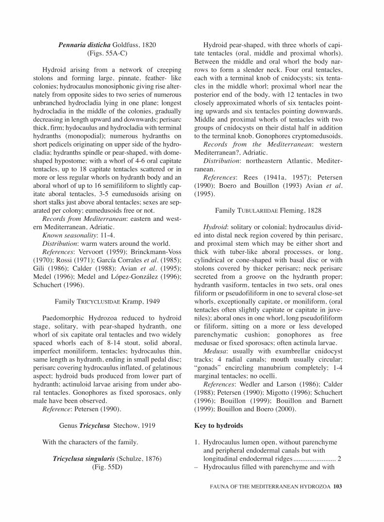
Pennaria disticha Goldfuss, 1820(Figs. 55A-C)
Hydroid arising from a network of creepingstolons and forming large, pinnate, feather- likecolonies; hydrocaulus monosiphonic giving rise alter-nately from opposite sides to two series of numerousunbranched hydrocladia lying in one plane; longesthydrocladia in the middle of the colonies, graduallydecreasing in length upward and downwards; perisarcthick, firm; hydocaulus and hydrocladia with terminalhydranths (monopodial); numerous hydranths onshort pedicels originating on upper side of the hydro-cladia; hydranths spindle or pear-shaped, with dome-shaped hypostome; with a whorl of 4-6 oral capitatetentacles, up to 18 capitate tentacles scattered or inmore or less regular whorls on hydranth body and anaboral whorl of up to 16 semifiliform to slightly cap-itate aboral tentacles, 3-5 eumedusoids arising onshort stalks just above aboral tentacles; sexes are sep-arated per colony; eumedusoids free or not.
Records from Mediterranean: eastern and west-ern Mediterranean, Adriatic.
Known seasonality: 11-4.Distribution: warm waters around the world.References: Vervoort (1959); Brinckmann-Voss
(1970); Rossi (1971); García Corrales et al. (1985);Gili (1986); Calder (1988); Avian et al. (1995);Medel (1996); Medel and López-González (1996);Schuchert (1996).
Family TRICYCLUSIDAE Kramp, 1949
Paedomorphic Hydrozoa reduced to hydroidstage, solitary, with pear-shaped hydranth, onewhorl of six capitate oral tentacles and two widelyspaced whorls each of 8-14 stout, solid aboral,imperfect moniliform, tentacles; hydrocaulus thin,same length as hydranth, ending in small pedal disc;perisarc covering hydrocaulus inflated, of gelatinousaspect; hydroid buds produced from lower part ofhydranth; actinuloid larvae arising from under abo-ral tentacles. Gonophores as fixed sporosacs, onlymale have been observed.
Reference: Petersen (1990).
Genus Tricyclusa Stechow, 1919
With the characters of the family.
Tricyclusa singularis (Schulze, 1876)(Fig. 55D)
Hydroid pear-shaped, with three whorls of capi-tate tentacles (oral, middle and proximal whorls).Between the middle and oral whorl the body nar-rows to form a slender neck. Four oral tentacles,each with a terminal knob of cnidocysts; six tenta-cles in the middle whorl; proximal whorl near theposterior end of the body, with 12 tentacles in twoclosely approximated whorls of six tentacles point-ing upwards and six tentacles pointing downwards.Middle and proximal whorls of tentacles with twogroups of cnidocysts on their distal half in additionto the terminal knob. Gonophores cryptomedusoids.
Records from the Mediterranean: westernMediterranean?, Adriatic.
Distribution: northeastern Atlantic, Mediter-ranean.
References: Rees (1941a, 1957); Petersen(1990); Boero and Bouillon (1993) Avian et al.(1995).
Family TUBULARIIDAE Fleming, 1828
Hydroid: solitary or colonial; hydrocaulus divid-ed into distal neck region covered by thin perisarc,and proximal stem which may be either short andthick with tuber-like aboral processes, or long,cylindrical or cone-shaped with basal disc or withstolons covered by thicker perisarc; neck perisarcsecreted from a groove on the hydranth proper;hydranth vasiform, tentacles in two sets, oral onesfiliform or pseudofiliform in one to several close-setwhorls, exceptionally capitate, or moniliform, (oraltentacles often slightly capitate or capitate in juve-niles); aboral ones in one whorl, long pseudofiliformor filiform, sitting on a more or less developedparenchymatic cushion; gonophores as freemedusae or fixed sporosacs; often actinula larvae.
Medusa: usually with exumbrellar cnidocysttracks; 4 radial canals; mouth usually circular;“gonads” encircling manubrium completely; 1-4marginal tentacles; no ocelli.
References: Wedler and Larson (1986); Calder(1988); Petersen (1990); Migotto (1996); Schuchert(1996); Bouillon (1999); Bouillon and Barnett(1999); Bouillon and Boero (2000).
Key to hydroids
1. Hydrocaulus lumen open, without parenchyme and peripheral endodermal canals but with longitudinal endodermal ridges........................ 2
– Hydrocaulus filled with parenchyme and with
FAUNA OF THE MEDITERRANEAN HYDROZOA 103
sm68s2Bsystema1 14/10/04 15:22 Página 103

longitudinal peripheral endodermal canals; hydrocaulus widening from base to distal end............................................................ Tubularia
2. Oral tentacles in one whorl; two, rarely up to fivelongitudinal endodermic ridges; medusa radially symmetrical with 2 or 4 groups of tentacles ..................................................................Ectopleura
– Oral tentacles in two whorls; up to 8 or more longitudinal endodermic ridges; medusae asymmetrical with one group of marginal tentacles............................................ Hybocodon
Key to medusae
1. With tentacular marginal bulbs ........................ 2– Without tentacular marginal bulbs..... Rhabdoon2. With normal, symmetrical umbrella................. 3– With asymmetrical umbrella, bell margin
obliquely set to the vertical axis ...... Hybocodon3. With longitudinal exumbrellar cnidocyst tracks
or rows .............................................. Ectopleura– Cnidocysts in scattered or in clumps ..................
........................................................... Plotocnide
Genus Ectopleura L. Agassiz, 1862
Hydroid: solitary or colonial; hydrocaulus high,simple, with open lumen, without parenchymaticendoderm and longitudinal endodermal canals, butweakly divided by two, rarely up to five, internal lon-gitudinal endodermic ridges; perisarc thin, coveringpyriform neck region, originating from collar on neckregion and does not cover whole neck; hydranth vasi-form with filiform (except in E. wrighti where they aremoniliform to capitate) oral tentacles in one whorl anda whorl of long, filiform, aboral tentacles; gonophoresright above aboral tentacles, producing free medusae,eumedusoid or fixed sporosacs.
Medusa: umbrella symmetrical, rounded, or pyri-form; 8 longitudinal exumbrellar cnidocyst rows,issuing in pairs from tentacular bulbs; manubriumshort, at most reaching bell margin; 2 opposite or 4equally developed, simple perradial marginal tenta-cles, moniliform or with abaxial cnidocyst clusters;4 radial canals.
Remarks: many hydroid-based nominal speciesof Ectopleura have recently been described, themedusae being known either as just liberated juve-niles or as medusa buds; some medusae withunknown cycle, and described long ago, could cor-respond to some of those hydroids. In groups withspecies based on either polyps or medusae only,
rearing experiments are necessary to elucidate lifecycles before assigning new specific names.
Recent references: Schuchert (2001a, 2003).
Key to hydroids
1. Colonies producing free medusae .................... 2– Colonies producing not released medusae ....... 32. Hydroid solitary, with 14-25 filiform oral
tentacles ....................................... E. dumortierii– Hydroid colonial, with 5-10 slightly capitate
tentacles .............................................. E. wrighti3. Gonophores cryptomedusoids; female gono-
phores with 8 laterally flattened crests surrounding the opening .................... E. crocea.
– Gonophores eumedusoids; female gonophores with 3-4 short round apical processes which may be flattened .......................................... E. larynx
Key to medusae
1. With four perradial marginal tentacles...................................................................... E. dumortieri
– With two opposite marginal tentacles .............. 22. «Gonads» surrounding manubrium and forming
4 sac-like interradial pouches ...... E. sacculifera– «Gonads» only surrounding manubrium.......... 33. With short conical apical chamber and apical
canal; marginal tentacles with six to nine abaxial cnidocysts clusters and a larger terminal one ............................................................... E. minerva
– Without apical chamber and apical canal; tentacles with a terminal knob of cnidocysts, one or two distal spherical rings of cnidocysts and proximally one abaxial cnidocysts cluster ...................................................................... E. wrighti
Ectopleura crocea (L.Agassiz, 1862)(Figs. 55E-F)
Hydroid colonial; hydrorhiza forming a dense matgiving rise to cluster of numerous polyps; stemunbranched, up to 70 mm high, perisarc firm andsmooth, with scattered groups of several annulations;coenosarc with 2-5 longitudinal endodermal ridges.Polyp reddish, with one oral whorl of 18-24 filiformtentacles with their bases adnate to hypostome, andone aboral whorl of 22-30 slender tentacles longerthan the oral ones; cnidocysts of aboral tentacles, con-centrated in a longitudinal band of the aboral side.Oral tentacles circular in cross-section, aboral onesfor-sided. Neck region more than 1/2 length of
104 J. BOUILLON et al.
sm68s2Bsystema1 14/10/04 15:22 Página 104

hydranth, finely and longitudinally striated, with a col-lar at lower edge which forms a shallow groove fromwhich perisarc covering the neck region is secreted.Gonophores in small groups on pedicels borne onblastostyles (about 8) in circles close to bases of abo-ral tentacles. Gonophores cryptomedusoid, withoutradial canals and ring canal, immature malegonophores elongate ovoid, spherical when mature,with four tentacle rudiments; female gonophoresovoid to spherical with eight, laterally flattened, thintransparent crests surrounding opening of bell throughwhich spadix often protrudes; newly released actinulawithout oral tentacles. Cnidocysts: Stenoteles anddesmonemes (in Brickmann-Voss, 1970).
Records from Mediterranean: western Mediter-ranean, Adriatic.
Known seasonality: 1 to 12.Distribution: circuntropical in shallow waters.References: Brinckmann-Voss (1970) as Tubu-
laria; Petersen (1990); Boero and Bouillon (1993)as Tubularia; Avian et al. (1995); Medel and López-González (1996) all as Tubularia.
Ectopleura dumortierii (Van Beneden, 1844)(Figs. 55G-J)
Colonies stolonal; hydrocaulus, erect, up to 100mm high, solitary, simple or slightly branched, end-ing in a terminal hydranth and increasing in widthfrom base to distal end; perisarc horn-coloured cov-ering hydrocaulus till proximal end of hydranth,irregularly annulated; hydrocaulus endoderm withup to 4 longitudinal ridges; neck region with glandcells forming an indistinct collar; hydranth flask-shaped, with a rounded hypostome; with one whorlof 14-25 rather short rounded oral filiform tentacles,with oval bases not adnate to hypostome and onewhorl of up to 30 long, aboral, semifiliform tenta-cles; up to 10, once dichotomously branched, blas-tostyles above aboral tentacles, bearing clusters ofmedusa buds at their terminal end.
Medusa: umbrella 2-3 mm high, nearly spherical,mesoglea very thick, especially in apical region; apicalcanal sometimes present; velum fairly broad; exum-brella with four pairs of cnidocyst tracks; manubriumvery large, spherical at base, tapering towards mouth,extensile; mouth simple, tube-like, mouth rim withcnidocysts; 4 perradial marginal tentacles with largebasal bulbs and prominent round cnidocyst clusters onabaxial surface; «gonads» completely surroundingmanubrium, living mouth area free; developmentthrough a pro-actinula and actinula stage.
Records from Mediterranean: eastern and west-ern Mediterranean; Adriatic Sea.
Known seasonality: 1-12.Distribution: Atlantic; Indo-Pacific, Mediter-
ranean.References: Kramp (1959a, 1961); Berhaut
(1970); Brinckmann-Voss (1970, 1987); Benovic(1973); Schmidt and Benovic (1979); Dowidar(1983); Castello i Tortella (1986); Benovic and Ben-der (1987); Goy (1973b); Gili (1986); Goy et al.(1988, 1990, 1991); Hirohito (1988); Petersen(1990); Boero and Bouillon (1993); Avian et al.(1995); Benovic and Lucic (1996); Medel andLópez-González (1996).
Ectopleura larynx (Ellis and Solander, 1786)(Figs. 56A-B)
Hydroid colonial, hydrorhiza forming dense matgiving rise to numerous stems in dense groups up to70 mm high; perisarc of hydrocauli firm and smoothbut with irregular annulations; coenosarc with 2 longi-tudinal ridges. Polyp with long hypostome, one oralwhorl of up to 20 long, slender, filiform tentaclesadnate to hypostome and forming longitudinal ridgesover distal half of hypostome, and one aboral whorl of20-25 longer filiform tentacles, slender, laterally flat-tened at their bases, distance between to adjacent ten-tacles equal to the tentacle base diameter. Neck regionwith longitudinal striations, nearly as long ashydranth, and with a collar grooved basally which pro-duces the thin perisarc covering the neck. Gonophoreson blastostyles in circles around oral and aboral tenta-cles. Male gonophores on, up to 12, long pedicelsborne on up to 12 long, pendant, and unbranched blas-tostyles; female gonophores on, up to 12, shorter blas-tostyles (with up to 8 gonophores) dichotomouslybranched. Gonophores eumedusoids, without radialcanals but with ring canal; female ovoid, with 3-4short round apical processes which may be flattenedgiving the “ears” appearance; male gonophores longerand slender, with very short, closely-set apicalprocesses; actinula, at liberation, with an aboral whorlof 6-13 tentacles with swollen tips; oral tentacles notyet developed or as 3-5 rudiments.
Records from Mediterranean: western Mediter-ranean.
Known seasonality: 3, ?Distribution: mainly northern (west and east)
Atlantic; northern Pacific; Mediterranean (recordsfrom southern hemisphere probably due to transportby ships (Petersen, 1990).
FAUNA OF THE MEDITERRANEAN HYDROZOA 105
sm68s2Bsystema1 14/10/04 15:22 Página 105

References: Gili (1986) as Tubularia; Petersen(1990); Medel and López-González (1996) as Tubu-laria; Schuchert (2001a).
Ectopleura minerva Mayer, 1900b(Fig. 56C)
Medusa: umbrella 2,5 mm high, pear-shaped toconical bell-shaped, narrowing at base; mesogleathicker apically but without clear apical projection;exumbrella with 4 pairs of longitudinal cnidocyststracks originating from the marginal bulbs and extend-ing near apex of umbrella; manubrium tubular, abouttwo third length of umbrella cavity, with short conicalapical chamber and apical canal, the size of bothdepending on age and preservation; mouth simple,tubular; radial canals and circular canal narrow; withtwo moderately broad opposite marginal tentacularbulbs and two slightly smaller non tentacular bulbs;the two opposite perradial marginal tentacles eachwith six to nine abaxial cnidocysts clusters and a larg-er terminal one; «gonads» completely surroundingmanubrium living most distal part free, tentacles andmanubrium typically cream in color.
Remarks: the medusa Ectopleura minerva hasbeen described from Florida, it is known from theBermudas, the Mediterranean, the Seychelles,India, China, Japan and the Bismarck Sea. Ecto-pleura minerva hydroid stage is still unknown,several Ectopleura types of hydroid have beendescribed producing unreleased or just releasedtwo tentaculated medusae, particularly E. pacificaThornely, 1900 from Papua New Guinea, but noneof them have their adult stage known and rearingexperiments will thus be necessarily before eluci-dating and clarifying their complex and confusesynonymy.
Records from Mediterranean: eastern and west-ern Mediterranean.
Known seasonality: 1-12.Distribution: Atlantic; Indo-Pacific; Mediterranean.References: Kramp (1961); Goy (1973b); Sugiu-
ra (1977); Bouillon (1978 a,b); Calder (1988); Hiro-hito (1988); Petersen (1990); Goy et al. (1988, 1990,1991); Schuchert (1996).
Hydroid: unknown.
Ectopleura sacculifera Kramp, 1957a(Fig. 56D)
Medusa: umbrella 3 mm high, 2.8 mm wideslightly conical, mesoglea thick, exumbrella with 8
cnidocysts tracks issuing in pairs from the four mar-ginal bulbs, continuing almost to apex, and forming8 slight crests along exumbrella; manubrium half aslong as umbrellar cavity; «gonads» surrounding themanubrium and forming four large, interradial, sac-like pouches hanging down from middle part ofmanubrium almost to mouth level; two long oppo-site marginal tentacles, their distal part with 20- 25abaxial clusters of cnidocysts and two rudimentarybulbs.
Hydroid: unknown.Records from Mediterranean: western Mediter-
ranean.Known seasonality: 2.Distribution: Indo-Pacific; Mediterranean.References: Kramp (1961); Brinckmann-Voss
(1970, 1987); Bouillon (1978b); Boero and Bouillon(1993).
Ectopleura wrighti Petersen, 1979(Fig. 56E)
Hydroid: colonial issued from a creepinghydrorhiza, stolons not clearly demarcated fromhydrocauli; hydranth very slender, vasiform withone oral whorl of 5-10 slightly capitate or monili-form tentacles which are not adnate to hypostomeand an aboral whorl of 11-20 filiform tentacles; neckregion long and slender, with low ring-shaped collarforming groove from which thin outer periderm issecreted; hydrocaulus slender, unbranched, of equalwidth throughout; inner lumen divided into two lon-gitudinal canals by two endodermal ridges formedby highly vacuolated cells, periderm flexible, thinand smooth, covering hydrocauli and stolons;medusa buds born above the aboral tentacle whorl;carried on 5-8 short, dichotomously branched blas-tostyles.
Medusa: umbrella nearly hemispherical, withfour longitudinal meridian pairs of cnidocyst tracks;with four perradial marginal bulbs; with two oppo-site marginal perradial tentacles with a terminalknob of cnidocysts and distally one or two sphericalcnidocyst knobs encircling tentacle, proximally oneabaxial cnidocyst cluster; manubrium nearly as longas subumbrellar cavity; completely encircled by«gonads».
Records from Mediterranean: hydroid easternand western Mediterranean, Adriatic. Only youngmedusae liberated in the laboratory known (Brinck-mann-Voss, 1970).
Known seasonality: 1-12.
106 J. BOUILLON et al.
sm68s2Bsystema1 14/10/04 15:22 Página 106

Distribution: Atlantic?; western Mediterranean.References: Brinckmann-Voss (1970, 1987);
Petersen (1979, 1990); Boero and Bouillon (1993);Avian et al. (1995), Peña Cantero and García Car-rascosa (2002).
Genus Hybocodon L. Agassiz, 1862
Hydroid: solitary, with high stems, hydrorhizairregularly branched; hydrocaulus tubular, withopen lumen, without parenchyme and longitudinalperipheral canals but weakly divided by eight ormore longitudinal endodermic ridge; perisarc origi-nating just below hydranth and much inflatedaround whole neck region; secreted from groovebetween hydranth and neck; oral tentacles filiformto pseudofiliform in two closely set whorls, aboraltentacles in one whorl, filiform to pseudofiliform;blatostyles dichotomously branched.
Medusa: bilaterally symmetrical, with umbrellamargin at oblique angle to vertical axis; no pointedapical process; with or without exumbrellar cnidocysttracks; manubrium cylindrical on short peduncle notextending beyond umbrellar margin; 4 radial canals,1 short, 2 medium sized and one longer; with 1 sim-ple or compound marginal bulb with 1-3 moniliformtentacles corresponding to the longest radial canal; 3remaining perradial bulbs rudimentary.
Hybocodon prolifer L. Agassiz, 1862(Figs. 56F-H)
Hydroid: colonies with hydrorhiza formed bybranching stolons embedded in sponges; hydro-caulus solitary or sparingly aggregated, long, gradu-ally enlarging to just below hydranth, with firmperisarc; endoderm of hydrocaulus with a centrallumen and several longitudinal ridges; neck regionbetween hydrocaulus and hydranth surrounded by aloose filmy, wrinkled perisarc; hydranth pear-shaped, with a rounded hypostome, with up to 50short oral filiform tentacles in 2 closely set whorls,the most distal being the shorter and up to 31 longeraboral filiform tentacles in one whorl, the base ofaboral tentacles adnate to basal part of hydranth;medusa buds on branching blastostyles, bearingnumerous buds, just above aboral tentacles; theolder medusa buds themselves carrying buds whilestill fixed on hydranths.
Medusa: umbrella 3 mm wide, up to 5 mm high,bell-shaped, evenly rounded, umbrella marginoblique to vertical axis, mesoglea moderately thick;
usually with 5 longitudinal exumbrellar cnidocysttracks, 2 issued from tentacular bulb, 1 track fromeach non tentacular bulb; velum moderately broad;manubrium large cylindrical mounted on a shortpeduncle never reaching beyond margin even in fullextension; mouth with a narrow ring of cnidocysts;1 tentacular bulb with 1 or more moniliform tenta-cles (with adaxial clasps according to Schuchert,1996) and with medusae buds, 3 non tentacularbulbs reduced; «gonads» completely surroundingmanubrium, living peduncle and most distal portionfree; medusa buds on marginal bulbs; eggs develop-ing in actinulae on manubrium which may later befound free in plankton.
Records from Mediterranean: western Mediter-ranean.
Known seasonality: 3-6.Distribution: Atlantic; Indo-Pacific; Antarctic,
Arctic; Mediterranean.References: Kramp (1961); Arai and Brinck-
mann-Voss (1980); Gili (1986); Boero and Bouillon(1993); Medel and López-González (1996);Schuchert (1996, 2001a).
Genus Plotocnide Wagner, 1885
Tubulariidae with exumbrellar cnidocysts scat-tered singly or in clumps; with a dome-shaped api-cal chamber lined with vacuolated endodermal cells.
Plotocnide borealis Wagner, 1885(Fig. 56I)
Medusa: umbrella up to 3.5 mm wide and high;spherical to bell-shaped, with rounded apex,mesoglea very thick mainly at apex, thinning slight-ly towards umbrella opening; with exumbrellarcnidocysts scattered singly or in clumps; with adome-shaped apical chamber lined with vacuolatedendodermal cells above manubrium; manubriumflask-shaped, half as long as umbrellar cavity;mouth simple with a ring of cnidocysts; «gonads» athick ring around manubrium, living mouth anduppermost portion of manubrium free; radial canalsand circular canal simple; with 4 threadlike, solidmarginal tentacles ending in a large, oval, swellingstudded with cnidocysts; marginal tentacular bulbssmall globular.
Remarks: the presence of this arctic species in theeastern Mediterranean is based on a single smallspecimen (less than 1 mm high) described by Goy etal. (1988), lacking apparently most of the character-
FAUNA OF THE MEDITERRANEAN HYDROZOA 107
sm68s2Bsystema1 14/10/04 15:22 Página 107

istic features of the species: no vacuolated apicalchamber, no apical mesoglea thickening, no exum-brellar cnidocysts, no ring-shaped «gonads». Thisidentification is doubtful
Hydroid: unknown.Known seasonality: 1.Distribution: Arctic and Subarctic, circumpolar;
Atlantic; Indo- Pacific; Mediterranean?References: Beyer (1955) Hand and Kan (1961);
Kramp (1961); Hartlaub (1907); Kramp (1942,1959a, 1961, 1968); Naumov (1960-1969); Arai andBrinckmann-Voss (1980); Goy et al. (1988, 1991);Boero and Bouillon (1993).
Genus Rhabdoon Keferstein and Ehlers, 1861
Tubulariidae with single hollow marginal tenta-cle ending in large, complex knob of cnidocyst clus-ters; without marginal tentacular bulbs; manubriumoccupying almost entire bell cavity; with vacuolatedcells containing refractive droplets along 4 radialcanals, at manubrium apex and bell margin;«gonads» surrounding distal 2/3 of manubrium.
Rhabdoon singulare Keferstein and Ehlers, 1861(Fig. 56J)
Medusa:umbrella 1.6 mm wide, 2.1 mm high,barrel-shaped, apex dome-shaped, mesoglea thin atthe apex larger at the umbrellar sides; exumbrellamargin thick, irregular, containing numerous cnido-cysts; subumbrellar opening and velum narrow;exumbrella with 4 perradial longitudinal exumbrel-lar cnidocyst tracks, 4 additional interradial andsometimes adradial ones; manubrium large flask-shaped occupying almost the entire subumbrellarcavity, upper third vacuolated; mouth simple, circu-lar, covered with cnidocysts; 4 protruding radialcanals; «gonads» on the two distal thirds of themanubrium; with a single hollow tentacles ending ina large knob consisting of numerous stalked capita-tions containing cnidocysts.
Hydroid: unknown.Records from Mediterranean: eastern and west-
ern Mediterranean; Adriatic Sea.Known seasonality: 1-11.Distribution: Atlantic; Indo-Pacific; Mediter-
ranean.References: Kramp (1961); Brinckmann-Voss
(1970, 1987); Benovic (1976); Benovic and Bender(1987), Goy (1973, 1983); Dallot, Goy and Carré(1988); Pagès et al. (1992); Boero and Bouillon
(1993); Avian et al. (1995); Benovic and Lucic(1996).
Genus Tubularia Linnaeus, 1758
Hydroid: solitary; hydrocaulus long, tubular,widening from base to distal end, inner lumen filledwith parenchymatic endoderm, penetrated by 8 ormore longitudinal endodermal peripheral canals, onewider than the others; circular or lobed basal disc, andsupporting tubes developed from lower part of stem;thin perisarc around neck secreted from groovebetween hydranth base and neck; hydranth vasiform,with two or more whorls of oral filiform and onewhorl of filiform aboral tentacles; bases of aboral ten-tacles continued as ridges over hydranth base; blas-tostyle with unbranched main stem, with or withoutthin side branches; gonophores reduced to eumedu-soid or to sessile cryptomedusoid, with or withoutdistal processes, in which the origin from a biradiallysymmetrical medusa, can be usually traced.
Refrences: Petersen (1990); Schuchert (2001).
Key
1. Female gonophore eumedusoid, without any tentacle, with four radial canals and a ring canal .......................................................... T. Indivisa.
– Female gonophore cryptomedusoid, with one tentacle-like process, without radial canal and ring canal ...................................... T. ceratogyne
Tubularia ceratogyne Pérès, 1920(Figs. 57A-B)
Hydroid solitary but growing in clusters.Hydrocaulus up to ca 60 mm, with one larger andabout seven smaller peripheral canals surroundingparenchymatic centre, perisarc firm, annulationsabsent; stem attached to substrate by irregularlyshaped basal disc forming one or two short rhi-zomes covered by thick perisarc and 2-4 support-ing tubes irregularly wrinkled originating fromlower part of stem. Hydranth with long oral tenta-cles adnate basally to hypostome and connected tohypostome by thin membrane for some distance;aboral tentacles about 30, with oval bases; neckregion nearly as long as hydranth, covered by thinperisarc originating in groove between hydranthbase and neck. Gonophores on short pedicels on 7-12 (usually 8) long and unbranched blastostyles;female ovoid, cryptomedusoid, without radial
108 J. BOUILLON et al.
sm68s2Bsystema1 14/10/04 15:22 Página 108

canals and ring canal, with one hollow, tentacle-like process bent over opening of gonophore; malespherical, cryptomedusoid, without distal process-es. Female gonophores inserted irregularly oversurface of blastostyle, male gonophores arrangedin four to five longitudinal rows; cnidome:desmonemes, anisorhiza, microbasic euryteles,stenoteles.
Records from Mediterranean: recorded onlyfrom the Strait of Gibraltar and nearby areas of Gulfof Cádiz.
Known seasonality and reproduction: 6;10;12.Records outside the Mediterranean: northeastern
Atlantic.References: Teissier (1965); Patriti (1970);
Hughes (1983); Petersen (1990); Medel and López-González (1996).
Tubularia indivisa Linnaeus, 1758(Figs. 57C-F)
Polyps solitary but in clusters; stem up to 200mm high (but usually shorter), perisarc firm andsmooth; coenosarc with one larger and 9-12 smallerperipheral canals around central lumen; stemattached to substrate by relatively broad and thicklobate basal disc, and several supporting tubes orig-inating from lower half of stem, which grow oversubstrate as short rhizomes. Hydranth with up to 6whorls of 40-60 long oral tentacles continued overhypostome as longitudinal ridges and 25-35 aboraltentacles with laterally-flattened oval bases; neckregion as long as hydranth, covered by thin perisarcoriginated in groove between the hydranth base anddistal part of the neck. Gonophores on short pedicelsborne on up to 12 unbranched blastostyles (eachwith up to 30 older gonophores); female gonophoreeumedusoid, ovoid to spherical, with four radialcanals of unequal length and a ring canal, withouttentacles; male gonophore cryptomedusoid, ovoid tospherical, with rounded distal end; actinula withwhorl of short oral tentacles at liberation; cnidome:desmonemes, anisorhiza, microbasic euryteles,stenoteles.
Records from Mediterranean: eastern and west-ern Mediterranean, Adriatic.
Distribution: temperate to colder waters of north-ern Atlantic and Pacific, Arctic Sea, Mediterranean.
References: Naumov (1960); Petersen (1990);Cornelius and Ryland (1990); Boero and Bouillon(1993); Avian et al. (1995); Medel and López-González (1996); Schuchert (2001a).
Suborder ZANCLEIDA Russell 1953
Hydroid: colony floating or fixed; fixed coloniesarising either from simple creeping stolonal tubes,from an encrusting basal mat, from upright branchedhydrorihza consisting of a central axis of perisarccovered by coenosarc, or from a calcified exoskele-ton; hydranths monomorphic or polymorphic, oraltentacles capitate or moniliform, aboral tentacles inwhorls or scattered, either capitate, moniliform,ramified capitate, reduced or without tentacles; freemedusae, eumedusoids or sporosacs.
Medusa: manubrium flask-shaped, with quadrateor octagonal base and cylindrical mouth tube;“gonads” usually interradial; exumbrellar cnidocystpouches or tracks; 0-2 or 4 marginal tentacles withor without abaxial cnidophores; marginal tentaclesdeveloped only at junction between radial and circu-lar canals; with or without ocelli.
References: Petersen (1990); Boero, Bouillon andGravier-Bonnet (1995); Bouillon (1999); Bouillon andBarnett (1999); Boero, Bouillon and Gravili (2000).
Key to hydroids
1. Hydroids............................................ Porpitidae.– Holonies fixed by hydrorhizae ......................... 22. Hydrorhiza incrusting forming an a crust-like
stolonal plate.................................. Rosalindidae– Hydrorhiza formed by creeping stolon tubes
recovered by perisarc ....................................... 33. Cnidocysts on hydranth body wall arranged in
conspicuous rounded patches; hydranth with moniliform or modified moniliform aboral tentacles.................................... Cladocorynidae.
– Cnidocysts not in patches on hydranth body wall; hydranth without moniliform tentacles......................................................... Zancleidae.
Key to medusae
1. Marginal tentacles terminating in a single large spherical cnidocyst knob.................... Porpitidae
– Marginal tentacles with numerous stalked capsules containing cnidocysts, stalk of the capsules threadlike, very extensile (cnidophores).......................................................... Zancleidae
Family CLADOCORYNIDAE Allman, 1872
Hydroid: stem simple or slightly branched, risingfrom a creeping stolon; hydranth club-shaped, oral
FAUNA OF THE MEDITERRANEAN HYDROZOA 109
sm68s2Bsystema1 14/10/04 15:22 Página 109

tentacles moniliform or capitate, in one whorl, abo-ral tentacles moniliform or branched capitate, scat-tered or in several whorls; cnidocysts on body wallarranged in conspicuous rounded patches or scat-tered around the base of oral and aboral tentacles;gonophores carried singly or on short, branchedpedicels, on lower or middle part of hydranth; withfree medusae or fixed cryptomedusoids sporosacs.
Medusa: only two exumbrellar pouches, contain-ing macrobasic euryteles, on non tentaculate perra-dial marginal bulbs; tentaculate perradial marginalbulbs very large, without cnidocyst pouches; tenta-cles with cnidophores; “gonads” interradial onmanubrium.
References: Wedler and Larson (1986); Bouillon,Boero and Seghers (1987); Petersen (1990); Boero,Bouillon and Gravier-Bonnet (1995); Migotto(1996); Schuchert (1996).
Genus Cladocoryne Rotch, 1871
Hydrocaulus long, unbranched or sparinglybranched, covered by perisarc, arising from a creep-ing hydrorhiza; hydranth club-shaped, with oralwhorl of 4-6 short capitate tentacles, one to fourwhorls of branched-capitate aboral tentacles; one ortwo patches of macrobasic eurytele cnidocysts onhydranth body; gonophores as cryptomedusoidfixed sporosacs or as medusa buds, on short pedicelsbetween or over aboral tentacles.
Cladocoryne floccosa Rotch, 1871(Fig. 57G)
Colonies composed of creeping hydrorhizae giv-ing rise erect and sparingly branched hydrocauli;perisarc yellowish to transparent, smooth or withseveral groups of annulations; hydranth nude withan oral whorl of 4-6 short capitate tentacles and 1-4whorls of branched capitate aboral tentacles; ovalareas of microbasic euryteles cnidocysts on thehydranth. Gonophores spherical (cryptomedusoids)on short pedicels between or over aboral tentacles.Cnidocysts: macrobasic euryteles and stenoteles.
Records from Mediterranean: eastern and west-ern Mediterranean, Adriatic.
Known seasonality: 4-7.Reproduction: 4-7Distribution: temperate and tropical waters
around the world.References: Rossi (1961); Brinckmann-Voss
(1970); Gili (1986); Boero and Bouillon (1993);
Avian et al. (1995); Medel and López-González(1996); Peña Cantero and García Carrascosa (2002).
Family PORPITIDAE Goldfuss, 1818
Hydroid: colony floating, with a chitinous inter-nal skeleton, covered by mantle, and forming afloating chamber; with a central large gastrozooidand marginal gastro-gonozooids and dactylozooids.
Medusa: 4 or 8 of exumbrellar stenotele cnido-cyst tracks issued from marginal bulbs; 4 or 8 radialcanals and a circular canal; manubrium short, coni-cal; with quadrate or octagonal base; mouth circular;“gonads” perradial or irregularly arranged perradial-ly and interadially; 2 opposite, perradial, capitatemarginal tentacles; with or without 2 additionalsmaller capitate tentacles adaxial to the first; tenta-cles with macrobasic euryteles; zooxanthellae gen-erally present.
References: Bouillon (1984d); Calder (1988);Petersen (1990); Pagès et al. (1992); Schuchert(1996); Bouillon (1999); Bouillon and Barnett(1999); Bouillon and Boero (2000).
Key to hydroids
1. Disc-shaped floating colony without sail............................................................................ Porpita
– Oval to elliptical-shaped floating colony; with a median sail............................................... Velella
Key to medusae
1. 4 radial canals; manubium with quadrate base; 4 capitate tentacles...................................... Velella
– 8 radial canals; manubrium octagonal; 2 capitate tentacles.................................................. Porpita
Genus Porpita Lamarck, 1801= Porpema Haeckel, 1888
Hydroid: colony floating, dark blue, diameter upto 30 mm, mostly smaller, with disk-shaped mantleand internal float, margin soft, flexible; central regionfirm, slightly convex, with a central pore and numer-ous stigmata; mantle with radiating endoderm canals;internal chitinous float consisting of a series of con-centric chambers; a disk-shaped reservoir of cnido-cysts between float and central gastrozooid; undersurface with one large central gastrozooid, a mediancircle of gastro-gonozooids, and a peripheral circle ofdactylozooids; central gastrozooid short and broad
110 J. BOUILLON et al.
sm68s2Bsystema1 14/10/04 15:22 Página 110

with a terminal mouth, without tentacles or prominentcnidocyst clusters; gastro-gonozooids clavate, lack-ing tentacles but with prominent cnidocyst clustersscattered over body, medusae develop near base inclusters; dactylozooids with a distal whorl of 4 capi-tate tentacles, body with varying number of short,small capitate tentacles in 3 vertical rows.
Medusa: with 8 radial canals; manubrium coni-cal, with octagonal base; 2 opposite marginal capi-tate tentacles, 6 non tentaculate bulbs; “gonads” 8,perradial; short exumbrellar cnidocyst tracks aboveeach bulb.
Porpita porpita (Linnaeus, 1758)(Figs. 57H-I)
With the characters of the genus.Records from Mediterranean: eastern and west-
ern Mediterranean, Adriatic.Known seasonality: 5-7; 10.Distribution: Atlantic; Indo-Pacific; Mediter-
ranean.References: Brinckmann-Voss (1970, 1987);
Daniel (1976); Bouillon (1984d); Castelló i Tortella(1986); Calder (1988); Pagès et al. (1992); Avian etal. (1995); Medel and López-González (1996);Bouillon and Boero (2000).
Genus Velella Lamarck, 1801
Hydroid: colony floating; float flattened, oval,elliptical, with a triangular sail; up to 40 mm longand 20 mm wide, higher in the centre than at theedges; two mirror images of the animal [left andright sailing; the prevalence of one form in oneregion may be due to sorting by prevailing winds(Edwards, 1966a)]; float and sail kept rigid by achitin support covered by mantle tissue; margin offloat soft and flexible; chitin float oval to slightly S-shaped with concentric air chambers; mantle tissuewith network of endoderm canals; in centre ofunderside a single large gastrozooid or “siphon”encircled by a ring of medusa producing gastro-gonozooids and a peripheral band of dactylozooids;central feeding zooid broadly oval with an elongat-ed hypostome, without tentacles or medusa buds;gastro-gonozooids spindle-shaped with a swollenmouth region, lacking tentacles but with warts ofcnidocyst clusters concentrated in distal half; onproximal half of hydranth numerous medusa budsgrowing in groups from short blastostyles; dactylo-zooids long and tapering, oval in cross section, with
cnidocysts concentrated in two lateral bands on thenarrow sides, mouth lacking; colour: float deeplyblue when alive, medusa buds yellow-olive fromsymbiotic algae.
Medusa: with 4 exumbrellar cnidocyst rows, 4radial canals; 2 pairs of opposite, perradial tentacles,a short adaxial one and a long abaxial one, each witha large terminal cnidocyst cluster; 2 perradial mar-ginal bulbs without tentacles; manubrium conicalwith quadrate base; mouth tubular; “gonads” irregu-larly arranged perradially and interradially.
Velella velella (Linnaeus, 1758)(Figs. 57J-K)
With the characters of the genus.Records from Mediterranean: eastern and west-
ern Mediterranean, Adriatic.Known seasonality: 2-5.Distribution: Atlantic; Indo-Pacific; Mediter-
ranean.References: Leloup (1929); Mackie (1959,
1960); Edwards (1963b, 1966a); Brinckmann-Voss(1964, 1970, 1987); Daniel (1976); Larson (1980);Bouillon (1984d); Castelló i Tortella (1986); Calder(1988); Pagès et al. (1992); Avian et al. (1995);Medel and López-González (1996); Bouillon andBoero (2000).
Family ROSALINDIDAE Bouillon, 1985
Hydroid: colonial; stolonal, with a Fig.-like ortrabecular chitinized skeleton, consisting of a thinperisarcal sheet covered by coenosarc and an exter-nal peridemal film; coenosarc supported by perisar-cal spines and trabeculae forming a more or lessthick framework of meshes isolated or sometimesorganised in rose-like alveolar stuctures surroundingthe polyps; hydranth plump sausage-shaped, with30-60 scattered capitate tentacles, almost sessile,sometimes with a basal perisarcal calice; cnidomecomprising subspherical stenoteles and macrobasicmastigophres; fixed gonophores or free medusae?,with two tentacles apparently provided withcnidophores, known in one species (Rosalinda nau-movi), carried singly or on short pedicels amongproximal tentacles; cnidome stenoteles macrobasicmastigophores and isorhiza.
Genus Rosalinda Totton, 1949
With the characters of the family.
FAUNA OF THE MEDITERRANEAN HYDROZOA 111
sm68s2Bsystema1 14/10/04 15:22 Página 111

Rosalinda incrustans Kramp, 1947(Figs. 58A-C)
Colonies 3 mm in height, with simple trabecularskeleton, with all hydranths naked.
Records from Mediterranean: western Mediter-ranean (Rosas Gulf).
Known seasonality: 12 ?Distribution: Atlantic, (west of Gibraltar),
Mediterranean.References: Vervoort (1966b); Watson (1978);
Antsulevich and Stepanjants (1985), Boero et al.(1995); Bouillon and Boero (2000).
Family ZANCLEIDAE Russell, 1953
Hydroid: colonial; hydrorhiza creeping, stolonal;perisarc enveloping hydrocaulus and hydrorhiza notlamellar, as a simple tube; hydrocaulus unbranched;polyps monomorphic or polymorphic; gastrozooideither with oral and aboral capitate tentacles, or withreduced capitate tentacles, or without tentacles;gonozooid and dactylozooid, when present, variedin expression.
Medusa: umbrella bell-shaped; 4 perradial exum-brellar cnidocyst pouches, either oval, clavate, elon-gate or linear, usually containing stenoteles; mouthsimple, circular, without oral tentacles (except inOonautes, of uncertain family affinity; see Capitataincertae sedis); 4 radial canals (exceptionally bifur-cated in Ctenaria, of uncertain family affinity; seeCapitata incertae sedis); marginal tentacles 0, 2 or 4,hollow, each with numerous abaxial cnidophores,with macrobasic euryteles; “gonads” usually interra-dial, rarely in a single mass around manubrium;without ocelli.
References: Wedler and Larson (1986); Calder(1988); Petersen (1990); Gravili et al. (1996);Schuchert (1996); Bouillon (1999); Bouillon andBarnett (1999); Boero et al. (2000); Bouillon andBoero (2000).
Key to hydroids
1. Gastrozooid reduced, without tentacles ....................................................................... Halocoryne
– Gastrozooid with numerous tentacles ... Zanclea
Genus Halocoryne Hadzi, 1917
Either eumedusoids with no tentacles and nomouth; with 4 radial canals; with four perradial
bulbs and four cnidocyst exumbrellar pouches;«gonads» surrounding manubrium; or medusaeeither Zanclea-like or with very elongated tentacularbulbs bearing short tentacles armed with short andstiff cnidophores.
Hydroid: stolonal, living in association with bry-ozoans; polymorphic; gastrozooids reduced, withouttentacles; hypostome armed or not with cnidocysts;dactylozooids columnar, slender, usually with one ortwo terminal cnidocyst knobs, sometimes with later-al rows of cnidocysts as well, without mouth; repro-duction by eumedusoids or free medusae.
Reference: Piraino et al. (1992).
Halocoryne epizoica Hadzi, 1917(Figs. 58D-E)
Hydroid: colonies stolonal, polymorphic, symbi-otic with bryozoans; hydrorhiza reticular, hydranthsnaked, sessile; gastrozooids very thin, cylindrical,lacking tentacles but with two prominent ectodermalswellings containing stenoteles on opposite sides ofthe mouth; dactylo-gonozooids stouter than gastro-zooids, without mouth and tentacles, with a terminalspherical cnidocyst cluster and up to five additionalcnidocysts clusters along one side of the body orarranged in a interrupted spiral towards hydrorhiza;released eumedusoids borne single or in clusters onshort pedicels on lower part of dactylo-gonozooids;cnidome: stenoteles of two sizes.
Medusa: eumedusoids; without mouth and tenta-cles; with 4 radial canals; with four perradial mar-ginal bulbs; with four cnidocyst exumbrellar pouch-es connected with marginal bulbs; gonads surround-ing manubrium. Cnidome: stenoteles in exumbrel-lar, macrobasic euryteles (?) sometimes in exum-brellar pouches, macrobasic mastigophores onexumbrella.
Records from Mediterranean: eastern and west-ern Mediterranean, Adriatic.
Known seasonality: 4-6?Distribution: endemic of Mediterranean Sea.References: Piraino et al. (1992); Avian et al.
(1995); Boero et al. (2000).
Genus Zanclea Gegenbaur, 1857
Hydroid: colonial, stolonal hydroids, often asso-ciated with bryozoans or molluscs; hydranthsmonomorphic or polymorphic. Polymorphiccolonies may present gastrozooids, dactylozooids,tentaculozooids and sometimes gastro-gonozooids;
112 J. BOUILLON et al.
sm68s2Bsystema1 14/10/04 15:22 Página 112

gastrozooids on unbranched short pedicels, oftenalmost sessile, elongated, cylindrical or claviformwith an oral whorl of capitate tentacles and numer-ous aboral capitate tentacles scattered or in severalwhorls over the entire body; tentacles rarely reducedto mere cnidocyst patches; perisarc covering hydro-hiza and entire hydrocaulus or confined to lowerpart of hydrocaulus only; dactylozooids and tentac-ulozooids when present varied in expression;medusa buds carried singly or in clusters on shortpedicels, either scattered among or under the aboraltentacles or on hydrorhiza or on hydranths reducedto blastostyles .
Medusa: Zancleidae with bell-shaped or almostspherical umbrella, lateral walls evenly thin,mesoglea slightly thicker at the apex; with exum-brellar perradial cnidocyst patches or tracts; mouthsimple, circular; with 4 simple radial canals; mar-ginal tentacles when present, 0, 2 or 4, with numer-ous abaxial cnidophores; «gonads» interradial;cnidome generally with stenoteles and macrobasiceuryteles, without desmonemes, no ocelli.
The genus Zanclea comprises at least 3 species inthe Mediterranean, Zanclea costata, Zanclea sessilisand Zanclea giancarloi (Gravili et al., 1996; Cerra-no et al., 1997; Boero et al., 2000).
Most of the various existing Zanclea species areactually not identifiable without a detailed study ofthe structure of the macrobasic euryteles cnidocystsof the polyp stage and the knowledge of their com-plete life cycle, the medusae are usually indis-cernible and must be referred as Zanclea spp. (Figs.58F-H).
General references: Russell and Rees (1936);Brinckmann-Voss (1970,1987); Benovic and Bender(1987); Goy (1973b); Millard and Bouillon (1973);Bouillon (1974a); Gili (1986); Goy et al. (1988,1990, 1991); Calder (1992); Boero and Bouillon(1993); Avian et al. (1995); Benovic and Lucic(1996); Gravili et al. (1996); Medel and López-González (1996); Cerrano et al. (1997); Boero et al.(2000).
Key to hydroids
1. Apotrichous macrobasic euryteles with shaft coiled in a horse-shoe shape in undischarged capsules............................................... Z. sessilis
– Macrobasic euryteles with a spirally coiled shaft along long axis of the capsules in undischarged capsules ............................................................ 2
2. Holotrichous macrobasic euryteles .... Z. costata
– Apotrichous macrobasic euryteles ............................................................................. Z. giancarloi
Zanclea costata Gegenbaur, 1857(Figs. 59A-D)
Hydroid: hydrorhiza on margin of bivalve shell;pedicel covered by corrugated to annulated perisarc;hydranths cylindrical, with an oral whorl of 4-6 ten-tacles and about 40-60 capitate aboral tentacles scat-tered over entire hydranth body; medusa budsbetween tentacles in the mid-upper part of hydranth.Cnidome: Stenoteles of two sizes in tentacle capita-tions; holotricous macrobasic euryteles with a shaftcoiled spirally along the main axis of undischargedcapsules, abundant in hydrorhiza and rare inhydranth body; microbasic mastigophores rare inhydranth and hydrorhiza.
Medusa: see diagnosis as for the genus.Records from Mediterranean: Ionian coast of
Apulia.Known seasonality: 1-7.Distribution: Mediterranean, all other records
have to be confirmed on basis of hydroid cnidomestudies.
References: Medel and López-González (1996);Cerrano et al. (1997); Boero et al. (2000).
Zanclea giancarloiBoero, Bouillon and Gravili, 2000
(Figs. 59E-I)
Hydroid: hydrorhiza reticular, growing under theskeleton of the bryozoan host; hydranths elongated,cylindrical, with a whorl of four-eight oral capitatetentacles and 50-60 aboral capitate tentacles scat-tered over the whole hydranth body; hypostomewhitish, hydranth transparent; gastric wall pink-salmon; pedicel covered by slightly corrugatedbrownish perisarc; medusa buds in clusters amongtentacles in the lower third of the hydranth.Cnidome: stenoteles of two sizes in tentacle capita-tions; apotrichous macrobasic euryteles of two sizes,both with shaft coiled along the long axis of capsule,the larger ones with spines for one fifth of theirlength when extruded, the smaller ones with spinesfor one tenth of their length, both present inhydrorhiza and hydranth body.
Medusa: see diagnosis as for the genus.Records from Mediterranean: Ionian Sea. Known seasonality: 6-9.Distribution: endemic to Mediterranean Sea.
FAUNA OF THE MEDITERRANEAN HYDROZOA 113
sm68s2Bsystema1 14/10/04 15:22 Página 113

References: Gravili et al. (1996); Boero et al.(2000).
Zanclea sessilis (Gosse, 1853)(Figs. 59J-M)
Hydroid: hydrorhiza reticular, covered by thebryozoan skeleton in the central part of the colonyand unrecovered at its margin; pedicel short; peris-arc thin, encrusted by sediment particles, not annu-lated, whitish, hydranth column cylindrical; four-sixoral capitate tentacles, 20-40 aboral capitate tenta-cles, scattered on the distal half or three quarters ofhydranth column; hypostome milk white, columntransparent, with reddish gastric wall; medusa budsin clusters, growing below the tentacled part of thehydranth, fertile hydranths gradually decreasing insize and tentacle number. Cnidome: apototrichousmacrobasic euryteles with shaft coiled in a horse-shoe shape, disposed in circle in between oral tenta-cles and in hydrorhiza, rare in hydranth column;stenoteles of two sizes in tentacle capitations.
Medusa: see diagnosis as for the genus.Records from Mediterranean: coast of Apulia.Known seasonality: 2-3.Distribution: Mediterranean, all other records
have to be confirmed on basis of hydroid cnidomestudies.
References: Medel and López-González (1996);Boero et al. (2000).
Subclass LAINGIOMEDUSAE Bouillon, 1978
Hydroid: unknown.Medusa: umbrella almost hemispherical, margin
lobed, divided by peronial grooves or similar struc-tures; 4 radial canals; no typical circular canal but asolid core of endodermal cells around umbrella mar-gin; tentacles solid, inserted on exumbrellar surfaceabove margin; tentacular bulbs in contact or not withthe endodermal circular core; alternating with thetentacles there may be narrow exumbrellar cnido-cyst bands or triangular ciliated fields; manubriumsimple, quadrangular, tubular or conical; mouthopening simple, quadrangular to circular; “gonads”in 4 masses on the manubrium or as epidermal lin-ing of interradial pockets of the manubrium; mar-ginal sense organs apparently missing; cnidome:macrobasic mastigophores or macrobasic euryteles.Sexual reproduction unknown.
Remark: The Laingiomedusae represent thesmallest group of Hydroidomedusa, two of the four
species presently include in this subclass, Kantiellaenigmatica and Laingia jaumotti present medusabudding with formation of a medusary nodule. Thischaracter and the presence of marginal tentacularbulbs allow their inclusion in the Hydroidomedusa.They present a mosaic of characters of Narcome-dusae and Hydroidomedusa, more informationabout their life cycle are needed to ascertain theiraffinities with the other subclasses.
References: Pagès et al. (1992); Bouillon (1999);Bouillon and Barnett (1999); Bouillon and Boero(2000).
Family LAINGIIDAE Bouillon, 1978c
Laingiomedusae with umbrella divided by pero-nial grooves or similar structures so that umbrellarmargin is lobed; four radial canals; no typical circu-lar canal but a solid core of endodermal cells aroundumbrella margin; tentacles solid, inserted on theexumbrellar surface above bell margin; alternatingwith the tentacles there may be narrow exumbrellarcnidocyst bands or triangular ciliated fields;manubrium simple quadrangular, tubular or conical;mouth opening quadrangular to circular; «gonads»in four masses on the manubrium or as epidermallining of interradial pockets of the manubrium; mar-ginal sense organs apparently missing. Cnidomeinclude macrobasic mastigophores or macrobasiceuryteles. Sexual reproduction unknown.
1. With exumbrellar cnidocyst bands; marginal tentacular bulbs largely separated from marginal circular strand ...................................... Kantiella
– Without exumbrellar cnidocysts bands but with interradial ciliated fields; marginal bulbs only somewhat displaced towards exumbrella......................................................................... Fabienna
Genus Fabienna Schuchert, 1996
Laingiidae with slightly lobed umbrella margin;with four perradial tentacles that have their originsomewhat displaced towards the exumbrella; inter-radial triangular ciliated fields; larger cnidocystsconfined to tentacle tips in one terminal clusterimmediately followed proximally by an adaxialcluster; the two clusters may fuse in older individu-als; cnidome includes macrobasic euryteles;«gonads» develop on manubrium only, in an interra-dial position.
Hydroid: unknown.
114 J. BOUILLON et al.
sm68s2Bsystema1 14/10/04 15:22 Página 114

Fabienna oligonema (Kramp, 1955) (= Pochella oligonema) (Fig. 60A)
Medusa: umbrella 2 mm wide and high,mesoglea thick; with interradial triangular ciliatedfields; with very broad gastric peduncle; manubriumcruciform; mouth with four simple inconspicuouslips; «gonads» interradial, oval, cushion-like; 4 sim-ple radial canals; 4 perradial marginal tentacles withcnidocysts in a terminal cluster and a adjacent clus-ter, fused in adults.
Records from Mediterranean: eastern Mediter-ranean.
Known seasonality: 8; 9; 12.Distribution: Atlantic; Mediterranean.References: Kramp (1961); Goy et al. (1988,
1990, 1991); Dowidar (1983); Boero and Bouillon(1993); Schuchert (1996).
Genus Kantiella Bouillon, 1978
Laingiidae with exumbrellar cnidocyst bands;«gonads» on walls of four manubrial interradialpouches; 4 short marginal tentacles with terminalcluster of cnidocysts, above peronia-like structures.
Hydroid: unknown.
Kantiella enigmatica Bouillon, 1978(Figs. 60B-C)
Medusa: umbrella 3-4 mm wide, 2-3 mm high,hemispherical, mesoglea thick at apex; exumbrellawith 4-8 exumbrellar cnidocyst bands; with shortand broad gastric peduncle; manubrium quadrangu-lar; mouth margin quadrate or circular, withoutmarked lips, mouth rim covered by cnidocysts; with4 simple radial canals with virtual cavity, no real cir-cular canal but a solid marginal endodermic core; 4short marginal tentacles with terminal cluster ofcnidocysts, inserted above peronia-like structures;«gonads» on interradial expansions of manubrium;with medusa buds on manubrium.
Records from Mediterranean: eastern Mediterranean.Seasonality: ?Distribution: Indo-pacific; Mediterranean.References: Bouillon (1978a, c ; 1985); Goy et
al. (1988, 1990, 1991).
Subclass LEPTOMEDUSAE Haeckel, 1866 (1879)
Hydroid: as “Thecata” hydroids; all parts ofcolony typically surrounded and protected by a rigid
chitinous perisarcal structure of definite shape:hydranth with hydrotheca, nematophore with nema-totheca and gonophores with gonotheca. Rarely withnaked hydranths.
Medusa: flatter than bell-shaped, typically withhemispherical or flattened umbrella; “gonads” con-fined to radial canals, exceptionally extending ontothe proximal part of manubrium; marginal senseorgans, when present, in form of ectodermal velarstatocysts, rarely cordyli, occasionally adaxial ocel-li; marginal tentacles peripheral and hollow (exceptin Obelia), with tentacular bulbs; cnidome: oftenmicrobasic mastigophores and merotrichousisorhizae. Reproduction through a complex planulastage with cnidoblasts, interstitial cells, neural cellsand usually two types of embryonic glandular cells.
Order CONICA Broch, 1910
Hydranth with a simple, generally conical orrounded-conical hypostome, without a “buccal cav-ity” beneath mouth opening; medusa varied inexpression.
Key to hydroids
1. Hydranth naked, without intertentacular membrane ........................................Melicertidae
– Hydranth typically with hydrotheca (when exceptionally naked, i.e. Eirenidae, with intertentacular membrane)................................ 2
2. Hydrothecae with operculum ........................... 3– Hydrothecae without operculum ...................... 83. Hydrothecae generally bilaterally symmetrical;
usually with marginal teeth .............................. 4– Hydrothecae radially symmetrical; without true
marginal teeth ................................................... 54. Hydrothecae generally pedicellate; usually with
annular perisarcal diaphragm; hydranths with a basally annular ectodermal fold ............................................................................ Thyroscyphidae
– Hydrothecae generally sessile adnate; with diaphragm in few pedicellate forms, others having an eccentric hydropore; in some species with an abcauline gastric caecum and mantle (ectodermal lamella); without a basally annular ectodermal fold .............................. Sertulariidae
5. Operculum of two pleated membranes meeting one another like a gabled roof...................................................................................... Tiarannidae
– Operculum of 4 or more valves sharply or not sharply demarcated from hydrothecal wall ...... 6
FAUNA OF THE MEDITERRANEAN HYDROZOA 115
sm68s2Bsystema1 14/10/04 15:22 Página 115

6. Hydrothecae sessile; all medusa families with pleated or segmented operculum and with “Cuspidella-like” colonies................................................ Campanulinidae in part; Cirrholovenellidae, ...........Laodiceidae, Mitrocomidae, Tiaropsidae.
– Hydrothecae pedicellate = all the medusa families with pleated or segmented operculum with “Campanulina-type” of hydroids ............. 7
7. Hydranth without intertentacular web..................................... Campanulinidae in part; ...................................Lovenellidae, Phialellidae
– Hydranth with intertentacular web....................................................Aequoreidae; Blackfordiidae; ..................... Campanulinidae in part Eirenidae; ...............................Lovenellidae, Malagazziidae
8. Hydrothecae saucer shaped or basin-shaped, usually to small to contain contracted hydranth ............................................................ Haleciidae
– Hydrothecae usually deep enough to contain contracted hydranth .......................................... 9
9. Hydrothecae always restricted to one side of stem or branches; nematophores present in regular arrangement, usually 3-5 per hydrothecae .........................................................................10
– Hydrothecae on two or more sides of stem or branches; nematophores if present seldom regularly arranged .......................................... 13
10.`Paired lateral nematothecae present and fused to hydrothecae........................... Aglaopheniidae
– Paired lateral nematothecae present or absent, when present not fused to hydrothecae .......... 11
11.Paired lateral nematothecae absent; median nematothecae usually reduced and seldom two-chambered............................. Kirchenpaueriidae
– Paired lateral nematothecae present; nematothecae usually two-chambered ........... 12
12.Hydrocladia arising from erect stem; no cauline .......hydrothecae; hydrocauli when polysiphonic giving rise to hydrocladia from a single axial tube............................................... Plumulariidae
– Hydrocladia arising from erect main stem or directly independently from hydrorhiza; stem or branches either with cauline hydrothecae or fascicled and giving rise to hydrocladia or pinnae from any of its component tubes............................................................................. Halopterididae
13.Hydrotheca with a definite floor always sessile and bilaterally symmetrical, no nematothecae ........................................................... Syntheciidae
– Hydrothecae with no definite floor; with or without diaphragm hydrothecae sessile or pedunculate bilaterally or radially symmetrical,
nematothecae present or absent...................... 1414.Colonies usually stolonal; hydrothecae
pedicellate; either with an annular perisarcal thickening and membranous diaphragm or thick diaphragm an no annular thickening; gonophores as swimming sporosacs, eumedusoids or free medusae, gonothecae single.............. Hebellidae
– Colonies usually erect; hydrothecae with or without pedicel, with or without diaphragm; without annular perisarcal thickening; gonophores as fixed sporosacs, gonothecae aggregated, exceptionally single or in pairs.....................................................................Lafoeidae
Key to medusae
1. Without statocysts or cordyli............................ 2– With statocysts or cordyli................................. 32. With large, broad, gastric peduncle, with
numerous filiform, solid tentaculiform structures without marginal bulbs and not in connection with circular canal, with numerous ocelli ............................................................. Orchistomatidae
– Gastric peduncle absent or very weakly developed, without marginal tentaculiform structures, rarely with ocelli; with base of manubrium attached over its whole surface.................................................................Melicertidae
3. With cordyli or cordyli like structures ............. 4– With statocysts.................................................. 74. Manubrium with 4 perradial lobes connected
with subumbrella; with cordyli like structures; «gonads» on manubrium and extending on perradial lobes ................................. Tiarannidae
– Manubrium without perradial lobes ................. 55. With cordyli like structures; gonads elongated
forming linear sacs on radial canals, separated from manubrium; with or without open statocysts ............................................ Teclaiidae
– With cordyli...................................................... 66. With 4 or 8 simple radial canals ..... Laodiceidae– With 4 or more branched radial canals ...............
........................................................... Hebellidae7. With open statocysts......................................... 9– With closed statocysts ...................................... 88. With closed statocysts and adaxial ocelli............
........................................................... Barcinidae– With closed statocyst and without ocelli........ 109. Open statocyts associated with ocelli..................
......................................................... Tiaropsidae– Open statocysts without ocelli..... Mitrocomidae10.With distinct gastric peduncle; with 8 or many
116 J. BOUILLON et al.
sm68s2Bsystema1 14/10/04 15:22 Página 116
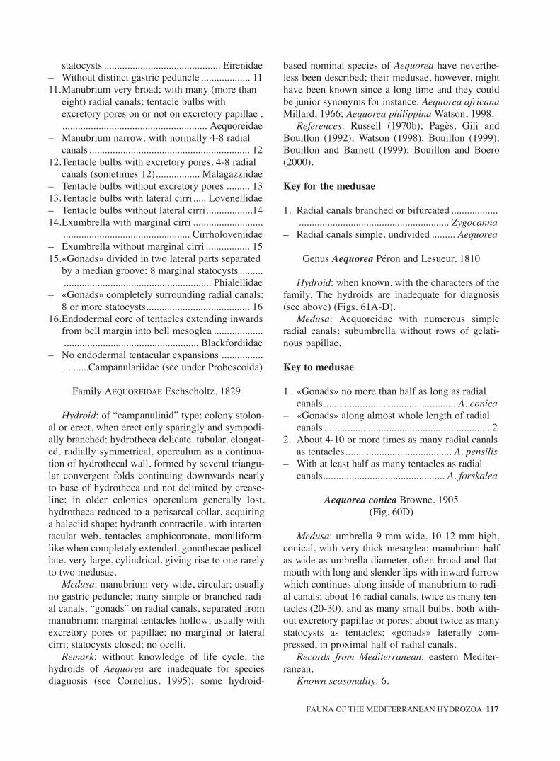
statocysts ............................................. Eirenidae– Without distinct gastric peduncle ................... 1111.Manubrium very broad; with many (more than
eight) radial canals; tentacle bulbs with excretory pores on or not on excretory papillae ......................................................... Aequoreidae
– Manubrium narrow; with normally 4-8 radial canals .............................................................. 12
12.Tentacle bulbs with excretory pores, 4-8 radial canals (sometimes 12)................. Malagazziidae
– Tentacle bulbs without excretory pores ......... 1313.Tentacle bulbs with lateral cirri..... Lovenellidae– Tentacle bulbs without lateral cirri..................1414.Exumbrella with marginal cirri ...........................
................................................. Cirrholoveniidae– Exumbrella without marginal cirri ................. 1515.«Gonads» divided in two lateral parts separated
by a median groove; 8 marginal statocysts .................................................................. Phialellidae
– «Gonads» completely surrounding radial canals; 8 or more statocysts........................................ 16
16.Endodermal core of tentacles extending inwards from bell margin into bell mesoglea ....................................................................... Blackfordiidae
– No endodermal tentacular expansions ..........................Campanulariidae (see under Proboscoida)
Family AEQUOREIDAE Eschscholtz, 1829
Hydroid: of “campanulinid” type; colony stolon-al or erect, when erect only sparingly and sympodi-ally branched; hydrotheca delicate, tubular, elongat-ed, radially symmetrical, operculum as a continua-tion of hydrothecal wall, formed by several triangu-lar convergent folds continuing downwards nearlyto base of hydrotheca and not delimited by crease-line; in older colonies operculum generally lost,hydrotheca reduced to a perisarcal collar, acquiringa haleciid shape; hydranth contractile, with interten-tacular web, tentacles amphicoronate, moniliform-like when completely extended; gonothecae pedicel-late, very large, cylindrical, giving rise to one rarelyto two medusae.
Medusa: manubrium very wide, circular; usuallyno gastric peduncle; many simple or branched radi-al canals; “gonads” on radial canals, separated frommanubrium; marginal tentacles hollow; usually withexcretory pores or papillae; no marginal or lateralcirri; statocysts closed; no ocelli.
Remark: without knowledge of life cycle, thehydroids of Aequorea are inadequate for speciesdiagnosis (see Cornelius, 1995); some hydroid-
based nominal species of Aequorea have neverthe-less been described; their medusae, however, mighthave been known since a long time and they couldbe junior synonyms for instance: Aequorea africanaMillard, 1966; Aequorea philippina Watson, 1998.
References: Russell (1970b); Pagès, Gili andBouillon (1992); Watson (1998); Bouillon (1999);Bouillon and Barnett (1999); Bouillon and Boero(2000).
Key for the medusae
1. Radial canals branched or bifurcated ............................................................................ Zygocanna
– Radial canals simple, undivided ......... Aequorea
Genus Aequorea Péron and Lesueur, 1810
Hydroid: when known, with the characters of thefamily. The hydroids are inadequate for diagnosis(see above) (Figs. 61A-D).
Medusa: Aequoreidae with numerous simpleradial canals; subumbrella without rows of gelati-nous papillae.
Key to medusae
1. «Gonads» no more than half as long as radial canals................................................... A. conica
– «Gonads» along almost whole length of radial canals ................................................................ 2
2. About 4-10 or more times as many radial canals as tentacles......................................... A. pensilis
– With at least half as many tentacles as radial canals............................................... A. forskalea
Aequorea conica Browne, 1905(Fig. 60D)
Medusa: umbrella 9 mm wide, 10-12 mm high,conical, with very thick mesoglea; manubrium halfas wide as umbrella diameter, often broad and flat;mouth with long and slender lips with inward furrowwhich continues along inside of manubrium to radi-al canals; about 16 radial canals, twice as many ten-tacles (20-30), and as many small bulbs, both with-out excretory papillae or pores; about twice as manystatocysts as tentacles; «gonads» laterally com-pressed, in proximal half of radial canals.
Records from Mediterranean: eastern Mediter-ranean.
Known seasonality: 6.
FAUNA OF THE MEDITERRANEAN HYDROZOA 117
sm68s2Bsystema1 14/10/04 15:22 Página 117

Distribution: Indo-Pacific; Mediterranean.References: Kramp (1961); Goy et al. (1988,
1990, 1991); Boero and Bouillon (1993).Hydroid: unknown.
Aequorea forskalea Péron and Lesueur, 1810(Figs. 60E-F, 61A-D)
Hydroid: colonies minute, stolonal with single orslightly branching erect hydrocauli which are imper-fectly annulated or spirally grooved throughout theirlength; hydrothecae cylindrical, with a long conicalfolded operculum tapering to a fine sharp point, thefolds continuing as striations of perisarc downwardsnearly to base of hydrotheca, hydrothecal base at rightangles to lateral walls; hydranths very extensile, withabout 20 amphicoronate filiform oral tentacles unitedat their base by a prominent intertentacular membra-nous web; gonothecae very large and cylindrical,blunt-ended, arising from hydrocaulus on shortimperfectly annulated stems just below hydranths andcontaining one rarely two medusa buds.
Medusa: umbrella large, up to 175 mm wide,saucer-shaped, thick in centre, gradually thinningtowards margin; manubrium half as wide as umbrel-la; radial canals usually 60-80, sometimes fewer orup to 160; «gonads» along almost whole length ofradial canals; tentacles generally fewer than radialcanals but varying from half to twice as many; ten-tacle bulbs elongate, conical; small bulbs few, scat-tered; excretory pores on short papillae; 5 -10 stato-cysts between successive radial canals.
Records from Mediterranean: eastern and west-ern Mediterranean; Adriatic.
Known seasonality: 1-12.Distribution: Atlantic; Indo-Pacific, Mediter-
ranean.References: Rees, 1938; Babnik (1948); Russell
(1953); Kramp (1961); Berhaut (1970); Goy (1973b);Brinckmann-Voss (1987); Gili (1986); Goy et al.(1988, 1990, 1991); Boero and Bouillon (1993); Cor-nelius (1995); Avian et al. (1995); Benovic and Lucic(1996); Medel and López-González (1996).
Aequorea pensilis (Eschscholtz, 1829)(Figs. 61E-F)
Medusa: umbrella up to 100 mm wide, more orless biconvex, with thin margin; manubrium 1/2-2/3as wide as umbrella; «gonads» extending alongalmost entire length of manubrium; 150-250 radialcanals; 10-16 tentacles and as many small rudimen-
tary bulbs; tentacle bulbs with long lateral exten-sions, no abaxial keel; without excretory papillaebut excretory pores present as slits; statocysts verynumerous.
Hydroid: unknown.Records from Mediterranean: ?Seasonality: ?Distribution: Atlantic; Indo-Pacific, Mediter-
ranean?References: Russell (1953); Kramp (1961);
Boero and Bouillon (1993).
Genus Zygocanna Haeckel, 1879
Medusa: Aequoreidae with numerous radialcanals, branched or bifurcated; exumbrella some-times with radial rows of gelatinous papillae.
Hydroid: unknown.
Zygocanna vagans Bigelow, 1912(Figs. 61G-H)
Medusa: umbrella up to 76 mm wide, flat, thin,but of hard consistency; exumbrella with radialbands of gelatinous papillae alternating with radialcanals; velum broad; manubrium occupying 1/3-1/2of umbrellar diameter, roof of the manubrium pre-senting a cruciform structure bifurcating two or fourtimes before reaching the manubrium peripherywhere the bifurcations give rise to about 30-45 nonbranched radial canals; mouth with crenulated lips;«gonads» extending along greatest part of radialcanals without reaching the umbrella margin; 28-70tentacles and several small rudimentary bulbs, withlong excretory papillae; statocysts very numerous.
Records from Mediterranean: Adriatic Sea.Known seasonality: 10.Distribution: Atlantic; Indo-Pacific; Mediter-
ranean.References: Babnik (1948) as Zygocanna. sp.;
Boero and Bouillon (1993); Avian et al. (1995).
Family AGLAOPHENIIDAE L. Agassiz, 1862
Hydroid: colony upright, mono- or polysiphonic,branched or unbranched, arising from creepinghydrorhiza or from anchoring filaments; hydrocla-dia alternate or opposite in one plane, or arrangedspirally; hydrothecae uniseriate, usually completelyadnate, margin usually cusped, with or withoutintrathecal septum, absent from hydrocaulus exceptin basal most segment; nematophores with nema-
118 J. BOUILLON et al.
sm68s2Bsystema1 14/10/04 15:22 Página 118

tothecae, not as naked sarcostyles; nemathothecae atleast partially fused to hydrothecae, one-chambered(monothalamic) and immovable; hydrotheca typi-cally flanked by three nematohtecae (five in somespecies of Aglaophenia and sometimes in Cladocar-pus), usually with one pair of lateral nematothecae,and with an unpaired median inferior nemathothecathat may be doubled or have two terminal apertures;sometimes also a pair of superior nematothecae;gonothecae lacking nematothecae, unprotected, orsurrounded by recurved branches in phylactocarp, ornearly completely enclosed within corbula bothrichly provided with nematothecae; fixed sporosacsor swimming gonophores.
References: Svoboda (1979); Svoboda and Cor-nelius (1991); Cornelius (1995); Migotto (1996);Calder (1997); Calder and Vervoort (1998); Watson(2000); Ansín Agís et al. (2001); Schuchert (2001a,2003).
Key to hydroids
1. Gonotheca unprotected ................. Gymnangium– Gonotheca protected in corbula or in
phylactocarp ..................................................... 22. Gonotheca protected in corbula replacing
hydrocladia with secondary ribs ...................... 3– No true corbula, gonotheca solitary and
protected by branched or unbranched phylactocarps.................................................... 4
3. Corbula ribs comprising nematothecae and hydrothecae ....................................... Lytocarpia
– Corbula comprising only nematothecae.......................................................................Aglaophenia
4. Phylactocarps formed by a modified hydrocladium, single or aggregated into pseudo-corbulae......................... Macrorhynchia
– Phylactocarps arising as appendages of an unmodified hydrocladium ................................ 5
5. Hydrocladia arranged in a spiral around stem .......................................................... Streptocaulus
– Hydrocladia arranged in two longitudinal rows......................................................... Cladocarpus
Genus Aglaophenia Lamouroux, 1812= Pentandra von Ledenfeld, 1884
Hydroid: colony erect, hydrocaulus branched orunbranched, monosiphonic or polysiphonic, arisingfrom creeping hydrorhiza or anchoring filaments;hydrocladia unbranched, pinnately arranged, arisingfrom alternate apophyses; hydrothecae only on
hydrocladia, typically more or less cone to sac-shaped, margin usually deeply toothed; intrathecalseptum variably developed; each hydrothecaflanked typically by a pair of lateral nematothecaeand a partly to wholly adnate, median inferior nema-totheca sometimes a pair of median superior nema-tothecae; gonothecae aggregated, enclosed within acorbula formed by modified hydrocladia bearingalternately inserted secondary ribs with nematothe-cae and lacking basal hydrothecae, corbula ribsfused or not; as fixed sporosacs, or released swim-ming gonophores.
Remark: Von Lendenfeld (1884) created Pentan-dra to accommodate P. balei and P. parvula, with 5nematothecae surrounding the hydrothecae: onemedian inferior, two lateral and a pair of superior(see diagnoses and Fig. 16 by Von Lendenfeld,1884), the last pair having been treated by manyauthors as a supplementary pair of lateral nema-tothecae! Bedot (1921) listed a series of genera andspecies showing deviations in numbers to the typi-cally three hydrothecal nematothecae and stated thatthere was no need to create new genera for suchvariations. We agree with his conclusions and con-sider Pentandra as congeneric with Aglaophenia.
References: Svoboda and Cornelius (1991);Ramil and Vervoort (1992a); Cornelius (1995);Calder (1997).
1. Stems without any basal prosegment .............................................................................. A. picardi
– Stems with one or more basal prosegment ...... 22. Hydrotheca with a greatly developed transverse
abcauline septum recurved at the tip.................................................................... A. kirchenpaueri
– Hydrotheca without this character ................... 33. Free part of mesial nematotheca long, usually
much longer than the free part of abcauline wall, and directed away from hydrotheca ............................................................................ A. tubulifera
– Mesial nematotheca without this character...... 44. Coenosarc with zooxanthellae; perisarc of stem,
hydrocladia and corbulae yellowish to brown . 5– Coenosarc without zooxanthellae; perisarc of the
stem, hydrocladia and corbulae not with a same colour................................................................ 6
5. Colonies growing only on Sea grasses, stem sometimes with intersegments; hooked apical stolons in summer ............................ A. harpago
– Colonies growing in different substrates, without intersegments neither hooked apical stolons.............................................................. A. tubiformis
FAUNA OF THE MEDITERRANEAN HYDROZOA 119
sm68s2Bsystema1 14/10/04 15:22 Página 119

6. Hydrotheca with the adcauline intrathecal septum long; hydrotheca usually narrowed basally, shape triangular more than tubular ..... 7
– Hydrotheca with the adcauline intrathecal septum usually short or absent; hydrotheca tubular............................................................... 8
7. Rim with non-uniform cusps, in side view mesial cusp inclined toward lumen; second pair of cusps bifid ................................................... A. parvula
– Rim with uniform cusps; second pair of cusps single .............................................. A. octodonta
8. Mature cormoid comprising one or many plumes branched or unbranched ................................... 9
– Mature cormoid comprising a single and unbranched plume ....................... A. lophocarpa
9. Branching dichotomous ....................... A. pluma– Branching trichotomous or irregularly bifid .. 1010.Ramus arising from apical end of a segment with
prosegments, comprising several roughly normally arranged cormidia ................ A. acacia
– Ramus always arising from a normal caulus segment which has one normally developed cladium............................................. A. elongata
Aglaophenia acacia Allman 1883(Figs. 62A-D)
Colonies pinnate (up to ca 200 mm), stems groupedclosely, monosiphonic and branched; axis brown, athe-cate basally, followed by a prosegment; remainder partsthecate, nodes oblique; internodes of the axis with threenematothecae and an abortive hydrotheca (pseudone-matotheca). Major branches (rami) widely spaced onstem, typically paired, or one of a pair replaced by acorbula; at point of branching, caulus bending back ver-sally resulting in a trichotomous arrangement. Sec-ondary hydrocauli with some of the basal internodeswithout hydrocladia, but with cormidia almost similarto those of the hydrocladia; however, the mesial nema-totheca is placed on the lower part of the internode,without contact with the abcauline hydrothecal wall.Hydrocladia white, alternate, closely set, one cormidi-um per internode, nodes transverse; hydrotheca narrowand deep (length/breadth ratio: 1-2), rim with 9 shallowcusp, outermost longest; intrathecal ridge short; mesialnematotheca short, reaching half length of hydrothecalabcauline wall, 1/3 of the nematothecae free; aperturegutter shaped; lateral nematothecae surpassinghydrothecal rim, slightly swollen. Male and female cor-bula white, with the costae completely fused, malewithout free costa; female sometimes first/last rib oneach side free.
Records from Mediterranean: eastern and west-ern Mediterranean.
Known seasonality: 1-12.Reproduction: 1, 2, 4-8, 12.Distribution: northern Atlantic (east and west),
Mediterranean.References: Fraser (1944); Svoboda (1979); Svo-
boda and Cornelius (1991); Boero and Bouillon(1993); Avian et al. (1995); Cornelius (1995);Medel and Vervoort (1995); Medel and López-González (1996); Ansín Agís et al. (2001); PeñaCantero and García Carrascosa (2002).
Aglaophenia elongata Meneghini, 1845(Figs. 62E-G)
Hydrocauli monosiphonic, up to 300 mm high,pinnate and branched, closely grouped. Axis brown-ish, basally with 1-4 prosegment, nodes oblique.Remainder internodes each bearing three nematothe-cae and one abortive hydrothecae (pseudonematothe-cae); nodes indistinct. Branches arising betweenmesial, proximal nematothecae and cladium or corbu-la, with 1-3 prosegment basally. Ramus and cladiumof same internode aligned parallel with each other; upto 4th order of branching. Hydrocladia yellow, alter-nate, spaced on stem, cormidia with one hydrothecaeand three gutter-shaped nematothecae. Hydrothecanarrow and deep (length/breadth ratio: 1.7-2.5), rimwith 9 uniform cusps; adnate mesial nematothecareaching 1/3-1/2 of the hydrotheca, free end short. Lat-eral nematothecae slightly above hydrothecal rim.Male and female corbulae in different colonies. Cor-bulae short, yellowish to white, 4-6 pairs of ribs fusedin female, with slits in male; free costa absent.Hydranth very large with usually long tentacles.
Records from Mediterranean: western and east-ern Mediterranean, Adriatic.
Known seasonality: 1,;4,;7-11.Reproduction: 4;6-11.Distribution: widely reported around the world
but records outside Mediterranean seem erroneous(Svoboda and Cornelius, 1991).
References: Svoboda (1979); Svoboda and Cor-nelius (1991); Boero and Bouillon (1993) Avian et al.(1995); Peña Cantero and García Carrascosa (2002).
Aglaophenia harpago von Schenck, 1965(Figs. 62H-K)
Hydrorhizae growing only along sea grass blade.Well developed monosiphonic hydrocauli brown, up
120 J. BOUILLON et al.
sm68s2Bsystema1 14/10/04 15:22 Página 120

to 18 mm, bending backwards; one prosegmentbasally separated by very deep oblique furrows,which allow bending in response to current surge;remainder parts of the axis composed of segmentsbearing one pseudonematothecae and three nema-tothecae gutter shaped; nodes slightly oblique; insome stems, intermediate segments with frontalnematothecae occurs distally, between hydrocladia-bearing segments. Caulus ended by thick, narrowhook may occurs specially in spring and summer.Coenosarc of the colony with zooxanthellae. Hydro-cladia brown, more spaced than in other Aglaophe-nia species; cormidia with one hydrothecae andthree nematothecae. Hydrothecae may vary fromlong and narrow in some colonies to wider andshorter in other ones (length/breadth ratio: 1.8, -1.4).Colonies dioecious, corbulae brown, short, 4-6 pairsof ribs fused in female, partly open in male.
Records from Mediterranean: eastern and west-ern Mediterranean, Adriatic.
Known seasonality: 2, 3, and 5, 6, 8, 9.Reproduction: 3-7.Distribution: endemic of Mediterranean Sea.References: Svoboda (1979); Gili (1986); Roca
(1986); Svoboda and Cornelius (1991); Boero andBouillon (1993); Avian et al. (1995); Medel andLópez-González (1996).
Aglaophenia kirchenpaueri (Heller, 1868)(Figs. 63A-D)
Hydrocauli pinnate, up to 200 mm high yellow-ish to brown, monosiphonic and unbranched; basalpart of the axis athecate, followed by up to threeprosegments, each with a frontal nematotheca.Remainder internodes with three nematothecae anda pseudonematotheca; nodes oblique. Hydrocladiayellow or brown, alternate, closely set; cormidiaeach with one hydrothecae and three nematothecae(one median inferior and two laterals); nodes trans-verse. Hydrotheca not deep (length/breadth at rim:1-1.3), with a greatly developed transverseabcauline septum recurved at the tip; rim with 9marginal cusps, first pair on both sides longest,remainder marginal cusps grading towards axis ofcormidium. Median nematothecae not surpassinghydrothecal rim, 2/3 adnate, aperture gutter-shaped.Lateral nematothecae projecting slightly abovehydrothecal rim; distal portion curved outwards,aperture gutter-shaped. Corbulae yellow or brown,male partially open, usually without free costae;female usually closed and with free costa.
Records from Mediterranean: western Mediter-ranean as far east as Adriatic Sea.
Known seasonality: 1-12.Reproduction: 2; 4-11.Distribution: northeastern Atlantic from British
coasts to Cape Verde Islands; Mediterranean.References: Patriti (1970); Svoboda (1979); Svo-
boda and Cornelius (1991); Ramil and Vervoort(1992a); Boero and Bouillon (1993); Cornelius(1995); Medel and Vervoort (1995); Medel andLópez-González (1996); Ansín Agís et al. (2001);Peña Cantero and García Carrascosa (2002).
Aglaophenia lophocarpa Allman, 1877(Figs. 63E-H)
Hydrocauli pinnate, up to ca 50 mm, monosi-phonic and unbranched; colour brown. Axis basallywith 1-3 prosegment with frontal nematothecae sep-arated by transverse nodes; remainder parts withinternodes bearing hydrocladia; each with onepseudonematothecae and three nematothecae; nodesindistinct. Hydrocladia widely spaced on stem;hydrothecae narrow and deep (length/breadth at rim:1.7-2.1), rim with 9 uniform cusps; short intrathecalseptum in lower third of hydrothecae; adnate part ofmesial nematotheca reaching the middle ofhydrotheca, free part short, foramen into hydrothe-cae usually open; lateral nematothecae reaching thehydrothecal rim or slightly overtopping. Coloniesdioecious; female corbulae brown, ribs fused; malewhite, ribs usually with slits between nematothecae,but fused ribs may occurs.
Records from Mediterranean: western and east-ern Mediterranean, Adriatic.
Known seasonality and reproduction: 4-9.Distribution: tropical eastern Atlantic (Azores,
Guinea Bissau); Caribbean; Mediterranean.References: Svoboda (1979); Gili, Vervoort and
Pagès (1989); Svoboda and Cornelius (1991); Ramiland Vervoort (1992a); Boero and Bouillon (1993);Ansín Agís et al. (2001).
Aglaophenia octodonta (Heller, 1868)(Figs. 63I-L)
Hydrocauli monosiphonic, yellowish to brownand unbranched. Axis basally, with several transversenodes and with 1-3 prosegments separated by obliquenodes; remainder parts composed of hydrocladiabearing internodes, with three gutter-shaped nema-tothecae and a pseudonematothecae; nodes oblique.
FAUNA OF THE MEDITERRANEAN HYDROZOA 121
sm68s2Bsystema1 14/10/04 15:22 Página 121

Hydrocladia transparent to yellowish, closely alter-nate; cormidia separated by transverse nodes.Hydrothecae not deep (length/breadth at rim: 1.1-1.4), narrowing basally and thus of triangular shape;rim with 9 uniform cusps; adcauline septum in lowerthird of hydrothecal cavity well developed; mediannematothecae 2/3 adnate, free end short almost reach-ing the hydrothecal rim; end of nematothecae usuallycurved outwards and reaching almost at level ofhydrothecal rim. Lateral nematothecae small, apicalpart swollen, reaching hydrothecal rim. Male corbulawhite, without free costa and usually partly open;female corbula yellow, usually closed, with one welldeveloped free costa.
Records from Mediterranean: eastern and west-ern Mediterranean, Adriatic.
Known seasonality and reproduction: throughoutthe year.
Distribution: northeastern Atlantic and Mediter-ranean.
References: Rossi (1961); Svoboda (1979); Gili(1986); Roca (1986); Svoboda and Cornelius(1991); Ramil and Vervoort (1992a); Boero andBouillon (1993); Avian et al. (1995); Medel andVervoort (1995); Medel and López-González(1996); Ansín Agís et al. (2001); Peña Cantero andGarcía Carrascosa (2002).
Aglaophenia parvula Bale, 1882(Figs. 64A-D)
Hydrocauli monosiphonic, up to 70 mm, yellow-ish to brown, unbranched or dichotomouslybranched. Axis basally provided or not with severaltransverse nodes and 1-3 prosegments separated byoblique nodes; remainder internodes each with threenematothecae and a pseudonematothecae. Hydrocla-dia alternate, closely set, divided into cormidia sep-arated by transverse nodes. Hydrotheca fairly deep,narrowing basally, with a triangular aspect; rim withnine unequal marginal cusps, outermost cusp single,unpaired, flexed interiorly; third pair bifid apex invaried degree; intrathecal septum well developed,almost reaching opposite hydrothecal wall; mesialnematothecae almost reaching hydrothecal rim; lat-eral nematohtecae short, not reaching hydrothecalrim. Colonies dioecious, male corbula with smallslits between nematophores of ribs; female corbularibs fused with exception of rachial basis.
Records from Mediterranean: found in the near-by areas of the Strait of Gibraltar.
Seasonality: ?
Distribution: eastern Atlantic; S Australia,Mediterranean?.
References: Millard (1975) as A. pluma parvula;Ramil (1988); Gili et al. (1989); Svoboda and Cor-nelius (1991); Medel and Vervoort (1995); Medeland López-González (1996); Ansín Agís et al.(2001).
Aglaophenia picardi Svoboda, 1979(Figs. 64E-H)
Hydrocauli monosiphonic and unbranched, up to80 mm, axis brown, without any basal prosegment,but several transverse nodes may occurs. Remainderparts composed of thecate internodes bearing hydro-cladia, each with three nematothecae and apseudonematothecae; nodes oblique. Hydrocladiaalternate, widely spaced, perisarc transparent, divid-ed into cormidia by transverse nodes. Hydrothecaedeep (length/breadth at rim: 1.5), rim with nine uni-form cusps, adcauline septum in lower third ofhydrotheca inconspicuous or absent; median nema-tothecae gutter shaped, almost complete adnate to thehydrothecae, this with almost a half of its length free.Lateral nematothecae reaching hydrothecal rim. Cor-bulae transparent to white, male with large openingsbetween the pairs of opposite costae; female closed,with the proximal pairs of ribs almost free.
Records from Mediterranean: eastern and west-ern Mediterranean, Adriatic.
Known seasonality: 1-12.Reproduction: 1-12Distribution: northeastern Atlantic (N Spain,
Cape Verde Is.), Black Sea; Mediterranean.References: Svoboda (1979); Isasi (1985); Svo-
boda and Cornelius (1991); Ramil and Vervoort(1992a); Avian et al. (1995); Medel and Vervoort(1995); Medel and López-González (1996); AnsínAgís et al. (2001); Peña Cantero and García Carras-cosa (2002).
Aglaophenia pluma (Linnaeus, 1758)(Figs. 64I-L)
Hydrocauli monosiphonic up to 150 mm, brown,unbranched or dichotomously branched; basal part ofthe axis athecate, followed by one or two proseg-ments; remainder internodes, each with three nema-tothecae and a pseudonematotheca; nodes oblique.Hydrocladia alternate, with whitish cormidia separat-ed by transverse nodes. Hydrotheca deep(length/breadth at rim: 1.3-1.5), rim with nine cusps
122 J. BOUILLON et al.
sm68s2Bsystema1 14/10/04 15:22 Página 122

of varied length; intrathecal adcauline septum usuallywell developed, median nematothecae 2/3 adnate, notreaching the margin of the hydrothecae; lateral nema-tothecae reaching the rim of the hydrothecae; apertureof nematothecae gutter-shaped. Colonies dioecious,male and female corbulae white, with free costa; maleclose, with slit like openings between the costae,female with fused costae and smaller slits. Hydranthsmall, transparent, cylindrical, hypostome held atlevel of hydrothecal rim, rounded and low; ten tenta-cles emerging of hydrothecal rim.
Records from Mediterranean: western Mediter-ranean.
Known seasonality and reproduction: almost allthe year.
Distribution: cited as cosmopolitan species, butthis need of evaluation due to identification prob-lems. Reliably reported from Shetlands, BritishIslands, France, Belgium, Spain and Portugal, in thenorth eastern Atlantic.
References: Vervoort (1946); Patriti (1970); Svo-boda (1979); Svoboda and Cornelius (1991); Boeroand Bouillon (1993); Medel and Vervoort (1995);Medel and López-González (1996); Ansín Agís et al.(2001); Peña Cantero and García Carrascosa (2002).
Aglaophenia tubiformisMarktanner-Turneretscher, 1890 (Figs. 65A-E)
Hydrocauli monosiphonic up to 150 mm, yel-lowish to brown, unbranched or dichotomouslybranched. Axis basally athecate, with several trans-verse nodes, followed by one (rarely two) proseg-ment separated by oblique nodes. Remainder intern-odes each with three nematothecae and a pseudone-matothecae. Hydrocladia alternate, yellowish tobrown, divided into cormidia by transverse nodes;cormidia composed of one hydrothecae and threenematothecae. Hydrotheca deep (length/breadth atrim: 1.5-2), rim with 9 more or less equal cusps,intrathecal adcauline septum moderately developed;mesial nematotheca arising between the middle andupper third of hydrotheca, free portion of variablelength; lateral nematothecae reaching or overtop-ping the hydrothecal rim; nematothecae with a gut-ter shaped aperture. Coenosarc filled with zooxan-thellae. Colonies dioecious; corbulae brown, withthe first pair of ribs free; male with slits betweenribs; female with almost completely fused ribs.
Records from Mediterranean: eastern and west-ern Mediterranean, Adriatic (but not Black Sea).
Known seasonality: 1-12.
Reproduction: 2-10.Distribution: northeastern Atlantic, Mediterraean.References: Svoboda (1979); Ramil (1988); Svo-
boda and Cornelius (1991); Ramil and Vervoort(1992a); Boero and Bouillon (1993); Alvarez(1993); Avian et al. (1995); Medel and Vervoort(1995); Medel and López-González (1996); PeñaCantero and García Carrascosa (2002).
Aglaophenia tubulifera (Hincks, 1861)(Figs. 65F-I)
Colonies pinnate up to 60 mm, composed ofmonosiphonic and unbranched hydrocauli withalternate hydrocladia. Axis basally with a proseg-ment separated by oblique nodes; occasionally alsowith several transverse nodes; remainder parts com-posed of bearing-hydrocladia internodes, each withone pseudonematothecae and three nematothecae;nodes transverse. Hydrocladia alternate, closelypacked, each composed of a large number ofcormidia; nodes transverse. Hydrotheca not deep;adcauline septum weakly developed, rim with 9marginal cusp, the median one longest. Nematothe-cae around hydrotheca without embayment; mesialnematothecae long, free part directed away fromhydrotheca. Coenosarc with zooxantellae. Coloniesdioecious; male corbula with slits between ribs,female with almost completely fused ribs and abasal free costa. Hydranth short, cylindrical, whenextending not projecting beyond hydrothecal rim;hypostome hemispherical, ten short tentacles.
Records from Mediterranean: western Mediter-ranean (Spain, Morocco).
Known seasonality: 5, 6, 7, 9.Reproduction: 5, 6, 7.Distribution: northeastern Atlantic, Mediterranean.References: Patriti (1970); Svoboda (1979); Gili
et al. (1989); Svoboda and Cornelius (1991); Boeroand Bouillon (1993); Medel and Vervoort (1995);Medel and López-González (1996); Ansín Agís etal. (2001).
Genus Cladocarpus Allman, 1874 = Aglaophenopsis Fewkes, 1881
= Nematocarpus Broch, 1918= Wanglaophenia Vervoot and Watson, 2003
Hydroid: colony erect, monosphonic or polysi-phonic, hydrocaulus branched or unbranched, bearingalternate usually unbranched hydrocladia; hydrocladiainternodes with numerous septa; hydrotheca deep,
FAUNA OF THE MEDITERRANEAN HYDROZOA 123
sm68s2Bsystema1 14/10/04 15:22 Página 123

often S-shaped, with or without intrathecal septa, usu-ally with a median abcauline tooth, with or withoutlateral teeth; nemathotheca usually with more than oneaperture, median inferior nematotheca short, usuallybelow hydrotheca, never reaching thecal margin,sometimes a superior nematotheca; gonothecae notcontiguous, usually protected by loose phylactocarpswith either unbranched or dichotomously branchedaxis made of a regular succession of segments eachwith 2-3 nematothecae and bearing an alternateapophysis supporting a nematophorous branch, usual-ly without hydrothecae, phylactocarp structuresresembling stag antlers.
Remarks: Ramil and Vervoort (1992 b) pointedout that the genus Cladocarpus contains two groupsof species with a different morphology of phylacto-carps. In the first one, the phylactocarp is homolo-gous with the hydrocaulus of Aglaopheniidae,unbranched or branched and with segments bearingnematophorous branches; in the second one, beingsimilar to the structure of hydrocladia, with segmentbearing each pairs (or a pair) of nematothecae inopposite pairs. The first group of species, in theiropinion, should remain in Cladocarpus Allman,1874 and the second group should be placed inStreptocaulus Allman, 1883. But as remarked bySchuchert (2001 a) some species of Cladocarpushave the proximal part of the phylactocaps of onetype and the distal one of the other (Cladocarpusbonneviae) and in some species of Cladocarpus thephylactocarps are not referable to one of thedescribed types (C. integer). Schuchert (2001a) con-sider therefore that the genus needs comprehensivephylogenetical analyses to recognize monophyletictaxa with sufficient reliability and that the types ofphylactocarps have to been defined more precisely.
1. Abcauline hydrothecal wall curvate................. 2– Abcauline hydrothecal wall straight ................ 32. Abcauline hydrothecal wall with double
curvature; with intrathecal septum .. C. sinuosus– Abcauline wall strongly concave; without
intrathecal septum ................................ C. tenuis3. Aperture of hydrotheca even ...... C. pectiniferus– Aperture of hydrotheca with a prominent
abcauline cusp .......................... C. multiseptatus
Cladocarpus multiseptatus (Bale, 1915)(Fig. 65K)
Colonies pinnate up to ca 15 mm; hydrocaulipolysiphonic composed of several tubes of which, a
“main” frontal axis bearing nematothecae. Hydro-cladia borne on apophyses, alternate; internodeswith one hydrotheca and three nematothecae, onemedian inferior and two lateral; nodes slightlyoblique; each internode with 9-13 transverse peris-arcal septa behind hydrothecae, 2-4 below and 1-2above. Hydrotheca tubular, deep, tapered basally,widing distally; adcauline wall entire adnate;abcauline wall slightly curved outwards at distalthird; margin sinuous, with a prominent medianabcauline cusp. Nematothecae tubular, graduallynarrowing towards distal end; median inferior notreaching hydrothecal base, with two apertures, onedistally, slit-shaped, the other one, circular, at theend of a short tube basally on the nematotheca, anddirected towards hydrocladial wall; lateral nema-tothecae reaching beyond hydrothecal rim, also withtwo apertures, one apical, with a sinuous rim, andanother one basal, on adcauline side, at end of shorttube with a incomplete internal septum at its base;cauline nematothecae with unique apical aperture.Gonothecae protected by unbranched phylactocarpswith nematothecae.
Records from Mediterranean: only Alboràn Sea.Known seasonality: 6.Distribution: N Spain, Australia and Mediter-
ranean (Alboràn Sea).References: Ramil and Vervoort (1992a);
Alvarez (1993); Medel and López-González (1996).
Cladocarpus pectiniferus Allman, 1883(Figs. 65L-N, 66A-C)
Hydrocauli polysiphonic basally, up to 100 mmhigh, growing from matting of perisarcal tubules.Frontal tube of the axis with a row of frontal nema-tothecae and apophyses bearing hydrocladia; usual-ly 2-5 nematothecae between two apophyses butvery variable, one nematothecae borne in the axil ofeach apophysis. Nematothecae tubular, with a singleapical aperture. Hydrocladia alternate, composed ofinternodes separated by slightly oblique nodes; eachinternode with one hydrotheca and three nematothe-cae, one median inferior and two laterals; internodeswith 17-27 septa. Hydrothecae tubular, elongated;walls almost straight; adcauline wall entire adnate,abcauline wall slightly curved outwards distally; rimsmooth, somewhat convex frontally. Nematothecaetubular, with two openings, one terminal with slight-ly serrated border, and one basal adcauline; lateralnematothecae with the two apertures connected,may occurs also; median inferior nematothecae
124 J. BOUILLON et al.
sm68s2Bsystema1 14/10/04 15:22 Página 124

reaching or not hydrothecal base; lateral nematothe-cae overtopping hydrothecal rim. Phylactocarps inpairs at first hydrocladial internode, composed ofrachis bearing gonothecae and paired nematothecae;rachis with four internodes, each with basal and dis-tal septum; first two segments with two pairs ofnematothecae, the rest with only one pair; nema-tothecae tubular, elongated, with two apertures, onedistally, finely serrated, and one basal, large andoval; one or two additional, smaller may occurs inbetween.
Records from Mediterranean: only Alboràn Sea.Known seasonality: 5, 6Distribution: northeastern Atlantic, from Iceland
to Morocco, and Alboràn Sea.References: Vervoort (1966); Ramil and Vervoort
(1992 a and b); Medel and López-González (1996);Schuchert (2001a).
Cladocarpus sinuosus Vervoort, 1966(Figs. 66D-I)
Colonies pinnate with monosiphonic or polysi-phonic and unbranched hydrocauli up to 53 mm;main frontal axis bearing a row of nematothecae andalternate hydrocladial apophyses; one nematothecaeis borne in the axil of each apophysis; hydrocladiumcomposed of hydrothecate internodes separated byoblique septa; internodes sinuous, with onehydrothecae and three nematothecae, one medianinferior and two laterals overtopping hydrothecalrim. each internode internally with 8-12 thick septa:3 basal, 6-8 behind hydrothecae and one apical;hydrotheca deep, with transverse adcauline septumrecurved at the tip at lower third; adcauline wallentire adnate, concave above insertion of septum;abcauline wall with double curvature, strongly con-vex in basal half, and strongly concave at about dis-tal third; rim even, with a unique median abcaulinecusp. Median inferior nematotheca almost reachinghydrothecal base, with two apertures, one distal andone basal towards hydrothecal wall; lateral nema-tothecae tubular, with two apertures, one terminaland one mesial, with intrathecal septum; caulinenematothecae also with two apertures, one terminaland one basal, towards axial wall; phylactocarpborne on first internode of hydrocladium, curvedand unbranched, with short internodes with two verylong, curved nematothecae with up to four apertures,one terminal and the rest along the upper surface;gonothecae below first pair of nematothecae, ovoidwith truncated distal end.
Records from Mediterranean: only Alboràn Sea.Known seasonality: 6.Distribution: eastern Atlantic (coasts of Africa)
and Alboràn Sea.References: Vervoort (1966); Millard (1975);
Gili et al. (1989); Ramil and Vervoort (1992a);Medel and López-González (1996).
Cladocarpus tenuis Clarke 1879(Figs. 66J-K)
Hydrocaulus unfascicled attaining a height ofabout 40 mm, very delicate, bearing alternate hydro-cladia, containing some oblique nodes in basalregion only, bearing a row of nematothecae on ante-rior surface, of which one is seated in the axil ofeach apophysis; hydrocladium consisting of weaklysigmoid thecate internodes separated by slightlyoblique nodes; internodes containing numeroussepta and bearing three nematothecae, one medianinferior and two laterals; hydrotheca very deep andslender, not sigmoidally curved, narrowest in thecenter then widening to margin, abcauline wall con-cave in the centre; adcauline wall straight; withoutintrathecal septum; margin with one medianeabcauline tooth and crenulated lateral edges; medianinferior nematotheca free from hydrotheca, short notreaching thecal base; with large fan-shaped terminalaperture, lateral nematotheca tubular, overtoppingthecal margin, with terminal aperture; cauline nema-tothecae similar to median inferior; phylactocarpborne on first internode of hydrocladium,unbranched, segmented, with two long nematothe-cae on each internode, nematotheca with three aper-tures, one terminal and two laterals; gonothecaeunknown.
Records from Mediterranean: western Mediter-ranean.
Seasonality: ?Distribution: Gulf of Mexico; West Indies;
Mediterranean.References: Nutting (1900); Vervoort (1966);
Millard (1975); Marinopoulos (1981).
Genus Gymnangium Hincks, 1874 = Haliaria Stechow, 1921 = Halicetta Stechow, 1921
Hydroid: colony erect, often stout, monosiphonicor polysiphonic, arising from a creeping hydrorhiza orfrom anchoring filaments; hydrocladia unbranched,alternate or opposite, giving off from opposite sides of
FAUNA OF THE MEDITERRANEAN HYDROZOA 125
sm68s2Bsystema1 14/10/04 15:22 Página 125

hydrocaulus; hydrothecae only on hydrocladia, typi-cally more or less cone-shaped, intrathecal septumpresent or absent, margin with or without cusps; eachhydrotheca with a pair of lateral nemathotecae and asingle adnate median inferior nematotheca, conspicu-ously longer than hydrotheca (2-3 times), and havingmore than one opening; gonotheca solitary, usuallyborne on hydrocladia, not protected by phylactocarpsor corbulae; fixed sporosacs, one species with swim-ming gonophores (G. ferlusi).
References: Cornelius (1995); Calder (1997);Schuchert (2003).
Gymnangium montagui (Billard, 1912)(Figs. 66L-O)
Colonies erect and monosiphonic up to 120 mm,with a yellowish to brown hydrocauli and a thickhydrorhiza. Axis segmented into short internodes;nodes slightly oblique; each internode with two later-al apophyses bearing hydrocladia, alternate, one basaland the another one distal; two cups-shaped nema-tothecae are located basally to each apophysis, anupper and a lower. Hydrocladia alternate, closely set,internodes short, nodes almost transverse; each intern-ode with one hydrotheca and three nematothecae;hydrotheca cup-shaped, 2/3 of adcauline wall adnate,a distinct, slightly curved abcauline internal septum athalf length of abcauline wall, dividing it into two con-vex portions; hydrothecal aperture inclined down-ward, rim sinuous, occasionally with indication of lat-eral cusp on each side; lateral nematothecae also cup-shaped, median nematotheca much elongated, curved,surpassing hydrothecal border considerably, free partat least half length of adnate part or more, opening gut-ter-shaped, along whole free length; gonothecae intwo rows on frontal aspect of colony, each attached tohydrocladial apophysis, colour white, conical, trun-cate distally and there with concavity.
Records from Mediterranean: Atlantic speciesfound in Algeciras Bay (Strait of Gibraltar).
Known seasonality: 5, 7, 12.Reproduction: 5.Records outside the Mediterranean: eastern
Atlantic.References: Millard (1975); Cornelius (1995);
Medel and Vervoort (1995); Medel and López-González (1996).
Genus Lytocarpia Kirchenpauer, 1872 = Acanthocladium Allman, 1883
= Thecocarpus Nutting, 1900
Hydroid: colony erect, branched or unbranched,bearing alternate unbranched pinnate hydrocladia;hydrotheca sac-shaped to deep, usually withintrathecal septum; generally with teeth or lobedmargin; mesial outer tooth of hydrothecal rim usual-ly prominent; median inferior nematotheca fairlyshort, not reaching hydrothecal margin, sometimes asuperior dissymmetrical nematotheca (i.e. L. pera-mata); corbulae formed by modified hydrocladiabearing secondary unfused ribs bearing a row ofapproximately 12 nemathothecae, some, in at leastone sex, bearing one hydrotheca; fixed sporosacs.
References: Calder (1997); Schuchert (2001a,2003).
1. Hydrothecate internodes with 2-3 poorly developed internal septa ..................... L. distans
– Hydrothecate internodes with 6-13 well developed internal septa .......... L. myriophyllum
Lytocarpia distans (Allman, 1877)(Figs. 67A-B)
Hydrocauli polysiphonic up to 200 mm high,growing on a hydrorhiza composed of numeroustubes. Frontal tube of the axis with a longitudinalrow of nematothecae and lateral hydrocladialapophyses, 2-5 nematothecae between two apophy-ses. Hydrocladia alternate, in the same plane, withinternodes separated by transverse nodes, each withone hydrotheca and three nematothecae, one medianinferior and two laterals; internal septa poorly devel-oped, two under the median nematothecae and onebehind hydrotheca at same level as intrathecal sep-tum. Hydrothecae elongated, narrowed basally;adcauline wall completely adnate, intrathecal sep-tum poorly developed; rim with shallow cusps, but aprominent one frontally. Median inferior nematothe-ca short, reaching the hydrothecal base; lateralnematothecae small, rim at same level of hydrothe-cal rim; all nematothecae with a gutter-shaped aper-ture. Corbulae with 10-14 costae, each with onebasal hydrotheca, 2-3 abcauline nematothecae and6-10 adcauline ones.
Records from Mediterranean: western Mediter-ranean.
Known seasonality: 8. Corbulae: 8.Distribution: northeastern Atlantic and Pacific;
Mediterranean.References: Bedot (1921); Vervoort (1972); Gili
(1986); Ramil and Vervoort (1992a); Boero andBouillon (1993); Medel and López-González (1996).
126 J. BOUILLON et al.
sm68s2Bsystema1 14/10/04 15:22 Página 126

Remark: species very similar to L. Myriophyllumbut clearly differs in the shape of hydrothecae, themedian inferior nematothecae, the adcauline walland in the development and number of internodalsepta (see Ramil and Vervoort, 1992a).
Lytocarpia myriophyllum (Linnaeus, 1758)(Figs. 66P-U)
Hydrorhiza composed of numerous fibers livingthe hydrocaulus over different levels. Stem polysi-phonic, up to about 350 mm, yellowish to brown,pinnate and unbranched; up to four well markedoblique nodes arranged basally; axis with a “main”frontal tube and many parallel secondary ones, allwith a longitudinal row of small cup-shaped nema-tothecae. Hydrocladia borne on main tube; alternate,in the same plane. Hydrocladial internodes eachwith one hydrothecae and three nematothecae, onemedian inferior and two laterals; a varied number(6-13) of perisarcal septa developed transversally inthe internodes; nodes transverse. Hydrotheca moreor less cylindrical, rounded and with transverseadcauline septum basally; rim with shallow cusps,but frontal one on abcauline wall very prominent.Mesial nematothecae reaching the lower forth ofhydrothecae, free part short, aperture with a deepadcauline embayment; lateral nematothecae slightlyovertopping hydrothecal rim, aperture with a deepadcauline embayment. Corbulae large, open, 5-20costae, each with usually one, but up to three basalhydrothecae and several nematothecae, usually 2abcauline and 5-9 adcauline; gonothecae ovoid,alternate, up to 24 in a same corbula.
Records from Mediterranean: western Mediter-ranean, Adriatic.
Known seasonality: 6-8. Corbulae: 8.Distribution: Arctic, eastern Atlantic (till Guinea
Bissau); Mediterranean; Indo-Pacific records needto be evaluated.
References: Gili, Vervoort and Pagès (1989);Ramil and Vervoort (1992a); Boero and Bouillon(1993); Cornelius (1995); Avian et al. (1995);Medel and Vervoort (1995); Medel and López-González (1996).
Genus Macrorhynchia Kirchenpauer, 1872 = Lytocarpus Allman, 1883
= Nematophorus Clarke, 1879
Hydroid: colony erect, hydrocauli branched orunbranched, polysiphonic, often stout, arising from
creeping hydrorhiza or anchoring filaments; hydro-cladia unbranched, pinnately arranged, arising alter-nately from apophyses on axial tube of hydrocaulusand branches; hydrothecae only on hydrocladia,more or less cone to sac-shaped; hydrothecal margindentate; abcauline or adcauline intrathecal septumpresent; cauline internodes with triangularnemathotheca; each hydrotheca with a pair of later-al nemathotecae and a single partly adnate medianinferior nematotheca; gonophores usually fixedsporosacs, exceptionally one species with swim-ming sporosacs (Macrorhynchia philippinaKirchenpauer, 1872), gonothecae on unbranchedphylactocarps formed by modified hydrocladia,occurring single or aggregated in pseudocorbula.
References: Calder (1997); Schuchert (2003).
Macrorhynchia philippina (Kirchenpauer, 1872)(Figs. 67C-H)
Colonies about 15-20 cm high; hydrocauli poly-siphonic, strongly and irregularly branched. Branch-es arising from peripheral tubes of stem, monosi-phonic or slightly polysiphonic, with a basal partbearing median nematothecae and a distal part bear-ing alternate hydrocladia. Axial tube of stem anddistal part of branches with internodes each with onehydrocladial apophysis and two nematothecae;mamelon present on anterior surface of apophysis.Hydrocladia not very long, with 10-15 internodeseach with one hydrotheca, two lateral nematothecaeand one median hydrotheca; nodes straight or slight-ly oblique. Hydrotheca with a thick abcaulineintrathecal septum and an adcauline intrathecal oneat lower level; margin with two pairs of low, round-ed cusps and a thickened abcauline median cuspcontinuous basally with the abcauline intrathecalseptum and projecting distally as a short spine. Lat-eral nematothecae tubular, projecting abovehydrothecal margin, with two openings, one distaland one mesial; free part of the median nematothecalong, overtopping hydrothecal margin, with threeopenings, one rounded distal, one on upper surfaceat base of free part, and one into the hydrohteca; 0-2 internodal septa. Phylactocarp borne on apophy-ses, with a hydrothecate internode basally, this fol-lowed by several gonothecae-bearing internodes, onwhich the hydrotheca has been replaced by andapophysis bearing a gonothecae; gonothecae large,disc-shaped, dioecious, giving free medusoids, thesewithout radial canals, circular canal, tentacles andtentacular bulbs.
FAUNA OF THE MEDITERRANEAN HYDROZOA 127
sm68s2Bsystema1 14/10/04 15:22 Página 127

Records from Mediterranean: eastern Mediter-ranean.
Seasonality: ?Records outside the Mediterranean: tropical watersof the Atlantic, Pacific and Indian Oceans.
References: Vervoort (1968); Gravier (1970);Millard (1975); Boero et al. (1997), Watson (2000).
Genus Streptocaulus Allman, 1883= Aglaophenopsis Fewkes, 1881
=Nematocarpus Broch, 1918
Hydroid: colony erect; hydrocauli branched orunbranched; hydrocladia pinnate in young colonies,gradually becoming spirally arranged by axis tor-sion with age; hydrothecae adnate, hydrothecal rimwith weakly developed cusps; three hydrothecalnematothecae present; fixed sporosacs protected byphylactocarps with axis homologous to a hydrocla-dium; rachis axis unbranched or irregularlybranched, divided in segments, each bearing one orseveral pairs of more or less opposite lateral nema-tothecae; rachis axis bearing one or more gonothe-cae; when axis and phylactocarps are long, the struc-ture appears centipede-like.
Remark: The genus Streptocaulus seems polyphylet-ic based on phylactocarp morphology (Calder, 1997).
References: Ramil and Vervoort (1992a); Medel andVervoort (1995); Calder (1997); Ramil et al. (1998).
Streptocaulus dollfusi (Billard, 1924)(Figs. 67I-K)
Hydrocauli long (up to about 400 mm), polysi-phonic, yellowish, occasionally irregularly branched.Only a frontal tube of the stem thecate and bearinghydrocladia, divided into internodes by means oftransverse nodes; segments with a distal bearinghydrocladia apophysis, and a row of frontal nema-tothecae (usually 3-5); one of them shorter, in the axilof the apophysis, occasionally also another onebesides of the apophysis; all nematothecae with twoapertures, one terminal and one basal at the adcaulinewall. Hydrocladia alternate, more or less in the sameplane; internodes with one hydrothecae and threenematothecae; nodes slightly oblique; six perisarcalring at the base of median nematothecae (one, butoccasionally doubled) and behind the hydrothecae(five). Hydrothecae long, fully adnate, abcauline wallconvex in lower half, straight and inclined in upperhalf; a transverse adcauline septum in lower third, thistypically recurved upwards. Hydrothecal rim with
rounded and weakly developed cusps on each side oronly sinuous side, and with a distinct abcauline,frontal cusp. Mesial nematothecae not reaching thehydrotheca base, tubular, rim circular, deep adcaulineembayment; lateral nematothecae overpassinghydrothecae, with two apertures, one apical and anoth-er one adcaulinar, opening into hydrothecal cavity.Phylactocarp composed of eight segments, each witha pair of opposite nematothecae, these with wide aper-tures; gonothecae on the basal segment; female cylin-drical, ends rounded, aperture sub-distal and oval,male shorter, ovoid, with a terminal aperture.
Records from Mediterranean: western Mediter-ranean (S. Spain).
Known seasonality: 7.Distribution: eastern Atlantic (from Morocco to
Senegal), Mediterranean?.References: Billard (1924, 1934); Vervoort
(1959); Patriti (1970); Boero and Bouillon (1993);Medel and Vervoort (1995); Medel and López-González (1996).
Family BARCINIDAE
Gili, Bouillon, Pagès, Palanques and Puig, 1999
Hydroid: unknown.Medusa: marginal vesicles closed; ocelli adaxial;
manubrium narrow, no peduncle; 4 simple radialcanals; 4 marginal tentacles; tentacular bulbs large,globular; “gonads” linear, ribbon-like, surroundingradial canals.
Genus BarcinoGili, Bouillon, Pagès, Palanques and Puig, 1999
With the characters of the family.
Barcino foixensisGili, Bouillon, Pagès, Palanques and Puig, 1999
(Figs. 67L-M)
With the characters of the family.Records from Mediterranean: western Mediter-
ranean.Known seasonality: 5.Distribution: endemic of Mediterranean SeaReferences: Gili et al. (1999).
Family BLACKFORDIIDAE Bouillon, 1984
Hydroid: colonies reptant, rarely slightly ramified,bearing 2-3 hydrothecae on each stem; hydrothecae,
128 J. BOUILLON et al.
sm68s2Bsystema1 14/10/04 15:22 Página 128

tubular exhibiting longitudinal striations, its lower partsharply broadened directly above hydrocaulus andseparated from it by a diaphragm; operculum consist-ing of numerous triangular flaps meeting centrally andshowing no clear demarcation from the hydrothecalmargin; hydranth with conical hypostome and a whorlof 12-16 filiform tentacles, an intertentacular membra-nous web present; gonothecae developing on stem oron stalk of the hydranths, sacciform, one medusa at atime in each gonophore
Medusa: manubrium narrow, short; mouth with 4long, fluted lips; numerous hollow tentacles; tenta-cle endodermal core extending inwards from bellmargin into bell mesoglea; 4 radial canals; “gonads”completely surrounding radial canals; marginal ten-tacles numerous 80- 250, no permanent rudimentarytentacles; numerous closed statocysts.
References: Valkanov (1935); Logvinenko (1959);Naumov (1960, 1969); Denayer (1973); Zhang Jin-biao (1977, 1979); Moore (1987); Bouillon et al.(1988b); Mills and Somner (1995); Bouillon (1999);Bouillon and Boero (2000); Mills and Rees (2000).
Genus Blackfordia Mayer, 1910
See family characters.
Blackfordia virginica Mayer, 1910(Figs. 67N; 68A-B)
Hydroid: with the characters of the family.Medusa: umbrella 14 mm wide, higher than a
hemisphere, with rounded apex; manubrium narrow,half as long as subumbrellar cavity, four long flutedlips; gonads linear, from corner of manubriumextending along somewhat more than half the lengthof radial canals; about 80 long marginal tentacles withfinger-shaped or broadly oval diverticula into bellmargin; one (rarely 2) statocysts between successivetentacles, each with 2-3 concretions; often with blackpigment granules at base of the statocysts.
Records from Mediterranean: eastern Mediter-ranean.
Known seasonality: 7.Distribution: Antlantic; Indo-Pacific; Mediter-
ranean; Caspian Sea.References: see above family.
Family CAMPANILINIDAE Hincks, 1868 auct.
Hydroid: colony stolonal or erect; hydrocaulusbranched or unbranched; hydrotheca usually cam-
panulate or cylindrical, with or without pedicel,always covered by operculum of several triangularflaps, sharply demarcated from hydrotheca or not;with or without diaphragm; with or withoutnematophore; gonophores as fixed sporosacs or asfree medusae.
Medusa: see remarks below.Remarks: The Campanulinidae represent a poly-
phyletic taxon, traditionally comprising species hav-ing hydroids of a generalised “campanulinid type».The distinction between two types of operculum(pleated or segmented, formed by numerous flapswhich may or not be delimited by a prominentcrease-line at the base of the cusps) has not the tax-onomic value that it was formerly given (seeLovenellidae).
Many “campanulinid” hydroids releasemedusae that are referable to unrelated medusa-based families This is not the only case of incon-sistency between hydroid and medusan morpholo-gy: Rees (1956), for instance, already showed thatthe hydroids (nearly 40 species) referred to thehydroid-based genus Perigonimus M. Sars 1846,which are as similar morphologically to each otheras are the “Campanulinid” hydroids, are referableto five medusa-based families: four of Anthome-dusae and even one of Leptomedusae. Most of thedescribed “Campanulinid” hydroids, unfortunate-ly, have unknown or poorly known life cycles and,as a consequence, cannot be confidently identifiedat a generic or family level. Due to the difficultyof assigning such operculate hydroids to family-group taxa, taxonomists have usually lumpedthem, for convenience, in the family Campanulin-idae. Only the knowledge of the complete lifecycles of those species will contribute to resolvethis situation. Calder (1991), to avoid the practiceof employing the Campanulinidae as a catch-allfamily, provisionally included the genera Opercu-larella and Plicatotheca (see remarks under Oper-cularella) in the Phialellidae and included into“Family incertae sedis” those genera which cannotbe assigned with any degree of certainty to a fam-ily (for instance Lafoeina and Egmundella).Calder (1991) proposed also a new definition ofthe family Campanulinidae covering more or lessthe Eirenidae Haeckel, 1879. He also argued thatCampanulina tenuis Van Beneden, 1847, themisidentified type genus of Hincks Campanulin-idae family (see Campanulina), could correspondto a hydroid with regressed hydrotheca, similarthose found in some aged eirenid hydroids and
FAUNA OF THE MEDITERRANEAN HYDROZOA 129
sm68s2Bsystema1 14/10/04 15:22 Página 129

that, due to this similarity, the two families shouldbe considered identical, with the name Campan-ulinidae having priority. We do not follow thisproposal: Campanulina tenuis is a poorlydescribed, non-operculate, species considered hereas an incertae sedis (see Campanulina). Thisspecies can in any case be attributed to an existinggenus of Eirenidae, a medusa-based family with avast array of hydroid types. Finally, campanulinidregressed hydroids similar to those described forsome Eirenidae exist in many other Leptomedusaefamilies. It appear thus that, at present, it is notpossible to obtain a complete and satisfactory clas-sification of campanulinid hydroids and that, asstated by Cornelius (1995), the Campanulinidaeparadox has still to be resolved, and little is to begained from attempting a new family diagnosis orfrom retaining the old one.
In the diagnosis given above we keep in theCampanulinidae only the genera from which thegonosome is known as fixed sporosacs, thespecies with identifiable medusae being trans-ferred to their medusae families in agreementwith the law of priority. The campanulinidhydroids with fixed sporosacs can represent theresults of multiple and independent medusareduction within different Leptomedusae familygroups and it is presently impossible to refer themsafely to any family with free medusae and toestablish their real phylogenetic relationships Itseems more reasonable to retain them under acommon denomination until further research willallow more natural groupings, than to includethem, without phylogenetic support, in anymedusa-based family; molecular biology tech-niques will surely help to resolve these ambigui-ties. The campanulinid genera with unknowngonosome will be provisionally included here inthe Campanulinidae incertae sedis. This appearsmore convenient than to put them in “Familyincertae sedis” as proposed by Calder (1991),since our proposal at least gives an idea of thegeneral morphology of the hydroid stage.
Reference: Schuchert (2001a).0
Key to campanuliniid hydroids with fixedsporosacs, unidentifiable medusa buds orunknown gonophores
1. Opercular valves seated in distinct embayments of thecal margin and sharply demarcated from it ............................................................. Calycella
– Operculum of numerous segments, opercular valves not sharply demarcated ......................... 2
2. Nematophores present ...................................... 3– Nematophores absent ....................................... 43. Hydrotheca sessile and tubular; nematothecae
oval to tubular; gonophores producing fixed sporosacs or medusa buds.................... Lafoeina
– Hydrotheca pedicellate widest at distal end, tubular to top-shaped; nematotheca not tubular; gonophores where known with medusa buds .............................................................. Egmundella
4. Hydrotheca pedicellate ..................................... 5– Hydrotheca sessile, rising directly from stolon,
long and tubular, gonophores unknown or containing unidentifiable medusa buds (when free adult medusae known, see medusae genera)................................................................. Cuspidellla
5. Gonophores unknown or containing unidentifi-able medusa buds (when free adult medusae known, see medusae genera) ........ Campanulina
– Gonophores as fixed sporosacs.... Opercularella
Genus Calycella Allman 1864
Hydroid: colony stolonal; pedicel usually sharplytwisted and short; hydrotheca tubular, deep, margincrenulated; operculum with a scalloped crease-lineat base of opercular segments not quite meeting inthe centre; hydranth without intertentacular web;nematophores absent; gonophore borne onhydrorhiza as fixed sporosacs, acrocyst with nomedusa stage has been recorded.
Remark: Calycella gracilis Hartlaub, 1897 hasunknown gonophores and is here considered asincertae sedis, its diagnosis is not taken in consider-ation in the above keys.
References: Cornelius (1995); Hirohito (1995);Blanco et al. (2000).
Calycella syringa (Linnaeus, 1767)(Figs. 68C-E)
Colonies composed of a tortuous stolon givingrise to numerous hydrothecae with spiral pedicels.Hydrotheca cylindrical, elongated, slightly narrowerbasally; rim with low cusps and with low typically8-10 flapped operculum; this often missing.Hydrothecal pedicels annulated; gonophores asfixed sporosacs, gonothecae on short and ringedpedicels; ovoid, aperture in male broad, in femalewith a short introverted tube extending insidegonothecae; embryos held in acrocyst.
130 J. BOUILLON et al.
sm68s2Bsystema1 14/10/04 15:22 Página 130

Records from Mediterranean: western Mediter-ranean, Adriatic.
Seasonality: ?Distribution: nearly cosmopolitan in coastal
waters.References: Hamond (1957); Ramil (1988); Cor-
nelius and Ryland (1990); Boero and Bouillon(1993); Cornelius (1995); Medel and López-González (1996); Schuchert (2001a).
Genus Lafoeina G.O. Sars, 1874 = KeratosumHargitt, 1909
Colonies stolonal, or irregularly branched, withpolysiphonic stem; hydrotheca cylindrical, pedicelabsent, operculum composed of numerous triangularsegments without basal crease-line; hydranth with-out intertentacular web; nematotheca tubular, with-out operculum, aperture minute and sub-distal onone side; gonotheca similar to hydrotheca, same sizeor larger; gonophores, when known, as fixedsporosacs or giving rise to medusae buds with fourtentacles and eight lateral cirri; adult medusaunknown.
References: Calder (1991); Cornelius (1995);Hirohito (1995); Blanco et al. (2000).
Lafoeina tenuis G.O. Sars, 1874(Figs. 69E-G)
Colonies minute, stolonal; stolon branched, usu-ally anastomosed, giving rise to hydrothecae andnematothecae. Hydrothecae tubular, narrower basal-ly, lacking pedicels, operculum with about 10 trian-gular segments. Nematothecae tubular, length vari-able, rounded distally, aperture lateral, subdistal;gonophores as free medusa described only withingonothecae; gonothecae similar to the hydrothecae.
Records from Mediterranean: western Mediter-ranean, Adriatic Sea.
Seasonality: ?Distribution: north Atlantic (eastern and west-
ern) and Indian Ocean, Mediterranean.References: Hadzi (1917, 1959); Bouillon (1971)
as Lafoeina vilaevelebitti; Millard and Bouillon(1973) as Egmundella amirantensis; Ramil (1988);Ramil and Vervoort (1992a); Peña Cantero and Gar-cía Carrascosa (2002) as Egmundella amirantensis;Cornelius (1995); Calder (1991) as Lafoeina ami-rantensis; Boero and Bouillon (1993) as E. amiran-tensis and L. tenuis; Altuna (1994) as Lafoeinatenuis; Hirohito (1995) as L. vilaevelebitti; Medel
and López-González (1996); Medel (1996); Watson(2000); Schuchert (2001a).
Genus Opercularella Hincks, 1868
Colonies stolonal or erect, sympodiallybranched; hydrotheca pedicellate, cigar-shaped orovoid; opercular flaps gradually merging with wallsof the hydrothecae without distinct boundaries,opercular segment not quite meeting in the centre;pedicel with 5 or more twist not well differentiatefrom hydrothecae; degenerate diaphragm present;hydranth intertentacular web where present not welldeveloped.
Gonophores as fixed sporosacs, usually thefemale usually with acrocyst within the planulaedevelop. Gonothecae on pedicel arising from mainstem or on hydrorhiza.
Remark: Rees (1939) revised the Campanulin-idae and proposed to put together for the sake ofconvenience into the genus Opercularella all cam-panuliniid species with fixed sporosacs or withunknown gonophores. Calder (1991) provisionallyincluded the genera Opercularella and Plicatothecain the Phialellidae without any convincing taxonom-ical reason if not to remove those two genera fromthe dubious scope of the Campanulinidae. But doingso he transferred the problem from one family toanother, making confusion in the Phialellidae.Almost all specialists agree that medusa reduction isnot a reliable phylogenetic taxonomic character toseparate two genera but, in a large taxon like theCampanulinidae, it is impossible, at the state of ourpresent knowledge, to attribute the species withfixed sporosacs (for instance Opercularella) to thecorresponding genera with free medusae. The genusOpercularella is accepted here in a more restrictedsense than Rees (1939), including only the formswith fixed gonophores, the “Opercularella-likehydroid” species with unknown gonophores or inde-terminable medusa buds being referred to the genusCampanulina incertae sedis, pending further infor-mation about their cycles.Recent references: Calder (1991); Cornelius (1995);Genzano (1995); Blanco et al. (2000); Schuchert(2001a).
Opercularella lacerata (Johnston, 1847)(Figs. 69H-K)
Colonies erect and branched, composed of deli-cate stems annulated and spirally grooved through-
FAUNA OF THE MEDITERRANEAN HYDROZOA 131
sm68s2Bsystema1 14/10/04 15:22 Página 131

out, giving rise hydrothecae pedicellate arrangedirregularly. Hydrothecae somewhat tubular, narrowerbasally and distally, operculum with 9-12 deep andpointed valves without basal crease-line; basaldiaphragm present; gonophores as fixed sporosacs;gonothecae usually pedicellate borne on stem or onstolon; male sub-cylindrical, female larger, narrowergradually towards basally, wider and truncate distally.
Records from Mediterranean: western Mediter-ranean.
Seasonality: ?.Distribution: north Atlantic Ocean. Indo-Pacific
records doubtful (Cornelius (1995)).References: García-Carrascosa (1981); Altuna
(1994); Cornelius (1995); Medel and López-González (1996); Schuchert (2001a).
“Campanulinidae” incertae sedis with gonophoresunknown or gonophores with indeterminablemedusa buds:
Genus Campanulina auct.
Hydroid: colony stolonal or erect; hydrothecatubular, with a pointed pleated or segmented oper-culum which may or not be delimited basally by acrease line; no nematophores; usually withdiaphragm; gonophores unknown or arising as inde-terminable medusa buds.
Remarks: the original diagnosis of Campanulinatenuis, type species of Campanulina, was a brief pre-liminary account made by Van Beneden in 1847 of anon operculate, non fertile hydroid, not correspond-ing to the concept of an operculate Campanulinidaeas understood by all further authors, Van Beneden(1867) included (see Rees, 1939, Calder, 1991). Cam-panulina tenuis has never been observed since itsdiagnosis and should be considered as an indetermi-nate species. The Genus Campanulina has slowlybeen the dumping ground for species belonging toseveral other Campanulinidae genera; even at the pre-sent day the same hydroid species can be founddescribed in literature under different genera names(Opercularella, Campanulina and Phialella) depend-ing the authors, showing the great confusion existingwithin the campanulinid hydroids and the definitionof their genera! Such confusion is partly linked to thedifficulty to distinguish morphologically from eachother the different hydroid species but, above all, tothe absence of knowledge about their life cycle. TheGenus Campanulina is here conserved as collectivegroup for the “Campanulinid type” species with
unknown or incompletely known cycle awaiting thediscovery of their type of gonophoral contents or ofthe determinable medusae allowing their final attribu-tion to a completely diagnosed genus.
Campanulina panicula G.O. Sars, 1874(Figs. 68F-I)
Stem erect, straight, monosiphonic, typicallysmooth and unbranched. Hydrothecae borne on sin-gle or on branched pedicels, alternate and laterallyto the main axis. Pedicels annulated basally;hydrotheca conical, narrower basally, diaphragmdistinct, operculum conical, with about 20 segments,without demarcation line basally; gonotheca borneon axis or on hydrothecal pedicels; shortly pedicel-lated, cylindrical, elongated, slightly asymmetricand truncate distally, gonothecal aperture broad;reproduction unknown perhaps a developingmedusa (Rees and Rowe, 1969).
Records from Mediterranean: western Mediter-ranean.
Seasonality: ?Distribution: Moderately deep to deep waters of
the Atlantic, Pacific and Indian Oceans (Ramil andVervoort, 1992a), Mediterranean.
References: Ramil and Iglesias (1988b); Ramiland Vervoort (1992a); Cornelius (1995); Medel andLópez-González (1996); Schuchert (2001a).
Genus Cuspidella Hincks, 1866(Figs. 69A-B)
Colonies stolonal, with tubular and usually ses-sile hydrothecae, lacking pedicel, in some speciesseparated from stolon by basal constriction; opercu-lum conical, several cups meeting centrally, with orwithout basal crease-line; hydranth extensile, tenta-cles amphicoronate, with or without basal web andhypostome conical. Gonophores, where known,growing to free medusae; gonothecae, where pre-sent, resembling hydrotheca but usually larger.
Remarks: What is named by Cornelius (1995)“Cuspidella facies”, describes stolonal colonies withtubular and usually sessile hydrothecae, lackingpedicels, in some species separated from stolon bybasal constriction; operculum conical, several cupsmeeting centrally, with or without basal crease-line;hydranth extensile, tentacles amphicoronate, with orwithout basal web and hypostome conical. Gonothe-cae, when are present, resembling hydrotheca butusually larger. This polypoid stage is found in sever-
132 J. BOUILLON et al.
sm68s2Bsystema1 14/10/04 15:22 Página 132

al hydroidomedusae families, and when described inignorance of the corresponding medusa stage, theyhave been, for convenience, referred to the nominalgenus Cuspidella. They must be considered as a“collective group” awaiting further investigations.In this way, Mediterranean species referred to thisgenus may be considered as doubtful species whichneed to be linked to their corresponding medusae.
Cuspidella costata Hincks, 1868
Records from Mediterranean: western Mediter-ranean
Known seasonality: 7, 8.Distribution: see remark.References: Aguirrezabalaga et al. (1988);
Altuna and García-Carrascosa (1990); Medel andLópez-González (1996).
Remark: This species is in part, conspecific withLafoeina tenuis and with Laodicea undulata.
Cuspidella humilis (Alder, 1862)
Hydroid: hydrothecae arising directly from acreeping hydrorhiza; without pedicel, but basal partof the hydrotheca distinctly constricted and some-times slightly undulated; hydrotheca more or lesscylindrical, narrowed basally; operculum with sever-al 12-16 triangular valves, with crease line in thediagnosis by Gili (1986), but not in Vervoort (1968).Hydranth long, hypostome with 12 filiform tentacles.
Known seasonality: 7, 8.Records from Mediterranean: western Mediter-
ranean.Distribution: ?References: García-Corrales, et al. (1979); Gili
(1986); Medel and López-González (1996).Remarks: doubtful records and species. This
species is considered partly as conspecific ofLafoeina tenuis (see Cornelius (1995).For Naumov(1951, 1969) Cuspidella humilis should be thehydroid of Staurophora mertensii.
The records of this species in the Mediterraneancoasts by García-Corrales et al, (1979), are consid-ered by Altuna (1994) as Calycella syringa (Lin-naeus, 1767). The Diagnosis of the species by Gili(1986) resembles to Lafoeina tenuis.
Genus Egmundella Stechow, 1921b
Hydroid: colony usually stolonal, infrequentlyerect and branched; hydrocaulus polysiphionic;
hydrotheca pedicellate, deeply campanulate,turbinate, widest at distal end, thin perisarcal shelfpresent; operculum cone-shaped not distinctlydemarcated from hydrotheca; diaphragm present orreduced; nematophores solitary or aggregated, bul-bous through clavate or tubular, on hydrorhiza or onboth hydrorhiza and hydrocaulus; gonophores giv-ing rise to medusa buds, gonotheca borne onhydrorhiza, non pedicellate or shortly pedicellate,similar to hydrotheca.
Remark: this genus is considered incertae sedispending futher knowledge about the nature of theadult medusae.
References: Calder (1991); Hirohito (1995).
Egmundella grimaldii Leloup, 1940(Figs. 69C-D)
Hydroid: hydrorhiza stolonal, polysiphonic;hydrotheca elongated, diaphragm thin; pedicel shortwith one proximal spiral annulus; nematothecafusiform, pedicellate; gonophore unknown.
Records from Mediterranean: western Mediter-ranean.
Distribution:?References: Leloup (1940b); Alvarez (1993).
Egmundella valdiviae Stechow, 1923
Hydroid: similar to E. grimaldii but with a longerpedicel.
Reference: Marinopoulos (1981)
Family CIRRHOLOVENIIDAE Bouillon, 1984
Hydroid: when known, colony stolonal of “Cus-pidella” type; hydrotheca sessile, tubular, closed bya pyramidal operculum formed by numerous flapsmeeting centrally and not clearly demarcated; nointertentacular web; gonotheca unknown.
Medusa: manubrium small; 4 simple radialcanals; “gonads” on radial canals separated frommanubrium; marginal tentacles hollow; with mar-ginal cirri; 4 or more closed statocysts.
Genus Cirrholovenia Kramp, 1959b
Hydroid: only known in C. tetranema, see fami-ly characters.
Medusa: 4-40 marginal tentacles; 7-8 marginalcirri between successive marginal tentacles.
FAUNA OF THE MEDITERRANEAN HYDROZOA 133
sm68s2Bsystema1 14/10/04 15:22 Página 133

Cirrholovenia tetranema Kramp, 1959b(Figs. 70A-C)
Hydroid: see family characters.Medusa: umbrella up to 1.5 mm wide and high,
mesoglea thin; manubrium small, cruciform; mouthwith very short simple lips; «gonads» thick, cylin-drical along almost entire length of radial canals; 4long perradial marginal tentacles, with broad bulbs;no rudimentary bulbs; 7-8 marginal cirri in eachquadrant; 4 interradial or 8 adradial statocysts some-times up to nine.
Records from Mediterranean: eastern and west-ern Mediterranean.
Known seasonality: 2-11.Distribution: Atlantic; Indo-Pacific; Mediterranean.References: Kramp (1959b; 1961); Goy (1973b);
Brinckmann (1965, 1987); Moreira (1975); Lakkisand Zeidane (1985); Goy et al. (1988, 1990, 1991);Boero and Bouillon (1993); Kubota (1995).
Family EIRENIDAE Haeckel, 1879
Hydroid: colony of benthic species stolonal orerect ramified; bivalve-inhabiting species withoutperisarc, with pedal disc, usually solitary; planktonicspecies (Eirene hexanemalis) solitary, polyp buddingtotally into a single medusa; hydrotheca cylindrical inyoung colonies of erect forms, with diaphragm andfolded pleated operculum formed by convergent flapsnot demarcated from the hydrothecal rim (Campan-ulina type); in older colonies of this type, operculumgenerally lost and hydrotheca reduced to perisarcalcollar, of haleciid type; hydrotheca usually reduced orabsent in stolonal colonies, hydranth naked, bornedirectly on hydrorhiza or on short pedicels (Cam-panopsis type); hydranth of commensal species elon-gated, extensile, with filiform tentacles in a singleamphicoronate whorl; intertentacular web present;gonophores on hydranths, hydrocaulus, orhydrorhiza, naked or more usually at least initially ina gonotheca, in form of medusae or medusoids withgonads on radial canals.
Medusa: manubrium small, usually on ratherwell differentiated gastric peduncle; 4-6 simple radi-al canals running from circular canal across under-side of bell and along peduncle to manubrium; withor without excretory papillae or pores; with hollowtentacles; with or without cirri or marginal warts;“gonads” on radial canals separated from manubri-um, in each species on well defined part(s) of radialcanal; 8 to many statocysts; without ocelli.
References: Russell (1963c); Calder (1991),Pagès et al. (1992); Bouillon (1999); Bouillon andBarnett (1999); Bouillon and Boero (2000); Kubota(2000).
Key to hydroids
The hydroids of the Eirenidae are indistinguish-able from each other they can be of “campanulina”,“campanopsid” or “eugymnanthea” type.
Key to medusae
1. With more than 8, typically with indefinite number of statocysts......................................... 2
– With usually 8 statocysts (rarely 12); without excretory papillae ............................................. 3
2. Without cirri; with or without excretory papillae ................................................................. Eirene
– With lateral cirri at base of some or all marginal tentacles; with excretory papillae .... Helgicirrha
3. Degenerated medusae without marginal tentacles .................................................... Eugymnanthea
– Normal medusae, with marginal tentacles ....... 44. Without cirri ..................................................... 5– With lateral cirri on marginal warts and usually
also on marginal tentacles; «gonads» on subumbrella, or on gastric peduncle or on both.................................................................. Eutima
5. Without cirri and marginal warts, «gonads» restricted to subumbrella ..................... Eutonina
– Without cirri, with marginal warts, with «gonads» along entire length of radial canals................................................................... Neotima
Genus Eirene Eschscholtz, 1829
Hydroid: Campanopsis or Campanulina type orapproaching it, see family characters.
Medusa: distinct gastric peduncle; no marginal orlateral cirri or marginal swellings; with or withoutexcretory pores; 4- 6 simple radial canals; “gonads”on subumbrellar part of radial canals, not extendingto gastric peduncle; numerous statocysts.Reference: Kubota (2000).
Remarks: some Eirene species have beendescribed only from the hydroid stage with medusabuds, the adult medusa stage being not known!They must, of course, be considered as indetermi-nate species; the eireniid hydroids alone being inad-equate for generic or specific diagnosis (i.e.: Eirenetroglodyta Watson, 1998).
134 J. BOUILLON et al.
sm68s2Bsystema1 14/10/04 15:22 Página 134

Eirene viridula (Péron and Lesueur, 1810)(Figs. 70D-H)
Hydroid: not reported from field, description onlybased on laboratory cultures. Colonies formed bybranching or anastomosing stolons with short erectedhydrocauli each bearing up to ca 5 hydranths; peris-arc smooth to sinuous throughout; hydrothecaereduced or lacking, depending on age; hydranths ofCampanopsis type, with cylindrical body taperingslightly basally and with a conical-rounded hypos-tome, with about 10-24 amphicoronate oral filiformtentacles; intertentacular membranous basal webreported; gonothecae long oblong, round-truncatedabove and below, with thin walls and possibly drop-ping during medusa development; gonophore proba-bly containing a single medusa bud.
Medusa: umbrella up to 30 mm wide or evenlarger, hemispherical, mesoglea of middle portion ofumbrella fairly thick, thinner at sides; velum nar-row; gastric peduncle elongated, slender, reachingbeyond exumbrellar margin; manubrium fairlyshort, small; mouth with four very long pointed lipswith crenulated margins; 4 radial canals an circularcanal narrow; «gonads» linear limited to disk of sub-umbrella, extending from somewhat beyond base ofpeduncle almost to exumbrellar margin, straight,slightly sinuous; about 70 or more marginal tenta-cles of different sizes, small and large alternating,marginal bulbs conical, with distinct excretorypapillae; 50 or more or statocysts.
Records from Mediterranean: eastern and west-ern Mediterranean; Adriatic Sea.
Known seasonality: 2-12.Distribution: Atlantic; Indo-Pacific; Mediter-
ranean.References: Kramp (1936, 1961); Gunzl (1959,
1964); Berhaut (1970); Goy (1973b); Bierbach andHofmann (1973); Schmidt (1973); Benovic (1976);Hüdgen (1978); Schmidt and Benovic (1979);Dowidar (1983); Lakkis and Zeidane (1985);Castello i Tortella (1986); Gili (1986); Benovic andBender (1987); Goy et al. 1988, 1990, 1991); Kub-ota and Horita (1992); Boero and Bouillon (1993);Cornelius (1995); Avian et al. (1995); Benovic andLucic (1996); Medel and López-González (1996).
Genus Eugymnanthea Palombi, 1935
Hydroid: hydroids epizootic on molluscsbivalves.
Eirenidae reduced to eumedusoid; with marginal
statocysts, without marginal tentacles; with or with-out manubrium.
Reference: Kubota (2000).
Eugymnanthea inquilina Palombi, 1935(Figs. 71A-B)
Hydroid: epizootic of molluscs bivalves, solitaryor forming small colonies; hydranth tubular, withouthydrothecae, with a conical hypostome; with a sin-gle whorl of 20-24 filiform tentacles; with an inter-tentacular membranous web, fixed to the host by abasal disc; often young hydranths budding from themiddle part of the primary hydranth body; 1-2medusa buds at the basal part of the hydranths.
Medusa: umbrella up to 0.55 mm high and wide,without manubrium and tentacles; four simple radialcanals; four sac-like «gonads» on radial canals, notlobular, 8 adradial statocysts with 3 or 4 statoliths.
Records from Mediterranean: eastern and west-ern Mediterranean, Adriatic.
Known seasonality: 7-9.Distribution: endemic of Mediterranean SeaReferences: Kramp (1961); Kubota (1979, 1983,
1985, 1987); Gili (1986); Gili et al. (1988); Boeroand Bouillon (1993); Piraino et al. (1994) Avian etal. (1995).
Genus Eutima McCrady, 1859
Hydroid: colonies formed either by singlehydranths or by erected colonies arising from creep-ing stolons or by epizoite naked polyps; in non epi-zoite forms the hydrocaulus is with smooth perisarc,the young colonies have a cylindrical hydrothecaewith diaphragm and a folded pleated operculumformed by convergent flaps not demarcated from thehydrothecal rim (Campanulina type); in oldercolonies of this type, the operculum is generally lostand the hydrothecae is reduced to a perisarcal collarlooking like an haleciid hydrothecae, usually thehydranths tentacles are connected basally by a mem-branous web.
Medusa: with distinct gastric peduncle; with lat-eral cirri (often difficult to observe and destroyedafter fixation); with marginal swellings or warts;without excretory pores; 4 simple radial canals;«gonads» on radial canals, beneath subumbrella oron gastric peduncle or on both; with 8 (exceptional-ly 12) statocysts.
References: Brooks (1884, 1886); Bouillon(1985a, 1995a); Cornelius (1995); Kubota (2000).
FAUNA OF THE MEDITERRANEAN HYDROZOA 135
sm68s2Bsystema1 14/10/04 15:22 Página 135

Key to hydroids
Hydroids inadequately known for diagnostic fea-tures. Eutima gracilis has gonophores includingabout eight medusa buds, Eutima. gegenbauri usual-ly one (see diagnosis).
Key to medusae
1. With 4 «gonads», restricted to gastric peduncle ............................................................ E. gracilis
– With 8 «gonads», 4 on subumbrella, 4 on gastric peduncle............................................................ 2
2. Peduncular «gonads» along greater part of gastric peduncle, 4 marginal tentacles ... E. mira
– Peduncular «gonads», short, in middle part of gastric peduncle; 8-18 marginal tentacles ................................................................ E. gegenbauri
Eutima gegenbauri (Haeckel, 1864)(Figs. 71C-G)
Hydroid: mostly known from reared material,only Graeffe (1884) recorded the hydroid in the fieldfrom the Adriatic Sea. Colonies arising from acreeping hydrorhiza; hydrocauli short with smoothperisarc reaching the base of the hydranth, hydrothe-cae reduced or lacking in old specimens; hydranthsof Campanopsis type, club-shaped with a singlewhorl a of about 18-20 filiform tentacles connectedbasally by a intertentacular membranous web;gonothecae cylindrical on a short stalk, containingone exceptionally two medusa buds.
Medusa: umbrella 20 mm wide, almost hemi-spherical, apical mesoglea thick; velum narrow;manubrium short, cross-shaped in transversal sec-tion; gastric peduncle very long, narrow, prismatic,broad based but tapering, extending far beyondexumbrella margin; mouth with four short lips withcrenulated margin; 4 straight radial canals and cir-cular canal narrow; eight «gonads», four short onmiddle portion of peduncle or nearer manubrium,four on subumbrella extending from base of pedun-cle almost to umbrella margin; 8-16 marginal tenta-cles (sometimes up to 32) without marked marginalbulbs and 60-80 marginal warts all with adaxialexcretory papillae armed with cnidocysts; marginaltentacles and marginal warts each with one or twolateral cirri; 8 statocysts.
Records from Mediterranean: eastern and west-ern Mediterranean, Adriatic.
Known seasonality: 1-12.
Distribution: Atlantic; Indo- Pacific; Mediter-ranean.
References: Claus (1881); Graeffe (1884); Rus-sell (1953, 1970a); Kramp (1961); Berhaut (1970);Goy (1973b); Benovic (1973); Schmidt and Benovic(1979); Castello i Tortella (1986); Gili (1986);Boero and Bouillon (1993); Cornelius (1995); Avianet al. (1995); Benovic and Lucic (1996); Medel andLópez-González (1996).
Eutima gracilis (Forbes and Goodsir, 1851)(Figs. 71H-L)
Hydroid: not known from field, reared by Wern-er and illustrated by Russell (1970a). Hydranths ofCampanulina type, long cylindrical with a singlewhorl of about 28 tentacles united by a large mem-branous basal web; hydrothecae lacking or stronglyreduced; gonothecae cylindrical, long, with truncateend and containing up to 8 developing medusae.
Medusa: umbrella up to 13 mm wide (exception-ally to 30 mm), almost hemispherical when fullygrown, mesoglea thick; with extremely long, narrowgastric peduncle extending far beyond bell marginand with small conical base; velum fairly narrow;manubrium short; cross-shaped in transversal section;mouth with 4 small, simple lips; 4 straight radialcanals and circular canal narrow; 4 «gonads» restrict-ed to narrow portion of gastric peduncle, extendingalmost from base of peduncle to manubrium; with 2-4 (sometimes more) long perradial marginal tentacleswithout distinct marginal bulb; with 40-80 or moremarginal warts; tentacles and warts usually all withone lateral cirri, on each side; 8 statocysts.
Records from Mediterranean: eastern and west-ern Mediterranean; Adriatic Sea.
Known seasonality: 2, 4-12.Distribution: Atlantic; Indo-Pacific?; Mediter-
ranean.References: Babnik (1948) Russell (1949,
1970a); Kramp (1961); Benovic (1973); Schmidtand Benovic (1979); Benovic and Bender (1987);Goy et al. (1988, 1990, 1991); Gili et al. (1991);Boero and Bouillon (1993); Cornelius (1995); Avianet al. (1995); Benovic and Lucic (1996); Medel andLópez-González (1996).
Eutima mira Mc Crady, 1859 (= E. orientalis)(Figs. 72A-B)
Hydroid: the development of this species hasbeen followed by Brooks 1884, 1886, who described
136 J. BOUILLON et al.
sm68s2Bsystema1 14/10/04 15:22 Página 136

a “campanulinid” hydroid which can not been attrib-uted to any specific hydroid species.
Medusa: umbrella up to 30 mm wide, usuallysmaller, as broad as high, nearly hemispherical,mesoglea thick, especially in apical region; with along slender, tapering gastric peduncle, 2-3 as long asumbrella diameter; manubrium small, flask-shaped,cruciform in section; mouth with 4 simple recurvedlips; 4 straight radial canals and narrow circular canal;8 elongate sinuous «gonads», 4 on middle third of thepeduncle, 4 on subumbrella; 4 long hollow marginaltentacles with elongated basal marginal bulbs; about100 marginal warts; marginal bulbs and warts usual-ly with lateral cirri; 8 statocysts.
Records from Mediterranean: eastern Mediter-ranean.
Known seasonality: 4, 5, 11.Distribution: Atlantic; Indo-Pacific; Mediter-
ranean.References: Kramp (1961); Schmidt (1973,
1976); Dowidar (1983); Goy et al. (1988, 1990,1991); Bouillon (1978a, 1984b, 1995b); Bouillon etal. (1988b); Boero and Bouillon (1993).
Genus Eutonina Hartlaub, 1897
Hydroid: of campanulinid erect type, hydrothecavery delicate, in young specimens cylindrical, withdiaphragm and conical operculum formed by con-vergent sharp flaps not demarcated from hydrothe-cal rim; in old specimens the hydrotheca disinte-grates, living just a crumpled membranous collarsheath; hydranth very long, with up to 20 amph-icoronate tentacles linked by a basal web; gonothe-ca cylindrical, tapered below, squarely-truncateabove, arising from stem just under a hydranth.
Medusa: with 8 statocysts; without cirri; withoutmarginal warts; “gonads” restricted to subumbrella,not extending onto peduncle.Reference: Rees J.T. (1978)
Eutonina scintillans (Bigelow, 1909)(Fig. 72C)
Medusa: umbrella 10 mm wide, 5 mm high,mesoglea thick; with short peduncle; manubriumglobular; mouth with 4 simple or crenulated lips;«gonads» along 1/4-1/3 of subumbrellar parts of radi-al canals; about 30 marginal tentacles; 8 statocysts.
Hydroid: unknown.Records from Mediterranean: western Mediter-
ranean; Adriatic Sea.
Known seasonality: 7, 10.Distribution: Indo-Pacific; Mediterranean.References: Kramp (1961); Goy (1973b); Boero
and Bouillon (1993); Avian et al. (1995); Benovicand Lucic (1996).
Genus Helgicirrha Hartlaub, 1909
Hydroid: campanopsid; colony with a net-likehydrorhiza giving rise to unbranched uprighthydranths; hydrorhiza and base of hydranthsenclosed in a thin and sticky perisarc; hydranthnaked, club-shaped, with 26 to 30 amphicoronatefiliform tentacles with a small intertentacular web;medusa buds borne in the middle of hydranth orsometimes even higher, single or up to three perhydranth.
Medusa: with lateral cirri at the base of some orall marginal tentacle bulbs; with excretory papillae.
1. Mouth with long, pointed, crenulated lips ........................................................................... H. cari
– Mouth with rudimentary lips ............. H. shulzei
Helgicirrha cari (Haeckel, 1864)(Figs. 72D-E)
Medusa: umbrella 25-50 mm wide, 6-15 mmhigh, rather flat, with thin mesoglea; gastric pedun-cle narrow, short, half as long as umbrella radius;manubrium small; mouth with long lanceolated lips,with crumpled margins; «gonads» linear, narrow,extending from near base of peduncle to nearumbrella margin; 50-60-short marginal tentacles,with large conical marginal bulbs and without later-al cirri; moreover about 100 still smaller tentacles,with small conical bulbs, each with one pair of lat-eral cirri; all bulbs with excretory papillae; withabout 100 statocysts.
Hydroid: unknown.Records from Mediterranean: western Mediter-
ranean.Known seasonality: 2, 6, 7, 10.Distribution: Atlantic; Mediterranean.References: Kramp (1936, 1961); Boero and
Bouillon (1993).
Helgicirrha schulzei Hartlaub, 1909(Figs. 72F-J)
Hydroid: colonies with a net like hydrorhiza giv-ing rise to unbranched upright hydranths;
FAUNA OF THE MEDITERRANEAN HYDROZOA 137
sm68s2Bsystema1 14/10/04 15:22 Página 137

hydrorhiza and base of hydranths enclosed in a thinand sticky perisarc; hydranths club-shaped, with aconical hypostome surrounded by 26 to 30 amph-icoronate filiform tentacles linked by a small basalintertentacular membranous web; medusa buds bornin the middle of the hydranths or sometimes evenhigher, single or up to three per hydranth.
Medusa: umbrella 30-40 mm wide, somewhatflatter than a hemisphere, mesoglea fairly thick;gastric peduncle narrow, elongated conical,extending beyond umbrella margin; manubriumsmall, short, somewhat square in transverse sec-tion; mouth with four very short, upwardly curvedand slightly folded lips; «gonads» on radial canals,straight, or sometimes sinuous, extending fromnear base of peduncle almost to umbrella margin;30-40 large marginal tentacles with elongated con-ical marginal bulbs and with or without lateralcirri; up to 100 or more small tentacles or rudi-mentary bulbs each with one pair of lateral cirri,tentacular and rudimentary bulbs with adaxialexcretory papillae; 50 or more statocysts.
Records from Mediterranean: eastern and west-ern Mediterranean, Adriatic.
Known seasonality: 1, 3-12.Distribution: Atlantic; Indo-Pacific (Red Sea);
Mediterranean.References: Babnik (1948); Kramp (1936, 1961);
Bouillon (1971); Goy (1973b); Benovic (1973);Brinckmann-Voss (1973); Schmidt (1976); Castelloi Tortella (1986); Gili (1986); Benovic and Bender(1987); Brinckmann-Voss (1987); Goy et al. (1988,1990, 1991); Boero and Bouillon (1993); Cornelius(1995); Avian et al. (1995); Benovic and Lucic(1996); Medel and López-González, (1996).
Genus Neotima Petersen 1962
Eutimidae with 8 statocysts, without cirri; withmarginal warts; with «gonads» on entire length ofradial canals. Hydroid unknown.
Neotima lucullana (Delle Chiaje, 1822)(Figs. 73A-B)
Medusa: umbrella up to 74 mm wide, flatter thana hemisphere, mesoglea thin; gastric peduncle withbroad base, somewhat longer than umbrella cavity,manubrium small; mouth with large crenulated lips;60-70 short marginal tentacles, 7 marginal wartsbetween successive tentacles; 8 statocysts.
Hydroid: unknown.
Records from Mediterranean: western Mediter-ranean; Adriatic.
Known seasonality: 3-10.Distribution: endemic of Mediterranean Sea.References: Kramp (1961); Petersen (1962); Gili
(1986); Brinckmann-Voss (1987); Boero and Bouil-lon (1993); Avian et al. (1995); Benovic and Lucic(1996); Medel and López-González (1996).
Family HALECIIDAE Hincks, 1868
Paedomorphic hydrozoa reduced to hydroidstage; colonies stolonal or erect arising from acreeping hydrorhiza; hydrothecae sessile or pedicel-late, shallow; hydranth much larger than hydrotheca,often robust, with or without intertentacular web,hydrothecal rim usually even, strongly to scarcelyflaring, hydrothecae lacking operculum; renovationcommon, regenerated hydrothecae arranged in tiers;diaphragm and a ring of large, often birefringentdesmocytes usually present basally; endoderm ofhydranths differentiated into proximal digestive partand distal on digestive pert, nematophores nema-tothecae and nematodactyls present or absent;gonophores usually as fixed sporosacs, some specieswith acrocysts, medusa stage totally suppressedfrom life cycle except exceptionally as swimmingsporosacs present in one genus (Nemalecium),gonothecae solitary or grouped into a glomulus,infrequently with naked gonophores.
Remarks: for the genera Campalecium andHydranthea see Lovenellidae.
References: García Corrales et al. (1978); Calder(1991); Cornelius (1975b, 1995, 1998); Migotto(1996); Calder and Vervoort (1998); Medel and Ver-voort (2000); Watson (2000).
1. Colonies without nematothecae.......... Halecium– Colonies with nematothecae ....... Hydrodendron
Genus Halecium Oken, 1815 = Baleum Billard,1929 = Plumalecium Antsulevich, 1982
= Sagamihydra Hirohito, 1995
Hydroid: colony usually erect, monosiphonic orpolysiphonic, branched or unbranched, arising froma creeping hydrorhiza, stem and branches divided ininternodes bearing apophyses near distal end;hydrothecae alternate, sessile or pedicellate, borneon apophyses when not pedicellate, shallow; rimcommonly everted and regenerated; a ring of largedesmocytes and a basal diaphragm; hydranth not
138 J. BOUILLON et al.
sm68s2Bsystema1 14/10/04 15:22 Página 138

retractable into hydrotheca, often with an annularbugle half way up the gastric column; intertentacu-lar web present or not; nematophores and nemato-dactyls absent; gonophores as fixed sporosacs some-times with acrocyst, gonothecae either solitary oraggregated to form a glomulus, usually sexuallydimorphic, sometimes with gonophoral polyps orarising from within hydrothecae; typically sexuallydimorphic.
References: Calder (1991); Cornelius (1975,1995, 1998); Hirohito (1995); Migotto (1996);Pagliara et al. (2000).
1. Hydrotheca recurved at the rim........................ 2– Hydrotheca not recurved at the rim ................. 72. Comma-shaped perisarc fold present at the base
of hydrophores .................................. H. sibogae– Hydrophores without this character ................. 33. Internodes smooth ............................................ 4– Internodes with annulations ............................. 64. Nodes oblique................................................... 5– Nodes transverse.............................. H. tenellum5. Primary hydrotheca separated from the
apophysis by a node, gonothecae with rows of spines............................................ H. muricatum
– Without these characters; hydrothecae with abcauline thickening................... H. delicatulum
6. Monosiphonic colonies with regularly annulated internodes; gonothecae corrugated and withoutterminal aperture.............................. H. pusillum
– Polysiphonic colonies with irregularly undulated internodes; female gonothecae not corrugated and with terminal aperture ........................................................................ H. labrosum
7. Internodes of varied length and with undulations .......................................................................... 8
– Internodes of equal length................................ 98. Nodes distinct; female gonothecae not
corrugated and without distal hydrotheca .................................................................. H. lankesteri
– No distinct nodes, several undulations present; female gonothecae corrugated and with two hydrothecae opening distally.............. H. nanum
9. Hydrothecae sessile ........................................ 10– Hydrothecae with a short hydrophore .................
........................................................ H. petrosum10.Hydrothecae shallow ...................................... 11– Hydrothecae deep, internodes with a thick
perisarc ............................................ H. liouvillei11.Hydrothecal renovations sessile, without
hydrophores......................................... H. sessile– Hydrothecal renovations provided with distinct
hydrophores .................................................... 1212.Colonies pinnate, hydrocladia straight, parallel;
female gonothecae with pair of hydrothecae opening distally............................. H. halecinum
– Colonies bushy, not regularly pinnate, branches flexuous; female gonothecae kidney-shaped; hydrothecae opening in centre of concavity ................................................................... H. beanii.
Halecium banyulense Motz-Kossowska (1911)(Figs. 73C-E)
Colonies with polysiphonic and denselybranched hydrocauli; only the extreme of branchingmonosiphonic. Perisarc thick, but thinner towardsdistal end of branches. Hydrotheca deep, margineverted. Female gonophores borne on stem andhydrocladia, numerous, with longitudinal undula-tions; male gonophores shorter than females, borneon upper parts of the colony, more or less smooth.
Records from Mediterranean: western Mediter-ranean.
Seasonality: ?Distribution: endemic of Mediterranean Sea.References: Motz-Kossowska (1911); Picard
(1958b); Boero and Bouillon (1993).
Halecium beanii (Johnston, 1838)(Figs. 73F-J)
Adult colonies up to ca 250 mm, composed oferect, polysiphonic and pinnately branched hydro-cauli; hydrocladia alternate, in the same plane, flex-uose, varied in length, some of them branched.Hydrocladia (but not their branching) borne lateral-ly, originate from base of hydrothecae, these becom-ing axillary. Monosiphonic parts of the axis andhydrocladia separated into internodes by means ofslightly oblique nodes; hydrothecae borne laterally,alternate, at upper part of internodes. Hydrothecashallow, walls almost parallel, rim not everted. Pri-mary hydrotheca sessile; renovation frequent, allwith hydrophores of similar length, smooth andshort. Female gonotheca kidney-shaped, with twohydrothecae in the middle of concave side; maleovoid, elongated, with a terminal aperture.
Records from Mediterranean: western Mediter-ranean, Adriatic.
Known seasonality: 2, 4-12.Reproduction: 9Distribution: eastern and western Atlantic, Indo-
Pacific, Arctic, sub-Antarctic, Mediterranean.
FAUNA OF THE MEDITERRANEAN HYDROZOA 139
sm68s2Bsystema1 14/10/04 15:22 Página 139

References: Millard (1975); Calder (1991);Boero and Bouillon (1993); Avian et al. (1995);Cornelius (1995); Medel and López-González(1996); Medel, García and Vervoort (1998); Medeland Vervoort (2000); Schuchert (2001a).
Halecium conicum Stechow, 1919(Figs. 73K-M)
Colonies small, slightly branched, with maxi-mum 5 to 6 hydrothecae; hydrotheca conical, large,relatively deep, rim slightly everted, gonophoresgenerally on hydrorhiza, irregular, almost ovalate infemale, more slender in male.
Records from Mediterranean: western Mediter-ranean.
Known seasonality: 10-6.Distribution: endemic of Mediterranean Sea.Reference: as Halecium minutum by Motz-Kos-
sowska (1911).
Halecium delicatulum Coughtrey, 1876(Figs. 74A-D)
Colonies up to 25 mm composed of monosi-phonic or polysiphonic and irregularly branchedhydrocauli, often growing in dense groups. Stemand branches internodes separated by oblique nodesalternately directed left and right; hydrantophoreswell developed, alternate, borne laterally at theupper part of internodes and ended by hydrothecae.Hydrotheca shallow, widening towards the aperture,rim even, circular and everted; with diaphragmbasally; also a perisarcal thickening (pseudodi-aphragm) at adcauline side of the hydrotheca is pre-sent; wall with desmocytes for attachment of thehydranth; several hydrothecae may originate fromprevious one, with more or less development of theirhydranthophores. Male gonotheca ovoid and elon-gated, compressed laterally; female gonothecaalmost spherical and slightly compressed laterally;aperture circular.
Records from Mediterranean: western Mediter-ranean.
Known seasonality: almost always present.Reproduction: 4-11.Distribution: eastern and western Atlantic,
Mediterranean, Pacific, Arctic, Antarctic.References: Patriti (1970); Vervoort (1972); Mil-
lard (1975); Ramil and Vervoort (1992a); Boero andBouillon (1993); Medel and López-González(1996); Medel, García and Vervoort (1998); Medel
and Vervoort (2000); Peña Cantero and Carrascosa(2002).
Halecium halecinum (Linnaeus, 1758)(Figs. 74E-J)
Well developed colonies with stiffly erect, poly-siphonic and pinnately branched hydrocauli up to190 mm high; hydrocladia straight, alternate, in thesame plane, at constant angle of 40º-60º to the mainaxis, arising frontally or backwards of the hydrothe-cae. Monosiphonic parts of the axis and hydrocladiadivided into internodes by means of slightly obliquenodes. Hydrothecae borne laterally, alternate, atupper part of internodes. Hydrotheca shallow, withalmost parallel walls, rim not everted; primaryhydrotheca sessile, adcauline side close to intern-ode; several renovations may occurs, thesehydrothecae with hydrophores of varied lengths, butusually secondary hydrotheca with the longest one.Male gonothecae ovoid and elongated; female grad-ually widening towards truncated distal end, aper-ture on side with two hydrothecae, on the other sidea shoulder more or less developed.
Records from Mediterranean: eastern and west-ern Mediterranean, Adriatic.
Known seasonality: present all the year.Reproduction: 1,5,10.Distribution: eastern and western Atlantic,
Mediterranean, Indo-Pacific, Arctic.References: Naumov (1960); Patriti (1970); Mil-
lard (1975); Boero and Bouillon (1993); Avian et al.(1995); Cornelius (1995, 1998); Medel and López-González (1996); Medel, García and Vervoort(1998); Medel and Vervoort (2000); Schuchert(2001a).
Halecium labrosum Alder, 1859(Figs. 74K-P)
Hydrocauli erect and polysiphonic, up to 100mm high. Internodes provided with irregular undu-lations, each inserted laterally at distal part of previ-ous internode with characteristic upward curve nearbase; nodes no distinct. Hydrotheca at the end ofinternode; short, rim everted and recurved down-wards; renovations frequent. Gonothecae borne onshort 1-2 ringed pedicels, without gonophoralhydranths; male oval and elongated, aperture distal,small; female ovoid, large, aperture on short collar.
Records from Mediterranean: eastern and west-ern Mediterranean, Adriatic.
140 J. BOUILLON et al.
sm68s2Bsystema1 14/10/04 15:22 Página 140

Known seasonality: 7-2.Reproduction: 9, 10, 12, 2.Distribution: eastern and western Atlantic,
Mediterranean, Pacific, Arctic.References: Gili (1986); Ramil (1988); Ramil
and Iglesias (1988a); Boero and Bouillon (1993);Altuna (1994); Avian et al. (1995); Cornelius (1995,1998); Medel and López-González (1996);Schuchert (2001a).
Halecium lankesteri (Bourne, 1890)(Figs. 75A-C)
Colonies monosiphonic, up to 80 mm, erectinside the water, unbranched or imperfectly pinnate.Internodes of irregular length, separated by slightlyoblique nodes, 0-10 (usually 1) internodes betweenhydrothecae. Hydrotheca borne laterally, alternate,at distal end of internode; sessile, short, walls almostparallel, rim not everted, aperture directed outwards,almost at right angle with the main axis; renovationsfrequent. Gonothecae borne on short pedicels belowhydrothecae; male ovoid and elongated, aperturedistal; female kidney-shaped, aperture laterally, atthe end of a tube at distal third, with 1-2 hydranths.
Records from Mediterranean: eastern westernMediterranean, Adriatic.
Known seasonality: 4-11.Reproduction: 7-11.Distribution: eastern Atlantic, Indian Ocean,
Mediterranean.References: Millard (1975); Gili (1986); Ramil
and Iglesias (1988a); Boero and Bouillon (1993);Altuna (1994); Cornelius (1995, 1998); Medel andLópez-González (1996); Peña Cantero and GarcíaCarrascosa (2002).
Halecium liouvillei Billard, 1934(Figs. 75D-F)
Hydrocauli erect, up to ca 34 mm, monosiphon-ic or polysiphonic basally, branched or unbranched.Axis with a zigzag appearance; often with someannulations basally. Internodes with thick perisarc;nodes oblique. Primary hydrotheca alternate, bornelaterally at upper part of internodes; sessile, deep,walls gradually diverging from base onward,adcauline side usually longer than abcauline one;adcauline wall separated from internode; hydrothe-cal rim occasionally everted but not recurved; reno-vations frequent, but secondary hydrophores usuallylonger and annulated. Female gonotheca arising
from inside of hydrotheca, on annulated pedicel nar-rower basally, gradually widening towards distalend, this being truncated with two adnate hydrothe-cae on side and a shoulder on the other side. Malegonotheca unknown.
Records from Mediterranean: western Mediter-ranean.
Known seasonality: 2, 5.Reproduction: 2.Distribution: eastern Atlantic, Mediterranean.References: Billard (1934); Patriti (1970); Ramil
and Iglesias (1988a); Altuna (1994); Medel, Garcíaand Vervoort (1998); Medel and Vervoort (2000).
Halecium mediterraneum Weissman, 1883
Colonies erect, hydrocauli monosiphonic andbranched, hydrophores borne laterally at the upperpart of internodes, nodes oblique, annulations atnodes may be present. Primary hydrotheca on welldeveloped hydrophore; rim everted; hydrothecalrenovations frequent. Gonothecae pear-shaped,somewhat elongate and with smooth perisarc.
Remarks: the description of H. mediterraneumby Gili (1986) is not according with his drawings,which resembles to H. delicatulum; the descriptionand drawings of H. mediterraneum by Llobet(1987) are according too with the species H. deli-catulum. Medel and López-González (1996) con-sider H. mediterraneum as conspecific with H. del-icatulum; in the present work it is considered as adoubtful species.
Halecium muricatum (Ellis and Solander, 1786)(Figs. 75G-J)
Big colonies composed of erect, polysiphonicand branched hydrocauli up to 200 mm. Internodesseparated by transverse to oblique nodes. Hydrothe-cae laterally, alternate at distal end of internodes,borne on short apophyses ended by a node and pro-vided with hydrophores; several renovations mayoccurs. Hydrotheca short, walls gradually diverging,rim even and curved downwards. Male and femalegonothecae similar, borne on short and thin pedicels,ovoid and flattened, provided with rows of spines,without gonophoral hydranths; growing on mainstem or, occasionally, in stolons.
Records from Mediterranean: western Mediter-ranean.
Known seasonality: 11.Reproduction: 11.
FAUNA OF THE MEDITERRANEAN HYDROZOA 141
sm68s2Bsystema1 14/10/04 15:22 Página 141
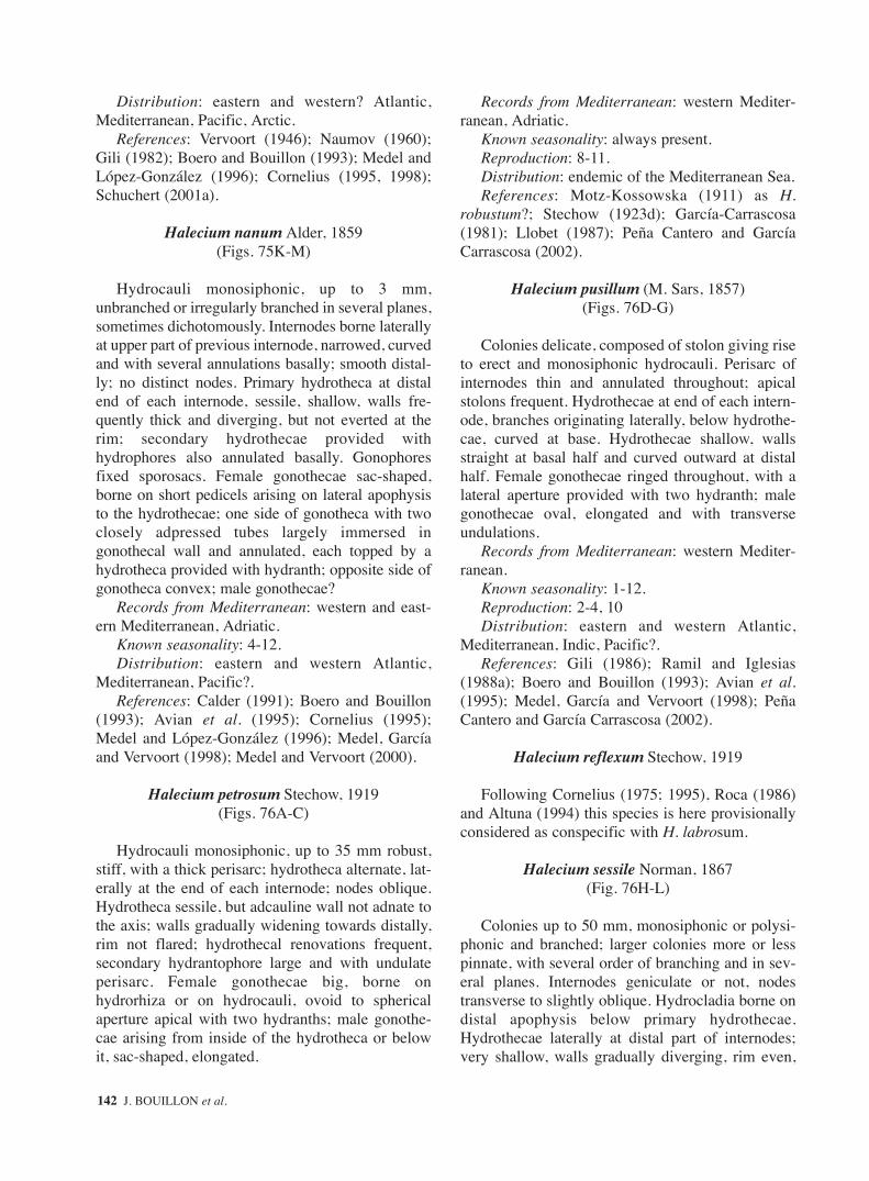
Distribution: eastern and western? Atlantic,Mediterranean, Pacific, Arctic.
References: Vervoort (1946); Naumov (1960);Gili (1982); Boero and Bouillon (1993); Medel andLópez-González (1996); Cornelius (1995, 1998);Schuchert (2001a).
Halecium nanum Alder, 1859(Figs. 75K-M)
Hydrocauli monosiphonic, up to 3 mm,unbranched or irregularly branched in several planes,sometimes dichotomously. Internodes borne laterallyat upper part of previous internode, narrowed, curvedand with several annulations basally; smooth distal-ly; no distinct nodes. Primary hydrotheca at distalend of each internode, sessile, shallow, walls fre-quently thick and diverging, but not everted at therim; secondary hydrothecae provided withhydrophores also annulated basally. Gonophoresfixed sporosacs. Female gonothecae sac-shaped,borne on short pedicels arising on lateral apophysisto the hydrothecae; one side of gonotheca with twoclosely adpressed tubes largely immersed ingonothecal wall and annulated, each topped by ahydrotheca provided with hydranth; opposite side ofgonotheca convex; male gonothecae?
Records from Mediterranean: western and east-ern Mediterranean, Adriatic.
Known seasonality: 4-12.Distribution: eastern and western Atlantic,
Mediterranean, Pacific?.References: Calder (1991); Boero and Bouillon
(1993); Avian et al. (1995); Cornelius (1995);Medel and López-González (1996); Medel, Garcíaand Vervoort (1998); Medel and Vervoort (2000).
Halecium petrosum Stechow, 1919(Figs. 76A-C)
Hydrocauli monosiphonic, up to 35 mm robust,stiff, with a thick perisarc; hydrotheca alternate, lat-erally at the end of each internode; nodes oblique.Hydrotheca sessile, but adcauline wall not adnate tothe axis; walls gradually widening towards distally,rim not flared; hydrothecal renovations frequent,secondary hydrantophore large and with undulateperisarc. Female gonothecae big, borne onhydrorhiza or on hydrocauli, ovoid to sphericalaperture apical with two hydranths; male gonothe-cae arising from inside of the hydrotheca or belowit, sac-shaped, elongated.
Records from Mediterranean: western Mediter-ranean, Adriatic.
Known seasonality: always present.Reproduction: 8-11.Distribution: endemic of the Mediterranean Sea.References: Motz-Kossowska (1911) as H.
robustum?; Stechow (1923d); García-Carrascosa(1981); Llobet (1987); Peña Cantero and GarcíaCarrascosa (2002).
Halecium pusillum (M. Sars, 1857)(Figs. 76D-G)
Colonies delicate, composed of stolon giving riseto erect and monosiphonic hydrocauli. Perisarc ofinternodes thin and annulated throughout; apicalstolons frequent. Hydrothecae at end of each intern-ode, branches originating laterally, below hydrothe-cae, curved at base. Hydrothecae shallow, wallsstraight at basal half and curved outward at distalhalf. Female gonothecae ringed throughout, with alateral aperture provided with two hydranth; malegonothecae oval, elongated and with transverseundulations.
Records from Mediterranean: western Mediter-ranean.
Known seasonality: 1-12.Reproduction: 2-4, 10Distribution: eastern and western Atlantic,
Mediterranean, Indic, Pacific?.References: Gili (1986); Ramil and Iglesias
(1988a); Boero and Bouillon (1993); Avian et al.(1995); Medel, García and Vervoort (1998); PeñaCantero and García Carrascosa (2002).
Halecium reflexum Stechow, 1919
Following Cornelius (1975; 1995), Roca (1986)and Altuna (1994) this species is here provisionallyconsidered as conspecific with H. labrosum.
Halecium sessile Norman, 1867(Fig. 76H-L)
Colonies up to 50 mm, monosiphonic or polysi-phonic and branched; larger colonies more or lesspinnate, with several order of branching and in sev-eral planes. Internodes geniculate or not, nodestransverse to slightly oblique. Hydrocladia borne ondistal apophysis below primary hydrothecae.Hydrothecae laterally at distal part of internodes;very shallow, walls gradually diverging, rim even,
142 J. BOUILLON et al.
sm68s2Bsystema1 14/10/04 15:22 Página 142

not everted; aperture at right angle with the axis ofinternode; hydrothecal renovation frequent, sec-ondary hydrothecae usually forming directly ondiaphragm of previous hydrothecae, appearing sev-eral hydrothecal rim very closed. Female gonothe-cae kidney shaped, with a tubular aperture on con-cave side with two hydranths. Male gonothecaeoval, elongated, slightly curved; aperture terminal.
Records from Mediterranean: western Mediter-ranean.
Known seasonality: 6.Distribution: eastern and western Atlantic,
Mediterranean, Indo-Pacific.References: Vervoort (1966); Ramil and Vervoort
(1992a); Boero and Bouillon 1993; Altuna (1994);Cornelius (1995, 1998); Medel and López-González(1996); Schuchert (2001a).
Halecium sibogae Billard, 1934(Figs. 76M-O)
Hydrocauli erect, up to 90 mm, polysiphonic andpinnately branched; perisarc thick in older parts.Distal parts of the colony monosiphonic, internodesgeniculate, nodes oblique. Each internode distallywith well developed primary hydrophore; nextinternode borne laterally to the hydrophore withoutdistinct apophysis. Adcauline wall of primaryhydrophore at place of fusion with wall of internodewith characteristic comma-shaped internal perisar-cal fold. Hydrotheca with distinct basal diaphragmand widening distally, rim flared. Hydrothecal reno-vations frequent, secondary hydrophores sometimeswith one-two basal annulations; length varied. Maleand female gonothecae similar, almost spherical,frequently showing a quadrangular cross section,and with one or two more or less developed basalprojections, more pronounced in the femalegononothecae, and apically with two more or lessdeveloped elevations between which is situated theaperture at the end of a cone.
Records from Mediterranean: Alborán Sea(species penetrating into the Mediterranean butprobably not yet dispersed in it).
Known seasonality: 6-8.Reproduction: 7.Distribution: eastern Atlantic warm waters,
Mediterranean.References: Billard (1934); Patriti (1970); Ramil
and Vervoort (1992a); Medel and López-González(1996); Medel et al. (1998); Medel and Vervoort(2000); Peña Cantero and García Carrascosa (2002).
Halecium tenellum Hincks, 1861(Figs. 77A-E)
Colonies composed of hydrocauli monosiphonicup to 20 mm high, very thin and delicate, irregularlybranched. Internodes long, in zigzag, separated bytransverse nodes. Hydrothecae borne laterally at upperpart of internodes, alternate and provided with a welldeveloped hydrophore; hydrotheca shallow, wallswidening from base to top giving a wide aperture; rimeverted and curved outwards. Secondary hydrotheca,provided with hydrophore, may occurs from primaryhydrothecae; branching may originate from hydrothe-cal base. Male gonothecae ovate and flattened; femalesimilar but slightly larger and broader.
Records from Mediterranean: western Mediter-ranean, Adriatic, Algeria, Black Sea.
Known seasonality: 10-5, 7, 9.Reproduction: 4-5.Distribution: eastern and western Atlantic,
Mediterranean, Indo-Pacific, Arctic, Antarctic.References: Vervoort (1959); Naumov (1960);
Patriti (1970); Millard (1975); Boero and Bouillon(1993); Cornelius (1995, 1998); Migotto (1996);Medel, García and Vervoort (1998); Medel and Ver-voort (2000); Schuchert (2001a).
Genus Hydrodendron Hincks, 1874 = Ophiodissa, 1919a = Scoresbia Watson, 1969
Colonies stolonal or erect, monosiphonic orpolysiphonic; hydrothecae short cylindrical, haleci-id type, pedicellate or on internodal apophyses, intwo alternate rows in erect colonies, basal part withor without desmocytes; hydranth with or without anintertentacular web; nematophore extensile, elon-gate, simple-ended or capitate; nematotheca, whenpresent, simple, sometimes minute; gonophores assolitary fixed sporosacs, gonothecae simple oraggregated in a glomulus.
Remark: the Genus Hydrodendron is heredefined as by Rees and Vervoort (1987) and Ver-voort (1987) so to include, among others, the generaOphiodissa and Scoresbia.
References: Calder (1991); Cornelius (1995);Hirohito (1995).
Hydrodendron mirabile (Hincks, 1866)(Figs. 77F-K)
Colonies up to 50 mm, composed of erect andmonosiphonic hydrocauli branched distally. Intern-
FAUNA OF THE MEDITERRANEAN HYDROZOA 143
sm68s2Bsystema1 14/10/04 15:22 Página 143

odes separated by transverse nodes. Hydrophoresborne laterally, alternate, at upper part of internodes,variable in length and usually with several annula-tions; perisarc of hydrophores thicker basally andprogressively thinner towards the distal end.Hydrotheca deep, widening from halfway onward,rim everted, occasionally curved downwards.Nematothecae present on hydrorhiza and onhydrophores, tubular in basal 2/3, cup-shaped distal-ly; nematophores elongate, capitate distally, strong-ly retractile. Stems with distal stolons developingfrom hydrothecae. Hydranth provided with 20 tenta-cles. Gonothecae borne hydrorhiza, on shortpedicels, barrel-shaped spacely ringed throughout,aperture wide on a short collar.
Records from Mediterranean: eastern and west-ern Mediterranean, Adriatic.
Known seasonality: 2, 4, 6, 7, 10-12.Reproduction: 7Distribution: eastern and western Atlantic, Indo-
Pacific, Mediterranean.References: Rees and Vervoort (1987); Gili, Ver-
voort and Pagès (1989); Boero and Bouillon (1993);Medel and López-González (1996) both asOphiodissa; Cornelius (1995); Medel, García andVervoort (1998); Medel and Vervoort (2000); PeñaCantero and García Carrascosa (2002).
Family HALOPTERIDIDAE Millard, 1962
Colonies with either erect hydrocauli, or withhydrocladia arising directly from hydrorhiza; hydro-caulus, when present, branched or unbranched,monosiphonic or polysiphonic, arising from a creep-ing or root-like hydrorhiza, giving rise to alternate oropposite, or irregularly arranged, branched orunbranched, hydrocladia; when arising from polysi-phonic hydrocauli and branches, hydrocladia givenoff from either a single axial tube or from superficialtubes; hydrothecae on hydrocladia typically large,with cusped or uncusped rim; cauline hydrothecaetypically present, well developed, less frequentlyatrophied, lacking on polysiphonic hydrocladia andbranches, when component tubes give rise to hydro-cladia; nematophores with nematothecae, not asnaked sarcostyles; nemathothecae typically welldeveloped, varied in structure, one- or two-cham-bered, movable or immovable, not fused to hydrothe-cae (except in the lateral nematothecae of Antennel-lopsis which are partly fused); a minimum of three,one median inferior and a pair of lateral, adjacent toeach hydrotheca; cnidome: usually microbasic
mastigophores of several size classes, sometimesisorhizas, microbasic euryteles and pseudostenoteles;gonophores as fixed sporosacs contained in a simplegonotheca, usually solitary, arising from caulus orhydrocladia, neither aggregated or protected by phy-lactocarps, frequently sexually dimorphic, with atleast the female normally bearing nematothecae.
References: Cornelius (1995), Migotto (1996);Calder (1997); Schuchert (1997); Peña Cantero etal., (1999); Watson (2000); Ansín Agís et al. (2001);Schuchert (2001).
1. Unbranched erect stems arising directly from creeping hydrorhiza; without hydrocladia ..................................................................... Antennella
– Erect structures branched, either as cormoids, branched hydrocladia or polysiphonic stems ... 2
2. Hydrocladia with large “mamelons” (atrophied hydrothecae) ..................................................... 3
– Hydrocladia without “mamelons”.....................43. With unbranched hydrocladia .. Pseudoplumaria– With hydrocladia branched in a regular fashion .
...................................................... Polyplumaria4. Stem normally monosiphonic, cormoids pinnate
........................................................... Halopteris– Stem polysiphonic and with one main axial tube
bearing hydrocladia ........................ Schizotricha
Genus Antennella Allman, 1877
Hydrocladia arising directly, independently, from acreeping hydrorhiza, normally unbranched, not poly-siphonic; hydrotheca cup- to vase-shaped, rim evenuntoothed; nematotheca two-chambered, movable;lateral nematothecae flanking each hydrotheca andborne on prominent peduncles adhering to hydrothe-cal wall; colony often monoecious, gonophores asfixed sporosacs, gonotheca solitary, sexually dimor-phic, borne on hydrocladia with basal nematothecae.
References: Hirohito (1995); Calder (1997);Schuchert (1997).
1. Ahydrothecate internodes with one single distal nematotheca, colonies dioecious.......... A. ansini
– Ahydrothecate internodes with two median nematothecae, colonies monoecious ................ 2
2. Hydrotheca surrounded by four nematothecae; distal chamber of lateral nematothecae widening distally .......................................... A. secundaria
– Hydrotheca surrounded by three nematothecae; distal chamber of lateral nematothecae globose, with two deep emargination ............ A. siliquosa
144 J. BOUILLON et al.
sm68s2Bsystema1 14/10/04 15:22 Página 144

Antennella ansiniPeña Cantero and García Carrascosa (2002)
(Figs. 77L-O)
Colonies with solonal hydrorhiza giving smallunbranched stems, proximal part of stem composedof one or several ahydrothecate internodes bearingup to 3 nematothecae. Remaining stem composed ofalternately arranged hydrothecate and ahydrothecateinternodes. Stem with up to 12 hydrothecae.Hydrothecal internode with one hydrotheca andthree nematotheca, one mesial inferior and two lat-erals. Mesial nematothecae short without apohysis,lateral nematothecae short, on short apophyses, dis-tal part rounded with deeply scooped inner and outerwalls. Ahydrothecate internodes longer thanhydrothecate ones with a single distal nematothecae.All nematothecae two-chambered.. Hydrothecaeabcaudally directed, cup-shaped, slightly wideningat their aperture, adcauline wall free for approxima-tively half its length, abcauline wall slightly convex.Hydrothecal aperture circular, rim slightly laterallydepressed. Colonies dioecious. Gonothecae usuallyon basal internodes, on short antero-lateral apoph-ysis, up to two gonophores per stem. Malegonophore fusiform basally curved with one or twonematothecae, aperture circular, situated at distaltruncated part of gonothecae. female gonotheca onshort apohysis with short basal segment stronglyflattened carrying one or two nematothecae, aper-ture larger than in male gonothecae.
Records from Mediterranean: western Mediter-ranean.
Known seasonality: 5, 7, 8.Reproduction: 5, 7, 8.Distribution: eastern Atlantic; Mediterranean.References: Peña Cantero and García Carrascosa
(2002).
Antennella secundaria (Gmelin, 1791)(Figs. 77P, 78A-D)
Colonies forming ramified, tubular stolons witherect stems, usually unbranched up to about 25mm;coenosarc reddish. Basal part of the axis dividedinto several internodes by transverse nodes, with avariable number of nematothecae. Rest of the stemheteromerously segmented. Hydrothecate segmentswith one hydrothecae surrounded by four nema-tothecae, one median inferior inmovable, one pair oflaterals, and one axillar. Non-hydrothecate intern-odes with two median nematothecae. Hydrotheca
cup-shaped, walls parallel to slightly divergent, 1/3to 1/2 adnate, rim even, aperture at 45º-55º with themain axis. Nematothecae all two-chambered andwith a very wide embayment; median inferiorinmovable, not reaching the level of the hydrotheca,median nematotheca on the internodes similar; axil-lar nematotheca similar but much shorter than thefirst; lateral nematothecae movable, borne on welldeveloped pedicels; basal chamber elongated, distalchamber widening distally. Gonothecae of bothsexes often on the same axis, borne under hydrothe-cae, on pedicels of two segments ; female elongat-ed, cylindrical, narrowed basally, with a circular lidand two opposite basal nematothecae; male shorter,curved, also provided with two basal lateral nema-tothecae.
Records from Mediterranean: eastern and west-ern Mediterranean, Adriatic.
Known seasonality: present all the year.Reproduction: 3 -11.Distribution: cosmopolitan species with a prefer-
ence for warmer waters.References: Patriti (1970); Millard (1975); Gili
and García-Rubíes (1985); Ramil (1988); Corneliusand Ryland (1990); Ramil and Vervoort (1992a);Boero and Bouillon (1993); Avian et al. (1995);Medel and Vervoort (1995); Medel, and López-González (1996); Schuchert (1997); Ansín Agís etal. (2001); Peña Cantero and García Carrascosa(2002).
Antennella siliquosa (Hincks, 1877)(Figs. 78E-I)
Ramified stolons with upright and unbranchedstems up to 45 mm high, with a whitish coenosarc.Basal part of the axis divided on a variable numberof segments bearing frontal nematothecae by meansof transverse nodes; remainder parts heteromerouslysegmented, with nodes alternatively oblique andtransverse. Hydrothecate segments with onehydrothecae and three nematothecae, one medianinferior, and two laterals ones. Non-hydrothecatesegments bearing two median nematothecae.Hydrotheca cylindrical, walls almost parallel, slight-ly divergent distally, 1/2 adnate, rim even, apertureat 35º-45º with the main axis. Nematothecae all two-chambered, aperture with embayment. Mediannematothecae all similar, but median inferior to thehydrotheca wider, this one not reaching the level ofthe hydrotheca; lateral nematothecae borne onpedicels, not reaching the hydrothecal margin, basal
FAUNA OF THE MEDITERRANEAN HYDROZOA 145
sm68s2Bsystema1 14/10/04 15:22 Página 145

chamber longer and elongated if compared with therest; distal chamber typically globose, with a deepemargination on inner and outer side. Gonothecaeon both sexes on the same axis, borne on pedicelswith two segments. Female elongate, somewhat flat-tened, truncated distally and curved basally, with upto four nematotheca; male shorter, pyriform,extreme rounded; two basal nematothecae, bothwith apical flat lid.
Records from Mediterranean: western Mediter-ranean, Adriatic.
Known seasonality: 5-10.Reproduction: 5, 7, 9, 10.Distribution: eastern Atlantic (England, France,
Strait of Gibraltar, Morocco, Ivory Coast), Mediter-ranean.
References: Vervoort (1959) as Antennelladiaphana f. siliquosa; Patriti (1970); García Cor-rales et al. (1978); Ramil and Vervoort (1992a) bothas Halopteris diaphana f. siliquosa; Boero andBouillon (1993); Avian et al. (1995); Medel andVervoort (1995); Medel and López-González(1996); Schuchert (1997); Ansín Agís et al. (2001).
Genus Halopteris Allman, 1877
Colonies typically erect, unbranched or branched,forming pinnate cormoids arising from a creepinghydrorhiza, often with a hinge-joint near base; hydro-caulus usually monosiphonic, rarely polysiphonic,bearing hydrothecae and pinnately arranged hydro-cladia; in polysiphonic stems, all tubes can give riseto cormidia; hydrocladia almost always unbranched,alternate or in opposite pairs, or opposite basally andalternate distally, in one plane, sometimes (as sec-ondary growth-form) arising independently fromhydrorhiza; hydrothecae cup-shaped, on hydrocaulusand hydrocladia, margin entire or with one medianabcauline cusp; nematothecae polymorphic, movableor immovable ones on a given colony, one- or two-chambered, lateral nemathotecae typically borne onprominent peduncles adhering to hydrothecal wall;gonophores as fixed sporosacs, gonothecae arisingfrom hydrocaulus or hydrocladia, solitary, with orwithout nematothecae.
References: Hirohito (1995); Calder (1997);Schuchert (1997).
1. Main axis homomerously segmented.................................................................. H. liechtensternii
– Main axis heteromerously segmented.............. 22. Hydrotheca with two pairs of lateral
nematothecae ................................. H. catharina– Hydrotheca with one pair of lateral nematothecae
........................................................ H. diaphana
Halopteris catharina (Johnston, 1833)(Figs. 78J-N)
Colonies pinnate up to 40 mm high arising frombranched stolons; stems monosiphonic andunbranched. Basal part composed of several seg-ments bearing two longitudinal rows of nematothe-cae, nodes transverse. Remainder axis heteromer-ously segmented by alternate oblique and transversenodes. Hydrocladia opposite, borne laterally bellowhydrothecae, on long stem apophyses. Hydrothecatesegments with one hydrothecae and five nematothe-cae, one median inferior and two pairs of lateralones. Non-hydrothecate segments with one to fournematothecae. Hydrocladia with a first athecate seg-ment; remainder segments as in the main axis, butnon-hydrothecates with 1-2 nematothecae.Hydrothecae cup-shaped, in the middle of theinternode, walls straight almost parallel, 1/2 adnate,rim smooth, aperture at 45º with the axis. Nema-tothecae all two-chambered and movable, upperchamber with a deep embayment. Outer pair of lat-eral nematothecae reaching the rim or slightlybeyond it, on long apophyses; inner pair at base ofthose apophyses. Gonothecae of both sexes on thesame colony, on the stem and hydrocladia. Femalecylindrical somewhat flattened, narrowed at base,with two nematothecae; truncated distally, withannular thickening and lid; basal pedicel with twosegments. Male gonothecae smaller and elongated.
Records from Mediterranean: western and east-ern Mediterranean.
Known seasonality: 1-4, 6-8, 12.Reproduction: 3, 6.Distribution: temperate and southern-boreal
regions of the eastern and western sides of theAtlantic.
References: Picard (1958b); Vervoort (1972);Ramil and Vervoort (1992a); Boero and Bouillon(1993); Cornelius (1995); Medel and LópezGonzález (1996); Schuchert (1997, 2001a); AnsínAgís et al. (2001).
Halopteris diaphana (Heller, 1868)(Figs. 78O-T)
Colonies pinnate with erect, monosiphonic andunbranched stems up to 12 mm. Basal parts with a
146 J. BOUILLON et al.
sm68s2Bsystema1 14/10/04 15:22 Página 146

varied number of segments separated by transversenodes bearing a row of median nematothecae.Remainder parts of the axis heteromerously seg-mented, with alternate oblique and transverse nodes.Hydrocladia alternate, on apophyses laterally to thehydrothecae. Hydrothecate internodes of the mainaxis and hydrocladia composed of one hydrothecaeand three nematothecae, one median inferior andtwo laterals. Non-hydrothecate segments with 1-2median nematothecae (usually one on the hydrocla-dia). Hydrotheca in the middle of the segment, cup-shaped, walls slightly widening, 1/2 adnate, rimeven, aperture at 50º-60º with the axis. All nema-tothecae two chambered and movable, withadcauline embayment. Median inferior nematothecanot reaching the hydrotheca, lateral ones on veryshort apophyses, just reaching the hydrothecal bor-der. Gonothecae of both sexes only seen on separatecolonies, on stem and hydrocladia, with two seg-mented pedicels. Female evenly curved for its entirelength, narrowed basally, with 2-4 basal nematothe-cae; distal end truncate, with a convex lid. Malegonothecae oblong, slightly curved, with 2 basalnematothecae.
Records from Mediterranean: western and east-ern Mediterranean, Adriatic.
Known seasonality: 1, 2, 4-10.Reproduction: 4, 6-9.Distribution: circumtropical species.References: Boero and Bouillon (1993); Avian et
al. (1995); Medel and Vervoort (1995); Medel andLópez-González (1996); Schuchert (1997); AnsínAgís et al. (2001); Peña Cantero and García Carras-cosa (2002).
Halopteris liechtensterniiMarktanner-Turneretscher, 1890
(Figs. 79A-G)
Colonies pinnate with erect, monosiphonic andunbranched stems up to 30 mm. Basal part com-posed of several segments with nematothecae, sep-arated by means of transverse nodes. Remaindermain axis homomerously segmented by means ofoblique nodes, but at distal extreme, heteromerous-ly segmentation may occurs by additional trans-verse nodes. Hydrocladia alternate, on lateralapophyses besides hydrothecae; most basal hydro-cladia sometimes branched; hydrocladia heteromer-ously segmented. Segments on main axis with onehydrothecae and 7-8 nematothecae; one medianinferior almost reaching hydrothecal base, two lat-
eral ones, seated on short pedicels and reaching thehydrothecal rim, two axillar and two or three medi-an superior. Hydrothecate segments on hydrocladiawith one hydrothecae surrounded by five nema-tothecae (one median inferior, two laterals, and twosmaller ones on the upper axil of the hydrothecae);non-hydrothecate segments with 1-2 median nema-tothecae. Hydrotheca cup-shaped, walls slightlydiverging towards margin, adcauline wall 1/2adnate, rim even and circular, aperture at 40º-50ºwith the main axis. Nematothecae all two cham-bered. Median inferior nematotheca with a distalchamber scoop-shaped, without adcauline wall; lat-eral nematotheca with a longer basal chamber; mar-gin of the distal chamber even or with shallowemarginations on adcauline wall; pair of nema-tothecae behind hydrothecae reduced, indistinctlytwo-chambered, scoop-shaped; remainder nema-tothecae similar to median inferior ones, but longer.Gonothecae of both sexes on same colony. Femaleborne on stem and hydrocladia, on two-segmentedpedicels; oval, truncated distally, with thickenedring of prosaic and lid; two basal nematothecae.Male borne on hydrocladia, smaller, sac-shaped andwithout nematothecae.
Records from Mediterranean: eastern and west-ern Mediterranean and Adriatic (central Mediter-ranean?).
Known seasonality: 3, 5-10.Reproduction: 5-8, 10.Distribution: endemic of Mediterranean Sea.References: Motz-Kossowska (1908) as Plumu-
laria; Rossi (1961); Boero and Bouillon (1993);Medel and Vervoort (1995); Medel and López-González (1996); Schuchert (1997); Avian et al.(1995); Peña Cantero and García Carrascosa (2002)
Genus Polyplumaria G. O. Sars, 1874
Colony with erect, strongly ramified, pinnatestems, rigid in appearance, typically polysiphonic;branches of hydrocaulus opposite or nearly so;hydrocladia branched, regularly arranged, placed onan apophysis with a much developed, large “mamel-on” (considered as an atrophied hydrotheca) on itssuperior surface; hydrothecal rim smooth;gonophores as fixed sporosacs, gonothecae with orwithout nematothecae.
Remarks: sometimes included in the Plumulari-idae.
References: Calder (1997); Schuchert (1997;2001a)
FAUNA OF THE MEDITERRANEAN HYDROZOA 147
sm68s2Bsystema1 14/10/04 15:22 Página 147

Polyplumaria flabellata G.O. Sars, 1874(Figs. 79H-L)
Hydrocauli erect, up to 350 mm, polysiphonicand branched, with some thick stolonal fibers basal-ly; branches arranged opposite o nearly so, usuallypolysiphonic over part of their lengths. Tubes ofstem and branches with nematothecae. Main axis ofbranches segmented into internodes, bearing alter-nate apophyses with a large mamelon and a pair ofnematothecae; hydrocladia borne on the apophyses,segmented, first internode shorter and athecate,occasionally fused with the apophysis, remainderones each with big hydrotheca and 4 nematothecae:one median inferior, two laterals, and one mediansuperior which may occasionally be placed on a sep-arate internode, nodes oblique. Hydrotheca cup-shaped, walls gradually widening, margin slightlyeverted and even; adcauline wall 1/3 adnate. Lateralnematothecae reaching hydrothecal rim or slightlyshorter, all nematothecae two-chambered, basalchamber longer and narrower, apical chamber cup-shaped and with deep adcauline embayment. Thehydrocladia may be branched, the branch originat-ing from the first internode. Male and femalegonothecae similar, borne on short pedicels nearbase of hydrocladium, inverted pear-shaped,obliquely truncate above, aperture large; 4 smallnematothecae near base.
Records from Mediterranean: only just inside theMediterranean, at the Strait of Gibraltar.
Known seasonality: 6, 7.Distribution: sub-tropical to cool-temperate
waters on the eastern Atlantic, Mediterranean.References: Gili, Vervoort and Pagès (1989);
Ramil and Vervoort (1992 a, c); Cornelius (1995);Medel and Vervoort (1995); Medel and Lopez-González (1996); Ansín Agís et al. (2001);Schuchert (2001a).
Genus Pseudoplumaria Ramil and Vervoort (1992)
Colonies composed of rigid, strongly ramified,polysiphonic, occasionally forked hydrocaulus, withalternate or opposite branches; hydrocladia alwaysunbranched, alternately arranged along axis, placedon an apophysis with a much developed, large“mamelon” (considered as an atrophied hydrotheca)on its superior surface; hydrotheca exclusivelyfound on the hydrocladia, hydrothecal bordersmooth; mobile, immobile and reduced nematothe-cae two-chambered (bithalamic) present.
Gonophores as fixed sporosacs, gonothecae singlewith a few nematothecae on basal region.
Remarks: sometimes included in the Plumulariidae.References: Calder (1997); Schuchert (1997).
Pseudoplumaria marocana (Billard, 1930)(Figs. 79M-P)
Colonies with a thick, polysiphonic, muchbranched stem about to 220 mm high, rising fromthick matting of hydrorhizal fibers; branches oppo-site, in one plane. Axial tubes with two longitudinalrows of small nematothecae; “main” frontal axiswith alternate apophyses bearing hydrocladia (2-6per internode), each with one “mamelon” surround-ed by three nematothecae. Hydrocladia with 1-2basal internodes with a single nematotheca, theremainder ones with one hydrotheca and four nema-tothecae; nodes oblique (but the first transverse).Hydrotheca big, cylindrical, margin not everted andeven. Median inferior nematotheca two-chambered,basal chamber very short and immobile, apicalchamber with a deep adcauline embayment; lateralnematothecae larger, reaching hydrothecal rim,movable, two-chambered, apical chamber with aminor embayment; fourth nematotheca found in axilbetween free part adcauline wall and internode,reduced to a peridermal sac; coenosarc of the colonywith zooxanthellae; gonothecae pyriform with twoshort basal segments.
Records from Mediterranean: found only at theentrance (Algeciras Bay, Strait of Gibraltar).
Known seasonality: 6, 7.Distribution: tropical temperate eastern Atlantic,
Mediterranean.References: Patriti (1970); Gili, Vervoort and
Pagès (1989) part of the material of Polyplumariaflabellata; Ramil and Vervoort (1992c), Medel andVervoort (1995); Medel and López-González(1996); Ansín Agís et al. (2001)
Genus Schizotricha Allman, 1883
Colonies with polysiphonic, erect stem, whichmay be branched or unbranched, with one mainaxial tube bearing pinnately arranged hydrocladiaand hydrothecae, and several undivided accessorytubes provided only with nematothecae; hydrocladiaalternate, the majority branched sympodially fromanterior or lateral surface immediately belowhydrothecae; hydrotheca cup-shaped, with smoothrim; nematotheca 2-chambered and movable, lateral
148 J. BOUILLON et al.
sm68s2Bsystema1 14/10/04 15:22 Página 148

nemathotecae not fused to their pedicel or tohydrothecae; gonophores as fixed sporosacs,gonothecae inserted on hydrocladia betweenhydrothecae and provided with nematothecae.
References: Schuchert (1997); Peña Cantero etal. (1999).
Schizotricha frutescens (Ellis and Solander, 1786)(Figs. 80A-E)
Colonies composed of erect, polysiphonic andpinnate stems up to about 75 mm high, arising fromflattened mass of hydrorhiza tubes. Hydrocladiaalternate, laterally under the hydrothecae. Sec-ondary and tertiary hydrocladia sometimes present,arising from the first hydrothecae of the hydrocla-dia. Primary tube of the stem with a longitudinalrow of hydrothecae. Hydrocladia segmented on longsegments by transverse nodes, with 1-5 hydrothe-cae, each one surrounded by 3 nematothecae, onemedian inferior and two laterals. Hydrotheca cup-shaped, deep, adcauline wall completely adnate, rimsmooth. Median inferior nematotheca some distancebelow hydrothecae; lateral nematotheca over reach-ing hydrothecal margin. Male and female gonothe-cae on different colonies, borne on axis and hydro-cladia below hydrothecae; pear-shaped, narrowedbasally, with two nematothecae.
Records from Mediterranean: western Mediter-ranean, Adriatic.
Known seasonality: 2, 6, 7.Reproduction: 6.Distribution: eastern temperate and subtropical
Atlantic, Mediterranean.References: Millard (1975); Ramil and Vervoort
(1992a); Boero and Bouillon (1993); Cornelius andRyland (1990); Avian et al. (1995); Cornelius(1995) as Polyplumaria; Medel and López-González (1996); Schuchert (1997, 2001a).
Family HEBELLIDAE Fraser, 1912
Hydroid: colony stolonal, hydrotheca campanu-late with smooth margin, short or long distinct pedi-cel; hydrothecal base with annular perisarcal thick-ening and membranous diaphragm or thickdiaphragm an no annular thickening; gonothecasolitary, with or without operculum, arising fromhydrorhiza; exceptionally with nematothecae(Bedotella); gonophores as fixed sporosacs, orswimming gonophores, or eumedusoids, or freemedusae.
Medusa: umbrella flat; manubrium short and flat,mouth with irregular lips; with marginal cordyli;with 4 or more branched radial canals; marginal ten-tacles hollow; “gonads” linear to sinuous on theradial canals; with or without marginal cirri; with orwithout adaxial ocelli; without statocysts.
Remarks: this family is usually included in theLafoeidae A. Agassiz, 1865, but has often been sep-arated as a subfamily by several authors (see Calder,1991; Boero et al., 1997 for a review). The familyHebellidae was established by Fraser (1912) toaccommodate the following genera (see Calder,1991): Bedotella; Halisiphonia, Hebella, Hebellop-sis and Scandia, whose hydroids typically havestolonal colonies, campanulate hydrothecae with adiaphragm or an annular thickening or both, and sin-gle gonothecae. As stated by Calder (1991), in theLafoeidae, Filellum is stolonal, a diaphragm is pre-sent in Abietinella, Cryptolaria and Zygophylax, andsingle gonothecae are found in some species ofCryptolarella. We recognise what Calder (1991)considered as a subfamily of the Lafoeidae, theHebellinae, as a separate family: the Hebellidae. Infact, although certain of the Hebellidae charactersare shared with some Lafoeidae genera, none ofthem have all those characters together; Filellum isstolonal but has no diaphragm and has aggregatedgonothecae; some Cryptollarella have a singlegonotheca, but they have no diaphragm and areerect; Abietinella, Cryptolaria and Zygophylax havea diaphragm but have erect colonies and coppinia.
Hebellid medusae, till recent years, were onlyknown by juvenile indeterminable stages. DeAndrade and Migotto (1997) reared immaturemedusae from a Hebella species, showing relation-ship with Laodiceidae, perhaps with the medusa-based genus Staurodiscus, and Migotto and DeAndrade (2000) elucidated the life cycle of Hebellafurax which should be referred to genus Toxorchis,possibly to T. kellneri (Toxorchis is here considered ascongeneric with Staurodiscus, see below). The Laodi-ceidae contain two distinct groups of medusa genera,those with unbranched radial canals from which sev-eral life cycles are known, the hydroids being all“Cuspidella-like”, and two genera with branchedradial canals, whose life cycle were unknown till theobservations of the above cited authors, and havingHebella hydroids. Those two particular genera arehere included in the family Hebellidae. The Lep-tomedusae contain so four families with cordyli orcordyli-like structure the Hebellidae, the Laodiceidae,the Teclaiidae and the Tiarannidae.
FAUNA OF THE MEDITERRANEAN HYDROZOA 149
sm68s2Bsystema1 14/10/04 15:22 Página 149

References: Calder (1991); Migotto (1996); DeAndrade and Migotto (1997); Boero et al. (1997);Migotto and De Andrade (2000); Watson (2000).
Key to hydroids and medusae
1. Gonophores producing fixed sporosacs ........... 2– Gonophores not producing fixed sporosacs;
hydrothecae sharply separated from pedicel.... 32. Colony with nematophores and nematothecae....
..............................................................Bedotella– Colony without nematophores .............. Scandia3. Gonophores as swimming sporosacs ..................
....................................................... Anthohebella – Gonophores not as above ................................. 44. With eumedusoids or immature medusae ...........
............................................................... Hebella– With free mature medusae; some or all of the
radial canals branched, branches joining or not ring canal ....................................... Staurodiscus
Genus Anthohebella Boero, Bouillon and Kubota,1997
Hydroid: colonies stolonal; hydrotheca on shortpedicel, campanulate, usually with thin annularthickening and thin membranous diaphragm; swim-ming gonophores, with velum, 4 radial canals, 4atentaculate marginal bulbs; “gonads” on spadix(manubrium); gonotheca solitary originating fromhydrorhiza.
Remarks: Watson (2000) described a new speciesof Anthohebella, A. darwinensis, but the gonophoresof this species are devoid of manubrium and no ref-erence is made in the description to the position ofthe gonads, although the gonophore is considered asnearly mature. This species is here mantained in theAnthohebella awaiting more information.
Anthohebella parasitica (Ciamician, 1880)(Figs. 80F-H)
Colonies stolonal; hydrothecae on short and annulatedpedicels, campanulate, margin curved, usually withannular thickening and a thin membranousdiaphragm; hydranth with conical hypostome; repro-duction by swimming gonophores, with velum, 4 radi-al canals, 4 atentaculate marginal bulbs; “gonads” onspadix (manubrium), gonothecae nearly twice thelength of the hydrotheca, originating from hydrorhiza,on short and annulated pedicels tapered basally, wideand truncated distally, with 4-5 operculars flaps.
Records from Mediterranean: eastern and west-ern Mediterranean, Adriatic.
Known seasonality: present all the year.Reproduction: 1-12Distribution: warm waters around the world.References: Boero (1980); Gili (1986), Medel
and López-González (1996), all as Hebella parasit-ica; Peña Cantero and García Carrascosa (2002).
Genus Bedotella Stechow, 1913
Colonies stolonal or erect, branched, polysiphon-ic; hydrothecae campanulate, borne irregularly onall surface of branches and sometimes onhydrorhiza, free, non adherent, distinctly pedicel-late, with thin diaphragm; margin even and slightlyeverted; nematophore in shortly globular peduncu-late nematotheca on hydrocaulus and hydrocladia;gonophores as fixed sporosacs, gonothecae solitary,strongly compressed, disc-shaped, not aggregated.
References: Ramil and Vervoort (1992a);Alvarez (1993b).
Bedotella armata (Pictet and Bedot, 1900)(Figs. 80I-K)
Colonies stolonal or with erect stems (occasional-ly as a combination of both), up to 15 mm high.Stolonal colonies with creeping tubes or an anasto-mosing, irregular network of stolonal tubes. Erectcolonies composed of a number of adnate stolonsbasally attached by means of creeping stolons thatmay bear hydrothecae and nematothecae. Perisarcthin and transparent over whole colony. Hydrothecacampanulate, walls narrowing towards basally, occa-sionally slightly asymmetric in lower third, rim circu-lar and everted. Hydrothecal renovations common.Hydrothecal pedicels with a few rings or undulationsof perisarc, near insertion on stolon. Nematothecaeglobular, with a short pedicel, arranged irregularly,frequently in groups of 4-5 nematothecae. Gonothe-cae of a big size (6 x 6 mm), fan-shaped, compressedlaterally borne on short pedicels.
Records from Mediterranean: western Mediter-ranean (France, only one record); also recorded atthe Strait of Gibraltar.
Distribution: Northeastern Atlantic, Mediter-ranean.
References: Rees and White (1966);Marinopoulus (1981) as Campanularia; Ramil andVervoort (1992a); Altuna (1994); Medel and López-González (1996).
150 J. BOUILLON et al.
sm68s2Bsystema1 14/10/04 15:22 Página 150

Genus Hebella Allman, 1888 = Hebellopsis Hadzi, 1913
Hydroid: colonies stolonal; hydrotheca on shortpedicel, campanulate to cylindrical, usually withannular thickening and membranous or perisarc-likediaphragm; gonophores either as released eumedu-soids with mature “gonads” on radial canals, or asmedusa already mature at liberation with 4 radialcanals, each with a proximal gonad; 4 perradialatentaculate bulbs and 4 small interradial atentacu-late bulbs; manubrium short; mouth and gastric cav-ity present; during life span some tentacles and moremarginal bulbs may grow, or as juvenile immaturefree medusa.
Medusa: adult medusa unknown.Remarks: the genus Hebella will probably have
to be split in the future when more life cycles will becompletely described, several hebelliform hydroidsgiving apparently rise to different medusa morpho-types. See also remarks under Scandia.
References: Blanco et al. (1994); Altuna Prados(1996); Boero, Bouillon and Kubota (1997).
Hebella furax Millard, 1957: see below Staurodiscus
Hebella scandens (Bale, 1888)(Figs. 81A-E)
Hydroid: colonies epizootic on other hydroids;hydrothecae with flared margin, mostly oblique,but bent 90° when growing over host hydrothecae,with smooth to slightly corrugated walls, asym-metrical, with perisarcal diaphragm; pedicelsshort, smooth or annulated; hydranths with 12-16tentacles; gonothecae containing up to 4 medusabuds, bigger than hydrothecae, asymmetrical, withundulated walls, truncated distally, taperingtoward base, on short pedicels, operculum with 4opercular flaps.
Records from Mediterranean: eastern and west-ern Mediterranean, Adriatic.
Known seasonality: 1, 2, 4-7, 11, 12.Distribution: Atlantic; Indo-Pacific; Mediter-
ranean.References: Rossi (1961); Millard (1975); Gili
(1986); Roca (1986); Calder (1991) as Hebellopsis;Genzano and Zamponi (1992); Avian et al. (1995);Medel and López-González (1996) as Hebellopsis;Boero et al. (1997); Peña Cantero and García Car-rascosa (2002).
Genus Scandia Fraser, 1912 = Croatella Hadzi, 1916
Colonies stolonal, sometimes sympodial, arisingfrom a creeping hydrorhiza; hydrotheca large, cam-panulate, usually borne on long pedicels, with abasal rounded annular perisarcal thickening;gonophores fixed sporosacs, gonotheca solitary.
Remarks: Scandia differs from Anthohebella andHebella in not being associated to supportinghydroids and in gonothecal content. It could bemerged with Hebella.
1. Female gonophore spherical.................. S. gigas– Female gonophore oval, more or less corrugated
.................................................. S. michael-sarsi
Scandia gigas (Pieper, 1828)(Figs. 81F-H)
Colonies composed of erect pedicels scarcely,dichotomously or irregularly branched and ended byhydrothecae. Perisarc of pedicels undulated or twist-ed throughout. Hydrothecae tubular, elongated,walls sometimes asymmetrical, narrowing graduallytowards the base, with a hydrothecal diaphragm;margin everted, rim even. Female gonothecae spher-ical, male sac-shaped and elongated.
Records from Mediterranean: eastern and west-ern Mediterranean, Adriatic.
Known seasonality: 1, 4-6, 8, 9, 11.Reproduction: 4-8.Distribution: temperate waters from western and
eastern Atlantic and North Pacific, Mediterranean.References: Boero (1981); Gili (1982; 1986);
Roca (1989); Boero and Bouillon (1993); Altuna(1994); Avian et al. (1995); Medel (1996); Medeland López González (1996); Peña Cantero and Gar-cia Carrascosa (2002).
Scandia michaelsarsi (Leloup, 1935)(Figs. 81I-L)
Colonies with long, erect pedicels unbranched orbranched, ended by hydrothecae. Pedicels irregular-ly wrinkled or twisted perisarc. Hydrothecae tubu-lar, deep, walls slightly asymmetrical, roundedbasally, margin everted, aperture circular; base ofthe hydrotheca with a thick hydrothecal diaphragm;gonophores unknown, gonothecae borne on stolon,with short pedicels, ovalate; the female longer andmore slender than the male, more or less corrugated,
FAUNA OF THE MEDITERRANEAN HYDROZOA 151
sm68s2Bsystema1 14/10/04 15:22 Página 151

truncate, with the aperture occupying only a part ofthe upper part.
Records from Mediterranean: western Mediter-ranean
Seasonality: ?Distribution: Atlantic, Mediterranean.References: Fraser (1944) as Scandia mutabilis;
Vervoort (1959) as Hebella michael-sarsi; García-Corrales et al. (1979); Boero and Bouillon (1993);Medel and López-González (1996); Boero et al.(1997).
Genus Staurodiscus Haeckel 1879= Toxorchis Haeckel, 1879
Hydroid: colonies generally epizootic on otherhydroids, mainly plumulariids and aglaopheniids,Hebella-like; with the hydrorhiza sometimes pene-trating into the perisarc of the host as a parasiticform; hydrothecae almost conical when growing onupper part of the host, cylindrical when growing onlower part of same host; asymmetrical to symmetri-cal; with everted margin, sharply or slightly oblique,with short to long, wrinkled or annulated pedicels;with membranous diaphragm (sometimes absent)and annular thickening; hydranth with 20-26 tenta-cles; gonophores as free medusae; gonotheca as bigor slightly bigger than hydrotheca, with four oper-cular flaps, on short pedicel, slightly undulatedwalls, truncated distally, tapering at base, containingup to three medusae.
Medusa: with 4 or more main primary radialcanals, some or all branching one or more times, pri-mary canal and some or all of the branches reachingcircular canal; “gonads” on primary radial canalsand branches; numerous tentacles and cordyli; withor without cirri; with or without ocelli.
Remarks: the life cycle Hebella furax Millard,1957 has been elucidated by Migotto and De Andrade(2000); the hydroid giving rise to a Staurodiscus (Tox-orchis) type of medusa, presumably Staurodiscus kell-neri Mayer, 1910. The differences between Staurodis-cus and Toxorchis are ambiguous and tenuous, themain character being the supposed mode of ramifica-tion of the radial canals. In Staurodiscus, ramificationsare described as branches formed after the develop-ment of the primary canal which first reach the circu-lar canal, whereas in Toxorchis ramifications werebelieved to be bifurcation of the primary canal whichhimself never reached the circular canal (see Kramp,1962; Bouillon, 1984b). Migotto and De Andrade(2000), studying the cycle of a Toxorschis medusa
(obtained from Hebella furax), described a develop-ment of the radial canals and of their branches similarto that observed in Staurodiscus. There are so present-ly no reasonable reasons to separate the two genera.
Staurodiscus kellneri Mayer, 1910(Figs. 82A-D)
.Diagnosis: see above.Records from Mediterranean: western Mediter-
ranean.Seasonality: ?Distribution: Indo-pacific, Mediterranean.References: García Corrales et al. (1979); Gili
(1982); Boero et al. (1997); De Andrade and Migot-to (1997); Migotto and De Andrade (2000).
Family KIRCHENPAUERIIDAE Stechow, 1921a
Colonies either with erect, branched orunbranched hydrocaulus, monosiphonic or polysi-phonic or stolonal (Ophinella); issued from a creep-ing hydrorhiza; hydrocladia alternate, arising inpolysiphonic hydrocauli from a single dominantaxial tube; hydrothecae small, occurring only onhydrocladia, with or without marginal cusps, with orwithout an abcauline intrathecal septum, adnate ornot; nematophores with nematothecae often rudi-mentary or occurring as naked sarcostyles; whenpresent, nematothecae simple, typically one-cham-bered although two-chambered in some taxa (i.e.:Naumovia, Ventromma), not fused to hydrothecae;paired lateral nematophores and nematothecaeabsent; gonophores as fixed sporosacs; gonothecasolitary, lacking nematothecae, on stem or hydrocla-dia, exceptionally on hydrorhiza (Pycnotheca).
Remark: cnidome usually composed of microba-sic mastigophores and sometimes pseudostenoteles,microbasic euryteles, and haplonemes. Stepanjantset al. (1998) described, in some specimens ofWimveria, desmoneme-like capsules from undis-charged cnidocysts. The presence of desmonemesseems very doubtful in Leptomedusae and resultpresumably of contamination by cnidocysts issuedfrom other species. The family Kirchenpaueriidaeneeds critical revision, the genera being not clearlydefined and their diagnoses often overlapping. Thegenera Naumovia, Ventromma and Wimveria presentonly slight differences with Kirchenpaueria andcould eventually be included in it.
References: Cornelius (1995); Migotto (1996);Calder (1997); Stepanjants et al. (1998); Peña Can-
152 J. BOUILLON et al.
sm68s2Bsystema1 14/10/04 15:22 Página 152

tero and Marques (1999); Ansín Agís et al. (2000).
1. Nematophores with well developed nematothecae .................................... Ventromma
– Nematophores nude or with reduced nematothecae............................. Kirchenpaueria
Genus Kirchenpaueria Jickeli, 1883
Stem unbranched, monosiphonic, with alternate,pinnate, unbranched hydrocladia, in simple formsarising directly from hydrorhiza; hydrotheca cup-shaped, without intrathecal septum, with unthootedrim, partially or completely adnate; no caulinehydrothecae; nematotheca typically 1-chambered andmovable, mesial nematothecae present or absent,sometimes poorly developed, reduced or even absent,nematophores with or without nematothecae presentalso on stem pending species; gonophores as fixedsporosacs, gonotheca solitary, not annulated, oftenwith longitudinal ridges and spines.
Recent references: Cornelius (1995); Schuchert(2001a).
1. Nematophores with club-shaped nematothecae ....................................................... K. bonnevieae
– Nematophores nude (stem and above hydrotheca) or with a minute frontal peridermal shield (below hydrotheca) ................. K. pinnata
Kirchenpaueria bonnevieae (Billard, 1906)(Figs. 82E-H)
Colonies pinnate with erect, monosiphonic andunbranched hydrocauli up to about 45 mm. Basalparts of the main axis unsegmented, with severalnematothecae. Remainder parts composed of seg-ments separated by transverse nodes; each internodebearing a latero-distal apophysis, one “mamelon” onupper surface of apophysis and two nematothecae,one basally and another on upper surface of apoph-ysis; two additional nematothecae may occurs.Hydrocladia borne on the apophyses at the upperpart of segments; alternate, with hydrothecateinternodes separated by slightly oblique nodes; eachsegment with one hydrotheca in lower half and twomedian nematothecae, below and above hydrothe-cae. Hydrotheca cup-shaped; completely adnate, rimsmooth, aperture tilted upwards. All nematothecaeone-chambered and movable, club-shaped, nar-rowed basally, with a circular aperture distally;gonothecae borne on apophyses, on short pedicels
two segmented; elongated, tapered basally, widelyand truncate distally.
Records from Mediterranean: Alborán Sea only.Known seasonality: 6.Distribution: North Atlantic, Indo-Pacific, Indian
Ocean; Alborán Sea.References: Vervoort (1966a); Millard (1975)
both as Kirchenpaueria triangulata; Ramil and Ver-voort (1992a); Medel and López-González (1996);Ansín Agís et al.(2001); Schuchert (2001a).
Kirchenpaueria pinnata (Linnaeus, 1758)(Figs. 82I-L, 83A-B)
Colonies pinnate with erect, monosiphonic, andoccasionally branched hydrocauli up to about 42 mm.Basal part of the axis with several internodes separat-ed by transverse nodes; the remainder segmented oninternodes bearing hydrocladia, these alternate, onlateral apophyses at the upper part of the segment.Apophysis with a naked nematophore on the upperside; another one is disposed laterally on the axis, justabove the apophysis. Hydrocladia with a first shorterathecate internode, with transverse node basally andoblique one distally (occasionally several of thisinternode may occurs); remainder internodes thecate,or in an irregular sequence of thecate and athecateinternodes separate by transverse nodes. Thecateinternodes with one hydrotheca and twonematophores, one median inferior and another medi-an superior. Hydrotheca cup-shaped, walls graduallydiverging, adcauline wall almost entire adnate. Medi-an inferior nematophore with a minute frontal perid-ermal shield; median superior behind hydrothecacompletely nude. Gonothecae of both sexes similar,borne on hydrorhiza or in a row on the axis; sac-shaped, with short pedicels, hexagonal in cross sec-tion, with six longitudinal carinae bearing digitiformprolongations of widely varied development, evenabsent in some gonothecae.
Records from Mediterranean: eastern and west-ern Mediterranean, Adriatic.
Known seasonality: present all the year.Reproduction: 1-12.Distribution: eastern Atlantic, Indo-Pacific,
Mediterranean.References: García-Corrales et al. (1978); Boero
and Bouillon (1993); Cornelius (1995); Avian et al.(1995); Medel and Vervoort (1995); Medel andLópez-González (1996); Ansín Agís et al.(2001);Schuchert (2001a); Peña Cantero and García Carras-cosa (2002).
FAUNA OF THE MEDITERRANEAN HYDROZOA 153
sm68s2Bsystema1 14/10/04 15:22 Página 153

Genus Ventromma Stechow 1923
Colonies erect, monosiphonic or polysiphonic,with branched or unbranched hydrocauli arisingfrom creeping hydrorhiza; hydocladia alternate, typ-ically unbranched; hydrothecae only on hydrocladia,cup-shaped, margin entire, without intrathecal sep-tum; nematophores usually with small, 2-chamberednematothecae, hydrothecate internodes with a medi-an inferior and a median superior nematotheca, nolateral nematophores and nematothecae;nematophores or nematothecae also on stems;gonophores as fixed sporosacs, gonotheca solitary,with transverse annulations.
Remark: Cornelius (1995) pointed out that thisgenus is not satisfactorily defined; this authorincludes Ventromma Stechow, 1923d in the Subfam-ily Plumulariinae L. Agassiz, 1862, instead of in thesubfamily Kirchenpaueriinae where most authorsinclude it and where it is even often regarded as con-generic with Kirchenpaueria Jickeli, 1883.
Ventromma halecioides (Alder, 1859)(Figs. 83C-G)
Colonies pinnate with erect, basally polysiphon-ic and often branched hydrocauli up to about 40 mmhigh. Monosiphonic parts of the axis separated intointernodes by transverse nodes; each segment with alateral apophysis distally bearing a nudenematophore; a cup-shaped nematotheca is bornelaterally on the segment, above the apophysis.Hydrocladia borne on the apophysis, alternate, het-eromerously segmented, nodes transverse; basal partbeginning with a short athecate internode. Thecatesegments with one hydrotheca and two nematothe-cae, one median superior and one median inferior atsome distance of the hydrothecae. Hydrotheca ondistal half of internode, cup-shaped, almost com-pletely adnate, abcauline wall slightly convex, rimsmooth and circular, aperture 80º with the axis.Nematothecae two-chambered, small, basal cham-bers very short, distal one cup-shaped, withadcauline embayment. Gonothecae borne on theaxis, barrel-shaped, narrowed basally, truncate dis-tally, with a circular lid; walls with transverse anddistinct ribs.
Records from Mediterranean: eastern and west-ern Mediterranean, including Black Sea, Adriatic.
Known seasonality: 6-3.Reproduction: 3, 6, 7, 9.Distribution: cosmopolitan species.
References: Gili and García-Rubíes (1985); Gili(1986); García-Corrales et al. (1978); Boero andBouillon (1993); Avian et al. (1995); Cornelius(1995); Medel and Vervoort (1995); Medel andLópez-González (1996); Peña Cantero and GarcíaCarrascosa (2002).
Family LAFOEIDAE A. Agassiz, 1865
Paedomorphic hydrozoa usually reduced tohydroid stage, hydroids colonial, either erect orstolonal, arising from a creeping hydrohiza,hydrothecae varying from tubular to campanulate inshape; radially or bilaterally symmetrical, usuallyadherent more seldom pedicellate; margin entire;operculum usually absent; with or withoutdiaphragm; without annular perisarcal thickening;hydranth with a conical hypostome surrounded by awhorl of filifom tentacles; abcauline diverticulumpresent or absent; nematophores present or absent.Cnidome: where known microbasic mastigophores.When known, gonophores as fixed sporosacs;gonothecae aggregated into coppinia or scapus,exceptionally solitary.
References: Calder (1991); Ramil and Vervoort(1992a); Blanco et al. (1994); Cornelius (1975b,1995); Hirohito (1995); Calder and Vervoort (1998).
1. Hydrotheca stalked........................................... 2– Hydrotheca not stalked, usually adherent ........ 32. Hydrothecae in two longitudinal rows: with
diaphragm or reduced annulus; nematophores and nematothecae usually present ... Zygophylax
– Hydrothecae irregularly on all sides; with no diaphragm or annular thickening; without nematophores .......................................... Lafoea
3. Mature colonies stolonal: hydrothecae adherent to hydrorhiza......................................... Filellum
– Mature colonies with erect, branching stem .... 44. Diaphragm present between hydrotheca and stem
apophysis; nematophores and nematothecae normally present .............................. Cryptolaria
– Normally no diaphragm and no nematophores ............................................................Acryptolaria
Genus Acryptolaria Norman, 1875
Colonies normally erect, stolonal when young,more or less alternately branched; hydrocaulus andhydrocladia polysiphonic, with an axial tube over-grown by accessory tubes; all branches with twolongitudinal rows of alternate hydrothecae, sessile,
154 J. BOUILLON et al.
sm68s2Bsystema1 14/10/04 15:22 Página 154

tubular, adnate to axial tube or immersed basally inaccessory tubes, curving outwards and becomingfree distally; diaphragm absent; nematophoresabsent; gonophores as fixed sporosacs, gonothecaeaggregated in coppinia with or without modifiedhydrothecal tubes.
Remark: Stepanjants (1979) described an opercu-lum in Acryptolaria operculata?
Acryptolaria conferta (Allman, 1877)(Figs. 83H-L)
Colonies composed of erect, polysiphonic andbranched stems bearing hydrocladia. Axis and mostpart of branches and hydrocladia, composed ofnumerous tubes longitudinally arranged and anasto-mosed with each other; monosiphonic parts with asympodial structure, axis straight to geniculate,hydrothecae lateral, in the same plane and alternate;tubular, basal part merging into the stolon, distal partfree and curved outwards, rim even and circular,hydrothecal renovations frequent. Gonothecae incoppiniae with several amphora-shaped gonothecae;acrocyst present in female gonothecae.
Remark: Ramil and Vervoort (1992) created thesubspecies minor based in its minor size and the lackof intermediate specimens between the two forms.
Records from Mediterranean: western Mediter-ranean.
Known seasonality: 4, 6, 9.Distribution: cosmopolitan.References: Vervoort (1968); Patriti (1970); Mil-
lard (1975); Roca (1986, 1989); Templado et al.(1986); Calder (1991); Ramil and Vervoort (1992a);Boero and Bouillon (1993); Medel and López-González (1996); Schuchert (2001a).
Genus Cryptolaria Busk, 1857
Colonies normally erect, pinnate, stolonal inyoung stages; main stem polysiphonic, occasionallyforked; hydrocladia sub-alternate, all in one plan;main axis with 2 or 4 longitudinal rows of sub-oppo-site hydrothecae; hydrotheca sessile, tubular, with-out pedicel, directly on apophysis, adcauline walladherent to hydrocladia for some distance at least insome part of colony, usually curved outwards, sepa-rated from apophysis by a distinct diaphragm; nema-tophores and nematothecae present; gonophores asfixed sporosacs, gonothecae aggregated to form acoppinia.
Reference: Rees and Vervoort (1987).
Cryptolaria pectinata (Allman, 1888)(Figs. 84A-D)
Colonies up to 40 mm high; hydrocauli erect,strongly polysiphonic and occasionally forked,basally with small disk of stolonal fibers as attach-ment to the substratum. Hydrocladia subalternate,also polysiphonic. Perisarc strong and thick, becom-ing thinner at the hydrothecae. Nodes absent.Hydrothecae biserially arranged in one plane onstem and hydrocladia, borne on small apophyses,tubular, narrowed basally and curved distally; about1/2 of the adcauline wall adnate to the internode, butaxillary hydrothecae often completely free fromaxial tube; diaphragm thick and oblique; rimsmooth, circular and slightly everted, hydrothecalrenovations frequent. Nematothecae present onfrontal part of hydrothecal apophyses, deep tumbler-shaped, rounded basally, with or practically withouta short pedicel, rim circular and not everted. Sec-ondary tubules parallel to axis and hydrocladia andcovering basal portion of hydrothecae, usually bear-ing many, irregularly distributed nematothecae.Gonophores, fixed sporosacs, gonothecae arrangedin coppinia flask-shaped, firmly adpressed for abouttwo-thirds of length and with slender, free neck pro-vided with one or two lateral openings and one ortwo curved distal horns; coppinia provided withbranched nematothecae which project above the sur-face. The coppiniae are dioecious.
Records from Mediterranean: only recorded atthe Strait of Gibraltar
Distribution: deep waters of the Atlantic andPacific Oceans, Strait of Gibraltar.
References: Ralph (1958); Millard (1975); Gili etal. (1989); Ramil and Vervoort (1992a); Medel andLópez-González (1996).
Genus Filellum Hincks, 1868
Colonies stolonal, stolon filiform, creeping onsubstrate, usually over other hydroids, irregularlybranched; hydrotheca sessile, tubular, arising singlyfrom hydrorhiza, adnate basally, curving centrallyand becoming free and bending upwards in varyingdegrees distally; no diaphragm and operculum; nonematophores and nematothecae; gonophores asfixed sporosacs, gonothecae either closely adpressedtogether, with lateral walls contiguous, touching eachother, or weakly aggregated, being isolated, not inclose contact; coppinia usually hermaphrodite, withor without modified hydrothecal protective tubes.
FAUNA OF THE MEDITERRANEAN HYDROZOA 155
sm68s2Bsystema1 14/10/04 15:22 Página 155

Reference: Peña Cantero et al. (1998).
1. Coppinia with protective tubes ........................ 2– Coppinia without protective tubes ......................
................................................ F. disaggregatum2. With minute corrugations over the upper surface
of the adnate part hydrotheca, coppinia with straight accesory tubes..................... F. serratum
– Without such corrugation, adnate part of hydrotheca smooth, coppinia with simple curved accessory tubes................................... F. serpens
Filellum disaggregatumPeña Cantero, García Carrascosa and Vervoort,
1998 (Figs. 84E-F)
Colonies stolonal, consisting of filiformhydrorhizal stolons, giving rise to hydrotheca par-allel to the stolons which, depending of substratumforms a more or less tight mesh. Hydrotheca tubu-lar adnate to substratum for more than half theirlength, hydrothecal aperture circular; rim even andsmooth, slightly widening and everted at aperture.Perisarc of hydrotheca smooth, hydrothecal reno-vations frequent. Coppinia with gonothecae irreg-ularly arranged, not in close contact, pear-shaped,with long neck. Widest part of gonothecae adnateto substratum, some gonothecae with renovationsof aperture. Protective tubes absent, defensivefunctions being carried out by normal hydroidsfrom which hydrothecae are situated neargonothecae. Planula continuing their developmentoutside the gonotheca connected to it by means ofa filament.
Records from Mediterranean: western Mediter-ranean, Chafarinas Islands.
Known seasonality: 8.Distribution: endemic of Mediterranean Sea.References: Peña Cantero et al. (1998); Peña
Cantero and García Carrascosa (2002).
Filellum serpens (Hassall, 1848)(Figs. 84G-H)
Colonies stolonal, tortuous, irregularly branched,generally epizootic on other hydroids; hydrothecaetubular, at irregular intervals, sessile, basal part (1/4-2/3) merging into the stolon, distal part curvingupwards from the stolon, adnate portion smooth; rimeven, usually everted, several hydrothecal renova-tions frequent; hydranth with 8-12 amphicoronatetentacles, hypostome conical; gonothecae in her-
maphrodite coppiniae; coppinia with simple curvedaccessory tubes.
Records from Mediterranean: eastern and west-ern Mediterranean, Adriatic.
Known seasonality: 1, 3, 5-7, 9-12.Distribution: cosmopolitan.References: Millard (1975); Gili (1982, 1986);
García-Carrascosa et al. (1987); Cornelius andRyland (1990); Boero and Bouillon (1993); Avian etal. (1995); Cornelius (1995); Medel and López-González (1996); Peña Cantero et al. (1998);Schuchert (2001a); Peña Cantero and García Carras-cosa (2002).
Filellum serratum (Clarke, 1879)(Figs. 84I-J)
Colonies stolonal and irregularly branched, gen-erally epizootic on other hydroids; hydrotheca ses-sile, basal part merging into the stolon, adnate por-tion provided with numerous external transverseridges, free part curving upwards from the stolon,tubular, rim even, usually everted, several hydrothe-cal renovations frequent; gonophores as fixedsporosacs, gonothecae aggregated to form a cop-pinia firmly adpressed cylindrical, with terminalaperture and with straight accessory tubes.
Records from Mediterranean: western Mediter-ranean.
Known seasonality: 4 - 7, 9, 10, 12.Distribution: cosmopolitan species.References: Millard (1975); García-Corrales et
al. (1979); Gili (1982, 1986); Roca (1986); Calder(1991); Boero and Bouillon (1993); Medel andLópez-González (1996); Peña Cantero et al. (1998);Schuchert (2001a); Peña Cantero and García Carras-cosa (2002).
Genus Lafoea Lamouroux, 1821
Colonies occasionally stolonal but usually erect,branched, with polysiphonic hydrocaulus, terminalbranches of hydrocaulus monosiphonic; hydrothe-cae irregularly arranged, tubular to deeply campan-ulate; radially symmetrical but sometimes bent andthus bilaterally symmetrical, usually free from stemor stolon, pedicel generally spirally twisted, some-times absent, not always well defined, diaphragmabsent, hydrothecal base indistinctly separated frompedicel by ring of small desmocytes; gonophores asfixed sporosacs, gonothecae aggregated in hermaph-rodite coppinia with modified tubes.
156 J. BOUILLON et al.
sm68s2Bsystema1 14/10/04 15:22 Página 156

Lafoea dumosa (Fleming, 1820)(Figs. 84K, 85A-D)
Colonies stolonal, erect, or both in the samestolon. Stolonal colonies monosiphonic; erectcolonies polysiphonic and irregularly branched,composed of parallel tubes each bearing hydrothe-cae at irregular intervals. Hydrotheca tubular, elon-gate, narrower basally, rim even; hydrothecalpedicels, when present, usually twisted. Hydranthwith approximatelly 20 tentacles, amphicoronate,hypostome conical. Gonothecae in hermaphroditecoppiniae.
Records from Mediterranean: western Mediter-ranean, Adriatic.
Known seasonality: 1, 5- 7, 9-11.Distribution: nearly cosmopolitan.References: García-Corrales et al. (1979); Gili
(1982, 1986); Roca (1986); Ramil and Vervoort(1992a); Boero and Bouillon (1993); Avian et al.(1995); Cornelius (1995); Medel and López-González (1996); Schuchert (2001a).
Genus Zygophylax Quelch, 1885
Colonies usually erect, pinnate or flabellate,rarely stolonal; stem occasionally forked, polysi-phonic with the exception of distal parts; hydrocla-dia in most species in one plane but sometimes invarious planes; hydrothecae arising from stem,branches and hydrocladia, alternately in two longi-tudinal rows and from the axial tube when polysi-phonic, tubular to deep campanulate, never adher-ent, usually merging into a pedicel of various length,with distinct diaphragm often reduced to mere annu-lus; abcauline caecum present (?); nematophoresand nematothecae generally present on hydrothecalapophyses and on hydrocladial tubes; gonophores asfixed sporosacs, gonothecae arranged in coppinia orscapus.
Reference: Rees and Vervoort (1987).
1. Gonothecae with numerous nematophorous ramules pointing in all directions.... Z. biarmata
– Gonotheca with no special accumulation of nematothecae..................................... Z. brownei
Zygophylax biarmata Billard, 1905(Figs. 85E-J)
Hydrocauli erect and polysiphonic, imperfectlypinnate. Monosiphonic parts of the axis slightly
geniculate, without nodes, hydrothecal apophysesalternate and in the same plane. Hydrotheca tubular,narrower basally, with a short peduncle, diaphragmoblique, walls slightly asymmetric, rim even, slight-ly everted; nematothecae inserting on hydrothecalapophysis, one on each side of hydrotheca, tubular,with a short and spherical pedicel, rim even andslightly everted; renovations frequent on hydrothe-cae and nematothecae; hydrocladia alternate, arisingbelow hydrothecae, on apophyses; hydrothecaearranged as in the main axis; secondary hydrocladiamay occurs; gonophores as fixed sporosacs, cop-pinia open, loose, composed of sessile globulargonothecae, each with an outwards and downwardscurving neck with terminal orifices, rim stronglyeverted; with numerous nematophorous ramulespointing in all directions, occasionally ended by asmall hydrotheca.
Records from Mediterranean: western Mediter-ranean.
Seasonality: ?Distribution: eastern Atlantic, Indian Ocean,
Mediterranean.References: Patriti (1970); Templado et al.
(1986); Rees and Vervoort (1987); Ramil and Ver-voort (1992a); Boero and Bouillon (1993); Altunaand Alvarez (1995); Medel and López-González(1996); Medel (1996).
Zygophylax brownei Billard, 1924(Figs. 85K-M)
Hydrocauli slightly polysiphonic basally, weaklygeniculate in monosiphonic regions, perisarc firm,pinnate, with alternate apophyses supporting hydro-cladia, nodes absent; each apophysis with axialhydrotheca and a nematotheca; hydrocladium sepa-rated from apophysis by distinct perisarcal constric-tion. Hydrocladia with internodes bearing one tofive alternate apophyses with hydrothecae, occa-sional nodes; hydrothecae large, almost cylindricalbut asymmetric, with adcauline wall usually moreconvex than abcauline, narrowing basally,diaphragm concave but perpendicular, attached bymeans of thickened perisarcal ring; rim circular,slightly everted, renovations frequent; pedicel irreg-ularly wrinkled, occasionally kinked; nematothecaecup-shaped, cylindrical, rounded basally, with shortpedicels, rim circular, renovations frequent;gonophores as fixed sporosac, gonothecae quite sep-arated from another, not conjoined in a coppinia, butclustered thickly round parts of stem, deep oval,
FAUNA OF THE MEDITERRANEAN HYDROZOA 157
sm68s2Bsystema1 14/10/04 15:22 Página 157
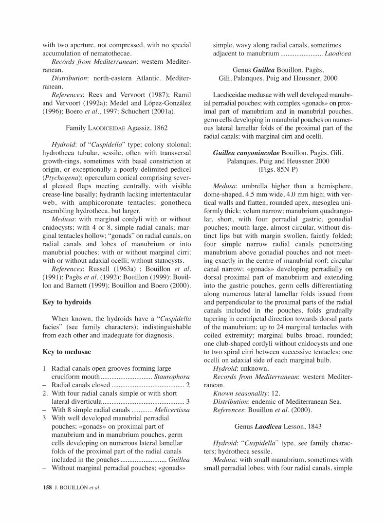
with two aperture, not compressed, with no specialaccumulation of nematothecae.
Records from Mediterranean: western Mediter-ranean.
Distribution: north-eastern Atlantic, Mediter-ranean.
References: Rees and Vervoort (1987); Ramiland Vervoort (1992a); Medel and López-González(1996); Boero et al., 1997; Schuchert (2001a).
Family LAODICEIDAE Agassiz, 1862
Hydroid: of “Cuspidella” type; colony stolonal;hydrotheca tubular, sessile, often with transversalgrowth-rings, sometimes with basal constriction atorigin, or exceptionally a poorly delimited pedicel(Ptychogena); operculum conical comprising sever-al pleated flaps meeting centrally, with visiblecrease-line basally; hydranth lacking intertentacularweb, with amphicoronate tentacles; gonothecaresembling hydrotheca, but larger.
Medusa: with marginal cordyli with or withoutcnidocysts; with 4 or 8, simple radial canals; mar-ginal tentacles hollow; “gonads” on radial canals, onradial canals and lobes of manubrium or intomanubrial pouches; with or without marginal cirri;with or without adaxial ocelli; without statocysts.
References: Russell (1963a) ; Bouillon et al.(1991); Pagès et al. (1992); Bouillon (1999); Bouil-lon and Barnett (1999); Bouillon and Boero (2000).
Key to hydroids
When known, the hydroids have a “Cuspidellafacies” (see family characters); indistinguishablefrom each other and inadequate for diagnosis.
Key to medusae
1 Radial canals open grooves forming large cruciform mouth............................. Staurophora
– Radial canals closed ......................................... 22. With four radial canals simple or with short
lateral diverticula.............................................. 3– With 8 simple radial canals ............ Melicertissa3 With well developed manubrial perradial
pouches; «gonads» on proximal part of manubrium and in manubrium pouches, germ cells developing on numerous lateral lamellar folds of the proximal part of the radial canals included in the pouches.......................... Guillea
– Without marginal perradial pouches; «gonads»
simple, wavy along radial canals, sometimes adjacent to manubrium ........................ Laodicea
Genus Guillea Bouillon, Pagès, Gili, Palanques, Puig and Heussner, 2000
Laodiceidae medusae with well developed manubr-ial perradial pouches; with complex «gonads» on prox-imal part of manubrium and in manubrial pouches,germ cells developing in manubrial pouches on numer-ous lateral lamellar folds of the proximal part of theradial canals; with marginal cirri and ocelli.
Guillea canyonincolae Bouillon, Pagès, Gili,Palanques, Puig and Heussner 2000
(Figs. 85N-P)
Medusa: umbrella higher than a hemisphere,dome-shaped, 4.5 mm wide, 4.0 mm high; with ver-tical walls and flatten, rounded apex, mesoglea uni-formly thick; velum narrow; manubrium quadrangu-lar, short, with four perradial gastric, gonadialpouches; mouth large, almost circular, without dis-tinct lips but with margin swollen, faintly folded;four simple narrow radial canals penetratingmanubrium above gonadial pouches and not meet-ing exactly in the centre of manubrial roof; circularcanal narrow; «gonads» developing perradially ondorsal proximal part of manubrium and extendinginto the gastric pouches, germ cells differentiatingalong numerous lateral lamellar folds issued fromand perpendicular to the proximal parts of the radialcanals included in the pouches, folds graduallytapering in centripetal direction towards dorsal partsof the manubrium; up to 24 marginal tentacles withcoiled extremity; marginal bulbs broad, rounded;one club-shaped cordyli without cnidocysts and oneto two spiral cirri between successive tentacles; oneocelli on adaxial side of each marginal bulb.
Hydroid: unknown.Records from Mediterranean: western Mediter-
ranean.Known seasonality: 12.Distribution: endemic of Mediterranean Sea.References: Bouillon et al. (2000).
Genus Laodicea Lesson, 1843
Hydroid: “Cuspidella” type, see family charac-ters; hydrotheca sessile.
Medusa: with small manubrium, sometimes withsmall perradial lobes; with four radial canals, simple
158 J. BOUILLON et al.
sm68s2Bsystema1 14/10/04 15:22 Página 158

or with short lateral diverticula; with simple wavy“gonads”; with or without marginal cirri; with orwithout adaxial ocelli.
Reference: Bouillon et al. (1991).Numerous non-valid species of Laodicea have
been described from Mediterranean. See Kramp (1959a, 1961); Boero and Bouillon (1993).
- Laodicea bigelowi Neppi and Stiasny, 1912 =Laodicea undulata (Forbes and Goodsir, 1851).
- Laodicea neptuna Mayer 1900 = doubtfulspecies.
- Laodicea ocellata Babnik (1948) = doubtfulspecies.
1. «Gonads» in short, complexly folded lateral diverticula of the proximal half of radial canals, tentacles without abaxial basal spurs; no marginal cirri ....................................... L. fijiana
– «Gonads» sinuous on radial canals contiguous with manubrium; tentacles with abaxial basal spurs; with marginal cirri................. L. undulata
Laodicea fijiana Agassiz and Mayer, 1899 (non Maas, 1905, 1906) (Fig. 86A)
Medusa: umbrella 6 mm high and wide; manubri-um quadrate, 1/4 height of umbrella cavity; «gonads»upon short complex lateral diverticula in proximalhalf of radial canals; about 70 marginal tentacleswithout basal spurs, very few cordyli, no cirri.
Kramp (1953, 1961) retains this species only forthe specimens of Fiji Islands? The L. fijiana describedfrom other localities are synonym of L. indica whicha possibly synonym of L. undulata, see below.
Records from Mediterranean: eastern Mediter-ranean.
Known seasonality: 11.Distribution: Indo-Pacific (Fiji Islands?);
Mediterranean.References: Kramp (1961); Schmidt (1973).Hydroid: unknown.
Laodicea undulata (Forbes and Goodsir, 1851)(Figs. 86B-G)
Hydroid: colonies of “Cuspidella type”, forminga creeping hydrorhiza from which arise single, ses-sile, cylindrical hydrothecae; terminal operculumpyramidal bearing 8-10 teeth meeting centrally,sharply demarcated from the hydrothecae margin;old hydrothecae with transverse growth rings;hydranth long, very extensile, based on a delicate
diaphragm; hypostome tall, conical; with a singlewhorl of 10-12 filiform, amphicoronate tentacle, nointertentacular membranous web; gonothecae ashydrothecae but twice as long, including severaldeveloping medusae at different stages.
Medusa: umbrella up to 37 mm wide, slightlyflatter than a hemisphere, saucer-shaped; mannerismshort, with square base and short per radial lobes;mouth with short, slightly folded, crenulated,recurred lips; 4 simple radial canals, circular canalnarrow; «gonads» elongated, with sinuated pendentfolds, issuing from corners of manubrium along per-radial lobes and half part of radial canals or almostto umbrella margin, «gonads» may be developedeven in small specimens and, the same individualmay present more than one sexual cycle; up to 400-600 hollow marginal tentacles with faintly devel-oped basal bulbs, small abaxial spurs on young mar-ginal tentacles, often absent in adults ones; 1-2 spi-ral cirri present (often lost by preservation) usually1 cordyli between tentacles or rudimentary bulbs;ocelli present each third or fifth tentacle, on eachtentacle when juvenile.
Remarks: L. undulata is very similar to L. indica,differing only by quantitative characters such as thenumber of ocelli and tentacles, several authorsregard them as conspecific.
Records from Mediterranean: eastern and west-ern Mediterranean, Black Sea; Adriatic Sea.
Known seasonality: 1-12.Distribution: Atlantic, Indo-Pacific?, Mediterranean.References: Russell (1936, 1953); Goy (1973b);
Schmidt and Benovic (1979); Bouillon (1984b,1985a); Benovic and Bender (1987); Gili (1986);Brinckmann-Voss (1987); Goy et al. (1988, 1990,1991); Bouillon, Boero and Fraschetti (1991); Ramiland Vervoort (1992a); Boero and Bouillon (1993);Cornelius (1995); Avian et al. (1995); Benovic andLucic (1996); Medel and López-González (1996);Mills et al. (1996).
Genus Melicertissa Haeckel, 1879
Laodiceidae medusa with 8 simple radial canals;with adaxial ocelli; with or without cirri.
Hydroid: unknown.
Melicertissa adriatica Neppi, 1915(no figure known)
Medusa: umbrella 46 mm wide, flatter than ahemisphere, with fairly thick mesoglea; manubrium
FAUNA OF THE MEDITERRANEAN HYDROZOA 159
sm68s2Bsystema1 14/10/04 15:22 Página 159

short, bell-shaped; mouth with 8 short crenulatedlips; «gonads» linear, along whole length of radialcanals; 8 perradial marginal tentacles and betweenthem 16 other marginal tentacles all alike; 3-5cordyli between successive tentacles each with ablack ocelli and even more marginal cirri.
Remarks: Picard, in Kramp (1961) page 444,refers doubtfully this species to Octogonade mediter-ranea Zoja, 1896, a Tiaropsidae with compoundsense organs, with different marginal tentacular struc-tures and, without cordyli (See Tiaropsidae).
Records from Mediterranean: Adriatic Sea.Known seasonality: 3-4.Distribution: endemic of Mediterranean Sea.References: Neppi (1915); Kramp (1959a, 1961);
Boero and Bouillon (1993); Avian et al. (1995);Benovic and Lucic (1996).
Genus Staurophora Brandt, 1834
Hydroid: a typical cuspedellid, hydrotheca ses-sile; gonothecae unknown.
Medusa: Laodiceidae with unusual cross-shapedmanubrium; mouth openings extending along the 4radial canals transformed for a long distance intoopen grooves, only most distal parts remaining freeand closed; mouth arms slit-like, with strongly fold-ed lips; «gonads» on diverticula in lateral walls ofcruciform, enlarged, mouth-radial canal complex;without cirri; with adaxial ocelli.
Staurophora mertensii Brandt, 1834(Figs. 86H-L)
Hydroid: colonies typically «cuspidellid», forminga reptant hydrorhiza from which arise at irregularintervals, wide, unstalked, cylindrical hydrothecae;operculum pyramidal formed of 10-12 triangular flapsmeeting centrally, sharply demarcated from hydrothe-cal margin; hydranths extensile, with a conical-round-ed hypostome, with up to 12 tentacles; no intertentac-ular membranous web; gonothecae not known.
Medusa: umbrella 100-300 mm wide, flatter thanhemispherical; mesoglea thick and smooth; exum-brella with numerous short centripetal furrows;manubrium, mouth and radial canals combined toform a large perradial cross reaching nearly umbrel-la, mouth openings extending along the 4 radialcanals transformed for a long distance into opengrooves, only most distal parts remaining free andclosed; mouth arms slit-like, with strongly foldedlips; «gonads» on diverticulae of the four radial
canals, more developed in middle region of eachcanal; numerous hollow, short, marginal tentacles(up to several thousand), with elongate conical mar-ginal bulbs and well developed pointed endodermalroots; tentacles approximately alternating withcordyli and each with adaxial ocelli.
Records from Mediterranean: medusa never foundin Mediterranean but doubtful records of the polypoidstage from south and eastern Spanish coast.
Seasonality: ?Distribution: Arctic (circumpolar); Indo-Pacific
(N. Japan, Alaska, bipolar); Atlantic; Mediter-ranean?.
References: Naumov (1951,1960-1969); Kramp(1961); García-Carrascosa (1981)?; García-Carras-cosa et al. (1987)? ; Boero and Bouillon (1993); Cor-nelius (1995); Medel and López-González (1996).
Family LOVENELLIDAE Russell, 1953
Hydroid: colony stolonal or erect, sympodial;hydrotheca pedicellate, elongate, everted-conicalto bell-shaped, with diaphragm; operculum coni-cal, formed either by many triangular plates onembayments in shallowly cusped hydrothecal mar-gin and well demarcated from hydrothecal wall bynoticeable crease line, or by a folded continuationof the hydrothecal wall, lacking hinge-like base;hydrothecae may renovate, but often collapsing,disintegrating in old specimens, just a crumpledcollar-shaped sheath remaining around hydranthbase; hydranth with endodermal epithelium differ-entiated into distinct parts, upper one digestive,basal one formed by chordal cells; with or withoutintertentacular web; gonophores as free medusae,gonothecae pedunculate.
Medusa: manubrium short; no gastric peduncle;no excretory pores; 4 simple radial canals; margin-al tentacles hollow, with lateral cirri; no marginalcirri; “gonads” on radial canals, not reachingmanubrium; without or with 8 (exceptionally 4 or12) or indefinite number of statocysts, 16 or morewhen adult; no ocelli.
Remarks: the family Lovenellidae was created byRussell (1953) for Leptomedusae with lateral cirri, 4radial canals, no marginal cirri, peduncle and excreto-ry pores, Lovenella-like hydroids with a well demar-cated operculum and hydrothecal margin embay-ments. Russell (1953) distinguished Lovenella, withan indefinite number of statocysts, from Eucheilotawith usually 8 statocysts. Kramp (1959a) adoptedthese views and later (Kramp, 1959b; 1961; 1968)
160 J. BOUILLON et al.
sm68s2Bsystema1 14/10/04 15:22 Página 160

added the genus Cirrholovenia with marginal cirri, somodifying Russell’s original definition. Calder (1971,1975) observed that the hydroid of Lovenella gracilislacks the opercular embayments that should be typicalof the genus Lovenella, having an operculum in con-tinuation with the hydrotheca. He consequently resur-rected the genus Dipleuron for this species (Calder,1991). Bouillon (1985a), considering the impossibili-ty to integrate the diagnostic characters of the polypsand medusae, separated Kramp’s Lovenellidae inthree families: the Cirrholoveniidae with medusaewith marginal cirri and “cuspidellid” hydroids; theEucheilotidae with medusae with lateral cirri, 8 stato-cysts and ”campanulinid” hydroids with a well demar-cated operculum but without hydrothecal embay-ments; and the Lovenellidae with lateral cirri, anindefinite number of statocysts and a “Lovenella” typeof hydroid, with a well demarcated opeculum withembayments of hydrothecal margin. The study of “Lovenellidae” life cycles, however, shows that if themedusae of these family present clear diagnostic char-acters, their hydroids are puzzling, all belong to a“campanulinid” type but their opercular structurespresent differences even within the same genus. It isthus hopeless to refer with confidence one or anothertype of opercular structure to a family-group taxon,particularly to a medusa-based family. After Kramp(1919, 1932b), too a great importance has been givento the opercular structures of the Campanulinida.Opercula can be different within the same family (see,for instance, the Tiaropsidae and the Lineolariidae) oreven within the same genus: in Phialella, for instance,some species have opercular flaps demarcated fromhydrothecae and other do not (Boero, 1987), and evendifferent hydrothecae on the same hydrocaulus maypresent the two opercular types. Opercular structure,thus, is evidently inconstant and cannot be used to dis-tinguish families or even genera. The more, in manycampanulinids the operculum can completely disap-pear, with the apical part of the hydrotheca, duringnormal colony growth, as shown by Werner (1968 aand b) in Eucheilota maculata and Eutonina indicansor can even be absent in some species of a normallyoperculate genus (many Eirenidae), only a little morethan a collar remaining at the base of the hydrothecaeof fully developed hydranths, looking like a haleciidtheca (see for instance Werner, 1968, fig.14 and Fig.87: Figs. 8 to 13). The family Eucheilotidae is conse-quently considered as synonym of the Lovenellidaeand the genera Eucheilota and Lovenella are againincluded in the Lovenellidae, being defined as abovefor the medusa stage, the hydroid being characterised
by “campanulinid polyps” with an operculum well ornot well demarcated from the hydrotheca, withhydrothecae having or not marginal embayments, orreduced to a basal collar; usually with an intertentacu-lar web; cnidome generally with merotrichous hap-lonemes. The family Cirrholoveniidae, with marginalcirri and “cuspedelliid” type of hydroids, is kept asvalid. Two genera, Hydranthea with free eumedusoidsand Campalecium with newly released medusae ofEucheilota type are here tentatively included in theLovenellidae. They were formerly considered asHaleciidae due to the collar shape of their hydrothe-cae; in our opinion they are campanulinid hydroidswith reduced thecae similar to reduced stages ofLovenellidae, but might well represent the basal statefrom which paedomorphic species with fixedgonophores and reduced hydrotheca originated theHaleciidae.
References: Russell (1963b); Pagès et al. (1992);Bouillon (1999); Bouillon and Barnett (1999);Bouillon and Boero (2000); Schuchert (2001a,2003).
Key to hydroids
1. Hydroid of “Campanulina” facies ................... 2– Hydroid with reduced hydrotheca, of “haleciid”
facies................................................................. 32. Hydranth with intertentacular web ... Eucheilota– Hydranth without intertentacular web.................
.............................................................Lovenella3. Gonophores producing eumedusoids;
gonothecae on hydrorhiza, reduced or absent.............................................................. Hydranthea
– Gonophores producing free medusae; gonothecae growing from pedicel just under hydrothecae .................................. Campalecium
Key to medusae
1. With usually no more than 8 statocysts ..........................................................................Eucheilota
– With an indefinite number of statocysts (16-32)............................................................ Lovenella
Genus Campalecium Torrey, 1902
Hydroid: colony typically stolonal, pedicel of var-ied length bearing terminal hydranth, often secondarypedicels forming sympodial branches; hydrothecacollar-shaped, shallow, often regenerated, with a dis-tinct diaphragm, large desmocytes; hydranth relative-
FAUNA OF THE MEDITERRANEAN HYDROZOA 161
sm68s2Bsystema1 14/10/04 15:22 Página 161

ly large, elongated, cylindrical, not retractable intohydrotheca; up to 30 amphicoronate tentacles; with orwithout intertentacular web endodermal epitheliumdifferentiated into distinct part the upper digestive,the basal part formed by chordal cells; gonophoresgiving rise to free medusae; gonothecae clavate orrounded arising beneath hydrothecal pedicel, eachwith several medusa buds.
Medusa: only medusa buds or juvenile medusaeof Eucheilota type presently known, pending thespecies.
References: Watson (1993); Bouillon and Boero(2000); Boero, unpublished observations.
1. Cnidome with merotrichous haplonemes............................................................... C. medusiferum
– Cnidome microbasic mastigophores ............................................................................ C. cirratum
? Campalecium cirratum “Millard and Bouillon(1975)”; not Haeckel, 1879 (Figs. 87A-B)
Hydroid: as for genus, cnidome microbasicmastigophores.
Medusa: only medusa buds known fromgonothecae, with four perradial marginal bulbs ofwhich two bear tentacles, with lateral cirri, withoutmarginal warts, with eight statocysts.
Records from Mediterranean: ?Distribution: Indo-Pacific; Mediterranean?Seasonality: ?References: Millard and Bouillon (1975); Calder
(1991); Altuna Prado, 1993b.
Campalecium medusiferum Torrey, 1902(Figs. 87C-E)
Hydroid: As for genus, cnidome with merotric-hous haplonemes
Medusa: Only young medusae known with fourperradial marginal tentacles, four perradial marginalbulbs and eight statocysts, without lateral cirri andmarginal warts.
Records from Mediterranean: eastern and west-ern Mediterranean, Adriatic.
Known seasonality: 5-9, 11, 12.Distribution: Atlantic, eastern Pacific, Mediter-
ranean.References: Motz-Kossowska (1911); Huvé
(1954); Boero (1981); Gili (1986); Boero and Sara(1987); Calder (1991); Altuna Prado (1993b) Avianet al. (1995).
Remarks: The systematic position of these twospecies is discussed under Lovenella cirrata.
Genus Eucheilota McCrady, 1859
Hydroid: colony of “campanulinid” type;hydrotheca with diaphragm, often reduced to a col-lar shaped sheath around base of adult hydranths,operculum well developed, not demarcated;hydranth with intertentacular web; gonotheca long,pedicellate on erect stems, with one to five medusabuds.
Medusa: with usually 8 statocysts.References: Ramil (1988); Cornelius (1995);
Bouillon and Boero (2000).Remarks: the polyp of the species Campomma
hincksii (Hartlaub, 1897) has been considered byCornelius (1995) as synonymous of Eucheilotamaculata. If it is so, Eucheilota maculata would bepresent in the Mediterranean, recorded under thename of Campomma hincksii. However, thehydroids of most Campanuliniidae are very similar,the medusa being necessary to identify the species;as the medusa has not been found in the Mediter-ranean, we consider the following records Mediter-ranean of “Eucheilota maculata” hydroids as doubt-ful: Gili (1982, 1984); Gili and Castelló (1985) all asCampanulina; Medel and López-González (1996)as Campomma.
Key to medusa
1. With medusa buds on radial canals; with 4 marginal tentacles and 4 or more rudimentary bulbs, all with 1-3 pairs of lateral cirri; 8 statocysts...................................... E. paradoxica
– Without medusa buds on radial canals............. 22. With 16 marginal tentacles an 16 rudimentary
bulbs all with one pair of lateral cirri; about 24 small knobs without cirri; 8 statocysts................................................................... E. ventricularis
– With 4 marginal tentacles with one pair of lateral cirri and a number of rudimentary bulbs, some of which have cirri; 8 statocysts; immature medusae................................................ E. maasi
Eucheilota maasi Neppi and Stiasny, 1911(Fig. 87F)
Medusa: only young medusa known. Umbrella3-5 mm wide, 3 mm high, mesoglea thick; manubri-um cylindrical, half as long as umbrella cavity;
162 J. BOUILLON et al.
sm68s2Bsystema1 14/10/04 15:22 Página 162

small, immature, «gonads» on middle part of radialcanals; 4 marginal tentacles with rounded bulbs withone pair of lateral cirri, a number of rudimentarybulbs, some of which flanked by cirri; 8 statocysts.
Hydroid: unknown.Records from Mediterranean: Adriatic Sea.Known seasonality: 4, 7, 9, 10, 12.Distribution: endemic of Mediterranean Sea.References: Neppi and Stiasny (1913); Kramp
(1961); Boero and Bouillon (1993); Avian et al.(1995); Benovic and Lucic (1996).
Eucheilota maculata Hartlaub, 1894(Figs. 87G-L)
Hydroid: see genus definition. Hydroid doubtful-ly reported from Mediterranean and Adriatic Seas.Medusa never collected in Mediterranean Sea
Eucheilota paradoxica Mayer, 1900(Fig. 87M)
Hydroid: not known from field. Carré and Carré(1990) obtained in culture hydranths differentiat-ing from medusae maintained during two monthsat 15°C. The hydroids developed on the radialcanal at the position usually occupied by thegonads. Those hydranths were able to capturepreys and to feed but medusae reduction occurredgradually and finally the medusae were reduced toa mass constituting the base of the polyps. Follow-ing Carré and Carré those polyps are very similarto those described for Eucheilota maculata byWerner (1968a), unfortunately the authors nevercould induce their settlement.
Medusa: umbrella 5 mm wide, higher than ahemisphere, sometimes with a slight apical projec-tion, mesoglea moderately thick; manubrium small,flask-shaped; mouth with 4 simple lips; 4 narrowradial canals and narrow circular canal; «gonads»along middle portion of radial canals; with 4 hollowmarginal tentacles with large bulbs, and 4 or morerudimentary bulbs; all bulbs with 1-3 pairs of later-al cirri of unequal size; with 8 marginal statocysts;active medusa budding on radial canals in non sex-ual mature specimens, the daughter medusae beginto develop medusa buds before they become free;frustules and hydranths developing on radial canalsat the position usually occupied by the «gonads»under certain environmental conditions.
Records from Mediterranean: eastern and west-ern Mediterranean
Known seasonality: 2-12.Distribution: Atlantic; Indo-Pacific; Mediter-
ranean.References: Kramp (1959b, 1961); Uchida and
Sugiura (1975); Schmidt and Benovic (1977);Bouillon (1984 a, b; 1995b); Castelló i Tortella(1986); Bouillon et al. (1988b); Dallot et al. (1988);Carré and Carré (1990); Goy et al. (1988, 1990,1991); Boero and Bouillon (1993).
Eucheilota ventricularis McCrady, 1959(Fig. 88A)
Medusa: umbrella 10 mm wide, hemispherical;manubrium short; mouth with four prominent lips;«gonads» linear, along middle 1/3 or 2/3 of radialcanals; 16 marginal tentacles and 16 rudimentarybulbs all with one pair of lateral cirri and about 24minute knobs without cirri; 8 statocysts.
Hydroid: unknown.Records from Mediterranean: eastern Mediter-
ranean.Known seasonality: 1-5, 12.Distribution: Atlantic; Indo-Pacific, Mediter-
ranean.References: Kramp (1959b, 1961); Bouillon,
1984b; Lakkis and Zeidane (1985)); Goy et al.,1988, 1990, 1991; Boero and Bouillon (1993).
Genus Hydranthea Hincks, 1868
Hydroid: colonies stolonal, hydrotheca short,collar-shaped, shortly pedicellate; hydranth elongat-ed, large, with intertentacular web with merotric-hous haploneme cnidocysts; gonophores as eumedu-soid, only short-lived male free eumedusoidsknown; without tentacles; with four radial canals;“gonads” on radial canals, 8 statocysts; gonothecaereduced or absent; when present, attached tohydrorhiza.
References: Cornelius (1995); Bouillon andBoero (2000).
Hydranthea margarica (Hincks, 1862)(Figs. 88B-D)
Hydroid: colonies stolonal bearing singlehydranths at irregular intervals, stolon with haustor-ial projections beneath providing additional attach-ment; hydrothecae very short, collar-shaped withslightly divergent walls and basal diaphragm, onsmooth short pedicels inserted directly to stolon;
FAUNA OF THE MEDITERRANEAN HYDROZOA 163
sm68s2Bsystema1 14/10/04 15:22 Página 163

hydranth very elongated, slender, not retractable inhydrothecae, widest just below tentacles base andtapering gradually to base, with a cone-shapedhypostome, with 20-30 amphicoronate webbed ten-tacles; endodermal epithelium differentiated intodistinct part the upper digestive, the basal partformed by chordal cells; gonothecae short or absent,when present a shallow dish on a short pedicel;gonophore a single eumedusoid; merotrichous hap-lonemes scattered or in clumps through tissues ofhydranth, stolon and particularly conspicuous ontentacular web, developing eggs reported inhydrorhiza of colonies lacking gonophores.
Medusa: released eumedusoids almost spherical,about 0.5 mm in diameter, surrounded by a thinperisarc, with four branched radial canals filled withpurplish or orange granules as well as ring canal;with eight statocysts; «gonads» on manubrium fill-ing the whole subumbrella, numerous cnidocyst onexumbrella.
Records from Mediterranean: eastern and west-ern Mediterranean, Adriatic.
Known seasonality: 5-8, 10, 11.Reproduction: 8, 10.Distribution: Atlantic, Indo-Pacific (Seychelles),
Mediterranean.References: Hincks (1868); Huvé (1954); Millard
and Bouillon (1973); Cornelius (1975b, 1995); Gili(1986); Boero and Sara (1987); Boero and Bouillon(1993); Avian et al. (1995); Peña Cantero and Gar-cía Carrascosa (2002).
Remarks: the presence of only eight statocysts inthe eumedusoid could indicate some relationship ofthis genus with the genus Eucheilota.
Hydranthea incertae sedis
Hydranthea aloysii (Zoja, 1893)
Colony reptant, stolon bearing single hydranthsat irregular intervals. Hydranth up to 4mm in height,on a slightly ringed pedicel, with 12-18 amphicoro-nate tentacles linked by an intertentacular web.Hydrotheca very short, delicate, with a thindiaphragm. Large cnidocysts kidney-shaped up to15 x 5.0 µm occuring throughout stolon, hydranthtissues and in tentacular web, small cnidocysts 9-10x 2.5 µm scattered throughout entire colony.Gonophores unknown.
Remark: this species is insufficiently described,its reproduction is unknown it could be any haliciidor lovenelliid, perhahs a juvenile of H. margarica.
Records from Mediterranean: western Mediter-ranean, Adriatic.
Known seasonality: not found in nature, onlyknown from laboratory rearing.
Distribution: endemic of Mediterranean Sea.References: Hadzi (1914) as Georginella
diaphana; Huvé (1954); Boero and Sara (1987);Boero and Bouillon (1993); Avian et al. (1995).
Genus Lovenella Hincks, 1868= Mitrocomium Haeckel, 1879
Hydroid: colony “Campanulina” type; stolonal orupright and sympodial; hydrotheca pedicellate, elon-gate, everted-conical to bell-shaped; operculum coni-cal, formed either by many triangular Figs. withembayments of the hydrothecal margin and welldemarcated from hydrothecal wall by noticeablecrease line, or formed by a folded continuation of thehydrothecal wall, lacking hinge-like base; diaphragmpresent; no intertentacular web; no nematophores.
Medusa: with an indefinite number of statocysts.Remark: all Lovenella-like hydroids with
unknown cycle must be included in the Campan-ulinidae incertae sedis.
Key of the medusae
1. 8-16 marginal tentacles, each with 3-4 pairs of lateral cirri; several marginal warts without cirri ............................................................. L. cirrata
– With more than 16 marginal tentacles, with no marginal warts .................................................. 2
2. With 16-24 marginal tentacles, each with 1-3 pairs of lateral cirri; «gonads» oval, longitudinal-ly divided, very close to circular canal ............................................................................ L. clausa
– With 21 marginal tentacles, each with one pair of lateral cirri; «gonads» oval, midway of radial canals length ...................................... L. gracilis
Remarks: included here there is information onLovenella chiquita Millard, 1957 whose juvenilemedusae are known from South Africa and thepolyps described doubtfully from the Spanish coasts(see below).
Lovenella chiquita Millard, 1957(Figs. 88E-H)
Hydroid: hydrorhiza creeping, often epizootic onother hydroids giving rise to short erected stems bear-
164 J. BOUILLON et al.
sm68s2Bsystema1 14/10/04 15:22 Página 164

ing a terminal hydranth or branched sympodially up to9 times each limb terminating in a hydrothecae, stemsannulated throughout; hydrothecae deep- campanulate,smooth, margin with 8-10 shallow bays; operculum of8-10 converging segments with a gap between theircentral points; diaphragm delicate; hydranth with over12 tentacles; gonothecae arising from hydrorhiza onshort annulated pedicels, smooth elongated, taperingbelow truncated above, containing two medusa.
Medusa: only newly liberated medusae of thisspecies are known. Umbrella 0.4 mm wide, 0.3 mmhigh, mesoglea thin; manubrium short; mouth quad-rangular simple; «gonads» not yet developed; with-out lateral cirri or marginal warts; 8 marginal tenta-cles; 8 statocysts.
Records from Mediterranean: western Mediter-ranean, only the polyp stage has been describedonce from the Spanish coast (García-Corrales et al.,1979), presumably a doubtful determination, thisspecies being considered as endemic from SouthAfrica, see Millard (1975).
References: Kramp (1961); García-Corrales et al.(1979); Boero and Bouillon (1993).
Remark: the juvenile medusae of the abovedescribed species do not present enough diagnosticfeatures to be assigned to one or another medusafamily, the polyp stage closely resembles that ofLovenella clausa hence its inclusion in the genusLovenella by Millard (1957, 1975) and Bouillon(1985a) and in the present paper, awaiting furtherinformations about the mature medusa stage.
Lovenella cirrata (Haeckel, 1879)(Fig. 88I)
Hydroid: colonies typically stolonal issued froma creeping hydrorhiza from which arise numerous(up to almost 100) solitary, almost sessile,unbranched; hydranths; hydrothecae very shallow,collar shaped, marginal rim somewhat everted, witha distinct basal diaphragm; hydranth very elongated,slender, tapering gradually to base and just belowtentacles base which is the widest part of the body,with a cone-shaped hypostome, with 24 tentacles inone whorl, amphicoronate, hydranths not retractableinto hydrothecae, intertentacular web present withlarge elongated cnidocysts (presumably merotric-hous haplonemes); gonothecae borne at the base ofthe hydrothecae, half as long as the living hydranth,tubular, conical tapering from end to base, apertureapical extending along whole width; gonophorewith 4 medusa buds (after Brinckmann, 1959a).
Medusa: umbrella 16 mm wide, almost hemi-spherical; manubrium short, urn-shaped; 4 simplelips; 4 sausage-shaped «gonads» on distal half ofradial canals; 8-16 marginal tentacles with conspic-uous basal bulbs, each flanked by 3-7 pairs of later-al spiral cirri; usually with 3-5 rudimentary margin-al warts between successive tentacles; about 16 sta-tocysts.
Records from Mediterranean: eastern and west-ern Mediterranean, Adriatic.
Known seasonality: 4, 6, 10.Distribution: Atlantic (Brazil, W. Africa); Indo-
Pacific (Malayan Archipelago, Bismarck Sea);Mediterranean.
References: Kramp (1959a, 1961); Brinckmann(1959a); Millard and Bouillon (1975); Boero(1981); Dowidar (1983); Boero and Sara (1987);Brinckmann-Voss (1987); Calder (1991); AltunaPrados (1993b); Pagès et al. (1992); Boero andBouillon (1993) Avian et al. (1995).
Remarks: Brickmann (1959a) reared youngmedusae, which at birth presented already lateral cirriand marginal warts from a hydroid that she tentative-ly identified with the Haleciidae Haleciella microthe-ca Hadzi, 1914. The comparison of the 14 days oldmedusae from laboratory with young and adultmedusae found in the plankton let her to identifythose young medusae as Lovenella cirrata. Haliciellamicrotheca considered as a Haleciid has been syn-onymized with Campalecium medusiferum Torrey,1902 by Huvé (1954), as Campalecium cirrata byMillard and Bouillon (1975) and Yamada (1995) andas Mitrocomium cirratum by Calder (1991).
In our opinion, the hydroid described by Brinck-mann (1959a) (see above for the description) is nota haleciid but is a campanulinid hydroid withreduced hydrotheca similar to those described byWerner in Eucheilota maculata and in Eutoninaindicans and found in several Eirenidae hydroids(see Werner, 1968 Fig. 14, and Figs. in Russell,1970a). Brinckmann in 1959 was not aware of theexistence of such hydrothecal structures and nor-mally interpreted the short priest collar-shapedhydrotheca of Lovenella cirrata as a haleciid one.Millard (1975) already pointed out the resemblanceexisting between the hydrothecae of haleciids tothose of some campanulinids which have loosedtheir operculae and distal parts; Boero and Sara(1987) underlined the possibility that Campanulin-idae hydranths with reduced hydrothecae could havebeen assigned to the Haleciidae and speculated thatsuch a type of campanulinids could be at the origin
FAUNA OF THE MEDITERRANEAN HYDROZOA 165
sm68s2Bsystema1 14/10/04 15:22 Página 165

of the haleciids which by paedomorphis loosed themedusa phase while the hydroids became moredeveloped. Very close affinities seems thus undeni-ably exist between this two families and the sub-order Haleciida proposed by Bouillon (1984b) tomatch with the other orders he introduced at thesame time should be suppressed, the Halecioideabeing included in the Campanulinida.
Hydranthea margarica hydroids liberate freeeumedusoids and have great affinities with the cam-panulinid polyps mainly by their particularly elon-gated body with a long non-digestive basal part,their unbranched polyps and merotrichous hap-lonemes cnidocysts. Hydranthea may representwhat could have been a first step to the loose of afree medusae stage in the Lovenellidae leading so bypaedomorphis and specialisation of the hydroid tothe Haleciids with fixed gonophores (see Boero andSara, 1987). Nemalecium lighti with campanulinidhydranths and eumedusoids is already a true haleci-id having pseudostenotele cnidocysts; this speciescould represent a further step in the evolution to thecolonial and polymorphic haleciids. If this is thecase one of the two families (Lovenellidae andHaleciidae) both created by Hincks (1868) shouldhave priority under the condition that the Haleciidsare monophyletic.
There is a great confusion between the “Cam-palecium” like species. It is far from excluded thatHaleciella microtheca, Campalecium medusiferum,Campalecium cirratum and Lovenella cirrata are allconspecific as suggested by Clader (1991) who unit-ed them all under the reintroduced generic name ofMitrocomium Haeckel, 1879. The differencesobserved at the hydroid level being nothing else thanvariations that could be expected within a singlespecies. If such is the case the genus LovenellaHincks, 1868 has nevertheless precedence over:Campalecium Torrey, 1902; Haliciella, Hadzi, 1914and Mitrocomium Haeckel, 1879.
However, we concur with Boero (1981a) thatLovenella cirrata, Campalecium medusiferum andCampalecium cirratum cannot be at the presentstage of our knowledge considered as synonyms.The hydranths of Lovenella cirrata are differentfrom those of Campalecium cirratum being almostsessile; solitary, their juvenile medusae present notonly lateral cirri but also the rudimentary warts char-acteristic of the adult medusa species. Only the newborne medusa of Campalecium medusiferum areknown and they are deprived from lateral cirri andwarts and cannot presently been assigned with certi-
tude to any Leptomedusae family, their polyps arerather similar to those of L. cirrata. Campaleciumcirratum medusae are only known from gonophoresas medusa buds which seems to present lateral cirri;their polyps have pedicels of variable length, theyoccasionally may present one or two sympodialbranches, they have microbasic mastigophorescnidocysts (Millard and Bouillon, 1975) instead ofthe merotrichous haplonemes present in the twoother species. More studies on Campaleciummedusiferum and C. cirratum life cycles areundoubtly needed, only the knowledge of their adultmedusae will clear up their real affinities. It is alsostill necessarily to clarify the problem of the syn-onymy between the Mediterranean, American Cali-fornia and Bermuda forms of Campalecium. It isinteresting to underline that presently the adultmedusae of L. cirrata are known from Mediter-ranean, from the Malayan Archipelago, from theBismarck Sea, from Brazil and W.African watersbut have never been found in eastern Pacific and theNorth American basin from where the polyps ofCampalecium medusiferum have been describedrespectively by Torrey (1902) and Calder (1991).
Lovenella clausa (Lovén, 1836)(Figs. 88J-K, 89A-D)
Hydroid: colonies comprising either singlepedicels or short erect shoots arising from a creep-ing hydrorhiza; pedicels with 2-5 rings belowhydrothecae and above origin from hydrorhiza;hydrothecae long and narrow, thin walled, roughlycylindrical, tapering towards base, widest at aper-ture; operculum of 8-10 triangular converging flapsseated in embayment of the hydrothecae margin andsharply demarcated from the wall; often 2-3 reno-vated hydrothecae inside oldest one; hydranth longextending length of hydrotheca beyond aperture,with ca 14 –16 tentacles, probably no intertentacularweb; gonothecae elongated, axillary, tapering fromtruncated end towards base, sides often slightly sin-uous, aperture terminal, containing up to 25 medusabuds maturing simultaneously
Medusa: umbrella 5-9 mm wide, hemispherical,mesoglea moderately thick; manubrium short,small; without gastric peduncle; mouth with 4 sim-ple lips; «gonads» oval, longitudinally divided, veryclose to circular canal; 16-24 marginal tentacles;marginal tentacular bulbs large, conical; 1-3 pairs oflateral, spiral cirri on either side of each tentacularbase; 16-23 statocysts.
166 J. BOUILLON et al.
sm68s2Bsystema1 14/10/04 15:22 Página 166

Records from Mediterranean: western Mediter-ranean. Medusa not known from Mediterranean,only dubious records of hydroids.
Known seasonality: 1, 4, 6, 7-9.Distribution: Atlantic; Mediterranean?References: Kramp (1961); Picard (1958b); Gili
(1986); Boero and Bouillon (1993); Cornelius(1995); Medel and López-González (1996); PeñaCantero and García Carrascosa (2002).
Lovenella gracilis (Clarke, 1882)(Figs. 89E-G)
Hydroid: colonies with monosiphonic hydro-caulus slightly branched or unbranched; divided intointernodes by septa at more or less regular intervals;typical annulations absent except on the hydranthpedicels and at base of the stems; hydrothecae alter-nate on short pedicels, operculum a folded continu-ation of the hydrothecal wall, lacking hinge-likebase, with about 8 facets when folded; 14 or moretentacles, no intertentacular web; diaphragm thin;gonothecae clavate, truncate terminally, borne nearthe base of hydrothecal pedicels, several medusabuds in each.
Medusa: umbrella about 2.1 mm wide, 1.1 mmhigh, hemispherical; velum wide; manubrium urn-shaped; mouth with 4 simple lips; 4 radial canalsand circular canal narrow; «gonads» oval midway ofradial canals length; oldest specimens with 21 mar-ginal tentacles each with 1 pair of cirri; 33 stato-cysts.
Remark: diagnosis taken from Calder (1971) andbased on raised material from polyp colonies.
Records from Mediterranean: western Mediter-ranean only juvenile medusae and hydroids known.
Known seasonality: 5.Distribution: Atlantic; Mediterranean.References: Huvé (1952 a, b); Picard (1958b);
Calder (1971, 1991); Boero and Bouillon (1993).
Family MALAGAZZIIDAE Bouillon, 1984
Hydroid: Where known of the “campanulinidtype”; colonies stolonal; hydrothecae shortly pedi-cellate, with a conical operculum formed by numer-ous convergent segment not clearly demarcatedfrom the hydrothecal wall; hydranths with an inter-tentacular web; gonothecae claviform arising fromthe stolons.
Medusa: with small manubrium; without gastricpeduncle; with 4-8, sometimes up to 12 radial
canals; «gonads» completely surrounding radialcanals and separated from manubrium; with adaxialexcretory papillae; no permanent rudimentary mar-ginal bulbs (all bulbs potentially transforming intotentacles); with closed statocysts; without ocelli;without cirri.
Genus Octophialucium Kramp, 1955
Malagazziidae medusa with normally 8 radialcanals; mouth with 8 lips.
Hydroid: where known of the “campanulinid”type, see family characters (Bouillon, 1984a, b).
Octophialucium funerarium(Quoy and Gaimard, 1827) (Figs. 89H-J)
Medusa: umbrella 30-40 mm wide, lens-shapedto saucer-shaped, mesoglea thick except near exum-brellar margin, virtually no subumbrellar cavity;velum fairly broad; manubrium very small, withbase in form of eight-rayed star; mouth with 8 sim-ple, short pointed lips; typically 8 straight, narrow,radial canals in adults (4 when young); «gonads»short, along distal 1/4 of radial canals, near margin,but not reaching circular canal; 64-128 marginal ten-tacles; marginal bulbs well developed, with excreto-ry papillae; usually one to three, usually two, stato-cysts between successive tentacles.
Hydroid: unknown.Records from Mediterranean: eastern and west-
ern Mediterranean; Adriatic.Known seasonality: 3-8, 12.Distribution: Atlantic; Mediterranean.References: Kramp (1961); Goy (1973b); Bouil-
lon (1985a, 1995a); Gili (1986); Benovic and Ben-der (1987); Boero and Bouillon (1993); Avian et al.(1995); Cornelius (1995); Benovic and Lucic(1996); Medel and López-González (1996); Mills etal. (1996).
Family MELICERTIDAE Agassiz, 1862
Hydroid: where known colonies stolonal withbranching stolons and erect shoots bearing one,sometimes two, hydranths; perisarc thinning awaycompletely below base of the hydranths, no hydrothe-ca; hydranth large, broad in the middle, attenuatebelow, tapering gently above, with narrow, amph-icoronate tentacles; without intertentacular web;gonophores borne on the column of hydranth, nogonothecae (hydroid only known in Melicertum).
FAUNA OF THE MEDITERRANEAN HYDROZOA 167
sm68s2Bsystema1 14/10/04 15:22 Página 167

Medusa: with base of manubrium attached overits whole surface; with eight simple or bifurcatedradial canals; with hollow marginal tentacles; with-out marginal or lateral cirri and statocysts; with orwithout ocelli.
Reference: Russell (1963a).
Genus Orchistomella Kramp, 1959
Melicertidae with eight or more radial canals, allof which arise from manubrium; with or withoutocelli, «gonads» unknown.
Hydroid: unknown.
Orchistomella graeffei (Neppi and Stiasny, 1911)(No figure known)
Medusa: umbrella 4 mm wide, highly vaulted,mesoglea very thick; gastric peduncle?; manubriumquite flat; mouth with 8 simple lips; 8 fully devel-oped radial canals and in each octant about 12 youngblindly ending canals; 8 marginal tentacles withlarge pear-shaped marginal bulbs; 2-3 rudimentarybulbs between successive tentacles. Probably youngspecimens of Aequorea (Picard in Kramp, 1961page 444).
Hydroid: unknown.Records from Mediterranean: western Mediter-
ranean; Adriatic Sea.Known seasonality: 7-10.Distribution: endemic of Mediterranean Sea.References: Neppi and Stiasny (1911); Kramp
(1959b, 1961, 1968); Goy (1973b); Boero andBouillon (1993); Avian et al. (1995); Benovic andLucic (1996).
Family MITROCOMIDAE Haeckel, 1879 (part); Torrey, 1909
Hydroid: usually poorly known, most of the“Cuspidella” type; hydrothecae tubular sessile; withpyramidal operculum made either of several trian-gular flaps, or of pleats in the continuing ofhydrothecal tube, all not well demarcated from thehydrothecal wall, lacking a crease-line at base of theflaps or pleats; hydranth extensile, with a single usu-ally amphicoronate whorl of filiform tentacles; nointertentacular web; no nematophores; gonophoreswhere known scarcely pedicellate, on hydrorhiza.
Medusa: with bases of manubrium attached tosubumbrella along continuation of radial canals;with 4 or more simple radial canals; marginal tenta-
cles hollow; marginal cirri present in some genera;with «gonads» oval or linear, only on radial canals;with open statocysts.
References: Kramp (1932a); Pagès et al. (1992);Bouillon (1999); Bouillon and Barnett (1999);Bouillon and Boero (2000).
Key to medusae
1. With marginal cirri ........................................... 2– Without marginal cirri; with numerous open
statocysts............................................ Foersteria2. With numerous open statocysts ........ Mitrocoma– With 8-16 open statocysts............. Mitrocomella
Genus FoersteriaArai and Brinckmann-Voss (1980)
Mitrocomidae medusae with 4 radial canals; withnumerous open statocysts; without marginal cirri.
Hydroid: unknown.
1. Manubrium short and very broad, 1/3 umbrella width, entirely coloured in dark purple-brown; 40 open statocysts; mouth with simple lips; with large rounded marginal bulbs........... F. antoniae
– Manubrium short, small, 1/7 umbrella width, with 4 interradial brown lines; 7080 open statocysts; mouth lips groove-shaped; marginal bulbs small, conical ............................. F. araiae
Foersteria antoniae Gili, Bouillon, Pagès, Palanques, Puig, and Heussner, 1998
(Figs. 90A-C)
Medusa: umbrella up to 6 mm wide, 3 mm high,flatter than a hemisphere, almost quadratic fromabove, mesoglea rather thick; without gastric pedun-cle; manubrium very short and large with quadraticbase, 1/3 of the width of the umbrella, intensely anduniformly coloured in dark purple-brown; mouthwith four simple, very short lips; radial canals andcircular canal narrow; «gonads» form and sizedepending on sex, male «gonads» elongated, onabout the 4/5 of the distal half of radial canals,female, «gonads» small, spherical, with few eggs (5-15) near circular canal; up to 40 marginal tentacles;marginal bulbs large, rounded, with two masses ofbrown pigment; up to 40 open statocyst, 0-2between successive marginal tentacle.
Records from Mediterranean: western Mediter-ranean.
168 J. BOUILLON et al.
sm68s2Bsystema1 14/10/04 15:22 Página 168

Known seasonality: 4 and 5.Distribution: endemic of Mediterranean Sea.References: Gili et al. (1998).Hydroid: unknown.
Foersteria araiae Gili, Bouillon, Pagès, Palanques and Puig, 1999
(Figs. 90D-E)
Medusa: umbrella up to 7 mm wide and high,hemispherical, mesoglea thick; without gastricpeduncle; manubrium very short, with square base,with 4 interradial dark brown lines, 1/7 as high asumbrellar cavity and 1/7 as wide as umbrella;mouth with four short, groove-shaped lips, sur-rounded by cnidocysts, four radial canals and cir-cular canal narrow; «gonads» small, oval or round-ed, split longitudinally, on distal half of radialcanals near but not in contact with circular canal;up to 40 long, coiled, marginal tentacles; marginaltentacular bulbs conical, small with brown pigmentinside; 70-80 open marginal statocysts, 1-3between each two tentacles.
Records from Mediterranean: western Mediter-ranean.
Known seasonality: 5.Distribution: endemic of Mediterranean Sea.References: Gili, Bouillon, Pagès, Palanques and
Puig (1999).
Genus Mitrocoma Haeckel, 1864
Hydroid: colonies where known of “Cuspidella”type, operculum with numerous sharp pointed, trian-gular flaps meeting centrally and presenting no clearlimits with the hydrothecal margin.
Medusa: with 4 radial canals; with numerousopen statocysts; with marginal cirri.
Mitrocoma annae Haeckel, 1864(Figs. 90F-J)
Hydroid: (Metschnikoff, 1886a) reared thehydroid of this medusa which is typically a «cuspi-dellid», the hydrothecae are sessile, cylindrical,wide and elongated and arise at irregular intervalsfrom a linear, creeping, stolon; hydrothecae closedby a pyramidal operculum with numerous sharppointed, triangular flaps meeting centrally and pre-senting no clear limits with the hydrothecal margin;hydranths extensile, with conical hypostome; 12-16filiform tentacles in a single whorl, no basal inter-
tentacular membranous web; gonothecae unknown.Medusa: umbrella 30-40 mm wide, flatter than a
hemisphere, mesoglea thick; manubrium very small,1/8-1/10 of umbrella diameter; mouth lips fairlyshort; radial canals and circular canal narrow;«gonads» linear, sinuous, along distal 1/2-3/4 ofradial canals, not reaching circular canal, with amedian division; 60-100 wrinkled marginal tenta-cles, with conical bulbs; with 3-8 marginal cirribetween successive marginal tentacles; 60-100 openstatocysts.
Records from Mediterranean: western Mediter-ranean; Adriatic.
Known seasonality: 1-3; 5.Distribution: endemic of Mediterranean Sea.References: Kramp (1932a, 1957b, 1961); Brin-
ckmann-Voss (1987); Boero and Bouillon (1993);Avian et al. (1995); Benovic and Lucic (1996).
Genus Mitrocomella Haeckel, 1879
Hydroid: where known, colonies of “Cuspidella”type; with pleated operculum, presenting no clearlimits with the hydrothecal margin; see family char-acters.
Medusa: with 4 radial canals; with marginal cirriwhich may or not be spirally coiled; with 8, 12 or 16(exceptionally up to 19) statocysts. Hydroid: Whereknown, colonies of “Cuspidella” type; with pleatedoperculum, presenting no clear limits with thehydrothecal margin; see family characters.
Mitrocomella brownei (Kramp, 1930)(Figs. 91A-D)
Hydroid: only an incomplete diagnosis of a pri-mary polyp is known (Rees and Russell, 1937).Hydrothecae tubular, arising from a creeping stolon,with pleated conical operculum of about 5-7 teethmeeting centrally and not clearly demarcated fromthe hydrotheca; hydranth extensile, with 8-12 fili-form, amphicoronate tentacles, no basal webbetween tentacles; hypostome conical; gonothecaeunknown.
Medusa: umbrella 4-9 mm wide; flatter than ahemisphere, mesoglea uniformly thin; manubriumsmall, short, quadratic; mouth with 4 simple, slight-ly recurved lips; 4 straight, narrow radial canalswiden proximally as enter manubrium, circularcanal narrow; 4 «gonads» near distal ends of radialcanals divided longitudinally, female somewhatelongated, male oval; typically 16, up to 24 margin-
FAUNA OF THE MEDITERRANEAN HYDROZOA 169
sm68s2Bsystema1 14/10/04 15:22 Página 169

al tentacles; 6-8 spiral marginal cirri between suc-cessive tentacles; typically 8 (-11) open statocysts.
Records from Mediterranean: western Mediter-ranean.
Known seasonality: 4; 5.Distribution: Atlantic; Indo-Pacific; Mediter-
ranean.References: Kramp (1930, 1932a, 1957b, 1961);
Gili (1986); Pagès et al. (1992); Boero and Bouillon(1993); Cornelius (1995); Medel and López-González (1996).
Family ORCHISTOMATIDAE Bouillon, 1984a
Leptomedusae with very short manubrium; withlarge gastric peduncle; mouth with 8-30 sinuous orcrenulated lips; with 8 or more radial canals, simple,ramified, or in clusters of 4; up to 64 marginal ten-tacles, laterally compressed; no marginal cirri, butnumerous filiform tentaculiform structures devoidof marginal bulbs, not in contact with circular canal,in each intertentacular space; «gonads» usually onproximal parts of radial canals; numerous (up to800) adaxial ocelli; no statocysts or cordyli; withoutexcretory pores or papillae.
Hydroid: unknown.
Genus Orchistoma Haeckel, 1879
With the characters of the family.
Orchistoma agariciforme Keller, 1884(Fig. 91E)
Medusa: umbrella up to 15 mm wide, 5 mm high;flat-topped with vertical sides, apical mesoglea verythick; manubrium cruciform, wide and very shal-low; large funnel-shaped, gastric peduncle, longerthan umbrella cavity; mouth with 4 groups of 4 longcomplexly folded lips; 16-20 or more radial canals,more or less in group of 4-5; one swollen gonad oneach radial canal, close near manubrium; 16-27 ormore short marginal tentacles; with elongated coni-cal marginal tentacular bulbs; about 8-10 tentaculi-form structures without bulbs and connections withthe circular canal, adnate to exumbrella, betweententacles; about 16-20 adaxial ocelli between suc-cessive tentacles, no ocelli on the tentacular bulbs.
Records from Mediterranean: western Mediter-ranean.
Known seasonality: 9-11.Distribution: Indo-Pacific; Mediterranean.
References: Kramp (1959a, 1961); Bouillon(1984 a, b); Boero and Bouillon (1993).
Family PHIALELLIDAE Russell (1953)
Hydroid: colony stolonal or erect sympodial,arising from a creeping hydrorhiza; hydrothecapedicellate, tubular to deeply campanulate, persis-tent, with a cone-shaped operculum formed by sep-arate triangular flaps demarcated or not fromhydrothecal margin by a basal crease line;diaphragm present; gonophores as free medusae,gonotheca usually stolonal, sometimes on erectshoot.
Medusa: manubrium small; no gastric peduncle;4 radial canals; “gonads” on radial canals, separatedfrom manubrium and divided into two lateral partsby a median groove; tentacles hollow, smooth ormoniliform; no excretory pores, lateral or marginalcirri; 8 closed statocysts, usually each on a bulbous-like swellings; without ocelli.
References: Russell (1963b); Boero (1987);Calder (1991).
Genus Phialella Browne, 1902
Medusa and hydroids with the characters of thefamily.
Phialella quadrata (Forbes, 1848):(Figs. 91F-J, 92A-C)
Hydroid: colonies formed of simple or alternate-ly branched hydranths rising from a creepingsmooth hydrorhiza; hydrocaulus erect, distinctlyannulated throughout; hydrothecae conical, campan-ulinid, on ringed pedicel closed by a ca 10 pleatedmembranous operculum formed of deep and acuteconvergent segments, meeting centrally and notclearly demarcated from the hydrothecal margin,with a delicate diaphragm; hydranths very extensilewith about sixteen oral filiform tentacles in onewhorl; gonothecae large, flat-topped, taperingsharply below, usually rising from hydrorhiza moreoccasionally from hydrocaulus on short annulatedstems.
Medusa: umbrella 13 mm wide, nearly hemi-spherical, mesoglea fairly thick; manubrium qua-dratic, short, with small base; with 4 short, slightlyfolded recurved lips; radial canals and circular canalnarrow; «gonads» on distal third of radial canals, butnot reaching bell margin, elongated-oval, with a
170 J. BOUILLON et al.
sm68s2Bsystema1 14/10/04 15:22 Página 170

characteristic median groove; 16-32 hollow margin-al tentacles with small globular marginal bulbs; noocelli; with 8 statocysts on cushion-like bulbousswellings; often with 4 black interradial spots onbase of manubrium.
Records from Mediterranean: eastern and west-ern Mediterranean, Adriatic.
Known seasonality: 6, 8, 10, 11.Distribution: Atlantic; Indo-Pacific; Mediter-
ranean.References: Huvé (1953) ?; Kramp (1961);
Schmidt (1973); Schmidt and Benovic (1977); Gar-cía-Corrales et al. (1979)?; Fulton et al. (1985); Gili(1986); Goy et al. (1988, 1990, 1991); Boero andBouillon (1993); Avian et al. (1995); Cornelius(1995); Medel and López-González (1996).
Family PLUMULARIIDAE McCrady, 1859;
Colonies upright, monosiphonic or polysiphon-ic, arising from creeping, rootlike, or disc-shapedhydrorhiza; hydrocauli branched or unbranched,hydrocladia alternate, opposite or in verticils, aris-ing in polysiphonic hydrocauli from a single axialtube; hydrothecae typically small (hydranths usual-ly too large to fit inside hydrotheca), with or with-out marginal cusps, uniseriate, usually at least par-tially adnate, occurring only on hydrocladia,cauline hydrothecae absent; nematophores withwell developed nematothecae, not as naked sar-costyles; all nematothecae (axillar, cauline orhydrothecal) usually two-chambered (bithalamic)and movable, a minimum of three nematothecaeadjacent to hydrothecae, one mesial inferior and apair of lateral ones; gonophores as fixed sporosacs,exceptionally as swimming gonophores; gonothe-cae solitary, without nematothecae; with or withoutphylactocarps (Fig. D: Figs. 2 and 3; Fig. G: Fig.B2; Fig. H: Fig. 2A).
References: García Corrales et al. (1978); Cor-nelius (1995); Hirohito (1995); Migotto (1996);Medel and Vervoort (1995); Calder (1997); Calderand Vervoort (1998); Peña Cantero and Vervoort(1999); Watson (2000); Ansín Agís et al. (2001).
1. Hydrocladia bearing a single hydrothecae, pseudoterminally .............................. Monotheca
– Hydrocladia bearing several hydrothecae ........ 22. Hydrocladia arranged in alternate verticils .........
.......................................................... Nemertesia– Hydrocladia not arranged in verticils..................
.......................................................... Plumularia
Genus Monotheca Nutting, 1900
Colonies minute, erect, monosiphonic, withunbranched or sparingly branched stems; hydrocladiaalternate, typically unbranched, short, each with twointernodes: a basal ahydrothecate one and second,bearing the terminal hydrotheca; hydrotheca large,only on hydrocladia, margin more or less entire, sinu-ous; adcauline side completely adnate to internode,hydrothecate internode with a single median inferiornematotheca and a terminal pair of lateral nematothe-cae above hydrothecae, cauline nematothecae presentor not ; nematothecae ordinarily bithalamic, movable;gonophores as solitary fixed sporosacs or swimminggonophores (M. obliqua, M. margaretta), gonothecaeat base of hydrocladium, usually large, ovate, truncat-ed distally and tapering at base, with wide terminalopening, usually lacking nematothecae, not protected.
Remarks: some Monotheca may occasionallyhave 2-3 hydrotheca per hydrocladium so, they areoften treated as a synonym of Plumularia (see Mil-lard, 1975; Bouillon, 1985; Hirohito, 1995).
References: Medel and Vervoort (1995); Migotto(1996); Calder (1997); Gravier and Migotto (2000).
1. Abcauline wall of hydrotheca convexe; one or two nematotheca in axil of hydrocladium.................................................................... M. obliqua.
– Abcauline wall of hydrotheca straight or concave; two nematothecae in axil of hydrocladium ................................ M. pulchella.
Monotheca obliqua (Johnston, 1847)(Fig. 92D-F)
Colonies with erect and unbranched stem up to40 mm, growing on flattened stolons with distinctinternal partitions. Stem zigzag, internodes slight-ly curved, each bearing a latero-distal apophysis,one to two nematothecae at the level of the apoph-ysis insertion, and another nematothecae on thesegment. Hydrocladia borne on the apophyses,each comprising a basal athecate internode andanother with one hydrotheca surrounded of threenematothecae, one median inferior which reachthe base of the hydrotheca, and two immediatelyabove and behind hydrotheca. All nematothecaetwo chambered, basal chamber narrow, tubular;distal cup-shaped. Hydrothecae cup-shaped, entireadnate, rim even but sinuous, aperture at rightangle with the internode. Gonothecae imperfectlyknown: male oval narrowing both ends, apical
FAUNA OF THE MEDITERRANEAN HYDROZOA 171
sm68s2Bsystema1 14/10/04 15:22 Página 171

aperture small; female cylindrical, truncate distal-ly, tapering basally, borne on stem belowhydrothecae.
Records from Mediterranean: western and east-ern Mediterranean, Adriatic.
Known seasonality: 1-6-10, 12.Reproduction: 2, 5-10.Distribution: cosmopolitan.References: García-Corrales et al. (1978); Boero
and Fresi (1986); Gili (1986); Roca (1987); Avian etal. (1995); Cornelius (1995); Medel and López-González (1996); Peña Cantero and García Carras-cosa (2002)
Monotheca pulchella (Bale, 1882)(Figs. 92G-K)
Colonies comprising strong and reticulatedhydrorhizae with erect, monosiphonic and usuallyunbranched hydrocauli up to about 7 mm high. Mainstem segmented into internodes by means of trans-verse nodes. Each segment with three nematothecaeand a latero-distal apophysis; nematothecae seatedone lower on the axis, and the others flanking theapophysis. Hydrocladia borne on the apophyses,alternate, each composed of two segments, the basalone athecate, limited by transverse nodes and with aninternal perisarcal rim distally; the distal one thecate,with one hydrotheca and three nematothecae, onemedian inferior reaching the base of the hydrotheca,and two laterals above the hydrotheca. Hydrothecadeep campanulate, completely adnate, margin evensomewhat everted, aperture almost 90º with the axis.Nematothecae all similar and two-chambered; basalchamber narrower and longer; distal chamber cup-shaped, inner side lowered. Gonothecae insertingusually on basal segments of stem; female big, cylin-drical, truncate distally, ringed throughout, apertureapical, with a circular lid.
Records from Mediterranean: western Mediter-ranean (Spain).
Known seasonality: 2, 5, 7, 8, 12.Reproduction: 2, 7, 8.Distribution: Atlantic coasts of the Strait of
Gibraltar; Indo-pacific (Australia, New Zealand),South Africa and Argentine, western Mediterranean(Spain)?
References: Ralph (1961); Watson (1973); Mil-lard (1975); García-Corrales et al. (1978) as Plumu-laria femina; Boero and Bouillon (1993) as Plumu-laria; Medel and Vervoort (1995); Medel andLópez-González (1996).
Genus Nemertesia Lamouroux, 1812 = Sciurella Allman, 1883
Colonies monosiphonic or polysiphonic,branched or unbranched; coenosarc of the stemcanaliculated or not; hydrocladia arranged in verti-cils in mature colonies, number of hydrocladia perverticil typically increasing with age, young coloniessometimes just biseriate, pinnate; hydrocladia of oneverticil typically alternating with those above andbelow, forming twice (or more) the number of longi-tudinal rows, increasing progressively up the colony;hydrothecae cup-shaped, margin even; hydrothecalnematothecae two-chambered, movable; gonophoresas fixed sporosacs; gonotheca unprotected, usuallyborne on hydrocladial apophysis, without nema-tothecae.
Remarks: Allman (1871) reported reduced medu-soids in Nemertesia antennina but his observationhas not been confirmed by further studies (see Mil-lard, 1975; Hughes, 1977).
Recent references: Schuchert (2001a, 2003).
1. Hydrocladia having a hydrotheca not on every internode or on alternate internodes (heteromerously segmented) ............................ 2
– Hydrocladia having a hydrotheca on every internode (homomerously segmented) ............. 4
2. First hydrocladial internode athecate, with one nematotheca; rest of hydrocladium composed of thecate intenodes separated by oblique nodes.............................................................. N. fascicule
– Thecate and athecate hydrocladial internodes alternating......................................................... 3
3. Non hydrothecate internodes usually with one nematothecae ................................. N. antennina
– Non hydrothecate internodes constantly with two nematothecae..................................... N. perrieri
4. Internodes with several transverse perisarcal septa ..................................... N. ventriculiformis
– Internodes without septa .................................. 55. Coenosarc of main stem not caniculate,
gonotheca crescent-shaped ............ N. norvegica– Coenosarc of main stem caniculate, gonotheca
curved-ovoid or elongated................................ 66. Hydrocladial internodes with 4 nematothecae ....
........................................................... N. ramosa– Hydrocladial internodes with 3 nematothecae ....
...................................................... N. tetrasticha
Nemertesia antennina (Linnaeus, 1758)(Figs. 92L-S)
172 J. BOUILLON et al.
sm68s2Bsystema1 14/10/04 15:22 Página 172

Hydrorhiza formed by a mass of intertwiningand branched fibers springing from basal part of thestems. Hydrocauli monosiphonic, up to 350 mmhigh, unbranched and divided into internodes bymeans of indistinct transverse nodes; each segmentbearing one or two verticils with four to sixapophyses; each verticil alternate in position withthe following; a “mamelon” and up to seven nema-tothecae on upper surface of each apophysis.Coenosarc of the stem canaliculated. Perisarc of thehydrocladia thin, contrary to the thick one of theapophyses. Hydrocladia heteromerously segmentedby means of oblique to transverse nodes; basal-most part with two shorter internodes each bearingone nematotheca; non-hydrothecate segments withone nematotheca (occasionally two), hydrothecateinternodes with one hydrotheca and three nema-tothecae, one median inferior and two laterals.Hydrotheca small, cup-shaped, entire adnate to theaxis, rim even, somewhat sinuous, aperture slightlydirected downwards. All nematothecae two cham-bered and conical; basal chamber longer than api-cal, this provided with adcauline embayment; insome colonies, basal chamber of lateral nematothe-cae rather elongated. Gonothecae borne on theapophyses, male and female similar, ovoid, withoval latero-distal aperture.
Records from Mediterranean: western Mediter-ranean, Adriatic.
Known seasonality: 5-8, 10-12.Reproduction: 7, 10.Distribution: cosmopolitan.References: Vervoort (1966); Gili (1982); Ramil
and Vervoort (1992a); Cornelius (1995); Avian et al.(1995); Medel and Vervoort (1995); Medel andLópez-González (1996); Schuchert (2001a).
Nemertesia falcicula (Ramil and Vervoort (1992)(Figs. 93A-E)
Colonies rising from an intertwining hydrorhizaof perisarcal fibers, supporting one tubular polysi-phonic stem giving rise to several monosiphonic andunbranched hydrocauli, up to 25 mm high;hydrorhiza covering basal part of the axis withnumerous two chambered nematothecae (basalchamber narrow and longer, distal one cup shaped);hydrocauli divided into internodes by straight, attimes obscure nodes; each internode with one tothree distal apophyses and one to four nematothecaeunder apophysis; in basal part of hydrocauli apoph-ysis and hydrocladia in decussate verticils of three,
in distal part apophysis and hydrocladia alternatelydirected left an right, forming a plumularoid form ofcolony in those part; each apophysis with “mamel-on” on superior surface, with two axillary and occa-sionally additional nematothecae above “mamelon”;first hydrocladial internodes with one nematotheca,rest of hydrocladium composed of thecate intenodesseparated by oblique nodes, each with one hydrothe-cae and four nematothecae: one mesial inferior, twolaterals, and one suparcalycine; hydrothecae small,cup-shaped, adcauline wall completely adnate;abcauline wall thickened in older hydrothecae; rimeven, perpendicular to the length of the axis; lateralnematothecae inserted on small apophyses nearhydrothecal rim; all nematothecae two-chamberedand movable; gonothecae inserted on apophyses,with a short pedicel, strongly curved, falcate; aper-ture laterally displaced, circular, apparently with acircular operculum; male and female gonothecae onsame colonies, female gonothecae in medio-distalpart, male gonothecae above female ones on distalpart of colony.
Records from Mediterranean: western Mediter-ranean.
Known seasonality and reproduction: 6-7.Distribution: Atlantic, Mediterranean.References: Ramil and Vervoort (1992a); Medel
and López-González (1996) all as Plumularia falci-cula; Ansín Agís et al., 2001.
Nemertesia irregularis (Quelch, 1885) = Nemertesia antennina.
Nemertesia norvegica (G.O. Sars, 1874)(Figs. 93F-K)
Colonies up to 100 mm, small, delicate, mainstem straight, usually monosiphonic; coenosarc ofmain stem not caniculated; internodes elongated; 2-8 hydrocladia per internode; hydrocladia biseriate,straight; alternate-pinnate basally, bushy distally;hydrotheca wholly adnate, uniseriate, with straight-sides with a tendency to jug-like lip towards outermargin; nematothecae typically one mesial and twolaterals, but arrangement varied between colonies;gonothecae borne single or by pairs, on minuteapophysis, crescent-shaped to sausage-shaped, aper-ture circular.
Records from Mediterranean: western Mediter-ranean.
Known seasonality: 1, 7.Reproduction: ?
FAUNA OF THE MEDITERRANEAN HYDROZOA 173
sm68s2Bsystema1 14/10/04 15:22 Página 173

Distribution: Atlantic and Mediterranean.References: Cornelius (1995); Schuchert (2000;
2001a); Ansín Agís et al. (2001).
Nemertesia perrieri (Billard, 1901)(Figs. 93L-Q)
Colonies with a intertwined hydrorhizal mattingspringing from basal parts of the axis; hydrocaulimonosiphonic, up to about 250 mm, each with sever-al internal coenosarcal tubes, with a thick perisarc,segmented into internodes by badly visible straighttransverse nodes; each internode with a verticil ofthree to four apophyses distally, verticils decussate;no nematothecae visible on hydrocaulus, each apoph-ysis with small “mamelon” on superior surface andfour or five nematothecae, two in the axil, one pairabove “mamelon” and one unpaired at distal end ofapophysis near node; hydrocladia with a thin perisarc,heteromerously segmented into hydrothecate andnon-hydrothecate internodes by means of transverseto oblique nodes; basal-most internode shorter,hydrothecate segments with one hydrotheca and threenematothecae, one median inferior and two laterals;non-hydrothecate internodes with two nematothecae,one in lower third one in upper third; all internodeswith two internal perisarc rings of varied develop-ment; hydrotheca small, cup-shaped, with divergingwalls, adcauline wall entirely adnate, rim smooth,even, without lateral undulations; all nematothecaetwo-chambered, conical, basal chamber longer thanapical, this with adcauline embayment, movable;gonothecae on apophysis, on short pedicel, ovoid,with elongated latero-distal and oval aperture.
Records from Mediterranean: western and cen-tral Mediterranean.
Known seasonality: 1 to 8.Reproduction: 2 to 8.Distribution: mainly eastern Atlantic; Pacific,
Mediterranean.References: Vervoort (1959); Gili, Vervoort and
Pagés (1989); Ramil and Vervoort (1992a) as N.irregularis; Medel and Vervoort (1995) as N. irreg-ularis; Medel and López-González (1996) as N.irregularis; Ansín Agís et al., (2001); Peña Canteroand García Carrascosa (2002).
Nemertesia ramosa (Lamarck, 1816)(Figs. 94A-F)
Well developed colonies composed of stronglyintertwined hydrorhizal matting, formed by many
ramified tubes giving rise large polysiphonic andbranched stems up to about 200 mm high. Hydro-cauli with a thick perisarc, one of the tubes ofgreater diameter, giving rise hydrocladia in themonosiphonic and weakly polysiphonic parts anddivided into internodes by means of transversenodes. Coenosarc of main stem canaliculate.Apophyses of hydrocladia at nodes, in oppositepairs in younger parts of the axis, in verticils of threeto ten in the older ones; verticils of succeeding pairsalternate. Apophyses with a “mamelon” on theupper surface and several nematothecae (up toseven); perisarc very thick, contrary to the thinperisarc of the hydrocladia. Hydrocladium separatedinto thecate internodes by means of transversenodes, each with one hydrothecae and four nema-tothecae, one median inferior, two laterals and onemedian superior at the distal part of the segment.Hydrothecae small, cup-shaped, entire adnate, rimeven, aperture at right angle with the axis. Nema-tothecae all two-chambered, conical; basal chamberlonger, apical chamber shorter, with a wideadcauline embayment. Male and female gonothecaesimilar, ovoid, obliquely truncated distally with asubterminal and oval aperture, narrower basally,with a short pedicel.
Records from Mediterranean: eastern and west-ern and central Mediterranean, Adriatic.
Known seasonality: 6, 7, 10-3.Reproduction: 1, 2, 7, 12.Distribution: eastern Atlantic; western Atlantic
only in Greenland, Mediterranean.References: García-Carrascosa et al. (1987) as N.
disticha; Ramil and Vervoort (1992a); Boero andBouillon (1993); Avian et al. (1995); Cornelius(1995); Medel and Vervoort (1995); Medel andLópez-González (1996); Ansín Agís et al. (2001);Schuchert (2001a); Peña Cantero and García Carras-cosa (2002).
Nemertesia tetrasticha (Meneghini, 1845)(Figs. 94G-K)
Polysiphonic colonies irregularly branched. Seg-ments of the stem with two lateral apophysis with one“mamelon” and four nematothecae at upper surface.Coenosarc of main stem canaliculate; hydrocladiaborne on the apophyses, two cladia per segment,alternate in position. Internodes of the hydrocladiumwith one cup-shaped hydrotheca and three nema-tothecae, one median inferior and two laterals; nodesslightly oblique. Gonothecae on short pedicels, borne
174 J. BOUILLON et al.
sm68s2Bsystema1 14/10/04 15:22 Página 174

on the axils of the stem with the apophyses; elongat-ed, aperture sub-apical and circular.
Records from Mediterranean: western Mediter-ranean, Adriatic.
Distribution: endemic of the Mediterranean Sea.References: García-Carrascosa et al. (1987) as N.
disticha; Boero and Bouillon (1993); Avian et al.(1995); Medel and López-González (1996).
Nemertesia ventriculiformis (Marktanner-Turneretscher, 1890)
(Figs. 94L-P)
Hydrocauli up to about 50 mm high, monosi-phonic, occasionally polysiphonic basally, andunbranched; hydrorhiza composed of many ramifiedtubes. Stem in adult colonies undivided, arrange-ment of apophyses along axis without characteristicpattern, but all in various vertical planes with ten-dency to form verticils; however many differentarrangement may occur, including an irregular dis-position. A “mamelon” and generally four nema-tothecae are disposed on upper surface of apophy-ses. Hydrocladia homomerously segmented; nodesoblique. Segments, also the apophyses, may developinternal transverse septa (up to 12 per internode).Internodes with one hydrotheca in basal third andfour nematothecae, one median inferior, two lateralsand one median superior (occasionally placed onseparated intermediated segment). Hydrothecasmall, cup-shaped, entire adnate, rim even, apertureat right angle with the axis. All nematothecae two-chambered. Gonothecae on short pedicels, elongat-ed and curved, aperture sub distal.
Records from Mediterranean: western Mediter-ranean.
Known seasonality and reproduction: 6.Distribution: temperate eastern Atlantic and
Mediterranean.References: Castric-Fey (1970); Vervoort (1959,
1972) both as N. incerta; Ramil and Vervoort(1992a); Medel and López-González (1996).
Genus Plumularia Lamarck, 1816
Colonies normally erect (stolonal when epi-zooic), arising from a creeping hydrorhiza or fromanchoring filaments; hydrocauli mostly monosi-phonic, branched or unbranched, giving off alternateapophyses; hydrocladia typically unbranched, pin-nately arranged or sometimes occurring in gradualspiral, but not in verticils, divided into internodes;
hydrothecae occurring only on hydrocladia, typical-ly two or more per hydrocladium, small, typicallycup-shaped, partly or almost totally adnate, marginentire, unthoothed; with or without intrathecal sep-tum; axil and cauline nematothecae variable in num-ber, three hydrothecal nematothecae, one medianand two lateral ones flanking each hydrotheca, ordi-narily bithalamic and movable; gonophores as soli-tary, unprotected, fixed sporosacs, neither armedwith nematothecae, sometimes with acrocyst.
1. With one nematothecae on non-hydrothecal segments ............................................. P. setacea
– With two nematothecae on non-hydrothecal segments ............................................. P. syriaca
Plumularia setacea (Linnaeus, 1758)(Figs. 95A-E)
Colonies pinnate composed of monosiphonic andunbranched hydrocauli up to ca 60 mm, with alter-nate hydrocladia. Stem divided into internodes bymeans of transverse nodes, each segment bearingtwo nematothecae, one basal and one distal, the lastalmost in the axil of a lateral apophysis. Hydrocla-dia borne on the apophyses of the stem, alternate, inthe same plane, heteromerously segmented intohydrothecate and non-hydrothecate internodes bymeans of transverse nodes. Hydrothecate internodeswith one hydrotheca and three nematothecae, onemedian inferior and two latero-superiors. Non-hydrothecate segments with one frontal nematothe-ca, usually with one perisarcal septum above andbelow it; septum also may occurs basally onhydrothecate internodes. First hydrocladial intern-ode athecate and shorter. Hydrotheca cup-shaped,entire adnate, rim even, aperture slightly tilteddownwards. Nematothecae all two-chambered, con-ical, basal chamber longer, apical one shorter andwider, provided with adcauline embayment.Gonothecae on the same stem or not, typically form-ing neat continuous line down central axis; femaleelongated fusiform, smooth, narrower distally with atubular neck more or less developed; apical aperturenarrow and circular; male narrower and smaller thanfemale, less attenuate above, tapering to a minuteorifice.
Records from Mediterranean: western and east-ern Mediterranean (up to Greece), Adriatic.
Known seasonality: 2-8, 11, 12.Reproduction: 2, 3, 5-9.Distribution: cosmopolitan.
FAUNA OF THE MEDITERRANEAN HYDROZOA 175
sm68s2Bsystema1 14/10/04 15:22 Página 175

References: Rossi (1961); Millard (1975); Gar-cía-Corrales et al. (1978); Gili (1982); Ramil andVervoort (1992a); Boero and Bouillon (1993); Avianet al. (1995); Cornelius (1995); Medel and Vervoort(1995); Medel and López-González (1996); Watson(2000); Ansín Agís et al.(2001); Schuchert (2001a)Peña Cantero and García Carrascosa (2002).
Plumularia syriaca Billard,1931(Fig. 95F)
Colony erect, not branched, monosiphonic, 5 cm inheight. Hydrocaulus with caulines nematothecae.Hydrocladia alternate, borne on apophyses of the stemhaving two or three nematothecae and a mamelon attheir base. Hydrocladia heteromerously segmentedinto hydrothecate and non-hydrothecate internodes bymeans of transverse nodes. Hydrothecate internodeswith one hydrotheca and three nematothecae, onemedian inferior and two latero-superiors. Non-hydrothecate segments with two nematothecae.Hydrotheca small, cup-shaped, entire, adnate, rimeven. Nematothecae all two-chambered, conical, basalchamber longer, apical one shorter and wider.Gonothecae not known.
Records from Mediterranean: eastern Mediter-ranean (Alexandrette).
Seasonality: ?Distribution: endemic of Mediterranean Sea.References: Billard (1930-1931); Boero and
Bouillon (1993).
Family SERTULARIIDAE Lamouroux, 1812
Colonies normally erect, exceptionally stolonal;hydrothecae bi- or multiseriate, exceptionally, secon-darily apparently uniseriate trough secondary modifi-cation, sessile through adnate to wholly sunk withinperisarc, or exceptionally pedicellate, radially to bilat-erally symmetrical, rim usually cusped, operculum of1-4 flaps; diaphragm in the few pedicellate forms,others having a clearly defined basal floor pierced bynarrow and eccentric hydropore (excepting Sertu-larella diaphana); hydranth completely retractable inhydrotheca, in some species with abcauline gastriccaecum when hydranth contracts and mantle (ecto-dermal lamella); cnidome: generally small and oftenlarge microbasic mastigophores and sometimes hap-lonemes; gonophore as fixed sporosacs, exceptional-ly as swimming gonophores (Amphisbetia opercula-ta), gonothecae solitary, usually sexually dimorphic,on stem or branches, acrocyst often present, in
Fraseroscyphus gonothecae arising from withinhydrothecal cavity.
Remarks: the Sertulariidae is the most specioseHydrozoan family and are generally easily recog-nised at family level. Their delimitation into generais, however, a hazardous and controversial questiondue to the great variability among genera, genericdiagnoses being poorly differentiated from eachother and often overlapping. Generic diagnoses aremostly based on colony form (stolonal or erect) andon thecae characters such as hydrothecal shape,position, structure, and number hydrothecal teeth,number of opercular flaps, etc., but some of thesecharacters are variable, even in a single colony.Hydranth characters such as the presence or absenceof an abcauline caecum, of an annular ectodermalfold (see glossary) have been given great importancefor separating genera, but those structures have alsonot been seriously and systematically investigated inall the described genera or species or even families.The Sertulariidae comprise an unreasonable numberof species, more than 500! (only 800 valid species ofmedusae have been recorded for all the superclassHydrozoa), population variations of a single specieshave often been given a specific rank, and it shouldbe refrained to describe new genera and specieswithout having actually studied specific populationvariations, both external and internal hydranth mor-phology and life cycles.
References: Nutting (1904); Splettstösser (1929);Vervoort (1993b) list; Cornelius (1995); Hirohito(1995); Migotto (1996); Calder and Vervoort(1998); Watson (2000); Schuchert (2001, 2003);Vervoort and Watson (2003).
1. Hydrothecae apparently uniseriate ............................................................................ Hydrallmania
– Hydrothecae biseriate ....................................... 22. Hydrotheca with operculum of one flap (valve) .
.......................................................................... 3– Hydrotheca with operculum of two or four flaps
.......................................................................... 53. Flap adcauline...................................... Diphasia– Flap abcauline .................................................. 44. Retracted hydranth with abcauline caecum.........
.............................................................. Thuiaria– Retracted hydanth without abcauline ceacum.....
................................................................ Salacia5. Operculum of four flaps; rim with four cusps ....
......................................................... Sertularella– Operculum of two flaps.................................... 66. Hydrothecal cusps markedly unequal .................
176 J. BOUILLON et al.
sm68s2Bsystema1 14/10/04 15:22 Página 176

........................................................ Amphisbetia– Hydrothecal cusps approximately equal .......... 77. Retracted hydranth without abcauline caecum;
hydrothecal pairs often grouped ....... Dynamena– Retracted hydranth with abcauline caecum;
hydrothecal pairs never grouped........ Sertularia
Genus Amphisbetia L. Agassiz, 1862
Colonies erect, usually branched, monosiphonic;hydrothecae opposite and biseriate, roughly tubularand partly adnate, with 2 long sharp abcauline mar-ginal cusps and sometimes a small adcauline one;operculum of 2 unequal valves, larger one adcauline,smaller one abcauline; retracted hydranth withabcauline caecum; gonophores as fixed sporosacs, orswimming gonophores (Amphibestia operculata),gonothecae solitary, large, usually ovate.
Amphisbetia operculata (Linnaeus, 1758)(Figs. 95G-I)
Colonies with erect stems (up to ca 145 mm) andreticulated hydrorhizae; hydrocauli dichotomouslybranched, no difference between hydrocaulus andhydrocladia structure, nodes transverse; hydrothecaetubular, one pair per internode, adcauline walls con-vex, 1/2 to complete adnate; abcauline walls straight,with a thick perisarc, aperture sloping inwards, rimwith a long abcauline cusp and one or two short lat-eral ones; reproduction by swimming gonophores;gonothecae of both sexes probably indentical, large,ovate, aperture terminal and circular, on a very shortcollar and one-flapped operculum.
Records from Mediterranean: western Mediter-ranean, Adriatic.
Known seasonality: 6, 7.Reproduction: 6.
Distribution: almost cosmopolitan, thoughavoiding pure Arctic and Antarctic.
References: Millard (1975); García-Corrales et al.(1980); Gili (1982); Ramil (1988); Boero and Bouil-lon (1993); Cornelius (1979, 1995); Medel andLópez-González (1996); Medel and Vervoort (1998).
Genus Diphasia L. Agassiz, 1862
Colonies erect, pinnately branched or unbranched;hydrocaulus mono- or polysiphonic; hydrocaulus, andhydrocladia when present, with hydrothecae in 2,rarely 3 longitudinal rows; hydrotheca tubular, sessile,adnate to partly sunk, usually expanding distally, in
opposite to alternate pairs, hydrothecal margin sprout-shaped, unthoothed or with 2 to 4 teeth, operculum ofone adcauline flap; gonophores as fixed sporosacs;gonothecae sometimes with spines forming broodchambers (marsupium) in females.
References: Calder (1991); Cornelius (1995);Hirohito (1995).
1. Hydrotheca typically totally transversally ringed ........................................................... D. delagei
– Hydrotheca not transversally ringed ................ 22. Main stem broader than hydrocladia in well
developed colonies ........................................... 3– Main stem same width as hydrocladia in well
developed colonies* ......................................... 43. Hydrotheca sharply out-turned, hydrocladia
rigidly straight, male and female gonothecae similar............................................ D. pinastrum
– Hydrotheca gradually out-turned, hydrocladia not rigidly straight, male and female gonothecae different......................................... D. margareta
4. Hydrotheca with longitudinal grooves on the wall; female gonotheca with brood chamber and without spines ................................... D. rosacea
– Hydrotheca without longitudinal grooves on the wall; female gonotheca without brood chamber and with spines............................... D. attenuata
*excepting in D. attenuata var. robusta.
Diphasia attenuata (Hincks, 1866)(Figs. 95J-N)
Colonies (up to ca 50 mm) with erect and mono-siphonic hydrocauli bearing hydrocladia arrangedpinnately, in the same plane; stem and hydrocladiasame width (excepting in the var. robusta). Hydrothe-cae in opposite pairs on axis and on hydrocladia,tubular, curved gradually (not sharply) outwards;adcauline wall 1/2-1/3 adnate; aperture circular, rimeven, with deep adcauline embayment; operculumcircular attached to adcauline side; hydrothecal reno-vations frequent. Male gonotheca cylindrical, with 6longitudinal ridges ending above in points; female 6-sided, with 1-3 whorls of elongated spines distallyand aperture on small terminal cone.
Records from Mediterranean: Strait of Gibraltar.Known seasonality: 1, 5, 6. Reproduction: 5.Distribution: mainly records from temperate and
tropical eastern Atlantic, but also several recordsfrom the Pacific, Strait of Gibraltar.
FAUNA OF THE MEDITERRANEAN HYDROZOA 177
sm68s2Bsystema1 14/10/04 15:22 Página 177

References: Vervoort (1959); Cornelius (1979,1995); Medel et al. (1991); Ramil and Vervoort(1992a); Medel and López-González (1996); Medeland Vervoort (1998) spp. robusta.
Diphasia delagei Billard, 1912(Figs. 96A-C)
Colonies up to 23 mm high, growing on othershydroids; hydrorhiza lineal, giving rise hydrocaulialternately to oppositely arranged, erect andunbranched. Basal most internode of the axis athe-cate, topped by oblique node; remainder internodesthecates, nodes indistinct. Hydrothecae in oppositepairs; tubular, elongated, adnate almost completelyto the internode, distal portion curved outwards;adcauline wall of a pair not touching; rim with ashallow adcauline sinus into which the circular oper-culum is attached. Hydrotheca totally and finelyringed transversally; rings well marked and runningdistally till close under hydrothecal rim; rings alsovisible on dorsal and frontal aspect of the internode.Gonothecae (male = female ?) borne under a pair ofhydrothecae, large, sack-shaped, disposed parallel tothe axis, a major part of its dorsal wall in direct con-tact with the internode; narrowed and curvedfrontally distally, with a small circular aperture; thegonotheca is also densely transversally ribbed.
Records from Mediterranean: only found at theStrait of Gibraltar (Tarifa, Spain).
Known seasonality: 7.Distribution: temperate and tropical eastern
Atlantic, Strait of Gibraltar.References: Cornelius (1979; 1995); Ramil and
Vervoort (1992a); Medel (1996); Medel and López-González (1996); Medel and Vervoort (1998).
Diphasia margareta (Hassall, 1841)(Figs. 96D-G)
Stem erect, robust (up to ca 210 mm), monosi-phonic and pinnately branched; nodes transverse;main axis thicker than hydrocladia in oldercolonies. Hydrothecae biseriated, opposite to sub-opposite on both stem and branches; at half theirlength sharply curving outwards, abcauline wallinternally with perisarcal thickening in area of cur-vature; adcauline wall 1/2-3/4 adnate; rim directedupwards, with a large adcauline embayment wherethe circular operculum is attached. Femalegonotheca large, pyriform, with four longitudinalribs bearing 1-3 spines; male smaller, quadrangular
in cross section, with a spine in each upper corner,aperture at the end of a cone.
Records from Mediterranean: western Mediter-ranean, Adriatic.
Known seasonality: 1, 2 5, 6, 7, 10.Reproduction: 1, 2, 7, 10.Distribution: temperate and sub-tropical north-
eastern Atlantic, Mediterranean.References: Vervoort (1959) as D. pinaster;
Teissier (1965) as D. pinaster; Cornelius (1979,1995) as D. pinaster; García et al. (1980); Ramil(1988); Gili, Vervoort and Pagés (1989); Ramil andVervoort (1992a); Boero and Bouillon (1993);Medel and López-González (1996); Medel and Ver-voort (1998); Schuchert (2001a).
Diphasia pinastrum (Cuvier, 1830)(Figs. 96H-L)
Colonies robust, with erect and monosiphonichydrocauli and hydrocladia arranged pinnately, sec-ondary branches may occurs; main stem thicker thanhydrocladia. Hydrothecae on opposite pairs, both onaxis and hydrocladia, tubular sharply curved out-wards to form an angle of c. 90º with the axis,adcauline wall 2/3 adnate, abcauline wall withstrong perisarcal peg or ledge at the lever of inflex-ion; hydrothecal rim even, with a large adcaulineembayment, aperture wide, operculum single,attached to the adcauline sinus. Gonothecae male =female, borne on basis of hydrothecae, quadrangularin cross section, shaped as an inverted pyramid, nar-rowing towards basally; apically with four bluntcusps, circular aperture in the middle, at the end of acone; interior of each cusp with fine canal commu-nicating with small opening at top of cusp.
Records from Mediterranean: Strait of Gibraltarand Alboràn Sea.
Known seasonality and reproduction: 6.Distribution: temperate eastern Atlantic and
Mediterranean.References: Patriti (1970); Cornelius (1979);
Ramil and Vervoort (1992a); Altuna (1994); Cor-nelius (1995) as D. alata; Medel and Vervoort(1998).
Diphasia rosacea (Linnaeus, 1758)(Figs. 97A-D)
Colonies small (up to 50 mm) composed of erectand monosiphonic hydrocauli with hydrocladiaalternately arranged (with some secondary branch-
178 J. BOUILLON et al.
sm68s2Bsystema1 14/10/04 15:22 Página 178

ing); hydrocladia same width as stem. Axis andhydrocladia with internodes separated by obliquenodes, each with a pair of opposite to sub-oppositehydrothecae. Hydrotheca tubular, gradually to ratherabruptly curved outwards, adcauline wall 1/2-1/3adnate; aperture circular, rim even, with slightadcauline embayment, operculum circular, attachedto the inner side. Male gonotheca straight-sided, nar-rowed basally, with 6-8 longitudinal ridges endingin blunt spines surrounding terminal cone and aper-ture. Female gonotheca tubular, narrowing towardsbasally, with 8 longitudinal ridges ending above andforming brood chamber; one opposite pair of spinesusually longer than remaining six and often notchedon outer edge.
Records from Mediterranean: only found at theStrait of Gibraltar.
Known seasonality: 6, 7.Reproduction: 7.Distribution: eastern and western Atlantic, Strait
of Gibraltar.References: Naumov (1960); Cornelius (1979,
1995); Ramil and Vervoort (1992a); Medel andLópez-González (1996); Schuchert (2001a).
Genus Dynamena Lamouroux, 1812
Colonies erect, branched or unbranched, monosi-phonic; hydrocaulus, and hydrocladia when present,with hydrothecae in two longitudinal rows;hydrothecae tubular, sessile, partly to completelyadnate, usually expending distally, in opposite tosub-opposite pairs, occasionally in groups of 2 ormore pairs per internode; hydrothecal margin triden-tate; median adcauline tooth smaller and less con-spicuous than lateral teeth; operculum of 2 flaps,adcauline one usually smaller than the abcauline anddivided into two parts by a median line; somespecies with mantle (ectodermal lamella), with orwithout distal batteries of large cnidocysts;gonophores as fixed sporosacs, planula often brood-ed in an external acrosyst, gonothecae solitary.
References: Calder (1991); Cornelius (1995);Hirohito (1995); Schuchert (2001a, 2003).
Dynamena disticha (Bosc, 1802)(Figs. 97E-H)
Hydrocauli erect, (up to ca 30 mm) monosiphon-ic, and unbranched. Axis athecate basally and endedby oblique node; remainder internodes thecates withtransverse nodes distinct or indistinct; each with an
opposite pair of frontally placed hydrothecae.Hydrotheca tubular, basal half parallel with the axis,distal half curving outwards forming 45º with theaxis; adcauline walls of each pairs of hydrothecae1/2 – 2/3 touching frontally. Gonothecae almostspherical, with rounded transverse ridges and trun-cate distally, aperture circular on a short collar; oper-culum with one valve circular.
Records from Mediterranean: eastern and west-ern Mediterranean, Adriatic
Known seasonality: 1-12.Reproduction: 2, 6-11.Distribution: circumglobal, in subtropical and
tropical seas.References: Rossi (1961); Millard (1975) as D.
cornicina; Boero and Fresi (1986); Calder (1991);Boero and Bouillon (1993); Cornelius (1979);Medel, García and García-Gómez (1991); Avian etal. (1995); Medel and López-González (1996);Medel and Vervoort (1998); Peña Cantero and Gar-cía Carrascosa (2002).
Genus Hydrallmania Hincks, 1868
Colonies erect, monosiphonic; hydrocaulus giv-ing off spirally-arranged pinnate branches bearingalternate hydrocladia; hydrothecae sessile, partiallyadnate, secondarily arranged along one side ofhydrocladia and appearing uniseriate though slight-ly inclined alternately to the left and right, contigu-ous, in groups of 3-10; young colonies and occa-sional branches of mature ones with alternate andbiseriate arrangement; hydrotheca with two margin-al cusps often ill-defined or absent; operculum oftwo delicate flaps; retracted hydranth with abcaulinecaecum; gonophores as solitary fixed sporosacs.
Hydrallmania falcata (Linnaeus, 1758)(Figs. 97I-L)
Colonies long (up to 640 mm) but drooping whenout of water, hydrocaulus monosiphonic, separatedinto athecate internodes by oblique nodes; hydrocla-dia pinnately branched, borne on apophysis with axil-lary hydrothecae, alternately arranged in basal part ofthe axis and becoming helicoidal in upper parts; basalmost internode short and athecate, the remainderswith three hydrothecae, one of which is axillary, andapophysis supporting a secondary hydrocladium,these being alternate, in a same plane. Hydrothecaeon primary and secondary hydrocladia in a singlerow, but alternately curved towards left and right
FAUNA OF THE MEDITERRANEAN HYDROZOA 179
sm68s2Bsystema1 14/10/04 15:22 Página 179

sides of the axis (but alternate and in two rows inyoung colonies). Hydrotheca more or less tubular butwidest basally, rim with two blunt lateral cusps bor-dering a large adcauline and a smaller abcaulinesinus; operculum of two flaps. Gonothecae male =female, ovate, aperture terminal, broad, circular.
Records from Mediterranean: only found at theStrait of Gibraltar (in the west side).
Known seasonality: 6, 7.Reproduction: 7.Distribution: mainly north Atlantic (western and
eastern), but also records from South Africa (probablydoubtful) and from N Pacific Ocean, Strait of Gibraltar.
References: Naumov (1960); Cornelius (1979,1995); Ramil and Vervoort (1992a); Vervoort(1993a); Medel and López-González (1996); Medeland Vervoort (1998); Schuchert (2001a).
Genus Salacia Lamouroux, 1816
Colonies erect, monosiphonic; hydrocladia,when present, either opposite or alternate and of dif-ferent structure than hydrocaulus, internodes beingof irregular length; hydrothecae on hydrocaulus andhydrocladia, in 2 longitudinal rows, in opposite orsubopposite pairs, sessile, partly or completelyadnate, without marginal cusps, hydrothecal aper-ture triangular, operculum with single abcauline cir-cular flap; retracted hydranth without abcauline cae-cum; gonophores as solitary fixed sporosacs.
References: Calder (1991); Hirohito (1995);Schuchert (2003).
Salacia desmoides (Torrey, 1902)(Figs. 98A-E)
Hydrocauli erect (up to ca 75 mm), monosiphon-ic, usually irregularly branched, hydrocladia similarin structure with hydrocauli. Athecate basal part ofthe axis of variable length, remainder thecatesinternodes with one or two pairs of oppositehydrothecae; nodes oblique. Hydrothecae tubular,straight at their bases and curved outwards distally,forming 90º with axis; adcauline walls of each pairspartially touching frontally; plane of aperture tiltedto the basal part of the axis; rim almost circular,operculum attached at the middle of abcauline part.Gonothecae barrel-shaped, with shallow transversegrooves, gradually disappearing on distal part, witha short collar and a wide circular aperture.
Records from Mediterranean: eastern and west-ern Mediterranean.
Known seasonality: 4, 5, 7, 8, 10.Reproduction: 7, 8.Distribution: tropical and subtropical Pacific and
Atlantic; also Indian Ocean, Mediterranean.References: Gili (1986) as S. dubia; Roca
(1987); Boero and Bouillon (1993) also as S.dubia; Peña Cantero (1995); Medel et al. (1991);Medel and López-González (1996); Medel andVervoort (1998); Peña Cantero and García Carras-cosa (2002).
Genus Sertularella Gray, 1848
Hydroid: colony erect, branched or unbranched,monosiphonic or polysiphonic; hydrocaulus andhydrocladia, when present, with two longitudinalrows of alternate, sessile hydrothecae; hydrothecalmargin with 4 teeth; submarginal teeth present orabsent, operculum pyramidal, composed of 4 trian-gular valves; retracted hydranth with abcauline cae-cum; gonophores as solitary fixed sporosacs, acro-cyst in some species.
Remarks: the number of inner teeth may some-times vary in the same colony; namely in S. miuren-sis, where they can vary from 0 to 5 (Hirohito,1995). Due to the great variability existing amongthe species belonging to this genus, there are not“good” characters to separate the different species.In this key, we have included only the species con-sidered valid; a revision of the specific characters isneeded in this genus.
References: Ramil, Parapar and Vervoort (1992);Vervoort (1993b) list; Hirohito (1995).
1. Hydrotheca with intrathecal projections .......... 2– Hydrotheca without intrathecal projections ..... 32. Hydrothecal walls symmetrical, aperture more or
less perpendicular to the hydrothecal axis ......................................................................... S. ellisii
– Hydrothecal walls asymmetrical, aperture tilted upwards; usually abcauline marginal cusp enlarged..................................... S. mediterranea
3. Stem and hydrocladia dichotomously branched.................................................... S. crassicaulis.
– Stem pinnate or irregularly branched............... 44. Colonies pinnate, stiff and polysiphonic when
adult; gonothecae often with 2 wide rounded apical cusps ............................................. S. gayi
– Colonies irregularly branched, flexuous, monosiphonic or polysiphonic basally; gonothecae with 2-4 pointed narrow apical cusps.............................................. S. polyzonias
180 J. BOUILLON et al.
sm68s2Bsystema1 14/10/04 15:22 Página 180

Sertularella crassicaulis (Heller, 1868)(Figs. 98F-I)
Hydrocauli erect, polysiphonic basally, up to ca60 mm, dichotomously branched in different planes;hydrocauli and hydrocladia similar in structure;internodes separated by oblique nodes alternatelysloping towards left or right side. Hydrothecae later-ally to the axis, alternate, one per internode, cylin-drical, almost as wide as long, adcauline wall 1/2adnate to the internode; aperture directed to theupper part of the axis. Gonothecae elongated, ovoid,with transverse undulations, apical distinct neckwith three cusps.
Records from Mediterranean: western Mediter-ranean, Adriatic.
Seasonality: present all the year.Reproduction: 10-2, 4-5.Distribution: endemic of the Mediterranean Sea.References: Stechow (1919); Picard (1956b);
Gili (1982, 1986); Boero and Bouillon (1993);Medel et al. (1991); Avian et al. (1995); Medel andLópez-González (1996).
Sertularella cylindritheca (Allman, 1888) = Sertularelloides cylindritheca (Allman, 1888)
Sertularella ellisii(Deshayes and Milne-Edwards, 1863)
(Figs. 99A-E)
Hydrocauli erect, up so 50 mm high, monosi-phonic, irregularly branched; stem and hydrocladiawith the same structure, divided into thecate intern-odes by oblique nodes alternatively directed to theleft and the right. Hydrothecae lateral to the axis,alternate, one per internode and in the same plane;tubular, walls symmetrical, swollen in basal half andnarrowed in distal half; abcauline wall 1/3 adnate;aperture more or less perpendicular to the hydrothe-cal length axis; 3-5 intrathecal projections more orless developed; when are 5, three are adcaulinar(one medium, in the same axis of the marginal cusp,and two laterals) and 2 are abcaulinar (lateral to thecusps). Gonothecae ovoid, transversally undulated,often smooth basally, neck with 3-4 apical cups.Great variability through the species and with inter-mediate forms; thus, the internodes may have sever-al weak annulations, aperture of hydrothecae insome colonies are tilted in adcauline or abcaulinedirection and the walls of the hydrotheca may havetransverse undulations.
Note: we consider that S. fusiformis Hincks, 1861and S. lagenoides Stechow, 1919 are conspecific ofS. ellisii.
Records from Mediterranean: eastern and west-ern Mediterranean, Adriatic.
Seasonality: present all the year.Reproduction: 2-12.Distribution: mainly eastern Atlantic, but also
records in western Atlantic and Pacific; Mediter-ranean.
References: Rossi (1961, 1971); Cornelius (1979,1995) as S. gaudichaudi ; Morri (1981); Gili(1986); Gili et al. (1989); Ramil et al. (1992a);Boero and Bouillon (1993) as S. gaudichaudi ;Medel et al. (1991); Medel and López-González(1996); Medel and Vervoort (1998); Peña Canteroand García Carrascosa (2002).
Sertularella gayi (Lamouroux, 1821)(Figs. 99F-I, 100A)
Well developed colonies with stems erect, poly-siphonic and pinnate (up to ca 160 mm); secondarybranches may be present; hydrorhiza strong, radial-ly ramified. Internodes thecates, separated byoblique nodes alternatively directed to the left andthe right. Hydrothecae present in the monosiphonicparts of the hydrocaulus and hydrocladia; lateral,alternate; tubular, swollen basally and slightly nar-rowed distally, usually with the margin tilted out-wards, in abcauline direction; approximately 1/2 ofthe adcauline wall adnate to the internode; oldhydrothecae many times with the abcauline wallthickened; adcauline wall many times with trans-verse undulations, more or less pronounced.Gonothecae elongated ovoid, distal half annulated;aperture circular usually flanked by two cusps, typi-cally one larger than the other, but 3-4 cusps mayoccurs; colonies dioecious, with acrocyst external tofemale gonothecae.
Records from Mediterranean: western Mediter-ranean.
Known seasonality: 1-10.Reproduction: 1, 5, 9.Distribution: eastern Atlantic; Mediterranean.References: Vervoort (1959, 1966, 1972); Picard
(1956b); Teissier (1965); Cornelius (1979, 1995);García-Corrales et al. (1980); Gili (1986); Gili et al.(1989); Medel et al. (1991); Ramil et al. (1992);Ramil and Vervoort (1992a); Boero and Bouillon(1993); Medel and López-González (1996); Medeland Vervoort (1998); Schuchert (2001a).
FAUNA OF THE MEDITERRANEAN HYDROZOA 181
sm68s2Bsystema1 14/10/04 15:22 Página 181

Sertularella mediterranea Hartlaub, 1901(Figs. 100B-D)
Hydrocauli erect (up to 23 mm high), monosi-phonic and irregularly branched; stem and hydrocla-dia divided into thecate internodes by oblique nodesalternatively directed to the left and the right.Hydrothecae tubular, displace laterally, alternate oneper internode, 1/3-1/2 of the adcauline wall adnate;abcauline and adcauline walls not symmetrical, thefirst one usually more or less straight, enlarged dis-tally in a pronounced marginal cusp; the second one,swollen basally and narrowing distally; margin ofthe hydrothecae tilted upwards in adcauline direc-tion; internal projections present, in the samearrangement that in Sertularella ellisii. Great vari-ability throughout the species, the marginalabcauline cusp may be not enlarged, and the aper-ture of the hydrotheca may vary its orientation eveninside of the same colony; the abcauline wall some-times undulated. Gonothecae elongated, ovoid,transversally ringed, and narrowed distally, with 3-4apical cusps; with acrocyst external to femalegonothecae.
Records from Mediterranean: eastern and west-ern Mediterranean, Adriatic
Known seasonality and reproduction: always.Distribution: eastern Atlantic, Mediterranean.References: Picard (1956b) as S. ellisi f. mediter-
ranea; Millard (1975); Cornelius (1979, 1995) as S.gaudichaudi; García-Corrales et al. (1980) as S.picta; Medel et al. (1991); Ramil and Vervoort(1992a); Medel and López-González (1996); Medeland Vervoort (1998); Peña Cantero and García Car-rascosa (2002).
Sertularella polyzonias (Linnaeus, 1758)(Figs. 100E-I)
Colonies erect but flexuous, up to ca 70 mm,main stem monosiphonic or polysiphonic basally,hydrocladia arranged irregularly, with some secondand third order branching. Thecate intenodes ofhydrocauli and hydrocladia separated by obliquenodes alternatively directed to the left and the right.Hydrothecae lateral, alternate, in the same plane,tubular, swollen basally and narrowed distally, wallsusually thin, approximately. 1/2 adcauline walladnate; margin slightly tilted outwards or perpendic-ular to the axis of the hydrotheca, internal projec-tions can be present, but this is incidental. Gonothe-cae elongated, horizontally ridged, narrowed distal-
ly, with 2-4 (usually 4) apical cusps; femalegonothecae with acrocyst.
Records from Mediterranean: western Mediter-ranean, Adriatic, Black Sea, Red Sea.
Known seasonality: almost always present.Reproduction: 1-12.Distribution: cosmopolitan.References: Picard (1956b); Naumov (1960);
Rossi (1971); Millard (1975); Cornelius (1979,1995); Gili et al. (1989); Medel et al. (1991); Ramiland Vervoort (1992); Ramil and Vervoort (1992a);Boero and Bouillon (1993); Avian et al. (1995);Medel and López-González (1996); Medel and Ver-voort (1998); Schuchert (2001a); Peña Cantero andGarcía Carrascosa (2002).
Sertularella tenella (Alder, 1856)(Figs. 100J-K, 101A)
Hydrorhiza tortuous giving rise erect and mono-siphonic stems, usually unbranched (up to ca 20mm), variously zigzag. Hydrothecae alternate, wallsmore or less symmetrical, usually with 3-6 annula-tions, sometimes slight or even absent; adcaulinewall 1/4 adnate, rim 4 cusped, aperture perpendicu-lar to the longitudinal axis of the hydrotheca.Gonothecae oval, with 3-4 apical cusps and trans-versally ribbed.
Records from Mediterranean: ?Distribution: Northern Atlantic, Caribbean Sea;
N Pacific Ocean.References: Vervoort (1993a); Boero and Bouil-
lon (1993); Cornelius (1995); Medel and López-González (1996); Schuchert (2001a).Note: doubtful species, probably conspecific withSertularella rugosa (Cornelius (1995).
Doubtful species
Sertularella cubica García-Corrales, Aguirre-Inchaurbe and González-Mora, 1980
(Fig. 101B)
Colonies erect, monosiphonic, simple or branched(with one or two hydrocladia). Hydrocaulus dividedinto internodes by oblique nodes. Hydrothecae alter-nate, narrow at base, widening abruptly towards rim,with a circular section basally being slightly rectan-gular distally; adcauline wall 1/3 adnate; rim largewith 4 cusps separated by shallow incisions; threeintrathecal cusps reduced to absent in some hydrothe-cae. Gonothecae not known.
182 J. BOUILLON et al.
sm68s2Bsystema1 14/10/04 15:22 Página 182

Records from Mediterranean: western Mediter-ranean.
Distribution: not known.References: García-Corrales et al. (1980).
Genus Sertularia Linnaeus, 1758
Colonies erect, arborescent, pinnate or simple,monosiphonic; hydrothecae borne on stem andbranches, sessile, partly adnate, oppositely or alter-nately in two longitudinal rows, hydrothecal marginwith two lateral cusps about midway of abcaulineand adcauline edges and, in some species, a thirdmedian adcauline one; operculum non pyramidal,two-valved, adcauline valve smaller than abcauline;retracted hydranth with abcauline caecum, in somespecies mantle or ectodermal lamella present, withor without distal batteries of large cnidocysts;gonophore as fixed sporosacs, sometimes planulabrooded in external acrocyst, exceptionally as swim-ming gonophores (Sertularia marginata).
1. Hydrotheca with transverse perisarcal septum at the curvature of abcauline wall ........................ 2
– Hydrotheca without transverse septum ............ 32. Adult colonies pinnate; internodes of the stem
with three hydrothecae, one most basal and two superior subopposite ...................... S. marginata
– Adult colonies unbranched; internodes of the stem with two opposite hydrothecae ............................................................................ S. turbinata
3. Colonies usually dichotomously branched, nodes transverse and oblique; perisarc of the gonotheca smooth and without apical cusps ........ S. distans
– Colonies usually unbranched, nodes oblique; perisarc of the gonotheca with transverse ribs and four apical cusps ..................... S. perpusilla
Sertularia distans Lamouroux, 1816(Figs. 101C-G)
Colonies erect, up to 40 mm high, monosiphonicand dichotomously branched, branches bornefrontally and dorsally to the axis; stem and branchesdivided into alternated athecates and thecates intern-odes; the athecates with basal transversal node anddistal oblique one, which constitute the basal nodeof several thecates internodes separated by more orless distinct transverse nodes. Hydrothecae arrangedin opposite pairs, usually separated frontally; tubularand curved outwards, forming 60º-90º with the axis;narrowed distally; adcauline wall 1/3-1/2 adnate to
the axis; margin with two lateral cusps and a thirdsmaller adcauline one, operculum with two valves,the adcauline smaller and folded in the middle.Gonothecae arising from short pedicels, elongate-ovoid, to nearly spherical in cross section, smooth,aperture nearly circular at the end of a short collar;operculum circular.
Records from Mediterranean: eastern and west-ern Mediterranean, Adriatic
Known seasonality: present all the year.Reproduction: 3, 4, 5, 7, 8.Distribution: temperate and tropical Atlantic,
Pacific and Indian Ocean; Mediterranean.References: Rossi (1971); Millard (1975); Calder
(1991) as Tridentata; Medel et al. (1991); Ramil andVervoort (1992a); Boero and Bouillon (1993); Cor-nelius (1995) as Tridentata; Medel and López-González (1996); Medel and Vervoort (1998); PeñaCantero and García Carrascosa (2002).
Sertularia marginata (Kirchenpauer, 1864)(Figs. 101H-K)
Hydrocauli monosiphonic, up to ca 25 mm, pin-nate when adult, unbranched when younger; straightto geniculate in younger parts; all internodes the-cates, except in the basal part of the colony andhydrocladia; nodes slightly oblique; axial internodeswith three hydrothecae (occasionally two), onebasal, axilary with the apophysis of the hydrocladi-um, and two subopposite. Hydrocladial internodeswith two hydrothecae opposite touching frontally;tubular and curved outwards slightly frontally andobliquely upwards, forming 30º-90º with the axis ofthe internode; adcauline wall 2/3 adnate, abcaulinebasally with a transverse line of perisarc to the levelof the elbow; margin with two lateral cusps, occa-sionally enlarged, and a third small adcaulinar; oper-culum with two valves, a larger semicircularabcauline flap, and a smaller adcauline flap foldedin the middle. Gonothecae large, drum-shaped, withtransverse ribs and two rounded lateral spines some-times very developed, aperture circular wide provid-ed of a circular operculum, releasing swimminggonophores.
Records from Mediterranean: western Mediter-ranean.
Known seasonality: 4 9.Distribution: tropical and subtropical Atlantic,
Pacific and Indian Ocean; Mediterranean.References: Patriti (1970); Millard (1975); Gar-
cía-Corrales et al. (1980); Calder (1991) as Triden-
FAUNA OF THE MEDITERRANEAN HYDROZOA 183
sm68s2Bsystema1 14/10/04 15:22 Página 183

tata; Medel et al. (1991); Boero and Bouillon(1993); Watson (1994); Medel and López-González(1996); Medel and Vervoort (1998).
Sertularia perpusilla Stechow, 1919(Figs. 102A-B)
Colonies erect, up to ca 7 mm, usually unbranched;internodes thecate excepting in basal parts, separatedby distinct or indistinct oblique nodes; hydrothecaeopposite, touching frontally, tubular and curved out-wards, forming 80º-90º with the axis; adcauline wall1/2 adnate to the axis; margin with three cusps, twolaterals and one abcauline; operculum with twovalves. Gonothecae elongated-ovoid, with transverseribs and four small apical cusps.
Records from Mediterranean: eastern and west-ern Mediterranean, Adriatic.
Known seasonality: present all the year.Reproduction: 3-9.Distribution: Mediterranean.References: Gili (1982, 1986); Boero and Fresi
(1986); Roca (1987); Boero and Bouillon (1993);Avian et al. (1995); Medel and López-González(1996); Peña Cantero and García Carrascosa (2002).
Sertularia turbinata (Lamouroux, 1816)(Figs. 102C-E)
Hydrocauli monosiphonic, up 10-16 mm high,unbranched, basal portion with one to several athecateinternodes separated of the remainder thecate ones byan oblique hinge-joint; nodes oblique, distinct orindistinct. Hydrothecae opposite, one pair per intern-ode, usually contiguous frontally and separated at theback; tubular and curved outwards, adcauline wall1/2-3/4 adnate to the axis, abcauline wall convexbasally; perisarc of the hydrothecae thickened near themargin, internal surface of abcauline wall with a ridgeof perisarc at elbow; hydrothecal aperture facing out-wards to obliquely upwards, margin with two largepointed lateral cusps and smaller median adcaulinecusp; operculum of two valves, a larger abcauline oneand a smaller adcauline flap folded in the middle.Hydranth with an abcauline diverticulum. Gonothecaebarrel-shaped, with transverse ribs and with a widedistal aperture.
Records from Mediterranean: western Mediter-ranean.
Seasonality: ?Distribution: Warmer parts of Atlantic, Pacific
and Indian Ocean, Mediterranean.
References: Vervoort, (1959); Millard, (1975)Boero and Bouillon, (1993); Watson, (1994); PeñaCantero, Svoboda and Vervoort (1997); Medel andVervoort, (1998).
Genus Thuiaria Fleming, 1828
Colonies pinnate, palmate or bottle-brush-shaped; hydrocladia subopposite, alternate, or allaround stem; hydrothecae on hydrocaulus andhydrocladia, cylindrical, broadened in lower part,narrowed toward aperture; partly or completelyadnate or totally sunk in cladia, hydrothecal rimusually circular (except in T. gonorhiza, where it issinuous), hydrothecal operculum of a singleabcauline attached flap; hydrothecae multiseriate,on hydrocaulus in two opposite, subopposite oralternate longitudinal rows, (except in a few specieslike: T. arctica, with 4 longitudinal rows, and T.zachsi, with 6 to 8 longitudinal rows) falling in thesame plane, on hydrocladia, with a similar distribu-tion to hydrocauli or as verticils formed by three ormore hydrothecae, verticils alternately crossing inright angles, the number of longitudinal rows ofhydrothecae being so twice the number ofhydrothecae (up to 13 rows recorded); retractedhydranth with abcauline caecum; gonophores asfixed solitary sporosacs, in many species femaleones forming a marsupium (acrocyst), in otherspecies female ones with distal spines forming anexternal brood chamber (pseudomarsupium),gonothecae on hydrocauli and hydrocladia, inserteddirectly under a hydrotheca.
Thuiara thuja (Linnaeus, 1758)(Figs. 102F-J)
Main stem unbranched, slightly zigzag, darkbrown to black, often lacking hydrocladia in basal1/3-3/4, in nature colonies up to 50-250 mm high,with hydrocladia all around stem, in form of bottlebush; hydrocladia usually dichotomous, pale horncoloured, inserted close together, forked 2-4 times,ending bluntly; young colonies pinnate; hydrothecaeonly on hydrocladia, alternate in two rows (rarelythree) entirely sunk within hydrocladia, cylindricalbelow, tapering above; operculum circular attachedon lower side; gonothecae male = female, egg-shaped, inverted-conical smooth to slightly rugose,tapering below and more sharply above, aperturecircular, often a short collar, no pedicel, acrocyst infemale.
184 J. BOUILLON et al.
sm68s2Bsystema1 14/10/04 15:22 Página 184

Records from Mediterranean: the record of thisspecies in the Mediterranean by Naumov (1969)needs confirmation.
Seasonality: ?Distribution: Atlantic, Pacific, Mediterranean?References: Naumov (1969); Cornelius (1995);
Peña Cantero, Svoboda and Vervoort (1997).
Family SYNTHECIIDAE
Marktanner-Turneretscher, 1890
Colonies erect, unbranched or with pinnatelyarranged hydrocladia, arising from a creepinghydrorhiza, commonly monopodial with terminalgrowing points, hydrothecae sessile, bilaterally sym-metrical, in two or more longitudinal rows on hydo-caulus and hydrocladia, alternate or opposite, partlyadnate, no real diaphragm but with a definite basalfloor perforated by a distinct hydropore, hydrothecalrim unthooted, operculum and nematophores absent;gonophore as fixed sporosacs, gonothecae arisingfrom within hydrothecal cavity or from fenestraebelow hydrothecae or from hydrorhiza.
Remarks: following Broch (1918) the hydrothecaof the Syntheciidae is lined by an ectodermal lamel-la and the hydranths should possess an abcaulinecaecum (Synthecium hians) but those charactershave not been verified for all the species.
References: Calder (1991); Watson (2000);Schuchert (2003).
Genus Synthecium Allman, 1872
Colonies erect, branched or unbranched, mono-or polysiphonic; hydrocaulus bearing hydrocladiausually in opposite pairs to subopposite pairs form-ing two longitudinal rows; two longitudinal rows ofopposite to subopposite sessile hydrothecae onhydrocaulus and hydrocladia, usually tubular, partlyadnate, margin entire; without operculum; hydranthswith abcauline caecum in some species; gonophoresas solitary fixed sporosacs, gonothecae generallydioeciuos arising from within hydrothecal cavity.
Synthecium evansi (Ellis and Solander, 1786)(Figs. 102K-L)
Colonies erect, pinnate and monosiphonic, up to100 mm high; thick perisarc in old colonies; hydro-cladia opposite, in the same plane. Stem and hydro-cladia divided into thecate internodes by distinct orindistinct transverse nodes, except in the most basal
part. Hydrothecae lateral and opposite, one to twopairs per internode, cylindrical, distal part curvingoutwards; adcauline wall 2/3 adnate to the axis; rimeven, circular, renovation of the margin very fre-quent. Gonothecae borne on the aperture of thehydrotheca, fusiform, pointed apically, with deeptransversal ridges.
Records from Mediterranean: western Mediter-ranean, Adriatic.
Known seasonality: present throughout all theyear.
Reproduction: 6, 7, 10, 11.Distribution: eastern Atlantic (Strait of Gibraltar
and Canary Islands), Mediterranean.References: Gili (1982, 1986); Roca
(1986,1989); Izquierdo et al. (1986); Boero andFresi (1986); Ramil and Vervoort (1992a); Boeroand Bouillon (1993); Avian et al. (1995); Medel andLópez-González (1996); Peña Cantero and GarcíaCarrascosa (2002).
Family TECLAIIDAE Bouillon, Pages, Gili, Palanques, Puig and Heussner 2000
Leptomedusae with 4 simple radial canals; withhollow tentacles; with 4 simple lips; with «gonads»elongated forming linear sacs on radial canals, sepa-rated from manubrium; with one to three cordyliformstructure between successive tentacles; without ocel-li; without cirri; with or without open stotocyst.
1. Medusae with open statocysts ......... Parateclaia– Medusae without statocysts .................... Teclaia
Genus Parateclaia Bouillon, Pages, Gili, Palanques, Puig and S. Heussner 2000
Medusa: Teclaiidae with open statocyst.Hydroid: unknown.
Parateclaia euromarge Bouillon, Pages, Gili,Palanques, Puig and S. Heussner 2000
(Figs. 103A-B)
Medusa: umbrella 6.0 mm wide, 4.5 mm high;somewhat flatter than hemispherical; mesoglea fair-ly thick at the apex, thinning towards umbrella mar-gin; exumbrella sprinkled with cnidocysts; velumnarrow; manubrium short, square, with large base,about 1/4 of subumbrella cavity height and 1/3 ofumbrella cavity width without gastric peduncle,colour light brown; mouth with 4 simple groove-
FAUNA OF THE MEDITERRANEAN HYDROZOA 185
sm68s2Bsystema1 14/10/04 15:22 Página 185

shaped lips, white in colour; as long as manubriumheight; with 4 simple radial canals not meeting inthe centre of the manubrial roof, circular canal nar-row; up to 24 hollow marginal tentacles; with elon-gated conical marginal bulbs each with two largebrown bands; «gonads» elongated, cylindricalextending along the middle 2/3 of the radial canalsand living both ends free; up to three cordyliformconical structures, each with central brown pigmentspots and terminal cnidocysts; one to two open sta-tocysts between successive marginal tentacles.
Records from Mediterranean: western Mediter-ranean.
Known seasonality: 12.Distribution: endemic of Mediterranean Sea.References: Gili et al. (2000),Hydroid: unknown.
Genus Teclaia Gili, Bouillon, Pagès, Palanques and Puig, 1998
Medusa: Teclaiidae without statocyst.Hydroid: unknown.
Teclaia recincolae Gili, Bouillon, Pagès, Palanques, and Puig, 1998
(Figs. 103C-F)
Medusa: umbrella 6 mm wide, 4 mm high, a lit-tle flatter than hemispherical, mesoglea rather thickin the apical region; manubrium square, flat, 1/4subumbrellar cavity height, 1/3 umbrella width;mouth with 4 groove-shaped simple lips; 4 radialcanal and circular canal narrow; «gonads» elongat-ed, linear, half to three-quarters length of radialcanals, close to manubrium, up to 26 marginal ten-tacles; marginal tentacular bulbs elongated, cylindri-cal; 1-2 cordyliform structures between successivetentacles, no cirri, no ocelli.
Records from Mediterranean: western Mediter-ranean.
Known seasonality: 5.Distribution: endemic of Mediterranean Sea.References: Gili et al. (1998).Hydroid: unknown.
Family THYROSCYPHIDAE Stechow, 1920
Colonies stolonal or erect, arising from a creep-ing hydrorhiza, commonly monopodial, with termi-nal growing points; hydrothecae radially to bilater-ally symmetrical, pedicellate, or both sessile and
pedicellate in the same colony, or sessile but adnateonly at diaphragm level (?); hydrothecal margineither entire or with 2 to 4 teeth; operculum of one,3 or 4 valves and either persistant or shed early;annular perisarcal diaphragm usually present;hydranths with mantle (ectodermal lamella) and abasal annular ectodermal fold; cnidome: microbasicand macrobasic mastigophores; gonophores as fixedsporosacs.
Remarks: Calder (1991) considered that the pres-ence of an ectodermic lamina or mantle lining theinterior of the hydrotheca could be diagnostic of theThyroscophidae but this structure is polyphyleticbeing also present in some Aglaopheniidae andamongst the Sertulariidae, for instance in someDynamena, Sertularella and Sertularia, and so is nota convenient family or generic character (see alsoCnidoscyphus below). We concur, however, withCalder that the presence of an ectodermal annularectodermal fold is, at the state of our knowledgeabout hydranth structure, a good diagnostic charac-ter for the delimitation of the Thyroscyphidae. Thegenera with an abcauline caecum previously includ-ed in this family are considered here as belonging tothe Sertulariidae.
1. Hydrotheca pedicellate................. Thyroscyphus– Hydrotheca sessile .................... Sertularelloides
Genus Sertularelloides Leloup, 1937
Hydroid: colony erect, simple, monosiphonic,arising from a strong ramified, reticulate hydrorhiza;hydrocauli monosiphonic, split in slender internodeswith a hydrotheca near distal end; hydrocladia alter-nate in the same plane and with same structure thanthe hydrocauli, arising immediately under a caulinehydrotheca; hydrothecae large, cylindrical, alternate,sessile or on renovate apophyses in old colonies,adcauline adnate portion much reduced; hydrothecalrim quadrangular, with 4 teeth separated by shallowembayments; operculum with 4 low flaps, which donot close the aperture of the hydrotheca, diaphragmpresent; hydranth with a basal annular ectodermalfold; gonophores as fixed solitary sporosacs,gonothecae larger than hydrothecae, arising justbelow a hydrotheca, pedicellate, elongate and quad-rangular apex with 4 marginal cusps.
Remarks: Sertularella mercatoris Leloup, 1937the type and only species of this genus, is conspe-cific with the nominal species Sertularella cylin-dritheca Allman, 1888. Microscopical examination
186 J. BOUILLON et al.
sm68s2Bsystema1 14/10/04 15:22 Página 186
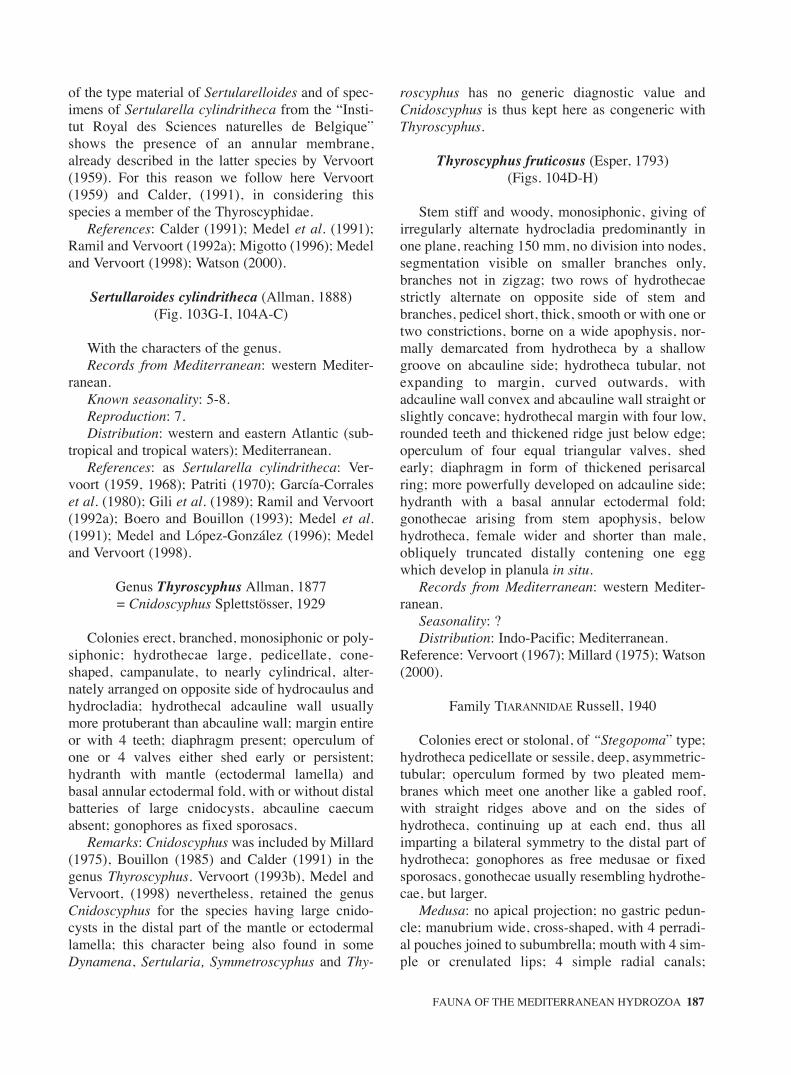
of the type material of Sertularelloides and of spec-imens of Sertularella cylindritheca from the “Insti-tut Royal des Sciences naturelles de Belgique”shows the presence of an annular membrane,already described in the latter species by Vervoort(1959). For this reason we follow here Vervoort(1959) and Calder, (1991), in considering thisspecies a member of the Thyroscyphidae.
References: Calder (1991); Medel et al. (1991);Ramil and Vervoort (1992a); Migotto (1996); Medeland Vervoort (1998); Watson (2000).
Sertullaroides cylindritheca (Allman, 1888)(Fig. 103G-I, 104A-C)
With the characters of the genus.Records from Mediterranean: western Mediter-
ranean.Known seasonality: 5-8.Reproduction: 7.Distribution: western and eastern Atlantic (sub-
tropical and tropical waters); Mediterranean.References: as Sertularella cylindritheca: Ver-
voort (1959, 1968); Patriti (1970); García-Corraleset al. (1980); Gili et al. (1989); Ramil and Vervoort(1992a); Boero and Bouillon (1993); Medel et al.(1991); Medel and López-González (1996); Medeland Vervoort (1998).
Genus Thyroscyphus Allman, 1877 = Cnidoscyphus Splettstösser, 1929
Colonies erect, branched, monosiphonic or poly-siphonic; hydrothecae large, pedicellate, cone-shaped, campanulate, to nearly cylindrical, alter-nately arranged on opposite side of hydrocaulus andhydrocladia; hydrothecal adcauline wall usuallymore protuberant than abcauline wall; margin entireor with 4 teeth; diaphragm present; operculum ofone or 4 valves either shed early or persistent;hydranth with mantle (ectodermal lamella) andbasal annular ectodermal fold, with or without distalbatteries of large cnidocysts, abcauline caecumabsent; gonophores as fixed sporosacs.
Remarks: Cnidoscyphus was included by Millard(1975), Bouillon (1985) and Calder (1991) in thegenus Thyroscyphus. Vervoort (1993b), Medel andVervoort, (1998) nevertheless, retained the genusCnidoscyphus for the species having large cnido-cysts in the distal part of the mantle or ectodermallamella; this character being also found in someDynamena, Sertularia, Symmetroscyphus and Thy-
roscyphus has no generic diagnostic value andCnidoscyphus is thus kept here as congeneric withThyroscyphus.
Thyroscyphus fruticosus (Esper, 1793)(Figs. 104D-H)
Stem stiff and woody, monosiphonic, giving ofirregularly alternate hydrocladia predominantly inone plane, reaching 150 mm, no division into nodes,segmentation visible on smaller branches only,branches not in zigzag; two rows of hydrothecaestrictly alternate on opposite side of stem andbranches, pedicel short, thick, smooth or with one ortwo constrictions, borne on a wide apophysis, nor-mally demarcated from hydrotheca by a shallowgroove on abcauline side; hydrotheca tubular, notexpanding to margin, curved outwards, withadcauline wall convex and abcauline wall straight orslightly concave; hydrothecal margin with four low,rounded teeth and thickened ridge just below edge;operculum of four equal triangular valves, shedearly; diaphragm in form of thickened perisarcalring; more powerfully developed on adcauline side;hydranth with a basal annular ectodermal fold;gonothecae arising from stem apophysis, belowhydrotheca, female wider and shorter than male,obliquely truncated distally contening one eggwhich develop in planula in situ.
Records from Mediterranean: western Mediter-ranean.
Seasonality: ?Distribution: Indo-Pacific; Mediterranean.
Reference: Vervoort (1967); Millard (1975); Watson(2000).
Family TIARANNIDAE Russell, 1940
Colonies erect or stolonal, of “Stegopoma” type;hydrotheca pedicellate or sessile, deep, asymmetric-tubular; operculum formed by two pleated mem-branes which meet one another like a gabled roof,with straight ridges above and on the sides ofhydrotheca, continuing up at each end, thus allimparting a bilateral symmetry to the distal part ofhydrotheca; gonophores as free medusae or fixedsporosacs, gonothecae usually resembling hydrothe-cae, but larger.
Medusa: no apical projection; no gastric pedun-cle; manubrium wide, cross-shaped, with 4 perradi-al pouches joined to subumbrella; mouth with 4 sim-ple or crenulated lips; 4 simple radial canals;
FAUNA OF THE MEDITERRANEAN HYDROZOA 187
sm68s2Bsystema1 14/10/04 15:22 Página 187

“gonads” folded on interradial walls of manubriumand/or on perradial manubrial pouches; marginaltentacles numerous, hollow; hollow cordyli-likestructures bearing cnidocysts; no ocelli.
References: Pagès et al. (1991; 1992); Bouillon(1999); Bouillon and Barnett (1999); Bouillon andBoero (2000).
Key to polyps
1. Colonies stolonal ................................ Modeeria– Colonies erect ................................... Stegopoma
Key to medusae
1. «Gonads» on perradial manubrial pouches only, widely split longitudinally ................ Krampella
– «Gonads» on manubrium and perradial gastric pouches, in regular transverse folds ... Modeeria
Genus Krampella Russell, 1957
Tiarannidae medusa with 4 perradial manubrialpouches extending almost to circular canal;«gonads» not sac-like nor folded on radial pouches,widely separated longitudinally; 8 marginal tenta-cles; up to five cirrus like tentaculae between suc-cessive marginal tentacles.
Hydroid: unknown.
1. With fine strands of tissue connecting walls of radial canals and manubrial pouches with exumbrellar surface; «gonads» elongated ......................................................................... K. dubia
– Without fine strands of tissue connecting walls of radial canals and manubrial pouches with exumbrellar surface; «gonads» oval to bean-shaped....................................... K. tardenti
Krampella dubia Russell, 1957(Figs. 104I-J)
Medusa: umbrella 4 mm high, 4.5 mm wide,hemispherical, mesoglea moderately thick;manubrium with large base, quadrate, with four per-radial pouches extending along all length of radialcanals; 4 radial canals, very short, linking the mostdistal parts of manubrial pouches to ring canal;about sixteen fine strands of tissue connecting wallsof radial canals and manubrial pouches with exum-brellar surface; «gonads» elongated, along distal 2/3of perradial manubrial pouches, widely separated
longitudinally; 4 perradial and 4 interradial margin-al tentacles; marginal tentacular bulbs conical; 3-6small marginal cirrus-like tentacles between eachpair of marginal tentacles; no statocysts nor ocelliseen.
Hydroid: unknown.Records from Mediterranean: western Mediter-
ranean; Adriatic Sea.Known seasonality: 5, 8, 10.Distribution: Atlantic; Mediterranean.References: Kramp (1961); Russell (1957,
1970a); Goy (1973b); Schmidt and Benovic (1977);Benovic and Bender (1987); Boero and Bouillon(1993); Benovic and Lucic (1996); Gili et al. (1998).
Krampella tardenti Gili, Bouillon and Pagès, 1998(Figs. 105A-C)
Medusa: umbrella 4.5 mm wide, 4 mm high,almost hemispherical, with a slightly conical orrounded apex, mesoglea of uniform thickness;manubrium large, 2/3 of bell height, with quadratebase, with 4 broad and long perradial pouchesextending almost till umbrella margin, manubriumwalls with 4 interradial bands of brown pigments;mouth quadrate, with 4 simple lips; radial canalsvery short, linking the most distal parts of manubri-al pouches to ring canal; no tissue strands linkingradial canals or manubrial pouches to exumbrella ; 8«gonads» oval to bean-shaped along the most distalthird of the manubrial perradial pouches, widelyseparated longitudinally; 4 perradial and 4 interradi-al marginal tentacles; marginal tentacular bulbs con-ical; 3 (2-4) long, solid cirrus like spirally coiledtentaculae between successive tentacles; no stato-cysts nor ocelli seen.
Hydroid: unknown.Records from Mediterranean: Antikhytira Strait,
eastern Mediterranean.Known seasonality: 6.Distribution: endemic of Mediterranean Sea.References: Gili et al. (1998).
Genus Modeeria Forbes, 1848
Hydroid: colonies stolonal. Hydrotheca pedicellate, deep and tubular, with margin produced on twosides. Operculum of two longitudinally pleatedmembranes seated in the embayment of the marginand meeting one another like a gable. Nodiaphragm, no nematothecae, no intertentacularweb; gonothecae similar to hydrothecae.
188 J. BOUILLON et al.
sm68s2Bsystema1 14/10/04 15:22 Página 188

Medusa: with «gonads» transversally folded oninterradial walls of manubrium and extending out-wards along the perradial pouches.
Modeeria rotunda (Quoy and Gaimard, 1827)(Figs. 105D-K)
Colonies stolonal; hydrothecae arising singlyfrom hydrorhiza at irregular intervals, large, tubular,tapering below into a smooth, straight or slightlycurved, non annulated pedicel; hydrothecal apertureclosed by an operculum formed by two pleatedmembranes which meet one another like a gabledroof, with straight ridges above and sides ofhydrotheca continuing up at each end, the all impart-ing a bilateral symmetry to the distal part of thehydrotheca; with a very thin diaphragm oftendestroyed by preservation; hydranth not extendingfar beyond hydrothecal aperture; up to 13 filiformtentacles in a single unicoronate whorl, no basalintertentacular membranous web, hypostome round-ed-conical; gonothecae resembling hydrothecaewith gabled operculum, but larger, pedicel reported-ly short to non existent; gonophore with up to 4developing medusae.
Medusa: umbrella 20 mm wide, somewhat lesshigh, hemispherical, mesoglea very thick, withrounded apex; manubrium short, broad, cruciform,perradial edges of manubrium connected over entirelength with subumbrella, forming 4 perradial pouch-es; mouth with 4 large, slightly crenulated lips; 4radial canals, straight, smooth, entering middle ofmanubrium to form the perradial pouches, narrowcircular canal; «gonads» in regular transverse foldson interradial walls of manubrium, extending out-wards on perradial pouches; 16-28 hollow marginaltentacles with large conical marginal bulbs; 1-3 (4)minute cordyli-like appendages with distal bundleof cnidocysts between successive tentacles;cnidome microbasic euryteles.
Records from Mediterranean: western Mediter-ranean, Adriatic.
Known seasonality: 4, 6.Distribution: Atlantic; Indo-Pacific; Antarctic;
Arctic; Mediterranean.References: Kramp (1961); Edwards (1963a,
1973b); Ramil and Vervoort (1992a); Alvarez(1993a); Altuna (1994); Avian et al. (1995); Cor-nelius (1995); Mills et al. (1996); Stepanjants et al.(1997); Brinckmann-Voss and Arai (1998); Gili etal. (1998); Peña Cantero and García Carrascosa(2002).
Genus Stegopoma Levinsen, 1893
Tiarannidae with pleated hydrothecal operculumin form of gabled roof, hydranth lacking intertentac-ular web, gonotheca resembling hydrotheca butlarger.
Remark: All species with a Stegopoma hydroidand with medusa buds are referable to the genusModeeria; the genus Stegopoma is here kept for thespecies with fixed sporosacs; all the species withunknown gonophores should be considered asTiarannidae incertae sedis.
Stegopoma bathyale Vervoort, 1966(Figs. 106A-C)
Colonies big, about 100 mm high, of sympodialstructure; hydrocauli strongly polysiphonic, thick,axis usually forked; branching irregular but in thesame plane. Monosiphonic parts divided into weak-ly geniculate or slightly undulate internodes, eachtopped by a pedicelate hydrotheca, following intern-ode arising basally to the hydrotheca. Perisarc thickon axis and in nodes but fairly abruptly thinning outon pedicel. Hydrotheca similar to those of Modeeriarotunda, but a trifle more bulky, slightly curved out-wards; hydrothecal pedicels wrinkled or indistinctlyringed, hydrothecal walls very thin. Branchesbecoming rapidly covered by secondary tubules,axillary hydrotheca remaining free; branchrebranching repeatedly. Gonophores as fixedsporosacs? Gonothecae borne on stem and branches,elongated, tubular, apparently produced by one ofsecondary tubules, completely adnate with branch oraxis and covered by net of anastomosing tubules;aperture apical and circular; it is unknown if theyproduce free medusae or not.
Records from Mediterranean: only records fromthe area of the Strait of Gibraltar (off Cádiz).
Distribution: temperate and subtropical easternAtlantic (from deep waters), Strait of Gibraltar.
References: Vervoort (1966); Ramil and Vervoort(1992a); Medel and López-González (1996).
Family TIAROPSIDAE
Boero, Bouillon and Danovaro, 1987
Hydroid: colony “Cuspidella” like; hydrothecatubular, sessile or with reduced pedicel; operculumof numerous flaps demarcated or not from the rest ofhydrotheca by a crease line; gonophores as freemedusae, gonotheca tubular or rounded, laterally
FAUNA OF THE MEDITERRANEAN HYDROZOA 189
sm68s2Bsystema1 14/10/04 15:22 Página 189

compressed, operculate or not, with short peduncle,growing singly from hydrorhiza.
Medusa: 4 or 8 radial canals (exceptionally up to16); one or two types of marginal tentacles (long andrudimentary, both with marginal bulbs); senseorgans compound, comprising an ecto-endodermalocellus and an open velar statocyst.
References: Boero, Bouillon and Danovaro(1987); Pagès et al. (1992); Bouillon (1999);Bouillon and Barnett (1999); Bouillon and Boero(2000).
1. With numerous (50-60) compound sense organs; mouth with eight lips ...................... Octogonade
– With 8 or 16 (exceptionally 48) compound sense organs; mouth with four lips ......... Tiaropsidium
Genus Octogonade Zoja, 1896
Tiaropsidae medusae with 8 radial canals; mouthwith 8 lips; with numerous compound statocysts;with two kinds of tentacles; without marginal cirri.
Hydroid: unknown.
Octogonade mediterranea Zoja, 1896(Figs. 106D-F)
Medusa: umbrella 60-70 mm, globular, withbulging sides, mesoglea thick; velum fairly broad;manubrium small, tubular, octagonal; with 8 smallbut distinct lips; «gonads» linear, along almostentire length of the 8 radial canals, living a short partfree at both ends; 16 long marginal tentacles and150-160 small rudimentary tentacles, containingdiverticula from the circular canal; 50- 60 com-pound sense organs.
Records from Mediterranean: western Mediter-ranean; Adriatic Sea.
Known seasonality: 2, 4, 6-12.Distribution: endemic of Mediterranean Sea.References: Zoja (1896); Kramp (1961); Picard
(1958b); Boero and Bouillon (1993); Avian et al.(1995); Benovic and Lucic (1996)
Genus Tiaropsidium Torrey, 1909
Hydroid: colonies of “Cuspidella” type; opercu-lum formed by several flaps sharply demarcatedfrom the hydothecal margin; gonothecae rounded,compressed laterally, without operculum.
Medusa: with 4 or more (up to 16) simple radialcanals; mouth with four lips; with 8 or 16 (rarely 48)
compound sense organs; with two kind of tentacles;without marginal cirri.
Tiaropsidium mediterraneum (Metschnikoff,1886a)
(Figs. 106G-I)
Hydroid: colonies stolonal; hydrothecae tubular,with short, wavy or straight annulated hydroclade,operculum with 7-10 flaps with broad base andpointed apex, meeting centrally and sharply demar-cated from the hydrothecal margin; hydranth com-pletely retractable in the hydrothecae; with onewhorl of amphicoronate tentacles; no intertentacularmembranous basal web; gonothecae borne onhydrorhiza, large, pyriform, almost not pedunculateand developed in one plane.
Medusa: umbrella 7 mm wide, 5 mm high,mesoglea thick; manubrium short, fairly broad;mouth with four short, simple lips; 4 radial canalsand circular canal narrow; 2 opposite, long, perradi-al tentacles, and 2 small perradial bulbs; with fiverudimentary tentacles in each quadrant, smaller thanthe marginal bulbs; «gonads» elongated, on distal2/3 of the radial canals; eight compound senseorgans.
Records from Mediterranean: western and cen-tral Mediterranean.
Known seasonality: 1, 2, 12.Distribution: endemic of Mediterranean Mediter-
ranean.References: Hadzi (1915); Huvé (1956); Kramp
(1961); Picard (1958b); Boero, Bouillon and Don-avaro (1987); Boero and Bouillon (1993).
Order PROBOSCOIDA Broch, 1910
Hydroid: hydranths with a complex, flared toglobose, more or less pedunculate hypostome, form-ing a “buccal cavity” beneath the mouth.
Medusa: varied in expression, with closed stato-cysts; never with cordyli, open statocysts, excretorypores, cirri or ocelli.
Key to hydroids (see family characteristics)
Key to medusae
1. Without permanent tenon-like rudimentary marginal bulbs......................... Campanulariidae
– With triangular, tenon-like permanent rudimentary marginal bulbs............ Phialuciidae
190 J. BOUILLON et al.
sm68s2Bsystema1 14/10/04 15:22 Página 190

Family CAMPANULARIIDAE Johnston, 1836
Hydroid: colony erect or stolonal; hydrothecaebell-shaped or campanulate, radially or, secondarily,bilaterally symmetrical; generally pedicellate, rimcusped or not, with basal diaphragm or inward annu-lar projection of perisarc; hydranth generally tubu-lar, with flared or globose hypostome delimiting a“buccal cavity”, gastric endoderm of uniform struc-ture; hydrothecal spherules present or not;gonophores as free medusae, eumedusoids orsporosacs, in gonothecae.
Medusa: manubrium short; no gastric peduncle;4 radial canals (except in Gastroblasta and Pseudo-clytia); with or without velum (without in Obelia);“gonads” completely surrounding radial canals, sep-arated from manubrium; tentacles hollow (except inObelia where they are solid and with a short prolon-gation of endoderm into bell mesoglea); with orwithout tenon-like rudimentary bulbs; no cirri,excretory papillae or pores; numerous (16-200)closed, velar, marginal statocysts (8 in Obelia, eachsituated on underside of the basal bulb of some mar-ginal tentacle); no ocelli. (Fig. 7: 3)
References: Nutting (1915); Russell (1963b);García Corrales et al. (1978); Östman (1983);Calder (1991); Pagès et al. (1992); Cornelius(1995); Hirohito (1995); Boero et al. (1996); Migot-to (1996); Bouillon (1999); Bouillon and Barnett(1999); Bouillon and Boero (2000); Medel and Ver-voort (2000).
Remarks: the classification of the Campanulari-idae is unsatisfactory; generic divisions, as in manyother families of Leptomedusae hydroids, are notwell defined, and vary even in modern works (see:Calder 1991, Cornelius 1982; 1995; Hirohito 1995).Hirohito (1995), for instance, taking in accountsome morphological characters such as presenceand absence of diaphragm, presence or absence ofsub-hydrothecal spherules, treated Orthopyxis andRhizocaulus as Campanularia, and Laomedea andGonothyraea as Obelia so simplifying greatly thegeneric contents of the family. But Obelia is a verydifferent from the other Campanulariidae genera atthe medusa level and the reduction of free medusaeto fixed gonophores may have occurred severaltimes independently during the evolution of theCampanulariidae and so it is impossible to referpresently the taxa with fixed sporosacs to anypresently known medusa genus. Nevertheless manygenera are very close to each other for instance:Hartlaubella and Lameodea, Rhyzocaulus and Cam-
panularia, Tulpa and Campanularia could bemerged without great difficulty.
Key to hydroids
1. Colony usually stolonal or with short unbranched uprights ............................................................. 2
– Colony with erect stems, bearing alternate hydrothecae ...................................................... 4
2. Hydrotheca with true diaphragm, with free medusae .................................................... Clytia
– Hydrotheca without true diaphragm; with annular perisarcal thickening near base ........... 3
3. Hydrothecal walls with unthickened perisarc; gonophores as fixed sporosac ..... Campanularia
– Pydrothecal walls with perisarc variably thickened; gonophores as eumedusoids ........................................................................ Orthopyxis
4. Gonophores released as free medusae ............. 5– Gonophores not released as free medusae ....... 75. Medusa with solid marginal tentacles; no velum
(see key below)........................................ Obelia– Medusa with hollow tentacles and velum........ 56. Medusa with one manubrium................... Clytia– Medusa with more than one manubrium ............
....................................................... Gastroblasta7. Gonophores forming degenerate medusae,
meconidia ...................................... Gonothyraea– Gonophores as fixed sporosacs or degenerate
medusae ............................................................ 88. Stem typically monosiphonic ............ Laomedea– Stem polysiphonic already early in life...............
....................................................... Hartlaubella
Key to medusa
1. With more than four radial canals.................... 2– With normally four radial canals...................... 32. With up to 20 radial and centripetal canals; with
numerous manubria each with 4 lips ......................................................................... Gastroblasta
– With up to seven radial canals; with one manubrium and as many lips than radial canals .........................................................Pseudoclytia
3. With reduced medusae; without manubrium; without tentacles ............................... Orthopyxis
– With normally developed medusae; with one manubrium with 4 lips; with tentacles ............. 4
4. With hollow marginal tentacles and normal velum = Clytia
– With solid marginal tentacles; without velum...................................................................... Obelia
FAUNA OF THE MEDITERRANEAN HYDROZOA 191
sm68s2Bsystema1 14/10/04 15:22 Página 191

Genus Campanularia Lamarck, 1816
Colonies stolonal, seldom erect and branched;hydrorhiza not anastomosing; hydrothecal pedicelunbranched; hydrothecae campanulate or bell-shaped,with entire or cusped margin, demarcated from pedi-cel basally by a variably developed annular perisarcalthickening; hydrothecal walls with unthickened peris-arc, not abruptly everted distally; true diaphragmabsent, sub-hydrothecal spherule present; gonophoresas fixed sporosacs, gonothecae on hydrorhiza.
References: Calder (1991); Cornelius (1995);Schuchert (2001).
1. Length of the hydrotheca > 500 mm, with lines running down from rim; cusps castellate; gonotheca truncate distally, aperture wide ..................................................................... C. hincksii
– Length of the hydrotheca < 500 mm, without lines; cusps pointed; gonotheca with a narrow aperture at the end of a neck of variable length.............................................................C. volubilis
Campanularia hincksii Alder, 1856(Figs. 107A-F)
Colonies up to 15 mm high, composed of stolonwith erect and unbranched pedicels bearing a singlehydrotheca at the top. Perisarc of the pedicelssmooth to sinuous, usually with several annulibasally and with a characteristic spherule belowhydrothecae. Hydrothecae cylindrical, sides parallel,clearly longer than wider, rim castellate, 8-15 cuspseither truncate, notched or distinctly bicuspidate,distal half of hydrothecae usually slightly polygonalon cross section, surface with longitudinal striae;basal diaphragm thick, conspicuous. Gonothecaeborne on stolon, male and female similar, cylindri-cal, perisarc smooth or irregularly undulated, trun-cated apically, with operculum circular; planulabrooded within the female gonotheca
Records from Mediterranean: eastern and west-ern Mediterranean; Adriatic, Black Sea.
Known seasonality: 1-12.Reproduction: 1-12.Distribution: almost cosmopolitan, avoiding
pure Arctic and Antarctic.References: Millard (1975); Cornelius (1982, 1995);
Gili (1986); Calder (1991); Boero and Bouillon (1993);Avian et al. (1995); Medel and López-González (1996);Medel and Vervoort (2000); Schuchert (2001a); PeñaCantero and García Carrascosa (2002).
Campanularia raridentata Alder, 1862(Fig. 107G)
Colonies stolonal, composed of erect pedicelseach supporting a hydrotheca, perisarc of pedicelssmooth, ringed basally and below hydrothecae.Hydrotheca cylindrical, sub-hydrothecal spherulepresent, rim with 6-8 wide rounded triangular cusps.Gonothecae usually borne on stolons from short andringed pedicels, barrel-shaped, smooth and with cir-cular distal apertures.
Records from Mediterranean: western Mediter-ranean.
Known seasonality: 4, 5, 6.Distribution: northeastern Atlantic.References: Gili and Castelló (1985) as Clytia
hemispherica raridentata; Gili and García-Rubíes(1985); Gili (1986); Llobet (1987); Llobet et al.(1991); Medel and López-González (1996).
Remarks: Campanularia raridentata describedby Hincks, 1868 was referred to Clytia hemisphaer-ica by several authors such as Vervoort (1968) orCornelius (1982). Llobet (1987) showed, in herdrawings, hydrothecae without diaphragm, but withannular thickening and gonothecae smooth and bar-rel-shaped. We agree with the author o.c., thatHincks, 1868 described something similar in C.hemispherica; the same occurs with Gili (1986). Therecord of Campanularia raridentata, Alder, 1862 byLlobet (1987) is here considered as doubtful.
Campanularia volubilis (Linnaeus, 1758)(Figs. 107H-M)
Stolonal colonies up to 3-4 mm high, with erectpedicels each with a single hydrotheca, arising atirregular intervals from the hydrorhiza. Pedicelssmooth to spirally annulated throughout. Hydrothe-cae with sub-hydrothecal spherule, rim with 10-12shallow bluntly pointed cusps, fine longitudinal stri-ae running to the cusps may be present. Male andfemale gonothecae similar, usually borne on stolon,flask shaped, smooth, aperture narrow at the end ofneck of indefinite length, absent when young. Plan-ula as dispersal stage.
Records from Mediterranean: western Mediter-ranean.
Distribution: NE Atlantic, eastern Pacific,Mediterranean.
References: Aguirrezabalaga et al. (1986); Cor-nelius and Ryland (1990); Boero and Bouillon(1993); Cornelius (1995); Medel and López-
192 J. BOUILLON et al.
sm68s2Bsystema1 14/10/04 15:22 Página 192

González (1996); Schuchert (2001a); Peña Canteroand García Carrascosa (2002).
Genus Clytia Lamouroux, 1812
Hydroid: colony reptant, unbranched stolonal orerect branched, usually minute, monosiphonic orpolysiphonic; hydrorhiza branched but not anasto-mosing; hydrothecae deep, campanulate, hydrothe-cal rim sinuous or deeply indented, with cleftsbetween round to sharply-pointed cusps; truehydrothecal diaphragm; subhydrothecal spheruleabsent (present in C. hummelincki); gonophores asfree medusae, gonotheca conical.
Medusa: manubrium short; velum normal; mar-ginal tentacles hollow; no tenon-like permanentrudimentary bulbs; numerous statocysts.
Remarks: very few species of Clytia medusae areclearly identified, most morphological characters usedto distinguish them possibly falling in the range ofvariation that can be expected in a single species andhaving little or no taxonomic value. Most of the Clytiaspecies described hereunder are only known by theirhydroids stage and should be considered as incertaesedis. The genus needs a careful revision, with carefulelucidation of life cycles and molecular work.
Reference: Pagliara et al. (2000).
Key for Hydroid
1. Hydrothecal cusps projecting inwards and with outwards perisarc projections in bays between nearby cusps .................................. C. viridicans
– Hydrothecal cusps different ............................. 22. Hydrothecal cusps oblique ............... C. gracilis.– Hydrothecal cusps upright................................ 33. Hydrothecal cusps with characteristic
longitudinal perisarcal ridge.............. C. linearis– Hydrothecal cusps without any perisarcal ridge
.......................................................................... 44. Hydrothecal cusps usually bicuspidate,
hydrotheca cylindrical, deep .......... C. paulensis– Hydrothecal cusps not bicuspidate, hydrotheca
campanulate, not deep...................................... 55. Hydrothecal diaphragm thin .... C. hemispherica5. Hydrothecal diaphragm thick........ C. noliformis
Key to medusa
1. «Gonads» small in the middle of the radial canals and giving rise to numerous blastostyles carrying medusa buds ................................... C. mccradyi
– «Gonads» thick; without blastostyles .............. 22. Manubrium, «gonads» and marginal bulbs bright
green .............................................. C. viridicans– Manubrium, «gonads» and marginal bulbs
transparent or of different colour ..................... 33. «Gonads» voluminous along almost whole
length of radial canals ...................................... 4– «Gonads» linear or oval, along maximum 1/3 of
the length of radial canals nearer to margin thanto manubrium ................................................... 5
4. With 16 marginal tentacles; usually 3 statocysts between successive marginal tentacles ......................................................................... C. discoida
– With 24-36 marginal tentacles; one statocysts between successive tentacles...... C. macrogonia
5. With oval «gonads»; 16 marginal tentacles; umbrella up to 6-8 mm wide............. C. gracilis
– With linear «gonads»........................................ 66. Up to 32 marginal tentacles and more than 16
statocysts in adults, umbrella up to 20 mm wide ................................................ C. hemisphaerica
– No more than 16 marginal tentacles and 16 statocysts in adults, umbrella 6 mm wide .................................................................. C. noliformis
Clytia discoida (Mayer, 1900)(Fig. 108A)
Medusa: umbrella 4 mm wide, almost hemi-spherical; manubrium urn-shaped, with bulgingsides, mouth with four recurved lips; 16 short mar-ginal tentacles with large basal bulbs; usually threestatocysts between tentacles; gonads on greater partof radial canals , thick, cylindrical, eggs very large.
Hydroid: unknown.Records from Mediterranean: western Mediter-
ranean.Seasonality: ?Distribution: Atlantic, MediterraneanReferences: Kramp (1959a, 1961); Bouillon and
Boero (2000).
Clytia gracilis (M. Sars, 1850)(Figs. 108B-G)
Hydroid: colonies delicate, up to 20 mm high,comprising a creeping hydrorhiza from which ariseseveral erect hydrocauli, some forking 2-3 times,with long internodes and pedicels; hydrothecaedeep-campanulate, with 8-12 large pointed cuspsinclined to one side, diaphragm thin, pedicel baseincurving; 1-ca 25 annuli at internodes and pedicel
FAUNA OF THE MEDITERRANEAN HYDROZOA 193
sm68s2Bsystema1 14/10/04 15:22 Página 193

base; hydranth about 7 times as long than broadwhen fully extended, with typically 18-20 tentacles;gonothecae arising from hydrorhiza on short slenderpedicels, long, urn-shaped, constricted abruptlybelow rim and below this more gradually, smoothwalled, wide mouthed; planktonic colonies havebeen described in Atlantic waters.
Medusa: umbrella up to 8 mm wide, nearly hemi-spherical, mesoglea thin; manubrium small quadrateto cruciform; mouth with 4 simple lips; 4 straightradial canals; «gonads» oval to round, with feweggs, along the third distal quarter of the radialcanals; up to 16 marginal tentacles; marginal bulbswith spherical base; 1-2 statocysts between succes-sive tentacles. This medusa is often confound withother Clytia medusae mainly with C. hemisphaeri-ca, the discrimination between those differentspecies may perhaps be possible by the use cnido-cysts microstructure (Östman, 1979, 1983).
Records from Mediterranean: hydroid found ineastern and western Mediterranean and Adriatic.
Known seasonality: 4-8, 10, 11.Reproduction: 6-9.Distribution: Atlantic; Indo-Pacific; Mediter-
ranean for the hydroid stage. Atlantic for the plank-tonic colonies.
References: Kramp (1961); Cornelius and Öst-man (1986); Calder (1991); Boero and Bouillon(1993); Vervoort (1993a); Avian et al. (1995); Cor-nelius (1995); Medel and López-González (1996);Medel and Vervoort (2000); Schuchert (2001a);Peña Cantero and García Carrascosa (2002).
Clytia hemisphaerica (Linnaeus, 1767)(Figs. 108H-K)
Hydroid: colonies usually stolonal but occasion-ally erect, arising from a creeping hydrorhiza;hydrothecal pedicels borne at close intervals, some-times forking; hydrothecae campanulate, rim withca 8-14 broad rounded-triangular cusps; diaphragmthin; pedicels straight, with one to several rings topand bottom and in some specimens also centrally;some pedicels with secondary pedicels, these havingcharacteristic upward curved basal region;hydranths 3-4 times as long than broad when fullyextended, with usual 24-30 tentacles; gonothecaearising from hydrorhiza on short, slender pedicels,tubular, typically with deeply concertinared wallsresembling a Chinese lantern, but gonothecal wallsmay be smooth in some specimens, wide mouthed(Calder, 1988; Cornelius, 1995).
Medusa: umbrella up to 20 mm wide, nearlyhemispherical or flatter, mesoglea fairly thin;manubrium small, quadrate, with small base; mouthalways with 4 simple lips; «gonads» oval or linear1/2-3/4 of length of radial canals without medianfurrow, nearer to margin than to manubrium; usual-ly 4 straight radial canals (sometimes more, up to12); typically 32 (16-58) marginal tentacles; mar-ginal tentacular bulbs globular, prominent; few par-tially developed marginal bulbs; velum narrow; 1-3usually 2 statocysts between successive tentacles.
Records from Mediterranean: eastern and west-ern Mediterranean; Adriatic.
Known seasonality: 1-12.Reproduction: 1-12.Distribution: Atlantic; Indo-Pacific; Mediter-
ranean.References: Trégouboff (1957); Roosen-Runge
(1970); Goy (1973b); Gili (1986); Benovic and Ben-der (1987); Calder (1991); Goy et al. (1988, 1990,1991); Cornelius (1982, 1995); Boero and Bouillon(1993); Avian et al. (1995); Benovic and Lucic(1996); Mills et al. (1996); Medel and López-González (1996); Medel and Vervoort (2000);Schuchert (2001a); Peña Cantero and García Carras-cosa (2002).
Clytia hummelincki (Leloup, 1935)(Figs. 109A-E)
Hydroid: colonies stolonal, up to 4.5 mm high,with a creeping hydrorhiza. Pedicels long, relativelythick, annulated basally and with occasional groupsof annuli elsewhere; distal end of pedicel with slightswelling just beneath a subhydrothecal spherule.Hydrotheca shallow, cup-shaped, hydrothecal wallswith relatively thin perisarc; margin smooth, occa-sionally oblique; diaphragm thin, slightly oblique,basal chamber shallow.
Medusa: only immature medusa known.Records from Mediterranean: Ionian coast of
southern Italy.Records outside the Mediterranean: eastern and
western Atlantic.References: Millard (1975); Cornelius (1982) as
Laomedea; Calder (1991); Boero et al. (1997b).
Clytia linearis (Thorneley, 1899)(Figs. 109F-I)
Hydroid: colonies reptant, up to 40 mm high,bearing erect pedicels unbranched or subsympodial-
194 J. BOUILLON et al.
sm68s2Bsystema1 14/10/04 15:22 Página 194

ly branched; pedicels and branches ringed basallyand distally, topped by hydrothecae; branches aris-ing laterally, at the upper part of the axis; hydrothe-cae very deep, cylindrical, sides almost parallel, nar-rowing basally, with a thin diaphragm, straight orslightly oblique; margin wide, 10-12 acute cuspswith characteristic longitudinal central perisarcalband or ridge extended further of the cusps.Gonothecae usually axillar, borne on short andringed pedicels, elongate-oval, truncate distally, nar-row sub-distally and basally; enclosing up to eightdeveloping medusa buds.
Medusa: only juvenile medusa just releasedknown.
Records from Mediterranean: eastern and west-ern Mediterranean, Adriatic.
Known seasonality and reproduction: throughoutthe year.
Distribution: tropical and subtropical waters ofAtlantic, Pacific and Indian Ocean, Mediterranean.
References: Rossi (1961); Marinopoulos (1979);Millard (1975); García-Corrales et al. (1978); Giliand García-Rubíes (1985); Boero and Fresi (1986);Cornelius (1987); Calder (1991); Ramil and Ver-voort (1992a); Boero and Bouillon (1993); AltunaPrados (1995); Avian et al. (1995); Migotto, 1996;Medel and López-González (1996); Medel and Ver-voort (2000); Peña Cantero and García Carrascosa(2002).
Clytia maccradyi (Brooks, 1888)(Fig. 109J)
Hydroid: Brooks (1888) saw young medusaereleased from a hydroid colony and identified themas C. mccradyi medusae. Brook’s description of thehydroid did not differ from that of C. hemisphaerica(see Cornelius 1982, Carré et al., 1995). Mayer(1910) doubted Brook’s observation; the hydroidphase of this medusae is thus still not known withcertitude.
Medusa: umbrella 15 mm wide, about twice asbroad than high; mesoglea of soft consistence;manubrium short and stout; mouth with fourrecurved lips; sexual development by four small,swollen, «gonads», situated in the middle part of theradial canals, asexual reproduction through hydroidblastostyles giving rise to medusae buds and origi-nating at the same place than «gonads» and insteadthem in sexually immature specimens; 16- 24 mar-ginal tentacles; marginal tentacular bulbs conical; 1-2 statocysts between tentacles.
Records from Mediterranean: eastern and west-ern Mediterranean.
Known seasonality: 7-10.Distribution: Atlantic; Indo-Pacific; Mediter-
ranean.References: Brooks (1888); Kramp (1961); Goy
(1970, 1972); Cornelius (1982); Bouillon et al. (1986);Benovic and Bender (1987); Brinckmann-Voss (1987);Goy et al. (1991); Boero and Bouillon (1993); Carré etal. (1995); Benovic and Lucic (1996).
Clytia macrogonia Bouillon, 1984(Fig. 109K)
Medusa: umbrella 4 mm wide, discoidal; manubri-um small, cruciform, with rounded perradial lobes;mouth with 4 simple lips; 4 radial canals and circularcanal narrow; «gonads» cylindrical, along almostwhole length of radial canals, females with few (about20) very large eggs; with 24- 36 short marginal tenta-cles; marginal tentacular bulbs large, globular; withone statocyst between successive tentacles.
Records from Mediterranean: eastern Mediter-ranean.
Seasonality: ?Distribution: Indo-Pacific, Mediterranean.References: Kramp (1961); Bouillon (1984); Goy
et al. (1991); Boero and Bouillon (1993).Hydroid: unknown.
Clytia noliformis auct. (McCrady, 1859)(Figs. 110A-E)
Hydroid: colonies stolonal with creepinghydrorhiza; pedicel strongly annulated basally anddistally; hydrothecae campanulate, margin withabout 13-15 blunt, broadly triangular teeth; with athick diaphragm; basal chamber shallow, subspheri-cal; hydranth with about 25-30 filiform tentacles;gonothecae laterally flattened, arising fromhydrorhiza, on short annulated stalks.
Medusa: umbrella 6 mm wide, 3 mm high, flatterthan a hemisphere; manubrium tubular; mouthquadrate, with four simple lips; 4 radial canals; 16marginal tentacles; 16 intervening statocysts;«gonads» elongated, on the distal third or fourth ofthe radial canals, female «gonads» bearing 20-35large ova. The newly hatched medusa of Clytianoliformis differs from corresponding stages of C.hemiphaerica by complete absence of «gonads».
Records from Mediterranean: western Mediter-ranean, Adriatic.
FAUNA OF THE MEDITERRANEAN HYDROZOA 195
sm68s2Bsystema1 14/10/04 15:22 Página 195
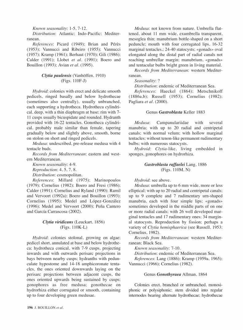
Known seasonality: 1-5, 7-12.Distribution: Atlantic; Indo-Pacific; Mediter-
ranean.References: Picard (1949); Brian and Pérès
(1953); Vannucci and Ribeiro (1955); Vannucci(1957); Kramp (1961); Berhaut (1970); Gili (1986);Calder (1991); Llobet et al. (1991); Boero andBouillon (1993); Avian et al. (1995).
Clytia paulensis (Vanhöffen, 1910)(Figs. 110F-J)
Hydroid: colonies with erect and delicate smoothpedicels, ringed basally and below hydrothecae(sometimes also centrally), usually unbranched,each supporting a hydrotheca. Hydrotheca cylindri-cal, deep, with a thin diaphragm at base; rim with 7-11 cusps usually bicuspidate and rounded. Hydranthprovided with 16-22 tentacles. Gonotheca cylindri-cal, probably male similar than female, taperinggradually below and slightly above, smooth, borneon stolon on short and ringed pedicels.
Medusa: undescribed, pre-release medusa with 4tentacle buds.
Records from Mediterranean: eastern and west-ern Mediterranean.
Known seasonality: 4-9.Reproduction: 4, 5, 7, 8.Distribution: cosmopolitan.References: Millard (1975); Marinopoulos
(1979); Cornelius (1982); Boero and Fresi (1986);Calder (1991); Cornelius and Ryland (1990); Ramiland Vervoort (1992a); Boero and Bouillon (1993);Cornelius (1995); Medel and López-González(1996); Medel and Vervoort (2000); Peña Canteroand García Carrascosa (2002).
Clytia viridicans (Leuckart, 1856)(Figs. 110K-L)
Hydroid: colonies stolonal, growing on algae;pedicel short, annulated at base and below hydrothe-ca; hydrotheca conical, with 7-9 cusps, projectinginwards and with outwards perisarc projections inbays between nearby cusps; hydranths with pedun-culate hypostome and 14-18 amphicoronate tenta-cles, the ones oriented downwards laying on theperisarc projections between adjacent cusps, theones oriented upwards being sustained by cusps;gonophores as free medusa; gonothecae onhydrorhiza either corrugated or smooth, containingup to four developing green medusae.
Medusa: not known from nature. Umbrella flat-tened, about 11 mm wide, exumbrella transparent,mesoglea thin; manubrium bottle-shaped on a shortpeduncle; mouth with four corrugated lips, 16-32marginal tentacles,; 24-40 statocysts; «gonads» ovalelongated along the distal part of radial canals notreaching umbrellar margin; manubrium, «gonads»and tentacular bulbs bright green in living material.
Records from Mediterranean: western Mediter-ranean.
Seasonality: ?Distribution: endemic of Mediterranean Sea.References: Haeckel (1864); Metschnikoff
(1886a,b); Russell (1953); Cornelius (1982);Pagliara et al. (2000).
Genus Gastroblasta Keller 1883
Medusa: Campanulariidae with severalmanubria; with up to 20 radial and centripetalcanals; with normal velum; with hollow marginaltentacles; without tenon-like permanent rudimentarybulbs; with numerous statocysts.
Hydroid: Clytia-like, living embedded insponges, gonophores on hydrorhiza.
Gastroblasta raffaelei Lang, 1886(Figs. 110M, N)
Hydroid: see above.Medusa: umbrella up to 6 mm wide, more or less
elliptical; with up to 20 radial and centripetal canals;up to 9 complete and 7 rudimentary urn-shapedmanubria, each with four simple lips; «gonads»sometimes developed in the middle parts of on oneor more radial canals; with 26 well developed mar-ginal tentacles and 17 rudimentary ones; 34 margin-al statocysts. Reproduction by fission; perhaps avariety of Clytia hemisphaerica (see Russell, 1953;Cornelius, 1982).
Records from Mediterranean: western Mediter-ranean; Black Sea.
Known seasonality: 7-10.Distribution: endemic of Mediterranean Sea.References: Lang (1886); Kramp (1959a, 1965);
Vannucci (1966); Cornelius (1982).
Genus Gonothyraea Allman, 1864
Colonies erect, branched or unbranched, monosi-phonic or polysiphonic; stem divided into regularinternodes bearing alternate hydrothecae; hydrothecae
196 J. BOUILLON et al.
sm68s2Bsystema1 14/10/04 15:22 Página 196

campanulate to bell-shaped, radially symmetrical;hydrothecal rim cusped; with true diaphragm;gonophores forming degenerate medusae (mecodonia),extruded from gonotheca but remaining attached whileplanula develops in a sac-like mecodonium with mar-ginal tentacles, but no mouth or sense organs; gonothe-ca urn-shaped, pedicellate axillary on stem.
Reference: Cornelius (1995).
Gonothyraea loveni (Allman, 1859)(Fig. 111A-E)
Hydrocauli erect, up to 100 mm, flexuous, mono-siphonic, internodes straight to strongly curved,somewhat irregularly branched and ringed basally.Hydrothecae borne distally on the internodes, alter-nate, hydrothecal pedicels usually annulatedthroughout; hydrothecae bell-shaped, deep, rim del-icate with 6-12 castellate cusps provided with aminute notch; diaphragm straight. Hydranth with19-33 tentacles. Gonothecae long, cylindrical, nar-rowing basally, borne on ringed pedicels; medusoidsdevelop internally and, when ripe, dangle in groupsof 2-3 at entrance to gonotheca; medusoids roughlyspherical, with 4-8 long, irregularly held tentacles, acircular canal, without mouth neither sense organs;males with about 5 marginal tentacles; females with8 marginal tentacles and 3-5 eggs which are fertil-ized and develop into planulae in situ.
Records from Mediterranean: western Mediter-ranean, Adriatic.
Known seasonality: 4, 7-9.Reproduction: 4, 9.Distribution: eastern and western Atlantic, Indo-
Pacific and Arctic Ocean, Mediterranean.References: Millard (1975); Östman (1979); Gili
(1982, 1986); Isasi and Sáiz (1986); Boero andBouillon (1993); Altuna (1994); Avian et al. (1995);Cornelius (1995); Medel and López-González(1996); Schuchert (2001a).
Genus Hartlaubella Poche, 1914
Colonies erect, polysiphonic, branched orunbranched, growing on branched but not anastomos-ing hydrorhiza; hydrocauli divided in regular intern-odes bearing alternate hydrothecae; hydrothecaecampanulate with castellated, often abraded rim, radi-ally symmetrical, on pedicels, with true diaphragm;gonophores as fixed sporosacs with large embryos,gonothecae axillary, inverted conical.
Reference: Cornelius (1995).
Hartlaubella gelatinosa (Pallas, 1766)(Figs. 111F-J)
Well developed colonies bushy, main stem erect,up to 200 mm, branched, thick and polysiphonic.Final branches monosiphonic, alternate on bothsides of the axis, tending to be disposed in rightangle pairs, more or less dichotomously branchedand flexuose. Internodes basally ringed, nearlystraight to curved. Hydrothecae borne on ringedpedicels, small, cylindrical, rim often abradedsmooth but firstly castellate, 12-14 blunt cusps eachwith a central notch of varied depth, gaps betweenrounded; diaphragm transverse. Gonothecae (maleand female similar) cylindrical, borne on ringedpedicels, narrower basally, wider distally, circularaperture on a short collar.
Records from Mediterranean: western Mediter-ranean, Black Sea.
Distribution: northeastern Atlantic, westernAtlantic and Indo-Pacific (New Zealand); Mediter-ranean.
References: Cornelius (1982, 1995); Corneliusand Ryland (1990); Boero and Bouillon (1993).
Genus Laomedea Lamouroux, 1812
Colonies erect, monosiphonic, branched orunbranched, growing on branched but not anasto-mosing hydrorhiza; hydrocauli divided in regularinternodes bearing alternate hydrothecae; hydrothe-ca campanulate, rim even or cusped, radially sym-metrical, on pedicels, with true diaphragm;gonophores as fixed sporosacs, gonothecae sessileor shortly pedicellate stolonal or axillary.
References: Östman (1982); Cornelius (1995).
1. Colonies monosiphonic or bisiphonic.............. 2– Colonies polysiphonic ....... L. pseudodichotoma2. Female gonotheca with a characteristic sub-
terminal introverted curving tubular aperture .......................................................... L. calceolifera
– Female gonotheca without this character......... 33. Internodes long, usually rigidly straight, in
zigzag ............................................... L. angulata– Internodes slightly curved or flexuose ............. 44. Rim of the hydrotheca smooth ......... L. flexuosa– Rim of the hydrotheca bimucronate....................
.......................................................... L. neglecta
Laomedea angulata Hincks, 1861(Figs. 111K-P)
FAUNA OF THE MEDITERRANEAN HYDROZOA 197
sm68s2Bsystema1 14/10/04 15:22 Página 197
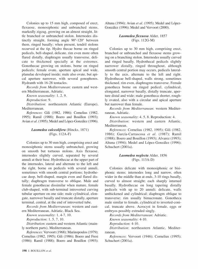
Colonies up to 15 mm high, composed of erect,flexuose, monosiphonic and unbranched stems,markedly zigzag, growing on an almost straight, lit-tle branched or unbranched stolon. Internodes dis-tinctly straight, forming angle 90º-120º betweenthem, ringed basally; when present, tendril stolonsrecurved at the tip. Hydro thecae borne on ringedpedicels, bell-shaped, delicate, rim even more oftenflared distally, diaphragm usually transverse, deli-cate to thickened specially at the extremes.Gonothecae growing on stolons, borne on ringedpedicels; female ovate, aperture apical and wide,planulae developed inside; male also ovate, but api-cal aperture narrower, with several gonophores.Hydranth with 16-28 tentacles.
Records from Mediterranean: eastern and west-ern Mediterranean, Adriatic.
Known seasonality: 1, 2, 9.Reproduction: 9.Distribution: northeastern Atlantic (Europe),
Mediterranean.References: Gili (1982, 1986); Cornelius (1982,
1995); Ramil (1988); Boero and Bouillon (1993);Avian et al. (1995); Medel and López-González (1996).
Laomedea calceolifera (Hincks, 1871)(Figs. 112A-F)
Colonies up to 30 mm high, comprising erect andmonosiphonic stems usually unbranched, growingon smooth but tortuous stolons. Axis flexuose,internodes slightly curved, separated by severalannuli at their base. Hydrothecae at the upper part ofthe internodes, lateral and alternate to the left andthe right, borne on pedicels with several annuli,sometimes with smooth central portions; hydrothe-cae deep, bell-shaped, margin even and flared dis-tally; diaphragm transverse to oblique. Male andfemale gonothecae dissimilar when mature, femaleclub-shaped, with sub-terminal introverted curvingtubular aperture on one side; male cylindrical, elon-gate, narrower basally and truncate distally, apertureterminal, central, at the end of introverted tube.
Records from Mediterranean: western and east-ern Mediterranean, Adriatic, Black Sea.
Known seasonality: 1, 4-7, 10.Reproduction: 1, 5, 7, 10.Distribution: eastern and western Atlantic (main-
ly northern parts), Mediterranean.References: Vervoort (1968); Marinopoulos (1979);
Cornelius (1982, 1995); Gili (1986); Boero and Fresi(1986); Ramil (1988); Boero and Bouillon (1993);
Altuna (1994); Avian et al. (1995); Medel and López-González (1996); Medel and Vervoort (2000).
Laomedea flexuosa Alder, 1857(Figs. 112G-M)
Colonies up to 30 mm high, comprising erect,branched or unbranched and flexuose stems grow-ing on a branching stolon. Internodes usually curvedand ringed basally. Hydrothecal pedicels slightlynarrower distally, ringed throughout, althoughsmooth central portion may occurs, pedicels lateral-ly to the axis, alternate to the left and right.Hydrothecae bell-shaped, walls strong, sometimesthickened, rim even, diaphragms transverse. Femalegonotheca borne on ringed pedicel, cylindrical,elongated, narrower basally, distally truncate, aper-ture distal and wide; male gonotheca shorter, rough-ly ovated, also with a circular and apical aperturebut narrower than female.
Records from Mediterranean: western Mediter-ranean, Adriatic.
Known seasonality: 4, 5, 8. Reproduction: 4.Distribution: western and eastern Atlantic,
Mediterranean.References: Cornelius (1982, 1995); Gili (1982,
1986); García-Carrascosa et al. (1987); Ramil(1988); Boero and Bouillon (1993); Alvarez (1993);Altuna (1994); Medel and López-González (1996);Schuchert (2001a).
Laomedea neglecta Alder, 1856(Figs. 113A-D)
Colonies delicate with monosiphonic or bisi-phonic stems; internodes long and narrow, oftenwider in the middle than at ends, 3-10 rings basally,curved to almost straight; each sharply inturnedbasally. Hydrothecae on long tapering distallypedicels with up to 20 annuli; delicate, wallsunthickened and cylindrical; diaphragm oblique totransverse; rim usually bimucronate. Gonothecamale similar to female, cylindrical to inverted-coni-cal, truncate above. Acrocyst in female, eggs orembryos possibly extruded singly.
Records from Mediterranean: Adriatic.Known seasonality: 4-10.Reproduction: 4-10.Distribution: northeastern Atlantic, Mediter-
ranean.References: Vervoort (1946); Cornelius (1995);
Schuchert (2001a).
198 J. BOUILLON et al.
sm68s2Bsystema1 14/10/04 15:22 Página 198

Laomedea pseudodichotoma Vervoort, 1959(Figs. 113E-H)
Hydrorhiza composed of a tangle mass of peris-arcal tubules, giving rise erect, polysiphonic andbranched stems up to 50 mm high; branches withsecondary ramifications. In monosiphonic parts,stem slightly geniculate, internodes separated by 2-3 annuli. Hydrothecae borne laterally at the extremeof each segment, hydrothecal pedicel oblique, ondistinct apophysis, with 4-8 annulations; hydrothecaelongated conical, rim even and slightly everted;diaphragm at basal part oblique. Male gonothecasac-shaped and truncated distally; the female thin,elongated and narrowing distally.
Remark: Cornelius, 1982 pointed out that thegonophores could be free heteromedusoids.
Records from Mediterranean: western Mediter-ranean (Alborán Sea and Chafarinas Islands).
Known seasonality: 1-8.Reproduction: 1-7.Distribution: temperate and tropical eastern
Atlantic; Mediterranean.References: Vervoort (1959, 1966); Cornelius
(1982, 1995); Ramil and Vervoort (1992a); PeñaCantero (1995); Medel and López-González (1996);Medel and Vervoort (2000); Peña Cantero and Gar-cía Carrascosa (2002).
Genus Obelia Péron and Lesueur, 1810
Hydroid: colonies erect, branched or unbranched,monosiphonic or polysiphonic, variably flexuose;stolons not anastomosing; internodes annulatedproximally, supporting distally a pedicellatehydrothecae on apophysis; hydrotheca bell-shapedto campanulate, radially symmetrical, margincusped or uncusped, true hydrothecal diaphragm, nosub-hydrothecal spherule; hydranth with globosehypostome forming a “buccal cavity”; gonophoresas free medusae, gonothecae inverted conical, usual-ly with raised terminal aperture but sometimes sim-ply truncated.
Medusa: umbrella 2.5- 6 mm wide, circular, flat,mesoglea very thin; without gastric peduncle; mouthwith 4 simple lips; 4 radial canals; «gonads» spher-ical to ovoid, sac-like, hanging from middle to endof the radial canals; numerous short, stiff solid, notextensile marginal tentacles; tentacles with shortendodermal roots extending into bell mesoglea; 8statocysts situated on underside of basal bulbs ofsome marginal tentacles.
Remarks: various nominal species of Obeliahydroids are common throughout the world but themedusae specimens of this genus cannot be reliablydetermined down to species level. The followingnominal species have been cited from Mediter-ranean: Obelia bidentata Clarke, 1875; Obeliadichotoma (L., 1758); Obelia fimbriata (Dalyell,1848); Obelia geniculata (L.1758); Obelia longissi-ma (Pallas, 1766) their medusa are undiscernableand are designed under Obelia spp. (Figs. 114A-B).
According to Zamponi and Genzano (1990 a), themedusae of Obelia dichotoma Hincks, 1868 andObelia longissima (Pallas, 1766), could be distin-guishable by their cnidome composed by atrichousisorhizas, atrichous anisorhizas and basitrichousisorhizas in O. dichotoma and by microbasicmastigophores and macrobasic mastigophores in O.longissima. The presence of macrobasic mastigo-phores appears nevertheless improbable in the genusObelia although they have been found in other Lep-tomedusae. Östman (1982) has found by electronmicroscopy minute differences in the cnidocyst finemorphology of newly liberated medusae O.dichotoma, O. longissima and O. geniculata.
Records from Mediterranean: eastern and west-ern Mediterranean, Adriatic Sea.
Known seasonality: 1-12.Distribution: cosmopolitan genus.References: Kramp (1961); Goy (1973b); Bouil-
lon (1984b); Fulton et al. (1985); Gili (1986); Cor-nelius (1975a, 1987, 1990, 1995); Benovic and Ben-der (1987); Brinckmann-Voss (1987); Zamponi andGenzano (1990 b); Pagès et al. (1992); Boero andBouillon (1993); Benovic and Lucic (1996); Medeland López-González (1996).
Key to the hydroid
1. Hydrothecal rim with bimucronate cusps (embayments sometimes uniform) .............................................................................. O. bidentata
– Hydrothecal rim smooth or with shallow cusps............................................................................ 2
2. Perisarc of internodes thickened, usually one side thicker than the other one; hydrothecal pedicel short, perisarc of hydrotheca thickened, rim smooth .......................................... O. geniculata
– Without these characters .................................. 33. Colony very long, flexible, stem brown to black;
in older colonies side branches ceasing growth at roughly uniform length but gradually shorter towards growing tip ..................... O. longissima
FAUNA OF THE MEDITERRANEAN HYDROZOA 199
sm68s2Bsystema1 14/10/04 15:22 Página 199

– Colony loosely fan-shaped, stem pale to mid-brown, never black; sides branches typically irregular in length ......................... O. dichotoma
Obelia bidentata Clarke, 1875(Figs. 113I-M)
Hydroid: well developed colonies up to 350 mmhigh, comprising polysiphonic although slendermain stems, from which, lateral branches, roughly inright-angles pairs are given on both sides. Branchesflexuose, giving alternately secondary branches withalternate branchlets provided with hydrothecalpedicels at nodes; third, even fourth, order ramifica-tion may occur. Internodes long, ringed basally;hydrothecal pedicels originated distally, alternate,ringed throughout or with smooth central portion.Hydrotheca almost cylindrical, elongated, oftenslightly asymmetric, rim with bimucronate cusps,but embayments sometimes uniform. Female andmale gonothecae equal, usually inverted-conical,aperture apically, on a short collar.
Records from Mediterranean: western Mediter-ranean, Adriatic, Black Sea.
Known seasonality: 5,6,8-1.Reproduction: 5-11.Distribution: warmer waters of eastern and west-
ern Atlantic and Indo-Pacific, Mediterranean.References: Cornelius (1982, 1995); Gili
(1986); Boero and Fresi (1986); Gili et al. (1989);Calder (1991); Boero and Bouillon (1993); Altuna(1994); Avian et al. (1995); Migotto (1996); Medeland López-González (1996); Medel and Vervoort(2000); Peña Cantero and García Carrascosa(2002).
Obelia dichotoma (Linnaeus, 1758)(Figs. 114C-G)
Hydroid: colonies very varied in size and shape.Stems erect, up to 35 cm high, monosiphonic andbranched, flexuose to straight, thickened in oldcolonies. Internodes with several annuli at theirbase. Hydrothecae alternate, lateral, borne on com-pletely annulate pedicels at the upper part of theinternodes. Hydrotheca bell-shaped, usually notvery deep, thin walled, many times thrown into finelongitudinal folds; rim even to crenate, slightlyflared; diaphragm transverse to oblique. Gonothecaeusually inverted-conical on annulate pedicels, trun-cated at the distal end, with a short distal neck whenmature, where the aperture is.
Records from Mediterranean: eastern and west-ern Mediterranean, Adriatic.
Known seasonality: present all the year.Reproduction: all the year.Distribution: cosmopolitan.References: Millard (1975); Cornelius (1975,
1982, 1995); Gili (1982, 1986); Calder (1991); Ramiland Vervoort (1992a); Boero and Bouillon (1993);Avian et al. (1995); Medel and López-González(1996); Medel and Vervoort (2000); Schuchert(2001a); Peña Cantero and García Carrascosa (2002).
Obelia geniculata (Linnaeus, 1758)(Figs. 114H-I, 115A-E)
Hydroid: colonies erect, usually unbranched,monosiphonic up to about 20 mm; stems markedlyflexuose, internodes curved, thickened (more evi-dent in older parts), usually one side thicker than theother one and alternate towards the internodes,which are ringed basally. Hydrothecal pedicels short(composed of 2-3 wide annuli), alternate, borne lat-erally at distal end of each internode. Hydrothecabell-shaped, not very deep, walls and diaphragmthickened, rim smooth. Gonothecae borne on thestem or on the stolon; on short annulated pedicels,male and female equal, inverted-conical to cylindri-cal, narrower at their base, wider distally domedwith a central tubular aperture.
Records from Mediterranean: eastern and west-ern Mediterranean, Adriatic.
Known seasonality and reproduction: throughoutthe year.
Distribution: cosmopolitan.References: Patriti (1970); Cornelius (1975,
1982, 1995); Millard (1975); Gili (1982, 1986);Boero and Bouillon (1993); Vervoort (1993a);Altuna (1994); Avian et al. (1995); Medel andLópez-González (1996); Migotto (1996); Medel andVervoort (2000); Schuchert (2001a); Peña Canteroand García Carrascosa (2002).
Obelia longissima (Pallas, 1766)(Figs. 115F-K)
Hydroid: colonies long and flexuose, up to 40 cmhigh; main stem monosiphonic, dark, with lateraland alternated branches; in older colonies may bepolysiphonic, and forked. Internodes usually nearlystraight, thickened in older portions of the stem,annulated basally; nodes (but not the internodes)dark in young branches. Side branches of the stem
200 J. BOUILLON et al.
sm68s2Bsystema1 14/10/04 15:22 Página 200

also flexuose, usually branched in two at their ori-gin; internodes pale when young, becoming darkerafter; internodes long, in zigzag, ringed basally.Hydrothecal pedicels alternate, at the end of eachinternode; hydrotheca elongate, bell-shaped, with-out longitudinal striae, perisarc very thin, diaphragmtransverse to oblique, rim shallow castellate or withshallow rounded cusps, slightly flared. Gonothecaeinverted-conical, narrower basally, aperture distallyon a short collar.
Records from Mediterranean: western Mediter-ranean, Adriatic.
Distribution: nearly cosmopolitan, Red Sea.References: Ramil (1988); Boero and Bouillon
(1993); Altuna (1994); Avian et al. (1995); Cor-nelius (1995); Medel and López-González (1996);Schuchert (2001a).
Genus Orthopyxis L. Agassiz, 1862
Hydroid: colonies stolonal or with shortunbranched uprights stems; hydrorhiza branchedand anastomosing; hydrotheca campanulate, funda-mentally radially symmetrical but sometimes biradi-ally symmetrical with walls laterally compressedand oval in cross section; perisarcal wall variablythickened; rim even or toothed; without truediaphragm but variably developed perisarcal thick-ening, sub-hydrothecal spherule; gonophores aseumedusoids either free, facultatively retained, ornever released; no manubrium and tentacles but with8 statocysts, gonothecae on hydrorhiza.
Remark: Sometimes considered as congenericwith Campanularia.
References: Calder (1991); Cornelius (1995).
Orthopyxis asymmetrica Stechow, 1919
Remarks: Cornelius (1982) considers Campanu-laria assymetrica Stechow, 1919 conspecific withOrthopyxis integra. Östman et al. (1987) did not finddistinct differences in the cnidome of both species.Nevertheless many authors studying Mediterraneanmaterial keep the two species separated (see PeñaCantero and García Carrascosa, 2002).
Orthopyxis crenata (Hartlaub, 1901) (= Eucopellabilabiata and Eucopella crenata)
(Figs. 116A-H)
Hydroid: colonies composed of unbranchederected pedicels bearing hydrothecae; stolon anasto-
mosing. Perisarc of pedicel thickened and usuallyundulated throughout; sub-hydrothecal spherulepresent. Hydrotheca bell-shaped, walls thickened inmore or less degree, ring of perisarc basally separat-ing a small spherical chamber; rim with 8-13 shortrounded cusps varied from deep and distinct to veryshallow; hydranth with about 14 tentacles. Gonothe-cae borne on stolons, on short pedicels; sac-shaped,truncate distally, strongly compressed laterally,perisarc irregularly undulated; containing 2 eume-dusoids buds.
Eumedusoid: umbrella pyriform, up to1.5 mmhigh, thin walls; exumbrella with 30-40 longitudinalmeridional ridges; circular canal narrow; 4 radialcanals closed and obliterated near apex, giving riseto numerous blindly ending side branches; nomanubrium; no marginal tentacles; «gonads» devel-oped between branches of radial canals, bell cavityalmost filled with sexual products; 8 adradial stato-cysts.
Records from Mediterranean: eastern and west-ern Mediterranean. Only hydroids known.
Known seasonality: 3-12.Reproduction: 3, 9.Distribution: Atlantic; Indo-Pacific; Mediter-
ranean.References: Millard (1975); Marinopoulos
(1979); Boero (1981); Cornelius (1982, 1992,1995); Gili (1986); Ramil (1988); Boero and Bouil-lon (1993); Medel and López-González (1996);Genzano and Zamponi (1997); Medel and Vervoort(2000); Peña Cantero and Carrascosa (2002).
Orthopyxis everta (Clarke, 1877)
Cornelius (1982) suggests that this species couldbe synonymous with O. crenata.
Orthopyxis integra (MacGillivray, 1842)(Figs. 116I-S)
Hydroid: stolonal colonies with pedicels bearinghydrothecae; hydrorhiza tortuous, anastomosing andthick. Pedicels thick-walled and usually undulatedthroughout; sub-hydrothecal spherule present.Hydrothecae bell-shaped, thick walled in variabledegree, rim smooth, usually unthickened and oftenflared; basally a ring of perisarc limiting a smallspherical chamber; hydranth with 25-30 tentacles.Gonothecae of both sexes similar, borne on stolons,on a short pedicels; sac-shaped, contour often irreg-ular, truncate distally, usually flattened, but some-
FAUNA OF THE MEDITERRANEAN HYDROZOA 201
sm68s2Bsystema1 14/10/04 15:22 Página 201
times circular in transverse section; females with 1-3 eumedusoids buds.
Eumedusoid: umbrella bell-shaped, higher thanwide, mesoglea fairly thick; 4 radial canals eithermeeting at apex or each closing before they meet,each one with one irregularly-lobed sac-like out-growth midway along each side; «gonads» on lobesof radial canals outgrowths; no marginal tentacles; 8statocysts; sexually mature on release.
Records from Mediterranean: eastern and west-ern Mediterranean, Adriatic.
Known seasonality: present throughout the yearReproduction: 4-8.Distribution: Atlantic; Indo-Pacific; Mediter-
ranean.References: Kramp (1961); Teissier (1965); Cor-
nelius (1982, 1995); Boero and Fresi (1986); Boeroand Bouillon (1993); Vervoort (1993a); Avian et al.(1995); Medel and López-González (1996); Medeland Vervoort, (2000); Schuchert (2001a); Peña Can-tero and García Carrascosa (2002).
Orthopyxis rubra (Behner, 1914) = Agastra rubraBehner, 1914
Remarks: Kramp (1961) considering the medusastage alone keeps A. rubra as a distinctive species. Nev-ertheless, the majority of authors consider it an invalidspecies (see Cornelius, 1982) or conspecific withOrthopyxis integra (see Stefani, 1959; Millard, 1975).
Genus Pseudoclytia Mayer, 1900
Hydroid: unknown.Medusa: more than 4 radial canals and a corre-
sponding number of manubrial lips.
Pseudoclytia pentata (Mayer, 1900)(Fig. 117A)
Medusa: umbrella up to 8 mm or more; withmore than four radial canals; manubrium flask-shaped; mouth with the same number of lips thanradial canals; «gonads» sac-like round or oval in themiddle or distal part of radial canals without medianfurrow; up to 20 marginal tentacles; tentacular bulbsglobular, prominent; few partially developed mar-ginal bulbs; velum narrow; normally 1, sometimesup to 3 statocysts between successive tentacles.
Records from Mediterranean: eastern and west-ern Mediterranean; Adriatic Sea.
Known seasonality: 8; 9.
Distribution: Atlantic; Indo-Pacific; Mediter-ranean.
References: Kramp (1959a, 1961); Goy (1970);Schmidt and Benovic (1977); Cornelius (1982);Bouillon (1984); Boero and Bouillon (1993).
Hydroid: unknown.
Subclass LIMNOMEDUSAE Kramp, 1938
Hydroid: when known, very simple, solitary orcolonial; small, sessile; with or without tentacles;often close to planula structure and budding planu-la-like structures or frustules; body plans range fromforms without mouth and permanent gastric cavity =feeding planula: Microhydrulidae, to forms withmouth and hypostome, but without tentacles, form-ing transitory colonies or definitive colonies with alimited number of individuals: Craspedacusta, Lim-nocnida, Olindias; to forms with hypostome andtentacles: Calpasoma, Gonionemus, Scolionema,Vallentinia; no perisarcal thecae, but cysts andstolons covered by chitin.
Medusa: usually without marginal cnidocyst ring(except Craspedacusta and Limnocnida); with“gonads” along radial canals or exceptionally onmanubrium (Armorhydra and Limnocnida). Margin-al tentacles peripheral, hollow, without true basalbulb, tentacles base usually with a parenchymaticendodermal core embedded in the umbrellarmesoglea. Marginal sense organs as internalenclosed ecto-endodermal statocysts embedded inthe mesoglea near ring canal or in the velum (only inCraspedacusta); without ocelli. Exceptionallyreduced medusoids (Monobrachium). Planulae,when known, with cnidoblasts but without embry-onic glandular cells.
The Limnomedusae is a small group of Hydroi-domedusa (medusa budding with entocodon) with adimorphic benthic-pelagic cycle; the hydroid stagesare small, poorly developed, rarely modular;medusa production is comparatively much reduced;many of the present-day Limnomedusae inhabitfresh-or brackish-waters and their medusae are oftenseasonal, whereas the hydroids and the restingstages are perennial, resisting adverse conditions:Craspedacusta cysts can survive 40 years whilecompletely desiccated.
Key to hydroids
1. Hydroids reduced to a spherical or irregular body having from 20 to 480 µm, without
202 J. BOUILLON et al.
sm68s2Bsystema1 14/10/04 15:22 Página 202

tentacles, mouth and gastric cavity ...................................................................... Microhydrulidae
– Hydroids with permanent mouth and gastric cavity ................................................. Olindiidae
Key to medusae
1. Reduced medusae, creeping burrowed in coarse sand sediments; without radial canals, statocysts and nerve system ....................... Armorhydridae
– Free swimming medusae, with radial canals, statocysts and nerve system .............. Olindiidae
Family ARMORHYDRIDAE
Swedmark and Teissier, 1958
Hydroid: polyp small, solitary, mesopsammic,attached to sand grains by mucoid periderm; about10 short but extensible capitate-like tentacles, smallhypostome, mouth inconspicuous; tentacular frus-tules and podocysts; gonophores as free medusae,borne on body (Lacassagne, 1968a; 1973).
Medusa: reduced, creeping within the intersticesin coarse sand sediments; umbrella margin with awhorl of two kind of solid tentacles, filiform andadhesive; manubrium voluminous, linked to subum-brella by longitudinal septa containing endodermaltubes; “gonads” on manubrium; velar opening nar-row; no radial canals, nerve system, statocysts orany other visible sense organ; sexes separate.
References: Swedmark and Teissier (1958d),Thiel (1988).
Genus Armorhydra Swedmark and Teissier, 1958
With the characters of the family.
Armorhydra janowicziSwedmark and Teissier, 1958 (Figs. 117B-C)
Medusa: umbrella 1-4 mm; with 8-30 marginaltentacles, filiform ones alternating with capitateones having a terminal adhesive papillae; about 12longitudinal septa; gonads on manubrium.
Hydroid: see family.Records from Mediterranean: western Mediter-
ranean, Adriatic.Known seasonality: 3, 4.Distribution: Atlantic; Mediterranean.References: Swedmark and Teissier (1958d);
Salvini-Plawen (1966); Lacasagne (1968a; 1973);Clausen (1971); Avian et al. (1995).
Family MICROHYDRULIDAE
Bouillon and Deroux, 1967
Reduced to a spherical or irregular body rang-ing from 20 to 480 µm, included in the superficialbiological layer covering immersed objects in thesea or epibiotic on bivalve shells; didermic butvery simple, without tentacles and mouth, gastriccavity becomes temporarly visible only whenfood is engulfed; body covered by mucus, with abasal lamella of periderm; sexual reproductionunknown, asexual reproduction by mobile frus-tules.
References: Thiel (1988); Jarms and Tiemann(1996).
Key to hydroids
1. Cnidocysts: microbasic euryteles .............................................................................. Microhydrula
– Cnidocysts: microbasic semiophoric euryteles ........................................................... Rhaptapagis
Genus Microhydrula Valkanov, 1965
Hydroid: body rounded or elongated, terminalend either hemispherical or cauliflower-like in shapeand armed with cnidocysts pending the species,ectoderm in a well definite unistratified layer, basallayer of periderm thin; frustules formed by a greatnumber of small cells; living in biotic substrata andin sediments or on burrowing bivalves; cnidome:microbasic euryteles.
Microhydrula pontica Valkanov, 1965(Figs. 117D-E)
Hydroid: 0.24 mm in height, living in biotic sub-strata and in sediments.
Records from Mediterranean: eastern Mediter-ranean.
Known seasonality: 6-9.Distribution: Atlantic; Mediterranean.References: Bouillon and Deroux (1967); Thiel
(1988); Jarms and Tiemann (1996).
Genus Rhaptapagis Bouillon and Deroux, 1967
Hydroid: body irregular; ectoderm not very regu-larly organised; peridermal basal lamella thick; frus-tules formed by a reduced number of large cells;cnidome: microbasic semiophoric euryteles.
FAUNA OF THE MEDITERRANEAN HYDROZOA 203
sm68s2Bsystema1 14/10/04 15:22 Página 203

Rhaptapagis cantacuzeneiBouillon and Deroux, 1967 (Fig. 117F)
With the characters of the genus.Records from Mediterranean: eastern Mediterranean.Known seasonality: 6-9.Distribution: Atlantic; Mediterranean.References: Bouillon and Deroux (1967); Thiel
(1988); Jarms and Tiemann (1996).
Family OLINDIIDAE Haeckel, 1879
Hydroid: usually solitary, seldom colonial; general-ly reduced, minute, either without tentacles or with onetentacle, or with a few tentacles in a single ring, some-times with dactylozooids; no theca; active asexualreproduction by buds or frustules; usually with freemedusae, exceptionally with free or fixed eumedusoids.
Medusa: with or without centripetal canals; inter-nal ecto- endodermal statocysts; simple, unbranchedradial canals; “gonads” on radial canals or excep-tionally on manubrium (Limnocnida); no ocelli.
References: Uchida (1929); Pagès et al. (1992);Bouillon (1999); Bouillon and Barnett (1999);Bouillon and Boero (2000).
Key to hydroids
1. With tentacles ................................................... 4– Without tentacles .............................................. 22. Enclosed in a long perisarcal tube, solitary,
marine ................................................... Olindias– Hydranth not enclosed in perisarc.................... 33. Fresh-water form; solitary but often forming
small transitory colonies ............ Craspedacusta– Sea water form; only primary polyps known,
inconspicuous (almost 0,1mm) atentaculate, gross morphology similar to those of Craspedacusta ..................................... Maeotias
4. Colonial, hydranth with one tentacle ..................................................................... Monobrachium
– Solitary, hydranth with more than one tentacle .............................................................................5
5. Small, flat, discoid; with not well defined hypostomial region .......................... Scolionema
– Small, with a conspicuous conical hypostome ........................................................... Gonionemus
Key to medusa
1. Statocysts in elongated vesicles enclosed in velum.......................................... Craspedacusta
– Statocysts spherical enclosed in mesoglea of umbrella margin ............................................... 2
2. With centripetal canals ..................................... 3– Without centripetal canals ................................ 43. With primary tentacles projecting above
umbrella margin and with terminal adhesive pads, secondary tentacles on umbrella margin without adhesive pads .......................... Olindias
– All tentacles on umbrella margin and without adhesive pads....................................... Maeotias
4. Tentacles in groups on bell margin ........ Gossea– Tentacles not in groups..................................... 55. With numerous statocysts .............. Gonionemus– With no more than 16 statocysts ..... Scolionema
Genus Craspedacusta Lankester, 1880
Hydroid: fresh-water, solitary or forming smallreptant colonies of 2 to 4, rarely 7 polyps; hydranthswithout tentacles, cylindrical, with apical mouth(hypostome) surrounded by cnidocysts forming aspherical capitulum under which the polyp is slight-ly tapering, forming a distinct neck; basal portion ofhydranths with periderm covering, attaching colonyto substrate; medusa buds lateral, on the middle orlower part of body column, often becoming terminalby hydranth reduction; asexual reproduction byfrustules, transversal division and resting stages(cysts).
Medusa: well developed marginal cnidocyst ring,no gastric peduncle; 4 simple radial canals; no cen-tripetal canals; “gonads” only on radial canals,hanging, pouch-like; evenly distributed marginaltentacles all of one kind, no adhesion organs; closedecto-endodermal statocysts in the velum and form-ing centripetal tubes.
Remarks: numerous species of Craspedacustahave been described, mainly from China, and it isnot unlikely that they represent nothing more thanvariations of a single species.
Craspedacusta sowerbyi Lankester, 1880(Figs. 118A-F)
Hydroid: with the character of the genus.Medusa: umbrella 10-20 mm wide, slightly flat-
ter than a hemisphere; mesoglea fairly thick; withwell developed marginal cnidocyst ring; velumbroad and well developed; manubrium large, upperportion conical with broad square base, taperingdownwards to cross-shaped distal region; mouthwith four simple or slightly folded lips, extending
204 J. BOUILLON et al.
sm68s2Bsystema1 14/10/04 15:22 Página 204

beyond umbrella margin; four straight radial canalsand circular canal broad and massive; four largesmooth triangular pouch-like gonads, with roundedcomers, hanging down into subumbrellar cavityfrom points of junction of radial canals withmanubrium; with 200 to 400 or more, hollow mar-ginal tentacles, in several series situated at differentlevels on umbrella margin, oldest four perradialmarginal tentacles being largest and highest; basesof marginal tentacles adherent to exumbrella; sur-face of marginal tentacles covered with evenly dis-tributed papillae each with three to ten cnidocysts;100 to 200, or more statocysts, usually about halfnumber of marginal tentacles; statocysts situated invelum, forming centripetal tubes with basal enlarge-ments near umbrella margin.
Records from Mediterranean: in freshwaters ofMediterranean coasts.
Known seasonality: 1-3.Distribution: cosmopolitan in freshwaters and
sometimes in brackish waters of temperate and trop-ical areas.
References: Payne (1924); Dejdar (1934); Kramp(1961); Reisinger (1957); Bennett (1966); Naumovand Stepanjants (1971); Acker (1976); Acker andMuscat (1976); Culberson (1976); Ludwig (1977);Dendy (1978); Dumont (1994); Avian et al. (1995);Stepanjants et al. (1997); Jankowski (2001).
Genus Gonionemus A. Agassiz, 1862
Hydroid: hydroid small, solitary, devoid ofhydrorhiza, with a conspicuous conical hypostomeand a circlet of 4-6 very long tentacles; medusabuds, frustules, cysts formed by intensive asexualbudding.
Medusa: Olindiidae without or with slightpeduncle; with 4 simple radial canal; without cen-tripetal canals; with folded gonads on radial canalsonly; with evenly distributed marginal tentacles allof one kind, with organs of adhesion; with numerousstatocysts enclosed in mesoglea.
Gonionemus vertens A. Agassiz, 1862(Figs. 118G-J, 119A-D)
Hydroid: polyp generation in form of a small,short, solitary, sessile conical or flask-shapedhydranths devoid of hydrorhiza, base large circular,with a thin basal periderm, with a conspicuous con-ical hypostome and a circlet of 4-6 very long tenta-cles; the polyps often embedded in detritus and
clinging to the substrate with some of their tentacles;medusa buds arising from the lower part of thehydranth; elongated, mobile frustules formed in thesame area, polyps may reduce themselves and trans-form in cysts.
Medusa: umbrella 15-20 mm wide, hemispheri-cal or somewhat flatter; mesoglea moderately thick;manubrium fusiform, slightly shorter than umbrellacavity; with a slight gastric peduncle; mouth with 4short slightly crenulated lips; gonads along greaterportion of radial canals, each in form of pendant sacfolded sinuously on alternate sides of radial canals;60-80 long rather stiff marginal tentacles, with spi-ral or annular cnidocysts clusters, each with anadhesive pad near distal end which is sharply bent;closed ecto-endodermal statocyst about as numerousas tentacles.
Records from Mediterranean: western Mediter-ranean, Adriatic.
Known seasonality: 6-9.Distribution: Arctic; Atlantic; Indo-Pacific;
Mediterranean.References: Joseph (1925); Werner (1950 a and
b); Kramp (1961); Picard (1951b, 1955a); Goy(1973b); Edwards (1976); Arai and Brinckmann-Voss (1980); Boero and Bouillon (1993) Avian et al.(1995).
Genus Gossea L. Agassiz, 1862
Medusa: Olindiidae with four simple radialcanals; with or without gastric peduncle; withoutcentripetal canals; with folded ribbon-like gonadsonly on radial canals; with one kind of tentacles,some of which are arranged in groups; without adhe-sive pads; with statocysts enclosed in exumbrellarmesoglea.
Hydroid: unknown.
Gossea corynetes (Gosse, 1853)(Figs. 119E-F)
Medusa: umbrella 12-16 mm wide, 8-10 mmhigh, bell-shaped or hemispherical, mesoglea fairlythick and rigid especially in apical region; manubri-um short, quadratic, one third to one half height ofsubumbrellar cavity; mouth with four simple crenu-lated lips; 4 gonads linear, wavy, forming deephanging pouches along two thirds of radial canals;24 marginal tentacles in eight groups, 4 perradialand 4 interrradial, all of about equal size, and 8-16smaller isolated marginal tentacles, one between
FAUNA OF THE MEDITERRANEAN HYDROZOA 205
sm68s2Bsystema1 14/10/04 15:22 Página 205

adjacent groups, all tentacles with rings of cnido-cysts, endodermal core of tentacles extending intomarginal mesoglea; 24 statocysts, 3 between adja-cent groups of tentacles.
Records from Mediterranean: eastern Mediter-ranean.
Known seasonality: 9, 11.Distribution: Atlantic; Mediterranean.References: Kramp (1961); Bouillon (1978a);
Dowidar (1983), Boero and Bouillon (1993).Hydroid: unknown.
Genus Maeotias Ostroumoff, 1896
Hydroid: only primary polyps known, incon-spicuous (almost 0,1 mm) atentaculate, gross mor-phology similar to that of Craspedacusta.
Medusa: with centripetal canals; numerous tenta-cles with tightly packed cnidocyst rings, all onumbrellar margin and without adhesive pads;“gonads” on radial canals, statocysts about in samenumber as tentacles.
References: Mills and Sommer (1995); Mills(2000); Rees and Gershwin, (2000); Mills and Rees(2000).
Maeotias marginata (Modeer, 1791)= Maeotias inexspectata Ostroumoff, 1896
(Figs. 119G-H)
Hydroid: see family characters.Medusa: umbrella up to 39 mm wide and 24 mm
high; with numerous centripetal canals of differentsize, up to 15 per quadrant; manubrium prismatic,with four perradial lobes extending onto radialcanals; mouth with four very long crenulated, frilly,lips with clusters of cnidocysts; gonads folded,extending only along the manubrial lobes, up to 600marginal tentacles with simple tightly packed ringsof cnidocysts and numerous short, club-shaped mar-ginal appendages corresponding to the base of theoldest tentacles; statocysts about in same number astentacles.
Records from Mediterranean: Black Sea; AzovSea; Danube estuary.
Known seasonality: 7-9.Distribution: Atlantic; Indo-Pacific; Mediter-
ranean.References: Kramp (1961); Borcea (1929);
Calder and Burrell (1969); Denayer (1973); Boeroand Bouillon (1993); Mills and Sommer (1995);Rees and Gershwin, (2000); Mills and Rees (2000)
Genus Monobrachium Mereshkowsky, 1877
Hydroid: colonies stolonal creeping, living onbivalve shells; hydrorhiza reticulated or incrustingor both reticulated and incrusting, covered or not bya thin perisarc; hydranth sessile, claviform, only oneoral filiform tentacle; hypostome large, club-shaped;sometimes dactylozooids in form of peduncledcnidocyst knobs; gonophores peduncled onhydrorhiza giving fixed or free eumedusoids, sexualcells borne in hydrorhizal coenosarc and migratingin eumedusoid radial canals after eumedusoid for-mation; with or without statocysts.
Reference: Marche-Marchad (1963).
Monobrachium parasiticumMereschkowsky, 1877
(Fig. 120A)
Hydroid: colonies stolonal, with densely reticulat-ed hydrothiza, often forming a fused mat on bivalvesmolluscs, growing usually on the apex zones andalong the grooves of the shell; thin perisarc present inreticulated part of colonies; hydranths grouped, tubu-lar, narrower at base, hypostome separated from bodyby a rather deep constriction, with only one long api-cal tentacle, with a considerable accumulation ofcnidocysts around hypostome; dactylozooids in formof short peduncled cnidocyst knobs; gonophores onhydrorhiza, on a short peduncle, ovoid, giving rise tofree or attached eumedusoids, with rudimentarymanubrium, without mouth, with 4 radial canals andup to 16 short marginal tentacles; gonads on radialcanals in eight longitudinal rows, living distal andproximal parts of the canals free.
Records from Mediterranean: western Mediter-ranean
Distribution: Atlantic; Pacific; Mediterranean.References: Ramil (1988); Besteiro et al. (1990);
Kubota (1991); Medel and López-González (1996);Jarms and Mühlenhardt-Siegel (1998).
Genus Olindias F. Müller, 1861
Medusa: Olindiidae with 4 radial canals andnumerous centripetal canals; numerous tentacles oftwo kinds: primary tentacles issuing above bell mar-gin, with distal adhesive pads and cnidocysts in trans-verse clasps, and secondary tentacles on bell margin,without adhesive pads, with cnidocysts in rings;gonads with papillliform processes; numerous mar-ginal clubs which may transform into tentacles, stato-
206 J. BOUILLON et al.
sm68s2Bsystema1 14/10/04 15:22 Página 206

cysts usually in pairs at base of primary tentacles.Hydroid: Only known in Olindias phosphorica
see below.
Olindias phosphorica (Delle Chiaje, 1841)(Figs. 120B-E)
Medusa: umbrella 40-60 mm wide, almost hemi-spherical, mesoglea fairly thick; 11-19 centripetalcanals per quadrant; 30-60 primary tentacles; usuallytwo statocysts at base of each primary tentacle; 100-120 secondary tentacles; 100-170 marginal clubs.
Hydroid: the polyps have not been yet found infield. Weill (1936) described from laboratory obser-vations a small solitary hydranth without tentaclesenclosed in a cylindrical or irregularly curvedhydrothecae covering more than half its length, andmuch longer than the polyp itself; mouth distal sur-rounded by large cnidocysts.
Records from Mediterranean: western Mediter-ranean, Adriatic.
Known seasonality: 6-11.Distribution: Atlantic; Mediterranean.References: Kramp (1961); Goy (1973b); Castel-
ló i Tortella (1986); Gili (1986); Brinckmann-Voss(1987); Boero and Bouillon (1993); Avian et al.(1995); Medel and López-González, 1996; Goy(1997).
Genus Scolionema Kishinouye, 1910
Hydroid: solitary, small, and discoid; with notwell defined hypostomial region, with up to 5 tenta-cles; presenting varied and intensive lateral asexualbudding: medusa buds, frustules, cysts.
Medusa: Olindiidae without or with slightpeduncle; with 4 simple radial canals; without cen-tripetal canals; with folded gonads on 1/2 to1/3 ofdistal part of radial canals only; with evenly distrib-uted marginal tentacles all of one kind, with rudi-mentary organs of adhesion; with never more than16 statocysts enclosed in mesoglea.
Scolionema suvaensis (Agassiz and Mayer, 1899)(Figs. 120F-H)
Hydroid: solitary, small, short, flat, discoid, withnot well defined hypostomial region, with up to 3-5tentacles; hydranths embedded in diatoms and detritus,presenting intensive lateral asexual budding of frus-tules or of resting stages (cysts) depending on environ-mental condition and more seldom medusa buds, after
medusa bud formation the hydranth is reduced to akind of frustule without mouth and tentacles.
Medusa: umbrella 9 mm wide, about 6 mm high,mesoglea thick; gastric peduncle indicated;manubrium cruciform, about half as long as umbrel-lar cavity; mouth with four small lips; gonadsextending along distal 1/2 - 1/3 of radial canals, rib-bon-shaped, much folded; 40 to 70 marginal tenta-cles of different lengths, with globular marginalbulbs, cnidocysts rings along whole length of tenta-cles, distal end sharply bent; adhesive pats rudimen-tary except in old tentacles; 16 statocysts; medusabudding on radial canals.Reference: Goy (1973a).
Limnomedusae incertae sedis
Genus Calpasoma Furmann, 1939
Hydroid: living in freshwater, of small size (100-600µm); solitary but sometimes bi- or tripolar; 2irregular whorls of tentacles at oral end, each tenta-cle consisting of a process of a single ectodermalcell (tentaculocyte) scattered with a few cnidocysts;hydranth reproducing only asexually, forming newpolyps of their own type or frustules, never produc-ing medusae; sometimes considered as a tentaculateform of Craspedacusta but no inter-conversion hasbeen observed between the two forms. Followingthe authors they are considered or as two distinctspecies or as two stable forms of the same species.
Calpasoma dactyloptera Fuhrmann, 1939(Fig. 121A)
With the characters of the family.Records from Mediterranean: fresh water aquar-
iums in Israel.Known seasonality: all the year ?Distribution: Brazil; Hawaii; Hungary; Israel;
Switzerland; USA (Indiana).References: Furmann (1939); Rahat and Camp-
bell (1974); Jankowski (2001).
Subclass SIPHONOPHORAE Eschscholtz, 1829(Figs. 14, 15, 19)
Pelagic, pleustonic or epibenthic Hydrozoa,forming highly polymorphic modular colonies ofpolypoid and medusoid zooids attached to a stem orstolon supported by a floating and/or swimmingsystem.
FAUNA OF THE MEDITERRANEAN HYDROZOA 207
sm68s2Csystema2 14/10/04 15:24 Página 207

Polypoid zooids of several sorts: pneumatophore,gastrozooids, dactylozooids, and bracts. All of themusually associated with the gonophores in repetitivegroups, or cormidia, along the stolon. All polypoidstructures without oral tentacles. The part of thestem below the floating system, bearing thecormidia, is the siphosome, usually representingmost of animal’s length. Floating system composedby pneumatophores and/or nectophores (swimmingbells) together forming the nectosome. The com-plete and fully developed animal is referred as thepolygastric stage. Histologically, the polypoid andmedusoid zooids resemble the corresponding typesof Hydroidomedusae.
Three orders of Siphonophorae may be distin-guished on the basis of the presence or absence ofeither an apical pneumatophore or of nectophoresgrouped in a nectosome: the Cystonectae possessingonly a pneumatophore; the Physonectae possessingboth a pneumatophore and a nectosome; the Caly-cophorae with only a nectosome.
Cnidome. The Siphonophorae have a cnidome of9 cnidocyst types depending on the suborders:acrophores, anacrophores, desmonemes, stenoteles,homotrichous anisorhizae, atrichous isorhizae,microbasic mastigophores and birhopaloids, 4 ofthem being exclusive to the group but not commonto all species: acrophores, anacrophores (doubtfullyrecorded for Tiaricodon coeruleus by Wenqiao andXu, 1990), homotrichous isorhizae andbirhopaloids. The cystonects seem to posses onlyisorhizae and stenoteles; the physonects have a gen-eral cnidome formed by acrophores, desmonemes,homotrichous anisorhizae, atrichous isorhizae,microbasic mastigophores, stenoteles andbirhophaloides, the latest being exclusively found inthe apolemiids Apolemia uvaria and Tottonia con-torta; the calycophorans have anacrophores,desmonemes, stenoteles, homotrichous anisorhizae,microbasic mastigophores. The singlet microtubulesof the cnidocyst cilium are very numerous, varyingfrom 300 to 400, whereas in the other Hydrozoa thenumber varies between 8 and 22.
Remarks: the Siphonophorae can be consideredas colonies of cormidia, formed by polypoid struc-tures that are so specialised to be assimilated to theorgans of an individual (the colony). They aresometimes considered as an enlarged larval nursecarrier or paedophore not becoming sexuallymature but budding off sexual medusoids whichmay be released along with other stem constituents(Totton 1965). The cnidome suggests affinity with
the Anthomedusae since desmonemes, typical ofthis subclass, are present in some groups; alsostenoteles are typical of Anthomedusae but areshared also with some Automedusae. Recent mole-cular analysis of siphonophore species also supporttheir relationship with Anthomedusae (Collins,2000, 2002). There is no alternation of benthic andpelagic life; the colonies remain pelagic along alltheir life-cycle (except the bentho-pelagic Rhodali-idae). Each gonophore has a limited number ofeggs (1 in the Physonects, 2 to 30 in the Caly-cophores, see above), but a cormidium can formsuccessive gonophores increasing the number ofeggs, and the modular colonies, furthermore, areformed by numerous cormidia. Compared to mostAntho-and Leptomedusae, whose benthic coloniesare long-lived, can undergo a resting phase and toproduce higher numbers of eggs or medusae, theSiphonophorae apparently have a much lowerreproductive rate but little is known on this part ofits biology.
References: Mackie and Boag (1963); Purcell(1984); Pagès and Gili (1992); Mackie et al, (1987);Kirkpatrick and Pugh (1984); Purcell and Mills(1988); Pugh (1999).
Order CYSTONECTAE Haeckel, 1887
Siphonophores with a relatively large pneu-matophore and without nectosome; pneumatophorewith apical pore; cormidia with gastrozooid, tentacleand gonodendron, without bracts; gonodendron withgonopalpons, gonophores and asexual swimmingbells.
Key to the families
1. Pleustonic, pneumatophore horizontal .......................................................................... Physaliidae
– Epi- and mesopelagic, pneumatophore oval to rounded ....................................... Rhizophysidae
Family PHYSALIIDAE Brandt, 1835
This family is monotypic for Physalia physalis,the Portuguese Man O’War.
Reference: Totton, (1960), Carré and Carré (1995).
Genus Physalia Lamarck, 1801
Physalids with huge, asymmetric pneu-matophore, purplish blue in colour, up to 30 cm in
208 J. BOUILLON et al.
sm68s2Csystema2 14/10/04 15:24 Página 208
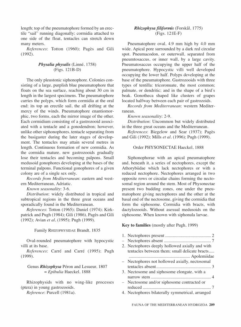
length; top of the pneumatophore formed by an erec-tile “sail” running diagonally; cormidia attached toone side of the float, tentacles can stretch downmany meters.
References: Totton (1960); Pagès and Gili(1992).
Physalia physalis (Linné, 1758)(Figs. 121B-D)
The only pleustonic siphonophore. Colonies con-sisting of a large, purplish blue pneumatophore thatfloats on the sea surface, reaching about 30 cm inlength in the largest specimens. The pneumatophorecarries the polyps, which form cormidia at the oralend; its top an erectile sail, the all drifting at themercy of the winds. Pneumatophore enantiomor-phic, two forms, each the mirror image of the other.Each cormidium consisting of a gastrozooid associ-ated with a tentacle and a gonodendron; however,unlike other siphonophores, tentacle separating fromthe basigaster during the later stages of develop-ment. The tentacles may attain several metres inlength. Continuous formation of new cormidia. Asthe cormidia mature, new gastrozooids graduallylose their tentacles and becoming palpons. Smallmedusoid gonophores developing at the bases of theterminal palpons. Functional gonophores of a givencolony are of a single sex only.
Records from Mediterranean: eastern and west-ern Mediterranean, Adriatic.
Known seasonality: 3-6.Distribution: widely distributed in tropical and
subtropical regions in the three great oceans andsporadically found in the Mediterranean.
References: Totton (1965); Daniel (1974); Kirk-patrick and Pugh (1984); Gili (1986), Pagès and Gili(1992); Avian et al, (1995); Pugh (1999).
Family RHIZOPHYSIDAE Brandt, 1835
Oval-rounded pneumatophore with hypocysticvilli at its base.
References: Carré and Carré (1995); Pugh(1999).
Genus Rhizophysa Péron and Lesueur, 1807 = Epibulia Haeckel, 1888
Rhizophysids with no wing-like processes(ptera) in young gastrozooids.
Reference: Purcell (1981a).
Rhizophysa filiformis (Forskål, 1775)(Figs. 121E-F)
Pneumatophore oval, 4.9 mm high by 4.0 mmwide. Apical pore surrounded by a dark red circularspot. Pneumacodon, or outerwall, separated frompneumtosaccus, or inner wall, by a large cavity.Pneumatosaccus occupying the upper half of thepneumatophore. Hypocystic villi well developedoccupying the lower half. Polyps developing at thebase of the pneumatophore. Gastrozooids with threetypes of tentilla: tricoronuate, the most common;palmate, or dendritic; and in the shape of a bird’sbeak. Gonotheca shaped like clusters of grapeslocated halfway between each pair of gastrozoids.
Records from Mediterranean: western Mediter-ranean.
Known seasonality: 2-9.Distribution: Uncommon but widely distributed
in the three great oceans and the Mediterranean.References: Biegelow and Sear (1937); Pagès
and Gili (1992); Mills et al. (1996); Pugh (1999).
Order PHYSONECTAE Haeckel, 1888
Siphonophorae with an apical pneumatophoreand, beneath it, a series of nectophores, except theAthorybiidae which lack nectophores or with areduced nectophore. Nectophores arranged in twoopposite rows or circular chains forming the necto-somal region around the stem. Most of Physonectaepresent two budding zones, one under the pneu-matophore giving nectophores and the other at thebasal end of the nectosome, giving the cormidia thatform the siphosome. Cormidia with bracts, withdactylozooids. Without asexual medusoids on thesiphosome. When known with siphonula larvae.
Key to families (mostly after Pugh, 1999)
1. Nectophores present ......................................... 2– Nectophores absent .......................................... 72. Nectophores deeply hollowed axially and with
tentacles between them; small delicate bracts............................................................ Apolemiidae
– Nectophores not hollowed axially, nectosomal tentacles absent................................................. 3
3. Nectosome and siphosome elongate, with a narrow stem ...................................................... 4
– Nectosome and/or siphosome contracted or reduced ............................................................. 7
4. Nectophores bilaterally symmetrical, arranged
FAUNA OF THE MEDITERRANEAN HYDROZOA 209
sm68s2Csystema2 14/10/04 15:24 Página 209

biserially ........................................................... 5– Nectophores dorso-ventrally flattened, usually
asymmetric in shape, arranged in spiral...................................................................... Forskaliidae
5. Nectophores with straight dorsal radial canal.. 6– Nectophores with sinuous dorsal radial canal.....
................................................... Pyrostephidae*6. Tentilla uncoiled; cnidoband hypertrophied; no
cnidocyst on terminal process.......... Erennidae*– Tentilla coiled; cnidoband normal . Agalmatidae7. Nectosome reduced or absent; siphosome
reduced to solid body or corm ...... Athorybiidae– Nectosome normal............................................ 88. Nectosome elongated; siphosome shortened into
laterally expanded spiral sac bearing enlarged palpons; bracts absent................. Physophoridae
– Nectosome and siphosome contracted to form a solid corm; with a pneumatophore and an aurophore ...................................... Rhodaliidae*
*not present in Mediterranean Sea.
Family AGALMATIDAE Brandt, 1835
Physonect siphonophores with a biserial arrange-ment of nectophores in the nectosome and a longusually contractile siphosome. This is rather a catchall family. For many agalmatids four types of cnido-cysts are present in the tentillum: homotrichousanisorhizas; either microbasic mastigophores orstenoteles; desmonemes and acrophores.
Remark: this is rather a catch all family. References: Carré and Carré (1995); Pugh (1999).
Genus Agalma Eschscholtz, 1825
Agalmatids with tricornuate tentilla consisting of acentral swelling and two contractile lateral filaments.
References: Pagès and Gili (1992); Mills et al. (1996).
Agalma clausi Bedot, 1888(Figs. 122A-C)
Siphosomal stem rigid. Leaf-like bracts withdeep red pigment spots on the surface, particularlythe younger ones. Tricornuate tentilla that can becompletely enclosed inside the involucrum.
Records from Mediterranean: Strait of Messina,Alborán Sea.
Known seasonality: 4.Distribution: Mediterranean and tropical Atlantic
(Mills et al. 1996).
References: Bedot (1888); Mills et al. (1996).
Agalma elegans (Sars, 1846) Fewkes, 1880(Figs. 122D-F)
Pneumatophore elongate, small, 1.1 mm high,apex red. Two rows of alternating nectophoresattached to the stem. Nectophores V-shaped, withtwo prominent lateral wings. An apico-lateral ridgerunning from the apex of the lateral wing to the mid-point of the nectophore, an infero-lateral ridge run-ning along the outer margin, and a latero-ventralridge located on the central portion of the nec-tophore. Nectosac T-shaped. Pedicular canal short.Siphosomal stem contractile. Bracts triangular, elon-gate, foliaceous in appearance, dorsal surface con-vex, 9 mm long, with a central bracteal canal,extending to about four-fifths the length of the bract,occasionally continuing as a very fine canal to thedistal end. Tentilla tricornuate.
Records from Mediterranean: eastern and west-ern Mediterranean.
Known seasonality: 12, 2 to 6.Distribution: widely distributed in tropical and
subtropical regions in the three great oceans and theMediterranean (Alvariño, 1971).
References: Totton (1956, 1965); Palma (1973);Kirkpatrick and Pugh (1984); Pagès and Gili (1992);Lakkis and Zeidane (1995); Pugh (1999).
Agalma okeni Eschscholtz, 1825(Figs. 122G-I)
Pneumatophore elongate, 7.0 mm high, apex pig-mented. Nectophores similar to those of A. elegans,but nectosac Y-shaped. Bracts firm, up to 5.0 mmlong, prismatic, triangular, gradually thickeningtowards the distal portion, which has three verticalridges delimiting four distal facets. Bracteal canalfiliform, central, not reaching the distal facets.
Records from Mediterranean: eastern and westenMediterranean.
Known seasonality: 5 and 6.Distribution: widely distributed in tropical and
subtropical regions in all seas and in the Mediter-ranean (Alvariño, 1971).
References: Totton (1965); Gili (1986); Pagès andGili (1992); Lakkis and Zeidane (1995); Pugh (1999).
Genus Cordagalma Totton, 1932
Agalmatids with unicornuate tentilla; with heart-
210 J. BOUILLON et al.
sm68s2Csystema2 14/10/04 15:24 Página 210

shaped nectophores devoid of lateral or vertical lat-eral ridges.
References: Pagès and Gili (1992); Margulis(1993); Carré and Carré (1995); Mills et al. (1996);Pugh (1999).
Cordagalma ordinata (Haeckel, 1888) = Cordalgama cordiforme Totton, 1932
(Figs. 123A-B)
Colonies extremely fragile, reaching up to 30 cmin length. Nectosome occupying a third of colonylength. Pneumatophore fusiform, apex lightly pig-mented. Two opposing rows of alternating nec-tophores. Adult colonies having up to 40 heart-shaped nectophores with two rounded latero-anteri-or lobes and an acute centro-inferior lobe, 2.0 mmhigh by 1.4 mm wide. Lateral radial canals, ascend-ing from the ostial canal, join the dorsal canal at theapex without describing a sigmoidal curve. Bractsshaped like a truncated pyramid, with four lateralfacets. Very distinctive tentilla with a larval struc-ture and simplified cnidome.
Records from Mediterranean: western Mediter-ranean.
Known seasonality: 4, 5.Distribution: uncommon but widely distributed
in warm waters throughout the world (Carré C.,1968b; Carré D., 1973; Gili, 1986), Great BarrierReef (Totton, 1932), Chile (Palma, 1977). Epipelag-ic species also present at depths down to 726 mdepth in the Alborán Sea (Mills et al., 1996).
References: Totton (1932, 1965); Carré C.(1968b); Carré D. (1973); Palma (1973, 1977); Pagèsand Gili (1992); Mills et al. (1996); Pugh (1999).
Genus Halistemma Huxley, 1859
Agalmatids whose tentilla have a single terminalfilament (unicornuate) and only a vestigial involu-crum. Characteristic sigmoidal courses for radialcanals on the nectosac of the nectophore that beginswith a downward sweep.
References: Pugh and Youngbluth (1988); Pagèsand Gili (1992); Pugh (1999).
Halistemma rubrum (Vogt, 1852)(Figs. 123C-G)
Pneumatophore oval, 5.0 mm high by 5.3 mmwide. Up to 46 nectophores per colony in tworows. Nectophore shape variable but squarish in
appearance, up to 5.2 mm in height, 6.6 mm inwidth, and 4.0 mm in dorso-ventral length. Twoprominent lateral wings and extensive centralthrust block containing the pedicular canal, moreconspicuous in older nectophores. Nectosac large,T-shaped. Characteristic pattern of lateral radialcanals describing three curves along the lateral sur-face of the nectosac; middle curve widest. Apico-lateral ridges not always reaching the level of theostium. Bracts thin and leaf-shaped, variable inshape but usually with two lateral processes and alarger central one.
Records from Mediterranean: eastern and west-ern Mediterranean.
Known seasonality: 2-6, 10,12.Distribution: widely distributed in the three great
oceans and in the Mediterranean (Alvariño, 1971).References: Totton (1965); Alvariño (1971) as
Stephanomia rubra; Carré D. (1971); Kirkpatrickand Pugh (1984); Avian et al. (1995); Lakkis andZeidane (1995); Pugh (1999).
Genus Lychnagalma Haeckel 1888
Agalmatids with nectophores arranged biserially;apico-lateral ridges prominent and divided close tothe base; lateral ridges extending out towards the topof the nectophore before reaching the apico-lateralridges. Characteristic large tentilla consisted of astalk, a tightly coiled cnidoband heavily armed withcnidocysts, and a terminal vesicle from which ariseeight regularly spaced filaments (Pugh and Harbi-son, 1986).
Lychnagalma utricularia (Claus, 1879)(Figs. 123, 124A-D)
Diagnosis: like the genusRecords from Mediterranean: Strait of Messina,
Alborán Sea.Known seasonality: 4.Distribution: Mediterranean and Atlantic
(Bahamas)References: Pugh and Harbison (1986); Mills et
al. (1996).
Genus Marrus Totton, 1954
Agalmatids with nectophores truncated apically,nectosacs with straight, unlooped radial canals; withunicornuate tentilla.
Reference: Andersen (1981).
FAUNA OF THE MEDITERRANEAN HYDROZOA 211
sm68s2Csystema2 14/10/04 15:24 Página 211

Marrus orthocanna Totton, 1954(Figs. 124E-G)
Nectophores longer than broad, up to 15 mm inlength, with no distinct lateral ridges. The apicalridges divide close to the ostium and delimit twonarrow, triangular distal facets. Thrust block well-developed and longer than the rounded lateralprocesses. Nectosac triangular. Bract roughly trian-gular with a flimsy, tacky appearance.
Records from Mediterranean: western Mediter-ranean.
Known seasonality: 6,12.Distribution: North Atlantic and Mediterranean.References: Totton (1965); Andersen (1981);
Kirkpatrick and Pugh (1984); Gili (1986).
Genus Nanomia A. Agassiz, 1865Agalmatids whose unicornuate tentillum has a
basal involucrum. Characteristic arrangement of thegonodendra in that male and female ones, attachedat the base of palpons, alternate on either side.
Reference: Pagès and Gili (1992).
Nanomia bijuga (delle Chiaje, 1841)(Figs. 125A-D)
Pneumatophore elongate, very small, 1.8 mm high,apex pigmented. Nectophores quadrangular, flattenedin the abaxial-adaxial plane, L-shaped in lateral view,up to 2.3 mm high. Lateral wings twisting towards theadaxial or ventral surface of the colony. Nectophorewith a well-developed ostial mouth, ostial velumbroad. Looped lateral radial canals on extensive nec-tosac. Pedicular canal long. Bracts leaf-like, variablein morphology, often with 3 processes at the distalend, occasionally with a cross ridge.
Records from Mediterranean: eastern and west-ern Mediterranean.
Known seasonality: 4-11.Distribution: widely distributed in warm and
temperate regions of the three great oceans, and theMediterranean (Alvariño, 1971).
References: Totton (1965); Carré D. (1969b);Alvariño (1971) as Stephanomia bijuga; Rottini(1971); Palma (1986); Gili (1986); Pagès and Gili(1992); Avian et al. (1995); Lakkis and Zeidane(1995); Pugh (1999).
Family APOLEMIIDAE Huxley, 1859
Uniquely, amongst the physonects, there is a ten-
tacle or clump of tentacles between each pair of nec-tophores. Nectophore deeply hollowed axially,forming a pair of large axial wings. Nectosac exten-sive, lateral radial canals follow an S-shape courseof varying complexity. Bracts small and flimsy. Thisfamily requires a through review.
References: Carré and Carré (1995); Pugh (1999).
Genus Apolemia Eschscholtz, 1829
Apolemiids with 1-6 tentacles between each pairof nectophores.
Reference: Carré and Carré (1995).
Apolemia uvaria (Lesueur,? 1811)(Figs. 126A-D)
Colonies reaching several metres in length. Pneu-matophore bulb-shaped, widening near the apex.Nectosome with up to twelve nectophores arranged intwo parallel rows along the stem. Largest nectophore3.7 mm high, 3.4 mm wide and 4.2 mm deep. Nec-tophore consisting of two wings looking like those ofa butterfly, with a deep ventral thrust block. Nectosaclarge. Lateral radial canals S-shaped with shortbranches on the upper loop. Groups of five to six nec-tosomal tentacles issuing from the base of the nec-tophores near the pedicular canal, at the base of mus-cular lamellae. Siphosome up to several metres inlength, composed of several cormidia. Each cormidi-um consists of a gastrozooid and about fifty palpons,both with thin filiform tentacles of a single type issu-ing from their bases. Palpons long and delicate.Bracts, like the nectophores, covered by opaque spotsbearing cnidocysts on the outer surface.
Records from Mediterranean: western Mediter-ranean.
Known seasonality: 8-4.Distribution: Seldom caught epiplanktonic
species. Cited in the Mediterranean, in the vicinityof Naples, Messina, Monaco, and Villefranche-sur-Mer (Alvariño, 1971). In the Atlantic Ocean citedoff the British Isles (Kirkpatrick and Pugh, 1984)and Norwegian fjords (Båmstedt et al., 1998).
References: Totton (1965); Carré and Carré (1973,1995), Alvariño (1971); Kirkpatrick and Pugh (1984);Pagès and Gili (1992); Båmstedt et al. (1998).
Family ATHORYBIIDAE Huxley, 1859
Physonects with relatively large pneumatophore.Nectosome greatly reduced or absent. Siphosome
212 J. BOUILLON et al.
sm68s2Csystema2 14/10/04 15:24 Página 212

reduced to a dense corm on which the cormidia arearranged in a spiral.
References: Biggs and Harbison (1978); Carréand Carré (1995).
Genus Athorybia Eschscholtz, 1829
Athorybiids without nectosome; pneumatophorelarge; bracts flimsy with inconspicuous rows ofcnidocysts.
References: Totton (1954); Pagès (2002).
Athorybia rosacea (Forskål, 1775)(Figs. 126E-F)
Pneumatophore large, red pigmented, withcormidia arranged in spiral around it. No nectosome.Elongate, flimsy bracts with 7 inconspicuous rowsof cnidocysts running down the convex dorsal side.
Records from Mediterranean: western Mediterranean.Known seasonality: 9-4.Distribution: Atlantic and Mediterranean.References: Pugh (1999); Pagès (2002).
Family FORSKALIIDAE Haeckel, 1888
Physonects with cylindrical or cone-shape necto-some, whose numerous nectophores have a multise-rial, spiral arrangement. Nectophores flatteneddorso-ventrally, often asymmetrical in shape. Nec-tosac restricted to basal half, with straight radialcanals. Siphosome also coiled, with gastrozooidsborne on long stalks. Gastrozooids borne on longpeduncles that are covered in bracts. Bracts usuallygelatinous and of variable shape of four types: stem,bolster and two kinds of knee-shape. Severalgonopalpons, with palpacles, present on gonoden-dra, which bear both male and female gonophores.
References: Carré and Carré (1995); Pugh(1999), Pugh (2003).
Genus Forskalia Kölliker, 1853
With the characters of the family.Reference: Pagès and Gili (1992); Pugh (1999);
Pugh (2003)
Forskalia asymmetrica Pugh 2003(Figs. 126G-H)
Large nectophores, with small rounded left axialwing, and a small central apical incision. No lateral
incisions or pockets. No rete mirabile. Adult bractsof four types. Gonodendra bearing long gono-palpons, with palpacles; with female gonophoresattached close to their bases. Male gonophoresattached at end of a stalk.
Records from Mediterranean: off Villefranche-sur-Mer, off Messina (Sicily) and in the Alborán Sea.
Known seasonality: 4.Distribution: Mediterranean and Atlantic (the
Bahamas; off Woods Hole, USA).Reference: Pugh (2003).
Forskalia contorta (Milne Edwards 1841) = F. leuckarti Bedot 1893
(Fig. 127A-F)
Large nectophores with large left axial wing,without central apical incision. Longitudinal flap onupper side in apical half. Lateral basal pocketsextend in flaps onto the lower surface. Nectosacwith pronounced lateral wings, with dorsal and ven-tral canals usually not arising from pedicular canal.No pigment spots on ostium. Rete mirabile presentor absent in pedicular canal. Three types of adultbract; with only one knee-shaped type, whosebracteal canal bends through a right-angle and has ashort side branch. Female gonophores, in twobunches, borne on long stalk, extending beyondmale ones.
Records from Mediterranean: western Mediter-ranean, Adriatic.
Known seasonality: 9-6.Distribution: rare, to date only caught in the
Mediterranean (Totton, 1965) and observed fromsubmersibles at depths between 150 and 600 m inthe Bahamas (Youngbluth, 1984).
References: Totton (1965); Youngbluth (1984);Pagès and Gili (1992); Avian et al. (1995); Pugh(1999), Pugh (2003).
Forskalia edwardsi Kölliker, 1853(Figs. 127G-H)
Nectophores with small left axial wing, and nocentral apical incision; with apical thickenedmesoglea. Small yellow pigment spot on ostiumwhere dorsal radial canal joins ring canal. No retemirabile. Four types of adult bract: knee-shapeones without side branch to bracteal canal. Gon-odendra with female gonophores not having elon-gated base. Palpacle of gonopalpon has appearanceof a string of beads.
FAUNA OF THE MEDITERRANEAN HYDROZOA 213
sm68s2Csystema2 14/10/04 15:24 Página 213

Records from Mediterranean: off Villefranche-sur-Mer.
Known seasonality: in winter.Distribution: western Mediterranean, Red Sea,
Atlantic and Pacific.References: Totton (1965); Youngbluth (1984);
Pagès and Gili (1992); Pugh (1999), Pugh (2003).
Forskalia formosa Keferstein and Ehlers 1861(Figs. 127I-K)
Nectophores with large, pointed left axial wing,and shallow but broad and rounded right wing. Smallapical incision between them. Small lateral, but nobaso-lateral pockets. No rete mirabile. Four types ofadult bract; knee-shaped ones without side branch tocanal. Gonodendra with female gonophores, in twobunches, attached close to bases of gonopalpons;male gonophores borne on long stalk.
Records from Mediterranean: off Villefranche-sur-Mer, off Messina (Sicily) and Alborán Sea.
Known seasonality: in winter.Distribution: Mediterranean and Atlantic (The
Bahamas).References: Avian et al. (1995); Mills et al.
(1996), Pugh (2003).
Family PHYSOPHORIDAE Eschscholtz, 1829
Physonects with a flimsy, apparently ridgelessnectophores each with an extensive nectosac, whichhas characteristic, looped lateral radial canals. Bothdorsal and ventral canals are sinuous. Siphosomecompact sac on which the simple, bractless cormidiaare borne in spiral. Each cormidium has a single,greatly enlarged palpon. Monotypic family for thespecies Physophora hydrostatica.
Reference: Carré and Carré (1995).
Genus Physophora Forskål, 1775
With the characters of the family.Reference: Pagès and Gili (1992).
Physophora hydrostatica Forskål, 1775(Figs. 128A-C)
Pneumatophore elongate, 4.5 mm high, 1.1 mmwide, with a pore in the apical reddish region. Nec-tosome consisting of up to 12 nectophores in twoalternate rows. Nectophore flimsy, devoid of con-spicuous ridges and up to 20 mm high. Nectosac Y-
shaped. Upper half of radial canals describes a high-ly characteristic sigmoidal curve. Pedicular canalprominent. Palpons characteristic by their large sizeand banana-shape, with long and fine palpacles.Palpons covering and protecting the rest of the ele-ments making up the bag-like siphosome and bear-ing clusters of cnidocysts on their distal ends.
Records from Mediterranean: western MediterraneanKnown seasonality: 9-4.Distribution: widely distributed in tropical and
subtropical regions in the three great oceans and inthe Mediterranean (Alvariño, 1971).
References: Totton (1965); Alvariño (1971);Kirkpatrick and Pugh (1984); Gili (1986); Pagès andGili (1992); Pugh (1999).
Order CALYCOPHORAE Leuckart, 1854
Highly polymorphic Siphonophorae withoutpneumatophore, with a reduced nectosome typicallyformed by one or two but sometimes more nec-tophores. Usually with a single bract per cormidia(except the Hippopodiidae without bracts), withoutdactylozooids (except in Stephanophyes), in somecases with asexual medusoid structures on sipho-some. Generally the cormidial units are detachedsuccessively from the stem and become eudoxid orsexual stage. Usually a calyconula larvae.
Key to the families (after Pugh, 1999)
1. Nectophores dorso-ventrally flattened ............. 2– Nectophores not dorso-ventrally flattened, bracts
present .............................................................. 32. ..........Up to 15+ similar, closely applied, dorso-
ventrally fattened nectophores bearing protuberances or spines; large but shallow nectosac; bracts absent ............... Hippopodiidae
– Small, flattened nectophores, with vestigial nectosac and reduced somatocyst...................................... Prayidae, subfamily Amphicaryoninae
3. Nectophores and bracts rounded, smooth-walled, with thick mesoglea.......................................... 4
– Nectophores and bracts pointed, toothed or of irregular shape .................................................. 6
4. Usually 2 nectophores ...................................... 5– Single, fragile, larval nectophore, with a simple
somatocyst and narrow hydroecium; small, fragile bract with a single canal ............................................................................ Sphaeronectidae
5. Two nectophores of approximately equal size, forming an apposed pair; somatocyst simple or
214 J. BOUILLON et al.
sm68s2Csystema2 14/10/04 15:24 Página 214

branched; bracts with 5 or 6 branches to the canal system.................. Prayidae, subfamily Prayinae
– Two nectophores of unequal size, with reduced somatocysts; nectosac of smaller nectophore usually reduced or obsolent; bracts with two branches to canal system..................................................Prayidae, subfamily Amphicaryoninae*
6. Two morphologically different nectophores; one (anterior) superimposed over the other (posterior) ......................................................... 8
– With usually a single nectophore ..................... 77. Single, usually large nectophore bearing simple
or toothed ridges; somatocyst usually branched; large bract without neck shield and extensively branched canal system.................................................... Prayidae, subfamily Nectopyramidinae*
– Single, usually small, apically pointed nectophore, with simple caecal somatocyst; bract conical or angular ............................................. 9
8. Posterior nectophore with a somatocyst; anterior nectophore with extensive opening of hydroecium onto ventral surface; small bract with phyllocyst and 2 canals extending into neck shield........................................... Clausophyidae
– Posterior nectophore without somatocyst; hydroecium of anterior nectophore usually opens basally; bracts conical or angular, with phyllocyst and, at most, one canal ................... 9
9. Conical stream-lined anterior nectophore, usually with shallow hydroecium; posterior nectophore, when present, usually apically truncated and of similar size or smaller than anterior one; conical bracts................ Diphyidae
– Anterior nectophore angular, with inflated somatocyst and deep hydroecium; posterior nectophore, when present, larger than anterior one; rigid angular bracts ...................... Abylidae
*not present in the Mediterranean Sea.
Family ABYLIDAE Agassiz, 1862
Calycophorans with rigid, angular nectophores, theposterior one, without a somatocyst, usually beingmuch larger, and bearing serrated ridges and teeth. Inall but one species the somatocyst of the anterior nec-tophore has curved over to occupy a ventral position.The hydroecium of the anterior nectophore is anenclosed tube opening basally. During development atemporary larval bract is formed before the larval nec-tophore. The latter is retained in the polygastric stageas the anterior nectophore.
References: Sears (1953); Carré and Carré(1995); Pugh (1999).
Genus Abyla Quoy and Gaimard, 1827
Abylid with 10 or 11 facets in anterior nec-tophores. The apical facet is divided by a transverseridge, and many ridges are serrated, particularlybasally. Dorsal nectosac and median hydroecium arelong tubes extending almost to apex of nectophore.The large oval somatocyst lies ventrally. Posteriornectophore with long, tapering apical apophysis, hasonly 4 ridges. The ventral ridges define the hydroe-cial wings and are heavily serrated basally. The lefthydroecial wing bears a toothed comb or flap. Five,usually serrated, ostial teeth. Prismatic bracts with 6facets, the dorsal one being rectangular. Very largephyllocyts, with 2 canals running down toward theventro-lateral corners of the apical facet. The bractscannot, at present, be identified specifically.
Reference: Pugh (1999).
Abyla haeckeli Lens and van Riemsdijk, 1908(Figs. 128D-F)
Anterior nectophore as wide as long, withoutwing-like processes. Transverse ridge separates ven-tral facet from apico-ventral one. Posterior nec-tophore with up to 5 teeth on comb. Lateral ostialteeth closer to dorsal tooth than ventral ones.
Reference: Pugh (1999).
Genus Abylopsis Chun, 1888
Abylid whose anterior nectophore has a pentagonaldorsal facet without a median ridge. Seven-facetanterior nectophore, but without an apical facet, andpentagonal dorsal and ventral facets. The somato-cyst has an apical diverticulum. Posterior nec-tophore has prominent basal teeth and with 5 ridgesand a short curved apical apophysis. The left lateralridge bifurcates close to the apex. Flaps on bothwings of hydroecium. Bracts with 7 facets. Phyllo-cyst with swollen apico-lateral branches and apicaldiverticulum, while distally is a narrow tube.
References: Pagès and Gili (1992); Pugh (1999).
Abylopsis eschscholtzi (Huxley, 1859)(Figs. 128G-K)
Dorsal and ventral facets of anterior nectophorealmost pentagonal, of nearly equal size, with strong-
FAUNA OF THE MEDITERRANEAN HYDROZOA 215
sm68s2Csystema2 14/10/04 15:24 Página 215

ly serrated ridges. Lateral radial canals forming aright angle from the pedicular canal and directedtowards the ostial canal, without ascending loop.Posterior nectophore less than twice as long as wide,with relatively large apical apophysis. Each hydroe-cial wing bearing a secondary wing whose innermargins is fused. Between four and eight teeth onthe curve on the left wing, three or four on the rightwing. Bracts cuboidal, their dorsal facet forms a reg-ular pentagon, their apico-lateral facets rectangular.
Records from Mediterranean: eastern and west-ern Mediterranean.
Known seasonality: 4 to 7.Distribution: common and abundant in the warm
and temperate regions in the three great oceans, lesscommon in the Mediterranean (Alvariño, 1971).Epipelagic distribution (Pugh, 1974).
References: Totton (1965); Alvariño (1971); Rot-tini (1971); Daniel (1974); Pugh (1974, 1999); Gili(1986); Pagès and Gili (1992); Avian et al. (1995);Lakkis and Zeidane (1995).
Abylopsis tetragona (Otto, 1823)(Figs. 129A-E)
Polygastric stage: anterior nectophore polyhedralwith seven faces. Ventral and dorsal surfaces pen-tagonal, the latter higher, up to 10 mm in height.Ridges weekly serrated. Hydroecium deep, reachingnearly the midpoint of the nectophore. Radial canalsof nectosac originating at the junction with thepedicular canal and rising towards the upper end,forming an ascending loop, and then descending tothe circular canal. Somatocyst globular, with an api-cal diverticulum at the level of the nectosac. Poste-rior nectophore up to 40 mm in length, rectangular,three times as long as wide, with a prominent apoph-ysis. Four radial canals in the upper half, five in thelower one because the left ventral canal has a blindtermination halfway up and a little above the originof a new canal forming a right angle and directedtowards the ostial canal. A “rete” at the base of theright ventral canal, from which a short, blind ventralcanal issues towards the left. Upper half of the rightwing of hydroecium bearing a comb-like structurewith about nine teeth. Lower portion bearing fivebasal teeth varying in size, right ventral toothlargest. Bract up to 7.0 mm high, elongated, penta-gon with apico-lateral facets trapezoidal. Upper halfof phyllocyst bearing two thick, lateral processesabove the hydroecium and extending towards theapical surface in the form of a narrow diverticulum.
Gonophore in the shape of a rectangular prism 4.6mm high with four distinct ridges ending in fouracute basal cusps, the ventral cusps somewhat larg-er than the dorsal ones.
Records from Mediterranean: eastern and west-ern Mediterranean.
Known seasonality: present all the year.Distribution: quite common in the temperate
regions of the three main oceans, and in the Mediter-ranean (Alvariño, 1971). Epipelagic (Pugh, 1974),but also present in the mesopelagic zone (Gili et al.,1987a).
References: Totton (1965); Carré C. (1967b);Alvariño (1971); Palma (1973); Pugh (1974, 1999);Bone and Trueman (1982); Gili (1986); Gili et. al.(1987a); Pagès and Gili (1992), Avian et al. (1995);Lakkis and Zeidane (1995); Buecher (1999);Gamulin and Krisnic (2000).
Genus Bassia Agassiz, 1862
Abylines whose anterior nectophore has not anapical diverticulum to the somatocyst, and with thehydroecium not extending below the basal facet.Somatocyst large and globular. Posterior nectophorewith 4 ridges ending in short basal teeth. Bract withmedian apical ridge has a quadrilateral dorsal facet.Phyllocyst is a long tube, swollen apically, withoutapico-lateral branches. Gonophore with four longi-tudinal ridges which end basally in minute teeth.
Bassia bassensis (Quoy and Gaimard, (1833)1834) (Figs. 129F-I)
Polygastric stage: ridges on specimens fixed in for-malin taking on a bluish-white hue (same applies toeudoxids). Anterior nectophore a seven-sided polyhe-dral with pentagonal dorsal and ventral surfaces. Twoquadrangular apico-lateral surfaces joined by a centralapical ridge. Two large baso-lateral surfaces separatedfrom the apico-lateral surfaces by a horizontal ridge.Hydroecium deep, reaching the midpoint of the nec-tophore, with a large quadrangular opening. Nectosacrelatively small, with lateral radial canals runningfrom the ostial canal to the junction with the pedicularcanal, with only a slight curve instead of an arc.Apices of hydroecium and nectosac more or less at thesame level. Somatocyst large, globular, devoid ofdiverticulum or apical projection. Posterior nec-tophore higher than the anterior nectophore, up to 15.0mm high, consisting of four ridges, not as thin as in theanterior nectophore. Bases of ventral ridges compris-
216 J. BOUILLON et al.
sm68s2Csystema2 14/10/04 15:24 Página 216

ing two prominent teeth, right tooth larger than lefttooth. Eudoxid: bract in the form of a seven-sidedpolyhedral with well-developed ridges. Dorsal surfacerhomboidal, ventral surface in the shape of a five-pointed star. Apico-lateral surfaces quadrangular.Baso-ventral margins of lateral surfaces describing acurve ending in a medial tooth. Hydroecium large,deep, hook-shaped, reaching to the midpoint of thebract. Phyllocyst is a long tube long, without lateralprocesses. Gonophore with four lateral ridges that endbasally in minute teeth.
Records from Mediterranean: eastern and west-ern Mediterranean.
Known seasonality: 3, 12.Distribution: Present in the temperate regions off
the three great oceans and in the Mediterranean(Alvariño, 1971).
References: Totton (1965); Palma (1973); Daniel(1974); Kirkpatrick and Pugh (1984); Gili (1986);Pagès and Gili (1992); Carré and Carré (1995);Lakkis and Zeidane (1995); Pagès et al. (2001).
Genus Ceratocymba Chun, 1888
Abylid with characteristically shaped bract, calleda cymba. Anterior nectophore with 7 facets. Apicalfacet not divided by a transverse ridge. Posterior nec-tophore long and narrow, without wing-like expan-sions. Short dorsal ridge ends on the dorsal tooth.Bracts with a median dorsal ridge. Left lateral facetdivided by another ridge. Bracts roughly triangularwith a concave apical facet and prominent lateralhorns. Phyllocyst with 2 thin ventro-lateral branches.Its distal end bends dorsally to form a blind sac.
References: Pagès and Gili (1992); Pugh (1999).
Ceratocymba sagittata (Quoy and Gaimard, 1827)(Figs. 130A-C)
Polygastric stage: anterior nectophore pyramidal,elongate, up to 40.0 mm high. Large, tapered apicalprocess called a pyramidal process. Four ridges con-verging at the apex. Characteristic apical surface ofthe family Abylinae no longer present. Hydroeciumbell-shaped, with a large opening, in the centre ofthe base of the nectophore. Nectosac narrow, high,reaching almost to the apex of the nectophore. Lat-eral radial canals ascending over a short segment ofthe nectosac from the pedicular canal, describing avery tight arc and then descending towards the ostialcanal. Somatocyst oval, positioned between the dor-sal surface of the nectophore and the dorsal surface
of the hydroecium; not extending past the apex ofthe hydroecium. Posterior nectophore elongated,oblong, up to 50.0 mm high, pointed apically. Leftventral wing ending in a characteristic prominent,elongate, serrated basal tooth. Comb on right ventralwing bearing six teeth; inferior margins of bothwings dentate. Eudoxid: bract roughly triangular,with two supra-lateral horns and a dorsal ridge end-ing in a point basally. Up to 20.0 mm high. Apicalsurface triangular, concave. Characterized by a rightlateral ridge, originating at the lower margin, stop-ping before it reaches the apico-dorsal ridge. Phyl-locyst cylindrical, with two filiform lateral projec-tions and a basal tip recurved upwards. Gonophorewith basal teeth varying in size. A relatively short,inconspicuous hook issuing from one of the ventralridges, curving towards the base of the hydroecium.Teeth located above the hook on the ventral marginof the opposite wing. A small dorsal tooth. Lateralridge near the bracteal wall deeper than the oppos-ing ridge at the apex.
Records from Mediterranean: Strait of Gibraltar.Seasonality: ?Distribution: widely distributed in the Atlantic
Ocean, where its range is very broad. Also present intemperate regions in other seas and in the Mediter-ranean (Alvariño, 1971).
References: Bigelow and Sears (1937); Totton(1965); Casanova (1980); Alvariño (1981); Kirk-patrick and Pugh (1984); Pagès and Gili (1992);Pugh (1999).
Genus Enneagonum Quoy and Gaimard, 1827
Abylids where the large, pyramidal anterior nec-tophore is the only developed. The conical somato-cyst is situated above the hydroecium, and extendsto a greater height than the nectosac. The bract iscuboidal, with slightly concave facets. Swollensomatocyst with 2 lateral and apical processes.
Reference: Pagès and Gili (1992).
Enneagonum hyalinum Quoy and Gaimard, 1827(Figs. 130D-F)
Polygastric stage developing only a single nec-tophore. Nectophore pyramidal, up to 15.0 mm wide.All surfaces homologous to those in other Abylopsi-nae, except that in E. hyalinum, there is a median ridgesubdividing what is equivalent to the dorsal surface inother Abylopsinae. The two dorsal surfaces and thetwo apico-lateral surfaces visible when viewed from
FAUNA OF THE MEDITERRANEAN HYDROZOA 217
sm68s2Csystema2 14/10/04 15:24 Página 217

the apex. A triangular basal surface beneath the dorsalsurfaces, the two baso-lateral surfaces, and the ventralsurface all visible when viewed basally. Ridges andbasal margins slightly dentate. Somatocyst carrot-shaped, consisting of two thickened lateral processesand an apical diverticulum, lacking a descending dor-sal segment, differentiating it from the somatocysts ofthe rest of the members of the family Abylidae. Arcfollowed by the lateral radial canal including a blinddiverticulum. Eudoxid: bract cuboidal, 4.0 mm high.Five surfaces: apical, dorsal, ventral, and two laterals.No baso-lateral surfaces, the entire space being occu-pied by the large hydroecial opening. Swollen phyllo-cyst with 2 lateral and apical processes. Gonophorewith a well-developed apophysis occupying nearly athird of the gonophore.
Records from Mediterranean: eastern and west-ern Mediterranean.
Known seasonality: 3.Distribution: common in warm and temperate
regions in the three great oceans, and in the Mediter-ranean (Alvariño, 1971). Epipelagic but also abun-dant in the mesopelagic zone down to a depth of1000 m (Pugh (1974).
References: Bigelow and Sears (1937); Totton(1965); Daniel (1974); Pugh (1974, 1999); Kirk-patrick and Pugh (1984); Gili (1986); Pagès and Gili(1992); Lakkis and Zeidane (1995).
Family CLAUSOPHYIDAE Totton, 1965
Both anterior and posterior nectophores possessa somatocyst: The phyllocyst of the eudoxid bractwhen known has two fine branch canals that rundown into the neck shield.
Genus Clausophyes Lens and Riemsdijk, 1908
Nectophores rounded, smooth, unridged, lateral-ly flattened. Anterior nectophore usually smallerthan posterior one, with deep hydroecium in basalhalf and with a long, thin somatocyst which has anelongated swelling towards its apex. Lateral canal ofboth nectophores are looped.
References: Pugh (1995, 1999).
Clausophyes ovata (Keferstein and Ehlers 1860)(Figs. 130G-I)
Polygastric stage: anterior nectophore up to 20mm in height, roughly triangular but rounded at its
base. The deep hydroecium reaches to about theheight of the nectophore and is open at the ostiallevel. Posterior nectophore up to 30 mm in height,with a large notched mouth. The hydroeciumextends the length of the nectophore and is boundedby large lateral wings. The somatocyst is long andnarrow.
Eudoxid: bract conical, up to 8 mm in height,with a rounded apex and an extensive neck-shield.The phyllocyst, slightly swollen basally reaches theapex. The gonophore is undescribed.
Records from Mediterranean: off Messina (Sici-ly), Adriatic, western Mediterranean.
Known seasonality: present all the year.Distribution: Atlantic, Pacific, MediterraneanReferences: Keferstein and Ehlers (1861, as
Diphyes ovata), Kirkpatrick and Pugh (1984); Patri-ti (1969) as Clausophyes massiliana, Gamulin andKrisnic (2000).
Family DIPHYIDAE Quoy and Gaimard, 1827
Calycophorae with polygastric stage with one ortwo dissimilar streamlined definitive nectophoresarranged serially. Anterior nectophore with somato-cyst, posterior not; hydroecium generally reduced inanterior nectophore; the nectosac occupies most ofthe nectophore.
References: Carré and Carré (1995), Pugh(1999).
Genus Chelophyes Totton, 1932
Diphyids with rigid nectophores; anterior onewith 5 ridges, dorsal one extends only a short dis-tance up from the ostium. Claw-shaped hydroecium.Posterior nectophore apically pointed, mouth-Fig.divided with two strong asymmetric teeth. Bractswith conical eudoxids, small rounded neck-shield,hydroecium cylindrical and long that almost stretch-es the apex.
References: Pagès and Gili (1992); Pugh (1999).
Chelophyes appendiculata (Eschscholtz, 1829)(Figs. 130J-K, 131A-C)
Polygastric stage: Anterior nectophore of firmconsistency. Up to 12.0 mm in height by 3.6 mm indorso-ventral width. Only three ridges, the two ven-tral ridges and the right lateral ridge (sensu Bigelow)converging at the apex. Left lateral ridge not reach-ing the apex and only a short section of the dorsal
218 J. BOUILLON et al.
sm68s2Csystema2 14/10/04 15:24 Página 218
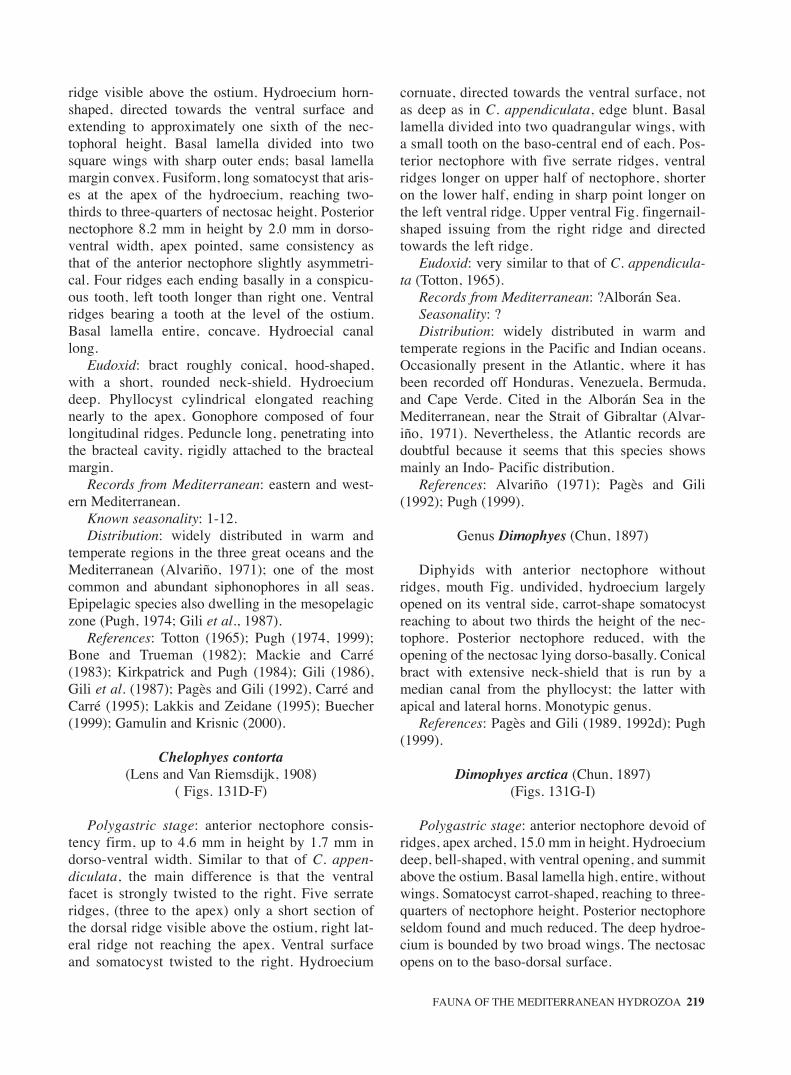
ridge visible above the ostium. Hydroecium horn-shaped, directed towards the ventral surface andextending to approximately one sixth of the nec-tophoral height. Basal lamella divided into twosquare wings with sharp outer ends; basal lamellamargin convex. Fusiform, long somatocyst that aris-es at the apex of the hydroecium, reaching two-thirds to three-quarters of nectosac height. Posteriornectophore 8.2 mm in height by 2.0 mm in dorso-ventral width, apex pointed, same consistency asthat of the anterior nectophore slightly asymmetri-cal. Four ridges each ending basally in a conspicu-ous tooth, left tooth longer than right one. Ventralridges bearing a tooth at the level of the ostium.Basal lamella entire, concave. Hydroecial canallong.
Eudoxid: bract roughly conical, hood-shaped,with a short, rounded neck-shield. Hydroeciumdeep. Phyllocyst cylindrical elongated reachingnearly to the apex. Gonophore composed of fourlongitudinal ridges. Peduncle long, penetrating intothe bracteal cavity, rigidly attached to the bractealmargin.
Records from Mediterranean: eastern and west-ern Mediterranean.
Known seasonality: 1-12.Distribution: widely distributed in warm and
temperate regions in the three great oceans and theMediterranean (Alvariño, 1971); one of the mostcommon and abundant siphonophores in all seas.Epipelagic species also dwelling in the mesopelagiczone (Pugh, 1974; Gili et al., 1987).
References: Totton (1965); Pugh (1974, 1999);Bone and Trueman (1982); Mackie and Carré(1983); Kirkpatrick and Pugh (1984); Gili (1986),Gili et al. (1987); Pagès and Gili (1992), Carré andCarré (1995); Lakkis and Zeidane (1995); Buecher(1999); Gamulin and Krisnic (2000).
Chelophyes contorta(Lens and Van Riemsdijk, 1908)
( Figs. 131D-F)
Polygastric stage: anterior nectophore consis-tency firm, up to 4.6 mm in height by 1.7 mm indorso-ventral width. Similar to that of C. appen-diculata, the main difference is that the ventralfacet is strongly twisted to the right. Five serrateridges, (three to the apex) only a short section ofthe dorsal ridge visible above the ostium, right lat-eral ridge not reaching the apex. Ventral surfaceand somatocyst twisted to the right. Hydroecium
cornuate, directed towards the ventral surface, notas deep as in C. appendiculata, edge blunt. Basallamella divided into two quadrangular wings, witha small tooth on the baso-central end of each. Pos-terior nectophore with five serrate ridges, ventralridges longer on upper half of nectophore, shorteron the lower half, ending in sharp point longer onthe left ventral ridge. Upper ventral Fig. fingernail-shaped issuing from the right ridge and directedtowards the left ridge.
Eudoxid: very similar to that of C. appendicula-ta (Totton, 1965).
Records from Mediterranean: ?Alborán Sea.Seasonality: ?Distribution: widely distributed in warm and
temperate regions in the Pacific and Indian oceans.Occasionally present in the Atlantic, where it hasbeen recorded off Honduras, Venezuela, Bermuda,and Cape Verde. Cited in the Alborán Sea in theMediterranean, near the Strait of Gibraltar (Alvar-iño, 1971). Nevertheless, the Atlantic records aredoubtful because it seems that this species showsmainly an Indo- Pacific distribution.
References: Alvariño (1971); Pagès and Gili(1992); Pugh (1999).
Genus Dimophyes (Chun, 1897)
Diphyids with anterior nectophore withoutridges, mouth Fig. undivided, hydroecium largelyopened on its ventral side, carrot-shape somatocystreaching to about two thirds the height of the nec-tophore. Posterior nectophore reduced, with theopening of the nectosac lying dorso-basally. Conicalbract with extensive neck-shield that is run by amedian canal from the phyllocyst; the latter withapical and lateral horns. Monotypic genus.
References: Pagès and Gili (1989, 1992d); Pugh(1999).
Dimophyes arctica (Chun, 1897)(Figs. 131G-I)
Polygastric stage: anterior nectophore devoid ofridges, apex arched, 15.0 mm in height. Hydroeciumdeep, bell-shaped, with ventral opening, and summitabove the ostium. Basal lamella high, entire, withoutwings. Somatocyst carrot-shaped, reaching to three-quarters of nectophore height. Posterior nectophoreseldom found and much reduced. The deep hydroe-cium is bounded by two broad wings. The nectosacopens on to the baso-dorsal surface.
FAUNA OF THE MEDITERRANEAN HYDROZOA 219
sm68s2Csystema2 14/10/04 15:24 Página 219

Eudoxid: up to 10 mm in height. The bract isconical with an extensive, thin neck shield. Thephyllocyst has apical and lateral horns and a finebasal process, which passes down the neck shield.The gonophore has a very small mouth and anytraces of a hydroecium.
Records from Mediterranean: western Mediter-ranean.
Known seasonality: present all the year.Distribution: cosmopolitan species, inhabiting
the three great oceans as well as the Antarctic, Arc-tic, and Mediterranean (Alvariño, 1971). Meso-pelagic distribution in tropical latitudes, moreepipelagic distribution in boreal and austral latitudes(Pugh, 1984; Pagès and Schnack-Schiel, 1996),though it may be found throughout the upper 1.000metres of the water column (Pugh, 1974).
References: Totton (1965); Pugh (1974, 1984,1999); Kirkpatrick and Pugh (1984); Pagès and Gili(1992).
Genus Diphyes Cuvier, 1817
Diphyids with 5 complete longitudinal ridges inthe anterior nectophores and 3 prominent dorsalteeth in general. Deep hydroecium. Posterior nec-tophores, when developed, also with 3 ostial teeth ingeneral and a long apical process (apophysis).Bracts generally helmet-shaped.
References: Pagès and Gili (1992); Pugh (1999).
Diphyes bojani (Eschscholtz, 1829)(Figs. 131J-K)
Polygastric stage: anterior nectophore elongatedbearing five long, variably serrated ridges converg-ing at the apex to form a pyramid, pentagonal incross-section, 10.0 mm in height by 3.0 mm indorso-ventral width. Three basal teeth all of thesame size. Nectosac cylindrical, reaching nearly tothe apex of the nectophore, upper third tapering.Hydroecium deep, extending to about one-third theheight of nectophore, narrower than in D. dispar.Somatocyst fusiform, tip reaching nearly to the apexof the nectosac. Basal lamella entire, bearing a thinmedian crest. Posterior nectophore with five serrat-ed ridges, upper third tapering, forming a prominentapophysis. Up to 6.6 mm in height by 2.2 mm indorso-ventral width.
Eudoxid: bract shield-shape that covers the upperventral half of the gonophore, different from thatfrom all other diphyids.
Records from Mediterranean: western Mediter-ranean.
Known seasonality: present all the year.Distribution: widely distributed in tropical and
subtropical regions in the three great oceans and inthe Mediterranean (Alvariño, 1971).
References: Totton (1965); Alvariño (1981);Pagès and Gili (1992); Pugh (1999).
Diphyes dispar Chamisso and Eysenhardt, 1821(Figs. 132A-C)
Polygastric stage: anterior nectophore large, upto 36.0 mm in height by 18.0 mm in dorso-ventralwidth. Five ridges; dorsal ridge slightly dentate,dorso-basal tooth larger than the two baso-lateralteeth. Nectosac cylindrical, the upper portiontapering to a narrow caecal extension ending closeto the apex of the nectophore. Hydroecial openinglarge, square, very deep, reaching to half of nec-tophore height or more. Somatocyst cylindrical, ofvariable length, but not exceeding the filiform apexof the nectosac, curving dorsally towards the nec-tosac until it touches. Basal lamella entire, marginconcave. The dorsal ostial tooth is considerablylarger than the lateral one. Mouth Fig. not divided.Posterior nectophore bearing five ridges, height27.0 mm, dorso-ventral width 13.0 mm. Prominentapical apophysis fitting into the hydroecium of theanterior nectophore. Hydroecial groove large,bounded ventrally by a flat hook-shaped platedirected towards one of the lateral walls. Tooth atbasal end of dorsal ridge dentiform, slightly largerthan the lateral teeth.
Eudoxid: Bract conical, hood-shaped, with alarge, rounded base. Hydroecium shallow, phyllo-cyst cylindrical, narrow, tapering near the tip, notreaching the apex of the bract. Gonophore with fourdenticulate ridges. Bases of the two dorsal ridgessharp, each of the two ventral ridges terminating ina small tooth and joined by an entire basal lamella.
Records from Mediterranean: eastern and west-ern Mediterranean.
Known seasonality: 9-12.Distribution: widely distributed in temperate and
warm regions of the three great oceans, and theMediterranean (Alvariño, 1971). Species epipelagic,occasionally present at greater depths (Pugh, 1974).
References: Cervigón (1958); Totton (1965); vanSoest (1973); Pugh (1974, 1999); Pagès and Gili(1992); Lakkis and Zeidane (1995); Gamulin andKrisnic (2000).
220 J. BOUILLON et al.
sm68s2Csystema2 14/10/04 15:24 Página 220

Genus Eudoxoides Huxley, 1859
Diphyids with small, rigid anterior nectophores,spirally twisted or not, with 5 serrated ridges, thedorsal one being complete. Mouth Fig. divided; noconspicuous ostial teeth. Posterior nectophore, whendeveloped, with curved furrow between apex andpedicel.Reference: Pagès and Gili (1989, 1992); Pugh(1999).
Eudoxoides spiralis (Bigelow, 1911)(Figs. 132D-H)
Polygastric stage: only one nectophore devel-oped, spiralled, up to 12 mm in height, consistencyfirm. Five twisted serrated ridges, but only four atapex as left ventral ridge joins the right ventral ridgejust below the apex. Bases of ventral ridges dissim-ilar, in that the right ridge starts from the ventralnotch in the hydroecium, while the left ridge starts atthe level of the ostium. Hydroecium deep, carrot-shaped, with a rounded apex. Bases of the lateralwalls of the hydroecium asymmetrical, with sharpends. Basal lamella divided into two lanceolatewings, right wing larger than left one somatocyst,carrot-shaped, arising from a tiny peduncle andreaching around the midpoint of the nectosac.
Eudoxid: bract hood-shaped about 4.0 mmheight, with two serrated ridges and a large, deepneck-shield. Phyllocyst thick and straight, reachingnearly the apex. Gonophore twisted, with fourslightly dentate ridges, truncated at the summit.
Records from Mediterranean: eastern and west-ern Mediterranean.
Known seasonality: 3-6, 9,10.Distribution: widely distributed in the temperate
regions of the three great oceans and the Mediter-ranean (Alvariño, 1971; Pugh, 1974).
References: Cervigón (1958), Totton (1965);Pugh (1974, 1999); Kirkpatrick and Pugh (1984);Gili (1986); Pagès and Gili (1992); Avian et al.(1995); Lakkis and Zeidane (1995).
Genus Lensia Totton, 1932
Diphyids with pyramidal anterior nectophores,generally ridged, number and disposition of theridges being variable, from 5 to many. Small, divid-ed mouth, with shallow hydroecium, rarely extend-ing above ostial level. No ostial teeth. Posterior nec-tophore, when developed, truncated apically with a
rounded mouth. Bracts helmet-shaped. Shape ofphyllocyst generally resembling that of somatocystof anterior nectophore; (need of review).
References: Pagès and Gili (1992); Pugh (1999).
Lensia campanella (Moser, 1925)(Fig. 133A-B)
Polygastric stage: anterior nectophore up to 6.3mm in height by 3.0 mm in dorso-ventral width lat-erally compressed. Often twisted due to preserva-tion. Walls smooth, rounded, bearing five very fine,barely discernible ridges, one dorsal, two laterals,and two ventral. Basal lamella slanted upwardstowards the ventral surface. Hydroecium very flat.Basal lamella short, divided into two halves withrounded margins. Nectosac large, occupying nearlythe entire nectophore. Somatocyst ovoid, slantedtowards the basal lamella, with a short, thin pedun-cle. Pedicular canal moving to the base of the soma-tocyst. Posterior nectophore not been described.
Eudoxid: bract conical with rounded apex, withminute hydroecium. Club-shaped phyllocyst.
Records from Mediterranean: eastern and West-ern Mediterranean.
Known seasonality: present all the year.Distribution: epipelagic (Pugh, 1974) common
in tropical regions of the three great oceans and inthe Mediterranean (Alvariño, 1971).
References: Totton (1965); Carrè D. (1967);Carré C. (1968a); Pugh (1974, 1999); Pagès and Gili(1992); Avian et al. (1995); Lakkis and Zeidane(1995); Gamulin and Krsinic (2000).
Lensia conoidea (Keferstein and Ehlers, 1860)(Figs. 133C-E)
Polygastric stage: anterior nectophore up to 20mm high, consistency firm, with five completeridges converging at an acute apex; basal end ofdorsal ridge forming a tooth extending to below thelevel of the ostium. Basal lamella short, wide, cleft,extending underneath the nectosac. Hydroeciumbasal and minute. Somatocyst fusiform and vacuo-lated, reaching the midpoint of the nectosac. Baseof basal lamella slanted upwards towards the ven-tral surface. Posterior nectophore rectangular butbearing five ridges, up to 20 mm high. Mesogloeathick, occupying nearly the entire ventral half ofthe nectophore. Basal lamella asymmetrical, with acentral notch; left lappet somewhat higher thanright lappet.
FAUNA OF THE MEDITERRANEAN HYDROZOA 221
sm68s2Csystema2 14/10/04 15:24 Página 221

Eudoxid: bract conical, approximately 4 mm inheight, with a long, swollen phyllocyst.
Records from Mediterranean: eastern and west-ern Mediterranean.
Known seasonality: present all the year.Distribution: common and abundant in all seas,
and in the Mediterranean (Bigelow and Sears, 1937;Alvariño, 1971), spanning a broad depth distributionfrom the surface down to the bathypelagic zone.
References: Bigelow and Sears (1937); Totton(1965); Carré D. (1967); Rottini (1971); Palma(1973); Pugh (1984,1999); Kirkpatrick and Pugh(1984); Gili (1986); Pagès and Gili (1992); Lakkisand Zeidane (1995); Gamulin and Krisnic (2000).
Lensia fowleri (Bigelow, 1911)(Figs. 133F-H)
Polygastric stage: anterior nectophore high, up to20.0 mm in height. Five complete ridges converging atthe apex. Lateral ridges curving slightly towards theventral surface at the base. Basal lamella large.Hydroecium extremely flat. Somatocyst ovoid orspherical, resting on the extensive basal lamella, entire-ly below the level of the ostium. Basal lamella dividedinto two wings. Posterior nectophore smaller and deli-cate. Hydroecium delimited, by two triangular-shapedwings, the small mouthFig. is undivided. Eudoxid: thebract is elongated, up to 4.5 mm in height, and round-ed apically, with a wide neck shield which bears a mar-ginal notch. The phyllocyst is small and globular.
Records from Mediterranean: eastern and west-ern Mediterranean.
Known seasonality: present all the year.Distribution: more frequent and abundant in the
Atlantic Ocean but extending into temperate regionsof the Pacific and Indian oceans and the Mediter-ranean (Bigelow and Sears, 1937; Alvariño, 1971).Reported at depths down to 800 m (Leloup andHentschel, 1935) but mainly dwelling in the upper250 m (Pugh, 1974).
References: Totton (1965); Gamulin (1966); Rot-tini (1971); Pugh (1974, 1999); Casanova (1980);Kirkpatrick and Pugh (1984); Pagès and Gili (1992);Avian et al. (1995); Lakkis and Zeidane (1995).
Lensia hotspur Totton, 1941(Fig. 133I)
Polygastric stage: anterior nectophore with fivecomplete ridges, lateral ridges closer to dorsal ridgethan to ventral ridges. Small, less than 10.0 mm in
height. Hydroecium minute, very flat, located belowthe level of the ostium. Somatocyst, short, angledobliquely, exhibiting great morphological variabili-ty, ovate or sausage-shaped, ventrally slanted, with ashort peduncle. Basal lamella divided into tworounded, dorsally slanted wings. Posterior nec-tophore difficult to distinguish from that of the otherLensia species but has a rounded notch on themouth-Fig.. Eudoxid: not yet discovered.
Records from Mediterranean: western Mediter-ranean, Alborán Sea.
Known seasonality: in winter.Distribution: widely distributed in tropical
regions of the Atlantic Ocean, but distribution rangeextending from latitude 59° N (Fraser, 1967) to 40°S. Isolated presence in the Indian and Pacific oceans(Alvariño, 1971). Recently cited in the Alborán Sea,near the Strait of Gibraltar (Dallot et al., 1988).
References: Pagès and Gili (1992); Pugh (1999).
Lensia meteori (Leloup, 1934)(Fig. 134A)
Polygastric stage: anterior nectophore very smalland delicate, apex rounded, apparently withoutridges, up to 7.0 mm high. Hydroecium narrow,high, slanting ventrally upwards, basal lamellaindistinguishable from ventral surface of hydroeci-um, divided into two quadrangular wings. Upperedge of hydroecium above the level of the ostium.Somatocyst small, pyriform or globe-shaped, with ashort pedicel. Posterior nectophore and eudoxidhave not been described.
Records from Mediterranean: eastern and west-ern Mediterranean.
Known seasonality: present all the year.Distribution: inhabiting temperate regions in the
three great oceans (Alvariño, 1971), and in theMediterranean (Leloup, 1934; Gili, 1986). Broadvertical distribution extending down to 800 m indepth (Pugh, 1974).
References: Totton (1965); Rottini (1971);Daniel (1974); Pugh (1974, 1999); Kirkpatrick andPugh (1984); Gili (1986); Pagès and Gili (1992);Avian et al. (1995); Carré and Carré (1995); Lakkisand Zeidane (1995); Gamulin and Krsinic (2000).
Lensia multicristata (Moser, 1925)(Figs. 134B-D)
Polygastric stage: anterior nectophore elongate,up to 15 mm in height, with seven longitudinal
222 J. BOUILLON et al.
sm68s2Csystema2 14/10/04 15:24 Página 222

ridges, latero-ventral ridges reaching neither theapex nor the ostial margin of the nectophore, lateralridges ending slightly above the level of the ostium.Hydroecium small, as a shallow groove locatedbelow the level of the ostium. Basal lamella wide,divided into two wings whose inner margins areformed by a rounded tooth directed towards thehydroecium. Somatocyst filiform, with an extreme-ly thin peduncle, reaching nearly the midpoint of thenectosac. Posterior nectophore with five ridges, lat-eral ridges not reaching the ostium. Basal lamellalow and broad. Eudoxid not yet discovered,although Kirkpatrick and Pugh (1984) suggestedthat it might be Eudoxia tenuis (Patriti, 1965).
Records from Mediterranean: Adriatic, eastern(Crete and Ionian Seas) and western Mediterranean.
Known seasonality: present all the year.Distribution: mesopelagic species (Pugh, 1984)
distributed in the temperate regions of the threegreat oceans, and the Mediterranean (Bigelow andSears, 1937; Alvariño, 1971).
References: Bigelow and Sears (1937); Totton(1965); Rottini (1971); Pugh (1974, 1999); Kirk-patrick and Pugh (1984); Pugh (1984); Gili (1986);Pagès and Gili (1992); Lakkis and Zeidane (1995);Gamulin and Krsinic (2000).
Lensia subtilis (Chun, 1886)(Figs. 134E-G)
Polygastric stage: anterior nectophore conical,fragile, laterally compressed, apex rounded, appar-ently devoid of ridges but with folds. Up to 10 mmin height. Hydroecium wide, short, slanting upwardstowards the ventral surface, upper edge above thelevel of the ostium. Somatocyst composed of a long,straight peduncle reaching to the midpoint of thenectophore, ending in a globe. Basal lamella small,divided into two wings. Posterior nectophoreapproximately the same size as the anterior nec-tophore, with five ridges, truncate apex, and round-ed small basal lamella.
Eudoxid: bract small and rounded with a shortphyllocyst. The gonophore has hydroecial folds onlyon the proximal part.
Records from Mediterranean: eastern and west-ern Mediterranean.
Known seasonality: 1-12.Distribution: temperate regions of the three great
oceans and the Mediterranean (Alvariño, 1971).Epiplanktonic, though it may extend down to depthsof 500-1000 m (Pugh, 1974; Gili et al., 1987a).
References: Cervigón (1958); Totton (1965);Pugh (1974, 1999); Ianora and Scotto di Carlo(1981); Kirkpatrick and Pugh (1984); Gili (1986);Pagès and Gili (1992); Avian et al. (1995); Carréand Carré (1995); Lakkis and Zeidane (1995);Gamulin and Krsinic (2000).
Lensia subtiloides (Lens and Van Riemsdijk, 1908)(Figs. 135A-B)
Polygastric stage: anterior nectophore 3.7 mmhigh by 1.4 mm wide, consistency firm with fiveridges converging at the apex. Hydroecium veryshallow, summit at the same level as the ostium.Basal lamella slanting upwards towards the ventralsurface. Mouth wide, with two short wings withrounded borders slightly overlapping. Somatocystclub-shaped, one-third/one-fifth of nectophoreheight. Posterior nectophore 3 mm in height with aslight apical promontory and a small tooth on theright side of the apex.
Eudoxid: bract rounded with club-shaped phyllo-cyst.
Records from Mediterranean: western Mediter-ranean.
Known seasonality: 12.Distribution: present, though uncommon, in tem-
perate coastal waters of the three great oceans andthe Mediterranean (Alvariño, 1971). Frequently col-lected together with Diphyes chamissonis, to whichappear to be associated in the Indo-Pacific region(Totton, 1954; Pagès et al., 1989).
References: Bigelow and Sears (1937); Totton(1965); Daniel (1974); Gili (1986); Pagès and Gili(1992); Lakkis and Zeidane (1995); Pugh (1999).
Genus Muggiaea Bush, 1851
Diphyids with posterior nectophore not devel-oped. Pyramidal anterior nectophore with 5 ridges.Deep hydroecium, not open ventrally, oblique,divided mouth. Somatocyst lies very close to nec-tosac wall.
References: Pagès and Gili (1989, 1992); Pugh(1999).
Muggiaea atlantica Cunningham, 1892(Figs. 135C-D)
Polygastric stage: Nectophore small, up to 7 mmin height, with five complete relatively straightridges converging at the apex. No basal teeth.
FAUNA OF THE MEDITERRANEAN HYDROZOA 223
sm68s2Csystema2 14/10/04 15:24 Página 223

Hydroecium bell-shaped, relatively deep, up to one-third of nectophore height, the lower half situatedbelow the level of the ostium. Somatocyst long, fil-iform, running closely along the ventral surface ofthe nectosac, ending in a small thickening at thelevel of the apex of the nectosac. Basal lamellawide, divided into two wings whose medial marginsoverlap.
Eudoxid: bract small, conical, dorsal surfacelonger than ventral surface. Sutural surface broad,flattened, suture prominent. Phyllocyst club-shaped.Hydroecial cavity shallow. Gonophore slightlytwisted, with four ridges running from the base tothe apex. Basal lamella short, curved.
Records from Mediterranean: eastern and west-ern Mediterranean.
Known seasonality: 1-12.Distribution: neritic species inhabiting warm and
temperate regions of the three great oceans and theMediterranean (Bigelow and Sears, 1937; Alvariño1971).
References: Russell (1938); Totton (1965);Daniel (1974); Purcell (1982); Kirkpatrick and Pugh(1984); Gili (1986); Gili et al. (1987a), Gili et al.(1987b); Gili et al. (1988); Pagès and Gili (1992);Lakkis and Zeidane (1995); Pugh (1999); Gamulinand Krsinic (2000).
Muggiaea kochi (Will, 1844)(Figs. 135E-F)
Polygastric stage: nectophore similar to that ofM. atlantica, with five ridges converging at theapex. Up to 7 mm in height. Lateral ridges describ-ing a characteristic sigmoidal curve. Hydroeciumconical, less deep than in M. atlantica, reaching one-quarter of nectosac height. Basal lamella dividedinto two equal rectangular wings. Somatocyst fili-form, cylindrical, reaching the midpoint of the nec-tosac.
Eudoxid: the bract is roughly conical, with aflattened facet, an asymmetrical basal process,and a very shallow hydroecial depression. Thephyllocyst is club-shaped. The gonophores havefour longitudinal ridges. There is a short, curvedmouth.
Records from Mediterranean: eastern and west-ern Mediterranean.
Known seasonality: 1-12.Distribution: Atlantic species abundant in neritic
waters of temperate regions (Alvariño, 1971). In thesouthern hemisphere collected to latitude 23° S off
Brazil (Leloup and Hentschel, 1935). In the Pacificreported in the vicinity of the Galapagos Islands(Alvariño and Leira, 1986) and near the PanamaCanal after entering from the Atlantic (Alvariño,1974), Mediterranean.
References: Totton (1965); Alvariño (1971,1974); Rottini (1971); Kirkpatrick and Pugh (1984);Gili (1986); Pagès and Gili (1992); Lakkis and Zei-dane (1995); Pugh (1999); Gamulin and Krsinic(2000).
Genus Sulculeolaria Blainville, 1834
Diphyids whith anterior nectophore with round-ed apex, and without ridges; posterior nectophore ofsimilar size with extensively looped lateral radialcanals. Replacement nectophores of both types fre-quently with different characters. Small leaf-likebracts with direct release of gonophores, withouteudoxid stage. Bracts of various species are not dis-tinguishable.
References: Carré C. (1979); Pagès and Gili(1992); Pugh (1999).
Sulculeolaria biloba (M. Sars, 1846)(Figs. 135G-I)
Polygastric stage: anterior nectophore conical,without ridges, apex rounded. Firm consistency. Upto 20 mm high. Ostial margin devoid of teeth. Radi-al canals with transverse commissures connected tothe ventral canal nearly at the level of the ostium.Somatocyst ovoid or filiform, sloping ventrally, one-sixth of nectophore height. Basal lamella dividedinto two large, rounded lappets, without protuber-ances. Posterior nectophore rectangular, up to 24.6mm in height. No ostial teeth. Basal lamella consist-ing of two lateral lappets and a central mouth, withfurrows reaching to half of lamella height; protuber-ances absent.
Records from Mediterranean: eastern and west-ern Mediterranean, Adriatic.
Known seasonality: 4.Distribution: warm, and temperate waters of the
three great oceans (Alvariño, 1971), and in theMediterranean (Gili, 1986). The numerous syn-onyms used for this species in the past make it diffi-cult to confirm some of the early records, as dis-cussed previously by Totton (1954).
References: Carré C. (1979); Kirkpatrick andPugh (1984); Pagès and Gili (1992); Avian et al.(1995); Lakkis and Zeidane (1995; Pugh (1999).
224 J. BOUILLON et al.
sm68s2Csystema2 14/10/04 15:24 Página 224

Sulculeolaria chuni(Lens and van Riemsdijk, 1908)
(Figs. 136A-B)
Polygastric stage: anterior nectophore conical,without ridges, apex rounded, consistency more del-icate than other species of the genus. Up to 8 mmhigh. Ostial margin without teeth. Radial canalswithout transverse commissures in the first anteriornectophore, but commissures present in the replace-ment nectophore. Somatocyst quite straight, filiformor fusiform, up to two-fifths to three-fifths of nec-tophore height. Basal lamella short, divided into twolappets, without protuberances. Posterior nec-tophore cylindrical, up to 8.6 mm in height. No teethon the ostial margin. Basal lamella divided into tworounded lappets separated by a shallow medialnotch. Secondary posterior nectophore similar.
Records from Mediterranean: eastern and west-ern Mediterranean.
Known seasonality: present all the year, mainly4-12.
Distribution: common species in equatorial andtropical regions in the three great oceans (Alvariño,1971) and the Mediterranean (Bigelow and Sears,1937; Carré C., 1979).
References: Cervigón (1958); Totton (1965);Rottini (1971); Palma (1973); Carré C. (1979); Iano-ra and Scotto di Carlo (1981); Gili (1986); Pagèsand Gili (1992); Avian et al. (1995); Lakkis and Zei-dane (1995); Pugh (1999); Gamulin and Krsinic(2000).
Sulculeolaria quadrivalvis Blainville, 1834(Figs. 136C-D)
Polygastric stage: anterior nectophore conical,apex rounded, without ridges firm consistency.Large, up to 20 mm in height. Ostial margin bearingtwo lateral and two dorsal teeth, well-developed,decreasing in size in the replacement anterior nec-tophores. Radial canals with transverse commis-sures linking the canals to the ventral canal at thelevel of the lower third of the nectosac. Transversecommissure tending to be smaller in replacementnectophores and sometimes not reaching the dorsalcanal in the second following nectophore. Somato-cyst elongate, sinuous, ventrally slanted, one-fifth totwo-fifths of nectophore height in size, tending to bestraighter in following nectophores. Two prominent,rounded basal lappets, each with a small protuber-ance on the dorsal margin, decreasing in size and
disappearing in replacement nectophores. Posteriornectophore up to 26.6 mm high. Nectosac with anoblique annular constriction affecting the upper dor-sal and lower ventral halves. Ostial margin with twodorsal and two lateral teeth. Basal lamella dividedinto two large, rounded lobes with a central furrowoccupying three-fourths of basal lamella length.Each lappet bearing an acute protuberance on theupper dorsal portion.
Records from Mediterranean: eastern and west-ern Mediterranean.
Known seasonality: present all the year.Distribution: widely distributed in tropical and
subtropical regions in the three great oceans and inthe Mediterranean (Bigelow and Sears, 1937; Alvar-iño, 1971).
References: Totton (1965); Alvariño (1981);Palma (1973); Carré (1979); Kirkpatrick and Pugh(1984); Gili (1986); Pagès and Gili (1992); Avian etal. (1995); Lakkis and Zeidane (1995); Pugh (1999);Gamulin and Krsinic (2000).
Sulculeolaria turgida (Gegenbaur, 1853)(Figs. 136E-F)
Polygastric stage: anterior nectophore conical,apex rounded, firm consistency, reaching 15 mm inheight. Ostial margin devoid of teeth. Somatocystsmall, ovoid or filiform, one-twentieth of nec-tophore height. Radial canals without transversecommissures in the first nectophore (NA1), buttransverse commissures are present in the replace-ment nectophore (NA2), attached to the ventralcanal above the level of the pedicular canal. Basallamella divided into two lappets by a deep furrowreaching to the ostial margin, shorter in NA2. Tooth-like protuberances absent. Posterior nectophorecylindrical, tapering towards the ostial margin. Upto 10.0 mm high. No teeth on the ostial margin.Basal lamella entire, large, rounded, devoid of pro-tuberances.
Records from Mediterranean: mainly Adriatic,eastern and western Mediterranean.
Known seasonality: 12-9.Distribution: uncommon but distributed in tem-
perate regions in the three great oceans, and in theMediterranean (Bigelow and Sears, 1937; Alvariño,1971). Epiplanktonic distribution.
References: Totton (1965); Carré (1979); Kirk-patrick and Pugh (1984); Gili (1986); Pagès and Gili(1992); Avian et al. (1995); Pugh (1999); Gamulinand Krsinic (2000).
FAUNA OF THE MEDITERRANEAN HYDROZOA 225
sm68s2Csystema2 14/10/04 15:24 Página 225

Family HIPPOPODIIDAE Kölliker, 1853
Calycophorae with biserial arrangement of up to16 or more flattened definitive nectophores in vary-ing stages of development, the youngest being api-cal, nectophores fitting tightly together around a thinstem which can be retracted between them; withoutbracts, somatocyst curving smoothly over mid-dor-sal surface of hydroecium and is without centralorgan.
Reference: Carré and Carré (1995).
Genus Hippopodius Quoy and Gaimard, 1827
Hippopodiid whose larval nectophore nectosachas only two radial canals.
References: Pagès and Gili (1992); Pugh (1999).
Hippopodius hippopus (Forskål, 1776)(Figs. 137A-D)
Colonies formed by up to 16 nectophoresarranged in two series opposite each other joined bya thin central stem. Upper nectophores younger,lower nectophores dropped from the colony as newnectophores form at the apex. Definitive nectophorehard and tough, horseshoe-shaped, up to 20.6 mmhigh and 16.0 mm wide. It may become opaque dur-ing fixation. Four rounded dorsal protuberancesvariable in size forming an arc above the ostium; thetwo central protuberances smaller. The nectosac isrelatively large and a rete mirabile is located on theventral radial canal, larger in juveniles.
Records from Mediterranean: eastern and west-ern Mediterranean.
Known seasonality: present all the year.Distribution: widely distributed in tropical and
subtropical regions in the three great oceans, and inthe Mediterranean (Bigelow and Sears, 1937; Alvar-iño, 1971; Pugh 1974).
References: (Bigelow and Sears, 1937); Totton(1965); Carré D. (1968a); Rottini (1971); Pugh(1974, 1999), Kirkpatrick and Pugh (1984, 1999);Gili (1986); Pagès and Gili (1992); Avian et al.(1995); Lakkis and Zeidane (1995); Gamulin andKrsinic (2000).
Genus Vogtia Kölliker, 1853
Hippopodiid of which the nectosac of larval nec-tophore has 4 radial canals.
References: Pagès and Gili (1992); Pugh (1999).
Vogtia glabra Bigelow, 1918(Figs. 137E-F)
Colonies composed of two parallel rows of nec-tophores attached obliquely to a central stem. Oldestnectophores placed at the base of the colony. Youngestnectophores elongate, up to 7.3 mm high by 4.6 mmwide, with a triangular apical process consisting ofthree rounded protuberances, a larger central protuber-ance and two smaller lateral protuberances, halfwayalong the height of the nectophore. Rete mirabile pre-sent. Deep hydroecial groove from the base of theapex on the ventral surface of the nectophore, protect-ed by lateral wings. Adult nectophore more rounded,up to 30.0 mm in diameter, horseshoe-shaped. Char-acterized by two rounded dorso-lateral processes andan additional apical process. Hydroecial groove shal-lower; hydroecium large and flattened.
Records from Mediterranean: eastern and west-ern Mediterranean.
Known seasonality: 5.Distribution: widely distributed in the Atlantic
Ocean from Ireland to Tristan da Cunha (Leloup,1955). Sporadically present in the Pacific and Indi-an oceans and the Mediterranean (Alvariño, 1971).
References: Pugh (1974, 1984, 1999); Kirk-patrick and Pugh (1984); Gili (1986); Pagès and Gili(1992); Lakkis and Zeidane (1995).
Vogtia pentacantha Kölliker, 1853(Fig. 137G)
Colonies organized like the rest of the hip-popodiids. Nectophore five pointed, up to 15 mmwith small gelatinous teeth on the ridges, but withsmooth facets except for occasional protuberancesnear the central ridge. Nectosac relatively small.Polygastric stage fragile and seldom found intact innet collections.
Records from Mediterranean: southern Adriatic,eastern and western Mediterranean
Seasonality: ?Distribution: Atlantic, Mediterranean.References: Bigelow and Sears (1937); Tré-
gouboff (1957); Rottini (1971); Gamulin andKrsinic (2000).
Vogtia serrata (Moser, 1925)(Fig. 137H)
Nectophore roughly triangular, up to 40 mm indiameter, with two small flaps at the centre of base.
226 J. BOUILLON et al.
sm68s2Csystema2 14/10/04 15:24 Página 226

Neither the distinctive ridges nor the facets bearspines or protuberances, and a small, deep hollowexist beneath each lateral process.
Records from Mediterranean: western Mediter-ranean.
Seasonality: ? .Distribution: infrequent but inhabiting tropical
and subtropical regions in the three great oceans,and the Mediterranean (Alvariño, 1971).
References: Bigelow and Sears (1937); Kirk-patrick and Pugh (1984); Pugh (1999).
Vogtia spinosa Keferstein and Ehlers, 1861(Fig. 138A)
Colonies composed of two parallel rows of nec-tophores attached obliquely to a central stem. Up to19 nectophores, pentagonal in shape, up to 20 mm inlength. Spine-like gelatinous tubercles closelyspaced on the upper surface of the nectophore. Ven-tral surface a deep concavity devoid of spines. Nec-tosac large, flattened, with four radial canals and arete mirabile on the ventral surface.
Records from Mediterranean: western Mediter-ranean.
Seasonality: ?Distribution: infrequent but inhabiting tropical
and subtropical regions in the three great oceans,and in the Mediterranean (Alvariño, 1971).
References: Bigelow and Sears (1937); Totton(1965); Pugh (1974, 1999); Kirkpatrick and Pugh(1984); Pugh, 1984; Pagès and Gili (1992).
Family PRAYIDAE Kölliker, 1853
Nectophores relatively large and usually round-ed, mesoglea abundant; larval nectophore some-times retained during polygastric stage or replacedby one to four definitive nectophores, whose soma-tocysts are often complexly branched; the eudoxidbracts are rounded and unridged.
References: Carré and Carré (1995); Pugh (1999).
Subfamily AMPHICARYONINAE Chun, 1888
Two nectophores differing in size. The larger,rounded one is believed to be the retained larval nec-tophore. The first definitive one smaller or vestigial.The bracteal canals are reduced to 2 long hydroe-cials. Bracts undistinguishable at present.
References: Carré and Carré (1995); Pugh(1999).
Genus Amphicaryon Chun, 1888
Prayid with two dissimilar nectophores; the larg-er, witch is possibly the retained larval nectophore,partly encloses the reduced or vestigial definitivenectophore. The nectosac of the latter does not havean external opening. The eudoxid bract has a pair oflateral hydroecial canals.
References: Pagès and Gili (1992); Pugh (1999).
Amphicaryon acaule Chun, 1888(Figs. 138B-C)
Polygastric stage: colonies globe-shaped, com-posed of two nectophores. Larval nectophore large,ovoid, up to 10.7 mm in height, higher than wide,partially surrounding the vestigial nectophore. Larg-er nectophore has a nectosac somewhat higher thanhalf nectophore height with four radial canals, thedorsal canal longer than the ventral canal. Upperportion of lateral canal forming a right angle. Vesti-gial nectophore is a disc within the ventral cavity ofthe larger nectophore. Nectosac with four simpleradial canals, not open to the outside.
Eudoxid: small and thin, with two straight canals.It is folded to form a shield to the bell-shapedgonophores.
Records from Mediterranean: western Mediter-ranean.
Known seasonality: 9-4.Distribution: present in tropical and subtropical
regions of the three great oceans, and of theMediterranean (Alvariño, 1971; Pugh, 1974).Epipelagic species that has been caught in themesopelagic zone (Pugh, 1974).
References: Bigelow and Sears (1937); Totton(1965); Palma (1973); Daniel (1974); Pugh (1974,1999); Kirkpatrick and Pugh (1984); Pagès and Gili(1992).
Subfamily PRAYINAE Chun, 1897
Prayid with two, occasionally up to 4, rounded,smooth-walled nectophores of similar size. Bractswith 6 canals, occasionally reduced to 5.
References: Carré and Carré (1995); Pugh (1999)..
Genus Desmophyes Haeckel, 1888
Prayid with usually two avoid flimsy, cylindricalnectophores but there can be up to 6 biseriallyarranged nectophores. Nectosac occupying less than
FAUNA OF THE MEDITERRANEAN HYDROZOA 227
sm68s2Csystema2 14/10/04 15:24 Página 227

half the height of the nectophores; with an ascendingbut no descending branch to the somatocyst; withfour straight radial canals. Somatocyst unbranchedand with a distinctive whitish swelling at the apex.Bracts small, compact and kidney-shaped, character-ized by the presence of a large white spherical orovoid central organ, giving rise to the thin bractealcanal. Gonophores fragile and reduced.
References: Pugh and Harrison (1987); Pugh (1992).
Desmophyes annectens Haeckel, 1888(Figs. 138D-F)
Polygastric stage: large swelling at the distal endof the ascending branch of the somatocyst. Nectosacoccupying one-quarter the nectosac height.
Eudoxid: bracteal dorsal canal arises centrallyfrom an inflated vesicle. Gonophore with two sym-metrically arranged mantle canals and without later-al flaps toward their apices.
Records from Mediterranean: Villefranche-sur-mer.
Known seasonality: ?Distribution: Atlantic; Mediterranean.References: Pugh and Harbison (1987); Pugh (1992).
Desmophyes villafrancae (Carré, 1969)(Figs. 139A-C)
Polygastric stage: nectosac occupying two-third/two-fifth the nectophore height
Eudoxid: bracteal dorsal canal arises from theright hydroecial canal. Gonophore with a singlemantle canal.
Records from Mediterranean: Villefranche-sur-mer (western Mediterranean).
Known seasonality: 5. Distribution: endemic of Mediterranean Sea.References: Carré C. (1969a); Pugh and Harbi-
son (1987); Pugh (1992).
Genus Lilyopsis Chun, 1885
Prayid whose polygastric stage has two, possiblymore very delicate and opposite conoid nectophoresup to 10 mm in height; large nectosacs. Definitivenectophore has a bifurcated somatocyst and sinuouslateral canals on the nectosac. Larval nectophore hasa simple somatocyst, slightly swollen at its tip, andstraight radial canals on the nectosac.
Eudoxid: bract like a cushion, with central origin ofdorsal canal. Gonophore with a single mantle canal.
References: Carré and Carré (1995); Pugh (1999).Lilyopsis rosea Chun, 1885
(Figs. 139D-F)
Diagnosis: like for the genusRecords from Mediterranean: western Mediter-
ranean.Known seasonality: ?Distribution: Atlantic; Mediterranean.References: Carré C. (1969b); Carré and Carré
(1995), Pugh and Harbison (1987), Pugh (1999).
Genus Prayola Carré, 1969
Prayid with an apposed pair of conoid nec-tophores, whose extensive nectosacs (>half theheight of the nectophore) open dorso-basally. Theradial canals on the nectosac are slightly curved,suggesting an open S. The very short somatocystpossesses neither an ascending nor a descendingbranch. The bracts have only five bracteal canals,there being no dorsal canal. The gonophores possessa hydroecial gutter and two mantle canals of equallength. No special, asexual nectophores are present.
Reference: Pugh and Harbison (1987).
Prayola tottoni Carré 1969(Figs. 140A-D)
Polygastric stage: both nectophores conoidal, ofsame size, up to 5 mm in height and 4.5 mm indiameter. Pedicular canal almost horizontal. Nec-tosac more than half of nectophore height. Radialcanals unequal, the ventral short straight the otherslightly curved. Simple and very short superficialsomatocyst, in both nectophores.
Eudoxid: bracts elliptical, cushion-like, with fiveshort branched bracteal canals, no dorsal canal.Gonophore subspherical; pedicular canal verticalgiving rise to two symmetrical mantle canals.
Records from Mediterranean: off Villefranche-sur-Mer (western Mediterranean).
Known seasonality: 4.Distribution: endemic of the Mediterranean Sea.References: Carré C. (1969c); Pugh and Harbi-
son (1987).
Genus Rosacea sensu Bigelow, 1911
Prayid with two medium, rounded nectophoreswith simple somatocyst without side branches. Sinu-ous lateral radial canals on nectosac. Bracts kidney-
228 J. BOUILLON et al.
sm68s2Csystema2 14/10/04 15:24 Página 228

shaped, but with characteristic arrangement of canals.References: Pugh and Harbison (1987); Pagès
and Gili (1992); Pugh (1999.
Rosacea cymbiformis (Delle Chiaje 1822)(Figs. 140E-F, 141A)
Polygastric stage: definitive nectophore lessrounded, more flattened and elongate than in R. pli-cata, up to 17.5 mm high. The two definitive nec-tophores are attached ventrally, one partially sur-rounding the other. Hydroecium shallow, compris-ing a ventral canal with two faint lateral projectionsextending, dorso-basally, from near the apex to theostium of the nectosac. Nectosac displaced dorsally,small, less than two-fifths of nectophore height, lat-eral radial canals curving three times, forming a Wfrom the pedicular canal to the ostial ring canal.Somatocyst with descending branch as in R. plicata.Eudoxid: bract less compact than that of R. plicata,with the dorsal bracteal canal arising proximal to thespur on the left hydroecial canal.
Records from Mediterranean: Adriatic and west-ern Mediterranean.
Known seasonality: 9-5.Distribution: relatively common species, more
abundant in the Atlantic, taken off the British Isles(Kirkpatrick and Pugh (1984), in the Bay of Biscayand off the Azores (Leloup, 1955), also taken in theMediterranean (Bigelow and Sears, 1937). Thedepth distribution appears to be more epiplanktonicthan that of R. plicata.
References: Totton (1965); Daniel (1974); Pur-cell (1981); Kirkpatrick and Pugh (1984); Pagès andGili (1992); Avian et al. (1995); Pugh (1999);Gamulin and Krsinic (2000).
Rosacea plicata sensu Bigelow 1911(Figs. 141B-C)
Polygastric stage: two types of nectophore, atemporary larval and a definitive one. Larval nec-tophore small, globe-shaped, 4.7 mm in size. Simi-lar to that of the Hippopodidae, except that the pal-lial canal makes a sharp turn around the centralorgan. This nectophore is replaced by two largerdefinitive nectophores up to 18.0 mm in height.Mesoglea thick but not rigid, and consequentlyspecimens when preserved and easily deformed,hindering identification and creating confusion withR. cymbiformis.Nectophores without ridges, globe-shaped or cylindrical. Nectosac dome-shaped, locat-
ed at the base of the nectophore, reaching to one thequarter nectophore height. Radial canals curvingtwice to form a horizontal S. Hydroecium ventral,deep, occupying a large part of the central portion ofthe nectophore without reaching either the apex orthe base. Somatocyst simple with descendingbranch.
Eudoxid: bract kidney-shaped and compact. Thelongitudinal bracteal canals are reduced to spurs,and the dorsal canals raise distal to the spur on theleft hydroecial canal. The gonophores are simpleand bell-shaped.
Remark: the nectophores of the two Rosaceaspecies can be difficult to tell apart. Generally R.cymbiformis lives in shallower depth than R. plicata.
Records from Mediterranean: western Mediter-ranean.
Known seasonality: 9.Distribution: mesopelagic species (Pugh, 1984)
widely distributed in the three great oceans and inthe Mediterranean (Alvariño, 1971).
References: Totton (1965); Kirkpatrick and Pugh(1984); Pugh (1984, 1999); Gili (1986); Pugh andHarbison (1987); Pagès and Gili (1992).
Family SPHAERONECTIDAE Huxley, 1859
Calycophorae that retain the larval nectophore asthe only one in the polygastric stage.
References: Carré and Carré (1995); Pugh(1999).
Genus Sphaeronectes Huxley, 1859
With the characters of the family.References: Pagès and Gili (1992); Pugh (1999).
Sphaeronectes bougisi Carré D, 1968(Figs. 141D-E)
Polygastric stage: nectophore small, 1.0 mm inheight; spheroidal, slightly conical apically. Nec-tosac sub spherical, occupying the quasi totality ofnectophore volume. Velum large, ventral. Pedicularcanal virtual, very short, giving rise sagittally to tworadial canals, the ventral short in almost immediatecontact with the marginal canal, the dorsal longerand running up to the apex of the nectosac, the twolateral canals are straight, issued from to differentpoints of the nectosac apex, the right one beinglocated more ventrally than the left one, both con-nected to the marginal canal. Hydroecium conical,
FAUNA OF THE MEDITERRANEAN HYDROZOA 229
sm68s2Csystema2 14/10/04 15:24 Página 229

symmetrical to the sagittal plan, triangular, small,located at the base of the ventral face. Somatocystwithout peduncle, vertical, fusiform, at the ventralside of the nectosac. Siphosome with 3 to 4 cormidi-al buds.
Eudoxid: only just detached cormidia known,bract hemispherical with a globulous phyllocyst,gastrozooid with a short peduncle.
Records from Mediterranean: western Mediter-ranean.
Known seasonality: 7.Distribution: endemic of the Mediterranean Sea.References: Carré D. (1968 b), Carré C. (1968 d);
Gili (1986).
Sphaeronectes fragilis Carré C. 1968(Figs. 141F-G)
Polygastric stage: nectophore spheroidal, slight-ly cylindrical, with thin walls, fragile, up to 5mm inheight. Pedicular canal virtual. Nectosac large up to4.5mm, subspherical. Radial canals looped with asecondary loop on the descending branch, theirintersection with dorsal canal in ventral position onthe nectosac. Hydroecium conical, symmetrical, inthe sagittal plan, not very deep, 2/5 of nectosacheight. Somatocyst vertical on ventral side of nec-tosac with a subspherical thickening and a longpeduncle.
Eudoxid: not known.Records from Mediterranean: eastern and west-
ern Mediterranean.Known seasonality: present all the year.Distribution: endemic of Mediterranean Sea.References: Carré C. (1968e); Avian et al.
(1995); Gamulin and Krsinic (2000).
Sphaeronectes gamulini Carré C. 1966(Figs. 142A-D)
Polygastric stage: nectophore spheroidal, slight-ly conical up to 1.5 mm in height and diameter. Nec-tosac subspherical, 3/4 of the nectophore height.Pedicular canal short but visible. Right lateral canalinserted slightly above left radial canal. Radialcanals looped, their intersection with dorsal canal inthe ventral position on the nectosac. Hydroeciumconical relatively deep, laterally flattened, 1/2 nec-tosac height, on the left of the sagittal plan. Somato-cyst horizontal, on the right lateral ventral side ofthe nectosac, with a terminal ovoid thickening and adistinct stalk of the same size
Eudoxid: bract hemispherical, flat, hydroeciumindistinct. Phyllocyst fungi form, globular with apeduncle of about the same size than body.
Records from Mediterranean: eastern and west-ern Mediterranean.
Known seasonality: present all the year.Distribution: endemic of the Mediterranean Sea.References: Carré (1968e); Ianora and Scotto Di
Carlo (1981); Avian et al. (1995); Gamulin andKrsinic (2000).
Sphaeronectes gracilis (Claus, 1873, 1874)(Figs. 142E-F)
Polygastric stage: nectophore spheroidal, quitefragile, up to 8.0 mm in height. Nectosac small,reaching to half of nectophore height. Four straightradial canals arising from the same point at the apexof the nectosac over which bends the long, narrow,tubular, hydroecium which runs from the externalopening to the pedicular canal at the apex of the nec-tosac. Velum broad. Pedicular canal long, readilydistinguishable. Somatocyst horizontal, short,fusiform, and curved with a distal swelling, directedtowards the dorsum above the apex of the nectosac.Eudoxid: (called diplophysa) bract small and spher-ical, with clear basal wings. The phyllocyst is long,obliquely angled and club-shaped. The gonophore isa simple bell, thickened towards its apex.
Records from Mediterranean: Adriatic, easternand western Mediterranean.
Known seasonality: present all the year.Distribution: common in Mediterranean (Trè-
gouboff, 1957; Patriti, 1964; Rottini, 1971; Ianoraand Scotto Di Carlo, 1981); in the Atlantic collectedoff the Canary Islands (Chun, 1892) and the BritishIsles (Kirkpatrick and Pugh, 1984). In the Pacificcollected off California (Purcell and Kremer, 1983);Chile (Palma, 1977) and Japan.
References: Totton (1965); Carré C. (1968e);Carré D. (1969a); Rottini (1971); Palma (1973);Ianora and Scotto Di Carlo (1981); Purcell and Kre-mer (1983); Kirkpatrick and Pugh (1984); Pagès andGili (1992); Avian et al. (1995); Lakkis and Zeidane(1995); Pugh (1999); Gamulin and Krsinic (2000).
Sphaeronectes irregularis (Claus, 1873)(Fig. 142G)
Polygastric stage: nectophore spheroidal slightlyconical, up to 7.1 mm in height and 5.7 mm in diam-eter. The radial canals are looped, their intersection
230 J. BOUILLON et al.
sm68s2Csystema2 14/10/04 15:24 Página 230

in the ventral part of the nectosac. Nectosac the 2/3of nectophore height. Pedicular canal virtual.Hydroecium conical laterally flattened, not verydeep, 1/3 of nectophore height. Somatocyst pyri-form not pedunculate, vertical on the ventral side ofthe nectosac.
Eudoxid: hemispherical, slightly conical. Phyllo-cyst ovoid to piriform with a very short peduncle,length less than 1/2 of the bract height. Hydroeciumnot well developed, basal wing not very distinct.
Records from Mediterranean: eastern and west-ern Mediterranean.
Known seasonality: present all the year.Distribution: Mediterranean, Pacific.References: Trégouboff (1957); Avian et al.
(1995); Lakkis and Zeidane (1995); Gamulin andKrsinic (2000).
Class AUTOMEDUSA Lameere, 1920 emend. (see Bouillon and Boero 2000).
(Actinulidae, Narcomedusae, Trachymedusae)
Subclass ACTINULIDAE Swedmark and Teissier, 1959
Free living, solitary, minute (up to 1.5-2 mm)members of the interstitial fauna of marine sand,resembling “actinuloid” larvae (e.g. Solmundellalarvae); umbrella present or reduced; manubrium, orgastric tube, elongated, terminating into a simplemouth-opening; without canal system; with or with-out a cone-shaped aboral adhesive organ formed byincurved ectoderm; with one or two amphicoronaterings of solid tentacles, either aboral or marginal;with or without brood chamber (= remains of sub-umbrellar cavity); sexual cells in the endoderm ofthe manubrium wall; free ecto-endodermal stato-cysts similar to those of the Trachy- and Narcome-dusae, inserted between adjacent tentacles; bodycovered by flagella; direct development and no clas-sical planula-like stage, embryonic developmentgiving rise to halhydrula larvae; no asexual repro-duction; cnidome containing either stenoteles ormicrobasic mastigophores and, among others, atric-hous anisorhizas and two particular cnidocysts:spirotele and aspirotele spironemes.
Distinctive Automedusa features: statocyst struc-ture, embryonic development, formation of thebrood chamber (subumbrellar cavity) by means of acircular invagination around the manubrium.
References: Swedmark and Teissier (1966); Lac-cassagne (1968a).
1. With conical aboral adhesive organ; a nerve ring; two aboral amphicoronate rings of tentacles; gonochoric; without brood pouch (i.e. subumbrellar cavity)...... Halammohydridae
– No aboral adhesive organ; no nerve ring; one marginal ring of tentacles of two kinds, adhesive and armed ones; with or without a brood pouch (i.e. subumbrellar cavity); hermaphroditic, viviparous ....................................... Otohydridae
Family HALAMMOHYDRIDAE Remane 1927
Body as a long gastric tube (manubrium) with aterminal mouth, with a small aboral cone, separatedfrom manubrium by a neck, bearing an adhesiveorgan; aboral nerve ring; one aboral whorl of amph-icoronate solid tentacles, alternating with ecto-endo-dermic statocysts; gonochoric; without brood pouch.
References: Thiel (1988); Bouillon and Boero(2000).
Genus Halammohydra Remane, 1927
With the characters of the family.References: Swedmark (1956, 1957); Clausen
(1967, 1971); Swedmark and Teissier (1958 a andb); Laccasagne (1968b).
1. 14 tentacles and 7 statocysts ...... H. octopodides– up to 28 tentacles and 12 statocysts ....................
.......................................................... H. schulzei
Halammmohydra octopodides Remane, 1927(Fig. 143A)
Hallammohydra usually with up to 14 amph-icoronate tentacles and 7 statocysts, tentacles with-out large basal thickening; one unique gonad; body0.3 to 0.4 mm high.
Records from Mediterranean: Adriatic.Seasonality: ?Distribution: Cosmopolitan.References: Salvini-Plauwen (1966); Thiel
(1988); Avian et al. (1995).
Halammohydra schulzei Remane, 1927(Figs. 143B-D)
Halammohydra with usually 28 amphicoronatetentacles (sometimes up to 32) and 12 statocysts;tentacles with a large basal thickening; generallytwo opposite gonads; body 0.7 to 0.8 mm high.
FAUNA OF THE MEDITERRANEAN HYDROZOA 231
sm68s2Csystema2 14/10/04 15:24 Página 231

Records from Mediterranean: Adriatic, westernMediterranean.
Seasonality: ?Distribution: Cosmopolitan.References: Salvini-Plauwen (1966); Thiel
(1988); Avian et al. (1995).
Family OTOHYDRIDAE
Swedmark and Teissier, 1958
Umbrella ovoid, containing the manubrium; onering of marginal tentacles of two kinds: adhesiveand armed ones; with one whorl of statocysts; withor without a brood pouch (= subumbrellar cavity);hermaphroditic, viviparous.
References: Swedmark and Teissier (1958 a andc); Clausen (1971); Laccasagne (1973); Bouillonand Boero (2000).
Genus Otohydra Swedmark and Teissier, 1958
With the characters of the family.
Otohydra vagans Swedmark and Teissier, 1958c(Figs. 143E-F)
Otohydridae with brood pouch, with up to 24,usually 12-16 tentacles; 8 to 12 statocysts; body0.35 mm long.
Records from Mediterranean: Adriatic.Seasonality: ?Distribution: Atlantic, Mediterranean.References: Swedmark and Teissier (1958c);
Thiel (1988); Avian et al. (1995).
Subclass NARCOMEDUSAE Haeckel, 1879
Umbrella usually flattened, with a central lens-shaped mass of mesoglea and much thinner rim.Umbrellar margin lobed, divided by peronial grooves.Tentacles solid, inserted on exumbrella at some dis-tance from margin, just above peronial grooves, with-out tentacular bulbs, their endodermal core in contactwith the manubrial endoderm passing through themesoglea of the umbrella as a “root”; sometimes smallsecondary tentacles on margin. Manubrium very broadand short, with entire circular periphery or with perra-dial or interradial peripheral pouches. Generally with-out radial canals; circular canal absent or looped intothe marginal flaps to form a “peripheral canal system”.“Gonads” on manubrium and/or on manubrial pouch-es. Medusa with direct development or with tentacled
larvae parasitizing other medusae, polychaetes, or fish-es; those primary larvae by successive budding giverise to numerous juvenile medusae or to secondary lar-vae which transform later on into juvenile medusae.They may also develop more complicate structures(stolo-prolifers) which give rise to numerous medusae,representing perhaps the first step to colony formationand modular life. All medusa buds never developthrough a medusary nodule. Longitudinal axis of thelarvae and adults correspond to the transversal planulaaxis (in the other medusae those axes coincide). Mar-ginal sense organs as free ecto-endodermal statocysts(only one species with closed ecto-endodermal stato-cysts). With or without otoporpae. Cnidome: atrichousand apotrichous isorhizae.
References: Uchida (1928); Bouillon (1987,1999); Bouillon and Barnett (1999); Bouillon andBoero (2000).
1. Without manubrium pouches ........ Solmarisidae– With manubrium pouches ................................ 22. Pouches perradial ............................... Cuninidae– Pouches interradial............................. Aeginidae
Family AEGINIDAE Gegenbaur, 1857 (sens em. Maas, 1904)
Narcomedusae with interradial divided manubri-al pouches containing the gonads; with or withoutperipheral canal system. With perradial primary ten-tacles living umbrella between marginal pouches, innumber at least half than manubrial pouches; with orwithout secondary tentacles on umbrella margin.Pouches extending beyond the point of origin of pri-mary tentacles. With or without otoporpae.
References: Bouillon (1999); Bouillon and Bar-nett (1999); Bouillon and Boero (2000).
1. With only 2 tentacles...................... Solmundella– With 4 or more tentacles .................................. 22. With 8 (or more) tentacles and twice as many
manubrial pouches; with secondary tentacles ................................................................ Aeginura*
– With 4-6 tentacles; 4-6 peronia and 8-12 manubrial pouches .................................. Aegina
*not present in the Mediterranean.
Genus Aegina Eschscholtz, 1829
Aeginidae with typically 8 primary manubrialpouches, occasionally 10 to 12; with peripheral
232 J. BOUILLON et al.
sm68s2Csystema2 14/10/04 15:24 Página 232
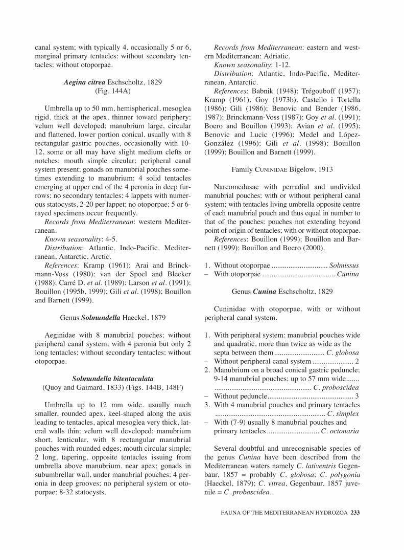
canal system; with typically 4, occasionally 5 or 6,marginal primary tentacles; without secondary ten-tacles; without otoporpae.
Aegina citrea Eschscholtz, 1829(Fig. 144A)
Umbrella up to 50 mm, hemispherical, mesoglearigid, thick at the apex, thinner toward periphery;velum well developed; manubrium large, circularand flattened, lower portion conical, usually with 8rectangular gastric pouches, occasionally with 10-12, some or all may have slight medium clefts ornotches; mouth simple circular; peripheral canalsystem present; gonads on manubrial pouches some-times extending to manubrium; 4 solid tentaclesemerging at upper end of the 4 peronia in deep fur-rows; no secondary tentacles; 4 lappets with numer-ous statocysts, 2-20 per lappet; no otoporpae; 5 or 6-rayed specimens occur frequently.
Records from Mediterranean: western Mediter-ranean.
Known seasonality: 4-5.Distribution: Atlantic, Indo-Pacific, Mediter-
ranean, Antarctic, Arctic.References: Kramp (1961); Arai and Brinck-
mann-Voss (1980); van der Spoel and Bleeker(1988); Carré D. et al. (1989); Larson et al. (1991);Bouillon (1995b, 1999); Gili et al. (1998); Bouillonand Barnett (1999).
Genus Solmundella Haeckel, 1879
Aeginidae with 8 manubrial pouches; withoutperipheral canal system; with 4 peronia but only 2long tentacles; without secondary tentacles; withoutotoporpae.
Solmundella bitentaculata(Quoy and Gaimard, 1833) (Figs. 144B, 148F)
Umbrella up to 12 mm wide, usually muchsmaller, rounded apex, keel-shaped along the axisleading to tentacles, apical mesoglea very thick, lat-eral walls thin; velum well developed; manubriumshort, lenticular, with 8 rectangular manubrialpouches with rounded edges; mouth circular simple;2 long, tapering, opposite tentacles issuing fromumbrella above manubrium, near apex; gonads insubumbrellar wall, under manubrial pouches; 4 per-onia in deep grooves; no peripheral system or oto-porpae; 8-32 statocysts.
Records from Mediterranean: eastern and west-ern Mediterranean; Adriatic.
Known seasonality: 1-12.Distribution: Atlantic, Indo-Pacific, Mediter-
ranean, Antarctic.References: Babnik (1948); Trégouboff (1957);
Kramp (1961); Goy (1973b); Castello i Tortella(1986); Gili (1986); Benovic and Bender (1986,1987); Brinckmann-Voss (1987); Goy et al. (1991);Boero and Bouillon (1993); Avian et al. (1995);Benovic and Lucic (1996); Medel and López-González (1996); Gili et al. (1998); Bouillon(1999); Bouillon and Barnett (1999).
Family CUNINIDAE Bigelow, 1913
Narcomedusae with perradial and undividedmanubrial pouches; with or without peripheral canalsystem; with tentacles living umbrella opposite centreof each manubrial pouch and thus equal in number tothat of the pouches; pouches not extending beyondpoint of origin of tentacles; with or without otoporpae.
References: Bouillon (1999); Bouillon and Bar-nett (1999); Bouillon and Boero (2000).
1. Without otoporpae .............................. Solmissus– With otoporpae ....................................... Cunina
Genus Cunina Eschscholtz, 1829
Cuninidae with otoporpae, with or withoutperipheral canal system.
1. With peripheral system; manubrial pouches wide and quadratic, more than twice as wide as the septa between them........................... C. globosa
– Without peripheral canal system...................... 22. Manubrium on a broad conical gastric peduncle;
9-14 manubrial pouches; up to 57 mm wide........................................................... C. proboscidea
– Without peduncle.............................................. 33. With 4 manubrial pouches and primary tentacles
........................................................... C. simplex– With (7-9) usually 8 manubrial pouches and
primary tentacles ............................ C. octonaria
Several doubtful and unrecognisable species ofthe genus Cunina have been described from theMediterranean waters namely C. lativentris Gegen-baur, 1857 = probably C. globosa; C. polygonia(Haeckel, 1879); C. vitrea, Gegenbaur, 1857 juve-nile = C. proboscidea.
FAUNA OF THE MEDITERRANEAN HYDROZOA 233
sm68s2Csystema2 14/10/04 15:24 Página 233

Cunina globosa Eschscholtz, 1829(Figs. 144C-D)
Umbrella up to 18 mm wide; conical or almostglobular; mesoglea thick; manubrium circular on abroad gastric peduncle; up to 10-14 manubrialpouches, wide and quadratic, more than twice aswide as the septa between them; 10-14 relativelyshort tentacles, issuing from the centre of the basesof the manubrial pouches at a short distance onlyabove the umbrella margin, no ectodermal padsbelow tentacles bases; peripheral canals well devel-oped; marginal lappets short and broad with 3 stato-cysts; otoporpae short and oval.
Records from Mediterranean: western Mediter-ranean.
Known seasonality: 4, 6; 7.Distribution: Atlantic, Indo-Pacific, Mediter-
ranean.References: Kramp (1961); Gili (1986); Bouillon
(1987,1999); Pagès et al. (1992); Boero and Bouil-lon (1993); Medel and López-González (1996); Giliet al. (1998); Bouillon and Barnett (1999).
Cunina octonaria McCrady, 1859(Figs. 144E, 145A, 149A-H)
Umbrella 5-7 mm wide, somewhat flatter than ahemisphere, usually 8 (7-9) square manubrialpouches, very close together; no peripheral canals;tentacles project about midway between margin andapex, a thick and broad ectodermal pad below baseof each tentacle; with 2-5 generally 3 statocysts oneach marginal lappet; otoporpae small; larvae devel-oped in manubrium or attached to the medusae.
Reproduction: development through a secondarylarval structure or a “stolon prolifer”.
Records from Mediterranean: eastern and west-ern Mediterranean.
Known seasonality: 2, 3, 6, 8-12.Distribution: Atlantic; Indo-Pacific; Mediter-
ranean.References: Kramp (1961); Schmidt (1973,
1976); Lakkis and Zeidane (1985); Goy et al. (1988,1990, 1991); Bouillon (1987); Boero and Bouillon(1993).
Cunina proboscidea E. and L. Metschnikoff, 1871(Figs. 145B-C)
Umbrella up to 57 mm wide, somewhat conical;manubrium long, conical, mouth often projecting
beyond velar opening; with a large gastric peduncle;9-14 manubrial pouches, long, rectangular, separat-ed by narrow spaces; peripheral canal system degen-erated; tentacles very short; marginal lappets blunt-ly rounded, each with 3-4 statocysts, otoporpae,short, club-shaped.
Hydroid: Complex development in the manubri-um of the mother medusae and afterwards in themanubrial cavity of Geryonia (Trachymedusae) (seeBouillon, 1987).
Records from Mediterranean: western Mediter-ranean.
Known seasonality: 3, 9.Distribution: Mediterranean.References: Ranson (1936); Kramp (1961);
Bouillon (1987); Boero and Bouillon (1993).
Cunina simplex Gili, Bouillon, Pagès, Palanques, Puig and Heussner, 1998
(Fig. 145D-E)
Umbrella higher than a hemisphere, almost glob-ular, 3.7 mm wide and 2.8 mm high; mesoglea thick,manubrium large, circular with 4 small, perradial,tongue-shaped, undivided manubrial pouches nar-rowing in width from base outwards; septa betweenpouches very wide; with 4 primary tentacles livingumbrella opposite centre of each stomach pouch;with 4 peronia; without secondary tentacles onumbrella margin; gonads on manubrium andmanubrial pouches walls; with a narrow peripheralcanal system; marginal lappets rectangular, largeeach with 3 small, circular, otoporpae and 1-2 stato-cysts per quadrant.
Records from Mediterranean: western Mediter-ranean.
Known seasonality: 4.Distribution: endemic of the Mediterranean Sea.References: Gili et al. (1998).
Genus Solmissus Haeckel, 1879
Cuninidae without otoporpae, without peripheralcanal system.
References: Bouillon (1999); Bouillon and Bar-nett (1999).
1. Exumbrella with numerous gelatinous warts or discoid cnidocysts patches; manubrial pouches pentagonal; umbrella up to 50 mm wide; maximum number of tentacles 16; 5-8 statocysts per marginal lappet ........ S. albescens
234 J. BOUILLON et al.
sm68s2Csystema2 14/10/04 15:24 Página 234

– Exumbrella smooth; manubriual pouches oval; umbrella up to 100 mm wide; number of tentacles 20-40; 2-5 statocysts per marginal lappet..................................................... S. incisa
Solmissus albescens Gegenbaur, 1857(Figs. 146A-B)
Umbrella up to 50 mm in diameter; central partof umbrella doubly convex, lenticular; bell collarthin, flexible, contractile; exumbrella scatteredeither with small but distinct gelatinous warts and/orwith flat discoid or elongated cnidocyst patcheswhich may be found only on the lappets; velumbroad; manubrium large, circular with 14-16 mar-ginal perradial pouches, pentagonal in shape andsomewhat wider than long, their outer angles lyingunder the tentacle roots; with wide mouth opening;with 14 -16 tentacles nearly as long as the umbrelladiameter, tapering and not very flexible; about 14-16marginal lappets, rectangular but with roundedangles on their outer margin, each with 5-8 stato-cysts; gonads developed in the subumbrellar ecto-derm of manubrium and manubrial pouches.
Records from Mediterranean: eastern and west-ern Mediterranean.
Known seasonality: 1-12.Distribution: endemic of the Mediterranean Sea.References: Trégouboff (1957); Kramp (1961);
Berhaut (1970); Goy (1973b); Gili (1986); Benovicand Bender (1987); Goy et al. (1988, 1990, 1991);Mills and Goy (1988); Boero and Bouillon (1993);Avian et al. (1995); Benovic and Lucic (1996);Medel and López-González (1996); Mills et al.(1996); Gili et al. (1998), Osborn (2000).
Solmissus incisa (Fewkes, 1886)(Figs. 146C-D)
Umbrella up to 100 mm wide, flat, disk-like, withthin and flexible margin, mesoglea fairly thick but softand fragile, exumbrella smooth; velum well developed;manubrium large, circular, covering subumbrella sur-face; 20 to 40 perradial manubrial pouches, oval in out-line, usually somewhat longer than wide, septabetween them alternating with tentacle roots; 20-40marginal tentacles, stiff, tapering, up to slightly longerthan diameter of umbrella; no peripheral canal system;gonads?; marginal lappets rectangular, about as long asbroad, each with 2-5 statocysts, no otoporpae.
Records from Mediterranean: western Mediter-ranean
Seasonality: ?Distribution: Atlantic, Indo-Pacific, Mediter-
ranean?.References: Ranson (1936); Russell (1953);
Kramp (1961); Arai and Brinckmann-Voss (1980);Larson et al. (1991); Boero and Bouillon (1993).
Remarks: S. incisa is closely related to S.albescens, Ranson (1936) suggested that S. incisa ismerely a giant form of S. albescens.
Family SOLMARISIDAE Haeckel, 1879
Narcomedusae without manubrial pouches, theperiphery of the manubrium being circular andunbroken; with or without peripheral canal system;gonads on manubrial wall or on manubrial walldiverticula; with numerous tentacles living umbrellaat level of periphery of the manubrium. With orwithout otoporpae.
References: Bouillon (1999); Bouillon and Bar-nett (1999); Bouillon and Boero (2000).
1. With peripheral canal system; with otoporpae................................................................. Pegantha
– Without peripheral canal system; without otoporpae.............................................. Solmaris
Genus Pegantha Haeckel, 1879
Solmarisidae with gonads forming diverticula ofmargin of oral manubrium wall; with peripheralcanal system; with otoporpae.
1. Exumbrella with deep radiating furrows from tentacles to apex, surrounded by ribs and supplementary ridges.......................... P. triloba.
– Exumbrella smooth; peripheral canals narrow throughout their length; with 12-16 marginal lappets ............................................ P. rubignosa
Two other Pegantha species, P. mollicina(Forskäl, 1775) and P. zonaria (Haeckel, 1879) havebeen described from the Mediterranean waters butare considered as unrecognisable species (seeKramp, 1961).
Pegantha rubiginosa (Kölliker, 1853)(Figs. 147A-B)
Umbrella up to about 16 mm wide, dome-shaped, mesoglea very thick, smooth, with 12-16marginal lappets, rectangular with rounded corners,
FAUNA OF THE MEDITERRANEAN HYDROZOA 235
sm68s2Csystema2 14/10/04 15:24 Página 235

each with 4-6 statocysts; the two middle otoporpaelong and narrow, frequently longer than the lappet,the lateral ones shorter; peripheral canals very nar-row throughout their length; gonads without radialdiverticulae.
Development through two sexual generationsand a “stolon prolifer”.
Records from Mediterranean: eastern and west-ern Mediterranean.
Known seasonality: 1-12.Distribution: Atlantic; Indo-Pacific; Mediter-
ranean.References: Kramp (1957b, 1959, 1961); Goy
(1973b); Schmidt (1976); Bouillon (1987); Boeroand Bouillon (1993); Avian et al. (1995); Goy(1997).
Pegantha triloba Haeckel, 1879(Figs. 147C, 148G)
Umbrella up to 30 mm wide, hemispherical;mesoglea very rigid, apex somewhat flattened;exumbrella with deep radiating furrows from ten-tacles to near apex, surrounded by ribs and sup-plementary ridges; velum broad; manubrium cir-cular, broad; mouth simple, circular; 12-16 tenta-cles somewhat longer than umbrella diameter; 12-16 marginal lappets ovate, with sharp pointedend, each with up to 20 statocysts; gonads adher-ing to base of manubrium consisting in centralsacs with variously subdivided lobes projectinginto each lappet cavities; otoporpae long taperingoutwards.
Development through primary larvae with eightcapitate tentacles budding of successive medusabuds and finally metamorphosing himself inmedusae.
Records from Mediterranean: western Mediter-ranean.
Known seasonality: 5.Distribution: Atlantic, Indo-Pacific, Mediter-
ranean.References: Vanhöffen (1913); Kramp (1961);
Schmidt (1973); Winkler (1982); Bouillon (1987);Pagès et al. (1992).
Genus Solmaris Haeckel, 1879
Solmarisidae without peripheral canal system;without otoporpae, with simple annular gonads.
References: Bouillon (1999); Bouillon and Bar-nett (1999).
1. With 6-8 statocysts on each marginal lappet; up to 35 mm wide; with 18-20 tentacles............................................................................ S. solmaris
– With 1-4 statocysts on each marginal lappet ... 22. Umbrella less than 7 mm wide when adult.........
........................................................ S. leucostyla– Umbrella more than 7 mm wide when adult ... 33. Umbrella 12-15 mm wide; with 30-36 or more
tentacles and lappets ........................... S. corona– Umbrella 15-23 mm wide; with 12-17 tentacles
and lappets ..................................... S. flavescens
Solmaris vanhoeffeni Neppi and Stiasny, 1911has been described from the Adriatic Sea, thisspecies is considered by Thiel (1936) as conspecificwith S. flavescens and Kramp (1961) regards it as ajuvenile stage of some other species.
Solmaris corona (Keferstein and Ehlers, 1861)(Figs. 147D-E)
Umbrella 12-15 mm wide, lens-shaped, withthick mesoglea; manubrium circular covering alllower surface of upper half of umbrella; gonadsforming a broad ring on outer part of subumbrellarmanubrium wall; with 30-36 or more marginal ten-tacles and marginal lappets, lappets rectangular upto twice as long as broad, usually with 2 (1-4) stato-cysts mounted on large cushion, with long bristles.
Records from Mediterranean: western Mediter-ranean.
Known seasonality: 7.Distribution: Atlantic; Indo-Pacific; Mediter-
ranean.References: Russell (1953); Trégouboff (1957);
Kramp (1961); Boero and Bouillon (1993).
Solmaris flavescens (Kölliker, 1853)(Figs. 148A-C)
Umbrella 15-23 mm wide, thick, lens-shaped,mesoglea of central umbrellar disk lenticular andquite thick, lateral sides thin; velum large; manubri-um flat and lenticular; gonads in the subumbrellarfloor of the manubrial margin; with 12-17 (usually13-15) long, stiff, marginal tentacles at right anglesto the umbrellar sides and as many marginal lappetsand peronia; marginal lappets thin, quadrate, eachwith 2- 4 statocysts.
Records from Mediterranean: western Mediter-ranean; Adriatic.
Known seasonality: 1-5, 9-12.
236 J. BOUILLON et al.
sm68s2Csystema2 14/10/04 15:24 Página 236

Distribution: Atlantic; Indo-Pacific; Mediter-ranean.
References: Trégouboff (1957); Kramp (1961);Gili (1986); Brinckmann-Voss (1987); Bouillon etal. (1988a); Boero and Bouillon (1993); Avian et al.(1995); Medel and López-González (1996); Goy(1997).
Solmaris leucostyla (Will, 1844)(Fig. 148D)
Umbrella 3-7 mm wide, flat to hemispherical;velum small; 12-26 slender tentacles and as manymarginal lappets; the lappets quadratic, each withusually one statocysts (1-3); perhaps a variety of S.flavescens.
Records from Mediterranean: western Mediter-ranean; Adriatic.
Known seasonality: 1-12.Distribution: endemic of Mediterranean Sea.References: Trégouboff (1957); Kramp (1961);
Berhaut (1970); Goy (1973b); Castelló i Tortella(1986); Gili (1986); Benovic and Bender (1987);Bouillon (1987); Boero and Bouillon (1993); Avianet al. (1995); Medel and López-González (1996);Mills, et al. (1996).
Solmaris solmaris (Gegenbaur, 1857)(Fig. 148E)
Umbrella 25-35 mm wide, flat, lenticular; conca-vo-convex; 18-20 long tentacles; marginal lappetsquadratic, each with 6-8 statocysts
Records from Mediterranean: western and cen-tral Mediterranean.
Known seasonality: 3, 5.Distribution: endemic of Mediterranean Sea.References: Trégouboff (1957); Kramp (1961);
Gili (1986); Boero and Bouillon (1993); Medel andLópez-González (1996).
Subclass TRACHYMEDUSAE Haeckel, 1866
Medusa with hemispherical or deep bell-shaped umbrella. Margin entire with a thickenedperipheral cnidocyst ring. Radial canals and circu-lar canal present. Velum often with heavy muscu-lature. With solid marginal tentacles or with a mix-ture of solid and hollow ones, without true tentac-ular bulbs, with endodermal cores continuing inthe mesoglea of the umbrella as short “roots”.With or without centripetal canals. Manubrium
with or without gastric peduncle. “Gonads” usual-ly on radial canals. Without polyp stage; a differ-entiated planula stage is lacking in a number ofTrachymedusae, the gastrula developing immedi-ately into young medusae, in others, the planulastage is retained and gives rise to a post-embryon-ic tentacled larval stage before transforming intomedusae. No adult or larval asexual buddingobserved. Marginal sense organs as free sensoryclubs exceptionally enclosed in the mesoglea or inthe velum. Cnidome: generally stenoteles associ-ated with microbasic euryteles or/and atrichousisorhizae.
References: Uchida (1928); Bouillon (1999);Bouillon and Barnett (1999); Bouillon and Boero(2000).
1. With centripetal canals ................... Geryoniidae– Without centripetal canals ................................ 22. With broad, circular manubrium and broad
radial canals .................................. Halicreatidae– Manubrium and radial canals narrow..................
.............................................. Rhopalonematidae
Family GERYONIIDAE Eschscholtz, 1829
Trachymedusae with gastric peduncle; 4-6 radialcanals (sometimes more); with centripetal canals;gonads on radial canals, flattened and leaf-shaped; 2kinds of marginal tentacles, solid and hollow; ecto-endodermal statocysts enclosed in mesoglea.
References: Bouillon (1999); Bouillon and Bar-nett (1999); Bouillon and Boero (2000).
1. With 6 radial canals, six gonads, mouth with six lips ....................................................... Geryonia
– Usually with 4 radial canals and gonads (sometimes more), mouth with four lips............................................................................. Liriope
Genus Geryonia Péron and Lesueur, 1810
Geryoniidae with six lips; six radial canals andsix gonads.
Geryonia proboscidalis (Forskäl, 1775)(Figs. 150A-C)
Umbrella 35-80 mm wide, almost hemispherical;manubrium small, on long, conical, gastric pedun-cle; mouth with 6 simple lips; with six radial canals;up to 7 centripetal canals between each radial
FAUNA OF THE MEDITERRANEAN HYDROZOA 237
sm68s2Csystema2 14/10/04 15:24 Página 237

canals; gonads heart-shaped, very broad above; 6long, perradial hollow tentacles with cnidocyst ringsand 6 small, solid interradial tentacles with adaxialcnidocyst clusters; 12 statocysts.
Records from Mediterranean: eastern and west-ern Mediterranean; Adriatic.
Known seasonality: 1-3, 6-8, 11, 12.Distribution: Atlantic, Indo-Pacific, Mediter-
ranean.References: Kramp (1961); Berhaut (1970); Goy
(1973b); Brinckmann-Voss (1987); Goy et al. (1988,1990, 1991); Boero and Bouillon (1993); Avian etal. (1995); Benovic and Lucic (1996); Medel andLópez-González (1996).
Genus Liriope Lesson, 1843
Geryoniidae with four lips; usually four radialcanals and four gonads, sometimes more.
Liriope tetraphylla(Chamisso and Eysenhardt, 1821)
(Figs. 150D-I)
Umbrella 10-30 mm wide, hemispherical,apex somewhat flattened; mesoglea thick, rigid;velum broad; manubrium small, on long, cylin-drical gastric peduncle, longer than umbrellardiameter; mouth with 4 simple lips boarded withcnidocysts; with normally 4 radial canals (some-times more); 1-7 centripetal canals in each quad-rant; with marginal cnidocyst ring; typically 4long hollow perradial tentacles with cnidocystrings and 4 small solid interradial tentacles withadaxial cnidocyst clusters; with gonads variablein shape and size, generally heart-shaped, oneither side of the middle of radial canals; 8 sta-tocysts.
Records from Mediterranean: eastern and west-ern Mediterranean; Adriatic; Black Sea.
Known seasonality: 1-12.Distribution: Atlantic, Indo-Pacific, Mediter-
ranean.References: Kramp (1961); Berhaut (1969,
1970); Goy (1973b); Castelló i Tortella (1986);Gili (1986); Benovic and Bender (1987); Brinck-mann-Voss (1987); Zamponi and Genzano (1988);Scemes and McNamara (1991); Boero and Bouil-lon (1993); Avian et al. (1995); Benovic andLucic (1996); Desouza et al. (1996); Medel andLópez-González (1996); Mills et al. (1996); Goy(1997).
Family HALICREATIDAE Fewkes, 1886
Trachymedusae with wide, circular manubrium;mouth circular, without distinct lips; without pedun-cle; without centripetal canals; with 8 or more broadradial canals; with numerous marginal tentacles ofdifferent size, but all structurally alike and arrangedin single series; each marginal tentacle with flexibleproximal portion and stiff spine-like distal portion;with free ecto-endodermal statocysts.
References: Bouillon (1999); Bouillon and Bar-nett (1999); Bouillon and Boero (2000).
1. With about 16 or more radial canals ............................................................................ Halitrephes
– With 8 radial canals ............................ Haliscera
Genus Haliscera Vanhöffen, 1902
Halicreatidae with 8 radial canals; with a contin-uous row of marginal tentacles; without papillae onthe exumbrella.
1. Umbrella hemispherical, with evenly rounded apex and fairly thin walls; gonads close to manubrium; six tentacles in each octant .................................................................. H. racovitzae.
– Umbrella with very thick dome-shaped apex; gonads well separated from manubrium, eight to twelve tentacles in each octant......................... 2
2. Umbrella with distinct conical apical projection; 64-72 marginal tentacles in adult ....... H. conica
– Umbrella with very thick, hemispherical apex, about 96 marginal tentacles in adult ............................................................................ H. bigelowi
Haliscera bigelowi Kramp, 1947(Figs. 151A-B)
Umbrella 15-19 mm wide, 9-10 mm high,almost hemispherical, umbrella with a very thickdome-shaped mesogleal apex comprising about2/3 of the umbrella height, mesoglea thin at sidesand margin; velum very broad; manubriumbroad, flat, slightly conical; mouth simple, circu-lar; 8 straight, broad radial canals and broad cir-cular canal; in adults, about 12 solid marginaltentacles in each octant, flexile proximally, stiffdistally; the base of each tentacles surrounded bya small thickening of marginal cnidocyst tissue;with 8 gonads broadly oval, flat, about 2/5 aslong as the radial canals, situated slightly nearer
238 J. BOUILLON et al.
sm68s2Csystema2 14/10/04 15:24 Página 238

the manubrium than to bell margin; 3 statocystsin each octant.
Records from Mediterranean: western Mediter-ranean; Adriatic.
Known seasonality: 1, 5, 7, 12.Distribution: Atlantic; Indo-Pacific, Mediter-
ranean; Arctic.References: Kramp (1961); Naumov (1960-
1969); Schmidt and Benovic (1977); Gili (1986);Boero and Bouillon (1993); Avian et al. (1995);Medel and López-González (1996); Gili et al.(1998).
Haliscera conica Vanhöffen, 1902(Fig. 151C)
Umbrella up to 18 mm wide, with thick, bluntlyconical apical projection; gonads oval on the middleportion of the eight broad radial canals; 8-9 margin-al tentacles, flexile proximally and stiff distally and2 statocysts in each octant, the base of each tentaclesurrounded by a broad thickening of the marginalcnidocyst tissue.
Records from Mediterranean: western Mediter-ranean.
Known seasonality: 2, 4-10.Distribution: Antarctic; Atlantic; Indo-Pacific;
Mediterranean.References: Kramp (1961); Goy (1973b); Dallot,
Goy and Carré (1988); Boero and Bouillon (1993),Mills et al, (1996); Goy (1997); Gili et al. (1998).
Haliscera racovitzae (Maas, 1906)(Figs. 151D-E)
Umbrella 8 mm wide, 4 mm high, almosthemispherical; with fairly thin walls, mesogleathin, flaccid, apex evenly rounded; velum verywide; manubrium a truncated cone, broad, flat;mouth simple, circular; 8 radial canals, narrowdistally, circular canal fairly narrow; gonads flat,shield-shaped, along 1/2- 2/5 of proximal part ofthe radial canals, very close to manubrium; with6 solid tentacles, flexile proximally and stiff dis-tally in each octant; with 2 statocysts in eachoctant.
Records from Mediterranean: western Mediter-ranean.
Known seasonality: 9.Distribution: Atlantic, Indo-Pacific, Antarctic,
Mediterranean.References: Kramp (1961); Gili et al. (1998).
Genus Halitrephes Bigelow, 1909
Halicreatidae with 16 or more radial canals; witha continuous row of tentacles; without papillae onexumbrella.
Halitrephes maasi Bigelow, 1909(Fig. 151F)
Umbrella up to 100 mm wide, low, rounded,smooth, without exumbrella papillae; mesoglea thin,soft and flaccid; velum well developed; manubriumsmall with simple circular mouth; 16-30 broad, ribbon-like radial canals, some may be bifurcated, circularcanal broad; 100-300 marginal tentacles, flexile proxi-mally and stiff distally in continuous row; shape of adultgonads unknown; number of statocysts unknown.
Records from Mediterranean: eastern Mediter-ranean.
Known seasonality: 3.Distribution: Atlantic, Indo-Pacific, Antarctic,
Mediterranean.References: Kramp (1961); Goy et al, (1991);
Gili et al. (1998).
Family PETASIDAE Haeckel, 1879
Trachymedusae with four radial canals; withoutpeduncle and centripetal canals; with well devel-oped manubrium; with 4 sac-like gonads on radialcanals; with marginal tentacles not in clusters, solid,with a terminal club-shaped knob of cnidocysts;with free statocysts.
References: Bouillon (1999); Bouillon and Bar-nett (1999); Bouillon and Boero (2000).
Genus Petasus Haeckel, 1879
Petasidae with marginal tentacles regularlyarranged, at equal intervals.
Petasus atavus Haeckel, 1879(Fig. 151G)
Umbrella 1 mm wide and high, globular;manubrium prismatic; mouth with 4 short lips; with4 protrusive, spindle-shaped or band-shaped gonads,along greater part of the radial canals; four marginaltentacles with ciliated club–shaped end; four freestatocysts.
Records from Mediterranean: eastern Mediter-ranean.
FAUNA OF THE MEDITERRANEAN HYDROZOA 239
sm68s2Csystema2 14/10/04 15:24 Página 239

Known seasonality: 4.Distribution: Atlantic, Mediterranean.References: Kramp (1959a, 1961); Mayer
(1910).
Family PTYCHOGASTRIIDAE Mayer, 1910
Trachymedusae with either simple manubriumwithout mesenteries, or with eight-lobed manubri-um, with eight mesenterial partitions; with marginaltentacles grouped into more or less well definedclusters, some with adhesive disks or with verynumerous tentacles, not in clusters but inserted atvarious levels of exumbrella; no centripetal canalsor peduncle; with 8 radial canals; gonads eitherattached onto manubrium, on sides of 8 manubriallobes, or on radial canals adjacent to manubrial lobe;free ecto-endodermal statocysts.
References: Bouillon and Boero (2000).
Genus Ptychogastria Allman, 1878
Ptychogastriidae with marginal tentacle in clus-ters, some with adhesive disks; manubrium with lat-eral lobes; with eight mesenterial partitions; gonadson the sides of the manubrial lobes or on radialcanals adjacent to manubrial lobes.
Ptychogastria asteroides (Haeckel, 1879)(Figs. 152A-C)
Umbrella 4-5 mm wide, hemispherical to bell-shaped, with fairly thin mesoglea and a small round-ed apical projection; exumbrella with 16 raised radi-ating ridges extending from centre of exumbrella tomargin where they end in 16 conical marginal lobesbeset with a few cnidocysts and numerous variedinclusions; manubrium eight-lobed; with eightmesenteries; mouth with four simple lips, rich incnidocysts and granular gland cells; with 8 radialcanal; with 8 egg-shaped gonads on proximal 1/3 ofradial canals adjacent to manubrium and linked tohim by the mesenterial septa; with 16 isolated solidtentacles armed with rings of cnidocysts and 200 to260 solid tentacles, most of them with adhesiveorgans, and few scattered cnidocysts, in 16 clustersarising from the marginal lobes, velum thick, pow-erful; 16 free statocysts.
Records from Mediterranean: western Mediter-ranean, Adriatic.
Known seasonality: 1-12.Distribution: endemic of the Mediterranean Sea.
References: Kramp (1961); Picard (1955a);Brinckmann-Voss (1987); Boero and Bouillon(1993); Avian et al. (1995); Gili et al. (1998, 1999).
Family RHOPALONEMATIDAE Russell, 1953
Trachymedusae with a narrow manubrium; withor without peduncle; without centripetal canals; usu-ally 8, rarely more, narrow radial canals; mouth withdistinct lips; with marginal tentacles evenly distrib-uted, sometimes of two kinds, each marginal tenta-cle of uniform structure throughout or with proximalportion differing from distal one; with gonads eitheron radial canals globular, linear, or hanging inpouches into subumbrellar cavity, or forming a con-tinuous ring around base of manubrium and extend-ing outwards along radial canals; with free, rarelyenclosed ecto-endodermal statocysts.
References: Bouillon (1999); Bouillon and Bar-nett (1999); Bouillon and Boero (2000).
1. Gonads in a continuous band around manubrium extending outwards on radial canals ..........................................................................Homoeonema
– Gonads isolated, on radial canal, sometimes adjacent to manubrium..................................... 2
2. Without gastric peduncle.................................. 3– With gastric peduncle....................................... 73. With 4 gonads only, pendent; 4 large and 24
small marginal tentacles ..................... Tetrorchis– With 8 (rarely more) gonads ............................ 44. With two kinds of marginal tentacles; with
enclosed statocysts ....................... Rhopalonema– With tentacles all of one kind; with free
club-shaped statocysts ...................................... 55. onads adjacent to manubrium (sometimes also 8
gonads free from manubrium); very numerous tentacles......................................... Arctapodema
– Gonads separated from manubrium ................. 66. Gonads globular, distal, contiguous to circular
canal; with 8 tentacles ......................... Sminthea– Gonads linear, with 48 or more marginal
tentacles......................................... Pantachogon7. Gastric peduncle short conical (in young
specimens almost invisible); gonads attached on subumbrellar portions of radial canals ............. 8
– Gastric peduncle long, slender; gonads attached to peduncular portions of radial canals ............ 9
8. With only two pendent gonads ................. Persa– With 8 gonads ................................. Amphogona9. Gonads linear on peduncle only.......... Ransonia– Gonads sausage-shaped, pendent ................... 10
240 J. BOUILLON et al.
sm68s2Csystema2 14/10/04 15:24 Página 240

10.Gonads attached to peduncle ................ Aglaura– Gonads attached to subumbrellar portions of
radial canals ......................................... Aglantha
Genus Aglantha Haeckel, 1879
Rhopalonematidae with a long and slender gas-tric peduncle; with eight pendent sausage-shapedgonads on subumbrellar portions of the eight radialcanals; with numerous tentacles all alike; with freeclub shaped marginal statocysts.
1. Gonads on radial canals close to base of peduncle, 8 statocysts ........................ A. digitale
– Gonads on radial canals about midway between peduncle and umbrela margin, 16 statocysts ..................................................................... A. elata
Aglantha digitale (O.F.Müller, 1766)(Figs. 152D-E)
Umbrella cylindrical, 10-40 mm high, abouttwice high than wide, with a small conical projec-tion, lateral mesogleal walls thin, subumbrellar mus-cles strong; peduncle slender, long, conical, almostas long as subumbrellar cavity; manubrium small;mouth with four simple lips, eight, long, sausage-shaped gonads, arising from the radial canals nearthe apex of the subumbrella and hanging freely insubumbrellar cavity; 80 or more solid marginal ten-tacles with a core of single endodermal chordalcells; 8 free statocysts.
Records from Mediterranean: western Mediter-ranean.
Known seasonality: 4Distribution: Atlantic; Indo-Pacific; Mediter-
ranean.References: Kramp (1924, 1959a); Russell
(1953); Gili (1986).Remarks: Kramp (1924) described this species
from the middle of the Strait of Gibraltar and esti-mated that this species was attempting to enter in theMediterranean where this species had howevernever been found (see Kramp, 1959a, 1961; Goy1972); Gili in 1986 collected 2 specimens in front ofBarcelona.
Aglantha elata (Haeckel, 1879)(Fig. 152F)
Umbrella 10-12 mm high, 3-4 mm wide, narrow,elongated, with pointed apical projection; gastric
peduncle cylindrical about half as long as bell cavi-ty; manubrium small, 1/8 as long as gastric pedun-cle; four triangular lips; eight gonads about as longas gastric peduncle, attached to about middle pointsof subumbrellar parts of radial canals; 40-48 mar-ginal tentacles; 16 free statocysts.
Records from Mediterranean: Adriatic Sea.Known seasonality: 8.Distribution: Atlantic; Pacific; Mediterranean.References: Kramp (1959a); Schmidt and Ben-
ovic (1977).
Genus Aglaura Péron and Lesueur, 1810
Rhopalonematidae with slender gastric peduncle;with 8 sausage-shaped gonads attached on peduncle,not on subumbrella; with numerous tentacles allalike, with free club-shaped statocysts.
Aglaura hemistoma Péron and Lesueur, 1810(Fig. 152G)
Umbrella 4-6 mm high, 3-4 mm wide; with ver-tical, parallel walls, with very narrow longitudinalridges, with flattened apex, mesoglea exceedinglythin; margin almost octagonal in cross section;velum extremely broad, usually hanging down-wards; gastric peduncle, conical, somewhat shorterthan subumbrellar radius; manubrium small, flask-shaped; mouth with four small, simple, projectinglips; 8 narrow radial canals and narrow circularcanal; gonads sausage-shaped attached on thepeduncle at the place of juncture of the radial canalswith the manubrium; 48-85 marginal tentacles allalike; 8 statocysts.
Records from Mediterranean: eastern and west-ern Mediterranean; Adriatic Sea.
Known seasonality: 1-12.Distribution: Atlantic, Indo-Pacific; Mediter-
ranean.References: Kramp (1961); Berhaut (1970);
Albertini-Berhaut (1971b); Goy (1973b); Castelló iTortella (1986); Gili (1986); Benovic and Bender(1987); Brinckmann-Voss (1987); Goy et al. (1988,1990, 1991); Boero and Bouillon (1993); Avian etal. (1995); Benovic and Lucic (1996); Medel andLópez-González (1996).
Genus Amphogona Browne, 1905
Rhopalonematidae with short, conical gastricpeduncle, exumbrella smooth; with ellipsoidal or
FAUNA OF THE MEDITERRANEAN HYDROZOA 241
sm68s2Csystema2 14/10/04 15:24 Página 241

sac-shaped, pendant gonads on the 8 radial canals,gonads usually of unequal size; with tentacles allalike, not densely crowded; with free club-shapedstatocysts.
Amphogona pusilla Hartlaub, 1909(Fig. 152H)
Umbrella 1.5- 6 mm wide, nearly hemispherical,with thin mesoglea; manubrium small, short; withshort conical gastric peduncle; mouth with four sim-ple lips; 8 simple radial canals; 8 gonads, sphericalto elongated, on distal part of the radial canals, liv-ing 1/4 of the length of radial canal near circularcanal free; 16 marginal tentacles with basal enlarge-ments, statocysts unknown.
Records from Mediterranean: western Mediter-ranean.
Known seasonality: 4.Distribution: Indo-Pacific; Mediterranean.References: Kramp (1961, 1965); Goy (1973b);
Boero and Bouillon (1993).
Genus Arctapodema Dall, 1907
Rhopalonematidae without gastric peduncle;with gonads on radial canals adjacent to manubrium;with 8 narrow radial canals; numerous tentacles, allalike, in a single row; free statocysts.
1. Manubrium with 16 radial folds; gonads pendent on radial canals, near base of manubrium.................................................................... A. australis
– Manubrium with 8 radial lobes; gonads extending from radial lobes of the manubrium outwards on proximal parts of radial canals, sometimes also pairs of small gonads on radial canals separated from manubrium....... A. ampla
Mills et al, (1996) found in the Alborán Sea anArctapodema sp. that they could not assign to eitherof the above-cited species.
Arctapodema ampla (Vanhöffen, 1902)(Figs. 152I-J)
Umbrella up to 15 mm wide, somewhat flatterthan a hemisphere, mesoglea thin; manubrium short,urn-shaped, with 8 radial lobes; mouth with four sim-ple lips; 8 simple radial canals; with 8 swollen gonadsof unequal size, some divided in two halves, extend-ing from radial lobes of manubrium outwards onto
proximal parts of radial canals; sometimes also 8pairs of small gonads on radial canals, separated frommanubrium; about 100 tentacles; 4-8 statocysts.
Records from Mediterranean: western Mediter-ranean, Adriatic.
Known seasonality: 3, 5, 7, 9-11.Distribution: Antarctic; Southern and tropical
Atlantic; Mediterranean.References: Kramp (1961); Goy (1973b); Boero
and Bouillon (1993); Avian et al. (1995).
Arctapodema australis (Vanhöffen, 1912)(Figs. 153A-C)
Umbrella up to 23 mm wide and 14 mm high,mesoglea thin; manubrium short and broad, with 16radial folds; mouth with four simple lips; 8 gonadsglobular or club-shaped, pendent, on the radialcanals near base of the manubrium; about 112 mar-ginal tentacles; statocysts unknown; manubrium redin adults.
Records from Mediterranean: western Mediter-ranean; Adriatic Sea.
Known seasonality: 4, 7-10.Distribution: Antarctic; Indo-Pacific; Mediter-
ranean.References: Kramp (1961); Schmidt and Benovic
(1977); Benovic and Bender (1987); Benovic andLucic (1996); Gili et al. (1998).
Genus Homoeonema (Maas, 1893). Browne, 1903
Rhopalonematidae without gastric peduncle;gonads forming a continuous band around base ofmanubrium and extending outwards along proximalhalf of 8 radial canals; numerous tentacles, all alike;vesicular statocysts.
Homoeonema platygonon Browne, 1903(Figs. 153D-G)
Umbrella up to 2 mm high, bell shaped, higherthan wide, with round or more rarely conical apexmesoglea thin, apical part containing numerous fatdroplets and irregular inclusions (crystals); nopeduncle; manubrium short, flat, quadratic; mouthwith 4 simple, short, recurved lips, the tips armedwith stenoteles; no apical process; up to 80 close-ly crowded marginal tentacles, issued from anexumbrellar marginal ring rich in cnidocysts andirregular inclusions; base of tentacles enlarged,
242 J. BOUILLON et al.
sm68s2Csystema2 14/10/04 15:24 Página 242

containing cnidocysts and inclusions, givingrather abruptly rise to the proximal parts of thetentacles which are straight, devoid of cnidocystsand have their endodermal core built by largecubical chordal cells (perhaps flexible), distalparts of the tentacles spirally coiled, crowded withcnidocysts, endodermal core formed by flattened,disk-shaped, chordal cells; no cirri or non tentacu-lar bulbs; eight fairly wide radial canals, full ofirregular inclusions, circular canal not visibleincluded in the marginal cnidocyst ring; velumvery large; gonads, in adults 8 elongated massesalong the 1/2 or 2/3 of the proximal part of theradial canals, their most proximal ends being fusedand forming a small ring surrounding the base ofthe manubrium, in young specimens the gonadsappear first as oval masses on the proximal thirdof the radial canals and then slowly extend down-wards and upwards, they finally become confluentin the upper interradial parts and encircle themanubrial base; vesicular statocysts.
Records from Mediterranean: western Mediter-ranean; Adriatic.
Known seasonality: 2, 4, 5, 7, 8, 10-12.Distribution: Atlantic; Mediterranean; Arctic.References: Kramp (1947, 1959, 1961); Boero
and Bouillon (1993); Avian et al. (1995); Benovicand Lucic (1996); Gili et al. (1998).
Genus Pantachogon Maas, 1893
Rhopalonematidae without gastric peduncle;with the apical outlines of the subumbrellar muscu-lar fields forming an entire circle; with gonads onthe 8 radial canals separated from manubrium; with48 or more tentacles all alike; free club-shaped mar-ginal statocysts.
1. Umbrella mitre-shaped, with gelatinous apical projection; 48 marginal tentacles....... P. militare
– Umbrella without apical projection; 64 marginal tentacles............................................. P. haeckeli
Pantachogon haeckeli Maas, 1893(Figs. 153H-J, 154A)
Umbrella about 20 mm wide and high, bell-shaped, sometimes wider than high, without apicalprojection, mesoglea fairly thin; umbrella with verystrong and conspicuous musculature, forming anentire circle; large specimens with 32 fine exum-brellar longitudinal meridional furrows; velum very
broad; manubrium small, varying in length, octago-nal at base; mouth with 4 simple, small, pointed lips;no gastric peduncle; 8 narrow, straight radial canals,circular canal narrow; gonads initially forming dis-continuous linear swellings along distal 2/3 oralmost whole length of the radial canals, whicheventually coalesce and become folded transversal-ly; 64 marginal tentacles all alike in one row; 64 freeclub-shaped statocysts.
Records from Mediterranean: eastern Mediter-ranean.
Known seasonality: 1, 3, 9, 11, 12.Distribution: Atlantic, Indo-Pacific, Antarctic,
Arctic, Mediterranean.References: Kramp (1961); Dowidar and El-
Maghraby (1970); Arai and Brinckmann-Voss(1980); Winkler (1982); Dowidar (1983); Goy et al.(1988, 1990, 1991); Boero and Bouillon (1993); Giliet al. (1998).
Pantachogon militare (Maas, 1893)(Fig. 154B)
Umbrella 7-10 mm wide, 6 mm high; mitre-shaped, with a well developed apical projection;gonads lancet-shaped, on distal half of 8 radialcanals; 48 tentacles all alike; 4 (or more) statocysts.
Records from Mediterranean: western Mediter-ranean.
Seasonality: ?Distribution: Atlantic; Mediterranean; Antarc-
tic?.References: Kramp (1959a, 1961); Boero and
Bouillon (1993).
Genus Persa McCrady, 1859
Rhopalonematidae with a short gastric peduncle;with only two oval or sausage-shaped gonads, pen-dant, near middle point of the subumbrellar portionsof two opposite radial canals; 8 radial canals; withnumerous long tentacles, all alike, each with a ter-minal knob; with free club-shaped statocysts.
Persa incolorata McCrady, 1859(Figs. 154C-D)
Umbrella 3 mm wide, 4 mm high, with or with-out a small apical projection, mesoglea very thin;velum broad; with a very retractile gastric peduncle;manubrium tubular, elongated; mouth with foursmall, broadly rounded, prominent lips; 8 narrow
FAUNA OF THE MEDITERRANEAN HYDROZOA 243
sm68s2Csystema2 14/10/04 15:24 Página 243

radial canals; with only two oval or sausage-shapedgonads, pendant, near middle point of the subum-brellar portions of two opposite radial canals; up to48 marginal tentacles all alike, each with a terminalknob; 8 club-shaped statocysts.
Records from Mediterranean: eastern and west-ern Mediterranean; Adriatic.
Known seasonality: 1-12.Distribution: Atlantic; Indo-Pacific; Mediter-
ranean.References: Kramp (1961); Berhaut (1970);
Albertini-Berhaut (1971b); Goy (1973b); Castelló iTortella (1986); Gili (1986); Benovic and Bender(1987); Goy (1987); Goy et al. (1988, 1990, 1991);Pagès et al. (1992); Boero et al, (1993); Avian et al.(1995); Benovic and Lucic (1996); Medel andLópez-González (1996); Gili et al. (1998).
Genus Ransonia Kramp, 1947
Rhopalonematidae with high conical umbrella(similar to Aglantha); with long and narrow gastricpeduncle; 8 radial canals; linear, discontinuous,gonads along peduncular portions of radial canals,not on subumbrella; numerous tentacles, all alike;statocysts unknown.
Ransonia krampi (Ranson, 1932)(Fig. 154E-G)
Umbrella 8 mm wide, 15 mm high, conical, withthin walls, with small apical projection; gonadsmore or less discontinuous along the eight radialcanals on the long, narrow peduncle; about 88 mar-ginal tentacles.
Records from Mediterranean: western Mediter-ranean.
Known seasonality: 4.Distribution: Atlantic; Mediterranean.References: Kramp (1959a, 1961); Gili (1986);
Boero and Bouillon (1993); Medel and López-González, 1996; Mills et al. (1996).
Genus Rhopalonema Gegenbaur, 1857
Rhopalonematidae without gastric peduncle;with gonads along the radial canals separated frommanubium; with marginal tentacles of two kinds:large, club-shaped, perradial tentacles with swollenends and inter-and adradial short, stiff, cirri-like ten-tacles also with swollen ends; with enclosed stato-cysts.
1. Umbrella with a distinct apical knob; gonads oval, in the middle third of radial canals; statocysts close beside tentacles........ R. velatum
– Umbrella without apical knob; gonads along distal 2/3 of radial canals; statocysts in the middle of spaces between tentacles ......................................................................... R. funerarium
Rhopalonema funerarium Vanhöffen, 1902(Figs. 155A-D)
Umbrella up to 17 mm wide and 14 mm high,hemispherical to somewhat conical but without api-cal projection, mesoglea stiff, but fairly thin; no gas-tric peduncle; velum very broad; manubrium nar-row, elongated, quadrilateral with octagonal base,contractile, hardly reaching velar opening; mouthwith 4 simple lips; 8 narrow straight radial canals,narrow circular canal; gonads as elongated, linearpouches, extending along distal 2/3 of the radialcanals; 8 large radial marginal tentacles withswollen ends, 3 very short, club-shaped cirrus-liketentacles in each octant, 32 enclosed statocysts inthe middle of the spaces between tentacles.
Remarks: Rhopalonema funerarium is consid-ered by several authors as a deep-water race of R.velatum. However, it is a distinct species for Kramp(1961; 1965; 1968). In fact, most of the charactersused to distinguish between the two presumedspecies (form of the umbrella, development of thegonads) appears to be nothing more than intraspe-cific variations, perhaps the position of the stato-cysts is the only valid character allowing the separa-tion of the two forms.
Records from Mediterranean: eastern and west-ern Mediterranean,Adriatic.
Known seasonality: 1-3, 6, 9, 10.Distribution: Atlantic, Indo-Pacific, Mediter-
ranean.References: Kramp (1961); Dowidar (1983); Gili
(1986); Benovic and Lucic (1996); Medel andLópez-González (1996); Gili et al. (1998).
Rhopalonema velatum Gegenbaur, 1857
Umbrella 8-10 mm wide, 6.6 mm high, somewhatflatter than a hemisphere, with a solid conical ordome-like apical thickening, mesoglea stiff, but fairlythin, except at apex; velum very broad almost closingumbrella cavity; manubrium narrow, elongated,cylindrical, with octagonal base, contractile, reachingextended almost velar opening; no gastric peduncle;
244 J. BOUILLON et al.
sm68s2Csystema2 14/10/04 15:24 Página 244

mouth with 4 simple or somewhat elongated lips,often strongly recurved; 8 straight radial canals andcircular canal narrow; gonads linear or oval on mid-dle third of the 8 radial canals; 8 large radial margin-al tentacles, 1-3 short cirrus-like tentacles in eachoctant, all tentacles with swollen ends; an enclosedstatocyst close besides each right side of the perradi-al tentacles and interradial cirrus-like tentacles.
Records from Mediterranean: eastern and west-ern Mediterranean; Adriatic Sea; Black Sea.
Known seasonality: 1-12 (see Albertini-Berhaut,1971a).
Distribution: Atlantic; Indo-Pacific; Antarctic;Mediterranean.
References: Kramp (1961); Albertini-Berhaut(1970a); Goy (1973b); Castello i Tortella (1986);Gili (1986); Benovic and Bender (1987); Brinck-mann-Voss (1987); Goy et al. (1988, 1990, 1991);Boero and Bouillon (1993); Avian et al. (1995);Benovic and Lucic (1996); Medel and López-González (1996); Gili et al. (1998).
Genus Sminthea Gegenbaur, 1857
Rhopalonematidae without gastric peduncle;with globular gonads on very distal parts of the 8radial canals; with only 8 perradial tentacles; withenclosed statocysts.
Sminthea eurygaster Gegenbaur, 1857(Figs. 155G-I)
Umbrella up to 6 mm wide and about half ashigh, umbrella-shaped, with a small apical projec-tion, mesoglea stiff, fairly thin; velum well devel-oped; manubrium short, cylindrical; mouth with 4short simple lips; no gastric peduncle; 8 straightradial canals, circular canal narrow; 8 perradial mar-ginal tentacles; gonads globular to egg-shaped onradial canals very close to circular canal; 8 interra-dial enclosed statocysts.
Records from Mediterranean: eastern and west-ern Mediterranean; Adriatic.
Known seasonality: 1, 5-9.Distribution: Atlantic; Indo-Pacific; Antarctic;
Mediterranean.References: Kramp (1961); Vannucci (1966);
Berhaut (1970); Goy (1973b); Gili (1986); Benovicand Bender (1986, 1987); Goy et al. (1988, 1990,1991); Boero and Bouillon (1993); Avian et al.(1995); Benovic and Lucic (1996); Medel andLópez-González (1996); Gili et al. (1998).
Genus Tetrorchis Bigelow, 1909
Rhopalonematidae without gastric peduncle;with only 4 pendant sausage-shaped gonadsattached to 4 of the 8 radial canals near the middlepoints; with 4 large perradial and several small mar-ginal tentacles.
Tetrorchis erythrogaster Bigelow, 1909(Figs. 155J-L)
Umbrella 10-12 mm wide and 8 mm high, pyri-form, apex mesoglea very thick, lateral mesogleathin; velum well developed; no gastric peduncle;manubrium tubular, brilliant carmine, reachingslightly beyond velar opening; mouth with 4 small,simple lips; 8 straight radial canals, circular canalnarrow; 4 sausage-shaped gonads attached to everysecond radial canals, middle to distal in position; 4large perradial tentacles opposite fertile radial canalsand 16-24 small tentacles not placed in reference tothe radial canals; statocysts unknown.
Records from Mediterranean: eastern Mediter-ranean.
Known seasonality: 10-12.Distribution: Atlantic; Indo-Pacific; Mediter-
ranean.References: Bigelow (1909); Kramp (1961);
Lakkis and Zeidane (1985); Goy et al. (1988, 1990,1991); Boero and Bouillon (1993); Gili et al. (1998).
Class POLYPODIOZAE
Family POLYPODIIDAE Poche, 1914.
See characters of subclass
Genus Polypodium Ussow, 1887
See characters of subclass.
Polypodium hydriforme Ussow, 1885(Fig. 156)
See characters of subclass, free stage 2-5 mm insize.
Records from Mediterranean: Fresh water basinsof the Azov and Black Sea.
Known seasonality: Free living stage: 5-7.Distribution: Fresh-water Basins of Russia; Rou-
mania; Iran, North America.References: Raikova (1958, 1973, 1980, 1988,
FAUNA OF THE MEDITERRANEAN HYDROZOA 245
sm68s2Csystema2 14/10/04 15:24 Página 245

1994); Suppes and Meyer (1975); Dick et al. (1991).Monteiro, Okamura and Holland (2002); Okamuraet al. (2002); Zrzavy (2001); Zrzavy and Hypsa(2003).
COLLECTION OF THE MATERIAL
Hydroids
Benthic hydroids have been traditionally col-lected with dredges and grabs, from ordinaryboats or research vessels. This practice has led tothe collection of a host of large colonies of Sertu-lariidae, Aglaopheniidae, Syntheciidae, Sertulari-idae, these being evident during sorting. The iden-tification of inconspicuous hydroids, either aliveor preserved, necessitates careful sorting of mate-rial, inspecting each substrate fragment while inliquid. Many hydroids are commonly associatedwith algae, sea grasses, sponges, other hydroids,anthozoans, bryozoans, molluscs (bivalve, gas-tropods, pteropods), annelids, crustaceans, ascidi-ans, fishes, and must be searched for under astereomicroscope. Hydroids are common inhabi-tants of shaded hard substrates. Intertidal speci-mens can be collected easily at low tides, where-as subtidal specimens are best collected bySCUBA diving, since the crevices and small cav-ities they prefer are difficult to sample from thesurface. While collecting, it is important to beaware of the substrates that are possibly con-ducive to hydroid settlement. These must be col-lected even if no specimens are evident. Kept inthe laboratory, in calm water, these substratesmight reveal fruitful catches.
Deep and soft-bottom species must be collect-ed from boats, with either dredges or grabs. Inshallow water, however, soft bottom hydroids canbe seen while SCUBA diving, since some speciesare rather conspicuous. Interstitial species are col-lected by the standard techniques of meiobenthicresearch.
Many hydroids are not recognisable if not fertile,and information on complete life cycle is necessaryfor an accurate identification. If at a specific site anhydroid without gonophores is located, it is advis-able to collect specimens periodically (at least onceper month) until ripe colonies are found These mustbe kept in the laboratory for gonophore rearing.Even groups that are usually thought to be com-pletely paedomorphic, and so deprived of medusae,
have recently been found to produce swimminggonophores. Far too many species and genera haveunknown hydroids or medusa stages, so that lifecycle elucidation is still a priority for the study ofthis group.
Medusae
Gelatinous plankton is very fragile and easilytorn and damaged when collected.
Hydromedusae can be caught by plankton netsthat are very slowly towed behind a large or asmall powered vessel or a even a row boat forabout ten to twenty minutes depending on theabundance of the plankton. For hydromedusae themesh size of the net should be about 200-250 µm,larger meshes let escape many small specimens,smaller meshes are to easily clogged and damagethe specimens. In coastal waters the opening of thenet should be from 30 cm to 1 m depending thepower of the vessel, much wider openings are usedin open sea and deep waters where the fauna issparser. In areas very rich in plankton, a hand netor even a bucket may be used. The richest catchesare generally obtained in the early morning or atdusk and on rising tides. For qualitative horizontalsubsurface sampling the plankton net is towed bya rope of 50 m or more behind the vessel to elim-inate turbulence. For sampling in few metersbelow the surface a buoy can be attached with arope of known length to one side of the ring open-ing and a weight on the other side. Samplingbetween fixed depths requires closing nets. Quan-titative sampling is possible with plankton nets fit-ted with a flow-meter a little behind their front,after calibration these water-meters give a mea-sure of the quantity of water filtered.
If it is impossible to sort the medusae immedi-ately, the catch should as quickly as possible befixed at once with formaldehyde, so to obtain afinal solution of 5% fixative (see fixation). If thematerial can be brought rapidly to the laboratory,the plankton samples, shielded from direct sun-light and kept as cool as possible should be exam-ined under stereo-microscope and the medusaeindividually removed with wide-mouthed pipettesand placed in finger bowls of clear sea water. Afterobservation they can either be kept for rearing oranaesthetised and fixed as described below. Thislast method is of course the most rewarding,allowing observation of the living animals charac-ters and perfect fixation.
246 J. BOUILLON et al.
sm68s2Csystema2 14/10/04 15:24 Página 246

TECHNIQUES
Fixation and preservation of the material
Hydroids
Colonies are usually preserved directly either informaldehyde or alcohol. This practice usually leadsto specimens with poorly preserved coenosarc,skeletal parts being the only well preserved items.This is the reason why the material coming from“expeditions” is very rich in large thecate colonies,whereas athecate and delicate forms are apparentlyabsent. In order to have properly preserved hydroidmaterial, the same techniques employed for themedusae should be used. Modern molecular tech-niques require fixation in alcohol, since DNAprobes are impossible in formalin-preserved materi-al. When possible, thus, a batch of specimens shouldbe preserved in alcohol. Whole mounts, obtained bydehydration of specimens and mount in microscopeslides, is very advisable. Mounted specimens can bestudied also after centuries, whereas those preservedin liquid tend to disintegrate. This is very importantfor the preservation of type material.
Medusae
Hydromedusae should be anaesthetised beforebeing fixed; most of the fixatives cause shrinkingand deformations. The animals should be allowed toextend in a vessel of water where the anaestheticsubstance should be added slowly, crystal by crystalor drop by drop. The most common anaesthetic sub-stances for marine medusae are menthol crystals,propylene phenoxetol and magnesium chloride(about 7.5% Mg Cl2, 6H20 in fresh water), the lastbeing the most recommended (Smaldon and Lee,1979). For general taxonomic purposes hydrome-dusae can be fixed in 10% buffered formaldehyde inseawater (the commercial formaldehyde being con-sidered as 100%) and preserved for short periods in5% formaldehyde. The effects of formaldehydepreservation on size and weight of hydromedusaehave been studied by de Lafontaine and Leggett(1989). Buffering with borax or calcium carbonateshould be avoid since the medusae may adhere toany precipitate formed by those chemicals on thebottom of the containers, so should be hexaminewhich destroy the mesoglea, the best buffer seemsto be sodium glycerophosphate. Alcohol should notbe used as fixative because it leads to shrinkage,
distortion and contraction of the specimens. Never-theless, for long term preservation, for instance inmuseum collections, the formaldehyde is not ade-quate causing auto-maceration of the tissues andshould be replaced by 70 % alcohol. The passagefrom formaldehyde to alcohol must imperatively begradual, going from formaldehyde to a very dilutealcoholic solution (less than 10%) and then, step bystep (10% by 10%), in several days, to the final 70% solution (Petersen, 1976). Polythene containersshould be avoided, chemical precipitates being ableto damage the specimens. For histological studiesthe best fixative is, after anaesthetisation, cold (5-8°C.) acidic Bouin’s fixative (= 75% of a solution ofsaturated aqueous solution of picric acid + 25%from a commercial formaldehyde: just before use5% glacial acetic acid should be added to this solu-tion). The material can afterwards be preserved fora long time in 5% commercial formaldehyde thespecimens being less affected after this treatment byformaldehyde auto-maceration.
A method of long-term storage has been devel-oped by Van Impe (1992) where the medusae aresuspended in a solid agar-agar gel colored withserva-blue and from which extraction is easy whenrequired. This method is particularly useful fortransportation and for long term conservation; allholotypes should be stored in such a gel which up toa certain point also avoids drying out and stain nice-ly the medusae tissues in blue.
In most of the Museum collections the majorityof the hydromedusae specimens, including holo-types, have been destroyed because of the disastroushabit of the museum keepers to put label cards intothe storing jars containing the specimens. Such acustom should be totally avoided because after a fewmanipulations or a single material expedition to aspecialist, only the label remains. Should also beavoided the cotton or paper material caps whosefibers adhere and damage the specimens.
Preparation of permanent microslides
Hydromedusae and siphonophores
Anaesthetisation: 1) with magnesium chloride7.5% in fresh water, add drop by drop to the jar con-taining the medusa or siphonophore in sea water tillcomplete anaesthetisation. 2) with menthol crystals,add crystal by crystal till complete anaesthetisation.
Fixation: buffered formaldehyde 10%Coloration: hemalun*; boracic carmin**
FAUNA OF THE MEDITERRANEAN HYDROZOA 247
sm68s2Csystema2 14/10/04 15:24 Página 247

1) mount in hydro-or aquamount:- from formaldehyde to water- from water to aquamount 50%-water 50%, half
an hour- to pure aquamount, half an hour- place a drop of aquamount on the stand-slide- place the material in the drop- put a little drop of aquamount on the cover slide- put the cover-slide on the stand-slide
2) mount in Canada balsam:
- from fixator to alcohol 10% and then step bystep, 10% by 10% to final alcohol 100%; all stepshalf an hour
- 75% alcohol 100% and xylol 25 %, half an hour- 50% alcohol 100% xylol 50%, half an hour- pure xylol, half an hour- balsam 50% xylol 50%, half an hour- place a large drop of balsam 50% xylol 50% on
the stand-slide- place the material in the drop- place a little drop of pure balsam on the cover
slide- put the cover-slide on the stand-slide
Hydroids
Anaesthetisation: a) with magnesium chloride7.5% in fresh water, add drop by drop to the jar con-taining the hydroid in sea water. b) with mentholcrystals, add crystal by crystal till complete anaes-thetisation.
Fixation: formaldehyde 10% or alcohol 7 5% orBouin’s fixative****
Coloration: hemalun*; boracic carmin**;hemalun, eosin, light green***
- from fixator to alcohol 75% (can stay for ever)- alcohol 75% to alcohol 90%, half an hour- alcohol 90% to alcohol 100%, half an hour- alcohol 100% to 50% alcohol 100% and 50%
pure xylol, half an hour- to pure xylol, half an hour- balsam 50% xylol 50%, half an hour- place a large drop of balsam 50% xylol 50% on
the stand-slide- place the material in the drop- put a little drop of pure balsam on the cover
slide- put the cover-slide on the stand-slide
*hemalun:- solution of 1gr of hematoxylin, 0.2gr of sodium
iodate, 50gr of potassium alum in 1000cc of distil-late water; colour from 5 to 20 minutes dependingsize
- differentiate (removing the excess of colour soto have a pure nuclear stain) in a solutions of 1 % ofalum
- wash in tap (calcareous) water till the colourbecomes dark bleu
- put in alcohol 75 % and mount in balsam or inkeep in water and mount in hydro-or aquamount(see above)
** boracic carmin:- solution of (Na2B407, 10H2O) (borax) 4% in
100cc of water + 2 to 3gr of carmin.- let the objects from a few minutes to several
ones in this solution depending their size- differentiate in a solution of 100cc of alcohol
75% + 6 droplets of HCl- when the colour is satisfactory put in alcohol
90% and mount in balsam or return to water andmount in hydro-or aquamount (see above)
***hemalun, eosin, light greenhemalun see*
- colour in eosin 1 % in distillate water for oneminute
- wash in distillate water and dedifferentiate inalcohol 70%
- put in alcohol 90%- colour in a solution of light green 0.5% alcohol
50 or 70 % for a few minutes- differentiate in 90% alcohol- transfer in alcohol 100% and mount in balsam
(see above)
**** Bouin’s fixativesaturated picric acid in distillate water 75% + pureformaldehyde 25%
- this two solutions can be mixed and prepared inadvance = 100%
- at the moment of fixation add 5% glacial aceticacid
- wash the hydroids in 75% alcohol till disap-pearing of picric acid; transfer them for staining orconservation in alcohol 75%
248 J. BOUILLON et al.
sm68s2Csystema2 14/10/04 15:24 Página 248

GLOSSARY
abaxial: away from the main axis, or on a site remotefrom it; in a medusa marginal tentacle, the outertentacular surface, in siphonophores the dorsal side.
abcauline caecum, or blind sac, or abcauline diver-ticulum: a finger-shaped, blind expansion of theabcauline wall of the hydranth in many Sertulari-idae and some Syntheciidae, mainly evident incontracted hydranths. Due in part to the attach-ment of the ectoderm of this region to the mantleand/or hydrothecal wall, preventing its completewithdrawal. It imparts a bilateral symmetry to thehydranth. In the Sertulariidae used in genericdiagnoses, although the caecum is less evidentwhen the hydranth expands. For instance in theSertulariidae present in Abietinaria, Calampho-ra, Cratitheca, Hydrallmania, Parascyphus, Ser-tularella, Sertularia, Symplectoscyphus,Thuiaria. In other genera of Sertulariidae thehydranth is withdrawn symmetrically into thehydrotheca and there is no caecum, although amantle may be present (for instance Dictyocladi-um, Diphasia, Dynamena, Idiellana, Salacia).
abcauline: on the side away from the caulus, theopposite is adcauline.
acnide tentacle: tentacle deprived from cnidocysts,sensory tentacle in capitate hydroids.
acraspedote: medusae lacking velum.acrocyst: a gelatinous, apparently structureless body
extending from the gonophore and held outsidethe gonothecal opening, where embryos develop(brood chamber).
acrosphere: conspicuous ectodermal knob at the endof a tentacle, laden with numerous cnidocysts =capitation.
actinula: creeping, postembryonic, tentaculated lar-val stage, characteristic of some Anthomedusae,resembles a small hydranth, usually with two ormore circles of tentacles, developing directly intoa hydroid stage. Not homologous to the tentacu-lated post-embryonic larvae of the Tra-chymedusae and Narcomedusae, inappropriatelycalled “Actinulae”, with only one aboral circle oftentacles, a different histological structure andgiving rise directly to medusae.
adaxial: position opposite to abaxial, facing towardsthe main axis; in a medusa marginal tentacle, theinner tentacular surface, in siphonophores theventral side.
adcauline: directed towards the caulus, seeabcauline.
adhesive pad: adhesive structure lacking cnidocysts,usually located near tentacle tip, sometimesalong tentacle surface.
adnate: having part or all of one side in contact with,or fixed to, another structure, (e.g. adaxial side ofa marginal tentacle fixed to the exumbrella inLeuckartiara adnata; hydrothecae having part orall of one side in contact with the supportingstem).
adradial: the axes or sectors lying between the per-radial and interradial ones; in a medusa with 4radial canals there are 4 perradial axes; 4 interra-dial axes and 8 adradial axes and 16 sectors.
alternate: in hydroids, hydrocladia or hydrothecaearising alternately on the left and the right side ofthe stem.
amphicoronate: alternate up and down arrangementof a single row of oral tentacles.
ampulla: superficial or internal rounded chamberfrom the outer surface (coenosteum) of calcare-ous hydrozoan colonies, containing thegonophores, usually forming blister-like convex-ities on the surface in the Stylasteridae, oftenwith an efferent duct.
anastomosing: branched structure in which somebranches rejoin and fuse with others to form anetwork.
aneural conduction: type of conduction where theimpulses are linked with electrical activities ofcellular membranes other than neural (e.g.epithelial cells, muscular cells, of both ectoder-mal and endodermal origin).
annular ectodermal fold = ring fold: folding of themantle, linking the proximal part of the hydranthto the hydrotheca, evident in contractedhydranths (Thyroscyphidae).
annular perisarcal ring or thickening: seediaphragm.
annulus - annulation: an encircling groove or anyring-shaped structure in a hydroid stem, usuallywith thinning of perisarc, allowing passive bend-ing. One in a series of rings in perisarc, typicallyin groups directly below hydranths, demarcatingthe internodes, at nodes, or where stems branch.
apical knob or chamber: small aboral chamber at theapex of the manubrium, protruding into the api-cal umbrellar mesoglea (e.g. some Coryne;Amphinema rubra; Euphysora furcata, Plotoc-nide borealis, etc.).
apical or umbilical canal: during the development ofa medusa bud, an opening provides continuityand exchanges between the “maternal” gastric
FAUNA OF THE MEDITERRANEAN HYDROZOA 249
sm68s2Csystema2 14/10/04 15:24 Página 249
cavity and that of the bud. Generally this openingdisappears after liberation, but in some medusaeit remains as a small canal or duct projectingfrom the manubrium into the apical mesogleaand often leading upwards to the outside (e.g.Coryne producta, Corymorpha nutans).
apical projection or process: a rounded or pointed,usually roughly conical mesoglear extension ofthe top of the umbrella (e.g. Amphinema,Leuckartiara).
apical wings: apico-lateral processes of physonectnectophores, which extend around stem.
apical: situated at the apex.apophysis: short process of the hydrocaulus that
bears the hydrocladia, or of the hydrocladia bear-ing the hydrothecae (see hydrophore).
arborescent: polyp colony with a stout hydrocaulusbearing many scattered branches at its distal end.
Athecate-athecata: In polyps, lacking a properhydrotheca (ahydrothecate): the hydroids of theAnthomedusae. In stems internodes lacking anythecate formation: hydrothecae, nematothcae,gonothecae etc.
athorybia stage: larval stage in physonect develop-ment bearing a pneumatophore, primary gastro-zooid, tentacles and a ring of larval bracts.
aurophore: gas secreting portions of the pneu-matophore, partially constricted off as bell-likebodies in the physonect family Rhodaliidae.
basal facet of mouthFig.: ventral surface of mouth incalycophoran anterior nectophore, which mayarticulate with posterior nectophore.
basal lamella: a thin extension of the nectophorebelow the ostium of the nectosac on its ventralside; one or more lamellae comprise the mouth-plate.
basal web = intertentacular web = umbrellulabasigaster: basal part of the siphonophore gastro-
zooid.battery: aggregation of various types of cnidocysts
within a single ectoderm cell, often in groups.bell: see umbrella.bicoronate: arrangement of oral tentacles in two
whorls.bicuspidate: having two cusps (said of hydrothecal
cusps).bifid: forked into two roughly equal parts.bimucronate: with two sharp points (e.g. the
hydrothecal cusps of Obelia bidentata).binomen: ( binominal) comprising the two names
that together form the scientific name of aspecies, comprising the genus name followed by
the specific name.biseriate: in two rows.blastostyle: carrier of gonophores generally reduced
to a didermic axis or stalk bearing the developinggonophores: medusae or their reduced deriva-tives medusoids or sporosacs.
blind canal: centrifugal or radial canal that do notjoin circular canal (e.g. Toxorchis); centripetalcanals that do not join radial canals or manubri-um (e.g. some Calycopsis).
bract: protective or buoyant siphosomal element,usually containing much mesoglea and gas-trovascular expansions. Absent in Cystonects,present both in Physonects (except the familyPhysophoridae) and Calycophores (except in theFamily Hippopodiidae)
brood chamber: protective chamber within whichplanulae develop (gonothecal acrocyst, marsupi-um, brood chamber of Eleutheria medusae etc.).
budding: asexual reproduction in which a neworganism develops as an outgrowth or bud fromthe parent, either remaining attached (to form acolony) or becoming detached from the parent.Common in Cnidaria. Budded polyps andAutomedusa derive from simple buds; themedusae of Hydroidomedusa derive from specialbuds, containing the entocodon.
bugle: in haleciids an annular thickening ofunknown function often present half way up thegastric column of the hydranths, perhaps thelimit between two histological distinct regions ofthe endoderm.
bulb: see tentacular or non tentacular marginal bulb.bushy: polyp colony whose hydrocaulus bears many
lateral branches throughout its length.butt = shaft: enlarged portion of cnidocyst tubule, it
may bear stylets and/or spines, either of uniformor not uniform diameter.
campanopsid: hydroids having hydrothecae reducedor absent of the form characteristic of the nominalgenus Campanopsis, i.e. hydranths with an inter-tacular web, each arising singly from a stolon;perisarc is thinning completely away below thehydranths, medusae buds formed on hydranth (seeHelgicirrha shulzei; Eirene viridula).
campanulate or campanuliform: bell-shaped.campanulinid: hydroids having hydrothecae of the
form characteristic of the nominal Genus Cam-panulina, i.e., with campanulate, pedicellatehydrotheca with conical operculum formed byseveral triangular, convergent pleats or segmentsmeeting centrally and which may be or may not
250 J. BOUILLON et al.
sm68s2Csystema2 14/10/04 15:24 Página 250

be sharply demarcated from the hydrothecal mar-gin, usually with diaphragm; such forms are notnecessarily closely related.
canaliculate: of a stem comprising several coenosar-cal tubes in a common perisarc.
capitate: tentacle or nematophore with a distinct dif-ferentiated, large ectodermal knobbed end(acrosphere) richly armed with cnidocysts andoften with sensory cells. Not to be confused witha simple accumulation of cnidocysts.
capsule (cnidocyst capsule): double-walled enve-lope of cnidocysts, containing the soft, coiledtubule, intracapsular liquid and bearing a distaloperculum.
cateniform: tentacle with cnidocysts in a distinctlarge terminal capitation and with numerous spi-rally arranged small cnidocyst clumps.
cathamnal lamella: endodermal sheet connecting theradial canals through the umbrellar jelly and sep-arating the outer from the inner mesoglea.
caulus: main stem (hydrocaulus).central organ: swelling at junction of the canal sys-
tem and stem in some Prayidae siphonophores.centrifugal canal: canal issued from the manubrium
and directed towards umbrellar margin.centripetal canal: canal issued from the circular
canal and directed towards the manubrium (e.g.Calycopsis).
cheval-de-frise: see ring palissade.chordal or chordoid: formed by a core of single disk-
like or cylindrical cells placed end to end in a sin-gle row (e.g. solid tentacles of Obelia medusae).
circlet: whorl or ring of tentacles whose bases arealmost at the same level.
circular or ring canal: simple canal running aroundthe umbrellar margin, linking the ends of theradial canals; occasionally the circular canal isnot hollow but consist of a solid core of endoder-mal cells (e.g. Proboscidactyla, Laingiome-dusae). In the Narcomedusae, with umbrellarmargin deeply cleft into broad flaps, the circularcanal when present, follows the edge of the mar-gin of the exumbrellar flaps and is called“peripheral canal system”, whose vertical partsare the peronial canals.
cirrus (cirri): small tentacular-like organ situated onthe umbrellar margin between the marginal ten-tacles, solid and devoid of swollen marginal ten-tacular bulbs. Two types are generally found:
spiral cirri: coiling spirally, with scattered cnido-cysts and a terminal cluster of cnidocysts (e.g.Mitrocomella).
flexile cirri: straight, do not coil and have cnidocystsin rings (e.g. Cosmetira).
Cirri may be immediately adjacent to the marginalbulbs and are then said lateral cirri (e.g.Eucheilota), they may also occur along theumbrellar margin in the inter-spaces betweenmarginal tentacles, they are then called marginalcirri (e.g. Cosmetira, Phialopsis).
cladium: a branchlet off the main stem or caulus(hydrocladium).
clasp: part of a marginal bulb embracing the exum-brella (e.g. Leuckartiara) (see exumbrellar spur).
cleptocnidae (cnidosacs): cnidocysts deriving fromcnidarian prey, kept in specialised structures ofnudibranchs, Nemertea, Turbellaria etc.
cnidae: a general term for the stinging or adhesivecells characteristic and source of the name of thephylum Cnidaria: cnidocysts, spirocysts and pty-chocysts.
cnidoband: structure on the tentillum bearing denseaggregations of various kinds of cnidocysts, andgives rise to endfilament.
cnidoblast: developing cnidocyte, often used as syn-onym with cnidocyte.
cnidocil: bristle-shaped projection adjacent to oper-culum at the distal end of a cnidocyte; serves astrigger to discharge the cnidocyst.
cnidocyst (or stinging cell): stinging organelle char-acteristic of the Cnidaria, it consists of a double-walled capsule, secreted by a particular cellcalled cnidocyte, containing a refringent fluid, adistal operculum, and a coiled and folded tubule(shaft, thread or internal tube) which everts andstraightens on discharge. Following the structureof the internal tube, different types of cnidocystsare recognised, they are of great use in taxonomy.Cnidocyst are used for prey capture, defense, andattachment. They are known to be under nervouscontrol (= nematocyst).
cnidocyst marginal ring = nettle ring: a dense bandof cnidocytes and cnidocysts encircling theexumbrellar margin, characteristic of the Tra-chymedusae but present also in some species ofother sub-classes.
cnidocyte: specialised cell type, usually located inthe ectoderm. It consists of a basal nucleus, a dis-tal cnidocil and contains the cnidocyst; cell wallswith supporting roots. Typically concentrated inthe tentacles.
cnidome: entire complement of cnidocyst types in agiven taxon.
cnidophore: cnidocyst-filled cellular capsules cov-
FAUNA OF THE MEDITERRANEAN HYDROZOA 251
sm68s2Csystema2 14/10/04 15:24 Página 251

ered by numerous long cilia and attached to ten-tacles by elongated, filiform and very contractilestalks of special structure; characteristic of mostof the Zancleida (see remarks in Weill 1934 p.404; Bouillon, 1974), not to be confused withbranched capitate tentacles or with the specialcnidocysts-bearing processes of Eudendriumracemosum and E. armatum.
cnidothylacium: cluster of cnidocysts enclosed in anendodermal canals running along the umbrella.
coenosarc: the living tissue of a hydroid colony, theliving tubes connecting the various polyps of acolony, formed by ectoderm, mesoglea and endo-derm, typically covered by perisarc.
coenosteum: calcareous exoskeleton in hydrozoancolonies (i.e. Milleporidae, Stylasteridae), bear-ing pores for the gastrozooids (gastropores),dactylozooids (dactylopores) and “nemato-phores” (nematopores). In Stylasteridae the tex-ture of the coenosteum is either a reticulate mazeor a series of straight parallel, longitudinal bandsof calcium carbonate (= linear coenosteal strips);these often covered by granules or bear imbricat-ed scales. The coenosteum has mainly to types oftexture: reticulate-granular and linear-imbricate.The two other combinations, i.e. reticulate-imbri-cate and linear- granular, are very rare.
colony: asexually-produced assemblage of zooidsthat have a common coenosarc, deriving from asingle event of sexual or asexual reproduction.
column: the tubular part of a polyp, excluding thehypostome, the tentacles and, if any, the mostbasal region.
commissural canals: transverse connections of thenectosac between lateral radial and dorsal canals(in Sulculeolaria spp. only).
compound sense organ: marginal sense organformed by an ecto-endodermal ocellus and anopen ectodermal statocyst (in the Tiaropsidae).
congeneric: species referable to the same genus.conspecific: referable to the same species.coppinia: a close aggregation of numerous gonothe-
cae together in a muff-like structure includingalso modified, elongate polyps (presumed defen-sive) (e.g. in the Lafoeidae).
corallum: the calcareous skeleton of a colony as awhole.
corbula (corbulae): protective basket-like group ofmodified hydrocladia (called corbulacostae orribs) typically provided with nematothecae,loosely fused or lying closely parallel, bentaround and protecting several gonangia in the
space within Aglaopheniidae. (see phylacto-carps). The primary modified hydrocladium,rachis or axis of the corbula, is called gonocladi-um, it bears the first secondary hydrocladium, orgonohydrocladium, which support the accessoryhydrocladia differentiated in corbulacosta.
corbulacosta (corbulacostae): one of the modifiedhydrocladia of a corbula; also called ribs, theyare alternately inserted to the gonocladium.
cordylus (cordyli): minute, marginal, club-shapedstructures situated on the umbrellar marginbetween the tentacles. With a narrow peduncleand a thick distal portion, either hollow or com-pletely filled by endoderm, with cnidocysts ornot, function unknown, probably sensory (e.g.Laodiceidae, Tiarannidae).
corm: contracted nectosome and siphosome of theRhodaliidae forming a globular mass below thepneumatophore.
cormidium: in thecate hydroids, a repeated unit ofthe cladium comprising an internode, onehydrotheca and usually three nematothecae,delimited by annuli; in siphonophores, an organ-ised group of siphosomal elements, usuallyincluding a gastrozooid, tentacle, palpons (inphysonects), blastostyle and bracts.
cormoid: erect polyp-bearing elements of a colonythat arise from a common hydrorhiza orhydrorhiza-like stem, e.g. one single feather-likestructure of a Halopteris colony or a simple stemof Antennella species. A cormoid is a hydro-caulus, or its homologue, and all accessory struc-tures.
craspedote: medusae with a velum.crenulated: having low rounded cusps or lobes sep-
arated by sharp but shallow notches (e.g. ofmouth lips).
cruciform: cross-shaped.crumpling: see introversion.cryptomedusoid: strongly reduced medusa; seldom
with free pelagic life (swimming sporosacs, andgonophores); without radial canals but with anendodermal lamina lining the exumbrellar ecto-derm: the umbrella endoderm (homologous tothe cathamnal lamella); subumbrellar cavityreduced or represented only by an ectodermallayer: the internal ectoderm, germ cells on spadix(= manubrium), in eccentric position.
cusp: distal projection (often called tooth) from therim of a hydrotheca or a gonotheca.
cuspidellid: hydroid morphologically referable tothe nominal Genus Cuspidella. Colonies stolon-
252 J. BOUILLON et al.
sm68s2Csystema2 14/10/04 15:24 Página 252

al; hydrotheca tubular, usually sessile, withoutpedicel; operculum conical, made of severalpleats or cusps meeting centrally, with or withoutcrease-line at base; differing from “campanulid”in lacking pedicel and diaphragm.
cyclosystem: an arrangement of pores in which sev-eral dactylopores surround central gastropore incalcareous Hydrozoa.
cymba: angular and flattened eudoxid bract, charac-teristic of Abyla spp.
cymose: colony not growing distally but from suc-cessive lateral branches.
cyst: generally chitinous, protective structure con-taining eggs, embryos or even portion of anorganism in an inactive stage. Cysts are restingstages, usually resistant to adverse environmentalconditions. They can be either part of the normallife cycle or appear depending on circumstantialconditions.
dactylopore: a small pore in the coenosteum of cal-careous Hydrozoa housing dactylozooids, maybe borne on projections.
dactylostyle: see style.dactylotome: the lateral slits which borders the gas-
tropores tube in Stylasteridae.dactylozooid (= machozooid): defensive polyp, usu-
ally highly extensible and mobile, richly armedwith cnidocysts, often a reduced and modifiedgastrozooid; usually deprived of mouth andeither without or with a reduced number of tenta-cles. Some with characteristic structure (see ten-taculozooid, nematophore, sarcostyle and spiralzooid), some with chemioreceptors.
desmocyte: minute chitinous rivet anchoring theskeleton (theca) to the mesoglea of the hydranth(= punctae or birefringent nodules).
diaphragm: protrusion of the endoderm partitioningthe gastric cavity in some hydroids (Corymor-phidae); in many thecate hydroids, a thin inward-ly projecting, circular, chitinous shelf at the baseof the hydrotheca, sometimes an annular thicken-ing of a less defined nature occupies the sameposition. The centre of the diaphragm is perforat-ed by a hole by which the ceoenosarc passes.
diastemas: a gap, a space between. In the Stylasteri-dae, when cyclosystems are aging, the dactylo-pores become obsolete and are filled withcoenosteum. The section of cyclosystem missingdactylopores is called a diastema.
diploblastic: being composed of two epithelia, i.e.formed by an outer ectoderm and an inner endo-derm, separated by a kind of relatively undiffer-
entiated connective layer, the mesoglea, usuallynot regarded as a real tissue layer.
direct development: development where the medusastage will give rise to another medusa withoutpassing through a hydroid phase (e.g. Tra-chymedusae) or where a hydroid will producedirectly another hydroid (e.g. Hydra).
distal: at the far end, near the end (opposite = prox-imal).
ectoderm: outermost cellular layer (epidermis).ectodermal lamella or lining = ectodermal lamina:
see mantle.ectodermal statocyst: marginal sense organ of equi-
librium developed in the velum and entirely ecto-dermal, formed in depressions or pockets of thevelum and either remaining open (open ectoder-mal statocysts of, e.g. Mitrocomidae, Tiaropsi-dae) or being sealed by velar tissues (closed ecto-dermal statocysts, e.g. the other Leptomedusae).Characterised by special cells or lithocytes con-taining one or more tiny polygonal or sphericalconcretion (statolith = otolith). Closed statocystswith a basal cushion of cells with sensory cilia.
ecto-endodermal ocelli: photoreceptors found in theTiaropsidae where the cup-shaped mass of pig-ment is formed by the endoderm of the circularcanal, the nerve elements being ectodermal. Theocelli of the other Hydroidomedusa are com-pletely ectodermal in origin. In the Tiaropsidaethe ocelli are associated with open ectodermalstatocysts forming a compound sense organ.
ecto-endodermal statocyst = tentaculocyst = sensoryclub: club-like, sense organ of equilibrium grow-ing out of the umbrellar margin in the fashion ofa tentacle; formed by an endodermal axis origi-nating from the circular canal and covered by theumbrellar ectoderm. With one or more distal,large endoderm cell (lithocytes) each containinga solid concretion (statolith). In this form theyare called “free ecto-endodermal statocysts orfree sensory clubs ” (e.g. Narcomedusae and Tra-chymedusae). In some species sensory clubs areenveloped by mesoglea or by an ectodermal vesi-cle embedded in the mesoglea, being called“closed ecto-endodermal statocysts” (e.g. Lim-nomedusae, few Trachymedusae and of a genusof Narcomedusae: Sigiweddelia).
ecto-theca: ectodermal layer surrounding in somespecies medusa buds and more generally reducedgonophores.
egestion: the elimination of undigested food throughthe mouth opening.
FAUNA OF THE MEDITERRANEAN HYDROZOA 253
sm68s2Csystema2 14/10/04 15:24 Página 253

embayment: a rounded or pointed gap between twoadjacent cusps along the rim of a hydrotheca.
embryo: an early developmental stage resultingfrom repeated cleavage and subsequent growthof a zygote. Embryological development passesthrough several stages, such as morula, blastula,and gastrula, the latter corresponding to theembryonic stage where the germ layers becomeestablished first. In the Hydrozoa, the gastrula isthe two-layered developmental stage in whichthe rudimentary endoderm layer differentiate =planula. In contrast to larvae, embryonic stagesare neither planktotrophyc nor lecythotrophicand cannot lead a long independent existence,except when encysted.
endoderm: innermost cellular layer, lines the gas-trovascular cavities.
entocodon = glockenkern = medusary nodule: asolid multistratified nodule produced betweenecto- and endoderm by an invagination of theapical budding zone during the morphogenesis ofmedusa buds or of fixed gonophores, later ondeveloping a cavity: the future subumbrellar cav-ity. Endodermal components of buds (manubri-um, gastro-vascular canals) formed by evagina-tion of the “mother” endoderm (spadix). In fewmedusae, budding is exclusively ectodermic(Bougainvillia niobe, Lizzia blondina, Hydrac-tinia minima, Rathkea octopunctata), theentocodon developing both ectodermal andendodermal components of the buds.
epibolic: a type gastrulation with the smaller cells ofthe animal pole growing down over the vegetalpole cells and enclose them, the large cellsbecoming the endoderm and forming an archen-teron.
epilithic: living on stones or inorganic hard sub-strates in general.
epiphytic: living on plants, without parasitisingthem.
epizoic: living on animals, without parasitisingthem.
eudoxid: reproductive stage of calycophorans thatusually becomes detached from polygastricstage. Each eudoxid comprises typically a bract,a gastrozooid, a tentacles with tentilla, and a suc-cession of sexual gonophores and, possibly asex-ual gonophores.
eumedusoid: reduced medusa with radial canals andsubumbrellar cavity, with or without manubrium;when present, manubrium not eccentric; general-ly without tentacles, usually with sense organs,
with velum; “gonads” on manubrium whenAnthomedusae, on radial canals when Leptome-dusae. Often with short free pelagic life; the firststep in medusa reduction.
excretory papillae: papillae situated either betweenmarginal tentacles, or at base of some marginalstructures (tentacular bulbs, non-tentacular orrudimentary bulbs, or marginal warts), or on theradial canals. With an opening, or excretory pore,in contact with the cavity of the bulbs or of thegastro-vascular system. Used for the eliminationof undigested material.
excretory pore: opening of the excretory papillae.Sometimes papillae are absent and pores open atthe surface of the supporting structures (seeexcretory papillae).
exumbrella: upper, aboral convex surface of theumbrella (see umbrella).
exumbrellar cnidocyst cluster or band: exumbrellarspecialised tissue in form of oval, club-shaped,spoon-shaped, or elongated patches containingcnidocysts, localised immediately above the mar-ginal bulbs (Zancleoidea) or on exumbrellar mar-gin between tentacles (e.g. Proboscidactyla).
exumbrellar spur: upwards growth of marginal ten-tacular bulbs, clasping umbrellar margin (e.g.Leuckartiara).
fascicled: stem comprising two or more coenosarctubes united in a composite single stem structure(= polysiphonic). Sometimes fascicled stems aredue to coalescence of colonies deriving fromseveral planulae that settled together on the samespot.
filiform: a straight tentacle, lacking prominentcnidocyst clusters, the cnidocyst appearing moreor less evenly distributed.
flabellate: fan-shaped hydroid colony.flexuose: hydroid with hydrocauli or hydrocladia
with successive internodes directed alternatelyleft and right, in a zigzag fashion. Flexuosecolonies are usually biseriate, bearing hydranthsin two opposite, often alternate rows.
frustule: little didermic tissue portion formed asexu-ally either by budding or by constriction, actingas dormant and/or dispersion stage. Generallyformed by polyps, exceptionally by medusae; alldevelop into polyps.
funnel: an expanded chamber at the bottom of thepneumatophore where the lining ectoderm ismodified to form the gas gland.
gas-gland: specialised area of modified ectodermwhich secretes gas to inflate pneumatophore (see
254 J. BOUILLON et al.
sm68s2Csystema2 14/10/04 15:24 Página 254

funnel), the gas gland may be simple (Agalma) orbranched (Rhizophysa).
gastric cavity: in medusae see manubrial cavity; inthe hydranth, simple undivided enlargement ofthe digestive part of the hydranth body.
gastric peduncle = peduncle: a cone-shaped medianextension from the subumbrellar mesoglea pro-jecting downwards into the subumbrellar cavity,bearing the manubrium terminally; the radialcanals run down along the peduncle to reach themanubrium; varied in shape and size (e.g. longand narrow in Eutima mira; large and pyramidalin Bougainvillia macloviana; very short inPhialopsis diegensis).
gastric pouches: see manubrial pouches.gastropore: the large pores housing the gastrozooids,
in calcareous Hydrozoa.gastrostome: the mouth of the gastropore.gastrovascular system: in medusae the coelenteron
or enteron, comprising the manubrium cavity(stomach) and the gastrovascular canals (i.e., theradial and circular canals and their derivatives).In hydroids the hydranth cavity and thecoenosarcal canals. It serves for the digestion anddistribution of food, the circulation of oxygen,waste, cnidoblasts and even gametes.
gastrozooid: feeding polyp with mouth and normal-ly with tentacles, without reproductive organs.
gemmule: asexual reproductive body, see cyst andfrustule.
glomulus: in certain Haleciidae, a grouping of sev-eral to numerous laterally contiguous gonothecaeeach with a tubular neck, jointly borne on anirregularly branched network of tubes.
gonad: an organ such as an ovary or a testis whichproduces sex cells or gametes. There are no realorgans in Hydrozoa, so this term is inappropriatealthough largely used by specialists. We use thisterm in brackets, being aware that, in medusae,“gonads” indicates the place, usually on manubri-um walls or/and on the radial canals, where thesex cells become mature. The position of thegerm cells has a considerable value in classifica-tion. When “gonads” are on the manubrium theymay completely surround it, being cylindrical, orbe in interradial, adradial or perradial position.When situated on the radial canals, they usuallydevelop on their lateral walls but, in somemedusae, they are continuous also over the ven-tral wall (e.g. Clytia hemisphaerica). Their posi-tion along the course of the radial canals is oftena diagnostic character as are their shape and size.
gonangium (gonangia): in colonial hydroids, areproductive unit consisting of the outergonotheca and enclosed blastostyle bearing oneor many gonophores.
gonodendron: in siphonophores, an organised groupof gonopalpons, gonophores and occasionallyasexual nectophores, developed from the sipho-some.
gonopalpon: a specialised palpon, budded from thegonodendron.
gonophoral polyp: polyp protruding from thegonothecal opening of some Halecium species,perhaps with defense and/or feeding function.
gonophore: asexual reproductive structure normallydeveloping into medusa buds; in many Hydro-zoa, however, the medusae are reduced to a vary-ing degree and are not liberated anymore,remaining attached to the hydroid, or to thesiphosome, in the gonophoral structures. Theyare then called fixed gonophores, or sporosacs,or fixed sporosacs since they are not releasedanymore. The gonophores give origin to the gen-erative elements, ova or spermatozoa (seemedusa reduction).
gonosome: the gonophores and their protectivestructures.
gonotheca: chitinous structure with a distal opening,surrounding and protecting a gonophore.
gonozooid: reproductive polyp, male or female,bearing gonophores; usually a modified gastro-zooid at various stages of morphological reduc-tion and often with few or no tentacles.
hermaphroditic: a colony of hydroids is hermaphro-ditic when all its zooids are hermaphroditic; buta colony may functionally be hermaphroditicwhen composed of both male and female zooids.
heteromedusoid = sporosac: highly atrophiedmedusa devoid of radial canals, umbrellar endo-derm, tentacles and sense organs; internal ecto-derm remnant of subumbrellar cavity still pre-sent.
heteromerous segmentation: segmentation of hydro-caulus or hydrocladia by alternate hydrothecateand non-hydrothecate segments, oblique andtransverse nodes thus alternating.
hinge joint: a joint consisting of diamond-shapedthickenings of the stem perisarc, set obliquely inthe sagittal plane and connected by relativelythin perisarc; it allows some bending in certaindirections but not in others. The function is toorientate the colony in a favorable position inrespect to food-bearing currents.
FAUNA OF THE MEDITERRANEAN HYDROZOA 255
sm68s2Csystema2 14/10/04 15:24 Página 255

hollow tentacle: tentacle either with a central cavityin continuation with the cavity of the tentacularbulbs or, when these are missing, of the circularcanal, or without any lumen but with an endo-dermal core formed by several peripheral rows ofcells (parenchymatic). In the Bythotiaridae thetentacles are hollow, but the mesoglea of the dis-tal part of the tentacles is often enlarged andreduces strongly the endodermal axis.
homomerous segmentation: segmentation of hydro-caulus or hydrocladia through uniform segments,usually separated by oblique nodes.
homonym: identical names denoting different taxa(see International Code of Zoological Nomencla-ture for further explanation).
hydranth: the feeding polyp of a hydroid colony.hydrocaulus: main stem of a fixed, erect hydroid
colony, typically bearing branches, the final ofwhich, or hydrocladia, bearing the hydranths.
hydrocladia: final lateral, hydranth-bearing branchof the main stem (or hydrocaulus) or of itsbranches, in an erect hydroid colony.
hydroecium: ventral cavity of calycophoran nec-tophore into which the siphosome may be whol-ly or partly retracted.
hydroid: the polypoid stage, or prolonged post-plan-ula larva, of the Hydroidomedusa.
hydrophore: perisarcal structure at the base of thehydranths in some members of the family Solan-deriidae. Often as two parallel triangular process-es. In some Leptomedusae species each hydrantharise from a short process situated near the thedistal end of an internode termed hydrophore(apophysis). In Haleciidea protusion or pedicelcarrying the hydrotheca.
hydropore: a hole in the definite floor of hydrotheca,in Syntheciidae, Sertulariidae and Plumulariidae,trough which the coenosarc passes, connects thehydranth with the rest of the gastrovascular system.
hydrorhiza (hydrorhizae): all structures by whichfixed hydroids are attached to the substratum,normally in form of a network of branching,anastomosed, creeping tubes or stolons;hydrorhizal tubes may fuse in a mat, becomingencrusting or forming other different structures.
hydrotheca (hydrothecae): chitinous structure sur-rounding entirely or partially the hydranth inmost Leptomedusae.
hydrotheca reduction: In some Campanulinida fam-ilies, for instance the Eirenidae and theEucheilotidae, only the newly developed polypshave a completely developed hydrotheca, with
age these become reduced, losing their opercu-lum and apical part, and are no longer highenough to accommodate the hydranths (haleciid-like).
hypocystic villus (villi): ectodermal processes of thegas-gland or pneumadenia in some Cystonectsiphonophores.
hypostome: distal end of the hydranth, carrying themouth at its end.
intathecal ridge: small internal perisarc extensioninto cavity of hydrotheca, for structural rein-forcement.
internode: a segment often dividing hydrocauli andhydrocladia by partitions or nodes, part betweentwo nodes, often delimited above and below byperisarcal annuli.
interradial: the radial axis lying in between two adja-cent perradii; between the radial canals. In Nar-comedusae the axis between gastric pouches.
intertentacular web = basal web = umbrellula: thin,transparent sheet, often containing cnidocysts,connecting the base of the hydranth tentacles ofsome leptomedusan hydroids.
intrathecal septum: internal and transversal shelf orridge of perisarc inside the hydrotheca.
introversion = crumpling: the turning inwards of theexumbrellar margin and the tentacles into thesubumbrellar cavity afterwards closed by thevelum, often with distal part of manubrium hang-ing out. An answer to various excitations like,stress, the presence of other medusae, chemicals,food, etc.; it depends on excitation of the epithe-lial aneural conduction.
involucrum: in siphonophores a fold rounds thebase, or the whole, of a tentillum.
juvenile: a developmental stage which has attained theadult body plan (i.e., symmetry, general bodyshape and major functional systems such as loco-motion and feeding), but not sexual reproduction.
lappet: a lobe-like extension around umbrellar mar-gin (some Laingiomedusae, the Narcomedusae).
larva: in the post-embryonic development of an ani-mal, an immature intermediate stage distinctlydifferent in morphology and physiology from thesexual adult. A complex life cycle can have morethan one larval stage. In the Hydrozoa, the plan-ula is often referred to as larva, whereas it ismore properly defined as a gastrula (either hol-low, coelogastrula, or solid, stereogastrula). In astrict sense, the larva of the Hydrozoa with acomplex cycle is the hydroid, even though,through paedomorphosis, many species perform
256 J. BOUILLON et al.
sm68s2Csystema2 14/10/04 15:24 Página 256

sexual reproduction in the hydroid stage, due tomedusa reduction or suppression.
lateral cirri: see cirri.lateral nematothecae: nematothecae arranged one
each side of hydrothecal aperture, to which theyare lateral in position.
lateral ridges: cross ridges, bordering the apicalwings, present on some physonect nectophores;there may be apico-lateral, infra-lateral, and ver-tical-lateral ridges.
ligula: an extensile outgrowth armed with cnido-cysts from the base of the adcauline side ofhydranths in some Salacia and Sertularia (anematophore? see mantle).
lip: lobe-like extension of manubrial margin sur-rounding the mouth (see mouth). Lips may be ofsimple or complicated structure (i.e. crenulated,folded, short, elongated, pointed, rounded)armed or not with cnidocysts distributed uni-formly or in clusters. In the Rathkeidae, lips areelongated, simple or branched and armed withterminal and usually also lateral cnidocyst knobs.
lithocyte: a cell containing a movable concretion orstatolith, closely associated with sensory cells,located in a statocyst. (see ectodermal statocystand ecto-endodermal statocyst).
lithostyle: see statocyst.mamelon: a minute mound or nipple-shaped peris-
arc protuberance found on the upper surface of ahydrocladia-bearing apophysis. A small aperturein the centre communicates with the coenosarc.Origin and function unclear: either associatedwith nematophores Plumulariidae andAglaopheniidae, or considered as an atrophiedhydrotheca in the Halopterididae.
mantle: In Porpitidae (Anthomedusae) an outfoldingalong the float edge of the hydroid stage, proba-bly acting as stabilizer. In Leptomedusaehydroids (some Sertulariidae and the Thy-roscyphidae), the mantle, or ectodermal support-ing lamella or “Haftlamella”, is a thin layer ofectoderm lining the interior of the hydrotheca.Usually issued from the base of the hydranth,wrapping the hydranth completely when with-drawn, forming a roofing plate (“deckenplatte”).Sometimes with specialised regions of attach-ment to the hydranth and to the hydrotheca. Insome genera a medio-basal annular lamella, theannular ectodermal fold, may link, like adiaphragm, the mantle to the hydranth body, inother genera the abcauline caecum region isattached to mantle directly or by a perculiar
extension. In some genera the distal part of themantle may contain cnidocysts, often in largeaggregations (= kind of nematophores?). Theligula is presumably a mantle differentiation. Amantle may also exist in some gonangia of pae-domorphic Leptomedusae, enveloping temporar-ily the gonophores.
mantle canal: in siphonopores, upper and lowerdiverticula of the pedicular canal at the point ofits entry into a nectophore or gonophore.
manubrial or gastric cavity (= stomach): central cav-ity of the manubrium in connection with the exte-rior by the mouth and ending in the radial canalsopenings, delimited by an endodermal layer his-tologically divided into several regions, namedaccording to function: oral, digestive, stomachal,cnidoblastic, or sexuated when the “gonads”develop on the manubrium. Rather uniform instructure throughout the various subclasses,except in Koellikerina (Bougainvilliidae) wherethe endoderm of the gastric cavity presentsnumerous conspicuous endodermal expansionssustained by a mesoglean axis and containingexcretory vacuoles.
manubrial or gastric pouch or pocket: lateral perra-dial or interradial extension of the manubrialcavity (e.g. in Narcomedusae, Tiarannidae,Gotoea).
manubrium: axial didermic projection of the subum-brella containing the gastric or stomachal cavity,distally bearing the mouth and proximally lead-ing to the radial canals. Manubria are greatly var-ied in shape and size, ranging from tubular tocruciform, quadratic, fusifom, barrel-shaped,flask-shaped, short, long, narrow or very large,etc. Erroneously considered as synonym withstomach (see stomach).
marginal bulbs: see tentacular or non tentacular mar-ginal bulbs.
marginal cirri: see cirri.marginal cnidocyst ring: see cnidocyst marginal
ring.marginal lappet: one in a series of lobe-like exten-
sions around umbrellar margin (e.g. Narcome-dusae).
marginal tentacle: a tentacle inserted on the edge, ormargin, of the umbrella.
marginal vesicle: see statocyst.marginal wart or swelling: small, wart-like swelling
of the umbrellar margin, never destined to carrytentacles and not in connection with the circularcanal (e.g. Eutima mira).
FAUNA OF THE MEDITERRANEAN HYDROZOA 257
sm68s2Csystema2 14/10/04 15:24 Página 257

marsupium: brood chamber formed by longitudinalgonothecal ribs, spines or leaves in femalegonophores of some species of sertulariidshydroids (see brood chamber).
medusa budding: asexual budding of medusae. Inhydroids, it occurs on the lateral wall of thepolyp, on the hydrorhiza, on the hydrocauli, onthe hydrocladia or on specialised structures.Common also among hydromedusae; medusabuds are formed either on the manubrium, theradial canals, the marginal bulbs or the subum-brellar rim. Medusa buds, in the Hydroidome-dusa, imply the presence of an entocodon (seeentocodon).
medusa reduction: in many Hydoidomedusae, themedusa becomes reduced, abortive, not livingthe colony anymore, the hydroid becoming thepaedomorphic carrier of the sexual cells. Medusareduction to fixed gonophores or sporosacsevolved independently in many Hydrozoa fami-lies and has no phylogenetic value. Reductionmay be more or less pronounced pending thespecies, ranging from stages similar to the adultmedusa (free or fixed medusoids) to stageswhere all medusan structures fail to develop, thegerm cells being located in the ectoderm of thepolyp body. Different main morphological stagesof medusa regression have been recognised anddescribed (see: eumedusoids; cryptomedusoid;heteromedusoid; styloids), they represent themost typical stages of reduction, with intermedi-ate grades in each type. In many species maleand female fixed gonophores belong to differenttypes of sporosac. Medusa reduction is excep-tional in Limnomedusae; at the species level it isless common in the Anthomedusae than in Lep-tomedusae where this phenomenon is the rule inmost of the families with conspicuous colonies,which never present a real free medusa stages,like the: Aglaopheniidae; Clathrozoidae; Haleci-idae; Halopterididae; Plumulariidae; Sertulari-idae; Syntheciidae. The small leptomedusancolonies are usually characterised by freemedusae, the smallest hydroids often producingthe biggest medusae!
medusa: free living, sexual, pelagic adult stage inthe cnidarian life cycle.
medusary nodule: see entocodon.mesentery: in some species, a perradial tissue layer
attaching the lateral walls of the manubrium tothe subumbrella (see: Leuckartiara octona, Neo-turris papua, Pandeopsis ikarii)
mesial (= median) nemathotecae: nematothecaelocated in the vertical mid-line of the hydrotheca,one below, or median inferior, and one abovehydrotheca, or median superior, more rarely onahydrothecate stems, cauline nematothecae, oron internodes.
mesoglea: a non-cellular substance lying betweenthe ectoderm and the endoderm; it forms thegelatinous bulk of the umbrella (the jelly of jel-lyfish) and a lamella-like layer (mesolamella) inpolypoid forms. Synonym with extracellularmatrix (ECM).
modular: consisting of a series of morphologicallysimilar structural units.
monilifiliform: with dispersed small isolated clus-ters of cnidocysts on the adoral side of the tenta-cle and with a continuous band of cnidocystsalong the aboral side.
moniliform: a tentacle with a terminal capitation andrather regularly spaced conspicuous clumps orbands of tall epidermal cells bearing cnidocysts.
monopodial: branching pattern of stems in which theoldest hydranth remains at the distal end.
mouth arm: expansion or dilatation of a perradialcorner of the manubrial mouth rim armed withcnidocyts clusters, usually open, groove-shaped(e.g. Hydractiniidae).
mouth: opening of the gastric cavity to exterior; inhydranth, at the end of the hypostome; inmedusae, at the end of the manubrium, simpleand circular or with either simple or complicatedlips (see lips). Serves for both ingestion andelimination (egestion).
mouth: see basal lamella.neck shield: thin extension of eudoxid bract, partly
surrounding gonophores.nectophore: Asexual medusoid swimming bells,
grouped together in the apical part of thesiphonophores l to form a region called necto-some. Differ from hydromedusae bell by theirbilateral symmetry. Velum, radial canals, ringcanal, endodermal lamella, a double nerve ring,striated muscles present. Manubrium, tentacles,“gonads”, sense organs absent. Polymorphic,serving for propulsion. Absent in Cystonectaeand in the physonect, Athorybia.
nectosac: the central cavity of nectophore, openingto exterior via an ostium and having muscularwalls with propulsive function (= subumbrellacavity).
nectosome: section of stem bearing the nectophores.nematocladium (nematocladia): the upper fused part
258 J. BOUILLON et al.
sm68s2Csystema2 14/10/04 15:24 Página 258

of a corbula rib.nematodactyl: specialised tentacle with glandular
base and bearing a strong cnidocyst armature(typical of Nemalecium).
nematocyst: = cnidocystnematophore: type of highly extensible dactylo-
zooid, mainly known in Leptomedusan hydroids,representing a strongly reduced hydranth richlyarmed with cnidocysts, without mouth or tenta-cles, with virtual or totally absent gastric cavity,either protected (see nemathotheca) or naked. Insome Aglaopheniids the nematophores have tworecognised portions, the body of the structurealso called “sarcostyle” (see sarcostyle) and theregion bearing the cnidocysts or cnidostyle. InStylasteridae see nematopore. (see tentaculo-zooid).
nematopore: small shallow pores in the coenosteumof Stylasteridae housing the “nematophores”,dense concentrations of long slender cnidocystsperpendicular to the branch surface, commonaround cyclosystems especially on pseudoseptaand lids.
nematotheca: chitinous theca of varied structure sur-rounding a nematophore. In the Plumularioideaeither sessile, immovable and one-chambered(monothalamic), or pedicillate, mobile and two-chambered (bithalamic). Present also in someanthomedusan hydroids (Merona).
neoteny: a type of paedomorphosis due to retarda-tion of somatic development, so that sexualmaturity is attained by organisms retaining juve-nile characters.
nerve ring: hydromedusae have two nerve ringsaround umbrella margin, usually on opposite sidesof the velum, separated by a mesoglean lamella: asubumbrellar one, above velum attachment (inneror upper nerve ring) and an exumbrellar one,below velum attachment (outer or lower nervering). The two are connected by neurites.
nettle ring: see cnidocyst ring.node: externally visible constricted section of
hydrocaulus or hydrocladium marking junctionof two internodes (see internode).
non-tentacular marginal bulb or rudimentary bulb:marginal bulb developed on the umbrella marginwithout bearing tentacles; such bulbs neverdevelop tentacles; others can be the result of ten-tacle reduction. It is necessary to distinguishbetween bulbs that are permanently without ten-tacles, permanent non-tentacular marginal bulbsor rudimentary marginal bulbs, as in Cirrhitiara
superba, Aequorea macrodactyla, and bulbs onwhich marginal tentacles will develop later onduring medusan growth (developing tentacularmarginal bulbs, as in Clytia and Malagazziidae).
oblate: flattened at the two poles; in medusae,species which contract primarily near bell marginwhen swimming, producing a low-velocity jet(e.g. Aequorea, Clytia).
ocellus (pl. ocelli): multicellular photoreceptor,common in Anthomedusae, usually abaxial oradaxial on marginal bulbs. Round, oblong orelongated spots, black, brown, yellow or red,consisting of a small mass or cupule of pigment-ed cells associated with nerve cells. A lens maybe present. Of ectodermal origin, except in theTiaropsidae (see ecto-endodermal ocelli).
octant: an eighth of the umbrella; the space betweenthe interradii in a medusa with 4 radial canals.
operculum: lid-like structure closing hydrothecae orgonothecae. Some comprise a single flap, othershave two, three, four or many flaps meeting in thecentre; opercular valves may be simple inwardsfolds of the distal part of the hydrothecae (pleat-ed), or segments of the primary covering of thehydrotheca seated and hinged in embayments(prominent crease-line) of the hydrothecal margin;they may be cast away during hydranth growth orafter medusa liberation. The term also refers to thelid covering the opening of cnidocysts.
opposite: in hydroids, two hydrocladia or hydrothe-cae that arise on the same level, one pointing tothe right, the other to the left.
oral tentacle: tentacle arising above the mouth rim ofsome medusae with circular mouth. Simple andlocated just above the mouth rim in the Cytaeidi-dae, simple or branched and situated well abovethe mouth rim in the Bougainvilliidae. Tentacleslocated immediately below the hypostome ofhydroids.
oral: near the mouth, the opposite end being aboral.ostium: nectosac opening, through which water is
expelled for propulsion (= velar opening).otoporpae: in some Narcomedusae, vertical, elon-
gated, oval or even rounded ectodermal tractswith bristles and cnidocysts running upwardsfrom each statocyst over the exumbrella margin.
paedomophosis: acceleration (progenesis) or retar-dation (neoteny) of somatic development, lead-ing to adults retaining juvenile features.
paedophore: an asexual, larval nurse carrier (i.e.colonies of siphonophores, the hydroids) bud-ding of the sexual adults medusoids or medusae.
FAUNA OF THE MEDITERRANEAN HYDROZOA 259
sm68s2Csystema2 14/10/04 15:24 Página 259

pallial canal: section of gastrovascular system whichjoins the radial canals on nectosac to the pedicu-lar canal or the somatocyst = pedicular canal
palmate: a form like that of an open palm or hand; inhydroids, a hydrocaulus with largely ascending,long branches converging strictly or loosely inone plane = thuja-like.
palpacle: a small tentacle borne on a palpon insiphonophores.
palpon: in siphonophores, a cormidial element,probably a reduced gastrozooid, which may havesensory or excretory functions?
pedicel: stalk of a hydrotheca, a gonotheca or ahydranth (= stem, hydroclade); in general, inanimals any stalk like structures (= pedicle =peduncle).
pedicle: in zoology a small stalk (= pedicel = pedun-cle).
pedicular canal: section of gastrovascular systemwhich joins the apical point of convergence ofthe radial canals to somatocyst in calycophoransiphonophores, may present descending, ascend-ing expansions or even branches. (see mantlecanal)
peduncle: see gastric peduncle. In general in animalsany stalk or stalk-like process (= pedicel = pedi-cle).
periderm: mucoproteinic coating (= glycocalyx) ofthe exposed surface of hydroids and medusae.Not to be confused with perisarc.
peripheral canal system: see circular canal.peripheral canal: in some hydroids, longitudinal
peripheral canals of the hydrocaulus, in medusae,see circular canal.
perisarc: the chitinous exoskeleton surrounding thecoenosarc of most hydroids. In the Anthomedusaethe polyps are usually never surrounded by peris-arc, in the Leptomedusae they usually are (seehydrothecae, gonothecae and nematothecae).
peronia: in Narcomedusae and some Laingiome-dusae, an ectodermal strands at the base of thetentacles rich in cnidocysts, muscles and nerves.The peronia are formed by the fusion of subum-brellar and exumbrellar ectoderm without inter-position of mesoglea. At the base of the peroniathe margin of the umbrella remains curved, giv-ing the umbrella its lobed, scalloped appearance(marginal lappets). The peronia and the exum-brellar position of the tentacles result from devel-opmental circumstances; during Narcomedusaedevelopment, the endodermal core of the tenta-cles issues from the manubrium; when umbrella
growth, the tentacles remain attached close to themanubrium and the tentacular ectoderm, main-taining its connection with the umbrella margin,extends on the exumbrella forming the peronia(see also tentacular roots).
peronial canal: in Narcomedusae the part of theperipheral canal system running vertically alongthe peronia (see circular canal).
perradial: the main radial axes of a medusa, corre-sponding in most species to the radial canals. InNarcomedusae the axis opposite each gastricpouch.
phagocytosis: the process by which cells surround,and engulf, a food particle which is then digest-ed. The feeding method employed in particularby some unicellular protozoans and Cnidaria.
phorocyt: nurse cell.phylactocarps: in some Aglaopheniidae, modified
hydrocladia or appendages to a hydrocladium,forming a protective structure typically armedwith nematohecae around gonothecae, similar tocorbula hydrocladia, but more widely spaced, notfused and less modified.
phylactogonium: in some mature Aglaopheniids,accesory branch of bifurcated hydrocladia bear-ing the gonangium and its defensive differentia-tions.
phyllocyst: reduced canal system in an eudoxydbract, equivalent to the somatocyst in a nec-tophore.
phyllozooid: = bractpinnate: hydroid colony with branches (alternate or
opposite) on each side of hydrocaulus, usuallynearly in one plane, resembling a feather.
planula: an embryonic free-swimming post blastulastage into which most of the Hydrozoa eggsbecome directly developed (= gastrula = coelo-gastrula or stereogastrula). Improperly calledlarva since, from a developmental point of view,it is an embryo (see embryo and larva). Planulaof Hydrozoa do not feed but live entirely offyolk, the planulae of some species have zooxan-thellae. Attached to substratum by their usuallybroader anterior end.
Fig.lets: see coenosteum.plumose: hydroid colony with closely arranged lat-
eral branches, usually in one plane (see pinnate).pneumadenia: gas-gland at the base of the pneu-
matophore cavity.pneumatocodon: outer ectodermal wall of the pneu-
matophore separated from the inner wall orpneumatosaccus by gastrovascular space lined
260 J. BOUILLON et al.
sm68s2Csystema2 14/10/04 15:24 Página 260

by endoderm, containing muscle fibers.pneumatophore: apical gas-filled float, present in
Cystonectae and Physonectae; in the latter itsfunction can be for orientation rather than buoy-ancy. Derived directly from the larval stage,probably represents a highly modified polyp. Insome species the gastrovascular cavity may bedivided by vertical septa clothed with endoderm(Anthophysa).
pneumatosaccus: inner ectodermal wall of the pneu-matophore, separated from pneumatocodon bygastrovascular space and typically lined by achitinous layer (see pneumatocodon).
podocyst: multicellular capsule from nipped-off por-tions of coenosarc, functioning as a cyst.
polygastric stage: in siphonophores, the completeanimal bearing both asexual and reproductiveelements (nectosome and siphosome).
polymorphic: bearing different morphs (in hydroids:gastrozooids, gonozooids, dactylozooids, etc.).
polyp: basic individual of the hydroids; may eitherbe isolated or form colonies; represented by dif-ferent types, such as hydranths, gonozooids anddactylozooids.
polysiphonic = fascicled: a hydroid stem made ofmore than one coenosarc and perisarc tube.
primary polyp: the hydranth formed by the develop-ment of a newly settled planula.
proboscis: hypostome in a hydranth.progenesis: a type of paedomorphosis due to accel-
eration of the gonad development, so that sexualmaturity is attained by organisms retaining juve-nile characters.
prolate: lenghtened in the direction of the polardiameter; in medusae, species which contractover their entire length when swimming, produc-ing a high velocity jet: i.e., Aglantha, Sarsia.
propagule: any asexual or sexual morph leading topropagation.
proximal: at the near end, towards or at the base ofattachment.
pseudofiliform: tentacles with scattered cnidocystsin a relatively low epidermis along the adoralside and a concentration of cnidocysts in tall epi-dermis on the aboral side.
pseudohydrotheca: a film-like, often gelatinous,flexible coat covering partly or entirely thehydranth body of some Anthomedusae hydroids,it has little form and adheres closely to thehydranth ectoderm, not homologous to the peris-arcal hydrothecae but apparently similar in func-tion (e.g. some bougainvilliids and pandeids).
pseudosepta: radially arranged ridges between thedactylozooids of a cyclosystem.
ptera: lateral aliform expansions in the gastrozooidsof the rhizophysid physonect genus Bathyphysa
quadrant: a quarter of the umbrella; the spacebetween perradii in a medusa with 4 radialcanals.
radial canal: canal leading from the perradial cornersof the manubrium to the circular canal. Usuallystraight and narrow, with smooth sides. In somespecies large, ribbon-like (e.g. Amphinema) andwith jagged outgrowths (e.g. Leuckartiara). Typ-ically four, but more numerous in manymedusae, exceeding sometimes more than 100(e.g. Aequorea). Normally simple, but in certainspecies branched and sometimes whose branchesnever reach the circular canal (e.g. Staurodiscus).Generally growing centrifugally, from themanubrium to the circular canal, except in a fewspecies where they arise centripetally (e.g.Melicertoides; the centripetal canals).Siphonophore nectosacs have four radial canalslinked by the ring canal. They are unequallydeveloped and their point of convergence is onlyexceptionally at the apex of the nectosac.
ramified capitate: branched tentacles with a capita-tion on each branch.
renovation: a new hydrotheca developing within anold one, sometimes repeatedly, resulting in a tierof hydrothecae one within the other; sometimesonly the hydrothecal margin renovates.
rete: (rete mirabile) in siphonophores a flattened,disc-shaped expansion on a canal; functionunknown.
rhizocaulomic: stolonal colonies with erecthydrorhiza formed by a bundle of parallelstolons.
rhizoid: in hydroids, lateral rootlike structure attach-ing the stolon to the subtratum
ring canal: see circular canal.ring fold: see annular ectodermal fold.ring palisade = cheval-de-frise: In Stylasteridae ring
of tiny, blunt spines projecting from the wall ofthe gastropore tube at the level of the gastrostyletip. It constricts the gastropore tube in a lowergastrostyle chamber and an upper funnel-shapedpart leading to the branch surface. When the gas-trostyle is absent, the ring palisade forms a solidring constricting the gastropore tube in a smallflat lower chamber, which contains the bulk ofthe gastrozooid and a larger spacious upperchamber into which the dactylozooids enter.
FAUNA OF THE MEDITERRANEAN HYDROZOA 261
sm68s2Csystema2 14/10/04 15:24 Página 261

rudimentary bulb: see non-tentacular marginal bulb.sail: thin erect, sail-like structure extending across
surface in Velella.sarcopore: see sarcostyle.sarcostyle: specialised type of nematophore found
mainly in the Plumularioidea and exceptionally ina few other families, naked, emerging through ahole of the perisarc (sarcopore) or protected by aminute nematotheca, or sarcotheca. Mobile,armed with cnidocysts, some distally rich in adhe-sive gland cells and playing a role in phagocytosisor in cleaning the surrounding perisarc. Body ofthe nematophores in some Aglaopheniids.
sarcotheca: see sarcostyle.scapus: a structure similar to coppinia but lacking
the protective elongate polyps.scutum: Fig. or shield-like process.semifiliform: tentacle with a capitation stretched
towards the aboral side.semimoniliform: tentacle with a large capitation and
numerous small cnidocyst clusters on the adoral side.sensory club: see ecto-endodermal statocyst.septum: an internal partition, for instance an inward
projection of hydroid perisarc which may occurin the stolon, hydrocaulus, hydrocladium orhydrothecae.
shaft: see butt.siphon: unusual specialisation for attachment and
strengthening, seen in Hartlaubella gelatinosaand some other hydroids, consisting of an unseg-mented tubular outgrowth of hydrocaulus,resembling a stolon, running back from the distalend of each segment of the main hydrocaulustoward the base of the colony and closely pressedto the main stems; the siphon branch out basallyinto the substratum and ends blindly.
siphosome: in siphonophores, section of the stembelow nectosome, usually long, bearingcormidia.
solid tentacle: tentacle without central cavity, withan endodermal core formed by a single row ofdisk-like or superimposed cylindrical vacuolatedcells (see chordal).
somatocyst: in calycophoran siphonophores, promi-nent extension of the stem into the nectophoremay contain oil droplets.
spadix: the central finger-shaped core formed by anevagination of the “mother” endoderm, coveredby entocodonial ectoderm, forming the manubri-um in a medusa or supporting ripe sex cells inmost of the reduced gonophores (see sporosacs).Its central cavity is continuous with that of the
colony.spermatophore: a compact mass or packet of sper-
matozoa either liberated as such or transferred toa female.
spherule = locule = basal space: a globular region ofpedicel directly beneath hydrotheca, formed bytwo adjacent annular constrictions.
sphincter (isthmus): cellular structure at the aboralend of hydranths that can be closed so to allowlocalised digestion of prey and to prevent thetransfer of too large prey pieces from the gastriccavity to the lumen of the stolonal system; inCampanulariids and Eudendriids also to the con-striction of the base of the hypostome.
spine: one of numerous chitinous spines projectingfrom perisarc-covered hydrorhizal network. Alsoone in the series of minute spines on tubule ofcnidocysts.
spiral zooid: modified polyp without mouth, with agastric cavity, bearing either terminal cnidocystaggregations or stout cnidocyst knobs or veryshort tentacles richly armed with cnidocysts andtending to twist or coil into spiral, characteristicof some Hydractiniidae (a type of dactylozooid).
spiral: colony appearance due to spiral arrangementof hydrocladia around the main stem.
sporosac: reduced type of gonophore remainingfixed to the hydroid and in which the sex cellsripen directly, of different types (see eumedu-soids, cryptomedusoids, heteromedusoids andstyloids).
spur canal: reduced longitudinal canals in gastrovas-cular system of siphonophore prayid eudoxidbracts; in medusae, see exumbrellar spur.
statocyst = lithostyle = tentaculocyst = sensory club:see ectodermal statocyst and ecto-endodermalstatocyst.
statolith = otolith: minute concretion composed oforganic material and minerals, mainly calciumcarbonate, enclosed within the lithocytes of sta-tocysts, their movement stimulates sensoryreceptors (see ectodermal statocyst and ecto-endodermal statocyst).
stem: in hydroids, a general term for any main erectstructure which can bear hydrocladia orhydranths; in siphonophores, the stem or stolonis divided in two distinct regions (see stolon).
stolon: in hydroids, creeping or erect hollow tubeprotected by perisarc and containing the sameecto-endodermal tissues of the polyps(coenosarc), generally adhering to the substrateforming a complex system, or hydrorhiza. Under
262 J. BOUILLON et al.
sm68s2Csystema2 14/10/04 15:24 Página 262
adverse environmental conditions only thestolons of many colonies survive, acting as rest-ing stages until proper conditions return. Insiphonophores, the stolon or stem is an extensileor not, ecto-endodermic tube (coenosarc) alongwhich the various types of zooids are borne,organised down such that two distinct regionscan be recognised the nectosome and the sipho-some (only one the siphosome in Cystonect),both forming the polygastric stage.
stolonal colonies: colonies where the growth is hor-izontal and the hydranths arise directly or fromshort unbranched pedicels from a commoncreeping hydrorhiza.
stomach: internal pouch or cavity of the manubriumor of the hydranth in which food digestion is ini-tiated. Often erroneously used instead ofmanubrium (see manubrium, manubrial or gas-tric cavity).
straight: hydroid colony with a non-flexuose mainstem, either biseriate (with hydranths in twoopposite rows) or uniseriate (with hydranths in asingle row).
striated muscle: a muscle with striated fibers, inHydrozoa present in the subumbrella of medusaeor reduced gonophores.
style: in some calcareous hydrozoans, upright spineat the base of gastropore, occasionally also indactylopore.
styloid sporosac: the most regressed type ofgonophore, without internal ectoderm andumbrellar endoderm; reduced to a single evagi-nation of the two germ layers, between which thegenital elements accumulate.
subumbrella: see umbrella.subumbrellar cavity: see umbrella.subumbrellar surface: see umbrella.sucker: adhesive structure in medusae, located either
along or at the tip of the tentacles or at the tip ofspecialised stalk-like structures, usually lackingcnidocysts.
swimming gonophore: pelagic stage derived fromstrongly reduced medusa stages (cryptomedu-soids and perhaps heteromedusoids) developingas free gamete carriers; usually without radialcanals and circular canal (present in Anthohebel-la); without tentacles; without sense organs; withsexual elements always on “manubrium” even inLeptomedusae, the manubrium or spadix ineccentric position. They can not been confusedwith eumedusoids, the first step of medusareduction, still with most of the original non
reproductive structure of the medusa: radialcanals, circular canal, velum, sense organs, withmaturation of the sexual cells according theclasses (on manubrium in Anthomedusae onradial canals in Leptomedusae) and with a noneccentric position of the manubrium. Foundmostly in Leptomedusae families with paedo-morphic hydroids characterized by the posses-sion of fixed and highly reduced gonophores;some are known in Anthomedusae (e.g. Pachy-cordyle).
swimming sporosac: liberated sporosac with a nonmedusoid swimming apparatus (ex. Dicoryne).
synonym: two or more names applied to the sametaxon. Synonymised species or genera are saidconspecific or congeneric.
synonymy: the list of synonyms applied to a taxon,other than the currently accepted name.
tabulae: in Milleporidae, horizontal calcareous Figs.crossing the cavities of the gastropores anddactylopores, the most recent secreted tabula cor-responds to the level of the alive part of thecoenosteum.
tentacle: usually highly contractile cnidocyst-bear-ing processes with sensory, defending, feedingand occasionally anchoring functions. In themedusae, the tentacles are usually on the umbrel-la margin = marginal tentacles, or on exumbrella.They may also occur around the mouth = oraltentacles; in hydroids, the tentacles may be locat-ed immediately below the hypostome = oral ten-tacles and/or in one or more distinct rings on thehydranth body = aboral tentacles or even scat-tered over the whole body. Tentacles types varyaccording to the distribution of cnidocyst arma-ture; the most common types are capitate, cateni-form, filiform, moniliform, pseudofiliform, ram-ified capitate.
tentaculae: small solid marginal tentacles (usuallywithout marginal bulbs, but in contact with thecircular canal) located between normal hollowtentacles (e.g. Amphinema rugosum).
tentacular marginal bulb: in most Antho- and Lep-tomedusae, a dilated portion of the proximal partof a marginal tentacle, next umbrellar margin,contains a cavity in communication with the cir-cular canal and with the tentacular cavity of hol-low tentacles. Of various shapes, mostly simplebut in some medusae compound, originating sev-eral tentacles (e.g. Hybocodon, Bougainvillia);they perform digestive activities; are centers ofcnidoblast formation and, in some species, bear
FAUNA OF THE MEDITERRANEAN HYDROZOA 263
sm68s2Dbiblio 14/10/04 15:26 Página 263

ocelli. In Anthomedusae and Leptomedusae a newmarginal tentacle is normally preceded by the for-mation of a bulb on which it will develop (seemarginal bulb). In some medusae there are no truetentacular bulbs: i.e. in the Limnomedusae, Nar-comedusae, Trachymedusae, in the majority of theBythotiaridae, in the Anthomedusae Eugtoeapetalina and Rhabdoon singularis.
tentacular root: projection of the endodermal tentac-ular core into the umbrella mesoglea (Blackfor-dia, Obelia, some Limnomedusae, Trachyme-dusae and the Narcomedusae) (see peronia).
tentaculiform structure: solid marginal structureresembling tentaculae, without marginal bulband with no contact with circular canal (exclu-sive to Orchistomatidae).
tentaculocyst: see ecto-endodermal statocyst.tentaculozooid: reduced dactylozooid similar to ten-
tacle in structure, with a solid core of chordalendoderm and no mouth or gastric cavity, richlyarmed with cnidocysts and often with chemosen-sory receptors; very extensible and contractile.
tentilla: in siphonophores, side branches of the gas-trozooid tentacle, bearing expanded terminalaggregations of cindocysts (see cnidoband).
theca: chitinous extension typically protecting anykind of polyp.
thecate: name for the hydroid stage of the Leptome-dusae, usually with thecae protecting hydranthsand gonophores.
thread: hollow thin tube coiled inside cnidocyst cap-sule. Turned inside out when discharged. Dis-charged threads may be differentiated into aproximal dilated section, shaft or butt, and a thin-ner distal section, the thread or tubule.
thrust block: in physonect siphonophores, aboralsection of a nectophore, separating apical wingsand abutting against nectosomal stem.
transverse commisures: = commissural canals.trophosome: all structures of a colony except the
reproductive ones.tubule: see thread.umbilical canal: see apical canal.umbrella: main body of medusa, excluding manubri-
um and tentacles; generally resembling a bell oran umbrella. Shapes: bell-shaped, bowl-shaped,dome-shaped, flat, hemispherical, pointed,saucer-shaped, tall, turreted. The outer, generallyconvex, surface is the exumbrellar surface(exumbrella); the inner concave surface is thesubumbrellar surface (subumbrella), the concav-ity being the subumbrellar cavity. The edge of
the umbrella is the umbrellar margin.umbrellula: see intertentacular web.vasiform: vase-shaped, with broad base and slender
top.velar ridge: in calycophoran siphonophores, dorsal
cross ridge parallel to the ostium, on nectophoresof Gilia reticulata and some Lensia species (e.g.Lensia lelouveteau)
velar: of the velum.velum: horizontal fold projecting inwards from
umbrellar margin, living a central, circular hole,the velar opening. It consists of two layers ofectoderm separated by a thin mesoglean lamella;the inner ectoderm, of subumbrellar origin, pos-sesses striated muscles. The velum serves in thepropulsion and the orientation of the medusa, byacting like a photographic diaphragm, duringswimming the medusa can adjust the diameter ofits aperture, which can become as wide as theumbrella or almost closed. (in Siphonophoresoften called ostium).
veronica: in siphonophores, a spiral movementwhich spreads the tentacles into a characteristicpattern.
verticillate: arranged in a succession of whorls,whorled.
zooid: in colonial hydroids, any of several types ofindividual polyps: dactylozooids, gastrozooids,gonozooids etc.
zooxanthellae: unicellular algae, living in symbioticassociation in the tissues of many Cnidaria.
ACKNOWLEDGEMENTS
The publication of this book has been partially sup-ported by a grant of the Spanish Ministerio de Cienciay Tecnología, REN2002-11397-E). This study waspartially supported by a PEET project of the NationalScience Foundation of the USA, by COFIN and FIRBprojects of MIUR, and by the Provincial Administra-tion of Lecce. J.B. gratefully acknowledges the finan-cial support of the “Foundation Universitaire Davidand Alice Van Buuren”, he is greatly indebted to MrJean-Philippe Bouillon for some of the medusa draw-ings, and finally last but not least he thanks his wife“Janine” in whose time this work has been done,mainly for her patience and daily support. We areindebted to Jordi Corbera for permission to reproducehis drawings previously published in Pagès et al.(1992), Pagès and Gili (1992), Gili et al. (1998b, c,1999) and Bouillon et al. (2000).
264 J.BOUILLON et al.
sm68s2Dbiblio 14/10/04 15:26 Página 264

REFERENCES
Acker, T.S. – 1976. Craspedacusta sowerbii: an analysis of anintroduced species. In: G.O. Mackie (ed.), Coelenterate Biolo-gy and behaviour, pp. 219-226. Plenum Publishing Corpora-tion, New York.
Acker, T.S. and A.M. Muscat. – 1976. The ecology of Craspeda-custa sowerbii Lankester, a freshwater hydrozoan. Am. Midl.Nat., 95: 323-336.
Agassiz, A. - 1865. North American Acalephae. In: Illustrated Cat-alogue of the Museum of Comparative Zoologie, at HarvardCollege, No. II, 234 pp.
Agassiz, A. and A.G. Mayer. – 1899. Acalephs from the FijiIslands. Bull. Mus. comp. Zool. Harv., 32: 157-189.
Agassiz, L. – 1862a. Contributions to the Natural History of theUnited States of America. 2nd monograph. Little, Brown andCo., Boston.
Agassiz, L. – 1862b. Tabular view of the whole order Hydroida.Contr. Nat. Hist. U.S.A., 4: 366-72.
Aguirrezabalaga, F., A. Altuna, M.D. Arraras, I. Miguel, A.Romero, M.J. Ruiz de Ocenda, D. San Vicente and M. Ibañez.– 1986. Contribución al conocimiento de la fauna marina de lacosta Vasca. IV. Lurralde, 9: 133-158.
Albertini-Berhaut, J. – 1971a. Variations saisonnières de la tra-chyméduse Rhopalonema velatum Gegenbaur 1856 dans legolfe de Marseille. Comparaison avec celles déjà observéesdans d’autres régions méditerranéennes. Bull. Soc. zool. Fr., 95:241-247.
Albertini-Berhaut, J. – 1971b. Distribution des trachymédusesPersa incolorata McGrady 1857, Aglaura hemistoma Péron etLesueur 1809 dans le golfe de Marseille. Bull. Soc. zool. Fr.,95: 677-687.
Alldredge, A.L. – 1983. The quantitative significance of gelatinouszooplankton consumers. In: M.J.R. Fasham (ed.), Flows ofenergy and materials in marine ecosystems: theory and prac-tice, pp. 407-433. Pergamon Press, New York.
Allman, G.J. – 1871-1872. A Monograph of the Gymnoblastic orTubularian Hydroid. Ray Society, London.
Allman, G.J. – 1888. Report on the Hydroida dredged by H.M.S.Challenger during the years 1873-76. Part II. Tubularinae,Corymorphinae, Campanularinae, Sertularinae and Thalamor-phora. Rep. Scient. Res. Voy. H.M.S. Challenger during theyears 1873-1876. Zoology, 23: 1-90.
Altuna Prados, A. – 1994a. Estudio faunístico, ecológico y bio-geográfico de los Cnidarios bentónicos de la costa vasca. Ph.D.Thesis, Univ. of Navarra.
Altuna Prados, A. – 1994b. El género Sarsia Lesson, 1843(Cnidaria: Hydrozoa) en la costa Vasca. Kobie, 21: 27-41.
Altuna Prados, A. – 1994c. Notas sobre los cnidarios bentónicosde la costa Vasca I.- Mitrocomium cirratum Haeckel, 1879 yHalecium liouvillei Billard, 1934 (Cnidaria: Hydrozoa).Kobie, 21: 43-54.
Altuna Prados, A. – 1995. Description de Clytia linearis (Thornely,1899) (Cnidaria, Hydrozoa) y su variabilidad en la CostaVasca; consideraciones biocenológicas, biogeográficas yecológicas sobre la especie. Kobie, 22: 59-66.
Altuna Prados, A. – 1996. Hebella scandens (Bale, 1888) (Cnidaria,Hydrozoa); una experiencia de cultivo con descripción de lafase medusa. Thalassas. 12: 53-61.
Altuna Prados, A. and C. Álvarez Claudio. – 1995. El género Zygo-phylax Quelch, 1885 (Cnidaria, Hydrozoa) en el golfo de Viz-caya. Misc. Zool., Barcelona, 17: 1-16.
Alvarez Claudio, M.C. – 1993a. Hidrozoos bentónicos y catálogode Antozoos de la plataforma y talud continentales de la costacentral de Asturias. Ph.D. Thesis, Univ. of Oviedo.
Alvarez Claudio, M.C. – 1993b. Bedotella armata (Cnidaria,Hydrozoa, Lafoeidae) in the Bay of Biscay, with description ofits gonothecae. Misc. Zool., Barcelona, 17: 265-267.
Alvariño, A. – 1957. Estudio del zooplancton del MediterranéoOccidental. Boln Inst. esp. Oceanogr., 81: 1-26.
Alvariño, A. – 1971. Siphonophores of the Pacific with a review ofthe world distribution. Bull. Scripps Inst. Oceanogr. tech. ser.,16: 14-32.
Alvariño, A. – 1974. Distribution of Siphonophores in the regionsadjacent to the Suez and Panama canals. Fish. Bull., 72: 527-546.
Alvariño, A. – 1981. Siphonophorae. In: D. Boltowskoy (ed.), Atlasdel zooplancton del Atlántico sudoriental, pp. 383-441. Institu-
to Nacional de Investigación y Desarrollo Pesquero, Ministeriode Comercio e Intereses Marítimos, Argentina.
Alvariño, A. and X. Leira. – 1986. El zooplancton del Pacífico Ecu-atoriano. Inv. Mar. CICIMAR, 3: 69-110.
Andersen, O.G.N. – 1981. Redescription of Marrus orthocanna(Kramp, 1942) (Cnidaria, Siphonophora). Steenstrupia, 7:293-307.
Ansín Agís J., F. Ramil and W.Vervoort. – 2001. Atlantic Leptoli-da (Hydrozoa, Cnidaria) of the families Aglaopheniidae,Halopteriidae, Kirchenpaueriidae and Plumulariidae collectedduring the CANCAP and Mauritania-II expeditions of theNational Museum of Natural History, Leiden, The Netherlands.Zool. Verh., Leiden, 333: 1-268.
Antsulevich A.E. and S.D. Stepanjants. – 1985. Novy predstavitel’redkogo roda Rosalinda (Hydroidea) v Dal’nevostochnykhvodakh. A new species of a rare genus Rosalinda (Hydroidea)in the Far East waters. Zool. Zh., 64: 1140-1147.
Arai, M.N. – 1988. Interactions of fish and pelagic coelenterates.Can. J. Zool., 66: 1913-1927.
Arai, M.N. and A. Brinckmann-Voss. – 1980. Hydromedusae ofBritish Columbia and Puget Sound. Can. Bull. Fish. aquat. Sci.,204: 1-192.
Arvanitidis, C., G. Bellan, P. Drakopoulos, V. Valavanis, C.Dounas, A. Koukouras and A. Elefhteriou. – 2002. Seascapebiodiversity patterns along the Mediterranean and the BlackSea: lessons from the biogeography of benthic polychaetes.Mar. Ecol. Progr. Ser., 244: 139-152.
Avian, M., F. Boero, C. Mills, L. Rossi and L. Rottini-Sandrini. –1995. Cnidaria, Ctenophora. In: A. Minelli, S. Ruffo and S. LaPosta (eds.), Checklist delle specie della Fauna Italiana.Bologna, 3: 1-38.
Avsar, D. – 1999. Physico-chemical characteristics of the easternMediterranean in relation to distribution of the newScyphomedusae (Rhopilema nomadica). Tr. J. Zool., 23(Suppl. 2): 605-616
Babnik, P. – 1948 . Hidromeduze iz srednjega in junega Jadrana vLetih 1939 in 1940. Hydromedusae from the middle and SouthAdriatic 1939 and 1940. Acta Adriat., 3: 275-340.
Ballard, L. and A. Myers. – 1996. Seasonal changes in vertical dis-tribution of five species of the family Bougainvilliidae(Anthomedusae) at Lough Hyne, south-west Ireland. Sci. Mar.,60: 69-74.
Båmstedt, U., J.H. Fosså, M.B. Martinussen and A. Fosshagen. –1998. Mass occurrence of the physonect Apolemia uvaria(Lesueur) in Norwegian waters. Sarsia, 83: 79-85.
Bavestrello, G. – 1985. Idroidi simbionti di paguri e gasteropodinella riviera ligure di levante. Oebalia, 11: 349-362.
Bavestrello, G. – 1987. Diagnosis of the hydroid Perarella propag-ulata sp. nov. (Cnidaria, Hydrozoa) and a discussion of rela-tions within the superfamily Bougainvillioidea Petersen. In: J.Bouillon, F. Boero, F. Cicogna and P.F.S. Cornelius (eds.),Modern trends in the Systematics, Ecology and Evolution ofHydroid and Hydromedusae, pp. 19-27. Clarendon Press,Oxford.
Bavestrello, G. and C. Cerrano. – 1992. Aggregate colonies inEudendrium glomeratum Picard 1952 (Cnidaria, Hydrozoa,Anthomedusae). Sci. Mar., 56: 333-335.
Bavestrello, G. and S. Piraino. – 1991. On two Eudendrium(Cnidaria, Hydrozoa) species from the Mediterranean Sea.Oebalia, 17: 197-207.
Bavestrello, G., S. Puce, C. Cerrano and A. Balduzi. – 2000. Lifehistory of Perarella schneideri (Hydrozoa, Cytaeididae) in theLigurian Sea. Sci. Mar., 64 (Suppl. 1): 147-150.
Bavestrello, G., S. Puce, C. Cerrano, L. Castellano and A. Arillo. –2000. Water movement activating fragmentation: a new disper-sal strategy for hydractiniid hydroids. J. mar. biol. Ass. U.K.,80: 361-362.
Bavestrello, G., C. Sommer and M. Sara. – 1992. Bi-directionalconversion in Turritopsis nutricula (Hydrozoa). In: J. Bouillon,F. Boero, F. Cicogna, J.-M. Gili and R.G. Hughes (eds.),Aspects of Hydrozoan Biology. Sci. Mar., 56: 137-140.
Bedot, M. – 1888. Sur l’Aglama clausi n. sp.. Revue suisse Zool., 5:73-91.
Bedot, M. – 1904. Siphonophores provenant des campagnes duyacht ‘Princesse Alice’, 1892-1902. Rés. Camp. scient. PrinceAlbert I Monaco., 27: 1-27.
Bedot, M. – 1911. Notes sur les Hydroïdes de Roscoff. Archs. Zool.exp. gén., 5 Sér. 6: 201-208.
FAUNA OF THE MEDITERRANEAN HYDROZOA 265
sm68s2Dbiblio 14/10/04 15:26 Página 265

Bedot, M. – 1919. Les variations d’Aglaophenia pluma (L.). Revuesuisse Zool., 27: 243-281.
Bedot, M. – 1921. Hydroïdes provenant des campagnes des yachtsHirondelle et Princesse-Alice (1887 à 1912). I. Plumularidae.Rés. Camp. scient. Prince Albert I de Monaco, 60: 1-73.
Bennett, I. – 1966. The fringe of the sea. Rigby, Adelaide.Benovic, A. – 1973. Idromedusae dell’Adriatico settentrionale nel-
l’anno 1965. Boll. Pesca Piscicol. Idrobiol., 28: 59-70.Benovic, A. – 1976. Hydromedusae (Cnidaria) from two stations in
the southern Adriatic and Tyrrhenian Seas in the year 1967-1968. Pubbl. Staz. zool. Napoli., 40: 1-10.
Benovic, A. and A. Bender. – 1986. Medusa from the open watersof the Adriatic Sea (1974-1976). Studia Marina, 17-18: 55-74.
Benovic, A. and A. Bender. – 1987. Seasonal distribution ofmedusae in the Adriatic Sea. In: J. Bouillon, F. Boero, F.Cicogna and P.F.S. Cornelius (eds.), Modern trends in the Sys-tematics, Ecology and Evolution of Hydroid and Hydrome-dusae, pp: 117-131. Clarendon Press, Oxford.
Benovic, A. and D. Lucic. – 1996. Comparison of hydromedusaefindings in the northern and southern Adriatic Sea. Sci. Mar.,60: 129-135.
Berhaut, J. – 1969. Variations mensuelles d’abondance de la tra-chyméduse Liriope tetraphylla (Chamisso et Eysenhard, 1821)dans le golfe de Marseille. Bull. Mus. Hist. nat. Paris, 40: 1222-1231.
Berhaut, J. – 1970. Étude qualitative, quantitative et écologique deshydroméduses du golfe de Marseille. Téthys, 1: 667-708.
Berrill, N.J. – 1948. The life cycle of Aselomaris michaeli, a newgymnoblastic hydroid. Biol. Bull. mar. biol. Lab. Woods Hole,95: 289-295.
Besteiro, C., J.S. Troncoso, J. Parapar, L.V. Salvini-Plawen and V.Urgorri. – 1991. Hallazgos de Monobrachium parasitum(Cnidaria, Hydrozoa) en asociacion con Digitaria digitaria(Mollusca, Bivalva). Iberus, 9: 91-96.
Beyer, F. – 1955. Plotocnide borealis Wagner in the Oslofjord. NyttMag. Zool., 3: 94-98.
Bianchi, C. N., and C. Morri. – 2000. Marine biodiversity of theMediterranean Sea: Situation, problems and prospects forfuture research. Mar. Poll. Bull., 40: 367-376.
Bierbach, M. and D.K. Hoffmann. – 1973. Experimentelle Unter-suchungen zum Stockwachstum und zur Medusenbildung beiden marinen Hydrozoon Eirene viridula. Helgoländer wiss.Meeresunters., 25: 63-84.
Bigelow, H.B. – 1909. Reports on the scientific results of the expe-dition to the eastern tropical Pacific, in charge of AlexanderAgassiz, by the U.S. Fish Commission Steamer ‘Albatross’,from October, 1904, to March, 1905, Lieut. Commander L. M.Garrett, U.S.N., commanding. XVI. The Medusa. Mem. Mus.comp. Zool. Harv., 37: 1-243.
Bigelow, H.B. – 1911a. Biscayan plankton collected during a cruiseof H.M.S. ‘Research’ 1900. Part XIII. The Siphonophora.Trans. Linn. Soc. Lond. (Zool.), 10: 337-358.
Bigelow, H.B. – 1911b. The Siphonophorae. Reports of the scien-tific research expedition to the tropical Pacific. Albatros. XXIII.Mem. Mus. comp. Zool. Harv., 38: 173-402.
Bigelow, H.B. – 1913. Medusa and Siphonophorae collected by theU.S. Fisheries Steamer ‘Albatross’ in the Northwestern Pacific,1906. Proc. U.S. natn. Mus., 44: 1-119.
Bigelow, H.B. – 1918. Some Medusa and Siphonophorae from thewestern Atlantic. Bull. Mus. comp. Zool. Harv., 62: 365-442.
Bigelow, H.B. – 1919. Hydromedusae, siphonophores andctenophores of the ‘Albatross’ Philippine Expedition. In: Con-tributions to the biology of the Philippine Archipelago andadjacent region. Bull. U.S. natn. Mus., 100: 279-362.
Bigelow, H.B. and M. Sears. – 1937. Siphonophorae. Rep. danishoceanogr. Exped Medit., 11. (Biol.) H. 2: 1-144.
Biggs, D.C. – 1977. Field studies on fishing, feeding, and digestionin Siphonophores. Mar. Behav Physiol. 4: 261-74.
Biggs, D.C. and G.R. Harbison. – 1976. The siphonophore Bathy-physa sibogae Lens and van Riemsdijk 1908 in the SargassoSea, with notes on its natural history. Bull. Mar. Sci., 26: 14-18.
Biggs, D.C. and G.R. Harbison. – 1978. Athorybia lucida, a newspecies of siphonophore (Physonectae, Athorybiidae) from theNorth Atlantic Ocean. Bull. Mar. Sci., 28: 537-542.
Biggs, D.C., P.R. Pugh and C. Carré. – 1978. Rosacea flaccida n.sp., a new species of siphonophore (Calycophorae Prayinae)from North Atlantic Ocean. Beaufortia, 27: 207-218.
Billard, A. – 1904a. Contribution à l’étude des Hydroides (multipli-
cation, régénération, greffes variations). Annls. Sci. nat. zool.,20: 1-251.
Billard, A. – 1904b. Hydroïdes récoltés par M.Ch. Gravier dans legolfe de Tadjourah. Bull. Mus. Hist. nat. Paris, 10: 480-485.
Billard, A. – 1924. Note sur une espèce nouvelle de Plumularide descôtes du Maroc (Cladocarpus dollfusi). Bull. Soc. zool. Fr., 49:87-89.
Billard, A. – 1931. Hydroïdes des côtes de Syrie (Mission Gruvel).In: A. Gruvel (ed.), Les états de Syrie. Richesse marines et flu-viales. Exploitation actuelle - Avenir. Bibltque. Faune Colon.Françaises, 3: 389-395.
Billard, A. – 1934. Note sur quelques hydroïdes du Maroc. Bull.Soc. zool. Fr., 59: 227-231, 468.
Billard, A. – 1938. Note sur une espèce de campanularidés (Clytiagravieri, Billard). Bull. Mus. natn. Hist. nat. Paris, 210: 429-432.
Bitar, G. and S. Bitar-Kouli. – 1995. Aperçu de bionomie bentiqueet répartition des différents faciès de la roche littorale à Han-nouch (Liban-Méditerranée Orientale). Rapp. P.-v. Réun.Comm. int. Explor. Mer Méditerr., 34: 19.
Blanco, O.M., M.O. Zamponi and G.N. Genzano. – 1994. Lafoei-dae de la Argentina (Coelenterata, Hydrozoa, Hydroida). Nat.patagón., Cien. Biol., 2: 1-31.
Blanco, O.M., M.O. Zamponi and G.N. Genzano. – 2000. Campan-ulinidae de la Argentina (Coelenterata, Hydrozoa, Hydroida).Rev. Mus. La Plata, 14: 267-278.
Bodo, F. – 1970. Etude du développement embryonnaire de l’An-thoméduse Cladonema radiatum Dujardin (Athecate, Capité,Cladonematidae). Ann. Embryol. Morphogen., 3: 309-313.
Boero, F. – 1980a. Hebella parasitica (Cnidaria, Hydroida): a the-cate polyp producing an anthomedusa. Mar. Biol., 59: 133-136.
Boero, F. – 1980b. Life cycles of hydroids and hydromedusae:some cases of difficult interpretation. Mem. Biol. mar.Oceanogr., suppl. 10: 181-197.
Boero, F. – 1981. Systematics and ecology of the hydroid popula-tion of two Posidonia oceanica meadows. P.S.Z.N.I, Mar.Ecol., 2: 181-197.
Boero, F. – 1984. The ecology of marine hydroids and effects ofenvironmental factors: a review. P.S.Z.N.I, Mar. Ecol., 5: 93-118.
Boero, F. – 1987. Life cycles of Phialella zappai n. sp., Phialellafragilis and Phialella sp. (Cnidaria, Leptomedusae, Phialelli-dae) from central California. J. nat. Hist., 21: 465-480.
Boero, F. – 1994. Fluctuations and variations in coastal marineenvironments. P.S.Z.N.I, Mar. Ecol., 15: 3-25.
Boero, F. – 1996. Episodic events: their relevance in ecology andevolution. P.S.Z.N.I, Mar. Ecol., 17: 237-250.
Boero, F. – 2001. Light after dark: the partnership for enhancingexpertise in taxonomy. Trends in Ecol. Evol. 16: 266.
Boero, F. – 2002. Ship-driven biological invasions in the Mediter-ranean Sea. CIESM Workshop Monographs, 20: 87-91.
Boero F., A. Balduzzi, G. Bavestrello, B. Caffa and R. Cattaneo-Vietti. – 1986. Population dynamics of Eudendrium glomera-tum (Cnidaria: Anthomedusae) on the Portofino Promontory(Ligurian Sea). Mar. Biol., Berl., 92: 81-85.
Boero, F., G. Belmonte, G. Fanelli, S. Piraino and F. Rubino. –1996. The continuity of living matter and the discontinuities ofits constituents: do plankton and benthos really exist?. Trendsin Ecol. Evol. 11: 177-180.
Boero, F. and J. Bouillon. – 1989. The life cycles of Octotiara rus-selli and Stomotoca atra (Cnidaria, Anthomedusae, Pandeidae).Zoologica Scr., 18: 1-7.
Boero, F. and J. Bouillon. – 1993. Zoogeography and life cycle pat-terns of Mediterranean hydromedusae (Cnidaria). Biol. J. Linn.Soc., 48: 239-266.
Boero, F., J. Bouillon and R. Danovaro. – 1987. The life cycle ofTiaropsidium roseum (Tiaropsidae, fam. nov., Leptomedusae,Cnidaria). Indo-Malayan Zool., 4: 293-302.
Boero, F., J. Bouillon and N. Gravier-Bonnet. – 1995. The life cycleof Pteroclava krempfi (Cnidaria, Hydrozoa, Cladocorynidae),with notes on Asyncoryne philippina (Asyncorynidae). Sci.Mar., 59: 65-76.
Boero, F., J. Bouillon and C. Gravili. – 2000. A survey of the Zan-cleidae, with diagnosis of new species. Ital. J. Zool., 67: 93-124.
Boero, F., J. Bouillon and S. Kubota. – 1997. The medusae of somespecies of Hebella Allman, 1888, and Anthohebella gen. nov.(Cnidaria, Hydrozoa, Lafoeidae) with a world synopsis of thespecies. Zool. Verh., Leiden, 310: 1-53.
266 J.BOUILLON et al.
sm68s2Dbiblio 14/10/04 15:26 Página 266

Boero, F., J. Bouillon and S. Piraino. – 1992. On the origins andevolution of hydromedusan life cycles (Cnidaria, Hydrozoa).Bolletino di Zoologia, In: R. Dallai (ed.), Sex origin and evolu-tion, pp. 59-68. Selected symposia and monographs U.Z.I. 6.Mucchi, Modena.
Boero, F., J. Bouillon and S. Piraino. – 1996. Classification and phy-logeny in the Hydroidomedusae (Hydrozoa, Cnidaria). In: S.Piraino, F. Boero, J. Bouillon, P.F.S. Cornelius and J.-M. Gili(eds.), Advances in Hydrozoan Biology. Sci. Mar., 60: 17-33.
Boero, F., J. Bouillon and S. Piraino. – 1997. Heterochrony, gener-ic distinction and phylogeny in the Family Hydractiniidae(Hydrozoa, Cnidaria). Zool. Verh., Leiden, 323: 25-36.
Boero, F. and F. Briand. – 2001. Executive summary. In: GelatinousZooplankton outbreaks: theory and practice. CIESM WorkshopSeries, 14: 7-22.
Boero, F. and P.F.S. Cornelius. – 1987. First records of Eudendri-um glomeratum (Cnidaria: Hydroida) in British and Irishwaters, and taxonomic comments. Ir. Nat. J., 22: 244-246.
Boero, F and E. Fresi. – 1986. Zonation and evolution of a Rockybottom hydroid community. P.S.Z.N.I, Mar. Ecol., 7: 123-150.
Boero, F., C. Gravili, F. Denitto, M.P. Miglietta and J. Bouillon. –1997. The rediscovery of Codonorchis octaëdrus(Hydroidomedusae, Anthomedusae, Pandeidae), with an updateof the Mediterranean hydroidomedusan biodiversity. Ital. J.Zool., 64: 359-365.
Boero F., C. Gravili, P. Pagliara, S. Piraino, J. Bouillon and V.Schmid. – 1998. The Cnidarian premises of metazoan evolu-tion: from triploblasty, to coelom formation, to metamery. Ital.J. Zool., 65: 5-9.
Boero, F. and M. Sarà. – 1987. Motile sexual stages and evolutionof Leptomedusae (Cnidaria). Boll. Zool., 54: 131-139.
Bone, Q. and E.R. Trueman. – 1982. Jet propulsion of the Caly-cophoran siphonophores Chelophyes and Abylopsis. J. mar.biol. Ass. U. K., 62: 315-327.
Borcea, I. – 1929. Maeotias inexpectata, dans le liman de Razelm(Roumanie). Annls scient. Univ. Jassy., 15: 643-655.
Bouillon, J. – 1957. Etude monographique du genre Limnocnida.(Limnoméduse). Annls Soc. r. Zool. Belg., 87: 253-500.
Bouillon, J. – 1961-1962. Sur le bourgeonnement médusairemanubrial de Rathkea octopunctata. Annls Soc. r. Zool. Belg.,92: 7-25.
Bouillon, J. – 1965. Diagnose préliminaire de trois hydroïdes deRoscoff. In: G. Teissier (ed.), Inventaire de la faune marine deRoscoff (Cnidaires-Cténaires), p. 54. Station biologique,Roscoff.
Bouillon, J. – 1966. Les cellules glandulaires des hydroïdes et deshydroméduses. Leur structure et la nature de leurs sécrétions.Cah. Biol. mar., 7: 157-205.
Bouillon, J. – 1967. Révision de la famille des Ptilocodiidae avec ladescription d’un nouveau genre et d’une nouvelle espèce. Bull.Acad. r. Belg. Cl. Sci., 53: 1106-1131.
Bouillon, J.- 1968a. Introduction to Coelenterates. In: M. Florkinand B.T. Scheer (eds.), Chemical Zoology, Vol. II, pp. 81-145.Academic press, London.
Bouillon, J. – 1968b. Sur la structures des tentacules adhésifs desCladonematidae et Eleutheriidae (Anthoméduses). Pubbl. Staz.zool. Napoli, 36: 471-504.
Bouillon, J. – 1971. Sur quelques hydroïdes de Roscoff. Cah. Biol.mar., 12: 323-364.
Bouillon, J. – 1974a. Diagnose de Teissiera milleporoides, nouveaugenre et nouvelle espèce de Zancleidae des Seychelles (Hydro-zoaires; Athécates; Anthoméduses), avec une révision deshydroïdes Pteronematoidea. Cah. Biol. mar., 15: 113-154.
Bouillon, J. – 1974b. Sur la structure de Paracoryne huvei, Picard,1957. (Coelenterata, Hydrozoa, Athecata). Mém. Acad. r. Belg.,Cl. Sci., 4º, 18: 5-45.
Bouillon, J. – 1975. Sur la reproduction et l’écologie de Paracorynehuvei Picard (Tubularioidea, Athecata, Hydrozoa, (Cnidaria).Archs Biol., 86: 45-96.
Bouillon, J. – 1978a. Hydroméduses de l’archipel des Séchelles etdu Mozambique. Revue Zool. afr., 92: 118-172.
Bouillon, J. – 1978b. Hydroméduses de la mer de Bismarck(Papouasie, Nouvelle-Guinée). I. Anthomedusae Capitata(Hydrozoa). Cah. Biol. mar., 19: 249-297.
Bouillon, J. – 1978c. Hydroméduses de la mer de Bismarck(Papouasie, Nouvelle-Guinée). II. Limnomedusa, Narcome-dusa, Trachymedusa et Laingiomedusa (sous-classe nov.). Cah.Biol. mar., 19: 473-483.
Bouillon, J. – 1980. Hydroméduses de la mer de Bismarck(Papouasie, Nouvelle-Guinée). III. Anthomedusae-Filifera(Hydrozoa-Cnidaria). Cah. Biol. mar., 21: 307-344.
Bouillon, J. – 1983. Sur le cycle biologique de Eirene hexanemalis(Goette 1886) (Eirenidae, Leptomedusae, Hydrozoa, Cnidaria).Cah. Biol. mar., 24: 421-427.
Bouillon, J. – 1984a. Révision de la famille des Phialuciidae(Kramp 1955) (Leptomedusae, Hydrozoa, Cnidaria), avec unessai de classification des Thecatae-Leptomedusae. Indo-Malayan Zool., 1: 1-24.
Bouillon, J. – 1984b. Hydroméduses de la mer de Bismarck(Papouasie Nouvelle-Guinée). IV. Leptomedusae (Hydrozoa,Cnidaria). Indo-Malayan Zool., 1: 25-112.
Bouillon, J. – 1984c. Sphaerocoryne peterseni: Nouvelle espèced’Anthoméduse de la Papouasie Nouvelle-Guinée. Indo-Malayan Zool., 1: 245 248.
Bouillon, J. – 1984d. Sur la méduse de Porpita porpita (Linné,1758) (Velellidae, Hydrozoa, Cnidaria). Indo-Malayan Zool. 1:249-254.
Bouillon, J. – 1985a. Essai de classification des Hydropolypes-Hydroméduses (Hydrozoa-Cnidaria). Indo-Malayan Zool., 2:29-243.
Bouillon, J. – 1985b. Notes additionelles sur les Hydroméduses dela mer de Bismarck (Hydrozoa-Cnidaria). Indo-Malayan Zool.,2: 245-266.
Bouillon, J. – 1987. Considérations sur le développement des Nar-coméduses et sur leur position phylogénétique. Indo-MalayanZool., 4: 189-278.
Bouillon, J.- 1995a. Classe des Hydrozoaires. In: P.P. Grassé andD. Doumenc (eds.), Traité de Zoologie, 3(2), pp. 29-416.Masson, Paris.
Bouillon, J. – 1995b. Hydromedusae of the New Zealand Oceano-graphic Institute. (Hydrozoa, Cnidaria). N.Z. Jl Zool., 22: 223-238.
Bouillon, J. – 1999. Hydromedusae. In: D. Boltvoskoy (ed.), SouthAtlantic Zooplancton, Vol. 1, pp. 385-465. Backhuys Publish-ers, Leiden.
Bouillon, J. and T.J. Barnett. – 1999. The marine fauna of NewZealand: Hydromedusae (Cnidaria: Hydrozoa). Niwa Biodiver-sity Memoir, New Zealand, 113: 5-136.
Bouillon J., F. Bazin, and J.J. Cleret. – 1969. Une Limnoméduse:Ostroumovia inkermanica dans le canal de Caen (Calvados,France). Bull. Soc. linn. Normandie, 10: 75-79.
Bouillon, J. and F. Boero. – 2000a. Hydrozoa: A new classificationin the light of old knowledge. Thalassia Salentina, 24: 3-46.
Bouillon, J. and F. Boero. – 2000b. Synopsis of the families andgenera of the Hydromedusae of the world, with a list of thespecies. Thalassia Salentina, 24: 47-296.
Bouillon, J. and F. Boero. – 2000c. Phylogeny and classification ofHydroidomedusae. Thalassia Salentina, 24: 1-296.
Bouillon, J., F. Boero, F. Cicogna and P.F.S. Cornelius. – 1987.Modern trends in the Systematics, Ecology and Evolution ofHydroids and Hydromedusae. Claredon Press, Oxford., 328 pp.
Bouillon, J., F. Boero, F. Cicogna, J.-M. Gili and R.G. Hughes. –1992. Non-siphonophoran Hydrozoa: what are we talkingabout?. Sci. Mar., 56: 279-284.
Bouillon, J., F. Boero and S. Fraschetti. – 1991. The life cycle ofLaodicea indica (Laodiceidae, Leptomedusae, Cnidaria). In:R.B. Williams, P.F.S. Cornelius, R.G. Hughes and E.A. Robson(eds.), Coelenterate Biology: Recent research on Cnidaria andCtenophora. Hydrobiologia, 216/217: 151-157.
Bouillon, J., F. Boero and N. Gravier-Bonnet. – 1986. Pseu-dostenoteles, a new type of nematocyst, and its phylogeneticmeaning within the Haleciidae (Cnidaria, Hydrozoa). Indo-Malayan Zool., 3: 63-69.
Bouillon, J., F. Boero, C. Gravili, J.-M. Gili and F. Pagès. – (inpress). An introduction to Hydrozoa for Taxonomists. Ann.Mus. natn. Hist. nat. Paris.
Bouillon, J., F. Boero and G. Seghers. – 1987. Redescription ofCladocoryne haddoni Kirkpatrick and a proposed phylogeny ofthe superfamily Zancleoidea (Anthomedusae, Hydrozoa,Cnidaria). Indo-Malayan Zool., 4: 279-292.
Bouillon, J., F. Boero and G. Seghers. – 1988a. Notes additionnellessur les Hydroméduses de la Mer de Bismark (Hydrozoa-Cnidaria). II. Indo-Malayan Zool., 5: 87-100.
Bouillon, J., F. Boero and G. Seghers. – 1988b. Notes addition-nelles sur les méduses de Papouasie Nouvelle-Guinée. III.Indo-Malayan Zool. 5: 225-253.
FAUNA OF THE MEDITERRANEAN HYDROZOA 267
sm68s2Dbiblio 14/10/04 15:26 Página 267

Bouillon, J., F. Boero and G. Seghers. – 1991. Notes additionnellessur les méduses de Papouasie Nouvelle-Guinée (Hydrozoa,Cnidaria). IV. Cah. Biol. mar., 32: 387-411.
Bouillon, J., M. Claereboudt and G. Seghers. – 1986. Hydromédus-es de la Baie de Hansa (Mer de Bismarck; Papouasie NouvelleGuinée). Répartition, conditions climatiques et hydrologiques.Indo-Malayan Zool., 3: 105-152.
Bouillon, J. and G. Deroux. – 1967. Remarques sur les Cnidaires dutype de Microhydrula pontica Valkanov, 1965, trouvés àRoscoff. Cah. Biol. mar., 8: 253-272.
Bouillon, J. and N. Gravier-Bonnet. – 1987. Pseudosolanderiapicardi, nouveau genre et nouvelle espèce de Rosalindidae dela Réunion (Anthomedusae, Hydrozoa, Cnidaria). Bull. Mus.natn. Hist. nat. Paris, 9(A): 755-771.
Bouillon, J. and P.A. Grohmann. – 1990. Pinushydra chiquitita gen.et sp. nov. (Cnidaria, Hydrozoa, Athecata), a solitary marinemesopsammic polyp. Cah. Biol. mar., 31: 291-305.
Bouillon J., P.A. Grohmann. – 1994. A new interstitial stolonalhydroid: Nannocoryne gen. nov. mammylia sp. nov. (Hydrome-dusae, Anthomedusae, Corynidae). Cah. Biol. mar., 35: 431-439.
Bouillon, J. and G. Houvenaghel. – 1970. Histophysiologie de ladigestion chez Cladonema radiatum, Dujardin 1843 (Anthome-duse). Pubbl. Staz. zool. Napoli., 38: 71-108.
Bouillon, J. and Cl. Levi. – 1971. Structure et ultrastructure desattaches hydranthes-hydrothèques chez les polypes Thecata. Z.Zellforsch. Mikrosk. Anat., 121: 218-231.
Bouillon, J., D. Medel and A.L. Peña Cantero. – 1997. The taxo-nomic status of the genus Stylactaria Stechow, 1921(Hydroidomedusae, Anthomedusae, Hydractyniidae) with thedescription of a new species. Sci. Mar., 61: 471-486.
Bouillon, J. and M. Nielsen. – 1974. Étude de quelques organes sen-soriels de cnidaires. Archs. Biol., Paris, 85: 307-328.
Bouillon, J., F. Pages, J.-M. Gili, A. Palanques, P. Puig and S.Heussner. – 2000. Deep-water Hydromedusae from Lacaze-Duthiers canyon (Banyuls-sur-Mer, Northwestern Mediter-ranean), including the description of two new genara (Guilleaand Paracteclaia). Sci. Mar., 64(Suppl. 1): 87-95.
Bouillon, J., G. Seghers and F. Boero. – 1988. Note sur les cnido-cystes des hydroméduses de la mer de Bismarck (Papouasie-Nouvelle Guinée). Indo-Malayan Zool., 5: 203-224.
Bouillon, J. and B. Werner. – 1965. Production of medusae buds bythe polyps of Rathkea octopunctata (M. Sars) (Hydroida Athe-cata). Helgoländer wiss. Meeresunters., 12: 137-148.
Boulenger, Ch. – 1908. On Moerisia lyonsi, a new hydromedusanfrom Lake Qurun. Q. Jl microsc. Sci., 52: 357-378.
Brian, A. and J.M. Pérès. – 1954. Recherches récentes sur lesinvertébrés bentiques dans la Méditerranée et l’Adriatique.Rapp. P.-v. Réun. Comm. int. Explor. scient. Mer Méditerr., 12:143-161.
Brandt, J.F. – 1835. Polypus. Acalephas. Discophoras etSiphonophoras, nec non Echinodermata continens. Prodomusdescriptionis observatum. 1: 1-76.
Brattström, H. – 1957. Branchiocerianthus norvegicus n. sp. fromthe Hardangerfjord, western Norway. Aarb. Univ. Bergen,mat.-naturv., 6: 1-10.
Brien, P. – 1961. Etude d’Hydra picardi (nov. spec.). Origine etrépartition des nématocystes. Gametogénèse. Involutionpostgamétique. Evolution réversible des cellules interstitielles.Bull. biol. Fr. Belg., 95: 301-364.
Brien, P. – 1963. Elements de zoologie et notions d’anatomie com-parée. Desoer (ed.), 1-535 pp.
Brien, P. and M. Reniers-Decoen. – 1950. Etude d’Hydra viridis. Lablastogénèse, la spermatogénèse, l’ovogénèse. Annls Soc. zool.Belg., 81: 33-110.
Briggs, J.C. – 1974. Marine Zoogeography. McGraw-Hill, NewYork.
Brinckmann, A. – 1959a. Ueber den Generationswechsel vonEucheilota cirrata (Haeckel 1879). Pubbl. Staz. zool. Napoli,31: 82-89.
Brinckmann, A. – 1959b. Ueber das Vorkommen von Niobia den-drotentaculata Mayer im Mittelmeer. Pubbl. Staz. zool. Napoli,31: 334-336.
Brinckmann, A. – 1962. The life cycle of Merga galleri sp. n.(Anthomedusae, Pandeidae). Pubbl. Staz. zool. Napoli, 33: 1-9.
Brinckmann, A. – 1964a. Observations on the biology and develop-ment of Staurocladia portmanni sp. n. (Anthomedusae,Eleutheridae). Can. J. Zool., 42: 693-706.
Brinckmann, A. – 1964b. Observations on the structure and devel-
opment of the medusa of Velella velella (Linné 1758). Vidensk.Meddr dansk naturh. Foren., 126: 327-336.
Brinckmann, A. – 1965a. The life cycle of the medusa Cirrholove-nia tetranema Kramp (1959a) (Leptomedusae, Lovenellidae)with a hydroid of the genus Cuspidella Hincks. Can. J. Zool.,43: 13-15.
Brinckmann, A. – 1965b. The biology and development of Rhysiaautumnalis gen. nov., sp. nov. (Anthomedusae-Athecatae,Rhysiidae fam. nov.). Can. J. Zool., 43: 941-952.
Brinckmann-Voss, A. – 1966. The morphology and development ofAcaulis ilonae sp. nov. (order Anthomedusae-Athecatae, Fam.Acaulidae). Can. J. Zool., 44: 291-301.
Brinckmann-Voss, A. – 1967. The hydroid of Vannuccia forbesii(Anthomedusae, Tubulariidae). Breviora, 263: 1-10.
Brinckmann-Voss, A. – 1970. Anthomedusae/Athecatae (Hydro-zoa, Cnidaria) of the Mediterranean. Part. I: Capitata. Fauna eFlora del Golfo di Napoli, 39: 1-96.
Brinckmann-Voss, A. – 1973. The life cycle of Eirene lactea(Mayer, 1900) and Helgicirrha schulzei Hartlaub, 1909 (phy-lum Cnidaria, class Hydrozoa, order Leptomedusae, familyEirenidae). Publ. Seto mar. biol. Lab., 20: 63-72.
Brinckmann-Voss, A. – 1987. Seasonal distribution of hydrome-dusae (Cnidaria, Hydrozoa) from the Gulf of Naples and vicin-ity, with observations on sexual and asexual reproduction insome species. In: J. Bouillon, F. Boero, F. Cicogna and P.F.S.Cornelius (eds.), Modern trends in the Systematics, Ecologyand Evolution of Hydroid and Hydromedusae, pp.133-141.Clarendon Press, Oxford.
Brinckmann-Voss, A. – 1989. Sarsia cliffordi n. sp. (Cnidaria,Hydrozoa, Anthomedusae) from British Columbia, with distri-bution records and evaluation of related species. Can. J. Zool.,67: 685-691.
Brinckmann-Voss, A. – 2000. The hydroid and medusa of Sarsiabella sp. nov. (Hydrozoa, Anthoathecatae, Corynidae), with acorrection of the ‘life cycle’ of Polyorchis penicillatus(Eschscholtz). In: C.E. Mills, F. Boero, A. Migotto and J.-M.Gili (eds.), Trends in Hydrozoan Biology - IV. Sci. Mar., 64(Suppl. 1): 189-195.
Brinckmann-Voss, A. and M.N. Arai. – 1998. Futher notes on Lep-tolida (Hydrozoa: cnidaria) from Canadian Pacific Waters.Zool. Verh., Leiden, 323: 37-68.
Brinckmann-Voss A., D.M. Lickey and C.E. Mills. – 1993. Rhysiafletcheri (Cnidaria, Hydrozoa, Rhysiidae), a new species ofcolonial hydroid from Vancouver Island (British Columbia,Canada) and the San Juan Archipelago (Washington, U.S.A.).Can. J. Zool., 71: 401-406.
Brinckmann, A. and K.W. Petersen. – 1960. On some distinguish-ing characters of Dipurena reesi Vannucci 1956 and Cladone-ma radiatum Dujardin 1843. Pubbl. Staz. zool. Napoli, 31:386-392.
Brinckmann, A. and M. Vannucci. – 1965. On the life-cycle of Pro-boscidactyla ornata (Hydromedusae, Proboscidactylidae).Pubbl. Staz. zool. Napoli, 34: 357-365.
Broch, H. – 1910. Die Hydroiden der Arktischen Meere. FaunaArct., 5: 128-248.
Broch, H. – 1912. Hydroidenuntersuchungen. Vergleichende Studienan Adriatischen hydroiden. K. nor. Vidensk. Selsk. Skr., 1: 1-65.
Broch, H. – 1916. Hydroida. (Part I). Danish Ingolf Exped., 5: 1-66.Broch, H. – 1918. Hydroida. (Part II). Danish Ingolf Exped., 5: 1-
206.Broch, H. – 1929. Craspedote Medusen. Teil II. Trachylinen. Nord.
Plankton, 6: 481-539.Brooks, W.K. – 1883. On the origin of alternation of generations in
Hydro-medusae. Johns Hopkins Univ. Circ., 2: 73.Brooks, W.K. – 1884. On the life history of Eutima, and on radial
symmetry in hydroids. Zool. Anz., 7: 709-711.Brooks, W.K. – 1888. The life history of Epenthesis mccradyi (n.
sp.). Stud. biol. Lab. Johns Hopkins Univ., 4: 147-162.Brooks, W.K. – 1895. The sensory clubs or cordyli of Laodice. J.
Morphol., 10: 287-304.Brooks, W.K. – 1896. The life history of the Hydromedusae. A Dis-
cussion of the origin of the medusae, and of the signification ofmetagenesis. Mem. Boston Soc. Nat. Hist., 3: 359-430.
Browne, E.T. – 1926. Siphonophorae from the Indian Ocean. Trans.Linn. Soc. Lond. (Zool.), 2: 55-86.
Buecher, E. – 1999. Apperance of Chelophyes appendiculata and Aby-lopsis tetragona (Cnidaria, Siphonophora) in the bay of Ville-franche, northwestern mediterranean. J. Sea Res., 41: 295-307.
268 J.BOUILLON et al.
sm68s2Dbiblio 14/10/04 15:26 Página 268

Bush, W. – 1851. Beobachtungen ueber Anatomie und Entwicklungeiniger wirbelosen Seetiere. Berlin.
Cabioch, L. - 1965. Sur la présence de dactylozoïdes chez Meronacornucopiae (Norman). Cah. Biol. mar., 6: 401-405.
Cairns, S.D. – 1986. A revision of the Northwest Atlantic Stylas-teridae (Coelenterata: Hydrozoa). Smithson. Contr. Zool. 418:1-131.
Cairns, S.D. – 1991 - The Marine Fauna of New Zealand: Stylas-teridae (Cnidaria: Hydroida). Mem. N.Z. oceanogr. Inst., 98 pp.
Calder, D.R. – 1971. Hydroid and hydromedusae of southernChesapeake Bay. Virginia Inst. mar. Sci., Spec. Pap. mar. Sci.,1: 1-125.
Calder, D.R. – 1972. Some athecate hydroids from the shelf watersof northern canada. J. Fish. Res. Bd Can., 29: 217-228.
Calder, D.R. – 1975. Biotic census of Cape Cod Bay: hydroids.Biol. Bull. mar. biol. Lab. Woods Hole, 149: 287-315.
Calder, D.R. – 1988. Shallow water hydroids of Bermuda. The Ath-ecatae. Life Sci. Contr. R. Ontario Mus., 148: 1-107.
Calder, D.R. – 1991. Shallow-water hydroids of Bermuda. The The-catae, exclusive of Plumularioidea. Life Sci. Contr. R. OntarioMus., 154: 1-140.
Calder, D.R. – 1992. Case 2806. Zanclea costata Gegenbaur, 1856(Cnidaria, Hydrozoa): proposed conservation of both genericand specific names. Bull. zool. Nom., 49: 184-186.
Calder, D.R. – 1993. Bougainvillia aberrans (Cnidaria, Hydrozoa),a new species of hydroid and medusa from the upper bathyalzone of Bermuda. Can. J. Zool., 71: 997-1002.
Calder D.R. – 1997. Shallow-water hydroids of Bermuda (Superfam-ily Plumularoidea). Life Sci. Contr. R. Ontario Mus., 161: 1-85.
Calder, D.R. and V.G.Jr. Burrell. – 1969. Brackish water hydrome-dusa Maeotias inexpectata in North America. Nature, 222: 694-695.
Calder, D.R. and W. Vervoort. – 1998. Some hydroids (Cnidaria:Hydrozoa) from the mid-Atlantic Ridge, in the North AtlanticOcean. Zool. Verh., Leiden, 319: 1-65.
Campbell, R.D. – 1987. A new species of Hydra (Cnidaria: Hydro-zoa) from North America with comments on species clusterswithin the genus. Zool. J. Linn. Soc., 91: 253-263.
Carré, C. – 1966. Sphaeronectes gamulini sp. n. Une nouvelleespèce de Siphonophore Calycophore méditerranéen. VieMilieu, 17: 1069-1076.
Carré, C. – 1967. Le développement larvaire d’Abylopsis tetragonaOtto, 1823 (Siphonophorae, Calycophore, Abylidae). Cah.Biol. mar., 8: 185-193.
Carré, C. – 1968a. L’eudoxie de Lensia campanella Moser, 1925avec des précisions sur le stade polygastrique (SiphonophoreCalycophore Diphyidae). Bull. Mus. natn. Hist. Nat. Paris, 40:438-445.
Carré, C. – 1968b. Diagnosis d’un Siphonophore AgalmidaeCordagalma cordiformis Totton 1932. Beaufortia, 212: 79-86.
Carré, C. – 1968c. Sphaeronectes fragilis n. sp., une nouvelleespèce de Siphonophores Calycophore méditerranéen, Bull.Inst. océanogr. Monaco, 67: 1-9.
Carré, C. – 1968d. Sphaeronectes bougisi sp. n., nouveauSiphonophore Calycophore Sphaeronectidae, du planctonméditerranéen, Bull. Mus. natn. Hist. Nat. Paris, 40: 446-449.
Carré, C. – 1968e. Contribution à l’étude du genre SphaeronectesHuxley, 1859. Vie Milieu, 19(A): 85-94.
Carré, C. – 1969a. Rosacea villafrancae sp. n., un nouveausiphonophore calycophore Prayinae de la mer Méditerranée.Beaufortia, 16(214): 109-117.
Carré, C. – 1969b. Sur le genre Lilyopsis Chun 1885, avec uneredescription de l’espèce Lilyopsis rosea Chun 1885(Siphonophore, Prayinae) et une Diagnosis de sa phase caly-conula. Cah. Biol. mar., 10: 71-81.
Carré, C. – 1969c. Prayola tottoni gen. sp. n., nouveau genre et nou-velle espèce de Siphonophore calycophore Prayinae de la MerMéditerranée. Vie Milieu, 20: 31-42.
Carré, C. – 1979. Sur le genre Sulculeolaria Blainville, 1834(Siphonophora, Calycophorae, Diphyidae). Ann. Inst.océanogr., Paris, 55: 27-48.
Carré, C. and D. Carré. – 1969. Etude de cnidome et de la cnido-genèse chez Apolemia uvaria Lesueur, 1811 (SiphonophorePhysonecte). Expl. Cell Res., 81: 237-249.
Carré, C. and D. Carré. – 1995. Ordre des Siphonophores. In: D.Doumenc (ed.), Traité de Zoologie. Anatomie. SystématiqueBiologie. Cnidaires. Ctènaires, Tome III Fascicule 2, pp. 523-596. Masson, Paris.
Carré, D. – 1967. Etude du développement larvaire de deuxSiphonophores Lensia conoidea (Calycophore) et Forskaliaedwardsi (Physonecte). Cah. Biol. mar., 8: 233-251.
Carré, D. – 1968a. Sur le développement postlarvaire d’Hip-popodius hippopus (Forskål). Cah. Biol. mar., 9: 417-420.
Carré, D. – 1968b. Sphaeronectes bougisi sp. n., nouveausiphonophore calycophore sphaeronectidae du planctonméditerranéen. Bull. Mus. natn. Hist. nat., 40: 446-449.
Carré, D. – 1969a. Etude de développement larvaire deSphaeronectes gracilis (Claus 1873) et de S. irregularis (Claus1873) (Siphonophore, Calycophore). Cah. Biol. mar., 9: 31-34.
Carré, D. – 1969b. Etude histologique du développement deNanomia bijuga (Chiaje 1841), Siphonophore PhysonecteAgalmidae. Cah. Biol. mar., 10: 325-341.
Carré, D. – 1971. Etude du développement d’Halistemma rubrumVogt (1852) Siphonophore Physonecte Agalmidae. Cah. Biol.mar., 12: 77-94.
Carré, D. – 1973. Etude du développement de Cordagalma cordi-formis Totton, 1932, Siphonophore Physonecte, Agalmidae.Bijdr. Dierk., 41: 113-118.
Carré, D. and C. Carré. – 1990. Complex reproductive cycle inEucheilota paradoxica (Hydrozoa: Leptomedusae): medusae,polyps and frustules produced from medusa stage. Mar. Biol.,Berl., 104: 303-310.
Carré, D. and C. Carré. – 2000. Origin of germ cells, sex determi-nation, and sex inversion in medusae of the genus Clytia(Hydrozoa, Leptomedusae): the influence of temperature. J.Exp. Zool., 287: 233-242.
Carré, D., C. Carré and C. Mills. – 1989. Novel cnidocysts of Nar-comedusae and a medusivorous Ctenophore, and confirmationof kleptocnidism. Tissue Cell, 21: 723-734.
Carré, D., C. Carré, F. Pagès and J.-M. Gili. – 1995. Asexual repro-duction in the pelagic phase of Clytia mccradyi (Hydrozoa,Leptomedusae). Sci. Mar., 59: 193-202.
Casanova, J.P. – 1980. Campagnes du ‘Meteor’ dans l’AtlantiqueNE. Siphonophores, Méduses et Thécosomes. Distribution ver-ticale et comparaisons faunistiques avec la Méditerranée. Mete-or Forsch Erbgeb., Reidhe D, 32: 15-32.
Castello,.G. – 1986. Cnidarios planctónicos de superficie: Faunís-tica y factores de distribución en la costa Catalana. TesisLicenciatura, Univ. Barcelona.
Castric-Fey, A. – 1970. Sur quelques hydraires de l’archipel desGlénans (Sud-Finistère). Vie Milieu, (A)21: 1-23.
Cerrano, C., D. Amoretti and G. Bavestrello. – 1997. The polyp andthe medusae of Zanclea costata Gegenbaur (Cnidaria, Hydro-zoa). Ital. J. Zool., 64: 177-180.
Cerrano, C., G. Bavestrello, S. Puce and M. Sarà.. – 1998. Biologi-cal cycle of Podocoryna exigua (Cnidaria: Hydrozoa) from asandy bottom of the Ligurian Sea. J. mar. biol. Ass. U.K., 78:1101-1111.
Cerrano, C., G. Bavestrello, C.N. Bianchi, R. Cattaneo-Vietti, S.Bava, C. Morganti, C. Morri, P. Picco, G. Sara, S. Schiaparelli,A. Siccardi and F. Sponga. – 2000. A catastrophic mass-mor-tality episode of gorgonians and other organisms in the Liguri-an Sea (North-western Mediterranean), summer 1999. Ecol.Let. 3: 284-293.
Cervigón, F. – 1958. Contribución al estudio de los sifonóforos delas costas de Castellón (Mediterráneo Occidental). Invest.Pesq., 12: 21-47.
Chamisso, A. de and C.G. Eysenhardt. – 1821. De animalibusquibusdam e classe vermium Linneana. Fasc. 2. Nova ActaLeopoldina, 10: 342-374.
Chiaje, S. delle. – 1828-1830. Memorie sulla storia e notomia deglianimali senza vertebre del regno di Napoli, Vol. I-IV. Napoli.
Chiaje, S. delle. – 1841. Memorie sulla storia e notomia degli ani-mali senza vertebre del regno di Napoli, Vol. V. Napoli.
Chun, C. – 1885. Ueber die cyclische Entwicklung derSiphonophoren. 2. S.B. preuss. Akad. Wiss., pp. 511-529.
Chun, C. – 1886. Ueber Bau und Entwicklung der Siphonophoren.Sitzber. Akad. wiss. Berlin, 38: 681-688.
Chun, C. – 1888. Bericht ueber eine nach den Canarischen Inselnim Winter 1887-1888 ausgefürte Reise. S.B. preuss. Akad.Wiss., 11: 41-73.
Chun, C. – 1892. Die Canarischen Siphonophoren in monographis-chen Darstellungen. II. Die Monophyiden. Abh. senckenb.naturf. Ges., 18: 57-114.
Chun, C. – 1895. Biologische Studien über pelagische Organismen(1). Die Knospungsgesetze der proliferirenden Medusen. Bib-
FAUNA OF THE MEDITERRANEAN HYDROZOA 269
sm68s2Dbiblio 14/10/04 15:26 Página 269

liotheca Zoologica, Stutgart., 75 pp.Chun, C. – 1897. Die Siphonophoren der Plankton-Expedition.
Ergen. Plankton Exp., 2 K.b.: 1-126.CIESM, 2001. Gelatinous zooplankton outbreaks: theory and prac-
tice. CIESM Workshop Series, Monaco, 14: 112.www.ciesm.org/publications/Naples01.pdf
CIESM, 2002. Alien marine organisms introduced by ships in theMediterranean and Black Seas. CIESM Workshop Monographs,Monaco, 20: 136. www.ciesm.org/publications/Istan-bul02.pdf
Claus, C. – 1873. Ueber die abstammung der Diplophysen undueber eine neue gruppe von Diphyiden. Nachr. Ges. Wiss. Got-tingen, 9: 257-261.
Claus, C. – 1877. Studien über Polypen und Quallen der Adria.Denkschr. Akad. Wiss. Wien., 38: 1-64.
Claus, C. – 1881. Breiträge zur kenntniss der Geryonopsiden undEucopiden-Entwicklung. Arb. Zool. Inst. Univ. Wien, Zool.Stat. Triest., 4: 89-120.
Clausen, C. – 1967. Morphological studies of HalammohydraRemane (Hydrozoa). Sarsia, 29: 349-370.
Clausen, C. – 1971. Interstitial Cnidaria: present status of their sys-tematics and ecology. In: N.C. Hulings (ed.), Proceedings of thefirst international conference on Meiofauna. Smithson. Contr.Zool., 76: 1-8.
Clausen, C. and L. Salvini-Plawen. – 1986. Cnidaria. In: L. Boto-saneanu (ed.), Stygofauna Mundi. A faunistic, distributionaland ecological synthesis of the worldfauna inhabiting subter-ranean waters, pp. 33-42. E.J. Brill/Dr. Backhuys, Leiden.
Collins, A.G. – 2000. Towards understanding the phylogenetic his-tory of Hydrozoa: Hypothesis testing with 18S gene sequencedata. Sci. Mar., 64 (Suppl. 1): 5-22
Collins, A.G. – 2002. Phylogeny of Medusozoa and the evolution ofcnidarian life cycles. J. evol. Biol., 15: 418-432.
Coma, R., J.-M. Gili and M. Zabala. – 1994. Trophic ecology of abenthic marine hydroid Campanularia everta. Mar. Ecol.Progr. Ser., 119: 211-220.
Coma, R., M. Ribes, J.-M. Gili, M. Zabala. – 2000. Seasonality incoastal benthic ecosystems. Trends in Ecol. Evol. 15: 448-453.
Cornelius, P.F.S. – 1975a. The hydroid species of Obelia (Coelen-terata: Hydroida: Campanulariidae) with notes on the medusastage. Bull. Br. Mus. nat. Hist., Zool., 28: 249-293.
Cornelius, P.F.S. – 1975b. A revision of the species of Lafoeidaeand Haleciidae (Coelenterata: Hydroida) recorded from Britainand nearby seas. Bull. Br. Mus. nat. Hist., Zool., 28: 373-426.
Cornelius, P.F.S. – 1979. A revision of the species of Sertulariidae(Coelenterata: Hydroida) recorded from Britain and nearbyseas. Bull. Br. Mus. nat. Hist., Zool., 34: 243-321.
Cornelius, P.F.S. – 1982. Hydroid and medusae of the family Cam-panulariidae recorded from the eastern North Atlantic, with aworld synopsis of genera. Bull. Br. Mus. nat. Hist., Zool., 42:37-148.
Cornelius, P.F.S. – 1987. The hydranths of Clytia linearis(Cnidaria, Hydrozoa) and related species. In: J. Bouillon, F.Boero, F. Cicogna and P.F.S. Cornelius (eds.), Modern trendsin the Systematics, Ecology and Evolution of Hydroid andHydromedusae, pp. 291-297. Clarendon Press, Oxford.
Cornelius, P.F.S. – 1990. European Obelia (Cnidaria, Hydroida):systematics and identification. J. nat. Hist., 24: 535-578.
Cornelius, P.F.S. – 1995. North-west European thecate hydroidsand their medusae (Cnidaria, Leptolida, Leptothecatae). Syn-opses of the British Fauna, n.s., 50(1): 1-347; 50(2): 1-386.
Cornelius, P.F.S. – 1998. Taxonomic characters from the hydranthsof live thecate hydroids: European Haleciidae (Cnidaria: Lep-thothecatae). Zool. Verh., Leiden., 323: 79-97.
Cornelius, P.F.S. and C. Östman. – 1986. On the names of twospecies of the genus Clytia Lamouroux, 1812 (Cnidaria, Hydro-zoa) common in western Europe. Z.N.(S.) 2493. Bull. zool.Nom., 43: 163-169.
Cornelius, P.F.S. and J.S. Ryland. – 1990. Hydrozoa. In: P.J.Hyward and J.S Ryland (eds.), The marine fauna of the BritishIsles and North-West Europe. Introduction and Protozoans toArthropods, pp. 101-159. Oxford University Press.
Coward, W.E. – 1909. On Ptilocodium repens a new gymnoblastichydroid epizoic on a Pennatulid. Proc. Akad. Wetensh. Amster-dam, Sect. Sci., 11: 635-641.
Culberson, D.E. – 1976. Encystment and excystment of the polyp ofthe fresh water jellyfish, Craspedacusta sowerbii (Cnidaria:Hydrozoa). J. Alabama Acad. Sci., 47: 125.
Cunningham, J.T. – 1892. On a species of siphonophore observed
at Plymouth. J. mar. biol. Ass. U.K., 2: 212-215.Cuvier, G.L. – 1817. Le règne animal distribué d’après son organi-
sation. Dallot, S., J. Goy and C. Carré. – 1988. Peuplerments de carnivores
planctoniques gélatineux et structures productives en Méditer-ranée occidentale. Oceanol. Acta, n.sp.: 193-209.
Damas, H. – 1939. Sur la présence dans la Meuse belge deBranchiura sowerbyi (Beddart), Craspedacusta sowerbyi etUrnatella gracilis. Annls Soc. zool. Belg., 69: 293-310.
Daniel, R. – 1974. Siphonophora from the Indian Ocean. Mem.Zool. Survey India, 15: 12-42.
Daniel, R. – 1976. Chondrophora of the Indian Ocean. J. mar. biol.Ass. India, 18: 110-121.
Dawydoff, C. – 1928. Traité d’Embryologie Comparée desInvertébrés. Masson and Cie., Paris, pp. 1-930.
De Andrade, L.P. and A.E. Migotto. – 1997. Is there a link betweenHebella hydroids (Hydrozoa, Lafoeidae) and Staurodiscusmedusae (Hydrozoa, Laodiceidae). VII Colacmar CongressoLatino-americano sobre Ciências do Mar, 1: 35-36.
De Blainville, H.M.D. – 1830. Zoophytes. In: Dictionnaire ds Sci-ences Naturelles, 60, pp. 1-548. F.G. Levrault, Paris.
De Blainville, H.M.D. – 1834. Manuel d’actinologie ou de zoophy-tologie, 2 vols, pp. 1-695. F.G. Levrault, Paris.
Dejdar, E. – 1934. Die Süsswassermeduse Craspedacusta sowerbyiLankester in monographischer Darstellung. Z. Morph. Ökol.Tiere, 28: 595-691.
De Lafontaine, Y. and W.C. Leggett. – 1989. Changes in size andweight of hydromedusae during formalin preservation. Bull.mar. Sci., 44: 1129-1137.
Delage, Y. and E. Herouard. – 1901. Les Coelentérés. In: Traité deZoologie Concrète, II, pp. 1-848. Paris,
Delamare-Debouteville, C. – 1960. Biologie des eaux souterraineslittorals et continentals. Hermann, Paris., 740 pp.
Della Tommasa, L., G. Belmonte, A. Palanques, P. Puig and F.Boero. – 2000. Resting stages in a submarine canyon: a com-ponent of shallow-deep-sea coupling? Hydrobiologia, 440:249-260.
Denayer, J.C. – 1973. Trois méduses nouvelles ou peu connues descôtes françaises: Maeotias inexspectata Ostroumov, 1896,Blackfordia virginica Mayer, 1910, Nemopsis bachei Agassiz,1849. Cah. Biol. mar., 14: 285-294.
Dendy, J.S. – 1978. Polyps of Craspedacusta sowerbyi as predatorson young striped bass. Prog. Fish-Cult., 40: 5-6.
Desouza, M.M., E. Scemes and E.G. Mendes. – 1996. Behavioralmodifications of Liriope tetraphylla (Chamisso and Eysen-hardt) (Cnidarians, Hydrozoa, Trachymedusae) induced byhyposmotic conditions. J. exp. mar. Biol. Ecol., 206: 223-236.
Dick, T.A., H.L. Holloway and A. Choudhury. – 1991. Polypodiumsp. (Coelenterata) from lake sturgeon (Acipenser fulvescens,Rafinesque) in the pairie region of Canada. J. Parasit., 77: 483-484.
Dowidar, N.M. – 1983. Medusa of the Egyptian Mediterraneanwaters. In: Workshop on jellyfish blooms in the Mediterranean.,pp. 9-16. Athens
Dowidar, N.M. – 1985. Medusa of the Egyptian Mediterraneanwaters. Rapp. P.-v. Réun. Comm. int. Explor. scient. MerMéditerr., 29: 239-243.
Dowidar, N.M and A.M. El-Maghraby. – 1970. The neritic zoo-plankton of the South eastern Mediterranean at Alexandria.Bull. Inst. Oceanogr. Fish., 1: 227-273.
Dumont, H. – 1994. The distribution and ecology of fresh-andbrackish-water medusae of the world. Hydrobiologia, 272: 1-12.
Edwards, C. – 1963a. On the Anthomedusae Tiaranna rotunda andModeeria formosa. J. mar. biol. Ass. U. K., 43: 457-467.
Edwards, C. – 1963b. Observations on the distribution of Velella inthe Atlantic and the Mediterranean. Rep. Challenger Soc., 3:24-25.
Edwards, C. – 1964.The hydroid of the Anthomedusa Bougainvilliabrittannica. J. mar. biol. Ass. U.K., 44: 1-10.
Edwards, C. – 1965.The hydroid of the medusa Neoturris pileata. J.mar. biol. Ass. U.K., 45: 443-468.
Edwards, C. – 1966a. Velella velella (L.): the distribution of itsdimorphic forms in the Atlantic Ocean and the Mediterranean,with comments on its nature and affinities. In: H. Barnes (ed.),Some contemporary studies in marine science, pp. 283-296,Allen and Unwin Ltd., London.
Edwards, C. – 1966b. The hydroid and the medusa Bougainvillia
270 J.BOUILLON et al.
sm68s2Dbiblio 14/10/04 15:26 Página 270

principis, and a review of the British species of Bougainvillia.J. mar. biol. Ass. U.K., 46: 129-152.
Edwards, C. – 1972. The hydroids and the medusae Podocoryneareolata, P. borealis and P. carnea. J. mar. biol. Ass. U.K., 52:97-144.
Edwards, C. – 1973a. The hydroid Trichydra pudica and its medusaPochella polynema. J. mar. biol. Ass. U.K., 53: 87-92.
Edwards, C.– 1973b. The medusa Modeeria rotunda and its hydroidStegopoma fastigiatum, with a review of Stegopoma and Stego-laria. J. mar. biol. Ass. U.K., 53: 573-600.
Edwards, C. – 1973c. Contributory thoughts on form, function,habitat and classification of hydroids and hydromedusae. In: T.Tokioka and S. Nishimura (eds.), Recent trends in coelenteratebiology. The Proceedings of the second International Sympo-sium on Cnidaria. Publ. Seto mar. biol. Lab., 20: 11-22.
Edwards, C. – 1976. A study in erratic distribution: the occurrenceof the medusa Gonionemus in relation to the distribution of oys-ters. Adv. mar. Biol., 14: 251-284.
Edwards, C. – 1978. The hydroids and medusae Sarsia occulta sp.nov., Sarsia tubulosa and Sarsia loveni. J. mar. biol. Ass. U.K.,58: 291-311.
Edwards, C. – 1983. The hydroids and medusae Sarsia piriformasp. nov. and Sarsia striata sp. nov. from the west coast of Scot-land, with observations on other species. J. mar. biol. Ass. U.K.,63: 49-60.
Edwards, C. and S.M. Harvey. – 1983. Observations on thehydroids Coryne pintneri and Thecocodium brieni new to theBritish list. J. mar. biol. Ass. U.K., 63: 37-47.
Eschscholtz, K.W. – 1825. Bericht über die zooloogische Ausbeutewährend der Reise von Kronstadt is St Peter und St Paul.Oken’s Isis, 16: 733-47.
Eschcholtz, F. – 1829. System der Acalephen. Eine ausfûhrlicheBeschreibung aller medusenartigen Strahltiere, pp. 1-90.Berlin.
Evans, F. – 1968. Le zooplancton de Malte. Pelagos, 9: 5-20.Falkowski, P.G. – 2002. The ocean’s invisible forest. Scient. Am.
287: 38-45.Fewkes, J.W. – 1880. Contributions to the knowledge of the tubu-
lar jelly-fishes. Bull. Mus. comp. Zool. Harv., 6: 127-146.Fewkes, J.W. – 1881. Studies of the jellyfishes of Narragansett Bay.
Bull. Mus. comp. Zool. Harv., 8: 141-182.Fewkes, J.W. – 1882. Notes on acalephs from the Tortugas, with a
description of new genera and species. In: Explorations of thesurface fauna of the Gulf Stream, under the auspices of the U.S.Coast Survey, by Alexander Agassiz. Bull. Mus. comp. Zool.Harv., 9: 251-289.
Fewkes, J.W. – 1886. Report on the medusae collected by theU.S.F.C. Steamer Albatross, in the region of the Gulf Stream,in 1883-84. Rep. U.S. Comm. Fish., 12: 927-980.
Fleming, J. – 1828. A history of British animals, exhibiting thedescriptive characters and systematical arrangement of the gen-era and species of quadrupeds, birds, reptiles, fishes, Mollusca,and Radiata of the United Kingdom. Edinburgh, Ball and Brad-futek, pp. i-xxiii, 1-565.
Forskal, P. – 1775. Descriptiones animalium quae in itinere orien-tali observavit, post mortem editit Carsten Niebuhr. Copen-hagen, Hauniae, pp-ixxiv, 1-164.
Forskal, P. – 1776. Icones rerum naturalium post mortem auctoriseditit Carsten Niebuhr. Hauniae, 15, 43 pls.
Francour, P., C.F. Boudouresque, J.G. Harmelin, M.L. Harmelin-Vivien, and J.P. Quignard. – 1994. Are the MediterraneanWaters Becoming Warmer? Information from Biological Indi-cators. Mar. Pollut. Bull. N. S., 28: 523-526.
Fraser, C. McLean. – 1912c. Some hydroids of Beaufort, NorthCarolina. Bull. Bur. Fish. U.S., 30: 337-387.
Fraser, C. McLean. – 1924. Acaulis primarius Stimpson. Trans. R.Soc. Can., 318 (sect. V): 167-173.
Fraser, C. McLean– 1944. Hydroid of the Atlantic coast of NorthAmerica, pp. 1-451. The Univ. of Toronto Press, Toronto.
Fraser, J.M. – 1967. Siphonophorae in the plankton to the north andwest of the British Isles. Proc. R. Soc. Edinb., (B)70: 1-30.
Fulton, R.S., and R.C. Wear. – 1985. Predatory feeding of thehydromedusae Obelia geniculata and Phialella quadrata. Mar.Biol., Berl., 87: 47-54.
Fuhrmann, O. – 1939. Sur Craspedacusta sowerbyi Lank. et unnouveau Coelentéré d’eau douce, Calpasoma dactyloptera, n.g., n. sp. (note préliminaire). Revue suisse Zool., 46: 363-368.
Gamulin, T. – 1966. Contribution to the knowledge of Lensia fow-
leri (Bigelow) (Siphonophora, Calycophora). Pubbl. Staz. zool.Napoli, 35: 16.
Gamulin, T. and F. Krsinic. – 2000. Calycophores (Siphonophora,Calycophorae) of the Adriatic and Mediterranean Seas. NaturaCroatica, 9: 1-198.
García-Carrascosa, A.M. – 1981. Hidrozoos tecados (HydrozoaCalyptoblastea) del litoral mediterráneo español: faunística,ecología, bionomía bentónica y biogeografía. Ph.D. Thesis,Univ. of Valencia.
García-Carrascosa, A.M., J.V. Escarti and R. Silvestre. – 1987.Cnidarios bentónicos de las Islas Columbretes. In: L.A. AlonsoMatilla, J.L. Carretero and A.M. García-Carrascosa (eds.), IslasColumbretes. Contribución al estudio de su medio natural, pp.363-389. Conselleria d’Obres Públiques, Urbanisme i Trans-ports, Generalitat Valenciana, València.
García Corrales, P., A. Aguirre Inchaurbe and D. González Mora. –1978. Contribución al conocimiento de los hidrozoos de lascostas españolas. Parte I: Halécidos, Campanuláridos yPlumuláridos. Bol. Inst. esp. Oceanogr., 4: 5-73.
García Corrales, P., A. Aguirre Inchaurbe and D. González Mora. –1980. Contribución al conocimiento de los hidrozoos de lascostas españolas. Parte III: ‘Sertulariidae’. Bol. Inst. esp.Oceanogr. 6: 1-67.
García Corrales, P. and A. Aguirre Inchaurbe. – 1985. La especieHalocordyle disticha (Goldfus, 1820) y sus sinonimias. Bol.Inst. esp. Oceanogr., 2: 85-96.
García-Corrales, P., V. Buencuerpo Arcas and M.V. Peinado DeDiego. – 1979. Contribución al conocimiento de los hidrozoosde las costas españolas. Parte II: ‘Lafoeidae’, ‘Campanulinidae’y ‘Syntheciidae’. Bol. Inst. esp. Oceanogr., 5: 5-39.
Gegenbaur, C. – 1853. Beiträge zur näheren Kenntniss deSchwimmpolypen. (Siphonophoren). Z. Wiss. Zool., 5: 442-54.
Gegenbaur, C. – 1857. Versuch eines Systemes der Medusen mitBeschreibung neuer oder wenig gekannter Formen; zugleichein Beitrag zur Kenntniss der Fauna des Mittelmeeres. Z. Wiss.Zool., 8: 202-273.
Genzano, G.N. – 1995. New records of hydropolyps (Cnidaria,Hydrozoa) from South-western Atlantic Ocean. Misc. Zool., 18:1-8.
Genzano, G.N. and M.O. Zamponi. – 1992. Los hidrozoos bentóni-cos de la costa de mar del Plata. Univ. N. Mar del Plata., 1-90.
Genzano, G.N. and M.O. Zamponi. – 1997. Frecencia de estudio ydiversidad de los hidrozoos bentónicos de la plataforma conti-nental Argentina. Frequency of study and diversity of benthicHydrozoa of the Argentine continental shelf. Cienc. Mar., 23:285-302.
Gibbons, M.J. and J.S. Ryland. – 1989. Intertidal and shallow waterhydroids from Fiji. I. Athecata to Sertulariidae. Mem. Qd. Mus.,27: 377-432.
Gili, J.-M. – 1982. Fauna de cnidaris de les illes Medes. TreballsInst. Cat. Hist. nat., 10: 1-175.
Gili, J.-M. – 1986. Estudio sistemático y faunístico de los Cnidar-ios de la costa catalana. Ph.D. Thesis, Univ. of Barcelona.
Gili, J.-M. – 2000. Frontline view of an invasion. Science, 287:1762.
Gili, J.-M., V. Alvà, R. Coma, C. Orejas, F. Pagès, M. Ribes, M.Zabala, W. Arntz, J. Bouillon, F. Boero and R.G. Hughes. –1998. The impact of small benthic passive suspension feedersin shallow marine ecosystems: the hydroids as an example.Zool. Verh., Leiden, 323: 99-105.
Gili, J.-M., J. Bouillon and F. Pagès. – 1998. A new species ofKrampella (Hydrozoa, Hydroidomedusae, Tiarannidae) fromdeep waters of Antikythira Strait (Cretan Sea, North EastMediterranean). Sci. Mar., 62: 135-139.
Gili, J.-M., J. Bouillon, F. Pagès, A. Palanques, P. Puig and S.Heussner. – 1998. Origin and biogeography of deep waterMediterranean Hydromedusae including the description of twonew species collected in submarine canyons of NorthwesternMediterranean. Sci. Mar., 62: 113-134.
Gili, J.-M., J. Bouillon, F. Pagès, A. Palanques and P. Puig. – 1999.Submarine canyons as habitats of prolific populations: threedeep-sea Hydroidomedusae in the western Mediterranean. J.Linn. Soc. Lond. Zool., 125: 313-329.
Gili, J.-M. and C. Castelló. – 1985. Hidropólipos de la costa nortedel Cabo de Creus (N.E. Cataluña). Miscel. Zool., Barcelona, 9:7-24.
Gili, J.-M. and A. García-Rubies. – 1985. Contribution à la con-naissance de la faune d’hydropolipes de l’île de Majorque.
FAUNA OF THE MEDITERRANEAN HYDROZOA 271
sm68s2Dbiblio 14/10/04 15:26 Página 271

Anales de Biol., secc. Biol. anim., 3, I: 37-53. Gili, J.-M. and R.G. Hughes. – 1995. The Ecology of marine ben-
thic hydroids. Oceanogr. mar. Biol. A. Rev., 33: 351- 426.Gili, J.-M., F. Pagès and T. Riera. – 1987. Distribución de las
especies más frecuentes de sifonóforos calicóforos en la zonanorte del Mediterráneo occidental. Invest. Pesq., 51: 323-338.
Gili, J.-M., F. Pagès, A. Sabatés and J.D. Ros. – 1988. Small scaledistribution of a cnidarian population in the western Mediter-ranean. J. Plank. Res., 10: 385-401.
Gili, J.-M., F. Pagès and F. Vives. – 1987. Distribution and ecolo-gy of a population of planktonic cnidarians in the westernMediterranean. In: J. Bouillon, Boero F., Cicogna F. and P.F.S.Cornelius (eds.), Modern trends in the Systematics, Ecologyand Evolution of Hydroid and Hydomedusae, pp. 157-170.Clarendon Press, Oxford.
Gili, J.-M., J.D. Ros and F. Pagès. – 1987. Types of bottoms andbenthic Cnidaria from the trawling grounds (littoral andbathyal) off Catalonia (NE Spain). Vie Milieu, 37: 85-98.
Gili J.-M., W. Vervoort and F. Pagès. – 1989. Hydroid from theWest African coast: Guinea Bissau, Namibia and South Africa.Sci. Mar. 53: 67-112.
Goldfuss, G.A. – 1818. Ueber die Classification der Zoophyten.Isis, pp. 1008-1013.
Goy, J. – 1970. Sur le bourgeonnement de trois hydroméduses:Eucodonium brownei Hartlaub, 1907, Phialidium mccradyi(Brooks 1888) et Scolionema suvaense (A. Agassiz et Mayer1899). C.r. hebd. Séanc. Acad. Sci., Paris, (D)270: 1392-1395.
Goy, J. – 1973a. Les Hydroméduses de la mer Ligure. Bull. Mus.natn. Hist. nat. Paris, (3)83, Zool., 62: 965-1008.
Goy, J. – 1973b. Gonionemus suvaensis: structural characters,developmental stages and ecology. In: T. Tokioka and S.Nishimura (eds.), Recent trends in research in coelenterate biol-ogy. The Proceedings of the second international symposiumon Cnidaria. Publs Seto mar. biol. Lab., 20: 525-536.
Goy, J. – 1983. Les hydroméduses dans les parages du Détroit deGibraltar. Rapp. P.- v. Réun. Commn int. Explor. scient. MerMéditerr., 28: 133-134.
Goy, J. – 1985. Coelentérés et planctonologie: In Van Präet (ed.),Apport des coelentérés à la biologie marine. Oceanis, 11: 325-337.
Goy, J. – 1991. Hydromedusae of the Mediterranean sea. Hydrobi-ologia, 216/217: 351-354.
Goy, J. – 1997. The medusae (Cnidaria, Hydrozoa) and their troph-ic environment: an example in the North-western Mediter-ranean. Ann. Inst. océanogr., 73: 159-171.
Goy, J., S. Lakkis and R. Zeidane. – 1988. Les méduses de laMediterranée orientale. Rapp. P.-v. Réun. Commn int. Explor.scient. Mer Méditerr., 31: 299.
Goy, J., S. Lakkis and R. Zeidane. – 1990. Les méduses de laMediterranée orientale. In: J. Godeaux (ed.), A propos desmigrations lessepsiennes, Bull. Inst. océanogr., no. spéc., 7: 79-88.
Goy, J., S. Lakkis and R. Zeidane. – 1991. Les Méduses (Cnidaria)des eaux libanaises. Annls Inst. océanogr., Monaco, 67: 99-128.
Graeffe, Ed. – 1884. Übersicht der Seethierfauna des Golfes vonTriest nebst Notizen über Vorkommen, Lebensweise, Ershein-ungs und Fortpflanzungszeit der eizelnen Arten. Arb. Zool. Inst.Univ. Wien Zool. Stat. Triest., 5: 33-362.
Gravier, N. – 1970. Libération des médusoides par Macrorhynchiaphilippina Kirchenpauer, 1872 (Hydroida, Plumulariidae). ReclTrav. Stn mar. Endoume, 10: 253-257.
Gravier, N. and A.E. Migotto. – 2000. Gonangium developmentand medusoid of Nemalecium lighti (Hargitt, 1924) (Cnidaria:Hydrozoa, Haleciidae). In: C.E. Mills, F. Boero, A. Migottoand J.-M. Gili (eds.), Trends in Hydrozoan Biology - IV. Sci.Mar., 64 (Suppl. 1): 207-213.
Gravili, C., F. Boero and J. Bouillon. – 1996. Zanclea species(Hydroidomedusae, Anthomedusae) from the Mediterranean.Sci. Mar. 60: 99-108.
Gray, J.E. – 1847. An outline of the arrangement of stony corals.Ann. Mag. nat. Hist., 19: 120-128.
Grayson, R.F. – 1971. The freshwater Hydras of Europe. 1. Areview of the European species. Arch. Hydrobiol., 68: 436-449.
Günzl, H. – 1959. Zur Physiologie der Medusenbildung bei Eireneviridula. Naturwissenschaften, 46: 337.
Günzl, H. – 1964. Untersuchungen über die Auslösung derMedusenknospung bei Hydroid-polypen. Zool. Jb., Anat., 81:491-528.
Hadzi, J. – 1914. Poredbena hidroidska istraivanja. III. Haleciellamicrotheca g. n., sp.n.; Georginella diaphana g. n., sp. n.;Halanthus adriaticus g. n., sp. n.; Campanopsis clausa (Hadzi)i o porodici Campanopsida uopce. Rad. jugosl. Akad. Znan.Umjetn., Mat. Prir. Razr., 202: 191-241.
Hadzi, J. – 1915. O regeneraciji (renovaciji) hidranta u tekatnihhidroida. Rad. jugosl. Akad. Znan. Umjetn., Mat. Prir. Razr.,208: 113-210.
Hadzi, J. – 1917. Rezultati biolokih istraivanja Jadranskoga mora.Hidroidi II. Halocoryne epizoica g. n. sp. n.; Laföeina vilae-velebiti sp. n. Prirod. Istraz. hrv. slav., mat.-prirod. Razreda (=Bull. Trav. Cl. Sci. Mat. Nat.) 11-12: 1-61.
Hadzi, J. – 1959. Stiri knidarioloske studije. (Vier knidarologischeStudien). Razpr. Slov. Akad. Znan. Umjet., 5: 45-103.
Haeckel, E. – 1864. Beschreibung neuer craspedote Medusen ausdem Golfe von Nizza. Jena, Zeits. Naturw., 1: 325-342.
Haeckel, E. – 1866. Generellen Morphologie. 2: 57.Haeckel, E. – 1879. Das System der Medusen. Gustav Fisher Ver-
lag, Jena.Haeckel, E.- 1887-1888. System der Siphonophoren. Jena, Zeits.
Naturw., 22: 1-46.Haeckel, E. – 1888. Report on the Siphonophorae. Rep. sci. res.
H.M.S. Challenger, Zool., 28: 1-380.Hamond, R. – 1957. Notes in the Hydrozoa of the Norfolk coast. J.
Linn. Soc. Lond. (Zool.), 43: 294-324.Hand, C. – 1957a. The systematic, affinities, and hosts of the one
tentacled commensal hydroid Monobrachium with new distrib-utional records. J. Wash. Acad. Sci., 47: 84-88.
Hand, C. and J.R. Hendrickson. – 1950. A two-tentacled, commen-sal hydroid from California (Limnomedusae, Proboscidactyla).Biol. Bull. mar. biol. Lab. Woods Hole, 99: 74-87.
Hand, C. and Lai Bing Kan. – 1961. The medusae of Chukcki andBeaufort seas of the arctic ocean including the description of anew species of Eucodonium (Hydrozoa: Anthomedusae).Techn. Pap. Arctic Inst. N. Am., 6: 1-23.
Harris, A. – 1990. Sessile animals of the sea shore. Chapman andHall (eds.), London, 377 pp.
Hartlaub, C. – 1907-1917. Craspedote Medusen. Teil I. Lief. 1-4.Nord. Plankton, 12: 1- 479.
Herberts, C. – 1964. Note au sujet de la reproduction de l´HydraireHydractinia aculeata (Wagner, 1833). Recl Trav. Stn. mar.Endoume, (Bull. 34)50: 161-165.
Hertwig, O. and R. Hertwig – 1878. Das Nervensystem und die Sin-nesorganen der Medusen. Vogel (ed.), Leipzig.
Hincks, T. – 1861-1862. A catalogue of the zoophytes of southDevon and south Cornwall. Ann. Mag. nat. Hist., 8: 152-161,251-262, 290-297, 360-366; 9: 22-30.
Hincks, T. – 1868. The History of the British hydroid zoophytes.John Van Voorst, London.
Hirohito. – 1988. Athecata. In: The hydroids of Sagami Bay, Part 1,pp. 1-179. Biological Laboratory Imperial Household, Tokyo.
Hirohito. – 1995. Thecata. In: The hydroids of Sagami Bay, Part 2,pp. 1-243. Biological Laboratory Imperial Household, Tokyo.
Horridge, G.A. – 1969. Statocysts of medusae and evolution ofstereocilia. Tissue Cell, 1: 341-353.
Hsü, K.J., L. Montader, D. Bernouilli, M.B. Cita, A. Erickson, R.E.Garrison, R.B. Kidd, F. Mélières, C. Müller, and R. Wright. –1978. History of the Mediterranean crisis. Rep. Deep-SeaDrilling Project, Washington US Govern, 42 (Suppl. 1): 1053-1078.
Huang, Jia-Qi. – 1999. Three new species of Genus Euphysorafrom China seas (Hydrozoa: Anthomedusae, Corymorphidae).Acta Oceanol. Sin., 18: 435-441.
Hündgen, M. – 1978. Biology of colonial hydroids. 1. Morphologyof polyp of Eirene viridula (Thecata: Campanulinidae). Mar.Biol., 45: 79-92.
Hughes, R.G. – 1983. The life-history of Tubularia indivisa(Hydrozoa: Tubulariidae) with observations on the status of T.ceratogyne. J. mar. biol. Ass. U.K., 63: 467-479.
Hughes, R.G. – 1977. Aspects of the biology and life-history ofNemertesia antennina (L.) (Hydrozoa: Plumulariidae). J. mar.biol. Ass. U. K., 57: 641-657.
Huvé, P. – 1952a. Révision des polypes campanulinides méditer-ranéens. Pt.1. Recl. Trav. Stn. mar. Endoume, 4: 34-52.
Huvé, P. – 1952b. Révision des polypes campanulides méditerran-néens. Pt. 2. Dipleuron gracilis (Clarke) 1882, nouvel hydraireCampanulinide européen. Vie Milieu, 3: 389-396.
Huvé, P. – 1953. Biologie de l’hydraire Hypsorophus quadratus
272 J.BOUILLON et al.
sm68s2Dbiblio 14/10/04 15:26 Página 272

(Forbes) 1848 en Méditerranée occidentale. Bull. Inst.océanogr. Monaco, 1019: 1-11.
Huvé, P. – 1954. Hydranthea et Campalecium, genres méditer-ranéens aberrants d’Hydroïdes de la famille des Haléciidae.Recl Trav. Stn. mar. Endoume, 13: 173-192.
Huvé, P. – 1956. Hydroida. Campanulinidae Tiaropsidium mediter-raneum (Metschnikoff) 1886. Iconographie de la faune et de laflore Méditerranéennes, n. sér. 1.
Huxley, Th.H. – 1859. The oceanic Hydrozoa; a description of theCalycophoridae and Physophoridae observed during the voy-age of H.M.S. ‘Rattlesnake’, in the years 1846-50. Royal Soci-ety of London.
Hyman, L.H. – 1940. Protozoa through Ctenophora. In: The Inver-tebrates, Vol. I, pp.1-726. McGraw-Hill, London.
Ianora, A. and B. Scotto Di Carlo. – 1981. The distribution andannual cycles of Siphonophora Calycophora in the Gulf ofNaples. Arch. Oceanogr. Limnol., 20: 51-65.
Isasi, I. – 1985. Fauna de Cnidarios bentónicos del Abra de Bilbao.M.Sc. Thesis, Univ. of País Vasco.
Isasi, I. and J.I. Saiz. – 1986. Sistemática de Cnidarios del Abra deBilbao. Cuad. Invest. Biol., 9: 67-74.
Iwasa, M. – 1934b. Revision of Stylactis and its allied genera, withdescription of Stylactella (Stylactis) yerii n. sp.. J. Fac. Sci.Hokkaido imp. Univ., 2: 241-277.
Izquierdo, M.S., P. García-Corrales and J.J. Bacallado. – 1986.Contribución al conocimiento de los hidrozoos caliptoblástidosdel Archipiélago Canario. Parte II: Plumulariidae. Contributionto the study of the calyptoblastid hydrozoans of the CanaryIslands (Spain): II. Plumulariidae. Bol. Inst. esp. Oceanogr., 3:49-66
Jankowski, T. – 2001. The freshwater medusae of the world - a tax-onomic and systematic study with some remarks on otherinland water jellyfish. Hydrobiologia. 462: 91-113.
Jarms, G. and U. Mühlenhardt-Siegel. – 1998. Monobrachium par-asiticum (Cnidaria: Hydrozoa) epizoic on Antarctic bivalvesand its polarity. Zool. Verh., Leiden, 323: 125-139.
Jarms, G. and H. Tiemann. – 1996. On a new hydropolyp withouttentacles, Microhydrula limopsicola n. sp., epibiotic on bivalveshells from the Antarctic. In: S. Piraino, F. Boero, J. Bouillon,P.F.S. Cornelius and J.-M. Gili (eds.), Advances in HydrozoanBiology. Sci. Mar., 60: 109-115.
Johnston, G.J. – 1836-1837. A catalogue of the zoophytes ofBerwickshire. Hist. Berwicksh. Nat. Club, 1: 107-108.
Johnston, G. – 1847. A history of the British zoophytes. 2nd edition,Vol. 1, pp. 1-488. Van Voorst, London.
Joseph, H. – 1925. Zur Morphologie und Entwicklungsgeschichtevon Haleremita und Gonionemus. Ein Beitrag zur systematis-chen Beurteilung der Trachymedusen. Z. wiss. Zool., 125: 374-434.
Kawamura, T. – 1915. Calcyonid Siphonophorae. I to V. Zool.Mag. Tokyo., 27: 135-142; 191-198; 317-324; 428-440; 577-586.
Keferstein, W. and E. Ehlers. – 1860. Auszug aus den Beobachtun-gen über die Siphonophoren von Neapel und Messina angestelltin Winter 1859-60. Nachr. Ges. wiss. Göttingen, 23: 254-262.
Keferstein W. and E. Ehlers. – 1861. Zoologische Beiträge gesam-melt in winter 1859-1860 in Neapel und Messina. W. Engel-man, Leipzig.
Kideys, A.E. and A.C. Gücü. – 1995. Rhopilema nomadica: Alessepsian scyphomedusan new to the Mediterranean coast ofTurkey. Israel J. Zool. 41: 615-617.
Kirkpatrick, P.A. and P.R. Pugh. – 1984. Siphonophores and Velel-lids. Synopsis of the British Fauna (New Series), No. 29, Lin-nean Society of London, London., 154 pp.
Kölliker, A. – 1853a. Die Schwimmpolypen oder Siphonophorenvon Messina, pp. 1-112. Wilhem Engelmann, Leipzig.
Kölliker, A. – 1853b. Bericht über einige im Herbste 1852 in Messi-na angestellte vergleichend-anatomische Untersuchungen. In:Gegenbaur, Kölliker and Müller (eds.), Z. wiss. Zool., 4: 298-370.
Kramp, P.L. – 1919. Medusae. Pt. 1. Leptomedusae. Dan. IngolfExped., 5: 1-111.
Kramp, P.L. – 1920. Athomedusae and Leptomedusae. Rep. Sci.Res. Michael Sars N. Atl. Deep-Sea Exped., 1910, 3: 1-14.
Kramp, P.L. – 1924. Medusae. Rep. Danish Oceanogr. Exped.,1908-1910, 2, Biol.(H1): 1-67.
Kramp, P.L. – 1930. Hydromedusae collected in the South Westernpart of the North Sea and in the eastern part of the Channel in
1903-14. Mém. Mus. r. Hist. nat. Belge, 45: 1-55.Kramp, P.L. – 1932a. A revision of the medusae belonging to the
family Mitrocomidae. Vidensk. Meddr dansk naturh. Foren.,92: 305-384.
Kramp, P.L. – 1932b. Hydroids. In: The Godthaab expedition 1928.Meddr Grönland, 79: 1-86.
Kramp, P.L. – 1933. Craspedote Medusen III. Leptomedusen. Nord.Plankton., 22: 541-602.
Kramp, P.L. – 1936. On the Leptomedusae of the genera EireneEschscholtz and Helgicirrha Hartlaub. Vidensk. Meddr dansknaturh. Foren., 99: 239-262.
Kramp, P.L. – 1938a. Die Meduse von Ostroumovia inkermanica(Pal. Ostr.) und die systematische Stellung der Olindiiden.Zool. Anz., 122: 103-108.
Kramp, P.L. – 1938b. Die Meduse von Ostroumovia inkermanica(Pal.-Ostr.) und die systematische Stellung der Moerisiiden undOlindiiden. Trud. chernomorsk. biol. Sta. Varna, 7: 45-68.
Kramp, P.L. – 1942. Medusae. In: The Godthaab Expedition, 1928.Meddr Grönland, 81: 1-168.
Kramp, P.L. – 1947. Medusae. Part III. Trachylina and Scyphozoa,with zoogeographical remarks on all the medusae of the north-ern Atlantic. Dan. Ingolf Exped., 5: 1-66.
Kramp, P.L. – 1949. Origin of the hydroid family Corymorphidae.Vidensk. Meddr dansk naturh. Foren., 111: 183-215.
Kramp, P.L. – 1955. A revision of Ernst Haeckel’s determinationsof a collection of Medusae belonging to the Zoological Muse-um of Copenhagen. In: Papers in marine Biology and Oceanog-raphy. H.B. Bigelow Commemoration Volume. Deep-Sea Res.,3 (Suppl.): 149-168.
Kramp, P.L. – 1957a. Hydromedusae from the Discovery collec-tions. Discovery Rep., 29: 1-128.
Kramp, P.L. – 1957b. Some Mediterranean Hydromedusae collect-ed by A.K. Totton in 1954 and 1956. Vidensk. Meddr dansknaturh Foren., 119: 115-128.
Kramp, P.L. – 1959a. The Hydromedusae of the Atlantic Ocean andadjacent waters. Dana-Rep., 46: 1-283.
Kramp, P.L.- 1959b. Some new and little known Indo-Pacificmedusae. Videnk. Meddr dansk naturh. Foren., 121: 223-259.
Kramp, P.L. – 1961. Synopsis of the medusae of the world. J. mar.biol. Ass. U.K., 40: 1- 469.
Kramp, P.L. – 1962. Medusa of Vietnam. Vidensk. Meddr dansknaturh. Foren., 124: 305-366.
Kramp, P.L. – 1965. The Hydromedusae of the Pacific and IndianOceans. Dana-Rep., 63: 1-162.
Kramp, P.L. – 1968. The Hydromedusae of the Pacific and IndianOceans. Sect. II and III. Dana-Rep., 72: 1- 200.
Kramp, P.L. and D. Damas. – 1925. Les Méduses de la Norvège.Introduction et partie spéciale. Vidensk. Meddr dansk naturh.Foren., 80: 217-323.
Kubota, S. – 1979. Occurrence of a commensal hydroid Eugym-nanthea inquilina Palombi from Japan. J. Fac. Sci. HokkaidoUniv., 21: 396-406.
Kubota, S. – 1983. Studies on the life history and systematics of theJapanese commensal hydroids living in bivalves, with some ref-erence to their evolution. J. Fac. Sci. Hokkaido Univ., 23: 296-402.
Kubota, S. – 1985. Systematic study on a bivalve inhabiting hydroidEugymnanthea inquilina japonica Kubota from central Japan.J. Fac. Sci. Hokkaido Univ., 24: 70-85.
Kubota, S. – 1987. The origin and systematics of four Japanesebivalve-inhabiting hydroids. In: J. Bouillon, F. Boero, F.Cicogna and P.F.S. Cornelius (eds.), Modern trends in the Sys-tematics, Ecology and Evolution of Hydroid and Hydrome-dusae, pp. 275-287, Clarendon Press, Oxford.
Kubota, S. – 1991. An undescribed colony form of Monobrachiumparasitum (Limnomedusae: Olindiasidae) commensal with anew host bivalve Cladella lubrica in northern Japan. Proc. Jap.Soc. syst. Zool., 45: 1-6.
Kubota, S. – 1995. Cnidome and growth of a Medusa of Cir-rholovenia tetranema (Leptomedusae, Cirrholoveniidae) inJapan. Publ. Seto mar. biol. Lab., 36: 365-378.
Kubota, S. – 2000. Parallel, paedomorphic evolutionary processesof the bivalve-inhabiting hydrozoans (Leptomedusae,Eirenidae) deduced from the morphology, life cycle and bio-geography, with special reference to taxonomic treatment ofEugymnanthea. In: C.E. Mills, F. Boero, A. Migotto and J.-M.Gili (eds.), Trends in Hydrozoan Biology - IV. Sci. Mar., 64(Suppl. 1): 241-247.
FAUNA OF THE MEDITERRANEAN HYDROZOA 273
sm68s2Dbiblio 14/10/04 15:26 Página 273
Kubota, S. and T. Horita. – 1992. A new Hydromedusa of the genusEirene (Leptomedusae; Eirenidae) from Toba, Japan. Zool. Sci.,9: 413-421.
Kubota, S. and Y. Takashima. – 1992. Rediagnosis of Sarsia japon-ica (Nagao) (Hydrozoa: Corynidae) from Hokkaido, Japan.Publ. Seto mar. biol. Lab., 35: 371-381.
Kühn, A. – 1913. Entwicklungsgeschichte und Verwandschafts-beziehungen der Hydrozoen. I. Teil: Die Hydroiden. Erg.Fortschr. Zool., 4: 1-284.
Lacassagne, M.M. – 1968a. Deux nouveaux type de nématocystesastomocnides chez les Actinulides (Hydrozoiaires): les eury-téloïdes spirotèles et aspirotèles. C.r. hebd. séanc. Acad. Sci.,Paris, 266: 892-894.
Lacassagne, M.M. – 1968b. Les anisorhizes atriches, nouveau typede nématocystes stomocnides présent chez deux formes d’Ha-lammohydra (Hydrozoaires, Actinulides). C.r. hebd. séanc.Acad. Sci., Paris, 266: 2090-2092.
Lacassagne, M.M. – 1973. Biologie des Hydrozoaires mésopsam-mique. Proc. II. Meiofauna Conference, York. (Abstract).
Lakkis, S. – 1971. Contribution à l’étude du zooplancton des eauxlibanaises. Mar. Biol., Berl., 11: 138-148.
Lakkis, S. and R. Zeidane. – 1985. Les Hydroméduses des eauxnéritiques libanaises: composition et distribution. Rapp. P.-v.Réun. Commn int. Explor. scient. Mer Méditerr., 29: 179-180.
Lakkis, S. and R. Zeidane. – 1995. Distribution of Siphonophores inLebanese waters (eastern Mediterranean). Proceedings of the6th International Conference on Coelenterate Biology, pp. 301-306.
Lamarck, J.B. – 1801. Système des animaux sans vertèbres. Paris,432 pp.
Lameere, A. – 1920 ó 1921. Notes de Zoogénie. III. Origine et Evo-lution des Acalèphes. Annls Soc. r. zool. malacol. Belg., 51: 58-62.
Lamouroux, J.V.F. – 1812. Extrait d’un mémoire sur la classifica-tion des polypes coralligènes non entièrement pierreux. Nouv.Bull. Sci. Soc. philom., Paris, 63: 181-188.
Lang, A. – 1886. Gastroblasta raffaelli. Eine durch eine Art unvoll-ständiger Theilung entstehende Medusen-Kolonie. Jena. Zeits.Naturw., 19: 753-763.
Larson, R.J. – 1980. The medusa of Velella velella (Linnaeus, 1758)(Hydrozoa, Chondrophorae). J. Plank. Res., 2: 183-186.
Larson, R.J. and G.R. Harbison. – 1990. Medusae from McMurdoSound, Ross Sea including the description of two new species,Leuckartiara brownei and Benthocodon hyalinus. Polar Biol.,11: 19-25.
Larson, R.J., G.I. Matsumoto, L.P. Madin and L.M. Lewis. – 1992.Deep-sea benthic and benthopelagic medusae: recent observa-tions from sumersibles and a remotely operated vehicle. Bull.mar. Sci., 51: 277-286.
Larson, R.J., C.E. Mills and G.R. Harbison. – 1991. WesternAtlantic midwater hydrozoan and scyphozoan medusae: in situstudies using manned submersibles. Hydrobiologia, 216/217:311-317.
Laval, Ph., J.-C. Braconnot, C. Carré, J. Goy, P. Morand and C.E.Mills. – 1989. Small-scale distribution of macroplankton andmicronekton in the Ligurian Sea (Mediterranean Sea) asobserved from the manned submersible Cyana. J. Plank. Res.,11: 665-685.
Leloup, E. – 1929. Recherches sur l’anatomie et de développementde Velella spirans Forsk. Archs Biol., 39: 395-478.
Leloup, E. – 1934. Siphonophores Calycophorides de l’OcéanAtlantique tropical et austral. Bull. Mus. r. Hist. nat. Belg., 10:1-87.
Leloup, E. – 1935. Hydraires calyptoblastiques des Indes Occiden-tales. Mém. Mus. r. Hist. nat. Belg., 2: 1-73.
Leloup, E. – 1940a. Quelques hydropolypes de la baie de Sagami,Japon. (2e note). Bull. Mus. r. Hist. nat. Belg., 16: 1-13.
Leloup, E. – 1940b. Hydropolypes provenant des croisières duPrince Albert Ier de Monaco. Rés. Camp. scient. Albert I deMonaco, 104: 1-38.
Leloup, E. – 1952. Coelentérés. In: Faune de Belgique, pp.1-283.Institut Royal des Sciences naturelles, Bruxelles, Belgique.
Leloup, E. – 1955. Siphonophores. Rep. Sci. Res. Michael Sars N.Atl. Deep-Sea Exped., 5: 124.
Leloup, E. and E. Hentschel. – 1935. Die verbreitung der Caly-cophoren Siphonophoren in Südatlankischen ozean. Wiss.Ergeb. Deutschen Atlantischen, Expedition ‘Meteor’ 1925-1927, 12: 1-31.
Lens, A.D. and T. Van Riemsdijk. – 1908. The siphonophora of the‘Siboga’ Expedition. Siboga Exped., 9: 1-130.
Lesueur, C.A. – 1811. Voyage de découvertes aux terres australes.Hist. Nat. Partie Iconographique et gravures par M.C.A.Lesueur, 1-5, 1 pl., Paris.
Leuckart, R. – 1853. Zoologische Untersuchungen. I. Siphono-phoren, pp. 1-95.
Leuckart, R. – 1854. Zur näheren Kenntniss der Siphonophoren vonNizza. Arch. Naturgesch. Jahrg., 22: 249-377.
Leuckart, R. – 1856. Beiträge zur Kenntniss der Medusenfauna vonNizza. Arch. Naturg. 22: 1-40.
Linko, A. – 1900. Observations sur les Méduses de la Mer Blanche.Trudy imp. S.-Peterb. Obshch. Estest. 29: 137-156.
Linnaeus, C. – 1758. Systema naturae, vol. 1, pp. 1-824. Tenth edi-tion. L. Salvii, Holmiae (Stockholm). L. Salvii.
Littlefield, C. – 1994. Cell-cell interactions and the control of sexdetermination in Hydra. Seminars Devl Biol., 5: 13-20
Llobet, I. – 1987. Faunística y distribución espacio-temporal deuna población de hidropólipos epibiontes del alga Halimedatuna en el Mediterráneo Occidental. M.Sc. Thesis, Univ. deBarcelona.
Llobet, I., R. Coma, M. Zabala, J.-M. Gili and R.G. Hughes. – 1991.The population dynamics of Orthopyxis crenata (Hartlaub,1901) (Hydrozoa, Cnidaria), an epiphyte of Halimeda tuna inthe northwestern Mediterranean. J. exp. mar. Biol. Ecol., 150:283-292.
Lo Bianco, S. – 1909. Notize biologiche riguardanti specialmente ilperiodo di maturità sessuale degli animali del Golfo di Napoli.Mitth. zool. Stat. Neapel, 19: 513-761.
Logvinenko, B.M. – 1959. On finding the medusa Blackfordia vir-ginica in the Caspian Sea. J. Zool. U.S.S.R., 38: 1257-1258.
Lotan, A., R. Ben-Hillel and Y. Loya. – 1992. Life cycle ofRhopilema nomadica: A new immigrant scyphomedusan in theMediterranean. Mar. Biol. 112: 237-242.
Ludwig, H.W. – 1977. 99.26 per cent water content in the fresh-water medusa Craspedacusta sowerbii. Z. Naturf., (C)32: 1011-1012.
Lütken, C. – 1850. Nogle Bemaerkninger om Medusernes systema-tiske Inddeling, navnlig med Hensyn til Forbes’s History ofBritish Naked-eyed Medusa. Vidensk. Meddr dansk naturh.Foren., 1850: 15-35.
Maas, O. – 1904. Méduses provenant des campagnes des yachts‘Hirondelle’ et ‘Princesse Alice’ (1888-1903). Rés. Camp. sci-ent. Albert I de Monaco, 28: 1-71.
Maas, O. – 1905. Die Craspedoten Medusen der Siboga-Expeditie.Siboga Exped., 10: 1-84.
McCrady, J. – 1859. Gymnophtalma of Charleston Harbor. Proc.Elliott Soc. Nat. Hist. Charleston, South Carolina, 1: 103-221.
Mackie, G.O. – 1959. The evolution of the Chondrophora(Siphonophora- Disconanthae): new evidence from behaviour-al studies. Trans. R. Soc. Can., 3, 53: 7-20.
Mackie, G.O. – 1960. The structure of the nervous system in Velel-la. Q. Jl. microsc. Sci., 101: 119-131.
Mackie, G.O. and D.A. Boag. – 1963. Fishing, Feeding and Diges-tion in Siphonophores. Pubbl. Staz. zool. Napoli, 33: 178-196.
Mackie, G.O. and D. Carré. – 1983. Coordination in a DiphyidSiphonophore. Mar. Behav. Physiol., 9: 139-170.
Mackie, G.O., P.R. Pugh and J.E. Purcell. – 1987. Siphonophorebiology. Adv. mar. Biol., 24: 97-262.
Maldonado, A. – 1985. Evolution of the Mediterranean basis and adetailed reconstruction of Cenozoic paleoceanography. In: R.Margalef (ed.), Western Mediterranean, pp. 17-59. PergamonPress, Oxford.
Mammen, T.A. – 1963. On a collection of hydroids from SouthIndia. I. Suborder Athecata. J. mar. biol. Ass. India, 5: 27-61.
Manton, S. – 1940. On two new species of the hydroid Myriothela.Scient. Rep. Br. Graham Land Exped., 1: 255-293.
Mapsone, G.M. – 1998. Bargmannia lata, an undecribed species ofphysonect siphonophore (Cnidaria, Hydrozoa). Zool. Verh.,Leiden, 323: 141-147.
Marche-Marchad, I. – 1963. Un nouvel hydraire non pélagiqueMonobrachium drachi n. sp. (Limnomedusae) trouvé dans laBaie de Gorée (Sénégal). C.r. hebd. Séanc. Acad. Sci., Paris,257: 1347-1349.
Marcus, N. and F. Boero. – 1998. Production and plankton commu-nity dynamics in coastal aquatic systems: the importance ofbenthic-pelagic coupling and the forgotten role of life cycles.Limnol. Oceanogr., 43: 763-768.
274 J.BOUILLON et al.
sm68s2Dbiblio 14/10/04 15:26 Página 274

Margalef, R. – 1985. Introduction to the Mediterranean. In: R. Mar-galef (ed.). Western Mediterranean, pp. 1-16. Pergamon Press,Oxford.
Margulis, R.Y. – 1971. The distribution of siphonophores of thegenus Lensia in the Atlantic Ocean. Oceanology, 11: 80-84.
Margulis, R.Y. – 1980. Redescription of Tottonia contorta andcomposition of the family Apolimidae (Siphonophora,Physophorae) (in Russian). Zool. Zh., 59: 342-348.
Margulis, R.Y. – 1989. Novye gidroidnye meduzy semestva Tubu-lariidae (Coelenterata, Hydrozoa). New hydroid jelly-fishes ofthe family Tubulariidae (Coelenterata, Hydrozoa). Zool. Zh.,68: 126-130.
Margulis R.Y. – 1993. Cordalgama tottoni Sp. n. - A newsiphonophore from the suborder physonectae (Cnidaria Hydro-zoa, Siphonophora) (in Russian). Zool. Zh., 72: 14-19.
Marinopoulos, J. – 1979. Biological survey of the eastern Mediter-ranean Sea: Hydroid (preliminary study). Rapp. P.-v. RéunCommn int. Explor. scient. Mer Méditerr., 25: 119-1120.
Marinopoulos, J. – 1981. Contribution à la connaissance desHydraires profonds de la Méditerranée. Rapp. P.-v. RéunCommn int. Explor. scient. Mer Méditerr., 27: 175-76.
Marinopoulus, J. – 1990. Le genre Eudendrium (Cnidaria): revisiondes espèces méditerranéennes. Rapp. P.-v. Réun. Comm. int.Explor. scient. Mer Méditerr., 32: 311.
Marinopoulus, J. – 1992. Contribution à l’étude du genre Eudendri-um (Hydrozoa: Hydroida) de la Méditerranée: taxonomie et phy-logénie. In: D. Bellan (ed.), Spéciation et biogéographie en merMéditerranée. Bull. Inst. océanogr. Monaco, no. spéc. 9: 53-66.
Mariscal, R.N. – 1974. Nematocysts. In: L. Muscatine and H.M.Lenhoff (eds.), Coelenterate Biology. Reviews and new per-spectives, pp. 129-178. Academic Press, London.
Marktanner-Turneretscher, G. – 1890. Die Hydroiden des k.k.naturhistorischen Hofmuseums. Ann. Naturh. Mus. Wien, 5:95-286.
Marques, A.C. – 1996. A critical analysis of a cladistic study of thegenus Eudendrium (Cnidaria: Hydrozoa), with some commentson the family Eudendriidae. J. Comp. Biol. 1: 153-162.
Marques, A.C., A.L.P. Cantero and W. Vervoort. – 2000. Mediter-ranean species of Eudendrium Ehrenberg, 1834 (Hydrozoa,Anthomedusae, Eudendriidae) with the description of a newspecies. J. Zool., 252 (Part 2): 197-213.
Marques, A.C., H. Mergner, R. Höinghaus, C.M.D. Santos and W.Vervoort. – 2000. Morphological study and taxonomical noteson Eudendrium (Cnidaria: Hydrozoa: Athecate/Anthome-dusae). Zool. Med. Leiden, 74: 76-118.
Matjasic, J. and B. Sket. – 1971. Jamski hidroid s slovenskagakrasa. A cave hydroid from Slovene karst. Biol. Vestnik., 19:139-145.
Matthews, D.C. – 1966. A comparative study of Craspedacustasowerbyi and Calpasoma dactyloptera life cycles. Pacif. Sci.20: 246-259.
Maurer, B.A. – 1999. Untangling ecological complexity, Themacroscopic perspective. University of Chicago Press, Chica-go., 251 pp.
Mayer, A.G. – 1910. Medusa of the world. Hydromedusae. Vol. I,II. Carnegie Institution, Washington, pp. 1-735.
Medel, M.D. – 1996. Estudio taxonómico de los hidrozoos delEstrecho de Gibraltar. Ph.D. Thesis, Univ. of Sevilla.
Medel Soteras, M.D., F.J. García and J.C. García-Gómez. – 1991.La familia Sertulariidae (Cnidaria: Hydrozoa) en el estrecho deGibraltar y la península ibérica: Aspectos taxonómicos y zoo-geográficos. Cah. Biol. mar., 32: 503-543.
Medel M.D., J.C. García-Gómez and J. Bouillon. – 1993. An unde-scribed species of Merona (Cnidaria: Hydrozoa: Clavidae)from southern Spain with remarks on other species of thegenus. J. nat. Hist. 27: 513-519.
Medel, M.D. F.J. García and W. Vervoort. – 1998. The familyHaleciidae (Cnidaria: Hydrozoa) from the Strait of Gibraltarand nearby areas. Zool. Meded., Leiden, 72: 29-50.
Medel, M.D. and P.J. López-González. – 1996. Updated catalogueof the Hydrozoans from the Iberian Peninsula and BalearicIslands with remarks on zoogeography and affinities. Sci. Mar.,60: 183-209.
Medel, M.D. and W. Vervoort. – 1995. Plumularian hydroids(Cnidaria: Hydrozoa) from the Strait of Gibraltar and nearlyareas. Zool. Verh., Leiden. 300: 1-72.
Medel, M.D. and W. Vervoort. – 1998. Atlantic Thyroscyphidaeand Sertulariidae (Hydrozoa, Cnidaria) collected during the
CANCAP and Mauritania-II expeditions of the National Muse-um of Natural History, Leiden, The Netherlands. Zool. Verh.,Leiden, 320: 3-85.
Medel, M.D. and W. Vervoort.- 2000. Atlantic Haleciidae andCampanulariidae (Hydrozoa, Cnidaria) collected during theCANCAP and Mauritania-II expeditions of the National Muse-um of Natural History, Leiden, The Netherlands. Zool. Verh.,Leiden, 330: 1-68.
Meinesz, A. – 1999. Killer algae, the true tale of biological inva-sion. Translated by Daniel Simberloff. The University ofChicago Press, 360 pp.
Meneghini G. – 1845. Osservazioni sull’ordine delle Sertularieedella classe dei Polipi. Memorie R. Ist. veneto Sci., 2: 183-199.
Metschnikoff, E. – 1886a. Embryologische Studien an Medusen. A.Hölder Publisher, Wien, 159 pp.
Metschnikoff, E. – 1886b. Medusologische Mittheilungen. Arb.Zool. Inst. Univ. Wien, Zool. Stat. Triest, 6: 237-266.
Miglietta, M.P., L. Della Tommasa, F. Denitto, C. Gravili, P.Pagliara, J. Bouillon and F. Boero. – 2000. Approaches to theethology of hydroids and medusae (Cnidaria, Hydrozoa). Sci.Mar., 64 (Suppl. 1): 63-71.
Migotto, A.E. – 1996. Benthic shallow-water hydroids (Cnidaria,Hydrozoa) of the coast of São Sebastião, Brazil, including achecklist of Brazilian hydroids. Zool. Verh., Leiden, 306: 1-125.
Migotto, A.E. and L.P. De Andrade. – 2000. The life cycle ofHebella furax (Cnidaria: Hydrozoa): a link between a lafoeidhydroid and a laodiceid medusa. J. nat. Hist., 34: 1871-1888.
Millard, N.A.H. – 1957. The Hydrozoa of False Bay, South Africa.Ann. S. Afr. Mus., 43: 173-243.
Millard, N.A.H. – 1959. Hydrozoa from ships’ hulls and experimen-tal plates in Cape Town docks. Ann. S. Afr. Mus., 45: 239-256
Millard, N.A.H. – 1962. The Hydrozoa of the south and west coastsof South Africa. Part I. The Plumulariidae. Ann. S. Afr. Mus.,46: 261-319.
Millard, N.A.H. – 1966. The Hydrozoa of the south and west coastsof South Africa. Part III. The Gymnoblastea and small familiesof the Calyptoblastea. Ann. S. Afr. Mus., 48: 427-487.
Millard, N.A.H. – 1975. Monograph on the Hydroida of southernAfrica. Ann. S. Afr. Mus., 68: 1-513.
Millard, N.A.H. and J. Bouillon. – 1973. Hydroid from the Sey-chelles (Coelenterata). Annls Mus. r. Afr. Centrale, Sci. Zool.,206: 1-106.
Millard, N.A.H. and J. Bouillon. – 1974. A collection of hydroidsfrom Moçambique, East Africa. Ann. S. Afr. Mus., 65: 1-40.
Millard, N.A.H. and J. Bouillon. – 1975. Additional hydroids fromthe Seychelles. Ann. S. Afr. Mus., 69: 1-15.
Miller, R.L. – 1972. Gel filtration of the sperm attractants of somemarine Hydrozoa. J. exp. Zool., 182: 281-297.
Mills, C.E. – 2000. The life cycle of Halimedusa typus, with dis-cussion of other species closely related to the family Halime-dusidae (Hydrozoa, Capitata, Anthomedusae). Sci. Mar., 64:97-106.
Mills, C.E. – 2001. Jellyfish blooms: are populations increasingglobally in response to changing ocean conditions? Hydrobi-ologia, 451: 55-68.
Mills, C.E., F. Boero, A. Migotto and J.-M. Gili. – 2000. Trends inHydrozoan Biology - IV. Sci. Mar., 64 (Suppl. 1): 1-284.
Mills, C.E. and J. Goy. – 1988. In situ observations of the behaviourof mesopelagic Solmissus Narcomedusae (Cnidaria, Hydrozoa).Bull. Mar. Sci., 43: 739-751.
Mills, C., P.R. Pugh, G.R. Harbison and S.H.D. Haddock. – 1996.Medusa, siphonophores and ctenophores of the Alborán Sea,south western Mediterranean. Sci. Mar., 60: 143-163.
Mills, C.E. and J.T. Rees. – 2000. New observations and correctionsconcerning the trio of invasive hydromedusae Maeotias mar-ginata (M. inexpectata), Blackfordia virginica, and Moerisiasp. in the San Francisco Estuary. Sci. Mar., 64: 151-155.
Mills, C. and F. Sommer. – 1995. Invertebrate Introductions inmarine habitats: two species of hydromedusae (Cnidaria) nativeto Black Sea, Maeotias inexspectata and Blackfordia virginicainvade San Francisco Bay. Mar. Biol., 122: 279-288.
Milne Edwards, H. 1841. – Observations sur la structure et les fonc-tions de quelques Zoophytes, Mollusques et Crustacés des côtesde France. Ann. sci. nat., 16: 193-232.
Moore, R.C. – 1956. A Treatise on Invertebrate Paleontology: PartF. Coelenterara. F1-F498. Geological Society of America andUniversity of Kansas Press, Lawrence. Kansas.
FAUNA OF THE MEDITERRANEAN HYDROZOA 275
sm68s2Dbiblio 14/10/04 15:26 Página 275

Moore, S.J. – 1987. Rediagnosis of the Leptomedusae Blackfordiavirginica. J. Mar. Biol. Ass. U. K., 67: 287-291.
Moreira, G.S. – 1975. Sobre duas Leptomedusae do litoral do esta-do de Sao Paulo. Ciênc. Cult., S. Paulo, 27: 556-558.
Moreno, I. and J. Fernádez-Alcázar. – 1984. Estudio del zooplanc-ton epiplanctónica de la costera de Gijón. VI. Sifonóforos.Cuad. Invest. Biol., 5: 21-28.
Morri, C.- 1980. Alcune osservazioni sulle Cordylophora italiane(Cnidaria, Hydroida). Atti V. conv. Gr. G. Gadio Varese, Mag-gio: 151-170.
Morri, C. – 1981. Idrozoi lagunari. Guide per il riconoscimentodelle specie animali delle acque lagunari e costiere italiane.Consiglio Nazionale delle Ricerche, Roma, AQ1/94, 6: 7-105.
Morri, C.-1982. Sur la présence en Méditerranée de Garveia fran-ciscana (Torrey, 1902) (Cnidaria, Hydroida). Cah. Biol. mar.,23: 381-391.
Morri, C. and F. Boero. – 1986. Marine fouling hydroids. In: Cata-logue of the main marine fouling organisms, 7, Hydroids: 1-91.Comité international permanent pour la recherche sur la préser-vation de matériaux en milieu marin. Office d’études marines etatmosphériques, Belgium.
Moser, F.- 1925. Die Siphonophoren der Deutschen Südpolar Expe-dition, 1901-1903. Dtsch. Südpol. Exped., 17: 15-41.
Motz-Kossowska, S. – 1905. Contribution à la connaissance deshydraires de la Méditerranée occidentale. I.- Hydraires Gym-noblastiques. Arch. Zool. exp. gén., 3: 39-98.
Motz-Kossowska, S. – 1908. Quelques considérations à propos dePlumularia liechtensterni Mark. Turn. et des espèces affines.Arch. Zool. exp. gén., 3: 53-59.
Motz-Kossowska, S. – 1911. Contribution à la connaissance deshydraires de la Méditerranée occidentale. II.- Hydraires calyp-toblastiques. Arch. Zool. exp. gén., 6: 325-352.
Naumov, D.V. – 1951. Nekotorye dannye o zhiznennykh tsiklakhmetageneticheskikh meduz. Some data on life cycle of metage-netic jellyfishes. Dokl. Akad. Nauk SSSR, 76: 747-750.
Naumov, D.V. – 1960-1969: Hydroid and hydromedusae of theUSSR. Keys to the fauna of the USSR. Zoological Institut of theAcademy of Science of the URSS, 70: 660.
Naumov, D.V. and S.D. Stepan’yants. – 1971. Novii vid meduzCraspedacusta (Hydrozoa) is morskoi laguni. A new species ofthe genus Craspedacusta (Hydrozoa) from a sea lagoon. Zool.Zh., 50: 1094-1097.
Navas-Pereira, D. and M. Vannucci. – 1990. Antarctic Hydrome-dusae and water masses. Pesq. antárt. bras., 2: 101-141.
Neppi, V. – 1912. Adriatische Hydromedusen. Sber. Akad. wiss.Wien, math.- nat. Kl., Abt. I, 121: 707-734.
Neppi, V. – 1915. Vorläufige Mitteilung über die während der Ter-minfarthen auf S.M. Schiff ‘Najade’ gesammelten AdriatischenMedusen. Anz. Akad. Wiss. Wien, 52: 2-5.
Neppi, V. – 1922. Meduse adriatiche (Risoltato dell’esame delmateriale raccolto durante le crociere adriatiche delle naveNajade negli anni 1911-14). Memorie R. Comitato Talassogr.ital., 101: 1-38.
Neppi, V. and G. Stiasny. – 1911. Die Hydromedusen des Golfesvon Triest. Zool. Anz., 38: 395-399.
Neppi, V. and G. Stiasny. – 1912. Nachtrag zu unsrer Mitteilung:Die Hydromedusen des Golfes von Triest. Zool. Anz., 39: 556-557.
Neppi, V. and G. Stiasny. – 1913. Die Hydromedusen des Golfes vonTriest. Arb. zool. Inst. Univ. Wien zool. Stn Triest, 20: 23-92.
Neto, T. And L. Lourenço. – 1973. Sifonóforos calicóforos doArquipélago de Cabo Verde. Notas Cent. Biol aquat. trop., 33:1-55.
Nutting, C.C. – 1900. American hydroids. Part I. The Plumularidae.Smithson. Inst. U.S. Nat. Mus., 4: 1-285.
Nutting, C.C. – 1904. American hydroids. Part II. The Sertulariidae.Spec. Bull. U.S. natn. Mus., 4: 1-325.
Nutting, C.C. – 1915. American hydroids. Part III. The Campanular-idae and Bonneviellidae. Smithson. Inst. U.S. Nat. Mus., 4: 1-126.
Osborn, D.A. – 2000. Cnidarian “Parasites” on Solmissus incisa, aNarcomedusa. In: C.E. Mills, F. Boero, A. Migotto and J.-M.Gili (eds.), Trends in Hydrozoan Biology - IV. Sci. Mar., 64(Suppl. 1): 157-163.
Osman, R.W. – 1977. The establishment and development of amarine epifaunal community. Ecol. Monogr., 47: 37-63.
Östman, C. – 1979. Two types of nematocysts in Campanulariidae(Cnidaria, Hydrozoa) studied by light and scanning electronmicroscopy. Zool. Scr., 8: 5-12.
Östman, C.- 1982. Nematocysts and taxonomy in Laomedea,Gonothyraea and Obelia (Hydrozoa, Campanulariidae). Zoo-logica Scr., 11: 227-241.
Östman, C. – 1983. Taxonomy of Scandinavian hydroids (Cnidaria,Campanulariidae) a study based on nematocyst morphologyand isoenzymes. Acta Univ. Upsal., 672: 1-22.
Östman, C. – 2000. A guideline to nematocyst nomenclature andclassification, and some note on the systematic value of nema-tocysts. In: C.E. Mills, F. Boero, A. Migotto and J.-M. Gili(eds.), Trends in Hydrozoan Biology - IV. Sci. Mar., 64: 31-46.
Otto, A.W. – 1823. Beschreibung einiger neuen Mollusken undZoophyten. Nova Acta Leopoldina, 11: 273-314.
Paes De Andrade, L. and A. Migotto. – 1997. Is there a link betweenHebella hydroids (Hydrozoa, Lafoeidae) and Staurodiscusmedusae (Hydrozoa, Laodiceidae)? VII Colacmar congressoLatino-Americana Sobre Ciéncias do Mar, 1: 35-36.
Pagès, F. – 2002.. The status of three rare siphonophores (Cnidaria,Hydrozoa) described by Tamiji Kawamura: Batyphysa japoni-ca, Athorybia longifolia and Forskalia misakiensis. Sci. Mar.66: 375-382.
Pagès, F. and J. Bouillon. – 1997. A rediagnosis of Paragotoeabathybia Kramp, 1942 (Hydroidomedusae: Corymorphidae)with a new diagnosis for the genus Paragotoea. Sci. Mar., 61:487-493.
Pagès, F., J. Bouillon and J.-M. Gili.- 1991. Four new species ofHydromedusae (Cnidaria, Hydrozoa) from the coast of south-western Africa. Zool. Scr., 20: 89-98.
Pagès, F. and J.-M. Gili. – 1989. Siphonophores (Cnidaria, Hydro-zoa) collected during the “Magga Dan” Expedition (1967) fromAfrica to Antarctica. Sci. Mar., 53: 53-57.
Pagès, F. and J.-M. Gili. – 1992. Siphonophores (Cnidaria, Hydro-zoa) of the Benguala Current (southeastern Atlantic). In: F.Pagès, J.-M. Gili and J. Bouillon (eds.), Planktonic Cnidariansof the Benguela Current. Sci. Mar., 56(Suppl. 1): 65-112.
Pagès, F., J.-M. Gili and J. Bouillon. – 1992. Medusae (Hydrozoa,Scyphozoa, Cubozoa) of the Benguela Current (southeasthernAtlantic). In: F. Pagès, J.-M. Gili and J. Bouillon (eds.), Plank-tonic Cnidarians of the Benguela Current. Sci. Mar., 56(Suppl.1): 1-64.
Pagès, F., H.E. González, M. Ramón, M. Sobarzo and J.-M. Gili. –2001. Assemblages of gelatinous zooplankton associated towater masses in the Humboldt Current System and potentialpredatory impact by Bassia bassensis (Siphonophora: Caly-cophorae). Mar Ecol. Prog. Ser., 210: 13-24
Pagès, F. and P. Pugh. – 2002. Fuseudoxid: the exclusive sexual stageof the calycophoran siphonophore Crystallophyes amygdalina(Clausophyidae, Crystallophyina). Acta Zool., 83: 329-336.
Pagès, F., P. Pugh and V. Siegel. – 1999. The discovery of anAntarctic epipelagic medusan in the Mediterranean. J. Plank.Res., 21: 2431-2435
Pagès, F. and S.B. Schnack-Schiel. – 1996. Distribution patterns ofthe mesozooplankton, principally siphonophores and medusae,in the vicinity of the Antarctic Slope Front (Eastern WeddellSea). J. Mar. Syst., 9: 231-248.
Pagès, F., H.M. Verheye, J.M. Gili and J. Flos. – 1991. Shortterneffects of coastal upwelling and wind reversals on epiplankton-ic cnidarians in the southern Benguela ecosystem. S. Afr. J.mar. Sci., 10: 203-211.
Pagliara, P., J. Bouillon and F. Boero. – 2000. Photosynthetic plan-ulae and planktonic hydroids: contrasting strategies of propag-ule survival. Sci. Mar., 64: 173-178.
Palma, S. – 1973. Contribución al estudio de los sifonóforos encon-trados frente a la costa de Valparaíso. 1) Taxonomía. Invest.Mar., 4: 17-88.
Palma, S. – 1977. Contribución al estudio de los sifonóforos encon-trados frente a la costa de Valparaíso. Aspectos ecológicos.Mem. II. Sinp. Latinoam. Oceanogr. Biol. Cumaná, Venezuela,24-28/10/75, 2: 119-133.
Palma, S. – 1986. Sifonóforos fisonectes colectados frente a PuntaCuraumilla, Valparaíso. Invest. Mar., 14: 69-78.
Palombi, A. – 1936. Eugymnanthea inquilina nuova Leptomedusaderivante da un atecato idroide ospite interno di Tapes decus-satus L. Pubbl. Staz. zool. Napoli, 15: 159-168.
Paspaleff, G.W.- 1938. I. Einleitung. II. Die Polypen Ostroumoviainkermanika (Pal.- Ostr.). In: P.L. Karmp and G.W. Paspaleff(eds.), Ueber die Polypen und Medusenformen vonOstroumovia inkermanika (Pal.- Ostr.). Trud. chernomorskbiol. Sta. Varna, 7: 27-44.
276 J.BOUILLON et al.
sm68s2Dbiblio 14/10/04 15:26 Página 276

Patriti, G. – 1964. Les Siphonophores Calycophores du Golfe dugolfe de Marseille. Recl. Trav. Stn. mar. Endoume, 35: 185-258.
Patriti, G. – 1969. Clausophyes massiliana sp. n. nouvelle espèce desiphonophore calycophore bathypélagique des eaux méditer-ranéennes. Téthys, 1: 255-260.
Patriti, G. – 1970. Catalogue des cnidaires et cténaires des côtesAtlantiques marocaines. Trav. Inst. scient. Chérifien, Zool., 35:1-149.
Payne, F. – 1924. A study of the fresh-water medusa, Craspeda-custa ryderi. J. Morph., 38: 387-430.
Pell, M. – 1918. Hydromedusae of Hungarian Adriatic (‘Najade’)Expeditions 1913, 1914. Allat. Köslem., 17: 22-32.
Pell, M.- 1938. The Hydromedusae of the Adriatic, collected by the‘Najade’. Math. természettud. Ert., 57: 919-930.
Peña Cantero, A.L. – 1995. Hidrozoos bentónicos de las Islas Cha-farinas: faunística, ecología, biocenología y biogeografía.Ph.D. Thesis, Univ. of Valencia.
Peña Cantero, A.L. and A.M. García Carrascosa. – 2002. The ben-thic hydroid fauna of the Chafarinas Islands (Alborán Sea,western Mediterranean). Zool. Verh., Leiden. 337: 1-180.
Peña Cantero, A.L., A.M. García Carrascosa and W. Vervoort. –1998. On the species of Filellum Hincks, 1868 (Cnidaria,Hydrozoa) with the description of a new species. J. nat. His.,32: 159-172.
Peña Cantero, A.L., A.M. García Carrascosa and W. Vervoort. –1999. Review of the genus Schizotricha Allman, 1883(Cnidaria, Hydrozoa, Halopteriidae). J. nat. Hist., 33: 351-386.
Peña Cantero, A.L. and A.C. Marques. – 1999. Phylogenetic analysisof the Antarctic genus Oswaldella Stechow, 1919 (Hydrozoa,Leptomedusae, Kirchenpaueriidae). Contrib. Zool., 68: 83-93.
Peña Cantero, A.L., A. Svoboda and W. Vervoort. – 1997. Speciesof Staurotheca Allman 1888 (Cnidaria: Hydrozoa) from recentantarctic expeditions with R.V. Polarstern, with the descriptionof six new species. J. nat. Hist., 31: 329-381.
Pérès, C. – 1920. Notes sur la faune marine du Boulonnais II. Tubu-laria ceratogyne n. sp. Bull. Soc. zool. Fr., 45: 27-33.
Pèron, F. and C.A. Lesueur. – 1807-1816. Voyage de découvertesaux terres Australes exécuté pendant 1800-04, et redigé parM.F. Péron. Paris.
Petersen, K.W. – 1962. A discussion of the genus Tima (Leptome-dusae, Hydrozoa). Vidensk. Meddr dansk naturh. Foren., 124:101-113.
Petersen, K.W. – 1976. Fixation and preservation of planktonic coe-lenterates. In: H.F. Steedman (ed.), Zooplankton fixation andpreservation, pp. 268-269. UNESCO, Paris.
Petersen, K.W. – 1979. Development of coloniality in Hydrozoa.In: G. Larwood and B. Rosen (eds.), Biology and systematics ofcolonial organisms. Symp. Syst. Assoc., 11: 105-139.
Petersen, K.W. – 1990. Evolution and Taxonomy in CapitateHydroid and Medusa. Zool. Jl linn. Soc., 100: 101-231.
Petersen, K.W. and M. Vannucci. – 1960. The life cycle of Koellik-erina (Anthomedusae, Bougainviliidae). Pubbl. Staz. zool.Napoli, 31: 473-492.
Picard, J. – 1949. Sur la présence en Méditerranée de Clytianoliformis (McGrady). Bull. Mus. Hist. nat. Marseille, 9: 184-190.
Picard, J. – 1951a. Contribution à l’étude des méduses de la familledes Moerisiidae. Bull. Inst. océanogr. Monaco, 994: 1-16.
Picard, J. – 1951b. Notes sur les hydroméduses méditerranéennesde la famille des Olindiadidae. Archs Zool. exp. gén., 88: 39-48.
Picard, J. – 1951c. Note sur les hydraires littoraux de Banyuls-sur-Mer. Vie Milieu, 2: 338-349.
Picard, J. – 1952. Note sur deux hydroïdes récoltés dans les étangsméditerranéens du littoral français. Vie Milieu, 2: 528-529.
Picard, J. – 1955a. Nouvelles recherches sur les Hydroméduses desHerbiers méditerranéens de Posidonies. Recl Trav. Stn mar.Endoume, 15: 59-71.
Picard, J. – 1955b. Sur la position systématique d’ Eucodoniumbrownei Hartlaub (1907). Recl Trav. Stn Mar. Endoume, 15:95-98.
Picard, J. – 1956a. Le premier stade de l’hydroméduse Pandea con-ica, issu du l’ hydropolype Campaniclava cleodorae. Bull. Inst.océanogr. Monaco, 1086: 1-11.
Picard, J. – 1956b. Les espèces et formes méditerranées du genreSertularella. Vie Milieu, 7: 258-266.
Picard, J. – 1957. Étude sur les Hydroïdes de la superfamillePteronematoidea, 1. Généralités. Bull. Inst. océanogr. Monaco,1106: 1-12.
Picard, J. – 1958a. Tregoubovia n. gen. atentaculata n. sp. NouvelleAnthoméduse, dépourvue de tentacules récolté dans le planctonprofond de Villefranche-sur-mer. Rapp. P.-v. Réun. Commn int.Explor. scient. Mer Méditerr., 14: 185-186.
Picard, J. – 1958b. Origines et affinités de la faune d’hydropolypes(Gymnoblastes et Calyptoblastes) et d’hydroméduses(Anthoméduses et Leptoméduses) de la Méditerranée. Rapp.P.-v. Réun. Commn int. Explor. scient. Mer Méditerr., 14: 187-199.
Picard, J. – 1960a. Le polype Coryne gemmifera (Forbes) 1848 et lasystématique générique chez les Corynidae. Rapp. P.-v. Réun.Commn int. Explor. scient. Mer Méditerr., 15: 109-112.
Picard, J. – 1960b. Merga tregoubovii, nouvelle anthoméduse Pan-deidae du plancton de Villefranche-sur-Mer. Rapp. P.-v. Réun.Commn int. Explor. scient. Mer Méditerr., 15: 333-336.
Piraino, S. – 1991. The adaptive pattern of growth and reproductionof the colonial hydroid Clavopsella michaeli. In: R.B.Williams, P.F.S. Cornelius, R.G. Hughes and E.A. Robson(eds.), Coelenterate Biology: Recent research on Cnidaria andCtenophora. Hydrobiologia, 216/217: 229-234.
Piraino, S. – 1992. The ‘stinging’ egg of Clavopsella michaeli(Berrill, 1948) (Hydrozoa, Cnidaria). Boll. Zool., 59: 251-256.
Piraino, S., F. Boero, J. Bouillon, P.F.S. Cornelius and J.-M. Gili. –1996. Advances in Hydrozoan Biology. Sci. Mar., 60: 1-243.
Piraino, S., J. Bouillon, F. Boero. – 1992. Halocoryne epizoica(Cnidaria, Hydrozoa), a hydroid that ‘bites’. In: J. Bouillon, F.Boero, F. Cicogna, J.-M. Gili and R.G. Hughes (eds.), Aspectsof hydrozoan biology. Sci. Mar., 56: 141-147.
Piraino, S., G. Fanelli, F. Boero. – 2002. Variability of species’roles in marine communities: change of paradigms for conser-vation priorities. Mar. Biol., 140: 1067-1074.
Piraino, S., C. Todaro, S. Geraci and F. Boero. – 1994. Ecology ofthe bivalve-inhabiting hydroid Eugumnanthea inquilina in thecoastal sounds of Taranto (Ionian Sea, SE Italy). Mar. Biol.,118: 695-703.
Piraino, S., F. Boero, B. Aeschbach and V. Schmid. – 1996. Revers-ing the life cycle: Medusa transforming into polyps and celltransdifferentiation in Turritopsis nutricula (Cnidaria, Hydro-zoa). Biol. Bull., 190: 302-312.
Poche, F. – 1914. Das System der Coelenterata. Arch. Naturgesch.,80: 47-128.
Por, F.D. – 1978. Lessepsian migration. The influx of Red Sea biotainto the Mediterranean by way of the Suez Canal. Ecol. Stud.,23: 1-228.
Por, F.D. – 1989. The legacy of Tethys. An aquatic biogeography ofthe Levant. Kluwer Academic Publishers, Dordrecht.
Prevot, E. – 1959. Morphologie et évolution des structures tentacu-laires chez les hydraires Gymnoblastes Capitata. Recl Trav. Stnmar. Endoume, 29: 91-126.
Pugh, P.R. – 1974. The vertical distribution of the Siphonophorescollected during the SOND cruise 1965. J. mar. biol. Ass. U. K.,54: 25-90.
Pugh, P.R. – 1983. Benthic Siphonophores: a review of the familyRhodaliidae (Siphonophora, Physonectae). Phil. Trans. R. Soc.,Lond., 301: 165-300.
Pugh, P.R. – 1984. The diel migrations and distribution within amesopelagic community in the North East Atlantic. 7.Siphonophores. Prog. Oceanogr., 13: 461-489.
Pugh, P.R. – 1992a. A revision of the subfamily Nectopyramidinae(Siphonophora, Prayidae). Phil. Trans. R. Soc., Lond., 335:281-322.
Pugh, P.R. – 1992. Desmophyes haematogaster, new species ofprayine siphonophore (Calycophorae, Prayidae). Bull. mar.Sci., 50: 89-96.
Pugh, P.R. – 1995. Clausophyes tropica (Siphonophorae, Caly-cophora), a new siphonophore species from the tropicalAtlantic. Bull. mar. Sci., 57: 453-459.
Pugh, P.R. – 1999. Siphonophorae. In: D. Boltvoskoy (ed.), SouthAtlantic Zooplancton, Vol. 1, pp. 467-513. Backhuys Publish-ers, Leiden.
Pugh, P.R., in press. – A revision of the family Forskaliidae.Pugh, P.R. and G.R. Harbison. – 1986. New observations on a rare
physonect siphonophore, Lychnagalma utricularia (Claus,1879). J. mar. biol. Ass. U. K., 66: 695-710.
Pugh, P.R. and G.R. Harbison. – 1987. Three new species ofprayine siphonophore (Calycophorae, Prayidae) collected by asubmersible, with notes on related species. Bull. mar. Sci., 41:68-91.
FAUNA OF THE MEDITERRANEAN HYDROZOA 277
sm68s2Dbiblio 14/10/04 15:26 Página 277

Pugh, P.R. and M.J. Youngbluth. – 1988. A new species of Halis-temma (Siphonophora: Physonectae: Agalmidae) collected bysubmersible. J. mar. biol. Ass. U.K., 68: 1-14.
Pulliam, R.H. – 1988. Sources, sinks, and population regulation.The American Naturalist, 132: 652-661.
Purcell, J.E. – 1981a. Feeding ecology of Rhizophysa eysenhardti asiphonophore predator of fish larvae. Limnol. Oceanogr., 26:424-432.
Purcell, J.E. – 1981b. Selective predation and caloric consumptionby the siphonophore Rosacea cymbiformis. Mar. Biol., 63: 283-294.
Purcell, J.E. – 1982. Feeding and growth of the siphonophore Mug-giaea atlantica Cunningham 1892. J. exp. mar. Biol. Ecol., 62:39-54.
Purcell, J.E.- 1984. The functions of the nematocysts in prey cap-ture by epipelagic siphonophores (Coelenterata, Hydrozoa).Biol. Bull., 166: 310-327.
Purcell, J.E. and P. Kremer. – 1983. Feeding and metabolism of thesiphonophore Sphaeronectes gracilis. J. Plank. Res., 5: 95-106.
Purcell, J.E. and C.E. Mills. – 1988. The correlation between nema-tocyst types and diets in pelagic Hydrozoa. In: D.A. Hessingerand H. Lenhoff (eds.), The Biology of Nematocysts: 463-485,Academic Press Inc., New York.
Quoy, J.R.C. and J.P. Gaimard. – 1827. Observations zoologiquesfaites à bord de l’Astrolabe, en mai 1826, dans le détroit deGibraltar. Annls Sci. nat., 10: 1-21, 172-193, 225-239.
Quoy, J.R.C. and J.P. Gaimard. – 1833. Voyage de découvertes del’Astrolabe pendant les années 1826, 1827, 1828-1829 sous lecommandement de M.J. Dumont-D’Urville. Zoologie, 4: 1-390.
Rahat, M. and R.D. Campbell. – 1974. Three forms of the tentacledand non-tentacled fresh-water coelenterate polyp generaCraspedacusta and Calpasoma. Trans. Am. microsc. Soc., 93:235-241.
Raikova, E.V. – 1973. Life cycle and systematic position of Poly-podium hydriforme Ussov (Coelenterata), a Cnidarian parasiteof the eggs of Acipensiridae. In: T. Tokioka and s. Nishimura(eds.), Recent trends in research in coelenterate biology. TheProceedeings of the second international symposium onCnidaria. Publ. Seto mar. biol. Lab., 20: 165-173.
Raikova, E.V. – 1980. Morphology, ultrastructure and developmentof the parasitic larva and its surrounding trophamnion of Poly-podium hydriforme Ussov (Coelenterata). Cell Tissue Res., 206:487-500.
Raikova, E.V. – 1988. On the systematic position of Polypodiumhydriforme Ussov, 1885 (Cnidaria). In: V.M. Koltum and S.D.Stepanjants (eds.), Porifera and Cnidaria. Modern and perspec-tive investigations, pp. 116-122. Akad. Nauk, SSSR.
Raikova, E.V. – 1994. Life cycle, cytology, and morphology ofPolypodium hydriforme, a coelenterate parasite of the eggs ofacipenseriform fishes. J. Parasit., 80: 1-22.
Ralph, P.M.- 1958. New Zealand thecate hydroids. Part II.- Fami-lies Lafoeidae, Lineolariidae, Haleciidae and Syntheciidae.Trans. R. Soc. N.Z., 85: 301-356.
Ralph, P.M. – 1959. The status and affinities of the AnthomedusanParagotoea bathybia Kramp. Proc. Zool. Soc. Lond., 133: 171-177.
Ralph, P.M. – 1961. New Zealand thecate hydroids. Part IV.- Thefamily Plumulariidae. Trans. R. Soc. N.Z., Zool., 1: 19-74.
Ramil Blanco, F.J. – 1988. Hidrozoos de Galicia. Ph.D. Thesis,Univ. of Santiago de Compostela.
Ramil, F.J. and A. Iglesias. – 1988a. La familia Haleciidae (Cnidaria,Hydrozoa) en las costas de Galicia. Thalassas, 6: 71-78.
Ramil, F.J. and A. Iglesias. – 1988b. Sobre la presencia de Opercu-larella panicula (Sars, 1873) (Cnidaria, Hydrozoa) en las costasde la peninsula Ibérica. Thalassas, 6: 79-82.
Ramil, F.J., J. Parapar and W. Vervoort. – 1992. The genus Sertu-larella Gray, 1848 (Cnidaria: Hydrozoa) along the coasts ofGalicia (Spain). Zool. Meded., Leiden, 66: 493-524.
Ramil, F.J. and W. Vervoort. – 1992a. Report on the Hydroida col-lected by the “BALGIM” expedition in and around the Strait ofGibraltar. Zool. Verh., Leiden, 277: 3-262.
Ramil, F.J. and W. Vervoort. – 1992b. Some considerations con-cerning the genus Cladocarpus (Cnidaria: Hydrozoa). In: J.Bouillon, F. Boero, F. Cicogna, J.-M. Gili and R.G. Hughes(eds.), Aspects of hydrozoan biology. Sci. Mar., 56: 171-176.
Ramil, F.J. and W. Vervoort. – 1992c. Pseudoplumaria gen. nov. anew Atlantic genus of the family Plumulariidae (Cnidaria:Hydrozoa). Zool. Meded., Leiden, 66: 485-492.
Ramil, F.J. and W. Vervoort and J.A. Ansín. – 1998. Report on theHaleciidae and Plumularioidea (Cnidaria, Hydrozoa) collectedby the French SEAMOUNT 1 Expedition. Zool. Verh., Leiden,322: 1-42.
Ranson, G. – 1936. Méduses provenant des campagnes du PrinceAlbert I de Monaco. Rés. Camp. scient. Albert I de Monaco, 92:1-245.
Rees, J.T. – 1978. Laboratory and field studies on Eutonina indi-cans (Coelenterata, Hydrozoa), a common Leptomedusa ofBodega Bay, California. Wasmann J. Biol., 36: 201-209.
Rees, J.T. – 2000. A pandeid hydrozoan, Amphinema sp., new andprobably introduced to central California: life history, morphol-ogy, distribution, and systematics. Sci. Mar., 64: 165-172.
Rees, J.T. and L.A. Gershwin. – 2000. Non-indigenous hydrome-dusae in California’s upper San Francisco Estuary: life cycles,distribution, and potential environmental impacts. Sci. Mar.,64: 73-86.
Rees, W.J. – 1938. Observations on British and Norwegianhydroids and their medusae. J. mar. biol. Ass. U.K., 23: 1-42.
Rees, W.J. – 1939a. A revision of the genus Campanulina vanBeneden, 1847. Ann. Mag. nat. Hist., 113: 433-447.
Rees, W.J. – 1939b. The hydroid of the medusa Dipurena halterataF. J. mar. biol. Ass. U.K., 23: 343-346.
Rees, W.J. – 1941a. Notes on British and Norwegian hydroids andmedusae. J. mar. biol. Ass. U.K., 25: 1-7, 129-141.
Rees, W.J. – 1941b. On the life history and developmental stages ofthe medusa Podocoryne borealis. J. mar. biol. Ass. U.K., 25:307-316.
Rees, W.J. – 1956a. On the hydroid Merona cornucopiae (Nor-man). J. mar. biol. Ass. U.K., 35: 499-506.
Rees, W.J. – 1956b. Revision of the hydroid genus Perigonimus.Bull. Br. Mus. nat. Hist., Zool., 3: 337-350.
Rees, W.J. – 1957. Evolutionary trends in the classification of cap-itate hydroids and medusae. Bull. Br. Mus. nat. Hist., Zool., 4:455-534.
Rees, W.J. – 1958. The relationship of Moerisia lyonsi Boulengerand the family Moerisiidae, with capitate hydroids. Proc. Zool.Soc. Lond., 130: 537-545.
Rees, W.J. – 1962. Hydroid of the family Cytaeidae L. Agassiz1862. Bull. Br. Mus. nat. Hist., Zool., 8: 381-400.
Rees, W.J. and M. Rowe. – 1969. Hydroids of the Swedish westcoast. Acta Regiae Soc. scient. litt. Gothoburg., Zool., 3: 1-23.
Rees, W.J. and F.S. Russell. – 1937. On rearing the hydroids of cer-tain medusae, with an account of the methods used. J. mar. biol.Ass. U.K., 22: 61-82.
Rees, W.J. and W. Vervoort.- 1987. Hydroids from the John Mur-ray Expedition to the Indian Ocean, with revisory notes onHydrodendron, Abietinella, Cryptolaria and Zygophylax(Cnidaria: Hydrozoa). Zool. Verh., Leiden, 237: 1-209.
Rees, W.J. and E. White. – 1966. New records and fauna list ofhydroids from the Azores. Ann. Mag. nat. Hist., ser. 13, 9:271-284.
Reisinger, E. – 1957. Zur Entwicklungsgeschichte und Entwick-lungsmechanik von Craspedacusta (Hydrozoa, Limnotrachyli-na). Z. Morph. Ökol. Tiere, 45: 656-698.
Remane, A. – 1927. Halammohydra, ein eigenartiges Hydrozoonder Nord- und Ostsee. Z. Morph. Ökol. Tiere, 7: 643-677.
Rengarajan, K. – 1973. Siphonophores obtained during the cruisesof R.V. Varuna from the west coast of India and the LaccadiveSea. J. mar. biol. Ass. India, 15: 12-51.
Ribera d’Alcalá, M., F. Conversano, F. Corato, P. Licandro, O.Mangoni, D. Marino, M.G. Mazzocchi, M. Modigh, M. Mon-tresor, M. Nardella, V. Saggiorno, D. Sarno, and A. Zingone. –2004. Seasonal patterns in plankton communities in a plurian-nual time series at a coastal Mediterranean site (Gulf ofNaples): an attempt to discern recurrences and trends. Sci. Mar.,68(Suppl. 1): 65-83.
Riedl, R. – 1959. Die Hydroiden des Golfes von Neapel und ihrAnteil an der Fauna unterseeischen Höhlen. In: Ergebnisse derösterreichischen Tyrrhenia-Expedition 1952, Teil xvi. Pubbl.Staz. zool. Napoli, 30 (suppl.): 591-755.
Riedl, R. – 1970. Fauna und Flora der Adria, ed. 2. Verlag PaulParey, Hamburg and Berlin. 640 pp.
Roca, I. – 1986. Estudio de los cnidarios bentónicos de las aguascosteras de Mallorca. Ph.D. Thesis, Univ. de les illes Balears.
Roca, I. – 1987. Hydroids on Posidonia in Majorcan waters. In: J.Bouillon, F. Boero, F. Cicogna and P.F.A. Cornelius (eds.),Modern Trends in the Systematics, Ecology and Evolution of
278 J.BOUILLON et al.
sm68s2Dbiblio 14/10/04 15:26 Página 278

Hydroids and Hydromedusae, pp. 299-310. Clarendon Press,Oxford.
Roca, I. – 1989. Hidroideos de fondos de pesca de arrastre de lascostas de Mallorca. VI Simp. Bentos Marino, pp. 43-53.
Roosen-Runge, E.C. – 1970. Life cycle of the hydromedusa Phia-lidium gregarium (A. Agassiz, 1862) in the laboratory. Biol.Bull. mar. biol. Lab. Woods Hole, 139: 203-221.
Rossi, L. – 1961. Idroidi viventi sulle scogliere del promontorio diPortofino (Golfo di Genova). An. Mus. Civ. St. nat. Genova, 72:68-85.
Rossi, L. – 1971. Guida a cnidari e ctenofori della fauna italiana.Qd. Civ. Staz. Idrob. Milano., 2: 1-101.
Rossi, S., J.-M. Gili and R.G. Hughes. – 2000. The effects of expo-sure to wave action on the distribution and morphology of theepiphytic hydrozoans Clava multicornis and Dynamena pumi-la. In: C.E. Mills, F. Boero, A. Migotto and J.-M. Gili (eds.),Trends in Hydrozoan Biology - IV. Sci. Mar., 64: 135-140.
Rottini, L. – 1971. Siphonophori del Mediterraneo Orientale: Maredi Creta e Jonio. Boll. Pesca Piscicol. Idrobiol., 26: 199-208.
Russell, F.S.- 1933 . The seasonal distribution of macroplancton asshown by catches in the 2-meter stramin ring-trawl in off-shorewaters of Plymouth. J. mar. biol. Ass. U.K., 19: 73-82.
Russell, F.S. – 1936. On the hydroid of Laodicea undulata. J. mar.biol. Ass. U.K.., 20: 581-588.
Russell, F.S. – 1938. On the development of Muggiaea atlanticaCunningham. J. mar. biol. Ass. U.K., 22: 441-446.
Russell, F.S. – 1940. On the nematocysts of hydromedusae III. J.mar. biol. Ass. U.K., 24: 515-523.
Russell, F.S. – 1949. On the hydroid of Eutima gracilis (Forbes andGoodsir). J. mar. biol. Ass. U.K., 28: 479-480.
Russell, F.S. – 1953. The medusae of the British Isles. Anthome-dusae, Leptomedusae, Limnomedusae, Trachymedusae andNarcomedusae. Cambridge University Press, London. 530 pp.
Russell, F.S. – 1957. On a new medusa, Krampella dubia, n. g., n.sp. J. mar. biol. Ass. U.K., 36: 445-447.
Russell, F.S. – 1963a. Hydromedusae. Families: Dipleurosomatidae,Melicertidae, Laodiceidae. Fiches ident. Zooplancton, 99: 1-4.
Russell, F.S. – 1963b. Hydromedusae. Families: Campanulariidae,Lovenellidae, Phialellidae. Fiches ident. Zooplancton, 101: 1-4.
Russell, F.S. – 1963c. Hydromedusae. Families: Phialuciidae,Eirenidae, Eutimidae. Fiches ident. Zooplancton. 102: 1- 4.
Russell, F.S. – 1970a. The medusae of the British Isles. PelagicScyphozoa with a supplement to the first volume on hydrome-dusae. Cambridge Universiy Press, London. 284 pp.
Russell, F.S. – 1970b. Hydromedusae. Family: Aequoridae. Fichesident. Zooplancton, 128: 1-4.
Russell, F.S. – 1977. Hydromedusae. Families: Zancleidae,Cladonemidae, Eleutheriidaei. Fiches ident. Zooplancton, 153:1-4.
Russell, F.S. and W.J. Rees.- 1936. On rearing the hydroid Zancleaimplexa and its medusa Zanclea gemmosa with a review of thegenus Zanclea. J. mar. biol. Ass. U.K., 21: 107-129.
Sacchi, C.F.- 1961. L’évolution récente du milieu dans l’étangsaumatre dit ‘Lago di Patria’ (Naples) analysée par sa macro-faune invertébrée. Vie Milieu, 12: 37-65.
Sandifer, P.A. and T.I.J. Smith. – 1979. Experimental aquacultureof the Malaysian prawn, Macrobranchium rosenbergii in SouthCarolina. In: T.V.R. Pillay and W.A. Dill (eds.), Advances inaquaculture, pp. 306-311. United Nations Food and AgricultureOrganization, Rome,
Sars, M. – 1846. Fauna littoralis Norvegicae 1. Christiania.Sassaman, C. and J.T. Rees. – 1978. The life cycle of Corymorpha
(= Euphysora) bigelowi (Maas, 1905) and its significance in thesystematics of Corymorphid Hydromedusae. Biol. Bull. mar.biol. Lab. Woods Hole, 154: 485-496.
Scemes, E. and J.C. McNamara. – 1991.The ultrastructure of theradial neuromuscular system of the jellyfish Lyriope tetraphyl-la (Hydrozoa, Trachymedusae): implications in crumplingbehavior. Biol. Bull. mar. biol. Lab. Woods Hole, 181: 474-483.
Schierwater, B. and A. Ender. – 2000. Sarsia marii n. sp. (Hydro-zoa, Anthomedusae) and the use of 16S rDNA sequences forunpuzzling systematic relationships in Hydrozoa. Sci. Mar., 64:117-122.
Schmidt, G.H.- 1983. The hydroid Tubularia larynx causing‘bloom’ of the ascidians Ciona intestinalis and Ascidiellaaspersa. Mar. Ecol. Progr. Ser., 12: 103-105.
Schmidt, H.-E – 1973. Hydromedusae from the eastern Mediter-ranean Sea. Israel J. Zool., 22: 151-167.
Schmidt, H.-E. – 1976. Comparison between the Hydroida fauna ofthe eastern Mediterranean and the Red Sea. In: Symposium onthe eastern Mediterranean Sea IBP/PM-UNESCO. Acta Adri-at., 18: 259-266.
Schmidt, H.-E. and A. Benovic. – 1977. Notes on the Hydrome-dusae (Cnidaria) from the Adriatic Sea. J. mar. biol. Ass. U.K.,57: 635-640.
Schmidt, H.-E. and A. Benovic. – 1979. Cruises of R/V “VilaVelebita” in the Kvarner region of the Adriatic Sea, XII.Hydromedusae (Cnidaria). Thalassia jugosl., 15: 3-4.
Schoener, T.W. – 1987. The geographical distribution of rarity.Oecologia, 74: 161-173.
Schuchert, P. – 1996. Athecate hydroids and their medusae (Cnidaria:Hydrozoa). New Zeal. Oceanogr. Inst. Mem., 106: 1-159.
Schuchert, P. – 1997. Review of the family Halopterididae (Hydro-zoa, Cnidaria). Zool. Verh., Leiden, 309: 1-162.
Schuchert, P. – 2001a. Hydroid of Greenland and Iceland (Cnidaria,Hydrozoa). Meddelelser om Grønland, Bioscience, 53: 1-184.
Schuchert, P. – 2001b. Survey of the family Corynidae (Cnidaria,Hydrozoa). Rev. Suisse Zool., 108: 739-878.
Schuchert, P. – 2003. Klasse Hydrozoa. In: R. Hofrichter, DasMittelmeer, pp. 534-570. Spektrum Akademischer Verlag,Heidelberg.
Schuchert, P. – 2004. Revision of the European athecate hydroidsand their medusae (Hydrozoa, Cnidaria): families Oceanidaeand Pachycordylidae. Rev. Suisse Zool., 11: 315-369.
Schuchert, P., in preparation. Catalogue of living Hydrozoa.Schulz, E. – 1950. Psammohydra nanna, ein neues solitäres Hydro-
zoon in der westlichen Beltsee. (Studien an Hydrozoa, II). Kiel-er Meeresforsch., 7: 122-137.
Sears, M. – 1953. Notes on siphonophores. 2. A revision of theAbylinae. Bull. Mus. comp. Zool. Harv., 109: 11-19.
Segonzac, M. and W. Vervoort. – 1995. First record of the genusCandelabrum (Cnidaria, Hydrozoa, Athecata) from the Mid-Atlantic Ridge: a description of a new species and a review ofthe genus. Bull. Mus. natn. Hist. Nat., Paris, sér 4, 17: 31-64.
Siddall, M.E., D.S. Martin, D. Bridge, S.S. Desser, and D.K. Cone.– 1995. The demise of a phylum of protists: Phylogeny of Myx-ozoa and other parasitic cnidaria. J. Parasit., 81: 961-967.
Singla, C.L. – 1975. Statocysts of Hydromedusae. Cell Tissue Res.,158: 391-407.
Smaldon, G and E.W. Lee. – 1979. A synopsis of Methods for theNarcotisation of Marine Invertebrates. Inf. Ser.R. Scot.Mus.(Nat. Hist.), 6: 1-96.
Spanier, E. and B.S. Galil. – 1991. Lessepsian migration: a contin-uous biogeographical process. Endeavour, 15: 102-106.
Splettstösser, W. – 1929. Beiträge zur Kenntnis der Sertulariiden.Thyroscyphus Allm., Cnidoscyphus nov. gen., ParascyphusRitchie. Zool. Jb., Syst., 58: 1-134.
Standing, J. – 1976. Fouling community structure: effects of thehydroid Obelia dichotoma on larval recruitment. In: G.O.Mackie (ed.), Coelenterate ecology and behaviour, pp. 155-164. Plenum Press, New York.
Steche, O. – 1911. Hydra und die Hydroiden, zugleich eine Ein-fürhung in die experimentelle Behandlung biologischer Prob-leme an niederen Tieren. Monographien einheimischer Tiere,3: 1-162.
Stechow, E. – 1919. Zur Kenntnis der Hydroidenfauna des Mit-telmeeres, Amerikas und anderer Gebiete, nebst Angaben übereinige Kirchenpauer’sche Typen von Plumulariden. Zool. Jb.,Syst., 42: 1-172.
Stechow, E. – 1920. Neue Ergebnisse auf dem Gebiete derHydroidenforschung. Sb. Ges. Morph. Phys., München, 31: 9-45.
Stechow, E. – 1921a. Neue Genera und Species von Hydrozoen undanderen Evertebraten. Arch. Naturgesch., (A)87: 248-265.
Stechow, E. – 1921b. Ueber Hydroiden der Deutschen Tiefsee-Expedition, nebst Bemerkungen über einige anderer Formen.Zool. Anz., 53: 223-236.
Stechow, E. – 1923a. Neue Hydroiden der Deutschen Tiefsee-Expe-dition, nebst Bemerkungen über einige anderer Formen. Zool.Anz., 56: 1-20.
Stechow, E. – 1923b. Die Hydroidenfauna der japanischen Region.J. Coll. Sci. Imperial Univ. Tokyo, 44: 1-23.
Stechow, E. – 1923c. Über Hydroiden der Deutschen Tiefsee-Expe-dition, nebst Bemerkungen über einige anderer Formen. Zool.Anz., 56: 97-119.
Stechow, E. – 1923d. Zur Kenntis der Hydroidenfauna des Mit-telmeeres, Amerikas und anderer Gebiete. II. Teil. Zool. Jb.,
FAUNA OF THE MEDITERRANEAN HYDROZOA 279
sm68s2Dbiblio 14/10/04 15:26 Página 279

Syst., 47: 29-270.Stefani, R. – 1959. Sulla variabilità ecologica di un Idrozoo (Cam-
panularia caliculata Hincks). Boll. Zool., 26: 115-120.Stepan’yants, S.D. – 1979. Gidroidy vod antarktiki i subantarktiki.
In: Rezul’taty biologicheskikh issledovanii sovetskikh antark-ticheskikh ekspeditsii, 6. Issled. Fauny Morei, 22: 1-99.
Stepan’yants, S.D., O.V. Sheiko and T.O. Napara. – 1990. Fabulo-sus kurilensis gen. et sp. n. (Hydrozoa, Cnidaria) - Novigidroidny polip na shel’fe Kuril’skikh ostrovov. Fabulosuskurilensis gen. et sp. n. (Hydrozoa, Cnidaria), a new hydroidpolyp of the Kuril Islands shelf. In: S.V. Vasilenko (ed.), Sis-tematika i ekologiya bespozvonochnykh dal’nevostochnykhmore i estuariev. Systematics and ecology of invertebrates ofseas and estuaries if the Far East. Trudy zool. Inst. Leningrad,218: 5-17.
Stepan’yants S.D., O.A. Timoshkin, B.A. Anokhin and T.O.Napara.- 2000. A new species of Pachycordyle (Hydrozoa,Clavidae) from Lake Biwa (Japan), with remarks on this andrelated Clavid genera. Sci. Mar. 64: 225-236.
Stepan’yants, S.D., A. Svoboda, A.L. Peña Cantero and O. Sheiko.– 1998. Wimveria gen. nov. for Schizotricha divergens Nau-mov, 1960, from the Commander Islands, with comments onthe scope of the family Kirchenpaueriidae (Cnidaria: Hydro-zoa). Zool. Verh., Leiden, 323: 221-234.
Stepan’yants, S.D., A. Svoboda and W. Vervoort. – 1997. The prob-lem of bipolarity, with emphasis on the Medusozoa (Cnidaria:Anthozoa excepted). In: J.C. den Hartog (ed.), Proceedings ofthe 6th international Conference on Coelenterate Biology,pp.455-464. The Leeuwenhorst, The Netherlands.
Stokes, D.R. – 1974. Physiological studies of conduction systems inthe colonial hydroid Hydractinia echinata. 1. Polyp specialita-tion. J. exp. Zool., 190: 1-18.
Sugiura, Y. – 1977. Occurrence of medusae of the genus Ectopleu-ra from Japanese waters. Annotnes zool. Jap., 50: 36-39.
Suppes, V.C. and F.P. Meyer. – 1975. Polypodium sp. (Coelentera-ta) infection of paddlefish (Polyodon spathula) eggs. J. Para-sit., 61: 772-774.
Sutherland, J. and R. Karlson. – 1977. Development and stability ofthe fouling community at Beaufort, North Carolina. Ecol.Monogr., 47: 425-446
Svoboda, A. – 1979. Beitrag zur Okologie, Biometrie und System-atik der Mediterranen Aglaophenia Arten (Hydroidea). Zool.Verh., Leiden, 167: 1-114.
Svoboda, A. and P.F.S. Cornelius. – 1991. The European andMediterranean species of Aglaophenia (Cnidaria: Hydrozoa).Zool. Verh., Leiden, 274: 1-72.
Swedmark, B. – 1956a. Etude de la microfaune des sables de Mar-seille. Archs Zool. exp. gén., 93, notes et revue 2: 70-95.
Swedmark, B. – 1956b. Variation morphologique des différentespopulations régionales d’Halammohydra. In: Colloque interna-tional de biologie marine. Station Biologique de Roscoff.Année biol., 33: 183-189.
Swedmark, B. – 1964. The interstitial fauna of marine sand. Biol.Rev., 39: 1-42.
Swedmark, B. and G. Teissier. – 1958a. Halammohydra et Otohy-dra, Hydrozoaires de la microfaune des sables et l’ordre desActinulides. Int. Congr. Zool., Sect. IV, 15: 1-3.
Swedmark, B. and G. Teissier. – 1958b. Halammohydra vermi-formis n. sp. et famille des Halammohydridae Remane. Bull.Soc. zool. Fr., 82: 38-49.
Swedmark, B. and G. Teissier. – 1958c. Otohydra vagans n. g., n.sp., Hydrozoaires des sables, apparenté aux Halammohy-dridées. C.r. hebd. séanc. Acad. Sci., Paris, 247: 238-240.
Swedmark, B. and G. Teissier. – 1958d. Armorhydra janoviczi n. g.n. sp. Hydroméduse benthique. C.r. hebd. séanc. Acad. Sci.,Paris, 247: 133.
Swedmark, B. and G. Teissier. – 1959. Halammohydra et Otohy-dra, Hydrozoaires de la microfaune des sables et l’ordre desActinulides. Proc. XV. int. Congr. Zool.: 330-331.
Swedmark, B. and G. Teissier. – 1966. The Actinula and their evo-lution significance. In: W.J. Rees (ed.), The Cnidaria and theirevolution. Symp. zool. Soc. Lond., 16: 119-133.
Tardent, P. – 1978. Coelenterata, Cnidaria. In: F. Siedel. (ed.), Mor-phogenese der Tiere, Lief. 1: A-I: 71-415. Gustav Fisher,Stuttgart, New york.
Tardent, P. – 1985. The differentiation of germ cells in Cnidaria. In:A. Monroy and H. Halvorson (eds.), The origin and evolutionof sex, pp. 163-197. Alan R. Liss, Inc., New York.
Teissier, G. – 1922. Sur le développement et la valeur mor-phologique du gonophore de Dynamena pumila L. Bull. Soc.zool. Fr., 47: 259-263.
Teissier, G. – 1965. Inventaire de la faune marine de Roscoff.Cnidaires-Cténaires. Trav. Stn Biol. Roscoff., 16:1-53.
Templado, J., A.M. García Carrascosa, L. Baratech, R. Capaccion,A. Juan, A. López Ibor, R. Silvestre and C. Massó. – 1986.Estudio preliminar de la fauna asociada a los fondoscoralígenos del mar de Alborán (SE de España). Boln Inst. esp.Oceanogr., 3: 93-104.
Thiel, H. – 1988. Cnidaria. In: R.P. Higgins and H. Thiel (eds.),Introduction to the study of meifauna, pp. 266-272. Smithson.Instn Press, Washington, D.C.
Thiel, M.E. – 1935. Zur Kenntis der Hydromedusenfauna desSchwarzen Meeres. Zool. Anz., 111: 161-174.
Thiel, M.E. – 1936. Systematische Studien an den Trachylinae derMeteorexpedition, zugleich ein Beitrag zu einer Revision derTrachylinae. Zool. Jb., Syst., 69: 1-92.
Thomas, M.B. and N.C. Edwards. – 1991. Cnidaria Hydrozoa. In:F.W. Harrison and J.A. Westfall (eds.), Microscopic anatomyof invertebrates, Vol. 2, pp. 91-184. J. Wiley & Sons.
Thomas, M.B., N.C. Edwards and R.P. Higgins. – 1995. Cryptohy-dra thieli n. gen., n. sp.: a meiofaunal marine hydroid (Hydroi-da, Athecata, Capitata). Invertebr. Biol., 114: 107-118.
Torrey H.B. – 1909. The Hydroida of the Pacific coast of NorthAmerica. Univ. Calif. Publs. Zool., 6: 11-31.
Torrey H.B. – 1909. The Leptomedusae of the San Diego region.Univ. Calif. Publs. Zool., 6: 11-31.
Totton, A.K. – 1930. Coelenterata. Part V.- Hydroida. Nat. Hist. Rep.Br. Antarct. (‘Terra Nova’) Exped., 1910, Zool., 5: 131-252.
Totton, A.K. – 1932. Siphonophora. Scientific Reports. Great Bar-rier Reef Expedition, 1928-29, 4: 317-374.
Totton, A.K. – 1941. New species of the siphonophores genusLensia. Ann. Mag. Hist. Lond., 8: 145-168.
Totton, A.K. – 1954. Siphonophora of the Indian Ocean togetherwith systematic and biological notes on related specimens fromother oceans. Discovery Rep., 27: 11-62.
Totton, A.K. – 1956. Development and metamorphosis of thelarva of Agalma elegans (Sars) (Siphonophora Physonectae).Papers Mar. Biol. and Oceanogr., Deep-Sea Res., 3 (Suppl.):239-241.
Totton, A.K. – 1960. Studies on Physalia physalis (L.). 1. Naturalhistory and morphology. Discovery Rep., 30: 301-368.
Totton, A.K. – 1965. A synopsis of the Siphonophora. British Muse-um (Natural History), London. 230 pp.
Trégouboff, G. – 1957. Hydroméduses. In G. Trégouboff and M.Rose, Manuel de Planctonologie méditerranéenne, Tome 1, pp.265-307. C.N.R.S., Paris.
Uchida, T. – 1927a. Studies on Japanese Hydromedusae. I.Anthomedusae. J. Fac. Sci. Tokyo Univ. (Zool.), 1: 145-241.
Uchida, T. – 1928. Studies on Japanese Hydromedusae. 2. Tra-chomedusae and Narcomedusae. Jap. J. Zool., 2: 73-97.
Uchida, T. – 1929. Studies on Japanese Hydromedusae. 3. Olindi-adae. Annotnes zoo. jap., 12: 351-373.
Uchida, T. – 1933. Medusa from the vicinity of Kamchatka. J. Fac.Sci. Hokkaido Imp. Univ., 2: 125-133.
Uchida, T. and Z. Nagao. – 1959. The life history of Japanesebrackish-warter hydroid, Ostroumovia horii. J. Fac. Sci.Hokkaido Univ., 14: 265-281.
Uchida, T. and Y. Sugiura. – 1975. On the seasonal change of theLeptomedusa, Eucheilota paradoxica. Proc. Jap. Acad. Sci.,51: 330-335.
Uchida, T. and Y. Sugiura. – 1977. On medusa-budding in theAnthomedusa, Podocoryne minima (Trinci). Publ. Seto mar.biol. Lab., 24: 52-57.
Van Beneden, P.J. – 1847. Un mot sur le mode de reproduction desanimaux inférieurs. Bull. Acad. r. Belg., 14: 448-462.
Van Beneden, P.J. – 1867. Recherches sur la faune littorale de Bel-gique. (Polypes). Mém. Acad. r. Belg., 36: 1-207.
Valkanov, A. – 1935. Notizen über die Brackwässer Bulgariens.I.Annls Univ. Sofia, Fac. Phys. Mat., 31: 249-303.
Valkanov, A. – 1938. Übersicht der Hydrozoenfamilie Moerisiidae.Jb. Univ. Sofia, Phys. Mat. Fak., Naturwiss., 34: 251-320.
Valkanov, A. – 1953. Revision der Hydrozoenfamilie Moerisiidae.Trudy morsk. biol. Sta. Stalin, 18: 33-47.
Valkanov, A. – 1965. Microhydrula pontica n. g. n., sp. Ein neuersolitärer Vertreter der Hydrozoen. Zool. Anz., 174: 134-147.
Van Der Spoel, S. and J. Bleeker. – 1988. Medusa from the Banda
280 J.BOUILLON et al.
sm68s2Dbiblio 14/10/04 15:26 Página 280

Sea and Aru Sea plankton, collected during the Snellius IIExpeditions, 1984-1985. Indo-Malayan Zool., 5: 161-262.
Van Impe, E. – 1992. A method for the transportation, long termpreservation and storage of gelatinous planktonic organisms.Sci. Mar., 56: 237-238.
Van Soest, R.W.N. – 1973. Planktonic coelenterates collected in theNorth Atlantic Ocean. Bijdr. Dierk., 43: 119-125.
Vanhoeffen, E. – 1913. Die Craspedoten Medusen der “VettorPisani”. Zoologica, Stuttg., 26: 1-34.
Vannucci, M. – 1956. Biological notes and description of a newspecies of Dipurena (Hydrozoa, Corynidae). Proc. Zool. Soc.Lond., 127: 479-487.
Vannucci, M. – 1957. On Brazilian hydromedusae and their distri-bution in relation to different water masses. Bolm Inst.oceanogr. Univ. S. Paulo, 8: 23-109.
Vannucci, M.- 1966. Total net plankton volume and hydromedusaefrom fixed stations in the Gulf of Naples. In: H. Barnes (ed.),Some contemporary studies in marine Sciences, pp. 675-697.Allen and Unwin Ltd., London.
Vannucci, M. and D. Navas.- 1973a. Distribution of hydromedusaein the Indian ocean. In: B. Zeitzschel, (ed.), The Biology of theIndian Ocean. Ecol. Stud., 3: 273 -281.
Vannucci, M. and D. Navas. - 1973b. On the ecology of Indian OceanHydromedusae. I.O.B.C. Handbook , 5: 1-54.
Vannucci, M. and L.C. Ribeiro. – 1955. O ciclo reprodutivo deClytia cylindrica L. Agass., 1862 (Hydrozoa; Campanulari-idae). Dusenia, 6: 69-80.
Vannucci, M. and W.J. Rees. – 1961. A revision of the genusBougainvillia (Anthomedusae). Bol. Inst. oceanogr. S. Paulo,11: 57-100.
Vannucci, M. and M.G.B. Soares Moreira. – 1966. Some hydrome-dusae from the Gulf of Naples with diagnosis of a new genusand species. Pubbl. Staz. zool. Napoli, 35: 7-12.
Vannucci, M. and M. Yamada. – 1959. The life cycle of Mergatergestina (Anthomedusae, Pandeidae). Pubbl. Staz. zool.Napoli, 31: 320-333.
Vargas-Hernández, J.M and E. Ochoa-Figueroa. – 1991. Un nuevogénero y diagnosis de una nueva especie para la familia Tubu-lariidae (Hidrozoa: Anthomedusae) en el Pacifico Mexicano.Brenesia, 33: 75-80.
Vervoort, W. – 1946. Hydrozoa (C 1) A. Hydropolypen. FaunaNederl., 14: 1-336.
Vervoort, W. – 1959. The Hydroida of the tropical west coast ofAfrica. Atlantide Report. Scient. Res. Danish Exped. coaststrop. W. Afr., 1945-1946, 5: 211-325.
Vervoort, W. – 1966a. Bathyal and abyssal hydroids. GalatheaReport, Scient. Res. Danish Deep-Sea Exped., 1950-1952, 8:97-174.
Vervoort, W. – 1966b. Skeletal structure in the Solanderiidae andits bearing on hydroid classification. In: The Cnidaria and theirevolution. Symp. Zool. Soc. London. 16: 373-396.
Vervoort, W. – 1967. The Hydroida and Chondrophora of the IsraelSouth Red Sea Expedition, 1962. In: Israel South Red SeaExpedition, 1962, Reports, No. 25. Bull. Sea Fish. Res. StnIsrael, 43: 18-54.
Vervoort, W. – 1968. Report on a collection of Hydroida from theCaribbean region, including an annotated checklist ofCaribbean hydroids. Zool. Verh., Leiden, 92: 1-124.
Vervoort, W. – 1972. Hydroid from the Theta, Vema and Yelchocruises of the Lamont-Doherty geological observatory. Zool.Verh., Leiden, 120: 1-247.
Vervoort, W. – 1987. Evaluation of taxonomic characters in theHydroida, particularly in the Thecata (=Leptomedusae). In: J.Bouillon, F. Boero, F. Cicogna, F and P.F.S.Cornelius (eds.),Modern trends in the Systematics, Ecology and Evolution ofHydroid and Hydromedusae, pp. 83-103. Clarendon Press,Oxford.
Vervoort, W. – 1993a. Report on hydroids (Hydrozoa Cnidaria) inthe collection of the Zoological Museum, University of Tel-Aviv, Israel. Zool. Meded., Leiden, 67: 537-565.
Vervoort, W. – 1993b. Cnidaria, Hydrozoa, Hydroida: Hydroidfrom the western Pacific (Philippines, Indonesia and New Cale-donia) I: Sertulariidae (Part 1). In: Résultats des CampagnesMUSORSTOM, 11. Mém. Mus. natn. Hist. nat. Paris, (Zool.),158: 89-298.
Vervoort, W. – 1995. Bibliography of Leptolida (non-Siphono-phoran Hydrozoa, Cnidaria). Works published after 1910. Zool.Verh., Leiden, 301: 1-432.
Vervoort, W. and J.E. Watson. – 2003. The marine fauna of NewZealand: Leptothecata (Cnidaria: Hydrozoa) (Thecatehydroids). NIWA Biodiv. Mem., 119: 1-538.
Vetter, E.W. and P.K. Dayton. – 1998. Macrofaunal communitieswithin and adjacent to a detritus-rich submarine canyon system.Deep-Sea Res.,II 45: 25-54.
Vogt, C. – 1852. Ueber die Siphonophoren. Z. Wiss. Zool., 3: 522-525.
Von Lendenfeld, R. – 1884. Die australischen Plumulariden. Zool.Anz., 7: 548-550.
Von Lendenfeld, R. – 1885. The Australian Hydromedusae. Proc.Linn. Soc. N.S.W., 9: 206-241; 345-353; 401-420; 467-492;581-634.
Von Salvini-Plawen, L.- 1966. Zur Kenntnis der Cnidaria des nor-dadriatischen Mesopsammon. In: VI. Meeresbiologisches Sym-posium. Veröff. Inst. Meeresf. Bremerh., 2: 165-186.
Von Salvini-Plawen, L. – 1987. Mesopsammic Cnidaria from Ply-mouth. J. mar. biol. Ass. U.K., 67: 623-637.
Von Schenck, D.A. – 1965. Aglaophenia harpago, a new species ofthe Plumulariidae (Hydroidea). Pubbl. Staz. zool. Napoli, 34:211-215.
Wang Wenqiao, Xu Zhenzu. – 1990. Nematocysts of some medusaefrom Xiamen Harbour. J. Oceanogr. Taiwan Strait., 9: 161-165.
Watson, J.E. – 1973. Hydroids. In: Pearson Island Expedition,1969-9. Trans. Roy. Soc. S. Aust., 97: 153-200.
Watson, J.E. – 1978. New species and new records of Australianathecate hydroids. Proc. R. Soc. Vict., 90: 301-314.
Watson, J.E. – 1985. The genus Eudendrium (Hydrozoa: Hydroida)from Australia. Proc. R. Soc. Vict., 97: 179-221.
Watson, J.E. – 1993. Two new species of Haleciidae (Hydrozoa:Hydroida) from Southern Australia. Proc. R. Soc. Vict., 105:81-84.
Watson, J.E. – 1994. Shallow water hydroids from eastern BassStrait. Vict. Nat., 111: 65-70.
Watson, J.E. – 1997. The hydroid fauna of the Houtman AbrolhosIslands, western Australia. In: F.E. Wells, (ed.), The MarineFlora and Fauna of the Houtman Abrolhos Islands, westernAustralia, pp. 503-546. Western Australian Museum, Perth.
Watson, J.E. – 1998. Troglodytes of a muddy bottom: the hydroidfauna of holes in the seabed. Zool. Verh., Leiden, 323: 247-256.
Watson, J.E. – 2000. Hydroid (Hydrozoa: Leptothecatae) from theBeagle Gulf and Darwin Harbour, northern Australia. The Bea-gle, Records of the Museums and Art Galleries of the NorthernTerritory, 16: 1-82.
Wedler, E. and R. Larson. – 1986. Athecate hydroids from PuertoRico and the Virgin Islands. Stud. Neotrop. Fauna Environ., 21:69-101.
Weill, R. – 1936. Existence de larves polypoïdes dans le cycle de laTrachyméduse Olindias phosphorica Della Chiaje. C.r. hebd.séanc. Acad. Sci., Paris, 203: 1018-1020.
Werner, B. – 1950a. Die Meduse Gonionemus murbachi Mayer imSylter Wattenmeer. Zool. Jb., Syst., 78: 471-505.
Werner, B.- 1950b. Weitere Beobachtungen über das Auftreten derMeduse Gonionemus murbachi Mayer im Sylter Wattenmeerund ihre Entwicklungsgeschichte. Verh. dt. Zool. Ges., 1949:138-151
Werner, B.- 1956. Ueber die entwicklungsphysiologische Bedeu-tung des Fortpflanzungswechsels der Anthomeduse Rathkeaoctopunctata M. Sars. Zool. Anz., 156: 159-177.
Werner, B.- 1958. Die Verbreitung und das jahreszeitlicheAuftreten der Anthomeduse Rathkea octopunctata M. Sars,sowie die Temperaturabhängigkeit ihrer Entwicklung undFortpflanzung. Helgoländer wiss. Meeresunters., 6: 138-170.
Werner, B. – 1959. Dauerstadien bei marinen Hydrozoen. Natur-wissenschaften, 46: 238.
Werner, B. – 1968a. Polypengeneration und Entwicklungs-geschichte von Eucheilota maculata (Thecata-Leptomedusae).Mit einem Beitrag zur Metodik der Kultur mariner Hydroiden.Helgoländer wiss. Meeresunters., 18: 136-168.
Werner, B. – 1968b. Polypengeneration und Entwicklung vonEutonina indicans (Thecata-Leptomedusae). Helgoländer wiss.Meeresunters., 18: 384-403.
Werner, B. – 1984: Stamm Cnidaria, Nesseltiere. In: V.A. Kaestner(ed.), Lehrbuch der speziellen Zoologie, 1(2, Cnidaria,Ctenophora, Mesozoa, Plathelminthes, Nemertini, Entoprocta,Nemathelminthes, Priapulida), pp. 1-305. G. Fischer, Stuttgart.
Westblad, E. – 1937. Boreohydra simplex n. gen., n. sp., ein Solitär-
FAUNA OF THE MEDITERRANEAN HYDROZOA 281
sm68s2Dbiblio 14/10/04 15:26 Página 281

polyp von der norwegischen Küste. Ark. Zool., 29: 1-6.Will, J.G. – 1844. Hora Tergestinae der im Herbste 1843 bei Tri-
este beobachteten Akalephen, 1-86. Leipzig.Winkler, J.Th. – 1982. The hydromedusae of the Amsterdam Mid
North Atlantic Plankton Expeditions 1980 (Coelenterata,Hydrozoa). Beaufortia, 32: 27-56.
Xu Zhen-Zu and Huang Jia-Qi. – 2003. On new species and recordsof Euphysora in Taiwan strait and its ajacent waters. J.Oceanogr. Taiwan Strait, 22: 136-144.
Xu Zhen-Zu, Huang Jia-Qi and Xu Chen. – 1991. On new speciesand record of hydromedusae in the upwelling region off theMinnan-Taiwan Bank fishing ground, China. Minnan-TaiwanBank Fishing Ground. Upwelling Ecosystem study, pp. 469-486. Science Press, Beijing.
Yamada, M. – 1961. Polyp and medusa of Podocoryne hartlaubiNeppi and Stiasny (Hydrozoa) from the Gulf of Naples. PubblsStaz. zool. Napoli, 32: 134-143.
Yamada, M. and K. Konno. – 1973. Polyp and medusa of thehydroid Sphaerocoryne multitentaculata (Warren) from Japan.In: T. Tokioka and S. Nishimura (eds.), Recent trends inresearch in coelenterate biology. Proceedings second interna-tional symposium on Cnidaria. Publs Seto mar. biol. Lab., 20:103-109.
Young, C.M. and F.S. Chia. – 1987. Abundance and distribution ofpelagic larvae as influenced by predation, behavior, and hydro-graphic factors. In: A.C. Giere et al. (eds.), pp. 385-463. Repro-duction of marine invertebrates. Vol. IX, General aspects: seek-ing unity in diversity. Blackwell Scientific Publications,Oxford.
Youngbluth, M.J. – 1984. Water column ecology: In situ observa-tions of marine zooplankton from a manned submersible. In:N.C. Fleming (ed.), Divers, Submersibles and Marine Science,pp. 45-57. Occasional Papers in Biology, No. 9, University ofNewfoundland.
Zaitsev, Y. and B. Öztürk. – 2001. Exotic species in the Aegean,Marmara, Black, Azov and Caspian Seas. 1-265.
Zamponi, M.O. and G.N. Genzano. – 1988. Nuevas ediciones a lamedusofauna de la región subantartica. II. Trachymedusae(Coelenterata: Hydrozoa). Neotrópica, 34: 33-39.
Zamponi, M.O. and G.N. Genzano. – 1990a. The use of nemato-cyst for identification of the common medusa-stage of theGenus Obelia Peron and Lesueur, 1810 (Leptomedusae, Cam-panulariidae) from the subantarctic region. Plankton Newslet-ter, 13: 21-23.
Zamponi, M.O. and G.N. Genzano. – 1990b. Ciclos biologicos decelenterados litorales. IV. La validez de Obelia longissima(Pallas, 1766) (Leptomedusae: Campanulariidae). Spheniscus,8: 1-7.
Zhang Jinbiao. – 1977. Studies on the Hydromedusae andCtenophores from the coastal waters of Jiangsu and Zhejuangprovinces. Marine Science Technol. (= Oceanol. Technol. sin.).7: 95-107.
Zhang Jinbiao. – 1979. A preliminary analysis on the Hydrome-dusae fauna of the China Sea area. Acta oceanol. sin., 1: 127-137.
Zibrowius, H. and S.D. Cairns. – 1992. Revision of the northeastAtlantic and Mediterranean Stylasteridae (Cnidaria: Hydrozoa).Mém. Mus natn. Hist. nat. Zool., 153: 9-135.
282 J.BOUILLON et al.
sm68s2Dbiblio 14/10/04 15:26 Página 282

FAUNA OF THE MEDITERRANEAN HYDROZOA 283
FIG. 1. – Organogenesis of the subumbrellar cavity in Automedusae. A to G: development of Liriope tetraphilla, Trachymedusae. A andB: planula; C: planula developing the gastric cavity and the oral ectodermal thickening; D: oral thickening forming the tentacles and thevelum; E: differentiation of the manubrium and the subumbrellar cavity, increasing development of the mesoglea; F: fully developed juve-nile medusae, left side: an interradial section; right side: a perradial section; G: young specimen of Liriope e. (A to C, E after Metschnikoff,1886a; D after Maas, 1905; F redrawn from Delage and Herouard, 1901; G after Russell, 1953). Arrows and asterisk: subumbrellar cavity;ect: ectoblastic thickening; gc: gastric cavity; hpt: secondary hollow perradial marginal tentacle; ma: manubrium; me: mesoglea; sta:
statocyst; sit: solid interradial marginal tentacle; te: marginal tentacle of young medusae; v: velum.
sm68s2Efig1 14/10/04 15:27 Página 283

284 J. BOUILLON et al.
FIG. 2. – Organogenesis of the subumbrellar cavity in Hydroidomedusa (Limnocnida tanganyicae). A: diferentiation of the ectoderm andendoderm in ectoblast and endoblast at the beginning the budding processes; B: ectoblastic proliferation and formation of the endoblasticplate; C: origin of the medusary nodule and the endodermal cup; D: growth of the medusary nodule, formation of the ectodermal tentacularmass and origin of the radial canal from the endodermal cup; E: appearance of the subumbrellar cavity within the medusary nodule, forma-tion of the tentacular cavity within the ectodermal tentacular mass and of the radial canals; F: almost fully grown medusae, showing the sub-umbrellar cavity, the manubrium, the radial canals, the circular canal, the gastric cavity, the velum, and the tentacles (from Bouillon, 1957).cc: marginal or circular canal; ecb: ectoblast; enb: endoblast; gc: gastric cavity of the budding hydroid or medusae; ma: manubrium; mn:medusary nodule; rc: radial canal; sc: subumbrellar cavity; teb: tentacular bud; tmc: cavity of the tentacular mass; tme: ectoderm of the future
tentacular mass; v: velum.
sm68s2Efig1 14/10/04 15:27 Página 284

FAUNA OF THE MEDITERRANEAN HYDROZOA 285
FIG. 3. – Morphology of hydroids. Anthomedusae. A to C: paedomorphic life cycle reduced to hydroid stage and fixed sporosac: A: Euden-drium ramosum; B: Clava multicornis; C: Hydractinia echinata; D and E: life cycle comprising a medusae stage: D: Neoturris pileata; E:Coryne eximia (A and C after Allman, 1871; B after Edwards, 1965; D after Stokes, 1974; E after Russell, 1953). dac: dactylozooid; fsp:female sporosac; ga: gastrozooid; hca: hydrocaulus; hcl: hydrocladium; ht: hydrothecae; hth: hydranth; hyr: hydrorhiza; mb: medusa bud;
msp: male sporosac; pe: pedicel; pht: pseudohydrotheca; she: shell; sp: fixed sporosac; tz: tentaculozooid.
sm68s2Efig1 14/10/04 15:27 Página 285

286 J. BOUILLON et al.
FIG. 4. – Morphology of the hydroids. Leptomedusae. Paedomorphic life cycle reduced to hydroid stage and fixed sporosac. A: Sertulari-idae (Abietinaria abietina): 1: whole colony; 2: part of hydrocladium; 3: detail of hydrotheca and gonotheca; 4: gonotheca. B and C: Plumu-lariidae: position of nematothecae; B: side view; C: front view. D: part of skeleton of a pedicellate and symmetrical hydrothecae (A afterLeloup, 1952; B, C and D after Millard, 1975). a: annulations; aph: apophysis; d: diaphragm; gt: gonothecae; hca: hydrocaulus; hcl: hydro-cladium; ht: hydrotheca; hyr: hydrorhiza; ino: internode; it: internal tooth; l1: first lateral nematothecae; l2: second lateral nematothecae; mi:median inferior nematothecae; ms1: first median superior nematothecae; ms2: second median superior nematothecae; mt: marginal tooth;
no: node; o: operculum; pe: pedicel; sp: fixed sporosac.
sm68s2Efig1 14/10/04 15:27 Página 286

FAUNA OF THE MEDITERRANEAN HYDROZOA 287
FIG. 5. – Morphology of hydroids. A, 1 to 8: some of the different forms, arrangement and number of tentacles occurring in hydrozoanhydranths: 1: Coryne with capitate tentacles; 2: Pennaria with semifiliform and capitate tentacles; 3: Tubularia with filiform and monilifili-fiorm tentacles; 4: Hydractinia with filiform tentacles; 5: Proboscidactyla with two filiform tentacles and a hypostomial capitulum of cnido-cysts; 6: Monobrachium with one filiform tentacle and a hypostomial capitulum of cnidocyst; 7: Craspedacusta without tentacles, with onlya hypostomial capitulum of cnidocysts; 8: Cladocoryne with ramified capitate tentacles. A, 9 to 20: various forms of hydrothecae found inhydrozoan hydranths: 9, 10, 11 and 19: tubular; 15 to 18: bell-shaped; 12, 13: adnate; 14: sunk; 20: pseudohydrotheca; 9: Halecium specio-sum; 10: Grammaria stentor; 11: Halecium labrosum; 12: Cladocarpus formosus; 13: Abietinaria abietina; 14: Thuiaria laxa; 15: Campan-ularia groenlandica; 16: Clytia gracilis; 17: Gonothyraea loveni; 18: Hartlaubella gelatinosa; 19: Halecium halecinum; 20: Bougainviliidaeor Pandeidae sp. (all after Naumov, 1969 modified). B, 1 to 11: form and structure of operculum: 1 and 2 Campanulinidae: 1: Calycellasyringa, operculum formed by numerous flaps meeting in the centre, each flap seated in a hinged embayment and demarcated by basal promi-nent crease line; 2: Campanulina panicula, operculum made by many segments which are simple inward folds of the distal part of thehydrothecae meeting centrally. 3: Sertulariidae arrangement of marginal teeth and opercular valves (adcauline side on right): a: four valvesand four teeth; b: three valves and three teeth; c: two valves and two teeth, adcauline larger; d: two valves and two teeth, abcauline larger; e:1 valve, hinge adcauline; f: 1 valve, hinge abcauline. 4 to 8 and 11: Sertulariidae; 9 Lafoeidae; 10 Tiarannidae. 4: Sertularella, 4 valves; 5:Symplectoscyphus, 3 valves; 6: Sertularia, 2 valves; 7: Abietinaria, 1 adcauline valve; 8: Thuiaria, 1 abcauline valve; 9: pseudo-operculumof Lafoea fructicosa; 10: gable-shaped operculum from Stegopoma plicatile; 11: Sertularella: hydrothecal shape and structure (1, 2 and 10after Cornelius, 1995; 3 and 11 after Millard, 1975; 4 to 9 after Naumov, 1969). abs: abcauline side; ads: adcauline side; ap: adnate part; fl:
floor; hp: hydropore; ino: internode; it: internal tooth; mt: marginal tooth; no: node; o: operculum.
sm68s2Efig1 14/10/04 15:27 Página 287

288 J. BOUILLON et al.
FIG. 6. – Morphology of the hydroids. Schematic longitudinal sections illustrating the structure of different gastrozooids. A: Ectopleura(Tubularia) larynx showing the parenchymatic diaphragm or cushion under the aboral tentacle whorl, proper to most Tubulariidae; B: Euden-drium ramosum pointing out the trumpet-shaped hypostome typical of the family; C: Laomedea flexuosa, outlining the globose hypostome andbuccal cavity distinctive of the Campanulariidae; D: Sertularella crassicaulis, a retracted hydranth showing the mantel and abcauline gastriccaecum; E: Bonneviella enterovillosa with an oral gastric cavity formed by an annular expansion of the tentacular bases; F: Thyroscyphus mar-ginatus presenting the mantel and its annular fold characteristic of the Thyroscyphidae. (A and B after Leloup, 1952; C and D after Kohn, 1913;E after Naumov, 1969; F after Harris, 1990). af: annular fold; agc: abcauline gastric caecum; at: aboral tentacle; btd: basal tentacular diaphragm;coe: coenosarc; d: diaphragm; de: desmocyte; ecd: ectoderm; end: endoderm; gc: gastric cavity; gv: gastric villosities; hb: hydranth bud; hca:hydrocaulus; ht: hydrothecae; hyp: hypostome; isp: internal spine; man: mantel; me: mesoglea; o: operculum; ogc: oral gastric cavity; ot: oral
tentacle; pd: parenchymatic diaphragm; sp: sporosac; sph: sphincter; ssp: suhydrothecal spherule; sto: stolon; te: tentacle.
sm68s2Efig1 14/10/04 15:27 Página 288

FAUNA OF THE MEDITERRANEAN HYDROZOA 289
FIG. 7. – Morphology of the hydroids. A: Schema of the different types of tentacular structures and their evolution: a: primordial cnidocystsbutton; b: capitate; c: moniliform; d: semimoniliform; e: filiform; f: cateniform; g: ramified-capitate; h: semifiliform; i: acnide; j: monifili-form; k: pseudofiliform. B, 1 to 14: various forms of gonothecae: 1: Campanularia hincksi; 2: Plumularia setacea; 3: Clytia hemispherica;4: Laomedea calceolifera; 5: Obelia sp.; 6 and 7: Halecium sp.; 8: Diphasia alata; 9: Halopteris catharina; 10: Symplectoscyphus tricuspi-datus; 11: Nemertesia sp.; 12: Macrorhynchia filamentosus; 13: Cladocarpus valdiviae; 14: Aglaophenia sp.; C, 1 to 7: various forms ofnematothecae: 1: Lovenella producta; 2: Lafoeina tenuis; 3: Hydrodendron mirabilis; 4: Halopteris catharina; 5: Kirchenpaueria sp.; 6: Plul-mulariidae; 7: Aglaophenia sp. (A after Prévot, 1959; B1-11, B14, C1, C2, C4-7 after Cornelius, 1995; B12-13 and C 3 after Millard, 1975).
sm68s2Efig1 14/10/04 15:28 Página 289

290 J. BOUILLON et al.
FIG. 8. – Morphology of the hydroids. Leptomedusae. A to D: different types of gonothecae: A: meconodia of Gonothyraea loveni; B: mar-supium of Diphasia rosacea; C: acrocyst of Dynamena pumila; D: gonangium with hydranths from Halecium halecinum. E to I: differenttypes protective organs: E: nematophore and nematothecae of a Plumularia; F: portion of a hydrocladium of Nemertesia antennina showingthe lateral and median nematothecae. G: hydrocladium portion of an Aglaophenia sp. with detail of the nematophores and nematothecae; H:coppinia of Lafoeidae; I: detail of a coppinia (all from Leloup, 1952). acr: acrocyst; cn: cnidocyste; cs: cnidostyle; end: endoderm; hcs: hydro-cladium segment; ht: hydrotheca; hth: hydranth; hyr: hydrorhiza; lnt: lateral nematotheca; mnt: median nematotheca; nt: nematotheca; prt:
tubes of the protective polyps; ps: perisarc; sar: sarcostyle.
sm68s2Efig1 14/10/04 15:28 Página 290

FAUNA OF THE MEDITERRANEAN HYDROZOA 291
FIG. 9. – A and B: Morphology of the hydroids: A: type of stem and branching: a: geniculate; b: straight; c: alternate; d: opposite; e: whorled;f: spiral; g: dichotomous. B: form of growth and colony formation (A and B after Millard, 1975). C: Morphology of the medusae. Diagramsof marginal tentacles of different medusae: a: Leuckartiara octona (Anthomedusae); b: Sarsia tubulosa (Anthomedusae); c: Corymorphanutans (Anthomedusae); d: Cosmetira pilosella (Leptomedusae); e: Gossea corynetes (Limnomedusae); f: Hybocodon prolifer (Anthome-dusae); g: Bougainvillia britannica (Anthomedusae); h: Clytia hemispherica (Leptomedusae); i: Proboscidactyla stellata (Anthomedusae) (C
after Russell, 1953).
sm68s2Efig1 14/10/04 15:28 Página 291

292 J. BOUILLON et al.
FIG. 10. – Morphology of the medusae. A: medusae of Zanclea sp. (Zancleidae, Anthomedusae) showing the cnidophores and the exumbrel-lar cnidocyst tracts; B: Leuckartiara octona (Pandeidae, Anthomedusae) showing the apical process, the mesenteries and the rudimentary mar-ginal bulbs; C: diagram defining the radii of a hydromedusae with 4 radial canals. (A and B after Mayer, 1910; C after Russell, 1953). apr: api-cal process; b: tentacular bulbs; cc: circular canal; cp: cnidophore; ecn: exumbrellar cnidocyst tract; ex: exumbrella; g: gonad; li: lip; ma:manubrium; mes: mesentery; mte: marginal tentacle; oc: ocelli; rc: radial canal; rte: rudimentary tentacles; sc: subumbrellar cavity; v: velum.
sm68s2Efig1 14/10/04 15:28 Página 292

FAUNA OF THE MEDITERRANEAN HYDROZOA 293
FIG. 11. – Morphology of the medusae. Detail of the umbrella, mouth and gonad structures: A: Diagrams of the umbrella shape of differentmedusae: a: Sarsia (Anthomedusae); b: Aglantha (Trachymedusae); c: Bougainvillia (Anthomedusae); d: Phialella (Leptomedusae); e:Aequorea (Leptomedusae); f: Obelia (Leptomedusae); g: Amphinema (Anthomedusae); h: Narcomedusae. B: Diagrams of the mouth form ofdifferent medusae: a: Sarsia (Anthomedusae); b: Clytia (Phialidium) (Leptomedusae); c: Cosmetira (Leptomedusae); d: Eirene (Leptome-dusae); e: Turritopsis (Anthomedusae); f: Hydractinia (Podocoryne) (Anthomedusae); g: Lizzia (Anthomedusae); h: Bougainvillia (Anthome-dusae). C: Diagrams of the gonad forms of different medusae: a and b: lateral view of manubrium: a: Sarsia; b: Dipurena. c and d: cross-sec-tions of the manubrium: c: Bougainvillia muscus; d: B. principis. e to h: types of gonads on radial canals: e: oval; f: linear; g: folded; h: sin-uous. i and j: cross-sections of gonads on radial canal: i: Clytia; j: Tiaropsis. k and l: lateral view of gonads: k: Craspedacusta; l: Aglantha
(all after Russell, 1953).
sm68s2Efig1 14/10/04 15:28 Página 293

294 J. BOUILLON et al.
FIG. 12. – Morphology of the medusae. A to C: Structure of sense organs: statocysts. A: different types of statocysts: a: open ectodermal velarstatocyst; b: closed ectodermal velar statocyst; c: free ecto-endodermal statocyst; d: enclosed ecto-endodermal statocyst. B: diagram of a radialsection of a closed ectodermal velar statocyst; C: diagram of a radial section of an open ectodermal velar statocyst. D to I: detail of marginalstructures: D: part of the bell margin of Eutima coerulea (Leptomedusae) showing a closed statocyst, marginal warts and lateral cirri; E: mar-ginal open statocyst and marginal cirri of Mitrocomella brownei, Leptomedusae; F: open statocyst of Mitrocoma (Leptomedusae); G: bell mar-gin of the Narcomedusae Pegantha rubiginosa showing the marginal lappets the peronia and the otoporpae; H: portion of the umbrella marginof Cosmetira pilosella (Leptomedusae) showing the marginal flexile cirri; I: part of the bell margin of Orchistoma pileus (Leptomedusae) show-ing the tentaculiform structures. J: detail of the lips of Hydractinia (Podocoryne) areolata (Anthomedusae): 1: outer side, 2: inner side showingthe free gastric endoderm (A, E and H after Russell, 1953; B and C after Singla, 1975; D, G and I after Mayer, 1910; F after Hertwig and Her-twig, 1878; J after Kramp and Damas, 1925). b: marginal tentacular bulb; cc: circular canal; ecd: ectoderm; end: endoderm; enr: exumbrellar orexternal nerve ring; etr: endodermal tentacular root; eve: external epithelium of the statocyst vesicle; ex: exumbrella; fmc: flexile marginal cirri;hmt: hollow marginal tentacle; in: subumbrellar or internal nerve; sta: statocyst; ive: internal epithelium of the statocyst vesicle; k: kinocilium;l: marginal lappet of Narcomedusa; lmc: lateral marginal cirri; lth: lithocyte; me: mesoglea; mte: marginal tentacle; oc: ocellus; ost: open stato-cyst; otp: otoporpae; pc: peripheral canal; per: peronia; rc: radial canal; smc: spiral marginal cirri; stl: statolith; t: tentaculiform structure of the
Orchistomidae; tr: tentacular root; se: sensory epithelium; st: solid marginal tentacle; v: velum; w: marginal wart.
sm68s2Efig1 14/10/04 15:28 Página 294

FAUNA OF THE MEDITERRANEAN HYDROZOA 295
FIG. 13. – Morphology of the medusae. Structure of sense organs: A to C: statocysts: A: Diagram of a radial section of a free ecto-endoder-mal statocysts of Aegina citrea, Narcomedusae; B: Diagram of a radial section of a free ecto-endodermal statocyst of Solmissus marshalli, Nar-comedusae; C: Diagram of a radial section of an enclosed ecto-endodermal statocyst of Rhopalonema velatum, Trachymedusae. D to G: ocel-li: D: simple ocelli from Neoturris (Anthomedusae); E: complex ocelli from Sarsia (Anthomedusae); F: ultrathin section through a complexocelli of Cladonema radiatum (Anthomedusae); G: open statocyst with ecto-endodermal ocelli from Tiaropsis (Leptomedusae). H and I:cordyli: H: cordylus from Laodicea (Leptomedusae); I: part of the bell margin of Laodicea showing the position of the cordyli (A and B afterSingla, 1975; C after Horridge 1969, D, E and G after Linko, 1900; F after Bouillon and Nielsen, 1974; H after Brooks, 1895; I after Kramp,1919). oph: photoreceptor portion of the ocelli; ax: axon; b: marginal bulb; bc: basal cushion; cc: circular canal; cce: endoderm of the circularcanal; ci: cilia; co: cordyli; cor: cornea; end: endoderm; enr: exumbrellar or external nerve ring; epc: embryonic pigmented cells; ex: exum-brella; inr: subumbrellar or internal nerve ring; k: kinocilium; ln: lens; lnr: lower nerve ring; me: mesoglea; mv: microvilly; ne: nerves; oc:ocelli; opc: pigment cup of the ocelli; pg: pigment; pgc: pigmented cell; pho:photoreceptor cell; rc: radial canal; sce: sensory cells; sci: senso-ry cilia; scl: sensory club; se: sensory epithelium; spa: sensorial papilla; sta: statocyst; ste: stereocilia; stl: statolith; stv: enclosing vesicle of the
statocyst; su: subumbrella; sw: sea water; te: tentacle; unr: upper nerve ring; v: velum; va: vacuole.
sm68s2Efig1 14/10/04 15:28 Página 295

296 J. BOUILLON et al.
FIG. 14. – Morphology of the Siphonophores. A to C: Agalma elegans (Physonectae): A: general structure of the polygastric stage; B: dis-tal part of a siphosome; C: detail of a side branch of the tentacle or tentilla (all after Totton, 1965). br: bract; cnb: cnidoband; ga: gastrozooid;go: gonophore; n: nectophore; pne: pneumatophore; pal: palpon; pap: palpacle; pnd: pneumadenia; sto: stolon; te: tentacle; ten: tentillium; tf:
terminal filament.
sm68s2Efig1 14/10/04 15:28 Página 296

FAUNA OF THE MEDITERRANEAN HYDROZOA 297
FIG. 15. – Morphology of the Siphonophores. Various morphological types. A: example of a monophyid calycophoran Muggiaea atlanti-ca, Diphyidae; B: specimen of Rhizophysa, Rhizophysidae, Cystonectae; C: whole polygastric phase of Rosacea cymbiformis, Prayinae, Caly-cophorae, with two opposite nectophores; D: specimen of the genus Stephalia with an aurophore, Rhodaliidae, Physonectae (A and B afterHyman, 1940; C after Totton, 1965; D after Haeckel, 1888). as: air sac; au: aurophore; bgg: branched gas gland; br: bract; bz: budding zone;deb: descending branch; ga: gastrozooid; go: gonophore; gd: gonodendron; gp: gonopalpon; hy: hydroecium; n: nectophore; ns: nectosac; ol:oleocyte; os: ostium; pac: pallial canal; pg: pigment; pne: float or pneumatophore; pnc: pneumatocodon; po: pore; rc: radial canal; s:
somatocyst; sto: stolon; te: tentacle; ten: tentillium.
sm68s2Efig1 14/10/04 15:28 Página 297

298 J. BOUILLON et al.
FIG. 16. – Life cycles. A: Typical Automedusae life cycle pattern. Life cycle of Aglaura hemistoma; B: Typical Hydroidomedusae life cycle pattern (A after various sources; B after Boero, Bouillon and Piraino, 1992). cy: cyst; fr: frustule; hyd: hydroid; m: medusae; pl: planula.
sm68s2Efig1 14/10/04 15:28 Página 298

FAUNA OF THE MEDITERRANEAN HYDROZOA 299
FIG. 17. – Life cycles. A: Hydromedusan life cycle with hydroid and medusae stages (Obelia); B: paedomorphic hydroidomesan life cyclewith fixed gonophore (Dynamena); C: schema of the life cycle of a Limnomedusae (Limnocnida tanganyicae), the dashed lines show theparts of the cycle that happens in bad ecological conditions. Stippled areas indicate frustules (normal resistant and dispersive stages); large
dots indicate resistant cysts; hatched areas show medusa budding (A and B after Cornelius, 1995; C after Bouillon, 1957).
sm68s2Efig1 14/10/04 15:28 Página 299

300 J. BOUILLON et al.
FIG. 18. – Various types hydroidomedusan life cycles. A to F: Life cycle of Eirene hexanemalis, Leptomedusae; the planula develop in apelagic solitary hydranth which transforms itself in a single medusa; G: Life cycle patterns in Laodicea indica (Lepomedusae) from Bismar-ck Sea, Papua New Guinea. During the wet season (left: 1, 2, 3) the planula development produces a hydroid colony which eventually willproduce medusae; during the dry season (right: a to f) the planula produces a gonotheca which will degenerate after producing a single medusa(A to F after Bouillon, 1983; G after Bouillon et al., 1991). ag: adult gonophore; dst: degenerating stolon; gco: gastrulating coeloblastula; go:gonophore; ht: hydrotheca; hth: hydranth; mb: medusa bud; o: operculum; pl: planula; ps: perisarc; spl: settling planula; sto: stolon; te:
tentacle; tp: terminal plate; yg: young gonophore; ym: young medusa ready to be liberated.
sm68s2Efig1 14/10/04 15:28 Página 300

FAUNA OF THE MEDITERRANEAN HYDROZOA 301
FIG. 19. – Siphonophores life cycle. A: developmental stages of a calycophorid; B: developmental stages of a physophorid (Aa afterMetschnikoff, 1874; Ab, Ac and Ad redrawn from Dawydoff, 1928; Ba, Bb, Bc, Bf after Chun, 1886; Bd after Delage and Hèrouard, 1901).br: bract; bz: primary budding zone; cb: cormidial bud; ecb: ectoblast; enb: primary endoblast; ft: fishing tentacle; ga: primary gasterozooid;gc: gastric cavity; mnn: medusary nodule of the primary bell or nectophore; mo: mouth; n1: primary deciduous nectophore; nb2: bud of thesecondary or permanent nectophore; ol: oleocyte; og: outline of the future gastrozooid; pa: pigmented area; pne: pneumatophore; s: somato-
cyst; ten: tentilla; un1: umbrella of the primary nectophore; v: velum.
sm68s2Efig1 14/10/04 15:28 Página 301

302 J. BOUILLON et al.
FIG. 20. – Development. Asexual reproduction. Regressive evolution of the medusa: A: fully developed medusa; B: eumedusoid; C: cryp-tomedusoid; D: heteromedusoid; E: styloid (all after Kühn, 1913). ecd: ectoderm; end: endoderm; gc: gastric cavity; ma: manubrium; ov: ovo
cyte; rc: radial canal; sel: subumbrellar endodermic lamella; sc: subumbrellar cavity; te: tentacle; v: velum.
sm68s2Efig1 14/10/04 15:28 Página 302

FAUNA OF THE MEDITERRANEAN HYDROZOA 303
FIG. 21. – Development. Asexual reproduction. Eumedusoids and swimming sporosacs: A: eumedusoid of Hydractinia carnea, Anthome-dusae having usually a short life-time; B: more regressed an ephemeral eumedusoid of Pennaria (Anthomedusae) with gonads on manubri-um; C: ephemeral regressed eumedusoid of Orthopyxis integra (Leptomedusae) with gonads on radial canals and without manubrium; D:gonophores and free swimming sporosac of Macrorhynchia (Lytocarpus) philippina (Leptomedusae), the gonads are on an eccentric manubri-um; E: free swimming female sporosac of Amphisbetia operculata (Leptomedusae) before spawning; F: idem E but after spawning, note alsoin the two figures the eccentric position of the manubrium (A, B and C after Kühn, 1913; D after Gravier, 1970; E and F after Teissier, 1922).
g: gonad; ma: manubrium; rc: radial canal; sc: subumbrellar cavity; te: tentacle; v: velum.
sm68s2Efig1 14/10/04 15:28 Página 303
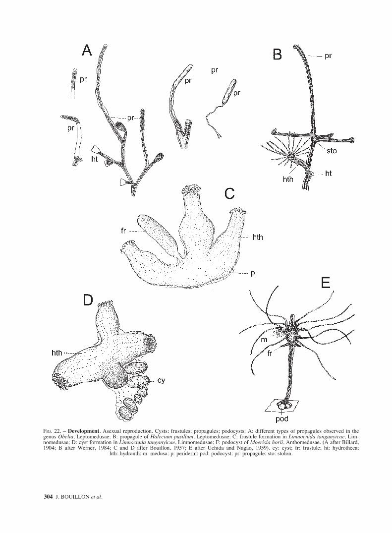
304 J. BOUILLON et al.
FIG. 22. – Development. Asexual reproduction. Cysts; frustules; propagules; podocysts: A: different types of propagules observed in thegenus Obelia, Leptomedusae; B: propagule of Halecium pusillum, Leptomedusae; C: frustule formation in Limnocnida tanganyicae, Lim-nomedusae; D: cyst formation in Limnocnida tanganyicae, Limnomedusae; F: podocyst of Moerisia horii, Anthomedusae. (A after Billard,1904; B after Werner, 1984; C and D after Bouillon, 1957; E after Uchida and Nagao, 1959). cy: cyst; fr: frustule; ht: hydrotheca;
hth: hydranth; m: medusa; p: periderm; pod: podocyst; pr: propagule; sto: stolon.
sm68s2Efig1 14/10/04 15:28 Página 304

FAUNA OF THE MEDITERRANEAN HYDROZOA 305
FIG. 23. – Morphology of cnidocysts. Different types of cnidocysts described in hydroids and medusa: A: anacrophore; B: acrophore; C:desmoneme; D: spirotele; E: aspirotele; F: atrichous isorhiza; G: basitrichous isorhiza; H: merotrichous isorhiza; I: apotrichous isorhiza; J:holotrichous isorhiza; K: atrichous anisorhiza; L.: homotrichous anisorhiza; M: heterotrichous anisorhiza; N: macrobasic atrichous mesotele;
O: microbasic mastigophore; P: macrobasic mastigophores. (all after Mariscal, 1974; except N after Bouillon et al., 1988).
sm68s2Efig1 14/10/04 15:28 Página 305

306 J. BOUILLON et al.
FIG. 24. – Morphology of cnidocysts. Different types of cnidocysts (end): A: microbasic amastigophores; B: macrobasic amastigophores; C:homotrichous microbasic eurytele; D heterotrichous microbasic eurytele; E: semiophoric microbasic eurytele; F: telotrichous macrobasiceurytele; G: merotrichous macrobasic eurytele; H: holotrichous macrobasic eurytele; I: stenotele; J: pseudostenotele; K: birhopaloid (A to I
and K after Mariscal, 1974; J after Bouillon et al.,1986).
sm68s2Efig1 14/10/04 15:28 Página 306

FAUNA OF THE MEDITERRANEAN HYDROZOA 307
FIG. 25. – Anthomedusae. Filifera. Bougainvilliidae. A to C: Bimeria vestita: A: part of stem with hydranths and male gonophores, B:hydranth showing perisarcal sheats round tentacles, C: female gonophores enveloped in a perisarcal coat; D to K: Bougainvillia: D:Bougainvillia aurantiaca: mature medusa; E to H: Bougainvillia britannica: E: hydroid colony with hydranths and medusa buds, F: adultmedusa, G: marginal bulb, H: oral tentacle; I: Bougainvillia maniculata mature medusa; J and K: Bougainvillia muscus: J: branch of a colony,K: details showing hydranths and medusa buds (A to C after Hirohito, 1988; D after Bouillon, 1978b; E after Edwards, 1964; F Kramp, 1959a;
G and H after Russell, 1953; I after Mayer, 1910; J and K after Leloup, 1952).
sm68s2Efig1 14/10/04 15:28 Página 307

308 J. BOUILLON et al.
FIG. 26. – Anthomedusae. Filifera. Bougainvilliidae. A and B: Bougainvillia muscus: mature medusae; C: Bougainvillia niobe: medusa withmanubrial medusa buds; D: Bougainvillia platygaster adult medusa; E to G: Dicoryne conferta: E: general view of a colony living on a gas-tropod shell, F: part of a colony with hydranths and gonozooids; G: swimming sporosacs (A after Russell, 1953; B after Calder, 1988; C after
Kramp, 1959a; D after Schuchert, 1996; E to G after Leloup, 1952).
sm68s2Efig1 14/10/04 15:28 Página 308

FAUNA OF THE MEDITERRANEAN HYDROZOA 309
FIG. 27. – Anthomedusae. Filifera. Bougainvilliidae. A: Dicoryne conybeari colony with gonozooids; B to G: Garveia: B to D: Garveiafranciscana: B: general view of a colony, C: branch of a colony, D: hydranth with gonophores; E: Garveia grisea: fragment of a colony withgonophores; F and G: Garveia nutans: F: fragment of a colony; G: part of colony with gonophores; H to L: Koellikerina fasciculata: H:hydranth with medusa buds, I: adult medusa, J: aboral view of manubrium, K: oral tentacle, L: isolated perradial marginal tentacle (A afterCornelius et al., 1990; B to D after Morri, 1981; E after Motz-Kossowska, 1905; F and G after Leloup, 1952; H after Petersen and
Vannucci, 1960; I to L after Mayer, 1910).
sm68s2Efig1 14/10/04 15:28 Página 309

310 J. BOUILLON et al.
FIG. 28. – Anthomedusae. Filifera. Bougainvilliidae. A to F: Lizzia: A to D: Lizzia blondina: A: adult medusa with manubrial medusa buds,B: mouth with oral tentacles, C: manubrium with gonad; D: perradial marginal tentacle group; E: Lizzia fulgurans medusa with manubrialmedusa buds; F: Lizzia octostyla medusa; G: Nubiella mitra adult medusa with manubrial medusa buds; H: Pachycordyle napolitana hydranth
(A, E and F after Kramp, 1959a; B to D after Russell, 1953; G after Bouillon, 1980; H after Calder, 1988).
sm68s2Efig1 14/10/04 15:28 Página 310

FAUNA OF THE MEDITERRANEAN HYDROZOA 311
FIG. 29. – Anthomedusae. Filifera. Bougainvilliidae. A to C: Rhizorhagium. A: Rhizorhagium arenosa part of colony with hydranth andgonophores; B and C: Rhizorhagium michaeli: B: branch of colony showing hydranth and succession of gonophores from distal juveniles toproximal stalk remnants; C: detail of hydranth with gonophores; D: Thamnostoma dibalia mature medusa; E: Velkovrhia enigmaticahydranths, one with fixed sporosac (A after Hincks, 1868; B and C after Berrill, 1948; D after Kramp, 1959a; E after Clausen and Salvini-
Plawen, 1986).
sm68s2Efig1 14/10/04 15:28 Página 311
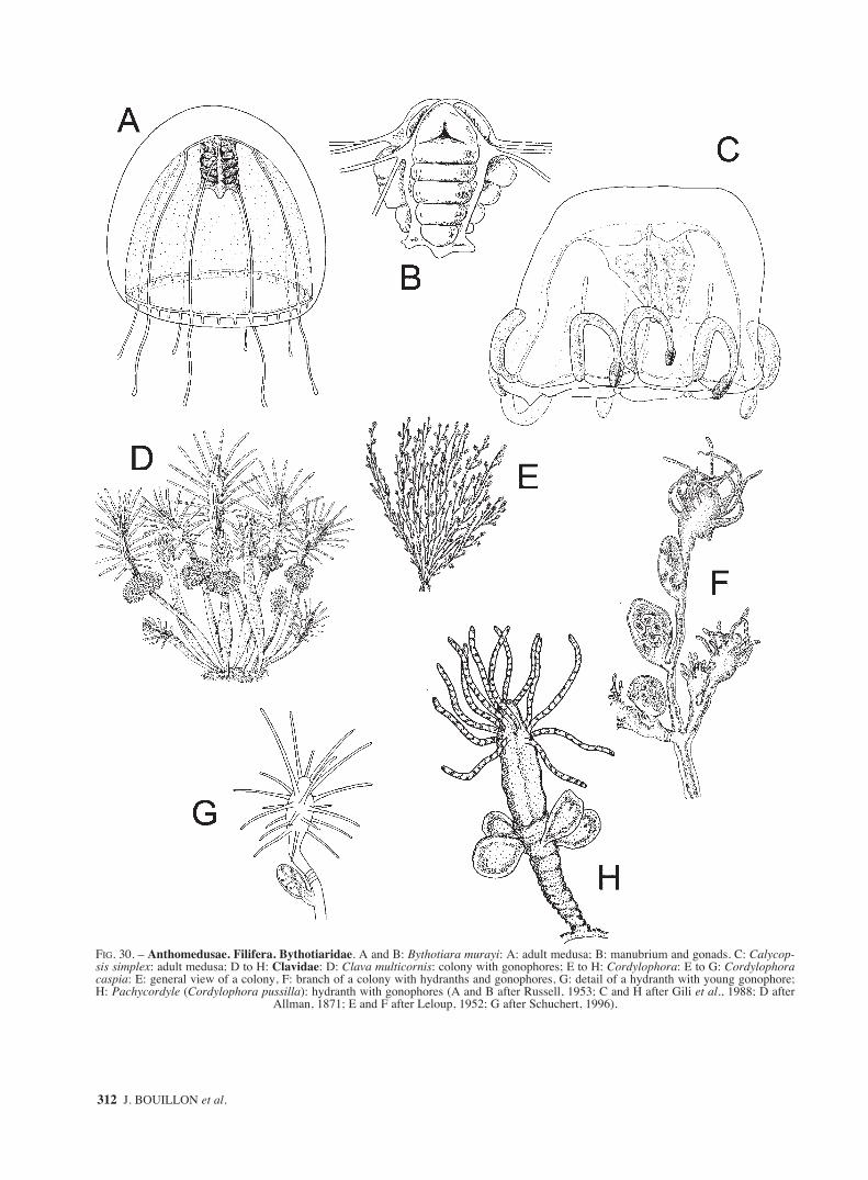
312 J. BOUILLON et al.
FIG. 30. – Anthomedusae. Filifera. Bythotiaridae. A and B: Bythotiara murayi: A: adult medusa; B: manubrium and gonads. C: Calycop-sis simplex: adult medusa; D to H: Clavidae: D: Clava multicornis: colony with gonophores; E to H: Cordylophora: E to G: Cordylophoracaspia: E: general view of a colony, F: branch of a colony with hydranths and gonophores, G: detail of a hydranth with young gonophore;H: Pachycordyle (Cordylophora pussilla): hydranth with gonophores (A and B after Russell, 1953; C and H after Gili et al., 1988; D after
Allman, 1871; E and F after Leloup, 1952; G after Schuchert, 1996).
sm68s2Efig1 14/10/04 15:28 Página 312

FAUNA OF THE MEDITERRANEAN HYDROZOA 313
FIG. 31. – Anthomedusae. Filifera. Clavidae. A to C: Corydendrium parasiticum: A: part of colony with hydranths; B: detail of hydranths;C: part of stem with hydranths and male gonophores within perisarcal tubes; D to H: Merona: D and E: Merona cornucopiae: D: part ofcolony growing on bivalve showing gastrozooids, one gonozooid and nematophores, E: nematothecae; F to H: Merona ibera: F: gonozooidand dactylozooids; G: fragment of a colony with hydranths, hydrothecae and dactylozooids; H: hydrorhiza with dactylozooids; I to L: Ocea-nia armata: I: polyp stage; J: adult medusa; K: part of marginal tentacle showing the position of the ocellus; L: part of mouth lip showing thecnidocyst clusters (A and C after Hirohito, 1988; B after Calder, 1988; D and E after Millard, 1975; F to H after Medel et al, 1993; I after
Schuchert, 1996; J to L after Mayer, 1910).
sm68s2Efig1 14/10/04 15:28 Página 313

314 J. BOUILLON et al.
FIG. 32. – Anthomedusae. Filifera. Clavidae. A to C and E: Turritopsis nutricula: A: part of stem of an erect colony with adnate branches,hydranths and medusa buds; B: drawing of the stem region showing the double layered structure of perisarc (coe: coenosarc; ps: perisarc);C: adult medusa; E: above base of marginal tentacles, ocelli omitted, left: tip of a marginal tentacle, below: margin of mouth lip showing thecnidocyst clusters; D: Turritopsis dohrnii young medusa. Cytaedididae. F to I: Cytaeis: F to H: Cytaeis propagulata: F: hydranths withgonophores; G: female gonophores; H: propagula; I: Cytaeis schneideri: hydranth with gonophore (A after Hirohito, 1988; B and D afterSchuchert, 1996; C after Kramp, 1968; D after Schuchert, 2004; E after Russell, 1953; F, G and H after Bavestrello, 1987; I after
Motz-Kossowska, 1905).
sm68s2Efig1 14/10/04 15:28 Página 314
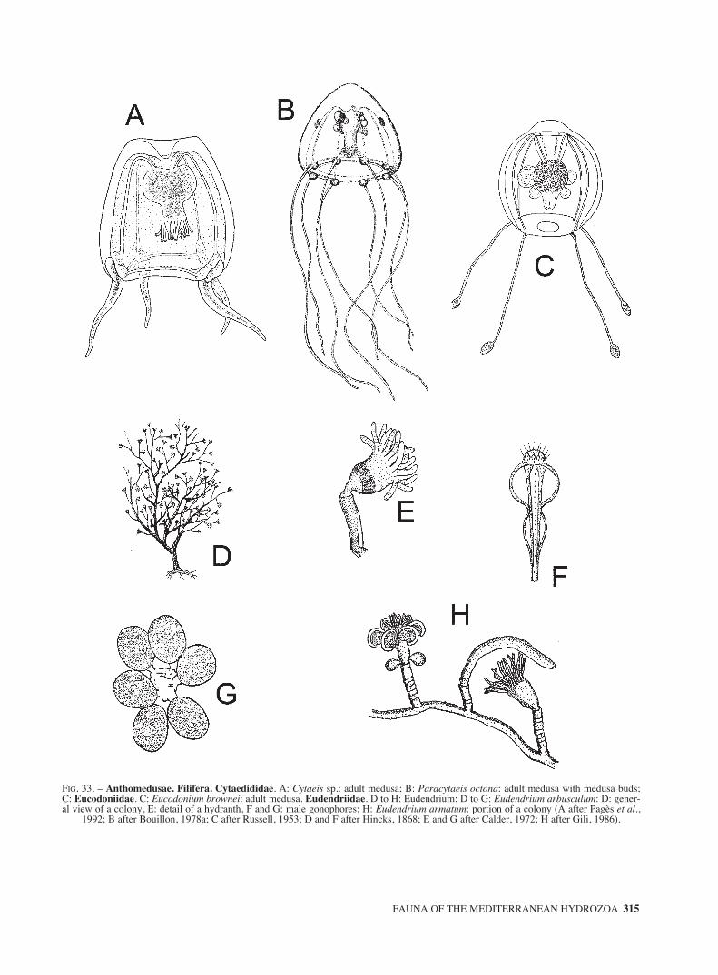
FAUNA OF THE MEDITERRANEAN HYDROZOA 315
FIG. 33. – Anthomedusae. Filifera. Cytaedididae. A: Cytaeis sp.: adult medusa; B: Paracytaeis octona: adult medusa with medusa buds;C: Eucodoniidae. C: Eucodonium brownei: adult medusa. Eudendriidae. D to H: Eudendrium: D to G: Eudendrium arbusculum: D: gener-al view of a colony, E: detail of a hydranth, F and G: male gonophores; H: Eudendrium armatum: portion of a colony (A after Pagès et al.,
1992; B after Bouillon, 1978a; C after Russell, 1953; D and F after Hincks, 1868; E and G after Calder, 1972; H after Gili, 1986).
sm68s2Efig1 14/10/04 15:28 Página 315

316 J. BOUILLON et al.
FIG. 34. – Anthomedusae. Filifera. Eudendriidae. A to K: Eudendrium: A: Eudendrium calceolatum fragment of a colony; B to D: Euden-drium capillare: B: general view of a colony; C: hydroclade with male gonophores; D: female gonophores; E to I: Eudendrium carneum: E:colony; F: detail of a hydranth; G: mature male blastostyle; H: young female blastosyle; I: old female blastostyle with spadices shed livingbasket-shaped capsules; J and K: Eudendrium elsaeoswaldae: J: colony; K: hydranth; L: Eudendrium fragile: part of colony; M to O: Euden-drium glomeratum: M: general aspect of a colony; N: hydranth with female gonophores; O: undischarged and discharged macrobasic eury-teles (A and L after Motz-Kossowska, 1905; B to D after Leloup, 1952; E, G to I after Millard, 1975; F after Calder,1988; J and K after
Stechow, 1923d; M to O after Boero et al., 1986).
sm68s2Efig1 14/10/04 15:28 Página 316

FAUNA OF THE MEDITERRANEAN HYDROZOA 317
FIG. 35. Anthomedusae. Filifera. Eudendriidae. A to M: Eudendrium: A to G: Eudendrium merulum: A: whole stem; B: hydranth show-ing cnidocyst arrangement; C: mature male gonophore; D and E: female gonophores; F: large microbasic euryteles from hypostome andhydranth body; G: small microbasic euryteles from tentacles; H to M: Eudendrium moulouyensis: H: part of a branch and two hydranths; Iand J: detail of hydranths; K: mature female blastostyle; L and M: microbasic euryteles. (A, C to G after Watson, 1985; B after Peña
Cantero and García Carrascosa, 2002; H to M after Marques et al., 2000).
sm68s2Efig1 14/10/04 15:28 Página 317

318 J. BOUILLON et al.
FIG. 36. – Anthomedusae. Filifera. Eudendriidae. A to P: Eudendrium: A to D: Eudendrium racemosum: A: general view of a colony, B:branch of a colony; C: detail of a hydranth; D: gonophores: at left female, at right male; E to G: Eudendrium rameum: E: part of a colony, F:part of stem with male blastostyles, G: fragment of stem with hydranths and female blastostyles; H to L: Eudendrium ramosum: H: generalview of a colony, I: hydroclade with male gonophores; J: detail of a hydranth; K: detail of a female gonophore; L: detail of a male gonophore;M to P: Eudendrium simplex: M: large discharged and undischarged macrobasic euryteles; N: mature male gonophores; O: hydranth, P:female gonophore, with sperm capsule; Q and R: Myrionema amboinense: Q: hydrocaulus, hydranths and male gonophore, R: hydranth withfemale gonophores (A to D after Morri, 1981; E to G after Hirohito, 1988; H to L after Leloup, 1952; M and N after Millard, 1975; O and P
after Boero, 1981; Q and R after Calder,1988).
sm68s2Efig1 14/10/04 15:28 Página 318

FAUNA OF THE MEDITERRANEAN HYDROZOA 319
FIG. 37. – Anthomedusae. Filifera. Hydractiniidae. A to N: Hydractinia: A: Hydractinia aculeata: fragment of a colony; B to I: Hydractiniaareolata: B: colony on a shell of Natica; C: portion of a colony; D: portion of colony hydranths omitted to show the arrangement of the spines;E: group of spiral zooids; F: tentaculozooid; G: male medusa; H: female medusa; I: quadrant of bell margin of ripe female. J to N: Hydractiniaborealis: J: colony living on a shell of Aporrhais, showing hydranths, gonozooids, blastostyles, spines and tentaculozooids; K: portion of acolony hydranths omitted to show the arrangement of the spines; L: two male medusae; M: adoral view of the lips: above of a maturing female,
below of a mature female; N: manubrium of a large mature female (A after Motz-Kossowska, 1905; B to N after Edwards, 1972).
sm68s2Efig1 14/10/04 15:28 Página 319

320 J. BOUILLON et al.
FIG. 38. – Anthomedusae. Filifera. Hydractiniidae. A to B: Hydractinia calderi: A: apical part of an adult gastrozooid; B dactylozooid. Cto F: Hydractinia carnea: C: colony living on the shell of a Nassarius, showing gastrozooids, gonozooids, blastostyles and spines; D: malemedusa; E: female medusa; F: mouth of a newly liberated medusae. G: Hydractinia echinata portion of a colony (A and B after Bouillon
et al., 1997; C to F after Edwards, 1972; G after Stokes, 1974).
sm68s2Efig1 14/10/04 15:28 Página 320

FAUNA OF THE MEDITERRANEAN HYDROZOA 321
FIG. 39. – Anthomedusaee. Filifera. Hydractiniidae. A: Hydractinia fucicola: part of a colony; B: Hydractinia hooperii: left and right: gas-trozooids, centre: a gonozooid with gonophores; C: Hydractinia inermis: portion of a colony with gonophores; D and E: Hydractinia mini-ma: medusae; F and G: Hydractinia minuta: medusae; H and I: Hydractinia pruvoti: H: fragment of a colony with blastostyle; I: eumedusoid(A after Castric-Fey, 1970; B after Peña Cantero and Carracosa, 2002; C, H and I after Iwasa, 1934; D after Kramp, 1968; E after Russell,
1953; F after Schuchert, 1996; G after Kramp, 1959a).
sm68s2Efig1 14/10/04 15:28 Página 321

322 J. BOUILLON et al.
FIG. 40. – Anthomedusae. Filifera. Niobiidae. A: Niobia dendrotentacula: medusa with tentacular bulbs transforming in medusa buds. Pan-deidae: B to G: Amphinema: B and C: Amphinema dinema: B: portion of a hydroid colony, C: medusa. D: Amphinema rubra: medusa; E andF: Amphinema rugosum: E: fragment of a hydroid colony. F: medusa. G: Amphinema turrida: medusa; H: Codonorchis octaedrus: hydranthand medusa bud (A after Kramp, 1959a; B after Leloup, 1952; C and F after Kramp, 1968; D after Goy, 1972; G after Bouillon, 1980; H after
Boero et al., 1997).
sm68s2Efig1 14/10/04 15:28 Página 322

FAUNA OF THE MEDITERRANEAN HYDROZOA 323
FIG. 41. – Anthomedusae. Filifera. Pandeidae. A: Codonorchis octaedrus: adult medusa; B to F: Leuckartiara: B: Leuckartiara brownei:medusa; C: Leuckartiara nobilis: medusa; D to F: Leuckartiara octona: D: portion of a hydroid colony; E: medusa; F: diagram of lateral viewof a marginal tentacle showing abaxial spur. G to J: Merga: G and H: Merga galleri: G: portion of a hydroid colony with hydranth and medusabuds; H: medusa. I and J: Merga tergestina: I: mature medusa; J manubrium and mouth (A after after Boero et al., 1997; B after Larson andHarbison, 1990; C and E after Kramp, 1959a; D and F after Russell, 1953; G and H after Brinckmann, 1962; I and J after Vannucci and
Yamada, 1959).
sm68s2Efig1 14/10/04 15:28 Página 323

324 J. BOUILLON et al.
FIG. 42. – Anthomedusae. Filifera. Pandeidae. A: Merga tregoubovii: adult medusa; B: Merga violacea: mature medusa; C to E: Neotur-ris pileata: C: portion of a hydroid colony; D: adult medusa; E: portion of umbrella margin. F to H: Octotiara russelli: F: hydroid colony withhydranth and medusa buds growing on the bryozoan Steginoporella mandibulata; G: detail of hydranth; H: adult medusa (A after Picard,1960; B after Kramp, 1959a; C after Edwards, 1965; D after Hartlaub, 1914; E after Russell, 1953; F and G after Boero and Bouillon, 1989;
H after Kramp, 1968).
sm68s2Efig1 14/10/04 15:28 Página 324

FAUNA OF THE MEDITERRANEAN HYDROZOA 325
FIG. 43. – Anthomedusae. Filifera. Pandeidae. A and B: Pandea conica: A: hydranth and medusa bud; B: adult medusa. Proboscidactyli-dae. C and D: Proboscidactyla ornata: C: left hydroid and medusa bud, right hydroid colony on the edge of a tube of the polychaete Sabel-laria; D: adult medusa with medusa buds on manubrium. Protiaridae. E to G: Halitiara: E: Halitiara formosa: adult medusa, F and G: Hali-tiara inflexa: F: hydroid colony; G: fully-grown medusa (A after Picard, 1956a; B after Pagès et al, 1992; C after Brinckmann-Voss and
Vannucci, 1965; D after Mayer, 1910; E after Bouillon, 1995; F after Bouillon, 1985b; G after Bouillon, 1980).
sm68s2Ffig2 14/10/04 16:16 Página 325

326 J. BOUILLON et al.
FIG. 44. – Anthomedusae. Filifera. Protiaridae. A: Protiara tetranema: medusa. Ptilocodiidae. B: Thecocodium brieni: two hydroidcolonies; C: Tregoubovia atentaculata: adult medusa. Rhatkeidae. D to H: Rathkea octopunctata: D: hydroid; E: medusa with manubrialmedusa buds; F: mouth; G: interradial marginal tentacle group; H: manubrium and gonads. Rhysiidae. I: Rhysia automnalis: male colony (Aafter Hartlaub, 1913; B after Bouillon, 1967; C after Bouillon et al, 2001; D, F to H after Russell, 1953; E after Naumov, 1969; I after
Brinckmann, 1965b).
sm68s2Ffig2 14/10/04 15:35 Página 326

FAUNA OF THE MEDITERRANEAN HYDROZOA 327
FIG. 45. – Anthomedusae. Filifera. Russelliidae. A: Russellia mirabilis: fully-grown medusa. Stylasteridae. B and C: Errina: B: generalaspect of a colony; C: detail of a branch (gpo: gastropore, dpo: dactylopore). Trichydridae. D and E: Trichydra pudica: D: hydroid colony,E: mature medusa. Capitata. Moerisiida. Hydridae. F to H: Hydra: F: Hydra viridis with bud and gonads; G: terminal moniliform portionof a tentacle of Hydra pirardi (isr: isorhiza, stn: stenoteles); H: encysted eggs: a: Hydra viridissima; b: Hydra attenuata; c: Hydra vulgaris(A after Pagès et al., 1999; B and C after Moore, 1956, D after Russell, 1953; E after Edwards, 1973a; F after Brien and Reniers-Decoen,
1950; G after Brien, 1961; H after Steche, 1911).
sm68s2Ffig2 14/10/04 15:35 Página 327

328 J. BOUILLON et al.
FIG. 46. – Anthomedusae. Capitata. Moerisiidae. A to G: Moerisia: A: Moerisia carine: fully-grown medusa; B to D: Moerisia inkerman-ica: B: hydroid showing pedal discs and medusa buds; C: hydroid showing lateral buds; D: adult medusa. E to G: Moerisia lyonsi: E and F:hydroids; G: mature medusa. H and I: Odessia maeotica: H: hydroid with medusa bud, I: mature medusa (A after Bouillon, 1978c;
B and C after Millard, 1975; D after Kramp, 1959a; E to G after Rees, 1958; H and I after Morri, 1981).
sm68s2Ffig2 14/10/04 15:35 Página 328

FAUNA OF THE MEDITERRANEAN HYDROZOA 329
FIG. 47. – Anthomedusae. Capitata. Protohydridae. A to E: Protohydra leuckarti: A: contracted hydranth; B and C: various stages of relaxedhydranths; D: hydranth feeding on a copepode; E: two stages of transversal fission. Sphaerocorynidae. F and G: Sphaerocoryne bedoti: F:hydroid; G: fully-grown medusa. Tubulariida. Acaulidae. H and I: Acauloides. H: Acauloides ammisatum hydroid, I: Acauloides ilonae:hydroid; J: Boreohydra simplex: hydranth; K: Psammohydra nanna: hydranth (A to E after Leloup, 1952; F after Hirohito, 1988; G afterPetersen, 1990; H after Bouillon, 1971; I after Brinckmann-Voss, 1966; J after Westblad, 1937; K after Clausen and Salvini-Plawen, 1986).
sm68s2Ffig2 14/10/04 15:35 Página 329

330 J. BOUILLON et al.
FIG. 48. – Anthomedusae. Capitata. Candelabridae. A: Candelabrum cocksi hydranth. Cladonematidae. B to D: Cladonema radiatum: B:hydroid; C: schema of hypostome showing the ectodermal glandular oral cavity. D: fully-grown medusa; E to H: Eleutheria: E and F:Eleutheria claparedei: E: hydranth, F: medusa. G and H: Eleutheria dichotoma: G: part of colony with hydranths developing medusa buds,H: mature medusa with planulae inside brooding pouch. I and J: Staurocladia portmanni: I: hydranth; J: adult medusa (A after Hyman, 1940;B after Leloup, 1952, C after Bouillon, 1971; D after Russell, 1953; E after Brinckmann-Voss, 1970; F after Mayer, 1910; G
after Hincks, 1861; H after Hincks, 1868; I after Bouillon, 1978b; J after Brinckmann, 1964a).
sm68s2Ffig2 14/10/04 15:35 Página 330

FAUNA OF THE MEDITERRANEAN HYDROZOA 331
FIG. 49. – Anthomedusae. Capitata. Corymorphidae. A to C: Corymorpha nutans: A: hydranth; B: adult medusa; C: tip of medusa tenta-cle. D to F: Eugotoea petalina: mature medusa: D and F: lateral views; E: oral view; G to I: Euphysora: G: Euphysora annulata: adult medusa;H and I: Euphysora bigelowi: H: hydranth; I: adult medusa. J: Paragotoea bathybia: mature medusa; K and L: Vannuccia forbesii: K:hydranth; L: mature medusa (A after Allman, 1872; B and G after Kramp, 1959a; C after Russell, 1953; D to F after Margulis, 1989; H after
Sassaman and Rees, 1978; I after Petersen, 1990; J after Pagès and Bouillon, 1997; K and L after Schuchert, 1996).
sm68s2Ffig2 14/10/04 15:35 Página 331

332 J. BOUILLON et al.
FIG. 50. – Anthomedusae. Capitata. Corynidae. A to F: Coryne: A to D: Coryne eximia: A and B: general view of two colonies; C: hydranthwith medusa buds; D: mature medusa. E and F: Coryne muscoides: E view of a part of a colony; F: hydranth with gonophores (A, B and D
after Schuchert, 1996; C and F after Schuchert, 2001b; E after Brinckmann -Voss, 1970).
sm68s2Ffig2 14/10/04 15:35 Página 332

FAUNA OF THE MEDITERRANEAN HYDROZOA 333
FIG. 51. – Anthomedusae. Capitata. Corynidae. A to K: Coryne. A and B: Coryne pintneri: A: detail of a colony; B: hydranth withgonophores. C and D: Coryne producta: C: two hydranths; D: adult medusa. E to G: Coryne prolifera: E: adult medusa; F: tentacle bulb withmedusa buds; G: medusa marginal tentacle tip. H and I: Coryne pusilla: H: part of a colony; I: hydranth with gonophores. J: Coryne epizoica;K: Coryne fucicola (A, B and K after Brinckmann-Voss, 1970; C, D and E after Russell, 1953; F,G, I and J after Schuchert, 2001b; H after
Hirohito, 1988).
sm68s2Ffig2 14/10/04 15:35 Página 333

334 J. BOUILLON et al.
FIG. 52. – Anthomedusae. Capitata. Corynidae. A to K: Dipurena: A: Dipurena gemmifera: medusa with medusa buds; B to E: Dipure-na halterata: B: hydroid colony in a sponge; C: longitudinal section of hypostome showing the glandular mucous button (gb) character-istic of the genus; D: adult medusa, E: marginal tentacular bulbs, lateral and frontal view. F to H: Dipurena ophiogaster: F: two hydranths;G: adult medusa; H: lateral view of a marginal bulb of a mature medusa. I to K: Dipurena reesi: I: hydranth; J: adult medusa; K: tip ofmedusa tentacle, note the long, spiny cnidocils (A, F to K after Schuchert, 2001b, B after Bouillon, 1971; C after Bouillon, 1968; D and
E after Pagès et al., 1992).
sm68s2Ffig2 14/10/04 15:35 Página 334

FAUNA OF THE MEDITERRANEAN HYDROZOA 335
FIG. 53. – Anthomedusae. Capitata. Corynidae. A to J: Sarsia tubulosa: A: fragment of a colony; B to D: detail of hydranths with medusabuds; E: hydranth regressing during reproductive exhaustion with gonophore shorty before release; F: subadult medusa, note enlarged radialcanals; G: mature medusa; H: basal part of manubrium; I and J: lateral and frontal view of a marginal bulb (A to I after Edwards, 1978; J
after Schuchert, 2001b).
sm68s2Ffig2 14/10/04 15:35 Página 335

336 J. BOUILLON et al.
FIG. 54. – Anthomedusae. Capitata. Euphysidae. A to E: Euphysa: A to D: Euphysa aurata: A: hydroid; B: longitudinal histological sec-tion of the basal papilla and statocyst of a hydranth (see asterisk figure A); C: mature medusa; D: medusa, view of a non tentacular margin-al bulb; E: Euphysa flammea fully-grown medusa; F: Siphonohydra adriatica diagram of a longitudinal section through a hydranth: left: radi-al, right: interradial. Paracorynidae. G: Paracoryne huvei: view of a mature colony (A after Rees, 1938; B after Bouillon and Grohman,1990; C and D after Russell, 1953; E after Kramp, 1959a; F after Salvini-Pawen, 1966; G after Bouillon, 1975). at: aboral tentacle; bu: bud;c: process covering the gonophore; dac: dactylozooid; di: digestive inclusions; egr: ectodermal granulations; ei: excretory inclusions; end:
endoderm; go: gonophore; ga: gastrozooid; gz: gonozooid; hyr: hydrorhiza; ot: oral tentacle; p: periderm; sta: statocyst.
sm68s2Ffig2 14/10/04 15:35 Página 336

FAUNA OF THE MEDITERRANEAN HYDROZOA 337
FIG. 55. – Anthomedusae. Capitata. Pennariidae. A to C: Pennaria disticha: A: portion of a colony; B: detail of branch with two hydranth;C: eumedusoid. Tricyclusidae. D: Tricyclusa singularis: hydranth with actinula larvae. Tubulariidae. E and J: Ectopleura: E and F: Ecto-pleura crocea: E: above left female hydranth with gonophores, below right detail of mature female gonophore, F: aboral ends of hydrocauliof newly-settled specimens showing the development of the stolons from the pedal disc area; G to J: Ectopleura dumortieri: G: hydranth; Hand I: lateral view of mature medusae; J: apical view of an adult medusa (A and B after Schuchert, 1996; C after Kramp, 1959a; D after
Bouillon, original; E and F after Petersen, 1990; G after Russell, 1953; H after Mayer, 1910; I and J after Rees, 1957).
sm68s2Ffig2 14/10/04 15:35 Página 337

338 J. BOUILLON et al.
FIG. 56. – Anthomedusae. Capitata. Tubulariidae. A to E: Ectopleura: A and B: Ectopleura larynx: A: general view of a colony; B: detailof branches with hydranth and gonophores. C: Ectopleura minerva: adult medusa; D: Ectopleura sacculifera: fully-grown medusa; E: Ecto-pleura wrighti: left: hydroid, right: blastostyle with two medusa buds; F to H: Hybocodon prolifer: F: hydranth; G and H: medusae with andwithout medusa buds. I: Plotocnide borealis: mature medusa; J: Rhabdoon singulare: adult medusa (A and B after Cornelius et al., 1990; Cafter Mayer, 1910; D after Bouillon, 1978b; E after Petersen, 1990; F after Russell, 1953; G and H after Kramp, 1959a; I after Arai and
Brinckmann-Voss, 1980; J after Vannucci and Soares Moreira, 1966).
sm68s2Ffig2 14/10/04 15:35 Página 338

FAUNA OF THE MEDITERRANEAN HYDROZOA 339
FIG. 57. – Anthomedusae. Capitata. Tubulariidae. A to F: Tubularia: A and B: Tubularia ceratogyne: A: hydranth, B: female blatostylewith an actinula escaping from a gonophore. C to F: Tubularia indivisa: C: portion of a colony; D: detail of a hydranth; E: cluster ofgonophores of a female colony; F: actinula. Cladocorynidae. G: Cladocoryne floccosa: hydranth with gonophores. Porpitidae. H and I: Por-pita porpita: H: polyp; I: medusa. J and K: Velella velella: J: polyp; K: medusa. (A to D after Leloup, 1952; E after Patriti, 1970; F after
Pèrèz, 1920; G after Bouillon, 1995; H and J after Pagès et al., 1992; I after Bouillon, 1984d; K after Brinckmann-Voss, 1964b).
sm68s2Ffig2 14/10/04 15:35 Página 339

340 J. BOUILLON et al.
FIG. 58. – Anthomedusae. Capitata. Rosalindidae. A to C: Rosalinda incrustans: A: semi-diagrammatic drawing of a part of coenosarc andspines; B: detail of a portion of colony on a spine of Anamathia rissoana showing three hydranths; C: detail of a hydranth. Zancleidae. Dand E: Halocoryne epizoica: D: general view of a part of colony living on a bryozoan host showing two polyps, one with medusa buds; E:newly released eumedusoid. F to H: Zanclea sp.: F: adult medusa; G: portion of a marginal tentacle with cnidophores; H:
detail of a cnidophore (A and B after Vervoort 1966b; C after Petersen, 1990; D and E after Bouillon, 1995; F to H after Russell, 1953).
sm68s2Ffig2 14/10/04 15:35 Página 340

FAUNA OF THE MEDITERRANEAN HYDROZOA 341
FIG. 59. – Anthomedusae. Capitata. Zancleidae. A to M: Zanclea: A to D: Zanclea costata: A: two hydranths with medusa buds; B: aboveleft microbasic mastigophores, below left and at right macrobasic euryteles of hydranths; C: mature medusa; D: macrobasic euyryteles frommedusan cnidophores. E to I: Zanclea giancarloi: E: hydranth with medusa buds; F: cnidome of hydroid comprising undischarged stenotelesof two sizes and discharged macrobasic euryteles of two types; G: newly released medusae; H: mature medusae I: cnidome of medusa com-prising undischarged stenoteles of two sizes, discharged and undischarged macrobasic euryteles from cnidophores and undischarged microba-sic euryteles from exumbrella. J to M: Zanclea sessilis: J: hydranth in different states of contraction, one with medusa buds; K: cnidocysts ofthe hydroid, stenoteles of two sizes, undischarged and discharged macrobasic euryteles; L: adult medusa; M: cnidome of the medusa,
stenoteles of two sizes, undischarged and discharged macrobasic euryteles from cnidophores (all after Boero et al., 2000).
sm68s2Ffig2 14/10/04 15:35 Página 341

342 J. BOUILLON et al.
FIG. 60. – Laingiomedusae. Laingiidae. A: Fabienna oligonema: left marginal tentacle, right adult medusa; B and C: Kantiella enigmatica:B: mature medusa; C: detail of a. tentacular bulb and tentacle. Leptomedusae. Aequoreidae. D to F: Aequorea: D: Aequorea conica: adultmedusa; E and F: Aequorea forskalea: E: adult medusa; F: portion of umbrella margin (A slightly modified, E and F after Kramp, 1959a; B
and C after Bouillon, 1978a; D after Pagès et al., 1992).
sm68s2Ffig2 14/10/04 15:35 Página 342

FAUNA OF THE MEDITERRANEAN HYDROZOA 343
FIG. 61. – Leptomedusae. Aequoreidae. A to F: Aequorea: A to D: Aequorea spp. hydroid: A: portion of a colony; B: branch of a colony witha gonophore; C: hydrotheca; D: basal web between tentacles of hydranth; E and F: Aequorea pensilis: E: fully-grown medusa; F: portion ofumbrella margin; G and H: Zygocanna vagans: G: oral view of an adult medusa; H: aboral view of the manubrium (A after Russell, 1953; Bafter Hincks, 1868; C after Cornelius, 1995; D after Rees, 1938; E and F after Kramp, 1959a; G after Pagès et al., 1992, H after Bigelow, 1919).
sm68s2Ffig2 14/10/04 15:35 Página 343

344 J. BOUILLON et al.
FIG. 62. – Leptomedusae. Aglaopheniidae. A to K: Aglaophenia: A to D: Aglaophenia acacia: A: general view of a colony; B: part of colonyshowing the insertion of corbula on hydrocaulus; C: detail of corbula on hydrocaulus; D: hydrotheca and associated nematothecae. E to G:Aglaophenia elongata: E: general view of a colony; F: part of branch with prosegments (prs); G: fragment of hydrocladia showing hydrothe-cae and associated nematothecae. H to K: Aglaophenia harpago: H: colony on Posidonia oceanica leaf bending backwards at slack water; I:apical region of hydrocaulus with hook (h) and intersegments (is); J: basal region of caulus, left frontal view, right lateral view (prs: proseg-ments); K: fragments of hydrocladia showing hydrothecae and associated nematothecae (A and D, after Medel and Vervoort, 1995; B, C, E
to K after Svoboda and Cornelius, 1991).
sm68s2Ffig2 14/10/04 15:35 Página 344

FAUNA OF THE MEDITERRANEAN HYDROZOA 345
FIG. 63. – Leptomedusae. Aglaopheniidae. A to L: Aglaophenia: A to D: Aglaophenia kirchenpaueri: A: general view of a colony; B: basalregion of hydrocaulus, showing prosegments and bases of hydrocladia; C: fragments of hydrocladia showing hydrothecae and associatednematothecae; D: male corbula; E to H: Aglaophenia lophocarpa: E: colony; F: base of cormoid with four prosegments (prs); G: fragmentsof hydrocladia showing hydrothecae and associated nematothecae; H: above fully-grown corbula with abnormally completely unfused ribs,below female corbula; I to L: Aglaophenia octodonta: I: general view of a colony; J: left: view of basal region of a caulus, right: end of a cla-dium (prs: prosegments); K: fragments of hydrocladia showing hydrothecae and associated nematothecae; L: female corbula (A to K after
Svoboda and Cornelius, 1991; L after Medel and Vervoort, 1995).
sm68s2Ffig2 14/10/04 15:35 Página 345

346 J. BOUILLON et al.
FIG. 64. – Leptomedusae. Aglaopheniidae. A to L: Aglaophenia: A to D: Aglaophenia parvula: A: general aspect of a colony; B: detail offork of a dichotomously branched colony; C: fragments of hydrocladia showing hydrothecae and associated nematothecae; D: female corbula;E to H: Aglaophenia picardi: E: three colonies springing from same stolon; F: basal part of cormoid, frontal view; G: fragments of hydrocla-dia showing hydrothecae and associated nematothecae, H: male corbula; I to L: Aglaophenia pluma: I: general view of a colony; J: detail ofhydrocladium; K: two hydrothecae and associated nematothecae; L: corbula (A to D after Svoboda and Cornelius, 1991; E, G and H after Medeland Vervoort, 1995; F after Ansín Agís et al., 2001; I after Bedot, 1919; J, K left and L after Cornelius,1995; K right after Millard, 1975).
sm68s2Ffig2 14/10/04 15:35 Página 346

FAUNA OF THE MEDITERRANEAN HYDROZOA 347
FIG. 65. – Leptomedusae. Aglaopheniidae. A to J: Aglaophenia: A to E: Aglaophenia tubiformis: A: general aspect of a colony, B and Cbasal parts of cauli (prs: prosegments), D: fragments of hydrocladia showing hydrothecae and associated nematothecae, E: female corbula. Fto J: Aglaophenia tubulifera: F: whole colony, G: region of insertion of basalmost hydrocladia, including prosegments, H: fragments of hydro-cladia showing hydrothecae and associated nematothecae, I: male corbula, J: female corbula. K to N: Cladocarpus: K: Cladocarpus multi-septatus: three hydrocladia internodes with hydrothecae and nematothecae, lateral view; L to N: Cladocarpus pectiniferus: hydrocladialinternodes with hydrotheca and nematotheca (A and C frontal view, B lateral view) (A to C, F to J after Svoboda and Cornelius, 1991; D and
E after Medel and Vervoort, 1995; K to N after Ramil and Vervoort, 1992a).
sm68s2Ffig2 14/10/04 15:35 Página 347
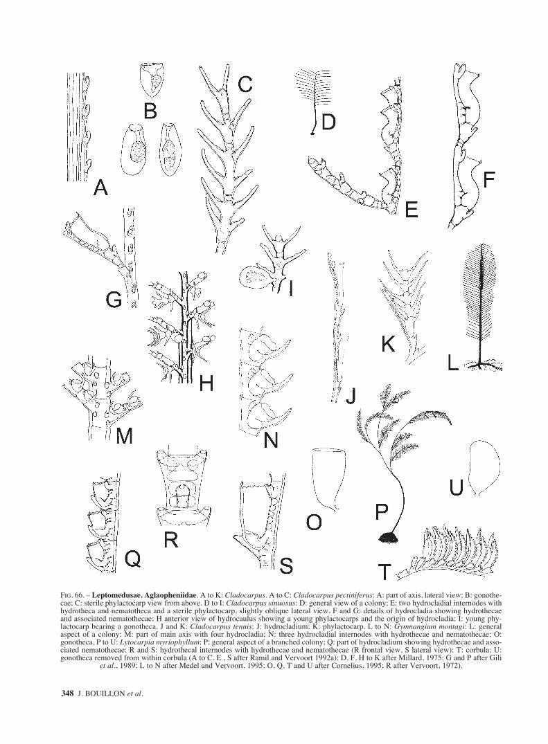
348 J. BOUILLON et al.
FIG. 66. – Leptomedusae. Aglaopheniidae. A to K: Cladocarpus. A to C: Cladocarpus pectiniferus: A: part of axis, lateral view; B: gonothe-cae; C: sterile phylactocarp view from above. D to I: Cladocarpus sinuosus: D: general view of a colony; E: two hydrocladial internodes withhydrotheca and nematotheca and a sterile phylactocarp, slightly oblique lateral view, F and G: details of hydrocladia showing hydrothecaeand associated nematothecae; H anterior view of hydrocaulus showing a young phylactocarps and the origin of hydrocladia; I: young phy-lactocarp bearing a gonotheca. J and K: Cladocarpus tenuis: J: hydrocladium; K: phylactocarp. L to N: Gymnangium montagi: L: generalaspect of a colony; M: part of main axis with four hydrocladia; N: three hydrocladial internodes with hydrothecae and nematothecae; O:gonotheca, P to U: Lytocarpia myriophyllum: P: general aspect of a branched colony; Q: part of hydrocladium showing hydrothecae and asso-ciated nematothecae; R and S: hydrothecal internodes with hydrothecae and nematothecae (R frontal view, S lateral view); T: corbula; U:gonotheca removed from within corbula (A to C, E , S after Ramil and Vervoort 1992a); D, F, H to K after Millard, 1975; G and P after Gili
et al., 1989; L to N after Medel and Vervoort, 1995; O, Q, T and U after Cornelius, 1995; R after Vervoort, 1972).
sm68s2Ffig2 14/10/04 15:35 Página 348

FAUNA OF THE MEDITERRANEAN HYDROZOA 349
FIG. 67. – Leptomedusae. Aglaopheniidae. A and B: Lytocarpia distans. A: general aspect of a colony; B: two hydrocladial internodes withhydrothecae and nematothecae, lateral view; C to H: Macrorhynchia philippina: C: part of colony with origins of hydrocladia; D a part ofhydrocladium in lateral view showing detail of hydrothecae and nematothecae; E: two hydrothecal internodes in anterior view; F: phylacto-carps bearing female gonophores; G: phylactocarp with two gonophores; H: eumedusoid; I to K: Streptocaulus dollfusi: I: part of main axiswith hydrocladia; J: two hydrocladial internodes with hydrothecae and nematothecae, lateral view; K: phylactocarp with a single gonotheca.Barcinidae. L and M: Barcino foixensis: L: mature medusa; M: portion of umbrella margin. Blackfordiidae. N: Blackfordia virginica:hydranths (A after AnsÌn AgÌs et al., 2001; B after Ramil and Vervoort, 1992a; C to F after Hirohito, 1995; G and H after Gravier, 1970;
I to K after Medel and Vervoort, 1995; L and M after Gili et al., 1999; N after Valkanov, 1935).
sm68s2Ffig2 14/10/04 15:35 Página 349

350 J. BOUILLON et al.
FIG. 68. – Leptomedusae. Blackfordiidae. A and B: Blackfordia virginica: A: fully-grown medusa; B: portion of umbrella margin. Cam-panulinidae. C to E: Calycella syringa: C: part of a colony (note sub-terminal annulus on some hydrothecae); D: detail of hydrotheca; E:two gonothecae, that on the left with an acrocyst. F to I: Campanulina panicula: F: general view of a part of a colony; G: panicle or groupof hydrothecae; H: detail of hydrotheca; I: gonotheca (A after Kramp, 1959a, B after Moore, 1987, C to I after Cornelius, 1995).
sm68s2Ffig2 14/10/04 15:35 Página 350

FAUNA OF THE MEDITERRANEAN HYDROZOA 351
FIG. 69. – Leptomedusae. Campanulinidae. A and B: Cuspidella spp. hydroid general facies (note the horizontal ridges on the hydrothe-cae: “renovations” or growing lines): A: hydrotheca operculum without crease line, B: hydrotheca operculum with a crease line; C and D:Egmundella: C: Egmundella grimaldii: portions of stolons with hydrothecae and nematothecae; D: Egmundella valdiviae: hydrotheca andnematotheca. E to G: Lafoeina tenuis: E: part of a colony showing hydrothecae and nematothecae intermingled, F: detail of hydrotheca; G:detail of operculum. H to K: Opercularella lacerta: H: part of colony; I: portion of stem; J: two hydrothecae; K: gonotheca with acrocyst (A,
E to G, I to K after Cornelius, 1995; B after Hirohito, 1995; C after Leloup, 1940b, D after Vervoort, 1966a, H after leloup, 1952).
sm68s2Ffig2 14/10/04 15:35 Página 351

352 J. BOUILLON et al.
FIG. 70. – Leptomedusae. Cirrholovenidae. A to C: Cirholovenia tetranema: A: adult medusa, B: portion of umbrella margin; C: polyp.Eirenidae. D to H: Eirene viridula: D: hydroid stage with gonothecae; E and F: adult medusae; G: manubrium and four mouth-lips; H: detailof umbrella margin (A, B, E to G after Kramp, 1968; C after Brinckmann-Voss, 1965a; D after Cornelius, 1995; H after Russell, 1953).
sm68s2Ffig2 14/10/04 15:35 Página 352

FAUNA OF THE MEDITERRANEAN HYDROZOA 353
FIG. 71. – Leptomedusae. Eirenidae. A and B: Eugymnanthea inquilina: A: polyp with gonophore; B: eumedusoid; C to L: Eutima: C to G:Eutima gegenbauri: C: hydroid; D: gonangia before liberation of the medusae; E: adult medusa; F: portion of umbrella margin; G: non ten-tacular marginal swelling or warts, with lateral cirri and adaxial excretory papillae. H to L: Eutima gracilis: H and I: hydranths: H: withgonangium. J: adult medusa; K: base of marginal tentacle with lateral cirri; L: manubrium and mouth (A and B after Morri, 1981; C and D,
H and I after Russell, 1970a; E after Russell, 1963c; F and G, J to L after Russell, 1953).
sm68s2Ffig2 14/10/04 15:35 Página 353

354 J. BOUILLON et al.
FIG. 72. – Leptomedusae. Eirenidae. A and B: Eutima mira: A: adult medusa; B: manubrium, gastric peduncle and gonads. C: Eutoninascintillans: mature medusa; D to J: Helgicirrha: D and E: Helgicirrha cari: D: mature medusa; E: portion of umbrella margin. F to J: Helgi-cirrha schulzei: F: hydroid colony; G: adult medusa; H: portion of umbrella margin showing marginal tentacles of two sizes; I: detail of mar-gin showing the lateral cirri, statocyst and excretory papillae; J: manubrium and mouth (A after Kramp, 1933; B and C after Kramp, 1968;
D and E after Mayer, 1910; F after Bouillon, 1971; G after Russell, 1963c, H to J after Russell, 1953).
sm68s2Ffig2 14/10/04 15:35 Página 354

FAUNA OF THE MEDITERRANEAN HYDROZOA 355
FIG. 73. – Leptomedusae. Eirenidae. A and B: Neotima lucullana: A: fully-grown medusa; B: part of umbrella margin and statocyst. Haleci-idae. C to M: Halecium: C to E: Halecium banyulense: C: hydrocladium with hydrotheca and gonophore; D: young female gonophore; E:mature female gonophore. F to J: Halecium beanii: F: general aspect of a colony; G: stem with hydrothecae and female gonophore; H: femalegonophore; I: male gonophore; J: regenerated hydrothecae; K to M: Halecium conicum: K: two type of stems; L: male gonophore; M: femalegonophore (A after Kramp, 1959a; B after Petersen, 1990; C to E , K to M after Motz-Kossowska, 1911; F after Cornelius, 1995; G to J after
Millard, 1975).
sm68s2Ffig2 14/10/04 15:35 Página 355

356 J. BOUILLON et al.
FIG. 74. – Leptomedusae. Haleciidae. A to C: Halecium delicatulum: part of stem from a small form; B: part of stem of a large form; C:female gonophores. D: male gonophore; E to J: Halecium halecinum: E: general view of a colony; F: hydrocladia with several hydranths; G:detail of a hydrocladial internodes; H male gonothecae; I and J: female gonothecae: J: with gonophoral polyps. K to P: Halecium labrosum:K: general aspect of a colony; L: portion of a hydroclade; M: hydrotheca with renovations; N: male gonophore; O and P: female gonophores
(A to D after Millard, 1975; E, G to I, K and P after Cornelius,1995; F and J, L to O after Cornelius et al., 1990).
sm68s2Ffig2 14/10/04 15:35 Página 356

FAUNA OF THE MEDITERRANEAN HYDROZOA 357
FIG. 75. – Leptomedusae. Haleciidae. A to D: Halecium lankesteri: A: part of a colony, B: detail of a branch; C: gonophores: left female,right male; D to F: Halecium liouvillei: D: part of axis with axillary hydrothecae and insertion of hydrocladium; E: hydrocladium; F:gonophores; G to J: Halecium muricatum: G: general aspect of a colony; H: hydrocladium, I: “chain” of renovated hydrothecae; J gonothe-ca. K to M: Halecium nanum: K: part of a colony; L: detail of a branch, M: female gonotheca (A to C , J after Cornelius et al., 1990; D and
E after Medel and Vervoort, 2000; F after Ramil et al., 1998; G to I, K to M after Cornelius, 1995).
sm68s2Ffig2 14/10/04 15:35 Página 357

358 J. BOUILLON et al.
FIG. 76. – Leptomedusae. Haleciidae. A to C: Halecium petrosum: A and B: fragments of hydrocladia with hydrothecae: A: with malegonotheca; B: with female gonotheca. C: detail of hydrothecal rim; D to G: Halecium pusillum: D: part of colony with hydranths and femalegonothecae; E: hydroid with planctonic propogules; F: hydroid with female gonotheca; G: hydrotheca and male gonophore and gonotheca;H to L: Halecium sessile: H: general view of a colony; I: detail of a branch of a colony; J: hydrotheca with renovated rim; K: part of branchwith male gonophores; L: female gonophore; M to O: Halecium sibogae: M: portion of stem; N: hydrocladial hydrotheca; O: female gonothe-ca (A to C after Motz-Kossowska, 1911; D, G, K and L after Hirohito, 1995, E and F after Boero, 1981, H to J after Cornelius, 1995; M after
Medel and Vervoort, 1995, N and O after Medel and Vervoort, 2000).
sm68s2Ffig2 14/10/04 15:35 Página 358

FAUNA OF THE MEDITERRANEAN HYDROZOA 359
FIG. 77. – Leptomedusae. Haleciidae. A to E: Halecium tenellum: A: part of stem; B: detail of two hydrothecae; C and D: femalegonophores; E male gonophore. F to K: Hydrodendron mirabile: F: part of male colony with hydranths and gonothecae; G: detail of part ofa colony with gonothecae; H: nematotheca and nematophore with two hydrothecae; I and J: detail of parts of hydrocladia with hydrothecaeand nematothecae; K: living hydranths and nematophores. Halopterididae. L to P: Antennella: L to O: Antnenella ansini: L: stem; M:hydroyhecate internode showing hydrotheca and nematothecae; N: female gonotheca; O: male gonotheca. P: Antennella secundaria: part offertile colony (A after Cornelius, 1995; B to E, I to K after Millard, 1975, F to H after Hirohito, 1995; L, M and O after Peña Cantero et al.
2002; P after Schuchert, 1997).
sm68s2Ffig2 14/10/04 15:35 Página 359

360 J. BOUILLON et al.
FIG. 78. – Leptomedusae. Halopterididae. A to D: Antennella secundaria: A: hydrocladium with proximal male and distal female gonothe-ca; B: part of axis with female gonothecae; C: internode with hydrotheca and associated nematothecae; D: male gonophore. E to I: Anten-nella siliquosa: E: part of fertile colony; F: part of stem with hydrotheca and associated nematothecae; G: several lateral nematothecae show-ing typical spanner-like shape; H: female gonotheca; I: male gonotheca. J to T: Halopteris: J to N: Halopteris catharina: J: normal plumosecolony; K: part of caulus with opposite branched hydrocladia; L: part of hydrocladium with main segment and intersegment; M: gonothecae:left: female, right: male; N: left median inferior nematothecae, right pair of lateral nematothecae. O to T: Halopteris diaphana: O: plumoseform of stem, P: part of axis with two hydrocladia; Q: part of hydrocladium with hydrotheca and associated nematothecae, R: lateral nema-tothecae, S: female gonotheca, T: male gonotheca (A after Millard, 1975; B to D, p. after Medel and Vervoort, 1995; E to N, O, q. to s. after
Schuchert, 1997).
sm68s2Ffig2 14/10/04 15:35 Página 360
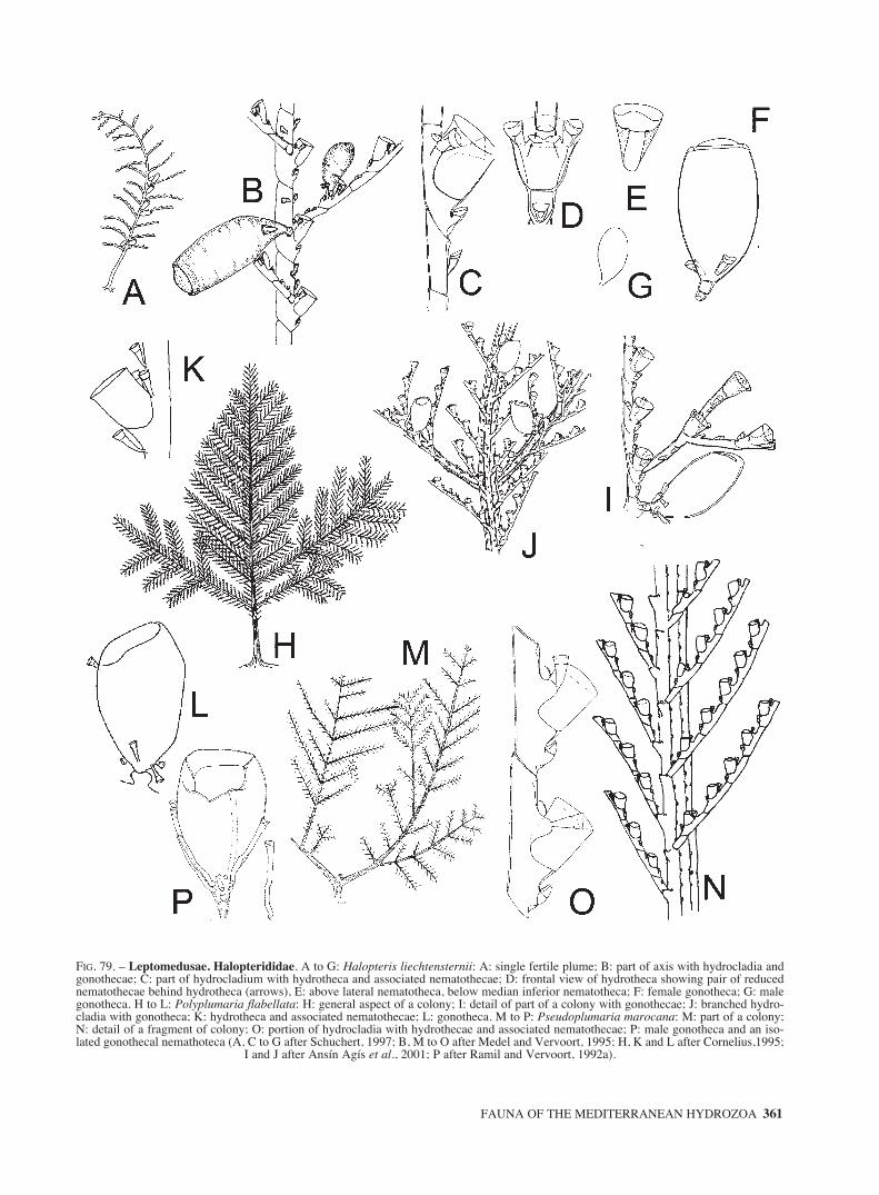
FAUNA OF THE MEDITERRANEAN HYDROZOA 361
FIG. 79. – Leptomedusae. Halopterididae. A to G: Halopteris liechtensternii: A: single fertile plume; B: part of axis with hydrocladia andgonothecae; C: part of hydrocladium with hydrotheca and associated nematothecae; D: frontal view of hydrotheca showing pair of reducednematothecae behind hydrotheca (arrows), E: above lateral nematotheca, below median inferior nematotheca; F: female gonotheca; G: malegonotheca. H to L: Polyplumaria flabellata: H: general aspect of a colony; I: detail of part of a colony with gonothecae; J: branched hydro-cladia with gonotheca; K: hydrotheca and associated nematothecae; L: gonotheca. M to P: Pseudoplumaria marocana: M: part of a colony;N: detail of a fragment of colony; O: portion of hydrocladia with hydrothecae and associated nematothecae; P: male gonotheca and an iso-lated gonothecal nemathoteca (A, C to G after Schuchert, 1997; B, M to O after Medel and Vervoort, 1995; H, K and L after Cornelius,1995;
I and J after Ansín Agís et al., 2001; P after Ramil and Vervoort, 1992a).
sm68s2Ffig2 14/10/04 15:35 Página 361

362 J. BOUILLON et al.
FIG. 80. – Leptomedusae. Halopterididae. A to E: Schizotricha frutescens: A: general vue of a colony; B: hydrocaulus with bases of hydro-cladia, C: fragment of hydrocladia, D: hydrotheca and associated nematothecae, E: gonotheca. Hebellidae. F to H: Anthohebella parasitica:F: various aspects of hydrothecae, G: fragment of colony with hydrotheca and gonotheca containing two medusa buds, H: female swimminggonophore; I to K: Bedotella armata: I: part of rhizocaulomic colony with two renovated hydrothecae, J: gonotheca in frontal and lateral view,K: stolonal nematothecae (A to C, E after Cornelius et al, 1990; D after Cornelius, 1995; F to H after Boero et al., 1997; I and K after Ramil
and Vervoort, 1992a; J after Álvarez Claudio, 1994).
sm68s2Ffig2 14/10/04 15:35 Página 362

FAUNA OF THE MEDITERRANEAN HYDROZOA 363
FIG. 81. – Leptomedusae. Hebellidae. A to E: Hebella scandens: A: straight hydrotheca, B: portion of fertile colony growing on a sertu-lariid, C: twisted hydrotheca, D and E: juvenile medusae. F to L: Scandia. F to H: Scandia gigas: F: hydrotheca, G: male gonophore, H:female gonophore; I to L: Scandia michael-sarsi: I: general view of a portion of a colony, J to L: various aspects of hydrothecae (A to D afterBoero et al, 1997; E after Altuna Prados, 1996; F to H after Boero, 1981; I, K and L after Garcia-Corrales et al., 1979; J after Vervoort, 1959).
sm68s2Gfig3 14/10/04 15:37 Página 363

364 J. BOUILLON et al.
FIG. 82. – Leptomedusae. Hebellidae. A to D: Staurodiscus kellneri: A: lateral view of hydrothecae showing variation of form, length andperisarc development, B and C: gonothecae; D: adult medusa. Kirchenpaueriidae. E to L: Kirchenpaueria: E to H: Kirchenpaueria bon-nevieae: E: fragment of colony with pinnate stem bearing gonothecae and hydrocladia, F: hydrocladia developping from axial apophysis, G:male? gonotheca, H: female? gonotheca; I to L: Kirchenpaueria pinnata: general view of a colony, J: hydrotheca and nematotheca, K and Lgonothecae (A and B after Migotto and de Andrade, 2000; C after Boero et al., 1997; D after Kramp, 1959a; E after Millard, 1975; F to H
after Ramil and Vervoort, 1992a; I, K and L after Leloup, 1952; J after Cornelius, 1995).
sm68s2Gfig3 14/10/04 15:37 Página 364

FAUNA OF THE MEDITERRANEAN HYDROZOA 365
FIG. 83. – Leptomedusae. Kirchenpaueriidae. A and B: Kirchenpaueria pinnata: A: part of hydrocladium, B: part of stem with origins ofhydrocladia; C to G: Ventromma halecioides: C: general aspect of a colony, D: monosiphonic part of axis with hydrocladia, E: hydrocaulusand part of an hydrocladium, F: hydrotheca with associated nematothecae; G: mature gonotheca. H to L: Lafoeidae. H to L: Acryptolariaconferta: H: colony, general view of heavy form, I: part of stem, J: portion of stem showing branching portions of peripheral tubes in posi-tion, K: coppinia with female gonothecae, L: cross section of a part of coppinia with female gonophores and modified hydrothecae (A andB, H to K after Millard, 1975; C and F after Cornelius, 1995; D and G after Medel and Vervoort, 1995; E after Calder, 1997; L after
Hirohito, 1995).
sm68s2Gfig3 14/10/04 15:37 Página 365

366 J. BOUILLON et al.
FIG. 84. – Leptomedusae. Lafoeidae. A to D: Cryptolaria pectinata: A: stem with basal coppinia, B: detail of stem from distal region toshow axillary hydrothecae and origins of hydrocladia, C: detail of origin of a hydrocladium showing hydrothecae and nematothecae, D: partof coppinia showing female gonothecae and nematothecae; E to J: Filellum. E and F: Filellum disaggregatum: E: hydotheca, F: coppinia indorsal view; G and H: Filellum serpens: G: general view of a colony, H: coppinia and a few hydrothecae; I and J: Filellum serratum: I: largehydrothecae, J: part of coppinia showing gonothecae and accessory tubes; K: Lafoea dumosa: general aspect of an erected colony, (A, B, D,
I and J after Millard, 1975; C after Hirohito, 1995; E and F after Peña Cantero et al 1998; G, H and K after Cornelius, 1995).
sm68s2Gfig3 14/10/04 15:37 Página 366

FAUNA OF THE MEDITERRANEAN HYDROZOA 367
FIG. 85. – Leptomedusae. Lafoeidae. A to D: Lafoea dumosa: A: details of a branche of an erected colony with stalked hydrothecae, B:details of a branch of an erected colony with unstalked hydrothecae, C: coppinia and a few hydrothecae, D: cross section of a coppinia withfemale gonophores and tubular hydrothecae. E to M: Zygophylax: E to J: Zygophylax biarmata: E: monosiphonic part of stem, F: twohydrothecae and their associated nematotheca, G: two gonothecae and some nematophorous tubules isolated from coppinia, H: gonothecae(lateral view), I: part of nematophorous tubule with a nematotheca and a small hydrotheca, J: strongly renovated hydrotheca and renovatednematotheca; K to M: Zygophylax brownei: K: monosiphonic part of stem with two hydrocladia, L: two hydrothecae and nematothecae, M:gonothecae. Laodiceidae. N to P: Guillea canyonincolae: N: general view of a mature medusa, O: adaxial view of a marginal tentacle show-ing ocellus, P: abaxial view of umbrella margin showing cordylus and lateral spiral cirri (A to C after Cornelius, 1995; D after
Hirohito, 1995; E to L after Ramil and Vervoort 1992a; M after Schuchert, 2001a; N to P after Bouillon et al, 2000).
sm68s2Gfig3 14/10/04 15:37 Página 367

368 J. BOUILLON et al.
FIG. 86. – Leptomedusae. Laodiceidae. A to G: Laodicea: A: Laodicea fijiana mature medusa; B to G: Laodicea undulata: B: general viewof hydroid colony, C: hydrotheca and hydranth with amphicoronate tentacles, D: a gonotheca with medusa buds and a young medusa justbefore release, E: diagram of closed operculum, F: fylly-grown medusa, G: adaxial view of marginal tentacle showing: ocellus, cordyli andendodermal processes; H to K: Staurophora mertensii: H: hydrotheca and hydranth, I: diagram of operculum, J: mature medusa, K: part ofthe umbrella of a fully-grown medusa, L: portion of mouth-lip seen from above (A after Kramp, 1968; B to E, H and I after Cornelius, 1995;
F and K after Kramp, 1959a; G and L after Russell, 1953; J after Russell, 1963a).
sm68s2Gfig3 14/10/04 15:37 Página 368

FAUNA OF THE MEDITERRANEAN HYDROZOA 369
FIG. 87. – Leptomedusae. Lovenellidae. A to E: Campalecium: A and B: Campalecium cirratum: A: parts of colony with hydranths andgonotheca, B: gonotheca with medusa buds ready to escape; C to E: Campalecium medusiferum: C: hydranth and gonotheca with medusa buds,D: lateral view of juvenile medusa, E: oral view of juvenile medusa; F to M: Eucheilota: F: Eucheilota maasi: mature medusa; G to L variousstages of hydroids of Eucheilota maculata: showing the regression of a lovenelliid type of hydrothecae to a haleciid type one: G: part of a juve-nile colony, H: detail of a juvenile hydranth, I: fully developed hydranth with its complete loveneliid hydrotheca, J: operculum of a juvenilehydranth, K: remains of the hydrotheca of a fully grown hydranth, haleciid type, L: renovated hydrotheca with the remains of previous haleci-id hydrotheca at base. M: Eucheilota paradoxica: medusa with medusa buds on radial canals (A and B after Millard and Bouillon, 1975; C to
E after Boero, 1981; F after Neppi and Stiasny, 1913; G to K after Werner, 1968a; L after Cornelius, 1995; M after Kramp, 1959a).
sm68s2Gfig3 14/10/04 15:37 Página 369

370 J. BOUILLON et al.
FIG. 88. – Leptomedusae. Lovenellidae. A: Eucheilota ventricularis mature medusa; B to D: Hydranthea margarica: B: part of colony with ahydranth and a medusoid, C: two tentacle bases with intervening web, D: male medusoids; E to K: Lovenella: E to H: Lovenella chiquita: E: partof colony with a branching stem and a gonotheca, F: two hydrothecae, G: young medusa escaping from gonotheca, H: newly liberated medusa;I: Lovenella cirrata: adult medusa; J and K: Lovenella clausa: J: part of a colony with hydrothecae and gonothecae, K: detail of a hydrotheca (Aafter Kramp, 1968; B, C, J and K after Cornelius, 1995; D after Boero and Sará, 1987; E to H after Millard, 1975; I after Pagès et al., 1992).
sm68s2Gfig3 14/10/04 15:37 Página 370

FAUNA OF THE MEDITERRANEAN HYDROZOA 371
FIG. 89. – Leptomedusae. Lovenellidae. A to D: Lovenella clausa: A and B: mature medusae, C: detail of a marginal bulb and cirri, D: abovesubumbrellar view of the longitudinally divided gonad; E to G: Lovenella gracilis: E: portion of a colony, detail of a branch with hydrothe-cae, G: medusa. Malagazziidae. H to J : Octophialucium funerarium: H and I: two aspects of fully-grown medusae, J: portion of umbrellamargin (A and H after Russell, 1963b; B to D, J after Russell, 1953; E after Huvè, 1952b; F and G after Calder, 1971; I after Gili, 1986).
sm68s2Gfig3 14/10/04 15:37 Página 371

372 J. BOUILLON et al.
FIG. 90. – Leptomedusae. Mitrocomidae. A to E: Foersteria: A to C: Foersteria antoniae: A: adult medusae, B: detail of umbrella margin,C: aboral view of manubrium; D and E: Foersteria araiae: D: fully-grown medusa, E: portion of umbrella margin; F to J: Mitrocoma annae:F: hydranth, G: mature medusa, H and I: portions of umbrella margin showing cirri and statocysts, J: oral view of open mouth (A to C after
Gili et al., 1998; D and E after Gili et al., 1999; F after Metschnikof, 1886; G to J after Mayer, 1910).
sm68s2Gfig3 14/10/04 15:37 Página 372

FAUNA OF THE MEDITERRANEAN HYDROZOA 373
FIG. 91. – Leptomedusae. Mitrocomidae. A to D: Mitrocomella brownei: A: hydranth, B: diagram of operculum, C: fully-grown medusa,D: portion of umbrella margin. Orchistomidae. E: Orchistomella agariciforme: general view of an adult medusa. Phialellidae. F to J:Phialella quadrata: F: general view of a colony, G: detail of a branch with hydrothecae and gonotheca, opercula of hydrothecae with or with-out a crease line in the same colony, H: detail of hydrotheca with a pleated operculum without basal crease line, I: detail of gonotheca, J:
fully-grown medusa (A, D, G after Russell, 1953; B, F , H, I after Cornelius, 1995, C, J after Kramp, 1959a; E after Bouillon, 1984b).
sm68s2Gfig3 14/10/04 15:37 Página 373

374 J. BOUILLON et al.
FIG. 92. – Leptomedusae. Phialellidae. A to C: Phialella quadrata: A: male gonad with the characteristic median groove, B: marginal ten-tacle and statocyst, C: detail of a statocyst with the typical cushion-like swellinig. Plumulariidae. D to K: Monotheca: D to F: Monothecaobliqua: D: detail of a branch, E: hydrotheca and associated nematothecae, F: above male gonotheca, below female gonotheca, G to K:Monotheca pulchella: G: three colonies rising from creeping stolon, H: stem with gonothecae, I: part of axis with three hydrocladia, J: partof hydrocladium with hydrotheca and associated nematotheca, K: detail of gonotheca; L to S: Nemertesia antennina: L: general view of acolony, M: upper part of stem with male gonothecae, N: detail of a hydrocladium, O: hydrotheca and nematothecae, P: transeversal sectionof a hydrocaulus showing arrangement of hydrocladia (number of rows not constant), Q: cross-section of a fascicled stem, R and S: gonothe-cae (A to C after Russell, 1953; D after Cornelius et al, 1990; E, F, O and P after Cornelius, 1995; G, I and K after Medel and Vervoort, 1995;
H and J after Millard, 1975; L, N, R, S after Leloup, 1952; M, Q after Hirohito, 1995).
sm68s2Gfig3 14/10/04 15:37 Página 374

FAUNA OF THE MEDITERRANEAN HYDROZOA 375
FIG. 93. – Leptomedusae. Plumulariidae. A to Q: Nemertesia: A to E: Nemertesia falcicula: A: general aspect of a colony, B: detail of acolony, C: part of hydrocladium, D: female gonotheca , E: male gonotheca; F to K: Nemertesia norvegica: F: general aspect of a colony, G:detail of colony, frontal view, H: hydrocladium, I: arrangement of hydrocladia (note just four rows), J and K gonothecae; L to Q: Nemerte-sia perrieri: L: general view of a colony, M: detail of a colony, frontal view, N: thecate internode with one supracalycine nematothecae, O:athecate and thecate internodes from hydrocladium, P and Q: gonothecae, frontal and lateral views (A to E, G, J, K, M, N, P and Q after
Ansín Agis et al, 2001; F, H and I after Cornelius, 1995; L and O after Medel and Vervoort, 1995).
sm68s2Gfig3 14/10/04 15:37 Página 375

376 J. BOUILLON et al.
FIG. 94. – Leptomedusae. Plumulariidae. A to P: Nemertesia: A to F: Nemertesia ramosa: A: general view of a colony, B: part of stem withthree hydrocladia per whorl and female gonothecae, C: arrangement of hydrocladia on hydrocaulus, D: detail of hydrocladium, E: hydothecaand associated nematothecae, F: gonotheca; G to K: Nemertesia tetrasticha: G: part of a colony, H: fragment of hydrocladium, I: hydrothecaand associated nematothecae, J: part of stem with gonothecae, K: detail of gonotheca; L to P: Nemertesia ventriculiformis: L: distal fragmentof a colony, M: hydrocladial internodes, N: apophysis, O: young gonotheca, P: left: female gonotheca, right: male gonotheca (A after Medeland Vervoort, 1995; B after Millard, 1975; C after Cornelius, 1995; D, E and F after Leloup, 1952; G, H and J after Garcia Carrascosa et al,1987; I after Meneghini, 1845, K after Stechow, 1919; L and p. after Ansín Agís et al, 2001; M to O after Ramil and Vervoort, 1992a).
sm68s2Gfig3 14/10/04 15:37 Página 376

FAUNA OF THE MEDITERRANEAN HYDROZOA 377
FIG. 95. – Leptomedusae. Plumulariidae. A to F : Plumularia. A to E: Plumularia setacea: A: general view of a colony, B and C: parts ofstem showing origins of hydrocladia and gonothecae, D: detail of hydrocladium, E: hydrotheca and associated nematothecae, F: Plumulariasyriaca: part of hydrocaulus and hydroclade. Sertulariidae. G to I: Amphisbetia operculata: G: part of colony, H: detail of a hydrocladium,I: gonotheca, J to N: Diphasia attenuata: J: part of a colony, K: portion of stem with hydrocladium, L: detail of hydrocladium, M: malegonotheca, N: female gonotheca (A and E after Cornelius, 1995; C and D after Millard, 1975; B, G to I, L to N after Leloup, 1952; F after
Billard, 1931, J and K after Cornelius et al., 1990).
sm68s2Gfig3 14/10/04 15:37 Página 377

378 J. BOUILLON et al.
FIG. 96. – Leptomedusae. Sertulariidae. A to L: Diphasia: A to C: Diphasia delagei: A: general aspect of a colony, B: detail from stems oftwo different colonies, C: gonotheca, D to G: Diphasia margareta: D: view of a colony, E: part of stem with hydrocladia, F: hydrocladiumwith gonothecae, G: female gonotheca; H to L: Diphasia pinastrum: H: stem, I: detail of hydrocladium, J: hydrotheca, K: hydrocladium withgonotheca, L: detail of gonotheca (A to C, H and J after Cornelius, 1995; D to F after Gili et al, 1989; G after Ramil and Vervoort, 1992a; I,
K and L after Cornelius et al., 1990).
sm68s2Gfig3 14/10/04 15:37 Página 378

FAUNA OF THE MEDITERRANEAN HYDROZOA 379
FIG. 97. – Leptomedusae. Sertulariidae. A to D: Diphasia rosacea: A: part of colony, B: hydrocladium, C: male gonotheca, D: femalegonotheca; E to H: Dynamena disticha: E: colony, F: part of stem showing the hydrothecae, G: detail of operculum, H: stem with hydrothe-cae and gonothecae; I to L: Hydrallmania falcata: I: general view of a colony, J: lateral view of a hydrocladium, K: frontal view of a
hydrocladium; L: gonotheca (A to D and I to L after Leloup, 1952; E to H after Medel et al., 1991).
sm68s2Gfig3 14/10/04 15:37 Página 379

380 J. BOUILLON et al.
FIG. 98. – Leptomedusae. Sertulariidae. A to E: Salacia desmoides: A: general view of colony, B: detail of stem with a lateral branch; C:hydrotheca with hydranth and opercula; D: stem with male gonothecae, E: part of stem with female gonotheca and acrocyst; F to I: Sertu-larella crassicaulis: F: general aspect of a colony, G: detail of a stem with female gonotheca, H: male gonotheca; I: female gonotheca (A and
B, F after Medel et al, 1991; C to E after Millard, 1975; G after Gili, 1986; H and I after Stechow, 1919).
sm68s2Gfig3 14/10/04 15:37 Página 380

FAUNA OF THE MEDITERRANEAN HYDROZOA 381
FIG. 99. – Leptomedusae. Sertulariidae. A to I: Sertularella: A to E: Sertularella ellisii: A: general view of a part of colony, B: detail ofstem ramification, C: detail of stem and hydrothecae, D and E: detail of gonothecae; F to I: Sertularella gayi: F: general view of a colony;G: detail of hydrotheca; H: gonotheca and hydrotheca; I: detail of end of gonotheca (A, G and I after Cornelius, 1995; B to D after Ramil et
al, 1992a; E, F and H after Medel et al., 1991).
sm68s2Gfig3 14/10/04 15:37 Página 381

382 J. BOUILLON et al.
FIG. 100. – Leptomedusae. Sertulariidae. A to K: Sertularella: A: Sertularella gayi: detail of hydrocladia; B to D: Sertularella mediterranea:B: general view of a colony; C: parts of stem, left with ramification; D: gonotheca; E to I: Sertularella polyzonias: E: view of a colony, F: fra-ments of colony; G: hydrotheca; H: gonotheca; I: detail of end of gonotheca; J and K: Sertularella tenella: J: hydrotheca; K: gonotheca (A, B,D to F, and H after Medel et al., 1991; C after Ramil et al., 1992a; G and I after Cornelius,1995; J and K after Cornelius et al., 1990).
sm68s2Gfig3 14/10/04 15:37 Página 382

FAUNA OF THE MEDITERRANEAN HYDROZOA 383
FIG. 101. – Leptomedusae. Sertulariidae. A to K: Sertularella: A: Sertularella tenella: stem; B: Sertularella cubica: branch; C to K: Ser-tularia: C to G: Sertularia distans: C: view of a part of colony, D: portion of stem, E: left: hydrothecae from middle part of stem, right:hydrotheca from lower part of stem with hinge-joint; F: gonotheca; G: part of branch with gonotheca and acrocyst; H to K: Sertularia mar-ginata: H: fragment of a colony; I: pinnate stem showing origins of hydrocladia and one rebranching hydrocladium; J: hydrothecae; K:gonotheca (A, C, D and G after Cornelius et al., 1990; B after García-Corrales et al., 1980; E, I and J after Millard, 1975; F and H after
Medel et al., 1991; K after Patriti, 1970).
sm68s2Gfig3 14/10/04 15:37 Página 383

384 J. BOUILLON et al.
FIG. 102. – Leptomedusae. Sertulariidae. A to F: Sertularia: A and B: Sertularia perpusilla: A: part of stem; B: a gonotheca and twohydrothecae; C to E: Sertularia turbinata: C and D: different aspect of stems; E: detail of gonotheca; F to J: Thuiaria thuja: F: view of partof a colony, G: portion of young pinnate colony; H: detail of a hydrocladium; I: detail of hydrothecae; J: gonothecae. Syntheciidae. K andL: Synthecium evansi: K: part of colony showing hydrocladium with secondary ramication; L: gonotheca (A and B after Gili, 1986; C to Eafter Millard 1975; F and J after Leloup, 1952; G to I after Cornelius, 1995; K after Ramil and Vervoort, 1992a; L after Izquierdo et al., 1986).
sm68s2Gfig3 14/10/04 15:37 Página 384

FAUNA OF THE MEDITERRANEAN HYDROZOA 385
FIG. 103. – Leptomedusae. Teclaiidae. A and B: Parateclaia euromarge: A: fully-grown medusa, B: portion of umbrella margin; C to F:Teclaia rencicolae: C: adult medusa, D: portion of umbrella margin; E and F: details of manubrium, Thyroscyphidae. G to I: Sertullaroidescylindritheca: G: general view of a colony; H: fragment of axis; I: hydrotheca with hydranth showing the ectodermal annular fold (A and Bafter Bouillon et al., 2000; C to F after Gili et al., 1999; G after Medel and Vervoort, 1995; H after Ramil and Vervoort, 1992a; I after
Vervoort, 1959).
sm68s2Gfig3 14/10/04 15:37 Página 385

386 J. BOUILLON et al.
FIG. 104. – Leptomedusae. Thyroscyphidae. A to C: Sertullaroides cylindritheca: A: stem with hydrothecae and gonotheca, B: some intern-odes from the higher parts of a colony; C: gonotheca; D to G: Thyroscyphus fruticosus: D: general view of a colony; E: detail of a hydrocla-dium, F: hydrothecae; G: hydrotheca with expanded hydranth showing the ectodermal annular fold; H: internode with hydrotheca andgonotheca. Tiarannidae. I and J: Krampella dubia: fully-grown medusa, I: lateral view, J: oral view (A after Migotto, 1996; B after Ver-voort, 1959; C after Medel et al., 1991; D to F after Millard, 1975; G after Vervoort, 1967; H after Watson, 2000; I and J after Russell, 1970a).
sm68s2Gfig3 14/10/04 15:37 Página 386

FAUNA OF THE MEDITERRANEAN HYDROZOA 387
FIG. 105. – Leptomedusae. Tiarannidae. A to C: Krampella tardenti: A: lateral view of a fully-grown medusa, B: oral view of a fully-grownmedusa, C: detail of exumbrellar margin and gonads; D to K: Modeeria rotunda: D: view of a part of colony, E: detail of hydrothecae: from leftto right, with long peduncle, not stalked and shortly stalked thecae, F: fully grown hydranth, G: diagramatic representation of opeculum, H: twogonothecae bearing medusa buds, I: fully-grown medusa, J: portion of umbrella with marginal tentacles and cordyli, K: detail of a cordyli (A toC after Gili et al., 1999; D, E, G after Cornelius, 1995; F and H after Edwards, 1973b; I and J after Kramp, 1920; K after Kramp, 1919).
sm68s2Gfig3 14/10/04 15:37 Página 387

388 J. BOUILLON et al.
FIG. 106. – Leptomedusae. Tiarannidae. A to C: Stegopoma bathyale: A: distal portion of a colony, B: portion of colony with hydrothecaeand gonotheca, C: detail of hydrotheca. Tiaropsidae. D to F: Octogonade mediterranea: D: adult medusa, E: portion of umbrella margin, F:open compound statocyst; G to I: Tiaropsidium mediterraneum: G: part of colony with expanded hydranth, H: detail of tentacular base show-ing the merotrichous isorhizae, I: half umbrella of mature medusa (A to C after Ramil and Vervoort, 1992a; D after Mayer, 1910; E after Pell,
1918; F after Trègouboff, 1957; G and H after Huvè, 1952a; I after Metschnikoff, 1886b).
sm68s2Gfig3 14/10/04 15:37 Página 388

FAUNA OF THE MEDITERRANEAN HYDROZOA 389
FIG. 107. – Leptomedusae. Campanulariidae. A to M: Campanularia: A to F: Campanularia hincksii: A: part of a colony on alga, B: sin-gle hydrotheca and pedicel, C: detail of hydrothecal rim, D: sub-hydrothecal spherule and basal chamber of hydrotheca, E: male gonotheca,F: female gonotheca; G: Campanularia raridentata: hydranth and gonotheca; H to M: Campanularia volubilis: H: detail of part of a colony,I: detail of hydrothecae and pedicels, J: hydrothecae, K: detail of hydrothecal rim, L: sub-hydrothecal spherule and basal chamber ofhydrotheca; M: two gonothecae from same colony the one right sexed female on contents (A to F, H, J to M after Cornelius,1995; G after
Gili, 1986; I after Cornelius et al., 1990).
sm68s2Gfig3 14/10/04 15:37 Página 389
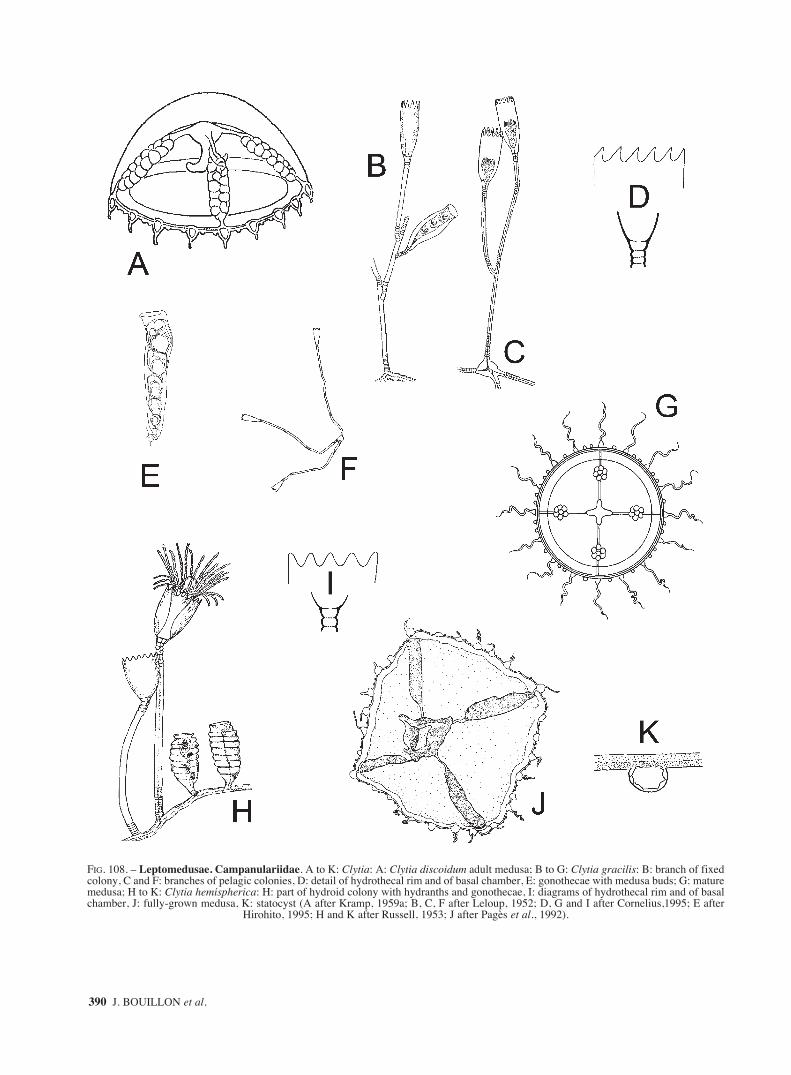
390 J. BOUILLON et al.
FIG. 108. – Leptomedusae. Campanulariidae. A to K: Clytia: A: Clytia discoidum adult medusa; B to G: Clytia gracilis: B: branch of fixedcolony, C and F: branches of pelagic colonies, D: detail of hydrothecal rim and of basal chamber, E: gonothecae with medusa buds; G: maturemedusa; H to K: Clytia hemispherica: H: part of hydroid colony with hydranths and gonothecae, I: diagrams of hydrothecal rim and of basalchamber, J: fully-grown medusa, K: statocyst (A after Kramp, 1959a; B, C, F after Leloup, 1952; D, G and I after Cornelius,1995; E after
Hirohito, 1995; H and K after Russell, 1953; J after Pagès et al., 1992).
sm68s2Gfig3 14/10/04 15:37 Página 390

FAUNA OF THE MEDITERRANEAN HYDROZOA 391
FIG. 109. – Leptomedusae. Campanulariidae. A to K: Clytia: A to E: Clytia hummelincki: A: detail of hydrothecae, B: hydrotheca and pedi-cel, C: hydrotheca, pedicel and gonotheca, D and E: gonothecae; F to I: Clytia linearis: F: part of stem with gonothecae, G: detail of part ofhydrocaulus with hydrothecae, H: detail of hydrotheca, I: detail of gonotheca; J: Clytia maccradyi: fully-grown medusa with blastostyles; K:Clytia macrogonia mature medusa (A and B after Leloup, 1935; C to E after Millard, 1975; F after Hirohito, 1995; G to I after Calder,1991;
J original after Bouillon; K after Bouillon, 1984a).
sm68s2Gfig3 14/10/04 15:37 Página 391

392 J. BOUILLON et al.
FIG. 110. – Leptomedusae. Campanulariidae. A to L: Clytia: A to E: Clytia noliformis: A and B: part of a colony; C: gonothecae; D: maturemedusa, E: detail of gonad. F to J: Clytia paulensis: F: detail of a part of colony; G: enlarged part of colony with gonotheca; H: detail ofhydrotheca; I: diagrams of hydrothecal rim and of basal chamber with oblique diaphragm; J: gonotheca. K to L: Clytia viridicans: K: alter-native life cycle pathways involving planula settlement either on the bottom or on air-water surface; L: detail of hydrothecal rim. M and N:Gastroblasta raffaelei: M: hydroid and gonotheca embedded in a sponge; N: mature medusa (A to C after Calder, 1991, D and E after Van-nucci and Ribeiro, 1955; F, I and J after Cornelius, 1995; G and H after Hirohito, 1995; K after Pagliara et al., 2000; L after Gravili et al.,
2003; M after Boero, 1980b; N after Lang, 1886).
sm68s2Gfig3 14/10/04 15:37 Página 392

FAUNA OF THE MEDITERRANEAN HYDROZOA 393
FIG. 111. – Leptomedusae. Campanulariidae. A to E: Gonothyraea loveni: A: part of colony with gonothecae and meconidiae, B: branchwith straight internodes, C: branch with flexuose internodes, D: detail of hydrotheca, E: diagrams of hydrothecal rim and of basal chamber;F to J: Hartlaubella gelatinosa: F: general view of a colony, G: detail of hydrocladium, H: detail of hydrotheca, I: diagrams of hydrothecalrim and of basal chamber, J: gonotheca; K to P : Laomedea angulata: K: general aspect of a colony, L: detail of two branches of a colony,one ending in a tendril, M: detail of hydrotheca, N: diagrams of hydrothecal rim and of basal chamber,O: male gonotheca, P: female
gonotheca (A and D after Leloup, 1952; B, C, E to P after Cornelius, 1995).
sm68s2Gfig3 14/10/04 15:37 Página 393

394 J. BOUILLON et al.
FIG. 112. – Leptomedusae. Campanulariidae. A to M: Laomedea: A to F: Laomedea calceolifera: A: a general aspect of a colony, B: detailof hydrocladia with female gonotheca, C: detail of hydrotheca, D: diagrams of hydrothecal rim and of basal chamber, E: two male gonothe-ca, F: female gonotheca; G to M: Laomedea flexuosa: G: general view of a colony, H and I: parts of hydrocladia, J: detail of hydrotheca, K:diagrams of hydrothecal rim and of basal chamber, L: female gonotheca, M: male gonotheca (a to F, J to M after Cornelius, 1995; G and H
after Hincks, 1868; I after Leloup, 1952).
sm68s2Gfig3 14/10/04 15:37 Página 394

FAUNA OF THE MEDITERRANEAN HYDROZOA 395
FIG. 113. – Leptomedusae. Campanulariidae. A to H: Laomedea: A to D: Laomedea neglecta: A: detail of a colony, B: hydrotheca, C: dia-grams of hydrothecal rim and of basal chamber, D: gonotheca; E to H: Laomedea pseudodichotoma: E: monosiphonic part of axis, F: detailof hydrotheca, G: female gonotheca, H: male gonotheca; I to M: Obelia bidentata: I: general view of a colony, J: part of hydrocladium withhydrotheca and gonotheca, K: above: detail of hydrothecal rim with uniform cusps, below: detail of hydrotheca, L: above: detail of hydrothe-cal rim with bimucronate hydrothecal rim, below diagram of basal chamber, M: gonotheca (A to D, K to M after Cornelius 1995, E after
Ramil and Vervoort, 1992a; F to H after Medel and Vervoort, 2000, I after Leloup, 1952; J after Hirohito, 1995).
sm68s2Gfig3 14/10/04 15:37 Página 395

396 J. BOUILLON et al.
FIG. 114. – Leptomedusae. Campanulariidae. A to I: Obelia: A and B: Obelia spp. A: mature medusa, B: portion of umbrella margin show-ing the position of the statocyst at the base of the tentacular bulb; C to G: Obelia dichotoma: C: general aspect of a colony, D: detail of hydro-cladial ramifications, E: below: hydrotheca with crenate rim, middle: diagram of basal chamber, above: diagram of hydrothecal rim, F: below:hydrotheca with even rim, middle: diagram of basal chamber, above: diagram of hydrothecal rim, G: gonotheca; H and I: Obelia geniculata:H: part of colony with erect unbranched stems, I: part of colony with erect branched stems (A and B after Kramp, 1959a; C to I after
Cornelius, 1995).
sm68s2Gfig3 14/10/04 15:37 Página 396

FAUNA OF THE MEDITERRANEAN HYDROZOA 397
FIG. 115. – Leptomedusae. Campanulariidae. A to E: Obelia geniculata: A: detail of a part of stem, B: portion of stem with hydranths andgonothecae, C: detail of hydrotheca, D: diagram of basal chamber, E: gonotheca; F to K: Obelia longissima: F: aspect of a large colony, G:detail of a portion of branch, H: detail of hydrotheca, I: diagrams of hydrothecal rim and of basal chamber, J: fragment of stem with a
hydrotheca and a gonotheca, K: gonotheca (A, C to H after Cornelius, 1995; B after Leloup, 1952).
sm68s2Gfig3 14/10/04 15:37 Página 397

398 J. BOUILLON et al.
116. – Leptomedusae. Campanulariidae. A to S: Orthopyxis. A to H: Orthopyxis crenata: A and B: different views of hydrothecae andpedicels, C: detail of hydrotheca, D: transverse section of a hydrotheca, E: gonotheca with male immature gonophore, F: gonotheca contain-ing male eumedusoid, G: gonothecae with immature female gonophore, H: spent female eumedusoid; I to S: Orthopyxis integra: I: generalaspect of a part of colony, J and K: different aspects of hydrothecae and pedicels, L: detail of hydrotheca, M: diagrams of hydrothecal rimand of basal chamber of a thickened hydrotheca, N: diagrams of hydrothecal rim and of basal chamber of unthickened hydrotheca, O: groovedwalled gonotheca, P: smooth walled gonotheca, Q: gonotheca with male gonophore, R: gonotheca with female gonophore, S: released eume-dusoid (A after Medel and Vervoort, 2000; B, E to H, K, Q and R after Hirohito, 1995; C and D after Millard, 1975; I, M to P, S after
Cornelius, 1995; J and L after Cornelius, 1982).
sm68s2Gfig3 14/10/04 15:38 Página 398

FAUNA OF THE MEDITERRANEAN HYDROZOA 399
117. – Leptomedusae. Campanulariidae. A: Pseudoclytia pentata: mature medusa. Limnomedusae. Armorhydridae. B and C: Armorhy-dra janowiczi: B: medusa; C: longitudinal histological section of a medusa. Microhydrulidae. D and E: Microhydrula pontica: D: histolog-ical section of a polyp; E: histological section of a frustule. F: Rhaptapagis cantacuzenei, histological section of a polyp (A after Kramp,1959a; B after Thiel, 1988; C after Lacassagne, 1968a; D to F after Bouillon and Deroux, 1967). cbl: cnidoblast; cnl: cnidocil; di: digestiveinclusion; ecd: ectoderm; end: endoderm; eng: endodermal gap; g: gonads; gc: gastric cavity; glc: glandular cell, ma: manubrium; meu:
microbasic eurytele; p: periderm; sc: subumbrellar cavity; seu: semiophoric eurytele; spt: septum; te: tentacle; v: velum.
sm68s2Gfig3 14/10/04 15:38 Página 399

400 J. BOUILLON et al.
FIG. 118. – Limnomedusae. Olindiidae. A to F: Craspedacusta sowerbii: A and B: two aspects of polyp colonies with a medusa bud; C: apolyp colony reducing itself in frustules and resting stages or cysts; D: fully-grown medusa; E: portion of umbrella showing the marginalcnidocyst ring and tentacular roots; F: portion of the velum with the centripetal tubes of the statocysts. G to I: Gonionemus vertens: G:hydranth; H and I: two hydranths developing a fustule. J: hydranth with a medusa bud (A and C after Damas, 1939; B, E and F after Russell,
1953, D after Tardent, 1978; G and I after Leloup, 1952; H and J after Werner, 1984). cy: cyst; fr: frustule; sta: statocyst, v: velum.
sm68s2Gfig3 14/10/04 15:38 Página 400

FAUNA OF THE MEDITERRANEAN HYDROZOA 401
FIG. 119. – Limnomedusae. Olindiidae. A to D: Gonionemus vertens: A and B: fully-grown medusae, C: exumbrellar margin showing thestatocysts, the adhesive pad and the sharply bended tentacular end, D: detail of the terminal end of a tentacle with adhesive pad; E and F:Gossea corynetes: E: mature medusa, F: schematic view of a portion of the umbrella margin sowing the disposition of the two types of ten-tacles and of the statocysts; G and H: Maeotias marginata (= M. inexpectata): G: whole medusa, H: portion of the umbrella margin showingthe base of the oldest tentacles with the tips twitch of (A and C after Leloup, 1952; B and D after Russell, 1953; E after Mayer, 1910; F after
Bouillon, 1978a; G after Borcea, 1929, H after Denayer, 1973).
sm68s2Gfig3 14/10/04 15:38 Página 401

402 J. BOUILLON et al.
FIG. 120. – Limnomedusae. Olindiidae. A: Monobrachium parasiticum: colony with almost mature eumedusoid; B to E: Olindias phos-phorica: B: hydroid, C and D: fully-grown medusae; E: adhesive pad on the aboral side of a primary tentacles. F to H: Scolionema suvaen-sis: F: polyp; G and H: adult medusae (A after Hand, 1957, B after Weill, 1936; C and G after Kramp, 1959a; D after Trègouboff, 1957; E
and H after Mayer, 1910; F reconstruction after Goy, 1973). dac: dactylozooid; eum: eumedusoid; ga: gastrozooid; p: periderm.
sm68s2Gfig3 14/10/04 15:38 Página 402

FAUNA OF THE MEDITERRANEAN HYDROZOA 403
FIG. 121. – Limnomedusae. Olindiidae. A: Calpasoma dactyloptera: hydranths showing different stages of reproduction. Siphonophorae.Physaliidae. B to D: Physalia physalis: B: colony; C: cluster of persons from sexualy mature colony; D: small part of a gonodendron. Rhyso-physidae. E and F: Rhizophyla filiformis: E: colony; F: tentilla (A after Matthews, 1966; B and E after Pagès and Gili, 1992; C and D after
Hyman, 1940; F after Pugh, 1999).
sm68s2Gfig3 14/10/04 15:38 Página 403

404 J. BOUILLON et al.
FIG. 122. – Siphonophorae. Agalmatidae. A to I: Agalma: A to C: Agalma clausi: A: nectophore; B: bract; C: tentilla. D to F: Agalma ele-gans: D: detail of a lateral view of a nectophore; E: detail of an upper view of a nectophore; F: bracts. G to I: Agalma okeni: G and H: upperand lateral view of nectophore; I: bract (A to C after Bedot, 1888; D to E after Totton, 1965; G and I after Pugh, 1999; H after Gili, 1986).alr: apico-lateral ridge; aw: apical wings; ilr: infra-lateral ridge; lr: lateral ridge; n: nectophore; os: ostium; rc: radial canal; thb: thrust block;
vlr: vertical lateral ridge.
sm68s2Gfig3 14/10/04 15:38 Página 404

FAUNA OF THE MEDITERRANEAN HYDROZOA 405
FIG. 123. – Siphonophorae. Agalmatidae. A to B: Cordalgama cordiformis: A: nectophore (ostial view); B: bract. C to G: Halistemmarubrum: C: polygstric stage; D: nectophore (lateral view); E: nectophore (from below); F: bract; G: tentillum. H and I: Lychnagalma utricu-laria: H: nectophore (upper dorsal view); I: nectophore (ostial view) (A after Pagès and Gili, 1992; C after Trègouboff, 1957; B, D to F afterPugh, 1999; G after Hyman, 1940, H and I after Pugh and Harbison, 1986). alr: apico-lateral ridge; cnb: cnidoband; fe: end of the tentillium
filament; ilr: infr-lateral ridge; inv: involucrum; lr: lateral ridge; vlr: vertical lateral ridge.
sm68s2Hfig4 14/10/04 15:39 Página 405

406 J. BOUILLON et al.
FIG. 124. – Siphonophorae. Agalmatidae. A to D: Lychnagalma utricularia. A: nectophore (lower ventral view), B: bracts (Lychnalgamaspp.), C: general view of four tentilla, note the coiled cnidoband enclosed within the involucrum and the terminal vesicle, D: detail of baseof a detached terminal vesicle showing the octoradial filaments. E to F: Marrus orthocanna: E: upper view of nectophore, F: lateral view of
nectophore, G: bract (A to D after Pugh and Harbison, 1986; E and F after Pugh, 1999; G after Gili, 1986).
sm68s2Hfig4 14/10/04 15:39 Página 406

FAUNA OF THE MEDITERRANEAN HYDROZOA 407
FIG. 125. – Siphonophorae. Agalmatidae. A to D: Nanomia bijuga: A: polygastric stage; B: upper view of nectophore; C: bract; D: part ofan internode of the stem showing alternating male and female gonodendra arising from the base of a series of palpons. (A and D after Tot-ton, 1965; B and C after Pugh, 1999); br: bract; fg: female gonophore; ga: gastrozooid; mg: male gonophore; n: nectophore; pal: palpon; pne:
pneumatophore; te: tentacle; ten: tentilla.
sm68s2Hfig4 14/10/04 15:39 Página 407

408 J. BOUILLON et al.
FIG. 126. – Siphonophorae. Apolemiidae. A to D: Apolemia uvaria: A: polygastric stage; B: nectophore (lateral view); C: nectophore (ostialview); D: bracts. Athorybiidae. E and F: Athorybia rosacea: E: polygastric stage (lateral view); F: dorsal view of a polygastric stage.Forskaliidae. G to K: Forskalia asymmetrica: G and H: inner and outer view of nectophores; I: inner and outer view of stem bratcs; J: innerview of bolster bract; K: inner view of knee-shaped bracts (A to D after Totton, 1965; E after Pugh, 1999; F after Trègouboff, 1957; G to K
after Pugh, 2003). lrc: lateral radial canal; os: ostium; ped: peduncle.
sm68s2Hfig4 14/10/04 15:39 Página 408

FAUNA OF THE MEDITERRANEAN HYDROZOA 409
127. – Siphonophorae. Forskaliidae. A to K: Forskalia. A to F: Forskalia contorta: A: polygastric stage, B: nectophore (upper view), C:nectophore (inner view), D: stem bracts, E: knee bracts, F: third type of bract; G and H: Forskalia edwardsi: G: nectophore (upper view), H:variuos types of bracts; I to K: Forskalia formosa: I: nectophore (upper view), J: nectophore (lower view), K: various types of bracts (A after
Trègouboff, 1957, B to F, I to K after Pugh, 2003; G and H after Kirkpatrick and Pugh, 1984).
sm68s2Hfig4 14/10/04 15:39 Página 409

410 J. BOUILLON et al.
FIG. 128. – Siphonophorae. Physophoridae. A to C: Physophora hydrostatica: A: polygastric stage, B: nectophore (upper view), C: palponAbylidae. D to F: Abyla haeckeli: D: posterior nectophore, E: ventral and lateral views of anterior nectophore, F: eudoxid; G to K: Abylop-sis eschscholtzi: G: polygastric stage (lateral view), H: anterior nectophore, I: posterior nectophore, J: eudoxid, K: bract. (A to C after Kirkpatrick and Pugh, 1984; D and I after Gili, 1986; E, H and K after Pugh, 1999; F after Totton, 1965; G and J after Pagès and Gili, 1992).
sm68s2Hfig4 14/10/04 15:39 Página 410

FAUNA OF THE MEDITERRANEAN HYDROZOA 411
FIG. 129. – Siphonophorae. Abylidae. A to E: Abylopsis tetragona: A: polygastric stage; B: posterior nectophore; C: anterior nectophore;D: eudoxid bract; E: gonophore. F to I: Bassia bassensis: F: polygastric stage (lateral view); G: polygastric stage (dorsal view); H: anteriornectophore (dorsal view); I: eudoxid (latero-ventral view) (A and G after Totton, 1965; B to E after after Kirckpatrick and Pugh, 1984; F, Hand I after Pagès and Gili, 1992). an: anterior nectophore; com: comb; dmt: dorso median tooth; dr: dorsal ridge; hy: hydroecium; ldt: leftdorsal tooth; ler: left ridge; lhw: left hydroecial wing; lvt: left ventral tooth; ns: nectosac; pac: pallial canal; pec: pedicular canal; pn:
posterior nectophore; rdt: right dorsal tooth; re: rete; rhw: right hydroecial wing; rir: right ridge; s: somatocyst.
sm68s2Hfig4 14/10/04 15:39 Página 411

412 J. BOUILLON et al.
FIG. 130. – Siphonophorae. Abylidae. A to C: Ceratocymba sagittata: A: polygastric stage; B: eudoxid bract; C: gonophore. D to F:Enneagonum hyalinum: D: polygastric stage (lateral view); E: eudoxid bract (lateral view), F: gonophore. Clausophyidae. G to I: Clausophyesovata: G: anterior nectophore; H: posterior nectophore; I: eudoxid bract. Diphyidae. J and K: Chelophyes appendiculata: J: polygastric stage;K: anterior nectophore (A after Totton, 1965; B, C, F to K after Kirckpatrick and Pugh, 1984; E, D, after Pagès and Gili, 1992). an: anterior
nectophore; dt: dorsal tooth; lvt: left ventral tooth; ns: nectosac; pex: pyramidal extension; pn: posterior nectophore; s: somatocyst.
sm68s2Hfig4 14/10/04 15:39 Página 412

FAUNA OF THE MEDITERRANEAN HYDROZOA 413
FIG. 131. – Siphonophorae. Diphyidae. A to C: Chelophyes appendiculata: A: posterior nectophore, B: detail of mouth-plate of posteriornectophore (dorsal view), C: eudoxid stage (dorsal view); D to F: Chelophyes contorta: D: anterior nectophore (ventral view), E: anterior nec-tophore (lateral view), F: posterior nectophore; G to I: Dimophyes arctica: G: anterior nectophore, H: posterior nectophore, I eudoxid stage;J and K: Diphyes bojani: J: anterior nectophore (lateral view), K: posterior nectophore (lateral view). (A, B, C, G to I after Kirkpatrick and
Pugh, 1984; D to F, J and K after Pagès and Gili, 1992).
sm68s2Hfig4 14/10/04 15:39 Página 413

414 J. BOUILLON et al.
FIG. 132. – Siphonophorae. Diphyidae. A to C: Diphyes dispar: A: polygastric phase, B: anterior nectophore (lateral view), C: bract withgonophore; D to H: Eudoxoides spiralis: D: polygastric stage, E: lateral view of a whole animal, F: enlarged view of the base of figure E, G:lateral view of a detached femelle gonophore, H: ventral view of the bract (A after Trègouboff, 1957; B and C after Pugh, 1999; D to H after
Totton, 1965).
sm68s2Hfig4 14/10/04 15:39 Página 414

FAUNA OF THE MEDITERRANEAN HYDROZOA 415
FIG. 133. – Siphonophorae. Diphyidae. A to I: Lensia: A and B: Lensia campanella: A: anterior nectophore (lateral view); B: bract withgonophore. C to E: Lensia conoidea: C: anterior nectophore; D: posterior nectophore; E: eudoxid stage (lateral view). F to H: Lensia fowleri:F: anterior nectophore; G: posterior nectophore; H: eudoxid bract (lateral view). I: Lensia hotspur: anterior nectophore (lateral view) (A, B and
I after Pugh, 1999; C to H after Kirkpatrick and Pugh, 1984). br: bract; ga: gastrozooid; go: gonophore; nsh: neck-shield; phy: phyllocyst.
sm68s2Hfig4 14/10/04 15:39 Página 415

416 J. BOUILLON et al.
FIG. 134. – Siphonophorae. Diphyidae. A to F: Lensia: A: Lensia meteori: anterior nectophore (lateral view); B to D: Lensia multicristata:B: anterior nectophore, C: posterior nectophore, D: eudoxid bract (all lateral view). E to G: Lensia subtilis: E: anterior nectophore, F:
posterior nectophore, G: eudoxid bract (all lateral view) (all after Kirkpatrick and Pugh, 1984).
sm68s2Hfig4 14/10/04 15:39 Página 416

FAUNA OF THE MEDITERRANEAN HYDROZOA 417
FIG. 135. – Siphonophorae. Diphyidae. A and B: Lensia subtiloides: A: anterior nectophore (lateral view), B: bract with gonophore; C to F:Muggiaea: C and D: Muggiaea atlantica: C: anterior nectophore (lateral view), D: bract; E: Muggiaea kochi: E: anterior nectophore, F: Mug-giaea sp.: eudoxid stage (all lateral view); G to I: Sulculeolaria. G to I: Sulculeolaria biloba: G: anterior nectophore (lateral view), H: pos-terior nectophore (lateral view), I: mouth-plate, detail of H (dorsal view) (A after Pagès and Gili, 1992; B and D after Pugh, 1999; C, E, F to
I after Kirkpatrick and Pugh, 1984).
sm68s2Hfig4 14/10/04 15:39 Página 417

418 J. BOUILLON et al.
FIG. 136. – Siphonophorae. Diphyidae. A to F: Sulculeolaria. A and B: Sulculeolaria chuni: A: anterior nectophore, B: posterior nectophore(lateral views); C and D: Sulculeolaria quadrivalvis: C: anterior nectophore, D: posterior nectophore (lateral views); E and F: Sulculeolaria turgi-da: E: anterior nectophore, F: posterior nectophore (lateral views) (A and B after Pagès and Gili, 1992; C to F after Kirkpatrick and Pugh, 1984).
sm68s2Hfig4 14/10/04 15:39 Página 418

FAUNA OF THE MEDITERRANEAN HYDROZOA 419
FIG. 137. – Siphonophorae. Hippopodiidae. A to D: Hippopodius hippopus: A: polygastric stage; B: definitive nectophore; C: larval nec-tophore; D: schema of the disposition of the nectophores in a colony. E to H: Vogtia: E and F: Vogtia glabra: E: definitive nectophore (dorsalview); F: young stage; G: Vogtia pentacantha: definitive nectophore (dorsal view); H: Vogtia serrata: definitive nectophore (dorsal view) (A toC, E to H after Kirkpatrick and Pugh, 1984; D after Trègouboff, 1957). cm: cormidia; n1, n2, n3, n4, n5: nectophores; ol5: oleocyte of nectophore;
sto: stolon.
sm68s2Hfig4 14/10/04 15:39 Página 419

420 J. BOUILLON et al.
FIG. 138. – Siphonophorae. Hippopodiidae. A: Vogtia spinosa, definitive nectophore, (dorsal view). Prayidae. B and C: Amphicaryonacaule: B: nectophores of polygastric stage, C: eudoxid stage; D to F: Desmophyes annectens: D: polygastric stage, E: nectophore (lateral
view), F: eudoxid bract with gonophore. (A to C, E after Kirkpatrick and Pugh, 1984; Dand F after Kawamura, 1915).
sm68s2Hfig4 14/10/04 15:39 Página 420

FAUNA OF THE MEDITERRANEAN HYDROZOA 421
FIG. 139. – Siphonophorae. Prayidae. A to C: Desmophyes villafranca: A: polygastric stage; B: nectophores (apical view); C: cormidia (api-cal view). D to F: Lilyopsis rosea: D: polygastric stage, E: nectophore (lateral view); F: male cormidia detached of the stolon (A to c afterCarrè, C 1969a; D to F after Carrè Cand Carrè D, 1995). bz: budding zone; br: bract; pec: pedicular canal; blc: bracteal longitudinal canal;dc: dorsal canal; rhc: right hydroecial canal; lhc: left hydroecial canal; cm: cormidia; vc: ventral canal; ff: fishing filament; ga: gastrozooid;mg: male gonophore; mc: mantel canal; vc: ventral canal; hy: hydroecium; llo: left lobe; rlo: right lobe; n1 and n2:nectophores 1 and 2; pgl:pigmentary lamella; mte: marginal tentacle; s1: somatocyst of nectophore 1; s2:somatocyst of nectophore 2; pgs: pigmentary spot; sto: stolon.
sm68s2Hfig4 14/10/04 15:39 Página 421

422 J. BOUILLON et al.
FIG. 140. – Siphonophorae. Prayidae. A to D: Prayola tottoni: A: polygastric stage; B: cormidia from below; C: gonophore (ventral view);D: nectophore (ventral view). E and F: Rosacea cymbiformis: E: young polygastric stage; F: two definitive nectophores (A to D after CarrèC, 1969c; E and F after Trègouboff, 1957). blc: bracteal longitudinal canal; br: bract; bz: budding zone; ft: fishing tentacle; ga: gastrozooid;go: gonophore; hy: hydroecium; lhc: left hydroecial canal; mc: mantel canal; pec pedicular canal; rhc: right hydroecial canal; s: somatocyst;
sto: stolon; vc: ventral canal.
sm68s2Hfig4 14/10/04 15:39 Página 422

FAUNA OF THE MEDITERRANEAN HYDROZOA 423
FIG. 141. – Siphonophorae. Prayidae. A to C: Rosacea: A: Rosacea cymbiformis: adult cormidia; B and C: Rosacea plicata: B: nectophore(lateral view); C: eudoxid bract (dorso-lateral view). Sphaeronectidae: D to G: Sphaeronectes: D and E: Sphaeronectes bougisi: nectophores;D: lateral view; E: ventral view. F and G: Sphaeronectes fragilis: nectophores: F: lateral view; G: apical view (A after Trègouboff, 1957; Band C after Kirkpatrick and Pugh, 1984; D and E after Carrè, 1968d; F and G after Carrè, 1968c). brc: bracteal canal; br: bract; fg: femalegonophore; ga: gastrozooid; hy: hydroecium; llc: left lateral canal; pec: pedicular canal; rlc: right lateral canal; s: somatocyst; si: siphono-
some; sto: stolon; te: tentacle; ten: tentillum.
sm68s2Hfig4 14/10/04 15:39 Página 423

424 J. BOUILLON et al.
FIG. 142. – Siphonophorae. Sphaeronectidae. A to G: Sphaeronectes: A to D: Sphaeronectes gamulini: A to C: nectophores: A: lateral view;B: apical view; C: ventral view. D: eudoxia. E and F: Sphaeronectes gracilis: E: polygastric stage; F: eudoxid stage. G: Sphaeronectes irreg-ularis: nectophore (A to D after Carrè, 1966, E after Pagès and Gili, 1992; F and G after Trègouboff, 1957). br: bract; ga: gastrozooid; go:
gonophore; ol: oleocyte; sto: stolon.
sm68s2Hfig4 14/10/04 15:39 Página 424

FAUNA OF THE MEDITERRANEAN HYDROZOA 425
FIG. 143. – Actinulidae. Halammohydridae. A to D: Halammohydra: A: Halammohydra octopodides general view; B to D: Halammohy-dra schulzei: B: general view; C: structural organization; D: above young actinula, below halhydrula. Otohydridae. E and F: Otohydravagans: E: general view; F: structural organization (A to E after Swedmark and Tesssier, 1966; F after Swedmark and Tessier, 1958c). ac:aboral cone; agt: tentacle of the aboral girdle; ao: adhesive organ; b: tentacular bulb; emb: embryo; gc: gastric cavity; ma: manubrium; mo:
mouth; nr: nerve ring; sc: subumbrellar cavity; sgt: tentacle of the subaboral girdle; sta: statocyst.
sm68s2Hfig4 14/10/04 15:39 Página 425

426 J. BOUILLON et al.
FIG. 144. – Narcomedusae. Aeginidae. A: Aegina citrea mature medusa; B: Solmundella bitentaculata fully-grown medusa. Cuninidae. C:to E: Cunina: C and D: Cunina globosa: C: adult medusa , D: portion of umbrella; E: Cunina octonaria mature medusa (A after Mayer, 1910;
B and C after Kramp, 1959a; D after Gili et al., 1998; E after Bouillon, 1987).
sm68s2Hfig4 14/10/04 15:39 Página 426

FAUNA OF THE MEDITERRANEAN HYDROZOA 427
FIG. 145. – Narcomedusae. Cuninidae. A to E: Cunina: A: Cunina octonaria mature medusa; B and C: Cunina proboscidea: B: wholemedusa; C: radial section through a statocyst and an otoporpae. D and E: Cunina simplex: D: aboral view; E: oral view (A after Bouillon,1987; B and C after Mayer, 1910; D and E after Gili et al., 1998). ex: exumbrella; otp: otoporpae; sco: solid core replacing the ring canal;
sta: statocyst; su: subumbrella; v: velum.
sm68s2Hfig4 14/10/04 15:39 Página 427

428 J. BOUILLON et al.
FIG. 146. – Narcomedusae. Cuninidae. A to D: Solmissus. A and B: Solmissus albescens: A: fully-grown medusa; B: portion of umbrellamargin; C and D: Solmissus incisa: C: whole medusa; D: portion of umbrella (A and B after Mayer, 1910; C after Russell, 1953; D afterBroch, 1929) ex: exumbrella; map: manubrial pouches; mo: mouth; otp: otoporpae; sco: solid core replacing the ring canal; sta: statocyst;
su: subumbrella; te: tentacle; v: velum.
sm68s2Hfig4 14/10/04 15:39 Página 428
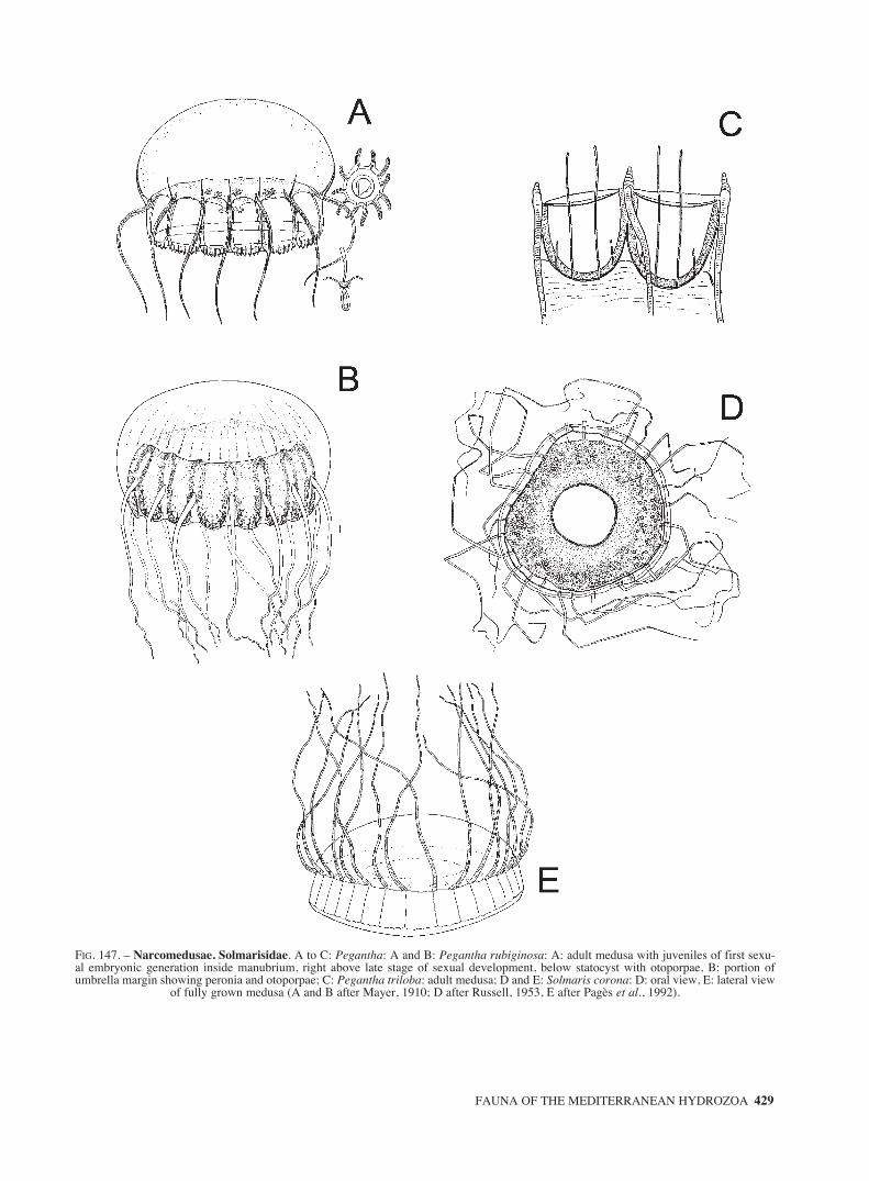
FAUNA OF THE MEDITERRANEAN HYDROZOA 429
FIG. 147. – Narcomedusae. Solmarisidae. A to C: Pegantha: A and B: Pegantha rubiginosa: A: adult medusa with juveniles of first sexu-al embryonic generation inside manubrium, right above late stage of sexual development, below statocyst with otoporpae, B: portion ofumbrella margin showing peronia and otoporpae; C: Pegantha triloba: adult medusa; D and E: Solmaris corona: D: oral view, E: lateral view
of fully grown medusa (A and B after Mayer, 1910; D after Russell, 1953, E after Pagès et al., 1992).
sm68s2Hfig4 14/10/04 15:39 Página 429

430 J. BOUILLON et al.
FIG. 148. – Narcomedusae. Solmarisidae. A to E: Solmaris: A to C: Solmaris flavescens: A: whole medusa; B: diagrammatic section of amedusa; C: detail of exumbrella. D: Solmaris leucostyla: fully-grown medusa; E: Solmaris solmaris: adult medusa. Narcomedusae devel-opment. F: Solmundella bitentaculata parasitized by Cunina peregrina at different stages of reproduction; G: different stages of developmentof Pegantha triloba parasitizing an Anthomedusae Pseudotiara tropica (A to C after Mayer, 1910; D after Gili, 1986; E after Trègouboff,1957; F and G after Bouillon, 1897). ex: exumbrella; fxt: tentacles of the primary larval stage serving for fixation; g: gonad; hm: manubriumof the host; mac: manubrium cavity; mb1, mb2 and mb3: successive stages of medusa buds formed by the primary larval parasitic stage; mcr:median crests of umbrellar lobes of Pegantha triloba medusae buds; mec: marginal endodermal core; nr: nerve ring; pls: primary larval stage;
su: subumbrella; te: tentacle; v: velum.
sm68s2Hfig4 14/10/04 15:39 Página 430

FAUNA OF THE MEDITERRANEAN HYDROZOA 431
FIG. 149. – Narcomedusae development (continued). Development of Cunina octonaria parasitizing Turritopsis nutricula, Anthomedusae.A: Young embryo (Em), blastula, in his phorocyte developing in the gastro-vascular pouches of the Cunina mother, B: free planula stage; C:bitentaculate primary larvae issued from the planula, D: primary larvae with four tentacles and manubrial tube, ready to parasite its futurehost, E: primary larvae fixed by the tentacles on the subumbrellar ectoderm of the Turritopsis host, F: primary larvae budding of secondarylarvae from their aboral end, G: young Cunina issued from a parasitic larvae, H: adult medusae from Cunina octonaria (copied from
Bouillon, 1987).
sm68s2Hfig4 14/10/04 15:39 Página 431

432 J. BOUILLON et al.
FIG. 150. – Trachymedusae. Geryoniidae. A to C: Geryonia proboscidalis. A and B: general view, C: statocyst. D to I: Liriope tretraphyl-la: D: adult medusa parasitised by Cunina octonaria; E: general view of adult medusa; F: base of a solid interradial marginal tentacle with acentripetal canal and statocyst; G: lateral view of a solid marginal tentacle showing a statocysts and the marginal cnidocyst ring of the umbrel-la; H: terminal portion of a hollow perradial tentacle; I: portion of margin of mouth-lip (A after Hyman, 1940; B after Trègouboff, 1957; C
after Hertwig O and R., 1878; D after Bouillon, 1987; E after Trègouboff, 1957; F, G, H and I after Russell, 1953).
sm68s2Hfig4 14/10/04 15:39 Página 432

FAUNA OF THE MEDITERRANEAN HYDROZOA 433
FIG. 151. – Trachymedusae. Halicreatidae. A to E: Haliscera: A and B: Haliscera bigelowi: A: fully-grown medusa, B: left: stiff distal endof marginal tentacle, middle: flexible proximal portion of marginal tentacle, right: statocyst; C: Haliscera conica mature medusa; D and E:Haliscera racovitzae: D: lateral view of mature medusa, E: oral view; F: Halitrephes maasi whole mature medusa. Petasidae. G: Petasus
atavus fully-grown medusa (A, F and G after Kramp, 1959a; B after Russell, 1953; C, D and E after Mayer, 1910).
sm68s2Hfig4 14/10/04 15:39 Página 433

434 J. BOUILLON et al.
FIG. 152. – Trachymedusae. Ptychogastriidae. A to C: Ptychogastria asteroides: A: lateral view of an adult medusa, B: detail of marginalborder, C: upper view of the umbrella of a juvenile specimen. Rhopalonematidae. D to F: Aglantha: D and E: Aglantha digitale: lateral viewsof adult medusae; F: Aglantha elata: fully-grown medusa; G: Aglaura hemistoma: mature medusa; H: Amphogona pusilla: adult medusa; Iand J: Arctapodema ampla: fully grown medusae (A to C after Gili et al., 1999; D after Hyman, 1940; E after Broch, 1929; F to I after Kramp,
1959a; J after Mayer, 1910).
sm68s2Hfig4 14/10/04 15:39 Página 434

FAUNA OF THE MEDITERRANEAN HYDROZOA 435
FIG. 153. – Trachymedusae. Rhopalonematidae. A to C: Arctapodema australis: A: lateral view of a mature medusa, B and C: aboral viewsof manubrium and gonads; D to G: Homoeonema platygonon: D: lateral view of mature medusa, E: detail of umbrella margin, F: aboral viewof a juvenile specimen, G: aboral view of a mature specimen; H to J : Pantachogon haeckeli: H and I: aspect of two fully grown medusae, J:portion of umbrella margin (A and C after Vanhoeffen, 1913; B after Kramp, 1959a; D to G after Gili et al., 1998; H and I after Mayer, 1910;
J after Russell, 1953).
sm68s2Hfig4 14/10/04 15:39 Página 435

436 J. BOUILLON et al.
FIG. 154. – Trachymedusae. Rhopalonematidae. A: Pantachogon haeckeli: apical view of the summit of the umbrella showing the manubri-um, the radial canals and the muscle fields; B: Pantachogon militare: fully-grown medusa; C and D: Persa incolorata: C: adult medusa, D:juvenile medusa; E to G: Ransonia krampi: E and F: lateral views of mature medusae, G: detail of manubrium (A after Russell, 1953; B, C
and D after Mayer, 1910; E after Gili, 1986; F and G after Kramp, 1959a).
sm68s2Hfig4 14/10/04 15:39 Página 436

FAUNA OF THE MEDITERRANEAN HYDROZOA 437
FIG. 155. – Trachymedusae. Rhopalonematidae. A to F: Rhopalonema. A to D: Rhopalonema funerarium: A: fully grown medusa, B: detailof manubrium, C: detail of interradial marginal tentacle, D: portion of umbrella margin; E and F: Rhopalonema velatum: E: mature medusa,F: statocyst; G to I: Sminthea eurygaster: G and H: lateral views of adult medusae, I: enlarged view of a gonad; J to L: Tetrorchis erythro-gaster: J: fully grown medusa, K: base of a small tentacle with rounded endodermic tentacular root, L: side view of the umbrella showing theposition of the a gonad and the endodermic tentacular root of a perradial tentacle (A to D after Russell, 1953; E, G to I after Mayer, 1910; F
after Hertwig and Hertwig, 1878; J to L after Bigelow, 1909).
sm68s2Hfig4 14/10/04 15:39 Página 437

438 J. BOUILLON et al.
FIG. 156. – Polypodiozoa. Polypodiidae. Polypodium. Life cycle of Polypodium hydriforme internal parasite of Acipenserid fishes. A: gen-eral cycle: 1: reversed stolon prolifer; 2: stolon prolifer living an infected acipenserid egg; 3: stolon prolifer in the water; 4: fragment of astolon; 5: polyp with 12 tentacles; 6: polyp with 24 tentacles; 7: polyp with 6 tentacles; 8: female polyp; 9: male polyp; 10: binucleate cellparasite of an acipenserid oocyte; 11: morula encapsulated in the trophamion; 12: planula; 13: budding planula; 14: stolon prolifer withouttentacles; 15: stolon prolifer reversed with internal tentacles. B: diagram of the formation of the trophamion: a: sexual medusa; b: gonad withbinucleated cells; c: binucleated cells; d to g: binucleated cells parasite of a fish oocyte; h: segmentation of the embryo; i: morula stage insidethe trophamion; j: planula stage inside the trophamion. C: ovary of an Acipenser, the large eggs are parasitised, the small ones are not. D: ato c: reversed buds with internal tentacles on a stolon prolifer (St.); d: stolon prolifer with external tentacles; e: medusae liberated from an
infected fish egg (Redrawn from Bouillon, 1987).
sm68s2Hfig4 14/10/04 15:39 Página 438

Abietinaria 249, 287Abietinaria abietina 286, 287Abietinella 149Abyla 215Abyla haeckeli 215, 410Abyla spp. 253Abylopsis 215Abylopsis eschscholtzi 215, 410Abylopsis tetragona 11, 216, 411Acanthocladium 126Acaulis 20Acauloides 20, 86Acauloides ammisatum 86, 86, 329Acauloides ilonae 86, 86, 329Acipenser 438Acryptolaria 154, 154Acryptolaria conferta 155, 365Acryptolaria operculata 155Actigia 94Aegina 232, 232Aegina citrea 233, 295, 426Aeginura 232Aequorea 22, 117, 259, 261, 117, 293, 342, 343Aequorea conica 117, 117, 342Aequorea forskalea 117, 118, 342Aequorea macrodactyla 259Aequorea pensilis 24, 117, 118, 343Aequorea philippina 117Agalma 210, 255, 404Agalma clausi 210, 404Agalma elegans 210, 296, 404Agalma okeni 210, 404Agastra rubra 202Aglantha 241, 241, 261, 293, 434Aglantha digitale 241, 241, 434Aglantha elata 434, 241, 241Aglaophenia 119, 119, 289, 290, 344,
344, 346, 347Aglaophenia acacia 120, 120, 344Aglaophenia elongata 120, 120, 344Aglaophenia harpago 119, 120, 344Aglaophenia kirchenpaueri 119, 121, 345Aglaophenia lophocarpa 120, 121, 345Aglaophenia octodonta 120, 121, 345Aglaophenia parvula 120, 122, 346Aglaophenia picardi 119, 122, 346Aglaophenia pluma 120, 122, 346Aglaophenia pluma parvula 122
Aglaophenia spp. 10Aglaophenia tubiformis 119, 123, 347Aglaophenia tubulifera 119, 123, 347Aglaophenopsis 123Aglaura 241, 241Aglaura hemistoma 14, 241, 298, 434Altairina 93Amalthaea 91Amphicaryon 227Amphicaryon acaule 227, 420Amphinema 13, 67, 67, 250, 261, 293, 322Amphinema dinema 68, 68, 322Amphinema rubra 68, 68, 249, 322Amphinema rugosa 68, 263, 322Amphinema rugosum 68, 69Amphinema turrida 69, 322Amphisbetia 29, 176, 177Amphisbetia operculata 176, 177, 303, 377Amphogona 26, 240, 241Amphogona pusilla 242, 434Anamathia rissoana 340Antennella 144, 144, 252, 359Antennella ansini 144, 144Antennella diaphana f. siliquosa 146Antennella secundaria 144, 145, 359, 360Antennella siliquosa 144, 145, 360Antennellopsis 144Anthohebella 29, 150, 263, 150, 151Anthohebella darwinensis 150Anthohebella parasitica 150, 362Anthophysa 261Antnenella ansini 360Apolemia 13, 212Apolemia uvaria 212, 408Aporrhais 319Arctapodema 240, 242Arctapodema ampla 242, 242, 434Arctapodema australis 242, 242, 435Arctapodema sp. 242Armorhydra 202, 203Armorhydra janowiczi 203, 399Aselomaris 41, 48Asyncoryne 19Athorybia 258, 213Athorybia rosacea 212, 408
Barcino 128Barcino foixensis 128, 349
FAUNA OF THE MEDITERRANEAN HYDROZOA 439
TAXONOMIC INDEX
(bold, full description of species and genus; italics, draws and schemes)
sm68s2Iindex 14/10/04 16:05 Página 439

Bassia 216Bassia bassensis 216, 411Bathyphysa 261Bedotella 149, 150, 150Bedotella armata 150, 362Bimeria 19, 28, 41, 42, 42Bimeria rigida 42Bimeria vestita 21, 42, 307Blackfordia 22, 129, 264Blackfordia virginica 129, 349, 350Bonneviella 19Bonneviella enterovillosa 288Boreohydra 86, 86Boreohydra simplex 87, 329Bougainvillia 20, 42, 42, 263, 293, 307Bougainvillia aurantiaca 43, 43, 291, 307Bougainvillia britannica 42, 43, 43, 291Bougainvillia conferta 45Bougainvillia macloviana 43Bougainvillia maniculata 43, 43, 307Bougainvillia multicilia 42, 44, 293Bougainvillia muscus 43, 44, 307, 308Bougainvillia niobe 28, 43, 44, 254, 308Bougainvillia platygaster 28, 43, 44, 308Bougainvillia principis 293Bougainvillia ramosa 21Branchiocerianthus 20, 90, 91Branchiocerianthus imperator 18, 19, 91Branchiocerianthus italicus 91Bythotiara 50, 50Bythotiara murayi 50, 312
Calamphora 249Calpasoma dactyloptera 207, 303Calpasoma 202, 207Calycella 130, 130Calycella gracilis 130Calycella syringa 27, 130, 133, 287, 350Calycopsis 50, 50, 250, 251Calycopsis simplex 50, 312Calyptospadix 42Campalecium 138, 161, 161, 166, 369Campalecium cirratum 162, 162, 166, 369Campalecium medusiferum 162, 162, 164, 166, 369Campanopsis 134, 250Campanularia 12, 150, 191, 192, 389Campanularia assymetrica 201, 201Campanularia everta 15Campanularia groenlandica 287Campanularia hincksi 192, 192, 289, 389Campanularia raridentata 192, 389Campanularia volubilis 192, 192, 389
Campanulina 129, 131, 132, 132, 134, 161,162, 164, 250
Campanulina panicula 132, 287, 350Campanulina tenuis 129, 132Campomma 162Campomma hincksii 162Candelabrum 19, 20, 87Candelabrum capensis 19Candelabrum cocksi 87, 330Candelabrum penola 18, 19Caulerpa spp. 7Caulerpa taxifolia 7Ceratocymba 217Ceratocymba sagittata 217, 412Chelophyes 218Chelophyes appendiculata 11, 218, 219, 412, 413Chelophyes contorta 219, 413Chlorohydra 81Cirholovenia tetranema 352Cirrhitiara superba 259Cirrholovenia 133, 161Cirrholovenia tetranema 134Cladocarpus 18, 119, 123, 124, 347, 348Cladocarpus bonneviae 124Cladocarpus formosus 287Cladocarpus integer 124Cladocarpus multiseptatus 124, 124, 347Cladocarpus pectiniferus 124, 124, 347, 348Cladocarpus sinuosus 124, 125, 348Cladocarpus tenuis 124, 125, 348Cladocarpus valdiviae 289Cladocoryne 19, 110, 287Cladocoryne floccosa 110Cladocoryne floccosa 28Cladocoryne floccose 339Cladonema 19, 22, 27, 88, 88Cladonema radiatum 88, 295, 330Clathrozoella drygalskii 21Clausophyes 218Clausophyes massiliana 218Clausophyes ovata 218, 412Clava 19, 27, 29, 51, 51Clava multicornis 51, 285, 312Clava squamata 28Clavopsella 48Climacocodoni 18Climacodon 21, 28Clio cuspidate 73, 74Clytia 12, 26, 27,191, 259, 192, 293,
390, 391, 392Clytia discoidum 390Clytia gracilis 193, 193, 287, 390
440 J. BOUILLON et al.
sm68s2Iindex 14/10/04 16:05 Página 440

Clytia gravieri 12Clytia hemisphaerica 10, 193, 196, 255, 194,
289, 291, 390Clytia hemispherica raridentata 192Clytia hummelincki 12, 15, 192, 194, 391Clytia linearis 193, 194, 391Clytia maccradyi 28, 391Clytia macrogonia 391Clytia noliformis 391Clytia paulensis 193, 196, 392Clytia viridicans 26, 193, 196, 392Cnidoscyphus 186, 187Codonorchis 67, 69Codonorchis octaedrus 12, 14, 69, 322, 323Cordagalma 210Cordagalma ordinata 211Cordalgama cordiforme 211, 405Cordylophora 27, 28, 51, 51, 312Cordylophora caspia 51, 51, 312Corydendrium 51, 52Corydendrium parasiticum 52, 313Corymorpha 27, 28, 90, 91, 91, 100Corymorpha nutans 18, 91, 250, 291, 331Corymorphai 20Coryne 94, 94, 249, 287, 333Coryne caespes 97Coryne epizoica 97, 333Coryne eximia 95, 95, 285, 333Coryne fucicola 97, 333Coryne muscoides 28, 95, 95, 333Coryne pintneri 95, 96, 97, 333Coryne producta 95, 96, 250, 333Coryne prolifera 28, 95, 96, 333Coryne pusilla 95, 96, 333Cosmetira 251, 293Cosmetira pilosella 291, 294Craspedacusta 19, 21, 25, 29, 202, 204,
204, 206, 207, 287, 293Craspedacusta sowerbi 400Craspedacusta sowerbyi 204Cratitheca 249Croatella 151Cryptolarella 149Cryptolaria 149, 154, 155Cryptolaria pectinata 155, 366Ctenaria 112Cunina 233, 233, 426, 427, 431Cunina fowleri 28Cunina frugifera 28Cunina globosa 233, 234, 426Cunina lativentris 233Cunina octonaria 233, 234, 426, 426, 427,
431, 432Cunina peregrina 430Cunina polygonia 233Cunina proboscidea 233, 234, 427 Cunina simplex 233, 234, 427Cunina vitrea 233Cuspidella 130, 132, 132, 158, 159, 168, 190, 253Cuspidella costata 133Cuspidella humilis 133, 133Cuspidella spp. 351Cytaeis 54, 54, 314, 315Cytaeis propagulata 55, 55, 314Cytaeis pusilla 55Cytaeis schneideri 11, 55, 55, 314Cytaeis spp. 55, 55Cytaeis tetraststyla 55Cytaeis vulgaris 55
Dentitheca 29Desmophyes 227Desmophyes annectens 228, 420Desmophyes villafrancae 228, 421Dicoryne 28, 42, 45, 263Dicoryne conferta 45, 45, 309Dicoryne conybeari 29, 45, 45, 309Dictyocladium 249Dimophyes 219Dimophyes arctica 219, 413Diphasia 21, 176, 177, 249, 378Diphasia alata 293Diphasia attenuata 177, 177, 377Diphasia attenuata var. robusta 177Diphasia delagei 177, 178, 378Diphasia margareta 177, 178, 378Diphasia pinaster 178Diphasia pinastrum 177, 178, 378Diphasia rosacea 177, 178, 290, 379Diphyes 220Diphyes bojani 220, 413Diphyes dispar 220, 414Diphyes ovata 218Dipleuron 161Dipurena 94, 97, 293, 334Dipurena doligogaster 98Dipurena gemmifera 28, 94, 97, 98, 98, 334Dipurena halterata 97, 98, 98, 334Dipurena ophiogaster 97, 98, 99, 334Dipurena reesi 97, 98, 99, 334Dynamena 174, 179, 186, 187, 249, 299Dynamena disticha 10, 179, 379Dynamena pumila 27, 290Dynamena.cornicina 179
FAUNA OF THE MEDITERRANEAN HYDROZOA 441
sm68s2Iindex 14/10/04 16:05 Página 441

Ectopleura 104, 104, 337, 338Ectopleura crocea 11, 104, 104, 337Ectopleura dumortieri 104, 105, 337Ectopleura larynx 104, 105, 288, 337Ectopleura minerva 104, 106, 338Ectopleura pacifica 106Ectopleura sacculifera 104, 106, 338Ectopleura wrighti 104, 106, 106, 338Egmundella 129, 130, 133, 351Egmundella amirantensis 131Egmundella grimaldii 133, 351Egmundella valdiviae 133, 351Eirene hexanemalis 26Eirene 134, 134, 293Eirene elliceana 28Eirene hexanemalis 134, 300Eirene troglodyta 134Eirene viridula 135, 250, 352Eleutheria 22, 23, 26, 27, 88, 89, 250, 330Eleutheria claparedei 28, 89, 89, 330Eleutheria dichotoma 28, 89, 89, 330Enneagonum 217Enneagonum hyalinum 217, 412Errina 79, 327Errina aspera 12, 13, 78, 79Eucheilota 160, 161, 162, 162, 164, 251, 369Eucheilota maasi 162, 162, 369Eucheilota maculata 161, 162, 163, 165, 369Eucheilota paradoxica 28, 162, 163, 369Eucheilota ventricularis 162, 163, 370Eucodonium 56Eucodonium brownie 56, 315Eucopella bilabiata 201Eucopella crenata 201Eudendrium 20, 28, 57, 315, 316, 317, 318Eudendrium amboinense 61Eudendrium arbusculum 57, 315Eudendrium armatum 10, 11, 13, 57, 57, 252, 315Eudendrium calceolatum 57, 58, 316Eudendrium capillare 57, 58, 315Eudendrium carneum 57, 58, 315Eudendrium cunninghami 58Eudendrium elsaeoswaldae 57, 59, 315Eudendrium fragile 57, 59, 315Eudendrium glomeratum 10, 13, 57, 59, 59, 315Eudendrium merulum 57, 59, 317Eudendrium motzkossowskae 26, 61Eudendrium moulouyensis 57, 60, 316Eudendrium racemosum 8, 10, 15, 57, 60, 252, 318Eudendrium rameum 10, 13, 57, 60, 318Eudendrium ramosum 57, 59, 60, 285, 288, 318Eudendrium simplex 57, 61, 318
Eudendrium tenellum 61Eudendrium vaginatum 56Eudoxoides 221Eudoxoides spiralis 221, 414Eugotea 91, 92Eugotoea petalina 39, 92, 264, 331Eugymnanthea 21, 28, 134, 135Eugymnanthea inquilina 135, 353Eumedusa 50Euphysa 23, 100, 101, 336Euphysa aurata 101, 101, 336Euphysa flammea 100, 101, 101, 336Euphysa japonica 100Euphysai 19Euphysomma 100Euphysomma brevia 100Euphysora 331Euphysora 90, 91, 92Euphysora annulata 92, 92, 331Euphysora bigelowi 88, 88, 327Euphysora furcata 249Eutima 134, 135, 353Eutima coerulea 294Eutima gegenbauri 136, 136, 353Eutima gracilis 21, 136, 136, 353Eutima mira 136, 255, 257, 354Eutima orientalis 136Eutonina 134, 137Eutonina indicans 161, 165Eutonina scintillans 137, 354
Fabienna 114, 114Fabienna oligonema 115, 342Filellum 149, 154, 155, 366Filellum disaggregatum 156, 156, 366Filellum serpens 156, 156, 366Filellum serratum 156, 156, 366Foersteria 10, 168, 168, 372Foersteria antoniae 10, 168, 168, 372Foersteria araiae 10, 168, 169, 372Foersteria bruuni 10 Foersteria purpurea 10Forskalia 213, 409Forskalia asymmetrica 213, 408Forskalia contorta 213, 409Forskalia edwardsi 213, 409Forskalia formosa 214, 409Forskalia leuckarti 213Forskalia spp. 12Fraseroscyphus 176Fukaurahydra 28
442 J. BOUILLON et al.
sm68s2Iindex 14/10/04 16:05 Página 442

Garveia 41, 42, 45, 309Garveia franciscana 45, 45, 309Garveia grisea 45, 46, 309Garveia nutans 45, 46, 309Gastroblasta 191Gastroblasta raffaelei 392Georginella diaphana 164Geryonia 234, 237, 237Geryonia proboscidalis 237, 432Gilia reticulata 264Gonionemus 28, 202, 204, 205Gonionemus vertens 205, 400, 401Gonothyraea 27, 201, 196Gonothyraea loveni 197, 287, 290, 393Gossea 204, 205Gossea corynetes 205, 291, 401Gotoea 257Gouldia minima 52Grammaria stentor 287Gravelya 41, 48Guillea 158, 158Guillea canyonincolae 158, 367Gymnangium 119, 125Gymnangium ferlusi 126Gymnangium montagui 126, 348Gymnogonos 28
Halammmohydra octopodides 231Halammohydra 231, 425Halammohydra octopodides 231, 425Halammohydra schulzei 231, 425Haleciella microtheca 165Halecium 21, 27, 138, 255, 138, 289, 355Halecium banyulense 139, 355Halecium beanii 139, 139, 140, 355Halecium delicatulum 139, 140, 141, 356Halecium halecinum 139, 140, 287, 290, 356Halecium labrosum 139, 140, 287, 356Halecium lankesteri 139, 141, 357Halecium liouvillei 139, 141, 357Halecium mediterraneum 141, 141Halecium minutum 140Halecium muricatum 139, 139, 357Halecium nanum 139, 142, 357Halecium petrosum 139, 142, 358Halecium pusillum 139, 142, 304, 358Halecium reflexum 142Halecium sessile 139, 142, 358Halecium sibogae 139, 143, 358Halecium speciosum 287Halecium tenellum 139, 143, 359Haliaria 125
Halicetta 125Haliciella 166Haliscera 238, 238, 433Haliscera bigelowi 238, 238, 433Haliscera conica 238, 239, 433Haliscera racovitzae 238, 239, 433Halisiphonia 149Halistemma 211Halistemma rubrum 211, 405Halitiara 21, 75, 75, 79, 325Halitiara formosa 75, 75, 325Halitiara inflexa 75, 76, 79, 325Halitrephes 238, 239Halitrephes maasi 239, 433Halmomises 82Halocoryne 112, 112Halocoryne epizoica 11, 55, 112, 340Halopteris 144, 252, 360Halopteris catharina 146, 146, 289, 289, 360Halopteris diaphana 146, 146, 360Halopteris diaphana f. siliquosa 146Halopteris liechtensternii 146, 147, 361Hartlaubella 191, 197Hartlaubella gelatinosa 197, 262, 287, 393Hataia 28Hebella 149, 150, 151, 151Hebella furax 152, 151Hebella parasitica 150Hebella scandens 151, 363Hebellopsis 149, 151Helgicirrha 134, 137, 354Helgicirrha cari 137, 137, 354Helgicirrha schulzei 21, 137, 137, 250, 354Heteractis 100Hinia incrassata 55Hippopodius 226Hippopodius hippopus 226, 419Homoeonema 240, 242Homoeonema platygonon 242, 435Hybocodon 27, 104, 263, 107Hybocodon prolifer 28, 103, 287, 334Hydra 17, 20, 21, 26, 27, 28, 29, 81, 253, 327Hydra attenuata 327Hydra pirardi 327Hydra viridis 327Hydra viridissima 327Hydra vulgaris 327Hydractinia 20, 62, 287, 293, 319Hydractinia (Podocoryne) areolata 294Hydractinia aculeata 62, 63, 319Hydractinia areolata 62, 63, 63, 319Hydractinia borealis 62, 63, 319
FAUNA OF THE MEDITERRANEAN HYDROZOA 443
sm68s2Iindex 14/10/04 16:05 Página 443

Hydractinia calderi 62, 64, 320Hydractinia carnea 62, 63, 64, 303, 320Hydractinia carnea exigua 64Hydractinia echinata 15, 62, 65, 285, 320Hydractinia exigua 65Hydractinia fucicola 62, 65, 321Hydractinia hooperii 65, 321Hydractinia inermis 12, 62, 65, 321Hydractinia minima 28, 63, 65, 66, 321Hydractinia minuta 63, 66, 321Hydractinia pruvoti 62, 66, 321Hydractinia-Stylactis 28Hydrallmania 176, 249, 179Hydrallmania falcata 179, 379Hydranthea 138, 161, 166, 163Hydranthea aloysii 164Hydranthea margarica 163, 164, 166, 370Hydrichthys 67Hydrocorella 20, 61Hydrocorella cryptogonia 62Hydrocoryne miurensis 18Hydrodendron 28, 138, 143Hydrodendron mirabile 143, 359Hydrodendron mirabilis 289Hypolytus 100, 101
Idiellana 249Imperia 152
Janaria 20, 61
Kantiella 114, 115Kantiella enigmatica 28, 114, 115, 342Keratosum 131Kinetocodium 62Kirchenpaueria 153, 153, 289, 364Kirchenpaueria bonnevieae 153, 153, 364Kirchenpaueria echinulata 28Kirchenpaueria pinnata 153, 153, 364, 365Kirchenpaueria triangulata 153Koellikerina 19, 41, 42, 46, 257Koellikerina fasciculata 46, 309Krampella 188, 188Krampella dubia 188, 188, 386Krampella tardenti 188, 188, 387
Lafoea 154, 156Lafoea dumosa 157, 366, 367Lafoea fructicosa 287Lafoeina 129, 130, 131Lafoeina amirantensis 131Lafoeina tenuis 131, 133, 288, 351
Lafoeina vilaevelebitti 131Laingia jaumotti 114Lameodea 191, 197, 394, 395Lameodea angulata 197, 197, 393Lameodea calceolifera 197, 198, 289, 394Lameodea flexuosa 28, 197, 198, 288, 394Lameodea neglecta 197, 198, 395Lameodea pseudodichotoma 197, 199, 395Laodicea 158, 158, 159, 295, 368Laodicea bigelowi 159Laodicea fijiana 159, 159, 368Laodicea indica 26, 300Laodicea neptuna 159Laodicea ocellata 159Laodicea undulata 133, 158, 159, 159, 368Larsonia 67Lensia 264, 221, 415, 416Lensia campanella 221, 415Lensia conoidea 11, 221, 415Lensia fowleri 222, 415Lensia hotspur 222, 415Lensia lelouveteau 264Lensia meteori 222, 416Lensia multicristata 222, 416Lensia subtilis 223, 416Lensia subtiloides 223, 417Leuckartiara 67, 70, 250, 251, 254, 261, 323Leuckartiara octona 70Leuckartiara adnata 249Leuckartiara brownei 70, 70, 323Leuckartiara nobilis 70, 70, 323Leuckartiara octona 21, 70, 70, 258, 291, 292, 323Lilyopsis 228Lilyopsis rosea 228, 421Limnocnida 19, 21, 28, 29, 202Limnocnida tanganyicae 28, 284, 299, 304Linvillea 84Liriope 22, 237, 238, 283Liriope tetraphilla 2388, 283, 432Lizzia 42, 47, 293, 310Lizzia blondina 12, 28, 47, 47, 79, 254, 310Lizzia fulgurans 47, 47, 310Lizzia octostyla 47, 47, 310Lovenella 160, 161, 164, 370Lovenella chiquita 164, 164, 370Lovenella cirrata 164, 165, 166, 370Lovenella clausa 164, 165, 166, 370, 371Lovenella gracilis 161, 164, 167, 371Lovenella producta 289Lychnagalma 211, 406Lychnagalma utricularia 211, 405, 406Lytocarpia 119, 126
444 J. BOUILLON et al.
sm68s2Iindex 14/10/04 16:05 Página 444

Lytocarpia distans 126, 126, 349Lytocarpia myriophyllum 13, 126, 127, 348Lytocarpus 127
Macrorhynchia 29, 119, 127Macrorhynchia filamentosus 289Macrorhynchia philippina 12, 127, 303, 349Maeotias 204, 206Maeotias inexspectata 206, 401Maeotias marginata 206, 401Margelopsis 17, 18, 19, 20, 26, 28Marrus 211Marrus orthocanna 212, 406Meiorhopalon 100, 101Melicertissa 158, 159Melicertissa adriatica 159Melicertoides 261Melicertum 24, 167Melicertum octocostatum 21Merga 67, 71, 323Merga galleri 71, 71, 323Merga tergestina 71, 72, 323Merga tregoubovii 71, 72, 324Merga violacea 71, 72, 324Merona 21, 51, 52, 259, 313Merona cornucopiae 52, 52, 313Merona ibera 52, 52, 313Microhydrula pontica 203, 399Microhydryula 203, 203Mitrocoma 168, 169, 294Mitrocoma annae 27, 169, 372Mitrocomella 168, 169, 251Mitrocomella brownei 169, 294, 373Mitrocomium 166Mitrocomium cirratum 165Mnemiopsis lleydi 14Modeeria 188, 188Modeeria rotunda 189, 189, 387Moerisia 28, 82, 328Moerisia carine 82, 82, 328Moerisia horii 304Moerisia inkermanica 82, 82, 328Moerisia lyonsi 82, 83, 328Monobrachium 19, 202, 204, 206, 287Monobrachium parasiticum 206, 402Monocoryne 19Monocoryne gigantea 18Monotheca 29, 171, 171, 374Monotheca obliqua 171, 171, 374Monotheca pulchella 171, 172, 374Muggiaea 223, 297, 417Muggiaea atlantica 11, 14, 223, 417
Muggiaea kochi 224, 417Muggiaea sp. 417Myrionema 57, 61Myrionema amboinense 61, 318Myriothela 27, 87
Namibia 152Nannocoryne 15, 27Nanomia 212, 407Nanomia bijuga 212, 407Nanomia cara 212, 407Nassarius 320Naumovia 152Nemalecium 29, 138, 259Nemalecium lighti 166Nematocarpus 123, 127Nemertea 251Nemertesia 171, 172, 289, 375, 376Nemertesia antennina 172, 172, 173, 290, 374Nemertesia disticha 174Nemertesia falcicula 172, 173, 375Nemertesia incerta 175Nemertesia irregularis 173, 174Nemertesia norvegica 172, 173, 375Nemertesia perrieri 172, 174, 375Nemertesia ramosa 172, 174, 376Nemertesia tetrasticha 172, 174, 376Nemertesia ventriculiformis 172, 175, 376Neotima 134, 138Neotima lucullana 138, 355Neoturris 67, 72, 295Neoturris papua 258Neoturris pileata 73, 285, 324Neoturris pileata 324Niobia 66Niobia dendrotentacula 28, 66, 322Nubiella 42, 47Nubiella mitra 47, 310Nudiclava 67
Obelia 22, 191, 199, 251, 264, 293, 299, 304, 396Obelia bidentata 199, 200, 250, 395Obelia dichotoma 10, 15, 199, 200, 200, 396Obelia fimbriata 199Obelia geniculata 10, 199, 200, 396, 397Obelia longissima 199, 200, 397Obelia spp. 199, 289, 396Oceania 51, 53Oceania armata 27, 53, 313Octogonade 190, 190Octogonade mediterranea 160, 190, 388Octophialucium 167
FAUNA OF THE MEDITERRANEAN HYDROZOA 445
sm68s2Iindex 14/10/04 16:05 Página 445

Octophialucium funerarium 167, 371Octorchis gegenbauri 21Octotiara 67, 73Octotiara russelli 73, 324Odessia 19, 81, 82, 83Odessia maeotica 83, 328Olindias 202, 204, 206Olindias phosphorica 207, 402Oonautes 112Opercularella 129, 131, 131, 132Opercularella lacerata 27, 131, 351Ophinella 152Ophiodissa 143, 144Orchistoma 24, 170Orchistoma agariciforme 170, 294Orchistomella 170Orchistomella agariciforme 373Orchistomella graeffei 168Orthopyxis 28, 191, 201, 398Orthopyxis asymmetrica 201Orthopyxis crenata 201, 395Orthopyxis everta 201Orthopyxis integra 201, 201, 202, 303, 398Orthopyxis rubra 202Ostroumovia 82Otohydra 232Otohydra vagans 232, 425
Pachycordyle 42, 48, 263Pachycordyle napolitana 48, 310Pachycordyle pusilla 48, 310Pandea 67, 73Pandea conica 74, 325Pandeopsis ikarii 258Pantachogon 240, 243Pantachogon haeckeli 243, 243, 435, 436Pantachogon militare 243, 243, 436Paracoryne 19, 26, 28, 102Paracoryne huvei 11, 102, 336Paracytaeis 54, 56Paracytaeis octona 56, 315Paragotoea 91, 93Paragotoea bathybia 93, 93, 331Parascyphus 249Parateclaia 185, 185Parateclaia euromarge 185, 385Parawrightia 41Pegantha 235, 235, 429Pegantha clara 16, 26Pegantha mollicina 235Pegantha rubiginosa 235, 235, 294, 429Pegantha triloba 235, 236, 429, 430
Pelagohydra 17, 18, 21Pelmatohydra 81Pennaria 19, 20, 28, 102, 287, 303Pennaria disticha 103, 337Pentandra 119Pentandra balei 119Pentandra parvula 119Perarella 54Perarella propagulata 54Perarella schneideri 11, 55Perigonimus schneideri 55Persa 240, 240Persa incolorata 243, 436Petasus 239Petasus atavus 239, 433Phialella 132, 161, 170, 293Phialella quadrata 170, 373, 374Phialidium 293Phialopsis 251Phialopsis diegensis 255Physalia 208Physalia physalis 208, 209, 403Physophora 214Physophora hydrostatica 214, 215, 410Plicatotheca 129, 131Plotocnide 104, 107Plotocnide borealis 107, 249, 338Plumalecium 138Plumularia 147, 171, 172, 175, 290, 377Plumularia falcicula 173Plumularia femina 172Plumularia setacea 26, 175, 175, 289, 377Plumularia syriaca 175, 176, 377Pochella 79Pochella polynema 80Podocoryna 62Podocoryna hartlaubi 63Podocoryna minima 254Podocoryne 293Podocoryne borealis 64Polyplumaria 144, 147, 149Polyplumaria flabellata 148, 148, 361Polypodium 16, 26, 245, 438Polypodium hydriforme 18, 26, 245, 438Porpita 110, 110Porpita porpita 111, 339Posidonia oceanica 7, 344Prayola 228Prayola tottoni 228, 422Proboscidactyla 74, 251, 254, 287Proboscidactyla ornata 28, 74, 325Proboscidactyla stellata 291
446 J. BOUILLON et al.
sm68s2Iindex 14/10/04 16:05 Página 446

Proboscydactyla 19, 24Protiara 75, 76Protiara tetranema 76, 326Protohydra 19, 21, 27, 84Protohydra leuckarti 15, 84, 329Psammohydra 86, 87Psammohydra nanna 87, 329Pseudoclytia 191, 202Pseudoclytia pentata 202, 399Pseudoplumaria 144, 148Pseudoplumaria marocana 148, 361Pseudosolanderia 20Pseudotiara tropica 430Ptychogastria 240Ptychogastria asteroides 11, 240, 434Ptychogena 158Pycnotheca 152
Ransonia 240, 244Ransonia krampi 244, 427Rathkea 77Rathkea octopunctata 28, 254, 77, 326Rhabdoon 104, 108Rhabdoon singulare 39, 108, 338Rhabdoon singularis 264Rhacostoma atlanticum 22Rhaptapagis 19, 203, 203Rhaptapagis cantacuzene 399Rhaptapagis cantacuzenei 204Rhizocaulus 191Rhizogeton 51Rhizophysa 255, 209, 297Rhizophysa filiformis 209, 403Rhizorhagium 41, 42, 48, 311Rhizorhagium arenosum 49, 49, 311Rhizorhagium michaeli 49, 49, 311Rhopalonema 240, 244, 437Rhopalonema funerarium 244, 244, 437Rhopalonema velatum 244, 244, 295,437Rhopilema nomadica 7Rhysia 17, 21, 78Rhysia autumnalis 12, 78, 326Rosacea 229, 423Rosacea cymbiformis 229, 297, 422, 423Rosacea plicata 229, 229, 423Rosacea villafrancae 13Rosalinda 111Rosalinda incrustans 112, 340Rosalinda naumovi 111Russellia 78Russellia mirabilis 12, 78, 327
Sabellaria 325Sagamihydra 138Salacia 176, 249, 257, 180Salacia desmoides 180, 380Salacia dubia 180Sarsia 261, 293, 295Sarsia 94, 99Sarsia tubulosa 100, 291, 335Scandia 149, 150, 151, 151, 363Scandia gigas 151, 151, 363Scandia michaelsarsi 151, 151, 363Schizoporella sanguinea 11Schizotricha 144, 148Schizotricha frutescens 149, 362Scolionema 202, 204, 207Scolionema suvaensis 207, 402Scoresbia 143Sertularella 10, 176, 180, 186, 249, 287,
381, 382, 383Sertularella crassicaulis 10, 180, 181, 288, 380Sertularella cubica 182, 383Sertularella cylindritheca 181Sertularella ellisii 180, 181, 381Sertularella fusiformis 181Sertularella gaudichaudi 106, 181Sertularella gayi 180, 181, 381, 382Sertularella lagenoides 181Sertularella mediterranea 180, 182, 382Sertularella miurensis 180Sertularella polyzonias 180, 182, 382Sertularella rugosa 182Sertularella tenella 182, 382, 383Sertularelloides 186, 186Sertularia 29, 177, 186, 187, 249, 257, 183,
287, 383, 384Sertularia argentea 28Sertularia distans 183, 183, 383Sertularia marginata 183, 183, 383Sertularia perpusilla 183, 184, 384Sertularia tridentata 183Sertularia turbinate 183, 184, 384Sertullaroides cylindritheca 181, 187, 385, 387Sigiweddelia 253Silicularia rosea 15Siphonohydra 100, 101Siphonohydra adriatica 102, 336Sminthea 240, 245Sminthea eurygaster 245, 437Solanderia 20Solmaris 327, 430Solmaris corona 236, 236, 429Solmaris flavescens 16, 26, 236, 236, 430
FAUNA OF THE MEDITERRANEAN HYDROZOA 447
sm68s2Iindex 14/10/04 16:05 Página 447

Solmaris leucostyla 236, 237, 430Solmaris solmaris 236, 237, 430Solmissus 233, 234, 428Solmissus albescens 11, 234, 235, 428Solmissus incisa 234, 235, 428Solmissus marshalli 295Solmundella 231, 232, 233Solmundella bitentaculata 11, 233, 426, 430Sphaerocoryne 19, 84Sphaerocoryne bedoti 84, 329Sphaeronectes 229, 423, 424Sphaeronectes bougisi 229, 423Sphaeronectes fragilis 230, 423Sphaeronectes gamulini 230, 424Sphaeronectes gracilis 230, 424Sphaeronectes irregularis 230, 424Stauridosarsia 99Staurocladia 22, 88, 90Staurocladia portmanni 90, 330Staurocoryne 94Staurodiscus 149, 150, 152, 152, 261Staurodiscus kellneri 152, 152, 364Staurophora 158, 160Staurophora mertensii 133, 160, 368Steginoporella mandibulata 73, 324Stegopoma 188, 189Stegopoma bathyale 189, 388Stegopoma plicatile 287Stephalia 297Stephanomia bijuga 212Stephanomia rubra 211Stephanophyes 25Stomotoca 67Streptocaulus 119, 124, 128Streptocaulus dollfusi 128, 349Stylactaria 65Stylactella 54Sulculeolaria 224, 417, 418Sulculeolaria biloba 224, 417Sulculeolaria chuni 225, 418Sulculeolaria quadrivalvis 225, 418Sulculeolaria spp. 252Sulculeolaria turgida 225, 418Symmetroscyphus 187, 249, 287Symplectoscyphus tricuspidatus 289Syncoryna 94Synthecium 185Synthecium evansi 185, 384Synthecium hians 185
Teclaia 185, 186Teclaia recincolae 186, 385
Teissiera medusifera 28Tetrorchis 145, 240Tetrorchis erythrogaster 245, 437Thamnostoma 41, 42, 46, 49Thamnostoma dibalia 49, 311Thamnostoma russelli 21Thecocarpus 126Thecocarpus distans 126Thecocar pus myriophyllum 126Thecocodium 76, 76Thecocodium brieni 14, 76, 326Thieliana 48Thuiaria 21, 176, 184, 249, 287Thuiaria arctica 27, 184Thuiaria gonorhiza 184Thuiaria laxa 287Thuiaria thuja 184, 384Thuiaria zachsi 184Thyroscyphus 186, 187, 187Thyroscyphus fructicosus 187, 386Thyroscyphus marginatus 288Tiaricodon 80Tiaricodon coeruleus 208Tiaropsidium 190, 190Tiaropsidium mediterraneum 190, 388Tiaropsis 23, 293, 295Tottonia contorta 208Toxorchis 152, 250Toxorchis kellneri 149Tregoubovia 76, 77Tregoubovia atentaculata 77, 326Trichydra 21, 75, 79, 79Trichydra pudica 80, 327Trichydra sp. 14Tricyclusa 103Tricyclusa singularis 103, 337Tubiclava 51Tubularia 26, 27, 28, 104, 105, 108, 287, 339Tubularia ceratogyne 108, 108, 339Tubularia crocea 11Tubularia indivisa 109, 109, 339Tubularia larynx 15Tulpa 191Turbellaria 251Turritopsis 51, 53, 293, 431Turritopsis dohrnii 53, 53, 314Turritopsis nutricula 14, 24, 64, 53, 54, 314, 431
Vallentinia 202Vannuccia 90, 91, 93Vannuccia forbesii 93, 332Velella 110, 261, 111
448 J. BOUILLON et al.
sm68s2Iindex 14/10/04 16:05 Página 448

Velella velella 12, 111, 339Velkovrhia 42, 49Velkovrhia enigmatica 49, 311Ventromma 152, 154Ventromma halecioides 154, 365Vestryman 152Vogtia 226, 419Vogtia glabra 226, 419Vogtia pentacantha 226, 419Vogtia serrata 226, 419Vogtia spinosa 227, 420
Wimveria 152
Zanclea 112, 112, 340, 341Zanclea costata 113, 113, 341Zanclea giancarloi 113, 113, 341Zanclea medusopolypata 28Zanclea sessilis 113, 114, 341Zanclea sp. 14Zanclea spp. 113, 292Zanclella 19Zygocanna 117, 118Zygocanna vagans 118, 343Zygophylax 149, 154, 157, 367Zygophylax biarmata 157, 157, 367Zygophylax brownie 157, 157, 367
FAUNA OF THE MEDITERRANEAN HYDROZOA 449
sm68s2Iindex 14/10/04 16:05 Página 449

sm68s2Iindex 14/10/04 16:05 Página 450