The contribution of forward masking to saccadic inhibition of ...

Fast Adaptive Unsharp Masking with Programmable Mediaprocessors
Unmin Bae, Vijay Shamdasani, Ravi Managuli, and Yongmin Kim
Unsharp masking is a widely used image-enhance-
ment method in medical imaging. Hardware-based
solutions can be developed to support high compu-
tational demand for unsharp masking, but they suffer
from limited flexibility. Software solutions can easily
incorporate new features and modify key parameters,
such as filtering kernel size, but they have not been
able to meet the fast computing requirement. Modern
programmable mediaprocessors can meet both fast
computing and flexibility requirements, which will
benefit medical image computing. In this article, we
present fast adaptive unsharp masking on two lead-
ing mediaprocessors or high-end digital signal proc-
essors, Hitachi/Equator Technologies MAP-CA and
Texas Instruments TMS320C64x. For a 2k · 2k 16-bit
image, our adaptive unsharp masking with a 201 ·201 boxcar kernel takes 225 ms on a 300-MHz MAP-CA
and 74 ms on a 600-MHz TMS320C64x. This fast un-
sharp masking enables technologists and/or physi-
cians to adjust parameters interactively for optimal
quality assurance and image viewing.
KEY WORDS: Adaptive unsharp masking algorithm,
programmable mediaprocessors, digital signal proc-
essors, fast computing-interactive unsharp masking,
medical imaging
MEDICAL IMAGES typically have a widedynamic range. For example, in a chest
radiograph, lungs hardly attenuate X rays,whereas thoracic spine absorbs them strongly.18
Clinicians are at times interested in subtledetails, such as lung nodules and lesions inthe thoracic spine. Unsharp masking, one of theimage-enhancement techniques, sharpens themiddle- to high-frequency components of animage while preserving low-frequency compo-nents. This leads to enhancing the details withweak contrast to make them more visible and atthe same time maintaining the large range ofintensities in the image. Before the advent ofdigital imaging, analog unsharp masking wasperformed on conventional radiographs.1 With
the development of digital acquisition systemsin medical imaging, unsharp masking has be-come a standard image-enhancement method inmany medical imaging modalities, e.g., com-puted radiography (CR), digital radiography(DR), and digital mammography.12 For exam-ple, after an image is acquired by laser scanninga luminescence phosphor plate, various adjust-ments of gray scales and unsharp masking en-hancements, depending on the examinationtype and other factors, are performed in CR,either automatically or under a technologist’scontrol. Pisano et al.14 studied radiologists’preference among eight different image-processing algorithms applied to digitalmammograms and found that the mammo-grams processed with unsharp masking weresignificantly preferred in the diagnosis ofmasses. With the wide acceptance of the PACS(picture archiving and communications system)workstation, radiologists would increasinglywant to enhance these images interactivelywhen making a diagnosis.Meeting the computational requirement for
fast unsharp masking has been challenging. X-ray images, such as digital radiographs, typicallyhave a resolution of more than 2k · 2k pixels at12 bits per pixel.2 Lowpass kernel size and otherparameters are ideally different according to the
From the Image Computing Systems Laboratory, Depart-
ments of Electrical Engineering and Bioengineering, Univer-
sity of Washington, Seattle, WA, USA.
Correspondence to: Yongmin Kim, Department of Bioen-
gineering, University of Washington, Box 352500, Seattle,
WA 98195-2500; tel: 206-685-2271; fax: 206-221-6837;
e-mail: [email protected]
Copyright � 2003 by SCAR (Society for Computer Ap-plications in Radiology)
Online publication 20 October 2003
doi: 10.1007/s10278-003-1650-2
230 Journal of Digital Imaging, Vol 16, No 2 (June), 2003: pp 230-239

lesion type, e.g., a small kernel (2.7 mm) forpneumothorax lines and a large kernel (25 mm)for lung nodules.15 When the kernel size cannotbe changed, large kernels generally yield betterresults than small kernel.3,15 Large-kernel con-volution with large images is computationallyintensive; e.g., convolving a 2k · 2k image with a125 · 125 boxcar kernel (corresponding to 25mm for a pixel size of 0.2 mm) alone requiresabout 66 billion arithmetic operations. In addi-tion, more sophisticated unsharp masking issometimes desired to support advanced algo-rithms that have been clinically proven to beefficacious in certain examination types—e.g.,lumbar spine using Kodak’s enhanced visuali-zation processing (EVP) algorithm.12
Various approaches ranging from applica-tion- specific boards to programmable systemshave been proposed and developed for compu-tation of unsharp masking. A global unsharpmasking system with a 15 · 15 Gaussian kernelfor lowpass filtering has been implemented byReza et al.16 with Xilinx FPGA (field program-mable gate array) to process 2 Mbytes/s of data,which corresponds to thirty 256 · 256 8-bitframes. Sivaswamy et al.17 implemented quad-ratic-filter-based unsharp masking on an AlteraEPLD (electrically programmable logic device)board, where a 512 · 512 16-bit image can beprocessed in 43 ms. Although FPGA/EPLD-based solutions offer some flexibility and speed,they have difficulty in incorporating a largechange in lowpass kernel size and introducingsignificantly different algorithms because oflimited computing resources and fixed I/O ar-rangements. In contrast, software approachescan provide flexibility in supporting diverse al-gorithms and dynamically changing to a differ-ent algorithm according to the examination andlesion types under consideration. However, tra-ditional software solutions with general-purposeprocessors have suffered from long processingtime; e.g., an adaptive unsharp masking algo-rithm on a 2k · 2k 12-bit image took 8.7 minuteson a VAX 8600 computer.9 To achieve highperformance with flexibility, a programmableprocessor with high computing power is desired.Mediaprocessors represent a new generation ofprogrammable processors that are optimized forprocessing images and video with an excellentcost/performance ratio. Our goal is to develop
software-based unsharp masking on these me-diaprocessors with the target performance thatis comparable to, if not better than that of thetraditional hardwired approach.
ADAPTIVE UNSHARP MASKING
Unsharp masking is performed by subtract-ing a lowpass-filtered image (flpf (x, y)) from aninput image (fin(x, y)) to get a highpass-filteredimage (fhpf (x, y)) and then adding the weightedhighpass-filtered image to the input image.
foutðx; yÞ ¼ finðx; yÞþCðx; yÞ½finðx; yÞ�flpfðx; yÞ�¼ finðx; yÞ þ Cðx; yÞfhpfðx; yÞ: ð1Þ
The cutoff frequency of the lowpass filterdetermines the characteristics of highpass fil-tering. A boxcar kernel is preferred in lowpass
Fig 1. Transfer function of the boxcar and Gaussian filters.
(a) With the same kernel size and (b) with a 201 3 201 boxcar
kernel and a 25 3 25 Gaussian kernel.
FAST ADAPTIVE UNSHARP MASKING 231

filtering because it is computationally simpleand its cutoff frequency can be made very lowby increasing the kernel size, as shown in Figure1. Even with the same kernel size, a boxcarkernel has a lower cutoff frequency than aGaussian kernel, but it has a larger gain in thestop band (i.e., middle and high frequency)because of the side lobes, as evident in Figure1a.13 However, if we increase the kernel size asshown in Figure 1b, we can achieve a very lowcutoff frequency with a boxcar kernel whilemaintaining a low gain in the stop band. Theoptimal boxcar kernel size depends on the le-sion size. With a boxcar kernel smaller than alesion, the lesion’s edge as well as its noise isemphasized, whereas a boxcar kernel largerthan a lesion leads to enhancement of the lesionwithout excessive noise.8,15 Prokop et al.15
evaluated the parameter setting for unsharpmasking of CR chest images using a receiveroperating characteristic (ROC) study. Unsharpmasking with large boxcar kernels (e.g., 25 mm)was found superior to that with small kernels(e.g., 1.4 mm) in detecting pulmonary and me-diastinal nodules. For fine-line structures, theresults with the small kernels were better thanwith the large kernels. The pixel size of currentcommercial systems ranges from 0.1 mm to 0.2mm for CR/DR (e.g., 0.2 mm in the GeneralElectric DR system and 0.16 mm in the CanonDR system) and from 0.05 mm to 0.1 mm fordigital mammography (e.g., 0.05 mm in the Fujidigital mammography system). We have testedthe boxcar kernel sizes from 7 · 7 to 201 · 201,which corresponds to 1.40 mm and 40.20 mmfor a pixel size of 0.2 mm in chest examinationsand 0.35 mm to 10.05 mm for a pixel size of0.05 mm in breast examinations.In global or linear unsharp masking, the gain
C(x,y)multiplied to the highpass-filtered image
(fhpf(x, y)) in Eq. (1) is kept constant over theentire image. Because X-ray photon noise isinversely proportional to the square root of theX-ray photon density, areas with low X-rayphoton density have lower signal-to-noise ratiosthan other areas. Uniform emphasis of high-frequency components over an entire imageleads to amplifying noise in low X-ray densityareas, such as bones.9 Hence, it is desirable forthe enhancement gain to be limited in the lowX-ray density areas. Figure 2 shows an adaptiveunsharp masking algorithm, in which the em-phasis gain, C(x,y), is adaptively controlledbased on local image characteristics. To detectlow X-ray density areas, the neighborhoodmean value can be considered, but the object ofinterest might be located in low-density areas,e.g., in the detection of lesions in thoracic orcervical spines. Thus, the low-density areascannot be suppressed blindly. Local spatial ac-tivity information that is related to the presenceof objects can be obtained by employing theSobel operator.5 The Sobel operator empha-sizes both contours of objects and high-fre-quency isolated noise. The emphasis gain needsto be increased for local regions with objectdetails and to be limited for noisy regions. Be-cause anatomical objects tend to change thepixel values smoothly compared to spike-likenoise, they do not produce very large gradientswith the Sobel operator. The maximum gradi-ent magnitude value in the 3 · 3 neighborhoodcan be used as a measure of whether isolatednoise is present in the local region. As shown inFigure 3, the emphasis gain is linearly propor-tional to the maximum gradient magnitude toenhance object details until a threshold isreached. If the maximum gradient magnitude isabove the threshold, the local region is likely tobe noisy, and the emphasis gain is clipped. This
Fig 2. An adaptive unsharp
masking algorithm.
232 BAE ET AL
threshold can be adjusted according to the bodypart and lesion type under consideration.
COMPUTING WITH MEDIAPROCESSORS
Mediaprocessors
Mediaprocessors achieve high performanceby heavily utilizing several levels of parallelism.Instruction-level parallelism allows multipleoperations to be initiated in a single clock cycle,whereas data-level parallelism allows a chosenoperation to be executed on multiple smallerdata partitions simultaneously. Examples in-clude TTI TriMedia, Texas InstrumentsTMS320C64x, and Hitachi/Equator Technolo-gies MAP-CA. These processors are accompa-nied by smart compilers for higher softwaredevelopment productivity. Thus, they can beprogrammed in C with intrinsics, which is aspecial C language extension to direct thecompiler to use a certain assembly languageinstruction. Compared to programming in as-sembly language, using C with intrinsics sig-nificantly reduces the time and effort ofmapping an algorithm on the processor.The MAP-CA has a VLIW (very long in-
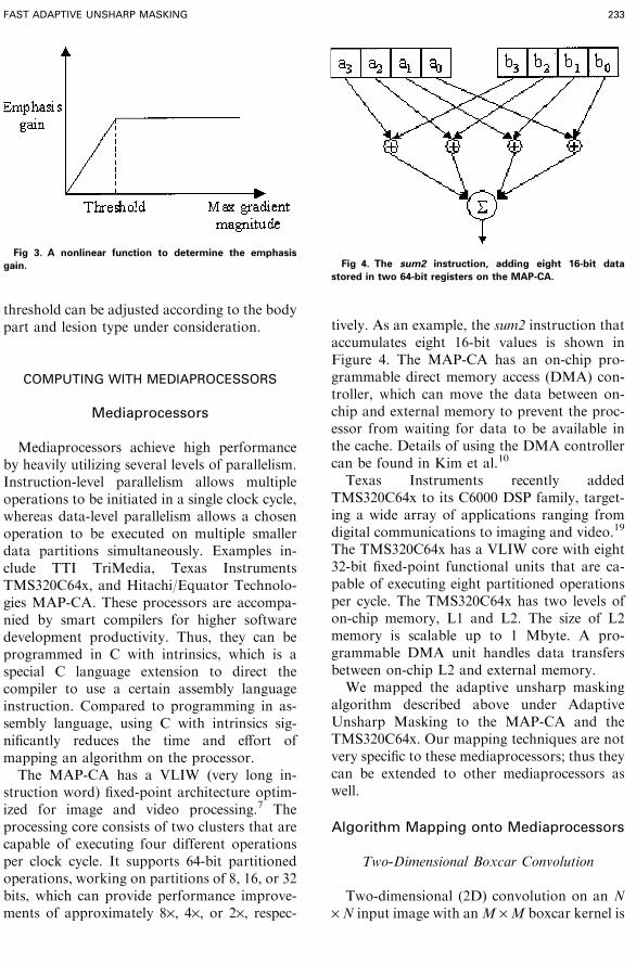
struction word) fixed-point architecture optim-ized for image and video processing.7 Theprocessing core consists of two clusters that arecapable of executing four different operationsper clock cycle. It supports 64-bit partitionedoperations, working on partitions of 8, 16, or 32bits, which can provide performance improve-ments of approximately 8·, 4·, or 2·, respec-
tively. As an example, the sum2 instruction thataccumulates eight 16-bit values is shown inFigure 4. The MAP-CA has an on-chip pro-grammable direct memory access (DMA) con-troller, which can move the data between on-chip and external memory to prevent the proc-essor from waiting for data to be available inthe cache. Details of using the DMA controllercan be found in Kim et al.10
Texas Instruments recently addedTMS320C64x to its C6000 DSP family, target-ing a wide array of applications ranging fromdigital communications to imaging and video.19
The TMS320C64x has a VLIW core with eight32-bit fixed-point functional units that are ca-pable of executing eight partitioned operationsper cycle. The TMS320C64x has two levels ofon-chip memory, L1 and L2. The size of L2memory is scalable up to 1 Mbyte. A pro-grammable DMA unit handles data transfersbetween on-chip L2 and external memory.We mapped the adaptive unsharp masking
algorithm described above under AdaptiveUnsharp Masking to the MAP-CA and theTMS320C64x. Our mapping techniques are notvery specific to these mediaprocessors; thus theycan be extended to other mediaprocessors aswell.
Algorithm Mapping onto Mediaprocessors
Two-Dimensional Boxcar Convolution
Two-dimensional (2D) convolution on an N· N input image with anM ·M boxcar kernel is
Fig 3. A nonlinear function to determine the emphasis
gain. Fig 4. The sum2 instruction, adding eight 16-bit data
stored in two 64-bit registers on the MAP-CA.
FAST ADAPTIVE UNSHARP MASKING 233
given by
flpfðx; yÞ ¼1
M�MXyþðM�1Þ=2
j¼y�ðM�1Þ=2
XxþðM�1Þ=2
i¼x�ðM�1Þ=2finði; jÞ;
ð2Þ
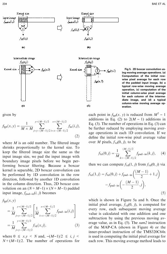
where M is an odd number. The filtered imageshrinks proportionally to the kernel size. Tokeep the filtered image size the same as theinput image size, we pad the input image withboundary image pixels before we begin per-forming boxcar filtering. Because a boxcarkernel is separable, 2D boxcar convolution canbe performed by 1D convolution in the rowdirection, followed by another 1D convolutionin the column direction. Thus, 2D boxcar con-volution on an (N+M)1) · (N+M)1) paddedinput image, fpad)in(i, j) becomes
flpfðx; yÞ
¼ 1
M
XyþðM�1Þ=2
j¼y�ðM�1Þ=2
1
M
XxþðM�1Þ=2
i¼x�ðM�1Þ=2fpad�inði; jÞ
24
35
¼ 1
M
XyþðM�1Þ=2
j¼y�ðM�1Þ=2f1dðx; jÞ; ð3Þ
where 0 £ x,y < N and, )(M)1)/2 £ i, j <N+(M)1)/2. The number of operations for
each point in flpf(x, y) is reduced from M2 ) 1additions in Eq. (2) to 2(M ) 1) additions inEq. (3). The number of operations in Eq. (3) canbe further reduced by employing moving aver-age operations in each 1D convolution. If wedefine the initial row-wise pixel average valueover M pixels, f1d(0, j), to be
f1dð0; jÞ ¼1
M
XðM�1Þ=2
k¼�ðM�1Þ=2fpad�inðk; jÞ; ð4Þ
then we can compute f1d(1, j) from f1d(0, j) via
f1dð1; jÞ ¼ f1dð0; jÞ þ fpad�inðM� 1Þ
2þ 1; j
� �
� fpad�in �ðM� 1Þ2
; j
� �;
ð5Þ
which is shown in Figure 5a and b. Once theinitial pixel average, f1d(0, j), is computed forevery row, each subsequent moving averagevalue is calculated with one addition and onesubtraction by using the previous moving av-erage value, as in Eq. (5). The sum2 instructionof the MAP-CA (shown in Figure 4) or theinner-product instruction of the TMS320C64xis used in computing the initial pixel average foreach row. This moving average method leads to
Fig 5. 2D boxcar convolution us-
ing moving average operations. (a)
Computation of the initial row-
wise pixel average for each row
of the padded input image, (b) a
typical row-wise moving average
operation, (c) computation of the
initial column-wise pixel average
for each column of the interme-
diate image, and (d) a typical
column-wise moving average op-
eration.
234 BAE ET AL

little change in computational cost as the box-car kernel size increases. The only increasecomes from the initial pixel average computa-tion for every row of the image in Eq. (4). If theimage size is much larger than the kernel size,even this dependency on the kernel size becomesnegligible. After the row-wise convolution, wecan repeat the moving average operations oneach column of the intermediate image, f1d(x, j),as shown in Figure 5c and d.
Emphasis Gain Control
The main computing components of thismodule are to apply the Sobel operator tocompute the gradient magnitude and to selectthe maximum value in a 3 · 3 neighborhoodarea. The kernel coefficients of the Sobel oper-ator5 have absolute values of only 0, 1, and 2.Partitioned addition and subtraction instruc-tions are used to process multiple pixels at atime. Multiplications by 2 are computed usingthe partitioned multiplication instruction thatcan be executed as fast as the partitioned ad-ditions/subtractions. Local maximum compu-tation requires several comparisons to rank-order nine pixels in the 3 · 3 neighborhood andselect the largest value. However, the MAP-CAand the TMS320C64x have the partitioned maxinstruction that compares multiple pairs ofpixels simultaneously and returns the maximumvalues as shown in Figure 6. Examples of Cwith intrinsics to support these partitioned op-erations and the corresponding generic C pro-grams can be found online at http://icsl.ee.washington.edu/verdant
RESULTS AND DISCUSSION
Figure 7 shows performance of 2D boxcarfiltering for a 2k · 2k 16-bit image with variouskernel sizes on a 300-MHz MAP-CA. Theprocessing time for 2D boxcar filtering is in-sensitive to the kernel size; e.g., a large increasein the kernel size from 7 · 7 to 201 · 201 resultsin a 6% increase in processing time from 81 msto 86 ms. This is interesting considering that theprocessing time of generalized convolution in-creases quadratically as the kernel size increas-es. In case of brute-force M · M boxcarconvolution, the total number of additions isM2 ) 1 for each point in the output image,which is still proportional toM2. However, twolevels of optimization improve the boxcar fil-tering performance substantially: (1) the sepa-rability property of 2D boxcar kernels reducesthe number of operations to 2(M ) 1) for everyfiltered output point and (2) the moving averagemethod further reduces this into just two addi-tions and two subtractions for each filteredoutput pixel. The kernel size affects only com-putation of the initial pixel average value in Eq.(4). For a large image, such as 2k · 2k, com-putation of the initial pixel average value isminimal compared to the subsequent movingaverage computation. As a result, Figure 7shows only a small increase in processing timewith the kernel size. Managuli et al.11 reportedgeneralized 2D convolution on the same MAP-CA mediaprocessor. As shown in Figure 8, theprocessing time of 2D generalized convolutionrapidly increases as the kernel size increases;e.g., 2D convolution of a 2k · 2k 16-bit imagewith a 3 · 3 generalized kernel takes 71 ms,whereas 25 · 25 generalized 2D convolutiontakes 859 ms. When a kernel is separable, 2Dconvolution can be computed by using two 1D
Fig 6. The max operation on four pairs of 16-bit data on the
MAP-CA.
Fig 7. Performance of 2D boxcar filtering for a 2k · 2k 16-bit
image on a 300-MHz MAP-CA.
FAST ADAPTIVE UNSHARP MASKING 235

convolutions. Figure 9 shows the processingtime of 2D convolution with different-size sep-arable kernels,11 which shows that separableconvolution is advantageous when the kernelsize is equal to or greater than 9 · 9. However,the overall processing time is still strongly de-pendent on the kernel size. For example, con-volution with a 9 · 9 separable kernel takes 131ms, whereas a 25 · 25 separable kernel takes387 ms. In contrast, 2D boxcar filtering with 25· 25 and 201 · 201 kernels takes only 82 ms and86 ms, respectively. In other words, 201 · 201boxcar convolution can be computed 4.5 timesfaster than 25 · 25 separable Gaussian convo-lution. At the same time, a 201 · 201 boxcarfilter has a much lower cutoff frequency than a25 · 25 Gaussian filter as shown in Figure 1b.This example illustrates the computational ad-vantage of using boxcar kernels compared tousing generalized kernels, especially for the caserequiring large kernels/low cutoff frequencies.Table 1 lists the overall performance of adap-tive unsharp masking with a 201 · 201 boxcarkernel for various image sizes. For a 2k · 2k 16-bit image, the 139-ms difference between Figure7 and Table 1 (MAP-CA) is attributable to theremaining processing parts of adaptive unsharpmasking—e.g., computing local image charac-
teristics, applying a nonlinear function, multi-plications, and subtractions/additions.The large on-chip memory of the
TMS320C64x is used to store intermediate re-sults, such as results of 1D boxcar convolution,thus saving data transfer time between on-chipand external memory. On the TMS320C64xrunning at a high clock frequency of 600 MHz,2D convolution with a 201 · 201 boxcar kerneltakes only 28 ms for a 2k · 2k 16-bit image,which corresponds to 35 frames/s. The totalprocessing time of adaptive unsharp maskingwith a 201 · 201 boxcar filter for a 2k · 2k 16-bitimage is 74 ms, compared with 225 ms with theMAP-CA as in Table 1. For the same algorithmand image size, the floating-point C implemen-tation takes 3.83 s on a 2.5-GHz Pentium 4. Thisperformance is 52 and 17 times slower than thatof TMS320C64x and MAP-CA, respectively.Even though our unsharp masking algorithm
is more complex than the global unsharpmasking implemented by Reza et al.,16 our dataprocessing rates of 37 Mbytes/s on the MAP-CA and 113 Mbytes/s on the TMS320C64x aremore than 18 times and 56 times faster thantheir processing rate of 2 Mbytes/s. As shown inFigure 9, convolution with a 15 · 15 Gaussiankernel, the main processing load of their globalunsharp masking, can be performed in 183 msfor a 2k · 2k 16-bit image (46 Mbytes/s) on a300-MHz MAP-CA. Our mediaprocessor-based unsharp masking easily allows largechanges in kernel and image sizes. In contrast,FPGA/EPLD implementations are not de-signed for kernels and/or images much largerthan the initially targeted kernel and image sizes(e.g., changing to digital mammograms), andwould typically require major hardware modi-fications to accommodate large changes. Our
Fig 8. Performance of 2D generalized convolution for a 2k ·2k 16-bit image on a 300-MHz MAP-CA.
Fig 9. Performance of 2D convolution with a generalized
separable kernel for a 2k · 2k 16-bit image on a 300-MHz MAP-
CA.
Table 1. Processing Time (ms) of Adaptive Unsharp Masking
with a 201 · 201 Boxcar Filter on a 300-MHz MAP-CA and on a
600-MHz TMS320C64x
Input Image Size
(16 bits)
MAP-CA
(300 MHz)
TMS320C64x
(600 MHz)
512 · 512 16 6
1k · 512 31 11
1k · 1k 59 20
2k · 1k 114 38
2k · 2k 225 74
236 BAE ET AL

adaptive unsharp masking performance of 6 mson the TMS320C64x with a 201 · 201 boxcarkernel on a 512 · 512 16-bit image is more than7 times faster than the 43 ms needed by 3 · 3quadratic-filter-based nonlinear unsharp mask-ing on an EPLD board for the same imagesize.17 For a 2k · 2k 8-bit image, the EPLDimplementation required 45.2 s because of thelimited amount of memory on the board. Thatis much slower than our TMS320C64x per-formance of 74 ms. In addition, the same me-diaprocessor-based hardware can perform otherneeded processing tasks in addition to fast un-sharp masking. This multi-functionality obvi-ates the need for additional hardware for theother tasks in medical imaging modalities—e.g.,detector-related artifact correction, modulationtransfer function (MTF) compensation, andgray-level adjustment. Depending on the mo-dality, lesion type and radiologist’s preference,other common and/or advanced image-en-hancement methods, such as histogram-based,wavelet-based processing, and Kodak’s EVPalgorithm, could be supported easily. Usingthe same mediaprocessor, CR and DR imagescan be compressed quickly (e.g., using JPEG20004) for image transmission and archiving.Images can be decompressed on the fly beforebeing displayed on the monitors of diagnostic
and review workstations. Furthermore, radiol-ogists can enhance the images with the en-hancement method that they prefer usingmultiple presets and/or interactively, and com-puter-intensive computer-aided diagnosis algo-rithms could be performed by taking advantageof the high computation power of mediaproc-essors.Figure 10 shows an original DR chest X-ray
image, and Figure 11 is the result of ouradaptive unsharp masking. The edge details inthe processed image are enhanced, and noiseamplification is relatively suppressed. The ef-fects of adaptive enhancement may not be evi-dent in Figure 11; therefore, the original X-rayimages and unsharp-masked images with dif-ferent parameter sets are made available forcomparison.6
Mediaprocessors have been developed pri-marily for the huge consumer electronics andtelecommunications markets, where potentialsales volume is enormous. The demand for highcomputing power at low cost has facilitatedextensive advances in computer architectureand ease of programming over the last 10 years.Medical imaging systems based on these proc-essors can directly benefit from the increasedprocessor clock speed, faster memory technol-ogy, and lower cost.
Fig 10. Original chest X-ray im-
age.
FAST ADAPTIVE UNSHARP MASKING 237

CONCLUSION
An adaptive unsharp masking algorithmhas been mapped onto high-performancemediaprocessors, the MAP-CA and theTMS320C64x. The processing time of adaptiveunsharp masking with a 201 · 201 boxcar filterfor a 2k · 2k 16-bit image is 224 ms and 74 ms,respectively, which is much faster than thatof existing solutions. Two-dimensional box-car filtering with a large kernel is performed asfast as with a small kernel, such as 7 · 7,whereas a kernel larger than 201 · 201 can bealso supported efficiently. The performanceof unsharp masking will directly benefit fromthe expected increase in the clock frequencyof mediaprocessors. For example, using therecently introduced 400-MHz MAP7 theprocessing time of adaptive unsharp maskingcan be reduced from 224 ms to about 170 ms.Similarly, with the future 1-GHz TMS320C64x,it can be completed in only about 45 ms. Thishigh performance can facilitate interactive im-age enhancements by technologists and physi-cians in quality control and primary diagnosis/review sessions. The high performance, alongwith low cost, provides an excellent cost/per-formance ratio. In addition to its speed and lowcost, mediaprocessor-based unsharp masking
can easily accommodate significant chang-es—e.g., kernel and image sizes, more effica-cious gain control algorithms in adaptiveunsharp masking, significantly different imageenhancement algorithms, or different and/oradditional tasks in medical image modalities inthe future.
REFERENCES
1. Armstrong II JD, Sorenson JA, Nelson JA, et al:
Clinical evaluation of unsharp masking and slit scanning
techniques in chest radiography. Radiology 147:351-356,
1983
2. Barry D, Cluff R, Duncan C, Kennedy J: High per-
formance parallel image processing using SIMD technology.
Proc SPIE 3658:344-351, 1999
3. Davies AG, Cowen AR, Parkin GJ, et al: Optimising
the processing and presentation of PPCR images. Proc SPIE
2712:189-195, 1996
4. DICOM Standards Committee Working Group 4:
Compression. Supplement 61:JPEG2000 Transfer Syntaxes.
NEMA, Virginia, 2000
5. Gonzalez RC, Woods RE: Digital image processing,
2nd ed. Upper Saddle River, NJ: Prentice-Hall, 2002
6. http://icsl.ee.washington.edu/verdant7. http://www.equator.com
8. http://www.fujimed.com/ndt/cr_process3.html
9. Ji T-L, Sundareshan MK, Roehrig H: Adaptive image
contrast enhancement based on human visual properties.
IEEE Trans Med Imaging 13:573-586, 1994
Fig 11. Enhanced chest X-ray im-
age using adaptive unsharp mask-
ing.
238 BAE ET AL

10. Kim D, Managuli R, Kim Y: Data cache and direct
memory access in programming mediaprocessors. IEEE
Micro 21:33-42, 2001
11. Managuli R, York G, Kim D, et al: Mapping of 2D
convolution on VLIW mediaprocessors for real-time per-
formance. J Electronic Imaging 9:327-335, 2000
12. Metter RV, Foos D: Enhanced latitude for dig-
ital projection radiography. Proc SPIE 3658:468-
478, 1999
13. Oppenheim AV, Schafer RW, Buck JR: Discrete-time
signal processing. 2nd ed. Upper Saddle River, NJ: Prentice-
Hall, 1999
14. Pisano ED, Cole EB, Major S, et al: Radiologists’
preference for digital mammographic display. Radiology
216:820-830, 2000
15. Prokop M, Schaefer CM, Oestmann JW, Galanski
M: Improved parameters for unsharp mask filtering of
digital chest radiographs. Radiology 187:521-526, 1993
16. Reza A, Delva J, Turney R, Chapman K: Medical
image enhancement using FPGA technology. Electronic
Engineering 70:53-54, 56, 1998
17. Sivaswamy J, Salcic Z, Ling KL: A real-time imple-
mentation of nonlinear unsharp masking with FPLDs. Re-
al-Time Imaging 7:195-202, 2001
18. Stahl M, Aach T, Dippel S: Digital radiography en-
hancement by nonlinear multiscale processing. Med Phys
27:56-65, 2000
19. Texas Instruments: TMS320C6000 CPU and In-
struction Set Reference Guide. Literature Number:
SPRU189F, 2000
FAST ADAPTIVE UNSHARP MASKING 239
Copyright © 2022 FDOKUMEN