Fascial Fitness: Training in the Neuromyofascial Web
11
Fascial Fitness: Training in the Neuromyofascial Web by Thomas Myers Research shows why taking a different approach to exercise and the movement brain is the wave of the future. If you are interested in the role of fascia in fitness training, the following questions lead to new take- aways: • Most injuries are connective-tissue (fascial) injuries, not muscular injuries—so how do we best train to prevent and repair damage and build elasticity and resilience into the system? • There are 10 times more sensory nerve endings in your fascia than in your muscles; therefore, how do we aim proprioceptive stimulation at the fascia as well as the muscles? • Traditional anatomy texts of the muscles and fascia are inaccurate, based on a fundamental misunder- standing of our movement function—so how can we work with fascia as a whole, as the “organ system of stability”? Consciously or unconsciously, you have been working with fascia for your whole movement career—it is unavoidable. Now, however, new research is reinforcing the importance of fascia and other connective tis- sue in functional training (Fascia Congress 2009). Fascia is much more than “plastic wrap around the mus- cles.” Fascia is the organ system of stability and mechano-regulation (Varela & Frenk 1987). Understanding this may revolutionize our ideas of “fitness.” Research into the fascial net upsets both our traditional beliefs and some of our new favorites as well. The evidence all points to a new consideration within overall fitness for life—hence the term fascial fitness. This article lays out the emerging picture of the fascial net as a whole and explores three of the many aspects of recent research that give us a better understanding of how best to train the fascial net. The Neuromyofascial Web Fascia is the Cinderella of body tissues—systematically ignored, dissected out and thrown away in bits (Schleip 2003). However, fascia forms the biological container and connector for every organ (including muscles). In dissection, fascia is literally a greasy mess (not at all like what the books show you) and so variable among individuals that its actual architecture is hard to delineate. For many reasons, fascia has not been seen as a whole system; therefore we have been ignorant of fascia’s overall role in biomechan- ics. Thankfully, the integrating mechano-biological nature of the fascial web is becoming clearer. It turns out that it really is all one net with no separation from top to toe, from skin to core or from birth to death (Shultz & Feitis 1996). Every cell in your body is hooked into—and responds to—the tensional environment of the fascia (Ingber 1998). Alter your mechanics, and cells can change their function (Horwitz 1997). This is a radical new way of seeing personal training—stretching, strengthening and shape-shifting—as part of “spatial medicine” (Myers 1998). Given the facts, many would prefer the term neuromyofascial web to the fascia-dissing musculoskele- tal system (Schleip 2003). As accustomed as we are to identifying individual structures within the fascial web—plantar fascia, Achilles tendon, iliotibial band, thoracolumbar aponeurosis, nuchal ligament and so on—these are just convenient labels for areas within the singular fascial web. They might qualify as ZIP codes, but they are not separate structures (see the sidebar “Muscle Isolation vs. Fascial Integration”). You can talk about the Atlantic, the Pacific and the Mediterranean oceans, but there is really only one in- terconnected ocean in the world. Fascia is the same. We talk about individual nerves, but we know the nervous system reacts as a whole. How does fascia webbing function as a system? © 2011 by IDEA Health & Fitness Inc. All rights reserved. Reproduction without permission is strictly prohibited.
-
Upload
khangminh22 -
Category
Documents
-
view
0 -
download
0
Transcript of Fascial Fitness: Training in the Neuromyofascial Web
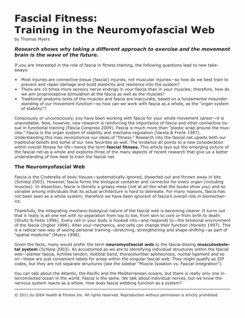
Fascial Fitness:Training in the Neuromyofascial Webby Thomas Myers
Research shows why taking a different approach to exercise and the movementbrain is the wave of the future.
If you are interested in the role of fascia in fitness training, the following questions lead to new take-aways:
• Most injuries are connective-tissue (fascial) injuries, not muscular injuries—so how do we best train toprevent and repair damage and build elasticity and resilience into the system?
• There are 10 times more sensory nerve endings in your fascia than in your muscles; therefore, how dowe aim proprioceptive stimulation at the fascia as well as the muscles?
• Traditional anatomy texts of the muscles and fascia are inaccurate, based on a fundamental misunder-standing of our movement function—so how can we work with fascia as a whole, as the “organ systemof stability”?
Consciously or unconsciously, you have been working with fascia for your whole movement career—it isunavoidable. Now, however, new research is reinforcing the importance of fascia and other connective tis-sue in functional training (Fascia Congress 2009). Fascia is much more than “plastic wrap around the mus-cles.” Fascia is the organ system of stability and mechano-regulation (Varela & Frenk 1987).Understanding this may revolutionize our ideas of “fitness.” Research into the fascial net upsets both ourtraditional beliefs and some of our new favorites as well. The evidence all points to a new considerationwithin overall fitness for life—hence the term fascial fitness. This article lays out the emerging picture ofthe fascial net as a whole and explores three of the many aspects of recent research that give us a betterunderstanding of how best to train the fascial net.
The Neuromyofascial Web
Fascia is the Cinderella of body tissues—systematically ignored, dissected out and thrown away in bits(Schleip 2003). However, fascia forms the biological container and connector for every organ (includingmuscles). In dissection, fascia is literally a greasy mess (not at all like what the books show you) and sovariable among individuals that its actual architecture is hard to delineate. For many reasons, fascia hasnot been seen as a whole system; therefore we have been ignorant of fascia’s overall role in biomechan-ics.
Thankfully, the integrating mechano-biological nature of the fascial web is becoming clearer. It turns outthat it really is all one net with no separation from top to toe, from skin to core or from birth to death(Shultz & Feitis 1996). Every cell in your body is hooked into—and responds to—the tensional environmentof the fascia (Ingber 1998). Alter your mechanics, and cells can change their function (Horwitz 1997). Thisis a radical new way of seeing personal training—stretching, strengthening and shape-shifting—as part of“spatial medicine” (Myers 1998).
Given the facts, many would prefer the term neuromyofascial web to the fascia-dissing musculoskele-tal system (Schleip 2003). As accustomed as we are to identifying individual structures within the fascialweb—plantar fascia, Achilles tendon, iliotibial band, thoracolumbar aponeurosis, nuchal ligament and soon—these are just convenient labels for areas within the singular fascial web. They might qualify as ZIPcodes, but they are not separate structures (see the sidebar “Muscle Isolation vs. Fascial Integration”).
You can talk about the Atlantic, the Pacific and the Mediterranean oceans, but there is really only one in-terconnected ocean in the world. Fascia is the same. We talk about individual nerves, but we know thenervous system reacts as a whole. How does fascia webbing function as a system?
© 2011 by IDEA Health & Fitness Inc. All rights reserved. Reproduction without permission is strictly prohibited.
Magically extracted as a whole, the fascialweb would show us all the shapes of thebody, inside and out. It would be just one bignet with muscles squirming in it like swim-ming fish. Organs would hang in it like jelly-fish. Every system, every organ and evenevery cell lives embedded within the sea of aunitary fascial net.
This concept is important because we are sostrongly inclined to name individual structuresand think that way clinically: “Oh, you toreyour biceps,” forgetting that “biceps” is ourconception. Our common scientific nomencla-ture gives a false impression, while the NewAge shibboleth is more literally true: thebody—and the fascial net in particular—isa single connected unity in which themuscles and bones float.
You can tear this net in injury, cut it with asurgeon’s scalpel, feed and hydrate it well orclog it with high-fructose corn syrup. No mat-ter how you treat it, it will eventually lose itselasticity. In your eye’s lens, for instance, thenet stiffens in a very regular way, requiringyou to use reading glasses at about age 50.In your skin, the net frays to cause wrinkles.Key elements like hip cartilage may fail youbefore you die, and need replacement, butwhen you finally breathe your last breath yourfascial web will still be the same single netyou started with.
It’s no small wonder that this system, like thenervous and circulatory systems, would de-velop complex signaling and homeostaticmechanisms (Langevin et al. 2006). Thelarger wonder is that we have not really seenor explored the connective-tissue system’s re-sponses until now.
A Definition of Terms
In medicine, the term fascia designates tis-sues with specific topology and histology, asdistinct from tendon, ligament or other speci-fied tissues. In this article, however, we areusing fascia as an overall name for this sys-temic net of connective tissue, because thereis no generalized term (Huijing & Langevin2009). Connective tissue includes the bloodand blood cells, and other elements not partof the structural net we are examining. Per-haps the closest term would be extra-cellu-lar matrix (ECM), which includes everythingin your body that isn’t cellular (see Figure 3).The ECM has three main elements:
— 2 —
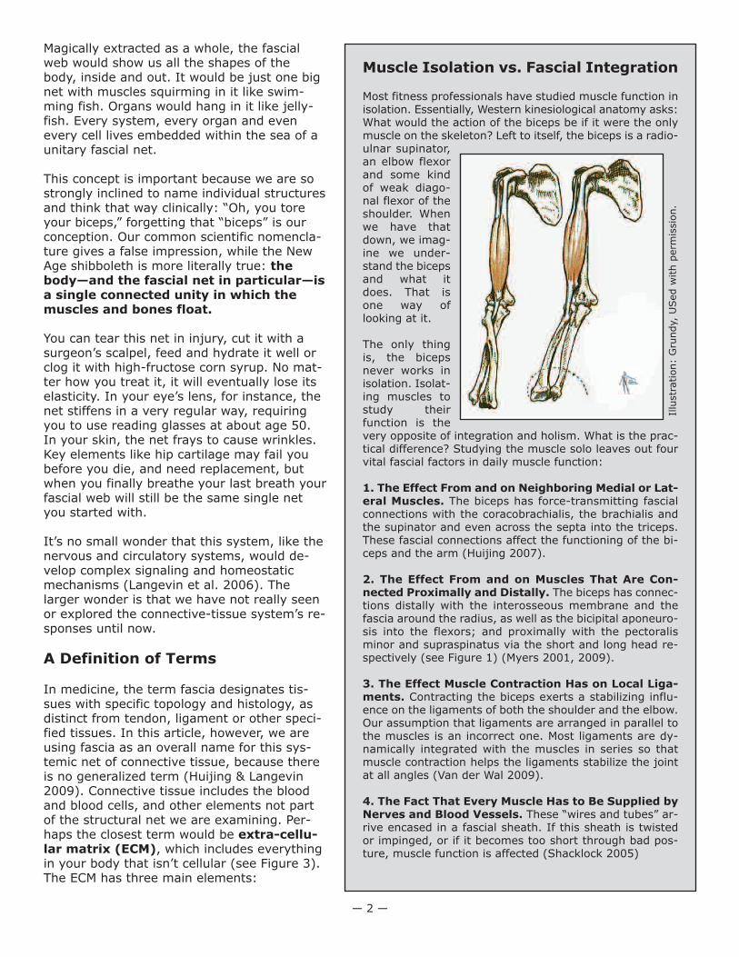
Muscle Isolation vs. Fascial Integration
Most fitness professionals have studied muscle function inisolation. Essentially, Western kinesiological anatomy asks:What would the action of the biceps be if it were the onlymuscle on the skeleton? Left to itself, the biceps is a radio-ulnar supinator,an elbow flexorand some kindof weak diago-nal flexor of theshoulder. Whenwe have thatdown, we imag-ine we under-stand the bicepsand what itdoes. That isone way oflooking at it.
The only thingis, the bicepsnever works inisolation. Isolat-ing muscles tostudy theirfunction is thevery opposite of integration and holism. What is the prac-tical difference? Studying the muscle solo leaves out fourvital fascial factors in daily muscle function:
1. The Effect From and on Neighboring Medial or Lat-eral Muscles. The biceps has force-transmitting fascialconnections with the coracobrachialis, the brachialis andthe supinator and even across the septa into the triceps.These fascial connections affect the functioning of the bi-ceps and the arm (Huijing 2007).
2. The Effect From and on Muscles That Are Con-nected Proximally and Distally. The biceps has connec-tions distally with the interosseous membrane and thefascia around the radius, as well as the bicipital aponeuro-sis into the flexors; and proximally with the pectoralisminor and supraspinatus via the short and long head re-spectively (see Figure 1) (Myers 2001, 2009).
3. The Effect Muscle Contraction Has on Local Liga-ments. Contracting the biceps exerts a stabilizing influ-ence on the ligaments of both the shoulder and the elbow.Our assumption that ligaments are arranged in parallel tothe muscles is an incorrect one. Most ligaments are dy-namically integrated with the muscles in series so thatmuscle contraction helps the ligaments stabilize the jointat all angles (Van der Wal 2009).
4. The Fact That Every Muscle Has to Be Supplied byNerves and Blood Vessels. These “wires and tubes” ar-rive encased in a fascial sheath. If this sheath is twistedor impinged, or if it becomes too short through bad pos-ture, muscle function is affected (Shacklock 2005)
Illu
str
ation:
Gru
ndy,
USed w
ith p
erm
issio
n.
• fibers: the strong pliable weave—consisting primarily of collagen (which has 12 types) and its cousinselastin and reticulin—that both separates compartments and binds them together
• glue: the variable and colloidal gels like heparin, fibronectin and hyaluronic acid that accommodatechange and provide the substrate for other cells like nerves and epithelia
• water: the fluid that surrounds and permeates the cells as a medium of exchange; mixes with the glueto make materials of differing properties; and keeps the fibers wet and pliable
Though the ECM will be our topic just below, the term fascia as we define it also includes fibroblasts andmast cells, which give rise to the fibers and glue and then remodel them in response to the demands ofinjury, training and habit.
The principal structural element in the ECM comprises the fibers collagen, elastin and reticulin. Collagen isby far the most common of these, and by far the strongest. This is the white, sinewy stuff in meat. Thecollagen fiber is a triple helix; if it was a half-inch thick, it would be about a yard long and look like an oldthree-strand rope (Snyder 1975). Collagen fibers can be arranged in regular directional rows, as they arein tendons or ligaments (dense regular), or in random crisscross ways, like felt (dense or loose irregular).
The collagen fibers cannot actually stick to each other but are glued together by other proteins called gly-coaminoglycans (GAGs), which are mucopolysaccharides, both of which are long words for snot. Weare held together by mucous, a colloidal substance, which, by varying its chemistry slightly, can display asurprising array of properties, from thick and sticky to fluid and lubricating. The fernlike molecules of mu-cous open to absorb water (they are hydrophilic) or close and bind to themselves when water is absent.Depending on their chemistry, they either bind layers together or allow them to slide on each other (Grin-nell 2008).
The phenomenon we call “stretch” or lengthening (and that scientists call “creep” or hysteresis) is a func-tion not of the collagen fibers lengthening but of the fibers sliding along each other on the glue of the hy-drated GAGs (Sbriccoli et al. 2005). Take the water out of the GAGs, and the result is tissue that ismightily reluctant to stretch (Schleip 2003).Most injuries occur when connective tissue is stretched faster than it can respond. The less it is hydrated,the less elastic response it has in it.
The Body Electric?Connective-tissue cells produce the fibers and the GAGs, and these materials are then altered to form aremarkable variety of building materials. If you were to try to recreate your structural body out of itemsyou could buy at Home Depot®, what would you need? Wood or PVC for the bones, silicon rubber for thecartilage, lots of string, wire, tubing, plastic sheeting, rubber bands, cotton, nets, grease and oil—the listgoes on. Would you try to build a body without duct tape?
— 3 —
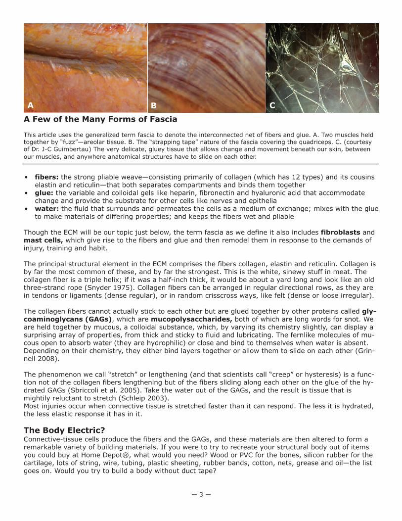
A Few of the Many Forms of Fascia
This article uses the generalized term fascia to denote the interconnected net of fibers and glue. A. Two muscles heldtogether by “fuzz”—areolar tissue. B. The “strapping tape” nature of the fascia covering the quadriceps. C. (courtesyof Dr. J-C Guimbertau) The very delicate, gluey tissue that allows change and movement beneath our skin, between
our muscles, and anywhere anatomical structures have to slide on each other.
A B C
Your body manufactures all these materials andmany more by mixing together various proportionsof the ECM’s fibers and glue and altering thechemistry in different ways (Snyder 1975). Inbone, the fiber matrix is there—much likeleather—but the mucousy ground substance hasbeen systematically replaced with mineral salts.Cartilage has the same leathery substrate, but theglue has been dried into a tough but pliable “plas-tic” that permeates the fibrous leather. In ligamentand tendon, almost all the glue has been squeezedout. In blood and joint fluid, the fiber exists only ina liquid form, until it hits the air, when it forms ascab. This manufactory in your body is fascinating:the dentin in your teeth, your gums, your heartvalves, even the clear cornea of your eye—are allformed in this fashion.
Remodeling and TensegrityYour muscles may determine your shape in the training sense, but connective tissue determines yourshape in the overall sense. It holds the bones together, pulling in on them as they press out (like a tenseg-rity system; see Figure 2).
The ECM is capable of remodeling itself in a variety of ways (Chen et al. 1997). Just as your muscles re-model themselves in response to training, the fascia remodels itself in response to direct signaling fromthe cells (Langevin et al. 2010); injury (Desmouli`ere, Chapponnier & Gabbiani 2005); long-held mechani-cal forces (Iatrides et al. 2003); use patterns (including emotional ones); gravity; and certain chemistrywithin your body (Grinnell & Petroll 2010). The complexities of remodeling are just now being explored inthe lab; the details will be revealed over the coming decade.
The idea of tensegrity (tension and integrity) and the phenomenon of remodeling are the basis for struc-tural therapy, including yoga and the forms of manual therapy commonly known as Rolfing® or StructuralIntegration and its deep-tissue relatives, including foam rolling. Change the demand—as we do in body-work and personal training—and the fascial system responds to that new demand. This common themepoints to a future where manual therapy and movement training combine to form a powerful method for
• restoring natural settings for posture and function;• steering small problems away from developing into big ones later on;• easing the long-term consequences from injury; and• extending functional movement farther and farther up the age scale.
How to Train the Neuromyofascial Web
If the fascia is a singular space-organizing adjustable tensegrity that traverses the whole body and regu-lates—both locally and as a whole—the biomechanics of tension and compression, we can then ask: Howcan we train this system, in conjunction with our work on muscles and neural control, to prevent and re-pair injury and build resilience into the system?
The answer to this question is still developing—rapidly—both in the laboratory and on the training floor.Some research is confirming our images and practices as they have developed and are traditionally ap-plied. Here we focus on a few surprising sets of findings that are (or soon will be) changing our ideas ofhow the neuromyofascial web really works and what role connective tissue plays in developing overall fit-ness for life. More of these results can be found at www.fascialftness.de or in the fascial fitness section ofwww.anatomytrains.com.
— 4 —
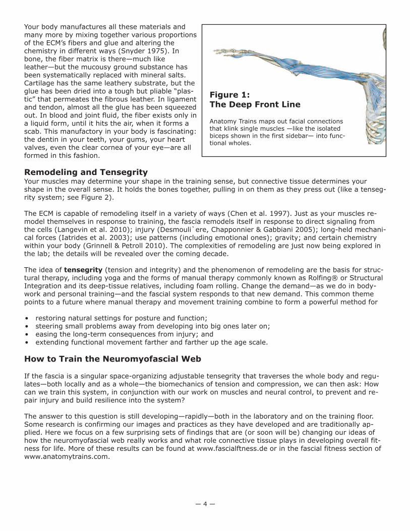
Figure 1:The Deep Front Line
Anatomy Trains maps out facial connectionsthat klink single muscles —like the isolatedbiceps shown in the first sidebar— into func-tional wholes.
Finding #1: Specific training can enhance the fascial elasticity essential tosystemic resilience.
Fascial elasticity has not been recognized until recently, and the mechanisms involved are still being stud-ied (Chino et al. 2008). Nevertheless, applications to training are already evident. The basic news is thatconnective tissue—even dense tissues like tendons and aponeuroses—is much more significantly elasticthan previously thought. The second essential part of that news is that fascial elasticity is stored and re-turned very quickly. In other words, it is more like a superball than a Nerf™ ball. Thus, fascial elasticity isa factor only when the motion is cyclic and quickly repeated, as in running, walking or bouncing, but notas in bicycling, in which the repetitive cycle is far too slow to take advantage of fascia’s elastic properties.
Measurements of calf lengthening during running have shown that much of the length required for dorsi-flexion is coming from an elastic stretch of the fascia, while the muscle is contracting isometrically (Kuboet al. 2006). This contradicts our previous understanding that the tendon was nonelastic, and that themuscles were lengthening and shortening during these cyclic motions prior to and following footfall.
The runners who train for and employ more of this elasticity will be using less muscle power (read: lessglucose) during their runs, as they are storing energy in the stretch and then getting it back during the re-lease. Thus, they will be able to run longer with less fatigue.
Building in this elasticity is a matter of putting a demand on the tissues to act in this way. Doing thisslowly (compared with muscle training) is a definite attribute of fascial training (it may take 6–24 monthsto build fascial elasticity).
What’s in:
• Bouncing. When you land on the ball of your foot, you decelerate and accelerate in such a way thatyou not only make use of but actually build elasticity into the tendons and entire fascial system. The besttraining effect seems to follow the pleasure principle: feel for that sense of elegance, an ideal resonancewith minimum effort and maximum ease.• Preparatory Countermovement. Preparing for a movement by making a countermovement—for ex-ample, flexing down before extending up to standing, winding up before a pitch, or moving the kettlebelltoward the body before moving it away—makes maximum use of the power of fascial elasticity to helpmake and smooth out the movement.
What’s out:
• Jerky Movements and Abrupt Changes of Direction. Imagine jumping rope but landing only onyour heels. The stress on all your systems would be enormous, and you would not build elasticity intothe fascial system.
• Big Muscle Demand for Push-Off. Using the fascial elastic recoil lessens the demand for huge mus-cle effort during push-off, making movement more controllable, less arduous and less fuel-consump-tive.
Finding #2: The fascial system responds better to variation than to a repetitive program.
The evidence suggests that the fascial system is better trained by a wide variety of vectors—in angle,tempo and load (Huijing 2007). Isolating muscles along one track (e.g., with an exercise machine) may beuseful for those muscles but is less than useful for all the surrounding tissues. Loading the tissue one wayall the time means it will be weaker when life—which is rarely repetitive—throws that part of the body acurve ball.
What's in:
• Whole-Body Movements. Engaging long myofascial chains and whole-body movements is the betterway to train the fascial system.
• Proximal Initiation. It’s best to start movements with a dynamic pre-stretch (distal extension) butaccompany this with a proximal initiation in the desired direction, letting the more distal parts of the
— 5 —
body follow in sequence, like an elasticpendulum.
• Adaptive Movement. Complex movementrequiring adaptation, like parkour (see thebeginning of the James Bond movie CasinoRoyale for a great example), beats repeti-tive exercise programs.
What's out:
• Repetitive Movement. Machines (orminds) that require clients to work in thesame line again and again do not build fas-cial resilience very well.
• Always Practicing With Upper-LevelLoads. Variable loads build different as-pects of the fascia. Sticking with near-limitloads will strengthen some ligaments butweaken others. Varying the load is the bet-ter way.
• Always Training in the Same Tempo.Likewise, varying the tempo of your trainingallows different fascial structures to buildstrength and elasticity.
Finding #3:The fascial system is far moreinnervated than muscle, so proprioceptionand kinesthesia are primarily fascial, notmuscular.
This is a hard concept for many fitness profes-sionals to get their heads around, but it is afact: there are 10 times as many sensory re-ceptors in your fascial tissues as there are inyour muscles (Stillwell 1957). The muscleshave spindles that measure length change (andover time, rate of length change) in the mus-cles. Even these spindles can be seen as fascialreceptors, but let’s be kind and give them tothe muscles (Van der Wal 2009). For each spin-dle, there are about 10 receptors in the sur-rounding fascia—in the surface epimysium, thetendon and attachment fascia, the nearby liga-ments and the superficial layers. These recep-tors include the Golgi tendon organs thatmeasure load (by measuring the stretch in thefibers), paciniform endings to measure pres-sure, Ruffini endings to inform the centralnervous system of shear forces in the soft tis-sues, and ubiquitous small interstitial nerveendings that can report on all these plus, ap-parently, pain (Stecco et al. 2009; Taguchi et al. 2009).
So when you say you are feeling your muscles move, this is a bit of a misnomer. You are “listening” toyour fascial tissues much more than to your muscles. Here are three interesting findings that go alongwith this basic eye-opener:
1. Ligaments are mostly arranged in series with the muscles, not in parallel (Van der Wal 2009). This
— 6 —
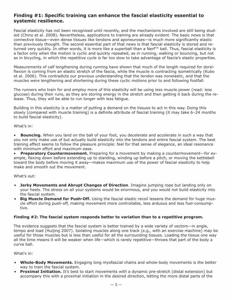
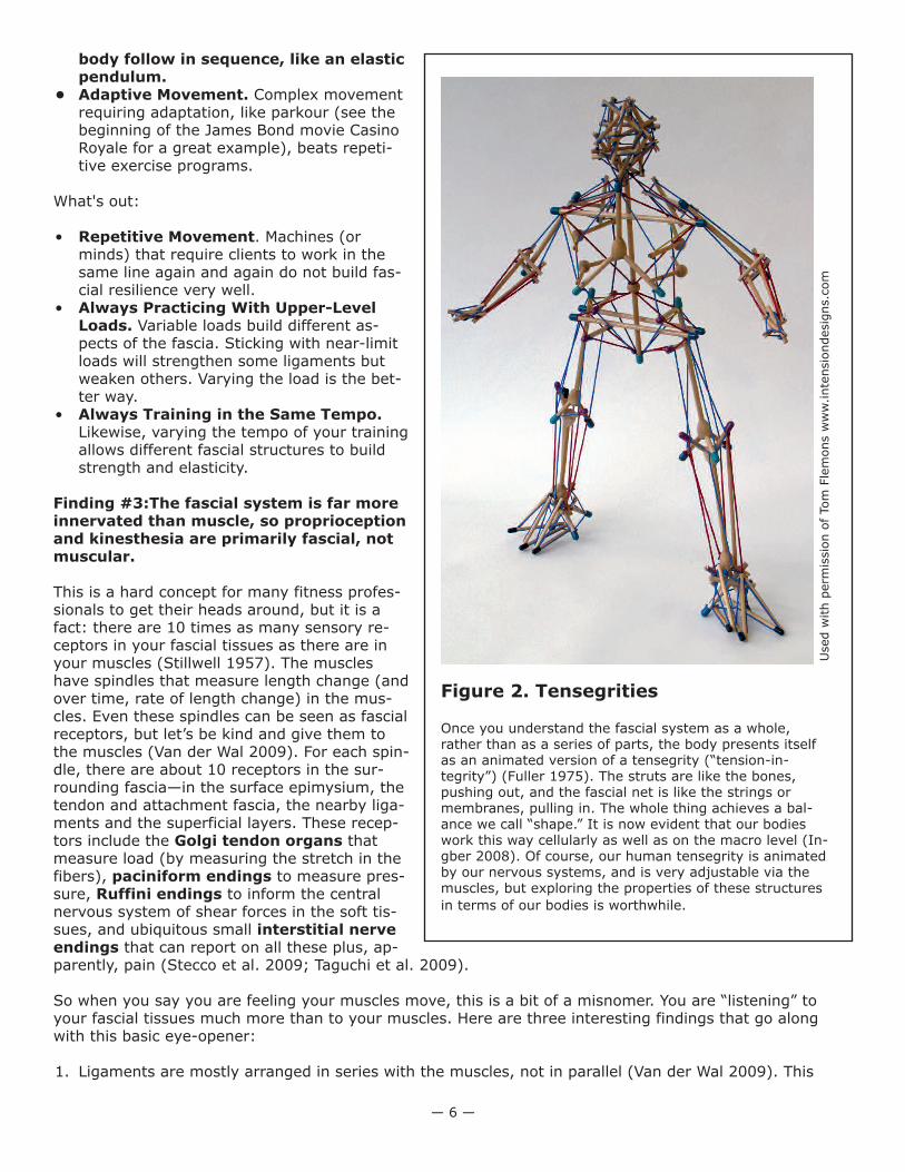
Figure 2. Tensegrities
Once you understand the fascial system as a whole,rather than as a series of parts, the body presents itselfas an animated version of a tensegrity (“tension-in-tegrity”) (Fuller 1975). The struts are like the bones,pushing out, and the fascial net is like the strings ormembranes, pulling in. The whole thing achieves a bal-ance we call “shape.” It is now evident that our bodieswork this way cellularly as well as on the macro level (In-gber 2008). Of course, our human tensegrity is animatedby our nervous systems, and is very adjustable via themuscles, but exploring the properties of these structures
in terms of our bodies is worthwhile.
Used w
ith p
erm
issio
n o
f Tom
Fle
mons w
ww
.inte
nsio
ndesig
ns.c
om
means that when you tense a muscle, the ligaments are automatically tensed to stabilize the joint, nomatter what its position. Our idea that the ligaments do not function until the joint is at its full exten-sion or torsion is now outmoded; for example, ligaments function all through a preacher curl, not justat the ends of the movement.
2. Nerve endings arrange themselves according to the forces that commonly apply in that location in thatindividual, not according to a genetic plan, and definitely not according to the anatomical division wecall a muscle. There is no representation of a “deltoid” inside your movement brain. That’s just a con-cept over in your cortex, not in your biological organization.
3. Apparently, sensors in and near the skin are more active in detecting and regulating movement thanthe joint ligament receptors (Yahia, Pigeon & DesRosiers 1993).
What's in:
• Skin and Surface Tissue Stimulation to Enhance Proprioception. Rubbing and moving the skinand surface tissues is important to enhance fascial proprioception. One weightlifter is having good re-sults scrubbing himself with a vegetable brush before going into competition.
• Directing Clients to Feel Their Fascial Tissues. Taking attention—your own and your client’s—awayfrom the muscles and putting it into the surrounding fascial tissues can help prevent injury and makethe perception of kinesthesia more accurate and fully informed. Sensuous body activity coupled with ahigh level of kinesthetic acuity (think: cat) may prevent injury better than being tough.
What's out:
• Isolated Muscle Orientation. Exercising a single muscle or muscle group is nearly impossible; everyexercise is stimulating multiple nerves, involving multiple muscles and employing fascial tissues allaround the site of effort, as well as “upstream” and “downstream” from it.
• Joint-Receptor Emphasis. Given that the ligaments are often tensed by the muscles, the emphasison joint receptors—while important—needs to be replaced with a more general attention to the wholearea, from the skin on down.
This discussion has focused on biomechanical factors; it has omitted nutritional and humoral considera-tions, as well as constitutional differences in fascia, which have recently come up for study. A deeper un-derstanding of the role of fascia in training changes your perspective, your work, your words and youreffect. Fascia is not just cling wrap.
References
Chen, C.S., et al. 1997. Geometric control of cell life and death. Science, 276 (5317), 1425–28.Chino, K., et al. 2008. In vivo fascicle behaviour of synergistic muscles on concentric and eccentric plantar flexion in
humans. Journal of Electromyography and Kinesiology, 18 (1), 79–88.Desmouliere, A., Chapponier, C., & Gabbiani, G. 2005. Tissue repair, contraction, and the myofibroblast. Wound Repair
Regeneration, 13 (1), 7–12.Fascia Congress. 2009. www.fasciacongress.org/2009.Fuller, R.B. 1975. Synergetics. New York: Macmillan.Grinnell, F. 2008. Fibroblast mechanics in three-dimensional collagen matrices. Trends in Cell Biology, 12 (3), 191–93.Grinnell, F., & Petroll, W. 2010. Cell motility and mechanics in three-dimensional collagen matrices. Annual Review of
Cell and Developmental Biology, 26, 335–61.Horwitz, A. 1997. Integrins and health. Scientific American, 276, 68–75.Huijing, P. 2007. Epimuscular myofascial force transmission between antagonistic and synergistic muscles can explain
movement limitation in spastic paresis. Journal of Biomechanics, 17 (6), 708–24.Huijing, P.A., & Langevin, H. 2009. Communicating about fascia: History, pitfalls and recommendations. In P.A. Huijing
et al. (Eds.), Fascia Research II: Basic Science and Implications for Conventional and Complementary Health Care.Munich, Germany: Elsevier GmbH.
Iatrides, J., et al. 2003. Subcutaneous tissue mechanical behaviour is linear and viscoelastic under uniaxial tension.Connective Tissue Research, 44 (5), 208–17.
Ingber, D. 1998. The architecture of life. Scientific American, 278, 48–57.Ingber, D. 2008. Tensegrity and mechanotransduction. Journal of Bodywork and Movement Therapies, 12 (3), 198–
200.Kubo, K., et al. 2006. Effects of series elasticity on the human knee extension torque-angle relationship in vivo. Re-
search Quarterly for Exercise & Sport, 77 (4), 408–16.
— 7 —
Langevin, H. 2006. Connective tissue: A body-wide signaling network? Medical Hypotheses, 66 (6), 1074–77.Langevin, H., et al. 2010. Fibroblast cytoskeletal remodeling contributes to connective tissue tension. Journal of Cellu-
lar Physiology. E-pub ahead of publication. Oct. 13, 2010.Myers, T.W. 1998. Kinesthetic dystonia. Journal of Bodywork and Movement Therapies, 2 (2), 101–14.Myers, T.W. 2009. Anatomy Trains: Myofascial Meridans for Manual and Movement Therapists. New York: Churchill-
Livingston.Sbriccoli, P., et al. 2005. Neuromuscular response to cyclic loading of the anterior cruciate ligament. The Amercian
Journal of Sports Medicine, 33 (4), 543–51.Schleip, R. 2003. Fascial plasticity—a new neurobiological explanation. Journal of Bodywork and Movement Therapies.
Part 1: 2003, 7 (1), 11–19; part 2: 2003, (2), 104–16.Shacklock, M. 2005. Clinical Neurodynamics. Burlington, MA: Butterworth-Heinemann.Shultz, L., & Feitis, R. 1996. The Endless Web. Berkeley, CA: North Atlantic Books.Snyder, G. 1975. Fasciae: Applied anatomy and physiology. Kirksville, MO: Kirksville College of Osteopathy.Stecco, C., et al. 2009. Treatment of phantom-limb pain according to the fascial manipulation technique: A pilot study.
In P.A. Huijing et al. (Eds.), Fascia Research II: Basic Science and Implications for Conventional and Complemen-tary Health Care. Munich, Germany: Elsevier GmbH.
Stillwell, D.L. 1957. Regional variations in the innervation of deep fasciae and aponeuroses. The Anatomical Record,127 (4), 635–53.
Taguchi, T., et al. 2009. The thoracolumbar fascia as a source of low back pain. In P.A. Huijing et al. (Eds.), Fascia Re-search II: Basic Science and Implications for Conventional and Complementary Health Care. Munich, Germany: El-sevier GmbH.
Van der Wal, J. 2009 The architecture of the connective tissue in the musculoskeletal system: An often overlookedfunctional parameter as to proprioception in the locomotor apparatus. In P.A. Huijing et al. (Eds), Fascia ResearchII: Basic Science and Implications for Conventional and Complementary Health Care. Munich, Germany: ElsevierGmbH.
Varela, F., & Frenk, S. 1987. The organ of form. Journal of Social and Biological Structures, 10 (1), 1073–83.Yahia, L.H., Pigeon, P., & DesRosiers, E.A. 1993. Viscoelastic properties of the human lumbodorsal fascia. Journal of
Biomedical Engineering 15, 425–29.
Thomas Myers studied with Drs. Ida Rolf, Moshe Feldenkrais, and Buckminster Fuller, and has practiced integrativebodywork for over 30 years in Europe, the UK, and the USA. He incorporates many movement and manual disciplinesinto his training programs. Tom is the author of the best-selling book, Anatomy Trains (2nd ed. Elsevier 2009), as wellas over 60 articles for trade magazines and journals, and a dozen video programs on fascial technique and dissection.He directs Kinesis, which offers professional and CE training worldwide.
IDEA Fitness Journal, Volume 8, Number 4April 2011
© 2011 by IDEA Health & Fitness Inc. All rights reserved. Reproduction without permission is strictly prohibited.
— 8 —

— 9 —

— 10 —

— 11 —