False-positive human immunodeficiency virus type 1 western blot tests in noninfected blood donors
8
False-positive human immunodeficiency virus type 1 Western blot tests in noninfected blood donors K.R. SAYRE, R.Y. DODD, G. TEGTMEIER, L. LAYUG, S.S. ALEXANDER, AND M.P. BUSCH Background: The manufacturers' criteria for a positive human immunodeficiency virus type 1 (HIV-1) Western blot (WB) test were recently revised to require reactiv- ity to only two of the following bands: p24, gp41, and gpl20/160. In a recent report, low-risk blood donors were identified in whom nonspecific reactivity to multiple env antigens in WB testing resulted in apparently false-positive WBs by these criteria. The present study was conducted to verify the existence of false-positiveWBs among noninfected donors and to assess the extent of this problem. Study Design and Methods: Four donors classified as WB-positive on the basis of env-only (3 cases) or p2Wenv-only (I case) patterns were investigated. Index and/ or follow-up specimens were tested by polymerase chain reaction (PCR), by over- lapping recombinant envantigens and synthetic peptides in enzyme immunoassays, and by deglycosylatedand denatured antigen WBs. WB records from American Red Cross blood centers were reviewedto determine the frequency of env-only and p24/ env-only patterns, relative to all positive WBs, from 1988 through 1993. Results: The four index-case donors denied risk and had stable WB reactivity dur- ing follow-up. HIV PCR was negative in all. fnv reactivity was restricted to non- glycosylated gp41 epitopes; no gpl20-specific reactivity was detected. For three of the four donors, env reactivity was mapped to a 20-amino acid N-terminal epitope of gp41. The rate of detectin WBs with these false-positive patterns increasedfrom 0.6 percent of all positive W\s from 1988 to 1990 (4/776) to 8 percent in 1991 and 1992 (52/683), and then it declined to 6 percent in 1992 and 1993 (47/783). fnv- only patterns predominated in 1991 and 1992, whereas p24lenv-only patterns were more frequent followin implementationof combined anti-HIV-l/HIV type 2 enzyme immunoassays in 1998 Conclusion: Low-risk blood donors can have false-positive results on WB tests. Increased detection of env-only and p24/env-only WBs appears related to the en- hanced sensitivity of newer enzyme immunoassays to gp41 and p24 antibodies. Donors with these patterns should undergo follow-up testing to document the pres- ence or absence of HIV infection. Abbreviations: CDC = Centers for Disease Control and Prevention; EiA(s) = enzyme lmmunoassay(s);FDA = Foodand Drug Admlnlstration; HIV-1 =human Imrnunodefl- ciencyvlrustype 1; HiV-2= HIVtype2; IFA= lmmunofluorescenceassay; OD=optlcai density; PBS= phosphate-bufferedsallne; PCR =polymerasechain reaction; RiBA = strip recombinant immunoblot assay; WB(s) = Western blot(s). THE WESTERN BLOT (WB) is the most widely employed supplemental assay for confirming human immunodefi- ciency virus (HIV) seropositivity of anti-HIV enzyme immunoassay (E1A)-reactive specimens. 1-4 Interpretative criteria for WBs have been reevaluated on several occa- sion~'.~,~ in response to 1) improvements in the sensitiv- ity and specificity of EIA and WB reagent^,^.^.^ 2) in- creased understanding of the serologic patterns associated From Ortho Diagnostics Systems, Inc.,Raritan, New Jersey; the Trans- missible Diseases Laboratory and the National Reference Laboratory for Infectious Disease, American Red Cross, Rockville, Maryland; the Com- munity Blood Center of Greater Kansas City, Kansas City, Missouri; and Irwin Memorial Blood Centers and the Department of Laboratory Medi- cine, University of California, San Francisco, California. Received for publication April 18, 1995; revision received July 28, 1995, and accepted August 29,1995. TRANSFUSION 1996:36:45-52. with evolving HIV serocon~ersion,~-'~ 3) experience with the patterns of nonspecific reactivity observed in low- risk11-17 and high-risk'* settings, and 4) determination of the serologic basis for non~pecificity.'~-*~ According to currently recommended riter ria,^-^ EIA-repeatably reac- tive specimens are classified as WB-positive if reactivity is detected to gp41 and gp120/160 env bands or to either of these env bands plus the p24 gag band. These revised criteria differ from earlier recommendations' in that re- activity to a third gene product (e.g., p3 1 or p66p0l bands) is not required, and reactivity to multiple env antigens alone is adequate for confirmation. These changes were adopted to reduce the number of HIV-infected persons, particularly those who seroconverted early and late-stage AIDS patients, who would be misclassified as indetermi- nate by earlier riter ria.^.^ 45
-
Upload
independent -
Category
Documents
-
view
3 -
download
0
Transcript of False-positive human immunodeficiency virus type 1 western blot tests in noninfected blood donors
False-positive human immunodeficiency virus type 1 Western blot tests in noninfected blood donors
K.R. SAYRE, R.Y. DODD, G. TEGTMEIER, L. LAYUG, S.S. ALEXANDER, AND M.P. BUSCH
Background: The manufacturers' criteria for a positive human immunodeficiency virus type 1 (HIV-1) Western blot (WB) test were recently revised to require reactiv- ity to only two of the following bands: p24, gp41, and gpl20/160. In a recent report, low-risk blood donors were identified in whom nonspecific reactivity to multiple env antigens in WB testing resulted in apparently false-positive WBs by these criteria. The present study was conducted to verify the existence of false-positive WBs among noninfected donors and to assess the extent of this problem. Study Design and Methods: Four donors classified as WB-positive on the basis of env-only (3 cases) or p2Wenv-only (I case) patterns were investigated. Index and/ or follow-up specimens were tested by polymerase chain reaction (PCR), by over- lapping recombinant envantigens and synthetic peptides in enzyme immunoassays, and by deglycosylated and denatured antigen WBs. WB records from American Red Cross blood centers were reviewed to determine the frequency of env-only and p24/ env-only patterns, relative to all positive WBs, from 1988 through 1993. Results: The four index-case donors denied risk and had stable WB reactivity dur- ing follow-up. HIV PCR was negative in all. f n v reactivity was restricted to non- glycosylated gp41 epitopes; no gpl20-specific reactivity was detected. For three of the four donors, env reactivity was mapped to a 20-amino acid N-terminal epitope of gp41. The rate of detectin WBs with these false-positive patterns increased from 0.6 percent of all positive W\s from 1988 to 1990 (4/776) to 8 percent in 1991 and 1992 (52/683), and then it declined to 6 percent in 1992 and 1993 (47/783). fnv- only patterns predominated in 1991 and 1992, whereas p24lenv-only patterns were more frequent followin implementation of combined anti-HIV-l/HIV type 2 enzyme immunoassays in 1998 Conclusion: Low-risk blood donors can have false-positive results on WB tests. Increased detection of env-only and p24/env-only WBs appears related to the en- hanced sensitivity of newer enzyme immunoassays to gp41 and p24 antibodies. Donors with these patterns should undergo follow-up testing to document the pres- ence or absence of HIV infection.
Abbreviations: CDC = Centers for Disease Control and Prevention; EiA(s) = enzyme lmmunoassay(s); FDA = Food and Drug Admlnlstration; HIV-1 =human Imrnunodefl- ciencyvlrustype 1; HiV-2= HIVtype2; IFA= lmmunofluorescenceassay; OD=optlcai density; PBS= phosphate-buffered sallne; PCR =polymerase chain reaction; RiBA = strip recombinant immunoblot assay; WB(s) = Western blot(s).
THE WESTERN BLOT (WB) is the most widely employed supplemental assay for confirming human immunodefi- ciency virus (HIV) seropositivity of anti-HIV enzyme immunoassay (E1A)-reactive specimens. 1-4 Interpretative criteria for WBs have been reevaluated on several occa- s i o n ~ ' . ~ , ~ in response to 1) improvements in the sensitiv- ity and specificity of EIA and WB reagent^,^.^.^ 2) in- creased understanding of the serologic patterns associated
From Ortho Diagnostics Systems, Inc., Raritan, New Jersey; the Trans- missible Diseases Laboratory and the National Reference Laboratory for Infectious Disease, American Red Cross, Rockville, Maryland; the Com- munity Blood Center of Greater Kansas City, Kansas City, Missouri; and Irwin Memorial Blood Centers and the Department of Laboratory Medi- cine, University of California, San Francisco, California.
Received for publication April 18, 1995; revision received July 28, 1995, and accepted August 29,1995.
TRANSFUSION 1996:36:45-52.
with evolving HIV serocon~ersion,~-'~ 3) experience with the patterns of nonspecific reactivity observed in low- risk11-17 and high-risk'* settings, and 4) determination of the serologic basis for non~pecificity.'~-*~ According to currently recommended riter ria,^-^ EIA-repeatably reac- tive specimens are classified as WB-positive if reactivity is detected to gp41 and gp120/160 env bands or to either of these env bands plus the p24 gag band. These revised criteria differ from earlier recommendations' in that re- activity to a third gene product (e.g., p3 1 or p66p0l bands) is not required, and reactivity to multiple env antigens alone is adequate for confirmation. These changes were adopted to reduce the number of HIV-infected persons, particularly those who seroconverted early and late-stage AIDS patients, who would be misclassified as indetermi- nate by earlier riter ria.^.^
45

46 SAYRE ET AL. TRANSFUSION Vol. 36, No. 1-1996
Recently, a group in Australia reported identifying low-risk, uninfected blood donors whose sera reacted nonspecifically with monomer (gp41) and oligomer (gp 120/gp 160) forms of gp4 1, which resulted in appar- ently false-positive WB interpretations.26 Unfortunately, this study lacked follow-up specimens from donors and hence could not unequivocally prove the absence of HIV infection. We report here serologic and virologic studies on index donations and follow-up samples from four United States blood donors with similar reactivity, as well as data documenting the increasing frequency with which these patterns have been observed in the blood donor set- ting.
Case Reports Case I : KC
This 67-year-old woman was a 42-time blood donor at the time of the index donation in April 1991. She is a retired office worker who never married, and she denied sexual contact of any sort. She did volunteer hospital work but had no direct patient contact.
Case 2: IRW-1 This 27-year-old woman was a first-time donor at the time
of the index donation in September 1993. She is married to a regular blood donor who has a negative testing history. She was asked to complete a Centers for Disease Control and Preven- tion (CDC) HIV risk factor questionnaire, and she denied all risk factors.
Case 3: IRW-2 This 27-year-old man had made one screen-negative dona-
tion prior to the index donation in June 1993. He is heterosexual,
and no HIV risk factors were identified by the administration of a CDC HIV risk factor questionnaire.
Case 4: ARC-SEM
This 24-year-old man had made two screen-negative dona- tions prior to the index donation in April 1995. He had no iden- tifiable risk factors, according to his responses on a CDC HIV risk factor questionnaire.
Materials and Methods Recombinant envelope antigen analysis
Five recombinant proteins derived from the envelope region of HIV- 1 were used in EIA format to characterize the env speci- ficity of all four donor samples. The recombinant proteins rep- resent overlapping regions of the HIV- 1 envelope genome from amino acid 272 through amino acid 767 (Fig. The specific sequences of each recombinant protein are as follows: env 4-5, 272-673; CBrE3, 350-674; env 9,474-752; env 686,474-752; and env 10,548-767. We deleted the hydrophobic regions from CBrE3 and env 686 to improve specificity and allow for effi- cient vector expression of the protein. Seroreactivity to each protein Was defined as an EIA optical density (OD)20.200 units. Env 4-5 and env 10 were also available in strip recombinant immunoblot assay (RIBA) format: env 4-5 in RIBA-HIV- 1’’ and env 10 in RIBA-HIV-1/2.29
Modi$ed WB analysis We examined reactivity to monomeric and multimeric forms
of gp41 and to deglycosylated components of HIV- 1 envelope by using modified WB strips.3”.” HIV-I IIIB viral lysate was treated with endoglycosidase-f (Endo-f type 2, Boehringer Mannheim, Indianapolis, IN) to remove the polysaccharide component. At the same time, we treated the viral lysate at 100°C for 5 minutes to disrupt the multimeric forms of gp41, thus obtaining only the deglycosylated monomer that migrates
1 - gp120 511 - gps1 - 863
env4-5 1’ of amino acids 705 to 752 272 Peptide sequences
NRVRRGYSPLSFRTHLPTPR (Peptide 1 I CBrE3 DLSFRTHLPTPR (Peptide l a )
(Peptide 2) HLPTPRGPDRPEGIEEEGGE
(Peptide 3) EEEGGERORDRSlRLVNGSL env 9
705 75: , env 686
1 env 10 767 -- I 5.48
I
1 I I I I I I I denotes deletion sequence II
1 200 300 400 500 600 700 000 063
Amino acid number FIG. 1. Schematic representation of recombinant antigens (dehoted at left) and synthetic peptides (detail) used in this study, relative to full length HIV- 1
env precursor proteins (gpl 60).27 Locations of gp 120 and gp41 are indicated at top. Boxes in CBrE3 and env 686 indicate locations of deletions engineered into the recombinant antigens to enhance vector expression and specificity.
TRANSFUSION 1996Vol. 36. No. 1 FALSE-POSITIVE HIV- 1 WESTERN BLOTS 47
at 34 kDa. The treated antigen was electrophoresed and trans- blotted by standard procedures. We tested sera (from positive control and KC) or monoclonal antibody (anti-gp41 peptide) by the procedure for the Food and Drug Administration (FDA)- licensed WB assay (Cambridge Biotech, Worcester, MA).
Peptide analysis
Three synthetic peptides (Peptides I, 2, and 3) were gener- ated (ICI BioProducts, Wilmington, DE), corresponding to the HIV- 1 envelope region to which there appeared to be donor sera reactivity, according to recombinant antigen EIA analysis (i.e., amino acids 705-752; see Results). The peptides were 20 amino acids long and overlapped each other by 6 amino acids (Fig. 1, detail). Peptides were dissolved in phosphate-buffered saline (PBS), with a final pH of 7.0. To improve solubilization, we initially dissolved basic peptides in 10-percent acetic acid be- fore adding them to PBS: we initially dissolved acidic peptides in dilute ammonium hydroxide before adding them to PBS. The peptides were used in competition studies in which we mixed each donor or seroconversion sample (see below) with various concentrations of peptide or PBS and incubated the mixtures for 30 minutes at room temperature. The entire volume (sample and peptide) was then tested by EIA or WB. Concentrations of peptides used in competition experiments ranged from 2 1.5 pnol per mL to 215 pmol per mL. On the basis of test results with Peptide 1, an additional peptide (IA) was generated to define further the specific epitope within Peptide 1 to which the donor sera react. This peptide was diluted in PBS to a final concentration of 1 pg per mL and used in competition studies as descpbed above.
Other assays Immunofluorescence assay (IFA) testing of samples from
KC, IRW-I, and IRW-2 was performed by using an FDA-
licensed HIV- 1 IFA (Fluorognost, Waldheim, Vienna, Austria), according to the manufacturer’s directions. We performed p24 antigen testing on donor sera using an FDA-licensed antigen- capture EIA (Abbott Laboratories, Abbott Park, IL) according to manufacturer’s directions. HIV- 1 culture and polymerase chain reaction (PCR) assay were performed on peripheral blood mononuclear cells as described elsewhere.”
Seroconversion samples We tested selected samples from commercial sermonversion
panels to determine if the HIV-I gp41 peptide (Peptide 1 ) that inhibited env reactivity of the donor sera also competes with specific HIV-I env antibodies appearing during early serocon- version. These included samples BBI-K6, -C6, and -J3 (Bos- ton Biomedical Inc, West Bridgewater, MA). We selected as the seroconversion sample the first sample that tested positive in the Cambridge Biotech HIV-I WB by current interpretative criteria.
Analysis of donor HIV-I WB data Within the American Red Cross system, all donor samples
found to be repeatably reactive for HIV in EIA are submitted to the American Red Cross National Reference Laboratory for Infectious Diseases (Rockville, MD). Each sample is evalpated by use of the Cambridge Biotech HIV-I WB (distributed by Ortho Diagnostics, Raritan, NJ), according to the manufacturer’s instructions. The results, including a notation of the observed band patterns, are maintained in a computerized database. We searched this database to determine the total number of samples with positive interpretations under current interpretive criteria and to determine the number of those samples displaying env- only or env p24-only diagnostic band patterns. The results were grouped according to screening EIAs employed at donor cen- ters.
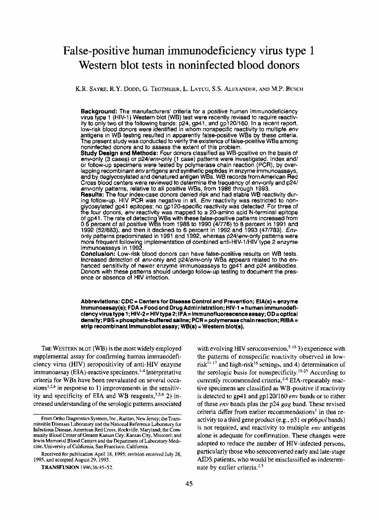
Table 1 . Summary of blood center testing data on four blood donors with suspected false-positive WBs Abbott Genetic Systems
Sample HIV-1 HIV-1/2 HIV-1, -2, RlBA RlBA anddate S:c* S:C or-l/2S:C CBCt HIV-1 WB HIV-1 HIV-1/2 IFA p24antigen Culture PCR KC
NDn ND 5/91 0.88 ND 0.22 (HIV-1) PO~gpl60/12O,~p41 ND ND Neg Neg ND ND 4/91 1.02 2.47 0.22 (HIV-1) Pos*gp160/120,gp41 Neg5 lndll gp41 Neg Neg
7/91 0.53 ND 0.14(HIV-l) Indgpl60/120 IRW-1
ND ND Neg Neg Neg Neg
9/93 ND 1.8 Neg(HIV-2) Posgp160/120,gp41,p24 ND Indgp41 Wk R” ND ND ND 10193 NO 1.5 NO Posgpl60/120,gp41,p24 NO ND NO ND ND Neg 5/95 ND ND Pos gpl60/120,gp41 ,p24 ND Ind gp41 ND ND ND Neg
6/93 ND 1.7 Neg (HIV-2) Posgp160/120,gp41 ND Indgp41 WkR ND ND ND 7/93 ND 1.2 ND Pos gpl60/120,gp41 ND ND ND ND ND Neg 5/95 ND ND Pos gpl60/120,gp41 ND Indgp41 ND ND ND Neg
4/95 ND 1.40 ND Pos gpl6O,gp41 ,pl7 ND ND ND ND ND ND 5/95 ND 1.01 ND Pos gpl6O,gp41 ,pl7 ND Neg ND Neg ND Neg
IRW-2
ARC-SEM
’ t Cambridge Biotech Corporation. All bands were 1+ intensity or greater, unless indicated. * Positive. 5 Negative. I1 Indeterminate. 1 Notdone. ’* Weakly reactive.
Signal-to-cutoff ratio of EIA ODs.

48 SAYRE ET AL. TRANSFUSION Val. 36. No. 1-1996
Results The four donors were identified by the individual blood
centers as having possibly false-positive WBs, on the basis of the donors’ denial of HIV risk factors and the restricted reac- tivity of WBs performed on both the index donations and follow-up specimens. Table 1 presents the results of all tests performed by the blood centers or blood center reference labo- ratories on serial samples from the four donors with suspected false-positive WBs. Figure 2 shows the Cambridge Biotech HIV-I WB results on the index and follow-up specimens from the four blood donors.
Donor KC was identified in April 1991 after making a do- nation that reacted in a screening anti-HIV-1 EIA with a WB showing 1+ gp41 and gp120/160 bands. The index donation was subsequently tested with a licensed anti-HIV-1/2 EIA (Abbott), and it reacted. The sample showed discrepant reactivity to re- combinant env antigens in RIBAs, failing to react to the env 4-5 antigen in RIBA HIV-1 while having strong reactivity to theenv 10 antigen contained in RIBA HIV-1/2. This was the first sug- gestion that env reactivity may be restricted to the N-terminal region of gp41 (Fig. I ) . Two follow-up specimens from KC showed declining EIA reactivity but persistent env reactivity in WB. All three serial specimens tested negative in the IFA and p24 antigen EIA. HIV culture and PCR were performed on the last follow-up specimen, and they were negative.
Donors IRW-I and IRW-2 were identified in 1993 after making donations screened as EIA-reactive in anti-HIV- 112 EIA, their WBs were interpreted as positive (gp160/120, gp41, and p24 and gp160/120 and gp41, respectively) and the anti-HIV- 2 EIAs showed no reaction. Sera from these index donations tested weakly reactive in IFA and indeterminate in RIBA HIV- 1/2 with a gp41 (env-10)-only pattern. Follow-up specimens collected approximately 4 weeks after the index donations had
5/91 10193 7n3 5/95 Positive Control 1/91 1 9193 I 6I93 I 4/95 I
A B C D E F G H ---- KC IRW-1 IRW-2 ARC-SEM
FIG. 2. HIV-1 WB results on serial specimens from each of the four blood donors uith suspected false-positive WBs. Dates of specimen col- lection are indicated at the top. Designations of specific viral antigens are indicated to the left of corresponding bands on the positive control strip.
reactivity in anti-HIV-1/2 EIA and WB that was similar to that of the index donations. Peripheral blood mononuclear cells from these follow-up specimens tested negative for HIV DNA in PCR. Additional follow-up specimens collected more than 18 months after the index donations tested EIA-negative and PCR- negative but yielded persistent env reactivity on WB and RIBA
Donor ARC-SEM tested anti-HIV- 1/2 EIA-reactive in April 1995. The donor was classified as WB-positive on the basis of I + reactivity to gp41 and gp160; I + reactivity to p17 was also observed. A follow-up specimen collected approximately 6 weeks after the index donation remained weakly reactive in anti- HIV-1/2 EIA and technically positive in WB, with I + gp160, gp41, and p17 bands. The follow-up specimen tested negative for HIV in p24 antigen EIA and PCR.
HIV- 1/2.
HIV- 1 recombinant envelope antigen analysis Table 2 summarizes the reactivity of the four index dona-
tion sera to five recombinant HIV-1 env antigens, as assayed by EIA. One donor sample (ARC-SEM) did not react to any recombinant proteins tested; the epitope to which this sample reacts therefore appears either to be located at the N-terminus of gp41 (i.e., beyond amino acid 767, the last amino acid found on env 10; see Fig. 1) or to require glycosylated or conforma- tional determinants not expressed in the recombinant antigens. Three donor samples (from KC, IRW-I, and IRW-2) reacted to env 9, env 686, and env 10, but not to env 4-5 and CBrE3 re- combinant proteins. As noted above, we also observed the re- activity of these three sera to env 10 in RIBA HIV-I/2 (Table 1). The epitope to which these three donor samples react there- fore appears to be located between amino acids 705 and 752 (Fig. 1).
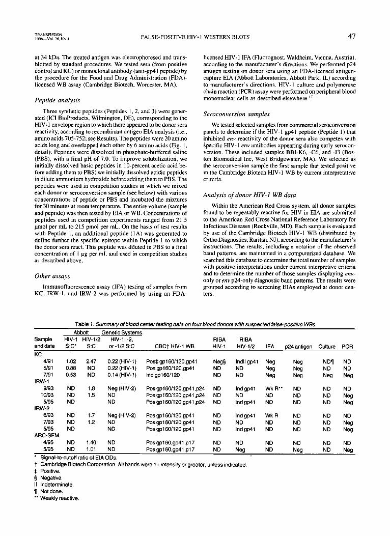
WB component analysis We tested the sample from KC with WB strips prepared with
deglycosylated and denatured HIV-I antigen; it reacted only to the 34-kDa deglycosylated monomer form of gp41 (Fig. 3). No reactivity to the gp120 antigen was observed. Therefore, we attributed the apparent reactivity of this donor sample to gp160 and gp120 bands on standard WB strips to its reactivity to the monomer, trimer, and tetramer forms of gp41.
Inhibition analysis of gp41 peptide To further map the epitope specificity of the three sera that
reacted with the recombinant gp41 antigens, we generated three peptides spanning amino acids 705 to 752 and used them in com- petition studies employing the env 686 and env 10 EIAs. Incu- bation with one of the three gp41 peptides (Peptide I ) inhib-
Table 2. Reactivity to recombinant HIV envelope antigen ElAs of index donation sera from four donors with env-only or
env/p24-only W5s EIA reactivity*
Sample env4-5 CBrE3 env9 env686 envlO
KC 0.030 0.033 0.441t 0.636t 0.567t IRW-1 0.055 0.025 0.771t 0.865t 0.896t IRW-2 0.007 0.006 0.674t 0.621t 0.738t
Values are in absorbance units. Reactivity is defined asr0.200
t Reactive.
ARC-SEM 0.007 0.027 0.017 0.027 0.048
units.
TRANSFUSION 1 9 9 6 V o l . 36. No. I FALSE-POSITIVE HIV- 1 WESTERN BLOTS 49
A O C O E F -- Standard WB Monomeric WB
(treated with Endo-F)
FIG. 3. Reactivity of KC's and control's antisera to WB strips prepared with standard versus Endo-F-treated and denatured HIV- 1 (monomeric) viral lysate. Lanes A and D, positive control sera; Lanes B and E, antisera to gp41 peptide; Lanes C and F, Donor KC. Note that the sample from KC yielded reactivity similar to that of the gp41 peptide antisera on the deglycosylated and denatured WB strips, that is, a single 34-kDa band corresponding to degl ycosy lated monomer.
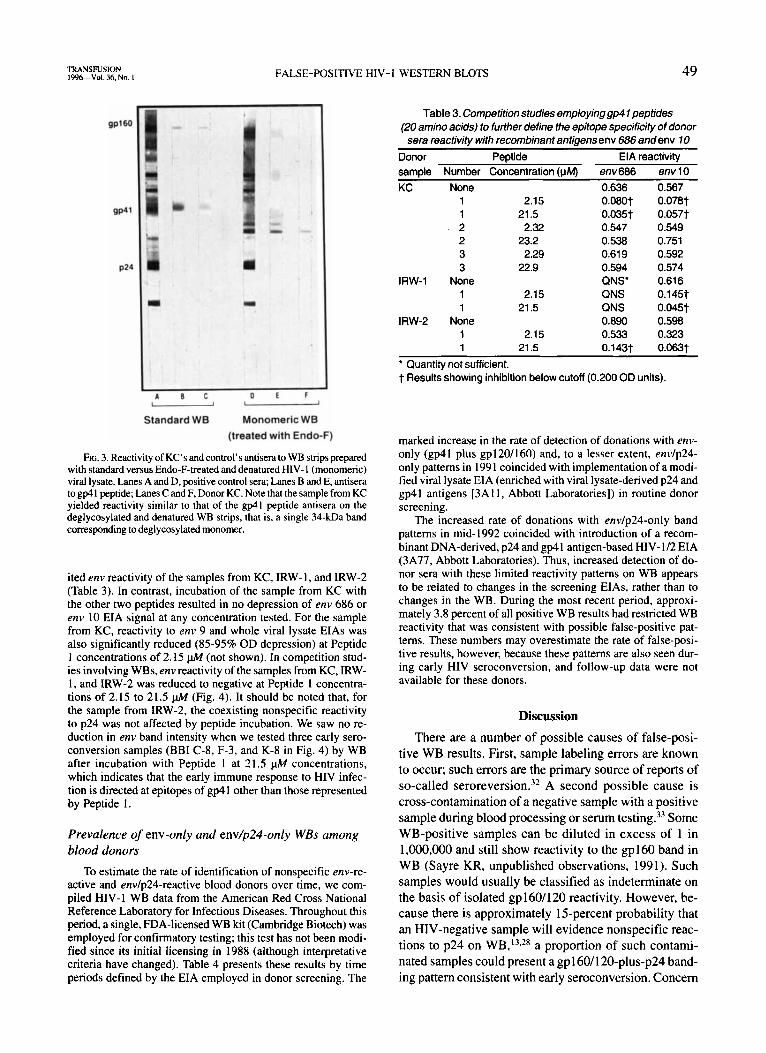
ited env reactivity of the samples from KC, IRW-I, and IRW-2 (Table 3). In contrast, incubation of the sample from KC with the other two peptides resulted in no depression of env 686 or env 10 EIA signal at any concentration tested. For the sample from KC, reactivity to env 9 and whole viral lysate EIAs was also significantly reduced (85-95% OD depression) at Peptide 1 concentrations of 2.15 pkf (not shown). In competition stud- ies involving WBs, env reactivity of the samples from KC, IRW- 1, and IRW-2 was reduced to negative at Peptide 1 concentra- tions of 2.15 to 21.5 pA4 (Fig. 4). It should be noted that, for the sample from IRW-2, the coexisting nonspecific reactivity to p24 was not affected by peptide incubation. We saw no re- duction in env band intensity when we tested three early sero- conversion samples (BBI C-8, F-3, and K-8 in Fig. 4) by WB after incubation with Peptide 1 at 21.5 pkf concentrations, which indicates that the early immune response to HIV infec- tion is directed at epitopes of gp41 other than those represented by Peptide 1.
Prevalence of env-only and env/p24-only WBs among blood donors
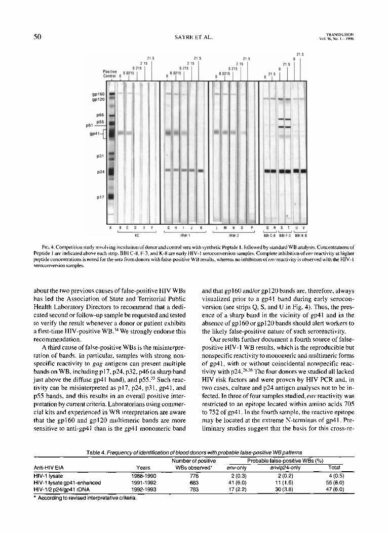
To estimate the rate of identification of nonspecific env-re- active and envlp24-reactive blood donors over time, we com- piled HIV-1 WB data from the American Red Cross National Reference Laboratory for Infectious Diseases. Throughout this period, a single, FDA-licensed WB kit (Cambridge Biotech) was employed for confirmatory testing; this test has not been modi- fied since its initial licensing in 1988 (although interpretative criteria have changed). Table 4 presents these results by time periods defined by the EIA employed in donor screening. The
Table 3. Competition studies employing gp4 1 peptides (20 amino acids) to further define the epitope specificity of donor sera reactivitv with recombinant antioensenv 686 andenv 10 -
Donor Peptide EIA reactivity Number Concentration luM env686 env 10 \r I
None 0.636 0.567 1 1 2 2 3 3
None 1 1
2.15
2.32
2.29
21.5
23.2
22.9
2.15 21.5
o .so t 0.035t 0.547 0.538 0.61 9 0.594 QNS' QNS QNS
0.078t 0.057t 0.549 0.751 0.592 0.574 0.61 6 0.145t 0.045t
IRW-2 None 0.890 0.598 1 2.15 0.533 0.323 1 21.5 0.143t 0.063t
* Quantity not sufficient. t Results showing inhibition below cutoff (0.200 OD units).
marked increase in the rate of detection of donations with env- only (gp41 plus gp120l160) and, to a lesser extent, envlp24- only patterns in 1991 coincided with implementation of a modi- fied viral lysate EIA (enriched with viral lysate-derived p24 and gp41 antigens [3All , Abbott Laboratories]) in routine donor screening.
The increased rate of donations with envlp24-only band patterns in mid-1992 coincided with introduction of a recom- binant DNA-derived, p24 and gp41 antigen-based HIV- 1/2 EIA (3A77, Abbott Laboratories). Thus, increased detection of do- nor sera with these limited reactivity patterns on WB appears to be related to changes in the screening EIAs, rather than to changes in the WB. During the most recent period, approxi- mately 3.8 percent of all positive WB results had restricted WB reactivity that was consistent with possible false-positive pat- terns. These numbers may overestimate the rate of false-posi- tive results, however, because these patterns are also seen dur- ing early HIV seroconversion, and follow-up data were not available for these donors.
Discussion There are a number of possible causes of false-posi-
tive WB results. First, sample labeling errors are known to occur; such errors are the primary source of reports of so-called serore~ers ion .~~ A second possible cause is cross-contamination of a negative sample with a positive sample during blood processing or serum testing.33 Some WB-positive samples can be diluted in excess of 1 in 1,000,000 and still show reactivity to the gp160 band in WB (Sayre KR, unpublished observations, 1991). Such samples would usually be classified as indeterminate on the basis of isolated gp160/120 reactivity. However, be- cause there is approximately 15-percent probability that an HIV-negative sample will evidence nonspecific reac- tions to p24 on WB,13s2* a proportion of such contami- nated samples could present a gp160/120-plus-p24 band- ing pattern consistent with early seroconversion. Concern
50 SAYRE ET AL. TRANSFUSION Vol. 36. No. 1--19%
21.5 21.5
A B C D E F - KC
0.0
G I
0.1 0 Oi
K L N P I I t I
IRW-1 IRW-2
O R S T U --(I nni c-8 BBI F-3 BBI n
FIG. 4. Competition study involving incubation of donor and control sera with synthetic Peptide I, followed by standard WB analysis. Concentrations of Peptide I are indicated above each strip. BBI C-8, F-3, and K-8 are early HIV- I seroconversion samples. Complete inhibition ofenv reactivity at higher peptide concentrations is noted for the sera from donors with false-positive WB results, whereas no inhibition ofenv reactivity is observed with the HIV- I seroconversion samples.
about the two previous causes of false-positive HIV WBs has led the Association of State and Territorial Public Health Laboratory Directors to recommend that a dedi- cated second or follow-up sample be requested and tested to verify the result whenever a donor or patient exhibits a first-time HIV-positive WB.34 We strongly endorse this recommendation.
A third cause of false-positive WBs is the misinterpre- tation of bands. In particular, samples with strong non- specific reactivity to gag antigens can present multiple bands on WB, including pl7, p24, p32, p46 (a sharp band just above the diffuse gp41 band), and ~ 5 5 . ' ~ Such reac- tivity can be misinterpreted as p 17, p24, p3 I , gp41, and p55 bands, and this results in an overall positive inter- pretation by current criteria. Laboratorians using commer- cial kits and experienced in WB interpretation are aware that the gp160 and gp120 multimeric bands are more sensitive to anti-gp41 than is the gp41 monomeric band
and that gp160 and/or gpl20 bands are, therefore, always visualized prior to a gp41 band during early serocon- version (see strips Q, S, and U in Fig. 4). Thus, the pres- ence of a sharp band in the vicinity of gp41 and in the absence of gp 160 or gp 1 20 bands should alert workers to the likely false-positive nature of such seroreactivity.
Our results further document a fourth source of false- positive HIV-1 WB results, which is the reproducible but nonspecific reactivity to monomeric and multimeric forms of gp41, with or without coincidental nonspecific reac- tivity with ~ 2 4 . ~ ~ 3 ~ ~ The four donors we studied all lacked HIV risk factors and were proven by HIV PCR and, in two cases, culture and p24 antigen analyses not to be in- fected. In three of four samples studied,env reactivity was restricted to an epitope located within amino acids 705 to 752 of gp41. In the fourth sample, the reactive epitope may be located at the extreme N-terminus of gp4 I . Pre- liminary studies suggest that the basis for this cross-re-
Table 4. Frequency of identification of blood donors with probable false-positive WB patterns Number of positive Probable false-positive WBs (%)
Anti-HIV EIA Years WBs observed* env-only envlp24-only Total HIV-1 lysate 1988-1 990 776 2 (0.3) 2 (0.2) 4 (0.5) HIV-1 lysate gp41-enhanced 1991-1992 683 41 (6.0) 11 (1.6) 55 (8.0) HIV-1/2 p24/~p41 rDNA 1992-1 993 783 17 (2.2) 30 (3.8) 47 (6.0)
According to revised interpretative criteria.
TRANSFUSION 1’)96Vol. 36. No. I FALSE-POSITIVE HIV- 1 WESTERN BLOTS 51
activity with HIV-1 gp41 epitopes may be infection by paramyxoviruses, naturally occurring carbohydrate anti- bodies in human sera, or autoantibodies against cellular protein^.^'-^^
We documented an increase in the rate of observation of env-only and envlp24-only WB results over the past 6 years, which appears to be related to the implementation of new donor-screening EIAs (e.g., second-generation anti-HIV- 1 and third-generation anti-HIV- 1/2 screening assays, Abbott Laboratories). These assays have been shown to have significantly increased sensitivity to env and gag antibodies, such that infected persons are iden- tified earlier during seroconversion.s.6 Unfortunately, this enhanced sensitivity appears also to have resulted in the detection of specimens with nonspecific reactivity, evi- denced by env-only and envlp24-only patterns on WB. The enhanced sensitivity of the new EIAs to early HIV- 1 infection is critical for transfusion safety and counterbal- ances the problem of inadvertent detection of samples with false-positive WB patterns documented here. None- theless, it is important that WB interpretative criteria be revised so that the patterns discussed herein are classi- fied as indeterminate rather than positive, or that mea- sures be introduced to identify possible false-positive band patterns so that proper donor notification occurs. After reviewing the findings of the present study with CDC and FDA scientists, we decided that a revision of WB inter- pretative criteria is not warranted at present. The ratio- nale is that the public health benefits of correct classifi- cation of a large number of infected persons as positive under the revised criteria (rather than their misclassi- fication as indeterminate under the earlier criteria) out- weigh the rare occurrence of false-positive WBs. On the other hand, an approach for “flagging” possible false- positive patterns was widely endorsed.
We therefore recommend that subjects with WB pat- terns associated with false positivity (i.e., those lacking p31 or gp120/160 band reactivity) be informed that, al- though the results on their specimen were technically positive, these patterns have been seen in low-risk blood donors who have been determined through follow-up test- ing not to be infected with HIV. For such persons, fur- ther testing should be performed on a specimen collected at least 2 weeks after the index specimen. Serum or plasma from this follow-up specimen should be tested for anti-HIV by licensed WB in parallel with the retesting of an aliquot of frozen sera from the index donation. Additional tests performed on the index and/or follow- up specimen, such as p24 antigen EIA, virus culture, or PCR for HIV- 1 RNA or DNA, may also be useful, if avail- able. Because the IFA may react with env false-positive samples, this test is of less utility in resolving these cases. If testing of follow-up samples indicates no change or loss of bands in WB (i.e., an absence of seroconversion), as well as an absence of HIV antigen, nucleic acids or virus
(if such tests are performed), the donor should be noti- fied that the earlier results probably represent a false- positive finding. A recommendation for final follow-up testing at 6 months should be given, consistent with CDC recommendations.* In contrast, if parallel WB testing shows an increase in the number of bands (including p3 1 and gp120/160) and a marked increase in band intensi- ties with the follow-up sera relative to the index dona- tion sera, or if direct virus assays are positive, the donor should be notified of the probability of recent HIV infec- tion, with evolving seroconversion. An additional follow- up sample, obtained 4 to 6 weeks after the first follow-up sample (that is, 8-12 weeks after the index donation) and retested as above, should be used to corroborate a final diagnosis of HIV infection.
Acknowledgments The authors thank Helen Ownby, PhD, and Dan Waxman, MD,
at the American Red Cross Blood Services, Southeast Michigan Re- gion, for donor history and specimen collection in Case 4; Chou-Pong Pau, PhD, CDC, for Peptide 1 EIA and IFA data; Eve Lackritz, MD, and Lyle Petersen, MD, at CDC and Jay Epstein, MD, at FDA for critical discussion; Leslie Tobler, DrPH, at Irwin Memorial Blood Cen- ters and Theresea Nicholson and Arlene Klingamen at Ortho Diag- nostics Systems for technical assistance; and Barbara Johnson for manuscript preparation.
1.
2.
3.
4.
5 .
6.
7.
8.
9.
10.
11.
References Interpretation and use of the Westem blot assay for serodiagnosis of human immunodeficiency virus type I infections. MMWR Morb Mortal Wkly Rep 1989;38(Suppl7):1-7. Interpretive criteria used to report Western blot results for HIV- 1-antibody testing-United States. MMWR Morb Mortal Wkly Rep 1991;40:692-5. O’Gorman MR, Weber D, Landis SE, et al. Interpretive criteria of the Western blot assay for serodiagnosis of human immuno- deficiency virus type 1 infection. Arch Pathol Lab Med
Revised recommendations for the prevention of human immu- nodeficiency virus (HIV) transmission by blood and blood prod- ucts. Bethesda: Center for Biologics Evaluation and Research, Food and Drug Administration, April 23, 1993. Zaaijer HL, van Exel-Oehlers P, Kraaijeveld T, et al. Early de- tection of antibodies to HIV-1 by third-generation assays. Lan- cet 1992;340:770-2. Gallarda JL, Henrard DR, Liu D, et al. Early detection of anti- body to human immunodeficiency virus type 1 by using an an- tigen conjugate immunoassay correlates with the presence of immunoglobulin M antibody. J Clin Microbiol I992;302379- 84. Saah AJ, Farzadegan H, Fox R, et al. Detection of early antibodies in human immunodeficiency virus infection by enzyme-linked immunosorbent assay, Western blot, and radioimmunoprecipi- tation. J Clin Microbiol 1987;25: 1605-10. Courouce AM. Following up indeterminate HIV- 1 western blots (letter). Lancet 19892: 1330-1. King R, Frey S, Beisha R, et al. The presence or absence of gpl2O/gpI60 bands on indeterminate western blots: predictive value for HIV seroconversion (letter). AIDS 1993;7:437-8. Read S, Cassol S, Coates R, et al. Detection of incident HIV infection by PCR compared to serology. J Acquir Immune Defic Syndr 1992;5: 1075-9. Dock NL, Lamberson HV Jr, O’Brien TA. et al. Evaluation of atypical human immunodeficiency virus immunoblot reactivity in blood donors. Transfusion 1988;28:412-8.
1991; I 15:26-30.
52 SAYRE ET AL. TRANSFUSION Vul. %.No. 1-1996
12. Nusbacher J, Naiman R. Longitudinal follow-up of blood donors found to be reactive for antibody to human immunodeficiency virus (anti-HIV) by enzyme-linked immunoassay (EIA+) but negative by western blot (WB-). Transfusion 1989;29:365-7.
13. Genesca J, Shih JWK, Jett BW, et al. What do western blot in- determinate patterns for human immunodeficiency virus mean in EIA-negative blood donors? Lancet I989;2: 1023-5.
14. Jackson JB, MacDonald KL, Cadwell J, et al. Absence of HIV infection in blood donors with indeterminate western blot tests for antibody to HIV- I . N Engl J Med I990;322:2 17-22.
15. Dock NL, Kleinman SH, Rayfield MA, et al. Human immuno- deficiency virus infection and indeterminate western blot pat- terns. Prospective studies in a low prevalence population. Arch Intern Med 1991;151:525-39.
16. Celum CL, Coombs RW. Lafferty W, et al. Indeterminate human immunodeficiency virus type 1 western blots: seroconversion risk, specificity of supplemental tests, and an algorithm for evalu- ation. J Infect Dis 1991;164:656-64.
17. Eble BE, Busch MP, Khayam-Bashi H, et al. Resolution of in- fection status of human immunodeficiency virus (HIV)- seroindeterminate and high-risk seronegative individuals with polymerase chain reaction and virus culture: absence of persis- tent silent HIV type I infection in a high-prevalence area. Trans- fusion 1992;32:503-8.
18. Phair J, Hoover D, Huprikar J, et al. The significance of western blot assays indeterminate for antibody to HIV in a cohort of homosexualhisexual men. The Multicenter AIDS Cohort Study. J Acquir Immune Defic Syndr 1992:5:988-92.
19. Hunter JB, Menitove JE. HLA antibodies detected by ELISA HTLV-111 antibody kits (letter). Lancet 1985;2:397.
20. Ameglia F, Dolei A, Benedetto A, et al. Antibodies reactive with nonpolymorphic epitopes on HLA molecules interfere in screen- ing tests for the human immunodeficiency virus (letter). J Infect Dis 1987;156:1034-5.
2 1. Blanton M, Balakrishnan K, Dumaswala U, et al. HLA antibod- ies in blood donors with reactive screening tests for antibody to the immunodeficiency virus (letter). Transfusion 1987;27: I 18-9.
22. Povolotsky J, Gold JWM, Chein N. et al. Differences in human immunodeficiency virus type 1 (HIV- I ) anti-p24 reactivities in serum of HIV-I-infected and uninfected subjects: analysis of indeterminate western blot reactions. J Infect Dis 1991; 163:247- 51.
23. Whetstone CA, Sayre KR, Dock NL. et al. Examination of whether persistently indeterminate human immunodeficiency virus type 1 Western immunoblot reactions are due to serologi- cal reactivity with bovine immunodeficiency-like virus. J Clin Microbiol 1992;30:764-70.
24. Langedijk JP, Vos WF, van Doornum GJ, et al. Identification of cross-reactive epitopes recognized by HIV- 1 false-positive sera (letter). AIDS 1992;6: 1547-8.
25. Jindal R, Solomon M, Burrows L. False positive tests for HIV in a woman with lupus and renal failure (letter). N Engl J Med
26. Healey DS. Bolton WV. Apparent HIV-I glycoprotein reactivity on western blot in uninfected blood donors. AIDS
27. Ratner L, Haseltine W, Patarca R. et al. Complete nucleotide sequence of the AIDS virus, HTLV-Ill. Nature 1985;313:277- 84.
28. Busch MP, El Amad Z, McHugh TM. et al. Reliable confirma- tion and quantitation of human immunodeficiency virus type 1
1993;328: 1281-2.
1993:7:655-8.
antibody using a recombinant-antigen immunoblot assay. Trans- fusion I99 I :3 1 : 129-37.
29. McNairn D, Kline R, Quinn TC. Evaluation of the ChironR RIBATM HIV-UHIV-2 Strip Immunoblot Assay (SIA) (abstract 370). Presented at the Second National Conference on Human Retroviruses, Washington DC, Jan-Feb 1995: 122.
30. Pinter A, Honnen WJ, Tilley SA, et al. Oligomeric structure of gp41, the transmembrane protein of human immunodeficiency virus type 1. J Virol 1989;63:2674-9.
3 I . Earl PL, Doms RW, Moss B. Oligomeric structure of the human immunodeficiency virus type I envelope glycoprotein. Proc Natl Acad Sci U S A 1990;87:648-52.
32. Ascher DP, Roberts C. Determination of the etiology of sero- reversals in HIV testing by antibody fingerprinting. J Acquir Immune Defic Syndr 1993:6:241-4.
33. Sabino EC, Delwart E, Lee TH, et al. Identification of low-level contamination of blood as basis for detection of human immu- nodeficiency virus (HIV) DNA in anti-HIV-negative specimens. J Acquir Immune Defic Syndr 1994:7:853-9.
34. Association of State and Territorial Public Health Laboratory Directors. Presented at the 8th Annual Conference on Human Retrovirus Testing, Reno, NV. 1993:2
35. Bukrinsky MI, Chaplinskas SA, Syrtsev VA. et al. Reactivity to gag- and env-related proteins in immunoblot assay is not nec- essarily indicative of HIV infection (letter). AIDS 1988;2:405-6.
36. Fenouillet E. Blanes N, Gluckman JC. gp 160 of commercial HIV Western blots is not gp160". Should criteria for seropositivity be revised?(letter). AIDS 1991:5:770.
37. Gonzalez-Scarano F, Waxham MN, Ross AM, Hoxie JA. Se- quence similarities between human immunodeficiency virus gp41 and paramyxovirus fusion proteins. AIDS Res Hum Retroviruses
38. Tomiyama T, Lake D, Masuho Y. Hersh EM. Recognition of human immunodeficiency virus glycoproteins by natural anti- carbohydrate antibodies in human serum. Biochem Biophys Res Commun 1991 ; I77:279-85.
39. Wu AF, Wood C, Wu TT. Possibility of HIV gp41 and thymosin beta-4 sharing the same antigenic epitope (letter). AIDS
1987;3:245-52.
1989;3:319-20.
Keith R. Sayre, MBA, Product Manager, AIDSlHepatitis Busi- ness Unit, Ortho Diagnostic Systems, Inc., Raritan, NJ.
Roger Y. Dodd, PhD, Head, Transmissible Diseases Laboratory, American Red Cross, Rockville, MD.
Gary Tegtmeier. PhD, Director of Research, Community Blood Center of Greater Kansas City, Kansas City, MO.
Lynne Layug, MPH, MT(ASCP). Technical Officer, National Ref- erence Laboratory for Infectious Disease. American Red Cross, Rock- ville, MD.
Steve S. Alexander, PhD. Principal Scientist, AIDSlHepatitis Research and Development, Ortho Diagnostic Systems.
Michael P. Busch. MD, PhD. Associate Professor in Residence. Department of Laboratory Medicine, University of California, San Francisco; and Vice President, Research and Scientific Services, Irwin Memorial Blood Centers, 270 Masonic Avenue, San Francisco, CA 941 18-4496. [Reprint requests]