Factors Influencing the Production of Recombinant SV40 Vectors
12
Factors Influencing the Production of Recombinant SV40 Vectors Maria Vera, 1 Jesus Prieto, 1 David S. Strayer, 2 and Puri Fortes 1, * 1 Laboratory of Vector Development, Division of Gene Therapy, Foundation for Applied Medical Research, School of Medicine, University of Navarra, Irunlarrea 1, 31008 Pamplona, Spain 2 Department of Pathology and Cell Biology, Jefferson Medical College, 1020 Locust Street, Philadelphia, PA 19107, USA *To whom correspondence and reprint requests should be addressed. Fax: 34 948 425700. E-mail: [email protected]. Available online 3 August 2004 Most gene therapy approaches employ viral vectors for gene delivery. Ideally, these vectors should be produced at high titer and purity with well-established protocols. Standardized methods to measure the quality of the vectors produced are imperative, as are techniques that allow reproducible quantitation of viral titer. We devised a series of protocols that achieve high-titer production and reproducible purification and provide for quality control and titering of recombinant simian virus 40 vectors (rSV40s). rSV40s are good candidate vehicles for gene transfer: they are easily modified to be nonreplicative and they are nonimmunogenic. Further, they infect a wide variety of cells and allow long-term transgene expression. We report here these protocols to produce rSV40 vectors in high yields, describe their purification, and characterize viral stocks using quality control techniques that monitor the presence of wild-type SV40 revertants and defective interfering particles. Several methods for reproducible titration of rSV40 viruses have been compared. We believe that these techniques can be widely applied to obtain high concentrations of high-quality rSV40 viruses reproducibly. Key Words: SV40, recombinant SV40 vectors, gene therapy, production, titration, DI particles, wtSV40 revertants INTRODUCTION Simian virus 40 (SV40) is an icosahedral nonenveloped polyomavirus with a double-stranded circular DNA of 5.2 kb [1,2]. Several properties make SV40 a good candidate to be used as a vector for gene therapy approaches: (i) it is easily modified to be nonreplicative ([3–5], references therein, and this report); (ii) it can be produced in large quantities [3,4]; (iii) it infects almost every cell type that has been tested, both dividing and quiescent [6–8]; (iv) it is not immunogenic [9,10]; (v) it allows long-term expression of the transgene [6,7,9–13]; (vi) its molecular biology is well studied; and (vii) the effects in humans of wild-type SV40 have been documented [14,15]. The advantages of SV40 as a gene therapy vector can be explained by the SV40 replicative cycle. SV40 binds the major histocompatibility complex class I (MHC I) at the cell surface [16]. MHC I is present on most cell types, explaining SV40Ts wide host range. Following virus entry into the cell, MHC I is shed, which may result in poor antigen presentation by SV40-infected cells [17]. The virus enters the cell via a caveolar pathway that delivers SV40 to a microtubular network that transports the virion to the endoplasmic reticulum [17,18]. Since SV40 traverses nuclear pores, it can infect nondividing cells productively. The SV40 genome is released in the nucleus as a nucleosome-coated mini- chromosome that can be integrated randomly into the host genome [19,20]. SV40 uses the cell machinery for replication and transcription. The SV40 early promoter drives expression of one alternatively spliced gene that encodes the large T antigen (Tag) and the small t antigen (tag) [2]. The late promoter, on the opposite strand, controls expression of the structural proteins, VP1, VP2, and VP3 (Fig. 1A). Both promoters, together with the regulatory sequences, origin of replication, and packaging signals, are located within approximately 500 bases [2]. Several of their functions are controlled by Tag. Tag is essential for genome replication and for late promoter-driven transcription. It also binds and inactivates p53 and the retinoblastoma protein, thereby immortalizing cells in culture [21,22]. Tag is mainly a nuclear protein, but it is produced in excess and inserts into the cell membrane, where it is the major virus antigen [23]. The Tag gene is removed to generate recombinant SV40 viruses (rSV40) ([3,4] and this report). This renders rSV40 replication deficient, nononcogenic, and nonimmunogenic, because the major antigen is not 1525-0016/$30.00 METHOD doi:10.1016/j.ymthe.2004.06.1014 MOLECULAR THERAPY Vol. 10, No. 4, October 2004 780 Copyright C The American Society of Gene Therapy
Transcript of Factors Influencing the Production of Recombinant SV40 Vectors

METHOD doi:10.1016/j.ymthe.2004.06.1014
Factors Influencing the Production of RecombinantSV40 Vectors
Maria Vera,1 Jesus Prieto,1 David S. Strayer,2 and Puri Fortes1,*
1Laboratory of Vector Development, Division of Gene Therapy, Foundation for Applied Medical Research, School of Medicine,
University of Navarra, Irunlarrea 1, 31008 Pamplona, Spain2Department of Pathology and Cell Biology, Jefferson Medical College, 1020 Locust Street, Philadelphia, PA 19107, USA
*To whom correspondence and reprint requests should be addressed. Fax: 34 948 425700. E-mail: [email protected].
Available online 3 August 2004
780
Most gene therapy approaches employ viral vectors for gene delivery. Ideally, these vectors shouldbe produced at high titer and purity with well-established protocols. Standardized methods tomeasure the quality of the vectors produced are imperative, as are techniques that allowreproducible quantitation of viral titer. We devised a series of protocols that achieve high-titerproduction and reproducible purification and provide for quality control and titering ofrecombinant simian virus 40 vectors (rSV40s). rSV40s are good candidate vehicles for gene transfer:they are easily modified to be nonreplicative and they are nonimmunogenic. Further, they infect awide variety of cells and allow long-term transgene expression. We report here these protocols toproduce rSV40 vectors in high yields, describe their purification, and characterize viral stocks usingquality control techniques that monitor the presence of wild-type SV40 revertants and defectiveinterfering particles. Several methods for reproducible titration of rSV40 viruses have beencompared. We believe that these techniques can be widely applied to obtain high concentrationsof high-quality rSV40 viruses reproducibly.
Key Words: SV40, recombinant SV40 vectors, gene therapy, production, titration, DI particles,wtSV40 revertants
INTRODUCTION
Simian virus 40 (SV40) is an icosahedral nonenvelopedpolyomavirus with a double-stranded circular DNA of 5.2kb [1,2]. Several properties make SV40 a good candidateto be used as a vector for gene therapy approaches: (i) it iseasily modified to be nonreplicative ([3–5], referencestherein, and this report); (ii) it can be produced in largequantities [3,4]; (iii) it infects almost every cell type thathas been tested, both dividing and quiescent [6–8]; (iv) itis not immunogenic [9,10]; (v) it allows long-termexpression of the transgene [6,7,9–13]; (vi) its molecularbiology is well studied; and (vii) the effects in humans ofwild-type SV40 have been documented [14,15].
The advantages of SV40 as a gene therapy vector canbe explained by the SV40 replicative cycle. SV40 bindsthe major histocompatibility complex class I (MHC I) atthe cell surface [16]. MHC I is present on most celltypes, explaining SV40Ts wide host range. Followingvirus entry into the cell, MHC I is shed, which mayresult in poor antigen presentation by SV40-infectedcells [17]. The virus enters the cell via a caveolarpathway that delivers SV40 to a microtubular networkthat transports the virion to the endoplasmic reticulum
[17,18]. Since SV40 traverses nuclear pores, it can infectnondividing cells productively. The SV40 genome isreleased in the nucleus as a nucleosome-coated mini-chromosome that can be integrated randomly into thehost genome [19,20].
SV40 uses the cell machinery for replication andtranscription. The SV40 early promoter drives expressionof one alternatively spliced gene that encodes the large Tantigen (Tag) and the small t antigen (tag) [2]. The latepromoter, on the opposite strand, controls expression ofthe structural proteins, VP1, VP2, and VP3 (Fig. 1A). Bothpromoters, together with the regulatory sequences, originof replication, and packaging signals, are located withinapproximately 500 bases [2]. Several of their functions arecontrolled by Tag. Tag is essential for genome replicationand for late promoter-driven transcription. It also bindsand inactivates p53 and the retinoblastoma protein,thereby immortalizing cells in culture [21,22]. Tag ismainly a nuclear protein, but it is produced in excess andinserts into the cell membrane, where it is the major virusantigen [23]. The Tag gene is removed to generaterecombinant SV40 viruses (rSV40) ([3,4] and this report).This renders rSV40 replication deficient, nononcogenic,and nonimmunogenic, because the major antigen is not
1525-0016/$30.00
MOLECULAR THERAPY Vol. 10, No. 4, October 2004
Copyright C The American Society of Gene Therapy

FIG. 1. Schematic view of wtSV40 genome and strategy to construct recombinant SV40 plasmids. (A) The wtSV40 genome is a 5.2-kb double-stranded DNA with
an origin of replication that overlaps with the SV40 early promoter (SVEP) and a SV40 late promoter (SVLP). SVEP drives the expression of Tag genes that encode
Tag and tag proteins. SVLP controls VP1, VP2, and VP3 protein expression (Capsid genes). Polyadenylation sequences are indicated (pA). (B) Generation of
recombinant SV40 virus genomes. (1) Tag gene has been replaced by an ampicillin-resistance (Ampr) gene, a bacterial origin of replication, and a small polylinker
(ClaI and XhoI) in pSL-4 [37]. (2) A longer polylinker has been introduced under the SVEP of pSL-4 to construct pSL-4pL. The polylinker has unique sites (sites A
indicated in black: ClaI, BglII, NheI, XmaI, BclI, Sal I, and XhoI) and sites that can be found at both sides of the Ampr gene (sites B indicated in gray: Xba I, Not I,
SacII, and Sac I). (3) A transgene can be easily cloned with restriction sites A under the control of SVEP. (4) To produce recombinant SV40 genomes (prSVX), the
Ampr gene is removed with restriction sites B and the plasmid is circularized.
METHODdoi:10.1016/j.ymthe.2004.06.1014
produced and capsid protein expression is not activated[2]. Removal of the Tag gene also generates approxi-mately 2.5 kb of free space in the SV40 genome to clonetransgenes. Removing the capsid genes creates approx-imately 2.5 kb of additional space. Thus, recombinantSV40 vectors can accommodate as much as 5 kb ofinserted DNA [24].
MOLECULAR THERAPY Vol. 10, No. 4, October 2004
Copyright C The American Society of Gene Therapy
The advantages of rSV40 as a gene therapy vectorhave encouraged several groups to study its efficacy inanimal models. Ex vivo infection of hematopoietic stemcells with rSV40 has permitted expression of multidrugresistance gene 1, h-globin [8,25,26], and the surfaceantigen of hepatitis B virus [7,27]. rSV40 vectors havebeen used successfully in animal models for liver
781

METHOD doi:10.1016/j.ymthe.2004.06.1014
diseases like Crigler–Najjar syndrome type 1 [10]. Inaddition, rSV40 has been employed for vaccination andimmunostimulation purposes ([9,28], and our unpub-lished results or to block HIV replication using differentstrategies [27–35].
Despite the great potential of rSV40 in gene therapyprotocols, few methods for rSV40 vector production,purification, quality control, and titering have beendescribed. For example, published methods for wild-type SV40 production and purification need to betested to see if they can be applied to rSV40 vectors[36]. Recently, methods to produce rSV40 vectors thatdo not analyze the factors influencing vector produc-tion have been proposed [3,4]. We have compared andmodified several protocols to define a method thatyields high titers of Tag-deleted nonreplicating rSV40viruses. The presence of contaminating wild-type (wt)SV40 or defective interfering (DI) particles was analyzedto control for quality of vector preparations. We havealso compared new or already described methods forrSV40 titering. We believe that these protocols couldbe widely used both in laboratories that already workwith rSV40 vectors and in groups that are tempted toexploit the strengths of SV40 as a gene deliveryvehicle.
RESULTS
Manipulation of the Viral GenomeGeneration of recombinant SV40 viruses lacking the Taggene is represented in Fig. 1B. We started with pSL-4, inwhich the Tag open reading frame has been replaced bythe ampicillin-resistance gene and bacterial origin ofreplication (Ampr; Fig. 1B1) [37]. We first introduced apolylinker with seven unique restriction sites after theSV40 early promoter (EP) of pSL-4 and four restrictionsites found at both sides of the Ampr gene, to generatepSL-4pL (Fig. 1B2). The Ampr gene, which is requiredonly for bacterial selection, can thus be removed easilyonce the desired transgene has been inserted (Fig. 1B3).Also, the rSV40 genome can be efficiently circularized(Fig. 1B4). We used this strategy to produce severalrecombinant SV40 genomes. Luciferase and GFP DNAswere cloned into pSL-4pL to produce prSVLUC andprSVGFP as described under Materials and Methods.These plasmids were used to produce SVLUC and SVGFPrecombinant viruses (rSVLUC and rSVGFP, respectively).
rSV40 Virus ProductionOur recombinant SV40 genomes do not replicate in cellsthat lack Tag, since Tag is essential for virus genomereplication and for transcription of capsid genes. There-fore we analyzed the ability of different Tag-expressingcell lines to produce rSV40s. The packaging cell linesused were COS-1, COS-7, CMT4, and COT2, which wereall derived from CV-1 cells, and 293T, derived from 293
782
cells (see Materials and Methods for details). In all of thecell lines tested Tag expression is constitutive except forCMT4 and COT2, in which Tag is under the control of aninducible metallothionein promoter [5,39].
We tested different transfection methods to intro-duce rSV40 genomes into packaging cell lines: calciumphosphate precipitation and coupling DNA to lipidslike Fugene and Lipofectamine. We used differentamounts of prSVGFP to transfect subconfluent cellsand 48 h after transfection monitored GFP expressionby FACS and visualized it by fluorescence microscopy.In all cases, calcium phosphate precipitation gavegreater than or equal to fourfold more GFP-expressingcells than did the other methods tested (data notshown). Efficiencies of transfection with calcium phos-phate for the different packaging cell lines werecomparable, except for 293T cells, which were trans-fected three- to fivefold more efficiently (data notshown). However, since 293T cells did not amplifyrecombinant viruses as well as the simian cell linestested (see below), we used COS-1 cells to standardizerSV40 virus production.
To check whether prSVLUC and prSVGFP plasmidswere able to produce rSV40 viruses, we transfected COS-1cells with these plasmids. We collected media and cells 3,5, or 7 days after transfection. We pooled both cells andmedia and subjected them to three cycles of freezing andthawing to break cell membranes and liberate the viruses.Then we used these lysates to infect CV-1 cells. Forty-eight hours postinfection we visualized GFP or lysed thecells to measure luciferase activity. Luciferase expressionwas higher in CV-1 cells infected with rSVLUC virusescollected on day 3 than on day 5 or 7 (data not shown).The rSVLUC viruses obtained were amplified by infectionof fresh COS-1 cells. Again, we observed luciferase activityin CV-1 cells infected with rSVLUC viruses collected onday 3, 5, or 7 post-COS-1 infection (Fig. 2A). We observedthe highest expression with day 3-collected viruses,which is in agreement with the time required for wtSV40to complete an infectious cycle in tissue culture monkeycells.
We never observed GFP expression, however, when weperformed the same experiments with prSVGFP. Forreasons that are so far unknown, other groups working onrSV40 have also failed to detect GFP expression fromrSVGFPviruses (A.Oppenheim,personalcommunication).
Wild-type SV40 infection is usually lytic in permissivecells. If cell lysis is also very efficient in producer cellsinfected with rSV40, most of the viruses should be foundin the supernatant. However, viruses that have beenreleased from lysed cells may also be attached toneighboring cell membranes. To see if infectious virusescan be found in both cell pellet and media, we collectedthese fractions 3 days after infection of COS-1 cells withrSVLUC. We used comparable amounts of both fractionsto transduce CV-1 cells and measured luciferase activity
MOLECULAR THERAPY Vol. 10, No. 4, October 2004
Copyright C The American Society of Gene Therapy

METHODdoi:10.1016/j.ymthe.2004.06.1014
(Fig. 2B). Even if luciferase activity was higher in CV-1cells infected with the fraction containing the cell pellet,both fractions yielded high levels of luciferase activity.
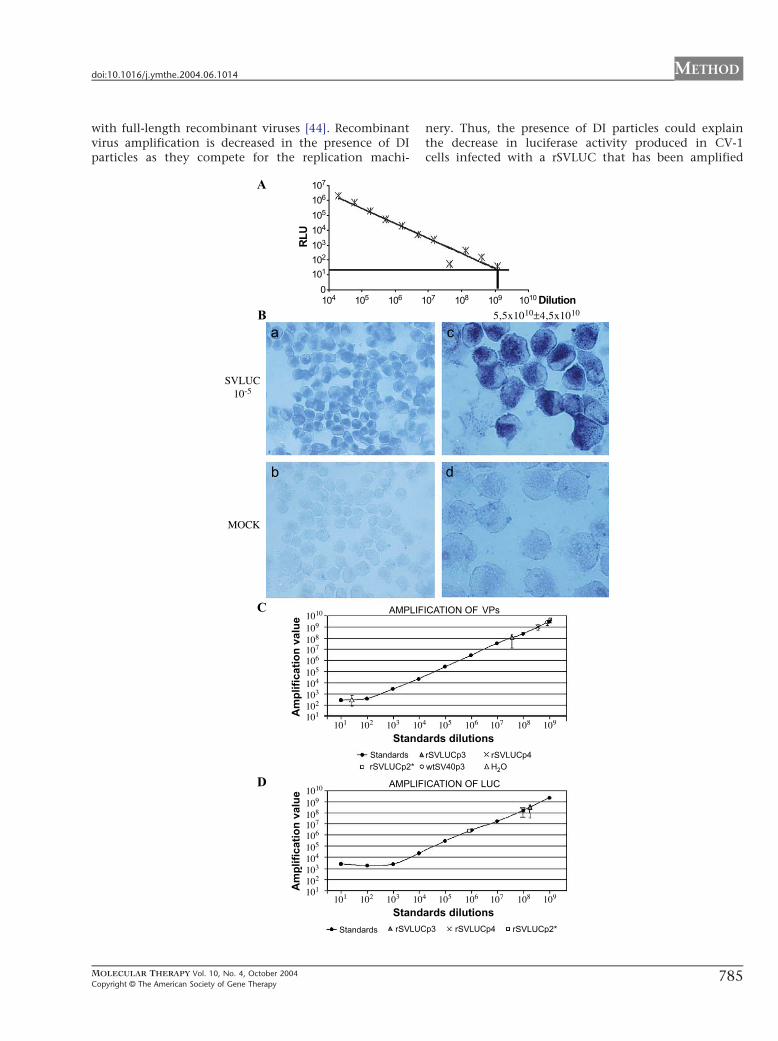
FIG. 2. Development of a method to produce rSV40 viruses.(A) Quantitation of lu
rSVLUC viruses were used to infect COS-1 cells and recombinant virus amplificatio
and lysed. The activity of recombinant viruses was quantified by infection of CV-1
Statistical analysis shown at the top of the graphic indicates significant (*) and no
viruses that accumulated 3 days after infection in COS-1 cells or supernatant. COS
separated from the supernatant fraction. Both fractions were lysed and comparab
titrated by measurement of luciferase activity. (C) Quantitation of luciferase-ex
rSVLUC viruses were amplified for two rounds in COS-1, COS-7, CMT4, and 293
cells in which luciferase activity was measured. The significant difference found be
expression of COS-1 and COT-2 cells. COT-2 cells were induced with heavy met
fixed and an immunofluorescence assay with anti-Tag antibody was carried out (
luciferase-expressing rSVLUC viruses produced in COS-1 cells infected once or thr
once or every 12 h three times with the regular amount or half the amount of rSV
luciferase activity was measured. (F) Quantitation of luciferase-expressing rSVLUC
activity was measured in CV-1 cells infected with virus produced after each rou
twice. Error bars indicate standard deviations.
MOLECULAR THERAPY Vol. 10, No. 4, October 2004
Copyright C The American Society of Gene Therapy
We also wanted to identify the most advantageouspackaging cell line. We amplified viruses produced inCOS-1 cells twice in COS-1, COS-7, CMT4, COT2, or 293T
ciferase-expressing rSVLUC viruses produced in COS-1 cells for 3, 5, or 7 days.
n was allowed for 3, 5, or 7 days. Then, cells and supernatants were collected
cells in which luciferase activity was measured in relative luciferase units (RLU).
nsignificant (ns) differences. (B) Quantitation of luciferase-expressing rSVLUC
-1 cells were infected with rSVLUC. 3 days after infection the cellular pellet was
le amounts were used to infect CV-1 cells in which recombinant viruses were
pressing rSVLUC viruses amplified in COS-1, COS-7, 293T, and CMT4 cells.
T cells. After each round of infection, viruses were titrated by infection of CV-1
tween 293T and CMT4 cells and COS cells is indicated with an asterisk. (D) Tag
als as described [39]. COT-2 cells (c, d) or uninduced COS-1 cells (a, b) were
a, c). Staining with DAPI of the same fields is shown (b, d). (E) Quantitation of
ee times consecutively with two different viral doses. COS-1 cells were infected
LUC virus. The viruses produced were titrated by infecting CV-1 cells in which
viruses produced after four rounds of amplification in COS-1 cells. Luciferase
nd of amplification. All the experiments were done in triplicate and repeated
783

METHOD doi:10.1016/j.ymthe.2004.06.1014
cells using identical conditions. Amplification was doneas described under Materials and Methods. After eachround of viral amplification, we titrated viruses able toexpress the transgene by infection of CV-1 cells in whichluciferase activity was measured. The results indicate thatCOT2 (data not shown), CMT4, and 293T cells are not aseffective as COS cell lines in producing luciferase-express-ing rSV40 vectors (Fig. 2C). Also, even though COS-1 andCOS-7 amplified rSVLUC viruses to similar extents in afirst round of infection, COS-1 cells produced moreluciferase-expressing virus in a second round of amplifi-cation (Fig. 2C). Therefore we used COS-1 cells for rSV40virus production.
As Tag is required for virus replication, we comparedTag expression in the different packaging cell lines.While all COS cells expressed Tag to a similar extent,heavy metal-induced COT2 cells expressed differentlevels of Tag (compare a and c in Fig. 2D). Also, someCOT2 cells showed undetectable levels of Tag (comparec and d in Fig. 2D). Heavy metal-induced CMT4 andCOT2 cells produced similar amounts of Tag, as deter-mined by immunofluorescence analysis (data notshown).
We compared rSVLUC production by infection ofCOS-1 cells, once or three times consecutively. Weused the viruses produced to infect CV-1 cells inwhich luciferase activity was measured. The resultsindicate that the highest virus production in terms ofluciferase activity was obtained with a single infection(Fig. 2E). By infecting with half the amount of virus,we reduced detected luciferase activity also by half(Fig. 2E). Increasing the amount of virus used to infectdid not significantly increase the amount of luciferase-expressing vector produced (data not shown). Surpris-ingly, when cells were infected three times consec-utively, luciferase activity in CV-1-infected cellsdecreased.
Finally, we wanted to know how many rounds of virusamplification could be done without altering rSV40yields. We carried out four rounds of amplification inCOS-1 cells. After each round, luciferase activity wasmeasured in rSVLUC-infected CV-1 cells. We found thatvirus could be amplified for up to three rounds, as theproducts of the fourth round gave much less luciferaseactivity (Fig. 2F).
FIG. 3. Titration of rSVLUC viruses. (A) Titration by luciferase activity measuremen
serial dilutions of rSVLUCp3. Titer was calculated with the highest dilution that sho
PCR. CV-1 cells were mock infected (MOCK, b and d) or infected with 10-fold seri
viral DNA was hybridized with a biotinylated probe and developed with strepta
(10�5, a and c) as indicated at the left. The quantitation of the result indicates th
1010 infective units/ml as indicated at the top. (C and D) Titration of viral partic
luciferase transgene (D) were used. Standards are 10-fold serial dilutions of prSVLU
standards are connected with a line. Amplification value of 2 Al of water is show
Amplification of 2 Al of each viral stock is shown. The amplification values of vira
indicated as particles/ml in Table 1. The amplifications were done more than
deviations.
784
After the third round of infection viruses were purifiedby sucrose gradient ultracentrifugation as described[3,4,36]. We have tested several modifications of theprotocol but could improve virus yield only when we useda Dounce homogenizer to break cell membranes. Thisincreased 1.5 times the amount of luciferase produced inrSVLUC-infected CV-1 cells (data not shown).
rSV40 Titration and Quality ControlsMeasurement of luciferase activity in rSVLUC-infectedCV-1 cells is an easy way to quantitate luciferase-express-ing rSVLUC viruses. To determine the titer of rSVLUCstocks, we infected COS-1 cells with serial 1:3 dilutions ofrSVLUC and measured luciferase expression 48 h post-infection. We used the higher dilution that yieldedluciferase activity to calculate virus titer as described [5],resulting in 1.2 � 109 F 1.4 � 108 transducing units/ml(Fig. 3A).
Measurement of luciferase activity, however, cannotbe applied to titrate vectors carrying other transgenes. So,we compared two techniques to titrate rSV40 viruses: insitu PCR, a method already described [3,4,36,43], andreal-time quantitative PCR. While in situ PCR countsinfective particles, quantitative PCR measures the num-ber of rSV40 genomes. Titering of purified stocks ofrSVLUC by in situ PCR after three rounds of amplification(rSVLUCp3) is shown in Fig. 3B. We obtained titers from1 � 1010 to 1 � 1011 infectious units (iu)/ml in threedifferent stocks. For quantitative PCR we used a pair ofprimers that amplify the region immediately after theSV40 late promoter, so they can be used to titer differentrSV40 vectors (see Material and Methods). Using thisapproach, we titered six different stocks of rSVLUCp3 andobtained from 1.0 � 1010 to 1.2 � 1011 viral genomes/ml(Fig. 3C and Table 1). We obtained similar titering resultswith primers that amplified the luciferase gene (Fig. 3Dand Table 1). The presence of viral genomes lacking theregions amplified by the two pairs of PCR primers usedcannot be excluded.
DI particles may be formed by replication orrecombination defects and amplified to high titersdue to their advantages in replication compared tofull-length viral genomes. If partial rSV40 genomes areof an appropriate size they may be encapsidateddirectly. Alternatively, they may be packaged together
t. Luciferase activity was measured 48 h after infection of COS-1 cells with 1:3
wed luciferase activity as described [5]. (B) Titration of infective units by in situ
al dilutions of a rSVLUCp3 viral stock. In situ PCR was carried out and amplified
vidin–peroxidase. Two different magnifications of a single dilution are shown
at the different rSVLUCp3 viral stocks tested have a titer of 5.5 � 1010 F 4.5 �les by quantitative PCR. Primers that amplify the SV40 late region (C) or the
C as indicated at the bottom of the graphic. Amplification values of each of the
n with a triangle (C) or was not detected and therefore is not indicated (D)
l stocks and water can be obtained by comparison with the standards and are
three times and the averages of several stocks are indicated with standard
MOLECULAR THERAPY Vol. 10, No. 4, October 2004
Copyright C The American Society of Gene Therap
.
y
METHODdoi:10.1016/j.ymthe.2004.06.1014
with full-length recombinant viruses [44]. Recombinantvirus amplification is decreased in the presence of DIparticles as they compete for the replication machi-
MOLECULAR THERAPY Vol. 10, No. 4, October 2004
Copyright C The American Society of Gene Therapy
nery. Thus, the presence of DI particles could explainthe decrease in luciferase activity produced in CV-1cells infected with a rSVLUC that has been amplified
785

Table 1: Comparison of viral stocks titrated by quantitative PCR and luciferase activity in CV-1 cells
VPs part/ml LUC part/ml LUC activity
rSVLUCp3 6.30 � 1010 + 5.63 � 1010 1.93 � 1011 + 1.81 � 1011 6.99 � 106 m 1.45 � 106
rSVLUCp4 7.01 � 1011 F 1.22 � 1011 9.97 � 1010 F 6.54 � 1010 2.41 � 106 F 1.55 � 106
rSVLUCp2* 2.12 � 1012 F 9.40 � 1011 1.05 � 109 F 1.06 � 108 8.14 � 105 F 3.30 � 105
wtSV40p3 1.28 � 1012 F 5.98 � 1011 Non detected Non determined
Viral DNA isolated from rSVLUCp3, rSVLUCp4, rSVLUCp2*, and wtSV40 stocks was subjected to quantitative PCR using primers that amplified the SV40 late region (VPs) or the luciferas
transgene (LUC). The amplifications were done more than three times with each of six, four, and two different stocks of rSVLUCp3, rSVLUCp4, and rSVLUCp2*, respectively. Averages of th
results are indicated with standard deviations. Values were corrected to viral particles/ml and are plotted as a graphic. The activity of recombinant viruses was quantified by infection of CV-1
cells in which luciferase activity was measured. Measurements were done in triplicate and repeated more than twice for each stock.
METHOD doi:10.1016/j.ymthe.2004.06.1014
four times (rSVLUCp4) compared to cells infected byrSVLUCp3 (see Fig. 2E). We analyzed DI particlecontamination by Southern blot. Viral DNA isolatedfrom four different stocks of rSVLUCp3 or sevendifferent stocks of rSVLUCp4 was hybridized withlabeled probes containing the sequences of SV40 ori-VP1 (Fig. 4) or luciferase (data not shown). DI particlesthat carry the SV40 ori or SV40 late region (VPs) butnot the luciferase gene were initially detected in thethird round of viral amplification (lanes 4 to 7) andincreased significantly in the fourth round of amplifi-cation (compare lanes 4–7 with lanes 8 and 10–15).Quantitation of the Southern blot indicates that the DIparticles represent 2.3 F 2.8% of passage 3 rSVLUCstocks and 36.2 F 8.9% of passage 4 rSVLUC prepa-rations. Surprisingly, DI particles were not detected inpassage 3 wtSV40 stocks (lane 9). The presence of DIparticles lacking the sequences used as probes (SV40ori-VP1 and luciferase) or whose difference in size isnot detected by Southern blot cannot be excluded.
The quality of a recombinant virus stock can bemeasured by the amount of infectious units/viralgenomes or the amount of DI particles and also by theamount of particles containing recombinant wtSV40sequences (rwtSV40) that may contaminate the rSV40viral stocks. Recombinant wtSV40 can be formed byhomologous recombination between the rSV40 DNAand the COS-1 cell genome in which a full-length SV40virus mutated in the origin of replication has beenintegrated [45]. We analyzed the presence of rwtSV40DNA by Southern blotting of viral DNA from rSVLUC.We used wtSV40 DNA as positive control. The Southernblot was hybridized with labeled probes containing the
MOLECULAR THERAPY Vol. 10, No. 4, October 2004786Copyright C The American Society of Gene Therap
e
e
sequences of SV40 ori-VP1 (Fig. 4), luciferase (data notshown), or Tag (Fig. 5A). However, rwtSV40 was notdetected in rSVLUC stocks (compare Figs. 4 and 5A,lanes 4–8). To increase the sensitivity of detection weattempted to amplify Tag from rSVLUC DNA usingdiluted wtSV40 DNA as a positive control. Again, wedid not detect rwtSV40 viral DNA in rSVLUC stocks (Fig.5B, compare lane 5 to lane 4). We also analyzed thepresence of rwtSV40 by quantitative PCR using Tagoligonucleotides. The level of rwtSV40 contamination inpassage 3 rSVLUC stocks was below the limit ofdetection (data not shown). Finally, rSVLUCp4 stocksor viral stocks obtained in two rounds of amplificationmaintained for 7 days each in COS-7 cells (rSVLUCp2*)were contaminated with replication-competent rwtSV40viruses, as they could be amplified in CV-1 cells (datanot shown). However, rwtSV40 viruses were notdetected after four amplifications of 3 � 108 iu ofrSVLUCp3 in CV-1 cells infected for longer than 1 week.As this technique was sensitive enough to detect a singlewtSV40, we conclude that contamination is lower than1 rwtSV40 per 3 � 108 infectious units of rSVLUCp3.
DISCUSSION
We devised protocols to produce and purify largeamounts of high-quality recombinant SV40 vectors (Fig.6). Our starting plasmid, pSL-4pL, carries a wtSV40genome lacking Tag, but with early promoter andpolyadenylation signals intact. As effective encapsidationis possible for SV40 genomes V5.7 kb, insert sizes for thesevectors should be smaller than 3.0 kb [24]. Additionalmodifications can be made to increase rSV40 insert
y

FIG. 4. Detection of DI particles. Southern blot of viral DNA hybridized with a SV40 ori-VP1 probe. DNAs tested were salmon sperm DNA (MOCK), plasmids
prSVLUC and pwtSV40, and viral genomes rSVLUCp3, rSVLUCp4, and wtSV40p3 as indicated at the top. The size of molecular weight markers is indicated on the
left. Arrows on the right indicate the positions of full-length viruses and DI particles.
METHODdoi:10.1016/j.ymthe.2004.06.1014
capacity. The SV40 capsid genes may be deleted, sinceexpression of these genes can be provided in trans by COScells ([24] and data not shown). Thus, inserts up to 5 kbmay be accommodated.
Once the transgene has been cloned, the ampicillin-resistance gene is deleted and the producer plasmid iscircularized to transfect COS-1 cells. Transfection doesnot seem to be a limitation to produce high titers ofrSV40 viruses: the virus stocks produced by transfectionare expanded by infecting COS-1 cells for up to threerounds of amplification (Fig. 6). This means that asingle transfection of 6 Ag of plasmid could produce upto 10 L of viruses, as every round of viral amplificationproduces a 10-fold increase in the volume of the viralmix.
We do not know why COS-1 cells were the mosteffective packaging cells in these studies. All thepackaging cells tested are from SV40-susceptible mon-key kidney origin (CV-1) except the 293T cells. Also, allcell lines but COT2 and CMT4 expressed similar levelsof Tag (Fig. 2D and data not shown). It was reported
FIG. 5. Detection of recombinant wtSV40. (A) Southern blot of viral DNA hybrid
markers are indicated on the left. The arrow on the right indicates the position
salmon sperm DNA (MOCK), plasmids prSVLUC and pwtSV40, and viral genome
are indicated on the left. Tag amplified sequences are seen at approx. 150 pb.
MOLECULAR THERAPY Vol. 10, No. 4, October 2004
Copyright C The American Society of Gene Therapy
that COS-1 and COS-7 cells respectively bear 1 and 5to 7 wtSV40 genomes, mutated at the origin ofreplication [38,45]. However, there may be considerablevariation in these cell lines, depending on their origin.Thus, multiply passaged COS-7 cells from ATCC wereshown to have only 1–2 copies of integrated wtSV40DNA, while EACC-derived COS-7 had 3–4 copies ofwtSV40 DNA, which increased to up to 12 copies uponpassage (data not shown). It has been reported thatsuch COS-7 cells have a higher probability than COS-1cells of homologous recombination that producesrwtSV40 viruses [45]. These may contaminate viralstocks packaged in COS-7 cells so that recombinantviruses cannot be productively amplified. Using PCR,and testing for growth in CV-1 cells, we have observedheavy rwtSV40 contamination in rSVLUC viral stocksgrown for 7 days and amplified twice in COS-7 cells(data not shown). These data differ from those of someother investigators using COS-7 cells from othersources ([24]; D. S. Strayer, unpublished data; L.Couture, personal communication).
ized with a Tag probe. Samples are as in Fig. 4. The sizes of molecular weight
of wtSV40 viral DNA. (B) PCR of viral DNA to amplify Tag. DNAs tested were
s rSVLUCp3 and wtSV40p3 as indicated. The sizes of molecular weight markers
787

FIG. 6. Method for rSV40 vector production. (1) prSV40 is transfected into COS-1 cells by calcium phosphate precipitation. (2) Three days after transfection
cells and supernatant are collected and frozen and thawed three times. (3) 1 ml of the virus mix is used to infect fresh COS-1 cells. (4) Three days after
infection viruses are collected as in step 2 and a new round of infection is done as before. (5) After a final round of infection is done, viruses are collected and
purified.
METHOD doi:10.1016/j.ymthe.2004.06.1014
Similarly, our data for COS-1 cells are divergent fromstudies reported by some other groups [45]. A ratio of 1rwtSV40/1000 rSV40 viruses was noted after threerounds of amplification in COS-1 cells [5]. However,we did not detect rwtSV40 until four rounds ofamplification in COS-1 cells. We believe that thesedifferences may reflect the short time (3 days) weallowed viruses to replicate, compared to other protocols(5, 7, or even 14 days). We found that such longerincubations did not increase virus titer (see Fig. 2B), butthey may increase the chances for homologous recom-bination to generate wtSV40.
rSVLUCp4 viral stocks were less effective than stocksfrom earlier passages. This may be because high-passagestocks had rwtSV40 and DI particles, which may havedecreased effective luciferase expression (Figs. 2F and 4and Table 1). Also, packaging cells may progressivelyinactivate a transgene, e.g., by methylation. It may be forthis reason that we and others have been unable toproduce rSV40s expressing GFP (this work, [23], and Aradand Oppenheim, personal communication) or thymidinekinase (data not shown) from packaging cell lines. In vitropackaging appears to be the only way to produce GFP-expressing rSVGFP [26].
Formation of DI particles when producing rSV40viruses has important implications. Several groups havedesigned Tag-expressing cells lines with a decreased riskof homologous recombination to produce rwtSV40 par-ticles ([5] and D. S. Strayer, unpublished), but DI particlesmay continue to be made in these settings. Purification
788
protocols that can separate full-length rSV40 viruses fromDI viruses may help address this problem. It is also ofnote that we did not detect DI particles in wtSV40p3viruses, suggesting that the multiplicities of infectionthat we are using for viral production are not a directcause of DI particle formation.
We found that rSV40 vector stocks produced accord-ing to our protocols should not be amplified more thanthree times, to maximize expression and to minimize theformation of DI particles or wtSV40 revertants (Figs. 2F, 4,and 5). Vector titers achieved were on the order of 1011/viral particles/ml. Over 10 other rSV40s produced in thelaboratory gave titers and quality similar to thoseofrSVLUCp3, indicating that luciferase DNA or expres-sion does not affect viral production (data not shown andM.V. et al., manuscript in preparation).
There may not be an ideal method for titering rSV40vectors. In situ PCR is the method used so far fortitration of rSV40 infective particles, but the specificityof this approach for quantitating vector genomes is afunction of the specificities of the PCR primers used.Therefore we tested real-time quantitative PCR, usingtwo sets of primers, one specific for VPs sequences andanother that amplified transgene sequences. Combined,these analyses can provide information about thequality of the particles: similar results with VP primersand LUC primers allow accurate quantitation ofrSVLUCp3 particles. If the two primer sets give differentnumbers, as for rSVLUCp2*, lower quality stocks arelikely.
MOLECULAR THERAPY Vol. 10, No. 4, October 2004
Copyright C The American Society of Gene Therapy

METHODdoi:10.1016/j.ymthe.2004.06.1014
Similar results were obtained by in situ PCR and byquantitative PCR or when quantitative PCR was used toquantify rSVLUC genomes in infected cells (data notshown). The last technique was used after cell fractiona-tion in nucleus and cytoplasm to reveal that 24 h afterinfection 70% of rSVLUC genomes are in the nuclearfraction (data not shown). However, not all rSVLUCgenomes express the transgene. If a direct comparisoncan be made, of the 1.0 � 1010 to 1.2 � 1011 infectivegenomes only 1.2 � 109 F 1.4 � 108 express luciferase astitered by measurement of luciferase activity. Silencing ofthe transgene, e.g., by methylation, could explain thisphenomenon.
Protocols for rSV40 production and titration describedhere offer methods for preparation of high-titer, good-quality, nonreplicative rSV40 viruses. We have foundthese vectors to be very efficient in different cell types invitro and in vivo (data not shown). Our methods may helplaboratories working with rSV40s and encourage newlaboratories to try this promising vector system.
MATERIALS AND METHODSCloning procedures. pSL-4 plasmid (kindly provided by S. de la Luna)
contains SV40 sequences in which the large T antigen gene was replaced
by the ampicillin-resistance gene and a small polylinker [37]. We first
wanted to increase the number of restriction sites in the polylinker to
facilitate easy cloning of transgenes and simple removal of the Ampr gene.
To that end, we introduced two hybridized oligos with polylinker
sequences at pSL-4 sites ClaI and XhoI, located between the SV40 EP
and the 5V end of the Ampr gene, to generate pSL-4pL (Fig. 1B1). The
polylinker contains the unique sites ClaI, BglII, NheI, XmaI, BclI, SalI, and
XhoI close to the EP sequences and sites XbaI, NotI, SacII, and SacI, which
are also found at the other end of the Ampr gene (Fig. 1B2). Positive clones
were selected and the polylinker was verified by sequencing (ABI Prism
310 genetic analyzer from Perkin–Elmer).
To generate a recombinant SV40 virus with the luciferase transgene
(rSVLUC), firefly luciferase gene was extracted with StuI and SalI from
pGL3 plasmid (Promega) and cloned into the same restriction sites of
pSL-4pL. To generate a recombinant SV40 virus with GFP (rSVGFP),
pEGFP-N1 (Clontech) was digested using SalI and NotI, and the GFP gene
was cloned at the same restriction sites of pSL-4pL. The resulting
plasmids, named prSVLUC and prSVGFP, respectively, were verified by
restriction analysis.
Cell lines. COS-1 and COS-7 (ECACC) [38] cell lines derive from the
monkey kidney fibroblast CV-1 cell line by integration of mutated SV40
genomes. CMT4 [39] and COT2 cells (kindly donated by A. Oppenheim)
are Tag-expressing cells derived from CV-1 cells in which expression of
Tag is under the control of the heavy-metal-inducible murine metal-
lothionein promoter [5]. The human embryonic kidney fibroblast cell
line 293T cells (donated by I. Narvaiza) constitutively express Tag. All
cell lines were grown in culture using DulbeccoTs modified EagleTsmedium (DMEM) (Gibco BRL/Life Technologies) supplemented with
10% fetal bovine serum (FBS) (Gibco BRL/Life Technologies), penicillin–
streptomycin, and glutamine as recommended. COS-1, COS-7, 293T,
CMT4, and COT2 cells were used to expand rSV40 viruses because of
their ability to express Tag.
Virus production. Transfections were done by calcium phosphate
precipitation, Fugene (Roche), or Lipofectamine (Invitrogen) as recom-
mended by the suppliers. Six micrograms of the rSV40 plasmid were
used to transfect 3.5 � 106 cells previously washed with DMEM 2% FBS.
MOLECULAR THERAPY Vol. 10, No. 4, October 2004
Copyright C The American Society of Gene Therapy
Seventy-two hours after transfection, both cells and media were
subjected to three cycles of freezing and thawing to break cell
membranes and liberate the virus. One milliliter of this lysate was then
used to transduce fresh cells. Cells (3.5 � 106) were washed twice with
PBS (Gibco BRL/Life Technologies) and incubated with 1 ml of the lysate
for 2 h at 378C. Then, the lysate was removed and cells were grown in
DMEM 2% FBS for 72 h, when the cells and supernatant were harvested.
This virus amplification cycle was repeated twice using identical
conditions.
Virus purification. rSV40 viruses were purified as described [3,4,36].
Briefly, 3.5 � 106 infected cells were harvested in a total volume of 10 ml
of cell debris and media. One milliliter of 10% Triton X-100 and 5%
sodium deoxycholate was added to dissociate the virus from cell
membranes, and a Dounce homogenizer was used to disaggregate cell
membranes. Cell debris was removed by centrifugation at 16,000g for 20
min. The virus present in the supernatant was concentrated in a
discontinuous sucrose gradient (1.5 ml of 75% sucrose and 2.5 ml of
20% sucrose) by centrifugation at 23,000 rpm for 3.5 h in an SW28.1 rotor
(Beckman). Eight fractions of 0.5 ml were collected by piercing the
bottom of the tube. Fractions 4 to 6 were pooled and dialyzed against PBS
overnight at 48C and using sterile conditions.
Viral DNA analysis. To isolate viral DNA, 800 Al of a standard stock of
purified virus was incubated in 0.01% SDS, 25 mM EDTA at pH 8.0 and
0.84 mg/ml of proteinase K (Roche) for 3 h at 378C. After phenol
extraction, viral DNA was precipitated with ethanol.
Titration of rSVLUC was done by three different methods: in situ PCR,
quantitative PCR, and luciferase activity measurement. In situ PCR was
used to titrate rSVLUC infection units as described [3,4]. Briefly, 5 � 105
CV-1 cells were transduced or mock transduced with 10-fold serial
dilutions of the virus stock. Twenty-four hours after transduction cells
were collected, bound to slides, and fixed in paraformaldehyde. After
proteinase K treatment, in situ PCR was done using primers that hybridize
specifically with the SV40 late promoter region: SV40 4.1 (5V-
ACACCTGGTTGCTGACTAAT-3V) a nd SV40 4 .2 (5V-CAG-
TATCTTCCCCTTCACAAA-3V). The PCR product was denatured and
hybridized to a biotinylated DNA probe (5V-AACTGACACCATTCCA-
CAGCTGGTTCTTTCCGCCTCAGAA-3V). In situ PCR was developed with
a streptavidin–peroxidase solution.
Titration of rSV40 particles was done by quantitative PCR [40]. Two
different pairs of primers and probes were used. To amplify the SV40 late
region, the primers used were SV394S (5V-GGTTATTTGAGGCCATGGTG-
3V) and SV460AS (5V-GATGACCTACGAACCTTAAC-3V), and the SV probe
(5V-TAACTGACACACATTCCACAGCTGGTTCTTTCCGCCTCAGA-3V). To
amplify the luciferase transgene we used primers LUC1F (5V-AACATAAA-
GAAAGGCCCGGC-3V) and LUC1R (5V-GCCTTATGCAGTTGCTCTCCA-
3V) and the probe SLUC (5V-CATTCTATCCGCTGGAAGATGGAACCG-3V).
The quantitative PCR was done with 10-fold serial dilutions of the
prSVLUC plasmid as standards, 2 Al of distilled water as a negative control,
and 2 Al of each viral DNA. The PCR was done following the instructions
of the manufacturer (Light Cycler; Roche). The following conditions were
used: 958C for 10 min and 40 cycles at 958C for 10 s, 408C for 1 min, and
608C for 20 s.
PCRs were performed to test for the presence of recombinant wild-
type SV40 in rSVLUC viral stocks. A pair of primers was used to amplify
100 bp between the SV40 EP and the Tag (SVEP, 5V-CTCGGCCTCTGAGC-
TATTCC-3V, and TAG, 5V-CCCCCAGGCACTCCTTTC-3V). The wtSV40
genome (kindly provided by J. Ortı́n) was used as a positive control and
viral DNA from rSVLUC was assayed. The following conditions were used:
948C for 2 min and 27 cycles at 948C for 30 s, 608C for 1 min, and 728C for
15 s and a final extension at 728C for 6 min.
The presence of rwtSV40 in rSVLUC viral stocks was also tested by
quantitative PCR. Conditions used were the same as described above. Tag
was amplified with primers Tag3944S (5V-ACATCCCAAGCAATAACAA-
CACA-3V) and Tag4028AS (5V-GGAAACTAAACAAGTGTCCTGGAAG-3V)
and quantified with the probe TTAQ (5V-CATCACATTTTGTTTCCATTG-
CATACTC-3V).
789

METHOD doi:10.1016/j.ymthe.2004.06.1014
Southern blots [41] were done to test for the presence of defective
SV40 particles and rwtSV40 in rSVLUC viral stocks. The blots were
hybridized with an SV40 origin of replication and VP1 probe (SV40ori-
VP1) a Tag and/or a luciferase probe. SVori-VP1 DNA was obtained by ClaI
and BamHI digestion of prSVLUC, Tag by PCR of wtSV40 viral DNA with
Tag4228S (GGAGCAGTGGTGGAATGC) and Tag3640AS (GTGTAGC-
CAAGCAACTCCA), and luciferase by StuI and SalI digestion of pGL3
plasmid. Probes were labeled with [32P]dCTP with a random-primed DNA
labeling kit (Roche). Prehybridization was performed at 608C for 2 h in 5
mM EDTA, 0.75 M NaCl2, 0.5% SDS, 50 mM Tris, and 200 Ag/ml tRNA and
the membrane was then hybridized to the probe for 14 h at 608C. Finally,
the membrane was washed three times with 2� SSC and 0.1% SDS at 608Cand radioactivity bound to the membrane was visualized by autoradiog-
raphy (Hyperfilm; Kodak) and phosphorimager analysis (Cyclone; Perkin–
Elmer).
Protein analysis. GFP expression was analyzed 48 h after transfection or
infection of CV-1 or COS cells with prSVGFP or rSVGFP, respectively. Cells
were washed and fixed with 4% paraformaldehyde in CSK buffer as
described [42]. Cells were mounted and fluorescence was observed in a
microscope (Nikon) or by FACS (Becton–Dickinson). Immunofluores-
cence assays to detect Tag were done with a 1:500 dilution of a
monoclonal anti-Tag antibody (provided by J. Ortin) and an anti-mouse
secondary antibody labeled with Cg3 and diluted 1:200 (Sigma). DNA was
stained with DAPI (Vectashield; Vector Laboratories).
The Simple-Luciferase Reporter Assay System (Promega) was used to
measure luciferase activity from cells transfected with prSVLUC or
transduced by rSVLUC viruses. Normally, 100 Al of virus stock was used
to infect 0.2 million CV-1 cells. Luciferase activity was also used to
quantify rSVLuc transducing units by infecting 1.5 � 105 COS-1 cells with
serial 1:3 dilutions of rSVLUC stocks. Forty-eight hours after infection
cells were harvested and luciferase activity was measured in a lumin-
ometer (Berthold LB9507) following the recommendations of the
supplier. In all cases dilutions of the virus or dilutions of the extracts
were made to discard saturation of the luciferase activity (data not
shown).
ACKNOWLEDGMENTS
We are thankful to S. de la Luna and J. Ortı́n for providing the pSL-4 vector,
anti-Tag monoclonal antibody, and wtSV40 virus; to I. Narvaiza for 293 T cells;
and to A. Oppenheim and U. Arad for strains COT2 and CMT4. Technical
assistance by N. Razquin and Y. Cuevas is gratefully acknowledged. We also
thank members of the lab for advice and I. Narvaiza, C. Smerdou, G.
Aseguinolaza, M. Zaratiegui, and S. Calarota for critically reading the manu-
script. This work was supported by CICYT (PM1999/0091 and SAF2003-
01804), FIS (01/1310 and 01/0843), Instituto Carlos III C03/02, the Education
and Health Departments of Navarra, and NIH Grants AI48244 and AI41399
and through the agreement between FIMA and the bUTE C.I.M.A. Project,Q M.V.
is an FPI Fellow.
RECEIVED FOR PUBLICATION JANUARY 15, 2004; ACCEPTED JUNE 7, 2004.
REFERENCES
1. Simmons, D. T. (1995). Transformation by polyomaviruses: role of tumor
suppressor proteins. In DNA Tumor Viruses: Oncogenic Mechanisms (G. Barbanti-
Brodano, M. Bendinelli, and H. Friedmon, Eds.), pp. 27 – 50. Plenum, New York/
London.
2. Cole, C. N. (1996). Polyomaviridae: the viruses and their replication. Fields Virology (B.
N. Fields, D. M. Knipe, and P. M. Howley, Eds.), pp. 1997 – 2025. Lippincott Williams
and Wilkins, Philadelphia.
3. Strayer, D. S. (2000). Effective gene transfer using viral vectors based on SV40. Methods
Mol. Biol. 133: 61 – 74.
4. Strayer, D. S., Lamothe, M., Wei, D., Milano, J., and Kondo, R. (2001). Generation of
recombinant SV40 vectors for gene transfer. Methods Mol. Biol. 165: 103 – 117.
5. Arad, U., Ben-Nun-Shaul, O., El-Latif, M. A., Nissim, O., and Oppenheim, A. (2002). A
new packaging cell line for SV40 vectors that eliminates the generation of T-antigen-
positive, replication-competent recombinants. Virology 304: 155 – 159.
6. Strayer, D. S. (1996). SV40 as an effective gene transfer vector in vivo. J. Biol. Chem.
271: 24741 – 24746.
790
7. Strayer, D. S., et al. (2000). Efficient gene transfer to hematopoietic progenitor cells
using SV40-derived vectors. Gene Ther. 7: 886 – 895.
8. Rund, D., et al. (1998). Efficient transduction of human hematopoietic cells with
the human multidrug resistance gene 1 via SV40 pseudovirions. Hum. Gene Ther. 9:
649 – 657.
9. Kondo, R., Feitelson, M. A., and Strayer, D. S. (1998). Use of SV40 to immunize against
hepatitis B surface antigen: implications for the use of SV40 for gene transduction and
its use as an immunizing agent. Gene Ther. 5: 575 – 582.
10. Sauter, B. V., et al. (2000). A replication-deficient rSV40 mediates liver-directed gene
transfer and a long-term amelioration of jaundice in Gunn rats. Gastroenterology 119:
1348 – 1357.
11. Strayer, D. S., and Milano, J. (1996). SV40 mediates stable gene transfer in vivo. Gene
Ther. 3: 581 – 587.
12. Strayer, D. S. (1999). Gene therapy using SV40-derived vectors: what does the future
hold? J. Cell. Physiol. 181: 375 – 384.
13. Strayer, D. S., et al. (2002). Durability of transgene expression and vector integration:
recombinant SV40-derived gene therapy vectors. Mol. Ther. 6: 227 – 237.
14. Carbone, M. (1999). Simian virus 40 and human tumors: it is time to study
mechanisms. J. Cell. Biochem. 76: 189 – 193.
15. Garcea, R. L., and Imperiale, M. J. (2003). Simian virus 40 infection in humans. J. Virol.
77: 5039 – 5045.
16. Stang, E., Kartenbeck, J., and Parton, R. G. (1997). Major histocompatibility complex
class I molecules mediate association of SV40 with caveolae. Mol. Biol. Cell. 8: 47 – 57.
17. Norkin, L. C. (1999). Simian virus 40 infection via MHC class I molecules and caveolae.
Immunol. Rev. 168: 13 – 22.
18. Norkin, L. C., Anderson, H. A., Wolfrom, S. A., and Oppenheim, A. (2002). Caveolar
endocytosis of simian virus 40 is followed by brefeldin A-sensitive transport to the
endoplasmic reticulum, where the virus disassembles. J. Virol. 76: 5156 – 5166.
19. Botchan, M., Stringer, J., Mitchison, T., and Sambrook, J. (1980). Integration and
excision of SV40 DNA from the chromosome of a transformed cell. Cell 20: 143 – 152.
20. Hara, H., and Kaji, H. (1987). Random integration of SV40 in SV40-transformed,
immortalized human fibroblasts. Exp. Cell Res. 168: 531 – 538.
21. Ozer, H. L. (2000). SV40-mediated immortalization. Prog. Mol. Subcell. Biol. 24:
121 – 153.
22. Sullivan, C. S., and Pipas, J. M. (2002). T antigens of simian virus 40: molecular
chaperones for viral replication and tumorigenesis. Microbiol. Mol. Biol. Rev. 66:
179 – 202.
23. Luborsky, S. W., and Chandrasekarank, K. (1980). Subcellular distribution of simian
virus 40 T antigen species in various cell lines: the 56K protein. Int. J. Cancer. 25:
517 – 527.
24. Strayer, D. S., Zern, M. A., and Chowdhury, J. R. (2002). What can SV40-derived
vectors do for gene therapy? Curr. Opin. Mol. Ther. 4: 313 – 323.
25. Dalyot-Herman, N., Rund, D., and Oppenheim, A. (1999). Expression of beta-globin in
primary erythroid progenitors of beta-thalassemia patients using an SV40-based gene
delivery system. J. Hematother. Stem Cell Res. 8: 593 – 599.
26. Kimchi-Sarfaty, C., Ben-Nun-Shaul, O., Rund, D., Oppenheim, A., and Gottesman, M.
M. (2002). In vitro-packaged SV40 pseudovirions as highly efficient vectors for gene
transfer. Hum. Gene Ther. 13: 299 – 310.
27. Goldstein, H., Pettoello-Mantovani, M., Anderson, C. M., Cordelier, P., Pomerantz, R.
J., and Strayer, D. S. (2002). Gene therapy using a simian virus 40-derived vector
inhibits the development of in vivo human immunodeficiency virus type 1 infection of
severe combined immunodeficiency mice implanted with human fetal thymic and liver
tissue. J. Infect. Dis. 185: 1425 – 1430.
28. McKee, H. J., and Strayer, D. S. (2002). Immune responses against SIV envelope
glycoprotein, using recombinant SV40 as a vaccine delivery vector. Vaccine 20:
3613 – 3625.
29. BouHamdan, M., Duan, L. X., Pomerantz, R. J., and Strayer, D. S. (1999). Inhibition of
HIV-1 by an anti-integrase single-chain variable fragment (SFv): delivery by SV40
provides durable protection against HIV-1 and does not require selection. Gene Ther. 6:
660 – 666.
30. BouHamdan, M., et al. (2001). Inhibition of HIV-1 infection by down-regulation of the
CXCR4 co-receptor using an intracellular single chain variable fragment against
CXCR4. Gene Ther. 8: 408 – 418
31. Jayan, G. C., et al. (2001). SV40-derived vectors provide effective transgene expression
and inhibition of HIV-1 using constitutive, conditional, and pol III promoters. Gene
Ther. 8: 1033 – 1042.
32. Strayer, D. S., Branco, F., Landre, J., BouHamdan, M., Shaheen, F., and Pomerantz, R. J.
(2002). Combination genetic therapy to inhibit HIV-1. Mol. Ther. 5: 33 – 41.
33. Cordelier, P., Calarota, S. A., and Strayer, D. S. (2002). Trans-activated interferon-
alpha2 delivered to T cells by SV40 inhibits early stages in the HIV-1 replicative cycle. J.
Hematother. Stem Cell Res. 11: 817 – 828.
34. Cordelier, P., Zern, M. A., and Strayer, D. S. (2003). HIV-1 proprotein processing as a
target for gene therapy. Gene Ther. 10: 467 – 477.
35. Cordelier, P., Van Bockstaele, E., Calarota, S. A., and Strayer, D. S. (2003). Inhibiting
AIDS in the central nervous system: gene delivery to protect neurons from HIV. Mol.
Ther. 7: 801 – 810.
MOLECULAR THERAPY Vol. 10, No. 4, October 2004
Copyright C The American Society of Gene Therapy

METHODdoi:10.1016/j.ymthe.2004.06.1014
36. Rosenberg, B. H., Deutsch, J. F., and Ungers, G. E. (1981). Growth and purification of
SV40 virus for biochemical studies. J. Virol. Methods 3: 157 – 176.
37. de la Luna, S., Martin, J., Portela, A., and Ortin, J. (1993). Influenza virus naked RNA can
be expressed upon transfection into cells co-expressing the three subunits of the
polymerase and the nucleoprotein from simian virus 40 recombinant viruses. J. Gen.
Virol. 74: 535 – 539.
38. Gluzman, Y. (1981). SV40-transformed simian cells support the replication of early
SV40 mutants. Cell 23: 175 – 182.
39. Gerard, R. D., and Gluzman, Y. (1985). New host cell system for regulated simian virus
40 DNA replication. Mol. Cell. Biol. 5: 3231 – 3240.
40. Rohr, U. P., Wulf, M. A., Stahn, S., Steidl, U., Haas, R., and Kronenwett, R. (2002). Fast
and reliable titration of recombinant adeno-associated virus type-2 using quantitative
real-time PCR. J. Virol. Methods 106: 81 – 88.
MOLECULAR THERAPY Vol. 10, No. 4, October 2004
Copyright C The American Society of Gene Therapy
41. Valcarcel, J., Fortes, P., and Ortin, J. (1993). Splicing of influenza virus matrix
protein mRNA expressed from a simian virus 40 recombinant. J. Gen. Virol. 74:
1317 – 1326.
42. Fortes, P., Lamond, A. I., and Ortin, J. (1995). Influenza virus NS1 protein alters the
subnuclear localization of cellular splicing components. J. Gen. Virol. 76: 1001 – 1007.
43. Strayer, D. S., Duan, L. X., Ozaki, I., Milano, J., Bobraski, L. E., and Bagasra, O.
(1997). Titering replication-defective virus for use in gene transfer. Biotechniques 22:
447 – 450.
44. Oppenheim, A., and Peleg, A. (1989). Helpers for efficient encapsidation of SV40
pseudovirions. Gene 77: 79 – 86.
45. Jasin, M., de Villiers, J., Weber, F., and Schaffner, W. (1985). High frequency of
homologous recombination in mammalian cells between endogenous and introduced
SV40 genomes. Cell 43: 695 – 703.
791