Kinetics and mechanism of the oxidation of disaccharides by Cr VI

Factors Affecting the Computation of the 13C Shielding inDisaccharides
Pablo G. Garay,[a] Osvaldo A. Martin,[a,b] Harold A. Scheraga,*[b] and Jorge A. Vila*[a,b]
Knowledge of the three-dimensional structures of glycans and
glycoproteins is useful for a full understanding of molecular
processes in which glycans are involved, such as antigen-
recognition and virus infection, to name a few. Among the
ubiquitous nuclei in glycan molecules, the 13C nucleus is an
attractive candidate for computation of theoretical chemical
shifts at the quantum chemical level of theory to validate and
determine glycan structures. For this purpose, it is important
to determine, first, which carbons can be used as probes to
sense conformational changes and, second, all factors that
affect the computation of the shielding, at the density func-
tional theory (DFT) level of theory, of those carbons. To answer
such questions, we performed a series of analyses on low-
energy conformations, obtained by sampling the glycosidic
torsional angles (/, w) every 10�, of 12 disaccharides. Our
results provide evidence that: (i) the carbons that participate
in the glycosidic linkage are the most sensitive probes with
which to sense conformational changes of disaccharides; (ii)
the rotation of the hydroxyl groups closest to the glycosidic
linkage significantly affects the computation of the shieldings
of the carbons that participate in the glycosidic linkage; (iii) it
is not possible to obtain the shieldings of one disaccharide
from the computed values of a different disaccharide or from
those disaccharides that differ in the anomeric state; and (iv) a
proper basis set distribution, a functional, and a step size, with
which to sample the conformational space, are necessary to
compute shieldings accurately and rapidly. VC 2014 Wiley Peri-
odicals, Inc.
DOI: 10.1002/jcc.23697
Introduction
During the last two decades, the science of glycobiology has
gained momentum, and the accumulated evidence has
prompted us to no longer treat glycans as molecules of sec-
ondary importance compared to other biomolecules such as
proteins and nucleic acids. For example, it is estimated that
50% of all proteins are glycosylated.[1] However, only 3.5% of
the proteins in the Protein Data Bank[2] (PDB) occur in a glyco-
sylated form.[3] There are many reasons for such a low percent-
age of glycosylated proteins in the PDB, among others, the
following: glycan chains are very flexible and, therefore, are
often removed to facilitate crystal growth; even when not
removed, glycans often yield poor quality electron density, pre-
venting accurate resolution of the three-dimensional (3D)
structure.[3] The rather large number of errors in the available
PDB entries containing glycans[3] point to another issue: the
need for more and better methods for evaluating the quality
of the 3D structures of glycan and glycoprotein molecules.
Consequently, the need for accurate and fast validation meth-
ods to detect flaws in glycan and glycoprotein structures, at
the residue/disaccharide level, is crucial.
Over the last several years, we have focused on developing
computational tools, such as the CheShift-2 server,[4,5] for auto-
mated validation of X-ray- and Nuclear Magnetic Resonance
(NMR)-determined protein structures, provided that the observed13Ca and/or 13Cb chemical shifts are available. The latter accom-
plishment encouraged us to start developing a new methodol-
ogy, based on density functional Theory (DFT)-computed 13C
shieldings, to validate, refine, and determine glycan, glycopro-
tein, and other glycoconjugated molecules. Achievement of this
goal would be an important step forward for the structural gly-
coscience field, because it is well-known that the measurement
of nuclear overhauser effect (NOEs) and J-couplings are experi-
mentally either difficult or unfeasible to obtain for such carbohy-
drates. As mentioned above, the available structural data for
glycans are sparse. As a consequence, it is unlikely that we can
envisage, in short term, the development of knowledge-based,
rather than physics-based, methods for predicting chemical shifts
in glycans. This is contrary to common practice in the protein
field in which several knowledge-based methods are available to
predict chemical shifts in proteins (Han et al.,[6] and references
therein), mainly because of the large number of high-resolution
protein structures in the PDB.
To attain the ambitious goal of developing a physics-based
method with which to validate, refine, and determine glycan,
glycoprotein, and other glycoconjugated structures, it is neces-
sary to start by examining, in detail, all the factors affecting
the computation, at the DFT-level of theory, of the 13C shield-
ing, as a function of the conformational changes in
[a] P. G. Garay, O. A. Martin, J. A. Vila
IMASL-CONICET, Universidad Nacional de San Luis, Italia 1556, 5700-San
Luis, Argentina
[b] O. A. Martin, H. A. Scheraga, J. A. Vila
Baker Laboratory of Chemistry, Cornell University, Ithaca, New York.
E-mail: [email protected] or [email protected]
Contract grant sponsor: National Institutes of Health; contract grant
number: GM14312 (H.A.S.); Contract grant sponsor: IMASL-CONICET;
contract grant number: PIP-112–2011-0100030 (J.A.V.); Contract grant
sponsor: UNSL; contract grant number: Project 328402 (J.A.V.)
VC 2014 Wiley Periodicals, Inc.
Journal of Computational Chemistry 2014, DOI: 10.1002/jcc.23697 1
FULL PAPERWWW.C-CHEM.ORG

disaccharides, for example, by testing the relative ability of the13C nuclei to sense variations of (/, (w) glycosidic torsional
angles.
The 13C nucleus is an attractive candidate for computation
of chemical shifts at the quantum chemical level of theory to
validate and determine glycan structures. In this regard, there
is experimental evidence[7,8] showing that the 13C chemical
shift of the carbon that participates in the glycosidic linkage
has a periodic dependence on the / and w dihedral angles.
Such evidence led Swalina et al.[9] to assume that the carbons
participating in the glycosidic linkage could be used as probes
for oligosaccharide structural determination. However, to the
best of our knowledge, there is no rigorous test of such
assumption. In addition, a few brief reports appeared about
systematic theoretical calculations of 13C chemical shifts in
polysaccharides and their dependence on the conformation of
the glycosidic bond.[10–13] In addition, a physics-based method
to determine the 3D structures of oligosaccharides has been
proposed.[9,14] This method is proof of a concept that the
chemical shifts of carbons can be used to obtain structural
information of glycans. However, some possible limitations are
involved in the proposed method of these authors: (i) the car-
bons that participate in the glycosidic linkage were adopted
as the probes with which to sense disaccharide conformations
without performing tests to assure that these carbons are, in
fact, the best choice; (ii) the effects of the rotamer states of
the hydroxyl groups were not considered; (iii) the 20� step
used to sample the torsional / and w angles may have been
too crude for an accurate prediction of chemical shifts,
because the 13C chemical-shift surface is rough; (iv) the basis
set 3-21G chosen to treat all atoms for the DFT-calculations,
may not be accurate enough; and (v) neither Swalina et al.[9]
nor Sergeyev and Moyna[14] analyzed the transferability of the
results between disaccharides. All these limitations, and other
factors affecting an accurate computation of the 13C shield-
ings, are addressed in the following sections.
Materials and Methods
Generation of disaccharide conformations
Even though glycans can be large and flexible molecules, their
conformations can be described essentially by the torsional
angles /, w, and x. From this point of view, the smallest repre-
sentative unit of a glycan is a disaccharide (two carbohydrates
linked by a glycosidic bond) and, hence, in the present work,
we will focus on studying disaccharides. The torsional angles
(/, w) of the glycosidic link of the disaccharides were defined
using the NMR convention, namely / (H1–C1–O–C40) and w(C1–O–C40–H40), see Figure 1.[15]
An initial template of each disaccharide was built with the
software SWEET2.[16] Although different puckering of the pyra-
nose ring, such as C (for chair), B (for boat), S (for skew), and H
(for half-chair),[17] may influence the 13C shieldings of the
probe nucleus, we decided to fix the puckering to C (chair),
and among the possible C conformations to 4C1, because this
is the most frequently observed puckering of the pyranose
ring in both X-ray-[18] and NMR-determined structures.[19]
Then, the energy of the corresponding geometry was mini-
mized using the molecular mechanics force-field MM3.[20] To
obtain the resulting energy-minimized conformation, each of
the glycosidic torsional angles (/, w) was varied every 10�
using the PyMOL package[21] and conformations with an inter-
nal energy higher than 2100 kcal/mol were removed. As a
result, we generated an ensemble for each disaccharide of
about 550 conformations, although the exact number of con-
formations differs for each disaccharide; thus, for example, 581
conformations were generated for maltose [a-D-Glcp-(1-4)-a-D-
Glcp]. It is important to highlight that conformations are gen-
erated using a rigid geometry approximation, that is, the bond
lengths and bond angles are fixed, and different conformations
differ only in their torsional angles. The 581 conformations
were generated by assuming a fixed arbitrary value for each of
the torsional angles of the hydroxyl groups, unless otherwise
noted.
The dependence of the 13C shielding on variations of the
torsional angle x (O1–C60–C50–H50), present in the glycoside
(1–6) link, was not treated in this work. However, in future
applications, we plan to allow the x torsional angle to sample
three rotameric states, namely 160�, 260�, and 180� rather
than only the two, viz., 160� and 260�, frequently seen in
structures deposited in the PDB; the reason to increase the
number of rotamers beyond those most commonly seen in
the PDB is based on the fact that the PDB contains only a
small fraction of a large diversity of glycans present in nature.
Computation of the 13C shieldings
For a given disaccharide conformation, a functional and a basis
set distribution (BSD) of the 13C isotropic-shielding values were
always computed using the gauge invariant atomic orbital pro-
cedure[22] and DFT methods as implemented in the GAUSSIAN
03 suite of programs.[23] The gas-phase 13C isotropic-shielding
calculations were always computed without explicit considera-
tion of inter- or intramolecular interactions. Implicit in this
Figure 1. Ball and stick representation of the maltose disaccharide [a-D-
Glcp-(1-4)-a-D-Glcp] in an arbitrary conformation and with the pyranose
ring in the 4C1 conformation of the C (chair) puckering. Each carbon atom
of the disaccharide (in black) is labeled following the IUPAC recommenda-
tion.[15] Red and gray colors are used to highlight the oxygen and hydro-
gen atoms, respectively. The torsional angles (/, w) of the glycosidic link
are highlighted in green. A blue arrow illustrates the v torsional angle (H2–
C2–O–H) of the hydroxyl group attached to the C2 nucleus; all other
hydroxyl groups in the disaccharide exhibit similar torsional angles.
FULL PAPER WWW.C-CHEM.ORG
2 Journal of Computational Chemistry 2014, DOI: 10.1002/jcc.23697 WWW.CHEMISTRYVIEWS.COM

decision is that the conformations of a given disaccharide
could be influenced by intra- or intermolecular interactions
but, once these conformations are established, the 13C
isotropic-shielding values depend mainly on the torsional
angles /, w, and the rotation of the hydroxyl groups.
Computations of the entropy
From the distribution of shieldings, computed for an ensemble
of disaccharide conformations, it is possible to calculate its
entropy S using the following equation:
S5X
iPðiÞ½ ln PðiÞ � (1)
where P(i) is the probability of observing a given carbon shield-
ing with value i. The probability P(i) was obtained from the
computed histogram of the shieldings using bins of 0.5 ppm.
We use the entropy, S, as a measure of the sensitivity of a
nucleus to variation in torsional angles, for the following rea-
son. The ideal nucleus with which to build an application that
provides structural information from the chemical shifts is one
in which there is a one-to-one correspondence between chem-
ical shift and conformation. In this circumstance, the distribu-
tion function of chemical shifts would be uniform.
Unfortunately, chemical shifts are multivalued functions of the
torsional angles, and chemical shifts tend to exhibit more
peaked distributions closer to a Gaussian distribution. Conse-
quently, a convenient way to measure the uniformity of a
shielding-distribution is the entropy of the distribution. In fact,
the entropy of the shielding distribution is a measure of the
information provided by a given nucleus on variations of the
/ and w torsional angles.
Results and Discussion
Test of the step size to sample the conformational space
It is important to determine accurately whether the samplings
of the (/, w) torsional angles (Fig. 1) must be specified every
10�, rather than 20� used in Ref. [9], because the sampling
step determines: (i) the total number of conformations for
which the shielding values must be computed at the DFT-level
of theory for significant accuracy, whereas as is known, the
computations are very CPU-time demanding, and (ii) the accu-
racy with which the chemical-shift value can be obtained by
interpolation of the computed shieldings. Consequently, to
obtain the error associated with a chosen step size quantita-
tively, for example, 20�, we start by analyzing a 1D problem,
that is, by fixing the torsional angle / to an arbitrary value.
Then, the shielding values are computed by DFT by sampling
the torsional angle w every 20�, namely for w 5 0�, 20�, 40�,
60�, 80�, and so forth, to obtain the intermediate shielding val-
ues by “interpolation,” namely for w 5 10�, 30�, 50�, 70�, and
so forth; from here on, the “interpolated” shielding values are
denoted as dinterpolate. The shielding values for the set of tor-
sional angles w of 10�, 30�, 50�, 70�, and so forth, are also
computed directly by DFT; from here on, the “true” shielding
values are denoted as dtrue. Then, the absolute difference
between the “interpolated” and the “true” shielding values,
that is, Dshielding 5 |dinterpolation 2 dtrue|, are used to assess
whether the sampling every 20� rather than 10� is a good
approximation. This procedure can be generalized straightfor-
wardly to other sets of (/, w) torsional angles of the glycosidic
linkage of the maltose disaccharide.
In Figure 2, we plot the normalized histogram of the
Dshielding values obtained using two interpolation methods,
namely a linear one (Fig. 2a) and a cubic one (Fig. 2b). Despite
the differences between the interpolation methods used, the
results shown in Figure 2 indicate that more than 50% of the
shielding differences, between the “interpolated” and the
“true” values are larger than 0.5 ppm. If 0.5 ppm is adopted as
a cutoff value beyond which the computed Dshielding difference
is considered important, by analogy with results obtained
from sequence-dependent effects in proteins,[24] then the
results shown in Figure 2 highlight the importance of the use
of 10� rather than 20� sampling, for an accurate shielding sam-
pling of the torsional angles of the glycosidic link. This result
is in line with those obtained for proteins.[4]
Figure 2. a) Normalized histogram of the Dshielding values (Dshielding 5 |dinterpolation
2 dtrue|) obtained using a linear interpolation method. The Dshielding values are
grouped within intervals of 0.5 ppm. b) same as (a) for a cubic interpolation
method.
FULL PAPERWWW.C-CHEM.ORG
Journal of Computational Chemistry 2014, DOI: 10.1002/jcc.23697 3

It is worth noting that sampling the (/, w) torsional angles
at, say, 5� rather than 10� may further improve the accuracy of
the shielding calculations. However, adoption of this finer-grid
sampling would require a significantly larger number of con-
formations for which the carbon shieldings need to be com-
puted by DFT and, hence, not feasible with existing
computational resources.
Determination of a probe carbon with which to sense
glycosidic torsional angle variations
To determine which 13C nuclei are the most sensitive ones
with which to sense the variation of the glycosidic torsional
angles / and w (Fig. 1), the shielding values for all the 13C
nuclei of 12 disaccharides were computed. Explicitly, eight
disaccharides with glycosidic linkage (1–4), viz., for a-D-Glcp-
(1–4)-a-D-Xxxp, where Xxx 5 Glu (Glucose), All (Allose), Alt
(Altrose), Gal (Galactose), Gul (Gulose), Ido (Idose), Man (Man-
nose), Tal (Talose), and four disaccharides with glycosidic link-
age (1-Y), viz., for a-D-Glcp-(1-Y)-a-D-Glcp, where Y 5 1, 2, 3,
and 6, will be considered. It should be noted that Y 5 5 is not
included because the C5 nucleus cannot participate in a glyco-
sidic linkage (Fig. 1). From the DFT-computed shieldings with
a B3LYP functional and a 6-3111G(2d,p) basis set, for �550
conformations of each of the 12 disaccharides, the entropy dis-
tribution for each disaccharide was obtained using eq. (1). The
results of these analyses are summarized in Figure 3. From this
figure, it can be seen that, for each of the eight disaccharides
with glycosidic linkage (1–4), the nuclei with the highest
entropy (darkest blue) are those that participate in the glyco-
sidic linkage, namely columns 13C1 and 13C40 for the results
from rows A to H. From the same Figure 3, it can also be seen
that for each of the four disaccharides with glycosidic linkage
(1-Y), that is, with Y 5 1, 2, 3, and 6, the nuclei with the high-
est entropy (darkest blue) are also those that participate in the
glycosidic linkage, namely columns 13C1, 13C10, 13C20, 13C30,
and 13C60 for the results from rows I to L. It is worth noting
that some panels of Figure 3 display comparable darkness, for
example, see columns 13C40 and 13C60 of row H. This happens
because the adopted coloring representation used to highlight
the entropy differences between nuclei makes it difficult to
distinguish it clearly, although the carbons that participate in
the glycosidic linkage are always the most sensitive nuclei, in
terms of entropy, with which to sense the variations of the /and w torsional angles.
Overall, these results, which support the assumption of Swa-
lina et al.,[9] provide solid evidence that the carbons that partici-
pate in the glycosydic linkage can be adopted as probes with
which to sense variations of the glycosidic torsional angles.
Analysis of the computed 13C shielding distributions for
maltose and the experimental ones from amylose
If the polysaccharide amylose is regarded as a collection of
linked maltose molecules [a-D-Glcp-(1-4)-a-D-Glcp], then an
interesting observation arises. The 13C NMR spectra observed
for amylose in aqueous solution, by Saito,[7] show three closely
spaced resonance peaks, namely those for the 13C3, 13C2, and13C5 nuclei, and three peaks significantly displaced among
them, namely those for the 13C1, 13C4, and 13C6 nuclei, respec-
tively.[7] Interestingly, the DFT-computed distribution of the
mean shielding value for the 13C nuclei of maltose, shown in
Figure 4, follows a comparable trend to that observed by
Saito[7] for amylose. Thus, from Figure 4, we can identify two
clusters, depending on how the shielding distributions are
Figure 3. Normalized entropy, computed using eq. (1), for all the 13C
shielding distributions of 12 disaccharides. Each of the 12 rows of the
figure corresponds to results obtained by using a particular disaccharide,
according to the following taxonomy: A–H columns are for eight
disaccharides with glycosidic linkage (1–4): a-D-Glcp-(1–4)- a-D-Xxxp, where
Xxx 5 Glu (Glucose), All (Allose), Alt (Altrose), Gal (Galactose), Gul (Gulose),
Ido (Idose), Man (Mannose), and Tal (Talose), and I–L columns are for four
disaccharides with glycosidic linkage (1-Y) where Y 5 1, 2, 3, 6, a-D-Glcp-(1-
Y)- a-D-Glcp. The blue-colored scale of the normalized entropy is shown as
a separate column.
Figure 4. Histograms of the gas-phase, computed 13C shieldings from 581
conformations of maltose [a-D-Glcp-(1–4)-a-D-Glcp] as described in the
Materials and Methods section. The shielding distributions for each of the
six carbons of the ring (Fig. 1) are distinguished using different colors.
[Color figure can be viewed in the online issue, which is available at
wileyonlinelibrary.com.]
FULL PAPER WWW.C-CHEM.ORG
4 Journal of Computational Chemistry 2014, DOI: 10.1002/jcc.23697 WWW.CHEMISTRYVIEWS.COM

grouped, viz., 13C3 (�99 ppm), 13C2 (�101 ppm), and 13C5
(�101 ppm), as one cluster, with almost superimposed shield-
ing, and 13C1 (�78 ppm), 13C4 (�107 ppm), and 13C6 (�115
ppm), as another cluster, with spread out shielding, with the
mean shielding value for each carbon indicated here in
parentheses.
Taken as a whole, the good correspondence between the
DFT-computed shielding values from maltose and the reso-
nance values observed by Saito,[7] for amylose, from NMR
spectroscopy support our quantum-chemical-based analysis of
the chemical shifts in glycans.
Test of BSDs
The smallest representative unit of a glycan is a disaccharide
and hence, in the present work, we will focus on studying
disaccharides. To improve the very CPU-time consuming DFT
calculations, we have tested 11 BSDs to find a trade-off
between accuracy and speed in the quantum chemical com-
putations of the shieldings of disaccharides. For this purpose,
the DFT-computations of shielding were performed on all
atoms of the 581 conformations of maltose using the B3LYP
functional. Initially, every C, O, and H atom in the disaccharide
of Figure 5a was treated with a uniform 6-3111G(2d,p) BSD;
designated from here on, as BSD [00]. The shielding values
computed using the BSD [00] were adopted as an “internal-
reference” for comparison with the shielding values computed
by the other BSD’s.
The dependence of both the accuracy and the speed of the
DFT-computed shieldings, relative to the 581 shielding values
computed with the BSD [00], was tested with a uniform 3-21G
BSD (Fig. 5b) and with nine locally dense BSD’s,[25] namely, six
locally dense BSD’s with the 3111G(2d,p)/3-21G basis set (Figs.
5c–5h) and 3 locally dense BSD’s with the 6-31G/3-21G basis
set (Figs. 5i–5k). Although the total number of possible combi-
nations of locally dense BSD’s is larger than the nine tested in
this work, an accurate and fast BSD, with which to compute
the shielding of the 13C1 and 13C40 nuclei, was determined
straightforwardly (see below).
The nine locally dense BSD are illustrated for the 13C1
nucleus in each panel of Figures 5c–5k in which a colored
disaccharide representation is inserted, with blue, red, and
green representing the 6-3111G(2d,p), 6-31G, and 3-21G basis
sets, respectively; this notation refers to the basic basis sets of
Pople and coworkers,[26] as implemented in Gaussian-03.[23]
The corresponding results for the 13C40 nucleus are shown in
Supporting Information Figure S1.
Analysis of the results of Figures 5b–5k indicates, first, that a
proper locally dense BSD is crucial for reproducing the results
obtained with the “internal-reference” BSD [00] accurately and,
second, that the computation of the shielding with the BSD [04]
represents the best option, in terms of both accuracy and CPU-
time, among all 10 BSD’s tested. Computation of the shielding
with the BSD [04] (R2 5 0.997, t 5 01:19:38, see Fig. 5e) is �4
times faster than that with the BSD [02] (R2 5 0.999, t 5 05:30:50,
see Fig. 5c) although �10 times slower than that with the BSD
[01] (R2 5 0.917, t 5 00:07:55, see Fig. 5b). It is worth noting that
a BSD [01], shown in Figure 5b, was used, after extrapolating it
to a large basis set, by Swalina et al.[9] Even though the BSD [01]
is notably faster than the BSD [04], the latter provides appreci-
ably better accuracy. Consequently, we will adopt the BSD [04]
for our future computations to develop a physics-based method
to validate and determine glycan structures.
Test of density functional
The results obtained using the B3LYP functional were adopted
here as an “internal-reference” because this functional has proven
to be a very good choice to predict the shielding tensor for a
great variety of compounds containing 13C, 15N, and 17O
nuclei.[27–29] However, B3LYP is not the fastest functional with
which to compute shieldings,[30] and this could severely limit the
applicability of the methodology because of the large number of
conformations that must be computed for each possible combi-
nation of disaccharides. Consequently, computation of the shield-
ing for each 13C1 of the 581 conformations of maltose using six
alternative functionals, all of them faster than B3LYP, was per-
formed for maltose, with the OPBE, OPW91, BPW91, OB98, BPBE,
and OLYP functionals.[30] The results of this test are shown in Fig-
ure 6. In each panel of this figure, the shielding values for the13C1 nucleus computed with the B3LYP functional with basis set
[00] are plotted on the y-axis versus the shielding values on the
x-axis, computed using each of the remaining six functionals
with the BSD [04]. The coefficient of determination, R2, and the
averaged CPU time, computed over all the 581 conformations,
are inserted in each panel. The corresponding results for the13C40 nucleus are shown in Supporting Information Figure S2.
Although all the functionals show similar accuracy, we focus
our attention on OB98 and OLYP because these two functionals
are slightly more accurate (R2 5 0.981 and R2 5 0.984, respec-
tively) than the remaining ones to reproduce the results
obtained with B3LYP, using BSD [04] and a uniform basis set
[00]. Finally, the functional OB98, rather than OLYP, was selected
because it will enable us to integrate the shieldings computed
for disaccharides with those computed previously in our group
for each of the 20 naturally occurring amino acids.[4,30] It is very
important to be consistent in this manner to compute shielding
in disaccharides and peptides with the same functional because
this will enable us to validate, refine, and determine glycan, pro-
tein, and glycoprotein structures.
Overall, the 13C1 shielding (d) computed with B3LYP and the
BSD [00] can be obtained rapidly and accurately, with the
OB98 functional and the BSD [04], using the following linear
regression:
dB3LYP523:766 1 1:012 3 d0B98 (2)
where 23.766 and 1.012 represent the linear regression
parameters from Figure 6d, for both the y intercept and the
slope of the regression, respectively.
Analysis of the transferability of the calculated shieldings
As mentioned in the Test of BSDs section, DFT computations
of shieldings are expensive and, hence, it is important to
FULL PAPERWWW.C-CHEM.ORG
Journal of Computational Chemistry 2014, DOI: 10.1002/jcc.23697 5

determine whether the shielding computed from a given
disaccharide could be used as a model template with which to
compute the shielding for several other disaccharides. Because
this question is very important for building a large database of
disaccharide shieldings, it will be analyzed here in detail.
The transferability of the shielding values between disaccha-
rides was tested by analyzing the coefficient of determination
(R2) computed for both the 13C1 and the 13C40 nucleus, respec-
tively, that is, these tests were performed only for glycosidic
(1–4) linkages. Thus, the shielding values from �550
Figure 5. The maltose structure in panel (a) is colored blue, so as to illustrate a uniform BSD 6–3111G(2d,p) ([00]). Graphical representations of the maltose
structure, colored according to the other BSD’s used to compute the 13C1 shieldings, are inserted in each panel, except panel (a), in blue, red, and green
indicating the 6-3111G(2d,p), 6-31G, and 3-21G basis set, respectively; b) Correlation between 13C1 shielding computed, for 581 conformations of maltose,
using the [00] versus [01] uniform BSD; c), d), e), f ), g), h), i), j), and k), same as (b) for the BSD [02], [03], [04], [05], [06], [07], [08], [09], and [10], respec-
tively; for (b)–(k), the averaged CPU-time, in hours:minutes:seconds, and the values of the coefficient of determination, R2, are inserted as a panel. [Color
figure can be viewed in the online issue, which is available at wileyonlinelibrary.com.]
FULL PAPER WWW.C-CHEM.ORG
6 Journal of Computational Chemistry 2014, DOI: 10.1002/jcc.23697 WWW.CHEMISTRYVIEWS.COM

conformations were computed for each of eight dissacharides,
namely for: a-D-Glcp-(1–4)-a-D-Xxxp, where Xxx 5 Glc (Glu-
cose), All (Allose), Alt (Altrose), Gal (Galactose), Gul (Gulose),
Ido (Idose), Man (Mannose), and Tal (Talose). The results for
the shielding correlations between all eight different disaccha-
rides are plotted in Figure 7. In this figure, those windows
Figure 5. (Continued).
FULL PAPERWWW.C-CHEM.ORG
Journal of Computational Chemistry 2014, DOI: 10.1002/jcc.23697 7

showing a coefficient of determination R2> 0.9 are highlighted
by red frames; only those 5 and 7 out of a total 28 windows
possess R2> 0.9 for the 13C1 and 13C40 nucleus, respectively.
This result enables us to conclude that there is not sufficient
transferability of shielding between different disaccharides. In
other words, it is not possible to reproduce all the computed
shielding values of the 13C1 or 13C40 nuclei of one disaccharide
from those of other disaccharides, accurately. Therefore,
explicit computation of the carbon shielding values for each
disaccharide separately, is needed.
Figure 6. a) Correlation between 13C1 shielding values computed from 581 maltose conformations with the B3LYP functional and the basis set [00] versus
the 13C1 shielding values computed with the OPBE functional and the BSD [04]; inserted, as a panel; the averaged CPU-time, and the value of the square
of the correlation coefficient, R, are shown; b), c), d), e), and f ) the same as (a) for the functionals OPW91, BPW91, OB98, BPBE, and OLYP, respectively, with
the BSD [04]. [Color figure can be viewed in the online issue, which is available at wileyonlinelibrary.com.]
FULL PAPER WWW.C-CHEM.ORG
8 Journal of Computational Chemistry 2014, DOI: 10.1002/jcc.23697 WWW.CHEMISTRYVIEWS.COM

Analysis of the a=b anomers
An anomer is one of two stereoisomers of a cyclic saccharide
that differs only in its configuration at the hemiacetal carbon
(13C1). In hemiacetals, the anomers are designated as a or baccording to the configurational relationship between the
hydroxyl group (AOH) of the anomeric center (13C1) and the
reference atom (13C5) of the anomer. Thus, the a anomer is
defined as the hydroxyl group of the anomeric center (13C1)
being on the opposite side of the ring as the reference atom
(13C5); while the b anomer is defined as the hydroxyl group of
the anomeric center (13C1) being on the same side of the ring
as the reference atom (13C5).
To determine the influence of the anomeric state on the13C1 shielding values, we analyzed the coefficient of determi-
nation (R2) between the anomeric 13C1 shielding values com-
puted for two arbitrarily selected disaccharides, namely a-D-
Glcp-(1–4)-a-D-Glcp (a-maltose, generally called maltose) and b-
D-Glcp-(1–4)-b-D-Glcp (cellobiose), using 581 conformations of
each disaccharide. The result (R2 5 0.033), shown in Figure 8a,
indicates that an accurate prediction of shieldings will require
the explicit computation of the carbon shielding values for
each anomeric state separately.
As an additional analysis to test the influence of the distant-
anomeric 13C10 nucleus (Fig. 1) on the shielding of the 13C1
nucleus, the shieldings for the following, arbitrarily selected,
disaccharides were computed: a-D-Glcp-(1-4)-a-D-Glcp (maltose)
and a-D-Glcp-(1-4)-b-D-Glcp (b-maltose). The results shown in
Figure 8b, in terms of the coefficient of determination R2
(0.977), indicate that the state (a=b) of the distant-anomeric13C10 nucleus does not influence the shielding of the 13C1
nucleus. In other words, the distant-anomeric 13C10 nucleus is
too far away to have any significant influence on the shielding
of the carbons participating in the glycosidic link.
Influence of the hydroxyl-group rotamers on the computed
shielding of 13C1
Hydroxyl groups close to the carbons that participate in the gly-
cosidic linkage should have an effect on the shielding of those
carbons, for example, as occurs for serine in proteins, where the
rotation of the hydroxyl group, two bonds from the 13Ca nucleus,
has a significant effect on the computation of the shielding.[30]
To determine whether rotation of the hydroxyl groups
attached to the various carbon atoms influences the shielding
value of the carbons that participate in the glycosidic linkage, the
following test was performed for maltose. Once the torsional
Figure 7. Scatter matrix displaying the correlation between computed
shielding values, of the 13C1 and 13C40 nuclei, for eight different (1–4)
linked disaccharides, namely for: a-D-Glcp-(1-4)-a-D-Xxxp, where Xxx 5 Glu-
cose (1), Allose (2), Altrose (3), Galactose (4), Gulose (5), Idose (6), Mannose
(7), and Talose (8); the x and y axes of the figure are labeled with the num-
ber identifying each disaccharide. The x and y axes of each square corre-
spond to the computed shielding value for the carbons of the given
disaccharide. Each of the 56 square-windows shows the correlations
between 13C shieldings, with the 28 square-windows of the upper-triangle
for the 13C1 nucleus, while the 28 from the lower-triangle are for the 13C40
nucleus. The empty windows on the diagonal represent the correlation of
each of the eight disaccharides with themselves, while those pairs of disac-
charides for which the coefficient of determination R2> 0.9 are highlighted
by a red-frame. [Color figure can be viewed in the online issue, which is
available at wileyonlinelibrary.com.]
Figure 8. a) Correlation between 13C1 shielding values from the disaccha-
rides a-D-Glcp-(1-4)-a-D-Glcp (maltose) and b-D-Glcp-(1-4)- b-D-Glcp (cello-
biose), using 581 conformations of each disaccharide; b) same as (a) for
the a-D-Glcp-(1-4)- a-D-Glcp (maltose) and a-D-Glcp-(1-4)-b-D-Glcp (b-malt-
ose). [Color figure can be viewed in the online issue, which is available at
wileyonlinelibrary.com.]
FULL PAPERWWW.C-CHEM.ORG
Journal of Computational Chemistry 2014, DOI: 10.1002/jcc.23697 9

angles of the glycosidic linkage were fixed, for example, / 5
w 5 0�, each hydroxyl group in one of the monomers (the one at
the nonreducing end) was allowed to adopt three rotameric
states, namely 160�, 260�, and 180�. The results of this analysis
indicate that rotation of the hydroxyl group could have a signifi-
cant effect on the computation of the shielding of the nearby car-
bon atom. The influence of the rotating hydroxyl group
decreases with the distance measured as the number of bonds
around the ring. Thus, the computed average shielding and
standard deviation values for the 13C1 nucleus are: 2.9 6 1.2,
0.4 6 0.2, 0.2 6 0.2, and 0.3 6 0.1 ppm, following the rotations of
the hydroxyl groups attached to the 13C2, 13C3, 13C4, and 13C6
nucleus, respectively. As a result, considerations of the torsional
angle variations of the hydroxyl groups attached to the first
neighbors of the carbons that participate in the glycosidic linkage
are crucial; in fact, their omission will introduce an error of�3 6 1
ppm in the computations of the shieldings of the carbons that
participate in the glycosidic linkage.
The above conclusion is in line with the basis set analysis
shown in Figure 4. The BSD [04] treats the hydroxyl group
attached to the 13C2 nucleus with a 6-3111G(2d,p) basis set
(Fig. 4e), while the BSD [05] differs from the BSD [04] only by
the basis set used to treat the same hydroxyl group, namely
with a 3-21G basis set (Fig. 4f ). Comparison of the coefficients
of determination between these two BSD’s, that is, R2 5 0.997
and 0.990 for the BSD [04] and [05], respectively, reveals the
influence of the hydroxyl group on the computed shielding
value of the 13C1 nucleus of maltose.
The fact that the torsional angle variations of the hydroxyl
groups, attached to the first neighbors of the carbons that
participate in the glycosidic linkage, are crucial for an accurate
computation of the shieldings, enables us to estimate the total
number of conformations needed to represent the accessible
conformational space of a given disaccharide. Thus, the com-
putation of the 13C1 and 13C30 shieldings for a-D-Glcp-(1-3)-a-D-
Glcp will require sampling of / and w every 10�, and of the
torsional angles of the hydroxyl groups attached to C2, C20,
and C40 every 120�. This implies that a total number of
�35,000 conformations must be generated. Even though
numerous conformations (�40%) will be removed, because
they are high-energy conformations, the remaining total num-
ber of conformations (�21,000) for which the shielding must
be computed, at DFT-level of theory, is enormous.
Sensitivity of the shielding of the carbon probe to changes
in the /, w torsional angles
As to whether the use of the carbon probes shielding are sen-
sitive enough for validation and refinement of glycan struc-
tures deserve to be analyzed. In this regards, on Figures 9a
and 9b we show, for the 13C1 and 13C40 nuclei, respectively,
the equal-shielding surfaces plot on a Ramachandran map
using 581 conformations of maltose (generated as described
in Materials and Methods section). As shown in Figure 9a, the13C1 shielding are very sensitive to the /, w torsional angle
variations, for example, for a fixed w 5 0�, a change of 10�
step of the / torsional angles, namely from 250� to 240�,
would lead to a shielding change of 1.87 ppm. An additional
graphical analysis for four different disaccharides, namely for a-
D-Glcp-(1-4)-a-D-Xxxp, where Xxx 5 Gal (Galactose), Alt (Altrose),
Ido (Idose), and Tal (Talose), shown in Supporting Information
Figures S3 and S4, lead to similar conclusions.
Overall, variations of the (/, w) torsional angles every 10�
would lead to changes, on average, larger than 1.0 ppm in the
computed shielding values of the carbon probes.
Conclusions
After a systematic analysis of all the factors affecting the computa-
tion of the 13C shielding in disaccharides, our results indicate that:
(i) the carbons that participate in the glycosidic linkage are always
the best probes with which to sense torsional angle variations of
the glycosidic link of disaccharides; (ii) the rotameric states of the
hydroxyl groups closer to the glycosidic linkage have a significant
effect on the 13C shieldings of the carbons participating in the
Figure 9. a) Contour plot for variation of the 13C1 shielding of the maltose
molecule based on 581 conformations, generated as described in the Mate-
rials and Methods section. The equal-shieldings surfaces, every 1.0 ppm,
are represented using different shades of blue. Gray color is used to high-
light the nonexplored regions of the Ramachandran map. The column on
the right-hand represents the range, in ppm, of the shielding variation; and
b) same as (a) for the 13C40 shielding. [Color figure can be viewed in the
online issue, which is available at wileyonlinelibrary.com.]
FULL PAPER WWW.C-CHEM.ORG
10 Journal of Computational Chemistry 2014, DOI: 10.1002/jcc.23697 WWW.CHEMISTRYVIEWS.COM

glycosidic linkage; (iii) the shielding values of the carbons partici-
pating in the glycosidic linkage for a given disaccharide cannot be
accurately reproduced by other disaccharides; (iv) the anomeric
(a=b) state should also be considered explicitly for an accurate
computation of the shielding of the carbons that participate in the
glycosidic linkage; (v) a fast and accurate prediction of the com-
puted 13C shielding values can be obtained using the OB98 func-
tional with a BSD [04]; and (vi) a step of 10�, rather than 20�,
should be used to sample the (/, w) torsional angles, otherwise
the 13C shielding interpolations will not be accurate enough.
Taking into account all the factors affecting the computation
of the 13C shielding, listed above, building an accurate method
for validation, determination, and refinement of glycan structures
will require that we compute the 13C shielding for a large num-
ber (up to �21,000) of low-energy conformations for each disac-
charide, and consideration of as many as possible disaccharides.
In this regard, our future work will focus on building a look-up
table of shieldings of the carbon participating in the glycosidic
linkage. The resulting look-up table will contain, among others,
the shielding-values for the following disaccharides: maltose, cel-
lobiose, lactose, trehalose, saccharose, isomaltose, and disaccha-
rides forming complex molecules such as galacto-
oligosaccharides, peptidoglycans, proteoglycans, gangliosides,
and so forth. Once the look-up tables are built, we will then be
in condition to validate, refine, and determine glycan structures,
if the chemical shifts of the carbon probe are available.
Acknowledgments
The research was conducted using the resources of Blacklight, a
facility of the National Science Foundation Terascale Computing
System at the Pittsburgh Supercomputer Center.
Keywords: glycans � quantum-chemical calculation of 13C-shiel-
dings � validation of glycan structures � glycoproteins
How to cite this article: P. G. Garay, O. A. Martin, H. A.
Scheraga, J. A. Vila. J. Comput. Chem. 2014, DOI: 10.1002/
jcc.23697
] Additional Supporting Information may be found in the
online version of this article.
[1] R. Apweiler, H. Hermjakob, N. Sharon, Biochim. Biophys. Acta 1999,
1473, 4.
[2] H. M. Berman, J. Westbrook, Z. Feng, G. Gilliland, T. N. Bhat, H. Weissig,
I. N. Shindyalov, P. E. Bourne, Nucleic Acids Res. 2000, 28, 235.
[3] T. L€utteke, Acta Crystallogr. D 2009, 65, 156.
[4] J. A. Vila, Y. A. Arnautova, O. A. Martin, H. A. Scheraga, Proc. Natl.
Acad. Sci. USA 2009, 106, 16972.
[5] O. A. Martin, J. A. Vila, H. A. Scheraga, Bioinformatics 2012, 28, 1538.
[6] B. Han, S. W. Ginzinger, Y. Liu, D. S. Wishart, J. Biomol. NMR 2011, 50, 43.
[7] H. Saito, Magn. Reson. Chem. 1986, 24, 835.
[8] M. C. Jarvis, Carbohydr. Res. 1994, 259, 311.
[9] C. W. Swalina, R. J. Zauhar, M. J. DeGrazia, G. Moyna, J. Biomol. NMR
2001, 21, 49.
[10] D. M. Durran, B. J. Howlin, G. A. Webb, M. J. Gidley, Carbohydr. Res.
1995, 271, C1.
[11] P. J. Wilson, B. J. Howlin, G. A. Webb, J. Mol. Struct. 1996, 385, 185.
[12] M. Hricov�ıni, O. L. Malkina, F. B�ızik, L. T. Nagy, V. G. Malkin, J. Phys.
Chem. A 1997, 101, 9756.
[13] P. Zhang, A. Klymachyov, S. Brown, J. Ellington, P. Grandinetti, Solid
State Nucl. Magn. Reson. 1998, 12, 221.
[14] I. Sergeyev, G. Moyna, Carbohydr. Res. 2005, 340, 1165.
[15] A. D. McNaught, Pure Appl. Chem. 1996, 69, 1919.
[16] A. Bohne, E. Lang, C.-W. von der Lieth, Bioinformatics 1999, 15, 767.
[17] IUPAC-IUB Joint Commission on Biochemical Nomenclature, Eur. J. Bio-
chem. 1980, 111, 295.
[18] S. S. C. Chu, G. A. Jeffrey, Acta Crystallogr. 1968, 24, 830.
[19] D. F. Wyss, G. Wagner, Curr. Opin. Biotechnol. 1996, 7, 409.
[20] N. L. Allinger, Y. H. Yuh, J. H. Lii, J. Am. Chem. 1989, 111, 8566.
[21] Schr€odinger, LLC, In PyMOL Molecular Graphics System, San Diego,
CA, 2013.
[22] K. Wolinski, J. F. Hinton, P. Pulay, J. Am. Chem. Soc. 1990, 112, 8251.
[23] M. J. Frisch, G. W. Trucks, H. B. Schlegel, G. E. Scuseria, M. A. Robb, J.
R. Cheeseman, J. A. Montgomery, J. T. Vreven, K. N. Kudin, J. C. Burant,
J. M. Millam, S. S. Iyengar, J. Tomasi, V. Barone, B. Mennucci, M. Cossi,
G. Scalmani, N. Rega, G. A. Petersson, H. Nakatsuji, M. Hada, M. Ehara,
K. Toyota, R. Fukuda, J. Hasegawa, M. Ishida, T. Nakajima, Y. Honda, O.
Kitao, H. Nakai, M. Klene, X. Li, J. E. Knox, H. P. Hratchian, In Gaussian
03, Revision E.01. Inc. Wallingford CT; 2004.
[24] J. A. Vila, P. Serrano, K. W€uthrich, H. A. Scheraga, J. Biomol. NMR 2010,
48, 23.
[25] D. B. Chesnut, K. D. Moore, J. Comput. Chem. 2009, 648.
[26] W. J. Hehre, L. Radom, P. Schleyer, J. A. Pople, Ab Initio Molecular
Orbital Theory. Wiley: New York, 1986.
[27] J. C. Facelli, J. Phys. Chem. B 1998, 102, 2111.
[28] M. B. Ferraro, J. Mol. Struct. Theochem. 2000, 528, 199.
[29] A. Bagno, Chem. Eur. J. 2001, 7, 1652.
[30] J. A. Vila, H. A. Baldoni, H. A. Scheraga, J. Comput. Chem. 2009, 30,
884.
Received: 8 May 2014Revised: 9 June 2014Accepted: 6 July 2014Published online on 00 Month 2014
FULL PAPERWWW.C-CHEM.ORG
Journal of Computational Chemistry 2014, DOI: 10.1002/jcc.23697 11

1
Supporting Information
for
Factors affecting the computation of the 13
C shielding in disaccharides
Pablo G. Garay,1 Osvaldo A. Martin,
1,2 Harold A. Scheraga
2,§ and Jorge A. Vila
1,2,§
1IMASL-CONICET, Universidad Nacional de San Luis, Italia 1556, 5700-San Luis,
Argentina; 2Baker Laboratory of Chemistry, Cornell University, Ithaca, NY, USA.
§ Corresponding authors: [email protected]; [email protected]

2
Figure Legends
Figure S1.- The maltose structure in panel (a) is colored blue, so as to illustrate a uniform
basis set distribution (BSD) 6-311+G(2d,p) ([00]). Graphical representations of the maltose
structure, colored according to the other BSDs used to compute the 13
C1 shieldings, are
inserted in each panel, except panel (a), in blue, red and green indicating the 6-
311+G(2d,p), 6-31G and 3-21G basis set, respectively; (b) Correlation between 13
C4'
shielding computed, for 581 conformations of maltose, by using the [00] versus [01]
uniform BSD; (c); (d); (e); (f); (g); (h); (i); (j) and (k), same as (b) for the BSD [02], [03],
[04], [05], [06], [07], [08], [09] and [10], respectively; for (b)-(k), the averaged CPU-time,
in hours:minutes:seconds, and the values of the square of the coefficient of determination,
R², are inserted as a panel.
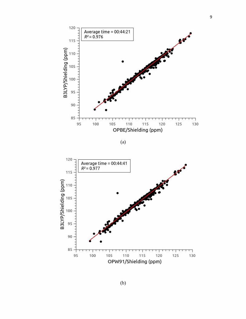
Figure S2.- (a) Correlation between 13
C4' shielding values computed from 581 maltose
conformations with the B3LYP functional and the basis set [00] versus the 13
C4' shielding
values computed with the OPBE functional and the BSD [04]; inserted, as a panel; the
averaged CPU-time, and the value of the square of the correlation coefficient, R, are shown;
(b); (c); (d); (e); and (f) the same as (a) for the functionals OPW91, BPW91, OB98, BPBE
and OLYP, respectively, with the BSD [04].
Figure S3.- (a) Contour plot for variation of the 13
C1 shielding of -D-Glcp-(1-4)--D-
Galp molecule based on 581 conformations, generated as described in the Materials and
Methods section. The equal-shieldings surfaces, every 1.0 ppm, are represented by using
different shades of blue. Grey color is used to highlight the non-explored regions of the
Ramachandran map. The column on the right-hand represents the range, in ppm, of the
shielding variation; and (b) same as (a) for the 13
C4' shielding.
Figure S4.- Same as Figure S3 for -D-Glcp-(1-4)--D-Altp.
Figure S5.- Same as Figure S3 for -D-Glcp-(1-4)--D-Idop
Figure S6.- Same as Figure S3 for -D-Glcp-(1-4)--D-Talp

3
(a)
(b)

4
(c)
(d)

5
(e)
(f)

6
(g)
(h)

7
(i)
(j)

8
(k)
Figure S1
9
(a)
(b)

10
(c)
(d)

11
(e)
(f)
Figure S2

12
(a)
(b)
Figure S3

13
(a)
(b)
Figure S4

14
(a)
(b)
Figure S5

15
(a)
(b)
Figure S6
Copyright © 2022 FDOKUMEN