Expression of thyroid transcription factor-1 in 16 human lung cancer cell lines
10
Expression of Thyroid Transcription Factor-1 in Normal and Neoplastic Lung Tissues Nobuki Nakamura, M.D., Eri Miyagi, M.D., Ph.D., Shin-ichi Murata, M.D., Ph.D., Akira Kawaoi, M.D., Ph.D., Ryohei Katoh, M.D., Ph.D. Department of Pathology, Yamanashi Medical University, Yamanashi, Japan The expression of thyroid transcription factor-1 in normal and neoplastic tissues and cell lines of the human lung was investigated using immunohisto- chemistry and in situ hybridization in conjunction with reverse transcription polymerase chain reaction. In normal lung tissues, immunoproducts of thyroid transcription factor-1 were observed in the nuclei of alveolar cells and bronchiolar cells. Interestingly, in distal bronchioles, immunohistochemistry and in situ hybridization revealed that thyroid transcription factor-1 was present not only in nonciliated cells (Clara cells) but also in ciliated cells and basal cells. In neoplastic tissues, thyroid transcription factor-1 was demonstrated in adenocarcinomas and small cell lung carcinomas with high frequency: 96% and 89% of cases, respectively. Thyroid transcription factor-1 was not detected in squamous cell carcinomas and large cell carcinomas. The strong immunoreactivity of thy- roid transcription factor-1 or simultaneous expres- sions of thyroid transcription factor-1 and surfactant protein A tended to correlate with the differentiation phenotypes in adenocarcinomas; they were more fre- quently present in the well-differentiated type than were moderately and/or poorly differentiated types. By reverse transcription polymerase chain reaction, expression of thyroid transcription factor-1 messen- ger RNA was observed in squamous cell carcinomas in addition to in adenocarcinomas and small cell lung carcinomas, and this finding was confirmed in the cell lines from squamous cell carcinomas. Only one case of 99 adenocarcinomas that originated in various or- gans other than lung and thyroid immunohisto- chemically expressed thyroid transcription factor-1. Our results suggest that thyroid transcription factor-1 can play an important role for the maintenance and/or differentiation process in bronchiolar and al- veolar cells. Thyroid transcription factor-1 expression associates with histologic types and/or differentiation of lung cancers and can be a valuable marker for the better understanding of their biological nature and pathological behavior. KEY WORDS: Immunohistochemistry, In situ hy- bridization Lung tumor, SP-A, TTF-1. Mod Pathol 2002;15(10):1058 –1067 Thyroid transcription factor-1 (TTF-1) is a homeodomain-containing nuclear transcription protein of Nkx2 gene family (1, 2). This kind of protein plays very important roles in the develop- ment, cell growth, and differentiation processes. TTF-1 was first identified in the follicular cells of the thyroid as a thyroid-specific DNA-binding protein, and it was subsequently demonstrated in the lung and in certain areas of the brain (3–5). In the thy- roid, TTF-1 activates the transcription of thyroglob- ulin, thyroperoxidase, and thyrotropin receptor genes in follicular cells and of several genes associ- ated with calcium metabolism in C cells (6 – 8). In the lung, the role of TTF-1 has been documented as a critical transcription factor that regulates the gene expression of lung-specific proteins such as Surfac- tant Proteins A (SP-A), B, and C and Clara cell secretory protein (9 –11). Recent immunohistochemical studies showed that TTF-1 is frequently expressed in lung carcino- mas (2, 12–24). However, the previously reported frequencies vary considerably in different series: 27–76% of adenocarcinoma cases (2, 12, 15), 83– 100% of small cell lung carcinoma cases (12–14), 0 –25% of large cell carcinoma cases (12, 20), and 0 –38% of squamous cell carcinoma cases (2, 14, 20). Therefore, it may be worthwhile to estimate the precise prevalence of TTF-1 expression in human lung carcinomas using different techniques to- gether with immunohistochemistry. Most previous immunohistochemical studies have focused on TTF-1 alone, and little is known about its correlation with surfactant proteins in hu- Copyright © 2002 by The United States and Canadian Academy of Pathology, Inc. VOL. 15, NO. 10, P. 1058, 2002 Printed in the U.S.A. Date of acceptance: June 21, 2002. Address reprint requests to: Ryohei Katoh, M.D., Ph.D., Department of Pathology, Yamanashi Medical University, Nakakoma-gunn Tamaho-cho Simokato 1110, Yamanashi, Japan; e-mail: [email protected] d.ac.jp; fax: 055-273-9534. DOI: 10.1097/01.MP.0000028572.44247.CF 1058
-
Upload
independent -
Category
Documents
-
view
0 -
download
0
Transcript of Expression of thyroid transcription factor-1 in 16 human lung cancer cell lines
Expression of Thyroid Transcription Factor-1 in Normaland Neoplastic Lung TissuesNobuki Nakamura, M.D., Eri Miyagi, M.D., Ph.D., Shin-ichi Murata, M.D., Ph.D.,Akira Kawaoi, M.D., Ph.D., Ryohei Katoh, M.D., Ph.D.
Department of Pathology, Yamanashi Medical University, Yamanashi, Japan
The expression of thyroid transcription factor-1 innormal and neoplastic tissues and cell lines of thehuman lung was investigated using immunohisto-chemistry and in situ hybridization in conjunctionwith reverse transcription polymerase chain reaction.In normal lung tissues, immunoproducts of thyroidtranscription factor-1 were observed in the nuclei ofalveolar cells and bronchiolar cells. Interestingly, indistal bronchioles, immunohistochemistry and in situhybridization revealed that thyroid transcriptionfactor-1 was present not only in nonciliated cells(Clara cells) but also in ciliated cells and basal cells. Inneoplastic tissues, thyroid transcription factor-1 wasdemonstrated in adenocarcinomas and small cell lungcarcinomas with high frequency: 96% and 89% ofcases, respectively. Thyroid transcription factor-1 wasnot detected in squamous cell carcinomas and largecell carcinomas. The strong immunoreactivity of thy-roid transcription factor-1 or simultaneous expres-sions of thyroid transcription factor-1 and surfactantprotein A tended to correlate with the differentiationphenotypes in adenocarcinomas; they were more fre-quently present in the well-differentiated type thanwere moderately and/or poorly differentiated types.By reverse transcription polymerase chain reaction,expression of thyroid transcription factor-1 messen-ger RNA was observed in squamous cell carcinomas inaddition to in adenocarcinomas and small cell lungcarcinomas, and this finding was confirmed in the celllines from squamous cell carcinomas. Only one caseof 99 adenocarcinomas that originated in various or-gans other than lung and thyroid immunohisto-chemically expressed thyroid transcription factor-1.Our results suggest that thyroid transcription factor-1can play an important role for the maintenance
and/or differentiation process in bronchiolar and al-veolar cells. Thyroid transcription factor-1 expressionassociates with histologic types and/or differentiationof lung cancers and can be a valuable marker for thebetter understanding of their biological nature andpathological behavior.
KEY WORDS: Immunohistochemistry, In situ hy-bridization Lung tumor, SP-A, TTF-1.
Mod Pathol 2002;15(10):1058–1067
Thyroid transcription factor-1 (TTF-1) is ahomeodomain-containing nuclear transcriptionprotein of Nkx2 gene family (1, 2). This kind ofprotein plays very important roles in the develop-ment, cell growth, and differentiation processes.TTF-1 was first identified in the follicular cells of thethyroid as a thyroid-specific DNA-binding protein,and it was subsequently demonstrated in the lungand in certain areas of the brain (3–5). In the thy-roid, TTF-1 activates the transcription of thyroglob-ulin, thyroperoxidase, and thyrotropin receptorgenes in follicular cells and of several genes associ-ated with calcium metabolism in C cells (6 – 8). Inthe lung, the role of TTF-1 has been documented asa critical transcription factor that regulates the geneexpression of lung-specific proteins such as Surfac-tant Proteins A (SP-A), B, and C and Clara cellsecretory protein (9 –11).
Recent immunohistochemical studies showedthat TTF-1 is frequently expressed in lung carcino-mas (2, 12–24). However, the previously reportedfrequencies vary considerably in different series:27–76% of adenocarcinoma cases (2, 12, 15), 83–100% of small cell lung carcinoma cases (12–14),0 –25% of large cell carcinoma cases (12, 20), and0 –38% of squamous cell carcinoma cases (2, 14, 20).Therefore, it may be worthwhile to estimate theprecise prevalence of TTF-1 expression in humanlung carcinomas using different techniques to-gether with immunohistochemistry.
Most previous immunohistochemical studieshave focused on TTF-1 alone, and little is knownabout its correlation with surfactant proteins in hu-
Copyright © 2002 by The United States and Canadian Academy ofPathology, Inc.VOL. 15, NO. 10, P. 1058, 2002 Printed in the U.S.A.Date of acceptance: June 21, 2002.Address reprint requests to: Ryohei Katoh, M.D., Ph.D., Department ofPathology, Yamanashi Medical University, Nakakoma-gunn Tamaho-choSimokato 1110, Yamanashi, Japan; e-mail: [email protected]; fax: 055-273-9534.
DOI: 10.1097/01.MP.0000028572.44247.CF
1058
man lung carcinomas (2, 12–27). And there havebeen no reports on the in situ expression of TTF-1mRNA. Therefore we investigated the expressionand localization of TTF-1 and SP-A in normal andneoplastic lung tissues by immunohistochemistryand nonisotopic in situ hybridization in conjunc-tion with reverse transcription (RT) polymerasechain reaction (PCR) to determine whether TTF-1expression profiles are of value for pathological di-agnosis and for prediction of the biological behav-ior of lung carcinomas.
MATERIALS AND METHODS
Tissue PreparationA series of surgical specimens from patients with
lung tumors comprised of adenocarcinoma (52 cas-es), squamous cell carcinoma (26 cases), small celllung carcinoma (18 cases), large cell carcinoma (8cases), and metastatic hepatocellular carcinoma (3cases) were selected from the surgical files of Ya-manashi Medical University Hospital to represent awide range of lung cancer. The histological typingsof lung carcinomas were classified according to thethird edition of Histological Typing of Lung Tumors(World Health Organization [WHO], 28). We deter-mined histological gradings (well, moderately, andpoorly differentiated) of the adenocarcinoma ac-cording to WHO classification (28), carried out byapplying conventional histological criteria to archi-tectural patterns and cytological features. Tumorswith extensive solid components were classified aspoorly differentiated carcinoma.
In addition, we examined 99 adenocarcinomasthat originated from other organs, including colon(25 cases), stomach (23 cases), pancreas (15 cases),prostate (15 cases), gallbladder (10 cases), andbreast (11 cases). All specimens were fixed in 10%neutral buffered formaldehyde, processed rou-tinely, and embedded in paraffin wax.
Cell LinesThe 14 cell lines consisted of 4 lung adenocarci-
noma cell lines (A549, 1– 87, 11–18, and LK87S), 4lung squamous cell carcinoma cell lines (Sq-1, Sq-19, EBC-1, and LK-2), 4 small cell lung carcinomacell lines (LK79, S2, LU65, and 87–5), and 2 lunglarge cell carcinoma cell lines (Lu99 and Lu99B). Allthese samples were provided from Cell ResourceCenter for Biomedical Research of Tohoku Univer-sity. These cell lines were incubated under the op-timal conditions for each cell line.
ImmunohistochemistryFor immunohistochemical analysis, the following
antibodies were used: mouse monoclonal antibody
against TTF-1 (NeoMarkers, Fremont, CA) andmouse monoclonal antibody against SP-A (DAKOJAPAN, Kyoto, Japan; 29) at a dilution of 1:50.
For TTF-1 immunostaining, heat pretreatment ofthe paraffin sections was employed for antigen re-trieval as described in the appending sheet. Sec-tions with 3- or 4-�M thickness were cut andmounted on poly-L-lysine– coated slides. Deparaf-finized sections were placed first in plastic Coplinjars filled with citrate buffer (pH 6.0) and incubatedfor 10 min at 120° C in an autoclave. After autoclavepretreatment, the sections were allowed to cool toroom temperature and then were exposed to 3%hydrogen peroxide in water to inactivate endoge-nous peroxidase.
Indirect immunoperoxidase staining was usedaccording to standard protocols (30). The sectionswere incubated for 2 hours at room temperaturewith the primary antibodies. Antimouse immuno-globulin G conjugate serum was applied to the sec-tions for 60 minutes at room temperature, and theperoxidase reaction was performed using 3,3'-diaminobenzidine tetrahydrochloride (3,3'-diaminobenzidine). The immunohistochemicalpreparations were counterstained with methylgreen or hematoxylin.
For serum controls, normal mouse serum orphosphate buffered saline (PBS) was used insteadof the primary antibody. Thyroid tissues were usedas a positive control.
Positive cases were represented as � or �� (�,positive cells �20% of tumor cells; ��, positivecells �20% of tumor cells). Two of the authors (NNand RK) estimated the immunoreactive cells sepa-rately and determined the positive cases.
In Situ HybridizationA plasmid encoding a mouse subclone of the
TTF-1 genome was supplied by Roberto di Lauro,Ph.D. (Stazione Zoologica A. Dohrn, Villa Comu-nale, Naples, Italy). For riboprobe preparation, a757-bp SacII fragment (149 –905 bp) of TTF-1 cDNAwas subcloned into Bluescript SK� (Stratagene; LaJolla, CA). The plasmid was linearized with restric-tion enzyme NotI and was washed by phenol– chlo-roform extraction. The purified, linearized plasmidwas used to generate labeled RNA antisense probesby performing in vitro transcription using T3 RNApolymerase and digoxigenin-11-uridine-5'-triphosphate (Boehringer Mannheim GmbH Bio-chemica, Mannheim, Germany). In the same way,the sense probe was made using the restrictionenzyme BglII and T7 RNA polymerase.
The in situ hybridization protocol was a modifi-cation of a method described by Katoh et al. (7). Inbrief, sections were deparaffinized with xylene, re-hydrated with a graded series of ethanol solutions,
TTF-1 in Lung Tissue (N. Nakamura et al.) 1059
rinsed in PBS at pH 7.4, postfixed with 4% parafor-maldehyde for 5 minutes, rinsed in PBS, and di-gested with 20 �g/mL proteinase K (Sigma, St.Louis, MO) at 37° C for 20 minutes. Slides were fixedagain with 4% paraformaldehyde for 5 minutes,rinsed with PBS, dehydrated in graded solutions ofethanol, and air dried.
Probes were diluted with hybridization solution(10 mmol/L Tris-HCl, pH 7.6, containing 50% form-amide, 200 �g/mL transfer RNA, 1� Denhardt’ssolution, 10% dextran sulfate, 600 mmol/L NaCl,0.25% sodium dodecyl sulfate, and 1 mmol/L eth-ylenediaminetetraacetic acid [EDTA]) to a final con-centration of 1 ng/mL, and sections were hybrid-ized with a heat-denatured riboprobe for 18 hoursat 55° C. The hybridization samples were washedsequentially in 5� SSC (sodium chloride and so-dium citrate) for 5 minutes at 55° C, then in 2� SSCcontaining 50% formamide for 30 minutes at 55° C;1� SSC is composed of 0.15 mol/� NaCl and 0.015mol/L sodium citrate. To degrade unbound ribo-probe, sections were treated with RNase A (1 �g/mLin 10 mmol/L EDTA) at 37° C for 30 minutes, thensequentially washed once in 2� SSC for 20 minutesat 55° C and twice in 0.2� SSC for 20 minutes at 55°C.
The hybridized probe was detected using a Nu-cleic Acid Detection Kit (Boehringer MannheimGmbH Biochemica) according to the manufactur-er’s instructions. Briefly, slides were incubated with1.5% blocking reagent for 30 minutes at room tem-perature, then with a 1:500 dilution of alkalinephosphatase–labeled sheep antidigoxigenin Fabfragment for 1 hour at room temperature. Afterwashing, slides were incubated with substrate solu-tion containing nitroblue tetrazolium salt andX-phosphate (5-bromo-4-chloro-3-indoyl phos-phate) for 2 to 12 hours, counterstained withmethyl green, and mounted using a glycerin– gela-tin solution.
Reverse Transcription–Polymerase ChainReaction
Total RNA were prepared by the acidguanidinium-phenol-chloroform system (ISOGEN,Nippon Gene Co Ltd, Toyama, Japan) as describedin the manufacturer’s instructions. Isolated totalRNA (20 �g) was fractionated by electrophoresis of1.5% agarose in 18% formaldehyde gels. The qualityof the extracted RNAs showing two clear bands of28S and 18S, corresponding to ribosomal RNAs,were selected for further studies. Five microgramsof total RNA was reverse-transcribed using murineleukemia virus reverse transcriptase. PCRs werecarried out according to the Gene Amp DNA am-plification reagent kit instructions (Perkin-Elmer,Norwalk, CT). Thirty cycles were performed. During
each cycle, the samples were heated to 94° C for 30seconds, cooled to 56° C for 60 seconds, and heatedto 72° C for 60 seconds. The RT-PCR reagent blankyielded no detectable products. A 16-�L sample ofeach 50-�L PCR solution was fractionated by elec-trophoresis in a 2% agarose gel. Gels were photo-graphed using Polaroid 655 film. The oligonucleo-tide primer sequences for TTF-1, SP-A, and �-actinare described in Table 1. RNAs extracted from thestomach, colon, and liver were used as the negativetissue control.
RESULTS
Immunohistochemistry and In Situ Hybridization
Normal TissuesIn the current immunohistochemical study, the
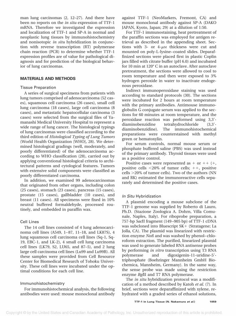
immunoreactive products of TTF-1 protein wereclearly identified in the nuclei of alveolar cells, pos-sibly indicating Type II cells (Fig. 1, A–B). This find-ing was supported by in situ hybridization; alveolarcells with round nuclei were strongly hybridizedwith a specific riboprobe for TTF-1 mRNA (Fig. 1C).TTF-1 protein was also observed in the nuclei ofciliated and nonciliated cells in terminal and respi-ratory bronchioles (Fig. 1F). There was a tendencyfor stronger TTF-1 immunostaining in nonciliatedClara cells than in ciliated bronchiolar cells. Fur-thermore, in parts of the distal bronchiole, TTF-1was observed in the nuclei of basal cells but not ingoblet cells (Fig. 1J). The mRNA of TTF-1, however,was evident with in situ hybridization in some gob-let cells as well as in basal cells in distal bronchiole(Fig. 1K). The immunoreactive products of SP-Awere observed in the cytoplasm of nonciliated Claracells and alveolar Type II cells (Fig. 1, D & H).
Neoplastic TissuesPositive Frequency of TTF-1 and SP-A in Lung
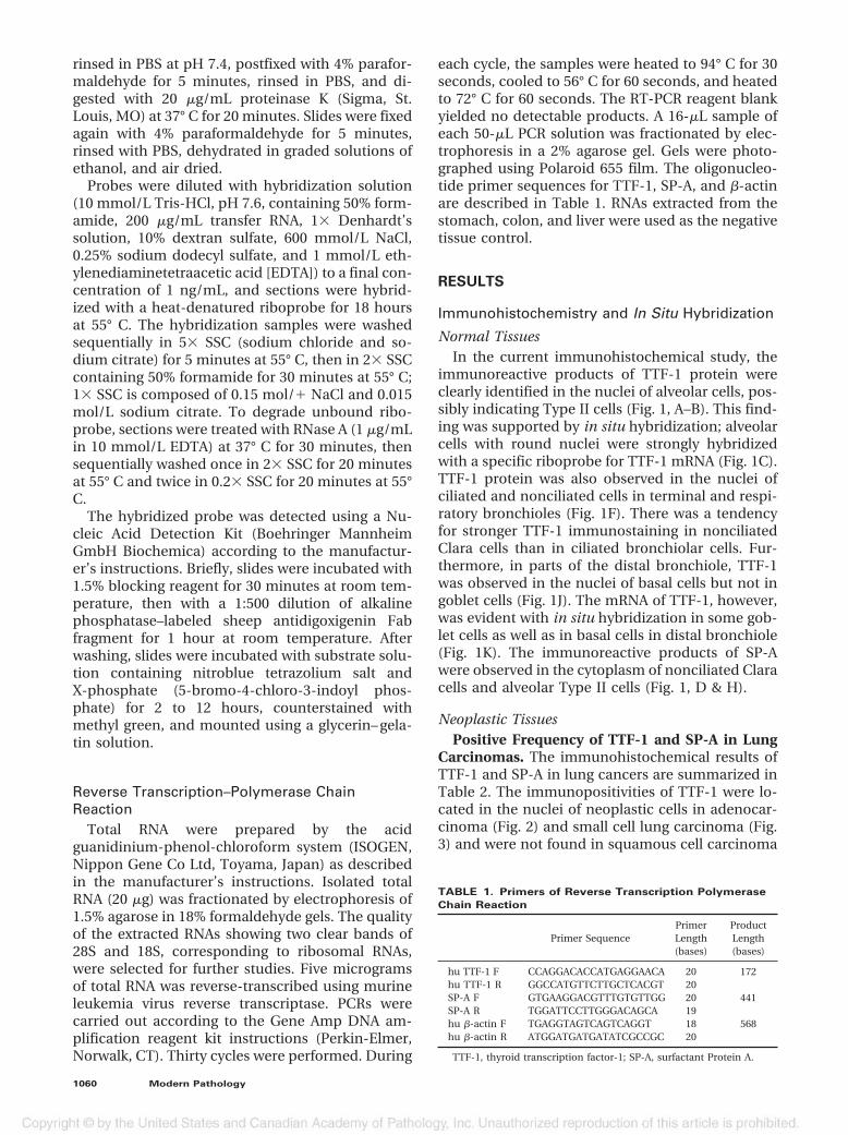
Carcinomas. The immunohistochemical results ofTTF-1 and SP-A in lung cancers are summarized inTable 2. The immunopositivities of TTF-1 were lo-cated in the nuclei of neoplastic cells in adenocar-cinoma (Fig. 2) and small cell lung carcinoma (Fig.3) and were not found in squamous cell carcinoma
TABLE 1. Primers of Reverse Transcription Polymerase
Chain Reaction
Primer SequencePrimerLength(bases)
ProductLength(bases)
hu TTF-1 F CCAGGACACCATGAGGAACA 20 172hu TTF-1 R GGCCATGTTCTTGCTCACGT 20SP-A F GTGAAGGACGTTTGTGTTGG 20 441SP-A R TGGATTCCTTGGGACAGCA 19hu �-actin F TGAGGTAGTCAGTCAGGT 18 568hu �-actin R ATGGATGATGATATCGCCGC 20
TTF-1, thyroid transcription factor-1; SP-A, surfactant Protein A.
1060 Modern Pathology
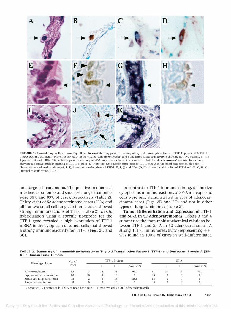
and large cell carcinoma. The positive frequenciesin adenocarcinomas and small cell lung carcinomaswere 96% and 89% of cases, respectively (Table 2).Thirty-eight of 52 adenocarcinoma cases (73%) andall but two small cell lung carcinoma cases showedstrong immunoreactions of TTF-1 (Table 2). In situhybridization using a specific riboprobe for theTTF-1 gene revealed a high expression of TTF-1mRNA in the cytoplasm of tumor cells that showeda strong immunoreactivity for TTF-1 (Figs. 2C and3C).
In contrast to TTF-1 immunostaining, distinctivecytoplasmic immunoreactions of SP-A in neoplasticcells were only demonstrated in 73% of adenocar-cinoma cases (Figs. 2D and 3D) and not in othertypes of lung carcinomas (Table 2).
Tumor Differentiation and Expression of TTF-1and SP-A in 52 Adenocarcinomas. Tables 3 and 4summarize the immunohistochemical relations be-tween TTF-1 and SP-A in 52 adenocarcinomas. Astrong TTF-1 immunoreactivity (representing ��)was found in 100% of cases in well-differentiated
FIGURE 1. Normal lung. A–D, alveolar Type II cell (arrow) showing positive staining of thyroid transcription factor-1 (TTF-1) protein (B), TTF-1mRNA (C), and Surfactant Protein A (SP-A; D). E–H, ciliated cells (arrowheads) and nonciliated Clara cells (arrow) showing positive staining of TTF-1 protein (F) and mRNA (G). Note the positive staining of SP-A only in nonciliated Clara cells (H). I–K, basal cells (arrows) in distal bronchioleshowing a positive nuclear staining of TTF-1 protein (K). Note the cytoplasmic expression of TTF-1 mRNA in the basal and bronchiole cells (J).Hematoxylin and eosin staining (A, E, I), immunohistochemistry of TTF-1 (B, F, J) and SP-A (D, H), in situ hybridization of TTF-1 mRNA (C, G, K).Original magnification, 600�.
TABLE 2. Summary of Immunohistochemistry of Thyroid Transcription Factor-1 (TTF-1) and Surfactant Protein A (SP-
A) in Human Lung Tumors
Histologic TypesNo. ofCases
TTF-1 Protein SP-A
� � �� Positive % � � �� Positive %
Adenocarcinoma 52 2 12 38 96.2 14 21 17 73.1Squamous cell carcinoma 26 26 0 0 0 26 0 0 0Small cell lung carcinoma 18 2 0 16 88.9 18 0 0 0Large cell carcinoma 8 0 0 0 0 8 0 0 0
�, negative; �, positive cells �20% of neoplastic cells; ��, positive cells �20% of neoplastic cells.
TTF-1 in Lung Tissue (N. Nakamura et al.) 1061
cancers, 76% of cases in moderately differentiatedcancers, and 50% of cases in poorly differentiatedcancers (Table 3). In contrast to this, strong SP-Aimmunoreactions (representing ��) were less fre-quently observed in all types in adenocarcinoma:37% of cases in the well-differentiated type, 31% ofcases in the moderately differentiated type, and25% of cases in the poorly differentiated type. Si-
multaneous positivity of TTF-1 and SP-A (repre-senting �/�) was most frequently observed in thewell-differentiated type of adenocarcinomas (89%)and less frequently in the moderately (62%) andpoorly (50%) differentiated adenocarcinomas (Ta-ble 4). The tumors showing TTF-1 positivity andSP-A negativity (representing �/�) were most fre-quently observed in poorly differentiated adenocar-
FIGURE 2. Human lung adenocarcinoma. A, hematoxylin and eosin stain. B, immunohistochemistry of thyroid transcription factor-1 (TTF-1)showing a positive staining of TTF-1 protein in the nuclei of adenocarcinoma cells. C, in situ hybridization showing the TTF-1 m RNA signals in thecytoplasms of cancer cells. D, immunohistochemistry of Surfactant Protein A showing it positively stained in the cytoplasms of the cancer cells.Original magnification, 100�.
FIGURE 3. Human small cell lung carcinoma. A, hematoxylin and eosin stain. B, immunohistochemistry of thyroid transcription factor-1 (TTF-1)showing the positive staining of TTF-1 protein in the nuclei of the small cell lung carcinoma cells. C, in situ hybridization showing the TTF-1 m RNAsignals in the cytoplasms of the cancer cells. D, immunohistochemistry of Surfactant Protein A showing no staining of it in the cytoplasms of thecancer cells. Original magnification, 400�.
1062 Modern Pathology
cinomas (50%). There were no tumors showingTTF-1 negativity and SP-A positivity in the presentseries.
Adenocarcinoma with Other Origins. We exam-ined the TTF-1 expressions in 99 adenocarcinomaswith other origins including the colon, stomach,pancreas, prostate, gall bladder, and breast (Table5). Among these cancers, TTF-1 was demonstratedonly in one case of stomach cancer, which showeda poorly differentiated adenocarcinoma. In this tu-mor, TTF-1 was identified in the nuclei of spindlecells and not in the cells forming tubules (data notshown). In addition, three cases of metastatic lungcancer that originated from the liver were also neg-ative for TTF-1 protein and mRNA and for SP-Aprotein.
Reverse Transcription Polymerase ChainReaction
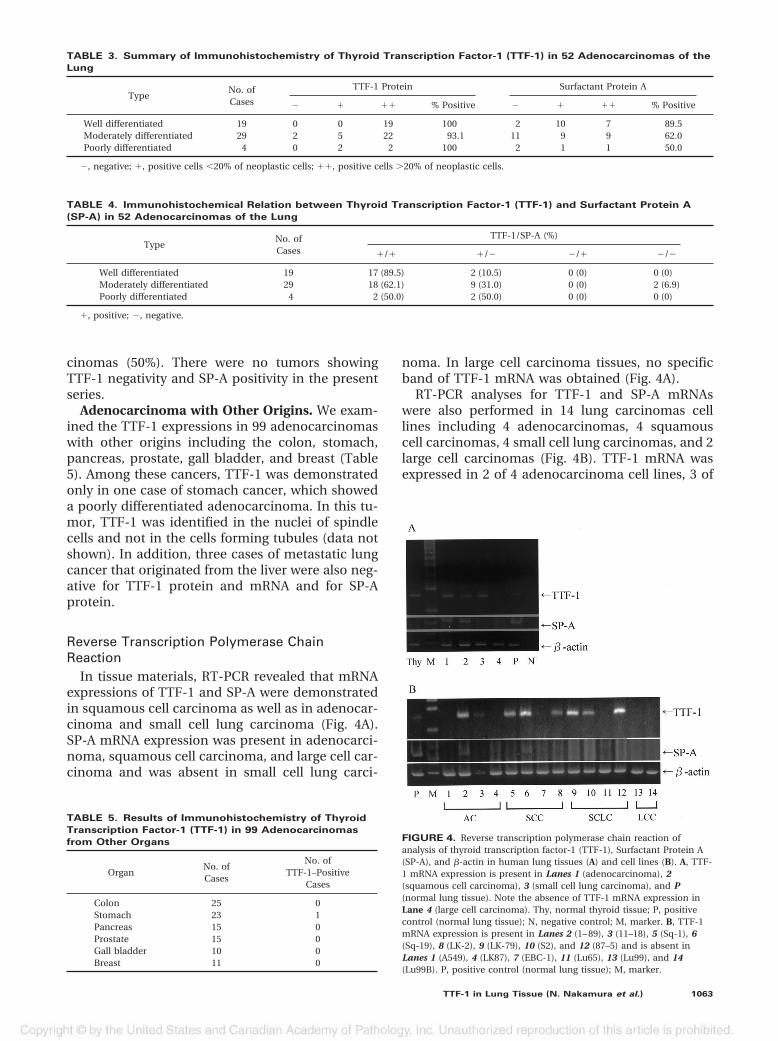
In tissue materials, RT-PCR revealed that mRNAexpressions of TTF-1 and SP-A were demonstratedin squamous cell carcinoma as well as in adenocar-cinoma and small cell lung carcinoma (Fig. 4A).SP-A mRNA expression was present in adenocarci-noma, squamous cell carcinoma, and large cell car-cinoma and was absent in small cell lung carci-
noma. In large cell carcinoma tissues, no specificband of TTF-1 mRNA was obtained (Fig. 4A).
RT-PCR analyses for TTF-1 and SP-A mRNAswere also performed in 14 lung carcinomas celllines including 4 adenocarcinomas, 4 squamouscell carcinomas, 4 small cell lung carcinomas, and 2large cell carcinomas (Fig. 4B). TTF-1 mRNA wasexpressed in 2 of 4 adenocarcinoma cell lines, 3 of
TABLE 3. Summary of Immunohistochemistry of Thyroid Transcription Factor-1 (TTF-1) in 52 Adenocarcinomas of the
Lung
TypeNo. ofCases
TTF-1 Protein Surfactant Protein A
� � �� % Positive � � �� % Positive
Well differentiated 19 0 0 19 100 2 10 7 89.5Moderately differentiated 29 2 5 22 93.1 11 9 9 62.0Poorly differentiated 4 0 2 2 100 2 1 1 50.0
�, negative; �, positive cells �20% of neoplastic cells; ��, positive cells �20% of neoplastic cells.
TABLE 4. Immunohistochemical Relation between Thyroid Transcription Factor-1 (TTF-1) and Surfactant Protein A
(SP-A) in 52 Adenocarcinomas of the Lung
TypeNo. ofCases
TTF-1/SP-A (%)
�/� �/� �/� �/�
Well differentiated 19 17 (89.5) 2 (10.5) 0 (0) 0 (0)Moderately differentiated 29 18 (62.1) 9 (31.0) 0 (0) 2 (6.9)Poorly differentiated 4 2 (50.0) 2 (50.0) 0 (0) 0 (0)
�, positive; �, negative.
TABLE 5. Results of Immunohistochemistry of Thyroid
Transcription Factor-1 (TTF-1) in 99 Adenocarcinomas
from Other Organs
OrganNo. ofCases
No. ofTTF-1–Positive
Cases
Colon 25 0Stomach 23 1Pancreas 15 0Prostate 15 0Gall bladder 10 0Breast 11 0
FIGURE 4. Reverse transcription polymerase chain reaction ofanalysis of thyroid transcription factor-1 (TTF-1), Surfactant Protein A(SP-A), and �-actin in human lung tissues (A) and cell lines (B). A, TTF-1 mRNA expression is present in Lanes 1 (adenocarcinoma), 2(squamous cell carcinoma), 3 (small cell lung carcinoma), and P(normal lung tissue). Note the absence of TTF-1 mRNA expression inLane 4 (large cell carcinoma). Thy, normal thyroid tissue; P, positivecontrol (normal lung tissue); N, negative control; M, marker. B, TTF-1mRNA expression is present in Lanes 2 (1– 89), 3 (11–18), 5 (Sq-1), 6(Sq-19), 8 (LK-2), 9 (LK-79), 10 (S2), and 12 (87–5) and is absent inLanes 1 (A549), 4 (LK87), 7 (EBC-1), 11 (Lu65), 13 (Lu99), and 14(Lu99B). P, positive control (normal lung tissue); M, marker.
TTF-1 in Lung Tissue (N. Nakamura et al.) 1063
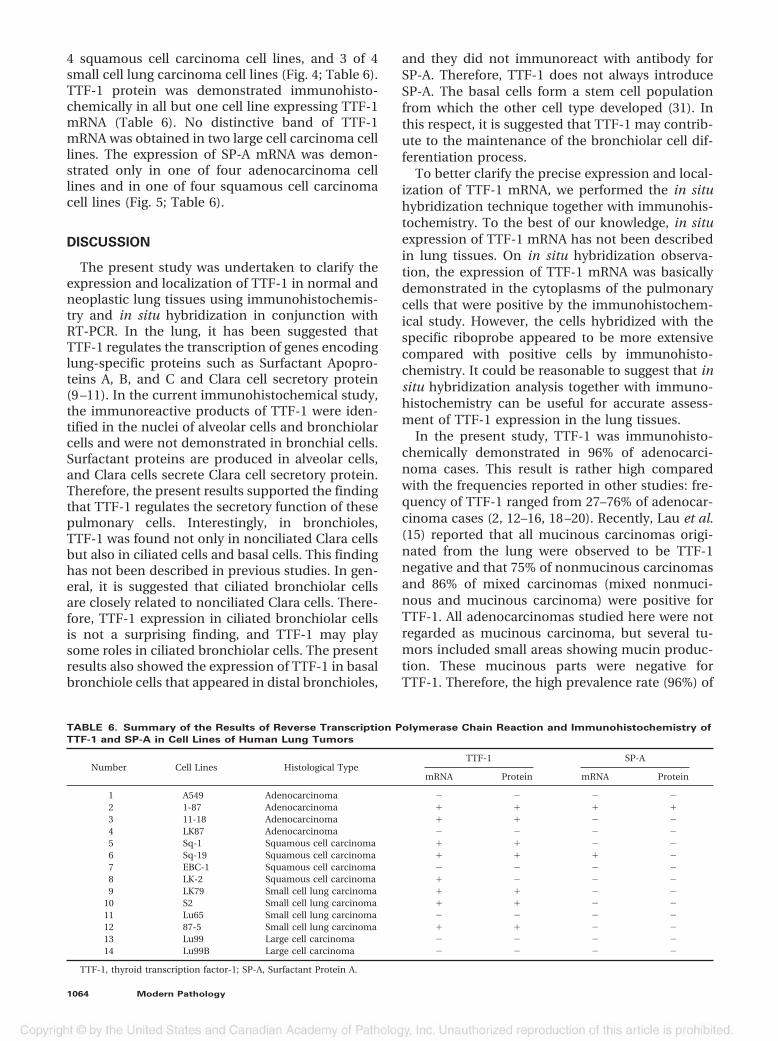
4 squamous cell carcinoma cell lines, and 3 of 4small cell lung carcinoma cell lines (Fig. 4; Table 6).TTF-1 protein was demonstrated immunohisto-chemically in all but one cell line expressing TTF-1mRNA (Table 6). No distinctive band of TTF-1mRNA was obtained in two large cell carcinoma celllines. The expression of SP-A mRNA was demon-strated only in one of four adenocarcinoma celllines and in one of four squamous cell carcinomacell lines (Fig. 5; Table 6).
DISCUSSION
The present study was undertaken to clarify theexpression and localization of TTF-1 in normal andneoplastic lung tissues using immunohistochemis-try and in situ hybridization in conjunction withRT-PCR. In the lung, it has been suggested thatTTF-1 regulates the transcription of genes encodinglung-specific proteins such as Surfactant Apopro-teins A, B, and C and Clara cell secretory protein(9 –11). In the current immunohistochemical study,the immunoreactive products of TTF-1 were iden-tified in the nuclei of alveolar cells and bronchiolarcells and were not demonstrated in bronchial cells.Surfactant proteins are produced in alveolar cells,and Clara cells secrete Clara cell secretory protein.Therefore, the present results supported the findingthat TTF-1 regulates the secretory function of thesepulmonary cells. Interestingly, in bronchioles,TTF-1 was found not only in nonciliated Clara cellsbut also in ciliated cells and basal cells. This findinghas not been described in previous studies. In gen-eral, it is suggested that ciliated bronchiolar cellsare closely related to nonciliated Clara cells. There-fore, TTF-1 expression in ciliated bronchiolar cellsis not a surprising finding, and TTF-1 may playsome roles in ciliated bronchiolar cells. The presentresults also showed the expression of TTF-1 in basalbronchiole cells that appeared in distal bronchioles,
and they did not immunoreact with antibody forSP-A. Therefore, TTF-1 does not always introduceSP-A. The basal cells form a stem cell populationfrom which the other cell type developed (31). Inthis respect, it is suggested that TTF-1 may contrib-ute to the maintenance of the bronchiolar cell dif-ferentiation process.
To better clarify the precise expression and local-ization of TTF-1 mRNA, we performed the in situhybridization technique together with immunohis-tochemistry. To the best of our knowledge, in situexpression of TTF-1 mRNA has not been describedin lung tissues. On in situ hybridization observa-tion, the expression of TTF-1 mRNA was basicallydemonstrated in the cytoplasms of the pulmonarycells that were positive by the immunohistochem-ical study. However, the cells hybridized with thespecific riboprobe appeared to be more extensivecompared with positive cells by immunohisto-chemistry. It could be reasonable to suggest that insitu hybridization analysis together with immuno-histochemistry can be useful for accurate assess-ment of TTF-1 expression in the lung tissues.
In the present study, TTF-1 was immunohisto-chemically demonstrated in 96% of adenocarci-noma cases. This result is rather high comparedwith the frequencies reported in other studies: fre-quency of TTF-1 ranged from 27–76% of adenocar-cinoma cases (2, 12–16, 18 –20). Recently, Lau et al.(15) reported that all mucinous carcinomas origi-nated from the lung were observed to be TTF-1negative and that 75% of nonmucinous carcinomasand 86% of mixed carcinomas (mixed nonmuci-nous and mucinous carcinoma) were positive forTTF-1. All adenocarcinomas studied here were notregarded as mucinous carcinoma, but several tu-mors included small areas showing mucin produc-tion. These mucinous parts were negative forTTF-1. Therefore, the high prevalence rate (96%) of
TABLE 6. Summary of the Results of Reverse Transcription Polymerase Chain Reaction and Immunohistochemistry of
TTF-1 and SP-A in Cell Lines of Human Lung Tumors
Number Cell Lines Histological TypeTTF-1 SP-A
mRNA Protein mRNA Protein
1 A549 Adenocarcinoma � � � �2 1-87 Adenocarcinoma � � � �3 11-18 Adenocarcinoma � � � �4 LK87 Adenocarcinoma � � � �5 Sq-1 Squamous cell carcinoma � � � �6 Sq-19 Squamous cell carcinoma � � � �7 EBC-1 Squamous cell carcinoma � � � �8 LK-2 Squamous cell carcinoma � � � �9 LK79 Small cell lung carcinoma � � � �
10 S2 Small cell lung carcinoma � � � �11 Lu65 Small cell lung carcinoma � � � �12 87-5 Small cell lung carcinoma � � � �13 Lu99 Large cell carcinoma � � � �14 Lu99B Large cell carcinoma � � � �
TTF-1, thyroid transcription factor-1; SP-A, Surfactant Protein A.
1064 Modern Pathology
TTF-1 in adenocarcinomas in the present studymay be due to lack of mucinous carcinoma cases.
There have been no studies reported on TTF-1expression in adenocarcinomas originating in or-gans other than the lung and thyroid. In this re-spect, we additionally examined 99 adenocarcino-mas that occurred in several other organs includingthe colon, stomach, pancreas, prostate, gall blad-der, and breast and demonstrated TTF-1 positivityonly in one case of stomach adenocarcinoma with apoorly differentiated phenotype. From these re-sults, the potential diagnostic value of TTF-1 inpulmonary carcinomas appears to be promising.
TTF-1 was implicated in regulation of differenti-ation genes in lung, such as Surfactant Proteins A,B, and C, and 10-kDa Clara cell gene promoters (1,5, 9 –11). Therefore, TTF-1 expression could be as-sociated with the functional differentiation in lungcarcinomas. The findings of the current immuno-histochemical study revealed that TTF-1 was iden-tified in all but two cases of adenocarcinoma, and astrong immunoreactivity of TTF-1 was more fre-quently observed in the well-differentiated pheno-type than in the less differentiated phenotypes.Therefore, TTF-1 expression could be related to thefunctional differentiation of adenocarcinoma. Lin-noila et al. (32) suggested that expression of Surfac-tant Apoprotein A and the 10-kDa Clara cell genepromoters were independent prognostic indicatorsfor survival and delayed development of metastasesin patients with early-stage non–small cell lungcancer. Taken together, it is suggested that a strongexpression of TTF-1 can predict a better prognosisof adenocarcinoma. However, Puglisi et al. (19) re-ported that TTF-1 protein is an adverse prognosticfactor and that a strong expression of TTF-1 is as-sociated with a higher risk of death.
Small cell lung carcinomas frequently expressTTF-1; the frequency of TTF-1 expression reported
in several different series has ranged from 83–100%(12, 13–16, 21, 22). Interestingly, more recent stud-ies showed that TTF-1 is also expressed in extrapul-monary small cell carcinomas (24, 33). In thepresent study, the high prevalence of TTF-1 wasdemonstrated in 90% of small cell lung carcinomacases. In addition, our in situ hybridization andRT-PCR analysis confirmed a strong expression ofTTF-1 mRNA in small cell lung carcinoma. There-fore, TTF-1 is suggested as a sensitive marker forsmall cell lung carcinoma. Although the precisesignificance of TTF-1 expression in small cell carci-noma is still unclear, the common expression ofTTF-1 in adenocarcinoma and small cell carci-noma, as shown in the current study, may supportthe view that small cell lung carcinoma developfrom an endodermal precursor and, therefore,small cell lung carcinoma and non–small cell lungcarcinoma appear to develop from a common epi-thelial stem cell (34).
In previous immunohistochemical studies, TTF-1was demonstrated in some cases of pulmonarysquamous cell carcinoma (2, 12–16, 18 –20). In ad-dition, it is known that some squamous cell carci-nomas show focal mucin on histochemical stains(28). We failed to demonstrate the immunohisto-chemical expression of TTF-1 in all 26 pulmonarysquamous cell carcinoma tissues. However, in con-trast, the present RT-PCR analysis revealed mRNAexpressions of TTF-1 and SP-A in similar materials.This finding may be the result of entrapment ofnormal lung cells expressing TTF-1 mRNA such asType II alveolar cells and bronchiolar cells. Indeed,squamous cell carcinoma tissues sometimes con-tain a small amount of normal lung cells in theirperipheral areas. In this respect, it suggested that insitu hybridization studies can be useful to rule outthis possibility. The present in situ hybridizationstudy with a specific riboprobe for TTF-1 revealed
FIGURE 5. Immunohistochemistry of thyroid transcription factor-1 (TTF-1) and Surfactant Protein A (SP-A) in two cell lines, 1– 87 (A–C) and Sq-19(D–F). Lines 1– 87 from human adenocarcinoma showing positive staining of TTF-1 protein (B) and SP-A protein (C). Sq-19 from human squamouscell carcinoma showing positive staining of TTF-1 protein (E) and negative of SP-A protein (F). Original magnification, 200�.
TTF-1 in Lung Tissue (N. Nakamura et al.) 1065
that the convincing expression of TTF-1 mRNA wasnot demonstrated in squamous cell carcinomacells. This result suggests that TTF-1 mRNA expres-sion detected by RT-PCR may be due to entrappednormal lung cells and, therefore, the expression ofTTF-1 mRNA may be absent or very small in squa-mous cell carcinoma cells. Surprisingly, however,RT-PCR analysis for RNAs extracted from the fourcell lines of pulmonary squamous cell carcinomarevealed that the expression of TTF-1 mRNA wasdemonstrated in three cell lines, and the expressionof SP-A mRNA, in one cell line. Therefore, it isreasonable to suggest that some squamous cell car-cinomas could have originated from the cells ex-pressing TTF-1 and/or SP-A.
In conclusion, TTF-1 is not only expressed fromnonciliated Clara cells and alveolar cells but alsofrom ciliated and basal cells in the distal bronchiolein connection with their functional ability and/ordifferentiation process. In neoplastic lung tissues,TTF-1 is one of most reliable markers for pulmo-nary adenocarcinoma and small cell lung carci-noma. Therefore, additional investigations of TTF-1may provide useful information on the assessmentof the functional nature and clinical behavior ofthese lung malignancies.
Acknowledgments: Gratitude is expressed to Syun-suke Kobayashi of the Cell Resource Center for Bio-medical Research’s, Tohoku University, Japan forgenerously donating the cell lines. We are also grate-ful to Mrs. Yoda, Mrs. Ito, and Mrs. Gomi for help inimmunohistochemical preparation.
REFERENCES
1. Ikeda K, Clark JC, Shaw-White JR, Stahlman MT, Boutell CJ,Whitsett JA. Gene structure and expression of human thyroidtranscription factor-1 in respiratory epithelial cells. J BiolChem 1995;270:8108 –14.
2. Bejarano PA, Baughman RP, Biddinger PW, Miller MA,Fenoglio-Preiser C, al-Kafaji B, et al. Surfactant proteins andthyroid transcription factor-1 in pulmonary and breast car-cinomas. Mod Pathol 1996;9:445–52.
3. Civitareale D, Lonigro R, Sinclair AJ, Di Lauro R. A thyroid-specific nuclear protein essential for tissue-specific expres-sion of the thyroglobulin promoter. EMBO J 1989;8:2537– 42.
4. Stahlman MT, Gray ME, Whitsett JA. Expression of thyroidtranscription factor-1(TTF-1) in fetal and neonatal humanlung. J Histochem Cytochem 1996;44:673– 8.
5. Lazzaro D, Price M, de Felice M, Di Lauro R. The transcrip-tion factor TTF-1 is expressed at the onset of thyroid andlung morphogenesis and in restricted regions of the foetalbrain. Development 1991;113:1093–104.
6. Katoh R, Kawaoi A, Miyagi E, Li X, Suzuki K, Nakamura Y, etal. Thyroid transcription factor-1 in normal, hyperplastic,and neoplastic follicular thyroid cells examined by immuno-histochemistry and nonradioactive in situ hybridization.Mod Pathol 2000;13:570 – 6.
7. Katoh R, Miyagi E, Nakamura N, Li X, Suzuki K, Kakudo K, etal. Expression of thyroid transcription factor-1 (TTF-1) in
human C cells and medullary thyroid carcinomas. HumPathol 2000;31:386 –93.
8. Oguchi H, Kimura S. Transcription factors and thyroid dis-ease. Mol Med 1995;32:518.
9. Zhang L, Whitsett JA, Stripp BR. Regulation of Clara cellsecretory protein gene transcription by thyroid transcriptionfactor-1. Biochim Biophys Acta 1997;1350:359 – 67.
10. Bohinski RJ, Di Lauro R, Whitsett JA. The lung-specific sur-factant protein B gene promoter is a target for thyroid tran-scription factor 1 and hepatocyte nuclear factor 3, indicatingcommon factors for organ-specific gene expression alongthe foregut axis. Mol Cell Biol 1994;14:5671– 81.
11. Bruno MD, Bohinski RJ, Huelsman KM, Whitsett JA, Korfha-gen TR. Lung cell-specific expression of the murine surfac-tant protein A (SP-A) gene is mediated by interactions be-tween the SP-A promoter and thyroid transcription factor-1.J Biol Chem 1995;270:6531– 6.
12. Fabbro D, Di Loreto C, Stamerra O, Beltrami CA, Lonigro R,Damante G. TTF-1 gene expression in human lung tumours.Eur J Cancer 1996;32A:512–7.
13. Holzinger A, Dingle S, Bejarano PA, Miller MA, Weaver TE,DiLauro R, et al. Monoclonal antibody to thyroid transcrip-tion factor-1: production, characterization, and usefulnessin tumor diagnosis. Hybridoma 1996;15:49 –53.
14. Harlamert HA, Mira J, Bejarano PA, Baughman RP, MillerMA, Whitsett JA, et al. Thyroid transcription factor-1 andcytokeratins 7 and 20 in pulmonary and breast carcinoma.Acta Cytol 1998;42:1382– 8.
15. Lau SK, Desrochers MJ, Luthringer DJ. Expression of thyroidtranscription factor-1, cytokeratin 7, and cytokeratin 20 inbronchioloalveolar carcinomas: an immunohistochemicalevaluation of 67 cases. Mod Pathol 2002;15:538 – 42.
16. Di Loreto C, Di Lauro V, Puglisi F, Damante G, Fabbro D,Beltrami CA. Immunocytochemical expression of tissue spe-cific transcription factor-1 in lung carcinoma. J Clin Pathol1997;50:30 –2.
17. Di Loreto C, Puglisi F, Di Lauro V, Damante G, Beltrami CA.TTF-1 protein expression in pleural malignant mesothelio-mas and adenocarcinomas of the lung. Cancer Lett 1998;124:73– 8.
18. Bohinski RJ, Bejarano PA, Balko G, Warnick RE, Whitsett JA.Determination of lung as the primary site of cerebral meta-static adenocarcinomas using monoclonal antibody to thy-roid transcription factor-1. J Neurooncol 1998;40:227–31.
19. Puglisi F, Barbone F, Damante G, Bruckbauer M, Di Lauro V,Beltrami CA, et al. Prognostic value of thyroid transcriptionfactor-1 in primary, resected, non-small cell lung carcinoma.Mod Pathol 1999;12:318 –24.
20. Kaufmann O, Dietel M. Thyroid transcription factor-1 is thesuperior immunohistochemical marker for pulmonary ade-nocarcinomas and large cell carcinomas compared to sur-factant proteins A and B. Histopathology 2000;36:8 –16.
21. Ordonez NG. Value of thyroid transcription factor-1 immu-nostaining in distinguishing small cell lung carcinomas fromother small cell carcinomas. Am J Surg Pathol 2000;24:1217–23.
22. Byrd-Gloster AL, Khoor A, Glass LF, Messina JL, Whitsett JA,Livingston SK, et al. Differential expression of thyroid tran-scription factor 1 in small cell lung carcinoma and Merkelcell tumor. Hum Pathol 2000;31:58 – 62.
23. Miettinen M, Franssila KO. Variable expression of keratinsand nearly uniform lack of thyroid transcription factor 1 inthyroid anaplastic carcinoma. Hum Pathol 2000;31:1139 – 45.
24. Agoff SN, Lamps LW, Philip AT, Amin MB, Schmidt RA, TrueLD, et al. Thyroid transcription factor-1 is expressed in ex-trapulmonary small cell carcinomas but not in other ex-trapulmonary neuroendocrine tumors. Mod Pathol 2000;13:238 – 42.
1066 Modern Pathology
25. Shimosato Y. Pulmonary neoplasms. In: Stephen SS, editor.Diagnostic surgical pathology. 3th ed. Vol 1. Philadelphia,PA: Lippincott Williams & Wilkins; 1999. p. 1069 –115.
26. Mizutani Y, Nakajima T, Morinaga S, Gotoh M, Shimosato Y,Akino T, et al. Immunohistochemical localization of pulmo-nary surfactant apoproteins in various lung tumors. Cancer1988;61:532–7.
27. Nicholson AG, McCormick CJ, Shimosato Y, Butcher DN,Sheppard MN. The value of PE-10, a monoclonal antibodyagainst pulmonary surfactant, in distinguishing primary andmetastatic lung tumours. Histopathology 1995;27:57– 60.
28. Travis WD, Colby TV, Corrin B, Shimosato Y, Brambilla E.Histological typing of lung and pleural tumors. In: WorldHealth Organization international classification of tumors.3rd ed. Berlin, NY: Springer; 1999.
29. Hiraike N, Sohma H, Kuroki Y, Akino T. Epitope mapping formonoclonal antibody against human surfactant protein A
(SP-A) that alters receptor binding of SP-A and the SP-A-dependent regulation of phospholipid secretion by alveolartype II cells. Biochim Biophys Acta 1995;1257:214 –22.
30. Nakane PK, Kawaoi A. Peroxidase-labeled antibody. A newmethod of conjugation. J Histochem Cytochem1974;22:1084 –91.
31. Alan S, James L. In: Respiratory system. Human histology.2nd ed. Barcelona, Spain: Mosby; 1997.
32. Linnoila RI, Jensen SM, Steinberg SM. Peripheral airway cellmarker expression in non-small cell lung carcinoma. AmJ Clin Pathol 1992;97:233– 43.
33. Kaufmann O, Dietel M. Expression of thyroid transcriptionfactor-1 in pulmonary and extrapulmonary small cell carci-nomas and other neuroendocrine carcinomas of variousprimary sites. Histopathology 2000;36:415–20.
34. Gazdar AF, Bunn PA, Minna JD, Baylin SB. Origin of humansmall cell lung cancer. Science 1985;229:679 – 80.
Book Review
Bennett JM, editor: The Myelodysplastic Syn-dromes: Pathobiology and Clinical Man-agement, 528 pp, New York, Marcel Dek-ker, Inc., 2002 ($165.00).
This is the 27th volume in the Basic and ClinicalOncology series, whose goal is to present newknowledge discovered at the intersection be-tween the laboratory and the clinic. The current,multi-authored book covers diverse facets of themyelodysplastic syndromes (MDS): epidemiol-ogy, pathogenesis, diagnosis, prognostic factors,drug resistance, and therapy.
As a diagnostic hematopathologist, I foundespecially illuminating the discussions of thenew World Health Organization classification ofhematological malignancies, hypocellular MDS,therapy-related MDS, and juvenile myelomono-cytic leukemia in children.
I can imagine that my clinical colleagues willappreciate the chapters on response criteria forMDS, treatment of anemia, and new investiga-tional strategies.
In general, the chapters are succinct andconcentrate on the most recent developments inthe field. Despite the many contributors (33!), thevolume reads well, with an erudite and literaryquality. I can recommend it to any pathologist orclinician who regularly attends the American So-ciety of Hematology meeting. It is practical,scholarly, and to the point.
Howard RatechMontefiore Medical Center/Albert Einstein
College of MedicineBronx, New York
TTF-1 in Lung Tissue (N. Nakamura et al.) 1067