Expression of the β4 integrin subunit in the mouse heart during embryonic development: Retinoic...
15
DEVELOPMENTAL DYNAMICS 201:8%103 (1896) Expression of the p4 Integrin Subunit in the Mouse Heart During Embryonic Development: Retinoic Acid Advances p4 Expression BEEREND P. HIERCK, ADRIANA C. GITTENBERGER-DE GROOT, LIESBETH VAN IPEREN, ANTJE BROUWER, AND ROBERT E. POELMANN Department of Anatomy and Embryology, University of Leiden, 2300 RC Leiah (BP.H., A.C.G.-D.G., L.vJ., RXPJ, and Hubrecht Laboratory, Uppsalalaun 8,3584 CT Utrecht (AS.), The Netherlands ABSTRACT Using immunohistochemical techniques as well as in situ hybridization we were able to elicit the expression pattern of the $4 integrin subunit in the murine heart during devel- opment. We show that $4 is not expressed in the heart before El3 and is afterwards restricted to the endocardium of the atrioventricular canal, the outflow tract, and the venous valves in the right atrium. As these are all sites of high shear stress in the heart, we propose a role for a6$4 in the tight adhesion of the endocardial cells to their base- ment membranes in these segments. Moreover, mouse embryos were treated with all-trans reti- noic acid, which was previously shown to induce congenital malformations, among which malfor- mations of the heart. We show an advanced ex- pression without ectopic localization of cardiac $4 after the administration of retinoic acid. This ad- vanced appearance of $4 was also shown in ext- racardiac tissue like migrating neural crest cells. Several hypotheses on the mechanism of p4 up- regulation and a possible role for a6$4 in the de- velopment of heart malformations after the ad- ministration of retinoic acid are discussed. 0 1996 Wiley-Liss, Inc. Key words: p4 Subunit, a6$4 Integrin, Heart De- velopment, Mouse, Retinoic Acid INTRODUCTION The development of the heart during mammalian embryogenesis is a complex process. A tightly sched- uled and regulated sequence of events is necessary to develop from two heart forming regions in the lateral mesoderm to a fully septated, four chambered heart with functional valves providing unidirectional flow, and a conduction system to direct regulated contrac- tion. A complicating factor in this process is the fact that the heart is the first organ to be functional during embryogenesis and that most of the looping, septation, migration, and remodelling must occur in a beating environment. Therefore, adhesion of cells to their sur- rounding extracellular matrix and to neighbouring cells is essential. Intercellular and cell-matrix interactions in the body 0 1996 WILEY-LISS, INC. often involve integrin receptors. Integrins are het- erodimer transmembrane molecules that consist of an a and p subunit that are non-covalently associated. To date 20 a, and 8 p subunits have been described and various combinations of an a and 0 subunit can give rise to receptors with their own ligand specificity and distinct functions in processes like migration, differen- tiation, and signal transduction (reviewed in Hynes, 1992). Recently, we described the differential expres- sion of the a6 integrin subunit in the developing mu- rine heart (Hierck et al., 1996). The p4 integrin sub- unit invariably associates to 016, regardless of its splice variants, e.g., A, B, X1, or X1X2 (Niessen et al., 1994b; Delwel et al., 19951, giving rise to the a6p4 receptor molecule that is predominantly involved in the adhe- sion of epithelial cells to their basement membrane. In stratified epithelia a6p4 is localized in specialized ad- hesion structures, termed hemidesmosomes, that me- diate firm adhesion to extracellular matrix molecules in the basement membrane (Sonnenberg et al., 1991; Jones et al., 1994 and references therein). Hemidesmo- somes provide linking between the anchoring fila- ments in the basement membrane and the intermedi- ate filament network in epithelial cells, with the a6p4 cell surface receptor as the most likely linking candi- date (Gmez et al., 1992). This integrin is also ex- pressed in cell types that do not form these adhesion structures like Schwann cells and peripheral nerve perineural fibroblasts (Sonnenberg et al., 1990; Feltri et al., 1994; Niessen et al., 1994a), immature thymo- cytes (Phillips et al., 19911, vascular smooth muscle cells (Cremona et al., 19941, cytotrophoblast cells (Ap- lin, 19931, and subsets of endothelial cells (Kennel et al., 1992; Natali et al., 1992). Moreover, carcinomas of various origin express a6p4 (Kajiji et al., 1987; Kennel et al., 1989). Two variants of human 04 (Suzuki and Naitoh, 1990) have been described, containing an insert of 53 or 70 amino acids, respectively (Hogervorst et al., 1990; Tamura et al., 1990), and one variant in which a 7 amino acids domain was deleted (Clarke et al., 1994). Received December 28, 1995; accepted April 16,1996. Address reprint requestidcorrespondence to Robert E. Poelmann, Dept. of Anatomy and Embryology, PO Box 9602, 2300 RC Leiden, “he Netherlands.
Transcript of Expression of the β4 integrin subunit in the mouse heart during embryonic development: Retinoic...

DEVELOPMENTAL DYNAMICS 201:8%103 (1896)
Expression of the p4 Integrin Subunit in the Mouse Heart During Embryonic Development: Retinoic Acid Advances p4 Expression BEEREND P. HIERCK, ADRIANA C. GITTENBERGER-DE GROOT, LIESBETH VAN IPEREN, ANTJE BROUWER, AND ROBERT E. POELMANN Department of Anatomy and Embryology, University of Leiden, 2300 RC L e i a h (BP.H., A.C.G.-D.G., L.vJ., R X P J , and Hubrecht Laboratory, Uppsalalaun 8,3584 CT Utrecht (AS.), The Netherlands
ABSTRACT Using immunohistochemical techniques as well as in situ hybridization we were able to elicit the expression pattern of the $4 integrin subunit in the murine heart during devel- opment. We show that $4 is not expressed in the heart before El3 and is afterwards restricted to the endocardium of the atrioventricular canal, the outflow tract, and the venous valves in the right atrium. As these are all sites of high shear stress in the heart, we propose a role for a6$4 in the tight adhesion of the endocardial cells to their base- ment membranes in these segments. Moreover, mouse embryos were treated with all-trans reti- noic acid, which was previously shown to induce congenital malformations, among which malfor- mations of the heart. We show an advanced ex- pression without ectopic localization of cardiac $4 after the administration of retinoic acid. This ad- vanced appearance of $4 was also shown in ext- racardiac tissue like migrating neural crest cells. Several hypotheses on the mechanism of p4 up- regulation and a possible role for a6$4 in the de- velopment of heart malformations after the ad- ministration of retinoic acid are discussed. 0 1996 Wiley-Liss, Inc.
Key words: p4 Subunit, a6$4 Integrin, Heart De- velopment, Mouse, Retinoic Acid
INTRODUCTION The development of the heart during mammalian
embryogenesis is a complex process. A tightly sched- uled and regulated sequence of events is necessary to develop from two heart forming regions in the lateral mesoderm to a fully septated, four chambered heart with functional valves providing unidirectional flow, and a conduction system to direct regulated contrac- tion. A complicating factor in this process is the fact that the heart is the first organ to be functional during embryogenesis and that most of the looping, septation, migration, and remodelling must occur in a beating environment. Therefore, adhesion of cells to their sur- rounding extracellular matrix and to neighbouring cells is essential.
Intercellular and cell-matrix interactions in the body
0 1996 WILEY-LISS, INC.
often involve integrin receptors. Integrins are het- erodimer transmembrane molecules that consist of an a and p subunit that are non-covalently associated. To date 20 a, and 8 p subunits have been described and various combinations of an a and 0 subunit can give rise to receptors with their own ligand specificity and distinct functions in processes like migration, differen- tiation, and signal transduction (reviewed in Hynes, 1992). Recently, we described the differential expres- sion of the a6 integrin subunit in the developing mu- rine heart (Hierck et al., 1996). The p4 integrin sub- unit invariably associates to 016, regardless of its splice variants, e.g., A, B, X1, or X1X2 (Niessen et al., 1994b; Delwel et al., 19951, giving rise to the a6p4 receptor molecule that is predominantly involved in the adhe- sion of epithelial cells to their basement membrane. In stratified epithelia a6p4 is localized in specialized ad- hesion structures, termed hemidesmosomes, that me- diate firm adhesion to extracellular matrix molecules in the basement membrane (Sonnenberg et al., 1991; Jones et al., 1994 and references therein). Hemidesmo- somes provide linking between the anchoring fila- ments in the basement membrane and the intermedi- ate filament network in epithelial cells, with the a6p4 cell surface receptor as the most likely linking candi- date (Gmez et al., 1992). This integrin is also ex- pressed in cell types that do not form these adhesion structures like Schwann cells and peripheral nerve perineural fibroblasts (Sonnenberg et al., 1990; Feltri et al., 1994; Niessen et al., 1994a), immature thymo- cytes (Phillips et al., 19911, vascular smooth muscle cells (Cremona et al., 19941, cytotrophoblast cells (Ap- lin, 19931, and subsets of endothelial cells (Kennel et al., 1992; Natali et al., 1992). Moreover, carcinomas of various origin express a6p4 (Kajiji et al., 1987; Kennel et al., 1989).
Two variants of human 04 (Suzuki and Naitoh, 1990) have been described, containing an insert of 53 or 70 amino acids, respectively (Hogervorst et al., 1990; Tamura et al., 1990), and one variant in which a 7 amino acids domain was deleted (Clarke et al., 1994).
Received December 28, 1995; accepted April 16, 1996. Address reprint requestidcorrespondence to Robert E. Poelmann,
Dept. of Anatomy and Embryology, PO Box 9602, 2300 RC Leiden, “he Netherlands.

90 HIERCK ET AL.
Murine p4 was recently characterized (Kennel et al., 1993). Only the mouse homologues of the human p4(- 53, - 70) and the p4( + 53) variant were reported whereas the p4( + 70) variant could not be detected in mice. Although differences in localization have been reported for the p4 variants, all appear to associate to 016.
The physiological extracellular ligand for a6p4 seems to be cell-type dependent. In human colon ade- nocarcinoma cells u6p4 appears to bind laminin 1 (Lee et al., 1992) and in cells transfected with p4 this integ- rin binds to laminins 1 and 5 (Niessen et al., 1994b). In addition, laminin 2 and 4 were recently described to be ligands for a6p4 in a similar expression model (Spinardi et al., 1995). However, it cannot be excluded that other, probably yet unidentified, extracellular ma- trix components will serve as physiological ligand for a6p4 in vivo. This could well be a (new) member of the laminin family of extracellular matrix molecules. Laminin is a large extracellular matrix molecule com- posed of three covalently linked polypeptide chains termed a, p, and y (Burgeson et al., 1994). To date 4 laminin a chains, 3 p chains, and 2 y chains have been described that can give rise to at least 7 laminin mol- ecules (reviewed in Timpl and Brown, 1994; Iivanainen et al., 1995), all with their own characteristics with respect to their structure, binding sites, etc. This diver- sity in laminin isoforms comprises a large potential of a6p4 ligands and complicates determining the actual ligand for this integrin receptor in vivo.
Retinoic acid (RA), a vitamin-A (retinol) derivative, is a powerful mediator of gene expression. It is impor- tant in normal embryonic development and both lack and excess of retinol or its active metabolites can cause various embryonic malformations (Wilson et al., 1953; Morriss-Kay, 1992; Twal et al., 1999, including heart malformations (Taylor, 1979, 1981; Lammer et al., 1985; Hart et al., 1990; Pexieder et al., 1992, 1995; Broekhuizen et al., 1995; Bouman et al., 1995).
In 1975 the effect of RA on cells in the skin was already described. Excess of RA caused an increase in the number of hemidesmosomes and increased adher- ence of keratinocytes to their basement membrane (Christophers and Wolff, 1975; Kwasigroch and Koch- har, 1975). Later, keratins, the intermediate filaments of epithelial cells, were shown to be upregulated by RA (Kim, 1992) as is the case for laminin, the major com- ponent of all basement membranes (Vasios et al., 1989; Vuolteenaho et al., 1990; Hierck et al., 1993; Huang and Chakrabarty, 1994; Ross et al., 1994). While in vitro data show that expression of some integrins is under RA control as well (Dedhar et al., 1991; Defilippi et al., 1991; Gaetano et al., 1994; Ross et al., 19941, and a6p4 is a major component of hemidesmosomes, linked to the intermediate filament network, we decided to study the expression of the p4 integrin subunit during normal and FtA induced abnormal development, with special emphasis on the heart.
Here we show that p4 is differentially expressed in
the heart and that the timing of expression is altered after treatment with RA. Based on these results we propose a role for a6p4 during normal cardiogenesis and in the development of cardiac malformations after embryonic administration of RA.
RESULTS Normal mouse embryos of distinct embryonic stages
(E9, E l l , E13, E15, EM), and retinoic acid treated em- bryos (E9, E l l , E13), were examined for protein and &A expression. Figures 1 and 2 show normal ex- pression of the p4 integrin subunit, while Figures 3-7 show normal and RA-induced abnormal expression patterns in an ascending series of staged embryos. As mRNA and protein expression clearly correlate, results from both in situ hybridization and immunohistochem- istry techniques are used complementarily, unless in- dicated.
First cardiac expression of the p4 integrin subunit was observed at E l 3 in a subset of endocardial cells. At this stage the murine heart is fully looped, but septa- tion has yet to be completed. The endocardium lining the endocardial cushions in the atrioventricular (AV) canal and outflow tract was highly positive (Fig. lA,C). In these segments p4 clearly colocalized with the a6 integrin subunit (compare Fig. 1A,C with B,D). More- over, endocardium lining the developing venous valves in the atrium, i.e., remnants of the walls of the right superior cardinal vein after incorporation in the right atrium, was positive for p4 (Fig. 1C). Epicardial cells, especially a small subset of cells in the AV-sulcus, also showed p4 staining (not shown).
This endocardial and epicardial expression pattern was very consistent during development. At El5 and El8 essentially the same pattern was observed (Fig. 21, although epicardial staining had progressed over the heart a t this stage, probably due to completed epicar- dial coverage of the myocardium.
Extracardially, endothelial staining for 04 was evi- dent from El3 onward. Venous endothelium, especially that of the cardinal veins entering the heart, was highly positive (Fig. 2 0 . Likewise, the pulmonary veins that arise in the dorsal mesocardium at this stage showed a high p4 expression. In contrast with the high venous expression, p4 staining in the arterial endothe- lium was much less. Only the most proximal part of the pulmonary trunk (Fig. 1A) and that of the aorta (not shown), close to the developing semilunar valves, is slightly positive for p4, whereas other arteries as well as the microvascular endothelium are negative.
In epithelia 84 expression was already observed at E l l . At this stage skin epithelium as well as the epi- thelium of the oesophageal-tracheal diverticulum showed a diffuse p4 staining throughout the cells (Fig. 4C). From E l 3 onward p4 was distinctly localized at the basal side of the cells, although in the oesophagus also the lateral epithelial membranes were stained for p4 (Fig. 2F).
From E9 to E l l a small cell population just beneath

p4 INTEGRIN IN THE DEVELOPING HEART 91
Fig. 1. lmmunohistochemical localization of p4 (A,C) and a6 (B,D) in an El3 mouse embryo. A: p4 staining is localized in the endocardium lining the developing semilunar valves (s) and pulmonary trunk (p). C: Moreover, expression is evident on the endocardium lining the atrio-
ventricular endocardial cushions (ec), the atrial valves (arrows), and in the pulmonary veins (arrowheads). Note that at all sites where 84 is expressed a6 is colocalized (B,D) although a6 staining exceeds p4 (Hierck et al., 1996). A0 = aorta. Bar = 100 Fm.

Fig. 2.

p4 INTEGRIN IN THE DEVELOPING HEART 93
Fig. 3. Staining for p4 protein in an untreated embryo at E9 (A$) and an E9 embryo treated with RA at E7 (B,D). B: Note the particular high staining intensity at the sites of dispersing somites, suggestive for mi- grating neural crest cell expression (arrows), whereas the untreated em-
bryo (A) is negative in this area. Positive endocardial cells (arrowheads) appear in the AV-canal after RA treatment (D) whereas these cells are negative in an untreated embryo (C) at the same stage. cj = cardiac jelly in the AV-canal, v = ventricle, nt = neural tube. Bar 5 100 pm.
Fig. 2. Antibody staining for the p4 integrin subunit in an El5 (A,C,E) and an El 8 (B,D,F) embryo. Expression in the endocardial cells lining the developing av (C,D) and semilunar valves (5) (A,B) is still evident. En- dothelial staining in the cardinal vein (vc) exceeds arterial staining in the aorta (Ao) and pulmonary trunk (p). C: Epicardial staining is indicated by the large arrows. E: High p4 expression can be observed in the ventral horn (vh) and peripheral nerves (pn), whereas the spinal ganglia (sg) contain positive cells (small arrows). F: The vagal nerve (v) and emerging recurrent nerve (r), as well as presumptive neural connections to the cardiac ganglia (asterisks), are highly positive at E18. In addition, oe- sophageal (0) and tracheal (t) epithelia are highly positive, although p4 staining in the trachea appears to be restricted to the basal cell surface. st = sympathetic trunk. Bar = 100 Fm.
the skin ectoderm in the pharyngeal arches and dorso- lateral thoracal wall was diffusely positive for p4 (Fig. 4A). This staining could not be detected after E13. At El3 neuronal j34 expression became evident. Spinal ganglia and their efferent nerves contained cells that were positive for this integrin subunit whereas the sympathetic trunk and its ganglia were negative. Dor- sal and ventral roots, emerging from the neural tube, were highly positive. These patterns were consistent until E l 5 (see Fig. 2E). Thereafter these structures were not included in this study. The vagal nerve, re- sponsible for the parasympathetic innervation of the heart, and its derivatives showed a minor p4 expres-

94 HIERCK ET AL.
Fig. 4.

p4 INTEGRIN IN THE DEVELOPING HEART 95
sion at El3 and were highly positive from El5 onward (Fig. 2F).
An experimental group of animals was treated with a single low dose of Retinoic Acid of 20 mg/kg at E7. Pilot experiments showed that this treatment resulted in minor to microscopically undetectable cardiac mal- formations, a t least up to E13. Animals treated this way show a distinct altered p4 expression as compared to the controls. No ectopic localization was observed, but p4 expression was clearly advanced, i.e., earlier in time. Cardiac mRNA and protein expression wag al- ready evident at E l l in all treated animals and even at E9 in one embryo, as shown in Figures 3-5. Strong and basally localized p4 staining was observed in the out- flow tract endocardium and in the endocardium of the AV-canal a t these stages (Figs. 3,4). In addition, in situ hybridization showed a high p4 mRNA expression in these cells (Fig. 5). The a6 integrin subunit showed no change in expression pattern after RA treatment in the heart, but still clearly colocalized with p4 in these em- bryos. Figure 6 shows that at El3 no differences in cardiac p4 protein staining could be observed any more between treated animals and controls, even though fu- sion of endocardial cushion appears delayed (Fig. 6D). p4 mRNA expression, however, was still clearly in- creased in these embryos (Fig. 7).
At E l l , thoracal epithelia, like the skin and laryn- geal epithelium, showed a more prominent p4 expres- sion upon RA treatment than controls. Moreover, cells were not diffusely positive, but a distinct basal local- ization of this integrin subunit was observed (Fig. 4C,D).
Interestingly, a t E9 early migrating neural crest cells in the region of dispersing somites were highly positive for p4 (Fig. 3B). The subectodermal cell popu- lation in the pharyncheal arches and dorso-lateral tho- racal wall, presumptively late migrating neural crest cells, could not be stained for p4 at E l l after RA treat- ment (compare Fig. 4A with B).
DISCUSSION Both laminins and integrins play major roles during
development. Laminin is the first extracellular matrix molecule to appear during embryogenesis (Cooper and MacQueen 1983; Wu et al., 1983; Dziadek and Timpl, 1985) and can mediate various processes like cellular
____ ____
Fig. 4. lmmunolocalization of the p4 integrin subunit in an RA treated embryo at E l 1 (B,D,F) and one untreated at the same stage of devel- opment (A,C,E). E and F are magnifications of the outflow tract region shown in C and D, respectively. A: p4 is normally expressed in the area directly underneath the pharyngeal ectoderm (arrowheads). an area in which extensive neural crest migration occurs (Smits-VanProoije et al., 1988), and in the ectoderm (ect) itself, although diffusely. No expression could be observed in the heart (C,E). After RA treatment the endocardium in the outflow tract and AV-canal (arrows) is positive (D,F), ectcdermalp4 is sharply localized to the basal cell surface (B,D), and subectodermal p4 staining is absent (B). ot = outflow tract, av = atrioventricular canal, otd = oesophageal-tracheal diverticulum. A,B: bar = 200 pm. C F : bar = 100 pm.
outgrowth, migration and differentiation (Timpl, 1989; Poelmann et al., 1990). Moreover, various integrins are expressed very early and show differential expression throughout development (Hierck et al., 1993; Suther- land et al., 1993; Tarone et al., 1993). Recently, we and others have shown differential expression of the a6 in- tegrin subunit during development of the murine heart and proposed a role in tissue stability and valve-forma- tion (Buck et al., 1993; Baldwin and Buck, 1994; Collo et al., 1995; Hierck et al., 1996). a6 can be associated to pl or p4 of which the latter combination is far more restricted in its expression pattern.
p4 in Normal Development In the heart, first expression of p4 was observed at
El3 on the endocardium lining the endocardial cush- ions that will contribute to the mesenchymal part of the valves. In the AV-canal and outflow tract, expres- sion was restricted to those endocardial cells that are in close apposition to each other. In these segments the orifices are narrow and the blood-flow is very high, resulting in a high level of shear stress on the endocar- dial cells (Icardo, 1989). This also applies to the en- docardium lining the venous valves in the atrium. The expression of p4 specifically a t these sites in the heart indicates a role for this cell-matrix receptor in the firm adhesion of these cells to their basement membrane. Although endothelial cells do not form hemidesmo- somes this integrin receptor appears to function in the heart in a way comparable to that in epithelial cells, p4 invariably colocalized with a6 while a basement mem- brane containing laminin, as assessed by staining with a polyclonal antiserum, always underlies P4-positive areas (data not shown). Together with the distinct basal localization of p4 in these endocardial cells it suggests a functional role for a6P4 in these areas.
During further development endocardial p4 staining essentially remains restricted to the same areas as at E13. At El8 p4 was still only expressed on the endocar- dium of the AV-valves, semilunar valves, and venous valves. Kennel and colleagues (1993) mentioned essen- tially the same pattern in mice 12 weeks after birth, suggesting a similar expression pattern in young adults. As has been suggested for the a6 integrin sub- unit (Hierck et al., 1996), the localization of p4 a t areas of high mechanical force could be mediated by trans- forming growth factor-p that can be upregulated in ar- eas of increased shear stress (Pelton et al., 1991; Ohno et al., 1992; Brand and Schneider, 1995). However, only the endocardial cells lining the cushions, and not the subendocardial cells inside the endocardial cush- ions, show p4 staining (Fig. 1). a6 is expressed in both cell types, suggesting a role for a6Pl in the cushion tissue.
In non-cardiac endothelia outside the heart, p4 pro- tein expression is restricted to the large veins whereas arterial and microvascular endothelium are negative. An exception is the proximal endothelium lining the aorta and pulmonary trunk, close to the semilunar

96 HIERCK ET AL.
Fig. 5.
p4 INTEGRIN IN THE DEVELOPING HEART 97
valves. The nature of the gradual decrease in p4 stain- ing in these large vessels remains unexplained. Re- markably, p4 mRNA was not detected at E l 3 at sites of protein expression whereas mRNA expression was dis- tinct at E l5 (not shown). A low level of mRNA with a high translation rate is likely to account for this dis- crepancy between detectable amounts of mRNA and protein.
Although the techniques we used do not allow assess- ment of ultrastructural localization of a6p4, it is ex- pected that the staining in the peripheral nervous sys- tem reflects expression in satellite cells rather than neuronal p4 expression. High p4 staining coincides with areas of myelinization of axons, like the dorsal and ventral roots emerging from the neural tube, areas in the spinal ganglia, and the peripheral nerves. On the other hand, areas in which no or few Schwann cells are present, like the sympathetic trunk and the neural tube itself, show less (34 staining. Therefore, in vitro data assigning p4 expression to Schwann cells and peri- neural fibroblasts (Niessen et al., 1994a) correlate with our in vivo data on expression of this integrin subunit in these satellite cells during mouse embryogenesis.
Retinoic Acid and Development The transcription and translation of integrin sub-
units and extracellular matrix components can be in- fluenced by several agents. Transforming growth fac- tor p (TGF-P) and retinoic acid (RA) are the most effective mediators of expression. Retinoic acid, a vita- min-A derivative, has various effects on cell lines in vitro. In F9 mouse teratocarcinoma cells, laminin and fibronectin expression was upregulated upon RA expo- sure (Hierck et al., 1993; Huang and Chakrabarty, 1994; Ross et al., 1994). Integrin subunits (al , a5,a6, av, p l , 03) are usually upregulated except that p4 is downregulated in lung carcinomas (Dedhar et al., 1991; Defilippi et al., 1991; Gaetano et al., 1994; Ross et al., 1994). In vivo, both lack and excess of retinol and its active metabolites can cause congenital malforma- tions, although the mechanisms might differ. The heart and vascular malformations include ventricular septa1 defects (VSD), transposition of the great arteries (TGA), double outlet right ventricle (DORV), and var- ious aortic arch malformations (Wilson et al., 1953; Taylor, 1979, 1981; Pexieder et al., 1992, 1995; Broekhuizen et al., 1995; Bouman et al., 1995). Fur- thermore, Morriss-Kay (1992) showed that a refined balance in retinoid metabolism is needed for normal morphogenesis and that the RA level has to be low. The embryo cannot synthesize RA by itself and is therefore
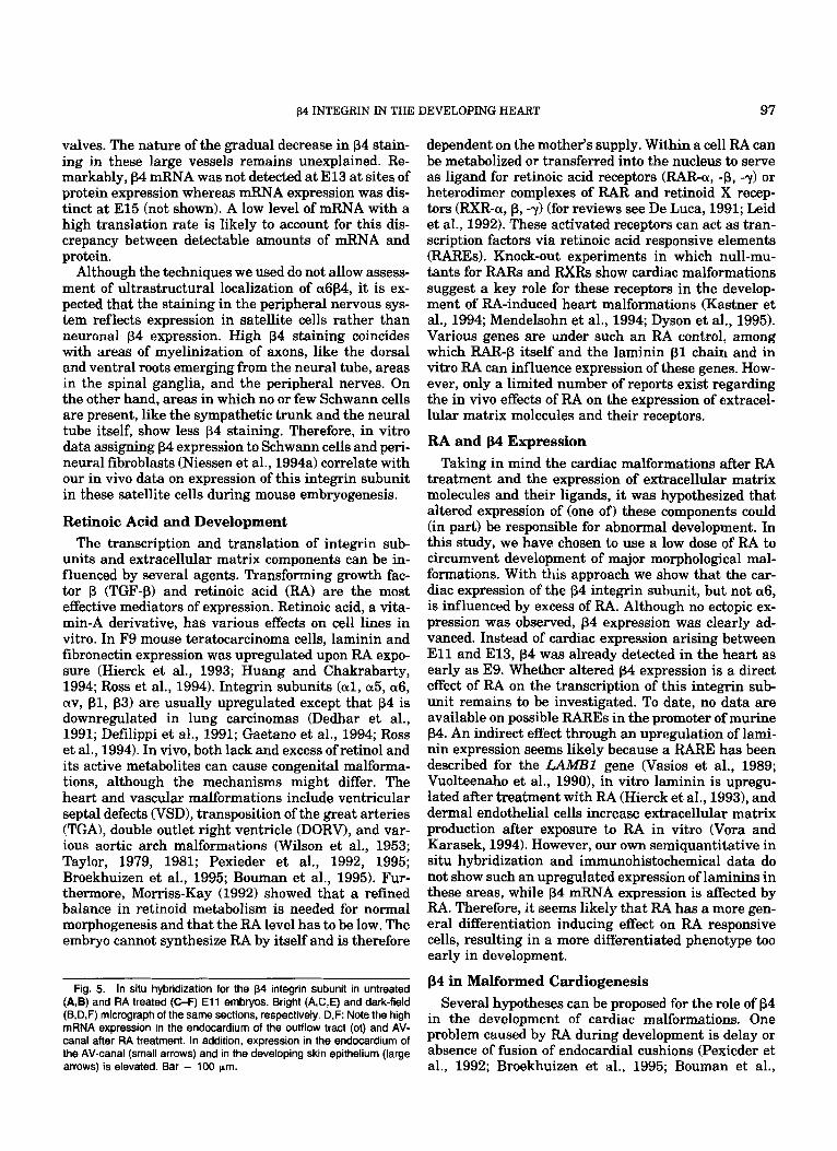
Fig. 5. In situ hybridization for the p4 integrin subunit in untreated (A,B) and RA treated (CF) E l 1 embryos. Bright (A,C,E) and dark-field (B,D,F) micrograph of the same sections, respectively. D,F: Note the high mRNA expression in the endocardium of the outflow tract (ot) and AV- canal after RA treatment. In addition, expression in the endocardium of the AV-canal (small arrows) and in the developing skin epithelium (large arrows) is elevated. Bar = 100 pm.
dependent on the mother’s supply. Within a cell RA can be metabolized or transferred into the nucleus to serve as ligand for retinoic acid receptors (RAR-a, -p, -y) or heterodimer complexes of RAR and retinoid X recep- tors (RXR-a, p, -7) (for reviews see De Luca, 1991; Leid et al., 1992). These activated receptors can act as tran- scription factors via retinoic acid responsive elements (RAREs). Knock-out experiments in which null-mu- tants for RARs and RXRs show cardiac malformations suggest a key role for these receptors in the develop- ment of RA-induced heart malformations (Kastner et al., 1994; Mendelsohn et al., 1994; Dyson et al., 1995). Various genes are under such an RA control, among which RAR-p itself and the laminin p l chain and in vitro RA can influence expression of these genes. How- ever, only a limited number of reports exist regarding the in vivo effects of RA on the expression of extracel- lular matrix molecules and their receptors.
RA and p4 Expression Taking in mind the cardiac malformations after RA
treatment and the expression of extracellular matrix molecules and their ligands, it was hypothesized that altered expression of (one of) these components could (in part) be responsible for abnormal development. In this study, we have chosen to use a low dose of RA to circumvent development of major morphological mal- formations. With this approach we show that the car- diac expression of the p4 integrin subunit, but not a6, is influenced by excess of RA. Although no ectopic ex- pression was observed, p4 expression was clearly ad- vanced. Instead of cardiac expression arising between E l l and E13, p4 was already detected in the heart as early as E9. Whether altered p4 expression is a direct effect of RA on the transcription of this integrin sub- unit remains to be investigated. To date, no data are available on possible RAREs in the promoter of murine p4. An indirect effect through an upregulation of lami- nin expression seems likely because a RARE has been described for the LAMB1 gene (Vasios et al., 1989; Vuolteenaho et al., 1990), in vitro laminin is upregu- lated after treatment with RA (Hierck et al., 19331, and dermal endothelial cells increase extracellular matrix production after exposure to RA in vitro (Vora and Karasek, 1994). However, our own semiquantitative in situ hybridization and immunohistochemical data do not show such an upregulated expression of laminins in these areas, while p4 mRNA expression is affected by RA. Therefore, it seems likely that RA has a more gen- eral differentiation inducing effect on RA responsive cells, resulting in a more differentiated phenotype too early in development.
p4 in Malformed Cardiogenesis Several hypotheses can be proposed for the role of p4
in the development of cardiac malformations. One problem caused by RA during development is delay or absence of fusion of endocardia1 cushions (Pexieder et al., 1992; Broekhuizen et al., 1995; Bouman et al.,

98 HIERCK ET AL.
Fig. 6. lmmunohistochemical localization of p4 in untreated (A,C) and RA treated (B,D) El3 mouse embryos. Staining is evident in the endocar- dium lining the endocardial cushions in the outflow tract (s) (A,B) and AV-canal (ec) (C,D). 0: Although in the RA treated embryo a ventricular
septum defect appears to develop (arrow), expression of p4 is unaltered. A0 = aorta, p = pulmonary trunk, rv = right ventricle, Iv = left ventricle. Bar = 100 pm.

p4 INTEGRIN IN THE DEVELOPING HEART 99
Fig. 7. mRNA expression of p4 in €13 embryos treated with RA (B,D) and untreated (A$). Dark-field micrographs (C,D) show Increased p4 expression after RA treatment in the endothelium of the pulmonary trunk (p) and Aorta (Ao) as compared to the control (C). Moreover, endocar-
dium in the outflow tract and endothelium in the aorta and pulmonary trunk (small arrows) as well as skin epithelium (large arrows) show in- creased p4 expression. Bar = 100 pm.

100 HIERCK ET AL.
1995). This can lead to abnormal septation and valves, development of VSD, and probably other malforma- tions (van Mierop et al., 1962). p4 that is expressed on the endocardial cushions at a too early stage coincides with or could even be responsible for the delay of fu- sion. Although the process of fusion is not fully under- stood it is clear that two cushions, each with their own endocardial lining, meet to form a continuity. This im- plies that two opposed endocardial cell layers must dis- appear. Various mechanisms can be suggested in this process: apoptosis (programmed cell death), migration, or incorporation of the endocardial cells in the cushion tissue. Apoptosis has been shown not to appear under normal conditions at these sites (Los and van Eijnd- thoven, 1973; Icardo, 1984). This is in contrast to in- corporation of cells in the underlying tissue. Markwald and coworkers (1975, 1977) first described the process of “epithelial to mesenchymal transformation” as a way to fill the cardiac jelly with cells recruited from the endocardium. The end of this process coincides in time with the normal appearance of p4 in the endocardium. It is comprehensive that endocardial cells that express this adhesion molecule at a too early stage in develop- ment adhere to the underlying basement membrane too firmly and that transformation is disturbed. In- creased adhesion could also inhibit or delay migration of these cells to other areas. The latter is interesting while endothelial cells actively downregulate a6P4 af- ter they have been induced to migrate (Sepp et al., 1995), indicating that high amounts of a6p4 are not favourable for migration. Moreover, a computer model, in which parts of the development of the heart can be simulated, correlates increased adhesiveness of en- docardial cells in the AV-canal to development of atrio- ventricular defects (Kurnit et al., 1985).
A further mechanism to be postulated is that an en- hanced adhesion of the endocardial cells lining the AV- canal and outflow tract to their basement membranes could influence the looping of the heart, by making the inner lining of these segments too stiff, In this way the normal apposition of cushions could be prohibited. Ab- normal looping after the administration of RA has been suggested as a mechanism leading to a spectrum of cardiac malformations (Bouman et al., 1995). Changes in volume and extracellular matrix composition of en- docardial cushions after RA-treatment (Davis and Sa- dler, 1981; Nakajima et al., 1995) and changes in the expression of endocardial cell adhesion molecules (this study) indicate that the endocardial cushions are main targets for excess of RA.
The expression of p4 in the subectodermal region in the pharyngeal arches at E l l suggests expression in migrating neural crest cells. In RA treated embryos this expression is lost. The neural crest cells appear to advance their p4 expression after RA treatment, as early migrating cells in E9 embryos were clearly pos- itive. The advanced expression of the 04 integrin sub- unit in these cells could be the cause of altered migra- tion (Lee et al., 19951, and therefore account for the
similarity between phenotypes of neural crest ablated and RA-treated embryos (Yasuda et al., 1986; Nishiba- take et al., 1987; Hart et al., 1990; Kirby, 1990).
Conclusions We showed differential expression of the p4 integrin
subunit during mouse embryonic development in the heart and surrounding tissue. Cardiac expression ap- pears correlated with regions of high mechanical force. Upon RA treatment p4 expression is advanced in the endocardial cells in the heart, but also in migrating neural crest cells. Coexpression with the a6 integrin subunit and localization to the basal cell surface sug- gest the functionality of the a6p4 receptor. The altered expression of this cell-matrix receptor molecule could influence both migration of cells and stiffness of car- diac segments and, therefore, contribute to the devel- opment of congenital malformations. Whether RA di- rectly acts on p4 expression or through its extracellular ligand is a subject of further investigation.
EXPERIMENTAL PROCEDURES Tissue Preparation
Female Swiss mice were caged overnight with males. Mice showing a vaginal plug were selected on the next morning and housed separately under standard CAHPA (Comitb Ad Hoc pour la Protection des Ani- maux) conditions. On the day of interest the mice were killed by cervical dislocation and the embryos were dis- sected free (EO is designated as the day of the vaginal plug). For immunohistochemistry, embryonic tissue was collected and immersion-fixed overnight in ace- tone at 4°C. Acetone was removed by rinsing in several changes of PBS-sucrose (6.8% sucrose), followed by im- mersion in OCT Compound (Tissue-Tek, Miles Inc., Elkhart, IN). The embryos were embedded in fresh OCT Compound, frozen in isopentanol, chilled in liquid nitrogen, and stored at -20°C. Six micron serial sec- tions were cut transversely through the thoracal region of the embryo on a 2800 Frigocut-E cryostat (Leica, NussLoch, Germany), collected on Starfrost slides (Knittel Glaser, Braunschweig, Germany), dehydrated in acetone (-20’0, and stored at -20°C. For in situ hybridization, embryos or isolated hearts were col- lected and immersion-fixed overnight in 4% parafor- maldehyde in PBS at 4°C. Tissue was routinely pre- pared for paraffin embedding, sectioned at 5 pm and mounted on aminopropyltriethoxysilane (Sigma, St. Louis, MO) treated glass slides.
Retinoic Acid Treatment On E7 20 mgkg all-trans Retinoic Acid (Sigma), dis-
solved in vegetable oil, was injected intraperitoneally in pregnant mice. On the day of interest embryos were dissected free and examined for macroscopic malforma- tions. The embryos were routinely prepared for immu- nohistochemistry or in situ hybridization (see above).

p4 INTEGRIN IN THE DEVELOPING HEART 101
Immunohistochemistry
Slides were post-fixed in ice-cold acetone for 10 min at -20°C and air dried. Endogenous peroxidase was inactivated by incubation in 0.3% HzO, in PBS-azide (PBS containing 0.1% Na-azide) for 30 min at room temperature. The slides were washed in PBS, incu- bated for 15 min in a background reducing solution (150 mM NaC1,lO mM Tris-HC1,5 mM EDTA, 0.25% (w/v) gelatin, 0.05% (v/v) Tween-20, pH 8.0) at room temperature and washed again (3 x 5 min PBS). The sections were incubated overnight with the pri-
mary antibody, diluted in PBSB (PBS containing 1% bovine serum albumin), in a moist chamber. Rat mono- clonal antibodies were visualized by a standard DAB reaction (0.04% diaminobenzidine tetrahydrochloride in 50 mM Tris-Maleic acid, pH 7.6, containing 0.06%0 H202) after subsequent incubation with Rabbit-anti- Rat antiserum (Dako, Carpinteria, CA; 1:1,000 in PBSB), Goat-anti-Rabbit antiserum (Nordic, Tilburg, The Netherlands, 1 5 0 in PBSB), and Rabbit-Peroxi- dase-anti-Peroxidase (Nordic, 1500 in PBSB). Sec- tions were counterstained in haematoxylin, dehy- drated and mounted in Entellan (Merck, Darmstadt, Germany).
In Situ Hybridization
In situ hybridization was performed according to Wilkinson and Green (1990) with minor modifications. Briefly, tissue sections were deparaffnized in xylene, rehydrated in a descending series of ethanol to PBS and post-fixed for 20 min. After treatment with Pro- teinase K (Merck), post-fixation and acetylation, sec- tions were dehydrated and used for hybridization.
Linearized cDNA was transcribed to anti-sense cRNA probes in the presence of 75 pCi [35SlUTP (Am- ersham, Arlington Heights, IL), 10 U placental RNAse inhibitor (Pharmacia, Uppsala, Sweden) and 10 U T3- or T7-RNA polymerase (Pharmacia). Sections were hy- bridized overnight to 1 X lo5 cpm/p1 of [35SlcRNA in hybridization solution (50% deionized formamide, 0.3 M NaC1, 20 mM Tris-HCl pH 8.0, 5 mM EDTA, 100 mM dithiothreitol [DTT], 1 x Denhardt's, 0.5 mg/ml calf t-RNA, 10% dextran sulphate) at 55°C. High strin- gency washing was performed in 100 mM Dl" in 50% formamide, 2 x SSC for 45 min at 65°C. After RNAse treatment (RNAse-A, Sigma) the sections were submit- ted again to high stringency washing. Sections were washed in 2 x SSC and 0.1 x SSC at 55"C, respec- tively, dehydrated through ascending series of etha- nols, and air dried.
Subsequently, sections were coated with Ilford G5 emulsion and exposed for 6 to 15 days at 4°C. Slides were developed in Kodak D19 developing solution, fixed in Polymax I (Kodak, Rochester, NY) and coun- terstained with haematoxylin. Examination was per- formed with a Leitz Dialux 20E8 microscope equipped with a dark field condenser.
Antibodies and Probes Antibodies used in this study were (1) Monoclonal
Ftat-anti-p4 (346-11A, Kennel et al., 1989; Sonnenberg et al., 19901, used 1:ZOO in PBSB; (2) Monoclonal Rat- anti-a6 (GoH3, Sonnenberg et al., 19861, used 1:lO in PBSB.
Probes used in this study were (1) a 370 bp (EcoRV- NotI) fragment of human p4 integrin subunit (Hoger- vorst et al., 1990); (2) a 410 bp (EcoRI-Pat11 fragment of murine a6 integrin subunit (Hierck et al., 1993).
High sequence homology between human and mu- rine p4 cDNA resulted in a strong hybridization signal of mouse tissue with human probes. All probes used in this study were designed to recognize all splice vari- ants described to date. All cDNAs were subcloned in pBluescript (Stratagene, La Jolla, CA) and linearized. One microgram was used for probe preparation.
ACKNOWLEDGMENTS The authors thank Dr. Arnoud Sonnenberg for gen-
erous gifts of antibodies.
REFERENCES Aplin, J.D. (1993) Expression of integrin a6P4 in human trophoblast
and its loss from extravillous cells. Placenta 14203-215. Baldwin, H.S., and Buck, C.A. (1994) Integrins and other cell adhe-
sion molecules in cardiac development. Trends Cardiovasc. Med.
Bouman, H.G.A., Broekhuizen, M.L.A., Baasten, A.M.J., Gitten- berger-de Groot, A.C., and Wenink, A.C.G. (1995) Spectrum of loop- ing disturbances in stage 34 chicken he& after retinoic acid treat- ment. Anat. Rec. 243:lOl-108.
Brand, T., and Schneider, M.D. (1995) The TGFp Superfamily in myo- cardium: Ligands, receptors, transduction, and function. J. Mol. Cell Cardiol. 275-18.
Broekhuizen, M.L.A., Bouman, H.G.A., Mast, F., Mulder, P.G.H., Git- tenberger-de Groot, A.C., and Wladimiroff, J.W. (1995) Hemody- namic changes in stage 34 chick embryos after treatment with all- trans retinoic acid. Ped. Res. 38:342-348.
Buck, C.A., Baldwin, H.S., DeLisser, H., Mickanin, C., Shen, H.M., Kennedy, G., Chen, A., Edelman, J.M. , and Albelda, S.M. (1993) Cell adhesion receptors and early mammalian heart development: An overview. C.R. Acad. Sci. Paris, Life Sci. 316:849-859.
Burgeson, R.E., Chiquet, M., Deutzmann, R., Ekblom, P., Engel, J., Kleinman, H., Martin, G.R., Meneguzzi, G., Paulsson, M., Sanes, J., Timpl, R., Tryggvason, K., Yamada, Y., and Yurchenco, P.D. (1994) A new nomenclature for the laminins. Matrix Biol. 14:209-211.
Christophers, E., and Wolff, H.H. (1975) Differential formation of des- mosomes and hemidesmosomes in epidermal cell cultures treated with retinoic acid. Nature 256209-210.
Clarke, A.S., h t z , M.M., and Mercurio, A.M. (1994) A novel struc- tural variant of the human beta 4 integrin cDNA. Cell Adhes. Com- mun. 21-6.
Collo, G., Domanico, S.Z., Klier, G., and Quaranta, V. (1995) Gradient of integrin a6A distribution in the myocardium during early heart development. Cell Adhes. Commun. 3:lOl-113.
Cooper, A.R., and MacQueen, H.A. (1983) Subunits of laminin are differentially synthesized in mouse eggs and early embryos. Dev. Biol . 96467- 47 1.
Cremona, O., Savoia, P., Marchisio, P.C., Gabbiani, G., and Chapon- nier, C. (1994) The a6 and p l integrin subunits are expressed by smooth muscle cells of human small vessels: A new localization in mesenchymal cells. J. Histochem. Cytochem. 421221-1228.
Davis, L.A., and Sadler, T.W. (1981) Effects of vitamin A on endocar- dial cushion development in the mouse heart. Teratnlogy 24:139- 148.
4178-187.

102 HIERCK ET AL.
Dedhar, S., Robertson, K., and Gray, V. (1991) Induction of expression of the avpl and avp3 integrin heterodimers during retinoic acid- induced neuronal differentiation of murine embryonal carcinoma cells. J . Biol. Chem. 266:21846-21852.
Defilippi, P., van Hinsbergh, V., Bertolotto, A., Rossino, P., Silengo, L., and Tarone, G. (1991) Differential distribution and modulation of expression of alphalhetal integrin on human endothelial cells. J. Cell Biol. 114:855-863.
De Luca, L.M. (1991) Retinoids and their receptors in differentiation, embryogenesis, and neoplasia. FASEB J. 5:2924-2933.
Delwel, G.O., Kuikman, I., and Sonnenberg, A. (1995) An alterna- tively spliced exon in the extracellular domain of the human a6 integrin subunit Functional analysis of the a6 integrin variants. Cell Adhes. Commun. 3:143-161.
Dyson, E., Sucov, H.M., Kubalak, S.W., Schmid-Schhbein, G.W., DeLano, F.A., Evans, R.M., Ross, J., and Chien, K.R. (1995) Atrial- like phenotype is associated with embryonic ventricular failure in retinoid X receptor u 4- mice. Proc. Natl. Acad. Sci. U S A . 92:7386- 7390.
Dziadek, M., and Timpl, R. (1985) Expression of nidogen and laminin in basement membranes during mouse embryogenesis and in tera- tocarcinoma cells. Dev. Biol. 111:372-382.
Feltri, M.L., Scherer, S.S., Nemni, R., Kamholz, J., Vogelbacker, H., Scott, M.O., Canal, N., Quaranta, V., and Wrabetz, L. (1994) p4 integrin expression in myelinating Schwann cells is polarized, de- velopmentally regulated and axonally dependent. Development 1201287-1301.
Gaetano, C., Melchiori, A., Albini, A., Benelli, R., Falcioni, R., Mod- esti, A., Modica, A., Scarpa, s., and Sacchi, A. (1994) Retinoic acid negatively regulates p4 integrin expression and suppresses the ma- lignant phenotype in a Lewis carcinoma cell line. Clin. Exp. Metast.
Gmez, M., Navarro, P., Quintanilla, M., and Cano, A. (1992) Expres- sion of a6p4 integrin increases during malignant conversion of mouse epidermal keratinocytes: Association o f p4 subunit to the cytokeratin fraction. Exp. Cell Res. 201:250-261.
Hart, R.C., McCue, P.A., Ragland, W.L., Winn, K.J., and Unger, E.R. (1990) Avian model for 13-cis-retinoic acid embryopathy: Demon- stration of neural crest related defects. Teratology 41:463-472.
Hierck, B.P., Thorsteinsd6ttir, S., Niessen, C.M., Freund, E., van Ip- eren, L., Feyen, A., Hogervorst, F., Poelmann, R.E., Mummery, C.L., and Sonnenberg, A. (1993) Variants of the asp1 laminin re- ceptor in early murine development Distribution, molecular clon- ing and chromosomal localization of the mouse integrin a6 subunit. Cell Adhes. Commun. 1:33-53.
Hierck, B.P., Poelmann, R.E., van Iperen, L., Brouwer, A,, and Git- tenberger-de Groot, A.C. (1996) Differential expression of a6 and other subunits of laminin binding integrins during development of the murine heart. Dev. Dyn. 206:lOO-111.
Hogervorst, F., Kuikman, I., von dem Borne A.E.G.Kr., and Sonnen- berg, A. (1990) Cloning and sequence analysis of beta-4 cDNA: An integrin subunit that contains a unique 118 kd cytoplasmic domain. EMBO J. 9765-770,
Huang, S., and Chakrabarty, S. (1994) Regulation of fibronectin and laminin receptor expression, fibronectin and laminin secretion in human colon cancer cells by transforming growth factor-pl. Int. J . Cancer 57:742-746.
Hynes, R.O. (1992) Integrins: Versatility, modulation, and signaling in cell adhesion. Cell 69:ll-25.
Icardo, J.M. (1984) The growing heart: An anatomical perspective. In: “Growth of the Heart in Health and Disease,” Zak, R. (ed). New York: Raven Press, pp. 41-77.
Icardo, J.M. (1989) Endocardia1 cell arrangement Role of hemody- namics. Anat. Rec. 225150-155.
Iivanainen, A., Sainio, K., Sariola, H., and Tryggvason, K. (1995) Primary structure and expression of a novel human laminin a4 chain. FEBS Lett. 365183-188.
Jones, J.C.R., Asmuth, J., Baker, S.E., Langhofer, M., Rnth, S.I., and Hopkinson, S.B. (1994) Hemidesmosomes: Extracellular matridin- termediate filament connectors. Exp. Cell Res. 213:l-11.
Kajiji, S.M., Davceva, B., and Quaranta, V. (1987) Six monoclonal
12:63-72.
antibodies to human pancreatic cancer antigens. Cancer Res. 47:
Kastner, P., Grondona, J.M., Mark, M., Gansmuller, A,, LeMeur, M., Decimo, D., Vonesch, J.L., Doll6, P., and Chambon, P. (1994) Ge- netic analysis of RXRa developmental function: Convergence of RXR and RAR signaling pathways in heart and eye morphogenesiis. Cell 78987-1003.
Kennel, S.J., Foote, L.J., Falcioni, R., Sonnenberg, A., Stringer, C.D., Crouse, C., and Hemler, M.E. (1989) Analysis of the tumor-associ- ated antigen TSP-180. Identity with a6+4 in the integrin super- family. J . Biol. Chem. 264:15515-15521.
Kennel, S.J., Godfrey, V., Ch’ang, L.Y., Lankford, T.K., Foote, L.J., and Makkiqje, A. (1992) The p4 subunit of the integrin family is displayed on a restricted subset of endothelium in mice. J. Cell Sci. 101:145-150.
Kennel, S.J., Foote, L.J., Cimino, L., Rizzo, M.G., Chang, L.Y., and Sacchi, A. (1993) Sequence of a cDNA encoding the p4 subunit of murine integrin. Gene 130:209-216.
Kim, K.H. (1992) A role of retinoic acid in the regulation of the mor- phology and the levels of intermediate filament proteins and mRNAs in PC12 cells. Exp. Cell Res. 203:374-382.
Kirby, M.L. (1990) Alteration of cardiogenesis after neural crest ab- lation. Ann. N.Y. Acad. Sci. 588289-295.
Kurnit, D.M., Aldridge, J.F., Matsuoka, R., and Matthysse, S. (1985) Increased adhesiveness of trisomy 21 cells and atrioventricular ca- nal malformations in Down syndrome: A stochastic model. Am. J. Med. Genet. 20385-399.
Kwasigroch, T.E., and Kochhar, D.M. (1975) Locomotory behavior of limb bud cells. Effect of excess vitamin A in vivo and in vitro. Exp. Cell Res. 95269-278.
Lammer, E.J., Chen, D.T., Hoar, R.M., Ap i sh , N.D., Benke, P.J., Braun, J.T., Curry, C.J., Fernhoff, P.M., Grix, A.W., Lott, LT., Ri- chard, J.M., and Sun, S.C. (1985) Retinoic acid embryopathy. N. Engl. J . Med. 313837-841.
Lee, E.C., Lotz, M.M., Steele, G.D., and Mercurio, A.M. (1992) The integrin u6p4 is a laminin receptor. J. Cell Biol. 117:671-678.
Lee, Y.L., Osumi-Yamashita, N., Ninomiya, Y., Moon, C.K., Eriksson, U., and Eto, K. (1995) Retinoic acid stage-dependently alters the migration pattern and identity of hindbrain neural crest cells. De- velopment 121:825-837.
Leid, M., Kastner, P., and Chambon, P. (1992) Multiplicity generates diversity in the retinoic acid signaling pathways. TIBS 17:427-433.
Los, J.A., and van Eijndthoven, E. (1973) The fusion of the endocar- dial cushions in the heart of the chick embryo. 2. Anat. Entwick- lungsgesch. 141:55-75.
Markwald, R.R., Fitzharris, T.P., and Smith W.N.A. (1975) Structural analysis of endocardial cytodifferentiation. Dev. Biol. 42160-180.
Markwald, R.R., Fitzharris, T I . , and Manasek, F.J. (1977) Structural development of endocardial cushions. Am. J. Anat. 14885-120.
Mendelsohn, C., Lohnes, D., Decimo, D., Lufkin, T., LeMeur, M., Chambon, P., and Mark, M. (1994) Function of the retinoic acid receptors (RARs) during development. I1 Multiple abnormalities at various stages of organogenesis in RAR double mutants. Develop- ment 1202749-2771.
Morriss-Kay, G. (1992) Retinoic acid and development. Pathobiology 60:264-270.
Nakajima, Y., Morishima, M., Yasui, H., Nakazawa, M., and Momma, K. (1995) Molecular mechanisms of complete transposition of the great arteries produced by all-trans retinoic acid in mouse embryo. In: “Developmental Mechanisms of Heart Disease,” Clark, E.B., Markwald, R.R., and Takao, A. (eds). New York Futura, pp. 315- 317.
Natali, P.G., Nicotra, M.R., Bigotti, A., and de Martino, C. (1992) Localization of the a6 and p4 integrin subunits in normal human non-lymphoid tissues. J. Cell Sci. 103:1243-1247.
Niessen, C.M., Cremona, O., Daams, H., Ferraresi, S., Sonnenberg, A,, and Marchisio, P.C. (1994a) Expression of the integrin a6P4 in peripheral nerves: Localization in Schwann and perineural cells and different variants of the p4 subunit. J . Cell Sci. 107543452.
Niessen, C.M., Hogervorst, F., Jaspars, L.H., de Melker, A.A., Delwel, G.O., Huleman, E.H.M., Kuikman, I., and Sonnenberg, A. (1994b)
1367-1376.

p4 INTEGRIN IN THE DEVELOPING HEART 103
The a6P4 integrin is a receptor for both laminin and kalinin. Exp. Cell Res. 211:360-367.
Nishibatake, M., Kirby, M.L., and van Mierop, L.H.S. (1987) Patho- genesis of persistent truncus arteriosus and dextroposed aorta in the chick embryo after neural crest ablation. Circulation 75255- 264.
Ohno, M., Lopez, F., Gibbons, G.H., Cooke, J.P., andDzau, V.J. (1992) Shear stress induced TGFpl gene expression and generation of ac- tive TGFpl is mediated via a K* channel. Circulation 86:187.
Pelton, R.W., Saxena, B., Jones, M., Moses, H.L., andGold, L.I. (1991) Immunohistochemical localization of TGFpl, TGFp2, and TGFp3 in the mouse embryo: Expression patterns suggest multiple roles during embryonic development. J. Cell Biol. 115:1091-1105.
Pexieder, T., Pfizenmaier-Rousseil, M., and Prados-Frutos, J.C. (1992) Prenatal pathogenesis of the transposition of great arteries. In: “Transposition of the Great Arteries, 25 Years After Rashkind Bal- loon Septostomy,” Vogel, M., and Buhlmeyer, K. (eds). Darmstadt: Steinkopff Verlag, pp. 11-27.
Pexieder, T., Blanc, O., Pelouch, V., Ogbdalovk, I., Milerovd and Oljtkdal, B. (1995) Late fetal development of retinoic acid-induced transposition of great arteries: Morphology, physiology, and bio- chemistry. In: “Developmental Mechanisms of Heart Disease,” Clark, E.B., Markwald, R.R., and Takao, A. (eds). New York: Fu- tura, pp. 297-307.
Phillips, J.H., McKinney, L., Azuma, M., Spits, H., and Lanier, L.L. (1991) A novel p4, a6 integrin-associated epithelial cell antigen involved in natural killer cell and antigen-specific cytotoxic T lym- phocyte cytotoxicity. J. Exp. Med. 1741571-1581.
Poelmann, R.E., Gittenberger-de Groot, A.C., Mentink, M.M.T., Delpech, B., Girard, N., and Christ, B. (1990) The extracellular matrix during neural crest formation and migration. Anat. Em-
Ross, S.A., Ahrens, R.A., and De Luca, L.M. (1994) Retinoic acid en- hances adhesiveness, laminin and integrin pl synthesis, and reti- noic acid receptor expression in F9 teratocarcinoma cells. J. Cell. Physiol. 159263-273.
Sepp, N.T., Cornelius, L.A., Romani, N., Li, L.J., Caughman, S.W., Lawley, T.J., and Swerlick, R.A. (1995) Polarized expression and basic fibroblast growth factor-induced down-regulation of the a6p4 integrin complex on human microvascular endothelial cells. J. In- vest. Dermatol. 104266-270.
Smits-VanProoije, A.E., Vermeij-Keers, C., Poelmann, R.E., Mentink, M.M.T., and Dubbeldam, J.A. (1988) The formation of mesoderm and mesectoderm in 5-41-somite rat embryos cultured in vitro, us- ing WGA-Au as a marker. Anat. Embryol. 177245-256.
Sonnenberg, A., Daams, H., van der Valk, M.A., Hilkens, J., and Hilgers, J. (1986) Development of mouse mammary gland: Identi- fication of stages in differentiation of luminal and myoepithelial cells using monoclonal antibodies against keratin. J. Histochem. Cytochem. 34:1037-1046.
Sonnenberg, A,, Linders, C.J.T., Daams, J.H., and Kennel, S.J. (1990) The asp1 (VLA-6) and a6p4 protein complexes: Tissue distribution and biochemical properties. J. Cell Sci. 96207-217.
Sonnenberg, A., Calafat, J., Janssen, H., Daams, H., van der Raaij- Helmer, L.M.H., Falcioni, R., Kennel, S.J., Aplin, J.D., Baker, J., Loizidou, M., and Garrod, D. (1991) Integrin a6/$4 complex is lo- cated in hemidesmosomes, suggesting a major role in epidermal cell-basement membrane adhesion. J. Cell Biol. 113:907-917.
Spinardi, L., Einheber, S., Cullen, T., Milner, T.A., and Giancotti, F.G. (1995) A recombinant tail-less integrin p4 subunit disrupts
byol. 182:29-39.
hemidesmosomes, but does not suppress a6p4-mediated cell adhe- sion to laminins. J. Cell Biol. 129473-487.
Sutherland, A.E., Calarco, P.G., and Damsky, C.W. (1993) Develop- mental regulation of integrin expression at the time of implanta- tion in the mouse embryo. Development 1191175-1186,
Suzuki, S., and Naitoh, Y. (1990) Amino acid sequence of a novel integrin p4 subunit and primary expression of the mRNA in epi- thelial cells. EMBO J. 9:757-763.
Tamura, R.N., Rozzo, C., Starr, L., Chambers, J., Reichardt, L.F., Cooper, H.M., and Quaranta, V. (1990) Epithelial integrin a6p4: Complete primary structure of a6 and variant forms of p4. J. Cell Biol. 11 1: 1593-1604.
Tarone, G., Russo, M.A., Hirsch, E., Odorisio, T., Altruda, F., Silengo, L., and Siracusa, G. (1993) Expression of p l integrin complexes on the surface of unfertilized mouse oocyte. Development 117:1369- 1375.
Taylor, I.M. (1979) The effect of retinoic acid on the developing ham- ster heart: An ultrastructural and morphological study. In: “Ad- vances in the Study of Birth Defects, Vol. 111,” Persaud, T.V.N. (ed). Lancaster: MTP Press, pp. 119-134.
Taylor, I.M. (1981) Some early effects of retinoic acid on the young hamster heart. In: “Perspectives in Cardiovascular Research, Vol. 5,” Pexieder. T. (ed). New York Raven Press, pp. 151-161.
Timpl, R. (1989) Structure and biological activity of basement mem- brane proteins. Eur. J. Biochem. 180:487-502.
Timpl, R., and Brown, J.C. (1994) The laminins. Matrix Biol. 14275- 281.
Twal, W., Roze, L., and Zile, M.H. (1995) Anti-retinoic acid mono- clonal antibody localizes all-trans-retinoic acid in target cells and blocks normal development in early quail embryo. Dev. Biol. 168: 225-234.
van Mierop, L.H.S., Alley, R.D., Kausel, H.W., and Stranahan, A. (1962) The anatomy and embryology of endocardial cushion defects. d. Thorac. Cardiovasc. Surg. 43:71-83.
Vasios, G.W., Gold, J.D., Petkovich, M., Chambon, P., and Gudas, L.J. (1989) A retinoic acid-responsive element is present in the 5’ flank- ing region of the laminin B1 chain. Proc. Natl. Acad. Sci. USA. 86:9099-9103.
Vora, M., and Karasek, M.A. (1994) Retinoids upregulate phagocyto- sis by human dermal microvascular endothelial cells. J. Cell. Phys- iol. 159:450-456.
Vuolteenaho, R., Chow, L.T., and Tryggvason, K. (1990) Structure of the human laminin B1 chain gene. J. Biol. Chem. 265:15611- 15616.
Wilkinson, D.G., and Green, J. (1990) In situ hybridization and the three-dimensional reconstruction of serial sections. In: “Postim- plantation Mammalian Embryos, a Practical Approach,” Copp, A.J., and Cockroft, D.L. (eds), pp. 155-172.
Wilson, J.G., Roth, C.B., and Warkany, J. (1953) An analysis of the syndrome of malformations induced by maternal vitamin A defi- ciency. Effects of restoration of vitamin A at various times during gestation. Am. J. Anat. 92:189-217.
Wu, T.C., Wan, Y.J., Chung, AX., and Darqjanov, I. (1983) Immuno- histochemical localization of entactin and laminin in mouse em- bryos and fetuses. Dev. Biol. 100:496-505.
Yasuda, Y., Okamoto, M., Konishi, H., Matsuo, T., Kihara, T., and Tanimura, T. (1986) Developmental anomalies induced by all-trans retinoic acid in fetal mice: I. Macroscopic findings. Teratology 34: 37-49.