Expression of the c-fgr and hck Protein-Tyrosine Kinases in Acute Myeloid Leukemic Blasts Is...
10
1991 77: 726-734 CL Willman, CC Stewart, TL Longacre, DR Head, R Habbersett, SF Ziegler and RM Perlmutter differentiation events in the monocytic and granulocytic lineages myeloid leukemic blasts is associated with early commitment and Expression of the c-fgr and hck protein-tyrosine kinases in acute http://bloodjournal.hematologylibrary.org/site/misc/rights.xhtml#repub_requests Information about reproducing this article in parts or in its entirety may be found online at: http://bloodjournal.hematologylibrary.org/site/misc/rights.xhtml#reprints Information about ordering reprints may be found online at: http://bloodjournal.hematologylibrary.org/site/subscriptions/index.xhtml Information about subscriptions and ASH membership may be found online at: reserved. Copyright 2011 by The American Society of Hematology; all rights 900, Washington DC 20036. weekly by the American Society of Hematology, 2021 L St, NW, Suite Blood (print ISSN 0006-4971, online ISSN 1528-0020), is published For personal use only. by guest on December 12, 2013. bloodjournal.hematologylibrary.org From For personal use only. by guest on December 12, 2013. bloodjournal.hematologylibrary.org From
-
Upload
independent -
Category
Documents
-
view
2 -
download
0
Transcript of Expression of the c-fgr and hck Protein-Tyrosine Kinases in Acute Myeloid Leukemic Blasts Is...

1991 77: 726-734
CL Willman, CC Stewart, TL Longacre, DR Head, R Habbersett, SF Ziegler and RM Perlmutter differentiation events in the monocytic and granulocytic lineagesmyeloid leukemic blasts is associated with early commitment and Expression of the c-fgr and hck protein-tyrosine kinases in acute
http://bloodjournal.hematologylibrary.org/site/misc/rights.xhtml#repub_requestsInformation about reproducing this article in parts or in its entirety may be found online at:
http://bloodjournal.hematologylibrary.org/site/misc/rights.xhtml#reprintsInformation about ordering reprints may be found online at:
http://bloodjournal.hematologylibrary.org/site/subscriptions/index.xhtmlInformation about subscriptions and ASH membership may be found online at:
reserved.Copyright 2011 by The American Society of Hematology; all rights900, Washington DC 20036.weekly by the American Society of Hematology, 2021 L St, NW, Suite Blood (print ISSN 0006-4971, online ISSN 1528-0020), is published
For personal use only. by guest on December 12, 2013. bloodjournal.hematologylibrary.orgFrom For personal use only. by guest on December 12, 2013. bloodjournal.hematologylibrary.orgFrom
Expression of the c-fgr and hck Protein-Tyrosine Kinases in Acute Myeloid Leukemic Blasts Is Associated With Early Commitment and Differentiation
Events in the Monocytic and Granulocytic Lineages
By Cheryl L. Willman, Carleton C. Stewart, Teri L. Longacre, David R. Head, Rob Habbersett, Steven F. Ziegler, and Roger M. Perlmutter
Two members of the src proto-oncogene family of intracellu- lar tyrosine kinases, c-fgr and hck, are selectively expressed in differentiated myeloid cells. To study the expression of these genes in acute myeloid leukemia (AML) and to deter- mine the specific myeloid lineages and stages of myeloid differentiation at which the expression of these genes is acquired, we used a series of 79 cases of de novo AML as a differentiation model. The levels of c-fgr, hck, and c-fms (encoding the colony-stimulating factor-1 receptor) mRNA transcripts were correlated with the presence of specific cell surface antigens and the morphologic and cytochemical features in these AML blasts. Relatively undifferentiated leukemic myeloblasts with an HLA-DR, CD34, CD33, CD13? cell surface immunophenotype (French-American-British [FAB] M1 or M2) were characterized by a lack of c-fms and e-fgr expression, while low levels of c-fms and e-fgr could be detected in undifferentiated myeloblasts (FAB M1 or M2). which also expressed CD14 at low antigen density. The hck transcripts were either undetectable in these cells or were expressed at low levels. In contrast, only hck mRNA tran- scripts could be identified in blasts with progranulocytic
ELLULAR PROTEINS with tyrosine kinase activity C may be divided into two groups based on differences in structure and function.’,’ The first group consists of transmembrane tyrosine kinases with an extracellular ligand- binding domain, a transmembrane region, and an intracel- lular tyrosine kinase domain. Some of these proteins are known to function as receptors for polypeptide growth factors such as epidermal growth factor, platelet-derived growth factor, the monocytic colony-stimulating factor CSF-1, and insulin.’ In contrast, the second group of tyrosine kinases, eg, the products of the c-src, c-abl, and c-fpsslfes proto-oncogenes, reside entirely intracellularly and their biologic roles in normal cells remain undefined.2
From the Departments of Cell Biology and Pathology, University of New Mexico School of Medicine, Albuquerque; the National Flow Cytomehy Resource, Los Alamos National Laboratory, Los Alamos, NM; St Jude Children’s Research Hospital, Memphis, TN; the Howard Hughes Medical Institute, University of Washington School of Medi- cine, Seattle; and the Southwest Oncology Group Leukemia Biology Program, San Antonio, TX.
Submitted January 12, 1990; accepted October IO, 1990. Supported by National Institutes of Health Grants KIl-DK-01284
(C.L. W ) , CA-45682 (R.M.P.), CA-32102 and CA-12213 supporting the Southwest Oncology Group Leukemia Biology Research Program, and RR-01315 supporting the National Flow Cytometry Resource.
Address reprint requests to Cheryl L. Willman, MD, University of New Mexico Cancer Center, 900 Camino de Salud NE, Albuquerque, NM 87131.
The publication costs of this article were defrayed in part by page charge payment. This article must therefore be hereby marked “advertisement” in accordance with 18 U.S.C. section 1734 solely to indicate this fact.
0 1991 by The American Society ofHematology. 0006-4971/91/7704-0017$3.0010
morphology (FAB M3). while c-fms, c-fgr, and hck were all expressed at high levels in blasts with differentiated my- elomonocytic or monocytic features (FAB M4 and M5). No e-fms, e-fgr, or hck transcripts were evident in leukemic cells of the erythroid lineage (FAB M6). When undifferentiated leukemic myeloblasts (HLA-DR, CD34, and CD33) were in- duced to differentiate in vitro to cells with monocytic charac- teristics, the expression of c-fms, c-fgc and the CD14 cell surface antigen were induced t o high levels, accompanied by the acquisition of hck and CDl3 expression. In contrast, when HLA-DR, CD34, and CD33 blasts were induced to differentiate in vitro to cells with granulocytic characteris- tics, only hck and CD13 expression were induced. Our data suggest that the acquisition of c-fgr andlor hck expression is associated with early commitment and differentiation events in distinct myeloid lineages. Assessment of the expression of these kinases may provide a molecular tool to assign lineage in AML in conjunction with morphology, cytochemistry, and cell surface antigen expression. o 1991 by The American Society of Hematology.
The src family of intracellular protein-tyrosine kinases contains at least eight members: three were first identified as cellular proto-oncogenes transduced by oncogenic retro- viruses (c-src, c&, and c-yes), while the remaining five members (lck, hck, blk, lya, andfln) were identified by their nucleotide sequence similarity to c-src. Each of these kinases has a similar domain structure, including: (1) a membrane-associated amino terminal domain that is highly divergent in sequence in each member and that may, in part, determine the unique functions and substrate specific- ities for each kinase; (2) internal conserved SH2 (for “src homology 2”) and SH3 (“src homology 3”) domains, which may regulate kinase activity; and (3) a highly conserved carboxy terminal tyrosine kinase catalytic
Several members of the src family are selectively ex- pressed in hematopoietic cells and map to chromosomal loci that are consistently altered in human leukemias, lymphomas, and myelodysplastic ~ y n d r o m e s . ~ - ~ ~ ~ These find- ings suggest that certain src family kinases may play an important role in the development and function of normal and neoplastic hematopoietic cells. Both lck6 and an iso- form of thefla kinase, arising through alternative splicing,’ are selectively expressed in T-lymphoid cells, while blk is expressed in B-lymphoid cells.8 In contrast, both c-fg3.” and hCk4,10.12 are expressed in differentiated myeloid cells. In- sight into the function of these hematopoietic kinases is provided by recent studies of the T-lineage kinase lck. Via its amino terminal domain, Zck couples with the CD4 and CD8 T-cell-associated antigens, thereby providing an intra- cellular tyrosine kinase domain for these cell surface receptors that lack their own intracellular signal transduc- ing domain^.'^.'^ Association of lck with CD4 and CD8 presumably plays a role in both the development and
726 Blood, Vol77, No 4 (February 15). 1991: pp 726-734
For personal use only. by guest on December 12, 2013. bloodjournal.hematologylibrary.orgFrom
c-FGR AND HCK EXPRESSION IN AML 727
functional activation of T cells. By analogy, other members of the src kinase family may couple with cell surface receptors unique to the lineages in which they are ex- pressed.
Although the c-& and hck tyrosine kinases are known to be expressed in differentiated myeloid cells, their role in the development and function of normal myeloid cells is presently unknown. The range of myeloid lineages that express c-& and hck, and how and when the expression of these genes is acquired during myelopoiesis, has not been determined. Studies of the expression of these genes in neoplastic myeloid cells have also not been reported. Our present study has two purposes: (1) to examine the struc- ture and expression of c-fgr and hck (correlated with the expression of c-fms, encoding the CSF-1 receptor on monocytic cells) in a series of primary, uncultured de novo acute myeloid leukemia (AML) samples and to correlate the expression of these genes with morphology/cytochemis- try and cell surface antigen expression; and ( 2 ) to use these AML samples representative of various myeloid lineages and frozen at various stages of myelopoiesis as a differenti- ation model, to determine which myeloid lineages ex- pressed c-fg and hck and when the expression of these genes was acquired during myeloid development.
MATERIALS AND METHODS
AML samples. Seventy-nine primary de novo AML samples were selected for study from the Southwest Oncology Group (SWOG) Myeloid Leukemia Repository at the University of New Mexico (UNM), Albuquerque. Samples were derived from pa- tients registered to two SWOG treatment protocols: 8124 and 8600. On receipt of fresh bone marrow aspirates or peripheral blood samples at UNM, leukemic blasts and mononuclear cells were isolated by centrifugation over Ficoll-Hypaque. The blast cell percentage was determined on cytospins of these cells, and the cell surface immunophenotypes of these leukemic cells were deter- mined as described below. Residual leukemic cells were cryopre- served as cell suspensions at -135°C. All samples selected for this study were bone marrow aspirates that, after Ficoll-Hypaque, contained at least 90% to 95% blasts as defined by the French- American-British (FAB) morphologic and cytochemical criteria,” thus minimizing contamination of the sample with residual normal bone marrow or peripheral blood cells that might express myeloid- associated cell surface antigens and genes. The 79 samples in- cluded: 18 FAB M1 (undifferentiated myeloblasts), 19 FAB M2 (blasts displaying limited myeloid differentiation), 11 FAB M3 (progranulocytic blasts), 19 FAB M4 (mixed blasts with evidence of monocytic and granulocytic differentiation), 10 FAB M5 (mono- cytic blasts), and 2 FAB M6 (erythroblasts) cases. No FAB M7 (megakaryocytic blasts) cases were available for study. Morphology and cytochemistry were reviewed on all cases by the SWOG Leukemia Pathology Committee. All selected cases had no evi- dence of karyotypic abnormalities involving either the c-fsr (lp36.1) or hck (20q11-12) I O C ~ . ~ , ~
For initial determination of the cell surface antigens expressed on AML blasts, each sample was analyzed using single-color flow cytometric techniques. Leukemic blasts isolated with standard Ficoll-Hypaque centrifugation were labeled with a panel of 22 primary mouse antihuman monoclonal antibodies (MoAbs) fol- lowed by an appropriate Fab, directly fluoresceinated (FITC) goat antimouse second-step reagent as described.” For controls, cells
Analysis of cell surface antigen expression on AML cells.
were incubated with the second-step reagent alone and with FITC mouse Ig proteins of the same isotype as the primary antibody to rule out nonspecific Fc binding. Propidium iodide (PI) was added to each sample to exclude any nonviable cells from flow cytometric analy~is,’~ and cell surface antigen expression was assessed on selectively gated leukemic blasts. All initial single- and two-color flow cytometric studies were performed using a Becton-Dickinson FACScan Flow Cytometer (Becton-Dickinson, Mountain View, CA) at UNM as described.” Cells were considered positive if greater than 25% of the cells demonstrated a significant fluores- cent shift as des~ribed.’~,’~ Although all samples were initially screened with an extended panel of 22 MoAbs, the antigens analyzed in this study were: HLA-DR and CD34 (MylO, the human hematopoietic progenitor cell antigen” (Becton-Dickinson), the pan-myeloid antigens CD33 (My9) and CD13 (My7) (Coulter Corp, Hialeah, FL); and the CD14 (My4) antigen (Coulter Corp), which reacts predominantly with monocytic cells, but which may be detected at low antigen density on granulocytes.2’,”
For multicolor flow cytometric analysis of undifferentiated AML blasts and blasts induced to differentiate in vitro, cryopreserved leukemic blasts were rapidly and gently thawed, washed free of cryopreservatives in serum-containing tissue culture medium, and stained with MoAbs before and after differentiation in vitro. Cells were labeled simultaneously with two overlapping sets of four MoAbs. These sets were: (1) biotinylated HLA-DR (detected by the binding of the fluorochrome allophycocyanin [APC] conju- gated to avidin [APC-B-HLA-DR]), CD34 (detected indirectly using a second-step reagent conjugated to Texas Red [TR-CD34]), CD33 directly conjugated to fluorescein (FITC-CD33), and CD13 directly conjugated to phycoelythrin (PE-CD13); and (2) APC-B- HLA-DR, CD14 (detected indirectly using a second-step reagent conjugated to Texas Red [TR-CD14]), FITC-CD33, and PE-CD13. Cells were washed in PAB (phosphate-buffered saline containing 0.1% sodium azide, and 0.5% bovine serum albumin) and stained simultaneously with these MoAbs in the following fashion: (1) cells were incubated with the primary unconjugated MoAb (either CD34 or CD14) for 15 minutes followed by washing in PAB; (2) a Texas Red-conjugated goat antimouse Fab, second-step reagent (TR-GAM) was added for 15 minutes to bind to the first primary unconjugated MoAb followed by washing in PAB; (3) mouse Ig (10 kg in 10 k b Sigma, St Louis, MO) was added to the cells to ensure that none of the following antibodies would bind to available binding sites on the TR-GAM and to block nonspecific binding to Fc receptors; (4) with no washing after the addition of the mouse Ig, FITC-conjugated MoAb no. 2, PE-conjugated MoAb no. 3, and biotin-conjugated MoAb no. 4 were added to the cells simulta- neously and the cells were incubated for 15 minutes; ( 5 ) after washing in PAF3 the cells were stained with allophycocyanin avidin for 15 minutes (to detect the biotinylated reagent); (6) cells were washed in PAB; and finally (7) propidium iodide (10 pg/mL) was added to the cells just before flow cytometric analysis to exclude any nonviable cells from the analysis that might nonspecifically bind MoAbs.”
The simultaneous binding of the four MoAbs to each individual cell in the sample (10,000 cells were analyzed per sample) was detected using the multi-laser flow cytometric system at the National Flow Cytometry Resource Center, Life Sciences Division, Los Alamos National Laboratory, Los Alamos, NM. The FITC, PE, and PI fluorochromes were excited by an argon laser tuned to 488 nm, while Texas Red and APC were excited by a dye laser tuned to 605 nm. Fluoresence emissions were detected at 530 2 15 nm (FITC), greater than 560 nm (PE and PI, with compensation), 630 2 15 nm (Texas Red, with compensation), and 670 % 15 nm (APC, with compensation). When cells are stained simultaneously with four MoAbs, 16 different combinations of shared antigen
For personal use only. by guest on December 12, 2013. bloodjournal.hematologylibrary.orgFrom

72% WILLMAN ET AL
reactivity, referred to as cluster groups, are possible as discussed in Results. The percentage of cells present in each cluster group in the viable cell populations under analysis was determined using computer software systems available at the National Flow Cytome- try Resource. Further details regarding the multiparameter flow cytometric studies may be obtained by contacting the investigators.
Cryopre- served blasts from five AML samples with undifferentiated myelo- blast morphology and cytochemistry and with a relatively undiffer- entiated myeloblastic cell surface immunophenotype (HLA-DR, CD34, and CD3321,22) were selected for differentiation in vitro. Cells were rapidly and gently thawed and were cultured at lo6 cells/mL in a 37°C incubator with 10% CO, in a-modified minimal essential medium (GIBCO, Grand Island, NY) supplemented with 10% fetal bovine serum (Hyclone, Logan, UT), 10% GCT- conditioned media containing human granulocyte-macrophage CSF (GM-CSF Sigma), penicillin/streptomycin, and either 12-0- tetradecanoyl-phorbol 13-acetate (TPA at lo-@ mol& Sigma) or retinoic acid (at mol& Sigma) as de~cribed?’.’~ After 48 hours of culture, cells were isolated and analyzed in three ways: (1) viability postculture was determined by dye exclusion; (2) cell surface immunophenotype was determined using multicolor flow cytometric techniques; (3) cytospins were prepared from each differentiated sample and the cells were stained with Wrights Stain, as well as Sudan Black and nonspecific esterase for morpho- logic and cytochemical analysis’’; and (4) total RNA was isolated for analysis of gene expression as described below.
Analysis of the gene erpression in AML blasts. Total RNA was isolated from cryopreserved undifferentiated AML blasts and from blasts induced to differentiate in vitro by lysis in guanidinium isothiocyanate followed by ultracentrifugation over cesium chlo- ride as described.’,’’ Ten micrograms of total RNA per sample was electrophoresed in 1% formaldehyde-agarose denaturing gels and transferred to nitrocellulose as described?~”’~’’ Northern blots were hybridized with ”-P-dCTP-labeled probes specific for c-fgr,’.’” hck,” and c-fm~.’~ Control for RNA loads was determined by hybridization of each filter with probes for p-actin (data not shown). After stringent washing, filters were dried and exposed to x-ray film with intensifying screens at -70°C for 72 hours to detect hybridization signals?,’” To quantitate the expression of c - w , c-&4 and hck, the levels of expression of these genes in leukemic samples were compared with the levels expressed in normal peripheral blood monocytes and granulocytes, isolated as described.” The relative levels of expression in the normal and leukemic cells was determined by soft laser scanning densitometry of autoradio- graphs. The level of expression of c-fms and c-& mRNA tran- scripts in peripheral blood monocytes and of hck in granulocytes was arbitrarily set at l+; the levels of expression in leukemic cells were then quantitated relative to the levels expressed in normal cells on a scale of 1 + + 4+, with 1 + representing baseline control levels in peripheral blood cells, 2+ representing a 2- to 5-fold increase, 3+ representing a 5- to 10-fold increase, and 4+ representing a 10- to 20-fold increase in mRNA transcript levels.
Induction of differentiation in AML blast samples.
RESULTS
The c-fgr and hck protein-tyrosine kinases are expressed in distinct myeloid lineages at specific stages of differentiation in de novo AML. To examine the expression of c&, hck, and c-fms in de novo AML, 79 primary (uncultured) human de novo AML samples were characterized by analyzing their: (1) morphology and cytochemistry according to established FAB criteria2’; (2) expression of a series of hematopoietic cell surface antigens, representative of particular stages of
myeloid differentiation, using single-color flow cytometric techniques; and (3) patterns of gene expression in Northern analysis (see Materials and Methods). The results of this correlative study are detailed in Table 1. All samples selected for study contained greater than 90% to 95% blasts after density centrifugation to minimize contamination of the leukemic blasts with residual myeloid cells that might express myeloid-associated cell surface antigens and genes. In our study, there was no consistent correlation of the expression of a subset of myeloid-associated cell surface antigens with FAB morphologic subtypes (Table 1; a study correlating the expression of a panel of 22 cell surface antigens with morphologic and cytochemical features is currently underway on a larger series of cases). As shown in Table 1, most FAB subgroups displayed considerable heter- ogeneity in cell surface antigen expression.
In contrast, expression of c-fgr, hck, and c-fins mRNA transcripts in the leukemic blasts (and expression of the p p 5 P k protein,” Ziegler S, Willman C, Perlmutter R: unpublished observations, January 1989) did correlate with particular myeloid differentiation stages defined both by morphology and cell surface antigen expression (Table 1 and Fig 1). Relatively undifferentiated AML blasts (FAB M1; HLA-DR, CD34’, CD33, CD13’) or blasts displaying limited myeloid differentiation (FAB M2) did not contain c-& or c-fins mRNA transcripts. The 2.2-kb hck mRNA transcript was either expressed at varying levels or was essentially undetectable in these relatively undifferentiated cells (Table 1 and Fig 1A). Low levels of the 4.7-kb c-fms and 2.6-kb c-fgr transcripts could be detected in some morphologically undifferentiated blasts that also expressed CD14 at low antigen density (Table 1, cases 9 through 12,22 and 23). AML blasts with promyelocytic (FAB M3) features tended to consistently express CD33 and CD13, but were heterogeneous with respect to expression of HLA-DR, CD34, and CD14 (Table 1). Despite this heterogeneity of cell surface antigen expression, all FAB M3 leukemic samples contained moderate to high levels of the hck transcript with no evidence of c-fins or c-fgr expression (Table 1 and Fig 1B).
High levels of c$gr and c-fms mRNA were detected only in AML blasts that displayed more differentiated my- elomonocytic (FAB M4) or monocytic (FAB M5) features. The blasts from these cases usually had an HLA-DR (DR), CD34’, CD33, CD13, and CD14 cell surface immunopheno- type (Table 1 and Fig 1C). These differentiated myelomono- cytic and monocytic blasts also expressed significant levels of hck mRNA (Fig 1C). Interestingly, no expression of c-fms, c&r, or hck was evident in myeloid leukemia cells derived from the erythroid lineage (FAB M6; Table 1).
The c-fms, c&r, and hck mRNA transcripts were of an appropriate size in all AML cases in which transcripts were detected, although higher levels of expression of these transcripts were seen in many AML cases when compared with the levels expressed by normal differentiated periph- eral blood granulocytes and monocytes (Table 1; see Materials and Methods). No c-fms, c-&, or hck transcripts were detected in a series of 30 acute lymphocytic leukemia
For personal use only. by guest on December 12, 2013. bloodjournal.hematologylibrary.orgFrom

c-FGR AND HCK EXPRESSION IN AML 729
Table 1. Correlation of Morphology, Phenotype, and Gene Expression in De Novo AML
mRNA Transcriptst
Patient* FAB Cell Surface lmmunophenotypet fms
AML-FA6 M1 1 2-4 5-7 8 9-10
11 12 13 14-17 18
AML-FA6 M2 19-21 22-23 24 25 26-30 31-34 35 36-37
AML-FAB M3 38-39 40 41 42 43 44-46 47 48
AMC-FAB-M4 49-50 51 52 53-54 55-64 65 66-67
AML-FAB M5 68-70 71-72 73-74 75 76 71
AML-FA6 M6 78 79
M1 M1 M1 M1 M1 M1 M1 M1 M1 M1
M2 M2 M2 M2 M2 M2 M2 M2
M3m M3 M3 M3 M3m M3 M3m M3
M4 M4 M4 M4 M4 M4 M4
M5 M5b M5 M5b M5 M5
M6 M6
HLA-DR, CD34, CD13 HLA-DR, CD34, CD33. CD13 HLA-DR, CD34, CD33, CD13 HLA-DR, CD34, CD33, CD13 HLA-DR, CD34, CD33, CD14(l0) HLA-DR, CD34, CD33, CD14(l0) CD34, CD33, CD14(lo)
HLA-DR. CD33. CD13 HLA-DR, 0 3 3 , CD13
HLA-DR, CD33, CD13, CD14(l0)
HLA-DR, CD34, CD33, CD13 HLA-DR, CD34,CD33, CD13, CD14 HLA-DR, 0 3 3 , CD13, CD14
HLA-DR, CD33 CD33, CD33 CD33 CD33, CD13, CD14
HLA-DR, CD33, CD13, CD14
CD34, CD33, CD13 HLA-DR(lo), CD34(lo), CD33, CD13 CD34. CD33 CD34, CD33, CD13. CD14(lo) CD34, CD33, CD13, CD14(lo) CD33, CD13 CD33, CD13 CD33, CD13, CD14
HLA-DR, CD34. CD33, CD13, CD14 HLA-DR, CD34. CD33, CD13, CD14 HLA-DR, CD33, CD13, CD14 HLA-DR. CD33, CD13, CD14 HLA-DR, CD33, CD13, CD14 HLA-DR, CD33, CD13, CD14 HLA-DR, CD33, CD13, CD14
HLA-DR, CD33. CD13, CD14 HLA-DR, CD33. CD13, CD14 HLA-DR, CD33, CD13, CD14 HLA-DR, CD33, CD13, CD14 HLA-DR, CD33 CD13. CD14
HLA-DR, CD33, glycophorin HLA-DR, CD33, glycophorin
0 0 0 0 1+ 2+ 1+ 0 0 0
0 I+ 0 1+ 0 0 0 0
0 0 0 0 0 0 0 0
I+ I+ I+ I+ 2+ 3+ 4+
1+ 1+ 4+ 3+ 1+ 4+
0 0
fsr hck
0 0 0 0 1+ 2+ 1+ 0 0 0
0 1+ 0 I+ 0 0 0 0
0 0 0 0 0 0 0 0
1+ 2+ 1+ I+ 3+ 3+ 4+
1+ 1+ 4+ 3+ 1+ 4+
0 0
0 0 1+ 2+ 0 2+ 1+ 3+ 0 2+
1+ 4+ 4+ 1+ 0 0 0 0
2+ 3+ 4+ 2+ 2+ 4+ 4+ 4+
2+ 2+ 1+ 2+ 4+ 3+ 4+
1+ 2+ 4+ 4+ 1+ 4+
0 0
*Patients with similar cell surface phenotypes, morphology, and genotypes are grouped. tCell surface antigens were considered positive if > 25% of cells demonstrated a fluorescence shift as described in Materials and Methods.
Antigens expressed at low antigen density on the blast population are designated as "(lo)." *Levels of mRNA transcripts were quantitated relative to the levels expressed in normal peripheral blood granulocytes and monocytes as
described in Materials and Methods.
cases, including cases of both the B- and T-cell lineages (data not shown). Although the expression of other proto- oncogenes (c-myc, c-myb, and c-fos) could be detected in most AML samples, there was no correlation of the
expression of these genes with any particular FAB subtype (in contrast to a previous study") or stage of myeloid differentiation (data not shown).
Induction of monocytic differentiation is associated with the
For personal use only. by guest on December 12, 2013. bloodjournal.hematologylibrary.orgFrom

730 WILLMAN ET AL
hck
c-fgr
A B C DR , CD34 DRY CD33
CD33 CD13 lo CD33y cD13 CD13 , CD14 I M2 M2 M1 M2MllM3M3 M3 M3M3IM4M4 M4M5M5'
expression of CDI4 and c-fgr, followed by CD13 and hck. Our initial studies suggested that undifferentiated AML cells lacked expression of the c-fg and hck kinases and that the expression of c-fg andlor hck might be correlated with commitment and differentiation events in distinct myeloid lineages, with hck being expressed both in blasts committed to the granulocytic and monocytic lineages, and c-fg being selectively expressed in blasts committed to the monocytic lineage.
To prove that c-jg andlor hck expression was acquired with myeloid differentiation, we selected five AML samples that were representative of undifferentiated myeloblasts (FAB M1; containing > 95% morphologically undifferenti- ated blasts). These AML cells expressed HLA-DR, CD34, and CD33 characteristic of normal undifferentiated myelo- blasts.2'.22 Morphologic and immunophenotypic differentia- tion was induced in vitro in two of the five samples with the combination of GM-CSF and TPA, and in one of the five samples with the combination of GM-CSF and retinoic acid. In these three samples, there was no significant change in viability during the period of in vitro culture (data not shown). The remaining two samples did not display any evidence of differentiation in vitro.
Leukemic blasts from the two undifferentiated AML samples that displayed striking evidence of monocytic differentiation after in vitro culture with GM-CSF and TPA (Figs 2 and 3) became adherent and acquired nonspecific esterase (a cytochemical marker of monocytic differen- tiation"; data not shown). Differentiation in vitro was also associated with the acquisition of a monocytic cell surface immunophenotype. Multiparameter flow cytometric analy- sis of the first of these undifferentiated AML samples before the induction of differentiation showed that the majority of the undifferentiated blasts coexpressed only HLA-DR, CD34 (data not shown), and CD33 (Fig 2A and B). (Results of the simultaneous expression of four cell surface antigens are displayed as a series of two-color flow cytometric contour plots comparing the coexpression of any two antigens under study [Figs 2 through 41. The results obtained from each AML sample after differentiation in vitro [contour plots labeled D] are overlain on the contour .plots of the undifferentiated cells [contour plots labeled U] for each combination of any two cell surface antigens [Figs 2 through 41). A small population (7%) of the undifferenti-
Fig 1. RNA blot analysis of hck and c-fgr mRNA transcripts in relatively undifferentiated leukemic my- eloblasts (A), leukemic blasts with granulocytic mor- phology (B), and leukemic blasts with myelomono- cytic or monocytic morphology (C). Corresponding cell surface immunophenotypes (including analysis of HLA-DR [DR], CD34, CD33, CD14, and CD13) and FAB morphologic and cytochemical subgroups are listed for each of the 15 AML samples. The lanes in these Northern blots correspond to the following AML cases detailed in Table 1: Lane 1, case 19; 2,20; 3 , l ; 4.21; 5,3; 6,40; 7.38; 8.39; 9,43; 10,44; 11,49; 12,55; 13,50; 14,73; 15,74.
ated blasts in this first sample also coexpressed CD14 (Fig 2A and C), while no significant expression of CD13 was evident on the undifferentiated cells (Fig 2B and C). With monocytic differentiation in vitro, there was a striking change in the expression of myeloid cell surface antigens. The majority of the differentiated blasts coexpressed high levels of the CD14 with CD33 (Fig 2A; differentiated cell histogram [D]). Expression of the CD13 antigen was also acquired and was coexpressed on the differentiated CD33', CD14' blasts (Figs 2B and C), while HLA-DR and CD34 expression remained essentially unchanged (data not shown). Before the induction of differentiation, the morpho- logically undifferentiated blasts in this sample expressed very low levels of the 4.7-kb c-fm transcript (encoding the receptor for the monocytic growth factor CSF-12") and extremely low, almost undetectable levels of the 2.6-kb c-fg transcript (Fig 2D). With monocytic differentiation, the c-fms and c-jg transcripts increased to high levels (Fig 2D). Although hck mRNA was undetectable in the undifferenti- ated blasts, expression of this transcript was induced with monocytic differentiation (Fig 2D).
Analysis of the second AML sample, which acquired monocytic characteristics after differentiation in vitro, showed that the undifferentiated AML blasts displayed a more "differentiated" cell surface phenotype in multicolor flow cytometric analysis (Fig 3) when compared with the first sample (Fig 2), even though the undifferentiated cells in each sample were morphologically indistinguishable. The majority of the undifferentiated blasts in the second AML sample coexpressed HLA-DR, CD34, CD33, and low levels of CD14 (Fig 3A). In addition to developing adherence and nonspecific esterase positivity (data not shown), the differ- entiated leukemic cells also displayed a striking change in expression of myeloid cell surface antigens (Fig 3). While CD14 expression increased to very high levels on CD33' blasts (Fig 3A), the expression of CD13 was also induced on the CD33', CD14' blasts (Fig 3B and 3C). HLA-DR and CD34 expression remained unchanged (data not shown). As in the previous sample (Fig 2), low levels of c-fms and c-fg transcripts could be detected in the undifferentiated AML blasts, and these transcripts increased to high levels after differentiation (Fig 3D). Although hck transcripts were not detectable in the undifferentiated blasts, hck
For personal use only. by guest on December 12, 2013. bloodjournal.hematologylibrary.orgFrom

C-FOR AND HCK EXPRESSION IN AML 731
Fig 2. (A through C). The results of the simulta- neous fourtolor, multiparameter flow cytometric analysis are displayed as a series of two-color flow cytometric contour plots comparing the simulta- neous coexpression of CD14 and CD33 (A), CD13 and CD33 [E), and CD14 and CD13 IC). The flow cytometric results obtained with each combination of antigens after differentiation in vitro (contour plot labeled D in each figure) are overlain on the contour plots of the corresponding undifferentiated cells (contour plot labeled U in each figure) [panel D). The expression of c-fms, c-fgr, and hck mRNA
ferentiated (U) AML blasts and blasts induced to differentiate in vitro into cells with monocytic char- acteristics (D). Striking changes in gene expression accompanied the changes in cell surface immu- nophenotype. See text for full discussion. C - t m s c-fgr hck
CD13 ”
D lILA- DR,CD34,CD33CD1Sb~HLA-M1,CD3$,CD33,CD~N,CM3
was assessed by RNA blot techniques in the undif- U D U D U D
W
mRNA was easily demonstrable after differentiation in vitro (Fig 3D).
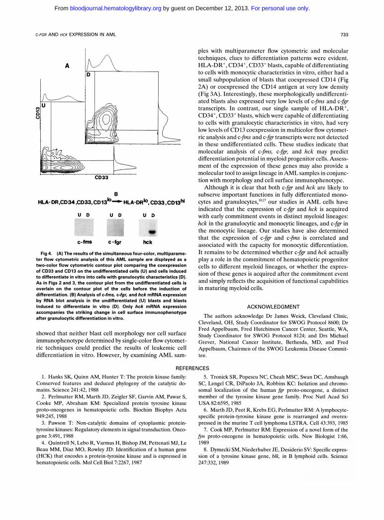
Induction of granulocytic differentiation in vitro is associ- ated with the acquisition of CD13 and hck qression. One of the undifferentiated AML samples displayed morpho- logic (data not shown) and immunophenotypic evidence of granulocytic differentiation in vitro after culture with GM- CSF and retinoic acid (as described in Materials and Methods). Interestingly, the morphology and cell surface immunophenotype of these undifferentiated AML blasts (Fig 4) were virtually identical to the cases that were capable of monocytic differentiation in vitro (Figs 2 and 3), although very low levels of CD13 were detectable on the undifferentiated blasts in multi-color flow cytometric analy- sis (Fig 4A). In contrast to the blasts that were capable of monocytic differentiation, these undifferentiated blasts had no evidence of c-fms or c-fgr expression (Fig 4B). Expres- sion of hck was also not detected. With differentiation, the blasts acquired high levels of coexpression of CD33 and CD13 (Fig 4A), while HLA-DR expression decreased (data not shown). After granulocytic differentiation, hck tran-
scripts were induced to significant levels in these cells, while there was no evidence of c-fms or cfgr expression (Fig 4B).
DISCUSSION
Although the c-& and hck protein-tyrosine kinases are known to be selectively expressed in differentiated myeloid cells, we were particularly interested in determining when the expression of c-fgr and hck was acquired during myeloid development and which myeloid lineages expressed these genes. Because both c-fgr and hck are proto-oncogenes that are selectively expressed in myeloid cells, we were also interested in determining their patterns of expression in AML cells and in correlating gene expression with the presence of particular myeloid-associated cell surface anti- gens and morphologic and cytochemical features in AML blasts. To address these issues, we used a series of primary uncultured de novo AML samples that were representative of specific myeloid lineages and that were arrested at specific stages of myeloid development, and thus were useful as a differentiation model.
In our studies, expression of the c-& protein-tyrosine
For personal use only. by guest on December 12, 2013. bloodjournal.hematologylibrary.orgFrom

732 WILLMAN ET AL
CD13 I'
D HLA- DR ,CD34,CD33,CD14 -HLA-oR, CD36,CD33, CDMM, CM3
U D U D U D
*
c-hns c-fgr hck
kinase and the c-fis gene encoding the CSF-1 receptor was not detected in morphologically undifferentiated leukemic myeloblasts with an HLA-DR, CD34, CD33, CD13' cell surface phenotype. However, low levels of c-fms and c$p could be detected in morphologically undifferentiated my- eloblasts, which also expressed the CD14 cell surface antigen. In several AML cases with undifferentiated mor- phology or in cases that displayed limited myeloid differen- tiation, hck transcripts were present at low levels. In contrast, only hck transcripts could be detected in blasts with progranulocytic morphology, while high levels of c-fis, c-&, and hck transcripts were present in differentiated blasts with myelomonocytic or monocytic features. Interest- ingly, leukemic erythroblasts did not express these myeloid lineage kinases. In all AML cases studies, the c-fms, c-fg, and hck transcripts were appropriately sized. However, the levels of expression of these genes in the more differenti- ated AML samples (FAB M3, M4, and M5) tended to be higher than the levels of expression detected in the differen- tiated peripheral blood granulocytes and monocytes used as a control to establish relative levels of expression. Whether
Fig 3. (A through D). Analysis of changes in cell surface immunophenotype and gene expression when a second undifferentiated AML sample was induced to differentiate in vitro into cells with monocytic characteristics. Refer to the legend for Fig 2 and the text for a full description of the figure.
the levels of c-fis, c$p, and hck transcripts in the leukemic cells are inappropriately high or simply reflect the appropri- ate level of expression for early stages of myeloid develop- ment in bone marrow-derived cells remains unclear. Our previous studies of normal murine bone marrow-derived cells indicated that monocyte precursor cells expressed significantly higher levels of c fp mRNA than more differ- entiated monocytic cells,P indicating that once the expres- sion of c-fgr is acquired in early-stage monocytes, it de- creases with progressive differentiation.
When undifferentiated leukemic myeloblasts were in- duced to differentiate in vitro, the induction of monocytic differentiation was accompanied by a significant increase in the level of c-fp and c-fms mRNA transcripts. With the acquisition of monocytic features, cell surface expression of CD14 increased, expression of CD13 was acquired, and hck expression was induced. In contrast, differentiation of undifferentiated blasts to cells with granulocytic features was associated with a maturation to a CD33, CD13 cell surface immunophenotype and acquisition of hck mRNA transcripts only. Our differentiation induction studies
For personal use only. by guest on December 12, 2013. bloodjournal.hematologylibrary.orgFrom
C-FGR AND HCK EXPRESSION IN AML 733
B HLA- DR.CD34 ,CD33,CD13boHLA-DRb, CD33,CDeM
U D U D U D
c-M c-fgr hck
Fig 4. (A) The results of the simultaneous four-color, multiparame- ter flow cytometric analysis of this AMI. sample are displayed as a two-color flow cytometric contour plot comparing the coexpression of CD33 and CD13 on the undifferentiated cells (U) and cells induced to differentiate in vitro into cells with granulocytic characteristics (D). As in Figs 2 and 3, the contour plot from the undifferentiated cells is overlain on the contour plot of the cells before the induction of differentiation. (E) Analysis of c-fms, c-fgr, and hck mRNA expression by RNA blot analysis in the undifferentiated (U) blasts and blasts induced to differentiate in vitro (D). Only hck mRNA expression accompanies the striking change in cell surface immunophenotype after granulocytic differentiation in vitro.
showed that neither blast cell morphology nor cell surface immunophenotype determined by single-color flow cytomet- ric techniques could predict the results of leukemic cell differentiation in vitro. However, by examining AML sam-
ples with multiparameter flow cytometric and molecular techniques, clues to differentiation patterns were evident. HLA-DR', CD34', CD33' blasts, capable of differentiating to cells with monocytic characteristics in vitro, either had a small subpopulation of blasts that coexpressed CD14 (Fig 2A) or coexpressed the CD14 antigen at very low density (Fig 3A). Interestingly, these morphologically undifferenti- ated blasts also expressed very low levels of c-fms and cfgr transcripts. In contrast, our single sample of HLA-DR', CD34', CD33' blasts, which were capable of differentiating to cells with granulocytic characteristics in vitro, had very low levels of CD13 coexpression in multicolor flow cytomet- ric analysis and c-fms and c-fgr transcripts were not detected in these undifferentiated cells. These studies indicate that molecular analysis of c-fms, c-fg, and hck may predict differentiation potential in myeloid progenitor cells. Assess- ment of the expression of these genes may also provide a molecular tool to assign lineage in AML samples in conjunc- tion with morphology and cell surface immunophenotype.
Although it is clear that both c-fgr and hck are likely to subserve important functions in fully differentiated mono- cytes and granulocyte^,'"^^' our studies in AML cells have indicated that the expression of cfgr and hck is acquired with early commitment events in distinct myeloid lineages: hck in the granulocytic and monocytic lineages, and c-fgr in the monocytic lineage. Our studies have also determined that the expression of c-fgr and c-fms is correlated and associated with the capacity for monocytic differentiation. It remains to be determined whether c-jgr and hck actually play a role in the commitment of hematopoietic progenitor cells to different myeloid lineages, or whether the expres- sion of these genes is acquired after the commitment event and simply reflects the acquisition of functional capabilities in maturing myeloid cells.
ACKNOWLEDGMENT
The authors acknowledge Dr James Weick, Cleveland Clinic, Cleveland, OH, Study Coordinator for SWOG Protocol 8600; Dr Fred Appelbaum, Fred Hutchinson Cancer Center, Seattle, WA, Study Coordinator for SWOG Protocol 8124; and Drs Michael Grever, National Cancer Institute, Bethesda, MD, and Fred Appelbaum, Chairmen of the SWOG Leukemia Disease Commit- tee.
REFERENCES 1. Hanks SK, Quinn AM, Hunter T The protein kinase family:
Conserved features and deduced phylogeny of the catalytic do- mains. Science 241:42, 1988
2. Perlmutter RM, Marth JD, Zeigler SF, Garvin AM, Pawar S, Cooke MP, Abraham KM: Specialized protein tyrosine kinase proto-oncogenes in hematopoietic cells. Biochim Biophys Acta 949:245, 1988
3. Pawson T Non-catalytic domains of cytoplasmic protein- tyrosine kinases: Regulatory elements in signal transduction. Onco- gene 3:491,1988
4. Quintrell N, Lebo R, Varmus H, Bishop JM, Pettenati MJ, Le Beau MM, Diaz MO, Rowley J D Identification of a human gene (HCK) that encodes a protein-tyrosine kinase and is expressed in hematopoietic cells. Mol Cell Biol7:2267, 1987
5. Tronick SR, Popescu NC, Cheah MSC, Swan DC, Amsbaugh SC, Lengel CR, DiPaolo JA, Robbins K C Isolation and chromo- somal localization of the human fg proto-oncogene, a distinct member of the tyrosine kinase gene family. Proc Natl Acad Sci USA 826595,1985
6. Marth JD, Peet R, Krebs EG, Perlmutter RM: A lymphocyte- specific protein-tyrosine kinase gene is rearranged and overex- pressed in the murine T cell lymphoma LSTRA. Cell 43:393, 1985
7. Cook MP, Perlmutter RM: Expression of a novel form of the fin proto-oncogene in hematopoietic cells. New Biologist 1:66, 1989
8. Dymecki SM, Niederhuber JE, Desiderio S V Specific expres- sion of a tyrosine kinase gene, blk, in B lymphoid cells. Science 247:332,1989
For personal use only. by guest on December 12, 2013. bloodjournal.hematologylibrary.orgFrom

734 WILLMAN ET AL
9. Willman CL, Stewart CC, Griffith JK, Stewart SJ, Tomasi TB: Differential expression and regulation of the c-src and c-& proto-oncogenes in myelomonocytic cells. Proc Natl Acad Sci USA 84:4480, 1987
10. Yi TL, Willman CL: Cloning of the murine c-& proto- oncogene cDNA and induction of csfq expression by proliferation and activation factors in normal bone marrow-derived monocytic cells. Oncogene 4:1081, 1989
11. Ley TJ, Connolly NL, Katamine S, Cheah MSC, Senior RM, Rohbins K C Tissue-specific expression and developmental regula- tion of the human& proto-oncogene. Mol Cell Biol9:92,1989
12. Ziegler SF, Marth JD, Lewis DB, Perlmutter RM: Novel protein-tyrosine kinase gene (hck) preferentially expressed in cells of hematopoietic origin. Mol Cell Biol7:2276, 1987
13. Veillette A, Bookman MA, Horak EM, Bolen JB: The CD4 and CD8 T cell surface antigens are associated with the internal membrane tyrosine-protein kinase ~ 5 6 " ~ . Cell 55:301,1988
14. Veillette A, Bookman MA, Horak EM, Samelson LE, Bolen JB: Signal transduction through the CD4 receptor involves the activation of the internal membrane tyrosine-protein kinase ~ 5 6 " ~ . Nature 338:257,1989
15. Marth JD, Lewis DB, Cooke MP, Mellins ED, Gearn ME, Samelson LE, Wilson CB, Miller AD, Perlmutter RM: Lymphocyte activation provokes modification of a lymphocyte-specific protein tyrosine kinase (~56"~) . J Immunol142:2430,1989
16. Marth JD, Lewis DB, Wilson CB, Gearn ME, Krebs EG, Perlmutter RM: Regulation of ~56'" during T cell activation: Functional implications for the src-like protein kinases. EMBO J 6:2727, 1987
17. Bennett JM, Catovosky D, Daniel M-T, Flandrin G, Galton DAG, Gralnick HR, Sultan C: Proposals for the classification of acute leukemias. French-American-British (FAB) Cooperative Group. Br J Haematol33:451,1976
18. Foucar K, Chen I-M, Crago S: Organization and operation of a flow cytometric immunophenotyping laboratory. Semin Diagn Pathol6:13,1989
19. Willman CL, Stewart CC: General principles of multiparam- eter flow cytometric analysis. Semin Diagn Pathol6:3,1989
20. Strauss LC, Rawley SD, LaRuss VF, Sharkis SJ, Stuart RK, Civin CI: Antigenic analysis of hematopoiesis. V. Characterization of My-10 antigen expression by normal lymphohematopoietic progenitor cells. Exp Hematol14:878,1986
21. Hurwitz CA, Strauss LC, Civin CI: Immunodiagnosis of acute nonlymphocytic leukemia, in Herberman RB, Mercer DW (eds): The Immunodiagnostics of Cancer. New York, NY, Dekker, (in press)
22. Civin CI, Loken M: Cell surface antigens on human marrow cells: Dissection of hematopoietic development using monoclonal antibodies and multiparameter flow cytometry. Int J Cell Cloning 5:267,1987
23. Koeffler P: Induction of differentiation of human acute myelogenous leukemia cells: Therapeutic implications. Blood 62: 709,1983
24. Griffith JD, Larcom P, Schlossman SF: Use of surface markers to identify a subset of acute myelomonocytic leukemia cells with progenitor cell properties. Blood 62:1300, 1983
25. Thomas PS: Hybridization of denatured RNA and small DNA fragments transferred to nitrocellulouse. Proc Natl Acad Sci USA 77:5201,1980
26. Sherr CJ, Rettenmier CW, Sacca R, Roussel MF, Look AT, Stanley ER: The c-fms proto-oncogene product is related to the receptor for the mononuclear phagocyte growth factor, CSF-1. Cell 41:665,1985
27. Ziegler SF, Wilson CB, Perlmutter RM: Augmented expres- sion of a myeloid-specific protein tyrosine kinase gene (hck) after macrophage activation. J Exp Med 168:1801,1988
28. Mavilio F, Testa U, Sposi NM, Petrini M, Pelosi E, Bordi- gnon S , Amadori S, Mandelli F, Peschle C: Selective expression of the fos proto-oncogene in human acute myelomonocytic and monocytic leukemias: A molecular marker of terminal differentia- tion. Blood 69:160, 1987
For personal use only. by guest on December 12, 2013. bloodjournal.hematologylibrary.orgFrom