Expression of TGF-β–induced matrix protein βig-h3 is up-regulated in the diabetic rat kidney and...
10
Kidney International, Vol. 64 (2003), pp. 1012–1021 Expression of TGF-–induced matrix protein ig-h3 is up-regulated in the diabetic rat kidney and human proximal tubular epithelial cells treated with high glucose SUK-HEE LEE,JONG-SUP BAE,SUN-HEE PARK,BYUNG-HEON LEE,RANG-WOON PARK, JE-YONG CHOI,JAE YONG PARK,SUNG-WOO HA,YONG-LIM KIM,TAE-HWAN KWON, and IN-SAN KIM Cell and Matrix Biology, National Research Laboratory, Kyungpook National University, Taegu, Korea; Department of Biochemistry, Department of Internal Medicine, School of Medicine, Kyungpook National University, Taegu, Korea; and Department of Physiology, School of Medicine, Dongguk University, Kyungju, Korea Expression of TGF-–induced matrix protein ig-h3 is up- regulated in the diabetic rat kidney and human proximal tubu- lar epithelial cells treated with high glucose. Backgrounds. ig-h3 is an extracellular matrix protein whose expression in several cell types is greatly increased by trans- forming growth factor- (TGF-). TGF- is believed to be involved in the development of diabetic nephropathy and thus we have assessed the possibility that ig-h3 may be a down- stream molecule in this pathogenic process. Methods. Immunoblotting and immunohistochemistry were done using an antibody against mouse ig-h3 protein. ig-h3 and TGF- concentrations were measured by enzyme-linked immunosorbent assay (ELISA). Cell adhesion and migration were assessed by measuring activity of N-acetyl--d-glucosami- nidase and using a transwell plate, respectively. Results. Immunohistochemistry revealed that ig-h3 occurs mainly in the basement membrane of proximal tubules, particu- larly the S3 segment but also to lesser extents in the basement membranes of the cortical thick ascending limb cells and the parietal glomerular epithelial cells in Bowman’s capsule. Im- munoblotting revealed that 68 kD bands were seen only in the cortex the outer stripe of the outer medulla. Rats with streptozotocin (STZ)-induced diabetes exhibited a marked and sustained increase in renal ig-h3 abundance. This was mir- rored by urinary ig-h3 levels. In vitro experiments with human primary renal proximal tubular epithelial cells revealed that their expression of ig-h3 was greatly increased by either TGF- or glucose. High glucose levels also stimulated TGF- produc- tion by renal proximal tubular epithelial cells and the high glu- cose–induced ig-h3 expression was almost completely blocked by anti-TGF- antibody. ig-h3 mediated renal proximal tubu- lar epithelial cells adhesion and migration. Conclusion. ig-h3 may be important in the development of Key words: diabetes mellitus, integrin, proximal tubule, renal failure, tubulointerstitial fibrosis. Received for publication July 30, 2002 and in revised form February 7, 2003 Accepted for publication April 23, 2003 2003 by the International Society of Nephrology 1012 diabetic nephropathy. Furthermore, the level of urinary ig-h3 may be useful as an early marker reflecting disease onset and progression. Diabetes mellitus is associated with a characteristic set of structural and functional kidney abnormalities re- ferred to as diabetic nephropathy, and chronic renal fail- ure is a common and serious complication. Consequently, it is of considerable interest to identify the factors re- sponsible for these abnormalities and that facilitate the progression of renal complications. Of these, the cyto- kine transforming growth factor- (TGF-) has emerged as a key factor in the development of renal hypertrophy and the accumulation of extracellular matrix in diabetic kidneys [1, 2]. In diabetic humans and animals, TGF- mRNA and protein levels in the glomeruli and tubulo- interstitium are significantly increased [3, 4]. Moreover, the administration of TGF- neutralizing antibodies to diabetic rats prevents the structural and functional kid- ney abnormalities from developing [5, 6]. In addition, altered urinary TGF- levels have been suggested to be an early predictor for assessing the progression of diabetic nephropathy [7]. Thus, TGF- and its upstream and downstream regulatory pathways could be potential tar- gets for blocking and monitoring the progression of dia- betic nephropathy. It is possible that the extracellular matrix protein ig- h3 may also be involved in diabetic nephropathy as ig- h3 expression in several cell types is highly induced by TGF- [8]. This protein has four characteristic internal repeat domains called fas-1 domains because they share homology with fasciclin 1, a Drosophilla neuroadhesion molecule [9]. We have identified a motif in each of the second and the fourth repeat domains that interacts with
-
Upload
independent -
Category
Documents
-
view
2 -
download
0
Transcript of Expression of TGF-β–induced matrix protein βig-h3 is up-regulated in the diabetic rat kidney and...
Kidney International, Vol. 64 (2003), pp. 1012–1021
Expression of TGF-�–induced matrix protein �ig-h3 isup-regulated in the diabetic rat kidney and human proximaltubular epithelial cells treated with high glucose
SUK-HEE LEE, JONG-SUP BAE, SUN-HEE PARK, BYUNG-HEON LEE, RANG-WOON PARK,JE-YONG CHOI, JAE YONG PARK, SUNG-WOO HA, YONG-LIM KIM, TAE-HWAN KWON,and IN-SAN KIM
Cell and Matrix Biology, National Research Laboratory, Kyungpook National University, Taegu, Korea; Department ofBiochemistry, Department of Internal Medicine, School of Medicine, Kyungpook National University, Taegu, Korea; andDepartment of Physiology, School of Medicine, Dongguk University, Kyungju, Korea
Expression of TGF-�–induced matrix protein �ig-h3 is up-regulated in the diabetic rat kidney and human proximal tubu-lar epithelial cells treated with high glucose.
Backgrounds. �ig-h3 is an extracellular matrix protein whoseexpression in several cell types is greatly increased by trans-forming growth factor-� (TGF-�). TGF-� is believed to beinvolved in the development of diabetic nephropathy and thuswe have assessed the possibility that �ig-h3 may be a down-stream molecule in this pathogenic process.
Methods. Immunoblotting and immunohistochemistry weredone using an antibody against mouse �ig-h3 protein. �ig-h3and TGF-� concentrations were measured by enzyme-linkedimmunosorbent assay (ELISA). Cell adhesion and migrationwere assessed by measuring activity of N-acetyl-�-d-glucosami-nidase and using a transwell plate, respectively.
Results. Immunohistochemistry revealed that �ig-h3 occursmainly in the basement membrane of proximal tubules, particu-larly the S3 segment but also to lesser extents in the basementmembranes of the cortical thick ascending limb cells and theparietal glomerular epithelial cells in Bowman’s capsule. Im-munoblotting revealed that �68 kD bands were seen only inthe cortex � the outer stripe of the outer medulla. Rats withstreptozotocin (STZ)-induced diabetes exhibited a marked andsustained increase in renal �ig-h3 abundance. This was mir-rored by urinary �ig-h3 levels. In vitro experiments with humanprimary renal proximal tubular epithelial cells revealed thattheir expression of �ig-h3 was greatly increased by either TGF-�or glucose. High glucose levels also stimulated TGF-� produc-tion by renal proximal tubular epithelial cells and the high glu-cose–induced �ig-h3 expression was almost completely blockedby anti-TGF-� antibody. �ig-h3 mediated renal proximal tubu-lar epithelial cells adhesion and migration.
Conclusion. �ig-h3 may be important in the development of
Key words: diabetes mellitus, integrin, proximal tubule, renal failure,tubulointerstitial fibrosis.
Received for publication July 30, 2002and in revised form February 7, 2003Accepted for publication April 23, 2003
2003 by the International Society of Nephrology
1012
diabetic nephropathy. Furthermore, the level of urinary �ig-h3may be useful as an early marker reflecting disease onset andprogression.
Diabetes mellitus is associated with a characteristicset of structural and functional kidney abnormalities re-ferred to as diabetic nephropathy, and chronic renal fail-ure is a common and serious complication. Consequently,it is of considerable interest to identify the factors re-sponsible for these abnormalities and that facilitate theprogression of renal complications. Of these, the cyto-kine transforming growth factor-� (TGF-�) has emergedas a key factor in the development of renal hypertrophyand the accumulation of extracellular matrix in diabetickidneys [1, 2]. In diabetic humans and animals, TGF-�mRNA and protein levels in the glomeruli and tubulo-interstitium are significantly increased [3, 4]. Moreover,the administration of TGF-� neutralizing antibodies todiabetic rats prevents the structural and functional kid-ney abnormalities from developing [5, 6]. In addition,altered urinary TGF-� levels have been suggested to bean early predictor for assessing the progression of diabeticnephropathy [7]. Thus, TGF-� and its upstream anddownstream regulatory pathways could be potential tar-gets for blocking and monitoring the progression of dia-betic nephropathy.
It is possible that the extracellular matrix protein �ig-h3 may also be involved in diabetic nephropathy as �ig-h3 expression in several cell types is highly induced byTGF-� [8]. This protein has four characteristic internalrepeat domains called fas-1 domains because they sharehomology with fasciclin 1, a Drosophilla neuroadhesionmolecule [9]. We have identified a motif in each of thesecond and the fourth repeat domains that interacts with
Lee et al: Up-regulation of big-h3 in diabetic nephropathy 1013
�3�1 integrin and thereby mediates corneal epithelial celladhesion [10]. We also reported that �ig-h3 forms afibrillar structure and interacts with several other extra-cellular matrix proteins, including fibronectin and type Icollagen [11]. �ig-h3 is also known to affect cell growth[8] and differentiation [12, 13]. In the rat kidney, an insitu hybridization study has revealed that �ig-h3 mRNAis expressed in proximal tubular epithelial cells (particu-larly in the S3 segment) and in the juxtaglomerular appa-ratus, and that these mRNA levels in the kidney are up-regulated in diabetes mellitus induced by streptozotocin(STZ) [14]. More recently, it has been suggested that �ig-h3 expression is associated with cyclosporine-inducednephropathy in humans who had received nonrenaltransplants [15]. However, to date, the immunolocaliza-tion of the �ig-h3 protein has not been demonstrated inthe kidney and it is unclear whether �ig-h3 actually doesplay a pathogenic role in the kidney diseases apparentlydriven by TGF-� (e.g., glomerulosclerosis and tubulo-interstitial fibrosis). To address these issues further, inthe present study we examined the immunolocalizationof �ig-h3 protein in the kidneys of normal rats. We foundit was predominantly expressed in the proximal tubulesand that renal levels of �ig-h3 protein were markedlyenhanced in diabetic rats. In vitro studies revealed thatprimary human renal proximal tubular epithelial cellsincrease their �ig-h3 expression when exposed to highglucose or TGF-� treatment, and that �ig-h3 can mediatethe adhesion and migration of renal proximal tubularepithelial cells. We developed an enzyme-linked immu-nosorbent assay (ELISA) to measure urinary �ig-h3 con-centrations and found that they mirrored renal �ig-h3protein levels, indicating urinary �ig-h3 could be usefulas an early predictor for the development of diabeticnephropathy and for monitoring the progression of thedisease.
METHODS
Experimental animals and protocols
Adult male Sprague-Dawley rats weighing 240 to 260 gmaintained on a standard rodent diet with free accessto water were randomly divided into control and experi-mental groups. Diabetes mellitus was induced by an in-traperitoneal injection of STZ (60 mg/kg body weight)(Sigma Chemical Co., St. Louis, MO, USA) in 0.1 mol/Lcitrate buffer, pH 4.5. Control rats received the citratebuffer alone. Blood glucose levels were determined 2 daysafter STZ injection and rats with blood glucose higherthan 250 mg/dL were considered to be diabetic. Fivegroups of rats were examined. Three groups consistedof diabetic rats sacrificed 5, 10, and 15 days after STZtreatment (N � 7, 8, and 8, respectively). A group offive control rats was sacrificed 15 days after receivingcitrate buffer. In a separate experiment, spot urine sam-
ples from six rats injected with STZ were collected for25 days. For the first four groups, rats were housed 24hours prior to sacrifice in metabolic cages for the mea-surement of water intake and urine output. The concen-tration of �ig-h3 in the urine was determined by indirectcompetitive ELISA (Regen Biotech, Seoul, Korea). Cre-atinine levels in plasma and urine were measured by theCreatinine Set (Asan Pharma., Seoul, Korea). Immedi-ately prior to sacrifice, rats were weighed and anesthe-tized by intraperitoneal injection of pentobarbital so-dium (1 mL/kg body weight). Blood was collected for thedetermination of plasma glucose and plasma creatinine.The right kidneys were rapidly removed and processedfor Western blot analysis (see below) while the left kid-neys were examined by immunohistochemistry (see be-low). With regard to the group of rats monitored for 25days, serial spot urine samples were taken and assessedfor the presence of �ig-h3. Urinary �ig-h3 levels werecorrected by the creatinine concentration in the urine.These rats were then sacrificed and their kidneys exam-ined by immunohistochemistry.
Antibodies against �ig-h3
The antihuman �ig-h3 antibody used has been charac-terized previously [16]. To make an antimouse �ig-h3antibody, we first made recombinant mouse �ig-h3 pro-tein. A DNA fragment encoding amino acids 24 to 618of mouse �ig-h3 was inserted into the BamH1/Xho1 sitesof pET-29b vector (Novagen, Madison, WI, USA) andexpressed in Escherichia coli. The his-tagged recombi-nant mouse �ig-h3 protein was purified using Ni-NTAresin (Qiagen, Valencia, CA, USA) according to themanufacturer’s instructions. Rabbits received a subcuta-neous injection of the protein (200 �g) in CompleteFreund’s Adjuvant followed by four immunizations spacedby 3 weeks with 200 �g protein in Incomplete Freund’sAdjuvant. Antibody titers were monitored by immu-noblot analysis using the recombinant �ig-h3 protein.The antiserum was further purified by Protein A affinitychromatography (Amersham Biosciences, Uppsala, Swe-den) according to the manufacturer’s protocol. The spec-ificity of the antimouse �ig-h3 antibody was tested byWestern blotting the supernatants of several mouse celllines (NIH3T3 fibroblasts, KS483 osteoblasts, and podo-cytes) treated with TGF-�, which revealed that only theexpected bands were detected (data not shown). Westernblotting rat kidney extracts or supernatants of severalTGF-�–treated rat cell lines [rat vascular smooth musclecells, buffalo rat liver cells (BRL) and normal rat kidney(NRK) cells] revealed that the antimouse �ig-h3 anti-body crossreacts to rat �ig-h3 (data not shown).
Electrophoresis and Western blotting
The right kidneys were divided into three parts (cortex �the outer stripe of outer medulla, inner stripe of outer
Lee et al: Up-regulation of big-h3 in diabetic nephropathy1014
medulla, and inner medulla) and homogenized with asolution of 0.3 mol/L sucrose, 25 mmol/L imidazole, 1mmol/L ethylenediaminetetraacetic acid (EDTA), pH7.2, containing 8.5 mmol/L leupeptin, and 100 �g/mLpefabloc, using an ultraturrax T8 homogenizer (IKA La-bortechnik, Staufen, Germany) at maximum speed for15 seconds. Each sample was then centrifuged at 4000gfor 15 minutes at 4�C to remove whole cells, nuclei, andmitochondria. The protein concentration in the superna-tant was measured by the Bradford method using theBio-Rad Protein Assay Kit (Bio-Rad, Hercules, CA,USA) according to the manufacturer’s protocol. Protein(100 �g) from each sample was solubilized at 100�C for5 minutes in Laemmli sample buffer and run on 10%polyacrylamide minigels (Bio-Rad Mini Protean II). Thegels were then subjected to Western blotting. After trans-fer by electroelution to nitrocellulose membranes, blotswere blocked overnight at 4�C with 3% skim milk inTris-buffered saline with 20% Tween (TBST), pH 7.4,and then incubated with primary antibodies for 2 hoursat room temperature. An antibody for �-tubulin waspurchased from Santa Cruz Biotechnology, Inc. (SantaCruz, CA, USA). The labeling was visualized with horse-radish peroxidase (HRP)–conjugated secondary anti-body (P448 diluted 1:3000; Dako, Glostrup, Denmark)using an enhanced chemiluminescence system (Amer-sham Pharmacia Bio-tech).
Immunohistochemistry
Kidneys from normal (N � 5) and diabetic rats (N �5, 25 days after STZ injection) were fixed by retrogradeperfusion via the aorta with 4% paraformaldehyde in 0.1mol/L cacodylate buffer, pH 7.4. For immunoperoxidasemicroscopy, kidney blocks containing all kidney zoneswere dehydrated and embedded in paraffin. For lightmicroscopy, the paraffin-embedded tissues were cut into3 �m sections using a rotary microtome. For immunoper-oxidase labeling, endogenous peroxidase was blocked by0.5% H2O2 in absolute methanol for 10 minutes at roomtemperature. To reveal antigens, sections were put in a1 mmol/l Tris solution, pH 9.0, supplemented with 0.5mmol/L ethyleneglycol tetraacetate (EGTA) and heatedin a microwave oven for 10 minutes. Nonspecific immu-noglobulin binding was prevented by incubating the sec-tions in 50 mmol/L NH4Cl for 30 minutes followed byblocking with phosphate-buffered saline (PBS) supple-mented with 1% bovine serum albumin (BSA), 0.05%saponin, and 0.2% gelatine. Sections were incubatedovernight at 4�C with antimouse �ig-h3 antibody or anti-Na�H� exchange (NHE3) antibody [17] diluted in PBSsupplemented with 0.1% BSA and 0.3% Triton-X 100(1:2000). Labeling was visualized with the HRP-conju-gated secondary antibody (P448, 1:200, Dako). Immuno-labeling controls were performed by using antibodiespreabsorbed with immunizing peptides. Microscopy was
performed using a Zeiss light microscope (Axioplan2imaging; Carl Zeiss, Inc., Jena, Germany).
ELISA measuring �ig-h3 levels
Plastic flat microtiter 96-well plates (Corning, NY)were coated overnight at 4�C with 0.5 �g/mL wild-type�ig-h3 protein in 20 mmol/L carbonate-bicarbonatebuffer, pH 9.6, with 0.02% sodium azide. The plates wererinsed three times in PBS-0.05% Tween 20 (PBST) andkept at 4�C. Lyophilized culture media or rat urine werepreincubated with anti-�ig-h3 antibodies (diluted 1:2000in PBST) in 96-well plastic round microtiter plates for90 minutes at 37�C, transferred to the precoated plate,and incubated for 30 minutes at room temperature. Theplates were rinsed three times in PBST and incubatedfor 90 minutes at room temperature with peroxidase-conjugated antirabbit IgG antibodies (diluted 1:2000 inPBST, Amersham Pharmacia Biotech). The plates wererinsed three times in PBST and incubated for 60 minutesat room temperature in the dark with 200 �L of thesubstrate solution (100 �g/mL o-phenylenediamine and0.003% H2O2). After stopping the reaction with 50 �L/well 8 N H2SO4, the absorbance was read at 490 nm usinga Bio-Rad Model 550 microplate reader.
Renal proximal tubule epithelial cells
The human primary renal proximal tubule epithelialcells are commercially available from Clonetics (SanDiego, CA, USA) and were maintained in renal epithe-lial cell growth medium (REGM) (Clonetics). Only thecells of the first and second passages were used in experi-ments. Confluent quiescent cells were incubated with theindicated TGF-� concentrations for 48 hours. �ig-h3 pro-tein levels in the cell supernatants were estimated byWestern blotting and ELISA. The cells were also exposedfor 3 and 5 days to 130 to 630 mg/dL glucose and �ig-h3protein and active TGF-�1 levels in the supernatants weremeasured by separate ELISAs (see above and R&D Sys-tems, Minneapolis, MN, USA, respectively). The totalprotein content of the culture supernatants was also mea-sured and TGF-�1 levels were expressed as picograms/milligrams of protein. Renal proximal tubule epithelialcells were also incubated for 3 or 5 days with normalglucose or with high glucose media plus 30 �g/mL anti-TGF-�1 antibody or 30 �g/mL nonspecific immunoglob-ulin G (IgG). The results shown are derived from threeseparate experiments, with duplicates performed in eachexperiment.
Cell adhesion assay
The cell adhesion assay was performed as describedpreviously [10]. Briefly, 96-well microculture plates (Fal-con, Becton-Dickinson, Mountain View, CA, USA) werecoated at 4�C overnight with proteins (10 �g/mL) dilutedin PBS. The plates were then rinsed three times in PBS
Lee et al: Up-regulation of big-h3 in diabetic nephropathy 1015
and uncoated surfaces were blocked for 1 hour at 37�Cwith PBS containing 2% heat-inactivated BSA. Theplates were rinsed again and 2 � 104 renal proximaltubule epithelial cells were added to each well in 100 �Lculture medium. After incubation for 1 hour at 37�C,unattached cells were removed by rinsing twice with PBS.Attached cells were incubated for 1 hour at 37�C in 50mmol/L citrate buffer, pH 5.0, containing 3.75 mmol/Lp-nitrophenyl-N-acetyl-�-d-glucosaminide (hexosamini-dase substrate) and 0.25% Triton X-100. The reactionwas stopped and color developed by the addition of 50mmol/L glycine buffer, pH 10.4, containing 5 mmol/LEDTA. The absorbance was measured at 405 nm in Bio-Rad Model 550 microplate reader. Triplicates were usedin each experiment. Data are shown as the mean areaat specific time points � SD.
Migration assay
The cell migration assay was performed using trans-well plates (8 �m pore size) (Costar, Cambridge, MA,USA). The undersurface of the membrane was coatedat 4�C overnight with 10 �g/mL BSA, fibronectin, or�ig-h3 protein diluted in PBS and then blocked with 2%BSA. The upper compartment was seeded with 3 � 105
renal proximal tubule epithelial cells per well in 200 �Lmedium. After 6 hours of migration, cells in the upperchamber of the filter were removed and nonmigratingcells on the top of the filters were removed with a cottonswab. Renal proximal tubule epithelial cells on the lowerside of the filter were fixed with 8% glutaraldehyde(Sigma Chemical Co.) and stained with 0.25% CrystalViolet (Sigma Chemical Co.) in 20% methanol (wt/vol).Each experiment used duplicate wells and, within eachwell, counting was done in nine-randomly selected mi-croscopic high power fields (�200).
Statistical analysis
The difference between two mean values was analyzedby the Student t test and was considered to be statisticallysignificant when P 0.05 or P 0.01.
RESULTS
Immunolocalization of �ig-h3 in the control rat kidney
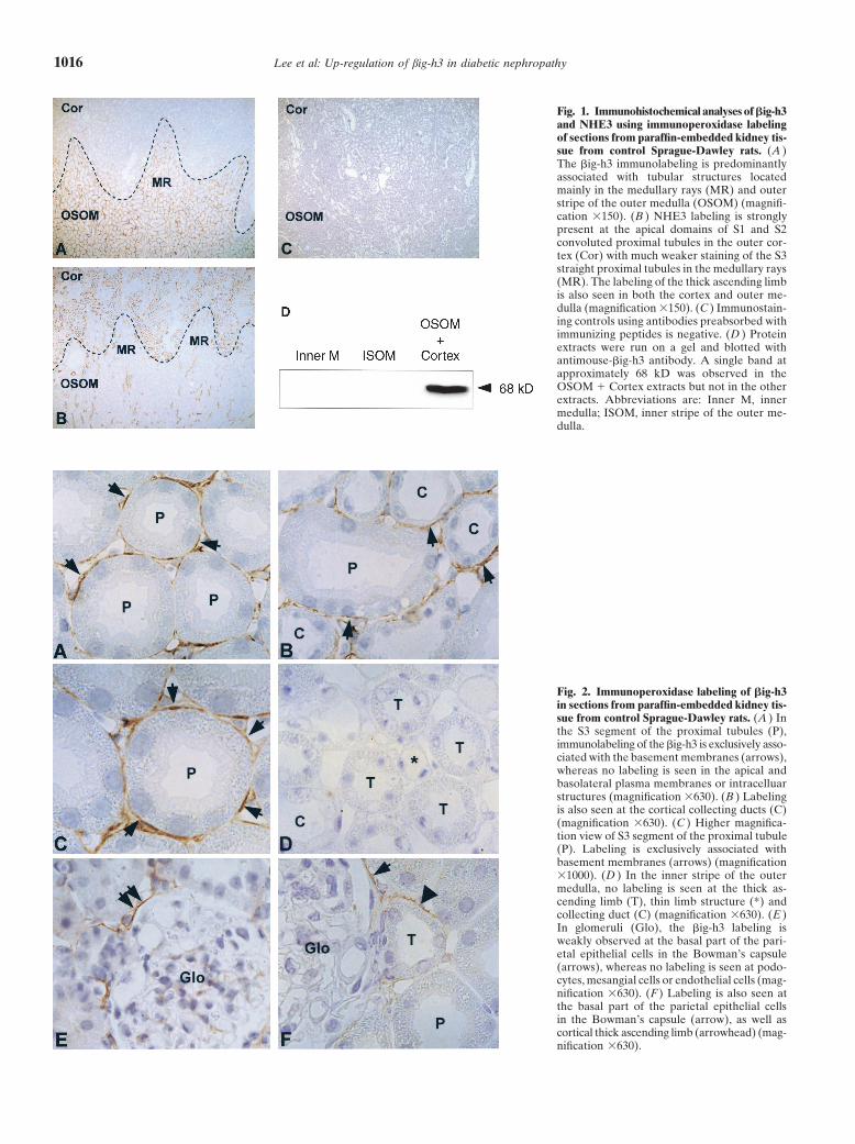
Immunoperoxidase microscopy revealed that �ig-h3labeling in control rat kidney was exclusively present atthe inner cortex and the outer stripe of the outer medulla(Fig. 1A). With higher magnification view of these zones,the most prominent labeling was observed at the proxi-mal tubule cells (Fig. 2A, arrows) in the medullary rays(Fig. 1A) where S3 straight proximal tubule segmentsare mainly present. In the S3 straight proximal tubule,immunolabeling of the �ig-h3 was associated with thebasement membranes (Fig. 2 A and C, arrows), whereasno labeling was seen in the apical and basolateral plasma
membranes or intracelluar structures (Fig. 2 A and C).In contrast, no or very weak labeling was seen in the S1and S2 segment of the proximal tubules (Fig. 2 E andF). The selective immunolocalization in the S3 segmentof the proximal tubule was further demonstrated by acontrol study of type 3 Na-H exchanger (NHE3) labelingin rat kidney (Fig. 1B). In contrast to the �ig-h3 labelingin the medullary rays (mainly S3 segment), NHE3 wasstrongly present at the apical domains of S1 and S2 con-voluted proximal tubules with much weaker staining ofthe S3 straight proximal tubule in the medullary ray (Fig.1B), as previously described [18]. Thus, this indicates theselective immunolocalization of �ig-h3 in the S3 segmentof the proximal tubule. In the inner cortex and the outerstripe of the outer medulla, weak labeling was also associ-ated with the basement membranes of the connectingtubules and collecting ducts (Fig. 2B). However, the la-beling in the collecting duct was not seen in the innerstripe of outer medulla and inner medulla. In the innerstripe of the outer medulla, no labeling was observed atthe thick ascending limb (Fig. 2D), thin limb structure(Fig. 2D) and collecting duct (Fig. 2D). In glomeruli, the�ig-h3 labeling was weakly observed at the basal part ofthe parietal epithelial cells in the Bowman’s capsule (Fig.2E and Fig. 2F, arrows), whereas podocytes, mesangialcells, or endothelial cells were unlabeled. Labeling isalso seen at the basement membrane of the cortical thickascending limb (Fig. 2F, arrowhead). Immunolabelingcontrols using antibodies preabsorbed with immunizingpeptides were negative (Fig. 1C).
Western blotting was performed using extracts fromthree different kidney zones from normal rat kidneys,namely, cortex � outer stripe of the outer medulla, innerstripe of the outer medulla, and inner medulla. A singleband with the predicted molecular mass of �ig-h3 (68 kD)was detected only in cortex � outer stripe of the outermedulla extracts (Fig. 1D). This is consistent with theimmunoperoxidase microscopy experiments that revealedthat �ig-h3 protein occurs in the basement membranesof the proximal tubular epithelial cells (mainly S3 seg-ments), cortical thick ascending limb cells, and parietalepithelial cells of Bowman’s capsule.
�ig-h3 protein levels in kidneys and urine aremarkedly increased in diabetic rats
As expected, compared to control rats, rats with STZ-induced diabetes mellitus had high blood glucose levelsand altered renal functions. For example, rats examined5, 10, or 15 days after STZ injection had a significantlyelevated urine output, water intake, and glomerular fil-tration rate (GFR) as measured by creatinine clearance(Table 1). This is consistent with previous observationson the early phase of diabetes mellitus [19–21]. The abun-dance of �ig-h3 protein in the kidneys was measured byWestern blotting analysis and quantitated by Scion Im-age (Scion Corporation, Frederick, MD, USA). Theabundance of �-tubulin was also measured as a internal
Lee et al: Up-regulation of big-h3 in diabetic nephropathy1016
Fig. 1. Immunohistochemical analyses of �ig-h3and NHE3 using immunoperoxidase labelingof sections from paraffin-embedded kidney tis-sue from control Sprague-Dawley rats. (A )The �ig-h3 immunolabeling is predominantlyassociated with tubular structures locatedmainly in the medullary rays (MR) and outerstripe of the outer medulla (OSOM) (magnifi-cation �150). (B ) NHE3 labeling is stronglypresent at the apical domains of S1 and S2convoluted proximal tubules in the outer cor-tex (Cor) with much weaker staining of the S3straight proximal tubules in the medullary rays(MR). The labeling of the thick ascending limbis also seen in both the cortex and outer me-dulla (magnification �150). (C ) Immunostain-ing controls using antibodies preabsorbed withimmunizing peptides is negative. (D ) Proteinextracts were run on a gel and blotted withantimouse-�ig-h3 antibody. A single band atapproximately 68 kD was observed in theOSOM � Cortex extracts but not in the otherextracts. Abbreviations are: Inner M, innermedulla; ISOM, inner stripe of the outer me-dulla.
Fig. 2. Immunoperoxidase labeling of �ig-h3in sections from paraffin-embedded kidney tis-sue from control Sprague-Dawley rats. (A ) Inthe S3 segment of the proximal tubules (P),immunolabeling of the �ig-h3 is exclusively asso-ciated with the basement membranes (arrows),whereas no labeling is seen in the apical andbasolateral plasma membranes or intracelluarstructures (magnification �630). (B ) Labelingis also seen at the cortical collecting ducts (C)(magnification �630). (C ) Higher magnifica-tion view of S3 segment of the proximal tubule(P). Labeling is exclusively associated withbasement membranes (arrows) (magnification�1000). (D ) In the inner stripe of the outermedulla, no labeling is seen at the thick as-cending limb (T), thin limb structure (*) andcollecting duct (C) (magnification �630). (E )In glomeruli (Glo), the �ig-h3 labeling isweakly observed at the basal part of the pari-etal epithelial cells in the Bowman’s capsule(arrows), whereas no labeling is seen at podo-cytes, mesangial cells or endothelial cells (mag-nification �630). (F ) Labeling is also seen atthe basal part of the parietal epithelial cellsin the Bowman’s capsule (arrow), as well ascortical thick ascending limb (arrowhead) (mag-nification �630).
Lee et al: Up-regulation of big-h3 in diabetic nephropathy 1017
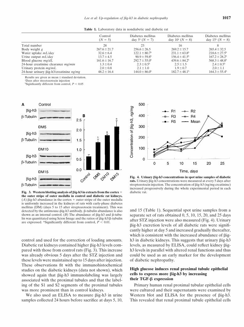
Table 1. Laboratory data in nondiabetic and diabetic rat
Control Diabetes mellitus Diabetes mellitus Diabetes mellitus(N � 5) day 5a (N � 7) day 10a (N � 8) day 15a (N � 8)
Total number 28 23 16 8Body weight g 267.6�21.7 256.6�26.5 269.2�15.7 265.4�32.5Water uptake mL/day 32.6�6.4 122.1�80.7b 231.1 �63.8b 218.6�27.5b
Urine output mL/day 13.7�4.5 90.9�59.8b 156.4 �41.5b 167.2�28.2b
Blood glucose mg/dL 161.6�16.7 292.7�55.0b 439.6 �84.2b 568.3�48.8b
24-hour creatinine clearance mg/min 1.3�0.4 2.3�0.5b 2.5 �1.5 2.4�0.5b
Urinary protein mg/mL 2.0�0.8 2.1�1.0 1.9�0.7 2.0�1.124-hour urinary �ig-h3/creatinine ng/mg 48.2�16.4 144.0�86.0b 182.7 �48.1b 164.3�55.4b
Results are given as mean � standard deviation.aDays after streetozotocin injectionbSignificantly different from control, P 0.05
Fig. 3. Western blotting analysis of �ig-h3 in extracts from the cortex �the outer stripe of outer medulla in control and diabetic rat kidneys.(A) �ig-h3 abundance in the cortex � outer stripe of the outer medullais uniformly increased in the kidneys of rats with early-phase diabetesmellitus (DM) (days 5 to 15 after streptozotocin treatment). This wasdetected by the antimouse-�ig-h3 antibody. �-tubulin abundance is alsoshown as an internal control. (B) The abundance of �ig-h3 and �-tubu-lin was quantitated using Scion Image and the ratios of �ig-h3/�-tubulinare expressed. *Significantly different from control, P 0.01.
control and used for the correction of loading amounts.Diabetic rat kidneys contained higher �ig-h3 levels com-pared with those from control rats (Fig. 3). This increasewas already obvious 5 days after the STZ injection andthese levels were maintained up to 15 days after injection.These observations fit with the immunohistochemicalstudies on the diabetic kidneys (data not shown), whichshowed again that �ig-h3 immunolabeling was largelyassociated with the proximal tubules and that the label-ing of the S1 and S2 segments of the proximal tubuleswas more prominent than in control kidneys.
We also used an ELISA to measure �ig-h3 in urinesamples collected 24 hours before sacrifice at days 5, 10,
Fig. 4. Urinary �ig-h3 concentrations in spot urine samples of diabeticrats. Urinary �ig-h3 concentrations were measured at every 5 days afterstreptozotocin injection. The concentration of �ig-h3 (ng/mg creatinine)increased progressively during the whole experimental period in eachdiabetic rat.
and 15 (Table 1). Sequential spot urine samples from aseparate set of rats obtained 0, 5, 10, 15, 20, and 25 daysafter STZ injection were also measured (Fig. 4). Urinary�ig-h3 excretion levels of all diabetic rats were signifi-cantly higher at day 5 and increased gradually thereafter,which is consistent with the increased abundance of �ig-h3 in diabetic kidneys. This suggests that urinary �ig-h3levels, as measured by ELISA, could reflect kidney �ig-h3 levels in parallel with altered renal functions and thuscould be used as an early marker for the developmentof diabetic nephropathy.
High glucose induces renal proximal tubule epithelialcells to express more �ig-h3 by increasingtheir TGF-� expression
Primary human renal proximal tubular epithelial cellswere cultured and their supernatants were examined byWestern blot and ELISA for the presence of �ig-h3.This revealed that renal proximal tubule epithelial cells
Lee et al: Up-regulation of big-h3 in diabetic nephropathy1018
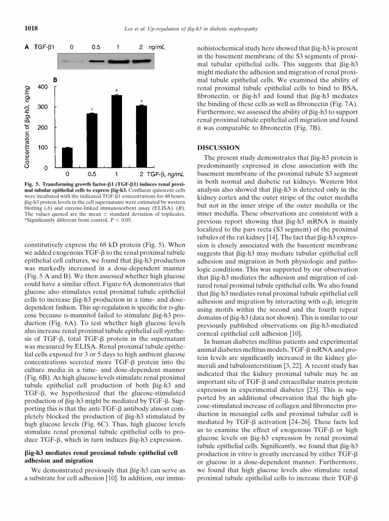
Fig. 5. Transforming growth factor-�1 (TGF-�1) induces renal proxi-mal tubular epithelial cells to express �ig-h3. Confluent quiescent cellswere incubated with the indicated TGF-�1 concentrations for 48 hours.�ig-h3 protein levels in the cell supernatants were estimated by westernblotting (A) and enzyme-linked immunosorbent assay (ELISA). (B).The values quoted are the mean � standard deviation of triplicates.*Significantly different from control, P 0.05.
constitutively express the 68 kD protein (Fig. 5). Whenwe added exogenous TGF-� to the renal proximal tubuleepithelial cell cultures, we found that �ig-h3 productionwas markedly increased in a dose-dependent manner(Fig. 5 A and B). We then assessed whether high glucosecould have a similar effect. Figure 6A demonstrates thatglucose also stimulates renal proximal tubule epithelialcells to increase �ig-h3 production in a time- and dose-dependent fashion. This up-regulation is specific for d-glu-cose because d-mannitol failed to stimulate �ig-h3 pro-duction (Fig. 6A). To test whether high glucose levelsalso increase renal proximal tubule epithelial cell synthe-sis of TGF-�, total TGF-� protein in the supernatantwas measured by ELISA. Renal proximal tubule epithe-lial cells exposed for 3 or 5 days to high ambient glucoseconcentrations secreted more TGF-� protein into theculture media in a time- and dose-dependent manner(Fig. 6B). As high glucose levels stimulate renal proximaltubule epithelial cell production of both �ig-h3 andTGF-�, we hypothesized that the glucose-stimulatedproduction of �ig-h3 might be mediated by TGF-�. Sup-porting this is that the anti-TGF-� antibody almost com-pletely blocked the production of �ig-h3 stimulated byhigh glucose levels (Fig. 6C). Thus, high glucose levelsstimulate renal proximal tubule epithelial cells to pro-duce TGF-�, which in turn induces �ig-h3 expression.
�ig-h3 mediates renal proximal tubule epithelial celladhesion and migration
We demonstrated previously that �ig-h3 can serve asa substrate for cell adhesion [10]. In addition, our immu-
nohistochemical study here showed that �ig-h3 is presentin the basement membrane of the S3 segments of proxi-mal tubular epithelial cells. This suggests that �ig-h3might mediate the adhesion and migration of renal proxi-mal tubule epithelial cells. We examined the ability ofrenal proximal tubule epithelial cells to bind to BSA,fibronectin, or �ig-h3 and found that �ig-h3 mediatesthe binding of these cells as well as fibronectin (Fig. 7A).Furthermore, we assessed the ability of �ig-h3 to supportrenal proximal tubule epithelial cell migration and foundit was comparable to fibronectin (Fig. 7B).
DISCUSSION
The present study demonstrates that �ig-h3 protein ispredominantly expressed in close association with thebasement membrane of the proximal tubule S3 segmentin both normal and diabetic rat kidneys. Western blotanalysis also showed that �ig-h3 is detected only in thekidney cortex and the outer stripe of the outer medullabut not in the inner stripe of the outer medulla or theinner medulla. These observations are consistent with aprevious report showing that �ig-h3 mRNA is mainlylocalized to the pars recta (S3 segment) of the proximaltubules of the rat kidney [14]. The fact that �ig-h3 expres-sion is closely associated with the basement membranesuggests that �ig-h3 may mediate tubular epithelial celladhesion and migration in both physiologic and patho-logic conditions. This was supported by our observationthat �ig-h3 mediates the adhesion and migration of cul-tured renal proximal tubule epithelial cells. We also foundthat �ig-h3 mediates renal proximal tubule epithelial celladhesion and migration by interacting with �3�1 integrinusing motifs within the second and the fourth repeatdomains of �ig-h3 (data not shown). This is similar to ourpreviously published observations on �ig-h3-mediatedcorneal epithelial cell adhesion [10].
In human diabetes mellitus patients and experimentalanimal diabetes mellitus models, TGF-� mRNA and pro-tein levels are significantly increased in the kidney glo-meruli and tubulointerstitium [3, 22]. A recent study hasindicated that the kidney proximal tubule may be animportant site of TGF-� and extracellular matrix proteinexpression in experimental diabetes [23]. This is sup-ported by an additional observation that the high glu-cose-stimulated increase of collagen and fibronectin pro-duction in mesangial cells and proximal tubular cell ismediated by TGF-� activation [24–26]. These facts ledus to examine the effect of exogenous TGF-� or highglucose levels on �ig-h3 expression by renal proximaltubule epithelial cells. Significantly, we found that �ig-h3production in vitro is greatly increased by either TGF-�or glucose in a dose-dependent manner. Furthermore,we found that high glucose levels also stimulate renalproximal tubule epithelial cells to increase their TGF-�
Lee et al: Up-regulation of big-h3 in diabetic nephropathy 1019
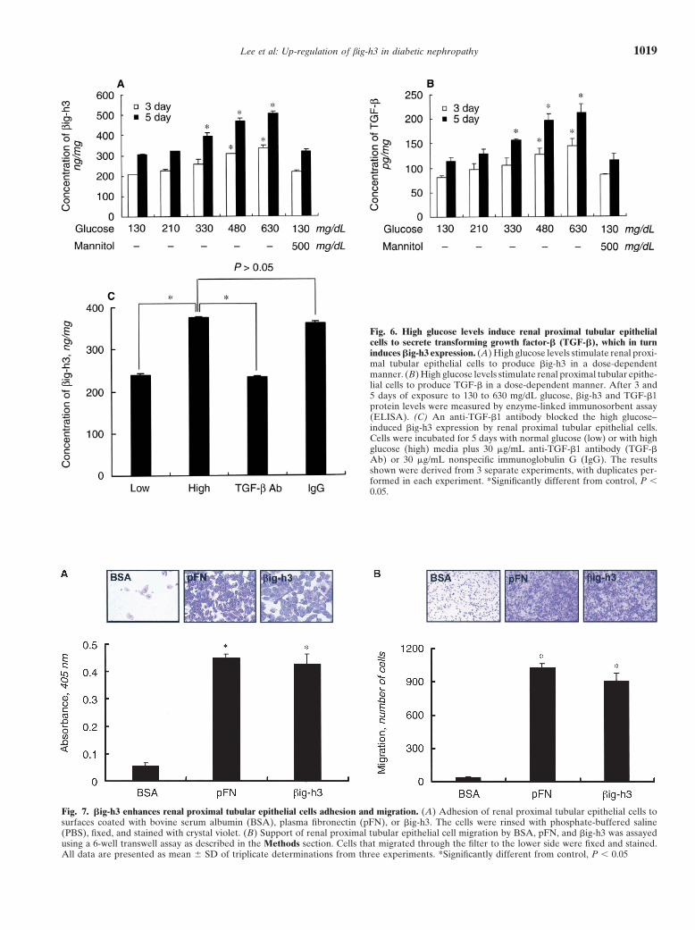
Fig. 6. High glucose levels induce renal proximal tubular epithelialcells to secrete transforming growth factor-� (TGF-�), which in turninduces �ig-h3 expression. (A) High glucose levels stimulate renal proxi-mal tubular epithelial cells to produce �ig-h3 in a dose-dependentmanner. (B) High glucose levels stimulate renal proximal tubular epithe-lial cells to produce TGF-� in a dose-dependent manner. After 3 and5 days of exposure to 130 to 630 mg/dL glucose, �ig-h3 and TGF-�1protein levels were measured by enzyme-linked immunosorbent assay(ELISA). (C) An anti-TGF-�1 antibody blocked the high glucose–induced �ig-h3 expression by renal proximal tubular epithelial cells.Cells were incubated for 5 days with normal glucose (low) or with highglucose (high) media plus 30 �g/mL anti-TGF-�1 antibody (TGF-�Ab) or 30 �g/mL nonspecific immunoglobulin G (IgG). The resultsshown were derived from 3 separate experiments, with duplicates per-formed in each experiment. *Significantly different from control, P 0.05.
Fig. 7. �ig-h3 enhances renal proximal tubular epithelial cells adhesion and migration. (A) Adhesion of renal proximal tubular epithelial cells tosurfaces coated with bovine serum albumin (BSA), plasma fibronectin (pFN), or �ig-h3. The cells were rinsed with phosphate-buffered saline(PBS), fixed, and stained with crystal violet. (B) Support of renal proximal tubular epithelial cell migration by BSA, pFN, and �ig-h3 was assayedusing a 6-well transwell assay as described in the Methods section. Cells that migrated through the filter to the lower side were fixed and stained.All data are presented as mean � SD of triplicate determinations from three experiments. *Significantly different from control, P 0.05
Lee et al: Up-regulation of big-h3 in diabetic nephropathy1020
production, and that addition of an anti-TGF-� antibodyto high glucose–stimulated renal proximal tubule epithe-lial cells almost completely blocks �ig-h3 expression.These results strongly indicate that high glucose levelsincrease �ig-h3 production by renal proximal tubule epi-thelial cells by activating TGF-�.
Our in vivo experiments also showed that �ig-h3 ex-pression is significantly increased in diabetic kidneys.Our observations support the notion that in diabetes,high glucose levels lead to the production of TGF-�by the proximal tubule [23], which then induces localexpression of �ig-h3. How could such increased �ig-h3expression have pathologic consequences? As it is nowevident that a progressive decline in renal function indiabetes mellitus is closely correlated with the degree ofrenal interstitial fibrosis [27, 28], it is possible that �ig-h3 might be critical in promoting such renal fibrosis.Consistent with this are our observations that �ig-h3 hasa fibrillar structure and interacts with several extracellu-lar matrix proteins, including type I collagen, fibronectin,and laminin [11]. Furthermore, �ig-h3 is known to beassociated with extracellular matrix proteins in manydifferent tissues [29, 30]. In addition, �ig-h3 is known tobe produced by fibroblasts and to mediate fibroblastadhesion [31]. However, it is unclear why �ig-h3 expres-sion is restricted to only the proximal tubules. The physi-ologic significance of the restricted expression remainsto be investigated.
Early changes in incipient diabetic kidney disease arecharacterized by an increase in kidney size, glomerularvolume, and enhanced GFR, and later by the develop-ment of mesangial cell proliferation, accumulation ofglomerular extracellular matrix, increased urinary albu-min excretion, glomerular sclerosis, and tubular fibrosis.The subsequent overt diabetic nephropathy is clinicallycharacterized by proteinuria, hypertension, and progres-sive renal insufficiency [20, 32–34]. Naturally, studiessearching for the pathogenic mechanisms responsible forthe onset of diabetic kidney disease have focused on theearly events. Recent studies have suggested that TGF-�might initiate the early diabetic renal changes and thatdownstream molecules triggered by TGF-� might be crit-ically involved in the development of diabetic kidneydisease [35, 36]. For this reason, urinary TGF-�1 hasbeen suggested to be an early marker for diabetic ne-phropathy [7, 37, 38]. We propose here that urinary �ig-h3 levels may also be an early marker as �ig-h3 is aTGF-�–induced gene product whose levels could be adirect index of TGF-� bioactivity [14]. This is supportedby our observations that �ig-h3 expression is up-regu-lated by high glucose and TGF-� levels and that early indiabetic nephropathy, renal �ig-h3 abundance increasesmarkedly. These observations led us to test whether uri-nary �ig-h3 concentrations could be early indicators ofdiabetic nephropathy. Indeed, we found that urinary �ig-
h3 concentrations increased significantly as early as 5days after the induction of diabetes. Thereafter, urinary�ig-h3 concentrations gradually increased further andwere maintained at high levels during the experimentalperiod (25 days). Since renal �ig-h3 abundance followsan identical pattern, the increased urinary �ig-h3 levelsappear to reflect the �ig-h3 levels in the kidney.
That �ig-h3 is mainly localized to its site of synthesisin the proximal tubular epithelium (particularly, in theS3 segment) suggests that changes in urinary �ig-h3 con-centrations during diabetic onset might reflect tubulardamage rather than glomerular abnormalities. Micro-albuminuria levels have been widely accepted as indicat-ing an increased risk of developing diabetic nephropathy.However, concerns have been raised that the prognosticvalue of microalbuminuria levels is not accurate enoughfor this marker to be used in clinical decision making orin the design of certain clinical trials [39]. In addition,there have been reports that tubular involvement mayprecede glomerular involvement in diabetic nephropa-thy [7, 40]. Thus, to detect the early stage of diabeticnephropathy and to monitor the disease progression,it is desirable to have markers that represent tubulardysfunction as well as measuring urinary albumin excre-tion. We propose that urinary �ig-h3 concentrations maybe such an early marker for diabetic nephropathy. Atthe very least, it could be useful as an index of TGF-�bioactivity in the kidney. These possibilities indicate thatit is warranted to test the clinical significance of urinary�ig-h3 concentrations in human patients.
ACKNOWLEDGMENT
This work was supported by a program of National Research Labo-ratory (M10104000036-01J0000-01610).
Reprint requests to In-San Kim, M.D., Ph.D., Department of Bio-chemistry, School of Medicine, Kyungpook National University, Taegu700-422, South Korea.E-mail: [email protected]
REFERENCES
1. Sharma K, Ziyadeh FN: Hyperglycemia and diabetic kidney dis-ease. The case for transforming growth factor-beta as a key media-tor. Diabetes 44:1139–1146, 1995
2. Wolf G, Ziyadeh FN: Molecular mechanisms of diabetic renalhypertrophy. Kidney Int 56:393–405, 1999
3. Yamamoto T, Nakamura T, Noble NA, et al: Expression of trans-forming growth factor beta is elevated in human and experimentaldiabetic nephropathy. Proc Natl Acad Sci USA 90:1814–1818, 1993
4. Sharma K, Ziyadeh FN, Alzahabi B, et al: Increased renal produc-tion of transforming growth factor-beta1 in patients with type IIdiabetes. Diabetes 46:854–859, 1997
5. Sharma K, Jin Y, Guo J, Ziyadeh FN: Neutralization of TGF-beta by anti-TGF-beta antibody attenuates kidney hypertrophyand the enhanced extracellular matrix gene expression in STZ-induced diabetic mice. Diabetes 45:522–530, 1996
6. Hill C, Flyvbjerg A, Rasch R, et al: Transforming growth factor-beta2 antibody attenuates fibrosis in the experimental diabetic ratkidney. J Endocrinol 170:647–651, 2001
7. Ellis D, Forrest KY, Erbey J, Orchard TJ: Urinary measurement
Lee et al: Up-regulation of big-h3 in diabetic nephropathy 1021
of transforming growth factor-beta and type IV collagen as newmarkers of renal injury: Application in diabetic nephropathy. ClinChem 44:950–956, 1998
8. Skonier J, Bennett K, Rothwell V, et al: Beta ig-h3: A trans-forming growth factor-beta-responsive gene encoding a secretedprotein that inhibits cell attachment in vitro and suppresses thegrowth of CHO cells in nude mice. DNA Cell Biol 13:571–584,1994
9. Zinn K, McAllister L, Goodman CS: Sequence analysis andneuronal expression of fasciclin I in grasshopper and Drosophila.Cell 53:577–587, 1988
10. Kim JE, Kim SJ, Lee BH, et al: Identification of motifs for celladhesion within the repeated domains of transforming growth fac-tor-beta-induced gene, beta ig-h3. J Biol Chem 275:30907–30915,2000
11. Kim JE, Park RW, Choi JY, et al: Molecular properties of wild-type and mutant betaIG-H3 proteins. Invest Ophthalmol Vis Sci43:656–661, 2002
12. Dieudonn SC, Kerr JM, Xu T, et al: Differential display of humanmarrow stromal cells reveals unique mRNA expression patternsin response to dexamethasone. J Cell Biochem 76:231–243, 1999
13. Kim JE, Kim EH, Han EH, et al: A TGF-beta-inducible cell adhe-sion molecule, betaig-h3, is downregulated in melorheostosis andinvolved in osteogenesis. J Cell Biochem 77:169–178, 2000
14. Gilbert RE, Wilkinson-Berka JL, Johnson DW, et al: Renalexpression of transforming growth factor-beta inducible gene-h3(beta ig-h3) in normal and diabetic rats. Kidney Int 54:1052–1062,1998
15. Langham RG, Egan MK, Dowling JP, et al: Transforming growthfactor-beta1 and tumor growth factor-beta-inducible gene-H3 innonrenal transplant cyclosporine nephropathy. Transplantation72:1826–1829, 2001
16. Lee EH, Seomun Y, Hwang KH, et al: Overexpression of thetransforming growth factor-beta-inducible gene beta ig-h3 in ante-rior polar cataracts. Invest Ophthalmol Vis Sci 41:1840–1845, 2000
17. Wang W, Kwon TH, Li C, et al: Reduced expression of Na-K-2Cl cotransporter in medullary TAL in vitamin D-induced hyper-calcemia in rats. Am J Physiol Renal Physiol 282:F34–F44, 2002
18. Kwon TH, Frokiaer J, Fernandez-Llama P, et al: Altered expres-sion of Na transporters NHE-3, NaPi-II, Na-K-ATPase, BSC-1,and TSC in CRF rat kidneys. Am J Physiol 277:F257–F270, 1999
19. Bohle A, Mackensen-Haen S, von Gise H: Importance of tubulo-epithelial changes in glomerular excretory and urine concentratingfunction of the kidney. Zentralbl Allg Pathol 132:351–363, 1986
20. Deckert T, Feldt-Rasmussen B, Borch-Johnsen K, et al: Naturalhistory of diabetic complications: early detection and progression.Diabet Med 8:S33–37, 1991
21. Nejsum LN, Kwon TH, Marples D, et al: Compensatory increasein AQP2, p-AQP2, and AQP3 expression in rats with diabetesmellitus. Am J Physiol Renal Physiol 280:F715–726, 2001
22. Sharma K, Ziyadeh FN: Renal hypertrophy is associated withupregulation of TGF-beta 1 gene expression in diabetic BB ratand NOD mouse. Am J Physiol 267:F1094–F1101, 1994
23. Ando T, Okuda S, Tamaki K, et al: Localization of transforming
growth factor-beta and latent transforming growth factor-betabinding protein in rat kidney. Kidney Int 47:733–739, 1995
24. Ziyadeh FN, Sharma K, Ericksen M, Wolf G: Stimulation ofcollagen gene expression and protein synthesis in murine mesangialcells by high glucose is mediated by autocrine activation of trans-forming growth factor-beta. J Clin Invest 93:536–542, 1994
25. Ziyadeh FN: Role of transforming growth factor beta in diabeticnephropathy. Exp Nephrol 2:137, 1994
26. Phillips AO, Steadman R, Morrisey K, et al: Exposure of humanrenal proximal tubular cells to glucose leads to accumulation oftype IV collagen and fibronectin by decreased degradation. KidneyInt 52:973–984, 1997
27. Lane PH, Steffes MW, Fioretto P, Mauer SM: Renal interstitialexpansion in insulin-dependent diabetes mellitus. Kidney Int43:661–667, 1993
28. Nath KA: Tubulointerstitial changes as a major determinant inthe progression of renal damage. Am J Kidney Dis 20:1–17, 1992
29. Kawamoto T, Noshiro M, Shen M, et al: Structural and phyloge-netic analyses of RGD-CAP/beta ig-h3, a fasciclin-like adhesionprotein expressed in chick chondrocytes. Biochim Biophys Acta1395:288–292, 1998
30. Rawe IM, Zhan Q, Burrows R, et al: Beta-ig. Molecular cloningand in situ hybridization in corneal tissues. Invest Ophthalmol VisSci 38:893–900, 1997
31. LeBaron RG, Bezverkov KI, Zimber MP, et al: Beta IG-H3, anovel secretory protein inducible by transforming growth factor-beta, is present in normal skin and promotes the adhesion andspreading of dermal fibroblasts in vitro. J Invest Dermatol 104:844–849, 1995
32. Deckert T, Poulsen JE, Larsen M: Prognosis of diabetics withdiabetes onset before the age of thirty-one. II. Factors influencingthe prognosis. Diabetologia 14:371–377, 1978
33. Deckert T, Poulsen JE, Larsen M: Prognosis of diabetics withdiabetes onset before the age of thirty-one. I. Survival, causes ofdeath, and complications. Diabetologia 14:363–370, 1978
34. Flyvbjerg A, Kessler U, Dorka B, et al: Transient increase inrenal insulin-like growth factor binding proteins during initial kid-ney hypertrophy in experimental diabetes in rats. Diabetologia35:589–593, 1992
35. Park IS, Kiyomoto H, Abboud SL, Abboud HE: Expression oftransforming growth factor-beta and type IV collagen in earlystreptozotocin-induced diabetes. Diabetes 46:473–480, 1997
36. Reeves WB, Andreoli TE: Transforming growth factor beta con-tributes to progressive diabetic nephropathy. Proc Natl Acad SciUSA 97:7667–7669, 2000
37. Sato H, Iwano M, Akai Y, et al: Increased excretion of urinarytransforming growth factor beta 1 in patients with diabetic ne-phropathy. Am J Nephrol 18:490–494, 1998
38. Yang CW, Vlassara H, Peten EP, et al: Advanced glycation endproducts up-regulate gene expression found in diabetic glomerulardisease. Proc Natl Acad Sci USA 91:9436–9440, 1994
39. Caramori ML, Fioretto P, Mauer M: The need for early predictorsof diabetic nephropathy risk: Is albumin excretion rate sufficient?Diabetes 49:1399–1408, 2000
40. Lapsley M, Flynn FV, Sansom PA: Beta 2-glycoprotein-1 (apoli-poprotein H) excretion and renal tubular malfunction in diabeticpatients without clinical proteinuria. J Clin Pathol 46:465–469, 1993