Diagnosis of Power Transformer Depending on Dissolved Gas ...
Upload
independentCategory
view
3download
0
Cellular Microbiology (2006)
8
(7), 1134–1146 doi:10.1111/j.1462-5822.2006.00697.xFirst published online 24 February 2006
© 2006 The AuthorsJournal compilation © 2006 Blackwell Publishing Ltd
Blackwell Publishing Ltd
Oxford, UK
CMICellular Microbiology 1462-5814© 2006 The Authors; Journal compilation © 2006 Blackwell Publishing Ltd
? 2006
8
711341146
Original Article
CD81 and malaria sporozoite host cell specificityO. Silvie
et al.
Received 19 October, 2005; revised 24 November, 2005; accepted 9December, 2005. *For correspondence. E-mail [email protected];Tel. (
+
33) 1 4077 9736; Fax (
+
33) 1 4583 8858.
Expression of human CD81 differently affects host cell susceptibility to malaria sporozoites depending on the
Plasmodium
species
Olivier Silvie,
1
* Céline Greco,
2
Jean-François Franetich,
1
Anne Dubart-Kupperschmitt,
3
Laurent Hannoun,
4
Geert-Jan van Gemert,
5
Robert W. Sauerwein,
5
Shoshana Levy,
6
Claude Boucheix,
2
Eric Rubinstein
2
and Dominique Mazier
1
1
Inserm, U511, F-75013 Paris, France; Université Pierre et Marie Curie-Paris 6, Faculté de Médecine Pitié-Salpêtrière, F-75013 Paris, France.
2
Inserm, U602, F-94807 Villejuif, France; Institut André Lwoff, IFR 89, F-94807 Villejuif, France; Université Paris-Sud 11, F-94807 Villejuif, France.
3
Institut Cochin, Département d’Hématologie, F-75014 Paris, France; Inserm, U567, F-75014 Paris, France; CNRS UMR 8104, F-75014 Paris, France; Université Paris Descartes-Paris 5, Faculté de Médecine René Descartes, UMR-S 8104, F-75014 Paris, France; AP HP, Centre Hospitalo-Universitaire Cochin-Port Royal, Maternité de Port-Royal, F-75014 Paris, France.
4
AP HP, Centre Hospitalo-Universitaire Pitié-Salpêtrière, Service de Chirurgie Digestive, Hépato-Bilio-Pancréatique et Transplantation Hépatique, F-75013 Paris, France; Université Pierre et Marie Curie-Paris 6, Faculté de Médecine Pitié-Salpêtrière, F-75013 Paris, France.
5
University Medical Center St Radboud, Departmentof Medical Microbiology, 6500HB Nijmegen, the Netherlands.
6
Division of Oncology, Department of Medicine, Stanford University Medical Center, Stanford, CA, USA.
Summary
Plasmodium
sporozoites can enter host cells by twodistinct pathways, either through disruption of theplasma membrane followed by parasite transmigra-tion through cells, or by formation of a parasito-phorous vacuole (PV) where the parasite furtherdifferentiates into a replicative exo-erythrocytic form(EEF). We now provide evidence that following inva-sion without PV formation, transmigrating
Plasmo-
dium falciparum
and
Plasmodium yoelii
sporozoitescan partially develop into EEFs inside hepatocarci-noma cell nuclei. We also found that rodent
P. yoelii
sporozoites can infect both mouse and human hepa-tocytes, while human
P. falciparum
sporozoites infecthuman but not mouse hepatocytes. We have previ-ously reported that the host tetraspanin CD81 isrequired for PV formation by
P. falciparum
and
P.yoelii
sporozoites. Here we show that expression ofhuman CD81 in CD81-knockout mouse hepatocytes issufficient to confer susceptibility to
P. yoelii
but not
P. falciparum
sporozoite infection, showing that thenarrow
P. falciparum
host tropism does not rely onCD81 only. Also, expression of CD81 in a humanhepatocarcinoma cell line is sufficient to promote theformation of a PV by
P. yoelii
but not
P. falciparum
sporozoites. These results highlight critical differ-ences between
P. yoelii
and
P. falciparum
sporozoiteinfection, and suggest that in addition to CD81, othermolecules are specifically required for PV formationduring infection by the human malaria parasite.
Introduction
Malaria remains the most important parasitic human dis-ease, responsible for millions of deaths each year.
Plas-modium
infection is initiated by the inoculation ofsporozoites in the host by a female
Anopheles
mosquito.Following transmission by the mosquito, sporozoites enterthe bloodstream and rapidly reach the liver and infecthepatocytes, where they further differentiate into a repli-cative exo-erythrocytic form (EEF) that will ultimately giverise to thousands of merozoites that initiate the patho-genic erythrocytic cycle. Infection of hepatocytes by
Plasmodium
sporozoites involves the formation of a mem-brane-bound compartment called the parasitophorousvacuole (PV), where the parasite resides for further devel-opment. Sporozoites can also enter cells directly withoutPV formation, by disrupting the cell plasma membrane(Mota
et al
., 2001). After gliding through the cell cyto-plasm, the parasites eventually egress by rupturing theplasma membrane again.
One characteristic feature of the cell-traversal mode ofentry is the absence of cell type selectivity, because

CD81 and malaria sporozoite host cell specificity
1135
© 2006 The AuthorsJournal compilation © 2006 Blackwell Publishing Ltd,
Cellular Microbiology
,
8
, 1134–1146
sporozoites from several
Plasmodium
species have beenshown to migrate through various types of cells from var-ious host species
in vitro
(Mota
et al
., 2001). This modeof cell entry reflects the need for the parasite to migratethrough different tissues of the host, first in the skin thenin the liver sinusoids, before it reaches hepatocytes (Bal-dacci and Menard, 2004; Frevert, 2004). Several parasiteproteins have been recently identified which are involvedin this process, including the sporozoite microneme pro-teins essential for cell-traversal (SPECT)-1 and SPECT-2(Ishino
et al
., 2004; 2005; Kaiser
et al
., 2004), and aprotein with a phospholipase activity (Matuschewski
et al
.,2002; Bhanot
et al
., 2005).In contrast with transmigration, infection of host target
cells is a much more restricted process. The PV formationpresumably depends on the antero–posterior transloca-tion of a tight junction formed by sporozoite adhesinsbound to their cognate receptors on the host cell surface(Baldacci and Menard, 2004; Kappe
et al
., 2004). Thenature of the parasite and host components forming thejunction still remains unknown. It is likely that the speci-ficity of molecular interactions between the parasite andthe host cell accounts for the selectivity of host cell typessupporting the development of EEFs. Sporozoites fromthe deadly human parasite
Plasmodium falciparum
onlyinfect hepatocytes from human and a few non-humanprimate species both
in vivo
(Druilhe
et al
., 1982; Meis
etal
., 1990; Zapata
et al
., 2002) and
in vitro
(Mazier
et al
.,1985; Millet
et al
., 1991). There is currently no cell linesupporting
P. falciparum
sporozoite infection and EEFdevelopment, in contrast with
Plasmodium vivax
androdent plasmodial species, which are able to develop notonly in primary hepatocytes (Mazier
et al
., 1982; Mazier
et al
., 1984) but also in hepatoma cell lines (Hollingdale
et al
., 1983; 1985). In particular, sporozoites from therodent parasite
Plasmodium yoelii
can infect mousehepatoma cell lines (Mota and Rodriguez, 2000), whileanother rodent parasite,
Plasmodium berghei
, can infectnot only mouse but also human hepatoma cells, such asthe human hepatocarcinoma cell line HepG2 (Hollingdale
et al
., 1983; Mota and Rodriguez, 2000).The narrow host cell tropism of
P. falciparum
sporozoi-tes could be determined by several factors, including sur-face receptors and host molecules required for theintracellular growth of the parasite. We have recentlyreported that the tetraspanin CD81, a receptor for thehepatitis C virus (HCV) (Pileri
et al
., 1998; Lindenbach
etal
., 2005; Wakita
et al
., 2005; Zhong
et al
., 2005), isrequired on hepatocytes for infection by
P. falciparum
and
P. yoelii
sporozoites (Silvie
et al
., 2003). In the absenceof evidence of a direct interaction between CD81 and theparasite, it seems that CD81 does not act directly as areceptor for the sporozoites but is rather involved indi-rectly. Here, we investigated whether CD81 expression
contributes to the host cell tropism during infection by
P.falciparum
and
P. yoelii
sporozoites.
Results
Host specificity of
P. falciparum
and
P. yoelii
sporozoites
in vitro
To gain further insight into host–parasite interactions dur-ing sporozoite infection, we first analysed the host speciesspecificity during infection of hepatocytes by
P. falciparum
and
P. yoelii
sporozoites. Primary hepatocytes isolatedfrom human and mouse liver fragments were inoculatedwith
P. falciparum
or
P. yoelii
sporozoites, and infectionwas monitored by counting EEFs after immunolabelling.As expected, we observed
P. falciparum
EEFs in humanhepatocytes and
P. yoelii
EEFs in mouse hepatocytes(Fig. 1A and B). Interestingly, while
P. falciparum
sporozo-ites failed to infect mouse hepatocytes (Fig. 1A),
P. yoelii
sporozoites efficiently infected human hepatocytes
in vitro
Fig. 1.
P. falciparum
and
P. yoelii
sporozoite host species tropism.A and B. Primary human and mouse hepatocytes were inoculated with 1
×
10
5
P. falciparum
(A) or 3
×
10
4
P. yoelii
sporozoites (B) and incubated for 72 h or 24 h, respectively, before quantification of EEFs in triplicate wells by immunofluorescence. Results are expressed as mean
±
standard deviation.C.
P. yoelii
EEFs (arrowheads) in primary human hepatocyte cultures were labelled 24 and 48 h post infection with anti-HSP70 antibodies (green). Nuclei were stained with DAPI (blue). Bar, 10
µ
m.

1136
O. Silvie
et al.
© 2006 The AuthorsJournal compilation © 2006 Blackwell Publishing Ltd,
Cellular Microbiology
,
8
, 1134–1146
(Fig. 1B), raising EEFs with a similar morphology as seenin mouse hepatocytes. Notably, some parasites displayedtypical features of maturing EEFs 48 h post infection, asevidence by staining with anti-HSP70 antibodies andDAPI (Fig. 1C).
Expression of human CD81 in transgenic mice confers susceptibility to
P. yoelii
but not to
P. falciparum
sporozoite infection
We have previously reported that expression of the tet-raspanin CD81 on hepatocytes is required for infection by
P. falciparum
and
P. yoelii
sporozoites (Silvie
et al
., 2003).To investigate whether CD81 accounts for the host spec-ificity during infection by
P. falciparum
sporozoites, wetested whether expression of human CD81 (hCD81) inmouse hepatocytes could confer susceptibility to infectionby
P. falciparum
and
P. yoelii
sporozoites. Primary hepa-tocytes isolated from hCD81 transgenic mice deficient forendogenous mouse CD81 (Masciopinto
et al
., 2002) wereinoculated with
P. falciparum
and
P. yoelii
sporozoites.While CD81-deficient mouse hepatocytes are completelyrefractory to infection by
P. yoelii
sporozoites (Silvie
et al
.,2003), expression of human CD81 in transgenic micecompletely restored
P. yoelii
sporozoite infectivity
in vitro
(Fig. 2A). As expected, an anti-human CD81 mAb couldblock infection of hCD81 transgenic but not wild-typemouse hepatocytes, while reciprocally an anti-mouseCD81 mAb was effective only in wild-type cells (Fig. 2Aand B). Human CD81 transgenic mice were also fullysusceptible to
P. yoelii sporozoite infection in vivo,because all mice (n = 5) injected intravenously with4 × 103 P. yoelii sporozoites developed a patent blood-stage infection 3 days post injection, similarly to wild-typecontrol mice. These results clearly demonstrate thathuman CD81 expressed in mouse hepatocytes is func-tional as regards to P. yoelii sporozoite infection.
We also attempted to infect hCD81 transgenic mousehepatocytes with P. falciparum sporozoites, but we con-stantly failed in obtaining any P. falciparum EEF in thesecells (data not shown), demonstrating that expression ofhuman CD81 is not sufficient to promote P. falciparumsporozoite infectivity in mouse hepatocytes. Based on thefact that P. falciparum sporozoites infect human but notmouse hepatocytes, and together with the absence ofevidence of a direct interaction between CD81 and theparasite (Silvie et al., 2003), these results suggest thatadditional human molecules may be required during hostcell infection by P. falciparum sporozoites.
Plasmodium yoelii and P. falciparum EEFs develop intranuclearly in hepatocarcinoma cells
Human hepatocarcinoma HepG2 cells have been
widely used as target host cells for P. berghei EEF cul-tures (Hollingdale et al., 1983). HepG2 cells do notexpress CD81 (Berditchevski et al., 1996; Charrin etal., 2001), and it has been reported that they do notsupport EEF development of P. falciparum (Hollingdaleet al., 1984; Mazier et al., 1985) and P. yoelii (Calvo-Calle et al., 1994; Mota and Rodriguez, 2000). Unex-pectedly, when we examined HepG2 cells infected withP. yoelii sporozoites, we constantly found small num-bers of EEFs in these cells. However, while in hepato-cytes EEFs are found in the cytoplasm next to the hostcell nucleus, parasites observed in HepG2 cells(n = 195) were all abnormally located in the infectedcell nucleus. Intranuclear localization was confirmed byconfocal microscopy analysis (Fig. 3). These intranu-clear forms displayed a limited degree of maturation,although most of the parasites had undergone severalrounds of nuclear divisions, as shown by DAPI staining.
Fig. 2. Expression of human CD81 in transgenic mice confers sus-ceptibility to P. yoelii sporozoite infection. Primary hepatocytes iso-lated from a human CD81 transgenic murine CD81-knockout mouse (A) or from a wild-type C57/Bl6 mouse (B) were infected with P. yoelii sporozoites (3 × 10⁴) in the presence of anti-human CD81 mAb TS81 or anti-mouse CD81 mAb MT81, or without mAb as a control, and cultured for 36 h before quantification of EEFs in triplicate wells. Results are expressed as mean ± standard deviation. **P < 0.01 as compared with control.

CD81 and malaria sporozoite host cell specificity 1137
© 2006 The AuthorsJournal compilation © 2006 Blackwell Publishing Ltd, Cellular Microbiology, 8, 1134–1146
Limited numbers of P. falciparum EEFs were alsoobserved in HepG2 cells, and as seen with P. yoeliithese parasites (n = 88) were all located in the infectedcell nucleus (Fig. 3).
The mouse hepatoma cell line Hepa1–6 supports infec-tion and complete development of P. yoelii sporozoites(Mota and Rodriguez, 2000). Hepa1–6 cells expressCD81 (Fig. 4A) and infection by P. yoelii sporozoitesdepends on CD81, as described with primary hepato-cytes (Silvie et al., 2003). Indeed, the knock-down ofmouse CD81 using small interfering RNA (siRNA)(Fig. 4A) reduced the number of infected Hepa1–6 cells(Fig. 4B), while the knock-down of the tetraspanin CD9(Fig. 4A) had no effect on infection (Fig. 4B), consistentwith our previous observation that CD9 is not requiredduring P. yoelii infection (Silvie et al., 2003). Also, anti-mouse CD81 mAbs could efficiently block infection ofHepa1–6 cells by P. yoelii sporozoites (Fig. 4C). Interest-ingly, P. yoelii intranuclear EEFs were also observed inHepa1–6 cells (Fig. 5A). Intranuclear localization of P.yoelii EEFs had not been reported in Hepa1–6 cells
before, although in our hands intranuclear EEFs repre-sented 10–25% of EEFs in these cells (Fig. 5B and C).Interestingly, anti-CD81 antibodies and siRNA targetingCD81 reduced the number of cytoplasmic P. yoelii EEFsbut had no effect on intranuclear EEFs in Hepa1–6 cells(Fig. 5B and C).
Fig. 3. P. yoelii and P. falciparum EEFs develop intranuclearly in HepG2 cells. HepG2 cells were infected with P. yoelii (5 × 10⁴) or P. falciparum (1 × 10⁵) sporozoites and cultured for 36 h or 72 h, respectively, before immunofluorescence labelling with anti-HSP70 antibodies (green) and DAPI (blue) and examination by confocal fluorescence microscopy. EEFs (arrowheads) are clearly located in the infected cell nucleus. Bar, 10 µm.
Fig. 4. Infection of mouse hepatoma Hepa1–6 cells by P. yoelii sporozoites depends on CD81.A. Hepa1–6 cells transfected with siRNA oligonucleotides targeting CD81 (siCD81), CD9 (siCD9) or with a control siRNA (cont) were stained with anti-CD81 mAb Eat1 (left panels) (Maecker et al., 2000) or anti-CD9 mAb 4.1F12 (right panels) (Le Naour et al., 2000) and analysed by flow cytometry.B. P. yoelii EEF number in Hepa1–6 cells transfected with siRNA oligonucleotides targeting CD81 (siCD81) or CD9 (siCD9), or witha control siRNA.C. Effects of anti-mouse CD81 mAbs Eat1 and Eat2 (Maecker et al., 2000) on P. yoelii infection of Hepa1–6 cells.

1138 O. Silvie et al.
© 2006 The AuthorsJournal compilation © 2006 Blackwell Publishing Ltd, Cellular Microbiology, 8, 1134–1146
HepG2 cells genetically engineered to express human CD81 support the complete development of P. yoelii but not P. falciparum EEFs
Because HepG2 cells do not express CD81, we wonderedwhether the exclusively intranuclear localization of P. fal-ciparum and P. yoelii EEFs was due to the lack of CD81.Therefore, we investigated the effects of CD81 expressionin HepG2 cells on infection by the two Plasmodium spe-cies. We tested two different HepG2 cell lines retrovirallytransduced to stably express human CD81. These twoHepG2/CD81 cell lines differ in the level of CD81 surfaceexpression as shown by FACS analysis (Fig. 6A), andthus are here referred to as HepG2/CD81+ and HepG2/CD81++. Confirming previous observations, CD81 was notdetected on the surface of parental HepG2 cells, whereasHepG2/CD81++ cells express ∼4 times more CD81 thanHepG2/CD81+. P. yoelii sporozoites readily infectedHepG2/CD81 cells, rising high numbers of EEFs(Fig. 6B). Furthermore, a vast majority of these EEFswere located in the infected cell cytoplasm, close to the
host cell nucleus, which corresponds to the normal local-ization of EEFs (Fig. 6C). These EEFs were bigger thanthe intranuclear forms seen in HepG2 cells, and displayeda degree of maturation that was comparable to that seenin mouse and human hepatocytes in vitro. Interestingly,the level of infection correlated well with the level ofexpression of CD81, as indicated by the higher number ofEEFs in HepG2/CD81++ as compared with HepG2/CD81+
(Fig. 6B). To test whether infectious merozoites could formin these EEFs, naive BALB/c mice were injected with P.yoelii-infected HepG2/CD81++ cells. Four out of five miceinjected with HepG2/CD81 cultures 48 h post infectiondeveloped blood-stage infections, 4 ± 1 days after injec-tion. This confirms that P. yoelii sporozoites can com-pletely develop in HepG2/CD81 cells, in a way similar tothe growth of P. berghei in HepG2 cells (Hollingdale et al.,1983).
We also analysed the fate of P. falciparum sporozoitesin HepG2/CD81+ and HepG2/CD81++ cells. In sharp con-trast with the very efficient infection of these cells by P.yoelii sporozoites, only limited numbers of EEFs could beobserved (< 25 per well), even in cells expressing thehigher level of CD81, and as seen with the parental CD81-negative HepG2 cells, these EEFs (n = 93) were alllocated in the infected cell nuclei (data not shown). Thisshows that CD81 expression in HepG2 cells is not suffi-cient to permit the development of cytoplasmic P. falci-parum EEFs.
Subcellular localization of Plasmodium EEFs reflects the mode of cell entry of sporozoites
Although most of P. yoelii EEFs were found in the cyto-plasm of infected HepG2/CD81 cells, intranuclear EEFswere also observed in these cells, in relatively small num-bers, and thus representing only a minor fraction of theEEFs cells (2–10%, Fig. 6B). Importantly, the number ofintranuclear EEFs was similar in HepG2/CD81+ andHepG2/CD81++ cells (Fig. 6B). Furthermore, siRNA tar-geting CD81 and anti-CD81 antibodies reduced the num-ber of P. yoelii cytoplasmic EEFs but had no effect on thenumber of intranuclear EEFs in HepG2/CD81 (data notshown), as seen with Hepa1–6 (Fig. 5B and C), confirm-ing that intranuclear localization of P. yoelii EEFs occursindependently of CD81. Intranuclear EEFs were stillobserved 72 h post infection in HepG2 cells, althoughtheir number tended to decrease (Fig. 6D). The numberof EEFs in HepG2/CD81 cells also decreased over time,probably due to the complete maturation of the parasites(Fig. 6E).
Interestingly, when we assessed infection at early timepoints (1–3 h) after inoculation of HepG2/CD81 cells withP. yoelii sporozoites, we could observe not only cytoplas-mic sporozoites but also intranuclear sporozoites, sug-
Fig. 5. P. yoelii EEFs develop intranuclearly in Hepa1–6 cells.A. Hepa1–6 cells were infected with P. yoelii sporozoites (5 × 10⁴) and cultured for 24 h before immunofluorescence labelling with anti-HSP70 antibodies (green) and DAPI (blue), and examination by flu-orescence microscopy. The arrowheads indicate an intracytoplasmic EEF (upper panels), and an intranuclear EEF (lower panels). Bar, 10 µm.B and C. Effects of anti-CD81 mAb MT81 (B) or siRNA targeting mouse CD81 (C) on P. yoelii EEF numbers in Hepa1–6 cells. **P < 0.01 as compared with control.

CD81 and malaria sporozoite host cell specificity 1139
© 2006 The AuthorsJournal compilation © 2006 Blackwell Publishing Ltd, Cellular Microbiology, 8, 1134–1146
gesting that intranuclear development of EEFs resultsfrom the intranuclear localization of sporozoites at thetime of or early after invasion. Sporozoites can enter cellsthrough two distinct pathways, either by disruption of thehost cell plasma membrane, followed by migration of theparasite through the cell, or by formation of a PV, whichis essential for the EEF development in hepatocytes(Mota et al., 2001). We have previously reported thatCD81 is required for invasion through the vacuole path-way, but is dispensable for sporozoite migration throughcells (Silvie et al., 2003). Because intranuclear localiza-tion of P. yoelii sporozoites did not depend on CD81, wehypothesized that these forms may result from the devel-opment of sporozoites arrested in the host cell nucleuswhile they were transmigrating. To directly test thishypothesis, we performed a cell wounding and repairassay using the membrane-impermeant macromoleculartracer dextran (Mota et al., 2001). Disruption and reseal-
ing of the cell membrane, which occurs when sporozoitesmigrate through cells, allows the entry and trapping ofdextran in traversed cells. After infection by P. yoelii sporo-zoites in the presence of rhodamine-labelled dextran, allexamined HepG2 (n = 82) (Fig. 7A) and HepG2/CD81cells (n = 54) (Fig. 7B) harbouring a parasite in theirnucleus were dextran-positive, both at early and latestages of infection, clearly demonstrating that indeedintranuclear localization of P. yoelii parasites results frominvasion of the host cell by traversing sporozoites. Para-sites in dextran-negative cells, resulting from invasion withPV formation, where only found in the cytoplasm ofHepG2/CD81 cells (Fig. 7B, middle and lower panels), butnot in HepG2 cells.
As mentioned above, sporozoites could be observed inthe cytoplasm of both HepG2 and HepG2/CD81 cellsearly after infection. In HepG2/CD81 cells, a majority ofcytoplasmic sporozoites were found in dextran-negative
Fig. 6. Expression of human CD81 in HepG2 cells confers susceptibility to P. yoelii sporozoite infection.A. HepG2, HepG2/CD81+ and HepG2/CD81++ were labelled with the anti-human CD81 mAb TS81 before analysis by flow cytometry. The grey line corresponds to the background fluorescence.B. HepG2, HepG2/CD81+ and HepG2/CD81++ were infected with P. yoelii sporozoites (3 × 10⁴) and incubated for 36 h before quantification of EEFs by immunofluorescence in triplicate wells. To determine the subcellular localization of the EEFs (intracytoplasmic or intranuclear), nuclei were stained with DAPI. Results are expressed as mean ± standard deviation.C. HepG2/CD81++ cells were infected with P. yoelii sporozoites (3 × 10⁴) and cultured for 36 h before immunofluorescence labelling with anti-HSP70 antibodies (green) and DAPI (blue), and examination by confocal fluorescence micros-copy. The arrowheads indicate an EEF, which is clearly located in the infected cell cytoplasm. Bar, 10 µm.D and E. HepG2 cells (D) and HepG2/CD81++ cells (E) were infected with P. yoelii sporozoites (3 × 10⁴) and incubated for 24–72 h before quantification of EEFs by immunofluorescence in triplicate wells. EEFs in HepG2 cells (D) are intranuclear, while EEFs in HepG2/CD81++ cells (E) are cytoplasmic.

1140 O. Silvie et al.
© 2006 The AuthorsJournal compilation © 2006 Blackwell Publishing Ltd, Cellular Microbiology, 8, 1134–1146
cells 1 h (55 ± 4%, n = 30) and 3 h (64 ± 1%, n = 63) postinfection, reflecting sporozoite entry with PV formation. Insharp contrast, essentially all the HepG2 cells harbouringa sporozoite in their cytoplasm were dextran-positive, bothat 1 h (92 ± 9%, n = 26) and 3 h (95 ± 8%, n = 25) postinfection, showing that P. yoelii sporozoites fail to enterHepG2 cells by forming a vacuole. To further determinewhether sporozoites which localized to the host cellnucleus formed a vacuole inside the nucleus, we per-formed immunofluorescence assays using the mAbNYLS3, which recognizes P. yoelii HEP17 protein, a
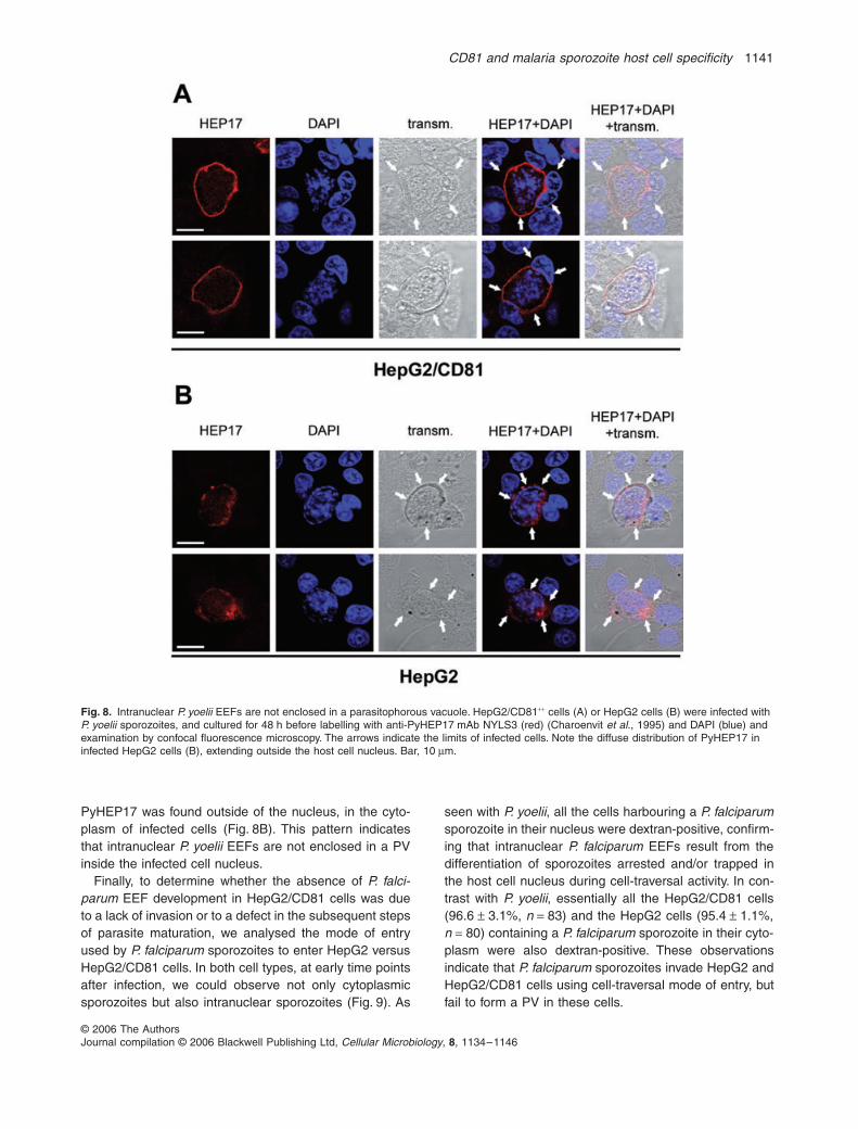
marker for the PV membrane (Charoenvit et al., 1995). Ininfected HepG2/CD81 cells, NYLS3 labelled the peripheryof cytoplasmic EEFs, consistent with PyHEP17 localiza-tion to the PV membrane (Charoenvit et al., 1995)(Fig. 8A). In contrast, PyHEP17 distribution in infectedHepG2 cells was not restricted to the periphery of theintranuclear parasites, as would be expected if theseEEFs were contained in a PV, and was more diffuse.Indeed, while intranuclear EEFs were entirely containedinside the host cell nucleus as determined with the label-ling with anti-HSP70 antibodies (as in Figs 3 and 7A),
Fig. 7. Subcellular localization of P. yoelii EEFs reflects the mode of cell entry of sporozoites.A. HepG2 cells were infected with P. yoelii sporozoites in the presence of rhodamine-dex-tran, and cultured for 12 or 48 h before labelling with anti-HSP70 antibodies (green) and DAPI (blue) and examination by confocal fluores-cence microscopy. The upper panels show a transforming sporozoite (12 h post infection, green) in the nucleus (in blue) of a dextran-positive (red) cell. The lower panels show an EEF (48 h post infection, green) in the nucleus (in blue) of a dextran-positive (red) cell. Bar, 10 µm.B. HepG2/CD81++ cells were infected with P. yoelii sporozoites in the presence of rhodamine-dextran, and cultured for 12 or 48 h before labelling with anti-HSP70 antibodies (green) and DAPI (blue) and examination by confocal fluorescence microscopy. The upper panels show a transforming sporozoite (12 h post infection, green) in the nucleus (in blue) of a dextran-positive (red) cell. The middle panels show a transforming sporozoite (12 h post infection, green) in the cytoplasm of a dextran-negative cell. Note the presence of a dextran-positive cell (red) next to the infected cell. The lower panels show an EEF (48 h post infection, green) in the cytoplasm of a dextran-negative cell. Bar, 10 µm.
CD81 and malaria sporozoite host cell specificity 1141
© 2006 The AuthorsJournal compilation © 2006 Blackwell Publishing Ltd, Cellular Microbiology, 8, 1134–1146
PyHEP17 was found outside of the nucleus, in the cyto-plasm of infected cells (Fig. 8B). This pattern indicatesthat intranuclear P. yoelii EEFs are not enclosed in a PVinside the infected cell nucleus.
Finally, to determine whether the absence of P. falci-parum EEF development in HepG2/CD81 cells was dueto a lack of invasion or to a defect in the subsequent stepsof parasite maturation, we analysed the mode of entryused by P. falciparum sporozoites to enter HepG2 versusHepG2/CD81 cells. In both cell types, at early time pointsafter infection, we could observe not only cytoplasmicsporozoites but also intranuclear sporozoites (Fig. 9). As
seen with P. yoelii, all the cells harbouring a P. falciparumsporozoite in their nucleus were dextran-positive, confirm-ing that intranuclear P. falciparum EEFs result from thedifferentiation of sporozoites arrested and/or trapped inthe host cell nucleus during cell-traversal activity. In con-trast with P. yoelii, essentially all the HepG2/CD81 cells(96.6 ± 3.1%, n = 83) and the HepG2 cells (95.4 ± 1.1%,n = 80) containing a P. falciparum sporozoite in their cyto-plasm were also dextran-positive. These observationsindicate that P. falciparum sporozoites invade HepG2 andHepG2/CD81 cells using cell-traversal mode of entry, butfail to form a PV in these cells.
Fig. 8. Intranuclear P. yoelii EEFs are not enclosed in a parasitophorous vacuole. HepG2/CD81++ cells (A) or HepG2 cells (B) were infected with P. yoelii sporozoites, and cultured for 48 h before labelling with anti-PyHEP17 mAb NYLS3 (red) (Charoenvit et al., 1995) and DAPI (blue) and examination by confocal fluorescence microscopy. The arrows indicate the limits of infected cells. Note the diffuse distribution of PyHEP17 in infected HepG2 cells (B), extending outside the host cell nucleus. Bar, 10 µm.

1142 O. Silvie et al.
© 2006 The AuthorsJournal compilation © 2006 Blackwell Publishing Ltd, Cellular Microbiology, 8, 1134–1146
Discussion
Infection of hepatocytes by Plasmodium sporozoites is aprerequisite for initiation of malaria, and therefore repre-sents an attractive target for antimalarial strategy. Plasmo-dium sporozoites can enter host cells by two distinctpathways, either through disruption of the plasma mem-brane followed by parasite migration through cells, or byformation of a vacuole where the parasite further differen-tiate into an EEF (Mota et al., 2001). Here we provideevidence that host cell invasion by migrating sporozoites,without PV formation, can result in the development ofintranuclear EEFs for both P. yoelii and P. falciparum.Intranuclear EEFs were observed in hepatocytic cell linesbut not in primary hepatocyte cultures. Unlike hepato-cytes, which do not proliferate in vitro, hepatocytic celllines undergo continuous cell divisions in culture, so wehypothesize that sporozoites may readily access the hostcell nucleus during mitosis, upon nuclear envelop break-down. Our data demonstrate that transformation of sporo-
zoites into EEFs can take place independently ofsporozoite–host cell interactions linked to PV formation.These results extend the previous description of P.berghei sporozoite transformation in the absence of hostcells, simply induced upon temperature switch to 37°C inthe presence of serum factors (Kaiser et al., 2003).
Previous studies have reported the intranuclear local-ization and differentiation of P. berghei sporozoites,although the pathway used for cell entry had not beencharacterized (Meis et al., 1984; Suhrbier et al., 1986).Intranuclear P. berghei were found not only in vitro inHepG2 cells but also in vivo in rat Kupffer cells, still theywere not observed in hepatocytes (Meis et al., 1984).Interestingly, as shown by electron microscopy, there wasno evidence of a PV membrane surrounding intranuclearP. berghei sporozoites (Meis et al., 1984). This result isconsistent with our data, which demonstrate that intranu-clear localization of P. yoelii and P. falciparum sporozoitesis linked to invasion without PV formation, as shown bydextran assays and NYLS3 labelling. It is noteworthy thatearly after infection, sporozoites could be found both inthe cytoplasm and in the nucleus of dextran-positive cells,as a result of invasion without PV formation. Nevertheless,only intranuclear sporozoites further developed into EEFs,while cytoplasmic parasites rapidly disappeared fromHepG2 cell cultures, maybe because they were degradedor because their presence induced the host cell death. Inthis regard, the circumsporozoite protein (CSP), whichcovers the sporozoite surface, can bind to ribosomes andinhibit protein synthesis (Frevert et al., 1998). Based onthis observation, one can speculate that the presence ofsporozoites lying directly in the cytoplasm of infected cellsalso has detrimental effects on the cell protein synthesisand viability. In contrast, localization in the nucleus mayprovide a particular niche suitable for both parasite differ-entiation and maintenance of the infected cell viability. Ifone essential role of the PV is to physically isolate theparasite from the infected cell cytoplasm, our results indi-cate that the nuclear envelop can replace a conventionalPV to fulfil such a function.
If P. yoelii intranuclear EEFs may also form in vivo, it isunlikely that these intranuclear parasites can cause ablood-stage infection. Indeed, we failed in obtaining infec-tion in mice injected with P. yoelii-infected HepG2 cells(data not shown), where only intranuclear EEFs candevelop. Although we cannot exclude that the absence ofinfection in injected mice was due to the very low numbersof parasites in HepG2 cultures, another likely explanationis the absence of complete maturation of intranuclearEEFs. In HepG2/CD81 cells, many cytoplasmic P. yoeliiparasites had the typical aspect of mature EEFs 48 h postinfection, with high numbers of small nuclei (as inFig. 1C), consistent with the differentiation of individualmerozoites. This aspect was never observed with intranu-
Fig. 9. P. falciparum sporozoites only use cell-traversal activity to invade HepG2 cells. HepG2 cells were infected with P. falciparum sporozoites in the presence of rhodamine-dextran (red), and incu-bated for 5 h before labelling with an anti-PfCSP antibody (green) and DAPI (blue), and analysis by fluorescence microscopy. Note that cells infected with intracytoplasmic (right panels) or intranuclear (left pan-els) sporozoites are both dextran-positive. Bar, 10 µm.

CD81 and malaria sporozoite host cell specificity 1143
© 2006 The AuthorsJournal compilation © 2006 Blackwell Publishing Ltd, Cellular Microbiology, 8, 1134–1146
clear EEFs inside HepG2 cells, where the parasitenuclear material was typically limited to a few indistinctDAPI-stained aggregates. It should be noted that thegrowth of intranuclear EEFs caused a major shrinkage ofthe infected host cell nucleus, which was reduced to a thinlayer surrounding the parasite (see Fig. 7A and 8B). It islikely that such a compression of the host nucleus hasdetrimental effects on cell functions and viability, which inturn may prevent the full maturation of the parasite. Nev-ertheless, the partial development of intranuclear para-sites in vivo may participate in the development ofimmune responses against Plasmodium pre-erythrocyticstages. In this regard, the partial EEF development result-ing from infection by radiation-attenuated (Suhrbier et al.,1990; Silvie et al., 2002) or genetically attenuated (vanDijk et al., 2005; Mueller et al., 2005a,b) sporozoites isassociated with the induction of protective immuneresponses against subsequent sporozoite challenge(Nussenzweig et al., 1967; Hoffman et al., 2002; van Dijket al., 2005; Mueller et al., 2005a,b).
In sharp contrast with the cell-traversal mode of entry,productive infection of hepatocytes requires the formationof a PV, which is essential for differentiation of EEFs andinfectious merozoites. Although this has not been defini-tively established with Plasmodium sporozoites, PV for-mation presumably results from an invagination of thehost cell plasma membrane due to the antero–posteriortranslocation of a moving junction formed by sporozoiteligands bound to their cognate receptors on the hepato-cyte (Baldacci and Menard, 2004; Kappe et al., 2004). Therange of host cells supporting the formation of the PV andthe subsequent EEF development likely depends on thespecificity of these ligand–receptor interactions. P. falci-parum sporozoites can infect human and chimpanzeehepatocytes (Mazier et al., 1985; Meis et al., 1990), butnot mouse liver cells in vitro. We have shown previouslythat the host tetraspanin CD81 is required for P. falciparumsporozoite invasion (Silvie et al., 2003). Because humanand chimpanzee CD81 are identical on the amino acidlevel (Flint et al., 1999), but differ from the mousesequence, we wondered whether CD81 could account forP. falciparum host selectivity during infection of hepato-cytes. We found that expression of human CD81 is notsufficient to promote infection of transgenic mouse hepa-tocytes by P. falciparum sporozoites, showing that thenarrow host tropism of P. falciparum sporozoites is notlinked to CD81 alone. In contrast, we found that P. yoeliisporozoites readily infect and form EEFs not only inmouse but also in human primary hepatocytes, clearlydemonstrating the absence of host species barrier for thisparasite during infection of hepatocytes. It should benoted, however, that P. yoelii sporozoites are not respon-sible for malaria in man, due to host species barriersoutside the liver. Notably, infection of human erythrocytes
by merozoites from rodent malaria parasites has neverbeen reported.
It has long been known that human hepatocarcinomaHepG2 cells are readily infected by P. vivax and P. bergheisporozoites, but do not support P. falciparum (Hollingdaleet al., 1984; Mazier et al., 1985) and P. yoelii EEF devel-opment (Calvo-Calle et al., 1994; Mota and Rodriguez,2000). The molecular mechanisms underlying this refrac-toriness were unknown. CD81, which is required for P.falciparum and P. yoelii but not P. berghei sporozoite inva-sion (Silvie et al., 2003), is not expressed on the surfaceof HepG2 cells (Berditchevski et al., 1996; Charrin et al.,2001). We now report that expression of human CD81 inHepG2 cells is sufficient to render them fully permissiveto infection by P. yoelii sporozoites, confirming that therefractoriness of HepG2 cells to P. yoelii is due to theabsence of CD81 expression. In sharp contrast, expres-sion of human CD81 in HepG2 cells is not sufficient torender them permissive to infection by P. falciparum. Theonly P. falciparum EEFs that we observed in these cellswere all intranuclear, deriving from transmigrating sporo-zoites that had entered the cell without PV formation.Based on this result, it is likely that in addition to CD81 P.falciparum requires other molecules, to establish a junc-tion and invade host cells by forming a PV. Such mole-cules are expressed and functional in human hepatocytesbut not in HepG2 cells, even when CD81 is present.
The availability of a cell line susceptible to P. falciparumsporozoite infection would constitute a major break-through for the development and testing of malaria pre-erythrocytic vaccines. The demonstration that HepG2cells can be engineered to support infection by P. yoeliipaves the way for the design, through additional geneticmanipulations, of a cell line supporting P. falciparum infec-tion. To this aim, it will be essential to better characterizethe mechanisms underlying sporozoite invasion and EEFdevelopment, and notably to identify the host factors spe-cifically required for infection by P. falciparum sporozoites.
Experimental procedures
Parasites and cells
Plasmodium yoelii (265BY strain) and P. falciparum (NF54 strain)sporozoites were obtained from dissection of infected Anophelesstephensi mosquito salivary glands. Mouse hepatoma cellsHepa1–6 (ATCC CRL-1830) were cultured in DMEM (Invitrogen,Cergy Pontoise, France) supplemented with 10% FCS (Biowest,Nuaillé, France), 2 mM glutamine, 50 µg ml−1 penicillin, 50 µgml−1 streptomycin and 100 µg ml−1 neomycin (Invitrogen). Humanhepatocarcinoma cells HepG2 cells (ATCC HB-8065) and HepG2stably expressing CD81 (HepG2/CD81+) (Bartosch et al., 2003)were cultured in DMEM supplemented as above, in culturedishes coated with rat tail collagen I (Becton Dickinson, Le Pontde Claix, France). Primary mouse hepatocytes were isolatedfrom wild-type and hCD81 transgenic mCD81-deficient mice

1144 O. Silvie et al.
© 2006 The AuthorsJournal compilation © 2006 Blackwell Publishing Ltd, Cellular Microbiology, 8, 1134–1146
(Masciopinto et al., 2002), as described (Renia et al., 1990), andcultured in William’s E medium (Invitrogen) supplemented asabove. Primary human hepatocytes were isolated and culturedas described (Silvie et al., 2004).
In vivo experiments
Wild-type C57BL/6 mice (n = 5) and CD81-knockout humanCD81 transgenic mice (n = 5) were injected intravenously with4 × 103 P. yoelii sporozoites. BALB/c mice (n = 5) were injectedintraperitonealy with 1 × 10⁶ P. yoelii-infected HepG2 or HepG2/CD81 cells. Mice were then examined daily for the presence ofmalaria parasites in their peripheral blood.
Construction of the CD81 lentiviral vector and transduction of HepG2 cells
The TRIP∆3-EF1α-CD81 plasmid was constructed by insertingthe human CD81 cDNA sequence in the TRIP∆3-EF1α vector(Sirven et al., 2001). Vector particles were produced by transientcalcium phosphate cotransfection of 293T cells by the TRIP∆3-EF1α-CD81 plasmid, an encapsidation plasmid and a vesicularstomatitis virus envelope expression plasmid, as described (Sir-ven et al., 2001). HepG2/CD81++ cells were obtained by trans-ducing HepG2 cells with concentrated lentiviral particles.
Sporozoite invasion assay
Hepa1–6 or HepG2 cells (15 × 104 per well), or primary mouse(9 × 10⁴ per well) or human (18 × 104 per well) hepatocytes wereseeded in 8-chamber plastic Laboratory-Tek slides (Nalge NuncInternational, Cergy Pontoise, France), 24–48 h (except forhuman hepatocytes, 3–7 days) prior to inoculation with P. yoelii(3–5 × 10⁴ sporozoites per well) or P. falciparum sporozoites(1 × 10⁵ sporozoites per well). After 3 h at 37°C, cultures werewashed and further incubated in fresh medium for 36 h (P. yoelii)or 72 h (P. falciparum) before quantification of infected cells intriplicate wells by immunofluorescence, as described (Silvie etal., 2003). For antibody-mediated inhibition assays, anti-humanCD81 mAb TS81 (Charrin et al., 2001) or anti-mouseCD81 mAbs Eat1, Eat2 (Maecker et al., 2000) and MT81 (Silvieet al., submitted) at 25 µg ml−1 were added to the cultures at thetime of sporozoite inoculation and removed 3 h later. Inhibitionresults were analysed for statistical significance using the one-way ANOVA followed by the Tukey multiple comparison test.
Small interfering RNA
We used small double stranded RNA oligonucleotides targetingmouse CD81 (5′-CGT GTC ACC TTC AAC TGT A-3′), mouseCD9 (5′-GAG CAT CTT CGA GCA AGA GAA-3′), or humanCD81 (5′-GCA CCA AGT GCA TCA AGT A-3′). A siRNA oligo-nucleotide targeting human CD53 (5′-CAA CTT CGG AGT GCTCTT C-3′) was used as a control siRNA throughout the study.Cells (5–10 × 106 cells in 400 µl of RPMI) were transfected with200 pmol of siRNA by electroporation (300 V, 500 microfarads)(Weil et al., 2002) using the Gene Pulser apparatus (Bio-Rad,Ivry, France). Following siRNA transfection, cells were culturedfor 48 h before flow cytometry analysis or sporozoite infection.
Immunofluorescence assays
After fixation of the cultures with cold methanol, EEFs werestained using a mouse polyclonal serum raised against Plasmo-dium heat shock protein 70 (HSP70) (Renia et al., 1990) or withthe anti-PyHEP17 mAb NYLS3 (Charoenvit et al., 1995), fol-lowed by goat anti-mouse FITC or TRITC conjugate (Sigma) andDNA stain diamidino-phenylindole (DAPI, Sigma), before exami-nation by fluorescence microscopy. The HSP70 labelling wasused to count EEFs. P. falciparum and P. yoelii sporozoites werestained using the anti-PfCSP mAb E9 (Stuber et al., 1990) andthe anti-PyCSP mAb NYS1 (Charoenvit et al., 1987),respectively, using a double labelling procedure as described(Silvie et al., 2002). For flow cytometric analysis, cells weredetached using a non-enzymatic solution (Invitrogen), washedand stained with saturating concentrations of primary antibodyas indicated. After washes in culture medium, cells were incu-bated with 10 µg ml−1 FITC-labelled secondary antibody (Beck-man Coulter, Villepinte, France), washed again three times andfixed with 1% formaldehyde in PBS. All incubations were per-formed for 30 min at 4°C. Analysis of cell-surface staining wasperformed using a FACScalibur flow cytometer (Becton-Dickin-son, San Jose, CA, USA).
Cell wounding and membrane repair assay
HepG2 and HepG2/CD81 cells were incubated for 3 h with P.yoelii or P. falciparum sporozoites in the presence of 1 mg ml−1
rhodamine-dextran, lysine fixable, 10 000 MW (MolecularProbes, Leiden, the Netherlands), as described (Mota et al.,2001). Cultures were then washed to remove extracellular dext-ran and further incubated as indicated in the figure legendsbefore fixation with 4% paraformaldehyde and analysis by fluo-rescence microscopy.
Acknowledgements
We thank M. Tefit, K. Farhati and T. Houpert for technical assis-tance. The HepG2/CD81+ cells were provided by F.L. Cosset(Ecole Normale Supérieure, Lyon, France), the anti-PfCSP mAbby G. Pluschke (Swiss Tropical Institute, Basel, Switzerland) andthe anti-PyCSP mAb NYS1 and the anti-PyHEP17 mAb NYLS3by Y. Charoenvit (Naval Medical Research Center, Silver Spring,Maryland). O.S. was supported by a fellowship from Inserm. Thiswork was supported in part by grants from Association NouvelleRecherche Biomédicale-Vaincre le Cancer, Association pour laRecherche sur le Cancer, and by US National Institutes of HealthGrant AI45900 and European Union FP5 contract number QLK2-CT-2002-00774.
References
Baldacci, P., and Menard, R. (2004) The elusive malariasporozoite in the mammalian host. Mol Microbiol 54: 298–306.
Bartosch, B., Vitelli, A., Granier, C., Goujon, C., Dubuisson,J., Pascale, S., et al. (2003) Cell entry of hepatitis C virusrequires a set of co-receptors that include the CD81 tet-raspanin and the SR-B1 scavenger receptor. J Biol Chem278: 41624–41630.
Berditchevski, F., Zutter, M.M., and Hemler, M.E. (1996)
CD81 and malaria sporozoite host cell specificity 1145
© 2006 The AuthorsJournal compilation © 2006 Blackwell Publishing Ltd, Cellular Microbiology, 8, 1134–1146
Characterization of novel complexes on the cell surfacebetween integrins and proteins with 4 transmembranedomains (TM4 proteins). Mol Biol Cell 7: 193–207.
Bhanot, P., Schauer, K., Coppens, I., and Nussenzweig, V.(2005) A surface phospholipase is involved in the migrationof Plasmodium sporozoites through cells. J Biol Chem 280:6752–6760.
Calvo-Calle, J.M., Moreno, A., Eling, W.M., and Nardin, E.H.(1994) In vitro development of infectious liver stages of P.yoelii and P. berghei malaria in human cell lines. Exp Par-asitol 79: 362–373.
Charoenvit, Y., Leef, M.F., Yuan, L.F., Sedegah, M., andBeaudoin, R.L. (1987) Characterization of Plasmodiumyoelii monoclonal antibodies directed against stage-spe-cific sporozoite antigens. Infect Immun 55: 604–608.
Charoenvit, Y., Mellouk, S., Sedegah, M., Toyoshima, T.,Leef, M.F., De la Vega, P., et al. (1995) Plasmodium yoelii:17-kDa hepatic and erythrocytic stage protein is the targetof an inhibitory monoclonal antibody. Exp Parasitol 80:419–429.
Charrin, S., Le Naour, F., Oualid, M., Billard, M., Faure, G.,Hanash, S.M., et al. (2001) The major CD9 and CD81molecular partner. Identification and characterization of thecomplexes. J Biol Chem 276: 14329–14337.
van Dijk, M.R., Douradinha, B., Franke-Fayard, B., Heussler,V., van Dooren, M.W., van Schaijk, B., et al. (2005) Genet-ically attenuated, P36p-deficient malarial sporozoitesinduce protective immunity and apoptosis of infected livercells. Proc Natl Acad Sci USA 102: 12194–12199.
Druilhe, P., Miltgen, F., Landau, I., Rinjard, J., and Gentilini,M. (1982) [Exo-erythrocytic schizogony of Plasmodium fal-ciparum in the monkey Cebus apella] (author’s transl.). CR Seances Acad Sci III 294: 511–513.
Flint, M., Maidens, C., Loomis-Price, L.D., Shotton, C.,Dubuisson, J., Monk, P., et al. (1999) Characterization ofhepatitis C virus E2 glycoprotein interaction with a putativecellular receptor, CD81. J Virol 73: 6235–6244.
Frevert, U. (2004) Sneaking in through the back entrance:the biology of malaria liver stages. Trends Parasitol 20:417–424.
Frevert, U., Galinski, M.R., Hugel, F.U., Allon, N., Schreier,H., Smulevitch, S., et al. (1998) Malaria circumsporozoiteprotein inhibits protein synthesis in mammalian cells.EMBO J 17: 3816–3826.
Hoffman, S.L., Goh, L.M., Luke, T.C., Schneider, I., Le, T.P.,Doolan, D.L., et al. (2002) Protection of humans againstmalaria by immunization with radiation-attenuated Plasmo-dium falciparum sporozoites. J Infect Dis 185: 1155–1164.
Hollingdale, M.R., Leland, P., and Schwartz, A.L. (1983) Invitro cultivation of the exoerythrocytic stage of Plasmodiumberghei in a hepatoma cell line. Am J Trop Med Hyg 32:682–684.
Hollingdale, M.R., Nardin, E.H., Tharavanij, S., Schwartz,A.L., and Nussenzweig, R.S. (1984) Inhibition of entry ofPlasmodium falciparum and P. vivax sporozoites into cul-tured cells; an in vitro assay of protective antibodies. JImmunol 132: 909–913.
Hollingdale, M.R., Collins, W.E., Campbell, C.C., andSchwartz, A.L. (1985) In vitro culture of two populations(dividing and nondividing) of exoerythrocytic parasites ofPlasmodium vivax. Am J Trop Med Hyg 34: 216–222.
Ishino, T., Yano, K., Chinzei, Y., and Yuda, M. (2004) Cell-passage activity is required for the malarial parasite tocross the liver sinusoidal cell layer. Plos Biol 2: E4.
Ishino, T., Chinzei, Y., and Yuda, M. (2005) A Plasmodiumsporozoite protein with a membrane attack complexdomain is required for breaching the liver sinusoidal celllayer prior to hepatocyte infection. Cell Microbiol 7: 199–208.
Kaiser, K., Camargo, N., and Kappe, S.H. (2003) Transfor-mation of sporozoites into early exoerythrocytic malariaparasites does not require host cells. J Exp Med 197:1045–1050.
Kaiser, K., Camargo, N., Coppens, I., Morrisey, J.M., Vaidya,A.B., and Kappe, S.H. (2004) A member of a conservedPlasmodium protein family with membrane-attack complex/perforin (MACPF)-like domains localizes to the micron-emes of sporozoites. Mol Biochem Parasitol 133: 15–26.
Kappe, S.H., Buscaglia, C.A., and Nussenzweig, V. (2004)Plasmodium sporozoite molecular cell biology. Annu RevCell Dev Biol 20: 29–59.
Le Naour, F., Rubinstein, E., Jasmin, C., Prenant, M., andBoucheix, C. (2000) Severely reduced female fertility inCD9-deficient mice. Science 287: 319–321.
Lindenbach, B.D., Evans, M.J., Syder, A.J., Wolk, B., Tellin-ghuisen, T.L., Liu, C.C., et al. (2005) Complete replicationof hepatitis C virus in cell culture. Science 309: 623–626.
Maecker, H.T., Todd, S.C., Kim, E.C., and Levy, S. (2000)Differential expression of murine CD81 highlighted by newanti-mouse CD81 monoclonal antibodies. Hybridoma 19:15–22.
Masciopinto, F., Freer, G., Burgio, V.L., Levy, S., Galli-Stam-pino, L., Bendinelli, M., et al. (2002) Expression of humanCD81 in transgenic mice does not confer susceptibility tohepatitis C virus infection. Virology 304: 187–196.
Matuschewski, K., Ross, J., Brown, S.M., Kaiser, K., Nus-senzweig, V., and Kappe, S.H. (2002) Infectivity-associ-ated changes in the transcriptional repertoire of themalaria parasite sporozoite stage. J Biol Chem 277:41948–41953.
Mazier, D., Landau, I., Miltgen, F., Druilhe, P., Lambiotte, M.,Baccam, D., and Gentilini, M. (1982) [In vitro infestation ofadult Thamnomys hepatocytes with Plasmodium yoeliisporozoites; schizogony and release of infecting merozoi-tes]. C R Seances Acad Sci III 294: 963–965.
Mazier, D., Landau, I., Druilhe, P., Miltgen, F., Guguen-Guil-louzo, C., Baccam, D., et al. (1984) Cultivation of the liverforms of Plasmodium vivax in human hepatocytes. Nature307: 367–369.
Mazier, D., Beaudoin, R.L., Mellouk, S., Druilhe, P., Texier,B., Trosper, J., et al. (1985) Complete development ofhepatic stages of Plasmodium falciparum in vitro. Science227: 440–442.
Meis, J.F., Hollingdale, M.R., Verhave, J.P., and Aikawa, M.(1984) Intranuclear localization of Plasmodium bergheisporozoites. Cell Biol Int Rep 8: 1016.
Meis, J.F., Ponnudurai, T., Mons, B., van Belkum, A., vanEerd, P.M., Druilhe, P., and Schellekens, H. (1990) Plas-modium falciparum: studies on mature exoerythrocyticforms in the liver of the chimpanzee, Pan troglodytes. ExpParasitol 70: 1–11.
Millet, P., Atkinson, C.T., Aikawa, M., Hollingdale, M.R., andCollins, W.E. (1991) Strain specificity in the liver-stagedevelopment of Plasmodium falciparum in primary culturesof new world monkey hepatocytes. Am J Trop Med Hyg 45:236–242.
Mota, M.M., and Rodriguez, A. (2000) Plasmodium yoelii:efficient in vitro invasion and complete development of

1146 O. Silvie et al.
© 2006 The AuthorsJournal compilation © 2006 Blackwell Publishing Ltd, Cellular Microbiology, 8, 1134–1146
sporozoites in mouse hepatic cell lines. Exp Parasitol 96:257–259.
Mota, M.M., Pradel, G., Vanderberg, J.P., Hafalla, J.C., Fre-vert, U., Nussenzweig, R.S., et al. (2001) Migration of Plas-modium sporozoites through cells before infection. Science291: 141–144.
Mueller, A.K., Labaied, M., Kappe, S.H., and Matuschewski,K. (2005a) Genetically modified Plasmodium parasites asa protective experimental malaria vaccine. Nature 433:164–167.
Mueller, A.K., Camargo, N., Kaiser, K., Andorfer, C., Frevert,U., Matuschewski, K., and Kappe, S.H. (2005b) Plasmo-dium liver stage developmental arrest by depletion of aprotein at the parasite–host interface. Proc Natl Acad SciUSA 102: 3022–3027.
Nussenzweig, R.S., Vanderberg, J., Most, H., and Orton, C.(1967) Protective immunity produced by the injection of x-irradiated sporozoites of Plasmodium berghei. Nature 216:160–162.
Pileri, P., Uematsu, Y., Campagnoli, S., Galli, G., Falugi, F.,Petracca, R., et al. (1998) Binding of hepatitis C virus toCD81. Science 282: 938–941.
Renia, L., Mattei, D., Goma, J., Pied, S., Dubois, P., Miltgen,F., et al. (1990) A malaria heat-shock-like determinantexpressed on the infected hepatocyte surface is the targetof antibody-dependent cell-mediated cytotoxic mecha-nisms by nonparenchymal liver cells. Eur J Immunol 20:1445–1449.
Silvie, O., Semblat, J.P., Franetich, J.F., Hannoun, L., Eling,W., and Mazier, D. (2002) Effects of irradiation on Plasmo-dium falciparum sporozoite hepatic development: implica-tions for the design of pre-erythrocytic malaria vaccines.Parasite Immunol 24: 221–223.
Silvie, O., Rubinstein, E., Franetich, J.F., Prenant, M., Bel-noue, E., Renia, L., et al. (2003) Hepatocyte CD81 isrequired for Plasmodium falciparum and Plasmodium yoeliisporozoite infectivity. Nat Med 9: 93–96.
Silvie, O., Franetich, J.F., Charrin, S., Mueller, M.S., Siau, A.,Bodescot, M., et al. (2004) A role for apical membraneantigen 1 during invasion of hepatocytes by Plasmodiumfalciparum sporozoites. J Biol Chem 279: 9490–9496.
Sirven, A., Ravet, E., Charneau, P., Zennou, V., Coulombel,L., Guetard, D., et al. (2001) Enhanced transgene expres-sion in cord blood CD34 (+)-derived hematopoietic cells,including developing T cells and NOD/SCID mouse repop-ulating cells, following transduction with modified trip len-tiviral vectors. Mol Ther 3: 438–448.
Stuber, D., Bannwarth, W., Pink, J.R., Meloen, R.H., andMatile, H. (1990) New B cell epitopes in the Plasmodiumfalciparum malaria circumsporozoite protein. Eur J Immu-nol 20: 819–824.
Suhrbier, A., Davies, C.S., and Sinden, R.E. (1986) Intranu-clear development of Plasmodium berghei in liver cells.Cell Biol Int Rep 10: 994.
Suhrbier, A., Winger, L.A., Castellano, E., and Sinden, R.E.(1990) Survival and antigenic profile of irradiated malarialsporozoites in infected liver cells. Infect Immun 58: 2834–2839.
Wakita, T., Pietschmann, T., Kato, T., Date, T., Miyamoto,M., Zhao, Z., et al. (2005) Production of infectious hepatitisC virus in tissue culture from a cloned viral genome. NatMed 11: 791–796.
Weil, D., Garcon, L., Harper, M., Dumenil, D., Dautry, F., andKress, M. (2002) Targeting the kinesin Eg5 to monitorsiRNA transfection in mammalian cells. Biotechniques 33:1244–1248.
Zapata, J.C., Perlaza, B.L., Hurtado, S., Quintero, G.E.,Jurado, D., Gonzalez, I., et al. (2002) Reproducible infec-tion of intact Aotus lemurinus griseimembra monkeys byPlasmodium falciparum sporozoite inoculation. J Parasitol88: 723–729.
Zhong, J., Gastaminza, P., Cheng, G., Kapadia, S., Kato, T.,Burton, D.R., et al. (2005) Robust hepatitis C virus infectionin vitro. Proc Natl Acad Sci USA 102: 9294–9299.
Copyright © 2022 FDOKUMEN