
Is It a Baby? Perceived Age Affects Brain Processing of Faces Differently in Women and Men
13
Is It a Baby? Perceived Age Affects Brain Processing of Faces Differently in Women and Men Alice Mado Proverbio 1 , Federica Riva 1 , Alberto Zani 2 , and Eleonora Martin 1 Abstract ■ It is known that infant faces stimulate visual and anterior brain regions belonging to the mesocortical limbic system (orbito- frontal cortex, anterior cingulate cortex, and nucleus accumbens) as well as the fusiform gyrus during face coding, suggesting a preferential response to baby schema. In the present investigation, faces of infants, children, and adults were presented to 40 male and female right-handed university students with technological objects (and inanimate scenarios to serve as targets) in a randomly mixed fashion. EEG was recorded from 128 scalp sites. In both sexes, the N1 response to infant faces was larger than the response to adult faces; however, the baby-specific N1 response was much larger in women than in men across the left hemisphere. The anterior N2 response to infants was greater than the response to children only in women, whereas the response to children of any age was larger than the response to adults in men. LORETA iden- tified the intracranial sources of N2 response to infants in the left fusiform gyrus (FG), as well as the uncus, cingulate, and orbito- frontal cortices. The FG, the limbic, and especially the orbito-frontal sources were much larger in women than in men. The data sug- gest a sex difference in the brain response to faces of different ages and in the preferential response to infants, especially with regard to activation of the mesocorticolimbic system. ■ INTRODUCTION The baby schema is defined as a set of infantile physical characteristics, which includes a round face, high forehead, big eyes, small nose and mouth, chubby cheeks, and a large head (Alley, 1981; Lorenz, 1971) that is perceived as cute (Glocker, Daniel, et al., 2009; Koyama, Takahashi, & Mori, 2006; McKelvie, 1993). The baby schema has attention- capturing effects (Tobias, David, Gilles, & Klaus, 2008; Brosch, Sander, & Socherer, 2007) and increases the moti- vation for caretaking behavior in adults (Tinbergen, 1951). Furthermore, it has been shown that the preference for baby schema in humans is present not only in adult indi- viduals but also in sexually immature children (Sanefuji, Ohgami, & Hashiya, 2007), thus supporting the hypothesis of an innate instinct related to species preservation. Several recent neuroimaging studies (Glocker, Langleben, et al., 2009; Kringelbach et al., 2008; Strathearn, Li, Fonagy, & Montague, 2008; Leibenluft, Gobbini, Harrison, & Haxby, 2004; Nitschke et al., 2004) have investigated the neural cir- cuits subtending this specific response to infant as opposed to adult faces. These studies have identified a set of struc- tures, predominantly involving orbito-frontal cortex (OFC) and the dopaminergic reward system (Galvan et al., 2005; Zald et al., 2004). Notably, there is a vast amount of data that supports the role of OFC in social cognition. Specifically, OFC is involved in appropriately regulating the expression of emotions in social situations and in theory of mind (Bechara, Damasio, & Damasio, 2000; Rolls, 2000). In an MEG study (Kringelbach et al., 2008), a stronger activity was found in medial OFC implicated in reward behavior and in the right fusiform face area, in response to infants as compared to adult faces: These data were interpreted as pro- viding evidence in humans of a potential brain basis for the “innate releasing mechanisms” described by Lorenz for affection with and nurturing of young infants. Other recent neuroimaging studies (Bartels & Zeki, 2004; Leibenluft et al., 2004; Nitschke et al., 2004) have recorded brain activation in mothers viewing pictures of their own children (“mater- nal love”). The results showed activation of brain areas linked to affect (amygdala) and, in particular, positive emo- tion (OFC and connected regions belonging to the pleasure/ reward circuitry, such as the periaqueductal gray matter). Although several neuroimaging studies did investigate the brain response to infantile cues (Glocker, Langleben, et al., 2009; Kringelbach et al., 2008; Strathearn et al., 2008; Leibenluft et al., 2004; Nitschke et al., 2004), these studies did not specifically distinguish between babies or infants and the baby schema. The first study to dissociate the baby schema from the infants that carry it, by para- metrically manipulating the baby schema content in an all-infant set of cues, was performed by Glocker, Langleben, et al. (2009). In that fMRI study, 16 nulliparous female par- ticipants viewed a pseudorandom sequence of unknown infant faces while rating the pictures for cuteness. Although all infant faces elicited an activation of the fusiform gyrus, thalamus, cingulate gyrus, insula, and OFC, faces judged 1 University of Milano-Bicocca, 2 Institute of Bioimaging and Molecular Physiology, CNR, Milano-Segrate, Italy © 2011 Massachusetts Institute of Technology Journal of Cognitive Neuroscience 23:11, pp. 3197–3208
Transcript of Is It a Baby? Perceived Age Affects Brain Processing of Faces Differently in Women and Men
Is It a Baby? Perceived Age Affects Brain Processing ofFaces Differently in Women and Men
Alice Mado Proverbio1, Federica Riva1, Alberto Zani2,and Eleonora Martin1
Abstract
■ It is known that infant faces stimulate visual and anteriorbrain regions belonging to the mesocortical limbic system (orbito-frontal cortex, anterior cingulate cortex, and nucleus accumbens)as well as the fusiform gyrus during face coding, suggesting apreferential response to baby schema. In the present investigation,faces of infants, children, and adults were presented to 40 maleand female right-handed university students with technologicalobjects (and inanimate scenarios to serve as targets) in a randomlymixed fashion. EEG was recorded from 128 scalp sites. In bothsexes, the N1 response to infant faces was larger than the responseto adult faces; however, the baby-specific N1 response was much
larger in women than in men across the left hemisphere. Theanterior N2 response to infants was greater than the response tochildren only in women, whereas the response to children of anyage was larger than the response to adults in men. LORETA iden-tified the intracranial sources of N2 response to infants in the leftfusiform gyrus (FG), as well as the uncus, cingulate, and orbito-frontal cortices. The FG, the limbic, and especially the orbito-frontalsources were much larger in women than in men. The data sug-gest a sex difference in the brain response to faces of different agesand in the preferential response to infants, especially with regardto activation of the mesocorticolimbic system. ■
INTRODUCTION
The baby schema is defined as a set of infantile physicalcharacteristics, which includes a round face, high forehead,big eyes, small nose andmouth, chubby cheeks, and a largehead (Alley, 1981; Lorenz, 1971) that is perceived as cute(Glocker, Daniel, et al., 2009; Koyama, Takahashi, & Mori,2006; McKelvie, 1993). The baby schema has attention-capturing effects (Tobias, David, Gilles, & Klaus, 2008;Brosch, Sander, & Socherer, 2007) and increases the moti-vation for caretaking behavior in adults (Tinbergen, 1951).Furthermore, it has been shown that the preference forbaby schema in humans is present not only in adult indi-viduals but also in sexually immature children (Sanefuji,Ohgami, & Hashiya, 2007), thus supporting the hypothesisof an innate instinct related to species preservation.Several recent neuroimaging studies (Glocker, Langleben,
et al., 2009; Kringelbach et al., 2008; Strathearn, Li, Fonagy, &Montague, 2008; Leibenluft, Gobbini, Harrison, & Haxby,2004; Nitschke et al., 2004) have investigated the neural cir-cuits subtending this specific response to infant as opposedto adult faces. These studies have identified a set of struc-tures, predominantly involving orbito-frontal cortex (OFC)and the dopaminergic reward system (Galvan et al., 2005;Zald et al., 2004). Notably, there is a vast amount of data thatsupports the role of OFC in social cognition. Specifically,OFC is involved in appropriately regulating the expression
of emotions in social situations and in theory of mind(Bechara, Damasio, & Damasio, 2000; Rolls, 2000). In anMEG study (Kringelbach et al., 2008), a stronger activitywas found in medial OFC implicated in reward behaviorand in the right fusiform face area, in response to infants ascompared to adult faces: These data were interpreted as pro-viding evidence in humans of a potential brain basis forthe “innate releasing mechanisms” described by Lorenz foraffection with and nurturing of young infants. Other recentneuroimaging studies (Bartels & Zeki, 2004; Leibenluft et al.,2004; Nitschke et al., 2004) have recorded brain activationin mothers viewing pictures of their own children (“mater-nal love”). The results showed activation of brain areaslinked to affect (amygdala) and, in particular, positive emo-tion (OFC and connected regions belonging to the pleasure/reward circuitry, such as the periaqueductal gray matter).
Although several neuroimaging studies did investigatethe brain response to infantile cues (Glocker, Langleben,et al., 2009; Kringelbach et al., 2008; Strathearn et al.,2008; Leibenluft et al., 2004; Nitschke et al., 2004), thesestudies did not specifically distinguish between babies orinfants and the baby schema. The first study to dissociatethe baby schema from the infants that carry it, by para-metrically manipulating the baby schema content in anall-infant set of cues, was performed byGlocker, Langleben,et al. (2009). In that fMRI study, 16 nulliparous female par-ticipants viewed a pseudorandom sequence of unknowninfant faces while rating the pictures for cuteness. Althoughall infant faces elicited an activation of the fusiform gyrus,thalamus, cingulate gyrus, insula, and OFC, faces judged
1University of Milano-Bicocca, 2Institute of Bioimaging andMolecular Physiology, CNR, Milano-Segrate, Italy
© 2011 Massachusetts Institute of Technology Journal of Cognitive Neuroscience 23:11, pp. 3197–3208
as cuter revealed significant clusters of activation in thenucleus accumbens, ACC, the left precuneus, and the leftfusiform gyrus. The activation of the nucleus accumbenssuggests that the baby schema is a positive incentive(rewarding sensory stimulus) that provides motivationaldrive to caretaking behavior. It should be mentioned thatthe latter study was carried out solely in female subjects;therefore, it is not possible to draw any specific inferencewith regard to the existence of sex differences in thebaby schema-driven brain response. Other behavioralstudies, however, seem to corroborate the hypothesis thatwomen might be more responsive to baby schema thanmen (Glocker, Daniel, et al., 2009; Proverbio, Matarazzo,Brignone, Del Zotto, & Zani, 2007; Alley, 1981) and arebetter able to decode infant expressivity (Proverbio et al.,2007; Babchuk, Hames, & Thompson, 1985). We previ-ously carried out an electrophysiological study (Proverbio,Brignone, Matarazzo, Del Zotto, & Zani, 2006b) aimedat investigating the neural response to baby schema infemale and male adult individuals who either were parentsthemselves of infants of the same age, or nulliparous, as inthe Glocker, Langleben, et al. study. ERP results revealeda larger sensory P100 response to faces in women thanin men (irrespective of parental role), a greater P100response in the left hemisphere in mothers than in nul-liparous women, and larger P3 amplitudes in mothers ver-sus all other groups (including fathers and nulliparouswomen). These findings may be interpreted as a sign ofgreater perceptual sensitivity or increased arousal responsein mothers at the view of unrelated infants.
Other studies utilizing the auditory modality have dem-onstrated a female versus male preference for infant vocal-izations (cry and laughter), supporting the hypothesis of asex difference in the parental response to infantile com-municative signals. For example, an fMRI study (Seifritzet al., 2003) provided evidence of a difference in brain re-sponse to social stimuli in women and men listening toinfants crying and laughing: Women but not men showeddeactivation in ACC in response to both infant crying andlaughter. This gender effect per se was interpreted as apreference of female individuals for specific sensory signals(infant vocalizations). In a more recent study (Sander,Frome, & Scheich, 2007), infant laughter and crying elicitedstronger activation in the amygdala and ACC of womenthan men. According to the authors, this indicated thatwomen were neurally predisposed to respond to preverbalinfant vocalizations.
The specific aim of this study was to further investigatethis matter by trying to elucidate whether the preferentialresponse to infants (measured in terms of amplitude ofvisual ERP responses to faces) extends to the faces of pre-pubertal children, potentially leading to differences in theamplitude of ERP component as a function of perceivedage. Neural responses to human faces of different ages(infants, children, and adults of both sexes), as well ascontrol stimuli (objects), were compared to investigatewhether there was an effect of perceived age on face pro-
cessing, possibly leading to differences in bioelectrical re-sponses to infant versus child or adult faces. Indeed, onthe basis of previous neuroimaging investigations, sup-porting the notion of a preferential response to infantfaces (and baby schema), we expected an effect of per-ceived age on visual (N1) as well as more anterior brainresponses (such as N2; Proverbio, Adorni, Zani, & Trestianu,2009; Proverbio, Zani, & Adorni, 2008). We expected toobserve greater potentials in response to faces with a higherbaby schema value (infants vs. children). Indeed, even with-out specifically manipulating baby schema, it is intrinsic inthe face morphology of 7- to 11-year-old children that theirfaces have a lower baby schema value (as previously de-fined; Alley, 1981; Lorenz, 1971) than those of infants. Wealso expected sex differences in the hemispheric lateraliza-tion of the N1 component, and possibly, in the amplitudeof brain response to infantile versus adult faces. Theseeffects have already been shown in previous evoked poten-tial studies (Proverbio, Riva, Martin, & Zani, 2010; Proverbioet al., 2006b).
METHODS
Participants
Forty healthy right-handed University students (20 menand 20 women) participated in this study as unpaid vol-unteers. They earned academic credits for their partici-pation. Their mean age was 22 and ranged from 19 to27 years. All had normal or corrected-to-normal vision,and reported no history of neurological illness or drugabuse. Their handedness was assessed by the Italian ver-sion of the Edinburgh Handedness Inventory, a lateralitypreference questionnaire reporting right-handedness andright ocular dominance for all participants. Experimentswere conducted with the understanding and the writtenconsent of each participant. Two men and two womenwere excluded from statistical analysis because of a noisyEEG. The experimental protocol was approved by theethical committee of the University of Milano-Bicocca. Allparticipants were nulliparous and had no daily contact withchildren (<9 years). A specific questionnaire excluded thepresence of siblings and young relatives in the subjectsʼfamilies and verified that none of the study participantswas engaged in professional activities related to child care(such as educator, teacher, babysitter, nurse, etc.).
Stimuli and Materials
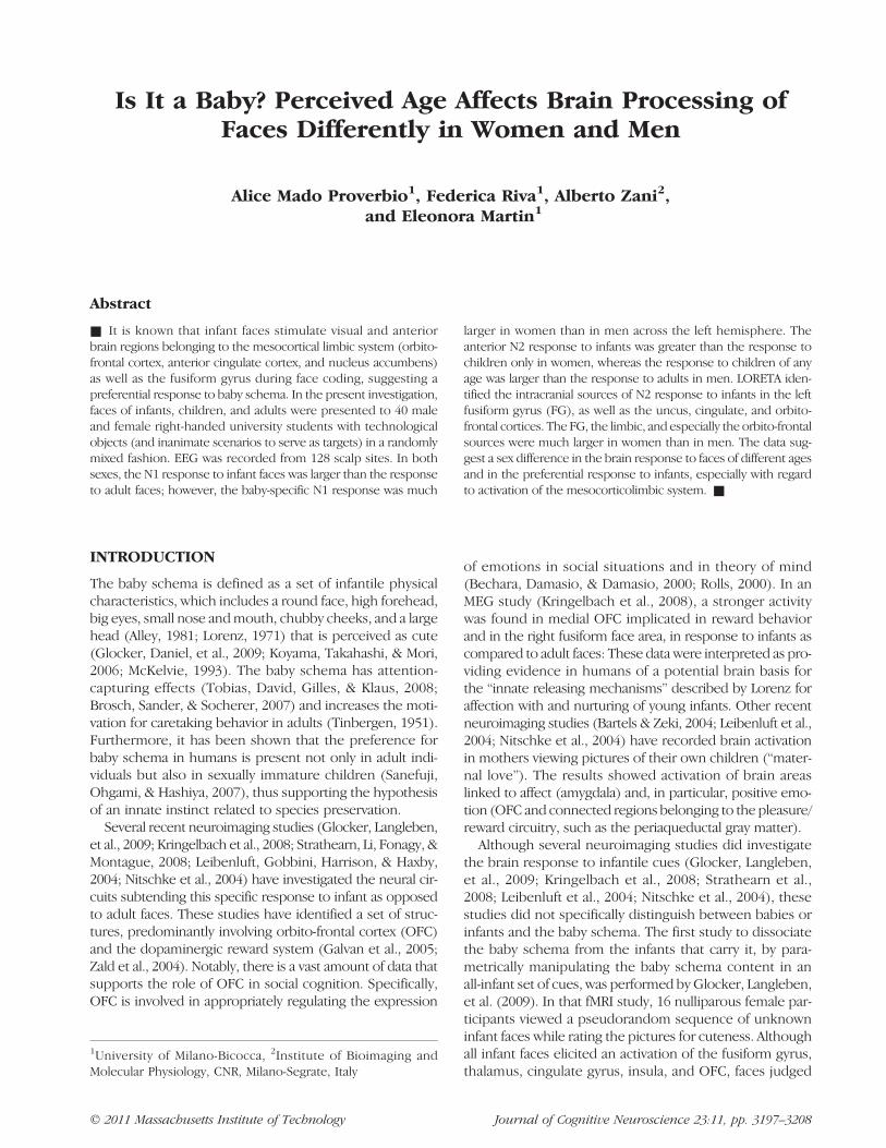
The stimulus set comprised 520 color pictures depictinggood-looking male and female faces of various ages (130adults, 130 prepubertal children, and 130 infants) and 130technologic/electronic objects of similar size and spatialdistribution (see Figure 1). Because all persons wereanonymous, their ages were actually unknown but wereestimated on the basis of facial resemblance by four in-dependent judges. Adult face attractiveness was also
3198 Journal of Cognitive Neuroscience Volume 23, Number 11
established by the same judges but without a specific ratingprocedure. Potentially gender-biased objects (such as anelectric iron or shaver) were not included. Faces includedthe neck and the upper portion of the chest. Normal pro-portions were maintained between infants and adults topreserve the authenticity of the perceptual experience. Ex-cept for the infant category (for which sex was sometimesindistinguishable), all faces depicted an equal number offemales and males. All people were smiling or showing apositive facial expression. There was no difference in facialexpression between age classes (closed mouth smile:adults = 50, children = 54, infants = 58; open-mouthedsmile: adults = 80, children = 76, infants = 72). Facesand objects were presented randomly mixed with 44 equi-luminant infrequent targets depicting natural or urbanlandscapes without visible persons (e.g., streets, offices,countryside, seascape). Stimulus size was 90 56″ × 8° 230 1″,and average luminance was 16.2 cd/cm2. ANOVA showed nodifference in stimulus luminance as a function of stimulustype (faces: adults = 16.4; children = 15.6; infants = 16.7.Objects= 16 cd/cm2). Each slide was presented for 800msecat the center of a PC screen with an ISI ranging from 1300 to1500 msec. The outer background was dark gray.
Procedure
In order to keep the subjectʼs attention focused on visualstimulation, the task consisted of responding as accu-rately and quickly as possible to photos displaying land-scapes (urban or natural scenarios without visible humans)by pressing a response key with the index finger of the left
or right hand while ignoring all other pictures. The twohands were used alternately during the recording session.The order of the hand and task conditions was counter-balanced across subjects. Participants were comfortablyseated in a darkened, acoustically and electrically shieldedtest area, facing a computer screen located 90 cm fromtheir eyes. Participants were instructed to fixate on the cen-ter of the screen, where a small transparent circle served asfixation point, and to avoid any eye or body movementsduring the recording session. Stimuli were presented fove-ally and subjectʼs eyes were aligned to the eyes of photo-graphed persons. Sequence presentation order differedacross subjects. An implicit task was employed to observeface-related attention-capturing effects.
EEG Recording and Analysis
EEGwas continuously recorded from128 scalp sites locatedaccording to the 10–5 International System (Oostenveld& Praamstra, 2001), at a sampling rate of 512 Hz. Horizontaland vertical eye movements were also recorded. Linkedears served as the reference lead. The EEG and electro-oculogram were amplified with a half-amplitude band passof 0.016–100 Hz. Electrode impedance was kept below5 kΩ. EEG epochs were synchronized with the onset ofstimuli presentation. Computerized artifact rejection wasperformed before averaging to discard epochs in whicheye movements, blinks, excessive muscle potentials, oramplifier blocking occurred. The artifact rejection criterionwas based on peak-to-peak amplitude exceeding 50 μV, andthe rejection rate was ∼5%. ERPs were averaged off-linefrom−100 msec before to 1000 msec after stimulus onset.ERP components were identified andmeasured, with refer-ence to the average baseline voltage over the interval from−100 msec to 0 msec, at sites and latency when maximumamplitude was achieved.
The peak amplitude of the occipito-temporal P1 com-ponent was measured at P9 and P10 in the 100–140 msectime window. The mean amplitude voltage of the N1 com-ponent was measured at P9, P10, PPO9h, PPO10h, TPP9h,and TPP10h electrode sites during the 150–180 msec timewindow. The mean amplitude voltage of anterior N2 wasmeasured at PF1, PF2, AF3, AF4, AFP3h, and AFp4h sitesduring the 250–300 msec time window. Mean amplitudevoltage of the N400/P400 complex was quantified at Fp1,Fp2, P1, P2, O1, and O2 electrode sites during the 400–500 msec time window.
ERP data were subjected to multifactorial repeatedmeasures ANOVA with one factor between groups (sex:males, females) and three factors within groups. Thewithin factors for ERP data were: stimulus content(adults, children, infants, and objects), hemisphere (left,right), and electrodes (depending on the ERP compo-nent of interest). Specifically, the electrode factor hadthree levels for the N1 component (occipito-temporal,lateral/occipital, and temporo/parietal), three levels for
Figure 1. Human faces of different sex and age (infants, children,and adults) along with an example of the target stimulus (scenariowithout visible humans) and control stimulus (technological object).
Proverbio et al. 3199
anterior N2 response (prefrontal, anterior frontal, anteriorfronto/parietal), and three levels for N400/P400 mean area(prefrontal, parietal, occipital). Face-specific N2 responsewas quantified only for infant, child, and adult faces. Multi-ple post-hoc comparisons were performed by means ofTukeyʼs test. Low-Resolution Electromagnetic Tomogra-phy (LORETA) was performed on ERP waveforms for eachERP component of interest.
LORETA, which is a discrete linear solution to the in-verse EEG problem, corresponds to the 3-D distributionof neuronal electric activity that yields maximum similar-ity (i.e., maximum synchronization), in terms of orientation
and strength, between neighboring neuronal populations(represented by adjacent voxels). In this study, an im-proved version of standardized weighted low-resolutionbrain electromagnetic tomography (sLORETA) was used.This version incorporates a singular value decomposition-based lead field weighting: swLORETA (Palmero-Soler,Dolan, Hadamschek, & Tass, 2007). Source space proper-ties were: grid spacing (the distance between two calcula-tion points) = 5 points; the estimated signal-to-noise ratio(SNR, which defines the regularization; a higher value forSNR means less regularization and less blurred results)was 3. LORETA was performed on group data to identify
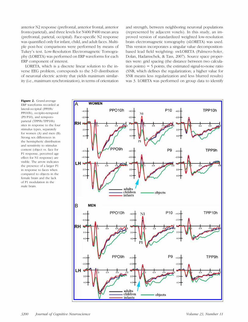
Figure 2. Grand-averageERP waveforms recorded atlateral-occipital (PP09h/PP010h), occipito-temporal(P9/P10), and temporo-parietal (TPP9h/TPP10h)sites in response to the fourstimulus types, separatelyfor women (A) and men (B).Strong sex differences inthe hemispheric distributionand sensitivity to stimuluscontent (object vs. face forP1 response, perceived ageeffect for N1 response) arevisible. The arrow indicatesthe presence of a larger P1in response to faces whencompared to objects in thefemale brain and the lackof P1 modulation in themale brain.
3200 Journal of Cognitive Neuroscience Volume 23, Number 11
statistically significant electromagnetic dipoles ( p< .05)—the larger the magnitude, the more significant the groupdifferences.
RESULTS
Figure 2 shows grand-average ERP waveforms recordedat posterior sites in response to face and technologicalobjects in women (A) and men (B). Consistent content-dependent modulation of both sensory and cognitivecomponents can be observed in viewers of both sexes.
P1 Latency
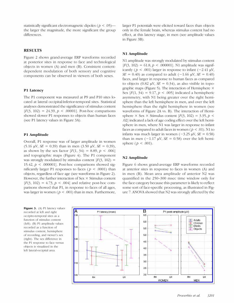
The P1 component was measured at P9 and P10 sites lo-cated at lateral occipital/inferior-temporal sites. Statisticalanalyses demonstrated the significance of stimulus content[F(3, 102) = 24.39, p < .00001]. Post-hoc comparisonsshowed slower P1 responses to objects than human faces(see P1 latency values in Figure 3A).
P1 Amplitude
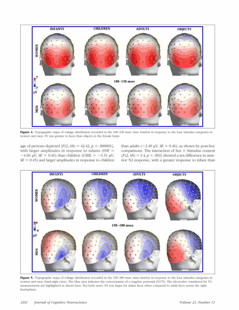
Overall, P1 response was of larger amplitude in women(5.16 μV, SE = 0.39) than in men (3.50 μV, SE = 0.39),as shown by the sex factor [F(1, 34) = 8.89, p < .006]and topographic maps (Figure 4). The P1 componentwas strongly modulated by stimulus content [F(3, 102) =15.42, p < .000001]. Post-hoc comparisons showed sig-nificantly larger P1 responses to faces ( p < .0001) thanobjects, regardless of face age (see waveforms in Figure 2).However, the further interaction of Sex × Stimulus content[F(3, 102) = 4.73, p < .004] and relative post-hoc com-parisons showed that P1, in response to faces of all ages,was larger in women ( p< .001) than in men. Furthermore,
larger P1 potentials were elicited toward faces than objectsonly in the female brain, whereas stimulus content had noeffect, at this latency stage, in men (see amplitude valuesin Figure 3B).
N1 Amplitude
N1 amplitude was strongly modulated by stimulus content[F(3, 102) = 61.8, p < .000001]. N1 amplitude was signif-icantly ( p < .001) larger in response to infant (−2.41 μV,SE = 0.40) as compared to adult (−1.66 μV, SE = 0.40)faces, and larger in response to human faces as comparedto objects (0.82 μV, SE = 0.34), as also visible in topo-graphic maps (Figure 5). The interaction of Hemisphere ×Sex [F(1, 34) = 9.17, p < .005] indicated a hemisphericasymmetry, with N1 being greater over the right hemi-sphere than the left hemisphere in men, and over the lefthemisphere than the right hemisphere in women (seewaveforms of Figure 2A vs. B). The interaction of Hemi-sphere × Sex × Stimulus content [F(3, 102) = 3.35, p <.02] indicated a lack of age coding effect over the left hemi-sphere in men, where N1 was larger in response to infantfaces as compared to adult faces in women ( p< .01). N1 toinfants was much larger in women (−3.25 μV, SE = 0.58)than in men (−1.17 μV, SE = 0.58) over the left hemi-sphere ( p < .001).
N2 Amplitude
Figure 6 shows grand-average ERP waveforms recordedat anterior sites in response to faces in women (A) andin men (B). Mean area amplitude of anterior N2 wasquantified in the 250–300 msec time window only forthe face category because this parameter is likely to reflectsome sort of face-specific processing, as illustrated in Fig-ure 7. ANOVA showed that N2 was strongly affected by the
Figure 3. (A) P1 latency valuesrecorded at left and rightoccipito-temporal sites as afunction of stimulus content(left). (B) P1 amplitude valuesrecorded as a function ofstimulus content, hemisphereof recording, and viewerʼs sex(right). The sex difference inthe P1 response to face versusobjects is visualized in theleft lateral-occipital area.
Proverbio et al. 3201
age of persons depicted [F(2, 68) = 42.42, p < .000001],with larger amplitudes in response to infants (INF =−4.06 μV, SE = 0.46) than children (CHIL = −3.31 μV,SE = 0.45) and larger amplitudes in response to children
than adults (−2.49 μV, SE = 0.46), as shown by post-hoccomparisons. The interaction of Sex × Stimulus content[F(2, 68) = 3.4, p< .003] showed a sex difference in ante-rior N2 response, with a greater response to infant than
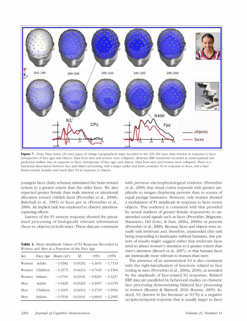
Figure 5. Topographic maps of voltage distribution recorded in the 150–180 msec time window in response to the four stimulus categories inwomen and men (back-right view). The blue area indicates the concentration of a negative potential (N170). The electrodes considered for N1measurements are highlighted as shown here. For both sexes, N1 was larger for infant faces when compared to adult faces across the righthemisphere.
Figure 4. Topographic maps of voltage distribution recorded in the 100–130 msec time window in response to the four stimulus categories inwomen and men. P1 was greater to faces than objects in the female brain.
3202 Journal of Cognitive Neuroscience Volume 23, Number 11

child faces only in female individuals (see Figure 6A vs. B),whereas N2 response to childrenwas of equal amplitude forthe two age classes of children (infants and children) butthe N2 response to adult faces was smaller in males. Post-hoc analysis of mean values given in Table 1 showed no sexdifference in the N2 response to children, but amuch largerN2 response was shown toward infants in women ( p< .01)than in men.In order to investigate the neural bases of infant-
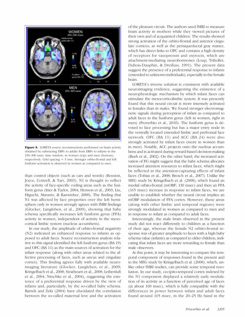
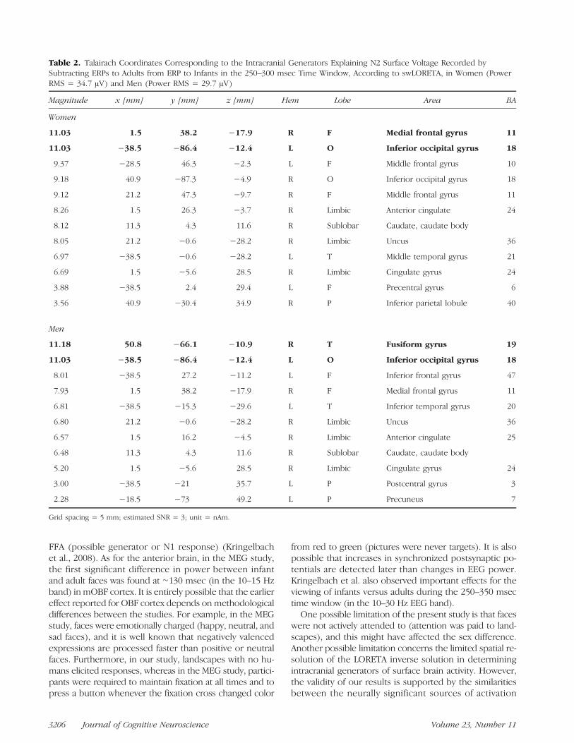
specific face response in the two sexes, two LORETAsource reconstructions were applied to the differencewaves, obtained by subtracting ERPs to adults from ERPsto infants, in the 250–300 msec time window (see Fig-ure 8). A list of significant sources of activation explain-ing the surface difference voltage is presented in Table 2.The main sources of activation included the left and rightmedial occipital/fusiform gyrus (BA 19), the right uncus(BA 20/36), the right medial orbito-frontal gyrus (BA 11),and right ACC (BA 23), which are mostly involved in affec-tive processing of faces. Notably, the orbito-frontal sourcewas much stronger in women (11.03 nA m) than in men(7.03 nAm).
N400/P400 Amplitude
In the 400–500 msec time window, N400 over anteriorbrain regions was larger in response to objects than faces(as displayed in Figure 7), whereas P400 over posteriorbrain regions was larger to objects than faces (as clearlyvisible in Figure 2). ANOVA showed the significance ofStimulus content × Electrode [F(6, 204) = 44.1, p <.00001], indicating much larger N400s to objects thanfaces at prefrontal sites (with no effect of face age),much larger P400 to objects than faces at occipital sites(with no effect of face age), and no stimulus content effectsat parietal sites.
DISCUSSION
The studyʼs goal was to investigate the time course andbrain topography of neural processing of human faces ofincreasing age, as opposed to objects, as a function of theviewerʼs sex. The hypothesis, supported by current ethol-ogist models that relate perceived face cuteness with in-creased brain response, was that in adult individuals the
Figure 6. Grand-average ERPwaveforms recorded atprefrontal (Fp1/Fp2), anteriorfrontal (AF3/AF4), and anteriorprefrontal (AFp3h/AFp4h)sites in response to the fourstimulus types, separately forwomen (A) and men (B).The trend can be observedclearly in the gradient of N2amplitude (indicated by anarrow), with larger N2 towardinfants than children andlarger N2 in response towardchildren than adults in thefemale (but not male) brain.
Proverbio et al. 3203
youngest faces (baby schema) stimulated the brain rewardsystem to a greater extent than the older faces. We alsoexpected greater female than male interest or attentionalallocation toward childish faces (Proverbio et al., 2006b;Babchuk et al., 1985) or faces per se (Proverbio et al.,2008). An implicit task was employed to observe attention-capturing effects.
Latency of the P1 sensory response showed the priori-tized processing of biologically relevant information(faces vs. objects) in both sexes. These data are consistent
with previous electrophysiological evidence (Proverbioet al., 2009) that visual cortex responds with greater am-plitude to images displaying persons than to scenes ofequal average luminance. However, only women showeda modulation of P1 amplitude in response to faces versusobjects. This evidence is consistent with that providedby neural markers of greater female responsivity to un-attended social signals such as faces (Proverbio, Brignone,Matarazzo, Del Zotto, & Zani, 2006a, 2006b) or persons(Proverbio et al., 2008). Because faces and objects were ac-tually task irrelevant and, therefore, unattended (the taskbeing responding to landscapes without humans), this pat-tern of results might suggest either that irrelevant facestend to attract womenʼs attention to a greater extent thanmenʼs attention (Brosch et al., 2007), or that humans facesare intrinsically more relevant to women than men.The presence of an asymmetrical N1 is also consistent
with the right-lateralization of functions related to facecoding in men (Proverbio et al., 2006a, 2010), as revealedby the amplitude of face-related N1 responses. RelatedERP data are paralleled by behavioral studies on chimericface processing demonstrating bilateral face processingin women (Bourne & Maxwell, 2010; Bourne, 2005). In-deed, N1 (known in the literature as N170) is a negativeoccipito/temporal response that is usually larger to faces
Figure 7. (Top) Time series (20 msec pass) of voltage topographical maps recorded in the 160–260 msec time window in response to faces(irrespective of face age) and objects. Data from men and women were collapsed. (Bottom) ERP waveforms recorded at centro-parietal andprefrontal midline sites in response to faces (irrespective of face age) and objects. Data from men and women were collapsed. There is afunctional dissociation between face and object processing, with a larger earlier and more posterior N2 in response to faces, and a laterfronto-central, broader and much later N4 in response to objects.
Table 1. Mean Amplitude Values of N2 Response Recorded inWomen and Men as a Function of the Face Age
Sex Face Age Mean (μV) SE −95% +95%
Women Adults −3.0382 0.65292 −4.3651 −1.7113
Women Children −3.4575 0.64314 −4.7645 −2.1504
Women Infants −4.5764 0.61641 −5.8291 −3.3237
Men Adults −1.9428 0.65292 −3.2697 −0.6159
Men Children −3.1665 0.64314 −4.4735 −1.8594
Men Infants −3.5518 0.61641 −4.8045 −2.2991
3204 Journal of Cognitive Neuroscience Volume 23, Number 11
than control objects (such as cars and words) (Rossion,Joyce, Cottrell, & Tarr, 2003). N1 is thought to reflectthe activity of face-specific coding areas such as the fusi-form gyrus (Itier & Taylor, 2004; Henson et al., 2003; Liu,Higuchi, Marantz, & Kanwisher, 2000). The finding thatN1 was affected by face properties over the left hemi-sphere only in women strongly agrees with fMRI findings(Glocker, Langleben, et al., 2009), showing that babyschema specifically increases left fusiform gyrus (FFA)activity in women, independent of activity in the meso-cortical limbic system (nucleus accumbens).In our study, the amplitude of orbito-frontal negativity
(N2) indicated an enhanced response to infants as op-posed to adult faces. Source reconstruction analysis rela-tive to this signal identified the left fusiform gyrus (BA 19)and OFC (BA 11) as the main sources of activation for theinfant response (along with other areas related to the af-fective processing of faces, such as uncus and cingulatecortex). This finding agrees fully with available neuro-imaging literature (Glocker, Langleben, et al., 2009;Kringelbach et al., 2008; Strathearn et al., 2008; Leibenluftet al., 2004; Nitschke et al., 2004), suggesting the exis-tence of a preferential response driven by the view ofinfants and, particularly, by the so-called baby schema.Bartels and Zeki (2004) have elucidated the correlationbetween the so-called maternal love and the activation
of the pleasure circuit. The authors used fMRI to measurebrain activity in mothers while they viewed pictures oftheir own and of acquainted children. The results showedstrong activation of the orbito-frontal and anterior cingu-late cortices, as well as the periaqueductal gray matter,which has direct links to OFC and contains a high densityof receptors for vasopressin and oxytocin, which areattachment-mediating neurohormones (Loup, Tribollet,Dubois-Dauphin, & Dreifuss, 1991). The present datasuggest the presence of a preferential response to infants(extended to unknown individuals), especially in the femalebrain.
LORETAʼs inverse solution is consistent with availableneuroimaging evidence, suggesting the existence of aneurophysiologic mechanism by which infant faces canstimulate the mesocorticolimbic system. It was presentlyfound that this neural circuit is more intensely activatedin females than in males. We found stronger electromag-netic signals during perception of infant as compared toadult faces in the fusiform gyrus (left in women, right inmen) (Proverbio et al., 2010). The fusiform gyrus is de-voted to face processing but has a major entry node inthe ventrally located extended limbic and prefrontal facenetwork. OFC (BA 11) and ACC (BA 24) were alsostrongly activated by infant faces (more in women thanin men). Notably, ACC projects onto the nucleus accum-bens and is activated during reward-based decision-making(Bush et al., 2002). On the other hand, the increased acti-vation of FG might suggest that the baby schema allocatesincreased attention resources to infant faces, which mightbe reflected in the attention-capturing effects of infantfaces (Tobias et al., 2008; Brosch et al., 2007). Unlike theMEG study by Kringelbach et al. (2008), which found anmedial orbito-frontal (mOBF; 130 msec) and then an FFA(165 msec) increase in response to infant faces, we areunable to establish whether the neural circuit implies anmOBF modulation of FFA cortex. However, these areas(along with other limbic and temporal regions) werestrongly modulated in women as compared to men andin response to infant as compared to adult faces.
Interestingly, the male brain observed in the presentstudy did not react differently to children as a functionof their age, whereas the female N2 orbito-frontal re-sponse was of greater amplitude to faces with a high babyschema value (infants) as compared to older children, indi-cating that infant faces are more rewarding to female thanmale observers.
At this point, it may be interesting to compare the tem-poral component of responses found in the present andin the MEG study by Kringelbach et al. (2008), which, un-like other fMRI studies, can provide some temporal reso-lution. In our study, occipito-temporal cortex indexed bythe N1 component displayed a relatively early modula-tion of its activity as a function of perceived age of faces(at about 160 msec), which is fully compatible with thedifferences in power between infant and adult facesfound around 165 msec, in the 20–25 Hz band in the
Figure 8. LORETA source reconstructions performed on brain activityobtained by subtracting ERPs to adults from ERPs to infants in the250–300 msec time window, in women (top) and men (bottom),respectively. Grid spacing = 5 mm. Stronger orbito-frontal and leftfusiform activation is observed in women as compared to men.
Proverbio et al. 3205
FFA (possible generator or N1 response) (Kringelbachet al., 2008). As for the anterior brain, in the MEG study,the first significant difference in power between infantand adult faces was found at ∼130 msec (in the 10–15 Hzband) in mOBF cortex. It is entirely possible that the earliereffect reported for OBF cortex depends on methodologicaldifferences between the studies. For example, in the MEGstudy, faces were emotionally charged (happy, neutral, andsad faces), and it is well known that negatively valencedexpressions are processed faster than positive or neutralfaces. Furthermore, in our study, landscapes with no hu-mans elicited responses, whereas in the MEG study, partici-pants were required to maintain fixation at all times and topress a button whenever the fixation cross changed color
from red to green (pictures were never targets). It is alsopossible that increases in synchronized postsynaptic po-tentials are detected later than changes in EEG power.Kringelbach et al. also observed important effects for theviewing of infants versus adults during the 250–350 msectime window (in the 10–30 Hz EEG band).One possible limitation of the present study is that faces
were not actively attended to (attention was paid to land-scapes), and this might have affected the sex difference.Another possible limitation concerns the limited spatial re-solution of the LORETA inverse solution in determiningintracranial generators of surface brain activity. However,the validity of our results is supported by the similaritiesbetween the neurally significant sources of activation
Table 2. Talairach Coordinates Corresponding to the Intracranial Generators Explaining N2 Surface Voltage Recorded bySubtracting ERPs to Adults from ERP to Infants in the 250–300 msec Time Window, According to swLORETA, in Women (PowerRMS = 34.7 μV) and Men (Power RMS = 29.7 μV)
Magnitude x [mm] y [mm] z [mm] Hem Lobe Area BA
Women
11.03 1.5 38.2 −17.9 R F Medial frontal gyrus 11
11.03 −38.5 −86.4 −12.4 L O Inferior occipital gyrus 18
9.37 −28.5 46.3 −2.3 L F Middle frontal gyrus 10
9.18 40.9 −87.3 −4.9 R O Inferior occipital gyrus 18
9.12 21.2 47.3 −9.7 R F Middle frontal gyrus 11
8.26 1.5 26.3 −3.7 R Limbic Anterior cingulate 24
8.12 11.3 4.3 11.6 R Sublobar Caudate, caudate body
8.05 21.2 −0.6 −28.2 R Limbic Uncus 36
6.97 −38.5 −0.6 −28.2 L T Middle temporal gyrus 21
6.69 1.5 −5.6 28.5 R Limbic Cingulate gyrus 24
3.88 −38.5 2.4 29.4 L F Precentral gyrus 6
3.56 40.9 −30.4 34.9 R P Inferior parietal lobule 40
Men
11.18 50.8 −66.1 −10.9 R T Fusiform gyrus 19
11.03 −38.5 −86.4 −12.4 L O Inferior occipital gyrus 18
8.01 −38.5 27.2 −11.2 L F Inferior frontal gyrus 47
7.93 1.5 38.2 −17.9 R F Medial frontal gyrus 11
6.81 −38.5 −15.3 −29.6 L T Inferior temporal gyrus 20
6.80 21.2 −0.6 −28.2 R Limbic Uncus 36
6.57 1.5 16.2 −4.5 R Limbic Anterior cingulate 25
6.48 11.3 4.3 11.6 R Sublobar Caudate, caudate body
5.20 1.5 −5.6 28.5 R Limbic Cingulate gyrus 24
3.00 −38.5 −21 35.7 L P Postcentral gyrus 3
2.28 −18.5 −73 49.2 L P Precuneus 7
Grid spacing = 5 mm; estimated SNR = 3; unit = nAm.
3206 Journal of Cognitive Neuroscience Volume 23, Number 11

provided by LORETA and those provided by neuroimagingand MEG studies on the same topic.
Conclusions
In summary, MEG (Kringelbach et al., 2008) and fMRI(Glocker, Daniel, et al., 2009) evidence support the exis-tence of a visual (left FFA) and orbito-frontal responsetriggered by baby schema in humans. In both studies,the gender of viewers was not considered, either becausesubjects were all females (Glocker, Daniel, et al., 2009) orbecause males and females were considered as a singlegroup. To our knowledge, the present investigation isthe first study where the brain response to faces was in-vestigated by varying the age of infants and by examin-ing both sexes. We discovered that the visual responseand the prefrontal response exhibited gradients that wereinversely correlated with the age of the individuals de-picted by the stimulus material. Women showed a greatermodulation of brain responses as a function of face age.Women showed earlier face/object discrimination (at theP1 rather than the N1 level), adult/infant discriminationin both left and right visual areas at the N1 level (insteadof only over the right hemisphere as in men), but espe-cially in the anterior response to baby schema (N2 level),with N2 of greater amplitude in response to infants thanchildren. Notably, the study participants (neither malenor female) had no particular familiarity with childrenʼsfaces, as the presence of siblings and young relatives inthe subjectsʼ families and involvement in professionalactivities related to childcare were criteria for exclusionfrom the study. For this reason, we believe that visualfamiliarity with baby schema was matched between sexesand that sex differences were more likely to be due toneurobiological factors (such as attention, motivation,and social instinct) as opposed to cultural factors.
Reprint requests should be sent to Alice Mado Proverbio,Department of Psychology, University of Milano-Bicocca,Via dellʼInnovazione 10, 20126 Milan, Italy, or via e-mail:[email protected].
REFERENCES
Alley, T. R. (1981). Head shape and the perception of cuteness.Developmental Psychology, 17, 650–654.
Babchuk, W. A., Hames, R. B., & Thompson, R. A. (1985). Sexdifferences in the recognition of infant facial expressions ofemotion: The primary caretaker hypothesis. Ethology &Sociobiology, 6, 89–101.
Bartels, A., & Zeki, S. (2004). The neural correlates of maternaland romantic love. Neuroimage, 21, 1155.
Bechara, A., Damasio, H., & Damasio, A. (2000). Emotion,decision making and the orbito-frontal cortex. CerebralCortex, 1, 295–307.
Bourne, V. J. (2005). Lateralised processing of positive facialemotion: Sex differences in strength of hemisphericdominance. Neuropsychologia, 43, 953.
Bourne, V. J., & Maxwell, A. M. (2010). Examining the sexdifference in lateralisation for processing facial emotion:Does biological sex or psychological gender identitymatter? Neuropsychologia, 48, 1284–1289.
Brosch, T., Sander, D., & Socherer, K. R. (2007). That babycaught my eye. Attention capture by infant faces. Emotion,7, 685–689.
Bush, G., Vogt, B. A., Holmes, J., Dale, A. M., Greve, D., Jenike,M. A., et al. (2002). Dorsal anterior cingulate cortex: A role inreward-based decision making. Proceedings of the NationalAcademy of Sciences, U.S.A., 99, 523–528.
Galvan, A., Hare, T. A., Davidson, M., Spicer, J., Glover, G., &Casey, B. J. (2005). The role of ventral frontostriatal circuitryin reward-based learning in humans. Journal ofNeuroscience, 25, 8650–8656.
Glocker, M. L., Daniel, D., Langleben, D. D., Ruparel, K.,Loughead, J. W., Gur, R. C., et al. (2009). Baby schema ininfant faces induces cuteness perception and motivation forcaretaking in adults. Ethology, 115, 257–263.
Glocker, M. L., Langleben, D. D., Ruparel, K., Loughead, J. W.,Valdez, J. N., Griffin, M. D., et al. (2009). Baby schemamodulates the brain reward system in nulliparous women.Proceedings of the National Academy of Sciences, U.S.A.,106, 9115–9119.
Henson, R. N., Goshen-Gottstein, Y., Ganel, T., Otten, L. J.,Quayle, A., & Rugg, M. D. (2003). Electrophysiological andhaemodynamic correlates of face perception, recognition andpriming. Cerebral Cortex, 13, 793–805.
Itier, R. J., & Taylor, M. J. (2004). Source analysis of the n170 tofaces and objects. NeuroReport, 15, 1261–1265.
Koyama, R., Takahashi, Y., & Mori, K. (2006). Assessing thecuteness of children: Significant factors and genderdifferences. Social Behavior and Personality, 34, 1087–1099.
Kringelbach, M. L., Lehtonen, A., Squire, S., Harvey, A. G.,Craske, M. G., Holliday, I. E., et al. (2008). A specific and rapidneural signature for parental instinct. PLoS ONE, 3, e1664.
Leibenluft, E., Gobbini, M. I., Harrison, T., & Haxby, J. V. (2004).Mothersʼ neural activation in response to pictures of theirchildren and other children. Biological Psychiatry, 56, 225.
Liu, J., Higuchi, M., Marantz, A., & Kanwisher, N. (2000). Theselectivity of the occipitotemporal m170 for faces.NeuroReport, 11, 337–341.
Lorenz, K. (1971). Studies in animal and human behavior.Cambridge, MA: Harvard University Press.
Loup, F., Tribollet, E., Dubois-Dauphin, M., & Dreifuss, J. J.(1991). Localization of high-affinity binding sites for oxytocinand vasopressin in the human brain. An autoradiographicstudy. Brain Research, 555, 220.
McKelvie, S. J. (1993). Perceived cuteness, activity level, andgender in schematic babyfaces. Journal of Social Behaviorand Personality, 8, 297–310.
Nitschke, J. B., Nelson, E. E., Rusch, B. D., Fox, A. S., Oakes, T. R.,& Davidson, R. J. (2004). Orbito-frontal cortex tracks positivemood in mothers viewing pictures of their newborn infants.Neuroimage, 21, 583.
Oostenveld, R., & Praamstra, P. (2001). The five percent electrodesystem for high-resolution EEG and ERP measurements.Clinical Neurophysiology, 112, 713.
Palmero-Soler, E., Dolan, K., Hadamschek, V., & Tass, P. A.(2007). swLORETA: A novel approach to robust sourcelocalization and synchronization tomography. Physics inMedicine and Biology, 52, 1783–1800.
Proverbio, A., Brignone, V., Matarazzo, S., Del Zotto, M., & Zani, A.(2006a). Gender differences in hemispheric asymmetry forface processing. BMC Neuroscience, 7, 44.
Proverbio, A. M., Adorni, R., Zani, A., & Trestianu, L. (2009).Sex differences in the brain response to affective scenes withor without humans. Neuropsychologia, 47, 2374–2388.
Proverbio et al. 3207
Proverbio, A. M., Brignone, V., Matarazzo, S., Del Zotto, M., &Zani, A. (2006b). Gender and parental status affect the visualcortical response to infant facial expression.Neuropsychologia, 44, 2987–2999.
Proverbio, A. M., Matarazzo, S., Brignone, V., Del Zotto, M., &Zani, A. (2007). Processing valence and intensity of infantexpressions: The roles of expertise and gender. ScandinavianJournal of Psychology, 48, 477–485.
Proverbio, A. M., Riva, F., Martin, E., & Zani, A. (2010). Facecoding is bilateral in the female brain. PLoS ONE, 5, e11242.
Proverbio, A. M., Zani, A., & Adorni, R. (2008). Neural markersof a greater female responsiveness to social stimuli. BMCNeuroscience, 30, 56.
Rolls, E. T. (2000). The orbito-frontal cortex and reward.Cerebral Cortex, 10, 284–294.
Rossion, B., Joyce, C. A., Cottrell, G. W., & Tarr, M. J. (2003).Early lateralization and orientation tuning for face, word,and object processing in the visual cortex. Neuroimage, 20,1609–1624.
Sander, K., Frome, Y., & Scheich, H. (2007). fMRI activationsof amygdala, cingulate cortex, and auditory cortex by
infant laughing and crying. Human Brain Mapping, 28,1007–1022.
Sanefuji, W., Ohgami, H., & Hashiya, K. (2007). Development ofpreference for baby faces across species in humans (homosapiens). Journal of Ethology, 25, 249.
Seifritz, E., Esposito, F., Neuhoff, J. G., Luthi, A., Mustovic, H.,Dammann, G., et al. (2003). Differential sex-independentamygdala response to infant crying and laughing in parentsversus nonparents. Biological Psychiatry, 54, 1367.
Strathearn, L., Li, J., Fonagy, P., & Montague, P. R. (2008).Whatʼs in a smile? Maternal brain responses to infant facialcues. Pediatrics, 122, 40–51.
Tinbergen, N. (1951). The study of instinct. London: OxfordUniversity Press.
Tobias, B., David, S., Gilles, P., & Klaus, R. S. (2008). Beyondfear: Rapid spatial orienting toward positive emotionalstimuli. Psychological Science, 19, 362–370.
Zald, D. H., Boileau, I., El-Dearedy, W., Gunn, R., McGlone, F.,Dichter, G. S., et al. (2004). Dopamine transmission in thehuman striatum during monetary reward tasks. Journal ofNeuroscience, 24, 4105–4112.
3208 Journal of Cognitive Neuroscience Volume 23, Number 11

This article has been cited by:
1. Alice Mado Proverbio, Roberta Mazzara, Federica Riva, Mirella Manfredi. 2012. Sex differences in callosal transfer andhemispheric specialization for face coding. Neuropsychologia 50:9, 2325-2332. [CrossRef]
2. Alberto Zani, Alice M Proverbio. 2012. Is that a belt or a snake? Object attentional selection affects the early stages of visualsensory processing. Behavioral and Brain Functions 8:1, 6. [CrossRef]


