Expression of Endoplasmic Reticulum Stress Proteins Is a Candidate Marker of Brain Metastasis in...
16
Biomarkers, Genomics, Proteomics, and Gene Regulation Expression of Endoplasmic Reticulum Stress Proteins Is a Candidate Marker of Brain Metastasis in both ErbB-2 and ErbB-2 Primary Breast Tumors Rebeca Sanz-Pamplona,* Ramón Aragüés, † Keltouma Driouch, ‡ Berta Martín,* Baldo Oliva, † Miguel Gil, § Susana Boluda, ¶ Pedro L. Fernández, Antonio Martínez,* Víctor Moreno,** Juan J. Acebes, †† Rosette Lidereau, ‡ Fabien Reyal, ‡‡ Marc J. Van de Vijver, §§ and Angels Sierra* 1 From the Biological Clues of the Invasive and Metastatic Phenotype Group,* Bellvitge Biomedical Research Institute (IDIBELL), Barcelona, Spain; the Structural Bioinformatics Group (GRIB-IMIM), † University Pompeu Fabra, Barcelona, Spain; INSERM U735, ‡ Centre René Huguenin, St. Cloud, France; the Medical Oncology Service, § Catalan Institute of Oncology-IDIBELL, Hospital Duran i Reynals, Barcelona, Spain; the Institute of Neuropathology-Anatomic Pathology Service, ¶ IDIBELL-University Hospital of Bellvitge, Barcelona, Spain; the Department of Anatomic Pathology, Clinical Hospital-IDIBAPS, University of Barcelona, Barcelona, Spain; the Unit of Biostatistics and Bioinformatics, IDIBELL-Catalan Institute of Oncology and University of Barcelona, Barcelona, Spain; the Neurosurgical Service, †† University Hospital of Bellvitge, Barcelona, Spain; CNRS UMR144, ‡‡ Institute Curie, Paris, France; and the Department of Pathology, §§ Academic Medical Center, Amsterdam, The Netherlands The increasing incidence of breast cancer brain me- tastasis in patients with otherwise well-controlled sys- temic cancer is a key challenge in cancer research. It is necessary to understand the properties of brain- tropic tumor cells to identify patients at risk for brain metastasis. Here we attempt to identify functional phenotypes that might enhance brain metastasis. To obtain an accurate classification of brain metastasis proteins, we mapped organ-specific brain metastasis gene expression signatures onto an experimental protein-protein interaction network based on brain metastatic cells. Thirty-seven proteins were differen- tially expressed between brain metastases and non- brain metastases. Analysis of metastatic tissues, the use of bioinformatic approaches, and the character- ization of protein expression in tumors with or with- out metastasis identified candidate markers. A multi- variate analysis based on stepwise logistic regression revealed GRP94, FN14, and inhibin as the best combi- nation to discriminate between brain and non-brain metastases (ROC AUC 0.85, 95% CI 0.73 to 0.96 for the combination of the three proteins). These mark- ers substantially improve the discrimination of brain metastasis compared with ErbB-2 alone (AUC 0.76, 95% CI 0.60 to 0.93). Furthermore, GRP94 was a better negative marker (LR 0.16) than ErbB-2 (LR 0.42). We conclude that, in breast carcinomas, certain proteins associated with the endoplasmic reticulum stress phenotype are candidate markers of brain metastasis. (Am J Pathol 2011, 179:564 –579; DOI: 10.1016/j.ajpath.2011.04.037) Brain metastases occur in 10% to 15% of breast cancer patients with advanced disease. 1–3 It can be assumed that up to 30% of metastatic breast cancer patients will undergo brain metastasis during the course of their disease. 4,5 This rate is increasing, which can be linked to greater survival in patients receiving chemotherapy Supported by grants from the Spanish Ministry of Health and Consumer Affairs FIS/PI041937 and FIS/PI071245, by the European Commission MetaBre contract No. LSHC-CT-2004-506049, and by the Spanish Minis- try of Education and Science SAF2004-0188-E. B.O. and R.A. acknowl- edge grants from the Spanish Ministry of Education and Science (MEC BIO2005-00533 and MCyT BIO2002-0369), and P.L.F. from the Marató- TV3, RETICC from Instituto Carlos III, and Xarxa de Bancs de Tumors de Catalunya. Accepted for publication April 11, 2011. Address reprint requests to Angels Sierra, M.D., Ph.D., Bellvitge Biomedical Research Institute-IDIBELL, Avda. Gran Via de Hospitalet Ll., 199, Hospitalet Ll., Barcelona 08908, Spain. E-mail: asierra@ idibell.cat. The American Journal of Pathology, Vol. 179, No. 2, August 2011 Copyright © 2011 American Society for Investigative Pathology. Published by Elsevier Inc. All rights reserved. DOI: 10.1016/j.ajpath.2011.04.037 564
Transcript of Expression of Endoplasmic Reticulum Stress Proteins Is a Candidate Marker of Brain Metastasis in...

The American Journal of Pathology, Vol. 179, No. 2, August 2011
Copyright © 2011 American Society for Investigative Pathology.
Published by Elsevier Inc. All rights reserved.
DOI: 10.1016/j.ajpath.2011.04.037
Biomarkers, Genomics, Proteomics, and Gene Regulation
Expression of Endoplasmic Reticulum StressProteins Is a Candidate Marker of Brain Metastasisin both ErbB-2� and ErbB-2� Primary Breast
TumorsRebeca Sanz-Pamplona,* Ramón Aragüés,†
Keltouma Driouch,‡ Berta Martín,* Baldo Oliva,†
Miguel Gil,§ Susana Boluda,¶
Pedro L. Fernández,� Antonio Martínez,*Víctor Moreno,** Juan J. Acebes,††
Rosette Lidereau,‡ Fabien Reyal,‡‡
Marc J. Van de Vijver,§§ and Angels Sierra*1
From the Biological Clues of the Invasive and Metastatic
Phenotype Group,* Bellvitge Biomedical Research Institute
(IDIBELL), Barcelona, Spain; the Structural Bioinformatics
Group (GRIB-IMIM),† University Pompeu Fabra, Barcelona,
Spain; INSERM U735,‡ Centre René Huguenin, St. Cloud,
France; the Medical Oncology Service,§ Catalan Institute of
Oncology-IDIBELL, Hospital Duran i Reynals, Barcelona,
Spain; the Institute of Neuropathology-Anatomic Pathology
Service,¶ IDIBELL-University Hospital of Bellvitge, Barcelona,
Spain; the Department of Anatomic Pathology,� Clinical
Hospital-IDIBAPS, University of Barcelona, Barcelona, Spain;
the Unit of Biostatistics and Bioinformatics,�� IDIBELL-Catalan
Institute of Oncology and University of Barcelona, Barcelona,
Spain; the Neurosurgical Service,†† University Hospital of
Bellvitge, Barcelona, Spain; CNRS UMR144,‡‡ Institute Curie,
Paris, France; and the Department of Pathology,§§ Academic
Medical Center, Amsterdam, The Netherlands
The increasing incidence of breast cancer brain me-tastasis in patients with otherwise well-controlled sys-temic cancer is a key challenge in cancer research. Itis necessary to understand the properties of brain-tropic tumor cells to identify patients at risk for brainmetastasis. Here we attempt to identify functionalphenotypes that might enhance brain metastasis. Toobtain an accurate classification of brain metastasisproteins, we mapped organ-specific brain metastasisgene expression signatures onto an experimentalprotein-protein interaction network based on brainmetastatic cells. Thirty-seven proteins were differen-
tially expressed between brain metastases and non-564
brain metastases. Analysis of metastatic tissues, theuse of bioinformatic approaches, and the character-ization of protein expression in tumors with or with-out metastasis identified candidate markers. A multi-variate analysis based on stepwise logistic regressionrevealed GRP94, FN14, and inhibin as the best combi-nation to discriminate between brain and non-brainmetastases (ROC AUC � 0.85, 95% CI � 0.73 to 0.96 forthe combination of the three proteins). These mark-ers substantially improve the discrimination ofbrain metastasis compared with ErbB-2 alone (AUC �
0.76, 95% CI � 0.60 to 0.93). Furthermore, GRP94 wasa better negative marker (LR � 0.16) than ErbB-2(LR � 0.42). We conclude that, in breast carcinomas,certain proteins associated with the endoplasmicreticulum stress phenotype are candidate markersof brain metastasis. (Am J Pathol 2011, 179:564–579;
DOI: 10.1016/j.ajpath.2011.04.037)
Brain metastases occur in 10% to 15% of breast cancerpatients with advanced disease.1–3 It can be assumedthat up to 30% of metastatic breast cancer patients willundergo brain metastasis during the course of theirdisease.4,5 This rate is increasing, which can be linkedto greater survival in patients receiving chemotherapy
Supported by grants from the Spanish Ministry of Health and ConsumerAffairs FIS/PI041937 and FIS/PI071245, by the European CommissionMetaBre contract No. LSHC-CT-2004-506049, and by the Spanish Minis-try of Education and Science SAF2004-0188-E. B.O. and R.A. acknowl-edge grants from the Spanish Ministry of Education and Science (MECBIO2005-00533 and MCyT BIO2002-0369), and P.L.F. from the Marató-TV3, RETICC from Instituto Carlos III, and Xarxa de Bancs de Tumors deCatalunya.
Accepted for publication April 11, 2011.
Address reprint requests to Angels Sierra, M.D., Ph.D., BellvitgeBiomedical Research Institute-IDIBELL, Avda. Gran Via de HospitaletLl., 199, Hospitalet Ll., Barcelona 08908, Spain. E-mail: asierra@
idibell.cat.
Brain Metastasis Prediction Markers 565AJP August 2011, Vol. 179, No. 2
and to the fact that it is difficult to cross the blood-brainbarrier with current systemic treatments.6 – 8 The diffi-culties in managing brain metastasis therapy result in amedian survival of 7 months, with brain metastasis being thecause of death or a major contributing factor in 68% ofpatients.9 Thus, there is a need for both prevention andimproved treatment of brain metastasis.2,3
The association of ErbB-2 overexpression with brainmetastasis has been attributed to both the inability of ahumanized antibody such as trastuzumab to penetratethe blood-brain barrier10 and the longer life span of pa-tients receiving therapy that improves visceral diseasecontrol.11 A longer life can lead to the onset of late tumorspread to the central nervous system. The predilection ofErbB-2� tumor cells for the central nervous system hasalso been reported.12 Thus, ErbB-2 may affect the devel-opment of breast cancer and increase the potential forbrain metastasis.
The development of metastasis in the central ner-vous system depends on the interaction of tumor cellswith host defenses and the brain microenvironment,which, surrounded by the blood-brain barrier and lack-ing lymphatic drainage, differs from lung, liver, lymphnode, or bone microenvironments.13 Moreover, mi-croenvironmental factors at the metastatic foci mayaffect the response of tumors to chemotherapy andmay condition drug resistance.14 Unraveling the bio-logical pathways that drive brain metastasis promisesinsight into how to limit or prevent this deadly aspect ofcancer progression.
Our aim was to identify proteins involved in the pro-gression of brain metastasis. Recently, a strategy basedon mapping expression profiles with protein interactionshas been described.15 The authors show that it is possi-ble to extract relevant biological information about dereg-ulated functions and the relationship between them, andto identify molecules that could be helpful as metastaticmarkers or therapeutic targets. We compared data ob-tained from an experimental protein-protein interactionnetwork (PPIN),16 which identifies biological pathwayscontributing to the organ-specific phenotype of brainmetastatic cells, with gene expression profile data17 ob-tained from published transcriptomic analysis of 23human breast cancer metastasis samples excised fromvarious anatomical locations, including the brain. Tocompare the expression and network data sets, wemapped the expression values of each gene onto itscorresponding protein in the network and searched forproteins whose activities are highly discriminative ofbrain metastasis. Protein expression analysis of tissuesfrom metastatic human brain and primary breast tu-mors provided candidate markers of brain metastasisin both ErbB-2� and ErbB-2� breast carcinomas.
Materials and Methods
Sample Collection
The Breast Cancer Committee of the Catalan Institute of
Oncology and the University Hospital of Bellvitge sup-plied samples from patients diagnosed between 1988and 2006. The series of 122 breast cancers included 71consecutive primary ductal breast carcinomas at initialdiagnosis from metastatic patients in treatment at thetime of the study, with one or several organs affected(Table 1), and 51 patients with positive lymph nodes atsurgery without metastatic progression after a minimumfollow-up duration of 5 years. Three patients had brain asthe unique metastasis location and 10 patients had dis-semination also at bone (n � 7), lung (n � 6), and liver(n � 4). A total of 48 tumors with bone metastasis, 23 withliver metastasis, and 31 with lung metastasis were in-cluded.
To optimize each immunohistochemical analysis, thecorresponding control tissues for the expression of eachprotein were also used. To validate protein expression,we included in the analysis six brain metastasis samplesmatched with the corresponding ductal breast carcinomato validate protein expression. As a validation set, weused a series of 295 breast tumors for which the tran-scriptomic data were publicly available.18,19
Identification of Brain Metastasis CandidateMarkers
The strategy for identifying novel cancer candidates hasbeen described elsewhere.20 The general procedure ofthe study, the steps of the analysis, and the levels ofprotein expression measured are shown as a flow chart inFigure 1A.
Experimental Proteomic Analysis and ProteinInteraction Network Analysis
To identify brain metastasis-associated proteins, weused a prior proteomic analysis that compared differen-tial expression of proteins between 435-P and 435-Br1cells.16 Briefly, the proteins differentially expressed bytwo-dimensional gel electrophoresis (Amersham EttanDIGE system; GE Healthcare, Little Chalfont, UK) in 435-Br1 cells were identified by peptide mass fingerprintingspectra recorded by a Voyager STR MALDI-TOF system(Applied Biosystems, Foster City, CA) in positive reflectormode with delayed extraction. The spectra were ana-lyzed using the m/z software package (ProteoMetrics,New York, NY). Proteins were identified against a nonre-dundant database (NCBInr) using online MASCOT sea-rch tool (http://www.matrixscience.com/search_form_select.html).
The protein network was based on 17 proteins knownto be differentially expressed between 435-P breast can-cer cells and the brain metastatic variant 435-Br1. Weused PIANA21 to combine data from DIP 2006.01.16,MIPS 2006.01, HPRD 2005.09.13, BIND 2006.01, andthe human interactions from two high-throughput ex-periments. The final PPIN included 628 proteins from13 known seeds (interacting proteins) identified by
MALDI-TOF (Figure 1B).
566 Sanz-Pamplona et alAJP August 2011, Vol. 179, No. 2
Human Brain Metastasis Transcriptomic Data
The protein-network approach for identifying markersof brain metastasis was based on results from a pre-viously analyzed microarray hybridization using theGeneChip human genome U133 Plus 2.0 array (Af-fymetrix, High Wycombe, UK; Santa Clara, CA), whichincludes more than 47,000 transcripts and variants, ac-cording to standard protocols for RNA extraction andprobe preparation.17 Briefly, to process and normalizeAffymetrix chips, robust multichip averaging RMA algo-rithms were used.22 All these computations were per-formed with the Bioconductor package version 2.0.23
Expression profiles were analyzed with BRB Array tools,version 3.3beta3 (Molecular Statistics and BioinformaticsSection, Biometric Research Branch, Division of CancerTreatment and Diagnosis, NIH-National Cancer Institute,Bethesda, MD).
The univariate t-test was used to identify genes differ-entially expressed in four brain metastases and metastasesin organs other than the brain (5 lung, 6 liver, 2 skin, and6 osteolytic bone metastases) (Figure 1C). Differenceswere considered significant when P � 0.001. This strin-gent threshold was used to limit the number of false positi-ves. These data sets, under the identification numberGSE11078, are freely available from the Gene Expres-sion Omnibus (GEO) repository at the National Centerfor Biotechnology Information (http://www.ncbi.nlm.nih.gov).
Identification of Candidate Genes and Pathways
Gene expression levels obtained from the microar-ray experiments were mapped onto the network pro-
Table 1. Distribution and Combinations of the VariousMetastases from Breast Cancer Tumors Included in theTissue Array Analysis
Metastatic involvement of organs
Brain Bone Liver Lung Total (no.)
In each organ (no.)13 48 23 31
As a unique organ [no. (%)]3 (23) 11 (23) 4 (17) 3 (10) 21
Multimetastatic combinations� � 4� � 0� � 1� � � 2� � � 1� � � 0
� � 5� � 5� � � 4
� � 2� � � � 2
Other multimetastatic combinations*24
Total number of patients with metastasis: 71.*One or more metastases in combination with other organs (lymph
nodes, skin, pleura, esophagus, and vagina).
teins, assuming that a protein might be differentially
Figure 1. Identification of candidate genes and pathways. A: Study designflow chart. B: The protein-protein interaction network (PPIN) for interactingproteins identified by mass spectrometry.16 Root proteins are in yellowboxes and linker proteins in blue boxes. C: Specific signature of brain
metastasis. Hierarchical clustering of a series of 23 breast cancer metastasesusing 1193 genes from the MetaBre brain-specific signature.17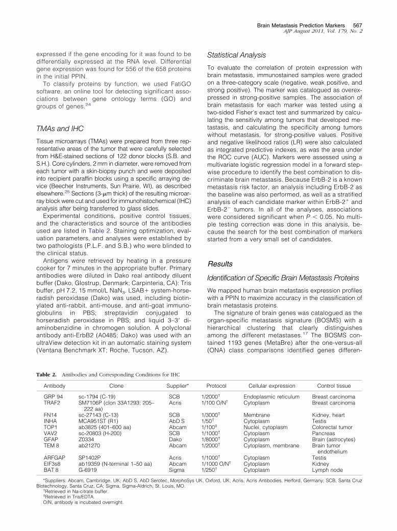
Brain Metastasis Prediction Markers 567AJP August 2011, Vol. 179, No. 2
expressed if the gene encoding for it was found to bedifferentially expressed at the RNA level. Differentialgene expression was found for 556 of the 658 proteinsin the initial PPIN.
To classify proteins by function, we used FatiGOsoftware, an online tool for detecting significant asso-ciations between gene ontology terms (GO) andgroups of genes.24
TMAs and IHC
Tissue microarrays (TMAs) were prepared from three rep-resentative areas of the tumor that were carefully selectedfrom H&E-stained sections of 122 donor blocks (S.B. andS.H.). Core cylinders, 2 mm in diameter, were removed fromeach tumor with a skin-biopsy punch and were depositedinto recipient paraffin blocks using a specific arraying de-vice (Beecher Instruments, Sun Prairie, WI), as describedelsewhere.25 Sections (3-�m thick) of the resulting microar-ray block were cut and used for immunohistochemical (IHC)analysis after being transferred to glass slides.
Experimental conditions, positive control tissues,and the characteristics and source of the antibodiesused are listed in Table 2. Staining optimization, eval-uation parameters, and analyses were established bytwo pathologists (P.L.F. and S.B.) who were blinded tothe clinical status.
Antigens were retrieved by heating in a pressurecooker for 7 minutes in the appropriate buffer. Primaryantibodies were diluted in Dako real antibody diluentbuffer (Dako, Glostrup, Denmark; Carpinteria, CA): Trisbuffer, pH 7.2, 15 mmol/L NaN3. LSAB� system-horse-radish peroxidase (Dako) was used, including biotin-ylated anti-rabbit, anti-mouse, and anti-goat immuno-globulins in PBS; streptavidin conjugated tohorseradish peroxidase in PBS; and liquid 3–3= di-aminobenzidine in chromogen solution. A polyclonalantibody anti-ErbB2 (A0485; Dako) was used with anultraView detection kit in an automatic staining system(Ventana Benchmark XT; Roche, Tucson, AZ).
Table 2. Antibodies and Corresponding Conditions for IHC
Antibody Clone Supplier*
GRP 94 sc-1794 (C-19) SCBTRAF2 SM7106P (clon 33A1293; 205–
222 aa)Acris
FN14 sc-27143 (C-13) SCBINHA MCA951ST (R1) AbD STOP1 ab3825 (401–600 aa) AbcamVAV2 sc-20803 (H-200) SCBGFAP Z0334 DakoTEM 8 ab21270 Abcam
ARFGAP SP1402P AcrisEIF3s8 ab19359 (N-terminal 1–50 aa) AbcamBAT 8 G-6919 Sigma
*Suppliers: Abcam, Cambridge, UK; AbD S, AbD Serotec, MorphoSyBiotechnology, Santa Cruz, CA; Sigma, Sigma-Aldrich, St. Louis, MO.
†Retrieved in Na-citrate buffer.‡
Retrieved in Tris/EDTA.O/N, antibody is incubated overnight.Statistical Analysis
To evaluate the correlation of protein expression withbrain metastasis, immunostained samples were gradedon a three-category scale (negative, weak positive, andstrong positive). The marker was catalogued as overex-pressed in strong-positive samples. The association ofbrain metastasis for each marker was tested using atwo-sided Fisher’s exact test and summarized by calcu-lating the sensitivity among tumors that developed me-tastasis, and calculating the specificity among tumorswithout metastasis, for strong-positive values. Positiveand negative likelihood ratios (LR) were also calculatedas integrated predictive indexes, as was the area underthe ROC curve (AUC). Markers were assessed using amultivariate logistic regression model in a forward step-wise procedure to identify the best combination to dis-criminate brain metastasis. Because ErbB-2 is a knownmetastasis risk factor, an analysis including ErbB-2 asthe baseline was also performed, as well as a stratifiedanalysis of each candidate marker within ErbB-2� andErbB-2� tumors. In all of the analyses, associationswere considered significant when P � 0.05. No multi-ple testing correction was done in this analysis, be-cause the search for the best combination of markersstarted from a very small set of candidates.
Results
Identification of Specific Brain Metastasis Proteins
We mapped human brain metastasis expression profileswith a PPIN to maximize accuracy in the classification ofbrain metastasis proteins.
The signature of brain genes was catalogued as theorgan-specific metastasis signature (BOSMS) with ahierarchical clustering that clearly distinguishesamong the different metastases.17 The BOSMS con-tained 1193 genes (MetaBre) after the one-versus-all(ONA) class comparisons identified genes differen-
rotocol Cellular expression Control tissue
000† Endoplasmic reticulum Breast carcinoma00 O/N† Cytoplasm Breast carcinoma
000† Membrane Kidney, heart0† Cytoplasm Testis00‡ Nuclei, cytoplasm Colorectal tumor000† Cytoplasm Pancreas000† Cytoplasm Brain (astrocytes)000† Cytoplasm, membrane Brain tumor
endothelium000† Cytoplasm Testis000 O/N† Cytoplasm Kidney50† Cytoplasm Lymph node
xford, UK; Acris, Acris Antibodies, Herford, Germany; SCB, Santa Cruz
P
1/21/1
1/31/51/11/11/81/2
1/11/11/2
s UK, O
568 Sanz-Pamplona et alAJP August 2011, Vol. 179, No. 2
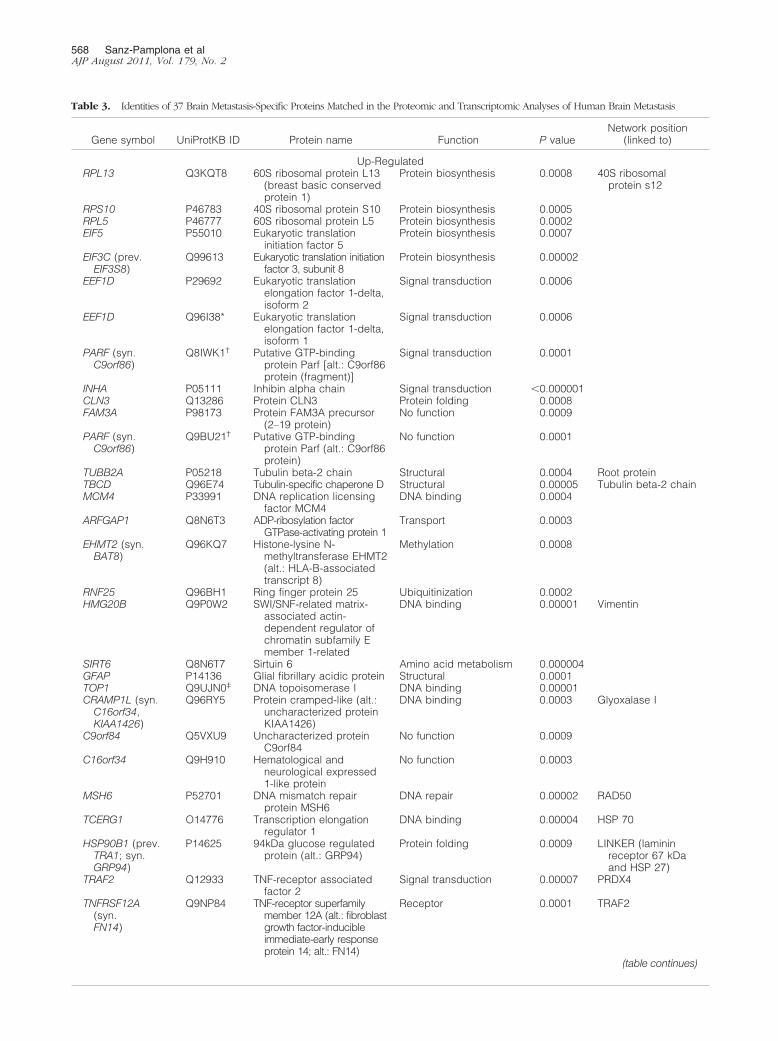
Table 3. Identities of 37 Brain Metastasis-Specific Proteins Matched in the Proteomic and Transcriptomic Analyses of Human Brain Metastasis
Gene symbol UniProtKB ID Protein name Function P valueNetwork position
(linked to)
Up-RegulatedRPL13 Q3KQT8 60S ribosomal protein L13
(breast basic conservedprotein 1)
Protein biosynthesis 0.0008 40S ribosomalprotein s12
RPS10 P46783 40S ribosomal protein S10 Protein biosynthesis 0.0005RPL5 P46777 60S ribosomal protein L5 Protein biosynthesis 0.0002EIF5 P55010 Eukaryotic translation
initiation factor 5Protein biosynthesis 0.0007
EIF3C (prev.EIF3S8)
Q99613 Eukaryotic translation initiationfactor 3, subunit 8
Protein biosynthesis 0.00002
EEF1D P29692 Eukaryotic translationelongation factor 1-delta,isoform 2
Signal transduction 0.0006
EEF1D Q96I38* Eukaryotic translationelongation factor 1-delta,isoform 1
Signal transduction 0.0006
PARF (syn.C9orf86)
Q8IWK1† Putative GTP-bindingprotein Parf [alt.: C9orf86protein (fragment)]
Signal transduction 0.0001
INHA P05111 Inhibin alpha chain Signal transduction �0.000001CLN3 Q13286 Protein CLN3 Protein folding 0.0008FAM3A P98173 Protein FAM3A precursor
(2–19 protein)No function 0.0009
PARF (syn.C9orf86)
Q9BU21† Putative GTP-bindingprotein Parf (alt.: C9orf86protein)
No function 0.0001
TUBB2A P05218 Tubulin beta-2 chain Structural 0.0004 Root proteinTBCD Q96E74 Tubulin-specific chaperone D Structural 0.00005 Tubulin beta-2 chainMCM4 P33991 DNA replication licensing
factor MCM4DNA binding 0.0004
ARFGAP1 Q8N6T3 ADP-ribosylation factorGTPase-activating protein 1
Transport 0.0003
EHMT2 (syn.BAT8)
Q96KQ7 Histone-lysine N-methyltransferase EHMT2(alt.: HLA-B-associatedtranscript 8)
Methylation 0.0008
RNF25 Q96BH1 Ring finger protein 25 Ubiquitinization 0.0002HMG20B Q9P0W2 SWI/SNF-related matrix-
associated actin-dependent regulator ofchromatin subfamily Emember 1-related
DNA binding 0.00001 Vimentin
SIRT6 Q8N6T7 Sirtuin 6 Amino acid metabolism 0.000004GFAP P14136 Glial fibrillary acidic protein Structural 0.0001TOP1 Q9UJN0‡ DNA topoisomerase I DNA binding 0.00001CRAMP1L (syn.
C16orf34,KIAA1426)
Q96RY5 Protein cramped-like (alt.:uncharacterized proteinKIAA1426)
DNA binding 0.0003 Glyoxalase I
C9orf84 Q5VXU9 Uncharacterized proteinC9orf84
No function 0.0009
C16orf34 Q9H910 Hematological andneurological expressed1-like protein
No function 0.0003
MSH6 P52701 DNA mismatch repairprotein MSH6
DNA repair 0.00002 RAD50
TCERG1 O14776 Transcription elongationregulator 1
DNA binding 0.00004 HSP 70
HSP90B1 (prev.TRA1; syn.GRP94)
P14625 94kDa glucose regulatedprotein (alt.: GRP94)
Protein folding 0.0009 LINKER (lamininreceptor 67 kDaand HSP 27)
TRAF2 Q12933 TNF-receptor associatedfactor 2
Signal transduction 0.00007 PRDX4
TNFRSF12A(syn.FN14)
Q9NP84 TNF-receptor superfamilymember 12A (alt.: fibroblastgrowth factor-inducibleimmediate-early responseprotein 14; alt.: FN14)
Receptor 0.0001 TRAF2
(table continues)
cessionsyn., g
Brain Metastasis Prediction Markers 569AJP August 2011, Vol. 179, No. 2
tially expressed in the 4 brain metastases versus the 19metastases to other organs.
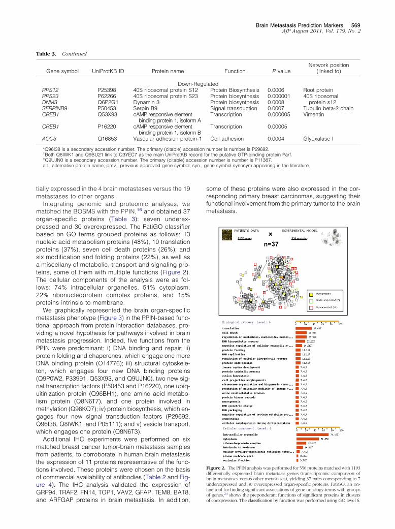
Integrating genomic and proteomic analyses, wematched the BOSMS with the PPIN,16 and obtained 37organ-specific proteins (Table 3): seven underex-pressed and 30 overexpressed. The FatiGO classifierbased on GO terms grouped proteins as follows: 13nucleic acid metabolism proteins (48%), 10 translationproteins (37%), seven cell death proteins (26%), andsix modification and folding proteins (22%), as well asa miscellany of metabolic, transport and signaling pro-teins, some of them with multiple functions (Figure 2).The cellular components of the analysis were as fol-lows: 74% intracellular organelles, 51% cytoplasm,22% ribonucleoprotein complex proteins, and 15%proteins intrinsic to membrane.
We graphically represented the brain organ-specificmetastasis phenotype (Figure 3) in the PPIN-based func-tional approach from protein interaction databases, pro-viding a novel hypothesis for pathways involved in brainmetastasis progression. Indeed, five functions from thePPIN were predominant: i) DNA binding and repair; ii)protein folding and chaperones, which engage one moreDNA binding protein (O14776); iii) structural cytoskele-ton, which engages four new DNA binding proteins(Q9P0W2, P33991, Q53X93, and Q9UJN0), two new sig-nal transcription factors (P50453 and P16220), one ubiq-uitinization protein (Q96BH1), one amino acid metabo-lism protein (Q8N6T7), and one protein involved inmethylation (Q96KQ7); iv) protein biosynthesis, which en-gages four new signal transduction factors (P29692,Q96I38, Q8IWK1, and P05111); and v) vesicle transport,which engages one protein (Q8N6T3).
Additional IHC experiments were performed on sixmatched breast cancer tumor-brain metastasis samplesfrom patients, to corroborate in human brain metastasisthe expression of 11 proteins representative of the func-tions involved. These proteins were chosen on the basisof commercial availability of antibodies (Table 2 and Fig-ure 4). The IHC analysis validated the expression ofGRP94, TRAF2, FN14, TOP1, VAV2, GFAP, TEM8, BAT8,
Table 3. Continued
Gene symbol UniProtKB ID Protein name
DownRPS12 P25398 40S ribosomal protein SRPS23 P62266 40S ribosomal protein SDNM3 Q6P2G1 Dynamin 3SERPINB9 P50453 Serpin B9CREB1 Q53X93 cAMP responsive elemen
binding protein 1, isofoCREB1 P16220 cAMP responsive elemen
binding protein 1, isofoAOC3 Q16853 Vascular adhesion prot
*Q96I38 is a secondary accession number. The primary (citable) acc†Both Q8IWK1 and Q9BU21 link to Q3YEC7 as the main UniProtKB r‡Q9UJN0 is a secondary accession number. The primary (citable) acalt., alternative protein name; prev., previous approved gene symbol;
and ARFGAP proteins in brain metastasis. In addition,
some of these proteins were also expressed in the cor-responding primary breast carcinomas, suggesting theirfunctional involvement from the primary tumor to the brainmetastasis.
Figure 2. The PPIN analysis was performed for 556 proteins matched with 1193differentially expressed brain metastasis genes (transcriptomic comparison ofbrain metastases versus other metastases), yielding 37 pairs corresponding to 7underexpressed and 30 overexpressed organ-specific proteins. FatiGO, an on-line tool for finding significant associations of gene ontology-terms with groups
24
Function P valueNetwork position
(linked to)
latedProtein Biosynthesis 0.0006 Root proteinProtein biosynthesis 0.000001 40S ribosomal
protein s12Protein biosynthesis 0.0008Signal transduction 0.0007 Tubulin beta-2 chainTranscription 0.000005 Vimentin
Transcription 0.00005
Cell adhesion 0.0004 Glyoxalase I
number is number is P29692.r the putative GTP-binding protein Parf.number is number is P11387.
ene symbol synonym appearing in the literature.
-Regu1223
trm Atrm Bein-1
essionecord fo
of genes, shows the preponderant functions of significant proteins in clustersof coexpression. The classification by function was performed using GO level 6.

alysis; bare un
570 Sanz-Pamplona et alAJP August 2011, Vol. 179, No. 2
We searched for references to brain metastasis signa-tures in published genomic data from experimental andclinical breast cancer and metastasis analysis. From our listof genes, only seven appeared in previous lists of geneexpression profiling predicting clinical outcomes of breastcancer26–35 (Table 4): EEF1D, MCM4, RPL5, RPS12, andCLN326 and also FAM3A and TBCD.27 GFAP, encoding aubiquitous protein in the central nervous system, also ap-peared in a list of genes differentially expressed betweenbrain and bone breast cancer metastasis.35
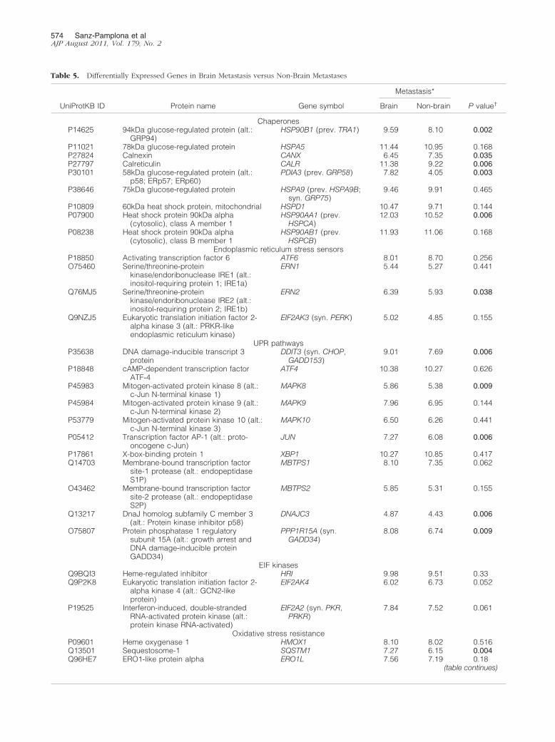
These findings indicate that cells metastasizing inbrain were enriched in cell structure, chaperones,stress and redox regulation, and intracellular transportproteins. The organ-specific character of this func-tional signature was also found in the transcriptomicdata from breast cancer brain metastasis (Figure 5 andTable 5). From these, the most differentially expressedin brain metastasis, compared with metastases in otherorgans, were GRP94 (P � 0.002), FN14 (P � 0.002),ARFGAP1 (P � 0.003), TRAF2 (P � 0.003), and PDG-FRA (0.002) genes. In contrast, other functions had norelevant expression in brain; for example, amino acidmetabolism genes were overexpressed only in liver.
After mapping transcriptomic into proteomic analyses,we concluded that molecules involved in protein foldingand chaperones might connect different functions andpresumably act by rescuing cells from endoplasmic re-
Figure 3. Functional classification of the PPIN. Proteins in functional clusterindicated in black type; the 37 brain metastasis proteins are indicated in whiboxes in dark gray indicate new functions added from the transcriptomic anproteomics and transcriptomic analysis. Proteins that were validated by IHC
ticulum stress responses.
Expression of Endoplasmic Reticulum StressPhenotype in Breast Cancer Primary Tumors IsAssociated with Brain Metastasis Progression
Because proteins were also expressed in primary tumors,to estimate the probability of specific brain metastasisoutcomes we further analyzed the proteins in a series ofprimary breast carcinomas using TMA technology. Weconsidered a marker to be positive when strong expres-sion was detected, to avoid false positives, and takinginto account the known expression in a control tissue(Figure 6).
Statistical analysis of the data showed significant as-sociations between brain metastasis progression andhigh expression of GRP94 (P � 0.0001), TRAF2 (P �0.001), and FN14 (P � 0.0001). As expected, ErbB-2expression was associated with brain metastasis with ahigh significance (P � 0.0001): 8/13 (62%) breast can-cers that progressed to brain metastasis were positive,versus 12% and 5% of breast carcinomas that relapsedin other locations or without metastasis (7/57 and 2/42,respectively). ErbB-2 expression was also associatedwith the absence of hormone receptors: ER, 55% versus30% (6/11 and 29/98, respectively, P � 0.016); PR, 73%versus 39% (8/11 versus 37/95, respectively, P � 0.009).A slight association with histological grade (HG) was alsoobserved: HG III 58% versus 47% (7/12 versus 45/96,
uped within a single box containing root and linker proteins. Functions areBoxes in light gray indicate the previous network16 of brain metastatic cells;oxes shaded from light to dark represent redundant functions identified in
derlined.
s are grote type.
P � 0.105). In addition, we did not find correlation be-

etastasis
Brain Metastasis Prediction Markers 571AJP August 2011, Vol. 179, No. 2
tween lung, bone, or liver metastasis and high expressionof these proteins.
We calculated the positive and negative likelihood ra-tios (LR) to assess the predictive accuracy of each mol-ecule as a brain metastasis marker, considering the sen-sitivity and the specificity of each (Table 6). The highestpredictive value for the presence of metastasis wasErbB-2 expression (positive LR � 6.7, P � 0.0001), fol-lowed by FN14 (positive LR � 3.01, P � 0.001), GRP94(positive LR � 1.89 P � 0.003), and TRAF2 (positiveLR � 1.67, P � 0.055). Furthermore, GRP94 was the bestnegative predictive marker (negative LR � 0.16), fol-lowed by TRAF2 (negative LR � 0.35), FN14 (negativeLR � 0.40), and ErbB-2 (negative LR � 0.42). Thus, theabsence of the endoplasmic reticulum stress (ERS) re-sponse phenotype in tumors predicted the absence ofbrain metastasis.
For a validation set, we used a series of 295 breasttumors for which the transcriptomic data were publiclyavailable.18,19 As expected, the highest predictive valuewas ErbB-2 expression (positive LR � 8.27, P �0.00001). Moreover, TRAF2 (positive LR � 2.36, P �0.026), GRP94 (positive LR � 1.72 P � 0.028), and FN14(positive LR � 1.78, P � 0.044) were associated withbrain metastasis.
A multivariate analysis based on stepwise logistic re-gression retained GRP94, FN14, and inhibin as the bestcombination to discriminate brain metastasis. The AUC
Figure 4. Validation at the protein expression level (brown) in matched tfunctional-type proteins in representative paraffin-embedded tumor-brain mOriginal magnification: �10 (H&E stain); �20 (all others).
value for this combination was 0.85 (95% CI � 0.73 to
0.96), substantially better than that for ErbB-2, whichwas the variable more strongly associated with brainmetastasis (AUC � 0.76, 95% CI � 0.58 to 0.93). Themodel that combined ErbB-2, GRP94, FN14, and in-hibin expression further increased the discrimination ofmetastatic disease in brain (AUC � 0.91, 95% CI �0.77 to 1.00). The ROC curves for the three models areshown in Figure 7.
We also performed a stratified analysis to check therelationship between ErbB-2 positivity and ERS re-sponse phenotype in binary combinations (Table 7). InErbB-2� tumors, FN14 had a high negative likelihoodratio to predict the absence of brain metastasis pro-gression (LR � 0.26, sensitivity � 0.8, P � 0.015).
Discussion
Fewer than 10% of breast cancer patients have detect-able distant metastasis at diagnosis.36 Thus, it is nec-essary to understand the properties of brain-tropic tu-mor cells to identify patients with risk of brainmetastasis and to effectively prevent it. Because weassume that metastasis colonization could alreadybe underway at the time of diagnosis, the ERS re-sponse phenotype might be a predictive tool to helpdecide on treatment under the risk of brain metastasis
ain metastasis samples by means of IHC analysis to identify representativepairs. H&E staining of each tissue is shown as viewed by light microscopy.
umor-br
progression.

572 Sanz-Pamplona et alAJP August 2011, Vol. 179, No. 2
The phenotype includes the overexpression of GRP94,FN14, and TRAF2, which are well correlated with brainmetastasis in breast cancer patients. Our search for amultivariate panel of markers to predict brain metastasisrevealed that the combination of GRP94, FN14, and in-hibin together has a better discriminate accuracy thanErbB-2 alone, and that the best accuracy is obtainedcombining all four markers. Although all variables inthese models were significantly associated with brainmetastasis in the multivariate models, the increase inpredictive accuracy measured by the difference in AUCwas not (because of the small sample size in the present
Table 4. In silico Validation of the Endoplasmic Reticulum StresReports
Reference Array platform Description o
26 Agilent 24479 60-mer oligos 97 Samples fpatients
30 Rosetta inkjet (24479 genes;breast adenocarcinoma)oligonucleotide microarray
279 Primary tdiverse typbreast, pro
27 Multiple gene expressionsignatures “metagenes”
86 LN� breaspatients
28 Affymetrix U133A 25-meroligos
LN� and LN�
patients wiinvasive brcancer
33 Affymetrix U133A 82 Breast capatients (ptumors)
31 Agilent 24479 60-mer oligos 161 PatientsI and II brecancer with�53 years
29 Agilent 22575 60-mer oligos 135 Tumor sa(no criteriaselection)
32 Operon 70-mer two-color21239 probes
35 Patients: ptumor andnode metapaired sam
35 Affymetrix U133A 8 Bone meta18 brainmetastasesprimary tum
19 Affymetrix U133A CN34-BrM2 aMDA231-Bbrain metacell lines.
368 Breast cprimary tum
LN, lymph node.
study). Indeed, TRAF2, which was associated with brain
metastasis, had many missing values and could not beincluded in the multivariate analysis. The ERS responsephenotype is indicative of a new tool to discriminate therisk of brain metastasis in both ErbB-2� and ErbB-2�
breast cancers. Moreover, the absence of the ERS re-sponse phenotype in tumors might predict the absenceof metastasis.
These biomarkers can help in selection of treatmentstrategies, furthering the current ambitious aim of iden-tifying treatment strategies that will cure patients withErbB-2� disease while ensuring minimal toxicity foreach individual patient.37 Hicks et al38 reported that
type, Taking Into Account Previous Experimental And Clinical
le Gene signature Match to present study
� 231 Prognosis reporters(risk of distantmetastasis)
0
430 Brca1 reporters 3 (EEF1D, MCM4, RPL5)2460 ER� reporters 3 (CLN3, MCM4, RPS12)
ofg,
128 Genes able todistinguish patientswith good versuspoor prognosis
0
er 143 Predictors of lymphnode metastasis
0
165 Predictors of breastcancer recurrence
2 (FAM3A, TBCD)
76-Gene signature todistinguish LN�
primary breastcancer to developdistant metastasiswithin 5 years
0
95 Genes predictors oflung metastasis
0
e 70-Gene predictor oflocal recurrence
0
70-Gene prognosticsignature (risk ofmetastasis)
0
79 Differentiallyexpressed genesbetween primarysamples andmetastasis samples
0
51 Brain metastasisspecific genes(versus bonemetastasis)
1 (GFAP)
17 Genes whoseexpression wascorrelated with brainrelapse
0
26 Genes whoseexpression wasincreased in brainmetastatic cell linesbut not in bone orlung metastatic celllines
0
s Pheno
f samp
rom LN
umorses (lunstate)
t canc
theast
ncerrimary
in stagastage
mplesfor
rimarylymphstasisples
stases,
and 3orsndrM2static
ancerors
EGFR expression, like ErbB-2, predicted the develop-

ormalize
Brain Metastasis Prediction Markers 573AJP August 2011, Vol. 179, No. 2
ment of brain metastasis. The incidence of brain me-tastasis in patients with breast cancer overexpressingErbB-2 who are being treated with trastuzumab is dou-ble that in other breast cancer patients; one-third willdevelop central nervous system metastasis, and thisoften occurs when they are responding to therapy atother sites or have a stable disease.39 One of theclinical questions is whether this receptor remains aviable target after disease progression,40 and whethertrastuzumab treatment can prevent brain metastasis orwhether it encourages the development of metastaticcells that have crossed the blood-brain barrier.
GRP94 overexpression in brain metastasis mightmodulate ERS responses through activation of PERK,ATF6, and IRE1.41 Downstream from GRP94 activation,transcription of chaperones and protein degradationmight increase in brain metastatic cells. (We are cur-rently exploring these pathways in our laboratory.) Be-cause therapy decisions should depend on the tumorphenotype, the known close correlation between brainmetastasis potential and ERS response phenotype inprimary tumors suggests that HSP90 and proteasomeinhibitors might be alternatives for treatment of breastcancer patients with a high risk of brain metastasis.42 Ithas been reported that inhibition of HSP90, whichhelps expression and folding of its client proteins, such
Figure 5. Differentially expressed genes in brain metastasis (black), compagray), based on a Mann-Whitney test calculated for each gene using the ndifferent expression between bracketed organs.
as ErbB-2, could simultaneously inhibit the expression
of viable receptors.40 Furthermore, the expression ofGRP94 has been associated with poor prognosis ingastric carcinomas,43 and with chemotherapy resis-tance of lung cancer cells and ovarian carcinomacells.44
The switch from dormancy to growth of cancer cellsin the brain may be dependent on stress responsemechanisms, subsequent coordination of detoxifica-tion and redox pathways,16 and cytokines produced byglial cells, which may contribute, in a paracrine man-ner, to the final development of brain metastasis. Weidentified overexpression of the FN14 gene, a memberof the tumor necrosis factor (TNF) superfamily of re-ceptors.45 FN14 is an immediate early response genewhose expression is directly activated after exposureto growth factors in fibroblasts; it is up-regulated inmigration-stimulated glioma cells in vitro, and it hasbeen associated with high-grade tumors.46 Throughactivation of MAPK8/JNK and NF-�B, the TRAF pro-teins mediate signal transduction of the TNF receptorsuperfamily members47; they could connect ERS re-sponses and FN14 signaling pathway activation. Be-cause FN14 and TRAF2 are overexpressed in breastcancer tumors that develop brain metastasis, and inbrain metastatic cells, the disruption of the TWEAK/FN14 feedback loop also emerges as a molecular tar-
metastases in other organs: lung (dark gray), bone (white), and liver (lightd log intensities (see further in Table 5). *P � 0.05, statistically significant
red with
geting strategy.
574 Sanz-Pamplona et alAJP August 2011, Vol. 179, No. 2
Table 5. Differentially Expressed Genes in Brain Metastasis versus Non-Brain Metastases
UniProtKB ID Protein name Gene symbol
Metastasis*
P value†Brain Non-brain
ChaperonesP14625 94kDa glucose-regulated protein (alt.:
GRP94)HSP90B1 (prev. TRA1) 9.59 8.10 0.002
P11021 78kDa glucose-regulated protein HSPA5 11.44 10.95 0.168P27824 Calnexin CANX 6.45 7.35 0.035P27797 Calreticulin CALR 11.38 9.22 0.006P30101 58kDa glucose-regulated protein (alt.:
p58; ERp57; ERp60)PDIA3 (prev. GRP58) 7.82 4.05 0.003
P38646 75kDa glucose-regulated protein HSPA9 (prev. HSPA9B;syn. GRP75)
9.46 9.91 0.465
P10809 60kDa heat shock protein, mitochondrial HSPD1 10.47 9.71 0.144P07900 Heat shock protein 90kDa alpha
(cytosolic), class A member 1HSP90AA1 (prev.
HSPCA)12.03 10.52 0.006
P08238 Heat shock protein 90kDa alpha(cytosolic), class B member 1
HSP90AB1 (prev.HSPCB)
11.93 11.06 0.168
Endoplasmic reticulum stress sensorsP18850 Activating transcription factor 6 ATF6 8.01 8.70 0.256O75460 Serine/threonine-protein
kinase/endoribonuclease IRE1 (alt.:inositol-requiring protein 1; IRE1a)
ERN1 5.44 5.27 0.441
Q76MJ5 Serine/threonine-proteinkinase/endoribonuclease IRE2 (alt.:inositol-requiring protein 2; IRE1b)
ERN2 6.39 5.93 0.038
Q9NZJ5 Eukaryotic translation initiation factor 2-alpha kinase 3 (alt.: PRKR-likeendoplasmic reticulum kinase)
EIF2AK3 (syn. PERK) 5.02 4.85 0.155
UPR pathwaysP35638 DNA damage-inducible transcript 3
proteinDDIT3 (syn. CHOP,
GADD153)9.01 7.69 0.006
P18848 cAMP-dependent transcription factorATF-4
ATF4 10.38 10.27 0.626
P45983 Mitogen-activated protein kinase 8 (alt.:c-Jun N-terminal kinase 1)
MAPK8 5.86 5.38 0.009
P45984 Mitogen-activated protein kinase 9 (alt.:c-Jun N-terminal kinase 2)
MAPK9 7.96 6.95 0.144
P53779 Mitogen-activated protein kinase 10 (alt.:c-Jun N-terminal kinase 3)
MAPK10 6.50 6.26 0.441
P05412 Transcription factor AP-1 (alt.: proto-oncogene c-Jun)
JUN 7.27 6.08 0.006
P17861 X-box-binding protein 1 XBP1 10.27 10.85 0.417Q14703 Membrane-bound transcription factor
site-1 protease (alt.: endopeptidaseS1P)
MBTPS1 8.10 7.35 0.062
O43462 Membrane-bound transcription factorsite-2 protease (alt.: endopeptidaseS2P)
MBTPS2 5.85 5.31 0.155
Q13217 DnaJ homolog subfamily C member 3(alt.: Protein kinase inhibitor p58)
DNAJC3 4.87 4.43 0.006
O75807 Protein phosphatase 1 regulatorysubunit 15A (alt.: growth arrest andDNA damage-inducible proteinGADD34)
PPP1R15A (syn.GADD34)
8.08 6.74 0.009
EIF kinasesQ9BQI3 Heme-regulated inhibitor HRI 9.98 9.51 0.33Q9P2K8 Eukaryotic translation initiation factor 2-
alpha kinase 4 (alt.: GCN2-likeprotein)
EIF2AK4 6.02 6.73 0.052
P19525 Interferon-induced, double-strandedRNA-activated protein kinase (alt.:protein kinase RNA-activated)
EIF2A2 (syn. PKR,PRKR)
7.84 7.52 0.061
Oxidative stress resistanceP09601 Heme oxygenase 1 HMOX1 8.10 8.02 0.516Q13501 Sequestosome-1 SQSTM1 7.27 6.15 0.004Q96HE7 ERO1-like protein alpha ERO1L 7.56 7.19 0.18
(table continues)

Brain Metastasis Prediction Markers 575AJP August 2011, Vol. 179, No. 2
Table 5. Continued
UniProtKB ID Protein name Gene symbol
Metastasis*
P value†Brain Non-brain
ProteasomeQ92611 ER degradation-enhancing alpha-
mannosidase-like 1EDEM1 6.53 5.41 0.088
O43242 26S proteasome regulatory subunit S3 PSMD3 9.38 7.45 0.043Q86TM6 E3 ubiquitin-protein ligase synoviolin SYVN1 8.59 8.77 0.441Q9UBV2 Protein sel-1 homolog 1 (alt.: suppressor
of lin-12-like protein 1)SEL1L 8.36 9.05 0.871
Q96DZ1 Endoplasmic reticulum lectin 1 (alt.: XTP3-transactivated gene B protein; XTP-3)
ERLEC1 (prev. C2orf30) 9.54 9.93 0.57
Glucose transportersP11166 Solute carrier family 2, facilitated glucose
transporter member 1 (alt.: GLUT-1)SLC2A1 8.96 7.97 0.081
P11168 Solute carrier family 2, facilitated glucosetransporter member 2 (alt.: GLUT-2)(liver)
SLC2A2 3.43 4.64 0.123
P11169 Solute carrier family 2, facilitated glucosetransporter member 3 (alt.: GLUT-3)(brain)
SLC2A3 6.60 6.13 0.871
P22732 Solute carrier family 2, facilitated glucosetransporter member 5 (alt.: GLUT-5)
SLC2A5 5.33 5.07 0.074
Q9UGQ3 Solute carrier family 2, facilitated glucosetransporter member 6 (alt.: GLUT-6)
SLC2A6 6.03 5.60 0.074
Q9NY64 Solute carrier family 2, facilitated glucosetransporter member 8 (alt.: GLUT-9)
SLC2A8 7.43 6.72 0.035
Amino acid metabolismP48067 Sodium- and chloride-dependent glycine
transporter 1 (alt.: GlyT-1)SLC6A9 5.06 4.78 0.035
P08243 Asparagine synthetase ASNS 7.78 8.15 0.49P32929 Cystathionine gamma-lyase CTH 4.06 4.75 0.144P11586 Methylenetetrahydrofolate
dehydrogenaseMTHFD1 8.07 8.48 0.035
Protein transportQ8N6T3 ADP-ribosylation factor GTPase-activating
protein 1ARFGAP1 7.25 6.21 0.003
P09496 Clathrin light chain A CLTA 5.51 4.80 0.009P09497 Clathrin light chain B CLTB 4.66 4.11 0.015Q00610 Clathrin heavy chain 1 CLTC 12.25 12.07 0.685P53675 Clathrin heavy chain 2 CLTCL1 6.86 6.72 0.33P61966 AP-1 complex subunit sigma-1A (alt.:
sigma-adaptin 1A)AP1S1 (prev. CLAPS1) 6.02 5.12 0.009
P20340 Ras-related protein Rab-6A RAB6A 6.66 6.30 0.074P61019 Ras-related protein Rab-2A RAB2A (prev. RAB2) 9.67 8.73 0.043P61106 Ras-related protein Rab-14 RAB14 9.65 8.38 0.002O95197 Reticulon-3 RTN3 9.19 8.73 0.019
Receptors and signal transductorsP04626 Receptor tyrosine-protein kinase erbB-2 ERBB2 7.50 4.17 0.009O95407 Tumor necrosis factor receptor
superfamily member 6BTNFRSF6B 7.37 6.85 0.003
Q9NS68 Tumor necrosis factor receptorsuperfamily member 19
TNFRSF19 5.14 4.53 0.006
Q9NP84 Tumor necrosis factor receptorsuperfamily member 12A (alt.: FN14)
TNFRSF12A (syn. FN14) 9.07 7.68 0.002
Q9HAV5 Ectodysplasin-A2 receptor (alt.: tumornecrosis factor receptor superfamilymember 27)
EDA2R (syn.TNFRSF27)
5.57 5.41 0.516
P16234 Alpha-type platelet-derived growth factorreceptor
PDGFRA 5.19 9.09 0.002
P00533 Epidermal growth factor receptor EGFR 6.92 6.42 0.012P17948 Vascular endothelial growth factor
receptor 1FLT1 7.09 7.47 0.088
Q13077 TNF receptor-associated factor 1 TRAF1 5.91 5.99 0.655Q12933 TNF receptor-associated factor 2 TRAF2 6.85 6.20 0.003Q13114 TNF receptor-associated factor 3 TRAF3 6.40 6.63 0.417Q9BUZ4 TNF receptor-associated factor 4 TRAF4 6.60 5.39 0.035O00463 TNF receptor-associated factor 5 TRAF5 6.75 7.55 0.330
*Based on a Mann-Whitney U-test calculated for each gene using normalized log-intensities.†
Statistically significant differences are highlighted in bold face type.alt., alternative name (proteins); prev., previously approved symbol (genes).
576 Sanz-Pamplona et alAJP August 2011, Vol. 179, No. 2
Figure 6. ERS response phenotype in breast cancer at first diagnosis. Representative tabulation of protein expression in breast cancer tissues. Tissues areshown as viewed by light microscopy. Low and medium intensities of staining were considered negative for semiquantitative purposes, and only tumors
with high intensity staining were considered positive. Insets: Standard positive control tissue sample used in each determination. Original magnification,�10.Table 6. Risk of Brain Metastasis Associated with Each Marker in Breast Cancer
Brain metastasis marker UniProtKB ID Sensitivity* Specificity*
LR
P value†Pos Neg
ErbB-2 P04626 8/13 (61.5) 90/99 (90.9) 6.70 0.42 �0.0001GRP94 P14625 12/13 (92.0) 55/107 (51.4) 1.89 0.16 0.003FN14 Q9NP84 9/13 (69.2) 80/104 (77.0) 3.01 0.40 0.001TRAF2 Q12933 9/11 (81.8) 45/88 (51.1) 1.67 0.35 0.055VAV2 P52735 2/13 (15,4) 95/107 (88.8) 1.38 0.95 0.65TOP1 Q9UJN0‡ 4/13 (30.8) 91/105 (86.6) 2.30 0.80 0.11Inhibin P05111) 0/13 (0) 97/107 (90.7) 0 1.10 0.60
*Variation in denominators derives from missing values in the IHC (eg, tissue lost, unviable staining, or background).†Fisher’s exact test, 2-sided.‡
Secondary accession number. The primary (citable) accession number is number is P11387.LR, likelihood ratio; Neg, negative LR; Pos, positive LR.
Brain Metastasis Prediction Markers 577AJP August 2011, Vol. 179, No. 2
FN14 overexpression can stimulate survival throughinteraction with the inhibitor-of-apoptosis proteins(IAPs).48 TNF-like weak inducer of apoptosis (TWEAK)/FN14 signaling recruits cIAP1-TRAF2 complex, whichis then targeted for lysosomal degradation. Cell sensi-tivity to TWEAK correlates with sensitivity to syntheticIAP antagonist. Studies with FN14-overexpressing tu-mor cells that could be selectively destroyed using aTWEAK-cytotoxin protein fusion suggest that FN14could be a new molecular target for treating metasta-sis.49 Indeed, the ERS response phenotype might in-dicate new opportunities in anticancer strategies forsanctuary sites and micrometastatic disease. Evidenceof such phenotypes can be used to develop morespecifically addressed therapies.
Microarray-based gene studies are difficult to inter-pret, because of the huge amount of data involved, andit is therefore a challenge to derive biological insights.
ROC CurveROC Curve
ErbB2GRP94+FN14+InhibinGRP94+FN14+Inhibin+ErbB2
ErbB2GRP94+FN14+InhibinGRP94+FN14+Inhibin+ErbB2
Sens
itivi
ty
SpecificitySpecificity
Figure 7. The area under the ROC curve (AUC) obtained with the integratedpredictive indexes. Markers were assessed in a multivariate logistic regres-sion model using a forward stepwise procedure to identify the best combi-nation to predict brain metastasis. For ErbB-2 alone, AUC � 0.76; for GRP94,FN14, and inhibin in combination, AUC � 0.85; and for ErbB-2, GRP94,FN14, and inhibin in combination, AUC � 0.91.
Table 7. Risk of Brain Metastasis Associated with Each Marker in
Brain metastasis marker*
ErbB-2�
Sensitivity† Specificity†
Novel markersGRP94 8/8 (100) 2/9 (22.2)FN14 5/8 (62.5) 6/9 (66.7)TRAF2 6/7 (85.7) 5/9 (55.6)
Traditional markersER 3/7 (42.9) 6/8 (75)PR 2/7 (28.6) 6/7 (85.7)Histologic grade III 6/8 (75.0) 3/9 (33.3)
*For novel markers, UniProtKB identifiers are as follows: GRP94, P146ER, P03372; progesterone receptor PR, P06401.
†
Variation in denominators derives from missing values in the IHC (eg, tissue‡Fisher’s test.We applied a PPIN-based approach that identifiesmarkers not as individual genes but as subnetworksextracted from PPINs, thus providing a systemic viewof the interactome. This method serves to filter infor-mation by picking out key protein functions as metas-tasis markers. Indeed, we have delineated a patho-genic mechanism of metastasis to the brain based onthe information from a proteomic study of brain metas-tasis cellular proteins. Further work is needed to con-firm the prognostic value of the ERS response pheno-type in a second validation step that includes a largeindependent group to increase the statistic power ofthe study and to assess the usefulness of the ERSresponse phenotype as a predictive tool at first diag-nosis.
To validate these markers, the main objective is toobtain a large collection of retrospective samples, far inexcess of the typical numbers required to obtain statisti-cal significance in the data. This could also lead to pre-ventive therapy for brain metastases at initial diagnosis,not only in breast cancer patients, but also for othercarcinomas with brain tropism.
Acknowledgments
We thank Dr. Marta Brell from the Neurosurgery Ser-vice and Dr. Sergio Herrero from the Pathology Ser-vice, both at the Bellvitge Hospital (C.S.U.B.), for theirexpert assistance and providing human metastasissamples. We also thank Mr. R. Rycroft for expert lan-guage advice. We acknowledge all of the partners ofthe MetaBre consortium for their collaboration andstimulating criticism: Marc Bracke (Ghent UniversityHospital, Belgium), Roberto Buccione (Consorzio Ma-rio Negri Sud, Italy), Vincent Castronovo (University ofLiège, Belgium), Philippe Clément-Lacroix (Prostrakan,France), Philippe Clézardin (INSERM, France), Su-zanne Eccles (Institute of Cancer Research, UK), AnnaTeti (University of l’Aquila, Italy), Maciej Ugorski (Wro-claw Agriculture University, Poland), and Gabri van derPluijm (Leiden University Medical Center, The Nether-lands).
t Cancer with Regard to ErbB-2 Expression
ErbB-2�
P value‡ Sensitivity† Specificity† P value‡
0.47 4/5 (80) 48/89 (53.9) 0.190.35 4/5 (80) 68/88 (77.3) 0.0150.15 3/4 (75) 36/71 (50.7) 0.62
0.61 2/4 (50) 21/83 (25.3) 0.281.0 1/4 (25) 29/81 (35.8) 0.151.0 1/4 (25) 42/45 (93.3) 0.62
14, Q9NP84; TRAF2, Q12933. For traditional markers: estrogen receptor
Breas
25; FN
lost, unviable staining, or background).

578 Sanz-Pamplona et alAJP August 2011, Vol. 179, No. 2
References
1. Weil RJ, Palmieri DC, Bronder JL, Stark AM, Steeg PS: Breast cancermetastasis to the central nervous system. Am J Pathol 2005, 167:913–920
2. Luck AA, Evans AJ, Green AR, Rakha EA, Paish C, Ellis IO: Theinfluence of basal phenotype on the metastatic pattern of breastcancer. Clin Oncol (R Coll Radiol) 2008, 20:40–45
3. Tosoni A, Ermani M, Brandes AA: The pathogenesis and treatment ofbrain metastases: a comprehensive review. Crit Rev Oncol Hematol2004, 52:199–215
4. Tham YL, Sexton K, Kramer R, Hilsenbeck S, Elledge R: Primarybreast cancer phenotypes associated with propensity for centralnervous system metastases. Cancer 2006, 107:696–704
5. Stemmler HJ, Heinemann V: Central nervous system metastases inHER-2-overexpressing metastatic breast cancer: a treatment chal-lenge. Oncologist 2008, 13:739–750
6. Carey LA, Ewend MG, Metzger R, Sawyer L, Dees EC, Sartor CI,Moore DT, Graham ML: Central nervous system metastases in womenafter multimodality therapy for high risk breast cancer. Breast CancerRes Treat 2004, 88:273–280
7. Slimane K, Andre F, Delaloge S, Dunant A, Perez A, Grenier J,Massard C, Spielmann M: Risk factors for brain relapse in patientswith metastatic breast cancer. Ann Oncol 2004, 15:1640–1644
8. Nathoo N, Chahlavi A, Barnett GH, Toms SA: Pathobiology of brainmetastases. J Clin Pathol 2005, 58:237–242
9. Kaal EC, Niël CG, Vecht CJ: Therapeutic management of brain me-tastasis. Lancet Neurol 2005, 4:289–298
10. Palmieri D, Smith QR, Lockman PR, Bronder J, Gril B, Chambers AF,Weil RJ, Steeg PS: Brain metastases of breast cancer. Breast Dis2006, 26:139–147
11. Bendell JC, Domchek SM, Burstein HJ, Harris L, Younger J, Kuter I,Bunnell C, Rue M, Gelman R, Winer E: Central nervous systemmetastases in women who receive trastuzumab-based therapy formetastatic breast carcinoma. Cancer 2003, 97:2972–2977
12. Palmieri D, Bronder JL, Herring JM, Yoneda T, Weil RJ, Stark AM,Kurek R, Vega-Valle E, Feigenbaum L, Halverson D, Vortmeyer AO,Steinberg SM, Aldape K, Steeg PS: Her-2 overexpression increasesthe metastatic outgrowth of breast cancer cells in the brain. CancerRes 2007, 67:4190–4198
13. Palmieri D, Chambers AF, Felding-Habermann B, Huang S, Steeg PS:The biology of metastasis to a sanctuary site. Clin Cancer Res 2007,13:1656–1662
14. Gu B, España L, Méndez O, Torregrosa A, Sierra A: Organ-selectivechemoresistance in metastasis from human breast cancer cells: in-hibition of apoptosis, genetic variability and microenvironment at themetastatic focus. Carcinogenesis 2004, 25:2293–2301
15. Chuang HY, Lee E, Liu YT, Lee D, Ideker T: Network-based classifi-cation of breast cancer metastasis. Mol Syst Biol 2007, 3:140
16. Martin B, Aragues R, Sanz R, Oliva B, Boluda S, Martinez A, Sierra A:Biological pathways contributing to organ-specific phenotype ofbrain metastatic cells. J Proteome Res 2008, 7:908–920
17. Landemaine T, Jackson A, Bellahcène A, Rucci N, Sin S, Abad BM,Sierra A, Boudinet A, Guinebretière JM, Ricevuto E, Noguès C, BriffodM, Bièche I, Cherel P, Garcia T, Castronovo V, Teti A, Lidereau R,Driouch K: A six-gene signature predicting breast cancer lung me-tastasis. Cancer Res 2008, 68:6092–6099
18. van de Vijver MJ, He YD, van’T Veer LJ, Dai H, Hart AA, Voskuil DW,Schreiber GJ, Peterse JL, Roberts C, Marton MJ, Parrish M, Atsma D,Witteveen A, Glas A, Delahaye L, van der Velde T, Bartelink H,Rodenhuis S, Rutgers ET, Friend SH, Bernards R: A gene-expressionsignature as a predictor of survival in breast cancer. N Engl J Med2002, 347:1999–2009
19. Bos PD, Zhang XH, Nadal C, Shu W, Gomis RR, Nguyen DX, Minn AJ,van de Vijver MJ, Gerald WL, Foekens JA, Massagué J: Genes thatmediate breast cancer metastasis to the brain. Nature 2009, 459:1005–1009
20. Aragues R, Sander C, Oliva B: Predicting cancer involvement ofgenes from heterogeneous data. BMC Bioinformatics 2008, 9:172
21. Aragues R, Jaeggi D, Oliva B: PIANA: protein interactions and net-work analysis. Bioinformatics 2006, 22:1015–1017
22. Irizarry RA, Hobbs B, Collin F, Beazer-Barclay YD, Antonellis KJ,
Scherf U, Speed TP: Exploration, normalization, and summaries ofhigh density oligonucleotide array probe level data. Biostatistics2003, 4:249–264
23. Gentleman RC, Carey VJ, Bates DM, Bolstad B, Dettling M, Dudoit S,Ellis B, Gautier L, Ge Y, Gentry J, Hornik K, Hothorn T, Huber W, IacusS, Irizarry R, Leisch F, Li C, Maechler M, Rossini AJ, Sawitzki G, SmithC, Smyth G, Tierney L, Yang JY, Zhang J: Bioconductor: open soft-ware development for computational biology and bioinformatics. Ge-nome Biol 2004, 5:R80
24. Al-Shahrour F, Minguez P, Tarraga J, Medina I, Alloza E, Montaner D,Dopazo J: FatiGO �: a functional profiling tool for genomic data.Integration of functional annotation, regulatory motifs and interactiondata with microarray experiments. Nucleic Acids Res 2007, 35(WebServer issue):W91–W96
25. Fernández PL, Nayach I, Fernández E, Fresno L, Palacín A, Farré X,Campo E, Cardesa A: Tissue macroarrays (“microchops”) for geneexpression analysis. Virchows Arch 2001, 438:591–594
26. van ’t Veer LJ, Dai H, van de Vijver MJ, He YD, Hart AA, Mao M,Peterse HL, van der Kooy K, Marton MJ, Witteveen AT, Schreiber GJ,Kerkhoven RM, Roberts C, Linsley PS, Bernards R, Friend SH: Geneexpression profiling predicts clinical outcome of breast cancer. Na-ture 2002, 415:530–536
27. Nevins JR, Huang ES, Dressman H, Pittman J, Huang AT, West M:Towards integrated clinico-genomic models for personalizedmedicine: combining gene expression signatures and clinical factorsin breast cancer outcomes prediction. Hum Mol Genet 2003, 12 SpecNo 2:R153–R157
28. Wang Y, Klijn JG, Zhang Y, Sieuwerts AM, Look MP, Yang F, TalantovD, Timmermans M, Meijer-van Gelder ME, Yu J, Jatkoe T, Berns EM,Atkins D, Foekens JA: Gene-expression profiles to predict distantmetastasis of lymph-node-negative primary breast cancer. Lancet2005, 365:671–679
29. Naderi A, Teschendorff AE, Barbosa-Morais NL, Pinder SE, GreenAR, Powe DG, Robertson JF, Aparicio S, Ellis IO, Brenton JD,Caldas C: A gene-expression signature to predict survival inbreast cancer across independent data sets. Oncogene 2007,26:1507–1516
30. Ramaswamy S, Ross KN, Lander ES, Golub TR: A molecular signa-ture of metastasis in primary solid tumors. Nat Genet 2003, 33:49–54
31. Nuyten DS, Kreike B, Hart AA, Chi JT, Sneddon JB, Wessels LF,Peterse HJ, Bartelink H, Brown PO, Chang HY, van de Vijver MJ:Predicting a local recurrence after breast-conserving therapy bygene expression profiling. Breast Cancer Res 2006, 8:R62
32. Feng Y, Sun B, Li X, Zhang L, Niu Y, Xiao C, Ning L, Fang Z, Wang Y,Zhang L, Cheng J, Zhang W, Hao X: Differentially expressed genesbetween primary cancer and paired lymph node metastases predictclinical outcome of node-positive breast cancer patients [Erratumappeared in Breast Cancer Res Treat 2007, 103:125–127]. BreastCancer Res Treat 2007, 103:319–329
33. Minn AJ, Gupta GP, Siegel PM, Bos PD, Shu W, Giri DD, Viale A,Olshen AB, Gerald WL, Massagué J: Genes that mediate breastcancer metastasis to lung. Nature 2005, 436:518–524
34. Bos PM, Boon PE, van der Voet H, Janer G, Piersma AH, BrüschweilerBJ, Nielsen E, Slob W: A semi-quantitative model for risk appreciationand risk weighing. Food Chem Toxicol 2009, 47:2941–2950
35. Klein A, Olendrowitz C, Schmutzler R, Hampl J, Schlag PM, Maass N,Arnold N, Wessel R, Ramser J, Meindl A, Scherneck S, Seitz S:Identification of brain- and bone-specific breast cancer metastasisgenes. Cancer Lett 2009, 276:212–220
36. Steeg PS: Tumor metastasis: mechanistic insights and clinical chal-lenges. Nat Med 2006, 12:895–904
37. Ignatiadis M, Desmedt C, Sotiriou C, de Azambuja E, Piccart M:HER-2 as a target for breast cancer therapy. Clin Cancer Res 2009,15:1848–1852
38. Hicks DG, Short SM, Prescott NL, Tarr SM, Coleman KA, Yoder BJ,Crowe JP, Choueiri TK, Dawson AE, Budd GT, Tubbs RR, Casey G,Weil RJ: Breast cancers with brain metastases are more likely to beestrogen receptor negative, express the basal cytokeratin CK5/6, andoverexpress HER2 or EGFR. Am J Surg Pathol 2006, 30:1097–1104
39. Lin NU, Winer EP: Brain metastases: the HER2 paradigm. Clin CancerRes 2007, 13:1648–1655
40. Piccart M: Circumventing de novo and acquired resistance totrastuzumab: new hope for the care of ErbB2-positive breast cancer.Clin Breast Cancer 2008, 8 Suppl 3:S100–S113
41. Dollins DE, Warren JJ, Immormino RM, Gewirth DT: Structures of

Brain Metastasis Prediction Markers 579AJP August 2011, Vol. 179, No. 2
GRP94-nucleotide complexes reveal mechanistic differencesbetween the hsp90 chaperones. Mol Cell 2007, 28:41–56
42. Orlowski RZ, Kuhn DJ: Proteasome inhibitors in cancer therapy: lessonsfrom the first decade. Clin Cancer Res 2008, 14:1649–1657
43. Zheng HC, Takahashi H, Li XH, Hara T, Masuda S, Guan YF, TakanoY: Overexpression of GRP78 and GRP94 are markers for aggressivebehavior and poor prognosis in gastric carcinomas. Hum Pathol2008, 39:1042–1049
44. Zhang L, Wang S, Wangtao, Wang Y, Wang J, Jiang L, Li S, Hu X,Wang Q: Upregulation of GRP78 and GRP94 and its function inchemotherapy resistance to VP-16 in human lung cancer cell lineSK-MES-1. Cancer Invest 2009, 27:453–458
45. Wiley SR, Winkles JA: TWEAK, a member of the TNF superfamily, is amultifunctional cytokine that binds the TweakR/Fn14 receptor. Cyto-kine Growth Factor Rev 2003, 14:241–249
46. Tran NL, McDonough WS, Savitch BA, Sawyer TF, Winkles JA,
Berens ME: The tumor necrosis factor-like weak inducer of apop-tosis (TWEAK)-fibroblast growth factor-inducible 14 (Fn14)signaling system regulates glioma cell survival via NFkappaBpathway activation and BCL-XL/BCL-W expression. J Biol Chem2005, 280:3483–3492
47. Hu P, Han Z, Couvillon AD, Kaufman RJ, Exton JH: Autocrine tumornecrosis factor alpha links endoplasmic reticulum stress to themembrane death receptor pathway through IRE1alpha-mediatedNF-kappaB activation and down-regulation of TRAF2 expression.Mol Cell Biol 2006, 26:3071–3084
48. Vince JE, Chau D, Callus B, Wong WW, Hawkins CJ, Schneider P,McKinlay M, Benetatos CA, Condon SM, Chunduru SK, Yeoh G,Brink R, Vaux DL, Silke J: TWEAK-FN14 signaling induces lyso-somal degradation of a cIAP1-TRAF2 complex to sensitize tumorcells to TNFalpha. J Cell Biol 2008, 182:171–184
49. Winkles JA, Tran NL, Berens ME: TWEAK and Fn14: new molecular
targets for cancer therapy? Cancer Lett 2006, 235:11–17