Expression and Activation by Epstein Barr Virus of Human Endogenous Retroviruses-W in Blood Cells...
13
Expression and Activation by Epstein Barr Virus of Human Endogenous Retroviruses-W in Blood Cells and Astrocytes: Inference for Multiple Sclerosis Giuseppe Mameli 1 , Luciana Poddighe 1 , Alessandra Mei 1 , Elena Uleri 1 , Stefano Sotgiu 2 , Caterina Serra 1 , Roberto Manetti 3 , Antonina Dolei 1 * 1 Department of Biomedical Sciences and Centre of Excellence for Biotechnology Development and Biodiversity Research, University of Sassari, Sassari, Italy, 2 Department of Neurosciences and MIS, University of Sassari, Sassari, Italy, 3 Department of Clinical, Experimental and Oncological Medicine, University of Sassari, Sassari, Italy Abstract Background: Proposed co-factors triggering the pathogenesis of multiple sclerosis (MS) are the Epstein Barr virus (EBV), and the potentially neuropathogenic MSRV (MS-associated retrovirus) and syncytin-1, of the W family of human endogenous retroviruses. Methodology/Principal Findings: In search of links, the expression of HERV-W/MSRV/syncytin-1, with/without exposure to EBV or to EBV glycoprotein350 (EBVgp350), was studied on peripheral blood mononuclear cells (PBMC) from healthy volunteers and MS patients, and on astrocytes, by discriminatory env-specific RT-PCR assays, and by flow cytometry. Basal expression of HERV-W/MSRV/syncytin-1 occurs in astrocytes and in monocytes, NK, and B, but not in T cells. This uneven expression is amplified in untreated MS patients, and dramatically reduced during therapy. In astrocytes, EBVgp350 stimulates the expression of HERV-W/MSRV/syncytin-1, with requirement of the NF-kB pathway. In EBVgp350-treated PBMC, MSRVenv and syncytin-1 transcription is activated in B cells and monocytes, but not in T cells, nor in the highly expressing NK cells. The latter cells, but not the T cells, are activated by proinflammatory cytokines. Conclusions/Significance: In vitro EBV activates the potentially immunopathogenic and neuropathogenic HERV-W/MSRV/ syncytin-1, in cells deriving from blood and brain. In vivo, pathogenic outcomes would depend on abnormal situations, as in late EBV primary infection, that is often symptomatic, or/and in the presence of particular host genetic backgrounds. In the blood, HERV-Wenv activation might induce immunopathogenic phenomena linked to its superantigenic properties. In the brain, toxic mechanisms against oligodendrocytes could be established, inducing inflammation, demyelination and axonal damage. Local stimulation by proinflammatory cytokines and other factors might activate further HERV-Ws, contributing to the neuropathogenity. In MS pathogenesis, a possible model could include EBV as initial trigger of future MS, years later, and HERV-W/MSRV/syncytin-1 as actual contributor to MS pathogenicity, in striking parallelism with disease behaviour. Citation: Mameli G, Poddighe L, Mei A, Uleri E, Sotgiu S, et al. (2012) Expression and Activation by Epstein Barr Virus of Human Endogenous Retroviruses-W in Blood Cells and Astrocytes: Inference for Multiple Sclerosis. PLoS ONE 7(9): e44991. doi:10.1371/journal.pone.0044991 Editor: Pablo Villoslada, Institute Biomedical Research August Pi Sunyer (IDIBAPS) – Hospital Clinic of Barcelona, Spain Received January 12, 2012; Accepted August 15, 2012; Published September 27, 2012 Copyright: ß 2012 Mameli et al. This is an open-access article distributed under the terms of the Creative Commons Attribution License, which permits unrestricted use, distribution, and reproduction in any medium, provided the original author and source are credited. Funding: This work was supported in part by grants from Fondazione Italiana Sclerosi Multipla Onlus (FISM) (grant Nu 2010/R/16, AD), and from Regione Autonoma Sardegna (grant 2010, AD). AM was supported by a research fellowship of Regione Sardegna, POR Sardegna, FSE 2007–2013, L.R.7/2007. The funders had no role in study design, data collection and analysis, decision to publish, or preparation of the manuscript. Competing Interests: The authors have declared that no competing interests exist. * E-mail: [email protected] Introduction Multiple sclerosis (MS) is a chronic neurological disease, which usually begins in early adulthood. It causes repeated unpredictable bouts of motor disorders, partial paralysis, sensory abnormalities and visual impairment, with demyelination and gliosis, various degrees of axonal pathology and episodic or progressive neuro- logical disability. The aetiology is unknown and complex, but results from an inflammatory process that, among other effects, attacks and destroys oligodendrocytes, the cells that form the myelin sheaths around axons in the brain and spinal cord [1]. The immunopathogenic phenomena leading to MS are thought to be triggered by an environmental (viral?) factor operating on a predisposing genetic background. The most consistent studies for a potential virus involvement in MS exist for the Epstein Barr virus (EBV) [2–3], and for two members of the W family of human endogenous retroviruses (HERV-W), reviewed in [4–7]. The MSRV element (MS- associated retrovirus) is the first known member of the HERV- W family [8–]; it has been detected and purified from cells of MS patients, as free virus-like particles, carrying RT activity and an RNA genome with terminal repeats, gag, pol and env regions. The other HERV-W member related to MS is, a replication- incompetent element located on chromosome 7q21–22, that has inactivating mutations in the gag and pol genes and is not able to form virus like particles. The env product of ERVW-1 has been named syncytin-1, since it is produced by placental trophoblasts and causes their fusion to form the syncytial layer, during PLOS ONE | www.plosone.org 1 September 2012 | Volume 7 | Issue 9 | e44991
Transcript of Expression and Activation by Epstein Barr Virus of Human Endogenous Retroviruses-W in Blood Cells...
Expression and Activation by Epstein Barr Virus ofHuman Endogenous Retroviruses-W in Blood Cells andAstrocytes: Inference for Multiple SclerosisGiuseppe Mameli1, Luciana Poddighe1, Alessandra Mei1, Elena Uleri1, Stefano Sotgiu2, Caterina Serra1,
Roberto Manetti3, Antonina Dolei1*
1 Department of Biomedical Sciences and Centre of Excellence for Biotechnology Development and Biodiversity Research, University of Sassari, Sassari, Italy, 2 Department
of Neurosciences and MIS, University of Sassari, Sassari, Italy, 3 Department of Clinical, Experimental and Oncological Medicine, University of Sassari, Sassari, Italy
Abstract
Background: Proposed co-factors triggering the pathogenesis of multiple sclerosis (MS) are the Epstein Barr virus (EBV), andthe potentially neuropathogenic MSRV (MS-associated retrovirus) and syncytin-1, of the W family of human endogenousretroviruses.
Methodology/Principal Findings: In search of links, the expression of HERV-W/MSRV/syncytin-1, with/without exposure toEBV or to EBV glycoprotein350 (EBVgp350), was studied on peripheral blood mononuclear cells (PBMC) from healthyvolunteers and MS patients, and on astrocytes, by discriminatory env-specific RT-PCR assays, and by flow cytometry. Basalexpression of HERV-W/MSRV/syncytin-1 occurs in astrocytes and in monocytes, NK, and B, but not in T cells. This unevenexpression is amplified in untreated MS patients, and dramatically reduced during therapy. In astrocytes, EBVgp350stimulates the expression of HERV-W/MSRV/syncytin-1, with requirement of the NF-kB pathway. In EBVgp350-treated PBMC,MSRVenv and syncytin-1 transcription is activated in B cells and monocytes, but not in T cells, nor in the highly expressingNK cells. The latter cells, but not the T cells, are activated by proinflammatory cytokines.
Conclusions/Significance: In vitro EBV activates the potentially immunopathogenic and neuropathogenic HERV-W/MSRV/syncytin-1, in cells deriving from blood and brain. In vivo, pathogenic outcomes would depend on abnormal situations, as inlate EBV primary infection, that is often symptomatic, or/and in the presence of particular host genetic backgrounds. In theblood, HERV-Wenv activation might induce immunopathogenic phenomena linked to its superantigenic properties. In thebrain, toxic mechanisms against oligodendrocytes could be established, inducing inflammation, demyelination and axonaldamage. Local stimulation by proinflammatory cytokines and other factors might activate further HERV-Ws, contributing tothe neuropathogenity. In MS pathogenesis, a possible model could include EBV as initial trigger of future MS, years later,and HERV-W/MSRV/syncytin-1 as actual contributor to MS pathogenicity, in striking parallelism with disease behaviour.
Citation: Mameli G, Poddighe L, Mei A, Uleri E, Sotgiu S, et al. (2012) Expression and Activation by Epstein Barr Virus of Human Endogenous Retroviruses-W inBlood Cells and Astrocytes: Inference for Multiple Sclerosis. PLoS ONE 7(9): e44991. doi:10.1371/journal.pone.0044991
Editor: Pablo Villoslada, Institute Biomedical Research August Pi Sunyer (IDIBAPS) – Hospital Clinic of Barcelona, Spain
Received January 12, 2012; Accepted August 15, 2012; Published September 27, 2012
Copyright: � 2012 Mameli et al. This is an open-access article distributed under the terms of the Creative Commons Attribution License, which permitsunrestricted use, distribution, and reproduction in any medium, provided the original author and source are credited.
Funding: This work was supported in part by grants from Fondazione Italiana Sclerosi Multipla Onlus (FISM) (grant Nu 2010/R/16, AD), and from RegioneAutonoma Sardegna (grant 2010, AD). AM was supported by a research fellowship of Regione Sardegna, POR Sardegna, FSE 2007–2013, L.R.7/2007. The fundershad no role in study design, data collection and analysis, decision to publish, or preparation of the manuscript.
Competing Interests: The authors have declared that no competing interests exist.
* E-mail: [email protected]
Introduction
Multiple sclerosis (MS) is a chronic neurological disease, which
usually begins in early adulthood. It causes repeated unpredictable
bouts of motor disorders, partial paralysis, sensory abnormalities
and visual impairment, with demyelination and gliosis, various
degrees of axonal pathology and episodic or progressive neuro-
logical disability. The aetiology is unknown and complex, but
results from an inflammatory process that, among other effects,
attacks and destroys oligodendrocytes, the cells that form the
myelin sheaths around axons in the brain and spinal cord [1]. The
immunopathogenic phenomena leading to MS are thought to be
triggered by an environmental (viral?) factor operating on a
predisposing genetic background.
The most consistent studies for a potential virus involvement in
MS exist for the Epstein Barr virus (EBV) [2–3], and for two
members of the W family of human endogenous retroviruses
(HERV-W), reviewed in [4–7]. The MSRV element (MS-
associated retrovirus) is the first known member of the HERV-
W family [8–]; it has been detected and purified from cells of MS
patients, as free virus-like particles, carrying RT activity and an
RNA genome with terminal repeats, gag, pol and env regions. The
other HERV-W member related to MS is, a replication-
incompetent element located on chromosome 7q21–22, that has
inactivating mutations in the gag and pol genes and is not able to
form virus like particles. The env product of ERVW-1 has been
named syncytin-1, since it is produced by placental trophoblasts
and causes their fusion to form the syncytial layer, during
PLOS ONE | www.plosone.org 1 September 2012 | Volume 7 | Issue 9 | e44991

pregnancy [9]. The syncytin-1 protein can be found intracellularly
and on the plasma membrane, but has not been detected
extracellularly, nor its RNA sequence in virus-like particles.
MSRVenv and syncytin-1 proteins share several biological
features, and are potentially pathogenic: they have pro-inflamma-
tory and superantigenic properties, and have been shown to cause
neurotoxic effects in vitro and in humanized or transgenic animal
models [10–11]: they may cause neuroinflammation, neurode-
generation, alterations of the immune system and stress responses;
both have been suggested as co-factors triggering the immuno-
pathogenesis of MS. Expression of HERV-W/MSRV/syncytin-1
occurs in astrocytes of MS lesions of the brain [11–12], as well as
in endothelial and microglial cells [13]. In a mouse model,
oligodendrocytes (which produce the myelin sheath of the nerve)
were shown to be sensitive to syncytin-mediated release of redox
reactants from astrocytes [11].
Studies from our group found repeatedly retrovirus-like MSRV
particles and MSRV-specific mRNA sequences in MS patients,
and MRSV presence/load strikingly paralleled disease behaviour.
A large multicentre study of MS patients and controls from
different European areas showed that MSRV presence and load in
blood and spinal fluid was significantly associated to MS in all
ethnic groups [14]. We have found direct parallelisms between
MSRV positivity/load (in blood, spinal fluid, and brain samples)
and MS temporal and clinical stages, as well as active/remission
phases: MSRV positivity of spinal fluids increased with MS
duration [15] and its presence in early MS was related to worse
prognosis in the next ten years: starting from comparable
conditions, after three [16], six [17], and ten years [18], mean
disability, annual relapse rate, therapy requirement and progres-
sion to secondary-progressive MS were significantly higher in
patients starting with MSRV-positive spinal fluids. A longitudinal
evaluation of patients, during efficacious therapy with interferon
(IFN)b, revealed that viremia fell rapidly below detection limits;
notably, a patient, after initial clinical and virological benefit, had
MSRV rescue, preceding strong disease progression and therapy
failure [19]; it was proposed that evaluation of plasmatic MSRV
could be considered the first prognostic marker for the individual
patient, to monitor disease progression and therapy outcome. This
possibility is reinforced by the study of patients with optic neuritis,
a disease frequently prodromic to MS: MSRV positivity of patients
was found significantly higher than that of pathological controls,
and the conversion to full-blown MS in the next 20 months
occurred only among MSRV-positive patients [20].
In line with our findings, an independent 1-year follow up study
of MS patients observed significant decreases in anti-HERV-Wenv
and anti-HERV-Henv antibody reactivity as a consequence of
IFN-b therapy, closely linked to efficacy of therapy/low disease
activity [21].
As for EBV, the risk of MS is low in EBV-negative individuals,
and increases several folds following EBV infection, particularly if
the EBV infection occurs in late adolescence or adulthood, when
the infection is symptomatic. In facts, in 35 to 50 per cent of the
cases, this delayed EBV primary infection causes infectious
mononucleosis (IM), that has been associated with a two-to-four
fold increased risk of MS, [22–26]. There is also an increased risk
of MS among EBV-positive children [27]. A meta-analysis showed
an association between the appearance of anti-EBV antibodies
and the MS onset, 5–20 years later [2]; another one reported that
the relative risk of MS for a past history of IM is 2.17 [3].
There is convincing epidemiological evidence that late EBV
primary infection/seroconversion is a risk factor for MS develop-
ment, but the relationship between EBV and MS pathogenesis
remains elusive. Studies that detected EBV DNA in the brain
failed to prove the association between EBV and MS [2,28–31],
without consistent evidence of increased copies of the EBV
genome in the blood or serological evidence of reactivation [25].
Furthermore, it remains to be determined whether EBV would
continue to play a role after disease initiation [22,25].
Given the above findings, a link between EBV and HERV-W/
MSRV/syncytin-1 seemed possible. Therefore we performed in
vitro experiments on cells transcribing MSRV and syncytin-1, and
with HERV-Wenv protein on the plasmamembrane, i.e. periph-
eral blood mononuclear cells (PBMC) from MS patients and
MSRV-positive healthy donors (HD) and astrocytes, that were
exposed to EBV or to its major envelope glycoprotein 350 (EBV
gp350).
Results
In silico analysis of HERV-W sequences in human DNAand RNA
Due to their repetitive nature, very few HERV elements have
been characterized. As other HERV families, the W family is
present in the human haploid genome in multiple copies, the
majority of them highly defective, but complete proviruses have
been also described, for a proposed total of ,140–250 copies with
internal sequences (more or less complete proviruses and
pseudogenes with stop-codons, deletions and truncations), plus
additional ,150–340 solitary HERV-W LTRs [4,32–33]. All the
known HERV-W sequences are closely related, and difficult to
distinguish each other [34–35]. For an updating of the HERV-
Wenv genes that have been detected, we performed an in silico
nucleotide BLAST search [36] in the current version of the human
genome, using as queries the MSRVenv and syncytin-1 sequences
(PV14 MSRV clone, GenBank accession number AF331500, and
ERVEW1env coding sequence NM_014590.3, respectively). From
the initial BLAST-identified regions, only the sequences covering
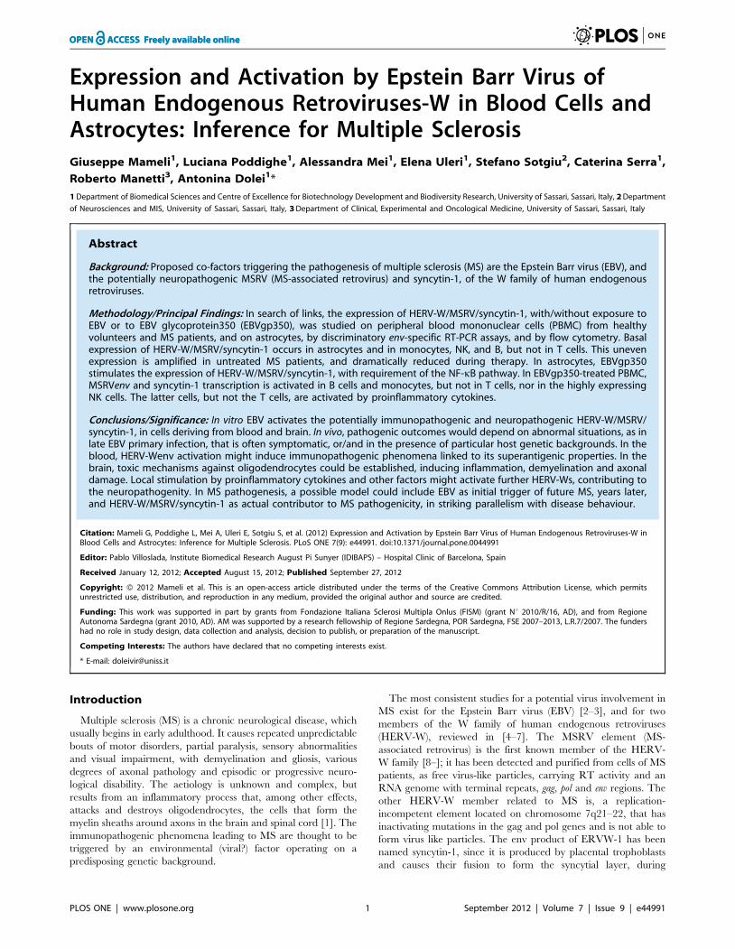
$80% of the genes were selected. As reported on Figure 1,
HERV-Wenv elements containing $80% of the env gene are
present on fourteen human chromosomes; full length HERV-Wenv
sequences are present on ten chromosomes (X, 3, 4, 5, 7, 12, 14,
15, 17, 18); on Table S1a and S1b of the Supporting Information
file more detailed data are reported.
At the RNA level, MSRVenv and syncytin-1 share .94%
identities [12]; at the protein level, no antibody specific for a
unique HERV-W has been identified so far [5]. The homology
between MSRVenv and syncytin-1 originated debates in the
attribution of possible pathogenic effects. A major difference is that
only MSRV has been detected as an extracellular virus, visualized
by electron microscopy, sedimenting at retrovirus buoyant density,
with RT activity and a polyA(+) RNA containing terminal repeats,
gag, pol and env sequences, while the syncytin-1 protein has been
found only intracellularly or on the plasma membrane [5]. Since
PCR and RT-PCR data from the literature have been obtained by
using primers unable to discriminate HERV-Wenv elements, we
developed discriminatory real-rime PCR assays, that could
selectively amplify either MSRVenv or syncytin-1 sequences.
Reliability of the assays with respect to specificity, sensitivity and
inter- and intra-assay variations were published [34].
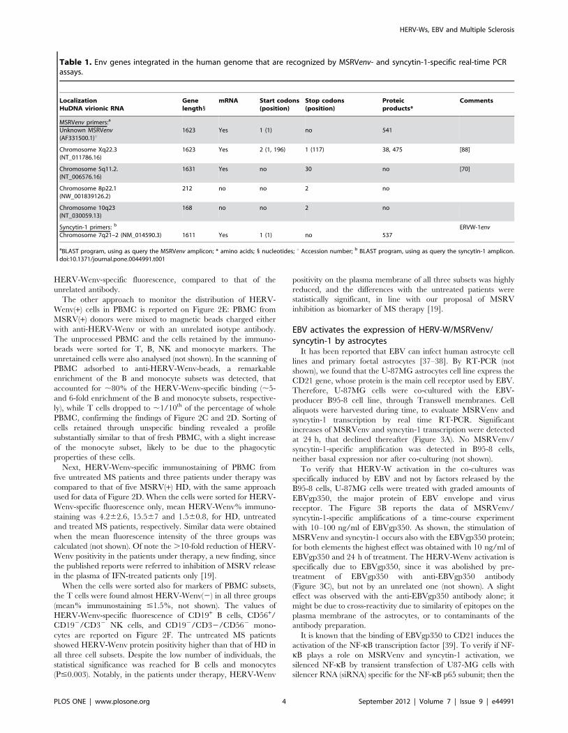
On Table 1 an in silico analysis of the human DNA sequences
recognized by the above MSRVenv- and syncytin-1 specific
primers, and of their transcription/translation products are
reported. As shown in the upper part of Table 1, apart from the
virionic MSRVenv RNA sequences, the BLAST analysis showed
that the 166 bp fragment amplified with MSRVenv-specific
primers is present only in two full length HERV-Wenv genes,
located on chromosomes Xq22.3 and 5q11.2, and in two largely
HERV-Ws, EBV and Multiple Sclerosis
PLOS ONE | www.plosone.org 2 September 2012 | Volume 7 | Issue 9 | e44991
defective env genes on chromosomes 8p22.1 and 10q23 (13% and
10% of the whole env gene, respectively). The latter two elements
are not transcribed, while the former two do. The element on
chromosome 5q11.2 cannot be translated, due to the presence of
multiple stop codons, while the one on chromosome Xq22.3, due
to a stop codon on position 115 originates two products, of 38 and
475 amino acids, respectively, instead of the 541 amino acids of
the complete env protein. The syncytin-1-specific primers, as
shown in the lower part of the Table 1, recognize a 100 bp
amplicon present on the chromosome 7q21.22 element only, that
has retained an uninterrupted open reading frame, and therefore
is fully transcribed and translated in the complete protein of 537
amino acids.
Basal expression of HERV-Wenv/MSRVenv/syncytin-1 byastrocytes and blood cells in vitro and in vivo
Previously we have shown HERV-Wenv protein immuno-
staining in up to 50% of the astrocytes of active lesions in the brain
of MS patients [12]. Therefore U87-MG glioblastoma-astrocyto-
ma cells were analysed for transcription and translation of HERV-
W elements.
As shown in the upper Panels of Figure 2, both MSRVenv and
syncytin-1 mRNAs are detected in these cells, when reliable
discriminatory real time RT-PCR assays are used (Figure 2A).
When the cultures are in logarithmic phase of growth, more than
one third of the cells expose the HERV-Wenv protein on the
plasma membrane, as detected by flow cytometry (Figure 2B).
Since no antibody capable of discriminate between MSRVenv and
syncytin-1 is available, the immuno-staining could be due either to
MSRVenv or syncytin-1, or both. Moreover, when comparing the
expression of the env genes, it must be pointed out that the RT-
PCR data detect the specific transcripts of the whole cells, while
the flow cytometry data are referred to proteins present on the
outer cell surface, only; thus they are representative only of a
fraction of the total env protein products present in the cells (and
are not indicative of the released products/virus particles).
The transcription of MSRV and syncytin-1 and the release of
MSRV from PBMC in the blood and in culture has been shown
repeatedly by several Authors, including us (reviewed in [5,7]. To
clarify whether MSRV and syncytin-1 are expressed preferentially
by a particular cell subpopulation, PBMC from MSRV(+) HD
were separated in T, B, NK and monocyte subsets, by immuno-
specific adsorption to magnetic beads. Each subpopulation was
divided in two aliquots, to be processed for real time RT-PCR and
flow cytometry studies. Part of the monocytes was kept to
differentiate in macrophages (MDM). As shown in Figure 2C,
the mRNA amounts of MSRVenv and syncytin-1 of whole PBMC
derive from an uneven accumulation in the various cell subsets.
The T cells were totally negative, while high levels of transcripts
were found in NK and B cells; in monocytes the expression was
lower, but it increased by 3–4 folds with differentiation to MDM.
To monitor the presence of the HERV-Wenv protein on the
plasma membrane, two different approaches were used. PBMC
taken from MSRV(+) individuals were labelled with four different
antibodies, each of them identified by a different fluorochrome.
The flow cytometry of PBMC subsets is reported on Figure 2D.
When the cells were sorted for HERV-Wenv-specific fluorescence
only (upper histogram of Figure 2D), 34% of PBMC were shown
to have HERV-Wenv on the plasma membrane. When the cells
were sorted also for markers of PBMC subsets (lower histograms of
Figure 2D), it appeared that the HERV-Wenv(–) PBMC fraction
was mainly attributable to CD3+ T cells, according to the mRNA
data of Figure 2C (and Figure 2E, see below); the sorting of CD19+
B cells, CD56+/CD192/CD32 NK cells, and CD192/CD32/
CD562 monocytes showed a significant shift of the peak of
Figure 1. Chromosomal representation of multiple sites of integration in human DNA of HERV-Wenv loci covering $80% of theHERV-Wenv gene. The in silico analysis of the current version of the human genome was performed by NCBI Basic Local Alignment Search Tool(BLAST) program, using as queries the MSRVenv and syncytin-1 sequences (PV14 MSRV clone, GenBank accession number AF331500, and ERVW-1envcoding sequence NM_014590.3, respectively). From the initial BLAST-identified regions, only the sequences covering $80% of the HERV-Wenv genewere selected as target and are reported. Red dashes: target:score $200; mauves dashes: target:score 80–200. Hits: number of loci; Hit GIs: Locusnumber all matches; MT: mitochondrial chromosome.doi:10.1371/journal.pone.0044991.g001
HERV-Ws, EBV and Multiple Sclerosis
PLOS ONE | www.plosone.org 3 September 2012 | Volume 7 | Issue 9 | e44991
HERV-Wenv-specific fluorescence, compared to that of the
unrelated antibody.
The other approach to monitor the distribution of HERV-
Wenv(+) cells in PBMC is reported on Figure 2E: PBMC from
MSRV(+) donors were mixed to magnetic beads charged either
with anti-HERV-Wenv or with an unrelated isotype antibody.
The unprocessed PBMC and the cells retained by the immuno-
beads were sorted for T, B, NK and monocyte markers. The
unretained cells were also analysed (not shown). In the scanning of
PBMC adsorbed to anti-HERV-Wenv-beads, a remarkable
enrichment of the B and monocyte subsets was detected, that
accounted for ,80% of the HERV-Wenv-specific binding (,5-
and 6-fold enrichment of the B and monocyte subsets, respective-
ly), while T cells dropped to ,1/10th of the percentage of whole
PBMC, confirming the findings of Figure 2C and 2D. Sorting of
cells retained through unspecific binding revealed a profile
substantially similar to that of fresh PBMC, with a slight increase
of the monocyte subset, likely to be due to the phagocytic
properties of these cells.
Next, HERV-Wenv-specific immunostaining of PBMC from
five untreated MS patients and three patients under therapy was
compared to that of five MSRV(+) HD, with the same approach
used for data of Figure 2D. When the cells were sorted for HERV-
Wenv-specific fluorescence only, mean HERV-Wenv% immuno-
staining was 4.262.6, 15.567 and 1.560.8, for HD, untreated
and treated MS patients, respectively. Similar data were obtained
when the mean fluorescence intensity of the three groups was
calculated (not shown). Of note the .10-fold reduction of HERV-
Wenv positivity in the patients under therapy, a new finding, since
the published reports were referred to inhibition of MSRV release
in the plasma of IFN-treated patients only [19].
When the cells were sorted also for markers of PBMC subsets,
the T cells were found almost HERV-Wenv(2) in all three groups
(mean% immunostaining #1.5%, not shown). The values of
HERV-Wenv-specific fluorescence of CD19+ B cells, CD56+/
CD192/CD32 NK cells, and CD192/CD32/CD562 mono-
cytes are reported on Figure 2F. The untreated MS patients
showed HERV-Wenv protein positivity higher than that of HD in
all three cell subsets. Despite the low number of individuals, the
statistical significance was reached for B cells and monocytes
(P#0.003). Notably, in the patients under therapy, HERV-Wenv
positivity on the plasma membrane of all three subsets was highly
reduced, and the differences with the untreated patients were
statistically significant, in line with our proposal of MSRV
inhibition as biomarker of MS therapy [19].
EBV activates the expression of HERV-W/MSRVenv/syncytin-1 by astrocytes
It has been reported that EBV can infect human astrocyte cell
lines and primary foetal astrocytes [37–38]. By RT-PCR (not
shown), we found that the U-87MG astrocytes cell line express the
CD21 gene, whose protein is the main cell receptor used by EBV.
Therefore, U-87MG cells were co-cultured with the EBV-
producer B95-8 cell line, through Transwell membranes. Cell
aliquots were harvested during time, to evaluate MSRVenv and
syncytin-1 transcription by real time RT-PCR. Significant
increases of MSRVenv and syncytin-1 transcription were detected
at 24 h, that declined thereafter (Figure 3A). No MSRVenv/
syncytin-1-specific amplification was detected in B95-8 cells,
neither basal expression nor after co-culturing (not shown).
To verify that HERV-W activation in the co-cultures was
specifically induced by EBV and not by factors released by the
B95-8 cells, U-87MG cells were treated with graded amounts of
EBVgp350, the major protein of EBV envelope and virus
receptor. The Figure 3B reports the data of MSRVenv/
syncytin-1-specific amplifications of a time-course experiment
with 10–100 ng/ml of EBVgp350. As shown, the stimulation of
MSRVenv and syncytin-1 occurs also with the EBVgp350 protein;
for both elements the highest effect was obtained with 10 ng/ml of
EBVgp350 and 24 h of treatment. The HERV-Wenv activation is
specifically due to EBVgp350, since it was abolished by pre-
treatment of EBVgp350 with anti-EBVgp350 antibody
(Figure 3C), but not by an unrelated one (not shown). A slight
effect was observed with the anti-EBVgp350 antibody alone; it
might be due to cross-reactivity due to similarity of epitopes on the
plasma membrane of the astrocytes, or to contaminants of the
antibody preparation.
It is known that the binding of EBVgp350 to CD21 induces the
activation of the NF-kB transcription factor [39]. To verify if NF-
kB plays a role on MSRVenv and syncytin-1 activation, we
silenced NF-kB by transient transfection of U87-MG cells with
silencer RNA (siRNA) specific for the NF-kB p65 subunit; then the
Table 1. Env genes integrated in the human genome that are recognized by MSRVenv- and syncytin-1-specific real-time PCRassays.
LocalizationHuDNA virionic RNA
Genelength1
mRNA Start codons(position)
Stop codons(position)
Proteicproducts*
Comments
MSRVenv primers:a
Unknown MSRVenv(AF331500.1)u
1623 Yes 1 (1) no 541
Chromosome Xq22.3(NT_011786.16)
1623 Yes 2 (1, 196) 1 (117) 38, 475 [88]
Chromosome 5q11.2.(NT_006576.16)
1631 Yes no 30 no [70]
Chromosome 8p22.1(NW_001839126.2)
212 no no 2 no
Chromosome 10q23(NT_030059.13)
168 no no 2 no
Syncytin-1 primers: b
Chromosome 7q21–2 (NM_014590.3) 1611 Yes 1 (1) no 537ERVW-1env
aBLAST program, using as query the MSRVenv amplicon; * amino acids; 1 nucleotides; u Accession number; b BLAST program, using as query the syncytin-1 amplicon.doi:10.1371/journal.pone.0044991.t001
HERV-Ws, EBV and Multiple Sclerosis
PLOS ONE | www.plosone.org 4 September 2012 | Volume 7 | Issue 9 | e44991

Figure 2. Basal expression of MSRVenv/ syncytin-1/HERV-Wenv in U87-MG astrocytes and PBMC subsets. A. Detection of MSRVenvand syncytin-1 mRNAs of U87-MG cells, by real time RT-PCR (means of three experiments run in duplicate, calculated by the 22DCt method; barsindicate standard deviation. B. Flow cytometry evaluation of the HERV-Wenv protein on U87-MG plasma membrane. Shaded histograms: HERV-Wenv-specific staining; open histograms: isotype control. C. MSRVenv and Syncytin-1 mRNA expression on PBMC from MSRV(+) donors as such, and afterimmunobeads separation in CD3+T, CD19+ B, CD56+/CD192/CD32 NK and CD192/CD3–/CD562 monocyte subsets, and subsequent monocytedifferentiation into MDM (bars indicate standard deviation). D. Flow cytometry evaluation of the HERV-Wenv protein on the membrane of PBMC froma representative MSRV(+) donor in toto and after sorting of the CD3+T, CD19+ B, CD56+/CD192/CD32 NK and CD192/CD32/CD562 monocyte subsets.Shaded histograms: HERV-Wenv-specific staining; open histograms: isotype control. E. Cell populations distribution of PBMC from a representativeMSRV(+) donor before, and after capture by magnetic beads charged with anti-HERV-Wenv or with an unrelated isotype antibody. The unprocessedPBMC and the cells retained by the immunobeads were sorted for T, B, NK and monocyte markers. The unretained cells were also analysed (notshown). F. Presence of surface HERV-Wenv protein in blood cells from five MSRV(+) HD, five untreated MS patients and three MS patients under
HERV-Ws, EBV and Multiple Sclerosis
PLOS ONE | www.plosone.org 5 September 2012 | Volume 7 | Issue 9 | e44991
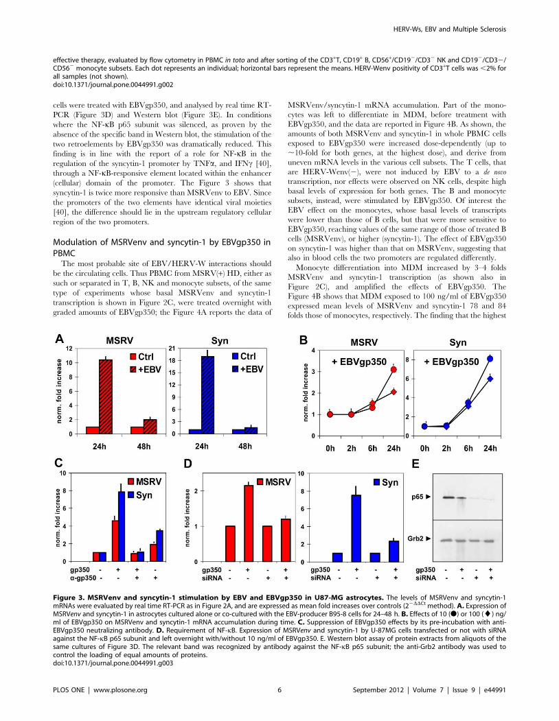
cells were treated with EBVgp350, and analysed by real time RT-
PCR (Figure 3D) and Western blot (Figure 3E). In conditions
where the NF-kB p65 subunit was silenced, as proven by the
absence of the specific band in Western blot, the stimulation of the
two retroelements by EBVgp350 was dramatically reduced. This
finding is in line with the report of a role for NF-kB in the
regulation of the syncytin-1 promoter by TNFa, and IFNc [40],
through a NF-kB-responsive element located within the enhancer
(cellular) domain of the promoter. The Figure 3 shows that
syncytin-1 is twice more responsive than MSRVenv to EBV. Since
the promoters of the two elements have identical viral moieties
[40], the difference should lie in the upstream regulatory cellular
region of the two promoters.
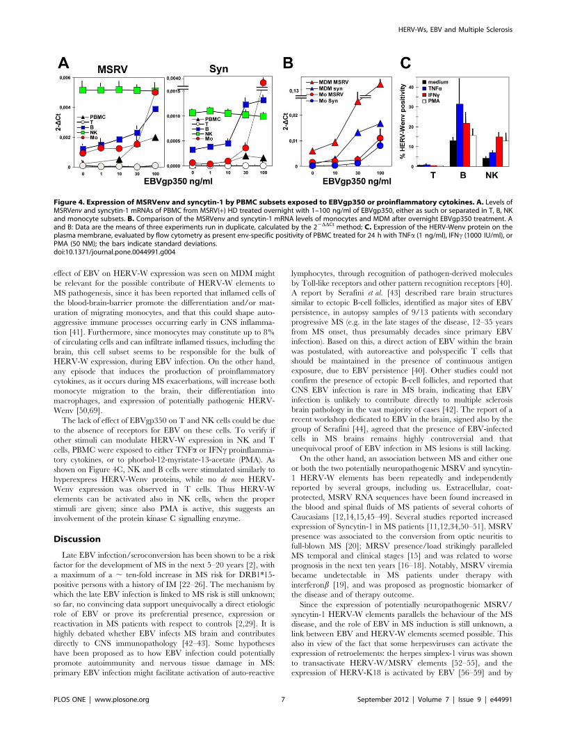
Modulation of MSRVenv and syncytin-1 by EBVgp350 inPBMC
The most probable site of EBV/HERV-W interactions should
be the circulating cells. Thus PBMC from MSRV(+) HD, either as
such or separated in T, B, NK and monocyte subsets, of the same
type of experiments whose basal MSRVenv and syncytin-1
transcription is shown in Figure 2C, were treated overnight with
graded amounts of EBVgp350; the Figure 4A reports the data of
MSRVenv/syncytin-1 mRNA accumulation. Part of the mono-
cytes was left to differentiate in MDM, before treatment with
EBVgp350, and the data are reported in Figure 4B. As shown, the
amounts of both MSRVenv and syncytin-1 in whole PBMC cells
exposed to EBVgp350 were increased dose-dependently (up to
,10-fold for both genes, at the highest dose), and derive from
uneven mRNA levels in the various cell subsets. The T cells, that
are HERV-Wenv(2), were not induced by EBV to a de novo
transcription, nor effects were observed on NK cells, despite high
basal levels of expression for both genes. The B and monocyte
subsets, instead, were stimulated by EBVgp350. Of interest the
EBV effect on the monocytes, whose basal levels of transcripts
were lower than those of B cells, but that were more sensitive to
EBVgp350, reaching values of the same range of those of treated B
cells (MSRVenv), or higher (syncytin-1). The effect of EBVgp350
on syncytin-1 was higher than that on MSRVenv, suggesting that
also in blood cells the two promoters are regulated differently.
Monocyte differentiation into MDM increased by 3–4 folds
MSRVenv and syncytin-1 transcription (as shown also in
Figure 2C), and amplified the effects of EBVgp350. The
Figure 4B shows that MDM exposed to 100 ng/ml of EBVgp350
expressed mean levels of MSRVenv and syncytin-1 78 and 84
folds those of monocytes, respectively. The finding that the highest
effective therapy, evaluated by flow cytometry in PBMC in toto and after sorting of the CD3+T, CD19+ B, CD56+/CD192/CD32 NK and CD192/CD32/CD562 monocyte subsets. Each dot represents an individual; horizontal bars represent the means. HERV-Wenv positivity of CD3+T cells was ,2% forall samples (not shown).doi:10.1371/journal.pone.0044991.g002
Figure 3. MSRVenv and syncytin-1 stimulation by EBV and EBVgp350 in U87-MG astrocytes. The levels of MSRVenv and syncytin-1mRNAs were evaluated by real time RT-PCR as in Figure 2A, and are expressed as mean fold increases over controls (22DDCt method). A. Expression ofMSRVenv and syncytin-1 in astrocytes cultured alone or co-cultured with the EBV-producer B95-8 cells for 24–48 h. B. Effects of 10 (N) or 100 (¤) ng/ml of EBVgp350 on MSRVenv and syncytin-1 mRNA accumulation during time. C. Suppression of EBVgp350 effects by its pre-incubation with anti-EBVgp350 neutralizing antibody. D. Requirement of NF-kB. Expression of MSRVenv and syncytin-1 by U-87MG cells transfected or not with siRNAagainst the NF-kB p65 subunit and left overnight with/without 10 ng/ml of EBVgp350. E. Western blot assay of protein extracts from aliquots of thesame cultures of Figure 3D. The relevant band was recognized by antibody against the NF-kB p65 subunit; the anti-Grb2 antibody was used tocontrol the loading of equal amounts of proteins.doi:10.1371/journal.pone.0044991.g003
HERV-Ws, EBV and Multiple Sclerosis
PLOS ONE | www.plosone.org 6 September 2012 | Volume 7 | Issue 9 | e44991
effect of EBV on HERV-W expression was seen on MDM might
be relevant for the possible contribute of HERV-W elements to
MS pathogenesis, since it has been reported that inflamed cells of
the blood-brain-barrier promote the differentiation and/or mat-
uration of migrating monocytes, and that this could shape auto-
aggressive immune processes occurring early in CNS inflamma-
tion [41]. Furthermore, since monocytes may constitute up to 8%
of circulating cells and can infiltrate inflamed tissues, including the
brain, this cell subset seems to be responsible for the bulk of
HERV-W expression, during EBV infection. On the other hand,
any episode that induces the production of proinflammatory
cytokines, as it occurs during MS exacerbations, will increase both
monocyte migration to the brain, their differentiation into
macrophages, and expression of potentially pathogenic HERV-
Wenv [50,69].
The lack of effect of EBVgp350 on T and NK cells could be due
to the absence of receptors for EBV on these cells. To verify if
other stimuli can modulate HERV-W expression in NK and T
cells, PBMC were exposed to either TNFa or IFNc proinflamma-
tory cytokines, or to phorbol-12-myristate-13-acetate (PMA). As
shown on Figure 4C, NK and B cells were stimulated similarly to
hyperexpress HERV-Wenv proteins, while no de novo HERV-
Wenv expression was observed in T cells. Thus HERV-W
elements can be activated also in NK cells, when the proper
stimuli are given; since also PMA is active, this suggests an
involvement of the protein kinase C signalling enzyme.
Discussion
Late EBV infection/seroconversion has been shown to be a risk
factor for the development of MS in the next 5–20 years [2], with
a maximum of a , ten-fold increase in MS risk for DRB1*15-
positive persons with a history of IM [22–26]. The mechanism by
which the late EBV infection is linked to MS risk is still unknown;
so far, no convincing data support unequivocally a direct etiologic
role of EBV or prove its preferential presence, expression or
reactivation in MS patients with respect to controls [2,29]. It is
highly debated whether EBV infects MS brain and contributes
directly to CNS immunopathology [42–43]. Some hypotheses
have been proposed as to how EBV infection could potentially
promote autoimmunity and nervous tissue damage in MS:
primary EBV infection might facilitate activation of auto-reactive
lymphocytes, through recognition of pathogen-derived molecules
by Toll-like receptors and other pattern recognition receptors [40].
A report by Serafini et al. [43] described rare brain structures
similar to ectopic B-cell follicles, identified as major sites of EBV
persistence, in autopsy samples of 9/13 patients with secondary
progressive MS (e.g. in the late stages of the disease, 12–35 years
from MS onset, thus presumably decades since primary EBV
infection). Based on this, a direct action of EBV within the brain
was postulated, with autoreactive and polyspecific T cells that
should be maintained in the presence of continuous antigen
exposure, due to EBV persistence [40]. Other studies could not
confirm the presence of ectopic B-cell follicles, and reported that
CNS EBV infection is rare in MS brain, indicating that EBV
infection is unlikely to contribute directly to multiple sclerosis
brain pathology in the vast majority of cases [42]. The report of a
recent workshop dedicated to EBV in the brain, signed also by the
group of Serafini [44], agreed that the presence of EBV-infected
cells in MS brains remains highly controversial and that
unequivocal proof of EBV infection in MS lesions is still lacking.
On the other hand, an association between MS and either one
or both the two potentially neuropathogenic MSRV and syncytin-
1 HERV-W elements has been repeatedly and independently
reported by several groups, including us. Extracellular, coat-
protected, MSRV RNA sequences have been found increased in
the blood and spinal fluids of MS patients of several cohorts of
Caucasians [12,14,15,45–49]. Several studies reported increased
expression of Syncytin-1 in MS patients [11,12,34,50–51]. MSRV
presence was associated to the conversion from optic neuritis to
full-blown MS [20]; MRSV presence/load strikingly paralleled
MS temporal and clinical stages [15] and was related to worse
prognosis in the next ten years [16–18]. Notably, MSRV viremia
became undetectable in MS patients under therapy with
interferonb [19], and was proposed as prognostic biomarker of
the disease and of therapy outcome.
Since the expression of potentially neuropathogenic MSRV/
syncytin-1 HERV-W elements parallels the behaviour of the MS
disease, and the role of EBV in MS induction is still unknown, a
link between EBV and HERV-W elements seemed possible. This
also in view of the fact that some herpesviruses can activate the
expression of retroelements: the herpes simplex-1 virus was shown
to transactivate HERV-W/MSRV elements [52–55], and the
expression of HERV-K18 is activated by EBV [56–59] and by
Figure 4. Expression of MSRVenv and syncytin-1 by PBMC subsets exposed to EBVgp350 or proinflammatory cytokines. A. Levels ofMSRVenv and syncytin-1 mRNAs of PBMC from MSRV(+) HD treated overnight with 1–100 ng/ml of EBVgp350, either as such or separated in T, B, NKand monocyte subsets. B. Comparison of the MSRVenv and syncytin-1 mRNA levels of monocytes and MDM after overnight EBVgp350 treatment. Aand B: Data are the means of three experiments run in duplicate, calculated by the 22DDCt method; C. Expression of the HERV-Wenv protein on theplasma membrane, evaluated by flow cytometry as present env-specific positivity of PBMC treated for 24 h with TNFa (1 ng/ml), IFNc (1000 IU/ml), orPMA (50 NM); the bars indicate standard deviations.doi:10.1371/journal.pone.0044991.g004
HERV-Ws, EBV and Multiple Sclerosis
PLOS ONE | www.plosone.org 7 September 2012 | Volume 7 | Issue 9 | e44991
human herpesvirus-6 [60–61]. To evaluate the possible interac-
tions between EBV and HERV-W/MSRV/syncytin-1, we
focused on cells related to MS pathogenesis and\or target of
EBV infection: (i) the astrocytes, that help demyelination, by
promoting inflammation, damage of oligodendrocytes and axons,
and glial scarring, but also allow remyelination by acting on
oligodendrocyte proliferation, and differentiation [62], and (ii)
PBMC subsets, that are the main site of EBV replication and
persistence, and play pivotal roles in MS in immune defence and
immunopathogenesis.
The in silico survey of the human genome reported in the present
paper shows that full length HERV-Wenv DNA sequences are
present on ten chromosomes, and that three additional ones have
elements containing $80% of the HERV-Wenv gene (Figure 1). As
for transcripts, data from GenBank indicate that only the ERVW-
1 locus on chromosome 7q21.22 is transcribed in a full length env
mRNA, that can be translated in a complete protein, the syncytin-
1. All the other HERV-Wenv genes present in the current version
of the human genome, either are not transcribed, or originate
HERV-Wenv fragments with stop codons and other gene
alterations [63–65].
The majority of HERV-Wenv studies used tools not discrimi-
nating the components of the HERV-W family [35]. At the RNA
level, MSRVenv and syncytin-1 share .94% identities [12]; at the
protein level, no antibody specific for a unique HERV-W are
available so far [5]. Therefore the origin of the HERV-Wenv,
whose expression has been found associated to MS is still debated.
It has been suggested that MSRV might be either an exogenous
HERV-W, or a nonubiquitous replication-competent member, or
a partly defective but nonubiquitous copy, seldom complemented
or recombined within the HERV-W family [4–5,66–70]. So far,
the real nature of MSRV remains to be elucidated. Whatever the
origin of MSRV is, a meta-analysis showed that ,9% of
Caucasian blood donors have circulating virionic MSRV/
HERV-W RNA [5].
The real-time RT-PCR assays employed in the present study
amplify selectively either MSRVenv or syncytin-1 sequences [34].
As shown on Table 1, the latter primers recognize only the
transcripts from the ERVW-1 element on the chromosome
7q21.22, while MSRVenv-specific primers, apart from the extra-
cellular MSRVenv gene, can amplify RNAs from the element on
chromosome Xq22.3 only; this element can originate an
incomplete protein, that is unglycosylated, does not form
oligomers, and is located intracellularly [64]; thus the present
version of the human genome (that presumably does not contain
sequences derived from MS patients) does not contain complete
HERV-Wenv elements, recognized by the MSRVenv-specific
primers, that can originate a full length MSRVenv protein.
Therefore the HERV-Wenv protein(s) on the plasma membrane
detected by the anti-HERV-Wenv antibody employed in the
present study could derive from the element present on the
chromosome 7q21.22 and/or from MSRV, only.
Firstly, we monitored the expression of MSRVenv and syncytin-
1 in astrocytes and PBMC, cells involved in MS pathogenesis,
and/or that could host the interactions among the above viruses.
As reported on Figure 2 A and B, in U87-MG astrocytes a basal
expression of both HERV-Wenv genes was detected at the mRNA
level; accordingly, the HERV-Wenv protein was detected on the
plasma membrane. This could be expected, since HERV-Wenv
expression by the astrocytes of active lesions in the brain of MS
patients has been reported [11,12,71].
The PBMC from both MS patients and healthy MSRV(+)
donors express MSRVenv and syncytin-1 mRNAs, and have the
HERV-Wenv protein on the outer surface of the plasma
membrane (Figure 2 C–F). The two elements, however, are not
uniformly expressed by all the PBMC, but derive from an uneven
expression in the various cell subsets. As shown in Figure 2E, when
PBMC were adsorbed to anti-HERV-Wenv-beads, ,80% of the
cells retained by the beads are B cells and monocytes. High levels
of expression of HERV-W/MSRVenv/syncytin-1 were found in
NK and B cells; in monocytes the expression was lower, but it
increased with differentiation to MDM. This is a novel finding for
endogenous retroviruses, but was shown for exogenous retrovirus-
es, such as HIV, whose expression in chronically infected
monocytes increases with differentiation to MDM [72]. The
expression of HERV-W in NK cells has not been reported
previously. These cells appear to express the highest levels of both
MSRVenv and syncytin-1 MSRVenv (see Figure 2C and basal
levels of Figure 4A). How HERV-W activation could influence the
dual role played by NK cells in MS, as inflammatory mediators of
damage and as immune regulatory cells suppressing harmful
inflammatory activity [73], remains to be elucidated. The T cells
were found totally negative for HERV-Wenv expression, both as
HERV-Wenv protein on the outer cell surface and as intracellular
MSRVenv and Syncytin-1 transcripts. This finding indicates that
the absence of the HERV-Wenv protein on the membrane of T
cells (Figures 2 and 4), that was reported by flow cytometry also by
Brudek et al. [51], is due to absence of env transcripts in the T cells,
that cannot be induced by none of the stimuli active on other cell
subsets (Figure 4).
Comparison of the HERV-Wenv expression of blood cells from
HD and MS patients, either naive or under effective therapy
(Figure 2F) showed that in the patients with active MS there is an
increase of 3–4-fold in the percentage of cells expressing the
HERV-Wenv protein on the membrane, for both whole PBMC
and B, NK, and monocyte subsets, indicating that HERV-Wenv
activation is similar in all the already HERV-W-positive PBMC
subsets, while the T cells remained negative. The specific
immunostaining sharply decreased in the patients under therapy.
This is a new finding, since our published report of inhibition of
MSRV release during effective anti-MS therapy was referred to
patients treated with interferonb [19]; in that case, the inhibition
of HERV-W/MSRV release could be explained in terms of
antiviral effects (related or not to the anti-MS action of interferon,
that was postulated to be due to its modulation of T and B cell
activity and effects on the blood-brain barrier). In the case of the
patients of Figure 1F, treated with glatiramer acetate, the anti-
HERV-W effect could be related to its induction of anti-
inflammatory cytokines [74], while that of azathioprine that
interferes with DNA synthesis could be related to its inhibition of
proliferation of cells, especially of T and B cells. In any case, our
findings confirm that efficacious anti-MS therapy is paralleled by
inhibition of HERV-W expression.
Following infection by EBV, U87-MG astrocytes increased the
transcription of both MSRVenv and syncytin-1 (Figure 3A). There
is no need of EBV entry or expression, since the effect was seen
also after exposure to the EBVgp350 major envelope protein. The
effect is abolished by silencing NF-kB (Figure 3C–D), a
transcription factor that is implicated in the production, by
activated astrocytes, of pro-inflammatory cytokines: this, in turn,
induces further release of cytokines, that contribute in vivo to
exacerbating the neurodegenerative process [75], and that in vitro
stimulate HERV-Wenv expression in astrocytes (unpublished data)
and PBMC (Figure 4C, and [69]. The HERV-Wenv proteins use,
as their cell surface receptors, the sodium-dependent neutral
amino acid transporter type 1 and 2 (ASCT-1 and ASCT-2),
involved in the cellular intake of amino acids [76]. Binding of
HERV-Wenv to ASCT-1 or -2 receptors might create critical
HERV-Ws, EBV and Multiple Sclerosis
PLOS ONE | www.plosone.org 8 September 2012 | Volume 7 | Issue 9 | e44991

reduction in the amino acid intake necessary for their normal
activity. Of note, reduced detection of these ASCT receptors at the
neuronal surface in schizophrenic cortex was reported [77] and
connected with receptor modulation or masking by HERV-Wenv
protein [6].
In the PBMC exposed to EBVgp350, MSRVenv and syncytin-1
are stimulated in B cells and monocytes, but not in T cells, nor in
the highly expressing NK cells. The latter cells, but not the T cells,
are activated by proinflammatory cytokines. Thus HERV-W
elements can be stimulated also in NK cells, when the proper
stimuli are given. Of particular interest, in our opinion, is the
monocyte/macrophage (M/M) cell compartment. These cells are
the most responsive to EBVgp350, reaching env levels higher than
those detected in B cells, particularly after cell differentiation into
macrophages. Since the monocytes can easily pass across the
endothelia, and through the blood-brain barrier, the HERV-W
expression (and release) by monocyte-macrophages can reasonably
account for the bulk of expression (and effects) of these elements.
One must remind the fundamental roles played by M/M, not only
in the clearance of virions or virally infected cells by phagocytosis,
in antigen processing, leading to the activation of the humoral
immune response and to generation of cytotoxic T-lymphocytes,
but also activation of the innate immunity, release of soluble
factors/cytokines, NK activation. Notably, neurotoxic mediators
released from M/M are thought to be involved also in the
pathogenesis of neuroAIDS, and accumulation of activated M/M
in the brain correlates with neurological injury: monocytes
infected with HIV have enhanced migratory capacity through
the blood–brain barrier; within the CNS, they differentiate and
mediate neuronal injury and dysfunction through the secretion of
viral proteins, inflammatory factors, excess glutamate, and other
neurotoxins [78–79]. Increased BBB permeability is a character-
istic hallmark of the CNS alterations observed in MS, suggesting a
causal relationship between inflammatory cell recruitment into the
CNS and the blood–brain barrier dysfunction [80], and HERV-
Wenv production by activated M/M could contribute to the
pathogenicity. To be noted, in this respect, that the HERV-Wenv
protein has been shown to activate innate immunity through
CD14/TLR4 and to promote Th1-like responses [81].
Conclusions
The present study demonstrates that basal expression of
MSRVenv and syncytin-1 is present in U87-MG astrocytes and
in PBMC from MS patients and some healthy donors; among
PBMC subsets, MSRVenv and syncytin-1 are expressed by NK, B
cells and monocytes, but not by T cells. It is shown for the first
time that EBV infection or cell exposure to the EBVgp350 protein
stimulates the expression of MSRVenv and syncytin-1, up to the
final protein product on the plasma membrane, and that this
activation involves the NF-kB pathway. Novel findings are also
HERV-Wenv expression by NK cells, and differential expression
and modulation of the HERV-Wenv protein in PBMC subsets
from MS patients (untreated and under therapy) and MSRV-
positive healthy donors. It is demonstrated that in vitro interactions
among the two proposed MS-cofactors, HERV-W and EBV,
occur in cells from blood and from brain. It is likely that it might
occur also in vivo. Therefore, in an individual, the possibility of the
activation of the immunopathogenic and neuropathogenic poten-
tial of HERV-W by superinfecting EBV is concrete, both in blood
and in brain. The original sin could lie in abnormal host reactions
to a late primary EBV infection, that often leads to IM; of note, a
mathematical model addressing the age-dependence of IM,
suggests that variation in host antibody responses and the
complexity of the pre-existing cross-reactive T-cell repertoire,
both of which depend on age, may play important roles in the
aetiology of IM [82]. During IM, an abnormal activation of
circulating cells occurs. Among other effects, EBV proteins might
deregulate also the expression of HERV-Ws, in B and M/M cells.
Activation of HERV-W elements- could induce an immune
cascade activated by HERV-Wenv protein; activated cells,
especially the M/M cells, could also go across the blood-brain
barrier. In the blood, the EBV/HERV-W interactions might
induce immunopathogenic phenomena linked to HERV-Wenv
superantigenic properties. In the brain, the EBV/HERV-W
interaction could establish also toxic mechanisms against the
oligodendrocytes, thus inducing inflammation, demyelination and
axonal damage. Local stimulation of astrocytes, by proinflamma-
tory cytokines and other factors might activate further expression
of HERV-Ws, contributing to the neuropathogenity, since
presence of MSRV in spinal fluids of MS patients is an early
event, predictor di worse progression [16–18].
Our findings reinforce the hypothesis of a possible involvement
of these viruses in MS pathogenesis, with the possibility for MSRV
of a direct role of effector of pathogenicity, and for EBV of an
initial trigger of future MS, years later, since the possibility of the
activation of the immuno- and neuro-pathogenic potential of
HERV-W by superinfecting EBV is concrete, both in blood and in
brain.
During the revision of the paper, a multicentric study has been
published [89] on the association of MSRVenv antigenemia with
MS, that independently confirmed our findings on increased
positivity/expression levels of PBMC MSRV RNA and of MSRV
DNA copy number in MS patients, and expression of MSRVenv
protein within brain MS lesions; the HERV-W Env protein
detection in MS sera confirmed our data on the association of
circulating HERV-W/MSRVenv with MS.
Materials and Methods
Ethics StatementThe study was approved by the Sassari AOU Ethics Commit-
tee. The patients and the volunteers gave written informed
consent.
CellsU-87MG: human glioblastoma-astrocytoma, epithelial-like cells,
grown in Dulbecco’s modified Eagle’s medium 10% foetal calf
serum (FCS, Life Technologies, Inc., Carlsbad, CA). B95-8:
marmoset B lymphoblastoid cells, chronically infected and
producing EBV. PBMC were isolated from heparinized peripheral
blood of MSRV(+) donors (HD, and MS patients) by layering on
Ficoll/Hipaque [19]. The PBMC and the B95-8 were maintained
in RPMI-1640 medium 10% FCS (Invitrogen, San Giuliano
Milanese, IT). Cell viability was assessed with the Trypan blue
exclusion method.
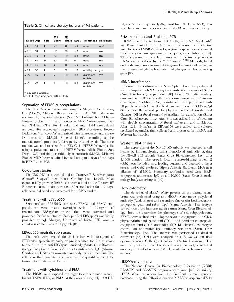
Blood samplesHD: Five already studied MSRV(+) volunteers of our cohort
[34], three females, two males (mean age 43615.0 years). MS
patients: eight patients (six females, two males; mean age
39613.0 years) attending the Department of Neurosciences,
University of Sassari, with a diagnosis of relapsing remitting MS
according to international guidelines [83]. Three patients were
untreated and three were under (effective) therapy (one was
treated with 2.6 mg/kg/day of azathioprine and two were treated
with 20 mg/day of glatiramer acetate). Table 2 reports the
anamnestic, clinical and therapy data of the patients.
HERV-Ws, EBV and Multiple Sclerosis
PLOS ONE | www.plosone.org 9 September 2012 | Volume 7 | Issue 9 | e44991
Separation of PBMC subpopulationsThe PBMCs were fractionated using the Magnetic Cell Sorting
kits (MACS, Miltenyi Biotec, Auburn, CA): NK cells were
obtained by negative selection (NK Cell Isolation Kit, Miltenyi
Biotec); to obtain B, T and monocytes, PBMC were treated with
anti-CD4/anti-CD8 (for T cells) and anti-CD14 monoclonal
antibody (for monocytes), respectively (BD Biosciences Becton
Dickinson, San Jose, CA) and mixed with microbeads (anti-mouse
Ig microbeads, MACS, Miltenyi Biotec), according to the
manufacturer’s protocols (.95% purity was achieved). The same
method was used to select from PBMC the HERV-Wenv(+) cells,
using a polyclonal rabbit anti-HERV-Wenv (Allele Biotec, San
Diego, CA) and the anti-rabbit Ig microbeads (MACS, Miltenyi
Biotec). MDM were obtained by incubating monocytes for 6 days
in RPMI 20% FCS.
Co-culture studiesThe U87-MG cells were plated on TranswellH Receiver plates
(CostarH Snapwell membranes, Corning Inc., Lowell, MA);
exponentially growing B95-8 cells were added on the TranswellHReservoir plates 0.4 mm pore size. After incubation for 1–2 days,
cells were collected and processed for mRNA studies.
Treatment with EBVgp350Semi-confluent U-87MG astrocytes, PBMC and PBMC sub-
populations were treated overnight with 10–100 ng/ml of
recombinant EBVgp350 protein, then were harvested and
processed for further studies. Fully purified EBVgp350 was kindly
provided by A.J. Morgan, University of Bristol, UK, and its
endotoxin content was ,25 pg/ml. [84].
EBVgp350 neutralization assayThe cells were treated for 24 h either with 10 ng/ml of
EBVgp350 (protein as such, or pre-incubated for 2 h at room
temperature with anti-EBVgp350 antibody (Santa Cruz Biotech-
nology, Inc., Santa Cruz, CA) or with anti-mouse IgG (Abcam,
Cambridge, UK) as an unrelated antibody, or with medium. The
cells were then harvested and processed for quantification of the
transcripts of interest, as below.
Treatment with cytokines and PMAThe PBMC were exposed overnight to either human recom-
binant TNFa, IFNc, or PMA, at the doses of 1 ng/ml, 1000 IU/
ml, and 50 nM, respectively (Sigma-Aldrich, St. Louis, MO), then
were harvested and processed for RT-PCR and flow cytometry.
RNA extraction and Real-time PCRRNAs were extracted from 50.000 cells, by mRNA DynabeadsH
kit (Dynal Biotech, Oslo, NO) and retrotranscribed; selective
amplification of MSRVenv and syncytin-1 sequences was obtained
by utilizing the corresponding primer pairs, as published in [34].
The comparison of the relative amounts of the two sequences in
RNAs was carried out by the 22DCt and 22DDCt Methods, based
on the different amplification of the gene of interest with respect to
the glyceraldehyde-3-phosphate dehydrogenase housekeeping
gene [85].
siRNA interferenceTransient knockdown of the NF-kB p65 subunit was performed
with p65-specific siRNA. using the transfection reagents of Santa
Cruz Biotechnology as published [40]. Briefly, 24 h after seeding,
semiconfluent U87-MG cells were rinsed once with Optimem
(Invitrogen, Carlsbad, CA); transfection was performed with
50 pmols of siRNA, at the final concentration of 0,125 mg/ml
(Santa Cruz Biotechnology, Inc.) by the method of Surabhi and
Gaynor [86] in foetal serum-free medium for transfection (Santa
Cruz Biotechnology, Inc.). After 6 h was added 1 ml of medium
with double concentration of foetal calf serum and antibiotics.
After 12 h, 10 ng/ml of EBVgp350 were added, and cultures
incubated overnight, then collected and processed for mRNA and
Western blot studies.
Western Blot analysisThe expression of the NF-kB p65 subunit was detected in cell
lysates by immunoblotting using monoclonal antibodies against
the NF-kB p65 subunit (Santa Cruz Biotechnology, Inc.) at a
1:1000 dilution. The growth factor receptor-binding protein 2
(Grb2) was included as a loading control, and detected using a
mouse anti-Grb2 antibody (Sigma Aldrich, St. Louis, MO) at a
dilution of 1:15,000. Secondary antibodies used were HRP-
conjugated anti-mouse IgG at a 1:10,000 (Santa Cruz Biotech-
nology Inc.), according to [40].
Flow cytometryThe detection of HERV-Wenv protein on the plasma mem-
brane was performed using anti-HERV-Wenv rabbit polyclonal
antibody (Allele Biotec) and secondary fluorescein isothiocyanate-
conjugated goat anti-rabbit IgG (Sigma-Aldrich). The isotype
control was a pre-immune rabbit serum (Santa Cruz Biotechnol-
ogy, Inc). To determine the phenotype of cell subpopulations,
PBMC were stained with allophycocyanin-conjugated anti-CD3,
phycoerythrin-conjugated anti-CD19, and peridinin-chlorophyll-
conjugated anti-CD56 antibodies (BD Biosciences). As isotype
control, an anti-rabbit IgG antibody was used (Santa Cruz
Biotechnology, Inc). The analysis was performed as detailed
elsewhere [87]. Cells were analysed on a FACS Calibur flow
cytometer using Cells Quest software (Becton-Dickinson). The
area of positivity was determined using an isotype-matched
antibody; a total of at least 56104 events for each sample were
acquired.
HERV-Wenv miningThe National Centre for Biotechnology Information (NCBI)
BLASTN and BLASTX programs were used [36] for mining
HERV-Wenv sequences from the GenBank human genome
database, using the following queries: MSRVenv sequence (PV14
Table 2. Clinical and therapy features of MS patients.
Patient Age SexMSyears
MSphase EDSS Treatment Response
MSu1 26 F ,1 RR #3 none n.a.*
MSu2 59 F ,1 RR #3 none n.a.
MSu3 19 F ,1 RR #3 none n.a.
MSu4 60 M 32 RR 6 none n.a.
MSu5 30 M 3 RR #3 none n.a.
MSt1 32 F 5 RR #3 azathioprine yes
MSt2 45 F 2 RR #3 glatirameracetate
yes
MSt3 22 F 1 RR #3 glatirameracetate
yes
*: n.a.: not applicable.doi:10.1371/journal.pone.0044991.t002
HERV-Ws, EBV and Multiple Sclerosis
PLOS ONE | www.plosone.org 10 September 2012 | Volume 7 | Issue 9 | e44991

MSRV clone, GenBank accession number AF331500), Syncytin-1
(ERVEW1env coding sequence, accession number NM_014590.3),
MSRVenv-specific 166 bp and syncytin-1-specific 100 bp ampli-
cons generated with the corresponding discriminatory primers
described in [34].
Statistical analysesThe significance of the results was evaluated by means of the
Epi InfoTM 7 database and statistics software program, (CDC/
WHO, Atlanta, GA, USA). P values of less than 0.05 were
determined to be significant.
Supporting Information
Table S1 Chromosomal location of MSRVenv-type and
Syncytin-1-type HERV-Wenv genes. Table S1a: HERV-Wenv
genes in human DNA, (.80 of total env, as detected by the PV14
MSRVenv sequence (AF331500). In silico nucleotide BLAST
search [36] in the current version of the human genome, using as
query the MSRVenv sequence (PV14 MSRV clone, GenBank
accession number AF331500). From the initial BLAST-identified
regions, the sequences covering .80% of the genes were selected.
(DOC). Table S1b: HERV-Wenv genes in human DNA, (.80 of
total env, as detected by the ERVEW1 coding sequence
(NM_014590.3). In silico nucleotide BLAST search as for Table
S1a, using as query the syncytin-1 sequence (ERVEW1env coding
sequence, GenBank accession number NM_014590.3).
(DOC)
Acknowledgments
The authors would like to thank A.J. Morgan, University of Bristol, UK,
for the generous gift of recombinant EBVgp350.
Author Contributions
Analyzed the data: AD GM CS LP AM RM. Wrote the paper: AD.
Conceived and coordinated the study: AD. Participated in the design of the
study: GM CS LP AM. Performed most of the experiments: GM CS LP
AM. Contributed to experiments: EU. Coordinated the flow cytometry
studies: RM. Performed the clinical studies: SS. Performed the statistics:
AD. Provided blood samples from MS patients: SS.
References
1. McQualter JL, Bernard CC (2007) Multiple sclerosis: a battle between
destruction and repair. J Neurochem 100:295–306.
2. Santiago O, Gutierrez J, Sorlozano A, De Dios Luna J, Villegas E, et al. (2010)Relation between Epstein-Barr virus and multiple sclerosis: analytic study of
scientific production. Eur J Clin Microbiol Infect Dis 29:857–866.
3. Handel AE, Williamson AJ, Disanto G, Handunnetthi L, Giovannoni G, et al.(2010) An updated meta-analysis of risk of multiple sclerosis following infectious
mononucleosis. PLoS One 5(9). pii: e12496.
4. Dolei A (2006) Endogenous retroviruses and human disease. Exp Rev ClinImmunol 2:149–167.
5. Dolei A, Perron H (2009) The multiple sclerosis-associated retrovirus and itsHERV-W endogenous family: a biological interface between virology, genetics,
and immunology in human physiology and disease. J Neurovirol 1:4–13.
6. Perron H, Lang A (2010) The Human Endogenous Retrovirus Link betweenGenes and Environment in Multiple Sclerosis and in Multifactorial Diseases
Associating Neuroinflammation. Clinic Rev Allerg Immunol 1:51–61.
7. Antony JM, Deslauriers AM, Bhat RK, Ellestad KK, Power C (2011) Humanendogenous retroviruses and multiple sclerosis: innocent bystanders or disease
determinants? Biochim Biophy Acta 2:162–176.
8. Perron H, Geny C, Laurent A, Mouriquand C, Pellat J, et al. (1989)Leptomeningeal cell line from multiple sclerosis with reverse transcriptase
activity and viral particles. Res Virol 140:551–561.
9. Mi S, Lee X, Li X, Veldman GM, Finnerty H, et al. (2000) Syncytin is a captiveretroviral envelope protein involved in human placental morphogenesis. Nature
403:785–789.
10. Firouzi R, Rolland A, Michel M, Jouvin-Marche E, Hauw JJ, et al. ( 2003)Multiple sclerosis-associated retrovirus particles cause T lymphocyte-dependent
death with brain hemorrhage in humanized SCID mice model. J Neurovirol9:79–93.
11. Antony JM, Van Marle G, Opii W, Butterfield DA, Mallet F, et al. (2004)
Human endogenous retrovirus glycoprotein-mediated induction of redoxreactants causes oligodendrocyte death and demyelination. Nat Neurosci
7:1088–1095.
12. Mameli G, Astone V, Arru G, Marconi S, Lovato L, et al. (2007a) Brains andperipheral blood mononuclear cells of multiple sclerosis (MS) patients
hyperexpress MS-associated retrovirus/HERV-W endogenous retrovirus, butnot Human herpesvirus 6. J Gen Virol 88:264–274.
13. Perron H, Lazarini F, Ruprecht K, Pechoux-Longin C, Seilhean D, et al. (2005)
Human endogenous retrovirus (HERV)-W ENV and GAG proteins: physio-logical expression in human brain and pathophysiological modulation in
multiple sclerosis lesions. J Neurovirol 11:23–33.
14. Arru G, Mameli G, Astone V, Serra C, Huang YM, et al. (2007) Multiplesclerosis and HERV-W/MSRV: a multicentric study. Int J Biomed Sci 3: 292–
297.
15. Dolei A, Serra C, Mameli G, Pugliatti M, Sechi S, et al. (2002) Multiple
sclerosis-associated retrovirus (MSRV) in Sardinian MS patients. Neurology 58:
471–473.
16. Sotgiu S, Serra C, Mameli G, Pugliatti M, Rosati G, et al. (2002) Multiple
sclerosis (MS)-associated retrovirus and MS prognosis: an observational study.
Neurology 59:1071–1073.
17. Sotgiu S, Arru G, Mameli G, Serra C, Pugliatti M, et al. (2006) MSRV in early
multiple sclerosis: a six-year follow-up of a Sardinian cohort. Mult Scler 12:698–703.
18. Sotgiu S, Mameli G, Serra C, Zarbo IR, Arru G, et al. (2010) Multiple sclerosis-
associated retrovirus and progressive disability of multiple sclerosis. Mult Scler
16:1248–1251.
19. Mameli G, Serra C, Astone V, Castellazzi M, Poddighe L, et al. (2008)
Inhibition of multiple-sclerosis-associated retrovirus as biomarker of interferon
therapy. J Neurovirol 1:73–77.
20. Sotgiu S, Arru G, Soderstrom M, Mameli G, Serra C, et al. (2006) MSRV and
optic neuritis. Mult Scler 12:357–359.
21. Petersen T, Møller-Larsen A, Thiel S, Brudek T, Hansen TK, et al. (2009)
Effects of interferon-beta therapy on innate and adaptive immune responses to
the human endogenous retroviruses HERV-H and HERV-W, cytokine
production, and the lectin complement activation pathway in multiple sclerosis.
J Neuroimmunol 215:108–116.
22. Ascherio A, Munger KL (2010) Epstein-Barr virus infection and multiple
sclerosis: a review. J Neuroimmune Pharmacol 3:271–277.
23. Munger K, Levin L, O’Reilly E, Falk K, Ascherio A (2011) Anti-Epstein-Barr
virus antibodies as serological markers of multiple sclerosis: a prospective study
among United States military personnel. Mult Scler 17:1185–1193.
24. Goldacre MJ, Wotton CJ, Seagroatt V, Yeates D (2004) Multiple sclerosis after
infectious mononucleosis: record linkage study. J Epidemiol Community Health
58:1032–1035.
25. Lucas RM, Hughes AM, Lay ML, Ponsonby AL, Dwyer DE, et al. (2011)
Epstein-Barr virus and multiple sclerosis. J Neurol Neurosurg Psychiatry
82:1142–1148.
26. Nielsen TR, Rostgaard K, Askling J, Steffensen R, Oturai A, et al. (2009) Effects
of infectious mononucleosis and HLA-DRB1*15 in multiple sclerosis. Mult Scler
15:431–436.
27. Banwell B, Krupp L, Kennedy J, Tellier R, Tenembaum S, et al. (2007) Clinical
features and viral serologies in children with multiple sclerosis: a multinational
observational study. Lancet Neurol 9:773–781.
28. Lunemann JD, Tintore M, Messmer B, Strowig T, Rovira A, et al. (2010)
Elevated Epstein-Barr virus-encoded nuclear antigen-1 immune responses
predict conversion to multiple sclerosis. Ann Neurol 2:159–169.
29. Fatima N, Toscano MP, Hunter SB, Cohen C (2011) Controversial role of
Epstein-Barr virus in multiple sclerosis. Appl Immunohistochem Mol Morphol
3:246–252.
30. Ludwin S, Jacobson S (2011) Epstein-Barr Virus and MS: Causality or
Association? Int MS J 17:39–43.
31. Giovannoni G (2011) Epstein-Barr Virus and MS. Int MS J 17:44–49.
32. Pavlicek A, Paces J, Elleder D, Hejnar J (2002) Processed pseudogenes of human
endogenous retroviruses generated by LINEs: their integration, stability, and
distribution. Genome Res 12:391–399.
33. Li F, Nellaker C, Yolken RH, Karlsson H (2011) A systematic evaluation of
expression of HERV-W elements; influence of genomic context, viral structure
and orientation. BMC Genomics 12:22.
34. Mameli G, Poddighe L, Astone V, Delogu G, Arru G, et al. (2009) Novel reliable
real-time PCR for differential detection of MSRVenv and syncytin-1 in RNA
and DNA from patients with multiple sclerosis. J Virol Methods 161:98–106.
35. Garson JA, Huggett JF, Bustin SA, Pfaffl MW, Benes V, et al. (2009) Unreliable
real-time PCR analysis of human endogenous retrovirus-W (HERV-W) RNA
expression and DNA copy number in multiple sclerosis. AIDS Res Hum
Retroviruses 25:377–378.
HERV-Ws, EBV and Multiple Sclerosis
PLOS ONE | www.plosone.org 11 September 2012 | Volume 7 | Issue 9 | e44991

36. Altschul SF, Gish W, Miller W, Myers EW, Lipman DJ (1990) Basic local
alignment search tool. J Mol Biol 215:403–410.
37. Menet A, Speth C, Larcher C, Prodinger WM, Schwendinger MG, et al. (1999)
Epstein-Barr virus infection of human astrocyte cell lines. J Virol 73:7722–7733.
38. Gasque P, Chan P, Mauger C, Schouft MT, Singhrao S, et al. (1996)
Identification and characterization of complement C3 receptors on human
astrocytes. J Immunol 156:2247–2255.
39. Sugano N, Chen W, Roberts ML, Cooper NR (1997) Epstein-Barr virus binding
to CD21 activates the initial viral promoter via NF-kappaB induction. J Exp
Med 186:731–737.
40. Mameli G, Astone V, Khalili K, Serra C, Sawaya BE, et al. (2007b) Regulation
of the syncytin-1 promoter in human astrocytes by multiple sclerosis-related
cytokines. Virology 362:120–130.
41. Ifergan I, Kebir H, Bernard M K, Wosik A, Dodelet-Devillers R, et al. (2008)
The blood-brain barrier induces differentiation of migrating monocytes into
Th17-polarizing dendritic cells. Brain 131:785–99.
42. Willis SN, Stadelmann C, Rodig SJ, Caron T, Gattenloehner S, et al. (2009)
Epstein-Barr virus infection is not a characteristic feature of multiple sclerosis
brain. Brain 132:3318–3328.
43. Serafini B, Rosicarelli B, Franciotta D, Magliozzi R, Reynolds R, et al. (2007)
Dysregulated Epstein-Barr virus infection in the multiple sclerosis brain. J Exp
Med 204:2899–2912.
44. Lassmann H, Niedobitek G, Aloisi F, Middeldorp JM, NeuroproMiSe EBV
Working Group (2011) Epstein-Barr virus in the multiple sclerosis brain: a
controversial issue – report on a focused workshop held in the Centre for Brain
Research of the Medical University of Vienna, Austria. Brain 134:2772–2786.
45. De Villiers JN, Treurnicht FK, Warnich L, Carr J, van Rensburg SJ, et al. (2006)
Analysis of viral and genetic factors in South African patients with multiple
sclerosis. Metab Brain Dis 21:163–169.
46. Garson JA, Tuke PW, Giraud P, Paranhos-Baccala G, Perron H (1998)
Detection of virion-associated MSRV-RNA in serum of patients with multiple
sclerosis. Lancet 351:33.
47. Nowak J, Januszkiewicz D, Pernak M, Liwen I, Zawada M, et al. (2003) Multiple
sclerosis-associated virus-related pol sequences found both in multiple sclerosis
and healthy donors are more frequently expressed in multiple sclerosis patients.
J Neurovirol 9:112–117.
48. Perron H, Garson JA, Bedin F, Beseme F, Paranhos-Baccala G, et al. (1997) The
Collaborative Research Group on Multiple Sclerosis: Molecular identification of
a novel retrovirus repeatedly isolated from patients with multiple sclerosis. Proc
Natl Acad Sci USA 94:7583–7588.
49. Serra C, Sotgiu S, Mameli G, Pugliatti M, Rosati G, et al. (2001) Multiple
sclerosis and multiple sclerosis-associated retrovirus in Sardinia. Neurol Sci
22:171–173.
50. Johnston JB, Silva C, Holden J, Warren KG, Clark AW, et al. (2001) Monocyte
activation and differentiation augment human endogenous retrovirus expression:
implications for inflammatory brain diseases. Ann Neurol 50:434–442.
51. Brudek T, Christensen T, Aagaard L, Petersen T, Hansen HJ, et al. (2009) B
cells and monocytes from patients with active multiple sclerosis exhibit increased
surface expression of both HERV-H Env and HERV-W Env, accompanied by
increased seroreactivity. Retrovirology 6:104.
52. Perron H, Suh M, Lalande B, Gratacap B, Laurent A, et al. (1993) Herpes
simplex virus ICP0 and ICP4 immediate early proteins strongly enhance
expression of a retrovirus harboured by a leptomeningeal cell line from a patient
with multiple sclerosis. J Gen Virol 74:65–72.
53. Lee WJ, Kwun HJ, Kim HS, Jang KL (2003) Activation of the human
endogenous retrovirus W long terminal repeat by herpes simplex virus type 1
immediate early protein 1. Mol Cells 15:75–80.
54. Ruprecht K, Obojes K, Wengel V, Gronen F, Kim KS, et al. (2006) Regulation
of human endogenous retrovirus W protein expression by herpes simplex virus
type 1: implications for multiple sclerosis. J Neurovirol 12:65–71.
55. Nellaker C, Yao Y, Jones-Brando L, Mallet F, Yolken RH, et al. (2006)
Transactivation of elements in the human endogenous retrovirus W family by
viral infection. Retrovirology 3:44.
56. Sutkowski N, Conrad B, Thorley-Lawson DA, Huber BT (2001) Epstein-Barr
virus transactivates the human endogenous retrovirus HERV-K18 that encodes
a superantigen. Immunity 15:579–589.
57. Sutkowski N, Chen G, Calderon G, Huber BT (2004) Epstein-Barr virus latent
membrane protein LMP-2A is sufficient for transactivation of the human
endogenous retrovirus HERV-K18 superantigen. J Virol 78:7852–7860.
58. Hsiao FC, Lin M, Tai A, Chen G, Huber BT (2006) Cutting edge: Epstein-Barr
virus transactivates the HERV-K18 superantigen by docking to the human
complement receptor 2 (CD21) on primary B cells. J Immunol 4:2056–2060.
59. Hsiao FC, Tai AK, Deglon A, Sutkowski N, Longnecker R, et al. (2009) EBV
LMP-2A employs a novel mechanism to transactivate the HERV-K18
superantigen through its ITAM. Virology 1:261–266.
60. Turcanova VL, Bundgaard B, Hollsberg P (2009) Human herpesvirus-6B
induces expression of the human endogenous retrovirus K18-encoded
superantigen. J Clin Virol 46:15–19.
61. Tai AK, Luka J, Ablashi D, Huber BT (2009) HHV-6A infection induces
expression of HERV-K18-encoded superantigen. J Clin Virol 46:47–48.
62. Williams A, Piaton G, Lubetzki C (2007) Astrocytes-friends or foes in multiple
sclerosis? Glia 55:1300–1312.
63. Bhat RK, Ellestad KK, Wheatley BM, Warren R, Holt RA, et al. (2011) Age-
and disease-dependent HERV-W envelope allelic variation in brain: association
with neuroimmune gene expression. PLoS One 6:e19176.
64. Roebke C, Wahl S, Laufer G, Stadelmann C, Sauter M, et al. (2010) A N-
terminally truncated envelope protein encoded by a human endogenous
retrovirus W locus on chromosome Xq22.3. Retrovirology 7:69.
65. De Parseval N, Lazar V, Casella JF, Benit L, Heidmann T (2003) Survey of
human genes of retroviral origin: identification and transcriptome of the genes
with coding capacity for complete envelope proteins. J Virol 77:10414–10422.
66. Komurian-Pradel F, Paranhos-Baccala G, Bedin F, Ounanian-Paraz A, Sodoyer
M, et al. (1999) Molecular cloning and characterization of MSRV-related
sequences associated with retrovirus-like particles. Virology 260:1–9.
67. Perron H, Garson JA, Bedin F, Beseme F, Paranhos-Baccala G, et al. (1997) The
Collaborative Research Group on Multiple Sclerosis: Molecular identification of
a novel retrovirus repeatedly isolated from patients with multiple sclerosis. Proc
Natl Acad Sci USA 94:7583–7588.
68. Perron H, Perin JP, Rieger F, Alliel PM (2000) Particle-associated retroviral
RNA and tandem RGH/HERV-W copies on human chromosome 7q: possible
components of a ‘chain-reaction’ triggered by infectious agents in multiple
sclerosis? J NeuroVirol 6:S67–S75.
69. Serra C, Mameli G, Arru G, Sotgiu S, Rosati G, et al. (2003) In vitro modulation
of the multiple sclerosis (MS)-associated retrovirus (MSRV) by cytokines:
implications for MS pathogenesis. J NeuroVirol 9:637–643.
70. Laufer G, Mayer J, Mueller BF, Mueller-Lantzsch N, Ruprecht K (2009)
Analysis of transcribed human endogenous retrovirus W env loci clarifies the
origin of multiple sclerosis-associated retrovirus env sequences. Retrovirology
6:37.
71. Antony JM, Ellestad KK, Hammond R, Imaizumi K, Mallet F, et al. (2007) The
human endogenous retrovirus envelope glycoprotein, syncytin-1, regulates
neuroinflammation and its receptor expression in multiple sclerosis: a role for
endoplasmic reticulum chaperones in astrocytes. J Immunol 179:1210–1224.
72. Elia C, Cassol E, Sidenius N, Blasi F, Castagna A, et al. (2007) Inhibition of HIV
replication by the plasminogen activator is dependent on vitronectin-mediated
cell adhesion. J Leukoc Biol 82:1212–1220.
73. Morandi B, Bramanti P, Bonaccorsi I, Montalto E, Oliveri D, et al. (2008) Role
of natural killer cells in the pathogenesis and progression of multiple sclerosis.
Pharmacol Res 57:1–5.
74. Mendes A, Sa MJ (2011) Classical immunomodulatory therapy in multiple
sclerosis: how it acts, how it works. Arq Neuropsiquiatr 69:536–543.
75. Kim DY, Hong GU, Ro JY (2011) Signal pathways in astrocytes activated by
cross-talk between of astrocytes and mast cells through CD40-CD40L.
J Neuroinflammation 8:25.
76. Lavillette D, Marin M, Ruggieri A, Mallet F, Cosset FL, et al. (2002) The
envelope glycoprotein of human endogenous retrovirus type W uses a divergent
family of amino acid transporters/cell surface receptors. J Virol 76:6442–6452.
77. Weis S, Llenos IC, Dulay JR, Verma N, Sabunciyan S, et al. (2007) Changes in
region- and cell type-specific expression patterns of neutral amino acid
transporter 1 (ASCT-1) in the anterior cingulate cortex and hippocampus in
schizophrenia, bipolar disorder and major depression. J Neural Transm
114:261–271.
78. Constantino AA, Huang Y, Zhang H, Wood C, Zheng JC (2011) HIV-1 clade B
and C isolates exhibit differential replication: relevance to macrophage-mediated
neurotoxicity. Neurotox Res 20:277–88.
79. Yadav A, Collman RG (2009) CNS inflammation and macrophage/microglial
biology associated with HIV-1 infection. J Neuroimmune Pharmacol 4:430–447.
80. Pfeiffer F, Schafer J, Lyck R, Makrides V, Brunner S, et al. (2011) Claudin-1
induced sealing of blood-brain barrier tight junctions ameliorates chronic
experimental autoimmune encephalomyelitis. Acta Neuropathol 122:601–614.
81. Rolland A, Jouvin-Marche E, Viret C, Faure M, Perron H, et al. (2006) The
envelope protein of a human endogenous retrovirus-W family activates innate
immunity through CD14/TLR4 and promotes Th1-like responses. J Immunol
176:7636–7644.
82. Huynh GT, Adler FR (2011) Mathematical modelling the age dependence of
Epstein-Barr virus associated infectious mononucleosis. Math Med Biol.
83. McDonald WI, Compston A, Edan G, Goodkin DHP, Hartung A, et al. (2001)
Recommended diagnostic criteria for multiple sclerosis: guidelines from the
International Panel on the Diagnosis of Multiple Sclerosis. Ann Neurol 50:121–
127.
84. D’Addario M, Ahmad A, Morgan A, Menezes J (2000) Binding of the Epstein-
Barr virus major envelope glycoprotein gp350 results in the upregulation of the
TNF-alpha gene expression in monocytic cells via NF-kappaB involving PKC,
PI3-K and tyrosine kinases. J Mol Biol. 2000 May 19;298(5):765–78.
85. Serra C, Biolchini A, Mei A, Kotenko S, Dolei A (2008) Type III and I
interferons increase HIV uptake and replication in human cells that overexpress
CD4, CCR5, and CXCR4. AIDS Res Hum Retroviruses 24:173–80.
86. Surabhi RM, Gaynor RB (2002) RNA interference directed against viral and
cellular targets inhibits human immunodeficiency Virus Type 1 replication.
J Virol 76:12963–12973.
87. Annunziato F, Romagnani P, Cosmi L, Beltrame C, Steiner BH, et al. (2001)
Macrophage-derived chemokine and EBI1-ligand chemokine attract human
thymocytes in different stage of development and are produced by distinct
subsets of medullary epithelial cells: possible implications for negative selection.
J Immunol 22:238–246.
HERV-Ws, EBV and Multiple Sclerosis
PLOS ONE | www.plosone.org 12 September 2012 | Volume 7 | Issue 9 | e44991

88. Ruprecht K, Obojes K, Wengel V, Gronen F, Kim KS, et al. (2006) Regulation
of human endogenous retrovirus W protein expression by herpes simplex virus
type 1: implications for multiple sclerosis. J Neurovirol 12:65–71.
89. Perron H, Germi R, Bernard C, Garcia-Montojo M, Deluen C, et al. (2012)
Human endogenous retrovirus type W envelope expression in blood and braincells provides new insights into multiple sclerosis disease. Mult Scler. 2012 Mar
30. [Epub ahead of print] doi: 10.1177/1352458512441381.
HERV-Ws, EBV and Multiple Sclerosis
PLOS ONE | www.plosone.org 13 September 2012 | Volume 7 | Issue 9 | e44991