Exploring the molecular basis of the enantioselective binding of penicillin G acylase towards a...
12
This article was originally published in a journal published by Elsevier, and the attached copy is provided by Elsevier for the author’s benefit and for the benefit of the author’s institution, for non-commercial research and educational use including without limitation use in instruction at your institution, sending it to specific colleagues that you know, and providing a copy to your institution’s administrator. All other uses, reproduction and distribution, including without limitation commercial reprints, selling or licensing copies or access, or posting on open internet sites, your personal or institution’s website or repository, are prohibited. For exceptions, permission may be sought for such use through Elsevier’s permissions site at: http://www.elsevier.com/locate/permissionusematerial
Transcript of Exploring the molecular basis of the enantioselective binding of penicillin G acylase towards a...

This article was originally published in a journal published byElsevier, and the attached copy is provided by Elsevier for the
author’s benefit and for the benefit of the author’s institution, fornon-commercial research and educational use including without
limitation use in instruction at your institution, sending it to specificcolleagues that you know, and providing a copy to your institution’s
administrator.
All other uses, reproduction and distribution, including withoutlimitation commercial reprints, selling or licensing copies or access,
or posting on open internet sites, your personal or institution’swebsite or repository, are prohibited. For exceptions, permission
may be sought for such use through Elsevier’s permissions site at:
http://www.elsevier.com/locate/permissionusematerial
Autho
r's
pers
onal
co
py
Exploring the molecular basis of the enantioselective binding of
penicillin G acylase towards a series of 2-aryloxyalkanoic acids:
A docking and molecular dynamics study
Antonio Lavecchia a,*, Sandro Cosconati a, Ettore Novellino a, Enrica Calleri b,Caterina Temporini b, Gabriella Massolini b, Giuseppe Carbonara c,
Giuseppe Fracchiolla c, Fulvio Loiodice c
a Dipartimento di Chimica Farmaceutica e Tossicologica, Universita di Napoli ‘‘Federico II’’,
Via D. Montesano, 49, I-80131 Napoli, Italyb Dipartimento di Chimica Farmaceutica, Universita di Pavia, Via Taramelli 12, I-27100 Pavia, Italy
c Dipartimento Farmaco-Chimico, Universita di Bari, Via Orabona 4, I-70126 Bari, Italy
Received 17 February 2006; received in revised form 4 July 2006; accepted 5 July 2006
Available online 8 August 2006
Abstract
In the present paper, molecular modeling studies were undertaken in order to shed light on the molecular basis of the observed enantioselectivity
of penicillin G acylase (PGA), a well known enzyme for its industrial applications, towards 16 racemic 2-aryloxyalkanoic acids, which have been
reported to affect several biological systems. With this intention docking calculations and MD simulations were performed. Docking results
indicated that the (S)-enantiomers establish several electrostatic interactions with SerB1, SerB386 and ArgB263 of PGA. Conversely, the absence
of specific polar interactions between the (R)-enantiomers and ArgB263 seems to be the main reason for the different binding affinities observed
between the two enantiomers. Results of molecular dynamics simulations demonstrated that polar interactions are responsible for both the ligand
affinity and PGA enantiospecificity. Modeling calculations provided possible explanations for the observed enantioselectivity of the enzyme that
rationalize available experimental data and could be the basis for future protein engineering efforts.
# 2006 Elsevier Inc. All rights reserved.
Keywords: Docking; Enantioselectivity; Molecular dynamics; Molecular modeling; Penicillin G acylase
1. Introduction
The natural world is characterized by the presence of chiral
compounds such as amino-acids and carbohydrates. These
molecules are composed of units all having the same chiral
configurations: the 21 essential amino acids are all L-
enantiomers, while most carbohydrates have the D-configura-
tion. Thus, critical physiological processes are homochiral,
showing 100% stereoselectivity, and only involve one of all the
possible stereoisomers of key molecules [1]. In this context,
also when exogenous chiral compounds such as drugs are
introduced in the body, physiological processes display a high
degree of enantioselection [2], with the stereoisomers having
very dissimilar effects on chiral targets (receptors, enzymes and
ion channels), due to their diverse drug/target interactions. In
fact, although enantiomers have identical chemical and
physical properties in achiral environments, except for the
property of rotation of polarized light, they can exhibit different
physiological and biochemical behaviour in a chiral biological
environment and may also have different pharmacodynamic
and pharmacokinetic activities. Furthermore, the enantiomers
of chiral drugs may display considerable differences in toxicity
[3], as underscored by the tragedy of thalidomide in the early
1960s. In fact, the inactive isomer of this sedative and
antinausea compound was found to be responsible for the
teratogenic effects.
In this scenario, drug chirality has become one of the major
issues in the design, discovery and development of new drugs
[4–8]. In fact, in the last decades, the demand of enantiomeri-
cally pure drugs has increased together with their introduction
www.elsevier.com/locate/JMGM
Journal of Molecular Graphics and Modelling 25 (2007) 773–783
* Corresponding author. Tel.: +39 081678613; fax: +39 081678613.
E-mail address: [email protected] (A. Lavecchia).
1093-3263/$ – see front matter # 2006 Elsevier Inc. All rights reserved.
doi:10.1016/j.jmgm.2006.07.001
Autho
r's
pers
onal
co
py
in the market (from 20% 10 years ago to 75% nowadays).
Therefore, the enantiomeric separation and analysis of chiral
drugs turned out to be indispensable for chemists. Actually,
several experimental procedures exist to separate racemates
and, among them, the high-performance liquid chromatography
(HPLC) seems to be the most commonly used technique. This
approach can be divided into three main types: (i) direct
separation (direct method) on a chiral stationary phase (CSP),
(ii) separation on an achiral stationary phase by adding a chiral
agent to the mobile phase, which then forms adducts with the
enantiomeric analytes (chiral mobile phase additive method),
and (iii) separation of the diastereomers formed by pre-column
derivatization with a chiral derivatization reagent (CDR)
(indirect method) [9]. Among these, protein-based CSPs are of
special interest because of their unique enantioselective
properties and because they are suitable for separating a wide
range of enantiomerically pure compounds [10,11]. CSPs
examples include albumins such as bovine and human serum
albumins, glycoproteins such as a1-acid-glycoprotein, ovomu-
coid, avidin and riboflavin binding protein, enzymes such as a-
chymotrypsin, cellobiohydrolase I, lysozyme, pepsin, and other
proteins [12]. Another enzyme, the penicillin G acylase (PGA),
has been recently employed to develop a chiral stationary phase
(PGA-CSP) [13–15]. This enzyme is well known for its
industrial application in the production of the b-lactamic
nucleus and for its enantioselectivity in the hydrolysis of amide
or ester bonds [16]. PGA has also proven to be almost
unbeatable in separating racemates because of its high
enantioselectivity and broad ligand tolerance. Most precisely,
several experiments clearly indicated that immobilized PGA
can be successfully used as chiral selector for acidic
compounds [17], such as 2-aryloxyalkanoic acids, isosteric
analogous and some 2-arylpropionic acids [18]. The absolute
configuration of these compounds has been shown to exert a
strong influence in activating the peroxisome proliferator
activated receptors (PPARs) [19,20], in allosterically modulat-
ing haemoglobin activity [21], in changing the skeletal muscle
membrane chloride conductance [22,23], in modulating the
prostaglandin-dependent platelet aggregation [24], and in
inhibiting the cyclooxygenase enzyme [25].
Recently, the X-ray structures of PGA complexed with a
series of phenylacetic acid analogues [26] and with penicillin G/
penicillin G sulphoxide ligands [27] were solved, shedding light
on the functional properties of PGA and on its catalytic
mechanism. PGA belongs to the newly recognized structural
superfamily of N-terminal nucleophile (Ntn) amidohydrolases
[28]. The other Ntn hydrolases identified so far are the
aspartylglucosaminidase (AGA) [29], proteasome (PRO) [30],
and glutamine-PRPP-amidotransferase (GAT) [31]. The
enzymes of this superfamily share similar three-dimensional
folds and have analogies in their functional mode. They also have
similar architectures of their active sites with corresponding
catalytic elements arranged in analogous ways [28]. The binding
site of PGA has been found to consist of three major regions that
are responsible for the ligand recognition by the enzyme: the
catalytic residue SerB1, the oxyanion hole (stabilizing the
negative charge present on the ligand carboxylate group) formed
by GlnB23, AlaB69, AsnB241 and a hydrophobic pocket which
is able to accommodate lipophilic groups [26,27,32].
Considering the biological importance of the above cited
acidic compounds and the potential industrial application of
PGA as a chiral selector for a wide range of molecules, herein
we performed a computational study in order to gain major
insights into the molecular basis of the stereoselective binding
of the enzyme towards a series of previously reported 2-
aryloxy-2-aryl-acetic acids (1–16) (Table 1) [33].
A. Lavecchia et al. / Journal of Molecular Graphics and Modelling 25 (2007) 773–783774
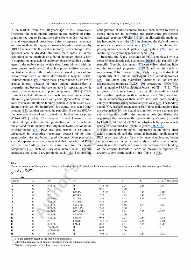
Table 1
Chemical structures of the synthesized 2-aryloxy-2-aryl-acetic acid derivatives 1–16, chromatographic parameters and differential free energy of binding
Analyte X Ar0 Ar k1a a Rs D1,2DGb (kcal/mol)
1 O 4-Cl-Ph Ph 1.35 (S)c 2.24 2.16 �0.477
2 O Ph Ph 0.36 1.00 – –
3 O Ph 4-Cl-Ph 1.12 (R) 1.21 0.47 �0.113
4 S Ph 4-Cl-Ph 2.22 1.19 0.43 �0.103
5 O 4-Cl-Ph 2-Cl-Ph 2.52 3.55 3.76 �0.750
6 O 4-Cl-Ph 4-Cl-Ph 5.58 1.00 – –
7 O 4-Cl-Ph 4-CF3-Ph 12.27 1.58 1.84 �0.271
8 O 4-Cl-Ph 4-CH3-Ph 2.47 1.00 – –
9 O 4-Cl-Ph 4-CH3O-Ph 1.90 1.11 0.47 �0.062
10 O 4-Cl-Ph 4-i-Pr-Ph 7.78 1.00 – –
11 O 4-Cl-Ph 4-Ph-Ph 40.66 1.14 0.54 �0.078
12 O Ph 1-Naph 4.57 (R) 1.92 2.50 �0.386
13 O 4-Cl-Ph 1-Naph 8.57 2.81 3.49 �0.611
14 O 2,6-Cl2-Ph Ph 0.87 1.00 – –
15 O 4-CH3O-Ph Ph 0.53 1.00 – –
16 O 2-Naph Ph 4.64 (R) 1.17 0.51 �0.093
a k1 is the retention factor of the first eluted enantiomer.b Differential free energy of binding calculated from the chromatographic data.c Absolute configuration of the less retained enantiomer.
Autho
r's
pers
onal
co
py
With this purposes, the 3D-structure of PGA solved through
X-ray crystallography (PDB Entry Code: 1AJN) [26] was used
to dock both isomers of titled compounds through the
automated docking program AutoDock [34], which has been
shown to be predictive in reproducing experimentally observed
binding poses [35]. The predicted binding conformations were
subsequently used as input for molecular dynamics (MD)
simulations using the software Q [36] in order to inspect the
ligand/enzyme binding interactions from a dynamical point of
view and to calculate the energetic contribution to the binding
of the selected compounds. Q implements the linear interaction
energy (LIE) method to calculate the ligand binding affinities
[37–39], which has proven to be successful for a number of
different systems as reported by several papers by Aqvist and
co-workers [40–43].
2. Computational methods
2.1. Docking
Docking simulations were carried out using AutoDock 3.0.5
version [34]. It combines a rapid energy evaluation through pre-
calculated grids of affinity potentials with a variety of search
algorithms to find suitable binding positions for a ligand on a
given protein. While the protein is required to be rigid, the
program allows torsional flexibility in the ligand. Docking of
both (R)- and (S)-enantiomers of compounds 1–16 to PGA was
carried out using the empirical free energy function and the
Lamarckian genetic algorithm (LGA) [34], applying a standard
protocol, with an initial population of 50 randomly placed
individuals, a maximum number of 1.5 � 106 energy evalua-
tions, a mutation rate of 0.02, a crossover rate of 0.80, and an
elitism value of 1. Proportional selection was used, where the
average of the worst energy was calculated over a window of
the previous 10 generations. For the local search, the so-called
pseudo-Solis and Wets algorithm was applied using a
maximum of 300 iterations per local search. The probability
of performing local search on an individual in the population
was 0.06, and the maximum number of consecutive successes
or failures before doubling or halving the local search step size
was 4.50 independent docking runs were carried out for each
ligand. Results differing by less than 1 A in positional root-
mean square deviation (rmsd) were clustered together and
represented by the result with the most favourable free energy
of binding (DGbind).
2.2. Ligand setup
The structures of the ligands 1–16 were generated using
standard bond lengths and bond angles of the SYBYL
fragment library [44]. Geometry optimizations were con-
ducted with the SYBYL/MAXIMIN2 minimizer by applying
the BFGS (Broyden, Fletcher, Goldfarb and Shannon)
algorithm [45] with a convergence criterion of 0.001 kcal/
mol A and employing the TRIPOS force field [46]. Partial
atomic charges were assigned using the Gasteiger–Marsili
formalism [47], which is the type of atomic charges used in
calibrating the AutoDock empirical free energy function.
Finally, the compounds were setup for docking with the help
of AutoTors, the main purpose of which is to define the
torsional degrees of freedom to be considered during the
docking process. All torsion angles for each inspected
compound were considered flexible.
2.3. Protein setup
The crystal structure of PGA in complex with p-nitrophe-
nylacetic acid (entry code: 1AJN) [26], recovered from
Brookhaven Protein Database [48], was used. In AutoDock,
the polar hydrogens are explicitly treated, while the non-polar
hydrogens are ‘‘unified’’ with their parent heavy atoms, by
increasing the heavy atom’s van der Waals’ radius. Therefore, the
structure was set up for docking as follows: polar hydrogens were
added using the BIOPOLYMERS module within SYBYL
program (residues Arg, Lys, Glu, and Asp were considered
ionized, while all His were considered neutral by default) and
Kollman united-atom partial charges were assigned. The grid
maps representing the proteins in the actual docking process were
calculated with AutoGrid. The grids (one for each atom type in
the ligand, plus one for electrostatic interactions) were chosen to
be sufficiently large to include not only the active site but also
significant portions of the surrounding surface. The dimensions
of the grids were thus 60 A � 60 A � 60 A, with a spacing of
0.375 A between the grid points. The centre of the grid was set to
be coincident with the mass centre of the ligand in the crystal
complex.
Figs. 2–4 of the calculated binding modes for compounds
(S)-3, (R)-3, (S)-11, (R)-11, (S)-12 and (R)-12 were made using
PYMOL software [49].
2.4. MD and binding free energy calculation
The software Q [36] was employed for the MD simulations
and analysis selecting the force field parameters of AMBER
parm94 implemented in the program [50]. This force field was
modified to include additional parameters for each inspected
ligand. Most of these parameters were adapted by analogy from
others included in the AMBER parm94 force field and the
missing ones were taken from those reported in literature [51].
Partial charges for each ligand were derived from the semi-
empirical quantum mechanics AM1-BCC method. The choice
of this method was dictated by the fact that results are very
similar to those obtained from widely used RESP charges in the
AMBER force field [52,53]. The values were adjusted to be
compatible with the AMBER force field by scaling the QM
charges to match the values for groups defined in the force field
library and by slight modifications to achieve electroneutral
groups of less than about 10 atoms. In both the bound and the
free states, the ligand was solvated with TIP3P [54] water
sphere of 18 A radius. In the MD simulation of the solvated
ligand–protein complexes, ionic groups of the protein within
the water sphere that were far away from the boundary were
modeled as charged (SerB1 N-terminal and ArgB263). The
resulting net charge for the sphere of simulation was +2, since
A. Lavecchia et al. / Journal of Molecular Graphics and Modelling 25 (2007) 773–783 775
Autho
r's
pers
onal
co
py
there were no Glu or Asp residues that could be ionized to
counterbalance negative charges.
The system was initially submitted to a stepwise warm-up
and equilibration procedure consisting of blocks of 4 ps at 0,
50, 100, 150, and 300 K, while the bath coupling was relaxed
from t = 1–5 fs, with all heavy solute atoms weakly restrained
to their initial positions (5 kcal mol�1 A�2). Productive MD
simulations for the data collection were performed at 300 K
(coupling to the temperature bath was set to t = 10 fs) with a
time step of 1 fs. A 10 kcal mol�1 A�2 force constant was
imposed on the atoms in the boundary zone between 16.5
and 18 A from the sphere centre. All the protein atoms
inside that sphere and all ligand atoms were completely free
to move, while the protein atoms outside the sphere of
simulation were fixed by a high (100 kcal mol�1 A�2)
harmonic constraint.
In the free simulation, the ligands were solvated in a 18 A
radius water sphere and were relaxed through 2 ps MD with a
bath coupling (t) of 0.2 fs followed by 5 ps at temperature of
300 K, applying a harmonic constraint of 10 kcal mol�1 A�2
on all ligand atoms. In this stage, the central atom of the ligand
was geometrically restrained to the centre of the grid by use of a
50 kcal mol�1 A�2 force constant, to ensure a homogenous
solvation. Time step and bath coupling were set to the same
values as in the bound state. Analysis of the MD results was
achieved with the tools present in the Q software package.
Fig. 5 of the complex between (S)-5, PGA and solvating waters
used as input for MD simulations was made using PYMOL
software [49].
3. Results
3.1. Docking studies
With the recent developments in search algorithms and
energy functions, computational docking methods have
become a helpful device to investigate ligand–protein inter-
actions in the absence of detailed experimental data and can
contribute to the understanding of its structural and energetic
basis. Among all docking software, AutoDock has been shown
to be predictive in reproducing experimentally observed
binding modes using an empirical free energy function and a
Lamarckian genetic search (LGA) algorithm [34,35]. For
docking calculations, the crystal structure of PGA in complex
with the 2-(4-nitrophenyl)acetic acid (PDB entry-code: 1AJN)
[26], was employed.
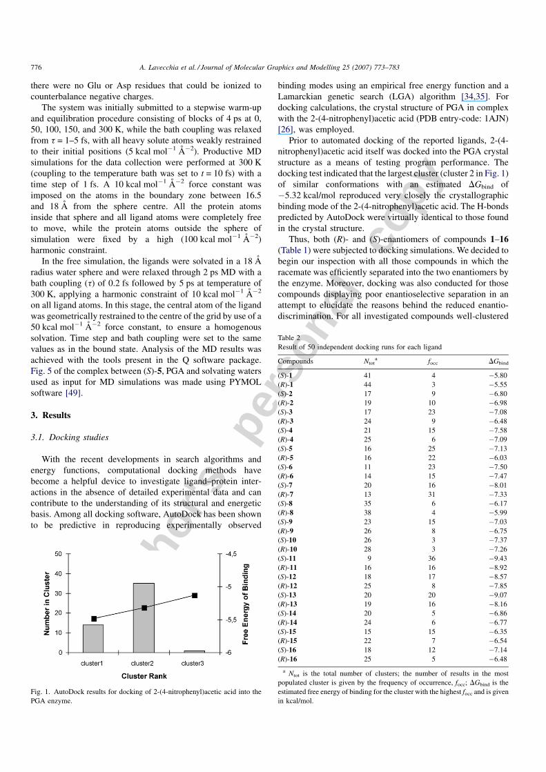
Prior to automated docking of the reported ligands, 2-(4-
nitrophenyl)acetic acid itself was docked into the PGA crystal
structure as a means of testing program performance. The
docking test indicated that the largest cluster (cluster 2 in Fig. 1)
of similar conformations with an estimated DGbind of
�5.32 kcal/mol reproduced very closely the crystallographic
binding mode of the 2-(4-nitrophenyl)acetic acid. The H-bonds
predicted by AutoDock were virtually identical to those found
in the crystal structure.
Thus, both (R)- and (S)-enantiomers of compounds 1–16(Table 1) were subjected to docking simulations. We decided to
begin our inspection with all those compounds in which the
racemate was efficiently separated into the two enantiomers by
the enzyme. Moreover, docking was also conducted for those
compounds displaying poor enantioselective separation in an
attempt to elucidate the reasons behind the reduced enantio-
discrimination. For all investigated compounds well-clustered
A. Lavecchia et al. / Journal of Molecular Graphics and Modelling 25 (2007) 773–783776
Fig. 1. AutoDock results for docking of 2-(4-nitrophenyl)acetic acid into the
PGA enzyme.
Table 2
Result of 50 independent docking runs for each ligand
Compounds Ntota focc DGbind
(S)-1 41 4 �5.80
(R)-1 44 3 �5.55
(S)-2 17 9 �6.80
(R)-2 19 10 �6.98
(S)-3 17 23 �7.08
(R)-3 24 9 �6.48
(S)-4 21 15 �7.58
(R)-4 25 6 �7.09
(S)-5 16 25 �7.13
(R)-5 16 22 �6.03
(S)-6 11 23 �7.50
(R)-6 14 15 �7.47
(S)-7 20 16 �8.01
(R)-7 13 31 �7.33
(S)-8 35 6 �6.17
(R)-8 38 4 �5.99
(S)-9 23 15 �7.03
(R)-9 26 8 �6.75
(S)-10 26 3 �7.37
(R)-10 28 3 �7.26
(S)-11 9 36 �9.43
(R)-11 16 16 �8.92
(S)-12 18 17 �8.57
(R)-12 25 8 �7.85
(S)-13 20 20 �9.07
(R)-13 19 16 �8.16
(S)-14 20 5 �6.86
(R)-14 24 6 �6.77
(S)-15 15 15 �6.35
(R)-15 22 7 �6.54
(S)-16 18 12 �7.14
(R)-16 25 5 �6.48
a Ntot is the total number of clusters; the number of results in the most
populated cluster is given by the frequency of occurrence, focc; DGbind is the
estimated free energy of binding for the cluster with the highest focc and is given
in kcal/mol.
Autho
r's
pers
onal
co
py
docking results could be obtained. The 50 independent docking
runs carried out for each ligand generally converged to a small
number of different positions clusters. Generally, the top
ranking clusters (i.e. those with the most favourable DGbind)
were also associated with the highest frequency of occurrence,
which suggests a good convergence behaviour of the search
algorithm. The best results in terms of free energy of binding
were all located in a similar position in the active site. Docking
results (the total number of clusters, the number of results in the
most populated cluster and the relative estimated free energy of
binding) are summarized in Table 2, and a graphical
representation of the binding modes of the most structurally
representative derivatives (S)-3, (R)-3, (S)-11, (R)-11, (S)-12and (S)-11, is given in Figs. 2–4. Results are briefly described in
the following section.
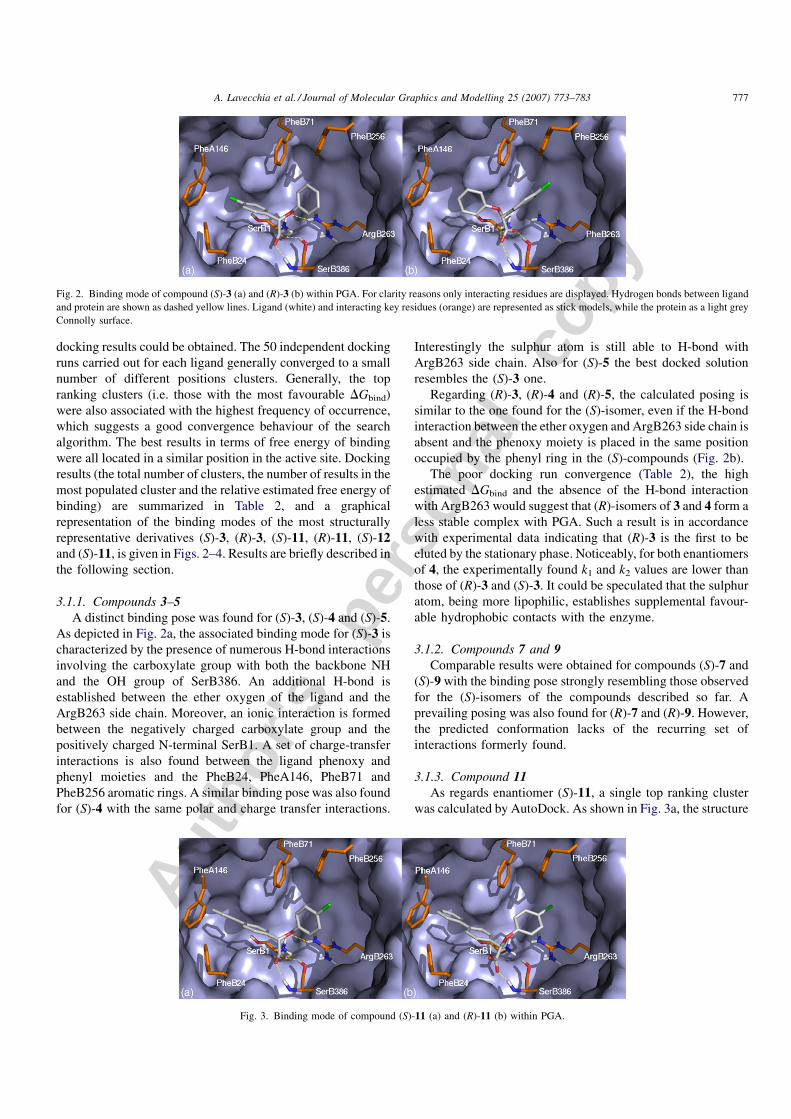
3.1.1. Compounds 3–5A distinct binding pose was found for (S)-3, (S)-4 and (S)-5.
As depicted in Fig. 2a, the associated binding mode for (S)-3 is
characterized by the presence of numerous H-bond interactions
involving the carboxylate group with both the backbone NH
and the OH group of SerB386. An additional H-bond is
established between the ether oxygen of the ligand and the
ArgB263 side chain. Moreover, an ionic interaction is formed
between the negatively charged carboxylate group and the
positively charged N-terminal SerB1. A set of charge-transfer
interactions is also found between the ligand phenoxy and
phenyl moieties and the PheB24, PheA146, PheB71 and
PheB256 aromatic rings. A similar binding pose was also found
for (S)-4 with the same polar and charge transfer interactions.
Interestingly the sulphur atom is still able to H-bond with
ArgB263 side chain. Also for (S)-5 the best docked solution
resembles the (S)-3 one.
Regarding (R)-3, (R)-4 and (R)-5, the calculated posing is
similar to the one found for the (S)-isomer, even if the H-bond
interaction between the ether oxygen and ArgB263 side chain is
absent and the phenoxy moiety is placed in the same position
occupied by the phenyl ring in the (S)-compounds (Fig. 2b).
The poor docking run convergence (Table 2), the high
estimated DGbind and the absence of the H-bond interaction
with ArgB263 would suggest that (R)-isomers of 3 and 4 form a
less stable complex with PGA. Such a result is in accordance
with experimental data indicating that (R)-3 is the first to be
eluted by the stationary phase. Noticeably, for both enantiomers
of 4, the experimentally found k1 and k2 values are lower than
those of (R)-3 and (S)-3. It could be speculated that the sulphur
atom, being more lipophilic, establishes supplemental favour-
able hydrophobic contacts with the enzyme.
3.1.2. Compounds 7 and 9Comparable results were obtained for compounds (S)-7 and
(S)-9 with the binding pose strongly resembling those observed
for the (S)-isomers of the compounds described so far. A
prevailing posing was also found for (R)-7 and (R)-9. However,
the predicted conformation lacks of the recurring set of
interactions formerly found.
3.1.3. Compound 11As regards enantiomer (S)-11, a single top ranking cluster
was calculated by AutoDock. As shown in Fig. 3a, the structure
A. Lavecchia et al. / Journal of Molecular Graphics and Modelling 25 (2007) 773–783 777
Fig. 2. Binding mode of compound (S)-3 (a) and (R)-3 (b) within PGA. For clarity reasons only interacting residues are displayed. Hydrogen bonds between ligand
and protein are shown as dashed yellow lines. Ligand (white) and interacting key residues (orange) are represented as stick models, while the protein as a light grey
Connolly surface.
Fig. 3. Binding mode of compound (S)-11 (a) and (R)-11 (b) within PGA.
Autho
r's
pers
onal
co
py
of the ligand fits into the binding cavity similarly to (S)-3 with
the biphenyl and p-Cl-phenoxy groups involved in numerous
charge-transfer interactions with PheB256, PheB71, PheB24
and PheA146. As calculated by AutoDock, (R)-11 occupies the
same space as the (S)-isomer, even though the carboxylate
group is unable to establish the commonly found polar
interactions with the PGA (Fig. 3b). The different values of
DGbind observed for both isomers of 11 suggest that the (S)-
enantiomer should be more retained by the stationary phase
than the (R)-one.
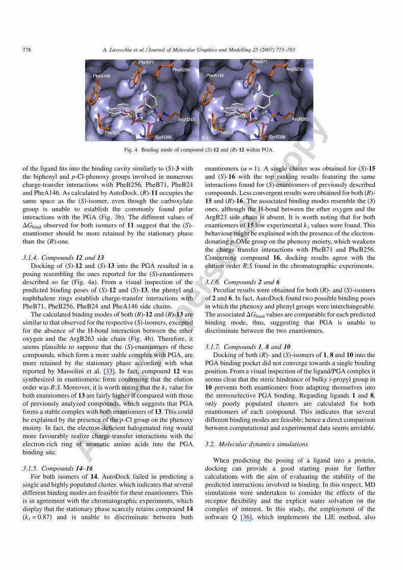
3.1.4. Compounds 12 and 13Docking of (S)-12 and (S)-13 into the PGA resulted in a
posing resembling the ones reported for the (S)-enantiomers
described so far (Fig. 4a). From a visual inspection of the
predicted binding poses of (S)-12 and (S)-13, the phenyl and
naphthalene rings establish charge-transfer interactions with
PheB71, PheB256, PheB24 and PheA146 side chains.
The calculated binding modes of both (R)-12 and (R)-13 are
similar to that observed for the respective (S)-isomers, excepted
for the absence of the H-bond interaction between the ether
oxygen and the ArgB263 side chain (Fig. 4b). Therefore, it
seems plausible to suppose that the (S)-enantiomers of these
compounds, which form a more stable complex with PGA, are
more retained by the stationary phase according with what
reported by Massolini et al. [33]. In fact, compound 12 was
synthesized in enantiomeric form confirming that the elution
order was R:S. Moreover, it is worth noting that the k1 value for
both enantiomers of 13 are fairly higher if compared with those
of previously analyzed compounds, which suggests that PGA
forms a stable complex with both enantiomers of 13. This could
be explained by the presence of the p-Cl group on the phenoxy
moiety. In fact, the electron-deficient halogenated ring would
more favourably realize charge-transfer interactions with the
electron-rich ring of aromatic amino acids into the PGA
binding site.
3.1.5. Compounds 14–16For both isomers of 14, AutoDock failed in predicting a
single and highly populated cluster, which indicates that several
different binding modes are feasible for these enantiomers. This
is in agreement with the chromatographic experiments, which
display that the stationary phase scarcely retains compound 14(k1 = 0.87) and is unable to discriminate between both
enantiomers (a = 1). A single cluster was obtained for (S)-15and (S)-16 with the top ranking results featuring the same
interactions found for (S)-enantiomers of previously described
compounds. Less convergent results were obtained for both (R)-
15 and (R)-16. The associated binding modes resemble the (S)
ones, although the H-bond between the ether oxygen and the
ArgB23 side chain is absent. It is worth noting that for both
enantiomers of 15 low experimental k1 values were found. This
behaviour might be explained with the presence of the electron-
donating p-OMe group on the phenoxy moiety, which weakens
the charge transfer interactions with PheB71 and PheB256.
Concerning compound 16, docking results agree with the
elution order R:S found in the chromatographic experiments.
3.1.6. Compounds 2 and 6Peculiar results were obtained for both (R)- and (S)-isomers
of 2 and 6. In fact, AutoDock found two possible binding poses
in which the phenoxy and phenyl groups were interchangeable.
The associated DGbind values are comparable for each predicted
binding mode, thus, suggesting that PGA is unable to
discriminate between the two enantiomers.
3.1.7. Compounds 1, 8 and 10Docking of both (R)- and (S)-isomers of 1, 8 and 10 into the
PGA binding pocket did not converge towards a single binding
position. From a visual inspection of the ligand/PGA complex it
seems clear that the steric hindrance of bulky i-propyl group in
10 prevents both enantiomers from adapting themselves into
the stereoselective PGA binding. Regarding ligands 1 and 8,
only poorly populated clusters are calculated for both
enantiomers of each compound. This indicates that several
different binding modes are feasible; hence a direct comparison
between computational and experimental data seems unviable.
3.2. Molecular dynamics simulations
When predicting the posing of a ligand into a protein,
docking can provide a good starting point for further
calculations with the aim of evaluating the stability of the
predicted interactions involved in binding. In this respect, MD
simulations were undertaken to consider the effects of the
receptor flexibility and the explicit water solvation on the
complex of interest. In this study, the employment of the
software Q [36], which implements the LIE method, also
A. Lavecchia et al. / Journal of Molecular Graphics and Modelling 25 (2007) 773–783778
Fig. 4. Binding mode of compound (S)-12 and (R)-12 within PGA.
Autho
r's
pers
onal
co
py
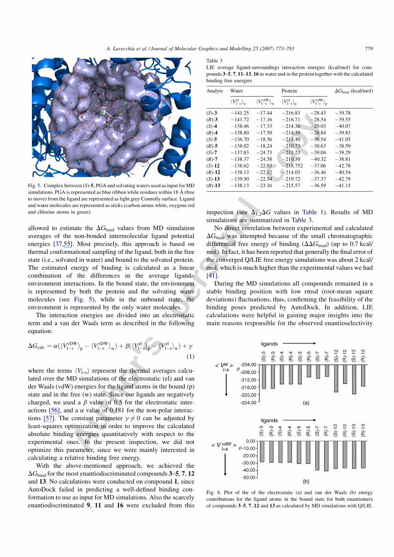
allowed to estimate the DGbind values from MD simulation
averages of the non-bonded intermolecular ligand potential
energies [37,55]. Most precisely, this approach is based on
thermal conformational sampling of the ligand, both in the free
state (i.e., solvated in water) and bound to the solvated protein.
The estimated energy of binding is calculated as a linear
combination of the differences in the average ligand–
environment interactions. In the bound state, the environment
is represented by both the protein and the solvating water
molecules (see Fig. 5), while in the unbound state, the
environment is represented by the only water molecules.
The interaction energies are divided into an electrostatic
term and a van der Waals term as described in the following
equation:
DGcalc ¼ aðhVvDWl�s ip � hV
vDWl�s iwÞ þ bðhVel
l�sip � hVell�siwÞ þ g
(1)
where the terms hVl–si represent the thermal averages calcu-
lated over the MD simulations of the electrostatic (el) and van
der Waals (vdW) energies for the ligand atoms in the bound (p)
state and in the free (w) state. Since our ligands are negatively
charged, we used a b value of 0.5 for the electrostatic inter-
actions [56], and a a value of 0.181 for the non-polar interac-
tions [57]. The constant parameter g 6¼ 0 can be adjusted by
least-squares optimization in order to improve the calculated
absolute binding energies quantitatively with respect to the
experimental ones. In the present inspection, we did not
optimize this parameter, since we were mainly interested in
calculating a relative binding free energy.
With the above-mentioned approach, we achieved the
DGbind for the most enantiodiscriminated compounds 3–5, 7, 12and 13. No calculations were conducted on compound 1, since
AutoDock failed in predicting a well-defined binding con-
formation to use as input for MD simulations. Also the scarcely
enantiodiscriminated 9, 11 and 16 were excluded from this
inspection (see D1,2DG values in Table 1). Results of MD
simulations are summarized in Table 3.
No direct correlation between experimental and calculated
DGbind was attempted because of the small chromatographic
differential free energy of binding (DDGbind) (up to 0.7 kcal/
mol). In fact, it has been reported that generally the final error of
the converged Q/LIE free energy simulations was about 2 kcal/
mol, which is much higher than the experimental values we had
[41].
During the MD simulations all compounds remained in a
stable binding position with low rmsd (root-mean square
deviations) fluctuations, thus, confirming the feasibility of the
binding poses predicted by AutoDock. In addition, LIE
calculations were helpful in gaining major insights into the
main reasons responsible for the observed enantioselectivity
A. Lavecchia et al. / Journal of Molecular Graphics and Modelling 25 (2007) 773–783 779
Fig. 5. Complex between (S)-5, PGA and solvating waters used as input for MD
simulations. PGA is represented as blue ribbon while residues within 18 A (free
to move) from the ligand are represented as light grey Connolly surface. Ligand
and water molecules are represented as sticks (carbon atoms white, oxygens red
and chlorine atoms in green).
Table 3
LIE average ligand-surroundings interaction energies (kcal/mol) for com-
pounds 3–5, 7, 11–13, 16 in water and in the protein together with the calculated
binding free energies
Analyte Water Protein DGbind (kcal/mol)
hVell�siw hVvdW
l�s iw hVell�sip hVvdW
l�s ip
(S)-3 �141.25 �17.44 �216.83 �28.43 �39.78
(R)-3 �141.72 �17.16 �216.71 �28.54 �39.55
(S)-4 �138.46 �17.33 �214.36 �29.03 �40.07
(R)-4 �138.80 �17.50 �214.39 �28.84 �39.83
(S)-5 �136.70 �18.36 �214.40 �30.54 �41.05
(R)-5 �138.02 �18.24 �210.73 �30.63 �38.59
(S)-7 �137.83 �24.73 �211.23 �39.04 �39.29
(R)-7 �138.37 �24.58 �210.30 �40.32 �38.81
(S)-12 �138.62 �22.03 �218.752 �37.06 �42.78
(R)-12 �138.13 �22.12 �214.03 �36.46 �40.54
(S)-13 �139.50 �22.54 �219.72 �37.37 �42.79
(R)-13 �138.13 �23.16 �215.57 �36.59 �41.15
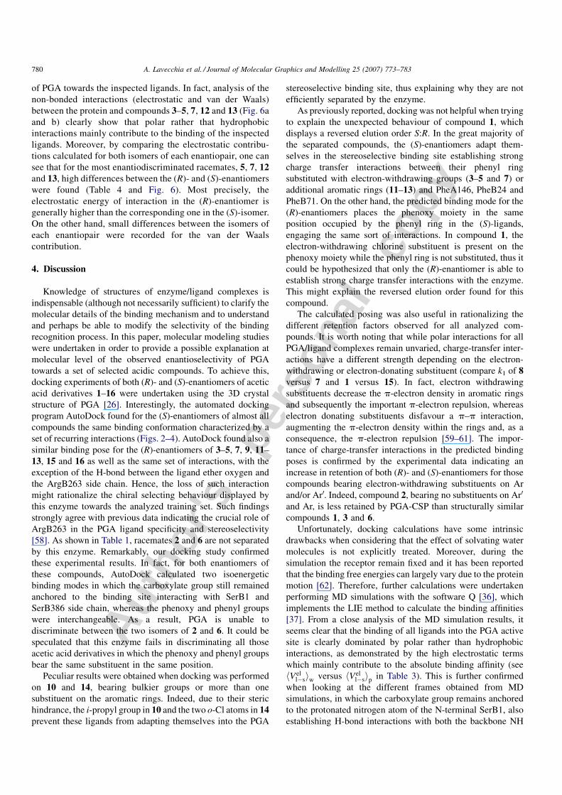
Fig. 6. Plot of the of the electrostatic (a) and van der Waals (b) energy
contributions for the ligand atoms in the bound state for both enantiomers
of compounds 3–5, 7, 12 and 13 as calculated by MD simulations with Q/LIE.
Autho
r's
pers
onal
co
py
of PGA towards the inspected ligands. In fact, analysis of the
non-bonded interactions (electrostatic and van der Waals)
between the protein and compounds 3–5, 7, 12 and 13 (Fig. 6a
and b) clearly show that polar rather that hydrophobic
interactions mainly contribute to the binding of the inspected
ligands. Moreover, by comparing the electrostatic contribu-
tions calculated for both isomers of each enantiopair, one can
see that for the most enantiodiscriminated racemates, 5, 7, 12and 13, high differences between the (R)- and (S)-enantiomers
were found (Table 4 and Fig. 6). Most precisely, the
electrostatic energy of interaction in the (R)-enantiomer is
generally higher than the corresponding one in the (S)-isomer.
On the other hand, small differences between the isomers of
each enantiopair were recorded for the van der Waals
contribution.
4. Discussion
Knowledge of structures of enzyme/ligand complexes is
indispensable (although not necessarily sufficient) to clarify the
molecular details of the binding mechanism and to understand
and perhaps be able to modify the selectivity of the binding
recognition process. In this paper, molecular modeling studies
were undertaken in order to provide a possible explanation at
molecular level of the observed enantioselectivity of PGA
towards a set of selected acidic compounds. To achieve this,
docking experiments of both (R)- and (S)-enantiomers of acetic
acid derivatives 1–16 were undertaken using the 3D crystal
structure of PGA [26]. Interestingly, the automated docking
program AutoDock found for the (S)-enantiomers of almost all
compounds the same binding conformation characterized by a
set of recurring interactions (Figs. 2–4). AutoDock found also a
similar binding pose for the (R)-enantiomers of 3–5, 7, 9, 11–
13, 15 and 16 as well as the same set of interactions, with the
exception of the H-bond between the ligand ether oxygen and
the ArgB263 side chain. Hence, the loss of such interaction
might rationalize the chiral selecting behaviour displayed by
this enzyme towards the analyzed training set. Such findings
strongly agree with previous data indicating the crucial role of
ArgB263 in the PGA ligand specificity and stereoselectivity
[58]. As shown in Table 1, racemates 2 and 6 are not separated
by this enzyme. Remarkably, our docking study confirmed
these experimental results. In fact, for both enantiomers of
these compounds, AutoDock calculated two isoenergetic
binding modes in which the carboxylate group still remained
anchored to the binding site interacting with SerB1 and
SerB386 side chain, whereas the phenoxy and phenyl groups
were interchangeable. As a result, PGA is unable to
discriminate between the two isomers of 2 and 6. It could be
speculated that this enzyme fails in discriminating all those
acetic acid derivatives in which the phenoxy and phenyl groups
bear the same substituent in the same position.
Peculiar results were obtained when docking was performed
on 10 and 14, bearing bulkier groups or more than one
substituent on the aromatic rings. Indeed, due to their steric
hindrance, the i-propyl group in 10 and the two o-Cl atoms in 14prevent these ligands from adapting themselves into the PGA
stereoselective binding site, thus explaining why they are not
efficiently separated by the enzyme.
As previously reported, docking was not helpful when trying
to explain the unexpected behaviour of compound 1, which
displays a reversed elution order S:R. In the great majority of
the separated compounds, the (S)-enantiomers adapt them-
selves in the stereoselective binding site establishing strong
charge transfer interactions between their phenyl ring
substituted with electron-withdrawing groups (3–5 and 7) or
additional aromatic rings (11–13) and PheA146, PheB24 and
PheB71. On the other hand, the predicted binding mode for the
(R)-enantiomers places the phenoxy moiety in the same
position occupied by the phenyl ring in the (S)-ligands,
engaging the same sort of interactions. In compound 1, the
electron-withdrawing chlorine substituent is present on the
phenoxy moiety while the phenyl ring is not substituted, thus it
could be hypothesized that only the (R)-enantiomer is able to
establish strong charge transfer interactions with the enzyme.
This might explain the reversed elution order found for this
compound.
The calculated posing was also useful in rationalizing the
different retention factors observed for all analyzed com-
pounds. It is worth noting that while polar interactions for all
PGA/ligand complexes remain unvaried, charge-transfer inter-
actions have a different strength depending on the electron-
withdrawing or electron-donating substituent (compare k1 of 8versus 7 and 1 versus 15). In fact, electron withdrawing
substituents decrease the p-electron density in aromatic rings
and subsequently the important p-electron repulsion, whereas
electron donating substituents disfavour a p–p interaction,
augmenting the p-electron density within the rings and, as a
consequence, the p-electron repulsion [59–61]. The impor-
tance of charge-transfer interactions in the predicted binding
poses is confirmed by the experimental data indicating an
increase in retention of both (R)- and (S)-enantiomers for those
compounds bearing electron-withdrawing substituents on Ar
and/or Ar0. Indeed, compound 2, bearing no substituents on Ar0
and Ar, is less retained by PGA-CSP than structurally similar
compounds 1, 3 and 6.
Unfortunately, docking calculations have some intrinsic
drawbacks when considering that the effect of solvating water
molecules is not explicitly treated. Moreover, during the
simulation the receptor remain fixed and it has been reported
that the binding free energies can largely vary due to the protein
motion [62]. Therefore, further calculations were undertaken
performing MD simulations with the software Q [36], which
implements the LIE method to calculate the binding affinities
[37]. From a close analysis of the MD simulation results, it
seems clear that the binding of all ligands into the PGA active
site is clearly dominated by polar rather than hydrophobic
interactions, as demonstrated by the high electrostatic terms
which mainly contribute to the absolute binding affinity (see
hVell�siw versus hVel
l�sip in Table 3). This is further confirmed
when looking at the different frames obtained from MD
simulations, in which the carboxylate group remains anchored
to the protonated nitrogen atom of the N-terminal SerB1, also
establishing H-bond interactions with both the backbone NH
A. Lavecchia et al. / Journal of Molecular Graphics and Modelling 25 (2007) 773–783780
Autho
r's
pers
onal
co
py
and OH group of SerB386. Moreover, while in the (S)-
enantiomers the ether oxygen H-bonds with ArgB263 side
chain, in the (R)-ones this interaction is absent or fairly instable.
From these considerations, it seems that polar interactions not
only provide most of the overall affinity of these compounds for
PGA, but are also responsible for the enantiospecificity of the
enzyme. In fact, as reported in Table 4 and Fig. 6, the main
differences in the ligand-surroundings interaction energies
between the (R)- and (S)-enantiomers were only recorded for
the electrostatic contributions in the bound state and not for the
non-polar ones.
5. Conclusions
The goal of this study was to elucidate the reasons behind the
enantioselective binding of a highly efficient chiral selector
such as PGA towards a set of acidic compounds in which the
absolute configuration has been reported to exert a strong
influence on several biological systems [19–24]. Thus, the 3D-
structure of the enzyme solved through X-ray crystallography
[26] was used to dock both (R)- and (S)-isomers of 1–16employing the automated docking software AutoDock. Dock-
ing results indicated that the (S)-enantiomers establish several
polar interactions with SerB1, SerB386 and ArgB263 of PGA.
Conversely, the absence of specific polar interactions between
the (R)-enantiomers and ArgB263 seems to be the main reason
for the different chromatographic retention factors observed
between (R)- and (S)-enantiomers, thus, explaining the PGA
enantioselective behaviour. Docking experiments were also
helpful in rationalizing the absence of enantioselectivity
observed for some tested compounds (2, 6, 10, 14 and 15).
In fact, the steric hindrance of bulky groups on the phenyl ring
(10) or an additional substituent on the phenoxy portion (14)
might prevent these ligands from adapting themselves into the
stereoselective binding site of PGA. It could be speculated that
this enzyme fails in discriminating all those acetic acid
derivatives in which the phenoxy and phenyl groups bear the
same substituent in the same position, as confirmed by docking
of 2 and 6. Moreover, this computational study underlines the
importance of the numerous charge-transfer interactions
established between the ligands and the enzyme. In particular,
an increase in the electron-withdrawing effect on both aromatic
rings might stabilizes the complex between the selected
compounds and PGA leading to an increase of the retention
factors. To evaluate the effect of the receptor plasticity on
ligand binding together with the influence of the water
solvation, MD simulations were performed employing the
software Q [36]. This computational approach revealed to be
really helpful in elucidating the molecular basis of the observed
enantioselectivity of PGA towards the selected compounds. In
fact, from an accurate analysis of MD results it seems clear that
polar interactions provide most of the overall affinity of these
compounds for PGA and are responsible for the enantiospe-
cificity of the enzyme.
In principle, the predictive power of the presented
theoretical approach could be assessed by performing a
comparison between experimental and calculated parameters
(DDGcalc versus DDGexp) to see if they quantitatively correlate.
Indeed, such a correlation might be hampered by some
limitations connected with the experimental procedure
adopted. In fact, it is worth noting that the DDGexp values
not only result from specific interactions with the enzyme active
site but also from interactions with the protein at other binding
sites and possibly also with the support. From this point of view,
a quantitative correlation between DDGcalc and DDGexp values
would mean comparing different factors which make such a
correlation somehow weak. However, even if the predictive
power of the presented computational approach cannot
unequivocally be assessed, the qualitative consistency of both
docking and MD simulations results with chromatographic data
suggests that our approach could be used to shed light on the
reasons of the observed enantioselective binding behaviour of
diverse biological targets. Moreover, the modeled complexes
between the inspected chiral compounds and PGA form the
basis for rational protein engineering efforts aimed at
improving the enantioselectivity of PGA, thus, enhancing its
industrial applications.
Acknowledgment
This work was supported by grant from Ministero
dell’Universita e della Ricerca Scientifica (grant no.
2002034857_003).
References
[1] W.A. Bonner, Parity violation and the evolution of biomolecular homo-
chirality, Chirality 12 (2000) 114–126.
[2] I. Agranat, H. Caner, J. Caldwell, Putting chirality to work: the strategy of
chiral switches, Nat. Rev. Drug Discov. 1 (2002) 753–768.
[3] H.Y. Aboul-Enein, L.I. Abou-Basha (Eds.), The Impact of Stereochem-
istry on Drug Development, and Use, Wiley, New York, NY, 1997.
[4] M. Eichelbaum, A.S. Gross, Stereochemical aspects of drug action and
disposition, Adv. Drug Res. 28 (1996) 2–64.
[5] R. Crossley (Ed.), Chirality, and the Biological Activity of Drugs, CRC
Press, Boca Raton, FL, 1995.
[6] D.J. Triggle, Stereoselectivity of drug action, Drug Discov. Today 2 (1997)
138.
[7] C.A. Challener (Ed.), Chiral Drugs, Ashgate Burlington, Vermont, 2001.
[8] M. Eichelbaum, B. Testa, A. Somogyi (Eds.), Stereochemical Aspects of
Drug Action, Springer, Heidelberg, 2002.
[9] X.X. Sun, L.Z. Sun, H.Y. Aboul-Enein, Chiral derivatization reagents for
drug enantioseparation by high-performance liquid chromatography
based upon pre-column derivatization and formation of diastereomers:
enantioselectivity and related structure, Biomed. Chromatogr. 2 (2001)
116–132.
[10] S. Allenmark (Ed.), Chromatographic Enantioseparation: Methods, and
Applications, 2nd ed., Ellis Horwood, New York, 1991 (Chapter 7).
[11] I.W. Wainer (Ed.), Drug Stereochemistry: Analytical Methods, and Phar-
macology, Marcel Dekker, New York, 1993 (Chapter 6).
[12] J. Haginaka, Protein-based chiral stationary phases for high-performance
liquid chromatography enantioseparations, J. Chromatogr. A 906 (2001)
253–273.
[13] G. Massolini, E. Calleri, A. Lavecchia, F. Loiodice, D. Lubda, C.
Temporini, G. Fracchiolla, P. Tortorella, E. Novellino, G. Caccialanza,
Enantioselective hydrolysis of some 2-aryloxyalkanoic acid methyl esters
and isosteric analogues using a penicillin G acylase-based HPLC mono-
lithic silica column, Anal. Chem. 75 (2003) 535–542.
A. Lavecchia et al. / Journal of Molecular Graphics and Modelling 25 (2007) 773–783 781
Autho
r's
pers
onal
co
py
[14] E. Calleri, G. Massolini, D. Lubda, C. Temporini, F. Loiodice, G.
Caccialanza, Evaluation of a monolithic epoxy silica support for penicillin
G acylase immobilization, J. Chromatogr. A. 1031 (2004) 93–100.
[15] E. Calleri, C. Temporini, G. Massolini, G. Caccialanza, Penicillin G
acylase-based stationary phases: analytical applications, J. Pharm.
Biomed. Anal. 35 (2004) 243–258.
[16] M. Arroyo, I. de la Mata, C. Acebal, M.P. Castillon, Biotechnological
applications of penicillin acylases: state-of-the-art, Appl. Microbiol.
Biotechnol. 60 (2003) 507–514.
[17] G. Massolini, E. Calleri, E. De Lorenzi, M. Pregnolato, M. Terreni, G.
Felix, C. Gandini, Immobilized penicillin G acylase as reactor and chiral
selector in liquid chromatography, J. Chromatogr. A 921 (2001) 147–160.
[18] E. Calleri, G. Massolini, F. Loiodice, G. Fracchiolla, C. Temporini, G.
Felix, P. Tortorella, G. Caccialanza, Evaluation of a penicillin G acylase-
based chiral stationary phase towards a series of 2-aryloxyalkanoic acids,
isosteric analogs and 2-arylpropionic acids, J. Chromatogr. A 958 (2002)
131–140.
[19] K. Liu, L. Xu, J.P. Berger, K.L. MacNaul, G. Zhou, T.W. Doebber, M.J.
Forrest, D.E. Moller, A.B. Jones, Discovery of a novel series of peroxi-
some proliferator-activated receptor alpha/gamma dual agonists for the
treatment of type 2 diabetes and dyslipidemia, J. Med. Chem. 48 (2005)
2262–2265.
[20] A. Pinelli, G. Godio, A. Laghezza, N. Mitro, G. Fracchiolla, V. Tortorella,
A. Lavecchia, E. Novellino, J.C. Fruchart, B. Staels, M. Crestani, F.
Loiodice, Synthesis, biological evaluation, and molecular modeling inves-
tigation of new chiral fibrates with PPARalpha and PPARgamma agonist
activity, J. Med. Chem. 48 (2005) 5509–5519.
[21] M.P. Grella, R. Danso-Danquah, M.K. Safo, G.S. Joshi, J. Kister, M.
Marden, S.J. Hoffman, D.J. Abraham, Synthesis and structure–activity
relationships of chiral allosteric modifiers of hemoglobin, J. Med. Chem. 4
(2000) 4726–4737.
[22] G. Bettoni, F. Loiodice, V. Tortorella, D. Conte-Camerino, M. Mambrini,
E. Ferrannini, S.H. Bryant, Stereospecificity of the chloride ion channel:
the action of chiral clofibric acid analogs, J. Med. Chem. 30 (1987) 1267–
1270.
[23] A. Liantonio, A. De Luca, S. Pierno, M.P. Didonna, F. Loiodice, G.
Fracchiolla, P. Tortorella, A. Laghezza, E. Bonerba, S. Traverso, L. Elia,
A. Picollo, M. Pusch, D. Conte-Camerino, Structural requisites of 2-( p-
chlorophenoxy)propionic acid analogues for activity on native rat skeletal
muscle chloride conductance and on heterologously expressed CLC-1, Br.
J. Pharmacol. 139 (2003) 1255–1264.
[24] D. Feller, V.S. Kamanna, H.A.I. Newman, K.J. Romstedt, D.T. Witiak, G.
Bettoni, S.H. Bryant, D. Conte-Camerino, F. Loiodice, V. Tortorella,
Dissociation of hypolipidemic and antiplatelet actions from adverse
myotonic effects of clofibric acid related enantiomers, J. Med. Chem.
30 (1987) 1265–1267.
[25] R.D. Larsen, E.G. Corley, P. Davis, P.J. Reider, E.J.J. Grabowski, a-
Hydroxy esters as chiral reagents: asymmetric synthesis of 2-arylpropio-
nic acids, J. Am. Chem. Soc. 111 (1989) 7650–7651 (and references
therein).
[26] S.H. Done, J.A. Brannigan, P.C. Moody, R.E. Hubbard, Ligand-induced
conformational change in penicillin acylase, J. Mol. Biol. 284 (1998) 463–
475.
[27] C.E. McVey, M.A. Walsh, G.G. Dodson, K.S. Wilson, J.A. Brannigan,
Crystal structures of penicillin acylase enzyme–substrate complexes:
structural insights into the catalytic mechanism, J. Mol. Biol. 313
(2001) 139–150.
[28] J.A. Brannigan, G. Dodson, H.J. Duggleby, P.C.E. Moody, J.L. Smith,
D.R. Tomchick, A.G. Murzin, A protein catalytic framework with an N-
terminal nucleophile is capable of self-activation, Nature 378 (1995) 416–
419.
[29] I. Mononen, K.J. Fischer, V. Kaartinen, N.N. Aronson, Aspartylglycosa-
minuria: protein chemistry and molecular biology of the most common
lysosomal storage disorder of glycoprotein degradation, FASEB J. 7
(1993) 1247–1256.
[30] J. Lowe, D. Stock, B. Jap, P. Zwickl, W. Baumeister, R. Huber, Crystal
structure of the 20S proteasome from the archaeon T. acidophilum at 3.4 A
resolution, Science 268 (1995) 533–539.
[31] J.L. Smith, E.J. Zaluzec, J.P. Wery, L. Niu, R.L. Switzer, H. Zalkin, Y.
Satow, Structure of the allosteric regulatory enzyme of purine biosynth-
esis, Science 264 (1994) 1427–1433.
[32] H.J. Duggleby, S.P. Tolley, C.P. Hill, E.J. Dodson, P.C. Moody, Penicillin
acylase has a single-amino-acid catalytic centre, Nature 373 (1995) 264–
268.
[33] G. Massolini, G. Fracchiolla, E. Calleri, G. Carbonara, C. Temporini, A.
Lavecchia, S. Cosconati, E. Novellino, F. Loiodice, Elucidation of the
Enantioselective Recognition Mechanism of a Penicillin G Acylase-Based
Chiral Stationary Phase Towards a Series of 2-Aryloxy-2-Arylacetic
Acids, Chirality 18 (2006) 633–643.
[34] G.M. Morris, D.S. Goodsell, R.S. Halliday, R. Huey, W.E. Hart, R.K.
Belew, A.J. Olson, Docking using a Lamarckian genetic algorithm and an
empirical binding free energy function, J. Comp. Chem. 19 (1998) 1639–
1662.
[35] D.S. Goodsell, G.M. Morris, A.J. Olson, Automated docking of flexible
ligands: applications of AutoDock, J. Mol. Recognit. 9 (1996) 1–5.
[36] J. Marelius, K. Kolmodin, I. Feierberg, J. Aqvist, Q: a molecular dynamics
program for free energy calculations and empirical valence bond simula-
tions in biomolecular systems, J. Mol. Graph. 16 (1998) 213–225.
[37] J. Aqvist, C. Medina, J.E. Samuelsson, A new method for predicting
binding affinity in computer-aided drug design, Protein Eng. 7 (1994)
385–391.
[38] J. Aqvist, T. Hansson, On the validity of electrostatic linear response in
polar solvents, J. Phys. Chem. 100 (1996) 9512–9521.
[39] J. Marelius, T. Hansson, J. Aqvist, Calculation of ligand binding free
energies from molecular dynamics simulations, Int. J. Quantum Chem. 69
(1998) 77–88.
[40] F. Osterberg, J. Aqvist, Exploring blocker binding to a homology model of
the open hERG K+ channel using docking and molecular dynamics
methods, FEBS Lett. 579 (2005) 2939–2944.
[41] J. Hulten, N.M. Bonham, U. Nillroth, T. Hansson, G. Zuccarello, A.
Bouzide, J. Aqvist, B. Classon, U.H. Danielson, A. Karlen, I. Kvarnstrom,
B. Samuelsson, A. Hallberg, Cyclic HIV-1 protease inhibitors derived
from mannitol: synthesis, inhibitory potencies, and computational pre-
dictions of binding affinities, J. Med. Chem. 40 (1997) 885–897.
[42] K. Ersmark, M. Nervall, E. Hamelink, L.K. Janka, J.C. Clemente, B.M.
Dunn, M.J. Blackman, B. Samuelsson, J. Aqvist, A. Hallberg, Synthesis of
malarial plasmepsin inhibitors and prediction of binding modes by
molecular dynamics simulations, J. Med. Chem. 48 (2005) 6090–6106.
[43] M. Almlof, J. Aqvist, A.O. Smalas, B.O. Brandsdal, Probing the effect of
point mutations at protein–protein interfaces with free energy calcula-
tions, Biophys. J. 90 (2006) 433–442.
[44] SYBYL Molecular Modeling System (Version 7.1), TRIPOS Assoc., St.
Louis, MO.
[45] J. Head, M.C. Zerner, A Broyden–Fletcher–Goldfarb–Shannon optimiza-
tion procedure for molecular geometries, Chem. Phys. Lett. 122 (1985)
264–270.
[46] J.G. Vinter, A. Davis, M.R. Saunders, Strategic approaches to drug design.
An integrated software framework for molecular modelling, J. Comput.
Aided Mol. Des. 1 (1987) 31–51.
[47] J. Gasteiger, M. Marsili, Iterative partial equalization of orbital electro-
negativity—a rapid access to atomic charges, Tetrahedron 36 (1980)
3219–3228.
[48] F.C. Bernstein, T.F. Koetzle, G.J.B. Williams, E.F.J. Meyer, M.R. Brice,
J.R. Rodgers, O. Kennard, T. Shimanouchi, T. Tasumi, The Protein Data
Bank: a computer-based archival file for macromolecular structures, J.
Mol. Biol. 112 (1977) 535–542.
[49] W.L. DeLano, The PyMOL Molecular Graphics System (2002), http://
www.pymol.org.
[50] W.D. Cornell, P. Cieplak, C.I. Bayly, I.R. Gould, K.M. Merz Jr., D.M.
Ferguson, D.C. Spellmeyer, T. Fox, J.W. Caldwell, P.A. Kollman, A
second generation force field for the simulation of proteins, nucleic acids,
and organic molecules, J. Am. Chem. Soc. 117 (1995) 5179–5197.
[51] G. Subramanian, M.G. Paterlini, D.L. Larson, P.S. Portoghese, D.M.
Ferguson, Conformational analysis and automated receptor docking of
selective arylacetamide-based k-opioid agonists, J. Med. Chem. 41 (1998)
4777–4789.
A. Lavecchia et al. / Journal of Molecular Graphics and Modelling 25 (2007) 773–783782
Autho
r's
pers
onal
co
py
[52] A. Jakalian, D.B. Jack, C.I. Bayly, Fast, efficient generation of high-
quality atomic charges. AM1-BCC model. II. Parameterization and
validation, J. Comput. Chem. 23 (2002) 1241–1623.
[53] W. Wang, W.A. Lim, A. Jakalian, J. Wang, J. Wang, R. Luo, C.I. Bayly,
P.A. Kollman, An analysis of the interactions between the Sem-5 SH3
domain and its ligands using molecular dynamics, free energy calcula-
tions, and sequence analysis, J. Am. Chem. Soc. 123 (2001) 3986–3994.
[54] W. Jorgensen, J. Chandrasekhar, J.D. Madura, R.W. Impey, M.L. Klein,
Comparison of simple potential functions for simulating liquid water, J.
Chem. Phys. 79 (1983) 926–935.
[55] T. Hansson, J. Marelius, J. Aqvist, Ligand binding affinity prediction by
linear interaction energy methods, J. Comput. Aided Mol. Des. 12 (1998)
27–35.
[56] F.S. Lee, Z.T. Chu, M.B. Bolger, A. Warshel, Calculations of antibody–
antigen interactions: microscopic and semi-microscopic evaluation of the
free energies of binding of phosphorylcholine analogs to McPC603, Prot.
Eng. 5 (1992) 215–228.
[57] G. Gotmar, T. Fornstedt, G. Guiochon, Apparent and true enantioselec-
tivity in enantioseparations, Chirality 12 (2000) 558–564.
[58] M. Guncheva, I. Ivanov, B. Galunsky, N. Stambolieva, J. Kaneti, Kinetic
studies and molecular modeling attribute a crucial role in the specificity
and stereoselectivity of penicillin acylase to the pair ArgA145–ArgB263,
Eur. J. Biochem. 271 (2004) 2272–2279.
[59] C.A. Hunter, Arene–arene interactions: electrostatic or charge transfer?
Angew. Chem. Int. Ed. Engl. 32 (1993) 1584–1586.
[60] F.J. Cozzi, S. Siegel, Interaction between stacked aryl groups in 1,8-
diarylnaphthalenes: dominance of polar/p over charge-transfer effects,
Pure Appl. Chem. 67 (1995) 683–689.
[61] S.B. Ferguson, E.M. Sanford, E.M. Seward, F. Diederich, Cyclophane–
arene inclusion complexation in protic solvents: solvent effects versus
electron donor–acceptor interactions, J. Am. Chem. Soc. 113 (1991)
5410–5419.
[62] S. Raza, L. Fransson, K. Hult, Enantioselectivity in Candida antarctica
lipase B: a molecular dynamics study, Protein Sci. 10 (2001) 329–338.
A. Lavecchia et al. / Journal of Molecular Graphics and Modelling 25 (2007) 773–783 783