Experimental design, formulation and in vivo evaluation of a ...
18
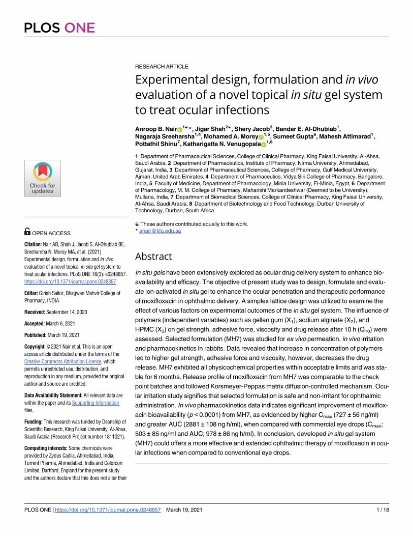
RESEARCH ARTICLE Experimental design, formulation and in vivo evaluation of a novel topical in situ gel system to treat ocular infections Anroop B. Nair ID 1☯ *, Jigar Shah 2☯ , Shery Jacob 3 , Bandar E. Al-Dhubiab 1 , Nagaraja Sreeharsha 1,4 , Mohamed A. MorsyID 1,5 , Sumeet Gupta 6 , Mahesh Attimarad 1 , Pottathil Shinu 7 , Katharigatta N. VenugopalaID 1,8 1 Department of Pharmaceutical Sciences, College of Clinical Pharmacy, King Faisal University, Al-Ahsa, Saudi Arabia, 2 Department of Pharmaceutics, Institute of Pharmacy, Nirma University, Ahmedabad, Gujarat, India, 3 Department of Pharmaceutical Sciences, College of Pharmacy, Gulf Medical University, Ajman, United Arab Emirates, 4 Department of Pharmaceutics, Vidya Siri College of Pharmacy, Bangalore, India, 5 Faculty of Medicine, Department of Pharmacology, Minia University, El-Minia, Egypt, 6 Department of Pharmacology, M. M. College of Pharmacy, Maharishi Markandeshwar (Deemed to be University), Mullana, India, 7 Department of Biomedical Sciences, College of Clinical Pharmacy, King Faisal University, Al-Ahsa, Saudi Arabia, 8 Department of Biotechnology and Food Technology, Durban University of Technology, Durban, South Africa ☯ These authors contributed equally to this work. * [email protected] Abstract In situ gels have been extensively explored as ocular drug delivery system to enhance bio- availability and efficacy. The objective of present study was to design, formulate and evalu- ate ion-activated in situ gel to enhance the ocular penetration and therapeutic performance of moxifloxacin in ophthalmic delivery. A simplex lattice design was utilized to examine the effect of various factors on experimental outcomes of the in situ gel system. The influence of polymers (independent variables) such as gellan gum (X 1 ), sodium alginate (X 2 ), and HPMC (X 3 ) on gel strength, adhesive force, viscosity and drug release after 10 h (Q 10 ) were assessed. Selected formulation (MH7) was studied for ex vivo permeation, in vivo irritation and pharmacokinetics in rabbits. Data revealed that increase in concentration of polymers led to higher gel strength, adhesive force and viscosity, however, decreases the drug release. MH7 exhibited all physicochemical properties within acceptable limits and was sta- ble for 6 months. Release profile of moxifloxacin from MH7 was comparable to the check point batches and followed Korsmeyer-Peppas matrix diffusion-controlled mechanism. Ocu- lar irritation study signifies that selected formulation is safe and non-irritant for ophthalmic administration. In vivo pharmacokinetics data indicates significant improvement of moxiflox- acin bioavailability (p < 0.0001) from MH7, as evidenced by higher C max (727 ± 56 ng/ml) and greater AUC (2881 ± 108 ng h/ml), when compared with commercial eye drops (C max ; 503 ± 85 ng/ml and AUC; 978 ± 86 ng h/ml). In conclusion, developed in situ gel system (MH7) could offers a more effective and extended ophthalmic therapy of moxifloxacin in ocu- lar infections when compared to conventional eye drops. PLOS ONE PLOS ONE | https://doi.org/10.1371/journal.pone.0248857 March 19, 2021 1 / 18 a1111111111 a1111111111 a1111111111 a1111111111 a1111111111 OPEN ACCESS Citation: Nair AB, Shah J, Jacob S, Al-Dhubiab BE, Sreeharsha N, Morsy MA, et al. (2021) Experimental design, formulation and in vivo evaluation of a novel topical in situ gel system to treat ocular infections. PLoS ONE 16(3): e0248857. https://doi.org/10.1371/journal.pone.0248857 Editor: Girish Sailor, Bhagwan Mahvir College of Pharmacy, INDIA Received: September 14, 2020 Accepted: March 6, 2021 Published: March 19, 2021 Copyright: © 2021 Nair et al. This is an open access article distributed under the terms of the Creative Commons Attribution License, which permits unrestricted use, distribution, and reproduction in any medium, provided the original author and source are credited. Data Availability Statement: All relevant data are within the paper and its Supporting Information files. Funding: This research was funded by Deanship of Scientific Research, King Faisal University, Al-Ahsa, Saudi Arabia (Research Project number 1811021). Competing interests: Some chemicals were provided by Zydus Cadila, Ahmedabad, India, Torrent Pharma, Ahmedabad, India and Colorcon Limited, Dartford, England for the present study and the authors declare that this does not alter their
-
Upload
khangminh22 -
Category
Documents
-
view
3 -
download
0
Transcript of Experimental design, formulation and in vivo evaluation of a ...
RESEARCH ARTICLE
Experimental design, formulation and in vivo
evaluation of a novel topical in situ gel system
to treat ocular infections
Anroop B. NairID1☯*, Jigar Shah2☯, Shery Jacob3, Bandar E. Al-Dhubiab1,
Nagaraja Sreeharsha1,4, Mohamed A. MorsyID1,5, Sumeet Gupta6, Mahesh Attimarad1,
Pottathil Shinu7, Katharigatta N. VenugopalaID1,8
1 Department of Pharmaceutical Sciences, College of Clinical Pharmacy, King Faisal University, Al-Ahsa,
Saudi Arabia, 2 Department of Pharmaceutics, Institute of Pharmacy, Nirma University, Ahmedabad,
Gujarat, India, 3 Department of Pharmaceutical Sciences, College of Pharmacy, Gulf Medical University,
Ajman, United Arab Emirates, 4 Department of Pharmaceutics, Vidya Siri College of Pharmacy, Bangalore,
India, 5 Faculty of Medicine, Department of Pharmacology, Minia University, El-Minia, Egypt, 6 Department
of Pharmacology, M. M. College of Pharmacy, Maharishi Markandeshwar (Deemed to be University),
Mullana, India, 7 Department of Biomedical Sciences, College of Clinical Pharmacy, King Faisal University,
Al-Ahsa, Saudi Arabia, 8 Department of Biotechnology and Food Technology, Durban University of
Technology, Durban, South Africa
☯ These authors contributed equally to this work.
Abstract
In situ gels have been extensively explored as ocular drug delivery system to enhance bio-
availability and efficacy. The objective of present study was to design, formulate and evalu-
ate ion-activated in situ gel to enhance the ocular penetration and therapeutic performance
of moxifloxacin in ophthalmic delivery. A simplex lattice design was utilized to examine the
effect of various factors on experimental outcomes of the in situ gel system. The influence of
polymers (independent variables) such as gellan gum (X1), sodium alginate (X2), and
HPMC (X3) on gel strength, adhesive force, viscosity and drug release after 10 h (Q10) were
assessed. Selected formulation (MH7) was studied for ex vivo permeation, in vivo irritation
and pharmacokinetics in rabbits. Data revealed that increase in concentration of polymers
led to higher gel strength, adhesive force and viscosity, however, decreases the drug
release. MH7 exhibited all physicochemical properties within acceptable limits and was sta-
ble for 6 months. Release profile of moxifloxacin from MH7 was comparable to the check
point batches and followed Korsmeyer-Peppas matrix diffusion-controlled mechanism. Ocu-
lar irritation study signifies that selected formulation is safe and non-irritant for ophthalmic
administration. In vivo pharmacokinetics data indicates significant improvement of moxiflox-
acin bioavailability (p < 0.0001) from MH7, as evidenced by higher Cmax (727 ± 56 ng/ml)
and greater AUC (2881 ± 108 ng h/ml), when compared with commercial eye drops (Cmax;
503 ± 85 ng/ml and AUC; 978 ± 86 ng h/ml). In conclusion, developed in situ gel system
(MH7) could offers a more effective and extended ophthalmic therapy of moxifloxacin in ocu-
lar infections when compared to conventional eye drops.
PLOS ONE
PLOS ONE | https://doi.org/10.1371/journal.pone.0248857 March 19, 2021 1 / 18
a1111111111
a1111111111
a1111111111
a1111111111
a1111111111
OPEN ACCESS
Citation: Nair AB, Shah J, Jacob S, Al-Dhubiab BE,
Sreeharsha N, Morsy MA, et al. (2021)
Experimental design, formulation and in vivo
evaluation of a novel topical in situ gel system to
treat ocular infections. PLoS ONE 16(3): e0248857.
https://doi.org/10.1371/journal.pone.0248857
Editor: Girish Sailor, Bhagwan Mahvir College of
Pharmacy, INDIA
Received: September 14, 2020
Accepted: March 6, 2021
Published: March 19, 2021
Copyright: © 2021 Nair et al. This is an open
access article distributed under the terms of the
Creative Commons Attribution License, which
permits unrestricted use, distribution, and
reproduction in any medium, provided the original
author and source are credited.
Data Availability Statement: All relevant data are
within the paper and its Supporting Information
files.
Funding: This research was funded by Deanship of
Scientific Research, King Faisal University, Al-Ahsa,
Saudi Arabia (Research Project number 1811021).
Competing interests: Some chemicals were
provided by Zydus Cadila, Ahmedabad, India,
Torrent Pharma, Ahmedabad, India and Colorcon
Limited, Dartford, England for the present study
and the authors declare that this does not alter their
Introduction
A key challenge frequently encountered during the development of ophthalmic delivery sys-
tems is the achievement of desired drug level at the target site, particularly within the anterior
cavity of the eye, for sufficient time. This is mainly due to the complex anatomy and highly
selective corneal barriers, which limit the entry of any exogenous substances to the ocular tis-
sues [1,2]. Different types of ophthalmic vehicles like eye drops, ointments, gels and polymeric
ocular inserts were developed in an attempt to enhance the pre-corneal residence time [3].
Among the various ophthalmic dosage forms evaluated so far, in situ gel drug delivery systems
has been an extensive area of research during last few decades. In situ gels are attractive since it
can be suitably applied as drops or solutions into the conjunctival sac, wherein they undergo a
phase conversion into a gel state upon exposure to either pH of the tear fluid, ocular surface
temperature, or ions exists on the tear film [4,5]. Transition to gel state in the corneal surface
extends the ocular residence resulting in better ocular bioavailability by minimizing rapid pre-
corneal elimination, particularly due to nasolacrimal drainage and eye blinking [6]. It can also
reduce the poor compliance due to frequent administration and risk of undesirable side effects
associated with systemic drug absorption by virtue of pre-corneal elimination [7].
In situ activated transparent gel formulations is ideal for ocular therapy as it can be admin-
istered as liquid dosage form and avoids blurred vision. Besides, they exhibit excellent physico-
chemical characteristics such as bioadhesion, ocular tolerance and sustained drug release
properties than conventional ophthalmic preparations as a consequence of prolonged pre-cor-
neal residence time. Presently, these type of dosage forms is employed in many ocular condi-
tions such as glaucoma, dry eye syndrome, Sjogren’s syndrome, age related macular
degeneration and trachoma [8]. Recently, low acyl gellan gum has received much attention as
a gelling agent in various drug delivery systems because of its excellent mechanical properties
and favorable rheological behavior [9–11]. The potential of gellan gum as an ophthalmic vehi-
cle was also demonstrated in various studies [12,13].
Moxifloxacin, a fourth-generation broad-spectrum fluoroquinolone derivative has excep-
tional activity against various gram-negative and gram-positive pathogens. It is used topically
for treating various ocular infections including conjunctivitis, bacterial keratitis, and kerato-
conjunctivitis. This drug exists as unionized form at neutral pH of tears and thereby causing
an enhanced corneal permeation and 2–3 folds higher concentration in the aqueous humour
than other fluoroquinolones derivatives [14]. Few attempts have been made to enhance the
ocular delivery of moxifloxacin using carriers including liposomes, microemulsions and
nanoemulsions [15–18]. In sight of this, the objective of present study was to demonstrate the
potential of optimized in situ ion activated gelling system to improve moxifloxacin therapeutic
efficacy in ocular therapy. Simplex lattice design plan was constructed to optimize and evaluate
in situ ophthalmic gel comprised of moxifloxacin (0.5% w/v). Various gelling agents and vis-
cosity enhancers were evaluated to assess their potential for developing in situ gel. Formulation
parameters like gelation time, viscoelastic nature, adhesive force and release behaviour of the
in situ gelling system were optimized. Ex vivo as well as in vivo evaluation of optimized in situgel (MH7) have been carried out to confirm the sustained ophthalmic delivery and treatment
efficacy of moxifloxacin.
Materials and methods
Chemicals
Moxifloxacin hydrochloride (purity of 99.99%), Poloxamer 188 and Poloxamer 407 were pro-
vided by Zydus Cadila Ltd., Ahmedabad, India. Sodium alginate (Zydus Cadila, Bangalore,
PLOS ONE In situ gel system to treat ocular infections
PLOS ONE | https://doi.org/10.1371/journal.pone.0248857 March 19, 2021 2 / 18
adherence to PLOS ONE policies on sharing data
and materials. None of the industries have any role
in study design, data collection and analysis,
decision to publish, or preparation of the
manuscript.
India) and carbopol 940P (Torrent Pharma, Ahmedabad, India) were received as gratis sam-
ple. Kelcogel F (low acyl gellan gum) was donated by CPKelco, Surrey, UK. Hydroxypropyl
methylcellulose (HPMC) F4M, and methyl cellulose (MC) were obtained from Colorcon Lim-
ited, Dartford, England. Calcium chloride, mannitol and methyl paraben were purchased com-
mercially from CDH Ltd., Mumbai, India.
Drug analysis
Quantification of moxifloxacin was performed using high-performance liquid chromatogra-
phy (HPLC) system (PU 2080, UV– 2075 plus, Jasco, Tokyo, Japan). The HPLC system utilized
is made of Phenomenex C-18 column (150 × 4.6 mm, i.d 5 μm) connected to UV-Visible
detector (MD-4010) and a software for data acquisition (ChromNAV 2.0, Jasco, Tokyo,
Japan). Chromatographic separation of moxifloxacin was accomplished using a mixture of
mobile phase consist of acetonitrile: potassium dihydrogen ortho-phosphate (0.02 mM) 20:80
v/v, adjusted to a pH 4.5 with phosphoric acid [19]. The temperature in the C-18 column was
set at 25˚C, while the rate of solvent flow was fixed at 1 ml/min to elute moxifloxacin and was
detected at 305 nm. Regression analysis indicates good linearity when moxifloxacin concentra-
tion was in the range of 25–300 ng/ml (r2 = 0.995).
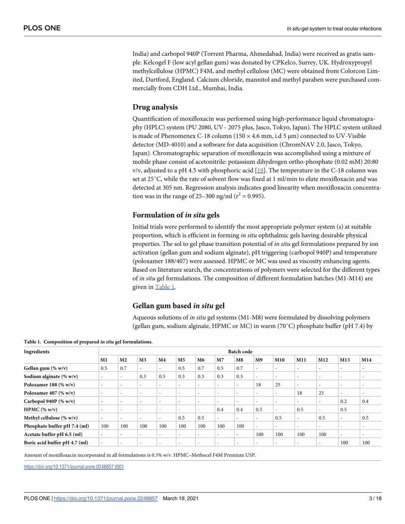
Formulation of in situ gels
Initial trials were performed to identify the most appropriate polymer system (s) at suitable
proportion, which is efficient in forming in situ ophthalmic gels having desirable physical
properties. The sol to gel phase transition potential of in situ gel formulations prepared by ion
activation (gellan gum and sodium alginate), pH triggering (carbopol 940P) and temperature
(poloxamer 188/407) were assessed. HPMC or MC was used as viscosity enhancing agents.
Based on literature search, the concentrations of polymers were selected for the different types
of in situ gel formulations. The composition of different formulation batches (M1-M14) are
given in Table 1.
Gellan gum based in situ gel
Aqueous solutions of in situ gel systems (M1-M8) were formulated by dissolving polymers
(gellan gum, sodium alginate, HPMC or MC) in warm (70˚C) phosphate buffer (pH 7.4) by
Table 1. Composition of prepared in situ gel formulations.
Ingredients Batch code
M1 M2 M3 M4 M5 M6 M7 M8 M9 M10 M11 M12 M13 M14
Gellan gum (% w/v) 0.5 0.7 - - 0.5 0.7 0.5 0.7 - - - - - -
Sodium alginate (% w/v) - - 0.3 0.5 0.3 0.3 0.3 0.3 - - - - - -
Poloxamer 188 (% w/v) - - - - - - - - 18 25 - - - -
Poloxamer 407 (% w/v) - - - - - - - - - - 18 25 - -
Carbopol 940P (% w/v) - - - - - - - - - - - - 0.2 0.4
HPMC (% w/v) - - - - - - 0.4 0.4 0.5 - 0.5 - 0.5 -
Methyl cellulose (% w/v) - - - - 0.5 0.5 - - - 0.5 - 0.5 - 0.5
Phosphate buffer pH 7.4 (ml) 100 100 100 100 100 100 100 100 - - - - - -
Acetate buffer pH 6.5 (ml) - - - - - - - - 100 100 100 100 - -
Boric acid buffer pH 4.7 (ml) - - - - - - - - - - - - 100 100
Amount of moxifloxacin incorporated in all formulations is 0.5% w/v. HPMC–Methocel F4M Premium USP.
https://doi.org/10.1371/journal.pone.0248857.t001
PLOS ONE In situ gel system to treat ocular infections
PLOS ONE | https://doi.org/10.1371/journal.pone.0248857 March 19, 2021 3 / 18
constant stirring [20]. The polymeric solution was subsequently cooled to room temperature
(25 ± 1˚C) to which specified quantities of moxifloxacin (0.5% w/v) was added and stirred
until completely dissolved. Terminal sterilization of the ophthalmic gels was carried out by
heating in an autoclave and kept in a refrigerator until further investigation.
Poloxamer based in situ gel
Poloxamer in situ forming gels (M9-M12) were formulated by the modified cold method.
Briefly, poloxamer (P188 and P407), HPMC and MC was slowly added to the required amount
of cold acetate buffer (pH 6.5) containing moxifloxacin (0.5% w/v) with constant stirring. The
partially dissolved Poloxamer solution was stored in refrigerator and stirred occasionally until
a clear homogenous solution is obtained.
Carbopol based in situ gel
Gels (M13 and M14) were formulated by dissolving carbopol 940P in a half quantity of modi-
fied boric acid buffer (pH 4.7) with constant stirring. HPMC and MC were dissolved in a sepa-
rate container using remaining half of vehicle with slight heating. The two solutions were then
mixed and the moxifloxacin (0.5% w/v) was added. The solutions were then equilibrated at
room temperature (25 ± 1˚C) for 24 h.
Evaluation of gels
Appearance and pH. Clarity testing was performed on all developed formulations by
visual observation of the samples to examine the presence of any transparent or coloured par-
ticulate matters or turbidity. The pH of the various gels was determined by calibrated pH
meter (Mettler Toledo MP-220, Greifensee, Switzerland) at 25 ± 0.5˚C as per the standard
procedure.
Drug content
The polypropylene vials containing the accurately weighed quantity (1 g) of formulations were
mixed with mobile phase using a laboratory shaker (EIE 405, EIE Instruments, Ahmedabad,
India) for 2–3 min. Lastly, aliquot of the solution was filtered through a filter membrane (pore
size of 0.2 μm), diluted using simulated tear fluid (STF) and injected into the HPLC system.
In vitro gelation
The gelation of gels was assessed using a polypropylene vial having STF as gelation solution,
equilibrated at 34 ± 0.5˚C using a water bath. Aliquot of each preparation (100 μl) were pre-
cisely transferred into separate vial followed by the gradual addition of STF (2 ml) using a
micropipette. Gelling capacity was observed by visual monitoring of the formation and mea-
suring the time needed for gelation and the time required to dissolve the gel formed [21].
Rheology
The viscosity of in situ gels was determined at different angular velocity (0.5 to 100 rpm) at
34 ± 1˚C using a Brookfield Viscometer (LVDVI prime, Middleborough, MA, USA). A typical
run involved consecutively varying and reversing the angular velocity for an identical period
of 6 sec at a controlled ramp speed [22]. To assess the variation of rheology typically observed
after ocular application, viscosity of gels was also determined after thinning the gel with STF in
25:7 ratios.
PLOS ONE In situ gel system to treat ocular infections
PLOS ONE | https://doi.org/10.1371/journal.pone.0248857 March 19, 2021 4 / 18
Gel strength and adhesive force
Gel strength and adhesive force was measured utilizing a QTS Texture analyzer (Brookfield
Engineering Labs, Inc., Middleboro, MA, USA) (S1 Fig). Preparation with STF were placed
into a cylindrical holder while precautions was taken to prevent the air entrapment in samples.
A cylindrical probe with approximately 38 mm of diameter was allowed to enter into sample
gel at a rate of 1 mm/s into a depth of 10 mm at a measurable force. The gel strength (as peak
load) and adhesive force (work needed to disturb the attractive forces between cylindrical
probe and sample) was computed from the resulting load–time plots [23,24].
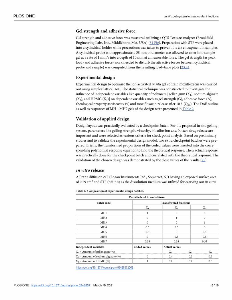
Experimental design
Experimental design to optimize the ion activated in situ gel contain moxifloxacin was carried
out using simplex lattice DoE. The statistical technique was constructed to investigate the
influence of independent variables like quantity of polymers [gellan gum (X1), sodium alginate
(X2), and HPMC (X3)] on dependent variables such as gel strength (G), adhesive force (A),
rheological property as viscosity (v) and moxifloxacin release after 10 h (Q10). The DoE outline
as well as responses of MH1-MH7 gels of the design were presented in Table 2.
Validation of applied design
Design layout was practically evaluated by a checkpoint batch. For the proposed in situ gelling
system, parameters like gelling strength, viscosity, bioadhesion and in vitro drug release are
important and were selected as various criteria for check point analysis. Based on preliminary
studies and to validate the experimental design model, two extra checkpoint batches were pre-
pared. Briefly, the transformed proportions of the coded values were inserted into the corre-
sponding polynomial response equation to find the theoretical response. Then actual response
was practically done for the checkpoint batch and correlated with the theoretical response. The
validation of the chosen design was demonstrated by the close values of the results [25].
In vitro release
A Franz diffusion cell (Logan Instruments Ltd., Somerset, NJ) having an exposed surface area
of 0.79 cm2 and STF (pH 7.4) as the dissolution medium was utilized for carrying out in vitro
Table 2. Composition of experimental design batches.
Variable level in coded form
Batch code Transformed fractions
X1 X2 X3
MH1 1 0 0
MH2 0 1 0
MH3 0 0 1
MH4 0.5 0.5 0
MH5 0.5 0 0.5
MH6 0 0.5 0.5
MH7 0.33 0.33 0.33
Independent variables Coded values Actual values
X1 = Amount of gellan gum (%) X1 X2 X3
X2 = Amount of sodium alginate (%) 0 0.4 0.2 0.3
X3 = Amount of HPMC (%) 1 0.6 0.4 0.5
https://doi.org/10.1371/journal.pone.0248857.t002
PLOS ONE In situ gel system to treat ocular infections
PLOS ONE | https://doi.org/10.1371/journal.pone.0248857 March 19, 2021 5 / 18
release of moxifloxacin from prepared gels [26]. Prepared formulations (MH1-MH7) or check
point batches (MH8 and MH9) or control was placed on a cellophane dialysis film (MWCO
12–14 kDa) previously fixed between the upper and lower chambers. The temperature of the
assembly was fixed at 37 ± 0.5˚C and receiver solution was mixed at 50 rpm using a magnetic
stirrer. At suitable time intervals, aliquots of sample (1 ml) were drawn and substituted with
the equivalent amount of STF held at the same temperature. A control experiment was per-
formed at same experimental conditions using similar strength of moxifloxacin solution. The
samples were later diluted with mobile phase and quantified for moxifloxacin by HPLC. The
data collected were analyzed to calculate regression coefficient (r2) and interpret release kinet-
ics using various mathematical models [27].
Ex vivo permeation
Trans-corneal permeation of moxifloxacin from optimized formulation (MH7) and control
(commercial eye drops—VigamoxTM; equivalent to 5 mg of moxifloxacin per ml) was carried
out using goat cornea as membrane. Whole eyeballs of goat were collected from a local abattoir
and the corneas were detached carefully from the adhering tissues. It was immediately stored
in normal saline (0.9% w/v) kept at 4˚C, until used. Isolated cornea was held between the
upper donor and lower receptor compartment of a Franz diffusion cell [28]. The cornea was
firmly fixed to expose the epithelial surface (0.79 cm2) facing the donor compartment. The
STF was used as receptor medium and the temperature of the receiver was set at 37 ± 0.1˚C.
Aliquots of samples (1 ml) were withdrawn at periodic time intervals and substituted with
equivalent volume of new STF. The sample withdrawn from the receptor compartment was
appropriately diluted and estimated for moxifloxacin by HPLC. The flux was determined as
described in the literature [29].
Differential scanning calorimetry (DSC)
Thermal behavior of moxifloxacin, physical mixture and MH7 were performed by DSC (DSC
60, Shimadzu, Kyoto, Japan). Samples were taken in crimped aluminium pan and sealed in air-
tight condition. The pan was scanned from 50–300˚C at a uniform heating rate of 5˚C/min. A
blank aluminium pan was used as reference sample [30].
Ocular irritation
Ocular irritation of MH7 was tested in albino rabbits (2–3 kg). The animals were housed
under normal atmospheric conditions and allowed to move freely. The animals were acclima-
tized to the laboratory environments for one week prior to the start of experiment. All animals
used for the study were given unrestricted access to both water and food. The experiments
were carried out by strictly following the guidelines stated by the Committee for the Purpose
of Control and Supervision of Experiments on Animals (CPCSEA), Ministry of Fisheries, Ani-
mal Husbandry and Dairying, India. The protocol approved by the Institutional Animal Ethics
Committee (IPS/PCEU/FAC10-11/002) for animal care at Nirma University was followed dur-
ing the experiment. In vivo ocular irritation experiment was carried out according to the
guidelines based on Draize technique [31]. Single instillation of 60 μl was applied in left eye of
individual rabbit whereas the right untreated eye is considered as control. The sterile formula-
tion was tested two times a day for 21 days. The rabbits were checked frequently for signs of
sensitivity reactions particularly redness, swelling, cloudiness, edema, haemorrhage, discharge
and blindness [32].
PLOS ONE In situ gel system to treat ocular infections
PLOS ONE | https://doi.org/10.1371/journal.pone.0248857 March 19, 2021 6 / 18
In vivo pharmacokinetics
The amount of moxifloxacin diffused into the aqueous humour of the rabbit eyes after oph-
thalmic administration was determined to compare the ocular bioavailability between MH7
and commercial moxifloxacin ophthalmic drops (0.5% w/v). In vivo pharmacokinetic investi-
gations were performed in New Zealand Albino rabbits (2–3 kg) with two groups (n = 6). The
protocol approved by the Institutional Animal Ethics Committee (IPS/PCEU/FAC10-11/002)
for animal care at Nirma University was followed during the experiment. Single topical instil-
lation (60 μl of 0.5% w/v drug) of MH7 was dropped in the lower cul-de-sac of one eye of indi-
vidual rabbit in the first group while similar strength and volume of commercial eye drops was
instilled into second group of rabbits. In each case, the untreated eye was considered as con-
trol. Both eyelids of all rabbits were lightly closed for 2 min to increase the contact of drug with
the corneal membrane. Before aqueous humour withdrawal, individual animal was anaesthe-
tized by intramuscular administration of xylazine and ketamine [33]. The samples (20 μl) of
aqueous humour collected using 29-gauge insulin syringe needle were mixed with acetonitrile,
and stored immediately at -80˚C until further investigation. The samples were centrifuged
(5000 rpm for 10 min) and the organic layer was assessed for drug content by HPLC.
Stability
The stability of MH7 batch was assessed as per the latest ICH guidelines. Appropriate quantity
of ophthalmic formulations placed in amber-coloured vials were stored for 6 months in a sta-
bility chamber at accelerated storage condition (40 ± 2˚C/75% ± 5% relative humidity) [26]. At
various time intervals, the samples were taken out and estimated for important physicochemi-
cal parameters including gelling capacity, pH, viscosity and in vitro drug release. The shelf life
of the optimized formulation was computed using the classical Arrhenius plot [34].
Statistical analysis
The statistical interpretation of experimental data was carried out by one-way ANOVA (SPSS
23, Chicago, IL, USA). The difference in values at p< 0.05 is considered statistically
significant.
Results and discussion
Evaluation of gels
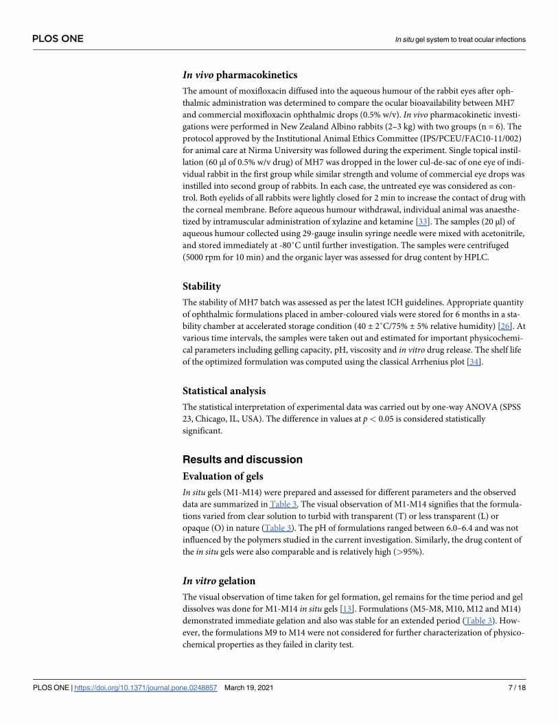
In situ gels (M1-M14) were prepared and assessed for different parameters and the observed
data are summarized in Table 3. The visual observation of M1-M14 signifies that the formula-
tions varied from clear solution to turbid with transparent (T) or less transparent (L) or
opaque (O) in nature (Table 3). The pH of formulations ranged between 6.0–6.4 and was not
influenced by the polymers studied in the current investigation. Similarly, the drug content of
the in situ gels were also comparable and is relatively high (>95%).
In vitro gelation
The visual observation of time taken for gel formation, gel remains for the time period and gel
dissolves was done for M1-M14 in situ gels [13]. Formulations (M5-M8, M10, M12 and M14)
demonstrated immediate gelation and also was stable for an extended period (Table 3). How-
ever, the formulations M9 to M14 were not considered for further characterization of physico-
chemical properties as they failed in clarity test.
PLOS ONE In situ gel system to treat ocular infections
PLOS ONE | https://doi.org/10.1371/journal.pone.0248857 March 19, 2021 7 / 18
Rheology
The viscosity is a critical factor determining the ocular residence time of the instilled formula-
tion [35]. The viscosity of M1-M8 are listed in Table 3. It was demonstrated that in situ gels
(M1-M8) displayed pseudo-plastic flow or shear thinning rheological behavior as demon-
strated by a drop in viscosity with higher angular velocity (Fig 1A). Viscosity of in situ gels
(M1-M8) with and without STF is presented in Fig 1B. Percentage variation in viscosity after
appropriate dilution of the formulations with STF is illustrated in S2 Fig. The data in the Figs
1B and S2 demonstrate that the dilution with STF remarkably improved the viscosity of
M1-M8 gels formulated with gellan gum as well as sodium alginate. This is due to the fact that
these polymers (gellan gum, sodium alginate and HPMC) have the inherent capacity to pro-
duce gel in companion with mono or divalent cations, available in STF analogous to the lach-
rymal fluid [5,13,36]. This peculiar phenomenon endorses the in situ gelling characteristic of
the M1-M8 gels.
Gel strength
Assessment of mucoadhesive force is an important parameter as it provides insight into the
retention of formulation on the mucous membrane [37]. The gel strength as well as adhesive
force of M1-M8 were determined using texture analyzer. It is apparent from the data in
Table 3 that the gel strength varied considerably among the gels tested. The gel strength was
relatively low when gel was prepared using single polymer (gellan gum or sodium alginate)
(M1-M4). However, combining these two polymers drastically improved the gel strength
(M5-M8), and the highest value (168 g) was noticed in M6, where in these polymers were com-
bined with MC (viscosity enhancing agent). A similar trend was noticed with adhesive force as
Table 3. Physicochemical characteristics of preliminary in situ gel formulations.
Parameters Batch code
M1 M2 M3 M4 M5 M6 M7 M8 M9 M10 M11 M12 M13 M14
Transparency T T T L L L T T L O L O O O
Gelling capacity + ++ + + +++ +++ +++ +++ ++ +++ ++ +++ ++ +++
Viscosity (cP) at 1 rpm 607 ± 72 837 ± 81 378 ± 56 452 ± 869 1140 ± 126 3700 ± 264 2175 ± 112 3050 ± 226 - - - - - -
Gel strength (g) 92 ± 8 98 ± 12 79 ± 16 86 ± 13 134 ± 9 168 ± 16 152 ± 17 157 ± 14 - - - - - -
T–Transparent; L–Less Transparent; O–Opaque.
+ No gelation and gels slowly dissolves; ++ gelation immediate and remains for a few hours; +++ gelation immediate and remains for an extended period.
https://doi.org/10.1371/journal.pone.0248857.t003
Fig 1. Rheology of in situ gels (M1–M8). (A) Rheological behavior and (B) viscosity with and without simulated tear
fluid (STF).
https://doi.org/10.1371/journal.pone.0248857.g001
PLOS ONE In situ gel system to treat ocular infections
PLOS ONE | https://doi.org/10.1371/journal.pone.0248857 March 19, 2021 8 / 18
well and the values ranged between 3.7–5.8 N mm. However, M6 was less transparent as com-
pared to gels prepared using HPMC (M7 and M8).
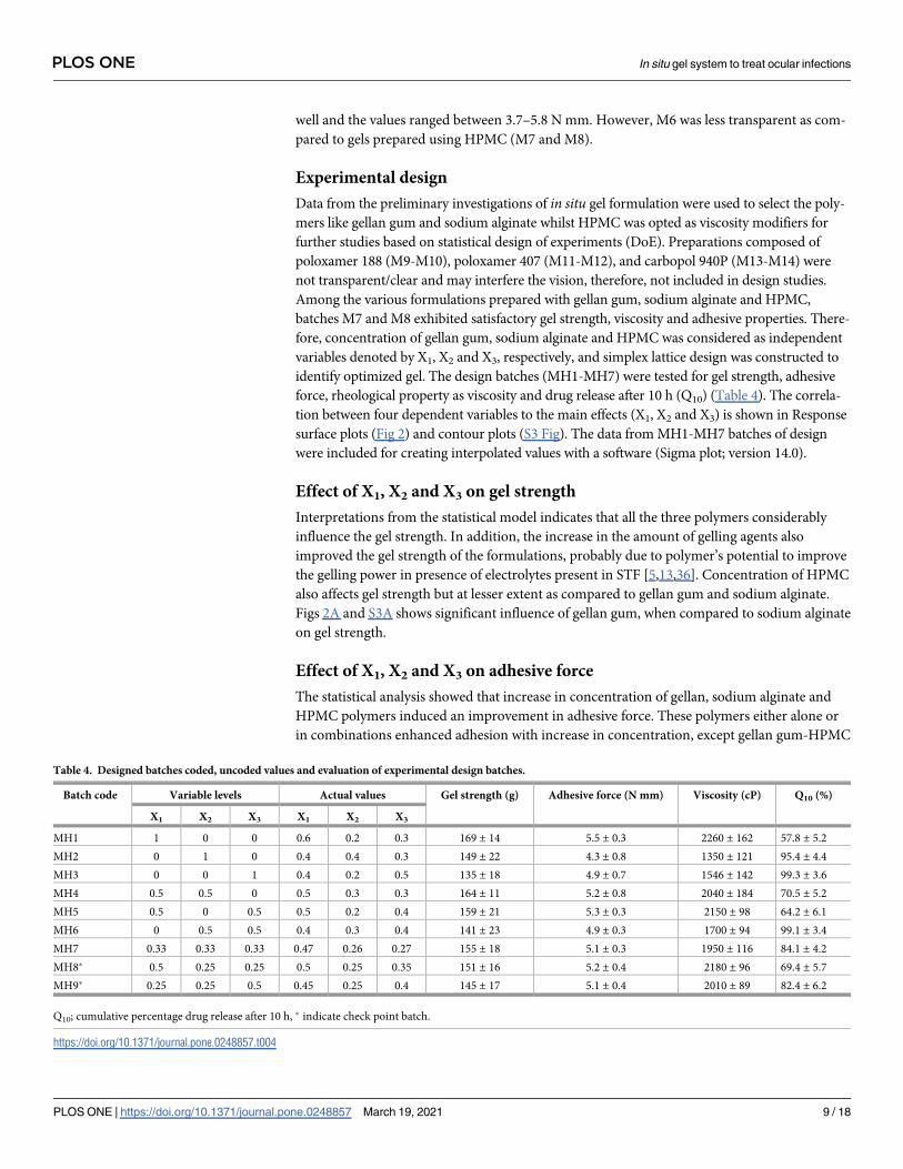
Experimental design
Data from the preliminary investigations of in situ gel formulation were used to select the poly-
mers like gellan gum and sodium alginate whilst HPMC was opted as viscosity modifiers for
further studies based on statistical design of experiments (DoE). Preparations composed of
poloxamer 188 (M9-M10), poloxamer 407 (M11-M12), and carbopol 940P (M13-M14) were
not transparent/clear and may interfere the vision, therefore, not included in design studies.
Among the various formulations prepared with gellan gum, sodium alginate and HPMC,
batches M7 and M8 exhibited satisfactory gel strength, viscosity and adhesive properties. There-
fore, concentration of gellan gum, sodium alginate and HPMC was considered as independent
variables denoted by X1, X2 and X3, respectively, and simplex lattice design was constructed to
identify optimized gel. The design batches (MH1-MH7) were tested for gel strength, adhesive
force, rheological property as viscosity and drug release after 10 h (Q10) (Table 4). The correla-
tion between four dependent variables to the main effects (X1, X2 and X3) is shown in Response
surface plots (Fig 2) and contour plots (S3 Fig). The data from MH1-MH7 batches of design
were included for creating interpolated values with a software (Sigma plot; version 14.0).
Effect of X1, X2 and X3 on gel strength
Interpretations from the statistical model indicates that all the three polymers considerably
influence the gel strength. In addition, the increase in the amount of gelling agents also
improved the gel strength of the formulations, probably due to polymer’s potential to improve
the gelling power in presence of electrolytes present in STF [5,13,36]. Concentration of HPMC
also affects gel strength but at lesser extent as compared to gellan gum and sodium alginate.
Figs 2A and S3A shows significant influence of gellan gum, when compared to sodium alginate
on gel strength.
Effect of X1, X2 and X3 on adhesive force
The statistical analysis showed that increase in concentration of gellan, sodium alginate and
HPMC polymers induced an improvement in adhesive force. These polymers either alone or
in combinations enhanced adhesion with increase in concentration, except gellan gum-HPMC
Table 4. Designed batches coded, uncoded values and evaluation of experimental design batches.
Batch code Variable levels Actual values Gel strength (g) Adhesive force (N mm) Viscosity (cP) Q10 (%)
X1 X2 X3 X1 X2 X3
MH1 1 0 0 0.6 0.2 0.3 169 ± 14 5.5 ± 0.3 2260 ± 162 57.8 ± 5.2
MH2 0 1 0 0.4 0.4 0.3 149 ± 22 4.3 ± 0.8 1350 ± 121 95.4 ± 4.4
MH3 0 0 1 0.4 0.2 0.5 135 ± 18 4.9 ± 0.7 1546 ± 142 99.3 ± 3.6
MH4 0.5 0.5 0 0.5 0.3 0.3 164 ± 11 5.2 ± 0.8 2040 ± 184 70.5 ± 5.2
MH5 0.5 0 0.5 0.5 0.2 0.4 159 ± 21 5.3 ± 0.3 2150 ± 98 64.2 ± 6.1
MH6 0 0.5 0.5 0.4 0.3 0.4 141 ± 23 4.9 ± 0.3 1700 ± 94 99.1 ± 3.4
MH7 0.33 0.33 0.33 0.47 0.26 0.27 155 ± 18 5.1 ± 0.3 1950 ± 116 84.1 ± 4.2
MH8� 0.5 0.25 0.25 0.5 0.25 0.35 151 ± 16 5.2 ± 0.4 2180 ± 96 69.4 ± 5.7
MH9� 0.25 0.25 0.5 0.45 0.25 0.4 145 ± 17 5.1 ± 0.4 2010 ± 89 82.4 ± 6.2
Q10; cumulative percentage drug release after 10 h, � indicate check point batch.
https://doi.org/10.1371/journal.pone.0248857.t004
PLOS ONE In situ gel system to treat ocular infections
PLOS ONE | https://doi.org/10.1371/journal.pone.0248857 March 19, 2021 9 / 18
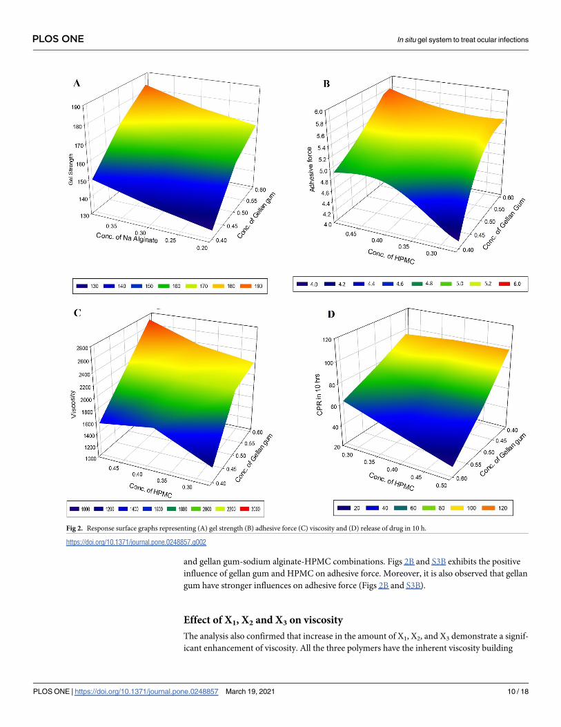
and gellan gum-sodium alginate-HPMC combinations. Figs 2B and S3B exhibits the positive
influence of gellan gum and HPMC on adhesive force. Moreover, it is also observed that gellan
gum have stronger influences on adhesive force (Figs 2B and S3B).
Effect of X1, X2 and X3 on viscosity
The analysis also confirmed that increase in the amount of X1, X2, and X3 demonstrate a signif-
icant enhancement of viscosity. All the three polymers have the inherent viscosity building
Fig 2. Response surface graphs representing (A) gel strength (B) adhesive force (C) viscosity and (D) release of drug in 10 h.
https://doi.org/10.1371/journal.pone.0248857.g002
PLOS ONE In situ gel system to treat ocular infections
PLOS ONE | https://doi.org/10.1371/journal.pone.0248857 March 19, 2021 10 / 18
capacity promoted by the layer of solvent sheath surrounding the individual particle. The data
also indicates that the increase in gellan gum and HPMC quantity proportionally enhanced
the viscosity, as compared to alginate polymer. The response surface plot and contour plots
depicted in Figs 2C and S3C displays the improvement in viscosity by gellan gum and HPMC.
In addition, it is also proved that stronger influence of HPMC on viscosity.
Effect of X1, X2 and X3 on drug release (Q10)
On evaluation of effect of three variables on drug release, it is observed that increase in amount
of all three polymers can possibly decrease the release of the drug. From Figs 2D and S3D it
was observed that increase in gellan gum and HPMC lead to more negative impact on the
drug release. This could be explained by the fact that an increase in the gel strength and viscos-
ity contributed by these polymers retarding the release rate of the drug.
The two check point batches (MH8� and MH9�) were examined for gel strength, adhesive
force, rheological property (viscosity) and drug release studies and compared with predicted
values observed in overlay plot of Design Expert software and found similarity between
observed and predicted values (S1 Table).
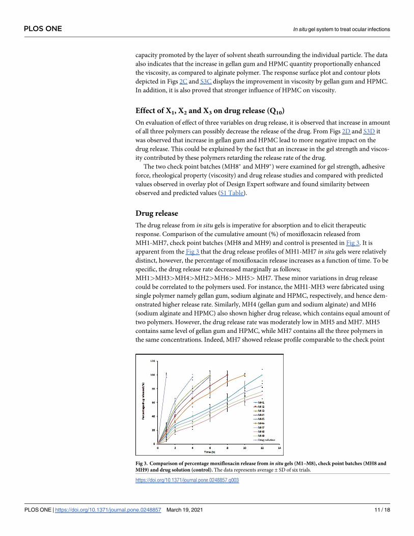
Drug release
The drug release from in situ gels is imperative for absorption and to elicit therapeutic
response. Comparison of the cumulative amount (%) of moxifloxacin released from
MH1-MH7, check point batches (MH8 and MH9) and control is presented in Fig 3. It is
apparent from the Fig 3 that the drug release profiles of MH1-MH7 in situ gels were relatively
distinct, however, the percentage of moxifloxacin release increases as a function of time. To be
specific, the drug release rate decreased marginally as follows;
MH1>MH3>MH4>MH2>MH6> MH5> MH7. These minor variations in drug release
could be correlated to the polymers used. For instance, the MH1-MH3 were fabricated using
single polymer namely gellan gum, sodium alginate and HPMC, respectively, and hence dem-
onstrated higher release rate. Similarly, MH4 (gellan gum and sodium alginate) and MH6
(sodium alginate and HPMC) also shown higher drug release, which contains equal amount of
two polymers. However, the drug release rate was moderately low in MH5 and MH7. MH5
contains same level of gellan gum and HPMC, while MH7 contains all the three polymers in
the same concentrations. Indeed, MH7 showed release profile comparable to the check point
Fig 3. Comparison of percentage moxifloxacin release from in situ gels (M1–M8), check point batches (MH8 and
MH9) and drug solution (control). The data represents average ± SD of six trials.
https://doi.org/10.1371/journal.pone.0248857.g003
PLOS ONE In situ gel system to treat ocular infections
PLOS ONE | https://doi.org/10.1371/journal.pone.0248857 March 19, 2021 11 / 18
batches (MH8 and MH9) and was selected for further studies. On the other hand, the release
of moxifloxacin in control experiments were immediate and exhibited almost complete release
in 1 h. Overall, these data signify that release of moxifloxacin is influenced by the polymers
studied.
Release rate data for MH7 was fitted using established mathematical models. The Sum of
Square of Residuals (SSR) values that measure the deviation from the mean were found to be
80.55, 245.66, 239.11, 23.85, and 108.49 for Zero order, First order, Higuchi model, Kors-
meyer-Peppas model, and Weibull model, respectively. Based on the data observed, moxifloxa-
cin release from MH7 fit well to the Korsmeyer-Peppas expression (S2 Table), which is usually
observed with gellan gum based in situ gels [38]. The observed n value (0.7837) suggests anom-
alous transport responsible for moxifloxacin release from MH7 [39,40].
A comparative in vitro release profile between MH7 and commercial eye drops of moxifloxa-
cin is presented in S4 Fig. The data in S4 Fig signifies that the moxifloxacin release was consid-
erably different at 15 min (17.78% and 3.72% for commercial eye drops and MH7, respectively)
as well as 2 h (100% and 24.64% for commercial eye drops and MH7, respectively). The results
demonstrated that the drug release from the MH7 was primarily controlled by the combination
of polymers (gellan gum, sodium alginate and HPMC). Comparison of in vitro release between
MH7 and commercial eye drops of moxifloxacin was accomplished by applying direct model-
independent mathematical method, the similarity factor (F2). The calculated similarity factor
value between MH7 and commercial eye drops was 16.94, proving that the both batches are dis-
similar in terms of in vitro drug release profile according to the literature [41].
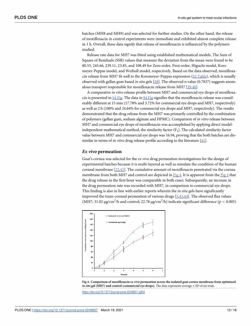
Ex vivo permeation
Goat’s cornea was selected for the ex vivo drug permeation investigations for the design of
experimental batches because it is multi-layered as well as simulate the condition of the human
corneal membrane [22,42]. The cumulative amount of moxifloxacin penetrated via the cornea
membrane from both MH7 and control are depicted in Fig 4. It is apparent from the Fig 4 that
the drug release in the first hour was comparable in both cases. Subsequently, an increase in
the drug permeation rate was recorded with MH7, in comparison to commercial eye drops.
This finding is also in line with earlier reports wherein the in situ gels have significantly
improved the trans-corneal permeation of various drugs [5,43,44]. The observed flux values
(MH7; 31.02 μg/cm2/h and control; 22.78 μg/cm2/h) indicate significant difference (p< 0.005)
Fig 4. Comparison of moxifloxacin ex vivo permeation across the isolated goat cornea membrane from optimized
in situ gel (MH7) and control (commercial eye drops). The data represents average ± SD of six trials.
https://doi.org/10.1371/journal.pone.0248857.g004
PLOS ONE In situ gel system to treat ocular infections
PLOS ONE | https://doi.org/10.1371/journal.pone.0248857 March 19, 2021 12 / 18
in corneal permeation of moxifloxacin between MH7 and control. However, the lag time in
both formulations were comparable (0.09 h). Overall, this study signifies that the moxifloxacin
in situ gel formulation could significantly improve the corneal permeation.
DSC
DSC technique was used to analyse the physical state of moxifloxacin including the transfor-
mation of thermodynamic properties that could have occurred inside the in situ gel. DSC ther-
mograms of drug, physical mixture and in situ gel are depicted in S5 Fig. A sharp endothermic
peak characterizing the melting point of moxifloxacin was observed at 262.87˚C, demonstrat-
ing its crystalline character [45]. The diffraction pattern of physical mixture shown characteris-
tic peaks of drug with reduced intensity at 262.87˚C, and a broad endothermic peak at
94.17˚C, which could be incurred as a result of melting point of polymers. The polymeric
endotherm in the formulation MH7 shifted to 61.58˚C, probably owing to in situ gel forma-
tion. However, the drug peak has disappeared in MH7 indicating the drug is in dissolved state
in the in situ matrix gel system.
Ocular irritation
Eye irritation score from individual rabbits was added to get the total irritation score that was
subsequently divided by the total number of rabbits used for the ocular irritancy test to obtain
the final eye irritation score. The calculated eye irritation score was 0.25 in control while for
MH7 it was 0.57, which demonstrates good ocular tolerance like marketed formulation. Fur-
ther, instillation of MH7 did not cause redness, swelling, or excessive lachrymation in the eye.
Absence of ocular damage or unexpected clinical manifestations to the various eye regions
(cornea, iris, or conjunctivae) were observed. Therefore, this study conclude that MH7 is safe
and non-irritant for ocular administration.
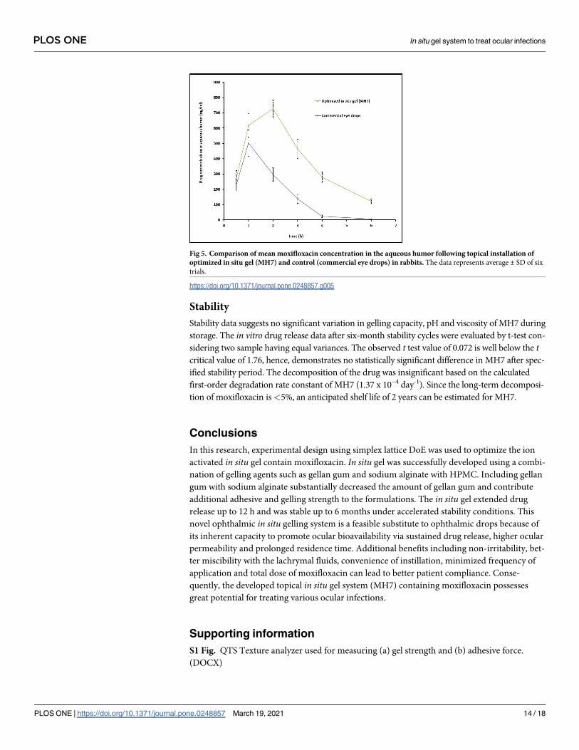
In vivoOcular bioavailability is computed based on the amount of moxifloxacin permeated into the
aqueous humour of rabbit eyes of first (MH7) and second groups (control). Various pharmaco-
kinetic parameters including tmax, Cmax and AUC were computed from the graph plotted
between concentrations (ng/ml) in aqueous humour and time (h) by non-compartment model
analysis [46]. It is apparent from the Fig 5 that the pharmacokinetic parameters are markedly
different for MH7 and control in aqueous humour in rabbits. After the completion of 1 h, the
moxifloxacin level was elevated in the aqueous humour (619.31 ± 77.06 ng/ml and
503.09 ± 85.44 ng/ml in MH7 and control, respectively). However, the drug level was signifi-
cantly higher (p< 0.0001) in MH7 at 2 h, but the moxifloxacin level declined sharply in conven-
tional eye drops (Fig 5). These data signify that ophthalmic drops retained in the ocular cavity
for short time because of extensive pre-corneal drug loss through nasolacrimal discharge and
tear turn over. Further, the tmax value for MH7 was 2 h, while it was 1 h in control. On the other
hand, MH7 showed higher Cmax (727 ± 56 ng/ml) and greater AUC (2881 ± 108 ng h/ml) (p<0.0001), when compared with commercial eye drops (Cmax; 503 ± 85 ng/ml and AUC; 978 ± 86
ng h/ml). Thus, it can be concluded from the available data that intraocular permeation of mox-
ifloxacin was significantly improved by design and developing in situ gel system. This observa-
tion is also in agreement with ex vivo permeation data wherein the flux was significantly higher
in MH7 (Fig 4). Therefore, ocular residence time of MH7 proves extended duration of action in
comparison to commercial eye drops. The average drug concentration noticed in Fig 5 in the
aqueous humour was more than the minimum effective concentration of moxifloxacin needed
for therapeutic response for different pathogens causing eye infections [47].
PLOS ONE In situ gel system to treat ocular infections
PLOS ONE | https://doi.org/10.1371/journal.pone.0248857 March 19, 2021 13 / 18
Stability
Stability data suggests no significant variation in gelling capacity, pH and viscosity of MH7 during
storage. The in vitro drug release data after six-month stability cycles were evaluated by t-test con-
sidering two sample having equal variances. The observed t test value of 0.072 is well below the tcritical value of 1.76, hence, demonstrates no statistically significant difference in MH7 after spec-
ified stability period. The decomposition of the drug was insignificant based on the calculated
first-order degradation rate constant of MH7 (1.37 x 10⁻⁴ day-1). Since the long-term decomposi-
tion of moxifloxacin is<5%, an anticipated shelf life of 2 years can be estimated for MH7.
Conclusions
In this research, experimental design using simplex lattice DoE was used to optimize the ion
activated in situ gel contain moxifloxacin. In situ gel was successfully developed using a combi-
nation of gelling agents such as gellan gum and sodium alginate with HPMC. Including gellan
gum with sodium alginate substantially decreased the amount of gellan gum and contribute
additional adhesive and gelling strength to the formulations. The in situ gel extended drug
release up to 12 h and was stable up to 6 months under accelerated stability conditions. This
novel ophthalmic in situ gelling system is a feasible substitute to ophthalmic drops because of
its inherent capacity to promote ocular bioavailability via sustained drug release, higher ocular
permeability and prolonged residence time. Additional benefits including non-irritability, bet-
ter miscibility with the lachrymal fluids, convenience of instillation, minimized frequency of
application and total dose of moxifloxacin can lead to better patient compliance. Conse-
quently, the developed topical in situ gel system (MH7) containing moxifloxacin possesses
great potential for treating various ocular infections.
Supporting information
S1 Fig. QTS Texture analyzer used for measuring (a) gel strength and (b) adhesive force.
(DOCX)
Fig 5. Comparison of mean moxifloxacin concentration in the aqueous humor following topical installation of
optimized in situ gel (MH7) and control (commercial eye drops) in rabbits. The data represents average ± SD of six
trials.
https://doi.org/10.1371/journal.pone.0248857.g005
PLOS ONE In situ gel system to treat ocular infections
PLOS ONE | https://doi.org/10.1371/journal.pone.0248857 March 19, 2021 14 / 18
S2 Fig. Change in viscosity (%) of prepared in situ gels (M1-M8) by addition of simulated
tear fluid (pH 7.4) STF (at 1.0 rpm).
(DOCX)
S3 Fig. Contour plots representing (A) gel strength (B) adhesive force (C) viscosity and (D)
release of drug in 10 h.
(DOCX)
S4 Fig. Comparison of percentage moxifloxacin release from MH7 and commercial moxi-
floxacin ophthalmic drops.
(DOCX)
S5 Fig. Differential scanning calorimetric curves of moxifloxacin, physical mixture and
optimized in situ gel (MH7).
(DOCX)
S6 Fig.
(JPG)
S1 Table. Comparison of the observed value with predicted values of check point batches.
(DOCX)
S2 Table. Model fitting for selected in situ gel (MH7).
(DOCX)
Acknowledgments
The authors thank the Deanship of Scientific Research at King Faisal University for the sup-
port to complete this project.
Author Contributions
Conceptualization: Anroop B. Nair, Jigar Shah.
Data curation: Anroop B. Nair, Jigar Shah, Shery Jacob, Bandar E. Al-Dhubiab, Nagaraja Sree-
harsha, Mohamed A. Morsy, Sumeet Gupta, Mahesh Attimarad, Pottathil Shinu, Kathari-
gatta N. Venugopala.
Formal analysis: Anroop B. Nair, Jigar Shah, Shery Jacob, Bandar E. Al-Dhubiab, Nagaraja
Sreeharsha, Mohamed A. Morsy, Sumeet Gupta, Mahesh Attimarad, Pottathil Shinu,
Katharigatta N. Venugopala.
Funding acquisition: Anroop B. Nair, Bandar E. Al-Dhubiab, Nagaraja Sreeharsha, Mohamed
A. Morsy, Mahesh Attimarad, Katharigatta N. Venugopala.
Investigation: Anroop B. Nair, Jigar Shah, Shery Jacob, Bandar E. Al-Dhubiab, Nagaraja Sree-
harsha, Mohamed A. Morsy, Sumeet Gupta, Mahesh Attimarad, Pottathil Shinu, Kathari-
gatta N. Venugopala.
Methodology: Anroop B. Nair, Jigar Shah, Shery Jacob, Bandar E. Al-Dhubiab, Nagaraja Sree-
harsha, Mohamed A. Morsy, Sumeet Gupta, Mahesh Attimarad, Pottathil Shinu, Kathari-
gatta N. Venugopala.
Writing – original draft: Jigar Shah, Nagaraja Sreeharsha, Katharigatta N. Venugopala.
Writing – review & editing: Anroop B. Nair, Shery Jacob, Bandar E. Al-Dhubiab, Mohamed
A. Morsy, Sumeet Gupta, Mahesh Attimarad, Pottathil Shinu, Katharigatta N. Venugopala.
PLOS ONE In situ gel system to treat ocular infections
PLOS ONE | https://doi.org/10.1371/journal.pone.0248857 March 19, 2021 15 / 18
References1. Varela-Fernandez R, Dıaz-Tome V, Luaces-Rodrıguez A, Conde-Penedo A, Garcıa-Otero X, Luzardo-
Alvarez A, et al. Drug Delivery to the Posterior Segment of the Eye: Biopharmaceutic and Pharmacoki-
netic Considerations. Pharmaceutics. 2020; 12(3):269. https://doi.org/10.3390/
pharmaceutics12030269 PMID: 32188045.
2. Jacob S, Nair AB, Shah J. Emerging role of nanosuspensions in drug delivery systems. Biomaterials
research. 2020; 24:3-. https://doi.org/10.1186/s40824-020-0184-8 PMID: 31969986.
3. Souto EB, Dias-Ferreira J, Lopez-Machado A, Ettcheto M, Cano A, Camins Espuny A, et al. Advanced
Formulation Approaches for Ocular Drug Delivery: State-Of-The-Art and Recent Patents. Pharmaceu-
tics. 2019; 11(9). Epub 2019/09/11. https://doi.org/10.3390/pharmaceutics11090460 PMID: 31500106;
PubMed Central PMCID: PMC6781321.
4. Makwana SB, Patel VA, Parmar SJ. Development and characterization of in-situ gel for ophthalmic for-
mulation containing ciprofloxacin hydrochloride. Results in Pharma Sciences. 2016; 6:1–6. https://doi.
org/10.1016/j.rinphs.2015.06.001 PMID: 26949596
5. Wu Y, Liu Y, Li X, Kebebe D, Zhang B, Ren J, et al. Research progress of in-situ gelling ophthalmic drug
delivery system. Asian Journal of Pharmaceutical Sciences. 2019; 14(1):1–15. https://doi.org/10.1016/
j.ajps.2018.04.008 PMID: 32104434
6. Irimia T, Ghica MV, Popa L, Anuţa V, Arsene AL, Dinu-Pırvu CE. Strategies for Improving Ocular Drug
Bioavailability and Corneal Wound Healing with Chitosan-Based Delivery Systems. Polymers (Basel).
2018; 10(11). Epub 2019/04/10. https://doi.org/10.3390/polym10111221 PMID: 30961146; PubMed
Central PMCID: PMC6290606.
7. Moisseiev E, Loberman D, Zunz E, Kesler A, Loewenstein A, Mandelblum J. Pupil dilation using drops
vs gel: a comparative study. Eye (Lond). 2015; 29(6):815–9. Epub 2015/04/11. https://doi.org/10.1038/
eye.2015.47 PMID: 25857606; PubMed Central PMCID: PMC4469672.
8. Tsai CH, Wang PY, Lin IC, Huang H, Liu GS, Tseng CL. Ocular Drug Delivery: Role of Degradable Poly-
meric Nanocarriers for Ophthalmic Application. Int J Mol Sci. 2018; 19(9). Epub 2018/09/22. https://doi.
org/10.3390/ijms19092830 PMID: 30235809; PubMed Central PMCID: PMC6164366.
9. Nair AB, Shah J, Aljaeid BM, Al-Dhubiab BE, Jacob S. Gellan Gum-Based Hydrogel for the Transder-
mal Delivery of Nebivolol: Optimization and Evaluation. Polymers (Basel). 2019; 11(10). Epub 2019/10/
19. https://doi.org/10.3390/polym11101699 PMID: 31623262; PubMed Central PMCID: PMC6836162.
10. Reed K, Li A, Wilson B, Assamoi T. Enhancement of Ocular In Situ Gelling Properties of Low Acyl Gel-
lan Gum by Use of Ion Exchange. J Ocul Pharmacol Ther. 2016; 32(9):574–82. Epub 2016/09/10.
https://doi.org/10.1089/jop.2016.0084 PMID: 27611484.
11. Singh G, Shilpa WA, Sarwal A. In-situ Gelling System for Mucoadhesive Site-Specific Drug Delivery for
Treatment of Recurrent Vaginal Candidiasis. Indian Journal Of Pharmaceutical Education And
Research. 2020; 54(4):921–34.
12. Pahuja P, Arora S, Pawar P. Ocular drug delivery system: a reference to natural polymers. Expert Opin
Drug Deliv. 2012; 9(7):837–61. Epub 2012/06/19. https://doi.org/10.1517/17425247.2012.690733
PMID: 22703523.
13. Sheshala R, Ming NJ, Kok YY, Singh TRR, Dua K. Formulation and Characterization of pH Induced in
situ Gels Containing Sulfacetamide Sodium for Ocular Drug Delivery: A Combination of CarbopolZ/
HPMC Polymer. Indian J Pharm Educ Res. 2019; 53(4):654–62.
14. El-Laithy HM, Nesseem DI, El-Adly AA, Shoukry M. Moxifloxacin-Gelrite in situ ophthalmic gelling sys-
tem against photodynamic therapy for treatment of bacterial corneal inflammation. Arch Pharm Res.
2011; 34(10):1663–78. Epub 2011/11/15. https://doi.org/10.1007/s12272-011-1011-5 PMID: 22076767.
15. Ferreira KSA, Santos BMAd, Lucena NdP, Ferraz MS, Carvalho RdSF, Duarte Junior AP, et al. Ocular
delivery of moxifloxacin-loaded liposomes. Arq Bras Oftalmol. 2018; 81(6):510–3. https://doi.org/10.
5935/0004-2749.20180090 PMID: 30231158
16. Khurana LK, Singh R, Singh H, Sharma M. Systematic Development and Optimization of an in-situ Gel-
ling System for Moxifloxacin Ocular Nanosuspension using High-pressure Homogenization with an
Improved Encapsulation Efficiency. Curr Pharm Des. 2018; 24(13):1434–45. Epub 2018/04/04. https://
doi.org/10.2174/1381612824666180403115106 PMID: 29611480.
17. Mahor A, Prajapati SK, Verma A, Gupta R, Iyer AK, Kesharwani P. Moxifloxacin loaded gelatin nanopar-
ticles for ocular delivery: Formulation and in-vitro, in-vivo evaluation. J Colloid Interface Sci. 2016;
483:132–8. Epub 2016/08/24. https://doi.org/10.1016/j.jcis.2016.08.018 PMID: 27552421.
18. Shah J, Nair AB, Jacob S, Patel RK, Shah H, Shehata TM, et al. Nanoemulsion Based Vehicle for Effec-
tive Ocular Delivery of Moxifloxacin Using Experimental Design and Pharmacokinetic Study in Rabbits.
Pharmaceutics. 2019; 11(5). Epub 2019/05/15. https://doi.org/10.3390/pharmaceutics11050230 PMID:
31083593; PubMed Central PMCID: PMC6571706.
PLOS ONE In situ gel system to treat ocular infections
PLOS ONE | https://doi.org/10.1371/journal.pone.0248857 March 19, 2021 16 / 18
19. Abdelaziz AA, Elbanna TE, Gamaleldeen NM. validated microbiological and HPLC methods for the
determination of moxifloxacin in pharmaceutical preparations and human plasma. Brazilian journal of
microbiology: [publication of the Brazilian Society for Microbiology]. 2012; 43(4):1291–301. Epub 2012/
06/01. https://doi.org/10.1590/S1517-83822012000400008 PMID: 24031955.
20. Balasubramaniam J, Kant S, Pandit JK. In vitro and in vivo evaluation of the Gelrite gellan gum-based
ocular delivery system for indomethacin. Acta Pharm. 2003; 53(4):251–61. Epub 2004/02/11. PMID:
14769232.
21. Khan N, Aqil M, Imam SS, Ali A. Development and evaluation of a novel in situ gel of sparfloxacin for
sustained ocular drug delivery: in vitro and ex vivo characterization. Pharm Dev Technol. 2015; 20
(6):662–9. Epub 2014/04/24. https://doi.org/10.3109/10837450.2014.910807 PMID: 24754411.
22. Ranch KM, Maulvi FA, Naik MJ, Koli AR, Parikh RK, Shah DO. Optimization of a novel in situ gel for sus-
tained ocular drug delivery using Box-Behnken design: In vitro, ex vivo, in vivo and human studies. Int J
Pharm. 2019; 554:264–75. https://doi.org/10.1016/j.ijpharm.2018.11.016 PMID: 30423418
23. Jones DS, Lawlor MS, Woolfson AD. Examination of the flow rheological and textural properties of poly-
mer gels composed of poly(methylvinylether-co-maleic anhydride) and poly(vinylpyrrolidone): rheologi-
cal and mathematical interpretation of textural parameters. J Pharm Sci. 2002; 91(9):2090–101. Epub
2002/09/05. https://doi.org/10.1002/jps.10195 PMID: 12210055.
24. Paradkar MU, Parmar M. Formulation development and evaluation of Natamycin niosomal in-situ gel
for ophthalmic drug delivery. Journal of Drug Delivery Science and Technology. 2017; 39:113–22.
25. Kumbhar SA, Kokare CR, Shrivastava B, Gorain B, Choudhury H. Preparation, characterization, and
optimization of asenapine maleate mucoadhesive nanoemulsion using Box-Behnken design: In vitro
and in vivo studies for brain targeting. Int J Pharm. 2020; 586:119499. https://doi.org/10.1016/j.ijpharm.
2020.119499 PMID: 32505580
26. Kotreka UK, Davis VL, Adeyeye MC. Development of topical ophthalmic In Situ gel-forming estradiol
delivery system intended for the prevention of age-related cataracts. PLoS One. 2017; 12(2):
e0172306-e. https://doi.org/10.1371/journal.pone.0172306 PMID: 28222100.
27. Shah H, Nair AB, Shah J, Bharadia P, Al-Dhubiab BE. Proniosomal gel for transdermal delivery of lor-
noxicam: optimization using factorial design and in vivo evaluation in rats. Daru. 2019; 27(1):59–70.
Epub 2019/02/01. https://doi.org/10.1007/s40199-019-00242-x PMID: 30701460; PubMed Central
PMCID: PMC6593013.
28. Nair A, Reddy C, Jacob S. Delivery of a classical antihypertensive agent through the skin by chemical
enhancers and iontophoresis. Skin Res Technol. 2009; 15(2):187–94. Epub 2009/07/23. https://doi.org/
10.1111/j.1600-0846.2009.00350.x PMID: 19622129.
29. Anroop B, Ghosh B, Parcha V, Kumar A, Khanam J. Synthesis and comparative skin permeability of
atenolol and propranolol esters. Journal of Drug Delivery Science and Technology. 2005; 15(2):187–
90. https://doi.org/10.1016/S1773-2247(05)50025-X
30. Nair AB, Al-Dhubiab BE, Shah J, Vimal P, Attimarad M, Harsha S. Development and evaluation of palo-
nosetron loaded mucoadhesive buccal films. Journal of Drug Delivery Science and Technology. 2018;
47:351–8. https://doi.org/10.1016/j.jddst.2018.08.014.
31. Draize JH, Woodard G, Calvery HO. METHODS FOR THE STUDY OF IRRITATION AND TOXICITY
OF SUBSTANCES APPLIED TOPICALLY TO THE SKIN AND MUCOUS MEMBRANES. J Pharmacol
Exp Ther. 1944; 82(3):377.
32. Wilson SL, Ahearne M, Hopkinson A. An overview of current techniques for ocular toxicity testing. Toxi-
cology. 2015; 327:32–46. Epub 2014/12/03. https://doi.org/10.1016/j.tox.2014.11.003 PMID:
25445805.
33. Nair A, Morsy MA, Jacob S. Dose translation between laboratory animals and human in preclinical and
clinical phases of drug development. Drug Dev Res. 2018; 79(8):373–82. Epub 2018/10/22. https://doi.
org/10.1002/ddr.21461 PMID: 30343496.
34. Harsha SN, Aldhubiab BE, Nair AB, Alhaider IA, Attimarad M, Venugopala KN, et al. Nanoparticle for-
mulation by Buchi b-90 nano spray dryer for oral mucoadhesion. Drug Des Devel Ther. 2015; 9:273–82.
https://doi.org/10.2147/DDDT.S66654 PMID: 25670882
35. Agrahari V, Mandal A, Agrahari V, Trinh HM, Joseph M, Ray A, et al. A comprehensive insight on ocular
pharmacokinetics. Drug Deliv Transl Res. 2016; 6(6):735–54. Epub 2016/11/01. https://doi.org/10.
1007/s13346-016-0339-2 PMID: 27798766; PubMed Central PMCID: PMC5319401.
36. Bhalerao H, Koteshwara KB, Chandran S. Levofloxacin Hemihydrate In Situ Gelling Ophthalmic Solu-
tion: Formulation Optimization and In Vitro and In Vivo Evaluation. AAPS PharmSciTech. 2019; 20
(7):272. https://doi.org/10.1208/s12249-019-1489-6 PMID: 31372767
37. Al-Dhubiab BE, Nair AB, Kumria R, Attimarad M, Harsha S. Development and evaluation of buccal films
impregnated with selegiline-loaded nanospheres. Drug Deliv. 2016; 23(7):2154–62. Epub 2016/10/18.
https://doi.org/10.3109/10717544.2014.948644 PMID: 25182182.
PLOS ONE In situ gel system to treat ocular infections
PLOS ONE | https://doi.org/10.1371/journal.pone.0248857 March 19, 2021 17 / 18
38. Salunke SR, Patil SB. Ion activated in situ gel of gellan gum containing salbutamol sulphate for nasal
administration. Int J Biol Macromol. 2016; 87:41–7. Epub 2016/02/24. https://doi.org/10.1016/j.
ijbiomac.2016.02.044 PMID: 26899173.
39. Nair AB, Al-Dhubiab BE, Shah J, Attimarad M, Harsha S. Poly(Lactic acid-co-glycolic acid) nano-
spheres improved the oral delivery of Candesartan Cilexetil. Indian J Pharm Educ Res. 2017; 51
(4):571–9. https://doi.org/10.5530/ijper.51.4.86
40. Nair AB, Shah J, Al-Dhubiab BE, Patel SS, Morsy MA, Patel V, et al. Development of asialoglycoprotein
receptor-targeted nanoparticles for selective delivery of gemcitabine to hepatocellular carcinoma. Mole-
cules. 2019; 24(24). https://doi.org/10.3390/molecules24244566 PMID: 31847085
41. Jacob S, Nair AB. An updated overview with simple and practical approach for developing in vitro-in
vivo correlation. Drug Development Research. 2018; 79(3):97–110. https://doi.org/10.1002/ddr.21427
PMID: 29697151
42. Pathak MK, Chhabra G, Pathak K. Design and development of a novel pH triggered nanoemulsified in-
situ ophthalmic gel of fluconazole: ex-vivo transcorneal permeation, corneal toxicity and irritation test-
ing. Drug Dev Ind Pharm. 2013; 39(5):780–90. Epub 2012/08/10. https://doi.org/10.3109/03639045.
2012.707203 PMID: 22873799.
43. Dubald M, Bourgeois S, Andrieu V, Fessi H. Ophthalmic Drug Delivery Systems for Antibiotherapy-A
Review. Pharmaceutics. 2018; 10(1). Epub 2018/01/19. https://doi.org/10.3390/
pharmaceutics10010010 PMID: 29342879; PubMed Central PMCID: PMC5874823.
44. Giuliano E, Paolino D, Fresta M, Cosco D. Mucosal Applications of Poloxamer 407-Based Hydrogels:
An Overview. Pharmaceutics. 2018; 10(3). Epub 2018/09/15. https://doi.org/10.3390/
pharmaceutics10030159 PMID: 30213143; PubMed Central PMCID: PMC6161217.
45. Giram PS, Shitole A, Nande SS, Sharma N, Garnaik B. Fast dissolving moxifloxacin hydrochloride anti-
biotic drug from electrospun Eudragit L-100 nonwoven nanofibrous Mats. Materials Science and Engi-
neering: C. 2018; 92:526–39. https://doi.org/10.1016/j.msec.2018.06.031 PMID: 30184779
46. Shah J, Nair AB, Shah H, Jacob S, Shehata TM, Morsy MA. Enhancement in antinociceptive and anti-
inflammatory effects of tramadol by transdermal proniosome gel. Asian Journal of Pharmaceutical Sci-
ences. 2020; 15(6):786–96. https://doi.org/10.1016/j.ajps.2019.05.001 PMID: 33363633
47. Speciale A, Musumeci R, Blandino G, Milazzo I, Caccamo F, Nicoletti G. Minimal inhibitory concentra-
tions and time-kill determination of moxifloxacin against aerobic and anaerobic isolates. Int J Antimicrob
Agents. 2002; 19(2):111–8. Epub 2002/02/19. https://doi.org/10.1016/s0924-8579(01)00486-1 PMID:
11850163.
PLOS ONE In situ gel system to treat ocular infections
PLOS ONE | https://doi.org/10.1371/journal.pone.0248857 March 19, 2021 18 / 18