Uptake of phenothiazines by the harvested chylomicrons ex vivo model: Influence of...
10
Research paper Uptake of phenothiazines by the harvested chylomicrons ex vivo model: Influence of self-nanoemulsifying formulation design Gul Shahnaz a , Markus Hartl b , Jan Barthelmes a , Katharina Leithner a , Federica Sarti a , Fabian Hintzen a , Deni Rahmat a , Willi Salvenmoser c , Andreas Bernkop-Schnürch a,⇑ a Department of Pharmaceutical Technology, University of Innsbruck, Innsbruck, Austria b Institute of Biochemistry, University of Innsbruck, Innsbruck, Austria c Institute of Zoology, Leopold-Franzens University, Innsbruck, Austria article info Article history: Received 7 July 2010 Accepted in revised form 31 January 2011 Available online 12 February 2011 Keywords: Chylomicrons (CM) Self-nanoemulsifying drug delivery system (SNEDDS) Phenothiazines Uptake abstract The aim of this study was to examine the potential of self-nanoemulsifying drug delivery systems (SNEDDS) on the uptake of the lipophilic and poorly water soluble phenothiazines thioridazine and chlor- promazine with the isolated plasma derived chylomicron (CM) ex vivo model. The multi-component delivery systems were optimized by evaluating their ability to self-emulsify when introduced to an aque- ous medium under gentle agitation. The uptake of phenothiazines by isolated plasma derived chylomi- crons was investigated with short chain triglyceride (SCT) SNEDDS, medium chain triglyceride (MCT) SNEDDS, and long chain triglyceride (LCT) SNEDDS. SNEDDS were also evaluated for their stabilities, dis- persibilities, percentage transmittances and by particle size analyses. For thioridazine a 5.6-fold and for chlorpromazine a 3.7-fold higher CM uptake could be observed using a LCT-SNEDDS formulation com- pared to the drugs without formulation. In contrast, ex vivo uptake by isolated CM was not significantly increased by SNEDDS formulations based on MCT and SCT. Compared with isolated CM, the CM sizes were increased 2.5-fold in LCT-SNEDDS, whereas in MCT-SNEDDS or SCT-SNEDDS only a small, non-sig- nificant (P < 0.05) increase in CM size was observed. These results show that distinct SNEDDS formula- tions containing phenothiazines are efficiently uptaken by plasma derived chylomicrons ex vivo. Ó 2011 Elsevier B.V. All rights reserved. 1. Introduction Following oral administration, highly lipophilic drugs (typically log P > 5, lipid solubility > 50 mg/g) are taken up by the systemic circulation via the intestinal lymphatic system. The key mecha- nism of the intestinal lymphatic transfer is the association of lipo- philic drugs with chylomicrons (CM), likely taking place in the intercellular space and in enterocytes with subsequent uptake by the intestinal lymphatic system [1–4]. The foremost reason for the broad interest in lymphatic transport is the potential to utilize this pathway in order to increase the oral bioavailability of highly lipophilic drugs. Lymphatic transport may contribute to the in- crease in the oral bioavailability of lipophilic compounds by a number of potential mechanisms: additional pathway of transfer of lipophilic compounds to the blood, bypass of the hepatic first-pass metabolism, and reduction in the intestinal first-pass metabolism [3]. The physicochemical properties of molecules required for lymphatic transport of drugs are thought to be high lipophilicity (log P > 5) and reasonable solubility in long chain tri- glycerides (LCT) [5]. The aim of the present study was to examine the impact of dif- ferent self-nanoemulsifying drug delivery system (SNEDDS) on the uptake of lipophilic, poorly water soluble drugs utilizing the iso- lated CM ex vivo model [5]. SNEDDS are isotropic mixtures of oil, surfactant and co-surfactant which form fine oil-in-water (o/w) nanoemulsions upon introduction in the aqueous phase under gen- tle agitation. Furthermore, the influence of the chain length of the lipid-based SNEDDS formulations on the size of chylomicrons and on the uptake extent of the drugs with the harvested CM ex vivo model had to be evaluated. For this investigation we selected the two structurally related phenothiazines chlorpromazine and thio- ridazine. Phenothiazines have been used since the 1950s as tran- quilizers and antipsychotic drugs. They have also been reported to have anti-infective [6] and anti-cancer activities [7]. Chlorprom- azine (log P = 5.6) and thioridazine (log P = 5.9) are highly lipid-sol- uble compounds. When administered orally, they are incompletely absorbed with a bioavailability of approximately 30–50%. Here, we show that enhanced drug ex vivo uptake using SNEDDS is deter- mined by the physicochemical properties of the drug and its affin- ity to the lipidic constituents that compose the chylomicron core. Because this process may be similar to SNEDDS uptake in vivo, 0939-6411/$ - see front matter Ó 2011 Elsevier B.V. All rights reserved. doi:10.1016/j.ejpb.2011.01.025 ⇑ Corresponding author. Institute of Pharmacy, Leopold-Franzens-University Innsbruck, Josef-Möller-Haus, Innrain 52c, A-6020 Innsbruck, Austria. Tel.: +43 512 507 5383; fax: +43 512 507 2933. E-mail address: [email protected] (A. Bernkop-Schnürch). European Journal of Pharmaceutics and Biopharmaceutics 79 (2011) 171–180 Contents lists available at ScienceDirect European Journal of Pharmaceutics and Biopharmaceutics journal homepage: www.elsevier.com/locate/ejpb
-
Upload
independent -
Category
Documents
-
view
1 -
download
0
Transcript of Uptake of phenothiazines by the harvested chylomicrons ex vivo model: Influence of...

European Journal of Pharmaceutics and Biopharmaceutics 79 (2011) 171–180
Contents lists available at ScienceDirect
European Journal of Pharmaceutics and Biopharmaceutics
journal homepage: www.elsevier .com/locate /e jpb
Research paper
Uptake of phenothiazines by the harvested chylomicrons ex vivo model:Influence of self-nanoemulsifying formulation design
Gul Shahnaz a, Markus Hartl b, Jan Barthelmes a, Katharina Leithner a, Federica Sarti a, Fabian Hintzen a,Deni Rahmat a, Willi Salvenmoser c, Andreas Bernkop-Schnürch a,⇑a Department of Pharmaceutical Technology, University of Innsbruck, Innsbruck, Austriab Institute of Biochemistry, University of Innsbruck, Innsbruck, Austriac Institute of Zoology, Leopold-Franzens University, Innsbruck, Austria
a r t i c l e i n f o
Article history:Received 7 July 2010Accepted in revised form 31 January 2011Available online 12 February 2011
Keywords:Chylomicrons (CM)Self-nanoemulsifying drug delivery system(SNEDDS)PhenothiazinesUptake
0939-6411/$ - see front matter � 2011 Elsevier B.V. Adoi:10.1016/j.ejpb.2011.01.025
⇑ Corresponding author. Institute of Pharmacy,Innsbruck, Josef-Möller-Haus, Innrain 52c, A-6020512 507 5383; fax: +43 512 507 2933.
E-mail address: [email protected] (A. Be
a b s t r a c t
The aim of this study was to examine the potential of self-nanoemulsifying drug delivery systems(SNEDDS) on the uptake of the lipophilic and poorly water soluble phenothiazines thioridazine and chlor-promazine with the isolated plasma derived chylomicron (CM) ex vivo model. The multi-componentdelivery systems were optimized by evaluating their ability to self-emulsify when introduced to an aque-ous medium under gentle agitation. The uptake of phenothiazines by isolated plasma derived chylomi-crons was investigated with short chain triglyceride (SCT) SNEDDS, medium chain triglyceride (MCT)SNEDDS, and long chain triglyceride (LCT) SNEDDS. SNEDDS were also evaluated for their stabilities, dis-persibilities, percentage transmittances and by particle size analyses. For thioridazine a 5.6-fold and forchlorpromazine a 3.7-fold higher CM uptake could be observed using a LCT-SNEDDS formulation com-pared to the drugs without formulation. In contrast, ex vivo uptake by isolated CM was not significantlyincreased by SNEDDS formulations based on MCT and SCT. Compared with isolated CM, the CM sizeswere increased 2.5-fold in LCT-SNEDDS, whereas in MCT-SNEDDS or SCT-SNEDDS only a small, non-sig-nificant (P < 0.05) increase in CM size was observed. These results show that distinct SNEDDS formula-tions containing phenothiazines are efficiently uptaken by plasma derived chylomicrons ex vivo.
� 2011 Elsevier B.V. All rights reserved.
1. Introduction
Following oral administration, highly lipophilic drugs (typicallylog P > 5, lipid solubility > 50 mg/g) are taken up by the systemiccirculation via the intestinal lymphatic system. The key mecha-nism of the intestinal lymphatic transfer is the association of lipo-philic drugs with chylomicrons (CM), likely taking place in theintercellular space and in enterocytes with subsequent uptake bythe intestinal lymphatic system [1–4]. The foremost reason forthe broad interest in lymphatic transport is the potential to utilizethis pathway in order to increase the oral bioavailability of highlylipophilic drugs. Lymphatic transport may contribute to the in-crease in the oral bioavailability of lipophilic compounds by anumber of potential mechanisms: additional pathway of transferof lipophilic compounds to the blood, bypass of the hepaticfirst-pass metabolism, and reduction in the intestinal first-passmetabolism [3]. The physicochemical properties of moleculesrequired for lymphatic transport of drugs are thought to be high
ll rights reserved.
Leopold-Franzens-UniversityInnsbruck, Austria. Tel.: +43
rnkop-Schnürch).
lipophilicity (log P > 5) and reasonable solubility in long chain tri-glycerides (LCT) [5].
The aim of the present study was to examine the impact of dif-ferent self-nanoemulsifying drug delivery system (SNEDDS) on theuptake of lipophilic, poorly water soluble drugs utilizing the iso-lated CM ex vivo model [5]. SNEDDS are isotropic mixtures of oil,surfactant and co-surfactant which form fine oil-in-water (o/w)nanoemulsions upon introduction in the aqueous phase under gen-tle agitation. Furthermore, the influence of the chain length of thelipid-based SNEDDS formulations on the size of chylomicrons andon the uptake extent of the drugs with the harvested CM ex vivomodel had to be evaluated. For this investigation we selected thetwo structurally related phenothiazines chlorpromazine and thio-ridazine. Phenothiazines have been used since the 1950s as tran-quilizers and antipsychotic drugs. They have also been reportedto have anti-infective [6] and anti-cancer activities [7]. Chlorprom-azine (log P = 5.6) and thioridazine (log P = 5.9) are highly lipid-sol-uble compounds. When administered orally, they are incompletelyabsorbed with a bioavailability of approximately 30–50%. Here, weshow that enhanced drug ex vivo uptake using SNEDDS is deter-mined by the physicochemical properties of the drug and its affin-ity to the lipidic constituents that compose the chylomicron core.Because this process may be similar to SNEDDS uptake in vivo,

Table 1Formulation of optimized lipid-based self-nanoemulsifying drug delivery systems(SNEDDS) for the lipophilic drug thioridazine used for association with plasmaderived harvested chylomicrons in the ex vivo model.
Formulationcomposition
% Ingredients(w/w)
Drug contents(m/m)
% Transmittance(T)
Zeta potential(mV)
SCT-O
Drug 1 92.3% 1.5 �17.1Tributyrin 40Tween 85 48Glycerol 6Ethanol 5
MCT-F
Drug 2 82.7% 0.1 �14.2Captex 355 35Tween 85 50Glycerol 8Ethanol 5
LCT-N
Drug 2 85.5% 0.0 �22.6Linseed oil 37.5Olive oil 17.5Tween 85 40Ethanol 3
172 G. Shahnaz et al. / European Journal of Pharmaceutics and Biopharmaceutics 79 (2011) 171–180
the ex vivo approach might represent a suitable model system forlipophilic drug delivery investigation.
2. Materials and methods
2.1. Materials
Captex 355 (Abitec Corporation, Janesville, WI, USA) and etha-nol (CRS, Australia) were used as purchased. Triacetin, Tween 85,linseed oil, olive oil, glycerol, ammonium acetate, acetonitrile,NaCl, KBr, free bases of chlorpromazine (CPZ) and thioridazine(THD) were purchased from Sigma–Aldrich, Austria. All chemicalswere of the highest purity available and used as received. All sol-vents were high performance liquid chromatography (HPLC) grade.
2.2. Methods
2.2.1. Drug solubility tests in nanoemulsion componentsThe solubility of phenothiazines in various oils (triacetin, lin-
seed oil, olive oil, Captex 355), surfactants (Tween 85), and co-sur-factants (ethanol) was determined by mixing excess amount ofphenothiazines with each of the individual components. The drugwas added to a 5-ml capacity stoppered glass vial and mixed for10 min with each component using a vortex mixer. The mixturevials were then kept at 37 ± 1.0 �C in an isothermal shaker (Memm-ert, Germany) for 72 h until homogeneity. The homogenate sam-ples were then centrifuged at 18,000 rpm for 30 min at 4 �C. Thesupernatant was removed by pipetting and the drug concentrationwas determined by HPLC at 254 nm as described later.
2.2.2. Preparation of SNEDDS formulationsApproximately 3 g of each formulation was prepared by firstly
weighing the drugs into 12-ml Teflon-lined screw-capped glassconical tubes, followed by addition of various proportions of glyce-rides and surfactants. The components were mixed by gentle stir-ring and heated in a 50 �C water bath until drug dissolvement. Themixture was cooled to ambient temperature, ethanol was added,and the mixture was stirred to ensure uniformity. The formulationwas equilibrated at ambient temperature for at least 48 h andexamined for signs of turbidity or phase separation prior to self-emulsification and particle size studies.
2.2.3. Thermodynamic stability testsThermodynamic stability tests were performed with selected
nanoemulsion formulations shown in Table 1. Nanoemulsions werecentrifuged at 18,000 rpm for 30 min at 4 �C. Formulations that didnot show any phase separation were subjected to six heating andcooling cycles in which the samples were incubated for each 48 hat 4 �C and 45 �C. The formulations, which were stable at these tem-peratures, were then subjected to three freeze–thaw cycles between-21 �C and + 25 �C. The physical stabilities of SNEDDS formulationswere evaluated by monitoring time-dependent changes in the phys-ical characteristics e.g. drug precipitation. The chemical stabilities ofthe phenothiazines in the SNEDDS formulations were analyzed byHPLC. The formulations which survived physical, chemical and ther-modynamic stability tests monitored by visual observations wereselected for further characterization.
2.2.4. Dispersibility testsThe efficiency of self-emulsification was assessed using the
standard European Pharmacopoeia (Ph. Eur., 2008) dissolutionapparatus 2 (Erweka, Germany). One milliliter of each formulationwas added drop wise to 200 ml of simulated intestinal fluid (SIF)pH 6.8 without enzymes at 37 �C as described (Ph. Eur., 2008).Gentle agitation was provided by a standard stainless steel dissolu-
tion paddle rotating at 60 rpm. The lipid-based formulations wereassessed visually according to the rate of emulsification, apparentstability of the resultant emulsion, final appearance of the emul-sion, and the particle size of the emulsion. Precipitation was eval-uated by visual inspection of the resultant emulsion after 24 h. Theformulations were then categorized according to their appearanceas whitish, dull white, and milky, and stable (no precipitation after24 h) or unstable (showing precipitation within 24 h). Stable for-mulations with small particle size that had passed the dispersibil-ity tests were selected for further studies.
2.2.5. Characterization of SNEDDS formulations2.2.5.1. Determination of drug contents. For determination of drugentrapment efficiencies, the phenothiazines were extracted fromLCT-SNEDDS (LCT-N), MCT-SNEDDS (MCT-F) and SCT-SNEDDS(SCT-O) formulations using one part of each formulation and nineparts of 100% methanol (v/v) followed by centrifugation at18,000 rpm for 30 min. The supernatant was diluted 2.5-fold withmethanol before being injected in the HPLC-UV chromatographicsystem as described later.
2.2.5.2. Percentage transmittance. A total of 1 ml of the SNEDDS for-mulation was diluted 10-fold with distilled water. Percentagetransmittance was measured spectrophotometrically (UV-1202,Shimadzu Co., Kyoto, Japan) at 560 nm using water as a blank.
2.2.5.3. Emulsion droplet size and zeta potential analysis. The dropletsizes of the phenothiazine emulsions were determined by photoncorrelation spectroscopy that analyses the fluctuations in lightscattering due to the Brownian motion of the particles using aPSS Nicomp™ 380 ZLS particle sizing system (Nicomp, SantaBarbara, CA, USA). The employed laser was a HeNe laser with awavelength of 635 nm. Scattered light was detected at a 90� angle.The Zetasizer PSS ZPW388 v1.65 software was used for dataacquisition. The zeta potential of the SNEDDS formulation wasinvestigated by measuring the electrophoretic mobility using aPSS Nicomp™ 380 DLS/ZLS device. The size was calculated assum-ing solid particles, and the number of weighting distribution wasfitted into a curve.
2.2.6. Screening of isolated plasma derived CM emulsionHuman blood plasma was kindly provided by the Blood bank of
the University Clinic Innsbruck. Chylomicrons (CM) were separated

G. Shahnaz et al. / European Journal of Pharmaceutics and Biopharmaceutics 79 (2011) 171–180 173
from 4-ml aliquots of human plasma by density gradient ultracentri-fugation [8]. Standard solutions with densities of 1.006, 1.019, and1.063 g/ml were prepared using appropriate amounts of NaCl andKBr as described [9]. All standard solutions contained 0.01% (w/v)EDTA and were kept at 4 �C up to four weeks. Four milliliters of plas-ma were adjusted to a density of 1.1 g/ml by adding the appropriateamount of KBr (0.57 g), and then a density gradient was built in 12-ml polyallomer tubes by placing 4 ml of plasma (1.1 g/ml) at the bot-tom, followed by addition of 3 ml of a 1.063 g/ml NaCl solution,2.5 ml of a 1.019 g/ml NaCl solution, and 2 ml of a 1.006 g/ml NaClsolution which were sequentially layered on top of the plasma.Samples were centrifuged at 40,000 rpm for 33 min at 15 �C usinga SW41 rotor (Beckman L-70 ultracentrifuge). After the run, thetop 1-ml fraction with a density of 1.006 g/ml representing the CMfraction with a particle flotation rate (Sf) > 400 at a solvent densityof 1.063 g/ml [5] was collected using a Pasteur pipette. The finalstandard CM emulsion was kept at 4 �C for maximal 24 h dependingon the drug incubation experiment.
2.2.7. Characterization of harvested chylomicrons2.2.7.1. Refractive index and density measurements. The refractiveindices of control NaCl and KBr gradient solutions before and afterultracentrifugation were determined to evaluate the density in eachregion of the preparative gradient fractions tube, including the 1-mltop-gradient fraction representing the harvested chylomicrons. Ini-tially, the top and the bottom contents of the gradient tube had densi-ties of 1.006 g/ml and 1.063 g/ml, respectively. The refractive index (n)of gradient fractions and harvested chylomicrons was measured usingan Abbe 5 refractometer (Bellingham and Stanley) and the density (q)calculated according to the equation q = (10,8601� n) � 13,4974.
2.2.7.2. Transmission electron microscopy. Air-dried dual stained(negative and positive staining) harvested chylomicrons were ana-lyzed by energy filter transmission electron microscopy (EFTEM).Chylomicrons were mixed 1:l (v/v) with 1% osmium tetroxideand fixed for 2–5 min. The fixed sample was then applied to a car-bon film on mica, and the film with the adherent sample waspicked up with a Formvar-treated grid. Excess sample was re-moved by the filter paper, but the grid was not allowed to dry be-fore being stained with 1% phosphotungstic acid/0.1% sucrose, or0.5% phosphotungstic acid/0.05% sucrose. The grid was air-driedprior to loading into the electron microscope (Zeiss Libra 120 EF-TEM) at 80 kV. Image analysis was achieved using an OlympusSiS iTEM 5.0 and TRS 2048 high speed camera.
2.2.7.3. Dynamic light scattering. The hydrodynamic diameter ofharvested chylomicron vesicles was determined by dynamic lightscattering (DLS) using a PSS Nicomp™ 380 DLS/ZLS using a HeNelaser source. Scattered light was detected at a 90� angle. The refrac-tive index (1.33) and the viscosity (0.93 Pa s) of distilled waterwere used at 23 �C for measurements. Zetasizer PSS ZPW388v1.65 software was used for data acquisition. The size was calcu-lated assuming solid particles and the number of weighting distri-bution was fitted into a curve.
2.2.7.4. Measurement of the zeta potential. The zeta potential of theharvested chylomicrons vesicle size was investigated by measuringthe electrophoretic mobility using a PSS Nicomp™ 380 DLS/ZLS de-vice. All measurements were carried out at room temperature. Onemilliliter of the sample solution was loaded into the electrode celland measured. The zeta potential used for data analysis was theaverage of five measurements.
2.2.8. Uptake of phenothiazines by harvested chylomicronsThe studies of phenothiazine uptake by harvested chylomicrons
were performed as described previously [5] except that all incuba-
tion experiments were carried out at physiological pH using simu-lated intestinal fluid (pH 6.8) as experimental medium for thechylomicron emulsion. Briefly, the pure drug stock solutions oftested phenothiazines (0.1 mg/ml) were prepared in 20% (v/v) eth-anol. An appropriate volume of drug stock solution was added to2 ml volumes of chylomicron emulsion in simulated intestinal fluid(pH 6.8) to reach a final concentration of 15 lM. Similarly, appro-priate volumes of LCT-SNEDDS (LCT-N), MCT-SNEDDS (MCT-F) andSCT-SNEDDS (SCT-O) formulations were added to 2 ml volumes ofchylomicron emulsion in simulated intestinal fluid (pH 6.8) toreach a final concentration of 15 lM. The chylomicron emulsionwas incubated with SNEDDS formulation or with drug stock solu-tion at 37 �C for 6 h under constant mixing using a magnetic stirrer.After the incubation, the chylomicron emulsion was adjusted to adensity of 1.1 g/ml by adding an appropriate amount of KBr solu-tion to a final volume of 4 ml. Subsequently, a density gradientwas built in a 12-ml polyallomer tube and chylomicrons withincorporated phenothiazines were separated by density gradientultracentrifugation (as described in Section 2.2.6). The buoyantdensities (q) of plasma lipoproteins separated by ultracentrifuga-tion in KBr gradients are conventionally reported as <0.95 g/ml,0.95–1.006 g/ml, 1.006–1.063 g/ml, and 1.063–1.21 g/ml for chylo-microns (CM), very low density lipoproteins (VLDL), low densitylipoproteins, (LDL), and high density lipoproteins (HDL), respec-tively. Chylomicrons with a buoyant density lower than that ofincubated SNEDDS formulations floated as milky cream layer onthe top of the gradient centrifuge tube (data not shown). The 1-ml top-gradient fraction representing chylomicrons loaded withphenothiazines was collected using a Pasteur pipette and kept at�80 �C until analysis.
To monitor possible diffusion of free phenothiazines throughthe density gradient, control experiments were performed testingSNEDDS formulations and pure drug solutions in simulated intes-tinal fluid without chylomicron emulsion. Chylomicrons loadedwith phenothiazines were characterized by density measurements,transmission electron microscopy, dynamic light scattering, andzeta potential measurement.
2.2.9. HPLC–UVThe amount of chylomicron phenothiazine uptake was analyzed
by high performance liquid chromatography (HPLC) using a MerckHITACHI device equipped with an L-2200 autosampler as describedpreviously [10] with following modifications. Separation of drugswas carried out by using the columns Econosphere CN 5u (C18,5 mm, 4.6 mm i.d. � 250 mm) at 40 �C. Eluents were (A) 30 mMammonium acetate in water including 0.05% triethylaminepH 5.86 (adjusted with acetic acid) and (B) methanol. Isocratic elu-tion using 10% of eluent A and 90% of eluent B was performed witha flow rate of 1.8 ml/min and running time of 20 min. Productswere detected by absorbance measurements at 220–300 nm witha diode array absorbance detector (Perkin Elmer 235 C). The injec-tion volume was 10 ll. The absorption maximum was found to beat 254 nm.
2.2.10. Statistical data analysisFor statistical analysis, analysis of variance and Student’s t-test
were used. A probability (P) of less than 0.05 (P < 0.05) was consid-ered to be statistically significant. All results are presented as meanvalue ± standard deviation (SD).
3. Results and discussion
3.1. Drug solubility tests in nanoemulsion components
The self-nanoemulsifying formulations consisting of oil,surfactants, co-surfactants, and the drug should form a clear and

174 G. Shahnaz et al. / European Journal of Pharmaceutics and Biopharmaceutics 79 (2011) 171–180
monophasic liquid at ambient temperature when introduced intoan aqueous phase, and should have good solvent properties to al-low presentation of the drug in solution. To test drug solubilityin single nanoemulsion components, chlorpromazine and thiorida-zine were tested in various vehicles (Fig. 1). In general, linseed oil,Tween 85, glycerol, ethanol, and olive oil provided higher solubilitycompared to the other vehicles.
3.2. Characterization and evaluation of SNEDDS formulations
Excipients with a definite regulatory status were chosen to for-mulate the lipid-based formulations. The triglyceride componentof the formulations was either a short chain glyceride (SCT), amedium chain glyceride (MCT), or a long chain glyceride (LCT)lipid. The short chain glyceride examined in this study was triace-tin which is a triester of glycerol and acetic acid. Medium chainglycerides (mono-, di-, and triglycerides) are commonly used inlipid-based formulations [11]. The glyceride employed in thisstudy was Captex 355, a C8:C10 triglyceride. The long chain glyce-rides examined in this study were the C18 triglycerides fromlinseed oil and olive oil. The formulation components of LCT-SNEDDS (LCT-N), MCT-SNEDDS (MCT-F) and SCT-SNEDDS (SCT-O)encapsulating thioridazine (THD) are listed in Table 1 includingdrug content and percentage transmittance (k max 560 nm) after10-fold dilution.
3.2.1. Stability testsNanoemulsions are thermodynamically stable systems that are
formed at a particular concentration of oil, surfactant and water. Itis the thermostability that distinguishes nano- or microemulsionsfrom emulsions that have kinetic stability and will eventuallyphase separate [12]. Therefore, SNEDDS formulations were testedfor their thermodynamic stabilities i.e. by testing their permanentphase separation, creaming, cracking, and coalescence usingcentrifugation, heating–cooling cycles, and freeze–thaw cycles(data not shown). Only those formulations that showed thermody-namic, physical, and chemical stability were selected for furtherinvestigations.
3.2.2. Dispersibility testsSelf-nanoemulsifying systems form fine oil–water emulsions
with only gentle agitations upon their introduction into aqueous
0
20
40
60
80
100
Ethanol
Captex 355
Glycerol
Tw
een 85
Linseed oil
Olive oil
Triacetin
Abs
olut
e so
lubi
lity
[% m
/m]
A B
Fig. 1. Solubility of (A) thioridazine and (B) chlorpromazine in various nanoemulsio
media. Free energy of nanoemulsion formation depends on theextent to which the surfactant lowers the surface tension of theoil–water interface and the change in dispersion entropy [13]. Con-sidering the stability, safety, and biocompatibility of the excipients,a SNEDDS consisting of a nonionic surfactant (Tween 85) and aco-surfactant (glycerol) was selected for the development of shortchain triglyceride (SCT) SNEDDS encapsulating the phenothiazinesTHD and CPZ. The results of self-emulsification, precipitation, andparticle size studies for SCT-SNEDDS encapsulating THD in simu-lated intestinal fluid (SIF) are shown in Table 2. Ethanol can be as-sumed to act as a cosolvent for phenothiazines (cf. Table 1)increasing the solubilization capacity of the vehicle Tween 85. Anincrease in the amount of the triacetin oil phase resulted in aproportional particle size increase because of the simultaneousdecrease in the surfactant/co-surfactant (S/CoS) ratio. Increase inthe S/CoS ratio led to a decrease in mean droplet size. Formulationcode SCT-O as shown in Table 2, with the highest proportion ofsurfactant (48% w/w) showed the lowest mean particle size. Thiscould be due to an increased surfactant proportion relative to co-surfactant. It is well known that the addition of surfactants tonanoemulsion systems causes the interfacial film to stabilize andto condense, whereas the addition of co-surfactant causes the filmto expand. Therefore, the relative proportion of surfactant to co-surfactant has different effects on the particle size.
Comparing the individual SCT-SNEDDS formulations, the med-ium chain triglyceride (MCT) SNEDDS based on Captex 355 (oilyphase) required at least 50% w/w Tween 85 to provide efficientemulsification in the simulated intestinal fluid (SIF) as shown inTable 3. As observed with the SCT-SNEDDS formulations (cf. Table2), the usage of the highest surfactant proportion relative to co-surfactant resulted in the formation of the lowest mean MCT-SNEDDS particle size (186 nm) encapsulating the phenothiazine.The formulation code MCT-F demonstrates a crucial role of the S/CoS ratio (Table 3).
For the long chain triglyceride (LCT), SNEDDS formulationscomprised of linseed oil/olive oil combinations, the solubility ofphenothiazines could be enhanced by increasing the proportionof linseed oil because the solubility of phenothiazines was twofoldhigher in linseed oil compared to olive oil (cf. Table 1). However,this strategy led to a less efficient emulsification due to an overallolive oil content reduction in the formulation. But a 2:1 (w/w) ratioof linseed oil to olive oil afforded a good balance between
0
20
40
60
80
100
Ethanol
Captex 355
Glycerol
Tw
een 85
Linseed oil
Olive oil
Tributyrin
Abs
olut
e so
lubi
lity
[% m
/m]
n components. Indicated values are means ± SD of at least three experiments.
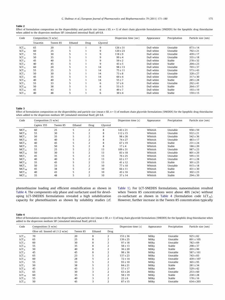
Table 2Effect of formulation composition on the dispersibility and particle size (mean ± SD, n = 3) of short chain glyceride formulations (SNEDDS) for the lipophilic drug thioridazinewhen added to the dispersion medium SIF (simulated intestinal fluid) pH 6.8.
Code Composition (% w/w) Dispersion time (sec) Appearance Precipitation Particle size (nm)
Triacetin Tween 85 Ethanol Drug Glycerol
SCT-A 65 20 5 1 9 126 ± 31 Dull white Unstable 873 ± 14SCT-B 60 25 5 1 9 120 ± 23 Dull white Unstable 792 ± 21SCT-C 55 30 5 1 9 118 ± 9 Dull white Unstable 439 ± 17SCT-D 50 35 5 1 9 99 ± 4 Dull white Unstable 335 ± 18SCT-E 45 40 5 1 9 59 ± 2 Dull white Stable 278 ± 32SCT-F 40 45 5 1 9 43 ± 5 Dull white Stable 204 ± 23SCT-G 60 20 5 1 14 98 ± 13 Dull white Unstable 703 ± 37SCT-H 55 25 5 1 14 75 ± 11 Dull white Unstable 573 ± 43SCT-I 50 30 5 1 14 73 ± 8 Dull white Unstable 328 ± 27SCT-J 45 35 5 1 14 60 ± 4 Dull white Unstable 317 ± 30SCT-K 40 40 5 1 14 55 ± 7 Dull white Stable 285 ± 28SCT-L 55 33 5 1 6 57 ± 6 Dull white Unstable 292 ± 41SCT-M 50 38 5 1 6 53 ± 5 Dull white Stable 264 ± 37SCT-N 45 43 5 1 6 49 ± 7 Dull white Stable 193 ± 19SCT-O 40 48 5 1 6 30 ± 4 Dull white Stable 159 ± 15
Table 3Effect of formulation composition on the dispersibility and particle size (mean ± SD, n = 3) of medium chain glyceride formulations (SNEDDS) for the lipophilic drug thioridazinewhen added to the dispersion medium SIF (simulated intestinal fluid) pH 6.8.
Code Composition (% w/w) Dispersion time (s) Appearance Precipitation Particle size (nm)
Captex 355 Tween 85 Ethanol Drug Glycerol
MCT-A 60 25 5 2 8 143 ± 21 Whitish Unstable 958 ± 59MCT-B 55 30 5 2 8 112 ± 15 Whitish Unstable 932 ± 21MCT-C 50 35 5 2 8 98 ± 26 Whitish Unstable 542 ± 36MCT-D 45 40 5 2 8 74 ± 27 Whitish Unstable 483 ± 17MCT-E 40 45 5 2 8 67 ± 19 Whitish Stable 231 ± 24MCT-F 35 50 5 2 8 17 ± 4 Whitish Stable 186 ± 20MCT-G 55 25 5 2 13 109 ± 31 Whitish Unstable 987 ± 23MCT-H 50 30 5 2 13 83 ± 18 Whitish Unstable 801 ± 29MCT-I 45 35 5 2 13 76 ± 25 Whitish Unstable 595 ± 33MCT-J 40 40 5 2 13 63 ± 17 Whitish Unstable 411 ± 28MCT-K 35 45 5 2 13 41 ± 12 Whitish Stable 301 ± 25MCT-L 50 33 5 2 10 77 ± 10 Whitish Unstable 915 ± 18MCT-M 45 38 5 2 10 66 ± 11 Whitish Stable 431 ± 30MCT-N 40 43 5 2 10 43 ± 16 Whitish Stable 302 ± 23MCT-O 35 48 5 2 10 37 ± 14 Whitish Stable 294 ± 39
G. Shahnaz et al. / European Journal of Pharmaceutics and Biopharmaceutics 79 (2011) 171–180 175
phenothiazine loading and efficient emulsification as shown inTable 4. The components oily phase and surfactant used for devel-oping LCT-SNEDDS formulations revealed a high solubilizationcapacity for phenothiazines as shown by solubility studies (cf.
Table 4Effect of formulation composition on the dispersibility and particle size (mean ± SD, n = 3) oadded to the dispersion medium SIF (simulated intestinal fluid) pH 6.8.
Code Composition (% w/w)
Olive oil: linseed oil (1:2 w/w) Tween 85 Ethanol Drug
LCT-A 70 20 8 2LCT-B 65 25 8 2LCT-C 60 30 8 2LCT-D 55 35 8 2LCT-E 50 40 8 2LCT-F 45 45 8 2LCT-G 65 23 5 2LCT-H 60 28 5 2LCT-I 55 33 5 2LCT-J 50 38 5 2LCT-K 45 43 5 2LCT-L 65 30 3 2LCT-M 60 35 3 2LCT-N 55 40 3 2LCT-O 50 45 3 2
Table 1). For LCT-SNEDDS formulations, nanoemulsion resultedwhen Tween 85 concentrations were above 40% (w/w) withoutco-surfactant as shown in Table 4 (formulation code LCT-N).However, further increase in the Tween 85 concentration typically
f long chain glyceride formulations (SNEDDS) for the lipophilic drug thioridazine when
Dispersion time (s) Appearance Precipitation Particle size (nm)
153 ± 36 Milky Unstable 925 ± 82139 ± 25 Milky Unstable 891 ± 105
97 ± 18 Milky Unstable 782 ± 6958 ± 13 Milky Stable 298 ± 3736 ± 20 Milky Stable 203 ± 9698 ± 36 Milky Unstable 587 ± 161
137 ± 23 Milky Unstable 743 ± 8372 ± 14 Milky Unstable 439 ± 10793 ± 10 Milky Unstable 365 ± 9584 ± 21 Milky Stable 281 ± 3457 ± 17 Milky Stable 529 ± 4263 ± 24 Milky Unstable 253 ± 6058 ± 19 Milky Stable 230 ± 2822 ± 6 Milky Stable 178 ± 1687 ± 15 Milky Unstable 634 ± 203

Fig. 2. Transmission electron microscope image of harvested chylomicrons at80 kV.
176 G. Shahnaz et al. / European Journal of Pharmaceutics and Biopharmaceutics 79 (2011) 171–180
resulted in the precipitation of phenothiazines upon addition ofsimulated intestinal fluid (SIF) and required at least three minutesto fully disperse.
3.3. Characterization of harvested chylomicrons
Chylomicrons from human plasma were prepared by densitygradient ultracentrifugation. Quantification of the harvested chylo-microns density was performed by refractometry. Due to their lowdensity (<0.95 g/ml), chylomicrons are confined to the top-gradi-ent fraction. Density calculations showed that before centrifuga-tion the density of the top-gradient fraction was 1.006 g/ml andafter centrifugation the density of the top-gradient fraction was0.55 g/ml. The density of the harvested chylomicrons thereforecorrelates favorably with results reported by other investigators[5].
The vesicle sizes of harvested chylomicrons were determined byenergy filter transmission electron microscope (EFTEM) (Fig. 2). Ves-icle sizes with a diameter of 130 ± 60 nm were detected. The imagein Fig. 2 shows individual round vesicles suggesting that aggregationdid not take place. Furthermore, electron microscopy showed a�1.5-fold increased in vesicle size of harvested chylomicrons after
0
20
40
60
80
100
60 80 100
120
130
140
150
Inte
nsit
y W
eigh
ted
Dis
trib
utio
n
Particle size (nm)
A
Fig. 3. Particle size distributions of (A) plasma derived isolated chylomicrons and (B) plaself-nanoemulsifying drug delivery system LCT-N SNEDDS. Indicated values are means ±
incubation with LCT-N SNEDDS. For further examination, vesicle sizeanalysis of harvested chylomicrons was performed using dynamiclight scattering. The intensity-weighted size distributions revealedtwo classes of vesicles (Fig. 3). Their mean diameters of chylomi-crons were 60–90 nm and 120–150 nm, whereas chylomicronsincubated with LCT-N SNEDDS displayed sizes of 60–90 nm and160–265 nm. The numbers of vesicles associated with the two peakswere evaluated by examining the scattering properties of thevesicles demonstrating that the majority were small ones. Thisassessment also suggested that the mean size in the first peak issignificantly smaller than the typical size of chylomicrons. Thiswas consistent with the finding that the harvested chylomicronsnot only contained apolipoprotein B-48 but also a similar amountof apolipoprotein B-100, which is associated with lipoproteins ofsmaller size. The larger vesicles of the second peak presumably rep-resent harvested chylomicrons. The first peak of vesicle size distribu-tions represented by apolipoprotein B-48 and apolipoprotein B-100is not significantly influenced by LCT-N SNEDDS. In contrast, a�2.5-fold increase in the second peak, probably representing harvestedchylomicrons, of vesicle size distributions was observed after 6-hincubation with LCT-N SNEDDS (Fig. 3A/B).
Moreover, all lipoprotein classes display a net negative chargeand studies have shown that this charge is due to both apolipopro-tein composition of the lipoprotein and its content of anionic lipids[14]. Accordingly, the zeta potential of harvested chylomicrons wasanalyzed showing a surface potential of �7.9 mV.
3.4. Uptake of molecules by harvested CM from SNEDDS formulations
The association of drugs with chylomicrons is a complex pro-cess, which involves the lipophilic core as well as surface apopro-teins. The plasma derived harvested chylomicron ex vivo modelbased on LCT-SNEDDS (LCT-N), MCT-SNEDDS (MCT-F) and SCT-SNEDDS (SCT-O) formulations is able to predict the degree of chy-lomicron association. Strong interaction with chylomicrons maybe attributed to the physicochemical properties of phenothiazines.Phenothiazines (PTZ) are useful organic compounds containing anitrogen (N) atom that easily looses one electron to form a cationradical because of high negative electron density [15]. Becausethe Mulliken atomic charge (with hydrogen summed into heavy
0
20
40
60
80
100
60 90 130
170
200
230
260
265
Inte
nsit
y W
eigh
ted
Dis
trib
utio
n
Particle size (nm)
B
sma derived isolated chylomicrons after incubation with the long chain triglycerideSD of at least three experiments.
G. Shahnaz et al. / European Journal of Pharmaceutics and Biopharmaceutics 79 (2011) 171–180 177
atoms) of N is �0.412953 [16], an attractive electrostatic interac-tion can occur between the N atom in PTZs and the polar head ofchylomicron phospholipids. Therefore, PTZs are positioned in themembrane of chylomicrons with the N atom oriented toward thepolar head of chylomicron phospholipids (Fig. 4). Furthermore,covalent bond formation between the electronegative nitrogenatom of PTZ and nitrogen atoms of apoprotein substructures ofchylomicrons ultimately causes a high degree of association. PTZcan form additional hydrogen bonds with triglycerides (TG) andcholesteryl ester substructures and can probably therefore diffuseto a higher extent into chylomicrons.
The association of plasma derived harvested chylomicrons withlipophilic phenothiazines was tested by either using the pure drugsolution or the relevant SNEDDS formulation based on short (SCT-
O), medium (MCT-F), and long chain triglycerides (LCT-N) (Fig. 5).The uptake of thioridazine was more efficient than the uptake ofchlorpromazine. Furthermore, the results show that the affinity oflipophilic drugs to plasma derived harvested chylomicrons was sig-nificantly influenced by the type of the SNEDDS formulation.SNEDDS based on long chain triglycerides (LCT-N) were the mosteffective systems when compared with the pure drug solution(S.S.) or with SNEDDS formulations based on medium (MCT-F) andshort chain triglycerides (SCT-O). For thioridazine a 5.6-fold and forchlorpromazine a 3.7-fold higher association with harvested chylo-microns could be observed using a SNEDDS formulation based onlong chain triglycerides (LCT-N). Thioridazine showed an almost2-fold higher affinity to chylomicrons than chlorpromazinealthough both compounds have similar structures and similar calcu-lated log P values. However, thioridazine displays a 2.4-fold highersolubility in long chain triglyceride components (cf. Fig. 1), whichcould explain the higher drug uptake of isolated chylomicrons.
Refractometry revealed that the density of harvested chylomi-crons was 1.8-fold increased after incubation with the LCT-SNEDDS(LCT-N) formulation when compared to the density of harvested
A
B
Fig. 4. (A) Electrostatic attractive interaction between the N atom of phenothiazines andiffusion across the biomembrane of chylomicrons. (For interpretation of the references t
chylomicrons before incubation, whereas no significant differencein the density of harvested chylomicrons was observed afterincubation with MCT-SNEDDS (MCT-F) and SCT-SNEDDS (SCT-O)formulations (data not shown). However, zeta potential studiesshowed no significant difference in the surface potential of har-vested chylomicrons after incubation with LCT-SNEDDS (LCT-N),MCT-SNEDDS (MCT-F), or SCT-SNEDDS (SCT-O) formulations (cf.Table 1). In addition, the intensity-weighted size distributions ofharvested chylomicrons revealed two classes of vesicles (cf.Fig. 3). Furthermore, transmission electron microscope confirmedthe above described observation by demonstrating an increase invesicle size of harvested chylomicrons after incubation with LCT-
N SNEDDS at 80 kV (cf. Fig. 6A) and at 80 kV after 70 electron lossenergy (cf. Fig. 6B). Fast and high absorption could be the reasonfor the bulky size of chylomicrons observed after LCT-K incubation(cf. Table 4). Previously, it has been observed that chylomicrons be-come large after administration of a corn oil bolus to lymph-cann-ulated rats [17]. Because chylomicrons can function as highlylipophilic model membranes, higher diffusibility or fusibility (cf.Fig. 4) of LCT-N being more lipophilic when compared to MCT-F
and SCT-O could be possible. Higher diffusion of LCT-N SNEDDSbased formulation into chylomicrons could be the reason for theincreased association of the drugs with the harvested chylomi-crons. Alternatively, the intensity-weighted vesicle size distribu-tion of harvested chylomicrons was not altered followingincubation with MCT-F SNEDDS and SCT-O SNEDDS formulations.
4. Presumptive additional features
SNEDDS as an appropriate lipid source may enhance lymphatictransport of lipophilic compounds by stimulating the production ofchylomicrons [1]. Lipids are hydrolyzed in the stomach and smallintestine to 2-monoglycerides (MG) and fatty acids (FA), then
d the polar head of chylomicron phospholipids. (B) Model of LCT-SNEDDS (LCT-N)o color in this figure legend, the reader is referred to the web version of this article.)

0
20
40
60
80
100
S.S
SCT-
O
MC
T-F
LC
T-N
% o
f dr
ug a
ssoc
iati
on
A
0
10
20
30
40
50
S.S
SCT-
O
MC
T-F
LC
T-N
% o
f dr
ug a
ssoc
iati
on
B
Fig. 5. Uptake of (A) thioridazine and (B) chlorpromazine by plasma derived harvested chylomicrons from different self-nanoemulsifying drug delivery systems (SNEDDS).Uptake from the pure drug (SS, stock solution) was measured as a control. Indicated values are means ± SD of at least three experiments.
Fig. 6. Transmission electron microscope image of (A) harvested chylomicrons after incubation with LCT-N SNEDDS formulation at 80 kV and (B) harvested chylomicrons afterincubation with LCT-N SNEDDS formulation with 70 electron loss energy at 80 kV.
178 G. Shahnaz et al. / European Journal of Pharmaceutics and Biopharmaceutics 79 (2011) 171–180
reabsorbed into the enterocyte, re-esterified into triglycerides (TG),and then incorporated into intestinal chylomicrons (Fig. 7). Follow-ing exocytosis from the basolateral membrane, the newly secretedchylomicron particles first enter the intercellular space beforemigrating via the lacteals into the circulation [18]. A mechanisticstudy of tight junction opening by self-microemulsifying drugdelivery systems (SMEDDS) containing the solubilizer Labrasoldemonstrated changes in subcellular localization of the tightjunction proteins F-actin and ZO-1 [19]. Medium chain mono-and diglycerides have been shown to modulate tight junctionfunction through a phospholipase C dependent pathway [20].Therefore, we hypothesize that the SNEDDS containing mediumchain mono- and diglycerides like Captex 355 can lead to a lossof tight junction integrity and might enter the intercellular spaceby opening tight junctions in vivo. So, intercellular space can pro-vide the possible uptake window for SNEDDS by chylomicrons.The appearance of chylomicrons in the intercellular spaces is fol-lowed by their passage into the lamina propria through gaps or dis-
continuities of the basement membrane [21]. Another possibilityfor lymphatic transport of lipid-based nanocarriers is specific up-take by membranous epithelial cells (M cells) of Peyer’s patches[22]. It is assumed that the SNEDDS can be absorbed transcellularlythrough M cells in the gut associated lymphoid tissue (GALT). Sub-sequently, SNEDDS may be transported into the intestinal laminapropria by this transport mechanism and may be delivered to theperiphery by lymphatic and/or vascular endothelium. The intesti-nal lymphatic tissue contains specialized M cells, which are sup-posed to facilitate transcytotic transport across the intestine.Small particles in the intestinal lumen confine to the apical sideof M cells and can be internalized by M cells through transcytosis.The SNEDDS in the present studies are assumed to be absorbed viathis mechanism. As reported earlier, there is a particle size-depen-dent exclusion phenomenon in the gastrointestinal mucosal tissuewith 100 nm size particles showing significantly greater tissue up-take than larger particles [23]. Therefore, SNEDDS may be absorbedthrough the lymphatic system and then transferred into systemic

Fig. 7. Schematic diagram illustrating the mechanism of intestinal drug transport from self-nanoemulsifying drug delivery systems (SNEDDS). Three possible pathways ofSNEDDS (D) uptake by chylomicrons (CM) via diffusion are as follows: (1) transcellular uptake with digestions and accumulation into CMs, (2) paracellular uptake by openingof tight junctions, (3) M cell uptake at membranous epithelial cells of the Peyer’s patches. SNEDDS incorporated chylomicrons (F) might be absorbed through the lymphaticsystem and then transferred into systemic circulation. The incorporation of the SNEDDS into the chylomicrons (F) can lead to a bypass of the hepatic first-pass metabolismand therefore avoid hepatotoxicity. (For interpretation of the references to color in this figure legend, the reader is referred to the web version of this article.)
G. Shahnaz et al. / European Journal of Pharmaceutics and Biopharmaceutics 79 (2011) 171–180 179
circulation with chylomicron associations as shown in Fig. 7. It islikely that the long chain triglyceride SNEDDS formulation will alsolead to maximize intestinal lymphatic chlorpromazine and thiorid-azine transport. Both chlorpromazine and thioridazine undergoextensive first-pass metabolism mainly by hydroxylation, N-deal-kylation, N-oxidation, and S-oxidation. Clinical experience to dateindicates that chlorpromazine therapy is associated with very rarebut severe idiosyncratic hepatotoxicity. Hepatic injury has alsobeen observed with the closely related antipsychotic drug thiorid-azine. High binding affinity of phenothiazines with chylomicronformulation based on long chain triglycerides indicates that theincorporation of phenothiazines into chylomicrons is an essentialstep to bypass the hepatic first-pass metabolism and hepatotoxic-ity prevention (Fig. 7). We hypothesize that a high degree ofSNEDDS association with chylomicrons can be utilized to improve
the oral bioavailability of highly lipid-soluble phenothiazines byusing the lymphatic route transfer to systemic circulation. Thelymphatic transport potential can also be utilized to bypass the he-patic first-pass metabolism as well as the known hepatotoxicity ofphenothiazines.
5. Conclusion
An optimized SCT, MCT, and LCT self-nanoemulsifying drugdelivery system (SNEDDS) containing phenothiazines was devel-oped with particles size less than 200 nm. The present study dem-onstrates that incorporation of phenothiazines into SNEDDS leadsto enhanced uptake by plasma derived chylomicrons, comparedto naked phenothiazines in solution. The findings also provide

180 G. Shahnaz et al. / European Journal of Pharmaceutics and Biopharmaceutics 79 (2011) 171–180
evidence for the importance of the fatty acid chain length inSNEDDS formulations. Therefore, this chylomicron ex vivo modelis suitable to predict the intestinal lymphatic transport potentialof phenothiazines. Enhanced uptake of phenothiazines by chylomi-crons appears to be an important prerequisite for efficient intesti-nal lymphatic transport and thus for the substantial improvementin oral bioavailability of lipophilic drugs.
Acknowledgements
The authors wish to thank the Higher Education CommissionPakistan (HEC) and the Austrian Agency for International Coopera-tion in Education and Research (ÖAD) for their support andfunding.
References
[1] M.R. Aji Alexa, A.J. Chackoa, S. Josea, E.B. Soutob, Lopinavir loaded solid lipidnanoparticles (SLN) for intestinal lymphatic targeting, Eur. J. Pharm. Sci. 42(2011) 11–18.
[2] N.L. Trevaskis, C.L. McEvoy, M.P. McIntosh, G.A. Edwards, R.M. Shanker, W.N.Charman, C.J.H. Porter, The role of the intestinal lymphatics in the absorptionof two highly lipophilic cholesterol ester transfer protein inhibitors (CP524,515and CP532,623), Pharm. Res. 27 (2010) 878–893.
[3] W. Faisal, C.M. O’Driscoll, B.T. Griffin, Bioavailability of lycopene in the rat: therole of intestinal lymphatic transport, J. Pharm. Pharmacol. 62 (2010) 323–331.
[4] S.M. Caliph, W.A. Fried Faassen, G.M. Vogel, C.J.H. Porter, Oral bioavailabilityassessment and intestinal lymphatic transport of Org 45697 and Org 46035,two highly lipophilic novel immunomodulator analogues, Curr. Drug Deliv. 8(2009) 359–366.
[5] P. Gershkovich, A. Hoffman, Uptake of lipophilic drugs by plasma derivedisolated chylomicrons: linear correlation with intestinal lymphaticbioavailability, Eur. J. Pharm. Sci. 26 (2005) 394–404.
[6] D. Ordway, M. Viveiros, C. Leandro, R. Bettencourt, J. Almeida, M. Martins, J.E.Kristiansen, J. Molnar, L. Amaral, Clinical concentrations of thioridazine killintracellular multidrug-resistant Mycobacterium tuberculosis, Antimicrob.Agents Chemother. 47 (2003) 917–922.
[7] N. Motohashi, M. Kawase, S. Saito, H. Sakagami, Antitumor potential andpossible targets of phenothiazine-related compounds, Curr. Drug. Targets 1(2000) 237–245.
[8] F. Karpe, A. Hamsten, K. Uffelman, G. Steiner, Isolation of triglyceride-richlipoproteins for subsequent determination of apolipoproteins B-48 and B-100,Meth. Enzymol. 47 (1996) 95–104.
[9] R.J. Havel, H.A. Eder, J.H. Bragdon, The distribution and chemical compositionof ultracentrifugally separated lipoproteins in human serum, J. Clin. Invest. 34(1955) 1345–1353.
[10] G. Zhang, A.V. Terry Jr., M.G. Bartlett, Simultaneous determination of fiveantipsychotic drugs in rat plasma by high performance liquid chromatographywith ultraviolet detection, J. Chromatogr. B. 856 (2007) 20–28.
[11] N.H. Shah, M.T. Carvajal, C.I. Patel, M.H. Infeld, A.W. Malick, Selfemulsifyingdrug delivery systems (SEDDS) with polyglycolyzed glycerides for improvingin vitro dissolution and oral absorption of lipophilic drugs, Int. J. Pharm. 106(1994) 15–23.
[12] M.J. Lawrence, G.D. Rees, Microemulsion based media as novel drug deliverysystem, Adv. Drug. Deliv. Rev. 45 (2000) 89–121.
[13] J.H. Yoo, S. Shanmugam, P. Thapa, E. Lee, P. Balakrishnan, R. Baskaran, S. Yoon,H. Choi, C.S. Yong, B.K. Yoo, K. Han, Novel self-nanoemulsifying drug deliverysystem for enhanced solubility and dissolution of lutein, Arch. Pharm. Res. 33(2010) 417–426.
[14] C.J. Stamler, D. Breznan, T.A-M. Neville, F.J. Viau, E. Camlioglu, D.L. Sparks,Phosphatidylinositol promotes cholesterol transport in vivo, J. Lipid Res. 41(2000) 1214–1221.
[15] P.F. Coccia, W.W. Westerfeld, The metabolism of chlorpromazine bylivermicrosomal enzyme systems, J. Pharmacol. Exp. Ther. 157 (1967) 446–458.
[16] F. Hartmann, L.D. Gruenke, J.C. Craig, D.M. Bisell, Chlorpromazine metabolismin extracts of liver and small intestine from guinea pig and from man, DrugMetab. Dispos. 11 (1983) 244–248.
[17] P. Degrace, C. Caselli, J.M. Rayo, A. Bernard, Intestinal lymph absorption ofbutter, corn oil, cod liver oil, menhaden oil, and eicosapentaenoic anddocosahexaenoic acid ethyl esters in rats, Lipids 31 (1996) 405–414.
[18] A.I. Mendeloff, The effects of eating and of sham feeding upon the absorptionof vitamin A palmitate in man, J. Clin. Invest. 33 (1954) 1015–1021.
[19] X. Sha, G. Yan, Y. Wu, J. Li, X. Fang, Effect of self-microemulsifying drugdelivery systems containing Labrasol on tight junctions in Caco-2 cells, Eur. J.Pharm. Sci. 24 (2005) 477–486.
[20] M. Tomita, M. Hayashi, S. Awazu, Absorption-enhancing mechanism of sodiumcaprate and decanoylcarnitine in Caco-2 cells, J. Pharmacol. Exp. Ther. 272(1995) 739–743.
[21] S.M. Sabesin, S. Frase, Electron microscopic studies of the assembly,intracellular transport, and secretion of chylomicrons by rat intestine, J.Lipid Res. 18 (1977) 496–511.
[22] B. Sarmento, S. Martins, D. Ferreira, E.B. Souto, Oral insulin delivery by meansof solid lipid nanoparticles, Int. J. Nanomed. 2 (2007) 743–749.
[23] M.P. Desai, V. Labhasetwar, G.L. Amidon, R.J. Levy, Gastrointestinal uptake ofbiodegradable microparticles: effect of particle size, Pharm. Res. 13 (1996)1838–1845.