Exome-Sequencing Confirms DNAJC5 Mutations as Cause of Adult Neuronal Ceroid-Lipofuscinosis
11
Exome-Sequencing Confirms DNAJC5 Mutations as Cause of Adult Neuronal Ceroid-Lipofuscinosis Bruno A. Benitez 1 , David Alvarado 2 , Yefei Cai 1 , Kevin Mayo 1 , Sumitra Chakraverty 1 , Joanne Norton 1 , John C. Morris 3 , Mark S. Sands 2,4 , Alison Goate 1,3,4,5 , Carlos Cruchaga 1,4 * 1 Department of Psychiatry, Washington University, St. Louis, Missouri, United States of America, 2 Department of Pediatrics, Washington University, St. Louis, Missouri, United States of America, 3 Department of Neurology, Washington University, St. Louis, Missouri, United States of America, 4 Hope Center Program on Protein Aggregation and Neurodegeneration, Washington University, St. Louis, Missouri, United States of America, 5 Department of Genetics, Washington University, St. Louis, Missouri, United States of America Abstract We performed whole-exome sequencing in two autopsy-confirmed cases and an elderly unaffected control from a multigenerational family with autosomal dominant neuronal ceroid lipofuscinosis (ANCL). A novel single-nucleotide variation (c.344T.G) in the DNAJC5 gene was identified. Mutational screening in an independent family with autosomal dominant ANCL found an in-frame single codon deletion (c.346_348 delCTC) resulting in a deletion of p.Leu116del. These variants fulfill all genetic criteria for disease-causing mutations: they are found in unrelated families with the same disease, exhibit complete segregation between the mutation and the disease, and are absent in healthy controls. In addition, the associated amino acid substitutions are located in evolutionarily highly conserved residues and are predicted to functionally affect the encoded protein (CSPa). The mutations are located in a cysteine-string domain, which is required for membrane targeting/binding, palmitoylation, and oligomerization of CSPa. We performed a comprehensive in silico analysis of the functional and structural impact of both mutations on CSPa. We found that these mutations dramatically decrease the affinity of CSPa for the membrane. We did not identify any significant effect on palmitoylation status of CSPa. However, a reduction of CSPa membrane affinity may change its palmitoylation and affect proper intracellular sorting. We confirm that CSPa has a strong intrinsic aggregation propensity; however, it is not modified by the mutations. A complementary disease- network analysis suggests a potential interaction with other NCLs genes/pathways. This is the first replication study of the identification of DNAJC5 as the disease-causing gene for autosomal dominant ANCL. The identification of the novel gene in ANCL will allow us to gain a better understanding of the pathological mechanism of ANCLs and constitutes a great advance toward the development of new molecular diagnostic tests and may lead to the development of potential therapies. Citation: Benitez BA, Alvarado D, Cai Y, Mayo K, Chakraverty S, et al. (2011) Exome-Sequencing Confirms DNAJC5 Mutations as Cause of Adult Neuronal Ceroid- Lipofuscinosis. PLoS ONE 6(11): e26741. doi:10.1371/journal.pone.0026741 Editor: Thomas H. Gillingwater, University of Edinburgh, United Kingdom Received August 24, 2011; Accepted October 2, 2011; Published November 4, 2011 Copyright: ß 2011 Benitez et al. This is an open-access article distributed under the terms of the Creative Commons Attribution License, which permits unrestricted use, distribution, and reproduction in any medium, provided the original author and source are credited. Funding: This work was supported by grants: NIH (1P30NS069329-01, P50AG005681), the Barnes-Jewish Hospital Foundation. This work was also supported by pilot funding from the Hope Center for Neurological Disorders and the Danforth Foundation Challenge at Washington University. We thank the Genome Technology Access Center in the Department of Genetics for help with genomic analysis. The Center is partially supported by NCI Cancer Center Support Grant #P30 CA91842 to the Siteman Cancer Center and by ICTS/CTSA Grant# UL1RR024992 from the National Center for Research Resources (NCRR), a component of the National Institutes of Health (NIH), and NIH Roadmap for Medical Research. This publication is solely the responsibility of the authors and does not necessarily represent the official view of NCRR or NIH. Competing Interests: The authors have declared that no competing interests exist. * E-mail: [email protected] Introduction The neuronal ceroid lipofuscinosis (NCLs) are the most common group of inherited neurodegenerative diseases in children, with an incidence in the U.S. of approximately 1 in 12,500 live births [1]. NCLs encompass a genetically heteroge- neous group of disorders, clinically characterized by progressive deterioration of cognitive and motor skills, visual impairment, and premature death [2]. The onset of the clinical symptoms in addition to the differences in ultrastructural features of the lipopigment inclusions underlie the nosological spectrum of the NCLs: infantile (INCL, Santavuori-Haltia, MIM 256730), late- infantile (LINCL, Jansky-Bielschowsky, MIM 204500), juvenile (JNCL, Batten disease, Spielmeyer-Vogt, MIM 204200), adult (ANCL, Kuf’s disease, MIM 204300), and Northern epilepsy (NE, progressive epilepsy with intellectual disability) [2]. Over the last fifteen years, at least 300 mutations in ten genes have been associated with NCLs such as CLN1 (PPT1 (MIM 256730)), CLN2 (TPP1 (MIM 204500)), CLN3 (MIM 204200), CLN5 (MIM 256731), CLN6 (MIM 601780), CLN7 (MFSD8 (MIM 610951)), CLN8 (MIM 600143), CLN10 (CTSD (MIM 610127)), CLCN6 (MIM 602726) and SGSH (MIM 605270) (NCL Mutation Database, URL). This genetic progress has led to the development of molecular testing tools and promising rational therapeutic approaches [3]. However, there is no cure for NCLs, and treatments are limited to palliative care. Adult onset NCLs (ANCLs) represent between 1.3% and 10% of NCLs cases [4]. ANCLs are rapidly worsening conditions with a wide range of age at onset (6–62 yr) and broad clinical variability. Two main clinical subtypes have been described: progressive myoclonus epilepsy (type A), and dementia with motor distur- bances, such as cerebellar, extrapyramidal signs, and dyskinesia PLoS ONE | www.plosone.org 1 November 2011 | Volume 6 | Issue 11 | e26741
-
Upload
independent -
Category
Documents
-
view
5 -
download
0
Transcript of Exome-Sequencing Confirms DNAJC5 Mutations as Cause of Adult Neuronal Ceroid-Lipofuscinosis
Exome-Sequencing Confirms DNAJC5 Mutations asCause of Adult Neuronal Ceroid-LipofuscinosisBruno A. Benitez1, David Alvarado2, Yefei Cai1, Kevin Mayo1, Sumitra Chakraverty1, Joanne Norton1,
John C. Morris3, Mark S. Sands2,4, Alison Goate1,3,4,5, Carlos Cruchaga1,4*
1 Department of Psychiatry, Washington University, St. Louis, Missouri, United States of America, 2 Department of Pediatrics, Washington University, St. Louis, Missouri,
United States of America, 3 Department of Neurology, Washington University, St. Louis, Missouri, United States of America, 4 Hope Center Program on Protein
Aggregation and Neurodegeneration, Washington University, St. Louis, Missouri, United States of America, 5 Department of Genetics, Washington University, St. Louis,
Missouri, United States of America
Abstract
We performed whole-exome sequencing in two autopsy-confirmed cases and an elderly unaffected control from amultigenerational family with autosomal dominant neuronal ceroid lipofuscinosis (ANCL). A novel single-nucleotidevariation (c.344T.G) in the DNAJC5 gene was identified. Mutational screening in an independent family with autosomaldominant ANCL found an in-frame single codon deletion (c.346_348 delCTC) resulting in a deletion of p.Leu116del. Thesevariants fulfill all genetic criteria for disease-causing mutations: they are found in unrelated families with the same disease,exhibit complete segregation between the mutation and the disease, and are absent in healthy controls. In addition, theassociated amino acid substitutions are located in evolutionarily highly conserved residues and are predicted to functionallyaffect the encoded protein (CSPa). The mutations are located in a cysteine-string domain, which is required for membranetargeting/binding, palmitoylation, and oligomerization of CSPa. We performed a comprehensive in silico analysis of thefunctional and structural impact of both mutations on CSPa. We found that these mutations dramatically decrease theaffinity of CSPa for the membrane. We did not identify any significant effect on palmitoylation status of CSPa. However, areduction of CSPa membrane affinity may change its palmitoylation and affect proper intracellular sorting. We confirm thatCSPa has a strong intrinsic aggregation propensity; however, it is not modified by the mutations. A complementary disease-network analysis suggests a potential interaction with other NCLs genes/pathways. This is the first replication study of theidentification of DNAJC5 as the disease-causing gene for autosomal dominant ANCL. The identification of the novel gene inANCL will allow us to gain a better understanding of the pathological mechanism of ANCLs and constitutes a great advancetoward the development of new molecular diagnostic tests and may lead to the development of potential therapies.
Citation: Benitez BA, Alvarado D, Cai Y, Mayo K, Chakraverty S, et al. (2011) Exome-Sequencing Confirms DNAJC5 Mutations as Cause of Adult Neuronal Ceroid-Lipofuscinosis. PLoS ONE 6(11): e26741. doi:10.1371/journal.pone.0026741
Editor: Thomas H. Gillingwater, University of Edinburgh, United Kingdom
Received August 24, 2011; Accepted October 2, 2011; Published November 4, 2011
Copyright: � 2011 Benitez et al. This is an open-access article distributed under the terms of the Creative Commons Attribution License, which permitsunrestricted use, distribution, and reproduction in any medium, provided the original author and source are credited.
Funding: This work was supported by grants: NIH (1P30NS069329-01, P50AG005681), the Barnes-Jewish Hospital Foundation. This work was also supported bypilot funding from the Hope Center for Neurological Disorders and the Danforth Foundation Challenge at Washington University. We thank the GenomeTechnology Access Center in the Department of Genetics for help with genomic analysis. The Center is partially supported by NCI Cancer Center Support Grant#P30 CA91842 to the Siteman Cancer Center and by ICTS/CTSA Grant# UL1RR024992 from the National Center for Research Resources (NCRR), a component ofthe National Institutes of Health (NIH), and NIH Roadmap for Medical Research. This publication is solely the responsibility of the authors and does not necessarilyrepresent the official view of NCRR or NIH.
Competing Interests: The authors have declared that no competing interests exist.
* E-mail: [email protected]
Introduction
The neuronal ceroid lipofuscinosis (NCLs) are the most
common group of inherited neurodegenerative diseases in
children, with an incidence in the U.S. of approximately 1 in
12,500 live births [1]. NCLs encompass a genetically heteroge-
neous group of disorders, clinically characterized by progressive
deterioration of cognitive and motor skills, visual impairment, and
premature death [2]. The onset of the clinical symptoms in
addition to the differences in ultrastructural features of the
lipopigment inclusions underlie the nosological spectrum of the
NCLs: infantile (INCL, Santavuori-Haltia, MIM 256730), late-
infantile (LINCL, Jansky-Bielschowsky, MIM 204500), juvenile
(JNCL, Batten disease, Spielmeyer-Vogt, MIM 204200), adult
(ANCL, Kuf’s disease, MIM 204300), and Northern epilepsy (NE,
progressive epilepsy with intellectual disability) [2].
Over the last fifteen years, at least 300 mutations in ten genes
have been associated with NCLs such as CLN1 (PPT1 (MIM
256730)), CLN2 (TPP1 (MIM 204500)), CLN3 (MIM 204200),
CLN5 (MIM 256731), CLN6 (MIM 601780), CLN7 (MFSD8 (MIM
610951)), CLN8 (MIM 600143), CLN10 (CTSD (MIM 610127)),
CLCN6 (MIM 602726) and SGSH (MIM 605270) (NCL Mutation
Database, URL). This genetic progress has led to the development
of molecular testing tools and promising rational therapeutic
approaches [3]. However, there is no cure for NCLs, and
treatments are limited to palliative care.
Adult onset NCLs (ANCLs) represent between 1.3% and 10%
of NCLs cases [4]. ANCLs are rapidly worsening conditions with a
wide range of age at onset (6–62 yr) and broad clinical variability.
Two main clinical subtypes have been described: progressive
myoclonus epilepsy (type A), and dementia with motor distur-
bances, such as cerebellar, extrapyramidal signs, and dyskinesia
PLoS ONE | www.plosone.org 1 November 2011 | Volume 6 | Issue 11 | e26741
(type B). However, there is some overlap, or a continuum of signs
between the two types, particularly late in the course of the disease.
Therefore, it is not always easy to differentiate them [4]. Unlike
other NCLs there is an absence of retinal degeneration [5].
Pathologically, the ceroid-lipofuscin accumulates mainly in
neurons and contains subunit C of the mitochondrial adenosine-
triphosphate synthase (SCMAS) [6], but has different ultrastruc-
tural appearances such as granular osmiophilic deposits (GRODs)
and fingerprint, curvilinear, or rectilinear structures [7].
ANCLs are genetically heterogeneous with either a sporadic,
autosomal recessive (Kufs’ disease, MIM 204300) or dominant
(Parry’s disease, MIM 162350) pattern of inheritance in confirmed
cases [4,5]. Three known NCL genes were previously associated
with atypical ANCLs such as PPT1 [8,9], CLN5, and N-
Sulfoglucosamine Sulfohydrolase (SGSH, MIM 605270) [10], suggesting
that some of the ANCLs were not distinct genetic entities and
raising the possibility that they actually represent an extreme of a
clinical spectrum of low penetrance and variable expressivity of
NCL mutants [10,11]. This knowledge, although significant, could
not contribute to molecular elucidation of the ANCLs.
That was the state of the art when we started to perform an
exome-sequencing study in the largest family (10 affected members
over 5 generations) with autosomal-dominant ANCLs, known
until now (Figure 1A) [12]. However, during the course of this
study, two genes, a well known NCL gene (CLN6) [11] and
DNAJC5 gene (MIM, 611203) [13] have been associated with
autosomal recessive (locus NCL4A) and dominant (locus NCL4B)
cases of ANCLs, respectively. There was no apparent correlation
of the underlying genetic defect with the clinical course and the
ultrastructural features between the studies and, to date, neither of
them has been independently replicated. Independent replication
studies are important especially in ANCLs, wherein a critical
evaluation of the literature led to the rejection of 68 (out of 118)
cases published as Kufs’ disease [5], which means that there is a
high rate of misdiagnosis in ANCLs. These results also indicate
that the genetic architecture of ANCL is more varied and complex
than previously thought.
In this study, we performed exome-sequecing in three family
members from a family with autosomal dominant ANCL with
early dementia [12] (Figure 1A). Several recent studies have
successfully identified the disease-causing genetic variant in non-
NCL diseases with similar pedigrees by exome-sequencing [14].
Exome-sequencing is a very powerful technique, especially in
families that are not big enough for classical linkage studies. In
previous studies, the pathogenic variant was identified by only
sequencing as low as two or three individuals [15].This method is a
hypothesis-free approach that allows for a targeted enrichment
and resequencing of nearly all exons of protein-coding genes.
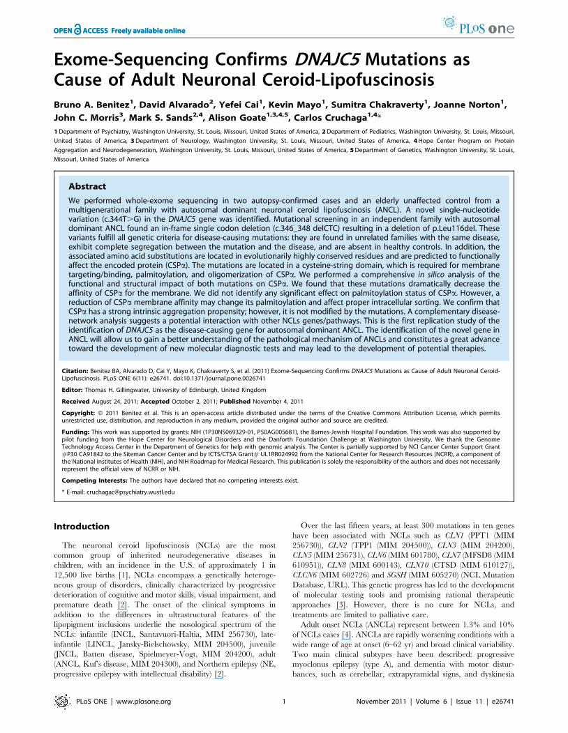
Figure 1. Pedigree of ANCL family, Sanger sequencing results and Multiple Alignment of CSPa. (A) Pedigree of the ANCL family. Blacksymbols denote affected individuals; open symbols denote unaffected individuals. (B) Chromatograms of exon 4 of DNAJC5 gene, showing sequencesof identified heterozygous mutations. (Upper panel) Sequence of an unaffected elderly control, (middle panel) sequence showing heterozygousmutation c.344T.G in the affected proband, (lower panel) sequence showing heterozygous mutation c.346_348delCTC in affected from a secondfamily (Validation set). Arrow and black short line indicate the position of the missense and deletion mutation, respectively. (C) Multiple Sequencealignment of Cysteine-string domain of CSPa amino acid sequence among homologous genes in mammals. pL115 is highlighted in yellow andindicated by the arrow.doi:10.1371/journal.pone.0026741.g001
DNAJC5 Mutations Cause ANCL
PLoS ONE | www.plosone.org 2 November 2011 | Volume 6 | Issue 11 | e26741

Protein-coding exons account for only 1% of the human genome,
but 85% of Mendelian diseases are caused by mutations in this
genomic space.
Results
Exome SequencingOverall, a mean of 158.6 million reads were generated for the
three samples. Approximately 80% of these were aligned to the
human reference genome (hg18) and 95% of these fell onto
targeted and enriched exons. Reads not corresponding to the
targeted bases of the exome were discarded (less than 2%). The
mean exome coverage was 94.7% with .56 fold coverage. After
filtering for a minimum length good quality sequence of 30 and a
minimal sequence read depth of 56 (standard parameters), we
identified on average 38,179684 coding single nucleotide
substitutions (SNSs) at a transitions-to-transversions ratio of 3.05,
and 2931610 small insertions and deletions (indels), among all
three individuals.
To identify pathogenic variants, we consecutively filtered these
variants by subjecting them to an analytical pipeline for high-
confidence variant calling and annotation. Briefly, we discarded (a)
common and known variants present in HapMap, the dbSNP130
Database or 1,000 Genomes Project at a frequency of greater than
5% for heterozygous and 30% for homozygous calls, and (b)
variants in intergenic or intronic regions [16]. After filtering,
2,3376102 SNPs and 21062 indels were identified as novels.
Next, we further focused on functionally significant variants such
as missense, nonsense, or splice-site changes, with sufficient and
consistent depth and quality values plus a high rate of concordance
among the three samples. Thus, we identified 96 SNSs (95
missense, 1 nonsense), and 13 indels which were private variants
for these members of this family (Table 1). There were 24
heterozygous non-synonymous coding variants and three hetero-
zygous indels (Table 1) that were present in both affected
individuals but absent in the control. In order to remove
systematic artifacts and rare variants, we checked these variants
against an additional Washington University exome database of 59
individuals, which reduced the list to 19 variants and three indels
(Table 2). These 22 high-quality variants had a median SNP
quality score of 228 (range 61 6095) and a read depth of 124.5
(range 9–381), and were selected for validation using two different
genotyping technologies: the MassARRAY SNP (Sequenom, Inc)
and KASPar v4.0 SNP (KBioscience) genotyping systems
[17,18,19]. Only one variant turned out to be a false positive,
which was most likely due to a mapping error. Thus, we have a
low rate of false discovery of 0.045.
Identification of the causative variantNext, we carried out an extended co-segregation analysis within
the family for all the validated SNPs. In total, we genotyped three
affected samples and three elderly healthy individuals (See
methods). As a result, only two SNSs, one located in PDCD6IP
(Programmed cell death 6-interacting protein, MIM 608074) and
one in DNAJC5, plus a deletion in the LIPJ gene (lipase-like, ab-
hydrolase domain containing 1, MIM 613921) segregated
perfectly with disease status (Table 2).
It has been shown that disease genes display significant
functional clustering in molecular networks [20,21]. One-third of
known disorders with two or more associated genes were found in
physical clusters of genes with the same phenotype [20].
Therefore, we used a disease-network approach using all NCL
genes as a training group (see methods) to prioritize further
validation among these three genes (PDCD6IP, DNAJC5 and LIPJ).
Surprisingly, we found that these three variants were in the top five
genes in the combined analysis (See: Table S1), suggesting that
they may be functionally or structurally related with NCLs
encoded genes and constituting true candidates as ANCLs
causative genes. We also noticed that out of these three variants,
PDCD6IP and DNAJC5 have the highest Genomic Evolutionary
Rate Profiling (GERP) and are predicted to be damaging for the
protein (Table 2). The deletion in the LIPJ gene was predicted to
remove a potential donor splice site by using the Human Splicing
Finder server (Consensus Values (CV) for wild type 69.84 and 36
for the mutant, an average reduction (-DCV) of 48.45) [22].
Taking into account this evidence, PDCD6IP, DNAJC5 and LIPJ
were all identified as excellent plausible candidates for the genetic
defect responsible for ANCL. Therefore, we performed genotyp-
ing for each variant in a cohort of 1,600 (3,200 chromosomes)
ethnically matched controls. The p.G429S variant in the PDCD6IP
gene and the deletion on the 39-splicesite in the LIPJ gene turned
out to be rare variants with a MAF of 0.01 and 0.03, respectively.
The only variant remaining is a single nucleotide substitution
(c.344T.G) that causes a p.L115R amino acid change in DNAJC5
gene. Sanger sequencing was performed to confirm the presence of
the mutation in DNAJC5 gene in all affected individuals in the
family (Figure 1B). We also analyzed the exome-sequencing data
of all individuals for mutations in previously reported genes
associated with ANCLs such as PPT1, CLN5, SGSH and CLN6
[8,9,10,11] and revealed no non-synonymous changes in any of
them.
The fact that the c.344T.G variant in DNAJC5 was present in
all the affected individuals but not in 1,600 control individuals
strongly indicates that this is the underlying genetic cause of the
ANCL phenotype in this family.
Table 1. Filtering process of coding single nucleotide substitutions.
SampleTotal CodingSNSs
Non-synonymousSNSs"
SNSs afterfiltering
SNSs inboth cases
SNSs uniqueto cases
SNSs afterremoving found inWash U exomes
Affected 38142 9202 674 96 24 19
Affected 38276 9285 636
Control 38120 9177 711
Percent of SNSs remaining1 100 24.1 1.8 0.25 0.06 0.04
"dbSNP, HapMap and 1000 Genomes commons variants were removed (het,5%, hom,30%).1Percentage of SNSs after each step.doi:10.1371/journal.pone.0026741.t001
DNAJC5 Mutations Cause ANCL
PLoS ONE | www.plosone.org 3 November 2011 | Volume 6 | Issue 11 | e26741
In order to confirm that mutations in the DNAJC5 gene causes
ANCL, we used Sanger sequencing to analyze the entire coding
sequence plus the exonic flanking region in three other
independent autosomal dominant familial cases of ANCL and
one of LINCL (internal validation set). We found an in-frame
single codon deletion (c.346_348 delCTC), which causes a deletion
of p.L116del (Figure 1B) in a second family. We designed a Kaspar
assay to test this deletion, and did not detect it in more than 3,200
chromosomes from our control samples. None of these variants
have been previously reported in dbSNP (build 134) or 1000
genomes project database (20101123 releases).
Thus, combining all the genetic analysis together; we found that
different novel mutations in DNAJC5 are present in unrelated
families and exhibit perfect segregation with disease status. These
variants are located in highly conserved regions (GERP score of
5.34), are predicted to be damaging and are not present in 1,600
controls. Therefore, our results replicate the recent report of
mutations in DNAJC5 gene as a cause of ANCL [13].
Bioinformatic analysisDNAJC5 is located at 20q13.33 with genomic coordinates:
62,526,454–62,567,383 (GRCh37) and encodes a 198-amino
acid residue protein (cMW 22.1 kDa) called CSPa for cysteine-
string protein-a (NP_079495.1). CSPa contains three highly
conserved domains: the N-terminal J-domain (15–83 aa), the
linker region (84–112), and the cysteine string domain (CSD)
(113–136 aa) [23].
The p.L115R and p.L116del mutations affect highly conserved
dileucine residues located immediately N-terminal to the CSD
(Figure 1C); this region is responsible for palmitoylation,
membrane binding/targeting and oligomerization of CSPa[24,25,26]. Therefore, we took advantage of the existence of
robust in silico tools (See methods), which are widely validated with
curated experimental data, to test the functional impact of the
mutations on these processes.
Palmitoylation AnalysisFirst, we performed an in silico analysis of the impact of the
mutations on the palmitoylation status of CSPa. As shown in
Figure 2A, changes in the p.C121-124L residues used as a positive
control, showed a significant reduction in the levels (3.21762.723)
of predicted palmitoylation compared to the wild type (WT)
(6.94262.579) (p,0.05 kruskall-wallis test), which is in agreement
with experimental data [25,26]. In contrast, we did not find any
significant (p.0.05) change in the pattern of palmitoylation
induced by p.L115R (7.09361.919) and p.L116del (6.67762.395)
over the wild type sequence, although, the p.L115R mutation
eliminates palmitoylation of p.C113.
Hydrophobicity Profile of CSPaThe CSD is a highly hydrophobic region, and residues in the N-
terminal half, are likely to play a key role in membrane association
[25,26]. The general index of hydrophobicity (mean 6 sd, for the
segment from 110–120 residues) for WT is 1.69960.69, for p.L115R
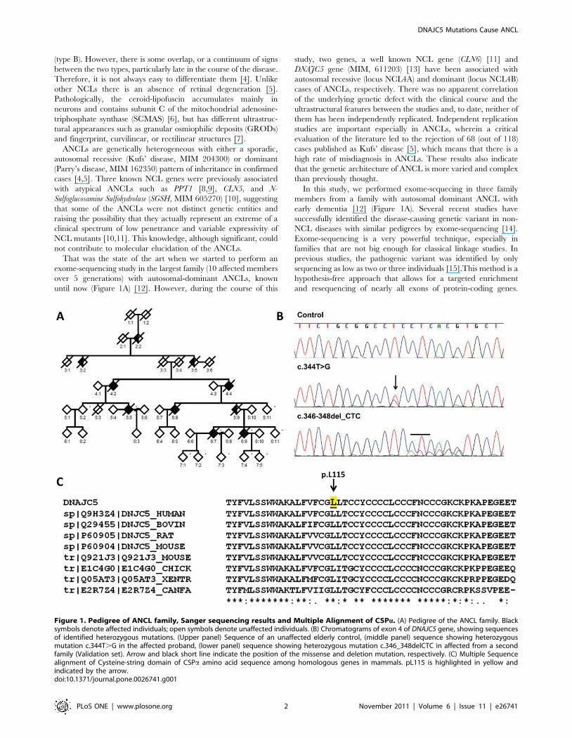
Table 2. Summary of exome sequencing results from ANCL.
Chr Position (bp) Ref Base Variant Gene AA Subs. Type of Change Polyphen2 SeggPat
1 16,915,434 T C NBPF1 K135E missense Benign No
2 130,899,804 T C CCDC74B H149R missense Benign No
3 64,072,900 * 2AA LOC100287879 None frameshift Not predicted No
3 33,883,492 G A PDCD6IP G429S missense Probably damaging Yes
3 75,788,010 A C ZNF717 V255G missense Benign No
4 155,191,162 G A DCHS2 T1701I missense Possibly damaging No
4 164,247,234 C G NPY1R G158A missense Possibly damaging No
4 148,653,570 A G ARHGAP10 I40V missense Benign No
4 162,421,268 A G FSTL5 M452T missense Benign No
5 180,376,974 T G BTNL8 H311Q missense Probably damaging No
6 132,030,966 G C ENPP3 L398V missense Unknown No
7 100,550,039 * +CTC MUC3A None coding Not predicted No
10 90,356,030 * 2TTTTA LIPJ None splice-3 Not predicted Yes
13 19,042,781 T C LOC729501 T434C missense Unknown No
13 115,012,439 A G CDC16 M311V missense Benign No
13 29,287,552 C T SLC46A3 V109I missense Benign No
15 43,038,399 C T TTBK2 R1110H missense Possibly damaging No
16 16,218,686 G A ABCC1 V1211I missense Possibly damaging No
16 25,123,254 C G LCMT1 S17T missense Probably damaging No
16 24,804,954 A T TNRC6A Q1112H missense Probably damaging No
20 62,562,226 T G DNAJC5 L115R missense Possibly damaging Yes
X 3,747,372 C T LOC389906 W263* nonsense Unknown No
Chr: chromosome; Position (bp) is according to Human Genome build 37 (hg37); Reference base: reference allele according to hg37; Variant: nucleotide found by exomesequencing; Gene: official Symbol provide by HGNC; AA Substitution: amino acid change resulting from the observed variant; Type of change: predicted change in thegene sequence; Protein prediction: based on Polyphen analyses of the predicted effect of the substitution on protein function; Segregation pattern: present in allaffected tested.doi:10.1371/journal.pone.0026741.t002
DNAJC5 Mutations Cause ANCL
PLoS ONE | www.plosone.org 4 November 2011 | Volume 6 | Issue 11 | e26741
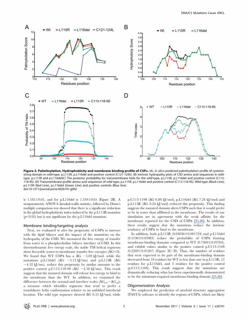
is 1.18160.65, and for p.L116del is 1.55960.65 (Figure 2B). A
nonparametric ANOVA (kruskal-wallis statistic), followed by Dunn’s
multiple comparison test showed that there is a significant reduction
in the global hydrophobicity index induced by the p.L115R mutation
(p,0.05) but is not significant for the p.L116del mutation.
Membrane binding/targeting analysisNext, we evaluated in silico the propensity of CSPa to interact
with the lipid bilayer and the impact of the mutations on the
hydropathy of the CSD. We measured the free energy of transfer
from water to a phosphocholine bilayer interface of CSD. In this
thermodynamic free energy scale, the stable TM helical segments
show favorable water-to-membrane transfer free energies (DG,0).
We found that WT CSPa has a DG 25.69 kJ/mol. while the
mutations p.L116del (DG 25.13 kJ/mo) and p.L115R (DG
24.32 kJ/mo), reduce this propensity by similar amounts to the
positive control p.C113-118-9S (DG 24.58 kJ/mo). This result
suggests that the mutated domain will release less energy to bind to
the membrane than the WT. In addition, we examined the
difference between the octonal and interface scales (DGwoc2DGwif);
a measure which identifies segments that tend to prefer a
transbilayer helix conformation relative to an unfolded interfacial
location. The wild type sequence showed DG 6.55 kJ/mol, while
p.C113-119S (DG 6.88 kJ/mol), p.L116del (DG 7.24 kJ/mol) and
p.L115R (DG 8.24 kJ/mol) reduced this propensity. This finding
suggests the mutated domain alters CSPa such that it would prefer
to be in water than affiliated to the membrane. The results of our
simulation are in agreement with the weak affinity for the
membrane reported for the CSD of CSPa [25,26]. In addition,
these results suggest that the mutations reduce the intrinsic
tendency of CSPa to bind to the membrane.
In addition, both p.L115R (0.0303660.001724) and p.L116del
(0.519060.05983) reduce the probability of CSPa forming
membrane-binding domains compared to WT (0.736960.03764),
and exhibit values similar to the positive control p.C113-116S
(0.226960.01267) (Figure 2C–D). Thus, the number of residues
that were expected to be part of the membrane-binding domain
decreased from 18 residues for WT to less than one in p.L115R, 12
residues for p.L116del, and 5 residues for the positive control
(p.C113-116S). This result suggests that the mutations are
dramatically reducing what has been experimentally demonstrated
to be the minimum-required membrane-binding domain [25,26].
Oligomerization AnalysisWe employed the prediction of amyloid structure aggregation
(PASTA) software to identify the regions of CSPa, which are likely
Figure 2. Palmitoylation, Hydrophobicity and membrane binding profile of CSPa. (A). In silico predicted palmitoylation profile of cysteine-string domain in wild-type, p.L115R, p.L116del and positive control (C121-124S). (B) Intrinsic hydropathy plots of CSD amino acid sequences in wild-type, p.L115R and p.L116del(C) The posterior probability for transmembrane helix for the wild-type, p.L115R, p.L116del and positive control (C113-118-9S). (D) Transmembrane profile amino acid sequences of wild-type, p.L115R, p.L116del and positive control (C113-118-9S). Wild-type (Black Line),p.L115R (Red Line), p.L116del (Green Line) and positive controls (Blue line).doi:10.1371/journal.pone.0026741.g002
DNAJC5 Mutations Cause ANCL
PLoS ONE | www.plosone.org 5 November 2011 | Volume 6 | Issue 11 | e26741
to stabilize the CSPa-CSPa dimers into aggregates [27]. We found
that the region between p.F110-P138 residues has a strong
propensity to assemble into antiparalell ß-sheets (See: Figure S1).
The same core region for oligomerization identified experimen-
tally [24,28]. On average this segment has a pairing energy score
of 225 in WT, 225.4 in p.L115R, and 225.3 in p.L116del
mutations supporting the antiparalell assembly. In addition, we
found that on average the aggregation propensity [h(k)] of WT
CSD is around 0.04 as well as p.L115R (0.04) and p.L116del
(0.04). The positive control (Ab-40) has an aggregation propensity
around 0.05 (See: Figure S1) [27].
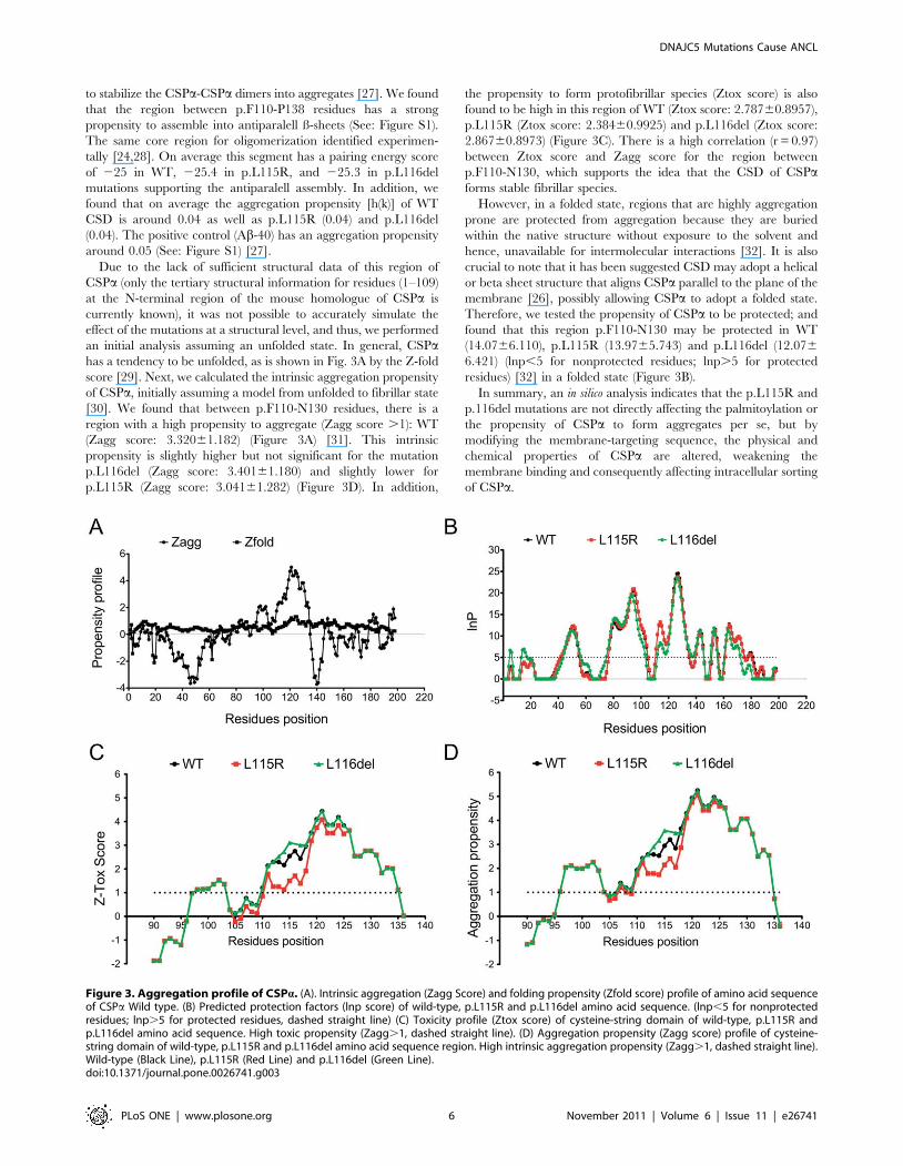
Due to the lack of sufficient structural data of this region of
CSPa (only the tertiary structural information for residues (1–109)
at the N-terminal region of the mouse homologue of CSPa is
currently known), it was not possible to accurately simulate the
effect of the mutations at a structural level, and thus, we performed
an initial analysis assuming an unfolded state. In general, CSPahas a tendency to be unfolded, as is shown in Fig. 3A by the Z-fold
score [29]. Next, we calculated the intrinsic aggregation propensity
of CSPa, initially assuming a model from unfolded to fibrillar state
[30]. We found that between p.F110-N130 residues, there is a
region with a high propensity to aggregate (Zagg score .1): WT
(Zagg score: 3.32061.182) (Figure 3A) [31]. This intrinsic
propensity is slightly higher but not significant for the mutation
p.L116del (Zagg score: 3.40161.180) and slightly lower for
p.L115R (Zagg score: 3.04161.282) (Figure 3D). In addition,
the propensity to form protofibrillar species (Ztox score) is also
found to be high in this region of WT (Ztox score: 2.78760.8957),
p.L115R (Ztox score: 2.38460.9925) and p.L116del (Ztox score:
2.86760.8973) (Figure 3C). There is a high correlation (r = 0.97)
between Ztox score and Zagg score for the region between
p.F110-N130, which supports the idea that the CSD of CSPaforms stable fibrillar species.
However, in a folded state, regions that are highly aggregation
prone are protected from aggregation because they are buried
within the native structure without exposure to the solvent and
hence, unavailable for intermolecular interactions [32]. It is also
crucial to note that it has been suggested CSD may adopt a helical
or beta sheet structure that aligns CSPa parallel to the plane of the
membrane [26], possibly allowing CSPa to adopt a folded state.
Therefore, we tested the propensity of CSPa to be protected; and
found that this region p.F110-N130 may be protected in WT
(14.0766.110), p.L115R (13.9765.743) and p.L116del (12.076
6.421) (lnp,5 for nonprotected residues; lnp.5 for protected
residues) [32] in a folded state (Figure 3B).
In summary, an in silico analysis indicates that the p.L115R and
p.116del mutations are not directly affecting the palmitoylation or
the propensity of CSPa to form aggregates per se, but by
modifying the membrane-targeting sequence, the physical and
chemical properties of CSPa are altered, weakening the
membrane binding and consequently affecting intracellular sorting
of CSPa.
Figure 3. Aggregation profile of CSPa. (A). Intrinsic aggregation (Zagg Score) and folding propensity (Zfold score) profile of amino acid sequenceof CSPa Wild type. (B) Predicted protection factors (lnp score) of wild-type, p.L115R and p.L116del amino acid sequence. (lnp,5 for nonprotectedresidues; lnp.5 for protected residues, dashed straight line) (C) Toxicity profile (Ztox score) of cysteine-string domain of wild-type, p.L115R andp.L116del amino acid sequence. High toxic propensity (Zagg.1, dashed straight line). (D) Aggregation propensity (Zagg score) profile of cysteine-string domain of wild-type, p.L115R and p.L116del amino acid sequence region. High intrinsic aggregation propensity (Zagg.1, dashed straight line).Wild-type (Black Line), p.L115R (Red Line) and p.L116del (Green Line).doi:10.1371/journal.pone.0026741.g003
DNAJC5 Mutations Cause ANCL
PLoS ONE | www.plosone.org 6 November 2011 | Volume 6 | Issue 11 | e26741
There are potential errors and biases in the assumptions in silico
analysis takes into account, therefore these results should be
interpreted cautiously. We used as proof of principle, controls with
the strongest experimental support available to validate our
findings. Nevertheless, further experimental studies are needed
to understand the pathogenicity of these mutations.
Discussion
We have confirmed that mutations in DNAJC5 cause autosomal
dominant ANCL [13]. However, ANCLs are disorders clinically
and genetically very heterogeneous [4]. The subsequent difficulty
in performing an accurate diagnosis had contributed as a limiting
factor in the identification of its genetic cause [5]. However, to
date, two different groups have been able to concurrently and
independently identify the same DNAJC5 gene and same
mutations using different and complementary approaches, which
consolidate and validate the results [13](shown here). However,
mutations in DNAJC5 are currently explaining approximately 25%
of the autosomal dominant ANCLs [13] Therefore, It is possible
that other forms of ANCLs may have another genetic cause.
This is the first replication study of the identification of
mutations in the DNAJC5 gene in ANCLs. By performing
whole-exome sequencing in a multigenerational family with
autosomal dominant ANCL (Figure 1A), we have identified a
novel single-nucleotide variation (c.344T.G) in DNAJC5. In
addition, using Sanger sequencing we found an in-frame single
codon deletion (c.346_348 delCTC) in an independent family
(Figure 1B). Thus, these variants fit genetic criteria for disease-
causing mutations: they are present in unrelated families
(Figure 1B); they exhibit perfect segregation with disease status
(Table 2), they are not present in any healthy controls tested, they
are located in evolutionarily highly conserved residues (Figure 1C),
and they are predicted to functionally affect the encoded protein
(CSPa) (Table 2).
Whole exome sequencing has proved useful to identify the
pathogenic variant in monogenic disease [14,15]. It is a rapid
and cost-effective method that only requires sequencing in a
small number of individuals. To date, different approaches to
whole exome sequencing have been used including various
designs, filtering methodologies and analytical strategies [15].
Some examples are sequencing several affected individuals
from different families, sequencing two affected individuals
from the same family, and combining whole-exome sequencing
data with linkage data, among others [13,14,15]. Our design
included two distantly-related affected individuals and a
healthy sibling of one of the affected (and first cousin of the
other affected individual) (Figure 1A). By selecting the two most
distantly-related affected individuals available, the number of
non pathogenic variants due to relatedness is dramatically
reduced (Table 1).
In fact, 674638 novel non-synonymous variants were found in
each sample, but only 95 were shared by the two affected
individuals. Subtracting the variants in common between the
control and affected individuals, 25 SNSs were remaining
(Table 1). Taking into account that on average 50% of genetic
variants are shared between siblings and 12.5% between first
cousins, we expected approximately 40 SNSs to remain after
filtering against the control. To control for the false positive
variants due to technical errors associated with Next-Generation
Sequencing Platforms, all the three samples were run in the same
flow cell. Thus, it is likely that the number of shared variants by
both affected individuals was initially overestimated, and these
artifacts were then filtered out by comparing with the control.
As done in other studies [15], additional affected family
members were genotyped to identify the variants that are present
in all of the affected individuals. A major challenge of whole-
exome sequencing is to uniquely identify the causative variant.
Most of the current approaches are based on first, removing
candidate variants (non-synonymous, non-sense and splice-site
variants) that are present in public databases (1000 genomes
project and dbSNP), and second, on selecting only the variants
present in the affected individuals. As a result, we found three
unique variants located in the PDCD6IP, LIPJ and DNAJC5 genes,
which were present in all the affected family members but not in
public databases. Interestingly, these three genes also exhibited the
highest values based upon their GERP scores (Table 2). In order to
elucidate the real cause of ANCL in this family, a large series of
population controls were screened to verify if these variants in the
PDCD6IP and LIPJ genes were present in healthy individuals.
Only 16 and 8 heterozygous carriers for the variants on the
PDCD6IP and LIPJ genes were found, respectively. It is important
to note that these variants were not present in the 1000 Genomes
Project or in the dbSNP Database at the time of the analysis.
Similar to others, [14,15] this study clearly shows that pathogenic
or causative variants can be identified by performing exome-
sequencing in a small number of family members. It is now clear
that by selecting two affected family members distant in the
pedigree and one unaffected sibling of one of the affected
individuals, and by running the three samples simultaneously,
the number of potential candidate variants is significantly
decreased. Our study also shows that although the public
databases are very useful in removing commonly found variants
(Table 1), they are still not comprehensive enough to eliminate all
possible rare variants and unequivocally identify the causative
mutation. Most importantly, screening a large number of non-
affected individuals is still necessary.
DNAJC5 gene encodes CSPa, which is a key element of the
synaptic molecular machinery and accounts for 1% of all vesicle
proteins [33,34], as well as part of the general exocytotic
machinery [35]. The synaptic vesicle localization and chaperone
activity of CSPa suggests that it may function in rescuing synaptic
proteins that have been unfolded by activity-dependent stress [36].
Deletion of CSPa in flies and mice results in neurodegeneration
and impairs synaptic function [36,37,38,39]. CSPa has mostly
been found associated with vesicles; however, it has a weak
membrane affinity. Furthermore, there is an inverse correlation
between membrane targeting of CSPa palmitoylation and
adequate intracellular trafficking [25,26]. Site-directed mutations
that enhance membrane association such as p.C121-124L prevent
adequate palmitoylation and lead to accumulation of CSPa in the
ER and Golgi apparatus [25,26]. In contrast, residue changes such
as p.C113-119S (p.L115R, p.L116del, shown here in Figure 2C–
D) reduce binding to the membrane resulting in inadequate
palmitoylation. They exhibit a localized punctuated pattern
throughout the cytoplasm [25,26], co-localize with markers of
ER–Golgi intermediate complex (Golgi SNARE proteins), and
show a significant reduction in synaptic regions [13].
A potential neuronal-specific effect of this disproportionate and
persistent CSPa missorting is a depletion of CSPa [13] and
possibly some of its SNARE partners the synapse, leading to a
disruption in neurotransmission and synaptic dysfunction as
displayed by the CSPa null animal models (mimicking loss of
function) [36,37,38,39].
CSPa self-associates, forming oligomers [24,28,40]. The pG83–
C136 residues constitute the core region for CSPa oligomerization
[24]. Our analysis revealed that a more localized region between
residues pF110-P138 has a tendency to form antiparallel ß-sheets
DNAJC5 Mutations Cause ANCL
PLoS ONE | www.plosone.org 7 November 2011 | Volume 6 | Issue 11 | e26741
species (See: Figure S1), consistent with reports of the CSPa-CSPadimers that are stable to temperature- and SDS-resistant particle
[28,40]. We did not find any significant increase in the intrinsic
tendency of the mutations to aggregate (Figure 3C–D). However,
the unattached form of the protein can induce conformational
changes that facilitate a more reactive oligomeric state (Figure 3A,
D). The effective increase in concentration of soluble CSPaproduced by these mutations can also increase the level of
macromolecular crowding, which in turn may dramatically
enhance the own propensity of CSPa to aggregate, as has been
shown for a-synuclein [41].
In a macromolecular crowded environment, the equilibrium
between protein folding and protein-protein interactions is driven
towards the lower volume (folded to unfolded) species [42]. CSPahas been shown to have a high affinity for unfolded proteins [23].
In a crowded environment, this can increase the likelihood of
CSPa to interact with itself (more stable CSPa-CSPa dimers can
be generated) and with other amyloidogenic partners such as a-
synuclein [38,43]. Recently, it has been shown that CSPa-CSPadimers appear to be the main form of CSPa found in the brains of
carriers of the ANCLs mutation [13].
Another potential target of abnormal interactions of CSPa are
the synaptic proteins, especially those which are central to synaptic
vesicles exocytosis, including proteins from the v- and t-SNAREs
complex, and the putative Ca2+ sensor synaptotagmin 1, which
undergoes palmitoylation in the golgi [44]. Abnormal CSPa-CSPadimers may impede appropriate synaptic vesicle targeting and
subsequently, disrupt neurotrasmission. Indeed, recently it was
shown that increasing the CSPa dimerization inhibits synaptic
transmission [28].
Synaptic dysfunction has been consistently reported in several
human and animal models of NCLs [45]. Several NCL-encoded
proteins have been found in synaptic compartments [46,47,48].
Furthermore, signs of synaptic dysfunction (reduction in synaptic
vesicle number) and degeneration have been demonstrated in
PPT1 deficient neurons in vitro [49], and synaptic pathology
(redistribution of SNARE complex and aggregates of Syp/
SNAP25) occurs early on in disease progression in the congenital
form of NCL [50]. Synaptic involvement in two different mouse
models of INCL was also recently demonstrated [45,51]. The
PPT1 null animal model displays alterations in the endocyclic\re-
cycling pathway of synaptic vesicles associated with the impair-
ment of depalmitoylation of the SNARE proteins [45], unlike
CSPa null models that do not exhibit any primary defects in the
endocytosis process or vesicle recycling [52]. To date, the
mechanisms causing synaptic vulnerability in NCLs remains
poorly understood.
Our genetic results confirm DNAJC5 is the disease-causing gene
of some ANCLs with autosomal dominant inheritance [13]. The in
silico analysis suggests reduced the membrane binding and
subsequent missorting of CSPa may play a crucial role in the
pathogenicity of these mutations. This mislocalization can by itself
affect the palmitoylation status and the propensity to aggregate.
Thus, a dominant-negative mechanism resulting from CSPapropensity to self aggregate may be involved in the pathogenicity.
The mutated CSPa may aggregate with the wild type, induces
mislocalization and subsequent reduction of CSPa levels in the
synapse
Since CSPa is a synaptic protein and the null animal models
show a progressive neurodegenerative phenotype, a better
understanding of the cellular and molecular characteristics of
synaptic vulnerability will be important for our understanding of
NCLs pathogenesis and for the effective development of
therapeutic approaches.
Materials and Methods
Patients and Study DesignThe Institutional Review Board (IRB) at the Washington
University School of Medicine in Saint Louis approved the study.
Prior to their participation, a written informed consent was
reviewed and obtained from family members. The Human
Research Protection Office (HRPO) approval number for our
ADRC Genetics Core family studies is 93-0006.
Peripheral blood/brain tissue was collected and genomic DNA
was isolated from several pathologically confirmed cases (4:2, 4:4,
5:5, 6:7 and 6:9) and unaffected samples (5:2, 5:11, 6:1 7:1, 7:2,
7:3, 7:5 and 7:6). The mean age at onset for the disease is
36 yr62.5 (range 32–40). Some of the unaffected samples, such as
5:2 (70 years old), 5:11 (65 years old) and 6:11 (46 years old), are
much older than the oldest known AAO (age = 40). In addition,
there are skin biopsies showing the absence of the disease. These
data indicate that individuals 5.2, 5:11 and 6:11 are very unlikely
to be a mutation carrier.
To identify the gene underlying disease in this family, the
affected individuals 5:5 and 6:7 and the unaffected sibling control
5.2 were selected for exome-sequencing. The affected samples are
distant enough in the family tree that they share about 1/8 of their
genome and since both are affected they should harbor the same
single causative variant. The control is an unaffected sibling of one
affected sample, which in theory should allow us to reduce the
non-pathogenic variants shared by these individuals by ,50%. By
combining the data from these three individuals we were able to
narrow down the number of potential pathogenic variants. We
also used DNA available from another affected (6:9) and two
unaffected samples (5:11, 6:11) to perform an extended segregation
analysis.
Validation Set of patient with ANCLsThere were at least 5 similarly affected individuals in the family
over 3 generations with apparent autosomal dominant inheritance.
The proband had behavioral difficulties that started in the mid
20 s, probably OCD-like. The first generalized seizure occurred in
the early 30 s and was followed by cognitive regression (loss of
speech-language and memory). There were also mobility problems
by early 30 s.
A patient selected from the second family is 70 years old with a
history of tremor, progressive encephalopathy with rectilinear
inclusions only (no FP, CL, GRODS) on skin biopsy. NCL gene
testing was negative. The daughter has early-adult onset tremors
and myoclonic jerks, with rectilinear inclusions in buffy coat. NCL
gene testing was negative
Patient has a late-infantile NCL with onset of progressive
myoclonic epilepsy at age four, visual loss at age five and
curvilinear and fingerprint bodies on nerve/muscle biopsy. This
patient is a carrier of a change c.896C.T/P299L in the CLN6
gene [11].
An individual from the third family (two sisters) had childhood
absence seizures, followed by the onset in her late 20 s of
myoclonus, generalized seizure disorder, and dementia. History of
autosomal dominant transmission and electron-microscopical
studies revealed an accumulation of dense osmophilic material
with a vague internal architecture resembling fingerprint shapes
and occasional curvilinear bodies [53].
Exome sequencingEnrichment of coding exons and flanking intronic regions was
performed using a solution hybrid selection method with the
SureSelectH human all exon 50 Mb kit (Agilent Technologies,
DNAJC5 Mutations Cause ANCL
PLoS ONE | www.plosone.org 8 November 2011 | Volume 6 | Issue 11 | e26741
Santa Clara, California) following the manufacturer’s standard
protocol. This step was performed by the Genome Technology
Access Center at Washington University in St Louis. The captured
DNA was sequenced by paired-end reads on the HiSeq 2000
sequencer (Illumina, San Diego, California). Next, raw sequence
reads were aligned to the reference genome NCBI 36/hg18 by
using Novoalign Version V2.07.00 (Novocraft Technologies,
Selangor, Malaysia). Base/SNP calling was perform by SNP
Samtools Version 0.1.7. SNP annotation was carried out using
version 5.07 of SeattleSeq Annotation server (see URL). [16].
SNP GenotypingTwo different genotyping technologies were used: MassARRAY
SNP (Sequenom, Inc) and KASPar. The principle of the
MassARRAY system is PCR-based, where different size products
are analyzed by SEQUENOM MALDI-TOF mass spectrometry
[17,18,19]. The KBioscience Competitive Allele-Specific PCR
genotyping system (KASP) is FRET-based endpoint-genotyping
technology, v4.0 SNP (KBioscience) [17,18,19]. The proportion of
SNPs with genotype call rates were .98% and the number of
samples that failed to give data for .98% of SNPs was extremely
small.
Sanger SequencingPrimers were designed using web-based Primer 3 (see URL)
(See: Table S2).
Protein-coding exons and 100 base of flanking upstream and
downstream intronic sequence of DNAJC5 (Transcript: ENS-
T00000360864) were amplified on Applied Biosystems (Applied
Biosystems, Carlsbad, California, USA) 96-Well GeneAmpH PCR
System 9700 Thermal Cyclers using a touchdown protocol. All
PCR products to be sequenced were amplified under the same
conditions (25-ml volume containing 106PfuUltraTM HF reaction
buffer (Stratagene, La Jolla, California, USA), 56Betaine (Sigma-
aldrich, St Louis, USA), 100 mmol/l each dNTP, 200 nmol/l each
primer, 0.4 PfuUltraTM High-Fidelity DNA Polymerase (Pro-
mega); PCR profile: 94uC followed by 34 cycles of 45 s at 94uC,
45 s at 62u, and 1 min at 72uC). PCR products purification was
completed with Exosap-IT (USB Corporation). Sequencing was
performed both in forward and reverse direction using BigDyeHTerminator v3.1 Cycle Sequencing Kit (ABI) on an ABI 3130
sequencer. Sequence traces were analyzed using Sequencher (v4.7,
Gene Codes Corp, Ann Arbor, Michigan, USA)
Bioinformatics AnalysisThe URLs used for the in silico analysis:
Online Mendelian Inheritance in man (OMIM), http://www.
omim.org
1000 Genomes, http://www.1000genomes.org/
dbSNP, http://www.ncbi.nlm.nih.gov/projects/SNP/
NCL Mutation Database, http://www.ucl.ac.uk/ncl/
PolyPhen-2, http://genetics.bwh.harvard.edu/pph2/
SIFT, http://sift.jcvi.org/www/SIFT_BLink_submit.html
SeattleSeq Annotation server, http://gvs.gs.washington.edu/
SeattleSeqAnnotation
Primer 3, http://frodo.wi.mit.edu primer3/input.htm
ClustalW2 - Multiple Sequence Alignment,
To analyze the degree of conservation of the predicted amino
acid substitutions, multiple sequence alignment was performed
using ClustalW [54]. The human (Homo sapiens) CSPa protein
sequence (Q9H3Z4) was compared to Mus musculus, Bos taurus,
Rattus norvegicus, Gallus gallus, Xenopus (Silurana) tropicalis and Canis
familiaris.
http://www.ebi.ac.uk/Tools/msa/clustalw2/
Disease-network analysis of NCLs genesIt has been shown that genes that are associated with
phenotypically close disorders are prone to have similar molecular
signatures, especially in inherited disease [21]. Here, we have used
a disease-network analysis approach as supporting in silico evidence
of the role of the candidate genes we identified by exome
sequencing. We found that the analysis is robust across different
algorithms and random subsets of training NCL disease genes.
We have used Endeavour [55], which was trained with all
possible features except BLAST. Endeavour has been recently
benchmarked using 450 pathway maps and 826 disease marker
sets, containing a total of 9911 and 12,432 genes, respectively. It
was reported that the area under the receiver operating
characteristic curves is 0.97 for pathway and of 0.91 for disease
gene sets [56].
Endeavour, http://homes.esat.kuleuven.be/;bioiuser/endeavour/
tool/endeavourweb.php
ToppGene [57] was used with the default training parameters.
Both softwares were trained using the causal genes of other NCLs
(NCL Mutation Database, URL) along with genes that are
associated with phenotypically close disorders (i. e. differential
diagnosis of ANCLs). They were tested against the variants that we
have identified by exome sequencing. As a control group we used
the entire genome as a training group (See: Table S1).
ToppGene, http://toppgene.cchmc.org/prioritization.jsp
In silico Palmitoylation analysisWe used as positive controls, artificially induced mutations that
have experimentally been proven to reduce the palmitoylation
status of CSPa such as p.C113-119S, p.C121-124S, p.C121-124A
and p.K137A [25,26].
We used the Clustering and Scoring Strategy for Palmitoylation
Sites Prediction (CSS-Palm) system. It has been shown that the
program’s prediction performance has a highly positive Jack-Knife
validation results (sensitivity 82.16% and specificity 83.17% for
cut-off score 2.6) [58].
CSS-Palm 3.0 Online Server, http://csspalm.biocuckoo.org/
online3.php
Analysis of the impact of mutations on the hydropathy ofCSPa
Protscale, http://web.expasy.org/protscale/
The hydrophobicity profile was calculated using the scale of
Kyte and Doolittle [59].
The TMHMM program was used to predict the transmem-
brane domain in the entire sequence of CSPa (accession number
Q9H3Z4). We included the A108-139K residues in the analysis
because it has been proven experimentally that this region
weakens the membrane affinity of CSPa [25,26].
TMHMM has shown that it can correctly predict 97–98% of
the transmembrane helices. Additionally, it can discriminate
between soluble and membrane proteins with both a specificity
and sensitivity better than 99% [60].
TMHMM Server v. 2.0, Prediction of transmembrane helices in
proteins
http://www.cbs.dtu.dk/services/TMHMM/
Using MPEx, we measured the free energy of transfer from
water to a phosphocholine bilayer interface of this domain, DG
28.12 for p.KPK137-39del and DG 26.97 for p.C121-24L. We
also calculated the values of the octanol/interface for these
changes; this measure identifies segments that tend to prefer a
transbilayer helix conformation relative to an unfolded interfacial
location. We found a DG of 2.91 kJ/mol for p.C121-124L, DG
DNAJC5 Mutations Cause ANCL
PLoS ONE | www.plosone.org 9 November 2011 | Volume 6 | Issue 11 | e26741
/
3.24 kJ/mol for p.KPK137-139del, and DG 6.55 kJ/mol for wild
type. These two changes in residues increased the membrane
affinity of CSPa, in agreement with the experimental findings
[25,26]. Besides the enrichment of hydrophobic residues in the
cysteine-string domain, the b-barrel analysis did not find any
membrane-spanning strands/regions [61].
The TMX approach uses an experiment-based whole-residue
hydropathy scale (WW scale), which includes the backbone
constraint, to identify TM helices of membrane proteins with an
accuracy greater than 99% [61,62].
Membrane Protein Explorer (MPEx), MPEx 3.2 server
http://blanco.biomol.uci.edu/mpex/
The SPLIT accuracy is 99% for predicting 178 transmembrane
helices in all membrane proteins or subunits of known 3D
structure [63].
Split 4.0, Membrane Protein Secondary Structure Prediction
Server,
http://split.pmfst.hr/split/4/
Analysis of conformational changes induced by themutations
There is only tertiary structural information for the 1–109
residue N-terminal region of mouse homologous CSPa (pdb entry
2CTW; Riken Structural Genomics Initiative), therefore, it was
not possible to simulate the effect of the mutations at the structural
level.
Prediction of amyloid structure aggregation(PASTA)bench-
marked on the dataset of 179 peptides derived from the literature
revealed close to an 80% true positive prediction with a ,20%
false positive rate at a PASTA energy threshold of 24.0 [27,64].
PASTA, http://protein.cribi.unipd.it/pasta/.
Zyggregator method is based on three physico-chemical
properties of the polypeptide chain, hydrophobicity, charge, and
the propensity to adopt a-helical or b-sheet structures. This
method reproduces to a remarkable extent (r = 0.85) the changes
in the aggregation rates observed experimentally for single amino
acid substitutions [31].
Ztox score uses the same equation as in Zagg but the difference
being that the parameters are fitted on a database of polypeptide
chains whose aggregation resulted in protofibrillar species, rather
than amyloid fibrils [65]. The propensities predicted by Ztox to
form protofibrillar aggregates correlate very strongly with their in
vivo effects (Stox, r = 0.83) [31].
Zyggregator: Prediction of Protein Aggregation Propensities,
http://www-vendruscolo.ch.cam.ac.uk/zyggregator.php
The folding propensity profile (Z-fold) is defined in terms of the
physicochemical properties of the amino acids such as hydropho-
bicity, secondary-structure propensity, and electrostatic charge
[29].
Sequence-Based Prediction of Folding Rates,
http://www-vendruscolo.ch.cam.ac.uk/camfold.php
The CamP method predicts if regions of a structure are
protected from hydrogen exchange with an accuracy in the range
80%–100%; The region is protected if lnp.5 for all of its amide
groups [32]
Sequence-Based Prediction of Protein Flexibility in Native
States
http://www-vendruscolo.ch.cam.ac.uk/camp.new.php
Supporting Information
Figure S1 Aggregation profile of CSPa. A. Aggregation
profile of WT CSPa. B. Aggregation profile of p.L115R mutation.
C. Aggregation profile of p.L116del mutation D. Aggregation
profile of Ab40 (it is used here as a positive control). This is the
output file from PASTA server (see Methods)
(TIF)
Table S1 Summary of comparison of results fromToppGene and Endeavour gene prioritization analysis.
(DOC)
Table S2 Set of primers used for Sanger Sequencing.
(DOC)
Acknowledgments
The authors especially want to thank the patients and their families, whose
help and participation made this work possible. We thank Dr. Katherine B
Sims who provided some of the samples used in this study.
Author Contributions
Conceived and designed the experiments: BAB AG CC. Performed the
experiments: BAB KM SC CC. Analyzed the data: BAB DA YC CC.
Contributed reagents/materials/analysis tools: JN MS JCM. Wrote the
paper: BAB AG CC.
References
1. Rider JA, Rider DL (1988) Batten disease: past, present, and future. Am J Med
Genet Suppl 5: 21–26.
2. Haltia M (2003) The neuronal ceroid-lipofuscinoses. J Neuropathol Exp Neurol
62: 1–13.
3. Hawkins-Salsbury JA, Reddy AS, Sands MS (2011) Combination therapies forlysosomal storage disease: is the whole greater than the sum of its parts? Hum
Mol Genet 20: R54–60.
4. Sadzot B, Reznik M, Arrese-Estrada JE, Franck G (2000) Familial Kufs’ disease
presenting as a progressive myoclonic epilepsy. J Neurol 247: 447–454.
5. Berkovic SF, Carpenter S, Andermann F, Andermann E, Wolfe LS (1988) Kufs’disease: a critical reappraisal. Brain 111(Pt 1): 27–62.
6. Palmer DN, Fearnley IM, Walker JE, Hall NA, Lake BD, et al. (1992)
Mitochondrial ATP synthase subunit c storage in the ceroid-lipofuscinoses(Batten disease). Am J Med Genet 42: 561–567.
7. Mole SE, Williams RE, Goebel HH (2005) Correlations between genotype,
ultrastructural morphology and clinical phenotype in the neuronal ceroidlipofuscinoses. Neurogenetics 6: 107–126.
8. van Diggelen OP, Thobois S, Tilikete C, Zabot MT, Keulemans JL, et al. (2001)Adult neuronal ceroid lipofuscinosis with palmitoyl-protein thioesterase
deficiency: first adult-onset patients of a childhood disease. Ann Neurol 50:
269–272.
9. Ramadan H, Al-Din AS, Ismail A, Balen F, Varma A, et al. (2007) Adult
neuronal ceroid lipofuscinosis caused by deficiency in palmitoyl protein
thioesterase 1. Neurology 68: 387–388.
10. Sleat DE, Ding L, Wang S, Zhao C, Wang Y, et al. (2009) Mass spectrometry-
based protein profiling to determine the cause of lysosomal storage diseases ofunknown etiology. Mol Cell Proteomics 8: 1708–1718.
11. Arsov T, Smith KR, Damiano J, Franceschetti S, Canafoglia L, et al. (2011)Kufs disease, the major adult form of neuronal ceroid lipofuscinosis, caused by
mutations in CLN6. Am J Hum Genet 88: 566–573.
12. Josephson SA, Schmidt RE, Millsap P, McManus DQ, Morris JC (2001)
Autosomal dominant Kufs’ disease: a cause of early onset dementia. J Neurol Sci188: 51–60.
13. Noskova L, Stranecky V, Hartmannova H, Pristoupilova A, Baresova V, et al.(2011) Mutations in DNAJC5, Encoding Cysteine-String Protein Alpha, Cause
Autosomal-Dominant Adult-Onset Neuronal Ceroid Lipofuscinosis. Am J HumGenet 89: 241–252.
14. Ng SB, Nickerson DA, Bamshad MJ, Shendure J (2010) Massively parallelsequencing and rare disease. Hum Mol Genet 19: R119–124.
15. Ku CS, Naidoo N, Pawitan Y (2011) Revisiting Mendelian disorders throughexome sequencing. Hum Genet 129: 351–370.
16. Alvarado DM, Buchan JG, Gurnett CA, Dobbs MB (2011) Exome sequencing
identifies an MYH3 mutation in a family with distal arthrogryposis type 1. J Bone
Joint Surg Am 93: 1045–1050.
17. Cruchaga C, Graff C, Chiang HH, Wang J, Hinrichs AL, et al. (2011)Association of TMEM106B gene polymorphism with age at onset in granulin
mutation carriers and plasma granulin protein levels. Arch Neurol 68:
581–586.
DNAJC5 Mutations Cause ANCL
PLoS ONE | www.plosone.org 10 November 2011 | Volume 6 | Issue 11 | e26741
18. Cruchaga C, Kauwe JS, Mayo K, Spiegel N, Bertelsen S, et al. (2010) SNPs
associated with cerebrospinal fluid phospho-tau levels influence rate of decline in
Alzheimer’s disease. PLoS Genet 6.
19. Cruchaga C, Nowotny P, Kauwe JS, Ridge PG, Mayo K, et al. (2011)
Association and Expression Analyses With Single-Nucleotide Polymorphisms in
TOMM40 in Alzheimer Disease. Arch Neurol 68: 1013–1019.
20. Feldman I, Rzhetsky A, Vitkup D (2008) Network properties of genes harboring
inherited disease mutations. Proc Natl Acad Sci U S A 105: 4323–4328.
21. Goh KI, Cusick ME, Valle D, Childs B, Vidal M, et al. (2007) The human
disease network. Proc Natl Acad Sci U S A 104: 8685–8690.
22. Desmet FO, Hamroun D, Lalande M, Collod-Beroud G, Claustres M, et al.
(2009) Human Splicing Finder: an online bioinformatics tool to predict splicing
signals. Nucleic Acids Res 37: e67.
23. Chamberlain LH, Burgoyne RD (2000) Cysteine-string protein: the chaperone
at the synapse. J Neurochem 74: 1781–1789.
24. Swayne LA, Blattler C, Kay JG, Braun JE (2003) Oligomerization characteristics
of cysteine string protein. Biochem Biophys Res Commun 300: 921–926.
25. Greaves J, Chamberlain LH (2006) Dual role of the cysteine-string domain in
membrane binding and palmitoylation-dependent sorting of the molecular
chaperone cysteine-string protein. Mol Biol Cell 17: 4748–4759.
26. Greaves J, Salaun C, Fukata Y, Fukata M, Chamberlain LH (2008)
Palmitoylation and membrane interactions of the neuroprotective chaperone
cysteine-string protein. J Biol Chem 283: 25014–25026.
27. Trovato A, Chiti F, Maritan A, Seno F (2006) Insight into the structure of
amyloid fibrils from the analysis of globular proteins. PLoS Comput Biol 2: e170.
28. Xu F, Proft J, Gibbs S, Winkfein B, Johnson JN, et al. (2010) Quercetin targets
cysteine string protein (CSPalpha) and impairs synaptic transmission. PLoS One
5: e11045.
29. Tartaglia GG, Vendruscolo M (2010) Proteome-level interplay between folding
and aggregation propensities of proteins. J Mol Biol 402: 919–928.
30. Tartaglia GG, Cavalli A, Pellarin R, Caflisch A (2005) Prediction of aggregation
rate and aggregation-prone segments in polypeptide sequences. Protein Sci 14:
2723–2734.
31. Tartaglia GG, Vendruscolo M (2008) The Zyggregator method for predicting
protein aggregation propensities. Chem Soc Rev 37: 1395–1401.
32. Tartaglia GG, Cavalli A, Vendruscolo M (2007) Prediction of local structural
stabilities of proteins from their amino acid sequences. Structure 15: 139–143.
33. Mastrogiacomo A, Kohan SA, Whitelegge JP, Gundersen CB (1998) Intrinsic
membrane association of Drosophila cysteine string proteins. FEBS Lett 436:
85–91.
34. van de Goor J, Ramaswami M, Kelly R (1995) Redistribution of synaptic vesicles
and their proteins in temperature-sensitive shibire(ts1) mutant Drosophila. Proc
Natl Acad Sci U S A 92: 5739–5743.
35. Blondeau F, Ritter B, Allaire PD, Wasiak S, Girard M, et al. (2004) Tandem MS
analysis of brain clathrin-coated vesicles reveals their critical involvement in
synaptic vesicle recycling. Proc Natl Acad Sci U S A 101: 3833–3838.
36. Fernandez-Chacon R, Wolfel M, Nishimune H, Tabares L, Schmitz F, et al.
(2004) The synaptic vesicle protein CSP alpha prevents presynaptic degener-
ation. Neuron 42: 237–251.
37. Zinsmaier KE, Eberle KK, Buchner E, Walter N, Benzer S (1994) Paralysis and
early death in cysteine string protein mutants of Drosophila. Science 263:
977–980.
38. Chandra S, Gallardo G, Fernandez-Chacon R, Schluter OM, Sudhof TC (2005)
Alpha-synuclein cooperates with CSPalpha in preventing neurodegeneration.
Cell 123: 383–396.
39. Schmitz F, Tabares L, Khimich D, Strenzke N, de la Villa-Polo P, et al. (2006)
CSPalpha-deficiency causes massive and rapid photoreceptor degeneration.
Proc Natl Acad Sci U S A 103: 2926–2931.
40. Gibbs SJ, Barren B, Beck KE, Proft J, Zhao X, et al. (2009) Hsp40 couples with
the CSPalpha chaperone complex upon induction of the heat shock response.
PLoS One 4: e4595.
41. Shtilerman MD, Ding TT, Lansbury PT, Jr. (2002) Molecular crowding
accelerates fibrillization of alpha-synuclein: could an increase in the cytoplasmic
protein concentration induce Parkinson’s disease? Biochemistry 41: 3855–3860.
42. Zhou HX, Rivas G, Minton AP (2008) Macromolecular crowding and
confinement: biochemical, biophysical, and potential physiological consequenc-es. Annu Rev Biophys 37: 375–397.
43. Burre J, Sharma M, Tsetsenis T, Buchman V, Etherton MR, et al. (2010) Alpha-
synuclein promotes SNARE-complex assembly in vivo and in vitro. Science 329:1663–1667.
44. Prescott GR, Gorleku OA, Greaves J, Chamberlain LH (2009) Palmitoylation ofthe synaptic vesicle fusion machinery. J Neurochem 110: 1135–1149.
45. Kim SJ, Zhang Z, Sarkar C, Tsai PC, Lee YC, et al. (2008) Palmitoyl protein
thioesterase-1 deficiency impairs synaptic vesicle recycling at nerve terminals,contributing to neuropathology in humans and mice. J Clin Invest 118:
3075–3086.46. Lehtovirta M, Kyttala A, Eskelinen EL, Hess M, Heinonen O, et al. (2001)
Palmitoyl protein thioesterase (PPT) localizes into synaptosomes and synapticvesicles in neurons: implications for infantile neuronal ceroid lipofuscinosis
(INCL). Hum Mol Genet 10: 69–75.
47. Luiro K, Kopra O, Lehtovirta M, Jalanko A (2001) CLN3 protein is targeted toneuronal synapses but excluded from synaptic vesicles: new clues to Batten
disease. Hum Mol Genet 10: 2123–2131.48. Heinonen O, Kyttala A, Lehmus E, Paunio T, Peltonen L, et al. (2000)
Expression of palmitoyl protein thioesterase in neurons. Mol Genet Metab 69:
123–129.49. Virmani T, Gupta P, Liu X, Kavalali ET, Hofmann SL (2005) Progressively
reduced synaptic vesicle pool size in cultured neurons derived from neuronalceroid lipofuscinosis-1 knockout mice. Neurobiol Dis 20: 314–323.
50. Partanen S, Haapanen A, Kielar C, Pontikis C, Alexander N, et al. (2008)Synaptic changes in the thalamocortical system of cathepsin D-deficient mice: a
model of human congenital neuronal ceroid-lipofuscinosis. J Neuropathol Exp
Neurol 67: 16–29.51. Kielar C, Wishart TM, Palmer A, Dihanich S, Wong AM, et al. (2009)
Molecular correlates of axonal and synaptic pathology in mouse models ofBatten disease. Hum Mol Genet 18: 4066–4080.
52. Ranjan R, Bronk P, Zinsmaier KE (1998) Cysteine string protein is required for
calcium secretion coupling of evoked neurotransmission in drosophila but not forvesicle recycling. J Neurosci 18: 956–964.
53. Sims KB, Cole AJ, Sherman JC, Caruso PA, Snuderl M (2011) Case records ofthe Massachusetts General Hospital. Case 8–2011. A 32-year-old woman with
seizures and cognitive decline. N Engl J Med 364: 1062–1074.54. Larkin MA, Blackshields G, Brown NP, Chenna R, McGettigan PA, et al. (2007)
Clustal W and Clustal X version 2.0. Bioinformatics 23: 2947–2948.
55. Tranchevent LC, Barriot R, Yu S, Van Vooren S, Van Loo P, et al. (2008)ENDEAVOUR update: a web resource for gene prioritization in multiple
species. Nucleic Acids Res 36: W377–384.56. Schuierer S, Tranchevent LC, Dengler U, Moreau Y (2010) Large-scale
benchmark of Endeavour using MetaCore maps. Bioinformatics 26: 1922–1923.
57. Chen J, Bardes EE, Aronow BJ, Jegga AG (2009) ToppGene Suite for gene listenrichment analysis and candidate gene prioritization. Nucleic Acids Res 37:
W305–311.58. Zhou F, Xue Y, Yao X, Xu Y (2006) CSS-Palm: palmitoylation site prediction
with a clustering and scoring strategy (CSS). Bioinformatics 22: 894–896.59. Kyte J, Doolittle RF (1982) A simple method for displaying the hydropathic
character of a protein. J Mol Biol 157: 105–132.
60. Krogh A, Larsson B, von Heijne G, Sonnhammer EL (2001) Predictingtransmembrane protein topology with a hidden Markov model: application to
complete genomes. J Mol Biol 305: 567–580.61. White SH, Ladokhin AS, Jayasinghe S, Hristova K (2001) How membranes
shape protein structure. J Biol Chem 276: 32395–32398.
62. Jayasinghe S, Hristova K, White SH (2001) Energetics, stability, and predictionof transmembrane helices. J Mol Biol 312: 927–934.
63. Juretic D, Zoranic L, Zucic D (2002) Basic charge clusters and predictions ofmembrane protein topology. J Chem Inf Comput Sci 42: 620–632.
64. Trovato A, Seno F, Tosatto SC (2007) The PASTA server for protein
aggregation prediction. Protein Eng Des Sel 20: 521–523.65. Luheshi LM, Tartaglia GG, Brorsson AC, Pawar AP, Watson IE, et al. (2007)
Systematic in vivo analysis of the intrinsic determinants of amyloid Betapathogenicity. PLoS Biol 5: e290.
DNAJC5 Mutations Cause ANCL
PLoS ONE | www.plosone.org 11 November 2011 | Volume 6 | Issue 11 | e26741


![ITAL]Hubble Space Telescope[/ITAL] Proper Motion Confirms the Optical Identification of the Nearby Pulsar PSR 1929+10](https://static.fdokumen.com/doc/165x107/6322a090078ed8e56c0a7f38/italhubble-space-telescopeital-proper-motion-confirms-the-optical-identification.jpg)

