Eroid-lipofuscinosis (Batten disease) - Massey Research Online
171
Copyright is owned by the Author of the thesis. Permission is given for a copy to be downloaded by an individual for the purpose of research and private study only. The thesis may not be reproduced elsewhere without the permission of the Author.
-
Upload
khangminh22 -
Category
Documents
-
view
2 -
download
0
Transcript of Eroid-lipofuscinosis (Batten disease) - Massey Research Online

Copyright is owned by the Author of the thesis. Permission is given for a copy to be downloaded by an individual for the purpose of research and private study only. The thesis may not be reproduced elsewhere without the permission of the Author.

Ceroid-I i pofusci nosis (Batten Disease)
A thesis presented in partial fulfilment of the requirements for the degree of Doctor of Philosophy
at Massey University
Valerie Jean Westlake
1995

Abstract
The ceroid-lipofuscinoses are a group of inherited neurodegenerative diseases occurring in
human beings and animals. Histologically there is generalised accumulation of a fluorescent
lipopigment within the cytoplasm of many cell types and selective necrosis of some
populations of neurons. Clinical signs include loss of vision, seizures and mental retardation
with premature death being the eventual outcome. Systematic analyses of isolated storage
material have previously resulted in the identification of subunit c of mitochondrial ATP
synthase as being a major constituent in the late infantile human, ovine, bovine and three
canine forms of ceroid-lipofuscinosis. Saposins A and D have been identified as being
stored in the infantile human disease.
Brain biopsy has been routinely used to diagnose ceroid-lipofuscinosis in sheep. The
efficacy of this technique was investigated and it was concluded that it was a safe and
reliable method for diagnosis in lambs aged 2 1/2 months. In contrast, diagnosis of the disease
by clinical examination could only be made comparatively late in the disease course at 9-
1 2 months. Repeated neurological examinations enabled "clinical staging" of the progression
of the disease.
Haematopoietic cell transplantation was carried out in foetal lambs withceroid-lipofuscinosis.
Stable engraftment of an average of 9% blood cells was achieved but under the conditions
of the experiment there was no alteration in the disease course or severity of lesions in
transplanted lambs with ceroid-lipofuscinosis as compared to affected lambs without
transplanted cells.
The observation of autofluorescence from storage cytosomes in the ceroid-lipofuscinoses
has lead to the assumption that they contain a fluorophore of critical significance to
expla nations of pathogenenesis. Studies on the nature of the fluorescence from storage
bodies and isolated storage body components allowed the conclusion that no single
significant fluorophore other than protein was present.

Abstract
Antibodies to subunit c of mitochondrial A TP synthase were produced. These and similar
antibodies from other sources were used for immunocytochemistry. The staining pattern
observed varied, depending on the fixation regime, the antibody used and the form of
disease. It was concluded that different epitopes were exposed in different forms of the
ceroid-lipofuscinoses. Positive immunostaining of storage material in muscle and cartilage
from the ear depended on the age of the patient and could assist but not replace current
methods of diagnosis. Storage cytosomes were also labelled using immunogold staining at
the ultrastructural level.
The ceroid-lipofuscinoses are a genetically diverse group of diseases which appear to have
complex biochemical systems underlying them. The hypothesis was developed that the
defect may lie in the disassembly of the Fo complex rather than in proteolysis per se. The
aggregation of subunit c with lipids could result in a complex structure resistant to
catabolism by proteases .

Acknowledgements
I wish to acknowledge and thank my two supervisors Professor R.D. Jolly and Dr. D.N.
Palmer for their advice and support during this study. I sincerely appreciate the opportunity
I have had to work on this research project and am particularly indebted to Professor Jolly
for providing the resources to enable the completion of this thesis.
I am very grateful for the technical assistance I have received from many people at Massey
University and other research institutions. I particularly wish to thank Miss. S.L. Bayliss for
her help with the development and utilisation of antibodies. I also wish to thank Mrs. P.
Davey and Mrs. P. Slack for preparing tissues for light and electron microscopy; Mr. R.
Bennett and Mr. D. Hopcroft of the Keith Williamson Electron Microscopy Unit,
Hort+Research, Palmerston North for printing the electron micrographs and giving advice
on electron microscopy; Mr. T. Watts for the production of thesis photographs; Mr. F.
Sharpe for helping with post mortem examinations; staff from the Sheep and Beef Research
Unit, Massey University for assistance with the sheep flock; staff from the Department of
Veterinary Clinical Sciences for technical assistance with surgery during disease diagnosis
and haematopoietic cell transplantation procedures and staff from the Department of
Chemistry and Biochemistry for the quantitative amino acid analyses.
Finally, I would like to thank Mr. S .R. Grant for his computing expertise and patience during
the production of this manuscript.
Other specific acknowledgements will be found in the text.
This work was supported by the U.S. Public Health grant 5ROl NS l 1238 .

Publ ications
Palmer, D.N., Bayliss, S .L., Clifton, P.A. and Grant (Westlake), V.J. (1993): Storage bodies
in the ceroid-lipofuscinoses (Batten disease) : Low-molecular-weight components,
unusual amino acids and reconstitution of fluorescent bodies from non-fluorescent
components. J. Inher. Metab. Dis. 16:292-295.
Palmer, D.N., Bayliss, S .L. and Westlake, V.J. (1995): Batten disease and the A TP synthase
subunit c turnover pathway: Raising antibodies to subunit c.Am. 1. Med. Genet. 56: (In
press) .
Westlake, V.J . , Jolly, R.D., Bayliss, S .L. and Palmer, D.N. (1995): Immunocytochemical
studies in the ceroid-lipofuscinoses (Batten disease) using antibodies to subunit c of
mitochondrial ATP synthase. Am. 1. Med. Genet. 56:(ln press).
Westlake, V.J. , Jolly, RD., Jones, B.R, Mellor, D.J . , Machon, R, Zanjani, E.D. and Krivit,
W. (1995): Hematopoietic cell transplantation in fetal lambs with ceroid-lipofuscinosis.
Am. J. Med. Genet. 56: (In press).

Table of Contents
List of Figures ................................................................................................................. v
List of Tables ................................................................................................................ viii
Chapter One - General Introduction - The Ceroid-lipofuscinoses ............................. 1
Introduction ....................................................................................................... 1
Classification of the Ceroid-lipofuscinoses ....................................................... 2
Infantile Ceroid-lipofuscinosis .......................................................................... 4
Late Infantile Ceroid-lipofuscinosis . ................................................................. 5
Early Juvenile (Late Infantile Variant) Ceroid-lipofuscinosis ........................... 5
Juvenile Ceroid-lipofuscinosis ..................................... ................. . .......... ......... 5
Protracted Juvenile Ceroid-lipofuscinosis ............................................... . ......... 6
Adult Ceroid-lipofuscinosis ................................ ............................................... 6
Ceroid-lipofuscinosis in Animals ..... ................................................................. 7
Ceroid-lipofuscinosis in South Hampshire Sheep ............................................. 8
Gross Pathology and Histopathology of Ovine Ceroid-lipofuscinosis .............. 9
General Histochemical Staining and Fluorescence of Storage Bodies
in the Ceroid-lipofuscinoses ............................... . . ........................................... 10
Ultrastructure of Storage Cytosomes ............................................................... 10
Biochemical Basis of the Ceroid-lipofuscinoses ............................................. 11
Genetic Linkage Studies in the Ceroid-lipofuscinoses .................................... 20
Chapter Two - Haematopoietic Cell Transplantation in Genetic Diseases -
Review ............................................................................................................. 21
Introduction ................................................. ........................ ............................ 21
Enzyme Replacement Therapy ............ ............................................................ 21
Tissue Transplantation .... ................................................................................. 23
Organ Transplantation .............................................................................. . ...... 24
Haematopoietic Cell Transplantation ............................. . . . ....................... . ...... 25

Table of Contents II
Foetal Transplantation .................................................................................. . . . 29
Gene Therapy ..................................................... .......... . . ................................. 30
Chapter Three - General Materials and Methods ..................................................... 35
Animals ..... . ................................ . . ..................................................... . . . .... .... . . . . 35
Preclinical Diagnosis of Lambs with Ceroid-lipofuscinosis . . . ........................ 35
Euthanasia and Treatment of Tissues .............................. ............................. ... 35
Isolation of Storage Bodies ........ . .. . . . ..................... . ................................. ........ 36
Extraction of Proteolipid from Storage Bodies ......... ................. . . . . . . . .............. 36
Fixation and Preparation of Tissues for Electron Microscopy ...... ............. ..... 37
Histopathological Methods ........... . . ........................ ......................................... 37
Fluorescence Microscopy ............................................................ . .. . . ... . .. ....... . . 37
Amino Acid Analysis ....................................... .............................. .......... . . . . .... 38
Lithium Dodecyl Sulphate-Polyacrylamide Gel Electrophoresis
(LDS-PAGE) .............................................................................................. . .... 38
Silver Staining of Polyacrylamide Gels .......................................................... 38
Quantitative Extraction of Subunit c from Tissues . . . ....................................... 39
Chemiluminescent Detection of Extracted Subunit c ...................... . .............. . 39
Special Methods ....... . . . . .... . ..... . . . .. . . .. . . . . . . . .. . . . . . . . . . . . . . . . . . . . . .......... . . . . . . . . . . . ......... . . . .. 40
Chapter Four - Diagnosis of Ceroid-Iipofuscinosis in Live Sheep ........................... 41
Introduction ....................................... . .. . . .......... . . . . . . . . . .................................. .... 4 1
Materials and Methods ..... . ............................... ...................................... .... . . . . . 4 1
Brain Biopsies ....................................... . ................................................ 41
Clinical Staging of Ceroid-lipofuscinosis .............................................. 42
Results . . . . ...... ................................................... ..................................... ........... 43
Brain Biopsies ..................... . . . ........... . . . ............................... . . ......... ........ 43
Clinical Staging of Ceroid-lipofuscinosis .......................................... . . . . 45
Discussion ................................................... . . . .................................................. 46
Summary ............................................ . .................................. ........................... 47

Table of Contents III
Chapter Five - Haematopoietic Cell Transplantation in Ceroid-Iipofuscinosis
Affected Foetal Lambs .................................................................................. 48
Introduction . . . . . . . . . . . . . ............ ........................ .......................... . . . . . . . . . . . . . . ............ 48
Materials and Methods ............ .... . . . . . . ................................... . . . . . . . . . . . . . . . . . . ......... 49
Animals . . . ......................... . . . . . . . . ......... .......... ...................... ...... ........ ....... 49
Surgery . . . . ......... ....................... ...... . . . . . . . . . ................... . . . . . . . . . . . . . . ....... . . . . . . . . 49
Engraftment of Donor Cells ........ ............... ...... .............................. ........ 50
Diagnosis of Disease Status . .. . .. ............. ................ ...................... .......... 51
Neurological Examination ... ............ . . ...... ............ . . . . . . . . ............. ........ . . . . . 51
Euthanasia and Gross Pathology ............. ........................ ........ .......... .. . . 51
Fixation and Preparation of Tissues for Microscopy . . . . . .. ............ . . . . . . .... 52
Scoring of Histopathological Sections ...... ............................... . . . . . . ........ 52
Retinal Lesion Assessment Scores .......... ....................... . . . . .............. . . . . . . . 52
Quantitative Assessment of Subunit c in Tissues ...... ........ ........... ........... 53
Results ........... ........................ ..... ......... ............................ ............... ...... . .......... 53
Neurological Assessment ................... ................. . . . . . . . . . . . ................ ........ 54
Gross Pathology ............................. .......... ........... . . . . . . . . . . .......... ............... 55
Histopathology ......... .......... ............ ............... ............ ............................. 56
Quantitative Detection of Subunit c by Enhanced Chemiluminescence 56 Discussion ..... . . .. .. . . . . . .. . . ..... .................. ..... . . . . . . . . . . . .................... .......... ............... 57
S urnrnary ........................... . . . . . . .............. . . . . . . . ....... .................................... ......... 60
Chapter Six - Fluorescence of Storage Bodies and Reconstitution of
Storage Bodies from Isolated Components ................................................. 61
Introduction . . . . . . . . . . . . . . . . . .............. ............ .................... . . . . . . . . . . . ................... .. ...... 6 1
Materials and Methods . . . .............. . . .. . . ........... ......... . . . . . . . . . . . . ................... .......... 62
Fluorescence of Storage Bodies in Suspension ......... . ..... . . ........ . . ........... 62
Absorbance Spectra of Dissolved Storage Bodies ......... ............... . . . . . . . . . 62
Phospholipid Extraction . ............... . . . . . . . . . . . . ...... ...................................... 63
The Manufacture of Liposomes ...... . . . . . . . . . ............................................... 63
Results .......... . . . . . . . . . . . . .................... ...... ....... ................. .... . . . . . . . . . . . . . . . . . . . ........... . . . 64
Fluorescence of Storage Bodies, Dehydrated Protein Aggregates and
Phospholipids in Suspension ......... ....... . . . . . . . . . . . . . . . . . . . . . . . ................... ........ 64
Absorbance Spectra of Dissolved Storage Bodies in Vitro ............ . . . . . . . . 64
Liposomes Containing Subunit c ......... ........... . . . . . . ............ ........... .......... 64

Table of Contents IV
Discussion ........................................................................................................ 68
Summary .......................................................................................................... 75
Chapter Seven - Raising Antibodies to Subunit c and Immunocytochemistry ....... 76
Introduction ..................................................................................................... 76
Special Materials and Methods ....................................................................... 77
Tissue Fixation and Processing for Immunocytochemistry ................... 77
Antigen Preparation ............................................................................... 78
Proteolipid or CNBr Digest in Freund's Adjuvant ................................. 78
Proteolipid or CNBr Digest in a Liposomal Adjuvant ........................... 78
Inoculation of Animals and Collection of Antisera ................................ 79
Other Antisera ........................................................................................ 79
ELISA Test .............................................................................................. 80
PAGE and Western Blotting ................................................................... 80
Immunostaining of the PVDF Membrane .............................................. 81
Immunocytochemistry of Paraffin Embedded Tissue Sections ............... 81
Immunocytochemistry of "Vibroslice" Sections .................................... 82
Cytochrome Oxidase Histochemistry ..................................................... 82
Culture of Fibroblasts ............................................................................ 83
Fixation and Embedding for Ultrastructural Immunocytochemistry .... 84
Immunogold Labelling ........................................................................... 84
Results ........................................................ ..................................................... 85
Determination of Antibody Response ..................................................... 85
Immunocytochemical Staining Characteristics ..................................... 86
Immunostaining of Muscle and Ear Cartilage ....................................... 95
Immunostaining at the Ultrastructural Level ......................................... 96
Ultrastructural Immunostaining of Fibroblasts ..................................... 96
Discussion ...................................................................................................... 100
Summary ........................................................................................................ 102
Chapter Eight - Summary and General Discussion ................................................ 104
Bibliography ................................................................................................................ 114

List of Fig u res
Figure 4-1: Clinical Staging of the Development of Ceroid-lipofuscinosis
in Sheep . . . . . . . . . . . . . . . . . . . . . . . . . . . . . . . . . . . . . . . . . . . . . . . . . . . . . . . . . . . . . . . . . . . . . . . . . . . . . . . . . . . . . . . . . . . . . . . . . . . 45
Figure 5-1: Staging of the Clinical Progression of Disease Symptoms in
Transplanted (total = 4) and Untransplanted (total = 5)
Sheep Affected with Ceroid-lipofuscinosis . . . . . . . . . . . . . . . . . . . . . . . . . . . . . . . . . . . . . . . . . . . . . . 54
Figure 6-1: Fluorescence of Storage Body Aggregates Isolatedfrom the
Pancreas of a Human Patient with Late Infantile Batten Disease Suspended in GlycerollWater x 400. Fluorescence Microscopy, Excitation Filter 440nm with 530nm Barrier Filter . . . . . . . . . . . . . . . . . . . . . . . . . . . . . . . . . . . . . . . . . . . . . . . . . . . . . . . . . . . . . . . . . . . . . . . . . . . . . . . 65
Figure 6-2: Fluorescence of Storage Body Aggregates Isolatedfrom the Pancreas of a Sheep Affected with Ceroid-lipofuscinosis Suspended in GlycerollWater x 400. Fluorescence Microscopy, Excitation Filter 440nm with 530nm Barrier Filter . . . . . . . . . . . . . . . . . . . . . . . . . . . . . . . . . . . . . . . . . . . . . . . . . . . . . . . . . . . . . . . . . . . . . . . . . . . . . . . 65
Figure 6-3: Fluorescence of Bovine Serum Albumin Aggregates Suspended in GlycerollWater x 100. Fluorescence Microscopy, Excitation Filter 440nm with 530nm Barrier Filter . . . . . . . . . . . . . . . . . . . . . . . . . . . . . . . . . . . . . . . . . . . . . . . . . . . . . . . . . . . . . . . . . . . . . . . . . . . . . . . 66
Figure 6-4: UV-visible Absorbance Spectrum of Proteolipid Extracted from Sheep Liver Storage Bodies in Chloroform/Methanol. . . . . . . . . . . . . . . . . . . 67
Figure 6-5: Electron Micrograph of Liposomes Made From Liver
Phospholipids and Subunit c Proteolipid (20: 1 ) x 33,500 . . . . . . . . . . . . . . . . . . . . 69
Figure 6-6: Electron Micrograph of Liposomes Made From Egg Phospholipids and Subunit c Proteolipid (20:1) x 1 2,500 . . . . . . . . . . . . . . . . . . . . 69
Figure 6-7: Electron Micrograph of Subunit c Proteolipid x 1 1 3 ,300 . . . . . . . . . . . . . . . . . . . . . 70

List of Figures
Figure 6-8: Electron Micrograph of Liposomes made from Egg Phospholipids and Subunit c Proteolipid (1: 1) Showing a
vi
"Lipid-droplet" like Structure (arrow) x 33,400 . . . . . . . ... . . . . . . . . . . . . . . . . . . . . . . . . . 70
Figure 6-9: Fluorescence of Altered Colloid in a Thyroid Glandfrom an Aged Horse x 1 00. Fluorescence Microscopy, Excitation Filter 440nm with 530nm Barrier Filter . . . . . . . . . . . . . . . . . . . . . . . . . . . . . . . . . . . . . . . . . . . . . . . . . . . . . . . . . . . . . . . . . . . . . . . . . . . . . . . 73
Figure 7-1: An ELISA Assay Showing the Response in D.D. Units Measured at 492nm of a Rabbit Inoculated with Subunit c in RIBI Adjuvant. Successive Dilutions of Serum were Incubated with 11lg of Subunit c . . . . . . . . . . . . . . . . . . . . . . . . . . . . . . . . . . . . . . . . . . . . . . . . . . . . . . . . . . . . . 85
Figure 7-2: Western Blot Developed using Serumfrom a Rabbit Inoculated with Subunit c in RIBI Adjuvant. The Lanes are: (a) Normal Sheep Liver Homogenate; (b) Affected Sheep Liver Homogenate; (c) Normal Sheep Pancreas Homogenate; (d) Affected Sheep Pancreas Homogenate; (e) Liver Storage Bodies; (j) Pancreas Storage Bodies and (g) Normal Sheep Mitochondria. (Courtesy of Miss s.L. Bayliss) . . . . . . . . . . . . . . . . . . . . . . . . . . . . . . . . . . . . . . . . . . . . . . . . . . . . . . . . . . . . . . . 87
Figure 7-3: Cerebral Cortexfrom (a) A Sheep Affected with Ceroid-lipofuscinosis and (b) A Phenotypically Normal Sheep Immunostained using Subunit c Antiserum (RabA); Paraffin Sections x 250. Haematoxylin Counterstain . . . . . . . . . . . . . . . . . . . . . . . . . . . . 88
Figure 7-4: Skeletal Muscle from (a) A Sheep Affected with Ceroid-lipojuscinosis and (b) A Phenotypically Normal Sheep Immunostained using Subunit c Antiserum (RabA); Paraffin Sections x 250. Haematoxylin Counterstain . . . . . . . . . . . . . . . . . . . . . . . . . . . . 89
Figure 7-5: Ear Cartilage from (a) A Sheep Affected with Ceroid-lipofuscinosis and (b) A Phenotypically Normal Sheep Immunostained using Subunit c Antiserum (RabA); Paraffin Sections x 250. Haematoxylin Counterstain . . . . . . . . . . . . . . . . . . . . . . . . . . . . 90

List of Figures
Figure 7-6: Cerebral Cortexfrom a Human Patient with Late Infantile Batten Disease Immunostained using Subunit c Antiserum (RabA);
Vll
Paraffin Section x 250. Haematoxylin Counterstain . . . . . . . . . . . . . . . . . . . . . . . . . . . . . 93
Figure 7-7: Cerebral Cortex from a Human Patient with Kuf Disease (Adult Batten Disease) Immunostained using Antiserum to the Synthetic NH2 Terminal Peptide of Subunit c from Dr. E. Kominami; Paraffin Section x 250. Haematoxylin Counterstain . . . . . . . . . . . . . . . . . . . . . . . . . . . . . 93
Figure 7-8: Cerebral Cortex from a Sheep Affected with Ceroid-lipofuscinosis Immunostained using Anti-mitochodrial Antibody (Chemicon International Inc, Temecula, California, U.S.A); Arrow Enlarged Mitochondria. Vibratome Section x 250 . . . . . . . . . . . . . . . . . . . . . 94
Figure 7-9: Muscle Biopsy from a Human Patient with Late Infantile Batten Disease Immunostained using Subunit c Antiserum (RabA); Paraffin Section x 250. Haematoxylin Counterstain . . . . . . . . . . . . . . . . . . . . . . . . . . . . . 97
Figure 7-10: Skin Biopsy from a Human Patient with Late Infantile Batten Disease Immunostained using Subunit c Antiserum (RabA); Paraffin Section x 250. Haematoxylin Counterstain . . . . . . . . . . . . . . . . . . . . . . . . . . . . . 97
Figure 7-11: Storage Cytosome Containing Curvilinear Profiles in Muscle from a Human Patient with Late Infantile Batten Disease; EM x 42,400 . . . . . . . . . . . . . . . . . . . . . . . . . . . . . . . . . . . . . . . . . . . . . . . . . . . . . . . . . . . . . . . . . . . . . . . . . . . . . . . . . . . . . . . . . . . . 98
Figure 7-12: Immunogold (15 nm) labelling of Storage Bodies in (a) Pancreas and (b) Skeletal Muscle of a Sheep Affected with Ceroid-lipofuscinosis using Subunit c Antiserum; EM x 36,400 . . . . . . . . . . . . . . . . . . . . . . . . . . . . . . . . . . . . . . . . . . . . . . . . . . . . . . . . . . . . . . . . . . . . . . . . . . . . . . . . . . . . . . . . . . . . 98
Figure 7-13: Apparent Immunogold Labelling of a Storage Body-like Structure in a Cultured Fibroblast from an Affected Sheep; EM x 76,500 . . . . . . . . . . . . . . . . . . . . . . . . . . . . . . . . . . . . . . . . . . . . . . . . . . . . . . . . . . . . . . . . . . . . . . . . . . . . . . . . . . . . . . . . . . . . 99

Table 1-1:
Table 1-2:
-------------
List of Tables
The Classification of the Main Forms of Ceroid-lipofuscinosis
by Age and Eponyms . . . . . . . . . . . . . . .. . . . . . . . . . . . . . .. .. . . . . . . . . . . . . . . . . . . . . . . . . . . . . . . . . . .. . . . . . . . . . . . . . 3
Hypotheses Concerning the Pathogenesis of the
Ceroid-lipofuscinoses.
Part A: Hypotheses Concerning Lipid Peroxidation . . . . . . . . . . . . .. ... . . . . . . . . . . . . . 1 2
Table 1-2: Hypotheses Concerning the Pathogenesis of the
Ceroid-lipofuscinoses.
Table 4-1:
Table 5-1.
Table 5-2.
Table 7-1:
Table 7-2.
Part B: Hypotheses not involving Lipid Peroxidation . . . . . . . . . . . . . . . .. . . . . . . . . . . . 1 3
The Results of Brain Biopsies in Lambs for the Diagnosis of
Ceroid-lipofuscinosis by Histopathology . . . . . . . . . . . . . . . . . . . . . . . . . . . . . . . . . . . . . . . . . . . . . . . . 44
Donor Cell Engraftment (%) in Lambs After
Intraperitoneal Foetal Haematopoietic Stem Cell
Transplantation at 58-60 Days Gestation . . . . . . . . . . . . . . . . . . . . . . . . . . . . . . ... . . . . .. . . . . . . . . 53
The Effect of Haematopoietic Cell Transplantation in Foetal
Lambs with Ceroid-lipofuscinosis as Evaluated by the
Severity of Lesions Noted in Various Tissues . . . . . . . .. .. . .. . . . . . . .. .. . . . . . . . . . . . .. . .. . 55
Source of Antisera and Description of the Antigen Used in
their Preparation . . . . . . . . . . .. .. . . . . . . . . . . . . . . .. . . . . . . . . . . . . . . . . . . . . . . . . . . .. . .. ... .. .. . .. . .. . . . . . . . . . . 79
Immunocytochemical Staining of Cerebral Cortex using
Antibodies to Semi-purified Subunit c and a Synthetic NH2 Terminal Peptide of Subunit c . . . . . . . . . . . . . . . . . . . . . . . .. . . . . . . . . . . . . . . . . . . . . . . . . . . . ... . .. . . . . . . . 9 1

Table 7-3.
List of Tables
Diagnosis of Ceroid-lipofuscinosis in Lambs by
Immunostaining of Muscle and Ear Cartilage Biopsies
IX
at 21/2 and 14 Months of Age . . . . . . . . . . . . . . . . . . . . . . . . . . . . . . . . . . . . . . . . . . . . . . . . . . . . . . . . . . . . . . . . . . 95

Chapter One
General I ntroduction - The Ceroid-l ipofusc i noses
Introduction
The ceroid lipofuscinoses are a group of inherited diseases occurring in both human beings
and animals that are characterised by the generalised accumulation and storage of a
fluorescent lipopigment within many cell types and selective necrosis of some populations
of neurons. The clinical signs of the disease are progressi ve with premature death being the
eventual outcome (Zeman 1 976). A variety of eponymic names abound for the various
forms of ceroid-lipofuscinosis reflecting the names of authors who originally described
cases of these diseases (Table 1 - 1 ) . These historical aspects are extensively reviewed by
Zeman and Dyken ( 1969) Zeman ( 1 970) and Zeman et al ( 1970).
The first detailed report of one of these diseases was by Batten ( 1903) who described the
clinicopathological effects of the disease in two sisters beginning at 4 and 6 years of age.
He later published another report containing details of an earlier onset form of the disease
beginning at age 3 1/2 years in siblings from another two families (Batten 1 9 1 4) . Between
these two reports similar disorders in children were described by Mayou ( 1 904), Spielmeyer
( 1 905), Vogt ( 1 905), Jansky ( 1 908), and Bielschowsky ( 1 9 1 3) . However, the earliest
clinical description of the disease now called ceroid-lipofuscinosis was probably made by
Stengel ( 1 826; reprinted in English 1 980) who described four affected siblings from a small
mining community in Norway who showed progressive visual failure, speech difficulties,
sensory motor regression and profound mental dullness by the age of 1 5 years. Although
no pathological studies were performed on these children, the clinical descriptions were so
succinct that a retrospective diagnosis of ceroid-lipofuscinosis was considered justified
(Zeman 1 976).

Chapter One General Introduction - The Ceroid-lipoJuscinoses 2
Classification of the Ceroid-lipofuscinoses
For over 50 years the ceroid-lipofuscinoses were combined with a variety of other diseases
as the "amaurotic familial idiocies", a subgroup under the general classification of
"lipidoses". The name "amaurotic familial idiocy" was originally proposed as a descriptive
term for a specific disease detailed by Sachs, which later became known by the eponym Tay
Sachs disease. It became expanded to cover a wider group of diseases including at some time
or another Tay-Sachs disease (GM2 gangliosidosi s ) , Niemann-Pick disease
(sphingomyelinase deficiency), Gaucher disease (glucocerebrosidase deficiency), Hurler
disease (a-L-iduronidase deficiency), Pelizaeus-Merzbacher disease (a X chromosome
linked demyelinating disease), as well as the variety of disease forms in the group now
termed ceroid-lipofuscinosis (Zeman 1 970). This was on the basis that the overriding
unifying feature was an ubiquitous ballooning ofthe neuronal perikarya which later became
designated as the Schaffer-Spielmeyer cell process.
In 1 969 Zeman and Dyken introduced the term "neuronal ceroid-lipofuscinosis" to cover
those of the original amaurotic familial idiocies where the lipopigment stored in cells had
tinctorial, histochemical and fluorescent similarities to the lipopigments ceroid and lipofuscin,
as distinct from the ganglioside accumulation in Tay-Sachs disease. The term amaurotic
familial idiocy has now become obsolete (Zeman 1 970).
Terminology for this group of diseases remains a contentious and sometimes a confusing
issue. The name neuronal ceroid-lipofuscinosis is misleading since pigment storage also
occurs in non-neuronal tissues, but the terminology is defended because secondary
degenerative changes are limited to the nervous system. For this reason the term "ceroid
lipofuscinosis" is also used. Batten originally argued on clinical and anatomical grounds
that many of the early amaurotic familial idiocies formed one entity clearly separated from
Tay-Sachs disease. Although he later reversed this argument, the term Batten disease is now
accepted as a generic name for this group of neurological diseases (Rider and Rider 1 988).
The various disease entities within the ceroid-lipofuscinoses have remained subdivided on
clinical grounds by age of onset and to a lesser degree, course of the disease. There is a
general consensus that there are five major forms in human patients (Kohlschiitter et al
1 993). These are called infantile ceroid-lipofuscinosis, late infantile ceroid-lipofuscinosis,
early juvenile variant ceroid-lipofuscinosis, juvenile ceroid-lipofuscinosis, and adult
ceroid-lipofuscinosis. In addition a congenital form (Norman and Wood 1 94 1 , Brown et al

Chapter One General Introduction - The Ceroid-lipofuscinoses 3
1 954, Hagburg et a1 1 965, Sandbank: 1 968, Edathodu et a1 1 984, Humpreys et a1 1 985,
Garborg et al 1 987) and protracted juvenile form (Goebel 1 993) are relatively well
described. There is considerable heterogeneity within the group encompassing the ceroid
lipofuscinoses as, with the exception of the infantile form in Finnish children, the
occurrence in human populations generally reflects contributions from a wide gene pool.
For this reason the classification of human forms of ceroid-lipofuscinosis is complicated
and a number of other variants have been described by different authors (Dyken 1 988, 1 989,
Wisniewski et a11 992b, Goebel 1 993).
Table 1-1: The Classification of the Main Forms of Ceroid-lipofuscin os is
by Age and Eponyms.
Classification by Age
Infantile
Late infantile
Early juvenile
Juvenile
Adult
Eponyms
Santavuori
Haltia
Hagberg
Jansky
Bielschowsky
Lake and Cavanagh
Batten
Mayou
Spielmeyer
Vogt
Sjogren
Kuf
Parry
Hallervorden

Chapter One General Introduction - The Ceroid-lipofuscinoses 4
In infants, children and most adult cases, these diseases have an autosomal recessive mode
of inheritance (Kohlschutter et al 1993), but in rare instances the adult form has been
reported to have a dominant mode of inheritance (Boehme et al 1 97 1 , Arnold et al I 987).
Collectively the ceroid-lipofuscinoses are probably the most common world wide hereditary
progressive neurodegenerative disorder in children (Dyken and Krawiecke 1 983). The
disease has been described in all common races and ethnic groups and shows no sex
preference (Zeman and Siakotos 1973). In a recent collective study of 3 1 9 patients in the
USA (Wisniewski et al 1 992b) no patients of Afro-American or Jewish ancestry were
identified. The ethnicity of most patients was caucasian, with ancestors mainly of European
origin. The incidence figures vary between forms of disease from one population to another
but a collective figure as high as 1 in 12 ,000 live births has been postulated (Rider and Rider
1 988). The late infantile and juvenile forms are the most common in most populations
(Wisniewski et al 1 988, 1 992b, Boustany et al 1988, Andermann et al 1 988) except that in
Finland the infantile disease is the most frequent form encounted with an incidence of 1 in
20,000 live births (Santavuori 1 988, Santavuori et al 1993a).
Infantile Ceroid-lipofuscinosis
The earliest onset form of ceroid-lipofuscinosis is the infantile form (Santavuori-Haltia
Hagberg) where retardation of psychomotor development is first observed at 6 to 1 8 months
of age. Early clinical signs include muscular hypotonia and motor clumsiness followed by
severe mental retardation, myoclonic jerks, hyperexcitability and progressive visual failure
over 1 2 to 22 months with blindness by 24 months of age. Ataxia develops and inability to
walk occurs by 1 2 to 1 8 months. Epileptic seizures appear after 14 to 24 months. Patients
enter a vegetative state by 3 years of age and remain in this condition until death ensues
between 6 and 1 5 years of age (Santavuori 1 988, Kohlschutter et al 1 993, Santavuori et al
1 993a) . Neuropathological investigation shows extreme atrophy both of the cerebrum and
cerebellum, and the skull bones are thickened. Histopathology of brain shows extensive
accumulation of storage material and severe neuronal loss, progressively leading to almost
complete loss of nerve cell bodies in the cerebral and cerebellar cortices and replacement
by "glial" tissue. Large numbers of macrophages containing quantities of storage material
persist in the brain, concomitant with loss ofaxons and myelin sheaths in the white matter
(Zeman 1 976). Peripheral neural and visceral cells show extensive accumulation of storage

Chapter One General Introduction - The Ceroid-lipofuscinoses 5
material , without evidence of cellular destruction, except in the retina where severe atrophy
is apparent (Haltia et al I 973).
Late Infantile Ceroid-lipofuscinosis
The late infantile form of ceroid-lipofuscinosis (Jansky-Bielschowsky) clinically presents
between 2 and 4 years of age with epilepsy followed by dementia, myoclonia and ataxia.
Visual deterioration occurs slowly, and death usually occurs before 1 5 years of age.
Neuropathological findings include severe cerebellar and cerebral atrophy with severe loss
of neurons, and pronounced reactive gliosis and microgliosis. Pseudolaminar cortical
necrosis, with malacic clefting at the terminal stage of the disease, is reported to occur
(Zeman et al 1 970, Jolly and Palmer 1 995). Storage material is also found in cells within
many other body tissues (Pampiglione and Harden 1 973, Harden and Pampiglione 1 982,
Wisniewski et al 1 988, Zeman et al 1 970). A variant form of late infantile ceroid
lipofuscinosis has been reported in the genetically isolated population of Finland.
Early Juvenile (Late Infantile Variant) Ceroid-lipofuscinosis
This is the most recent addition to the classification ofthe ceroid-lipofuscinoses (Kohlschiitter
et al I 993). The age of onset of this form is 4 to 7 years, and the age at death is 10 to 30 years.
The lesions are similar to those of the juvenile form (see below) but no changes in
lymphocytes are present. Mental retardation is the main presenting symptom, followed by
ataxia and constant myoclonus (Lake and Cavanagh 1978, Libert et al 1 982, Santavuouri
et al 1 982, 199 1 , 1 993b, Wisniewski et a1 1 992b, Kohlschiitter et aI 1 993) .
Juvenile Ceroid-lipofuscinosis
In the juvenile form of ceroid-lipofuscinoses (Spielmeyer-Sjogren-Batten-Mayou-Vogt)
the initial presenting symptom is visual impairment which develops between 4 and 9 years
(Zeman 1 970, Boustany et al 1 988, Santavuori 1 988, Wisniewski et al 1 988, 1 992b).
Mental retardation develops insidiously and progresses slowly until dementia becomes
profound late in the course of the disease. Epileptic seizures manifest between 8 and 1 6
years and motor dysfunction due to extrapyramidal, pyramidal and cerebellar disturbances

Chapter One General Introduction - The Ceroid-lipofuscinoses 6
manifests as Parkinson-like dysfunction with inability to walk by the end of the course of
disease. Some patients have a very protracted course and the age of death varies widely from
1 4 to 40 years. At post mortem examination the macroscopic appearance of the brain may
be normal, but it is decreased in weight and the skull bones are slightly thickened.
Histopathology shows neurons distended by perikaryonal granular material, but the number
of neurons is usually not greatly reduced (Zeman 1 976). Selective necrosis of stellate cells
in layers II and III and of pyramidal cells in layer V of the isocortex is reported to occur
(Braak and Goebel 1 978, 1 979). Astrocytes contain storage material and the white matter
appears well preserved. Subtle changes including atrophy of the granular cell layer in the
cerebellum may be present, but it is apparently not unusual for the cerebellum to appear
normal (Zeman 1 976). Storage material also accumulates in a wide variety of extraneuronal
cells. Vacuolated lymphocytes are a manifestation of this storage and are of diagnostic
importance, but are only seen in the classical form of juvenile ceroid-lipofuscinosis (Van
Bagh and Hortling 1 948) .
Protracted Juvenile Ceroid-Iipofuscinosis
Protracted juvenile ceroid-lipofuscinosis has been proposed as a classification for patients
affected with ceroid-lipofuscinosis which become symptomatic during childhood (age of
onset 6- 1 8 years), but die as adults (age at death >32 years). The clinical symptoms show
the same spectrum and order of appearance as classical juvenile ceroid-lipofuscinosis but
the disease progression is well outside the normal range. This form has been reported in
eight patients as summarised by Goebel ( 1993). However, two of these patients may have
had a variant form as there was reportedly no visual impairment which is a typical presenting
clinical feature in juvenile ceroid-lipofuscinosis.
Adult Ceroid-Iipofuscinosis
The adult form (Kuf-Parry-Hallervorden) is considered to be the rarest form of ceroid
lipofuscinosis with an incidence between 1 .3 and 1 0% of all cases of ceroid-lipofuscinosis
(Wisniewski et al 1 992b) . This form has been reported to have two phenotypes; type A
showing progressive myoclonus epilepsy , dementia and marked photosensitivity and type
B showing dementia with motor disturbances and facial dyskinesia (Berkovic et al 1 988).
Visual failure is not a clinical symptom in this form of ceroid-lipofuscinosis, although Dom

Chapter One General Introduction - The Ceroid-lipofuscinoses 7
et al ( 1979) suggest that it may be present in the late stages of the disease. In most cases the
disease starts around the age of 30 years but occasionally presents during adolescence
(Berkovic et al 1 988). Onset in one patient has been reported as late as 63 years of age
(Constantinidis et al 1 992). Histopathology shows storage of autofluorescent lipopigment
in neuronal cytosomes. The degree of storage is more severe than in old age (age pigment)
but less severe than in juvenile ceroid-lipofuscinosis. Storage material in cells outside the
nervous system has been documented in only a few reported cases of adult ceroid
lipofuscinosis. These cases report storage material in visceral tissues (Bignarni et al 1 969,
Dom et al 1 979, Constantinidis et al 1 992) and in skeletal muscle fibres, cardiac muscle cells
and in perivascular smooth muscle cells in various visceral organs (Martin 1 993).
Ceroid-Iipofuscinosis in Animals
Ceroid-lipofuscinosis has been described in many animal species. These include Beefmaster
(Read and Bridges 1 969) and Devon cattle (Harper et al 1988, Martinus et al 1 99 1 , Jolly
et al 1 992) ; Siamese cats (Green and Little, 1 974); a Japanese cat (Nakayama et al 1 993);
South Hampshire sheep (Jolly and West 1 976, Jolly et al 1 980, 1 982, 1 989, Palmer et al
1 986a, b, 1 989, 1 990) ; Swedish sheep (Hirplid and Haltia 1 993); Rambouillet sheep
(Edwards et al 1 994); Nubian goats (Fiske and Storts, 1 988); mice (mnd) (Bronson et al
1 993, Faust et al 1 994, Pardo et al 1 994) and many breeds of dogs (Jolly et al 1 994) . The
diseases in various breeds of dogs are classified on the basis of age of onset and the course
of each disease into three classes, namely prepubertal-protracted, early adult-acute and
adult onset. The prepubertal-protracted form of disease occurred in a cohort of Dalmations
( Malkusch 1984, Goebel and Dahme 1 985, 1 986, Goebel et al 1 988). The early adult-acute
course disease is described in English setters (Koppang 1 970, 1 97311 974, 1 987, 1 988, 1 992,
Armstrong et al 1 982); Border collies (Taylor and Farrow 1 988a, 1 992, Studdert and Mitten
1 99 1 ) ; Salukis (Appleby et al 1 982); Blue heelers (Cho et al 1 986, Wood et al 1 987, Sisk
et al 1 990); Chihuahuas (Rac and Giesecke 1 975, J 0 11 Y and Hartley 1977); Golden retrievers
(Patterson in Jolly et al 1 994); Pekingese (Kelly in Jolly et al 1 994) ; Spitz (Pickett et al
1 992); a "golden spaniel" (Fankhauser 1965) and a Yugoslavian shepherd (Bichsel and
Vandevelde 1 982).
Adult forms of ceroid-lipofuscinosis have been described in Tibetan terriers (Cummings
and de Lahunta 1 977, Riis and Loew 1988, Cummings et al 1 99 1 , Alroy et al 1 992, Riis et
al 1 992) ; Corgis (Kelly in Jolly et al 1 994); a wire haired Dachshund (Cummings and

Chapter One General Introduction - The Ceroid-lipofuscinoses 8
deLahunta 1 977); long-haired Dachshunds (Vandevelde and Fatzer 1 980); Cocker spaniels
(Hanichen and Piischner 1 97 1 , Nimmo Wilkie and Hudson 1 982, Jolly et al 1 994); a
Springer spaniel (Fankhauser 1 965); a Terrier cross dog (Hoover et al 1 984) and in
miniature Schnauzers (Sutton in Jolly et aI 1 994) .
Genetic information showed the disease to be inherited as a Mendelian recessive in English
setter (Koppang 1 973/ 1 974) and Border collie dogs (Studdert and Mitten 1 99 1 ) , and in
Rambouillet (Edwards et al 1 994) and South Hampshire sheep (Jolly et al 1 980, 1 982).
Anecdotal evidence suggests similar inheritance for the disease in Swedish sheep, Devon
cattle, Nubian goats and Tibetan terrier and Saluki dogs. In most other animal species too
few cases have been recorded to confirm the mode of inheritance but there is no reason from
the reports available to suggest other than autosomal recessive inheritance. Ceroid
lipofuscinosis in South Hampshire sheep has been extensively studied and is reviewed
below.
Ceroid-lipofuscinosis in South Hampshire Sheep
Ceroid-lipofuscinosis was first diagnosed in an inbred flock of South Hampshire sheep on
clinical and histopathological grounds (Jolly and West 1 976). The South Hampshire breed
was developed by crossing Southdown and Hampshiredown breeds followed by inbreeding
to "fix type". An experimental flock has been established and maintained as an animal
model for the human diseases (Jolly et a11 980, 1 982, 1 988, 1 989, 1 990) . Affected lambs
appeared normal from birth to 7 months when neurological signs were first noted (Mayhew
et al 1 985). These signs included a delayed response to the presence of an observer, and a
depressed eye preservation (menace) response. When travelling, affected sheep held their
heads high and tilted them to move from one visual field to another. These sheep tended to
graze alone, lag behind the flock when moved, and became difficult to control with a sheep
dog. Around 8 months of age, sheep started to move with their heads low to the ground,
particularly when passing through doorways or along corridors. By 9 months of age visual
deficit was evident with loss of the "menace" response, baulking at gateways, drains or
shadows and depressed pupillary reflexes and "blink to bright light" response. After 1 0
months to 1 year, lambs with ceroid-lipofuscinosis were smaller than normal. Behavioural
changes became more evident with intermittent, vigorous and unpredictable struggling
during normal restraint in a sitting position. There were spontaneous episodes of head
nodding, jaw champing and twitching of ears, eyelids, lips and muzzle. These episodes

Chapter One Genera/ Introduction - The Ceroid-lipofuscinoses 9
increased in severity when the animals were disturbed or excited. After 1 8 months of age,
sheep were depressed and appeared almost oblivious to their environment. "Menace" and
"blink to bright light" responses were absent, pupils were widely dilated and pupillary
reflexes were very sluggish although present. No abnormalities were detected by examination
of the fundus. Animals wandered aimlessly, often in circles. They spent considerable time
eating and drinking and the wool of the lower jaw was constantly wet from periods of sham
drinking (Jolly et al 1 980) . Episodes of tremor of the face, head, neck and limbs occurred
intermittently, but this was controlled by phenobarbitone and diazepam suggesting that they
were partial seizures that did not generalise as sheep have a high seizure threshold (Mayhew
et aI 1 985). Sheep have lived up to 29 months of age but were usually euthanased on humane
grounds prior to this.
Gross Pathology and Histopathology of Ovine Ceroid-Iipofuscinosis
In the affected sheep with ceroid-lipofuscinosis the only gross pathological change noted
at necropsy was cerebral atrophy (Jolly et a11 980, 1 982, 1 989, Mayhew et al I 985) . There
was essentially no difference in brain weights between lambs affected with ceroid
lipofuscinosis and normal lambs at birth but brains from affected lambs increased in weight
more slowly than normal brains and began to exhibit atrophy from 4 to 6 months of age.
Brains from affected sheep were approximately half the weight of normal brains when the
sheep were 25 - 29 months of age. Atrophy was mainly associated with the forebrain with
much less effect on the brainstem and cerebellum. The brain texture was firmer than normal
and the lateral ventricles were slightly enlarged and there was thinning of the septum
pellucidum and corpus callosum.
Histopathology of atrophic brains showed severe and obvious loss of neurons in the cerebral
cortex (Jolly et a11 980, 1 982, Mayhew et aI 1 985). This loss occurred in a laminar pattern
accompanied by a fibrillary astrocytosis that was evident from 1 0 weeks of age in affected
lambs (Jolly et aI 1 989) . These changes became severe until in the terminal phase of the
disease, i.e. from 25 months, relatively few neurons remained. The observed astrocytosis
had three components; hyperplasia, hypertrophy and condensation of tissue associated with
neuronal loss and the presence of increased numbers of lipopigment-laden macrophages.
Changes in the white matter consisted of Wallerian-type degeneration and a fibrillary
astrocytosis that was less severe than in the grey matter. Autofluorescent storage bodies

Chapter One General Introduction - The Ceroid-lipofuscinoses 1 0
ranging from < 0.5 Ilm up to 1 5 Ilm i n diameter were seen in neurons and many other cell
types within the body.
General Histochemical Staining and Fluorescence of Storage Bodies in the Ceroid
lipofuscinoses
There is a general similarity in the histochemical staining and fluorescence properties of
storage bodies in all forms of the disease that essentially defines them as belonging within
the group termed ceroid-lipofuscinosis (Jolly and Palmer 1 995). Characteristically storage
bodies show yellow autofluorescence under blue light (440-590 nm) . This autofluorescence
was comparatively mild in the congenital form in Swedish sheep (Jolly and Palmer 1 995).
Ceroid-lipofuscinosis storage bodies do not dissolve in the aqueous or organic solvents used
in histological slide processing and thus remain intact in paraffin sections. With haematoxylin
and eosin (H&E) staining, the bodies appear refractile and slightly eosinophilic. The storage
material stains strongly with fat stains such as Sudan black and Sudan IV but less intensely
with Sudan III or oil-red-O. The staining is generally positive with the periodic acid Schiff
(PAS) method although this can be variable, particularly outside the nervous system. In
most forms of ceroid-lipofuscinosis storage cytosomes in nervous tissue stain strongly with
Luxol fast blue (LFB), but the degree of staining varies widely in other tissues. Staining with
LFB is slight or absent in the infantile human (Lake 1 984), adult miniature Schnauzer dog
and congenital form in Swedish lambs (Jolly and Palmer 1 995). The material in storage
bodies is unstained by Schmorl ' s stain, which stains both melanins and lipofuscins in aged
tissues (Stevens 1 982), or the Masson-Fontana method which is commonly used to stain
melanins but may stain some lipofuscins (Stevens 1982) .
Ultrastructure of Storage Cytosomes
In the 1 960's much attention was given to the electron microscopic observations of unique
cytosomes in lysosomes within the nervous system (Gonatas et al 1 963, Zeman and
Donahue 1 963, Suzuki et al 1 968), and within non-neuronal tissues. These cytosomes
consisted of membrane bound profiles commonly described as membrane bound granular
osmophilic deposits (GROD), or "Finnish snowballs", pure granular profiles, fingerprint
bodies or curvilinear bodies, rectilinear bodies and lamellar profiles (Lake 1 984) . It was

Chapter One Genera/ Introduction - The Ceroid-lipofuscinoses 1 1
concluded that ultrastructural analysis of storage bodies promised little insight into the
biochemical nature of the ceroid-lipofuscinosis disease group (Goebel et al 1 979). In the
1 970' s efforts were made to classify the various ceroid-lipofuscinosis disorders on the basis
of these ultrastructural profiles but these have proved to be controversial. Some studies have
claimed that all the different profiles may be seen in all clinical forms of the disease (Zeman
et al 1 970, Goebel et al 1 979, Rapola 1 993). In contrast a recent study by Wisniewski et al
( 1992b) claimed that the ultrastructure of bodies present in skin or conjunctiva punch
biopsies and/or in buffy coat leucocytes are of nosological significance. Granular osmiophilic
profiles were found in 36 infantile cases, curvilinear profiles in 1 1 6 late infantile cases and
fingerprint profiles in 1 63 juvenile cases of ceroid-lipofuscinosis. Three adult cases showed
fingerprint and curvilinear profiles, while one other adult case showed no characteristic
changes in extraneuronal tissues.
Biochemical Basis of the Ceroid-Iipofuscinoses
The underlying metabolic defect in all forms of ceroid-lipofuscinosis remains unknown.
From the histological staining and fluorescent properties of storage material a defect in lipid
metabolism was assumed and historically the ceroid-lipofuscinoses have been strongly
connected with the lipidoses. Model studies by Chio and Tappel ( 1 969a, b) reported that
malonaldehyde, which is a product of lipid peroxidation, reacted with proteins or
phospholipids to generate Schiff bases with the same fluorescent properties as lipofuscin.
As a direct result of these studies it was postulated that the primary defect involved in the
formation of storage material was a defect in the metabolism of polyunsaturated fats
(Zeman and Dyken 1 969, Zeman 1 974, Zeman 1 976). Support for this hypothesis came
from Armstrong et al ( 1 973) who proposed a deficiency in leucocyte peroxidase in a patient
with late infantile ceroid-lipofuscinosis and two patients with the juvenile form of the
disease. Numerous other studies appeared over the next few years affirming this leucocyte
peroxidase abnormality, but other studies questioned these results and the methodology of
measurement which proved to be inappropriate and inaccurate (Parry et al 1 976).
The first analytical studies on the material stored in the ceroid-lipofuscinoses were carried
out by Siakotos and colleagues (Siakotos et al 1972, Siakotos and Koppang 1 973, Siakotos
et al 1 978, Siakotos and Munkres 1 98 1 ) . They isolated two fluorescent substances. The first
was called an "acidic fluorophore" and was from the lipid components in storage material
and believed to be a highly fluorescent Schiffbase polymer formed as a consequence oflipid

Chapter One General Introduction - The Ceroid-lipofuscinoses 1 2
peroxidation. The second was a fluorescent polymer found in the neutral lipids that was
subsequently designated as "polymalonaldehyde" on the basis of its chromatographic and
fluorescent properties. A number of hypotheses concerning pathogenesis of pigment
formation developed from the lipid peroxidation theory (Table 1 -2 Part A) but these have
not stood the tests of time or experimentation. Other hypotheses not involving lipid
peroxidation have also been developed (Table 1 -2 Part B).
Table 1-2: Hypotheses Concerning the Pathogenesis of the Ceroid-lipofuscinoses.
Part A: Hypotheses Concerning Lipid Peroxidation.
Proposed defect
Abnormal lipid peroxidation
Peroxidase deficiency
Disturbances of fatty acid metabolism
Disturbances in iron metabolism
Reference
Zeman 1 974
Siakotos et al 1 972
Siakotos and Koppang 1 973
Siakotos and Munkres 1 98 1
Siakotos et a11 988, 1 993
Armstrong et al 1 973
Svennerholm et al 1 975
Pullarkat et al 1 982
Gutteridge et al 1 982a, b

Chapter One General Introduction - The Ceroid-lipofuscinoses
Table 1-2: Hypotheses Concerning the Pathogenesis of the Ceroid-lipofuscinoses.
Part B: Hypotheses not involving Lipid Peroxidation.
Proposed defect
Defects in retinoic acid and
dolichol metabolism
Defective metabolism of
dolichol-linked oligosaccharides
Lack of cathepsin D activity
Defective thiol endoprotease
Inhibition of cathepsin B activity by
accumulating abnormal peroxides
Defective very low density lipoprotein
synthesis
Defective processing of amyloid
precursor protein
Abnormal protein s-methylated
methionine
Abnormal protein trimethyllysine
Reduced lysosomal phospholipase A 1
activity
Deficient mobilisation of calcium
Reference
Wolfe et al 1 977
Ng Ying Kin et al 1 983
Hall and Patrick 1 985
Pullarkat et al 1 988
Wolfe et al 1 987
Dawson and Glaser 1 988
Gillis et al 1 987
Wisniewski and Maslinska 1 989
Katz and Gerhardt 1 990
Katz and Rodrigues 1 99 1
Katz and Gerhardt 1 992
Dawson et al 1 993
Bennett et al 1 993
1 3

Chapter One General Introduction - The Ceroid-lipofuscinoses 1 4
A defect in dolichol metabolism was suggested as a possible cause of ceroid-lipofuscinosis
as the dolichol content of brain tissue was shown to be increased in the infantile, late
infantile andjuvenile forms of disease (Ng Ying Kin etal l 983, Wolfe etal l 98 1 , 1 983, Hall
and Patrick 1 988, Hall et al 1 989). Studies on dolichol metabolism in fibroblasts in cell
culture failed to show any such abnormalities (Paton and Poulos 1 984). Increased
concentrations of dolichol in cerebral cortex have also been found in patients with
Alzheimer disease, in aged individuals (Wolfe et al 1982a, b, Pullarkat and Reha 1982, Ng
Ying Kin et al 1 983) and in tissues from other inborn errors of metabolism involving
lysosomal proliferation (Hall et al I 989) . These findings suggest that alterations in dolichol
concentrations may be a secondary phenomenon.
Higher than normal concentrations of phosphorylated dolichol-containing compounds,
largely in the form of dolichyl pyrophosphoryl oligosaccharides, were found in brains of
patients with infantile, late infantile and juvenile ceroid-lipofuscinosis (Hall and Patrick
1985, 1 988, Pullarkat et al 1988, Hall et al 1990, Daniel et al I 992). Consequently, a defect
in the metabolism of dolicholpyrophosphate-linked oligo saccharides involved in the
glycosylation of proteins was suggested as a possible biochemical basis for the disease
(Pullarkat 1990, Pullarkat and Zawitosky 1993). However, Andersson et al ( 1987) found
that phosphorylated dolichol concentrations also increased with age. Wolfe et al ( 1 988a, b)
demonstrated increased dolichyl phosphate concentrations in brains from patients with
Alzheimer disease, GM,-gangliosidosis and GM2 -gangliosidosis indicating increased brain
phosphorylated dolichol concentrations are not unique to the ceroid-lipofuscinoses. Hall et
al ( 1 992) showed that the synthesis and turnover of dolichyl phosphate appeared to be
normal in fibroblasts cultured from juvenile Batten disease patients. Quantitative analysis
showed that phosphorylated dolichol-containing compounds isolated from storage cytosomes
in each childhood form of Batten disease accounted for 7% or less of the cytosome dry
weight. In sheep these compounds comprised 1 -2% of the dry weight of storage cytosomes
isolated from brain and pancreas and 0.5% and 0. 1 % respectively of storage cytosomes
isolated from liver and kidney (Hall et al 1989).
Immunocytochemical studies have implied that ceroid-lipofuscinosis may invol ve abnormal
processing of p-amyloid precursor protein (Wisniewski and Maslinska 1 989, Wisniewski
1990a, b, Kitaguchi et a1 1 990, Wisniewski and Kida 1 990, Wisniewski et al 1 992a, 1 993).
This protein is a membrane glycoprotein involved in the regulation of cell growth and
neuronal development. The suggestion that this protein may be abnormally processed in
Batten disease is of potential importance to its pathogenesis as Yankner et al ( 1 989)

Chapter One General Introduction - The Ceroid-lipofuscinoses 1 5
proposed that some fragments of �-precursor protein may be neurotoxic for animal neurons
in tissue culture. However, strong immunostaining of cells has also been observed in some
cases of mucopolysaccharidosis. No quantitative biochemical studies have been reported
and the significance of the immunocytochemical results to the ceroid-lipofuscinoses
remains to be determined.
Apart from the earlier work of Siakotos and colleagues (see above) the only complete
analytical studies on ceroid-lipofuscinosis material were carried out by Palmer and
colleagues. Palmer et al ( 1985) investigated the phospholipid fatty acid compositions in
brain grey matter from ceroid-lipofuscinosis affected sheep and found that they were similar
to those of normal sheep. Sheep are ruminants and must conserve their restricted essential
fatty acids for structural functions so any defect in fatty acid metabolism should be reflected
in phospholipid fatty acid compositions. This was not the case, so it was concluded that the
pathogenesis of the disease did not involve any abnormality in fatty acid metabolism. These
conclusions are supported by the findings in Zellwegers syndrome (failure of peroxisome
biogenesis) where defective oxidation of very long chain fatty acids is present. The clinical
and pathological features of this syndrome were dissimilar to those observed in Batten
disease (Lazarow and Moser 1 989).
Further studies by Palmer et al ( 1 986a, b, 1 988) were based on the assumption that the nature
of the lipopigment stored within cells must in some way reflect the underlying biochemical
lesion. To avoid the complication of secondary changes (e.g. brain atrophy and associated
demyelination) or changes in lipids due to lipopigment aging in post-mitotic neurons,
visceral lipopigment extracted from liver, pancreas and kidney was used. The major
component of the storage bodies was initially shown to be protein by elemental analysis and
amino acid analysis of the residue remaining after extraction of the lipopigment lipids.
Gravimetric analysis showed this proteinaceous component comprised between 64% and
76% of the total lipopigment mass. Silver staining of lipopigment proteins separated by
sodium dodecyl sulphate polyacrylamide gel electrophoresis (SDS-PAGE) showed two
major protein bands with apparent molecular weights of Mr 3 ,500 and Mr 14,800. The
dominant band was the Mr 3 ,500 protein. These were not normal lysosomal proteins and
they were relatively insensitive to Coomassie blue and Amido black stains.
The other major component in the disease related storage material was lipid (Palmer et al
1 986a). These consisted of 50% phospholipids and 50% neutral lipids. The phospholipids
were those found in normal mammalian membranes including phosphatidylcholine,

Chapter One General Introduction - The Ceroid-lipofuscinoses 1 6
phosphatidylethanolamine, phosphatidylserine, and phosphatidylinositol and an additional
known lysosomal marker bis (monoacylglycero) phosphate. Neutral lipids in the lipopigment
were ubiquinone, dolichyl esters, dolichol, free fatty acids and cholesterol (Palmer et al
1 986a) . These lipids and 1 -2% metals were indicative of the lipopigment cytosomes
functioning as lysosomes at some stage in their biogenesis (Palmer et al 1 988). No
significant differences were found between the lipids of normal and ovine ceroid
lipofuscinosis affected livers or in their fatty acid profiles, so there was no evidence for an
abnormality in phospholipid or polyunsaturated fatty acid metabolism in this disease.
Isolated storage bodies retained the characteristic fluorescent properties observed in situ;
however the fluorescence was lost on separation into various components. Extracted lipids
from storage bodies contained a number of weak fluorophors, but none of these was a major
lipid component or could be equated with a putative Schiff base polymer consequent to lipid
peroxidation (Palmer et al I 986a) .
Protein sequencing of purified total lipopigment proteins revealed a dominant sequence
identical to the NH2 terminus ofthe highly hydrophobic dicyclohexylcarbodiimide (DCCD)
binding proteolipid of proton translocating mitochondrial A TP synthase (Palmer et al
1 989). This 75 residue protein is highly conserved between species and has a Mr of 7608
in mammals (Sebald etal I 979, Gay and Walker 1 985). lt is also known as subunit c, subunit
9 or the lipid-binding subunit of mitochondrial ATP synthase. The term proteolipid derives
from the lipid-like solubility of this and other proteins in chloroform/methanol mixtures
(Folch and Lees 1 95 1 ) , but there is no implication of covalently bound lipid. Subunit c is
highly hydrophobic and consequently has a high detergent to protein ratio in micelles for
polyacrylamide gel electrophoresis (PAGE), hence the apparent molecular weight of 3 .5
kDa (Palmer et al 1 988, 1 992) . Sequencing of a higher Mr storage body component, at an
apparent molecular weight of 14.8 kDa after PAGE, showed this band was an aggregate of
subunit c (Fearnley et aI 1 990).
The major stored component in ovine ceroid-lipofuscinosis has been shown to be complete
subunit c and from sequencing it has been shown to be identical to subunit c as it is
assembled into the mitochondrial ATP synthase complex. Mitochondrial import sequences
present at the N-terminal end of precursors of subunit c have been removed (Palmer et al
1 988, 1 990, Fearnley et aI 1 990) . The mature subunit c is not cleaved in any way and there
was no evidence of N or C terminal cleavage or "raggedness" .

Chapter One General Introduction - The Ceroid-lipofuscinoses 1 7
The N -terminal sequence of mitochondrial ATP synthase subunit c has also been found to
be present in storage body preparations from juvenile and late infantile forms of the human
disease (Palmer et al 1990, 1 992), in a Devon heifer (Martinus et al 1 99 1 ), and in English
setter, Border collie and Tibetan terrier dogs (Jolly et al 1 992, Palmer and Jolly pers.
comm.). Immunocytochemical studies suggested that subunit c is also stored in Kuf disease
(adult ceroid-lipofuscinosis) (Hall et al 1 99 1 a, Westlake et al 1 995, see chapter seven). In
contrast, subunit c was not detected in storage material in the infantile form of Batten disease
(Palmer et al 1 990, 1 992, Ezaki et al 1 993) but saposins A and D were shown to accumulate
(TyyneHi. et al I 993).
Subunit c is an integral inner membrane component of the membrane bound Fl1-ATP
synthase complex of mitochondria, chloroplasts and bacteria. This A TP synthase complex
catalyses ATP production from ADP and inorganic phosphate. The energy for this comes
from the coupled release of a proton across the transmembrane potential gradient (Issartel
et al 1 992). The enzymes from various sources differ in the complexity of their subunits.
The simplest is that from Escherichia coli (Fillingame 1 98 1 ) which has at least eight
different subunits. Mitochondrial ATP synthases are even more complicated and contain at
least 1 6 different polypeptides (Collinson et al I 994). Each complex contains several copies
of some of these polypeptides including multiple copies of subunit c (Foster and Fillingame
1 982) .
In mammals subunit c is coded for on nuclear genes. Initially two genes called P I and P2
were described (Gay and Walker 1 985, Dyer et al 1 989) and recently a third gene called P3
has been demonstrated (Yan et al 1 995). These genes encode for the same mature protein
but have different N-terminal presequences which direct the importation of mature protein
into mitochondria. The presequences are removed during or shortly after the import of
subunit c into the mitochondria (Schmidt et al 1984, Pfaller et al 1 988, Hartl et al 1 989,
Hendrick et al I 989) .
The sequences of the P I and P2 gene products including lead sequences from affected and
normal sheep have been determined from cDNA libraries using the polymerase chain
reaction (PCR) technique. These gene products were identical in affected and normal sheep,
thus the disease is not caused by mutations in the mitochondrial import sequences (Medd
et al 1 99 1 , Palmer et al 1 990, Medd et al I 993). Northern blot hybridisation showed no gross
differences in the concentrations ofthe mRNAs for P I and P2 between affected and normal
sheep, hence subunit c is not synthesised in greater amounts in diseased animals. This

Chapter One General Introduction - The Ceroid-lipofuscinoses 1 8
implied that the accumulation of subunitc in lysosomes in ceroid-lipofuscinosis was not due
to overloading of the mitochondrial import pathway (Medd et al I 993).
Medd et al ( 1 993) isolated a mutated cDNA related to P2 from the gene library derived from
a sheep with ceroid-lipofuscinosis. This raised the possibility that the pseudogenes were
l inked to the genetic cause of the disease. However, restriction analysis of the genomic DN A
from an additional 1 4 sheep showed that the different pseudogenes were not linked to the
gene causing ceroid-lipofuscinosis.
Additional evidence for the exclusion of a P I or P2 gene-related cause in the ceroid
lipofuscinoses comes from gene linkage studies. The gene for the j uvenile form of human
ceroid-lipofuscinosis maps to the long arm of chromosome 1 6 (Gardiner 1 992, see below),
whereas human P I and P2 genes are on human chromosomes 1 7 and 1 2 respectively (Dyer
and Walker 1 993).
It appears that mature subunit c is processed normally into the inner mitochondrial
membrane. It does not accumulate there as LDS-PAGE preparations of inner mitochondrial
membranes from both affected and normal sheep showed barely detectable but similar
amounts of subunit c (Fearnley et aI 1 990) . Oxidative phosphorylation was measured using
isolated kidney mitochondria and was found to be as tightly coupled from affected sheep
as in those from normal sheep (Palmer et al I 99 1 ) . There is no evidence for disruption of
mitochondrial function at least in regard to oxidative phosphorylation. Taken together, all
the experimental evidence thus far indicates that ovine ceroid-lipofuscinosis is the result of
a lesion in a specific catabolic pathway for subunit c. Jolly et al ( 1 995) extended this
hypothesis to include the possibility that association of subunit c with lipids may form a
complex which is insusceptible to the normal cell turnover mechanisms of either of these
constituents separately. Very little is known about the catabolism of mitochondrial proteins,
only that lysosomes digest whole mitochondria sequestered by autophagy. Lysosomal
proteases, called cathepsins, have a wide range of specificities (Luzikov 1 985, Pfeifer
1 987). However, the specificity of storage which occurs and the observation that the
degradation rates of inner mitochondrial membrane proteins vary greatly (Hare 1 990)
suggest that there are specific pathways by which these proteins are catabolised that have
yet to be described.
A minor protein sequence was found in ovine pancreatic storage bodies, additional to
subunit c (Palmer et al 1 989). This was homologous with amino acids 7 - 1 7 of a proteolipid

Chapter One General Introduction - The Ceroid-lipofuscinoses 1 9
isolated from bovine chromaffin granule H+ -ATPase known as vacuolar ATPase subunit c
(Mandel et al 1 988) and to the N-terminal sequence of a 1 7 kDa protein isolated from mouse
gap junction preparations (Walker et al 1 986). The significance of this minor sequence is
not known (Palmer et al 1990).
Biochemical studies on mnd mice, originally described as a model for motor neuron
degeneration, have shown that subunit c of mitochondrial F) Fo-ATP synthase and another
related proteolipid, subunit c of vacuolar H+ -ATPase accumulate within storage bodies
from a variety of tissues (Faust et al I 994) . Subunit c of mitochondrial F )FO-ATP synthase
was reported to be dramatically increased in brain, kidney, liver, heart and pancreas, while
subunit c of vacuolar H+-ATPase was shown to occur in lesser amounts in brain, kidney and
Ii ver, and was not detected in heart or pancreas. Aged mice and mice from other mutant lines
U uvenile bare and mucopolysaccharidosis type VII) did not accumulate either of these
proteolipids. Dolichol pyrophosphate-linked oligosaccharides were also shown to be
increased 1 7-fold in mnd mouse brain.
Subunit c in storage bodies has been reported to contain high levels of S-methyl methionine
(Katz and Gerhardt 1 990) and trimethyllysine (Katz and Rodrigues 1 99 1 ) . S-methyl
methionine was identified after isolation by thin layer chromatography (TLC) analysis of
acid digests of brain storage material and mass spectral analyses. Trimethyllysine was
identified after TLC analysis of acid digests of storage material from both retina and
cerebral cortex, based on chromatographic comparison with an authentic trimethyllysine
standard. It was also identified in the protein hydrolysates from ceroid-lipofuscinosis
affected tissues by standard amino acid analysis. Based on staining with ninhydrin of TLC
and on quantitative amino acid analysis, it was estimated that trimethyllysine represented
approximately 1 mol% of the amino acids in the total hydrolysate of partially purified
storage bodies from retina, and about 0.7 mol% of the amino acids from cerebral cortex
storage body preparations. These results were interpreted to indicate that in both tissues
approximately 20% of the total storage body protein lysine residues were methylated. The
hypothesis that this is disease-related has not been substantiated due to the lack of data from
appropriate normal control mitochondrial preparations. The presence oftrimethyllysine has
been confirmed in ovine storage bodies (Palmer et al 1 993) but amino acid analyses from
normal sheep liver mitochondria showed that these also contained a similar amount of
trimethy llysine.

Chapter One General Introduction - The Ceroid-lipofuscinoses 20
Genetic Linkage Studies in the Ceroid-lipofuscinoses
The gene locus in infantile ceroid-lipofuscinosis has been assigned to the vicinity of the L
myc oncogene at 1 p 32 (a region on the short arm of chromosome 1 ) using random linkage
studies (JarveUi et al 1992, Vesa et al 1 995). Prenatal and carrier diagnosis by linkage
analysis were reported to be highly reliable in infantile ceroid-lipofuscinosis families (Vesa
et al I 995). The gene for juvenile onset Batten disease has been localised by genetic linkage
analysis to the interval between loci D 1 6S297 and D 1 6S57 on chromosome 1 6 p I2. 1 - 1 1 .2
(Eiberg et al 1 989, Gardiner et al 1 992, Callen et al 1 99 1 , Mitchison et al 1 994) . That for
the Finnish late infantile variant form has been assigned to chromosome 1 3q3 1 -32
(Savukoski et al 1 995). The gene locus for late infantile ceroid-lipofuscinosis remains
unknown, although linkage analysis has excluded it from the infantile,juvenile, and Finnish
late infantile variant loci (Savukoski et al 1 995, Sharp et al I 995).

Introduction
Chapter Two
Haematopoietic Cel l Transplantation in Genetic Diseases - Review
The concept of transplantation of body parts or tissues has existed within mythology for
centuries. There are chimeric legendary characters in many cultures, and there is logic in
the concept of replacing a diseased or defective tissue or organ completely with a healthy
one. During the past three decades the collective body of knowledge on transplantation and
immunity has burgeoned, to the extent that transplantation of body parts and tissues has
become a practical reality.
The rationale for the use of cell transplantation in genetic diseases developed from work
carried out in the 1 960' s-70' s on metabolic collaboration in fibroblast cultures from patients
with Hurler, Hunter and Sanfilippo forms of mucopolysaccharidosis (MPS types I, II and
IlIA). This work showed that correction of aberrant patterns of mucopolysaccharide
metabolism occurred when defective fibroblasts were cultured with (a) normal fibroblasts,
(b) fibroblasts from patients with differing syndromes, (c) concentrated media in which a
different fibroblast type had been grown, (d) normal human urine or (e) homogenates of
connective tissue such as newborn mouse skin and bovine sclera (Fratantoni et al 1 968,
Neufeld and Fratantoni 1 970, Neufeld and Cantz 1 97 1 , von Figura et aI 1 975).
Enzyme Replacement Therapy
Research involving the culture of fibroblasts as noted above, stimulated a direct approach
to therapy of various enzyme deficiencies by attempting to replace the deficient enzyme.
In these studies, enzyme replacement therapy was attempted using repeated infusions of
plasma, whole blood, leukocytes, or enzyme purified from various sources. Such attempts
at treatment were mainly on patients with different forms of mucopolysaccharidosis (Di
Ferrante et a1 1 97 1 , Dean et a1 1 973, Knudson et a1 1 97 1 , Moser et a1 1 974, Nishioka et al

Chapter Two HCT in Genetic Diseases - Review 22
1 979) . Initially the results of this enzyme replacement appeared promising in heritable
metabolic disorders where there was no central nervous system involvement such as in
Fabry disease (Brady et al 1 973) and Gaucher disease (Brady et al 1 975). However, the
outcome of longer term studies did not fulfil earlier hopes and the results were mainly
equivocal (Desnick et al 1 976, Tager et al I 980).
Enzyme replacement by intravenous infusion of glucocerebrosidase in Gaucher disease
was investigated, initially with discouraging results (Beutler et al 1 977, Beutler 1 98 1 ,
Brady et al 1 974). However, a biotechnology firm was able to purify large amounts of
glucocerebrosidase and sequentially deglycosylate the oligosaccharide chains of the native
enzyme to permit efficient delivery of the enzyme to the lysosomes of macrophages.
Treatment with this enzyme by intravenous infusion is reported to be safe and effective in
non-neuronopathic Gaucher disease (Barton et al 1 990, 1 99 1 , Brady et al 1 990) . This
product is now commercially available.
Treatment by intravenous infusion of purified enzyme appears unsuitable for inherited
metabolic diseases with central nervous system manifestations (Erbe 1 974, Brady et al
1 975). Neurons in the central nervous system are relatively inaccessible due to the blood
brain barrier preventing entry of enzyme and they have a low endocytic rate (Ohata et al
1 990). Alteration of the blood-brain barrier was undertaken in an attempt to increase
enzyme uptake by nervous tissue. This was performed by exposure to hyperbaric oxygen
immediately after enzyme injection in cats (Rattazzi et al 1980), or by the administration
of arabinose or mannitol in rats and monkeys (Barranger et al I 980) . Intrathecal injection
of enzyme in humans has been attempted in metachromatic leukodystrophy (Greene et al
1 969) and in Tay-Sachs disease (Godel et al 1978, von Specht et al I 979). No beneficial
effect was reported in any of these studies, possibly due to low neuronal uptake by
endocytosis.
It has been reported that effective uptake and delivery of an exogenous lysosomal enzyme
to lysosomes is possible in cultured neurons after conjugation of the enzyme with tetanus
toxin fragment C (Dobrenis et al I 992). Cerebral cortex cell cultures from G� gangliosidosis
cats were exposed to tetanus toxin fragment C conjugated hexosarninidase A and ganglioside
storage was virtually eliminated. In contrast, only a minimal effect on ganglioside storage
was shown after exposure of identical cultured cells to hexosarninidase A enzyme alone.
Further studies in this area are required before this vector system becomes clinically
applicable.

Chapter Two HCT in Genetic Diseases - Review 23
A logical advancement in the area of enzyme replacement therapy appeared to be the use
of carrier systems to deliver exogenous enzymes to where they are needed and to prolong
the latency of these enzymes. Phospholipids can form closed concentric bilayer membranes
(liposomes or vesicles) and almost any substances can be incorporated into these as long as
their formation is unimpeded. Liposomes have been tried as vehicles for enzyme replacement
therapy (Hudson et al I 980) . However liposomes, instead of preventing immune response
to entrapped enzymes, can act as immunologic adjuvants (Allison and Gregoriadis 1 974),
enhancing both humoral and cell-mediated immunity (Manesis et al I 979) . The application
of liposomes as carriers in enzyme replacement therapy did not produce any significant
improvement in clinical symptoms in the patients treated and enzyme delivered in this
manner proved to be highly antigenic (Gregoriadis et al 1980, Hudson et al 1 980).
Tissue Transplantation
Attempts were made to overcome the difficulty of a short half life of exogenous enzymes
by transplanting skin (Dean et a1 1975) or cultured fibroblasts (Dean et al 1 976, 1 979). The
transplantation of HLA-compatible cultured fibroblasts was attempted in patients with
Hunter disease (MPS type II) (Dean et al 1 97 6) and Sanfilippo A syndrome (MPS type IlIA)
(Dean et al 1 98 1 ) . In both cases the fibroblasts were reported to survive and release the
required lysosomal enzymes increasing the catabolism of accumulated sulphates or
glycosaminoglycans, and as a result it was reported that the evolution of the clinical
syndromes had been slowed, at least in the short term. Other workers (Purkiss et al 1 983)
involved in long term studies concluded that fibroblast transplantation does not change the
clinical syndromes observed. They raised the possibil ity that a short term effect of the
immunosuppressive drugs was to increase the excretion of low molecular mass
glycosaminoglycans at the expense of the higher molecular mass fraction.
Human amniotic epithelial cells have been reported to be a good source of lysosomal
enzymes that do not stimulate an immunogenic response (Akle et al 198 1 , Adinolfi et al
1 982, Scaggiante et al I 99 1 ) . Therapeutic trials of amniotic tissue transplantation have been
carried out in patients with I-cell disease, Tay-Sachs disease, Gaucher disease, metachromatic
leukodystrophy, Farber disease, GM ) gangl ios idos i s and various forms of
mucopolysaccharidosis (Muenzer et al 1 985, Tylki-Szymanska et al 1 985, Yeager et al
1 985, Sakuragawa et aI 1 992). Despite advantages such as no graft-versus-host reaction and
a readily available source of tissues, the procedure proved to be of questionable value for

Chapter Two HCT in Genetic Diseases - Review 24
clinical use. Improvement in disease status of some patients after treatment was reported but
the epithelial cells had a limited survival time necessitating repeated implantation. As
amniotic tissue contains a small number of fibroblasts and mononuclear cells which may
stimulate a local immunogenic reaction with repeated implantation, epithelial cells from
primary cultures were considered a more efficacious source of enzyme (Sakuragawa et al
1 992).
Organ Transplantation
The modern era of pharmacologic immunosuppression was initiated by Schwartz and
Dameshek' s ( 1 959) application of immunosuppressant drugs. Immunosuppression coupled
with the recognition of his to com pat ability antigens enabled whole organ transplantation to
become available as a therapeutic option. Organ transplantation is a potential cure for
inherited diseases where it is sufficient to replace diseased organs or provide a source of
exogenous enzyme without curing the metabolic defect. There are some serious limitations
to organ transplantation. Firstly obtaining fresh, biologically functional organs is a major
problem and while dramatic progress has been made in the area of immunosuppression, the
survival of the organs is limited in many cases (Terasaki 1 99 1 ). The surgery involved is
complicated and expensive and can only be performed at relatively few locations.
Uraemia is the primary indication for renal transplantation in Fabry disease but the results
gained in transplant patients have been equivocal (Van den Bergh et al I 976) . Splenic and
renal transplantation have been attempted in patients with Gaucher disease (Groth et al
1 97 1 , 1 979). The results of these transplants are difficult to interpret due to rejection
problems and difficulty in assessing the activity of the enzyme supplied. Liver transplantation
has been reported in Gaucher disease, not as a treatment, but as a tool in the management
of the hepatic complications (DuCerf et al I 992). Whole organ transplantation is successful
in the treatment of some genetic diseases such as cystic fibrosis (Whitehead et aI 1 99 1 ) .
Other metabolic diseases however required a population of permanent, self replicating
normal enzyme producing cells capable of whole body distribution, such as is the case with
bone marrow transplantation.

Chapter Two HCT in Genetic Diseases - Review 25
Haematopoietic Cell Transplantation
Bone marrow transplantation by infusion became a practical manoeuvre around 1 956-60
with the development of reliable methods for collecting, defatting, filtering and storing bone
marrow. However, by 1 966 it was found that only grafts from closely matched individuals,
such as identical twins, were readily successful. Total body irradiation could ensure
engraftment of bone marrow from almost any donor but graft-versus-host disease killed the
recipients in most cases. The development of mixed lymphocyte culture to check the
genomic compatibility of recipient and donor made bone marrow transplantation a viable
therapeutic option for some diseases. In principle, marrow transplantation can be expected
to ameliorate any enzyme-deficient state provided that enzyme-competent cells, or enzyme
released from them, circulate to sites of disease and are present at physiologically relevant
concentrations.
The first cure of a genetic disease using bone marrow transplantation was reported in 1 968
(Gatti et al 1 968). The patient was a boy afflicted with sex-linked severe combined
immunodeficiency disease who was successfully transplanted with compatible marrow
from a sister. Since then successful bone marrow transplantation from compatible donors
has been carried out in patients with many other inborn errors of metabolism, mainly
involving errors expressed in lymphocytes, phagocytes, erythrocytes and osteoclasts
(Hobbs 1 98 1 ) .
The expectations and limitations of haematopoietic cell transplantation in the generalised
inherited lysosomal storage disease a-mannosidosis (a-mannosidase deficiency) was
described by Jolly et al ( 1976). A calf with mannosidosis, which was born as a twin with
a normal calf of different sex, was shown to be chimeric in that it had a population of
lymphocytes from both sexes, and serum a-mannosidase activity between normal and
affected levels. There was a reduction in storage oligosaccharides in all tissues measured
including brain and a marked reduction in vesiculation of visceral cells. However, the calf
had neurological signs of advanced mannosidosis at 14 months when it was euthanased.
Studies have also been carried out on bovine generalised glycogenosis type II in Shorthorn
cattle. It has been shown that purified enzyme is taken up by cultured skeletal muscle from
affected animals and, after 48 hours in culture, the glycogen concentration in these muscle
cells was comparable to the concentration observed in cultures from non-affected animals
(Di Marco et al 1 985). Intracellular levels of acidic a-glucosidase activity equivalent to

Chapter Two Her in Genetic Diseases - Review 26
1 0% of the activity in normal muscle cultures appeared sufficient for the hydrolysis of
stored lysosomal glycogen. These findings suggested that enzyme replacement therapy
may be possible in this disease. Subsequently, Howell et al ( 1 987) reported the use of
multiple transplants of normal bovine amnion in bovine generalised glycogenosis type II .
The morphological and biochemical changes associated wi th the disease were not significantly
altered; however the treated animals lived as long, or longer than untreated animals
maintained under similar conditions. Given the biochemical and morphological findings
and the fact that it was difficult to rule out the effect of management factors, it was concluded
that amnion transplantation could not be considered as a suitable method of enzyme
replacement therapy for glycogenosis type II.
The efficacy of haematopoietic cell transplantation as a treatment for glycogenosis type II
has also been investigated in twin cattle (Howell et aI 1 990). Over 95% of cattle twins have
cojoint placentae and are blood chimeras. Three sets of such chimeras between normal and
affected calves were studied. Increased activity of acidic a-glucosidase was detected in
leucocytes, lymph nodes, spleen and liver from the affected calves, but the activity did not
increase in muscle. Muscle lesions and clinical signs of glycogen storage disease developed.
In addition to the studies in cattle mentioned above, haematopoietic cell transplantation has
also been described in canine a-fucosidosis (Taylor et al 1 986, 1988, 1 992, Taylor and
Farrow 1 988b) ; canine mucopolysaccharidosis type I (Shull et al 1 987, 1 988); murine
globoid cell leukodystrophy (galactosy lceramidase deficiency) (Hoogerbrugge et al 1 988,
Yeager et al 1 984, 199 1 ) ; murine mucopolysaccharidosis type VII (Birkenmeier et a1 1 99 1 ,
Bou-Gharios et a1 1 992, Sands et al I 993); feline a-mannosidosis (Walkley et al 1 994) and
in feline mucopolysaccharidosis type VI (Gasper et aI 1984). Donor-derived macrophages
have been demonstrated in the central nervous system after bone marrow transplantation in
mice affected with globoid cell leukodystrophy (Hoogerbrugge et al 1988). These cells
have been reported to induce a partial reversal of the enzyme deficiency indicating that the
blood-brain barrier is not necessarily an insurmountable obstacle to treatment of disease in
the central nervous system.
Dogs affected with fucosidosis had increased levels of a-fucosidase enzyme activity in
leukocytes, plasma, and neuronal and visceral tissues when bone marrow transplantation
was carried out before the onset of clinical signs (Taylor et a1 1986, 1 988, 1 992, Taylor and
Farrow 1 988b) . Substantial enzyme replacement was shown in visceral organs and
significant enzyme was demonstrated in the central nervous system, although this took up

Chapter Two Her in Genetic Diseases - Review 27
to 6 months to establish. Reduced severity and slowed progression of clinical neurological
disease was observed accompanied by a lesser degree of storage disease pathology, evident
on histological evaluation. No obvious clinical benefit was evident in dogs engrafted for less
than 6 months regardless of the age at which engraftment was performed, or in dogs with
advanced disease. Similarly, dogs affected with mucopolysaccharidosis type I exhibited
some degree of metabolic correction within the central nervous system after bone marrow
transplantation at 5 months of age (Shull et al 1987, 1988).
The study in cats with a-mannosidosis also illustrated that a remarkable degree of
correction can be achieved by bone marrow transplantation in a lysosomal storage disorder
(Walkley et aI 1994). In this study bone marrow transplantation was performed on three
kittens with a-mannosidosis at 8, 1 0 and 1 2 weeks of age using phenotypically normal
siblings as donors. End stage disease in untreated affected cats is usually reached by 6-8
months of age. The cats transplanted at 1 0 and 1 2 weeks were euthanased at 2 1 months and
1 1 months after transplantation respectively. In these two cats, skeletal abnormalities
(patellar luxations and pelvic limb joint deformities) and mild symptoms of cerebellar
dysfunction were reported. The cat transplanted at 8 weeks was still alive at 23 months of
age with mild cerebellar signs but no skeletal deformities. Levels of activity of acidic
a-mannosidase were determined in various tissues from the two cats which were euthanased.
Enzyme activity varied remarkedly between tissues, i.e. from > 1 00% of normal in CSF and
bone marrow to only 3-6% of normal in kidney, but in all instances it was greater than that
measured in an untreated animal with a-mannosidosis. These reports provide evidence that
lysosomal hydrolases present in cells derived from donor bone marrow can gain access to
the central nervous system and can significantly compensate for some genetic metabolic
defects.
Bone marrow transplantation in murine mucopolysaccharidosis type VII has been shown
to correct some biochemical and pathological abnormalities (Birkenmeier et a1 1 99 1 , Bou
Gharios et al 1 992, Sands et al 1993). However, bone marrow transplantation in the
neonatal period in these and normal mice, had detrimental consequences (Bastedo et al
1994). In this study the regression in behaviour in transplanted normal mice and lack of
improvement in transplanted mutants even though biochemical improvement was obtained,
was attributed to the transplant procedure itself which involved exposure to radiation. This
report highlights the harmful effects of marrow ablation particularly during the neonatal
period. An alternative approach such as temporary enzyme replacement to prevent disease
progression may be indicated during the newborn period until the transplant procedure can

Chapter Two HCT in Genetic Diseases - Review 28
be withstood. Neurological complications and neuropathological abnormalities resulting
from haemorrhagic events and infections have also been reported in human patients after
bone marrow transplantation (Mohrmann et al 1 990). Patients requiring transplantation are
predisposed to haemorrhage and infections both due to their disease-induced compromised
state and due to the induced immunosuppression of the pre-transplant and post-transplant
chemotherapeutic regimens and radiation therapy. Bone marrow transplantation is a
procedure which has provided therapy for some life-threatening diseases but it is not
without serious and broad-ranging side effects.
The efficacy of bone marrow transplantation in the treatment of metabolic storage diseases
has been examined in children (Krivit and Shapiro 1 99 1 , 1 994). Bone marrow transplantation
is the only treatment available for many of these diseases and there are considerable risks
and costs associated with the procedure. There are severe problems in assessing the
response to this treatment in human patients in the absence of controls or the ability to
accurately assess diseased organs. Bone marrow transplantation appears beneficial in late
infantile and j uvenile metachromatic leukodystrophy (ary Isulphatase A deficiency); globoid
cell leukodystrophy (galactocerebrosidase deficiency) ; adrenoleukodystrophy (very long
chain fatty acid accumulation); Hurler syndrome (a-L-iduronidase deficiency); Hunter
syndrome (iduronate sulphatase deficiency) and Gaucher disease (glucocerebrosidase
deficiency) . Bone marrow transplantation had no beneficial outcome in Niemann-Pick
disease (sphingomyelinase deficiency) or in glycogen storage disease type II (lysosomal
acid a-glucosidase deficiency). Patients with other inherited storage diseases have also
been transplanted, but the results have been inconclusive.
Foetal liver has been used as a source of haematopoietic stem cells and the precursors of
hepatocytes to treat patients with inborn errors of metabolism (Touraine et al 1 99 1 ) . Pre
transplant conditioning was not required but moderate and prolonged immunosuppression
was used. The clinical condition in some patients was improved after foetal liver cell
transplantation but the procedure had to be repeated to maintain the benefit.
Human umbilical cord blood has been suggested as a possible source of stem cells for
haematopoietic transplantation (Broxmeyer et al 1 99 1 ) . The use of umbilical cord blood
avoids the invasive bone marrow harvest procedure and if cord blood could be harvested
and stored routinely it would provide a valuable resource. However, only a limited number
of stem cells are available from an umbilical cord blood sample and loss of some of these
cells would be caused by cryopreservation. A child with Fanconi anaemia received a stem

Chapter Two HCT in Genetic Diseases - Review 29
cell transplant using umbilical cord blood from an HLA-matched sibling (Kohli-Kumar et
al 1 993). The engraftment was slow but it was comparable to other transplants in patients
with Fanconi anaemia using HLA matched sibling bone marrow. There was no graft
versus-host disease.
It can only be concluded at this stage that, despite some obvious clinical successes in terms
of disease modification, haemopoietic cell transplantation does not appear to be a panacea
for all the inherited metabolic storage diseases. Many questions remain unanswered within
this area and a true understanding of the principles and limitations involved remains to be
fully elucidated.
Foetal Transplantation
Transplantation ofhaematopoietic stem cells as a treatment for children with genetic defects
is limited by donor availability, graft rejection, the complications of immunosuppression
and the occurrence of graft-versus-host disease. These problems can be overcome by
transplanting normal haematopoietic stem cells from a preimmune foetal donor into an
unrelated preimmune foetal recipient (Flake et a11 986, Harrison et a11 989, Zanjani et al
1 99 1 , Duncan et al 1 992). Intraperitoneal foetal transfusion of blood for the treatment of
rhesus sensitisation established this route as a possible method for haematopoietic
transplantation (Liley 1 963) .
Immunocompetence develops early in all mammals of long gestation (Solomon 1 97 1 ) .
There is thus a period of immunological tolerance early in foetal development when a foetus
is incapable of rejecting transplanted cells. Transplantation in a preimmune foetus does not
require recipient bone marrow conditioning regimes such as radiation or chemotherapy, or
long term immunosuppression after grafting (Harrison et al 1 989). A foetal donor is
preferable as graft-versus-host disease does not develop (Zanjani et al 1 99 1 ) . Recipient
compromisation due to the disease is minimised by foetal transplantation (Murnane et al
1 99 1 ). This is of particular importance in neurodegenerative diseases. However, reliable
prenatal diagnosis is a necessity and without cytoablative processes to enhance donor cell
engraftment, only low levels of stable engraftment appear to occur.
Foetal therapy and gene therapy usually involve the use of haematopoietic stem cells. The
rna j ori ty of the mature haematopoietic cells are deri ved from onl y a few primiti ve stem cells.

Chapter Two HeT in Genetic Diseases - Review 30
Most haematopoietic stem cells are quiescent in vivo (Lemischka et al 1 986) . It is not
practical to deplete the recipient's stem cells with radiotherapy and cytotoxic chemotherapy
prior to foetal transplantation in utero. Transplanted stem cells are therefore in competition
with endogenous stem cells. Engraftment of donor cells may occur but the proportion of
active stem cells of donor origin giving rise to mature haematopoietic cells may be low in
numbers relative to host cells. This was demonstrated by Pearce et al ( 1 989) who attempted
bone marrow transplantation by intraperitoneal injection of allogeneic cells into normal 60
day old female goat foetuses. The percentage of donor cell engraftment was 1 -2% from the
foetal donors but no chimerism was demonstrated in recipients of mature allogeneic bone
marrow. Stable engraftment of myeloid and erythroid precursors in bone marrow and for
blood lymphocytes has also been demonstrated in rhesus monkeys after in utero
transplantation of foetal haematopoietic stem cells (Duncan et al I 992). The percentage of
engrafted cells was 4-5% within the peripheral blood and bone marrow.
In many instances total replacement of the stem cell pool by donor cells may not be
necessary but any procedure which can increase the proportion of donor cells may improve
the therapeutic outcome. Possible manoeuvres to enhance the success of donor stem cell
engraftment include increasing the dose of donor cells, introducing transplanted cells in a
continuous steady fashion over an extended time course and using species-specific growth
factors (Zanjani et al 1 992).
Foetal stem cell transplantation minimises the immunological complications associated
with cell transplantation and minimises the effects of the disease process. However, the
practical limitations which include low engraftment levels, difficulties of in utero diagnosis
in inherited diseases and ethical considerations which inhibit the use ofliver stem cells from
an aborted foetus, restrict the clinical applications of this procedure in human patients.
Gene Therapy
In recent years there have been significant advances in the know ledge of the molecular basis
of inherited diseases and the genes responsible for many disorders have been cloned and
characterised. Parallel advances have been made in developing techniques to introduce
foreign gene sequences into cells. Gene therapy involves the insertion of an intact version
of a gene into genetically compromised cells, thereby restoring their function. Genes can
be transferred into germline or somatic cells. Ethical considerations preclude experimental

Chapter Two HCT in Genetic Diseases - Review 3 1
germline gene therapy in humans but genetic modification of a patient' s somatic cells with
the purpose of correcting severe genetic disorders has been deemed ethically acceptable
(Anderson 1984, Walters 1986). Somatic gene therapy involves the insertion of the normal
gene into target cells such that sufficient quantities of the gene product are produced in the
cellular sites of pathology to correct the metabolic defect. Genes can be transferred to
selected cells by virus vectors or by a number of physical or chemical procedures (Mulligan
1 993).
Haematopoietic cells have been the main target of gene transfer studies. These cells have
a wide distribution around the body and bone marrow transplantation has proved beneficial
in some patients with genetic disorders. Most gene transfer studies have focused on using
retroviruses as the vector but these viruses cannot infect non-dividing cells efficiently, can
only accommodate approximately l Okb of foreign DNA and the fear remains that these
viruses may be associated with the development of neoplasia. Other vectors including
herpes simplex virus, parvovirus, and liposomes, as well as implantation of genetically
modified fibroblasts have been proposed, and are under investigation. Parvoviruses have
been reported to give high efficiency transduction into slow or noncycling immature and
mature human haematopoietic progenitors, without requiring pre-incubation of cells in the
presence of growth factors (Zhou et aL 1994).
Retroviral-mediated gene transfer techniques have advanced rapidly during the past
decade. Initial in vitro studies using recombinant retroviruses for gene transfer were
encouraging, but early in vivo studies in mice were disappointing because of the transient
and low levels of expression of the transferred gene (Lehn 1990) . Retroviral technology for
gene transfer in vivo relies on the use of "packaging cells". These cells express all viral
proteins but do not produce infectious viruses themselves. The retroviral vector is transfected
into packaging cells and its RNA is packaged by the viral proteins provided by the cells.
These packaging cells allow the generation of helper-free replication-defective recombinant
retroviruses capable only of "single hit" infection. Packaging cell lines have been constructed
which provide a safe and efficient method for the transfer of genes in gene therapy protocols
(Moen 1 99 1 , Einerhand and Valerio 1 992). Studies have shown that long-term functional
gene expression occurs after retroviral transfection of foetal liver in utero in rats (Clapp et
al 1 99 1 ) , and after retroviral-mediated gene transfer to haematopoietic stem cells in mice
(Hollander et aL 1992).

Chapter Two HCT in Genetic Diseases - Review 32
Gene therapy has been attempted in �-glucuronidase deficient mice (mucopolysaccharidosis
type VII) using microinjection of a human �-glucuronidase gene into nuclei of the deficient
mouse zygotes (Kyle et al 1 990) and by vector-mediated gene transfer to deficient mice
(Moullier et al 1 993, Wolfe et al 1 992a, b). Transgenic mice expressed high levels of
�-glucuronidase enzyme in tissues and were phenotypically normal after microinjection of
a human �-glucuronidase gene into nuclei of deficient mouse zygotes (Kyle et al 1 990).
Autologous implants of genetically modified skin fibroblasts on collagen lattices were
implanted in the peritoneal cavity in mice affected with mucopolysaccharidosis type VII
(Moullier et al 1 993). These implants were reported to be stable for over 5 months. The
macroscopic characteristics of the affected mice were not improved by the treatment but
there was less histological evidence of lysosomal storage in the liver and spleen than that
observed in un-implanted affected mice.
Retroviral vector-mediated gene transfer in cultured canine mucopolysaccharidosis type
VII retinal pigment epithelial cells resulted in correction of the metabolic defect (Wolfe et
aI 1 990) . In retroviral vector-mediated transgenic mucopolysaccharidosis type VII mice,
expression of low levels of �-glucuronidase over a long-term only partially corrected the
disease (Wolfe et aI 1 992a). The results from these gene transfer studies involving cultured
cells and animal models of human diseases are encouraging but problems remain to be
overcome.
Retroviral-mediated foetal gene transfer has been successfully carried out in sheep (Kantoff
et a11 989, Ekhterae et a11 990) but the gene transfer efficiency and expression levels were
low. Hence the success of gene transfer would rely on a sufficient level of engraftment with
gene transfer cells, or that low enzyme levels were sufficient to be corrective.
Nolta et al ( 1 992) have demonstrated that retroviral vectors can efficiently transfer the
glucocerebrosidase gene into long-lived haematopoietic progenitor cells from deficient
Gaucher human bone marrow in culture. These cells reportedly expressed physiologically
relevant levels of glucocerebrosidase enzyme activity.
Engraftment of haematopoietic stem cells can occur in completely untreated transplant
recipients (Wu and Keating 1 993). A technique has been described where canine
haematopoietic stem cells in long-term marrow cultures were exposed to retrovirally
carried genetic material on multiple occasions and triggered into entering the cell cycle

Chapter Two HCT in Genetic Diseases - Review 33
(Bienzle et a1 1 994). These activated cells continued normal programs of proliferation,
differentiation and self-renewal in vivo. Their progeny were maintained at levels between
5 and 30% for up to 2 years in dogs without the requirement that endogenous haematopoiesis
be suppressed through chemo or radiotherapy prior to adoptive transfer. This study
indicated that haematopoietic stem cells in long term marrow culture may be ideal targets
for gene therapy particularly in patients such as neonates where marrow-ablative conditioning
is undesirable.
Gene therapy within the central nervous system can occur by colonisation of the brain by
cells derived from retrovirus transduced haematopoietic stem cells with a similar effect to
bone marrow transplantation. Alternatively DNA sequences can be introduced into the
central nervous system by (a) injection of a recombinant virus vector, (b) by transplanting
cells that have been infected with a virus vector or (c) transplanting cells which have been
genetically modified by a number of physical or chemical procedures.
Herpesvirus vector-mediated gene transfer was carried out into post-mitotic neurons in
mice affected by mucopolysaccharidosis type VII (Wolfe et al 1 992b). Cells expressing
�-glucuronidase enzyme activity were present in the mice brains for up to 4 months post
innoculation. This study demonstrated the potential of this approach for long-term expression
of foreign genes in the central nervous system but too few cells were corrected to alter the
disease phenotype in these mice.
Transplantation of cells within the central nervous system is a possible therapeutic approach
for certain neurodegenerative disorders. Human patients with severe Parkinson disease
have received grafts of foetal central nervous system tissue (Lindvall et al 1 990, Spencer
et a1 1 992). Functional improvement has been reported in some patients but this procedure
is still considered experimental (Doering 1994). The results from foetal tissue grafts have
raised the possibility that similar, or more successful treatment, may be provided by
genetically modifying some cells from a patient and directly implanting these modified cells
into the brain. This approach has been taken by Doering and Chang ( 1 99 1 ) who have
transfected rat fibroblasts by calcium phosphate precipitation with a plasmid that encoded
human growth hormone and the neomycin resistance genes. These fibroblasts were grafted
into the brains of other rats. The gene product was successfully expressed in the brain and
also entered the systemic circulation but the length of time of expression was limited.
Further investigation is needed in this area to improve the stability of gene expression, the

Chapter Two Her in Genetic Diseases - Review 34
longevity of the implant and to determine the reaction of the host immune system to the new
cells and their gene products.
Organ, tissue and cell transplantation has been one of the major therapeutic advances of our
time. This review discussed this methodology particularly in relation to lysosomal storage
diseases. There has been some success in treating some of these diseases but there are many
equivocal results as well as high risks and high costs associated with this therapy. It is
anticipated that molecular biology through gene therapy will provide superior and eventually
less expensive treatment. Gene therapy became a clinically relevant procedure with the
treatment of the first human patient (Culliton 1 990). Many inherent problems remain
unsolved in this new field but the clinical applications can be expected to expand rapidly.

Chapter Three
General Materials and Methods
Animals
Sheep with ceroid-lipofuscinosis were bred in a flock maintained for this purpose by mating
7-8 month old homozygous affected ram lambs with heterozygous ewes. To maintain
genetic variation and to increase flock numbers, homozygous ram lambs have on occasion
been outbred to normal Romney ewes. Resultant obligate heterozygous ewe lambs were
retained in the breeding flock. Control tissues were obtained from age, and usually sex,
matched New Zealand Romney sheep. All sheep were maintained on pasture at Massey
University under standard New Zealand husbandry conditions.
Preclinical Diagnosis of Lambs with Ceroid-Iipofuscinosis
Diagnosis of ceroid-lipofuscinosis in lambs before they were clinically affected was carried
out at 2 1/2 - 3 months of age by histopathology of brain biopsies obtained by the method of
Dickson et al ( 1 989), with minor modifications (see chapter four). In brief this procedure
was carried out under general anaesthesia and involved obtaining a 14 G needle biopsy core
of grey matter from the cerebral cortex. This tissue was fixed in 1 0% neutral buffered
formalin, embedded in paraffin wax and stained with Sudan black. When examined under
a light microscope, black granular material was observed in the neuronal cytoplasm in
biopsy sections from affected lambs. Preclinical diagnosis of ceroid-lipofuscinosis made by
brain biopsy was later confirmed either by development of characteristic clinical signs,
evidence of brain atrophy and/or histopathology.
Euthanasia and Treatment of Tissues
For most purposes and unless otherwise stated, sheep affected with ceroid-lipofuscinosis
and normal control animals were euthanased at 1 2-24 months of age. This was carried out
by intravenous sodium pentobarbitone, or sodium thiopentone anaesthesia followed by

Chapter Three General Materials and Methods 36
exsanguination by severance of carotid arteries and jugular veins when in the anaethetised
state. Tissues were obtained within minutes of euthanasia and placed in chilled 0. 1 M
phosphate buffered saline pH 7.2, frozen at either -20 or -70 degreesCelsius, or fixed in 1 0%
neutral buffered formalin for light microscopy or 3% glutaraldehyde in 0. 1 M phosphate
buffered saline pH 7.2 for electron microscopy.
Isolation of Storage Bodies
Weighed amounts of pancreas or liver were homogenised in 1 0 volumes of distilled water
for 60 seconds in a Sorvall Omnimixer (Ivan Sorvall Inc.) . The homogenate was sonicated
for 3 minutes at approximately 14 microns amplitude using a MSE Soniprep 1 50 ultrasonic
disintegrator then filtered through glass wool and pelleted by centrifugation at 1 ,400gmax for
30 minutes at 4°C in a Sorvall general laboratory bench centrifuge (GLC- l ) , or a MSE
Mistral 6L centrifuge. The resultant supernatant was discarded, the pellet resuspended in
deionised water and then centrifuged at 1 2,000gmax for 20 minutes at 4°C in a Sorvall RC5
centrifuge (Dupont). The supernatant was again discarded and any intermediate layer
material present on top of the pellet was gently washed off with deionised water. This
preparation was divided for further processing for electron microscopy, polyacry lamide gel
electrophoresis (PAGE), fluorescence studies, or resuspended in distilled water and stored
at -20°C until required.
Extraction of Proteolipid from Storage Bodies
The pellet of storage body material suspended in a small volume of deionised water was
pipetted slowly into 40 volumes of chloroform/methanol (2: 1 v/v) containing 100mM
ammonium acetate in a conical flask on an automatic magnetic stirrer. The resulting
solution was filtered through Whatman no. 1 filter paper into a separating funnel, where 0.2
volumes of deionised water was added. The flask was gently swirled then either left to stand
while the contents separated into two phases or the bottom phase was run through phase
separating paper (Whatman I PS). If the bottom phase appeared cloudy a few drops of
methanol was added. The sample was then dried in a rotovap (Buchi Rotavapor-R) at 35°C
to approximately one fifth volume. Ten volumes of diethyl ether was added and the flask
sealed and allowed to stand in an ice-salt bath for 2 hours while the proteolipid precipitated.

Chapter Three General Materials and Methods 37
Proteolipids were harvested by centrifugation at 1 ,400grnax for 5 minutes at 4°C in a Sorvall
general laboratory centrifuge (GLC 1) . The proteolipid pellet was redissolved in chloroform!
methanol (2: 1 v/v) with 1 00 mM ammonium acetate. After taking a 200 III sample for amino
acid analysis, the solution was sealed in glass vials and stored in a dark room.
Fixation and Preparation of Tissues for Electron Microscopy
Representative samples of storage body preparations and cubes of tissue < 1 mm3 obtained
as soon as possible after death were fixed in 3 % glutaraldehyde in 0. 1 M phosphate buffered
saline pH 7.2. These samples were post-fixed for 1 hour in 1 % osmium tetroxide,
dehydrated in graded alcohols and propylene oxide then embedded in epoxy resin. Thin
sections (60- 1 00 nm) were stained for 6 minutes with saturated uranyl acetate, rinsed in
distilled water, then stained for a further 6 minutes with saturated lead citrate, rinsed again,
then left to air dry. Sections were examined in a Phillips 200 transmission electron
microscope.
Histopathological Methods
Slices of tissue 5-7 mm thick were fixed in 1 0% neutral buffered formalin and processed
into paraffin wax. Whole brains were fixed in excess of20 volumes of 10% neutral buffered
formalin, then 5-7 mm thick slices were processed into paraffin wax. Histological sections
of these tissues were stained with Haematoxylin and Eosin (H & E), Periodic Acid Schiff
(PAS) (Culling 1 974), Luxol Fast Blue (LFB), Sudan Black (Culling et al 1 985) and a
section left unstained for fluorescence microscopy.
Fluorescence Microscopy
Fluorescence of unstained sections was examined using an Olympus BH-2 microscope
(Olympus, Japan), barrier filter 530 nm, excitation filter 440 nm.

Chapter Three General Materials and Methods 38
Amino Acid Analysis
Samples were hydrolysed in vacuo in 6 M hydrochloric acid containing 0. 1 % phenol at
1 10°C for 24 hours. The hydrolysates were dried in vacuo over NaOH pellets and
quantitative amino acid analysis was carried out on a Beckman 1 1 9L amino acid analyzer.
Protein concentrations were calculated from this data.
Lithium Dodecyl Sulphate-Polyacrylamide Gel Electrophoresis (LDS-PAGE)
Soluble proteins were separated by LDS-PAGE by the method of Laemmli ( 1 970) on 1 5%
and 20% polyacrylamide gels with a bisacrylamide to acrylamide ratio of 1 :29 w/w and with
1 0% w/w sucrose added to the main gel. Fresh and frozen protein samples were dissolved
in 1 % LDS and added to a boiled solution containing 1 2% glycerol and 6 Jlg/ml
bromophenol blue solution. Apparent molecular weights of proteins were determined by
comparison of their migration rates with those of molecular weight standards (Sigma kit no.
MW-SDS 70L).
Silver Staining of Polyacrylamide Gels
After electrophoresis (and after electroblotting if this was carried out), proteins were fixed
in the gel by immersion for 1 hour in 1 2% w/v trichloroacetic acid. The gels were then
washed for 1 hour in 40% methanol containing 10% acetic acid, washed twice for 30
minutes each wash in 10% ethanol containing 5% acetic acid and finally washed for 10
minutes in an oxidising solution containing 3 .4 mM potassium dichromate and 3 .2 mM
nitric acid. After removal of the oxidising agent from the gels by washing in deionised water,
they were stained for 1 0 minutes in 1 2 mM silver nitrate. Gels were then rinsed for 2 minutes
with deionised water and developed by successive additions of a solution containing 0.28
M sodium carbonate and 6.33 mM formaldehyde. Development was continued until the
desired staining intensity was reached and then curtailed by washing the gels for 5 minutes
in 5% (v/v) acetic acid. Addition of the silver nitrate solution and subsequent colour
development steps were carried out under a photographic safe light (Kodak, Wratten series
filter OB). The gels were destained by incubation in a solution of 30 mM potassium
ferricyanide and 65 mM sodium thiosulphate (Farmer' s reducer). This reducing agent was

Chapter Three General Materials and Methods 39
completely removed by multiple washes in deionised water and the gels were stained again
in 1 2 mM silver nitrate and developed as before.
Quantitative Extraction of Subunit c from Tissues
Two grams of tissue (pancreas or liver) was homogenised in 30 rn1 chloroform/methanol!
ammonium acetate (2: 1 : 100 mM) for 1 minute. This homogenate was transferred into a
flask and combined with the homogeniser rinsings which used a further 1 0 ml chloroform/
methanol!ammonium acetate (2: 1 : 1 00 mM). After incubation for 1 hour at room temperature
on an orbital shaker (Lab-line Instruments Inc. , Melrose Park, ILL, USA) at 1 50 RPM this
homogenate suspension was filtered. The residue was resuspended in 40 rn1 chloroform/
methanol/ammonium acetate (2: 1 : 1 00 mM) and incubated again as above, then filtered and
combined with the initial filtrate. Serial dilutions were made in triplicate ranging from 1 : 1
to 1 : 5 1 2. Aliquots of 3 /-11 were dotted onto wet polyvinylidene difluoride (PVDP)
membranes (Immobilon-P, Millipore Corp., Bedford, MA, USA), these membranes were
dried and stored at 4°C.
Chemiluminescent Detection of Extracted Subunit c
Proteins on the PVDP membranes (from above) were detected using the Biorad Immun-lite
IT chemiluminescent protein detection system (Biorad Laboratories, Hercules, CA, USA) .
All incubations were carried out at room temperature. PVDP membranes were made wet
in methanol for 3 seconds then washed twice in Tris-buffered saline (TBS) containing 200
mM Tris and 5 M sodium chloride with pH 7.5, for 10 minutes each wash. This was followed
by incubation for 30 minutes in 5% non-fat dried milk in TBS and a further two washes for
1 0 minutes each in TBS containing 0.05% Tween-20 (TTBS). Primary antibody (anti
subunit c -RabA see chapter seven) was applied after dilution to 1 :3000 in 1 % non-fat dried
milk in TTBS for 3 hours. The membranes were then washed three times for 1 0 minutes each
wash and incubated for 1 hour in secondary antibody, consisting of a 1 :300 dilution of goat
anti-rabbit-alkaline phosphatase conjugate in 1 % non-fat dried milk in TTBS. Again the
membranes were washed three times for 1 0 minutes in TTBS, incubated in enhancer
solution, rinsed quickly in TTBS, incubated in substrate solution, drained and sealed in
plastic. These PVDP blots were exposed to hyperfilm-ECL (Amersham International Inc. ,
Buckinghamshire, UK) for 3 , 5 and 8 minutes. The film was developed in a Kodak X-Omat

Chapter Three General Materials and Methods 40
automatic processor (Eastman Kodak Co., Rochester, NY, USA). Densitometry
measurements were carried out by Dr. W. T. Jordan at Victoria University, Wellington, New
Zealand.
Special Methods
Special methods relating to particular experiments are included in the appropriate chapters.

Chapter Four
Diagnosis of Ceroid- l i pofusci nosis i n Live Sheep
Introduction
Clinical signs of ceroid-lipofuscinosis in sheep develop slowly and progressively after 7
months of age. They include delayed response to the presence of an observer; a depressed
eye preservation (menace) response; holding their heads high when travelling and tilting
their heads to move from one visual field to another; grazing alone; lagging behind the flock
when moved and becoming difficult to control with a sheep dog (Jolly et al 1 980, Mayhew
et al 1 985).
Early diagnosis of the disease is necessary so that animals can be used for breeding and
consequently they can be available for experimental purposes at an early age. Diagnosis of
the disease in lambs is carried out routinely by histopathology of brain biopsies prior to the
onset of clinical signs. This is necessary so that the flock of sheep can be maintained by
mating homozygous affected 7-9 month old ram lambs with heterozygous ewes.
This section records (a) the results of brain biopsies carried out for the early diagnosis of
ceroid-lipofuscinosis and (b) the development of a semi-objective means of assessing the
onset and progression of clinical disease ("clinical staging").
Materials and Methods
Brain Biopsies
Brain biopsies were carried out according to the method of Dickson et al ( 1 989) with minor
modifications. Prior to surgery, lambs were given a subcutaneous injection of 900,000 units
of procaine penicillin and 900,000 units of benethamine penicillin (Propen LA, Glaxo
Animal Health N.Z. Ltd). In 1 989- 1 990 anaesthetic induction was by intravenous 5%
sodium thiopentone (Intraval Sodium, May and Baker Ltd., Dagengam, England) at a dose
of 1 0- 1 5 mg/kg liveweight. From 1 99 1 anaesthetic induction was by a mixture of 0.5 mg/

Chapter Four Diagnosis of Ceroid-lipofuscinosis in Live Sheep 42
kg diazepam (Pamlin, Parnell Laboratories, New Zealand limited) and 5 mg/kg ketarnine
(Parnell Laboratories, New Zealand limited) given intravenously to effect. Lambs were
then intubated, placed in sternal recumbancy and anaesthesia was maintained using a
mixture of up to 2% halothane and 30 ml/kg oxygen on a rebreathing circuit. An area over
either the left or the right frontal bone was clipped and shaved from j ust behind the orbit to
in front of the ear prior to routine surgical skin disinfection and draping.
An area approximately 1 -2 cm lateral to the midline and 1 cm caudal to a line joining the
lateral canthi of the eyes was surgically prepared and a 1 cm incision made through skin and
underlying fascia. A 2.0 mm hole was drilled through the frontal bone with a sterile dental
air drill, with care taken to avoid puncturing the dura mater. A 1 4 G needle cut down to 4-
6 mm with a non-bevelled but sharpened end, attached to a 1 0 ml syringe was inserted
through the hole. On contact with the dura mater the syringe plunger was withdrawn to the
5 ml mark and the needle was gently bored through the dura and 2-3 mm into the cerebral
cortex by a to and fro rotatory motion. A core of cortical tissue was removed by withdrawing
the needle while maintaining negative pressure. This was expelled immediately into 1 0%
neutral buffered formalin. If a clearly visible core of tissue was not obtained the procedure
was repeated once only. The skin incision was closed with a single interrupted suture of 0.5
metric monofilament nylon. The cerebral cortex tissue sample was processed into paraffin
wax and 3-4 �m sections were cut and stained with Sudan black.
Clinical Staging of Ceroid-lipofuscinosis
The methodology for clinically staging ceroid-lipofuscinosis was based on the clinical
description of the ovine form of the disease by Mayhew et al ( 1 985). The neurological status
of normal lambs and lambs with ceroid-lipofuscinosis was assessed at monthly intervals
over a 7 month period, from 5 months of age. This was carried out "blind" by two
veterinarians (including the author) who reached a consensus on scoring. Initially a full
neurological examination following the method of Brewer ( 1 983) was carried out on each
sheep. Many of the parameters examined, such as patella reflexes and crossed extensor
reflexors, were found to be unhelpful in the diagnosis of ceroid-lipofuscinosis. These
aspects of the neurological examination were abandoned and a modified procedure
evaluating 5 different parameters was adopted. In the modified procedure, head carriage and
postural traits were assessed while the sheep were herded from the paddock into a darkened
shed. The sheep were then handled individually and the "menace" and "blink to bright light"

Chapter Four Diagnosis oj Ceroid-lipoJuscinosis in Live Sheep 43
responses were assessed. The final part of the examination consisted of observing the sheep
negotiate a simple maze and assessing the ease with which they traversed the obstacles
(sight and cognitive faculties). The maze involved the sheep being released into a race,
changing direction to find their way obliquely across a pen, going through an open gateway,
up a step, changing direction again and going down a step to join other sheep. For each
category the assessment for each sheep was given an arbitrary maximum of 1 0 with a range
of 0- 1 0. A zero score was given for a totally absent response, while delayed or otherwise
abnormal responses were graded between these values. The scores at each examination
were summed and expressed as a "neurological score".
Results
Brain Biopsies
Over a 5 year period 252 sheep were subjected to brain biopsy (Table 4- 1 ). Lambs could be
clearly diagnosed by histopathological examination as affected with ceroid-lipofuscinosis,
or normal, depending on whether or not a proportion of neurons contained several Sudan
black stained inclusions. In 1 990 two deaths occurred. One of these lambs was euthanased
after 3 hours of assisted breathing following anaesthesia as it could not breath unaided.
Postmortem examination showed this lamb had "enzootic pneumonia" which had not been
detected by clinical examination prior to anaesthesia. The other lamb died during recovery
after uneventful surgery and anaesthesia. No abnormalities were evident at post mortem
examination. All other lambs (250) recovered rapidly from anaesthesia, and there were no
losses or complications directly attributable to the surgery. Skin wounds healed by first
intention as observed when the sutures were removed at 7- 1 0 days after surgery.
Samples were obtained which enabled diagnosis in 247 of the 252 sheep biopsied (98%).
Two errors in diagnosis were made, one false positive and one false negative. These were
related and occurred due to a clerical error in the handling of specimens. Samples which
were unsuitable for diagnosis consisted solely of white matter without grey matter,
meninges, a blood clot, bone fragments, or various combinations of these.

Chapter Four Diagnosis of Ceroid-lipofuscinosis in Live Sheep
Year
1 989
1 990
1 99 1
1 992
1 993
Total
Table 4-1: The Results of Brain Biopsies in Lambs for the Diagnosis of
Ceroid-lipofuscinosis by Histopathology. +
Number of Biopsy Biopsy Clinical* Clinical * No sample Errors
biopsies number number number number False False
total positive negative positive negative positive negative
44 2 1 23 20 22 0 1# 1#
68 24 42 24 42 2 0 0
53 28 25 28 25 0 0 0
42 1 7 24 1 7 24 1 0 0
45 1 8 25 1 8 25 2 0 0
252 108 1 39 1 07 1 38 5 1 1
44
Deaths
0
2
0
0
0
2
+ All biopsies were carried out by a surgical team including and organised by the author. * Eventually developed clinical disease and/or confirmed by further histopathological
examination.
# Error due to specimen handling.

Chapter Four Diagnosis of Ceroid-lipofuscinosis in Live Sheep 45
Clinical Staging of Ceroid-lipofuscinosis
Objective assessment of early neurological manifestations was difficult but as the disease
progressed the clinical signs became increasingly pronounced (Figure 4- 1 ) .
Figure 4-1: Clinical Staging of the Development of Ceroid-lipofuscinosis in Sheep.
50 - - - - - - - - - - - - - - - - - - - - - -
Q) 40 I-< 0 (,)
C/.l - 30 CI:I
(,) . .... OIl 0
20 -0 I-< :l Q)
Z 1 0
0
7 8 9 1 0 1 1 1 2 1 3 1 4
Age (months)
Key
- - - Phenotypically normal lambs (total = 1 1) .
Lambs affected with ceroid-lipofuscinosis (total = 5).
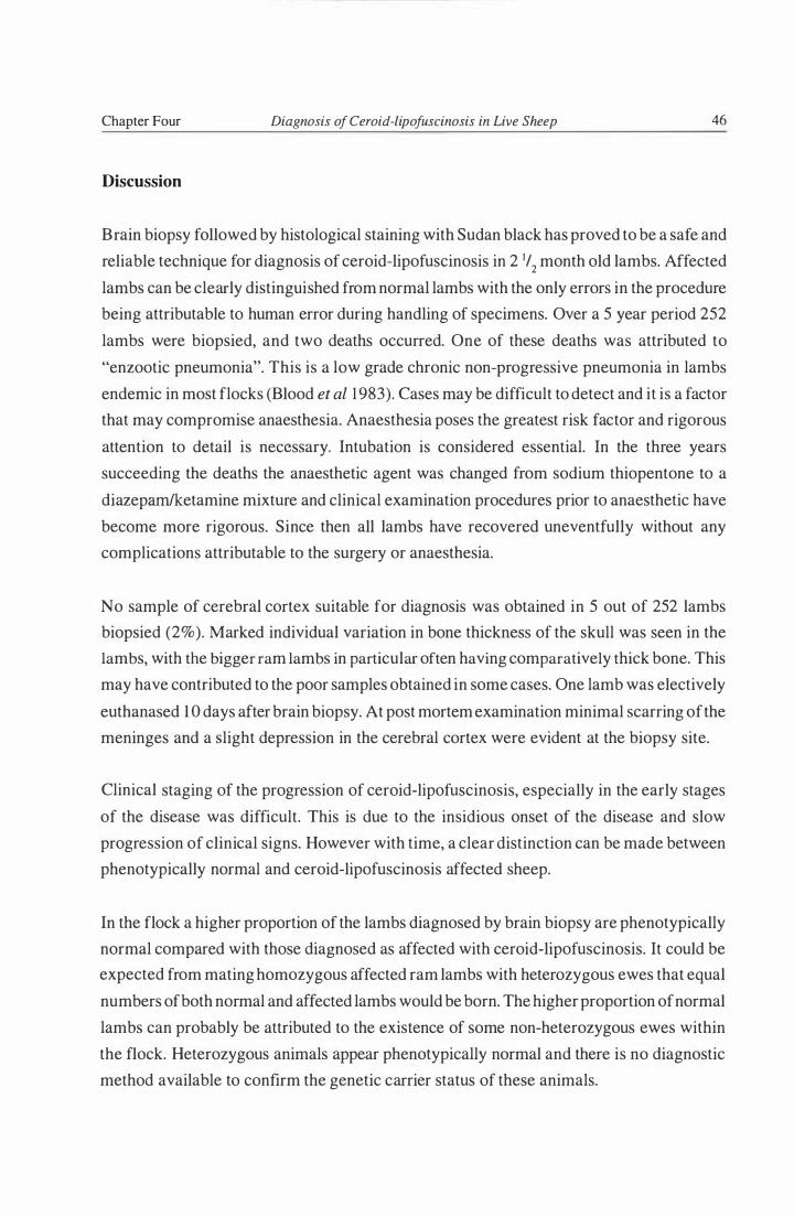
Chapter Four Diagnosis of Ceroid-lipofuscinosis in Live Sheep 46
Discussion
Brain biopsy followed by histological staining with Sudan black has proved to be a safe and
reliable technique for diagnosis of ceroid-lipofuscinosis in 2 '/2 month old lambs. Affected
lambs can be clearly distinguished from normal lambs with the only errors in the procedure
being attributable to human error during handling of specimens. Over a 5 year period 252
lambs were biopsied, and two deaths occurred. One of these deaths was attributed to
"enzootic pneumonia". This is a low grade chronic non-progressive pneumonia in lambs
endemic in most flocks (Blood et al 1 983). Cases may be difficult to detect and it is a factor
that may compromise anaesthesia. Anaesthesia poses the greatest risk factor and rigorous
attention to detail is necessary. Intubation is considered essential. In the three years
succeeding the deaths the anaesthetic agent was changed from sodium thiopentone to a
diazeparn/ketamine mixture and clinical examination procedures prior to anaesthetic have
become more rigorous. Since then all lambs have recovered uneventfully without any
complications attributable to the surgery or anaesthesia.
No sample of cerebral cortex suitable for diagnosis was obtained in 5 out of 252 lambs
biopsied (2%). Marked individual variation in bone thickness of the skull was seen in the
lambs, with the bigger ram lambs in particular often having comparatively thick bone. This
may have contributed to the poor samples obtained in some cases. One lamb was electively
euthanased 1 0 days after brain biopsy. At post mortem examination minimal scarring of the
meninges and a slight depression in the cerebral cortex were evident at the biopsy site.
Clinical staging of the progression of ceroid-lipofuscinosis, especially in the early stages
of the disease was difficult. This is due to the insidious onset of the disease and slow
progression of clinical signs. However with time, a clear distinction can be made between
phenotypically normal and ceroid-lipofuscinosis affected sheep.
In the flock a higher proportion of the lambs diagnosed by brain biopsy are phenotypically
normal compared with those diagnosed as affected with ceroid-lipofuscinosis. It could be
expected from mating homozygous affected ram lambs with heterozygous ewes that equal
numbers of both normal and affected lambs would be born. The higher proportion of normal
lambs can probably be attributed to the existence of some non-heterozygous ewes within
the flock. Heterozygous animals appear phenotypically normal and there is no diagnostic
method available to confirm the genetic carrier status of these animals.

Chapter Four Diagnosis of Ceroid-lipofuscinosis in Live Sheep 47
Summary
A brain biopsy technique has proved to be safe and reliable for the diagnosis of ceroid
lipofuscinosis in 2 1/2 month old lambs. Diagnosis by a clinical staging procedure was
difficult in the early stages of the disease but with time a clear distinction can be made
between phenotypically normal and ceroid-lipofuscinosis affected sheep.

----.�------------�
Chapter Five
Haematopoietic Cel l Transplantation i n Ceroid- l ipofusci nosis Affected Foetal Lam bs
Introduction
Haematopoietic cell transplantation has proved a successful treatment for some genetic
diseases (see chapter two). These results have encouraged surgeons to carry out
haematopoietic cell transplantation in other diseases where children are severely affected
by inherited metabolic defects and where current conventional management and treatment
offered to these patients is ineffective. For many inherited metabolic diseases the risks and
costs associated with haematopoietic cell transplantation compare favourably with current
methods of treatment. However, early intervention is essential as the secondary effects of
disease such as the loss of neurons may be irreversible.
The ultimate therapy for inborn errors of metabolism would be gene therapy. The normal
gene would be inserted into some somatic cells from a patient and these cells would be
reimplanted to provide an endogenous source of gene product. Haematopoietic stem cells
could be suitable carriers for therapeutic genes as they have the ability to differentiate into
a variety of progeny cells including erythroid, thromboid, myeloid, macro phages and
lymphoid cells. Macrophages derived from haematopoietic stem cell transplantation
circulate widely reaching most tissues including the central nervous system (Hoogerbrugge
et al 1 988). The effectiveness of haematopoietic cell transplantation, where macrophages
are a supplier of corrective enzymes, is hence a prerequisite for attempting gene therapy.
The complete lack of any effective treatment for ceroid-lipofuscinosis and the relentlessly
progressive tragic nature of the disease leads desperate parents to consider treatment options
such as bone marrow transplantation where there is any possibility the outcome may be
favourably altered (Parental statement, 5th International Conference on ceroid-lipofuscinosis,
Newark, USA, 1 994) . However, given the high risk and ethical requirements of trying
treatment options in children, animal models have a role to play in evaluating the
effectiveness of new therapeutic regimes. This study was initiated in conjunction with a

Chapter Five Hcr in Ceroid-lipofuscinosis Affected Foetal Lambs 49
group of bone marrow transplant therapists from the USA, with the objective of investigating
the use of haematopoietic cell transplantation in foetal lambs affected with ceroid
lipofuscinosis. This would allow intervention as early in the disease process as practical.
Foetuses at 58-60 days gestation are not yet immunologically competent (Solomon 1 97 1 )
thus tissues from normal donors can be used without having to match HLA tissue antigens.
Materials and Methods
Animals
The recipients were South Hampshire foetuses of haemoglobin BB bloodtype at 58-60 days
of gestation. These were the result of mating BB bloodtype ewes heterozygous for the
ceroid-lipofuscinosis genotype with homozygous ceroid-lipofuscinosis affected 6 month
old ram lambs ofBB bloodtype. As such these recipient lambs would be either homozygous
affected genotype or heterozygous clinically normal phenotype. The latter were to act as
controls. The donors were normal Romney foetuses at 45-48 days of gestation of AA
bloodtype. These were the result of mating normal Romney ewes with normal Romney rams
of AA bloodtype. To ensure the foetuses were the correct ages, the oestrus cycles of all the
ewes were synchronised by the use of progesterone impregnated intravaginal devices
(CIDRs, Ciba-Geigy, New Zealand) .
Surgery
All surgical procedures were performed using standard aseptic surgical techniques. Prior
to surgery all sheep were starved for 24 hours and water was withheld for 1 2 hours. The
donor ewes were anaethetised using 35-40 m1 5% sodium thiopentone via the jugular vein
and the uterus was exteriorised, ligated and removed via a ventral midline incision. The
donor ewes were then euthanased using sodium pentobarbitone.
The foetus/foetuses were removed from the uterus and their sex noted by examination of
their external genitalia. The liver was removed and homogenised in a Potter-Elmingehem
homogeniser in Iscoves Modified Dulbecco medium (IMDM) supplemented with up to
10% foetal calf serum and heparin. An aliquot was taken for cell counting using a
haemocytocrit and the number of cells (hepatocytes and haematopoietic) in the total

Chapter Five HCT in Ceroid-lipofuscinosis Affected Foetal Lambs 50
preparation was calculated. Livers from both male and female foetuses were homogenised
together. The liver homogenate was centrifuged and the pellet resuspended at the desired
concentration in IMDM.
The recipient ewes were given 60 mg of progesterone parenterally 1 6 hours prior to surgery
to ensure uterine relaxation and help prevent spontaneous abortion (Pearson and Mellor
1 977). Immediately prior to surgery they were given 4 rnl Streptopen antibiotic (Pitman
Moore, New Zealand Limited) subcutaneously. This antibiotic cover was continued for the
following 3 days after surgery. Anaesthesia was induced in the recipient ewes using sodium
thiopentone; they were then intubated and anaesthesia maintained by halothane and
oxygen. After routine skin preparation and draping, a ventral midline incision was made
through the abdominal wall. The uterus was exteriorised and the foetus located and held
gently while the uterus was superficially incised and blunt dissected to the chorionic
membrane. Hepatic donor cells ( l OlO cells/kg estimated foetal body weight) were injected
through the chorionic membrane into the peritoneal cavity of the foetal lamb. The
abdominal wall was closed and skin sutured using standard surgical techniques. The sheep
were hospitalised for 7 days following surgery. At 10- 1 3 days post-surgery the stitches were
removed. All recipient ewes recovered uneventfully .
Note: Surgery was performed by two surgical teams over a4 day period. One of these teams
included the author who was also responsible for the overall logistical arrangements of the
procedure and for care of the animals. The uterine surgery was performed by Professor D.
Mellor. The preparation and peritoneal injection of foetal liver cell suspensions was carried
out by Dr I. Zanjani.
Engraftment of Donor Cells
Blood and bone marrow samples were taken at 2, 9 and 1 5 months after birth. Donor cell
engraftment was monitored by isoelectric focusing and chromatography of haemoglobin on
carboxymethy1cellulose columns and by karyotype analysis of peripheral blood lymphocytes
and bone marrow progenitor-derived erythroid and myeloid haematopoietic colonies
(Zanjani et aI 1 99 1 ) .
Note: Isoelectric focusing, chromatography and karyotype analysis were carried out by Dr
I. Zanjani.

Chapter Fi ve HCT in Ceroid-lipofuscinosis Affected Foetal Lambs 5 1
Diagnosis of Disease Status
A needle biopsy of cerebral cortex was obtained from transplanted and non-transplanted
lambs at 2.5 months of age by the method of Dickson et al ( 1989) and fixed in 10% neutral
buffered formalin. Microscopic examination of Sudan black stained paraffin sections
allowed diagnosis of affected or non-affected individuals (see chapter three).
Neurological Examination
The neurological status of each sheep was assessed at monthly intervals over a 7 month
period from 5 months of age as described in chapter three. This was carried out by two
veterinarians, including the author, who reached a consensus on scoring for each of 5
different characteristics. Initially head carriage and postural traits were assessed while the
sheep were herded from the paddock into a darkened shed. Following this the sheep were
handled individually and the "menace" and "blink to bright light" responses were assessed.
The final part of the examination consisted of observing the ease with which the sheep
traversed a few obstacles. This involved the sheep being released in a race, changing
direction to find their way obliquely across a pen, through an open gateway, up a step,
changing direction again and going down a step to join other sheep. Head carriage and
postural responses were again assessed during this phase. For each category the assessment
for each sheep was given an arbitrary maximum of 10 with a range of 0 - 10. A zero score
was given for a totally absent response, while delayed or otherwise abnormal responses
were graded between these values. The scores at each examination were summed and
expressed as a "neurological score". This scoring system was subjective, and as the disease
process was progressive, the worst overall score, previous or current, was used.
Euthanasia and Gross Pathology
Lambs were euthanased with sodium thiopentone anaesthesia followed by exsanguination.
A postmortem examination was carried out on all sheep involved in this study and brain
weights recorded.

Chapter Five HCT in Ceroid-lipofuscinosis Affected Foetal Lambs 52
Fixation and Preparation of Tissues for Microscopy
Tissue samples approximately 5-7 mm thick from pancreas (right lobe), liver (mid cranial
right lobe), kidney (cranial third of right kidney), and cardiac muscle (tip of left ventricle)
were taken from each sheep during post mortem examination and fixed in 1 0% neutral
buffered formalin. Brains were fixed whole. Blocks from each tissue and cerebral cortex
(level with the caudal corpus callosum), cerebellum (mid saggital section) and brain stem
(caudal to fourth ventricle) were embedded in paraffin wax and microtome sections (7�m)
were stained with periodic acid schiff (PAS), luxol fast blue (LFB), Sudan black (SB),
haematoxylin and eosin (H&E). One section was deparaffinised but left unstained for
fluorescence microscopy.
Scoring of Histopathological Sections
Staining intensity and the amount of storage material stained by various stains was
arbitrarily graded by consensus of two pathologists including the author. An increasing
intensity scale with values based on the photomicrographs of Jolly et ai. ( 1 989) was used
where "0" equated to no evidence of storage material, and "5" equated to the amount of
storage material evident in sections from severely affected animals at a late stage in the
disease course.
Retinal Lesion Assessment Scores
At post mortem examination an eye was removed from each animal. An incision was made
posterior to the corneo-scleral junction and the anterior segment removed. The vitreous
humor was extracted and the posterior segment was fixed in 1 0% neutral buffered formalin.
After fixation a retinal section was taken by dividing the posterior section in an anterior
posterior direction through the optic disk. This sample was embedded in paraffin wax by
routine methodology and sectioned for histological staining and microscopic examination.
The H&E stained retinal sections were scored "blind" by the two pathologists (including
the author) on a consensus basis. An assessment of retinal atrophy was made on the basis
of the number of cells in the outer nuclear layer and of the proportion of cones to rods. An
arbitrary scoring system was used ranging from 0 to 5, where "0" was a normal retina, while
"5" would have been for the severe changes seen in advanced ovine ceroid-lipofuscinosis

Chapter Five HCT in Ceroid-lipofuscinosis Affected Foetal Lambs 53
(Jolly et a1 1 982, Graydon and Jolly 1 984). In instances where retinal sections exhibited
regional differences a score was given based on the overall impression of the atrophic
changes.
Quantitative Assessment of Subunit c in Tissues
Quantitative extraction of subunit c from tissues and chemiluminescent detection of this
extracted subunit c was carried out as described in chapter three.
Results
Foeti from 28 sheep received in-utero haematopoietic cell transplantation. Donor cell
engraftment was monitored at 2, 9 and 1 4 months after birth, and identified nine lambs with
transplanted cells. Of these four were shown to have ceroid-lipofuscinosis by histopathology
of brain biopsies and five were unaffected (Table 5- 1 ) .
Table 5-1 . Donor Cell Engraftment (%) in Lambs After Intraperitoneal Foetal
Haematopoietic Stem Cell Transplantation at 58-60 Days Gestation. *
Cell Type Diagnosis Age of Lambs (months)
2 9
Erythroid oeL 4.68 ± 0.3 1 6.54 ± 0.60
N 5 .22 ± 0.32 5 .8 1 ± 0.54
Myeloid OeL 7.23 ± 0.63 14.94 ± 1 .60
N 8.43 ± 0.70 1 1 .55 ± 1 .40
Lymphoid OeL 6.85 ± 0.2 1 9.57 ± 0.42
N 7.50 ± 0.36 8.86 ± 0.35
OCL Ceroid-lipofuscinosis affected lambs (total = 4).
* N Phenotypically normal lambs (total = 5). ± Standard error.
14
6. 1 7 ± 0.5 1
5.76 ± 0.58
1 2.97 ± 1 .39
1 1 .38 ± 0.98
9.93 ± 0.53
9.42 ± 0.44

Chapter Five HCT in Ceroid-lipofuscinosis Affected Foetal Lambs 54
Neurological Assessment
No difference was observed in the clinical progression of neurological disease between the
transplanted and untransplanted sheep affected with ceroid-lipofuscinosis (Figure 5- 1 ) .
Figure 5-1: Staging of the Clinical Progression of Disease Symptoms in Transplanted
(total = 4) and Untransplanted (total = 5) Sheep Affected with Ceroid-lipofuscinosis.
60
Q) 50 I-< 0 (.)
- - - - -� -� - - - - - - - - - - - - - - - - - - - - - - - -
......... C/.l - 40 ro
(.) . -00
- ....... .......
0 30 -0 I-< ;:s Q) 20 Z c: ro Q) 10
�
0 8 9 10 1 1 1 2 1 3 1 4
Age (months)
Key
- - - - - . Transplanted and non-transplanted phenotypically normal lambs (total = 1 1).
-- -- Transplanted lambs affected with ceroid-lipofuscinosis (total = 4).
--- Non-transplanted lambs affected with ceroid-lipofuscinosis (total = 5).

Chapter Five HCT in Ceroid-lipofuscinosis Affected Foetal Lambs 55
Gross Pathology
Variable degrees of brain atrophy were observed in affected sheep at autopsy (Table 5-2).
There was no significant difference between the weights of brains from transplanted and
non-transplanted affected lambs. In contrast there was a significant difference (T -test
p<O.O I ) between the brain weights of all affected lambs when compared to normal controls.
Table 5-2. The Effect of Haematopoietic Cell Transplantation in Foetal Lambs with
Ceroid-lipofuscinosis as Evaluated by the Severity of Lesions
Noted in Various Tissues. *
Transplanted Non-transplanted Normald Parameter
OCL total = 4 OCL total = 5 total = 11
Brain weighta 66.0 ± 2.8 g 65.0 ± 5.5 g 88.0 ± 9.0 g
Retinal atrophyb 2.6 ± 1 . 1 1 .3 ± 0.8 0.0
Pancreasc 2.2 ± 0.4 2.3 ± 0.5 0.0
Liver Kupffer cellsc 3 .2 ± 0.8 3 .5 ± 0.5 0 .9 ± 0.8
Liver hepatocytesC 3 .2 ± 1 .3 3 .8 ± 0.4 0.0
Kidneyc 3 .6 ± 0.5 3.7 ± 0.5 1 .8 ± Oo4
Cerebal cortexC 3 .0 ± 0.0 3.2 ± 004 0.0
Cerebellumc 1 .8 ± 0.2 2.0 ± 0.3 0.0
* OCL Ceroid-lipofuscinosis affected lambs.
± Standard deviation.
b
c
d
Transplanted and non-transplanted vs. normal brain weights; significant P < 0.0 1 .
Mean score of the degree of retinal atrophy i n histological sections.
Mean intensity score of the amount of storage material in Sudan black stained
histological sections.
Transplanted and non-transplanted phenotypically normal lambs.

Chapter Five HCT in Ceroid-lipofuscinosis Affected Foetal Lambs 56
Histopathology
Characteristic storage cytosomes in the brain of ceroid-lipofuscinosis affected sheep were
noted with H&E, SB, LFB and PAS staining and also by fluorescence microscopy. In other
tissues the LFB stain showed no staining, and PAS method showed variable staining. In all
tissues (normal and affected) fluorescence was observed from red blood cells, and
fluorescent material was present in Kupffer cells in the liver and kidney parenchymal cells.
Assessments of tissue pigment storage were carried out using SB stained sections (Table
5-2) but there were no observed differences between the transplanted and untransplanted
affected sheep groups. A laminar zone deficient in neurons was observed in sections of
cerebral cortex taken through the occipital pole in all affected lambs. This was not observed
in the normal controls. In all cases the retinas of normal sheep were scored a but there was
considerable variation in the severity of the retinal changes between the different affected
sheep (Table 5-2).
Quantitative Detection of Subunit c by Enhanced Chemiluminescence
Pancreatic tissue samples were too small and not of sufficient quality (fibrous tissue, blood
and fat were attached) to allow quantitative subunit c extraction. Quantitative extraction
was successfully carried out from liver samples. Densitometry measurements of reaction
product in the various serial dilutions were compared. The liver from normal sheep
contained an average of 468 J..lg subunit c per gram of tissue, the liver from sheep affected
with ceroid-lipofuscinosis contained an average of2,59 1 J..lg subunit c per gram of tissue and
the liver from sheep affected with ceroid-lipofuscinosis which received haematopoietic cell
transplantation contained an average of 1 ,765 J..lg subunit c per gram of tissue. There was
no significant difference between the amount of subunit c extracted from transplanted
affected sheep compared with the amount extracted from untransplanted affected sheep (T
test p > 0.05). There was however a significant difference (T-test p < 0.0 1 ) between the
amount of subunit c extracted from both these groups of affected sheep and the quantity
extracted from phenotypically normal sheep.

Chapter Five HCT in Ceroid-lipofuscinosis Affected Foetal Lambs 57
Discussion
This study attempted to incorporate the optimal conditions for successful treatment of
ceroid-lipofuscinosis by haematopoietic stem cell transplantation except that ablative
processes to enhance donor cell engraftment were not possible under the conditions of foetal
transplantation. In this trial foetal lambs were transplanted at 58-60 days gestation
(gestation in sheep is 145 days), a time prior to the onset of overt disease or immunological
competence (Solomon 197 1 ) . The donor cells were from normal non-heterozygote foetal
lambs and would be expected to eventually express the full complement of normal enzymes.
Foetal livers were chosen as the source of cells as this is the primary organ ofhaematopoiesis
at this gestational age in foetal lambs.
An average of 9% engraftment of donor cells was achieved. However no clinical,
pathological or biochemical benefit could be demonstrated in affected lambs. In recording
the data, only that for brain weight and quantitative subunit c extraction from liver, were
fully objective. The other semi-objective data were scored by consensus of two clinicians/
pathologists under "blind" experimental conditions. For this reason levels of significance
have not been stated although standard deviations are included for pathological data to
indicate the range of variation recorded. Comparison of the retinal sections from different
sheep was particularly difficult as there were sampling errors due to inconsistency both in
the plane of section and of the exact retinal region examined and in some cases the different
retinal layers had separated and parts were missing in the sections.
Specific cell layers within the cerebral cortex are difficult to identify in non-primates.
However, in the cerebral cortex of all affected lambs there was a laminar zone deficient in
neurons in occipital pole sections. This finding of selective necrosis of certain neurons has
also been reported in the juvenile and late infantile forms of disease (Zeman et al 1 970,
Braak and Goebel 1 978, 1 979, Jolly 1 995) and in affected sheep (Jolly et al I 989) . 1t has
been postulated that this loss of cells implies a toxicity inherent in the disease process, rather
than cell death due to overloading of the cells with storage bodies per se (Jolly 1 995).
There are two likely reasons for the failure of haematopoietic stem cell transplantation to
affect the ceroid-lipofuscinosis disease process. Firstly this may be a disease that is not
amenable to treatment by metabolic collaboration with the transplanted cells. Secondly the
level of donor cell transplantation (approximately 9%) may be too low to produce any
discern i ble benefit to nervous or visceral systems.

Chapter Five Hcr in Ceroid-lipofuscinosis Affected Foetal Lambs 58
In normal tissues the levels of any enzyme present exceeds the basic minimum required for
any substrate metabolism. The progression of any metabolic storage disease will be
influenced by the presence and amount of residual enzyme activity. It is reasonable to
assume "that the more profound the enzyme deficiency, the more severe the disorder and
conversely the higher the residual activity of the mutant enzyme the greater the protection
against disease" (Neufeld 1 99 1 ) . A model of this concept (Conzelmann and Sandhoff 1 983/
84) proposes a critical threshold of activity above which the enzyme is capable of keeping
up with the substrate metabolism and below which it cannot, so that accumulation occurs.
The kinetics are such that at the critical threshold of enzyme activity, even quite small
amounts of additional enzyme could markedly affect the rate of substrate accumulation and
hence progression of disease. Therefore 9% transplanted cells might be able to influence the
course of disease in some way. However, under the conditions of this experiment such
transplantation failed to influence the disease progression in the recipient lambs. Nevertheless
the feasibility of foetal transplantation of haematopoietic stem cells was demonstrated.
Methods of improving the ratio of donor to recipient cells in tissues need to be developed.
One example of this which has been described involved using modified media to enhance
haematopoietic stem cell colony growth (Zanjani et al 1990) . This technique enabled
enhancement of donor cell engraftment from original levels between 4- 1 5 % to 1 2- 1 6%, but
still further enhancement of engraftment beyond these levels would be desirable. The
engraftment level of9% in this study compared favourably to the engraftment levels gained
in an equivalent study in goats reported by Pearce et al ( 1 989) (see chapter two).
The nature of the storage disease has an influential effect on the likel y outcome of enzyme
or haematopoietic cell transplantation. The metabolic defect in ceroid-lipofuscinosis
remains undefined but at least in the late infantile form it is unlikely to be a cathepsin
deficiency (Ezaki et al 1993, 1 995). Storage material may also be complexed in such a
tertiary structure within cells that it is resistant to the effect of endogenous enzymes or
exogenous enzyme replacement. Subunit c of mitochondrial ATP synthase has a propensity
for self aggregation due to its hydrophobicity and it is possible that this property may be
involved in the pathogenesis of its accumulation (Jolly 1 995).
Allogenic bone marrow transplantation has also been carried out in English Setter dogs
affected with ceroid-lipofuscinosis at 4 1/2 months of age (Deeg et a1 1989, 1 990) . This
treatment was unsuccessful in altering the course of the disease in the four dogs transplanted
but cells from heterozygous donors were used which may not have expressed optimal levels
of enzyme activity and the transplantation may have been too late in the disease course. The

Chapter Fi ve HCT in Ceroid-lipofuscinosis Affected Foetal Lambs 59
experiences of Taylor et al ( 1 986, 1 988, 1992) and Taylor and Farrow ( 1 988b) in bone
marrow transplantation of dogs with fucosidosis, showed that the age of engraftment was
important with best results occurring with early engraftment. Central nervous system
enzyme replacement was a gradual process taking up to 6 months to achieve substantial
levels compared with less than 2 months in other tissues.
Bone marrow transplantation has been attempted in a clinically unaffected yet pathologically
affected sibling of a patient with late infantile Batten disease (Lake et al 1 995). The effect
of the transplantation was monitored for 9 months until the donor marrow became displaced
by the host cells . No removal of storage material could be demonstrated but the patient
passed the age at which his sibling developed symptoms by 3 months. This apparent
postponement of symptoms could be the effect of natural variation in the onset of disease,
or more encouragingly, could be due to transplantation preventing further build-up of
storage material.
Lake et al ( 1 995) carried out co-culture experiments using lymphocytes from normal
subjects cultured with those from a clinically unaffected yet pathologically affected sibling
of a patient. Pathognornic curvilinear bodies were found in these co-cultured lymphocytes
up to and including the 6th day, indicating the bodies remain in the cells in culture. Messer
et at ( 1 995) describe the fusion of homozygous mnd mouse embryos (a model for Batten
disease) with normal embryos. In these embryos neurons filled with storage material could
be seen adjacent to normal cells implying that metabolic collaboration did not occur.
The tight junctions between endothelial cells which form the blood-brain barrier and
prevent plasma proteins entering the brain are present from very early in foetal development
(Bradbury 1979). However, various studies using a variety of test compounds (including
inulin, sucrose, mannitol, adenine, arginine, choline, tryptophan, pyruvate and lactate) have
shown that the rate of entry of these substances into the brain is much higher in the foetus
and up until just after birth, than in older animals. It is theoretically possible that this
increased permeability of the blood-brain barrier may also facilitate optimal colonisation
of the central nervous system by transplanted normal donor cells. Even if this is not the case,
studies on bone marrow transplantation performed in both animals and children several
weeks or more after birth have shown transplantation to be clearly beneficial to central
nervous system disease for example, in a-mannosidosis cats transplanted at 8, 10 and 1 2
weeks after birth (Walkley et aI 1994), see chapter two. Hence consideration o f the blood
brain barrier as an impediment to the correction of central nervous system disease after bone

Chapter Five HCT in Ceroid-lipofuscinosis Affected Foetal Lambs 60
marrow transplantation may be overstated as neuronal enzyme replacement has been
achieved by the invasion of cells of haematogenous origin (Taylor et al 1 986, 1 988, 1 992,
Shull etal l 987, 1 988, Hoogerbrugge etal l 988, Birkenmeier etal l 99 1 , Yeager etal l 99 1 ,
Krivit and Shapiro 1 99 1 , Walkey et al 1994) . Other studies also provide evidence that
microglial cells within the brain are derived from bone marrow precursors ( Perry et al 1 985,
Hickey and Kimura 1 988, Hickey et al 1 992, Unger et al I 993).
Extensive research effort is currently being expended in the area of gene therapy. The idea
of gene transfer is worthy but the current severe technical limitations of early ceroid
lipofuscinosis diagnosis, gene transfer and engraftment preclude successful practical
application of this technique at this stage. It remains necessary to prove that treatment by
exogenous enzymes, or treatment by some other process using transplanted cells is
beneficial in Batten disease before effort is expended in the area of gene transfer.
Given that conventional medicine currently fails totally in the treatment of patients with
ceroid-lipofuscinosis, it is extremely disappointing that there is very little evidence thus far
to provide any support for the efficacy of bone marrow transplantation in this disease.
Appropriate tissue culture models of this group of diseases are now available (Ezaki et al
1 993, 1 995, Bennett et al 1 993, Dunn et al 1 994, Lake et al 1 995). Further investigation on
cell culture mechanisms of metabolic collaboration in this disease are warranted before
further transplantation studies are carried out.
Summary
Haematopoietic cell transplantation was carried out in foetal lambs affected with ceroid
lipofuscinosis at 58 - 60 days of gestation. Cells from homozygous normal foetal donors
were used. An average of 9% engraftment of donor cells was achieved but no clinical or
pathological benefit was demonstrated. Two possible reasons why this procedure was
unsuccessful were proposed; firstly, the level of engraftment may have been too low and
secondly, ceroid-lipofuscinosis may not be a disease which is amenable to treatment by
metabolic collaboration.

Introduction
Chapter Six
Fluorescence of Storage Bodies and Reconstitution of Storage Bodies
from Isolated Com ponents
Historically the ceroid-lipofuscinoses have been grouped with the lipidoses on the basis of
their histological staining properties which have implied a lipid-based pathogenesis of these
diseases. The stored material in ceroid-lipofuscinosis also shows yellow autofluorescence
under blue light and this observation has lead to the assumption that some intrinsic
fluorophore is present. Intensive research and much speculation has been expended trying
to determine the nature of this fluorophore, as clarification of this issue has often been
considered central to the elucidation of the pathogenesis of the ceroid-lipofuscinoses. Chio
and Tappel ( 1 969a, b) reported that Schiff base polymers with the same fluorescence as
lipofuscin were created by the reaction of malonaldehyde, a product of peroxidation of
polyunsaturated fatty acids with phospholipids or the amino groups of proteins. This
schema appeared to be directly applicable to Batten disease and consequently abnormal
lipid peroxidation was proposed as being central to the pathogenesis of this group of
diseases (Zeman and Dyken 1 969, Zeman 1 974, Zeman 1976). As a direct result of this
hypothesis, biochemical studies in Batten disease have focused almost exclusi vely on lipids
until comparatively recently. This has been discussed in detail in chapter one.
Studies on the emission spectra of the stored material in late infantile human, English setter
dog and South Hampshire sheep forms of Batten disease showed emission in the yellow
orange region (520-540 nm) and not in the blue region (460-480 nm) expected for the
particular Schiff base polymers (Eldred et a1 1982, Dowson et a1 1 982, Katz et a1 1 988) that
result from lipid peroxidation. Therefore it was considered unlikely that the autofluorescent
granules which accumulate in tissues as a result of ceroid-lipofuscinosis are products of
lipid peroxidation. Alternatively, if the disease-related fluorophors are the result of lipid
peroxidation, they must be generated via mechanisms substantially different from those
which occur in vitro. Studies have shown that peroxidation artefacts can be produced during

Chapter Six Fluorescence of Storage Bodies and Reconstitution of Storage Bodies . . . 62
organic solvent extractions (Katz et al 1 988, Kikugawa et al 1 994). Processes which
produced this peroxidation resulted in the generation of blue fluorescence from tissues
showing yellow fluorescent lipofuscin in situ.
Palmer et al ( 1 986a) extracted small quantities of a number of weakly fluorescent lipids
from sheep liver storage bodies using chloroform-methanol and reported that the mass of
residual insoluble material remaining after lipid extraction was not fluorescent. It was
concluded that these fluorescent lipids were carotenoids resulting from vitamin A and
chlorophyll breakdown and not related to the disease.
This chapter explores the phenomenon of storage cytosome fluorescence and describes the
reconstitution of liposomes from normal phospholipids with subunit c isolated from ovine
storage bodies.
Materials and Methods
Fluorescence of Storage Bodies in Suspension
Storage bodies isolated from ovine, canine and human late infantile forms of ceroid-
lipofuscinosis (see chapter three) were suspended in a water: glycerol mixture ( 1 : 1 ) . These
suspensions were then examined under an Olympus BH-2 microscope with an Olympus
BH2-RFL reflected light fluorescence attachment (Olympus Optical Co, Ltd, Tokyo,
Japan) using a 440 nm excitation filter and a 530 nm barrier filter.
Absorbance Spectra of Dissolved Storage Bodies
Ovine pancreatic and liver storage bodies (see general materials and methods, chapter three)
were dissolved in 1 % lithium dodecyl sulphate (LDS) and in chloroform/methanol!
ammonium acetate (2: 1 : 1 OOmM). Extracted subunit c obtained after ether precipitation was
also dissolved in chloroform/methanol/ammonium acetate (2: 1 : 100mM). Absorbance
spectra of these solutions were measured in a double beam ratio reading SP8-400 UV -Vis
spectrophotometer (Pye Unicam Ltd, York Street, Cambridge, England) . Fluorescence
spectroscopy was carried out in an Aminco SPF-500 spectrofluorometer (American

Chapter Six Fluorescence of Storage Bodies and Reconstitution of Storage Bodies . . . 63
Instrument Company, Silverspring, Ohio, USA). Quinine sulphate at a concentration of 0. 1
�g/m1 in 0.05 M sulphuric acid was used as the fluorescence standard.
Phospholipid Extraction
Phospholipids were extracted from egg yolk and normal sheep liver. This was carried out
by homogenising 10 g of egg yolk or liver in 200 m1 of chloroform/methanol (2: 1 ) . The
resulting suspension was filtered into a separating funnel and 40 m1 0.74% potassium
chloride was added. These separating funnel contents were vigorously mixed then allowed
to stand until the two phases separated. The lower phase was collected and evapourated to
dryness in a rotary evaporator, then resuspended in 5 m1 of chloroform. Phospholipids were
precipitated by the addition of 50 m1 of acetone at 4°C for 1 hour, followed by centrifugation
at 1 ,400gmax for 5 minutes at 4°C in a Sorvall general laboratory bench centrifuge (GLC- l ) .
After this centrifugation the supernatant was discarded and the precipitate rinsed with cold
acetone, centrifuged as above and again the supernatant was discarded. The precipitate was
dried with nitrogen gas and then resuspended in 10 m1 of chloroform. A measured aliquot
of this solution was then taken so that the concentration of phospholipids in chloroform
could be determined from the dry weight.
The Manufacture of Liposomes
Concentrations of subunit c in chloroform/methanol/ammonium acetate (2: 1 : 100mM)
were determined by quantitative amino acid analyses (see chapter three). Known ratios (by
weight) of either egg yolk or liver phospholipids and subunit c proteolipid were combined
and the absorbance spectra of these solutions measured. To make liposomes, solutions of
phospholipids and proteolipid were combined in small lidded flasks. The solvent was
evapourated to dryness under nitrogen at 35°C and approximately 1 m1 0.67% potassium
chloride solution was added to each flask for every 20 mg phospholipid and proteolipid
present. The flasks were sealed and shaken on a Lab-line orbital shaker (Lab-line
Instruments Inc, Melrose Park, Illinois, USA) at 37°C for 12 hours then sonicated at 10 �
for 10 seconds. Aliquots were taken for fluorescence microscopy and electron microscopy.
The latter were fixed in 3% glutataldehyde in 0. 1 M phosphate buffer and processed as
recorded in chapter three.

Chapter Six Fluorescence of Storage Bodies and Reconstitution of Storage Bodies . . . 64
Results
Fluorescence of Storage Bodies, Dehydrated Protein Aggregates and Phospholipids in
Suspension
Isolated liver and pancreas storage bodies from ovine, canine and human late infantile forms
of ceroid-lipofuscinosis exhibited sparkling yellow-green fluorescence when suspended in
a water:glycerol mixture ( 1 : 1 ) and viewed in a fluorescence microscope (Figures 6- 1 and
6-2). Protein aggregates of bovine serum albumin and casein also exhibited sparkling
yellow-green fluorescence when suspended in a water:glycerol mixture and viewed in the
same way (Figure 6-3) . Similar suspensions of phospholipids did not show any fluorescence.
Absorbance Spectra of Dissolved Storage Bodies in Vitro
Ovine pancreatic and liver storage bodies dissolved completely in 1 % LDS . The UV -visible
absorbance spectra of these solutions was unremarkable, there being an absorbance
maximum at 275 nm and a small shoulder at 4 1 2 nm (Figure 6-4). Similar spectra were
obtained from the total chloroform/methanol soluble components of the storage bodies
(lipids plus the proteolipid) and the ether precipitated subunit c redissolved in chloroform!
methanol. When fluorescence spectroscopy of these solutions was attempted, high
concentrations were required to get any signal at all, even at the most sensitive settings of
the instrument. A concentration of 1 .38 mg/mI, redissolved liver storage body proteolipid
gave a weak spectrum with an emission maximum of 500 nm and a smaller peak at 680 nm
when using an excitation maximum of 400 nm. These emission and excitation maxima peak
heights were equalled by those of a 0. 1 �g/mI solution of quinine sulphate (excitation
maximum 350 nm, emission maximum 450 nm). Dilution of the samples resulted in a
proportionate drop in signal strength, eliminating the possibility that internal absorption of
the emitted light caused the lack of observed fluorescence.
Liposomes Containing Subunit c
Storage body-like structures were reconstituted when chloroform/methanol solutions of
phospholipids and proteolipid were mixed, the solvent evaporated and the resultant film
sonicated in aqueous buffer. This reconstitution resulted in similar structures over a wide

Chapter S ix Fluorescence of Storage Bodies and Reconstitution of Storage Bodies . . . 65
Figure 6-1: Fluorescence of Storage Body Aggregates ISf!latedfrom the Pancreas of a Human Patient with Late Infantile Batten Disease Suspended in GlycerollWater x 400.
Fluorescence Microscopy, Excitation Filter 440nm with 530nm Barrier Filter.
Figure 6-2: Fluorescence of Storage Body Aggregates Isolatedfrom the Pancreas of a Sheep Affected with Ceroid-lipofuscinosis Suspended in GlycerollWater x 400. Fluorescence Microscopy, Excitation Filter 440nm with 530nm Barrier Filter.

Chapter Six Fluorescence of Storage Bodies and Reconstitution of Storage Bodies . . .
Figure 6-3: Fluorescence of Bovine Serum Albumin Aggregates Suspended in GlycerollWater x 100.
Fluorescence Microscopy, Excitation Filter 440nm with 530nm Barrier Filter.
66

Chapter Six Fluorescence of Storage Bodies and Reconstitution of Storage Bodies . . .
Figure 6-4: UV-visible Absorbance Spectrum oj Proteolipid ExtractedJrom Sheep Liver Storage Bodies in Chloroform/Methanol.
Optical Density
(O.D. Units)
0.4
0.3
0.2
0. 1
o
67
240 300 400 500 600
Wavelength (nm)

Chapter Six Fluorescence of Storage Bodies and Reconstitution of Storage Bodies . . . 68
range of proteolipid to phospholipid ratios (2: 1 - 1 :20 w/w), the excess phosphol ipid
remaining as long strands of phospholipid bilayer. The reconstituted bodies exhibited
somewhat similar multi lamel lar ultrastructure to that shown by isolated storage bodies
(Figures 6-5 and 6-6) whereas the proteolipid alone was quite amorphous (Figure 6-7) .
Areas simi lar to those often referred to as l ipid droplets were also observed under some
conditions (Figure 6-8). The components of the reconstituted bodies were non-fluorescent
when examined in solution, singly or combined, under a fluorescence microscope or in a
spectrophotometer. However, the reconstituted bodies exhibited s imilar sparkl ing
fluorescence to the storage bodies when viewed in a fluorescence microscope.
Discussion
Fluorescence is the name given to the phenomenon whereby a body emits l ight during the
absorption and excitation by a primary l ight source. The emitted l ight is always of a longer
wavelength than the primary excitation l ight. Some substances fluoresce in the visible
region when excited with l ight of an appropriate wavelength without being specially
treated, stained or label led. In a histological context this phenomenon is called
autofluorescence to distinguish it from the fluorescence seen after using histological stains
that exhibit fluorescence. Light is absorbed by molecules into certain wel l defined quantised
energy states. The accompanying transition of an atomic or molecular orbital from a lower
to a h igher energy state, which is accompl ished by the absorption of the radiant energy, is
the basis of all methods of absorption spectroscopy. The electrons in an atomic or a
molecular orbital may subsequently descend from the higher energy state back to a lower
one, a transition that is usual ly accompanied by the emission of radiant energy. This
transition is the basis of al l methods of emission spectroscopy. Many molecules may absorb
energy but do not fluoresce because al l the energy absorbed is rapidly lost by coll is ional
deactivation and released as heat (Gui lbault 1990). Fluorescence cannot occur without l ight
absorption but any portion of the spectrum where absorption occurs can produce fluorescence.
For any s ingle molecule the fluorescence peak wi l l be at the same wavelength regardless
of the excitation wavelength used but the intensity of emission wil l vary with the relative
strength of the absorption.
Fluorescence in ceroid-lipofuscinosis is not a simple phenomenon. These, and previous
studies (Palmer et al 1 986a, b, Katz et al 1988) have shown it cannot be attributed to the
existence of a single identifiable fluorophore, with identifiable molecular properties

Chapter Six Fluorescence of Storage Bodies and Reconstitution of Storage Bodies . . . 69
Figure 6-5: Electron Micrograph of Liposomes Made From Liver Phospholipids and Subunit c Proteolipid (20: 1) x 33,500.
Figure 6-6: Electron Micrograph of Liposomes Made From Egg Phospholipids and Subunit c Proteolipid (20: 1) x 1 2 ,500.

Chapter Six Fluorescence of Storage Bodies and Reconstitution of Storage Bodies . . . 70
Figure 6-7: Electron Micrograph of Subunit c Proteolipid x 1 1 3 ,300.
Figure 6-8: Electron Micrograph of Liposomes made from Egg Phospholipids and Subunit c Proteolipid (1 : 1) Showing a "Lipid-droplet" like Structure (arrow) x 33 ,400.

Chapter Six Fluorescence of Storage Bodies and Reconstitution of Storage Bodies . . . 7 1
consistently displayed in situ and after extraction i n various solutions. I n general , for any
single molecule, fluorescence can vary widely depending on the influences of any factor
which changes the energy level of the absorbing orbital (e.g. ionisation with pH changes and
hydrogen bonding) and any other factors that influence the rate at which the energy can be
dispersed by vibration compared with the t ime required for the fluorescence decay. Thus
the effects which influence the differences in energy between the ground and electronically
exc i ted states of molecules in mixed solvents are potentially very complex.
The major component of storage bodies in the ovine and late infantile human forms of
ceroid-l ipofuscinosis is the protein, subunit c of mitochondrial A TP synthase . Nearly all
proteins are capable of fluorescence after irradiation with ul traviolet l ight due to aromatic
amino acid residues, especially tryptophan (emissionmax 330-340nm) and tyrosine (emissionmax
303nm) but also to a lesser extent phenylalanine (emissionmax 258 and 282nm). The tertiary
structure of proteins can also have a major effect on the observed fluorescence (Chen 1990) .
Proteins in solution are dynamic, constantly moving structures and thus the uniqueness of
protein tertiary structures may be compromised in solution . The ultraviolet absorption
spectra of total storage bodies and of the proteolipid in solution showed an absorbance
maximum at 275, which is slightly less than the traditional 280 peak associated with protein
absorption. This observation is consistent with the amino acid composition of subunit c and
the lack of tryptophan in this molecule (Sebald and Hoppe 1 981).
Chloroform/methanol or LDS solutions of ceroid-lipofuscinosis storage bodies, or their
components, were not fluorescent. The u ltraviolet-visible spectra of these solutions showed
only a very smal l absorption in the 400 nm region, the common excitation region clai med
to excite the characteristic fluorophor. Without absorption of energy there is none to be
radiated at a longer wavelength which i s the requirement for fluorescence, hence the storage
bodies examined did not contain a single characteristic fluorophore of the type postulated.
A concentrat ion of 1 . 38 mg/ml redissolved l iver storage body proteolipid gave a weak
spectrum with an emission maximum of 500nm and a smaller peak at 680nm, using an
excitation maximum of 400nm. These emission and excitation maxima peak heights were
equalled by those of a 0. 1 )..lg/ml solution of quinine sulphate (excitation maximum 350nm,
emission maximum 450nm). This means that the storage body components in solution were
1 3 ,800 t imes less fluorescent than the quinine sulphate standard.
It i s possible that microscopic autofluorescence may not require a strong intrinsic fluorophore.
It is frequently observed that molecules exhibit higher fluorescence quantum yields when

- - ----- - -----------
Six Fluorescence Bodies and Reconstitution Bodies . . . 72
incorporated in solids, rather than in liquid solution (Wehry 1 990) . This effect is due to the
protein tertiary structure and solvent interactions with solute molecules, following excitation
in solution. The immediate environment of a particular solute molecule in a rigid solid is
virtually constant, whereas it changes continually in l iquid solution. Quenching is the name
given to the effect where collision of the excited fluorophore with other molecules drains
off the energy of the excited state. In some cases this results from very specific molecular
interactions , for example quinine fluoresces in sulphuric acid but not in hydrochloric acid
because collisions with chloride ions quench its fluorescence.
Protein conglomerates in histological tissue sections may show autofluorescence. This can
be demonstrated in hepatic globules of the protein alpha- I -antitrypsin which show
moderately intense blue-white autofluorescence under ultraviolet light and yellow-green
autofluorescence under blue light when examined microscopical ly (DeLellis et af 1 972) .
Additionally, colloid in the equine thyroid gland, which consists of the protein thyroglobulin,
may undergo age-related degenerative changes showing as a basophilia of colloid. Under
UV or blue light this colloid showed yellow, or yellow-green autofluorescence in unstained
sections (Figure 6-9) (Dalefield et aI 1 994). This demonstrates that sl ight in vivo alterations
in protein structure may lead to observable microscopic fluorescence within tissue sections .
Most fluorescence studies on the ceroid-lipofuscinoses have concentrated on detecting
fluorophors in chloroform/methanol based extracts . Katz et af ( 1 988) reported that the
insoluble material remaining after chloroform/methanol extraction of canine storage
granules was fluorescent but Palmer et al ( 1 986a) reported that the residual material from
ovine tissues was not fluorescent. Ovine storage bodies solubiIised in LDS show virtual ly
no absorption in the 400-500nm region and visually lose their f1uorescence, compared with
when they were suspended in water or water/glycerol . Analyses of storage bodies have
shown that the dominant species (50-70% by weight) is protein and the identification of
other major constituent groups does not leave room for any quantitatively significant
unidentified fluorescent constituent.
Subunit c is thought to form a "hairpin" in the inner mitochondrial membrane, with
hydrophobic amino acid residues within the membrane and hydrophilic residues on the
membrane surface. A molecule containing both a nonpolar hydrophobic chain and one or
more polar, hydrophilic functional groups is called an "amphiphilic" molecule. Within this
context LDS , egg yolk and liver phospholipids and subunit c are all amphiphilic molecules.
Such molecules may form aggregates of colloidal dimensions ("micelles") in liquid

Chapter Six Fluorescence of Storage Bodies and Reconstitution of Storage Bodies . . .
Figure 6-9: Fluorescence of Altered Colloid in a Thyroid Gland from an Aged Horse x 100.
Fluorescence Microscopy, Excitation Filter 440nm with 530nm Barrier Filter.
73

Chapter Six Fluorescence of Storage Bodies and Reconstitution of Storage Bodies . . . 74
solutions. Micellar systems are complex, dynamic systems, that are by nature heterogenous.
There are inherent problems associated with the interpretation of fluorescence of molecules
in such systems as the fluorescence properties of a molecule will depend on the specific
microenvironment seen by that molecule during its excited state lifetime. The introduction
of a fluorescent molecule into the interior of a micelle may bring about significant
alterations in the fluorescence spectrum, quantum yield, and decay time (Wehry 1 990) .
Subunit c is highly hydrophobic in nature and consequently has a high detergent to protein
ratio in LDS micelles for polyacrylamide gel electrophoresis (PAGE), hence the apparent
molecular weight of 3 .5 kDa (Palmer et a1 1988, 1 992). The arrangement of subunit c in LDS
micelles and in phospholipid micelles (liposomes), could be expected to be radically
different, hence the fluorescence properties observed could be expected to be at variance.
These differences could only be subjectively evaluated under a fluorescence microscope or
under UV light, as during attempted quantitative measurement of phospholipid micelle
turbid suspensions, artefacts occurred due to scattering of incident and fluorescence beams.
Under the electron microscope the phospholipid liposomes with subunit c had a structural
resemblance to storage bodies.
Other proteins have been studied as models of membrane proteins, or proteins that act on
membranes. Melittin is a 26 residue peptide isolated from bee venom in which the N
terminal 6 amino acids are polar but the remainder of the molecule is hydrophobic. This
protein can exist in a monomeric form or as a tetrameric aggregate in solution and contains
one fluorescent tryptophan residue (Brown et al 1980, Lauterwein et al 1 980). The
fluorescence emission spectrum of melittin is very sensitive to interaction with some
phospholipids (especially unsaturated lecithins), with the fluorescence spectrum changing
to shorter wavelengths when this interaction occurs (Mollay and Kreil 1 973). The self
association of melittin is thought to be accompanied by major conformational changes in
the secondary structure of the peptide units involved (Talbot et al 1 979). NMR spectral
studies have suggested that melittin forms similar conformations when aggregated, and
when micelle-bound (Brown et aI 1980).
This study was undertaken because the observation of microscopic autofluorescence in the
ceroid-lipofuscinoses has lead to much speculation that the chemical species responsible for
the fluorescence must be central to the pathogenesis in this group of diseases. There is a vast
discernible discrepancy between the sparkling fluorescence in paraffin tissue sections and
the results of quantitative analyses of isolated components of ceroid-lipofuscinosis storage
bodies in chloroform/methanol/ammonium acetate and LDS solutions which are not

Chapter Six Fluorescence of Storage Bodies and Reconstitution of Storage Bodies . . . 75
fluorescent. B iochemical analyses by Palmer et al ( 1 986a, b) preclude any quantitatively
significant unidentified fluorescent constituent occupying more than 2-3% by weight of the
storage body mass. The phenomenon of autofluorescence in the ceroid-lipofuscinoses is
complex but it is concluded that there is no major fluorescent chemical species that is central
to the pathogenesis of this disease other than protein.
Summary
A characteristic feature of the storage material in ceroid-lipofuscinosis is that it shows
autofluorescence, either in situ or when isolated bodies are in suspension. However, when
these storage cytosomes were dissolved the fluorescence was lost. The only significant
absorbance spectra for dissolved storage bodies was at 275 nm which was in the range
expected for protein. Emission and excitation maxima peak heights of the dissolved storage
bodies were low, being 1 3,800 times less fluorescent than the quinine sulphate standard. It
was concluded that the inherent fluorescence of storage material was associated with the
tertiary arrangement of protein in its particular environment.

Introduction
Chapter Seven
Raisi ng Anti bodies to Subu n it c and Immunocytochemistry
Monoclonal and polyc1onal antibodies have proved to be useful tools in the study of the
FJFO-ATP synthase complex of mitochondria, chloroplast' s and bacterial cell membranes
(Tzagoloff and Meagher 197 1 , Sebald and Hoppe 1 98 1 , Sanadi 1 982, Amzel and Pedersen
1 983, Vignais and Satre 1984, Hatefi 1 985, Senior 1985, Godinot and Di Pietro 1 986,
Gautheron and Godinot 1 988). An intramembrane component of the Fo portion of this
complex, subunit c , is the main constituent of the material stored in some forms of ceroid
lipofuscinosis (Palmer et a1 1989, 1 990, 1 992, Fearnley et a1 1 990, Martinus et a1 1 99 1 , Jolly
et al 1 992, Faust et al 1 994) . Despite years of intensive study of ceroid-lipofuscinosis
(Batten disease) using available human tissue and animal models, the underlying biochemical
anomaly in the disease remains unknown. It has been postulated (Palmer et al 1 995) that
subunit c storage diseases arise from different mutations affecting a specific degradation
pathway of subunit c. The normal degradative pathway of subunit c is unknown. Antibody
studies could be useful in the elucidation of this pathway. In addition, diagnosis of Batten
disease is difficult requiring invasive tissue sampling and histological staining. No stain is
highly specific for the storage of subunit c. Diagnosis of ceroid-lipofuscinosis has been
attempted by quantitative measurement using an ELISA antibody method to detect subunit
c extracted from urine (Wisniewski et al 1994) and various tissue homogenates but the
specificity of the results of such tests is controversial.
Subunit c is a very hydrophobic transmembrane protein which is highly conserved between
species. The amino acid sequences are identical in cattle, sheep and human beings (Medd
et a1 1 993) and are similar to the equivalent protein in prokaryotic membranes (Sebald and
Hoppe 1 98 1 ) . To elicit an antibody response prolonged immunisation schedules, consisting
of up to nine immunisations, have previously been required (Loo and Bragg 1 982) and
working concentrations of polyclonal sera are reported to range from 1 : 100 to 1 :500.

Chapter Seven Raising Antibodies to Subunit c and Immunocytochemistry 77
Antibodies have potential applications for improving the specific diagnosis and determining
the pathogenesis of subunit c accumulation in Batten disease. Additionally, they may aid
in the elucidation of normal mitochondrial proteolipid degradation. This chapter describes
the production and characterisation of antibodies produced to subunit c by a variety of
methods and describes the use of these antibodies for immunocytochemistry.
Special Materials and Methods
Tissue Fixation and Processing for Immunocytochemistry
Both perfusion and immersion techniques were used for ovine tissue fixation. For perfusion
fixation, animals were deeply anaesthetised using 30 mg/kg sodium pentobarbitone.
Carotid arteries and jugular veins were isolated, the head was severed from the carcass and
a solution of 0. 1 % glutaraldehyde and 4.0% paraformaldehyde in O. l M phosphate buffer
was perfused into the carotid arteries. The brain and eyes were removed from the skull and
post-fixed for 2-5 hours in perfusion solution and then stored at 4°C in O. l M phosphate
buffer with 0.0 1 % calcium chloride. Tissues were also fixed by immersion in 10% neutral
buffered formalin for varying times followed by immersion for the same time in 0. 1 M
phosphate buffer at 4°C, before processing into paraffin wax.
Tissues were obtained by biopsy or at postmortem from cases of human late infantile Batten
disease. These tissues were immersion fixed in 10% neutral buffered formalin for
approximately 24 hours prior to embedding in paraffin wax. Archival blocks of paraffin
embedded tissue were obtained from adult onset (Kuf) patients, English setter, Chihuahua,
Border collie, Tibetan terrier, miniature Schnauzer, Corgi and Cocker spaniel dogs and
from Devon cattle and Swedish lambs. Fixation times for these tissues were unknown.
Note: Paraffin blocked brain sections from three patients with Kuf disease were supplied
by Dr. L.S. Wolfe and Dr. lH. Deck, Mc Gill University, Montreal, Canada; Border collie
tissues by Dr. V.P. Studdert, University of Melbourne, Australia; English setter tissues by
Dr. F. Lingaas, Norwegian College of Veterinary Medicine, Norway; Golden retriever
tissues from Dr. lS. Patterson, University of Michigan, USA and brain from the miniature
Schnauzer from Dr. R.H. Sutton, University of Queensland, Australia.

Chapter Seven Raising A ntibodies to Subunit c and Immunocytochemistry 78
Antigen Preparation
S ubuni t c proteol ipid was extracted from isol ated l iver s torage bodies by
chloroform:methanol: arnrnonium acetate extraction (see general materials and methods,
chapter three) . The extracted proteolipid was precipitated with diethyl ether at -70°C, then
washed with methanol and distilled water. The concentration and purity of the sample was
determined by quantitative amino acid analysis. Some of the washed proteolipid was
cleaved with cyanogen bromide (CNBr). This cleavage was carried out by dissolving the
washed proteolipid in distilled 1 00% formic acid containing 100 mg/rnl CNBr (Martinus
1 990) . The resulting solution was gassed with nitrogen and left in the dark at 25°C for 24
hours. Solvents were removed by drying in vacuo over NaOH pellets and the digest was
stored at -20°C until required.
Proteolipid or CNBr Digest in Freund's Adjuvant
The washed ether precipitate of proteolipid or the products of CNBr clevage were dissolved
in a small volume of trifluroacetic acid (approximately 5 J..lI trifluroacetic acid for 1 mg
protein) then dried down over NaOH pellets in vacuo. The precipitates were resuspended
in PBS to give approximately 2 mg/rnl protein, sonicated and mixed with an equal volume
of Freund's adjuvant (complete or incomplete) to make a stable suspension of 1 mg protein!
rnl final volume.
Proteolipid or CNBr Digest in a Liposomal Adjuvant
Washed ether precipitated proteolipid (4 mg) or the products of CNBr proteolipid digestion,
were dissolved in 20 f..ll of trifluroacetic acid then transferred into a vial that contained RIBI
adjuvant (0.5 mg of trehalose dimycolate, 0.5 mg monophosphoryl lipid A, 0.5 mg of
mycobacterium cell wall skeleton in oil and Tween-80; RIBI Irnrnunochemical Research
Inc . , Hamilton, Montana). The trifluoroacetic acid was dried off in vacuo over NaOH
pellets. When the contents were dry, the vial was warmed to 40°C for 10 minutes and 2 rnl
sterile 1 45 mM NaCI was added. The vial contents were mixed in a vortex mixer then
sonicated for 30 seconds, prior to storage at 4°C until required.

Chapter Seven Raising Antibodies to Subunit c and Immunocytochemistry 79
Inoculation of Animals and Collection of Antisera
New Zealand white rabbits and broiler chickens (3 months of age) were inoculated
subcutaneously with 2 mg of antigen in 1 rnl of Freund' s complete adjuvant or with 1 mg
of antigen in 0.5 rnl of RIBI adjuvant in one or two different sites on the back. Booster
injections of 1 mg of antigen in 0.5 rnl of Freund's incomplete adjuvant or of 0.5 mg of
antigen in 0.25 rnl of the RIBI adjuvant were given 2 1 -28 day intervals until an immune
response was detected. Blood samples were taken from the brachial wing vein (chickens)
and the marginal ear vein, or the central ear artery (rabbits) prior to the first inoculation and
1 0 days after each subsequent immunisation. After an immune response was detected, blood
samples were taken at varying intervals but at least a week apart. Further booster injections
were given when the immune response fell .
Other Antisera
Additional Antisera obtained are l isted in Table 7- 1 .
Table 7-1 : Source of Antisera and Description of the Antigen
used in their Preparation.
Source of Antibody Antigen used in the Preparation of Antibody
Dr. E. Kominami
Dr. D.N. Palmer
Chemicon*
Dr. Y. Hatefi
Dr. R. AUardyce
Dr. A. Hille-Rehfeld
Synthetic NH2 terminus of subunit c residues 1 - 1 1 .
Synthetic NH2 terminus of subunit c residues 1 - 1 1 ,
Hydrophilic loop region of subunit c residues 34-44.
65 kDa human mitochondrial protein.
Beef heart DCCD binding proteolipid.
Sheep liver storage body proteolipid.
Human �-hexosaminidase.
*Chemicon International Inc. , Temecula, California, USA.

� -------
Chapter Seven Raising Antibodies to Subunit c and Immunocytochemistry 80
ELISA Test
The antibody titres of the antisera raised were determined by an ELISA test. This was
carried out by diluting a chloroform-methanol solution of subunit c to contain at least 70%
methanol to avoid damage to the ELISA plate. A 100 III aliquot of this solution containing
I llg of subunit c was placed in each well of a Falcon 39 12 microtest III flexible assay plate
(Becton Dickinson Labware, Oxnard, California, USA) and the solvent was dried off.
Several different brands of ELISA plates were tried as some brands of microtitre plates
reacted with high concentrations of antibody in the absence of subunit c coating. The coated
plates were blocked with 3% coffee whitener (DYC Foods, Auckland, NZ) in PBS for 1
hour, washed three times with PBS and incubated with 100 III per well of serum diluted with
PBS containing 0. 1 % Tween 20 (PBST) and 3% coffee whitener for 1 hour. Serum dilutions
screened were 1 / 10, 1 /50 with sequential two fold dilutions thereafter. Plates were then
washed three times with PBST prior to 100 III biotinylated antispecies antibody diluted 1 /
1 0,000 with PBST being added to each well and incubated for 1 hour. Plates were washed
a further three times with PBST and incubated for 1 hour with 100 III per well of streptavidin
biotinylated horse radish peroxidase complex (Amersham International Inc . ,
Buckinghamshire, UK) diluted 1 1 1 6,000 with PBST. After another three washes with
PBST, the colour was developed with a solution of 4 mM o-phenylenediamine and 0.04%
v/v H202 in 0. 1 M citric acid 0.2 M phosphate buffer, pH 5, for 1 5 minutes in the dark. The
reaction was stopped by the addition of 100 III of 1 M H2S04 and the absorbance read at 492
nm in a 340 A TC ELISA reader (SLT Lab Instruments, Salzburg, Austria).
PAGE and Western Blotting
Proteins were separated on LOS/polyacrylamide gels containing 1 5% polyacrylamide that
were 1 .5 mm thick (Laemmli 1 970) then transferred to polyvinylidene difluoride (PVDF)
membranes (Immobilon-P, Millipore Corp., Bedford, MA, USA). Protein transfer to the
PVDF membrane was carried out in a mini Trans-Blot cell (Bio-rad Laboratories, Hercules,
CA, USA) for 45 minutes with a current of 250mA and 1 5V in a solution of 25 mM Tris and
1 92 mM glycine in 20% methanol (see general materials and methods, chapter three).
Proteins adsorbed to the membrane were detected using either colloidal gold total protein
stain (Bio-Rad Laboratories) or Coomassie Blue in aqueous solution (Gultekin and
Heermann 1 988). After blotting the membrane was rinsed in distilled water, dried between
sheets of blotting paper and stored at -20°C until required.

Chapter Seven Raising Antibodies to Subunit c and Immunocytochemistry 8 1
Immunostaining of the PVDF Membrane
Staining of the PVDF membrane was carried out either by enhanced chemiluminescence
as described in chapter three or by the following staining procedure. All steps were done at
room temperature with gentle agitation. The unoccupied protein binding sites in the
membrane were blocked by incubation in 3% BSA in PBST for 1 hour. This was followed
by three 10 minute washes in PBST. At this stage, depending on the number of antisera
being used, the membrane was either incubated in one reagent solution (approximately 10
rnI ) or i t was placed in a 1 5 slit plate as described by Gi.iltekin and Heermann ( 1 988).
Approximately 500 �l reagent solution was used per slit. Incubation in the primary antisera
was carried out for 1 hour, then the membrane was rinsed three times for 10 minutes each
with PBST. This was followed by an incubation in biotinylated antispecies IgG, diluted
1 : 500 for rabbit sera or 1 : 1000 for chicken sera for 1 hour and another three rinses for 10
minutes each in PBST. The membrane was then incubated in streptavidin biotinylated horse
radish peroxidase diluted 1 : 1000 for 1 hour and rinsed three times in 10 rnl PBST as
previously. Detection of antibody binding was by enzymatic staining with 1 0 rnl of 0.05%
3,3 '-diaminobenzidine tetrahydrochloride dihydrate (DAB) (Sigma Chemical Co) in PBS
with 1 0 �l 30% hydrogen peroxide (BDH limited) and 10 �l of a 1 % nickel and cobalt
chloride solution, for 10 minutes. Colour development was terminated by washing the
membrane with distilled water.
Immunocytochemistry of Paraffin Embedded Tissue Sections
All immunocytochemistry incubations were carried out at room temperature in a humidity
chamber. Sections from paraffin embedded tissues were cut at 5 �m thickness, placed on
double washed microscope slides, dewaxed and brought to water. This was followed by two
washes for 5 minutes each in phosphate buffered saline (PBS) and a preincubation for 1 hour
in 1 % normal goat serum (NGS) in PBS to block non-specific protein binding sites. The
sections were then incubated in anti-subunit c rabbit sera (RabA) diluted 1 :2,000, anti-NH2-
terminal peptide of subunit c (from Dr. E. Kominarni) diluted 1 : 500, anti-NH2-terminal
peptide of subunit c (from Dr. D.N. Palmer) diluted 1 : 100, anti-�-hexosarninidase diluted
1 : 200 or anti-hydrophilic loop region of subunit c diluted 1 : 20. The diluent was 1 % NGS
in PBS and the sections were incubated in the primary antibody solution for 2 hours. After
three washes for 1 minute each in PBS the sections were incubated in biotinylated anti
rabbit IgG diluted 1 :200 in 1 % NGS in PBS for 30 minutes. Sections were again washed

Chapter Seven Raising Antibodies to Subunit c and Immunocytochemistry 82
three times for 1 minute each in PBS, then incubated for 30 minutes in avidin biotinylated
horseradish peroxidase complex (Vector Laboratories, Burlingame, California, USA)
made according to manufacturers instructions. This was followed by a further 3 washes for
1 minute each in PBS. Colour development occurred by reaction in a 1 mg/ml solution of
3,3 '-diaminobenzidine tetrachloride containing 0.02% H202 v/v in PBS for 6- 10 minutes.
Sections were counterstained with Mayers haematoxylin before dehydration and mounting
using DPX mountant. Control sections were treated by the same procedure except that the
primary antibody was omitted, or replaced with pre-immune rabbit sera.
Immunocytochemistry of "Vibroslice" Sections
Blocks of perfusion-fixed tissue were cut at 35 �m on a "Vibroslice model 752" (Campden
Instruments Ltd. , U.K.) . Sections were pre-treated with 0.6% hydrogen peroxide in
methanol to inactivate endogenous peroxidases and incubated for 1 hour in 1 % NGS in PBS.
For some sections 0.05% triton X- I 00 (BDH Chemicals Ltd. , Poole, England) was included
in this NGS incubation, in subsequent washes and in the primary antibody and bridge
solutions. Primary antibodies were used at 1 : 10,000 for RabA antibody, 1 : 500 for anti
mitochondrial antibody (Chemicon), or 1 :2,000 for anti-NH2-terminal peptide (from Dr. E.
Kominami) during overnight incubation at 4°C. Incubation times for the bridging antibody
(biotinylated anti-rabbit IgG, as recorded above, or biotinylated anti-mouse IgG), and
avidin biotinylated horseradish peroxidase complex (as above) were 30 minutes, and were
preceded by three washes in 0. 1 % NGS in PBS. A final three washes were carried out in PBS
alone, then peroxidase label was demonstrated using 0.05% 3,3 '-diaminobenzidine
tetrachloride with 0.0 1 % hydrogen peroxide in �BS for 10 minutes in the dark. Sections for
light microscopic study were dehydrated and mounted on 1 % gelatin-coated microscope
slides, with and without H & E staining. For electron microscopic study a section was cut
into squares less than 1 mrn in size and flat embedded on glass slides in araldite-epoxy resin.
After polymerization these sections were remounted on epoxy resin blanks and cut into thin
sections.
Cytochrome Oxidase Histochemistry
Vibroslice sections (as described above) were incubated for 1 -2 hours in 0.05 M PBS pH
7.4 containing 0.5 mg/ml 3,3 '-diarninobenzidine tetrachloride, 2 �g/ml catalase, 1 mg/ml

Chapter Seven Raising Antibodies to Subunit c and Immunocytochemistry 83
Cytochrome C (Sigma, type II), 75 mg/ml sucrose and 4 J.lI/ml of a 1 % cobalt chloride and
1 % nickel chloride solution. The sections were then washed in distilled water, dehydrated
and mounted on 1 % gelatin-coated microscope slides, with and without H & E staining.
Control sections were incubated in medium, as above, with the addition of 0.65 mg/ml
potassium cyanide.
Culture of Fibroblasts
Fibroblasts from normal sheep and sheep affected with ceroid-lipofuscinosis were obtained
from a tissue bank. These were thawed, then re-cultured. During the growth phases,
fibroblasts were cultured in Eagle' s minimum essential medium pH 7.4 (Sigma Chemical
Co., St. Louis, USA), a growth media containing 10% foetal calf serum and 1 % PSK
antibiotics (penicillin 100 units/ml, streptomycin 100 J.lg/ml and kanamycin 100 units/ml).
Fibroblasts from ceroid-lipofuscinosis affected sheep were re-cultured for six passages,
while fibroblasts from normal sheep were cultured for eight passages. To passage cells, the
growth media was first discarded, the cells were rinsed twice in PBS at 37°C, then the cells
were incubated in a solution containing 0.05% trypsin, 0.02% versene and antibiotics (as
above) (A TV solution) at 37°C for 2 minutes, or until the fibroblasts were suspended in
solution. Growth medium was added to counteract the effect of the trypsin, then aliquots of
suspended cells were pipetted into new flasks containing growth medium. After the last
growth phase during the final passage, the cells were maintained at confluence in minimum
essential medium containing 2% foetal calf serum and 1 % PSK antibiotics for 4 days.
Two different protocols were used to remove fibroblasts from the growth flasks. In the first
protocol, fixative solution at 37°C was added to the culture flasks, then the cells were
manually scraped off the culture surface. These cells in fixative solution were transferred
by pipette to 1 .5 ml tubes and pelletted by centrifugation. The cells were processed as
described below. In the second protocol, the cultured fibroblasts were incubated in A TV
solution at 37°C to remove them from the culture surface, then the resulting solution was
transferred by pipette and centrifuged as above. The supernatant was discarded, fixative
solution was added, and the cells were processed as described below.

Chapter Seven Raising Antibodies to Subunit c and Immunocytochemistry 84
Fixation and Embedding for Ultrastructural Immunocytochemistry
Tissue blocks (approximately 1 mm3), or cells or material precipitated in small centrifuge
tubes, were fixed in 1 % paraformaldehyde containing 0.5% glutataldehyde in 0. 1 M PBS,
pH 7.2 for 2 hours at room temperature. Fixation was followed by a 30 minute incubation
at room temperature in 0.05 M ammonium chloride in PBS (to block aldehyde groups), then
a wash in 1 % BSA in PBS for 1 5 - 1 8 hours. Tissue blocks were dehydrated in two changes
of 50% ethanol for 1 5 minutes each, then four changes of70% ethanol for 30 minutes each,
before transferring into LR white resin (London Resin Co., Basingstoke, Hampshire). The
resin mixes consisted of two changes of 1 : 1 100% ethanol to resin for 30 minutes, followed
by three incubations in 1 :2 100% ethanol to resin for 1 hour each, then four changes of pure
resin followed by embedding in resin in gelatin capsules at 55°C for 24 hours. Ultrathin
sections (80- 100nm) were cut and mounted on 300 mesh nickel grids.
Immunogold Labelling
Incubations were performed by floating grids on droplets of reagent solutions on Parafilm.
All incubations were carried out at room temperature unless otherwise stated. Grids were
first transferred to distilled water for 10 minutes, followed by incubation in 5% normal goat
serum (NGS) in Tris-buffered saline pH 8.0 (TBS) for 1 5 minutes. This was followed by
incubation in primary antibody diluted in 1 % NGS in TBS for 24 hours, at 4°C. The primary
antibodies were diluted to 1 :2,000 for RabA antibody, 1 :2,000 for anti-NH2-terminal
peptide (from Dr. E. Kominami), 1 : 100 for anti-NH2-terminal peptide (from Dr. D.N.
Palmer) and 1 :20 for anti-hydrophilic loop region of subunit c . After this incubation, grids
were rinsed by 2 x 5 minute washes in 0. 1 % NGS in TBS, then labelled with 1 5 nm goat
anti-rabbit IgG-gold (Auroprobe EM GAR IgG G 1 5 Amersham International,
Buckinghamshire England) diluted 1 :25 in 0. 1 % NGS in TBS for 30 minutes. Sections were
then washed for 2 x 5 minutes in 0. 1 % NGS in TBS, in distilled water for 5 minutes, then
left to air dry. After immunolabelling, sections were first stained with saturated uranyl
acetate for 10 minutes then rinsed with distilled water. Secondly, sections were stained for
1 0 seconds with saturated lead citrate which had been diluted 50% with distilled water. The
sections were rinsed again with distilled water and left to air dry.

Chapter Seven Raising Antibodies to Subunit c and Immunocytochemistry 85
Results
Determination of Antibody Response
Antibody responses were monitored by an enzyme linked immunosorbant assay (ELISA)
and immunostaining of Western blots. The highest antisera titre occurred in a rabbit (RabA)
using proteolipid extracted from storage cytosomes in the RIBI adjuvant system. With this
antigen in RIBI adjuvant, a recognisable immune response occurred in two innoculated
rabbits 1 0 days after four injections at monthly intervals. This response increased after an
additional three injections over 6 months and was at its maximum a month after each
injection. One of these rabbits (RabA) however had a much higher immune titre than the
other rabbit (RabB). An ELISA result is shown in Figure 7- 1 . No immune response was
demonstrated in rabbits using the cyanogen bromide digest fragments in the RIBI system,
or with the complete molecule or its cleavage fragments in Freund's adjuvant.
Figure 7-1 : An ELISA Assay Showing the Response in D.D. Units Measured at 492nm of a Rabbit Inoculated with Subunit c in RIBI Adjuvant. Successive
Optical Density
(O.D.Units)
Key
•
Dilutions of Serum were Incubated with IJ1g of Subunit c.
2.0
1 .0
o
•
1
2
I I
5 1 0
1 1 1 1 1 1 1 1 - - -- -- --
50 1 00 200 400 800 1 600 3200 6400
Dilution of Serum
Sera from a rabbit inoculated with subunit c .
Preimmune rabbit sera.

Chapter Seven Raising Antibodies to Subunit c and Immunocytochemistry 86
Inoculation with complete subunit c proteolipid and the cyanogen bromide digest fragments
in Freund's adjuvant resulted in antisera production after 6-7 immunisations in chickens.
However, this antisera never reached the activity of the rabbit antisera from either RabA or
RabB.
Immunostaining of Western blots with the antisera confirmed the ELISA results (Figure 7-
2). Rabbits innoculated with the proteolipid in the RIBI system produced the most active
antisera, which is consistent with the ELISA results. Monomeric subunit c and a 24,000
MW putative oligomer of subunit c were specifically stained in affected sheep liver and
pancreas homogenates at the antisera dilution of 1 : 3 ,000. Storage bodies isolated from these
tissues also contained a 1 4,800 MW putative oligomer. In control liver and pancreas
homogenates, these bands were barely visible, but monomeric subunit c stained specifically
in blots of normal mitochondrial proteins. Proteolipid extracts of storage bodies and normal
mitochondria stained in the same way.
Immunocytochemical Staining Characteristics
Immunocytochemical staining of histological sections initially showed some positive
results with RabA antiserum but the staining was very variable. Fixation regimes were
therefore investigated. Different fixation protocols for ovine tissues prior to immunostaining
with various anti-subunit c antibodies showed that immunostaining was highly sensitive to
the time in 10% neutral buffered formalin. This could be reversed to a certain degree by
washing in phosphate buffer pH 7.2. Optimum fixation was achieved by immersion in 1 0%
neutral buffered formalin for 4 - 24 hours followed by washing in phosphate buffer, pH 7.2,
for an equivalent period before embedding in paraffin wax. Perfusion fixation followed by
vibroslice sectioning or paraffin embedding gave more intense immunostaining results than
immersion fixation techniques.
Strong immunocytochemical staining of storage material was obtained in many different
tissues (Figures 7-3 , 7-4, 7-5), including cerebral cortex (Table 7-2), liver, kidney, pancreas
and heart muscle in late infantile human and ovine ceroid-lipofuscinosis with antibodies
raised against purified subunit c (RabA) and synthetic NH2 terminal peptide of subunit c
from Dr. E. Kominami. Variable but much lighter staining occurred in Devon cattle with
ceroid-lipofuscinosis and various canine forms of the disease.

Chapter Seven Raising Antibodies to Subunit c and Immunocytochemistry
a
Figure 7-2: Western Blot Developed using Serum from a Rabbit Inoculated with Subunit c in RIBI Adjuvant.
b
The Lanes are:
(a) Normal Sheep Liver Homogenate; (b) Affected Sheep Liver Homogenate; (c) Normal Sheep Pancreas Homogenate; (d) Affected Sheep Pancreas Homogenate; (e) Liver Storage Bodies; (f) Pancreas Storage Bodies and (g) Normal Sheep Mitochondria.
(Courtesy of Miss SL Bayliss)
c d e f
k Da
2 4
1 4 . 8
3 . 5
9
87

Chapter Seven Raising Antibodies to Subunit c and Immunocytochemistry
Figure 7-3: Cerebral Cortex from (a) A Sheep Affected with Ceroid-lipofuscinosis and
88
(b) A Phenotypically Normal Sheep Immunostained using Subunit c Antiserum (RabA); Paraffin Sections x 250. Haematoxylin Counterstain .
•
,
(a) as shown above - (b) as shown below.

Chapter Seven Raising Antibodies to Subunit c and Immunocytochemistry
Figure 7-4: Skeletal Muscle from (a) A Sheep Affected with Ceroid-lipofuscinosis and
89
(b) A Phenotypically Normal Sheep Immunostained using Subunit c Antiserum (RabA); Paraffin Sections x 250. Haematoxylin Counterstain.
(a) as shown above - (b) as shown below.

Chapter Seven Raising Antibodies to Subunit c and Immunocytochemistry
Figure 7-5: Ear Cartilage from
(a) A Sheep Affected with Ceroid-lipofuscinosis and
90
(b) A Phenotypically Normal Sheep Immunostained using Subunit c Antiserum (RabA); Paraffin Sections x 250. Haematoxylin Counterstain.
(a) as shown above - (b) as shown below.

Chapter Seven Raising Antibodies to Subunit c and Immunocytochemistry
Table 7-2. Immunocytochemical Staining of Cerebral Cortex using Antibodies to
Semi-purified Subunit c and a Synthetic NH2 Terminal Peptide of Subunit c.
9 1
Form of Ceroid-Iipofuscinosis Subunit c antibody NH2 terminal antibody
Late infantile human +++++ +++
Kuf + +++
Ovine ++++ +++
English setter + +
Border collie + +
Tibetan terrier ++ (variable) 0
Swedish lambs + +
Schnauser 0 0
Corgi + (variable) 0
Cocker spaniel ++ +
Golden retriever + 0
Devon cattle + +

Chapter Seven Raising Antibodies to Subunit c and Immunocytochemistry 92
Storage bodies within cells from sheep tissues generally showed intense staining with RabA
and synthetic NH2 terminal peptide of subunit c from Dr. E. Kominami antisera but the
staining was limited to the periphery of the cytosomes in neurons (Figure 7-3) . Storage
material within cells in human late infantile Batten disease was consistently intensely
immunostained regardless of the cell type (Figure 7 -6). In the ovine and late infantile human
forms of Batten disease, immunocytochemical staining was more intense with the antibodies
against purified subunit c, whereas in Kuf disease (Adult onset Batten disease) the staining
was more intense with the antibodies raised to the synthetic NHz terminal peptide of subunit
c (Figure 7 -7). Staining of storage material in the cerebral cortex and other tissues (where
available) was mild and variable in English setter, Border collie, Tibetan terrier, Corgi,
Cocker spaniel and Golden retriever dogs affected with ceroid-lipofuscinosis. No staining
was evident in tissue from the miniature Schnauzer dog.
The synthetic NHz terminal peptide antisera raised by Dr. D.N. Palmer stained storage
bodies within sections from late infantile human and Kuf disease mildly to moderately well
but ovine and canine storage bodies were only faintly stained. Sections stained with this
antisera exhibited high non-specific background staining.
Sections stained using antisera raised to the hydrophilic loop region of subunit c again
showed high background staining. Human late infantile storage bodies were stained to a
mild to moderate degree, but ovine storage bodies remained unstained. Antisera raised to
DCCD-binding proteolipid immunostained late infantile human, Kuf and ovine storage
bodies to a mild to moderate degree but again showed high, apparently non-specific
background staining.
Antisera to human �-hexosaminidase gave effective staining on the late infantile human
storage bodies . No reaction was observed with ovine and canine sections.
Immunostaining of vibroslice sections of cerebral cortex from ceroid-lipofuscinosis
affected sheep using an anti-mitochondrial antibody (Table 7- 1 ) showed staining of some
mitochondria which appeared enlarged (Figure 7-8). These enlarged mitochondria were
also observed when cytochrome oxidase histochemistry was carried out.

Chapter Seven Raising Antibodies to Subunit c and Immunocytochemistry
Figure 7-6: Cerebral Cortexfrom a Human Patient with Late Infantile Batten Disease Immunostained using Subunit c Antiserum (RabA);
Paraffin Section x 250. Haematoxylin Counterstain.
Figure 7-7: Cerebral Cortexfrom a Human Patient with Kuf Disease (Adult Batten Disease) Immunostained using Antiserum to the Synthetic NH2
Terminal Peptide of Subunit cfrom Dr. E. Kominami; Paraffin Section x 250. Haematoxylin Counterstain.
93

Chapter Seven Raising Antibodies to Subunit c and Immunocytochemistry
Figure 7-8: Cerebral Cortex from a Sheep Affected with Ceroid-lipofuscinosis Immunostained using Anti-mitochodrial Antibody
(Chemicon International Inc, Temecula, California, U.S.A); Arrow Enlarged Mitochondria. Vibratome Section x 250.
94

Seven Antibodies to Subunit c and 95
Immunostaining of Muscle and Ear Cartilage
Immunostaining for diagnostic purposes was investigated in lambs known to be affected
with ceroid-lipofuscinosis, and an equal number of unaffected control animals (Table 7-3) .
Strong positive staining of muscle and ear cartilage occurred in all affected lambs at 14
months of age (Figures 7-4 and 7-5) but was not observed in the preclinical state in lambs
aged 2 1 /2 months. In muscle fibres and in ear cartilage, immunostaining of structures was
observed that were not identifiable by histochemistry or fluorescence microscopy as typical
storage cytosomes. Diagnosis of ceroid-lipofuscinosis from muscle biopsy by histological
staining with Sudan black, or fluorescence microscopy was possible in lambs at 14 months
of age but diagnosis by immunostaining was easier as the distinction in staining patterns was
clearly defined. Diagnosis by histochemical or fluorescence microscopy was not possible
with ear cartilage samples.
Table 7-3. Diagnosis of Ceroid-lipofuscinosis in Lambs by Immunostaining of Muscle
and Ear Cartilage Biopsies at 2 1/2 and 14 Months of Age.
Age
Tissue 2 1 /2 months 1 4 months
OCL Norm,ll OCL Normal
Muscle 01 10* 01 1 4 1 0/ 1 0 9/9
Ear cartilage 01 1 0 01 1 4 1 0/ 1 0 1 0/ 1 0
Key
OCL
Normal
*
Lambs diagnosed with ceroid-lipofuscinosis by histopathology of
brain biopsies.
Lambs shown to be phenotypically normal by histopathology of
brain biopsies.
Number diagnosed with the correct phenotype/number examined
by immunocytochemistry .

Seven Antibodies to Subunit c and 96
Immunostaining of a muscle biopsy from a child suspected as having late infantile Batten
disease gave positive staining equivalent to that in affected lambs (Figure 7-9). However,
in this case there was no advantage over histochemistry or fluorescence microscopy of the
muscle biopsy . Immunostaining in a skin biopsy from the same child was also noted (Figure
7 - 10) . The diagnosis was confirmed by electron microscopy of the muscle biopsy which
showed curvilinear bodies typical of the late infantile form of disease (Figure 7 - 1 1 ) .
Histochemistry, fluorescence microscopy and immunostaining of a 3 year old clinically
unaffected sibling did not demonstrate storage cytosomes. In view of the age factor
demonstrated in lambs, caution with interpretation of this observation i s necessary at this
stage.
Immunostaining at the Ultrastructural Level
Immunogold staining of storage bodies occurred with all the antibodies to subunit c ,
antibodies to synthetic NH2 terminal peptide of subunit c ,md with the antibody raised to the
hydrophilic loop region of subunit c. This staining was seen in a variety of ovine tissues
including cerebral cortex, cerebellum, heart muscle, pancreas (Figure 7 - 12a) and skeletal
muscle (Figure 7 - 1 2b). A higher density background scattering of gold particles occurred
with anti-NH2-terminal peptide antibody from Dr. D.N. Palmer, with the anti-hydrophilic
loop region of subunit c and with the anti DCCD-binding proteolipid antibody than with
RabA antibody or the anti-NH2-terminal peptide antibody from Dr. E. Kominami. No
specific staining was observed in mitochondria or in any other cellular compartments or
components using these antibodies.
Ultrastructural Immunostaining of Fibroblasts
The fixation protocol which gave the best results for the fibroblasts involved removal of the
fibroblasts from the culture flask prior to incubation in the fixative solution. This method
resulted in the most efficient harvest of fibroblasts, despite the extra steps involved.
Apparent immunogold staining of storage body-like structures was suggested in a small
proportion of the fibroblasts from affected sheep but this staining was equivocal (Figure 7-
1 3) . There was no specific labelling of mitochondria or labelling in close association with
mitochondria in either affected or normal sheep.

Chapter Seven Raising Antibodies to Subunit c and Immunocytochemistry
Figure 7-9: Muscle Biopsy from a Human Patient with Late Infantile Batten
Disease Immunostained using Subunit c Antiserum (RabA);
Paraffin Section x 250. Haematoxylin Counterstain.
Figure 7-10: Skin Biopsy from a Human Patient with Late Infantile Batten
Disease Immunostained using Subunit c Antiserum (RabA);
Paraffin Section x 250. Haematoxylin Counterstain.
97

Chapter Seven Raising Antibodies to Subunit c and Immunocytochemistry
(a)
Figure 7- 1 1 : Storage Cytosome Containing Curvilinear Profiles in Musclefrom a Human Patient with Late Infantile Batten Disease; EM x 42,400.
Figure 7-12: Immunogold (15 nm) labelling of Storage Bodies in (a) Pancreas and (b) Skeletal Muscle of a Sheep Affected with
Ceroid-lipofuscinosis using Subunit c Antiserum; EM x 36,400.
(b) .
I
98

Chapter Seven Raising Antibodies to Subunit c and Immunocytochemistry
Figure 7- 13: Apparent Immunogold Labelling of a Storage Body-like Structure in a Cultured Fibroblast from an Affected Sheep;
EM x 76,500.
99

Chapter Seven Raising Antibodies to Subunit c and Immunocytochemistry 100
Discussion
The current study has proved that it is possible to produce high titre antibodies to subunit
c of mitochondrial A TP synthase in rabbits using the liposomal RIBI system. Adjuvants,
such as Freund' s, have traditionally been formulated to delay the clearance of water soluble
peptides long enough to enable antigen recognition to take place. In contrast, a liposomal
system causes hydrophobic proteins like subunit c to be solubilised for long enough to
enable antigen recognition to take place (Alving 1 99 1 ). The resulting lack of activity from
protocols using Freund' s adjuvant suggests that antigen aggregation and lack of recognition
is a problem which can frustrate attempts to raise antibodies to proteins like subunit c.
The specificity of antisera raised to subunit c proteolipid isolated from sheep liver storage
bodies (RabA) was demonstrated on Western blots. This antisera was shown to compare
favourably with antisera raised against synthetic pep tides from the N-terminal portion of
subunit c (Hall et al 1 99 1b, Kominami et al I 992). A protein band at 3 ,500 MW was stained
by RabA antisera on Western blots of protein from storage material, isolated subunit c
proteolipid from storage material and subunit c from normal mitochondria. This suggests
that the same antigen is present in the protein from storage bodies and in the mitochondrial
protein.
Antibodies to subunit c were used for immunocytochemistry to detect subunit c storage in
tissues from human and animal patients with various forms of ceroid-lipofuscinosis. The
most critical singular determinant for successful immunostaining was fixation for less than
24 hours. Phosphate buffer immersion following formalin fixation improved immunostaining
intensity. This can probably be attributed to crosslinking of the antigenic site proteins which
may be partially reversed by the phosphate buffer immersion. The faint intensity of
immunocytochemical staining in the canine, Devon cattle and Swedish lamb forms of the
disease where the fixation regimes were unknown may be disease-related epitope differences
(see below), due to prolonged fixation or due to epitope masking by other proteins which
may also be present.
Polyclonal antibodies against subunit c have also been raised in rabbits by Hall et al ( 1 99 1 b)
using storage material from human late infantile Batten disease, pancreas from a case of
ovine ceroid-lipofuscinosis and a synthetic peptide corresponding to residues 36-48 of the
subunit c sequence coupled via bis-diazobenzidine to ovalbumin. These antibodies were
used for immunocytochemistry at the light microscopy level on tissues from late infantile,

Chapter Seven Raising Antibodies to Subunit c and Immunocytochemistry 1 0 1
juvenile and Kuf forms of Batten disease (Hall et al 1 99 1 a, b , Lake and Hall 1 993). No
antibody binding was reported to occur in tissues from normal patients. Comparison of the
reported results from immunostaining with these antibodies, with RabA antibody, shows
comparable staining intensity. However, the antibodies raised by Hall et al ( 199 1 b)
reportedly stain late infantile and juvenile storage bodies irrespectively of the fixation
conditions of the original tissue sample (Lake and Hall 1993). The specific epitope
recognised by RabA may be particularly susceptible to alteration during fixation or
alternatively, the epitopic sites in the sheep storage material may be sensitive to overfixation.
Different epitopes may be exposed in the different forms of ceroid-lipofuscinosis in humans
and animals. This suggestion is supported by the different fixation susceptibilities (see
above) and the differential staining intensity of storage bodies with different antibodies in
different diseases. The whole storage cytosomes are consistently stained with anti subunit
c antibodies in neurons in the cerebral cortex of a patient with late infantile human Batten
disease. In contrast in the ovine form of the disease only the periphery of the storage bodies
in neurons may be stained. Additionally, storage bodies from a patient with Kuf disease
exhibited enhanced staining with synthetic NH2 terminal peptide of subunit c antibody
compared with whole subunit c antibody, whereas the reverse occurred in late infantile
human and ovine Batten disease. Electron microscopy has revealed wide variability in the
ultrastructure of storage bodies. This is evident between different forms of Batten disease
and within different tissues within the same form of the disease. These differences are likely
to be manifestations of different tertiary structures of the stored material ; hence different
epitopes are likely to be exposed at the surface of the storage bodies.
Immunocytochemical studies have indicated apparent accumulation of subunit c in neurons
in sections from patients with other neurological disorders such as Sanfilipo disease,
(mucopolysaccharidosis type IlIA) (Lake and Hall 1993 , Kida et aI 1 993), Niemann Pick
type C, mucopolysaccharidoses types I, II , and lIlA, polysulphatase deficiency, mucolipidosis
I, sialidosis, Niemann-Pick type A and in GM, and GM2 gangliosidosis (Elleder pers.
comm.). The significance of these reports remains to be determined.
Subunit c antibodies detect storage material within some muscle fibres and in ear cartilage
chondrocytes which the traditional histological stains (Sudan black, PAS and LFB) and
fluorescence microscopy do not identify. This material may be an "early" form of storage
body. Staining of this material illustrates the enhanced sensitivity of antibody staining over
traditional histological stains.

Chapter Seven Raising Antibodies to Subunit c and Immunocytochemistry 1 02
Late infantile human storage bodies were stained with antisera to human �-hexosaminidase.
The positive staining of these storage bodies confirms that storage is lysosomal. No reaction
was observed with ovine and canine sections possibly because of species-specific epitope
differences in the �-hexosaminidase enzyme.
Ultrastructural immunocytochemistry shows clear immunogold labelling of storage bodies
but does not show any evidence of subunit c accumulation in mitochondria or in any other
cellular organelles. Immunogold labelling of cultured fibroblasts suggested that storage
bodies were labelled but these results were equivocal. Only a few storage body-like
structures were seen in the cultured fibroblasts. This is not surprising given that the observed
incidence of storage bodies will depend on the initial load combined with additional protein
accumulation occurring during culture but will be diluted by rapid cell division. Fibroblasts
multiply exponentially during the growth phase in culture and as subunit c appears to
turnover only every 4-6 days (Grisolia et a1 1 985) the dilution effect for storage cytosomes
can be expected to be considerable during the culture of fibroblasts. Tertiary lysosome-like
structures may also form in cultured fibroblasts as a result of conditions during cell culture
(Joshi et aI 1 989). These structures may be confused with storage bodies. The small size and
hence low intensity of immunogold labelling also contributes to the difficulty in clearly
identifying storage bodies in cultured fibroblasts. This is frustrating as the efficacy of using
fibroblasts for metabolic studies in Batten disease would be enhanced by a reliable
phenotype indicator of genotype.
Immunocytochemical staining using an antibody to subunit c of mitochondrial A TP
synthase is a useful adjunct to other methods of diagnosis for some forms of Batten disease
when using muscle biopsy samples in humans and sheep, and ear cartilage biopsy in sheep.
Muscle biopsy appears to be an inadequately explored method of diagnosis of Batten
disease, irrespective of whether histochemistry, fluorescence microscopy, electron
microscopy or immunostaining is used.
S ummary
High titre antibodies have been raised to subunit c of mitochondrial ATP synthase using the
RIBI liposomal adjuvant system. Immunocytochemical staining has been carried out at the
light and electron microscopy levels on tissues from animal and human forms of Batten
disease. Strong immunocytochemical staining of storage bodies occurs in developed

Chapter Seven Raising Antibodies to Subunit c and Immunocytochemistry 1 03
disease but not in the preclinical stage in sheep, hence this technique has limitations as a pre
clinical diagnostic test. Immunocytochemical staining patterns of storage bodies differs in
different forms of Batten disease, hence different epitopes appear to be exposed. Different
antibodies raised to subunit c appear to differ in their sensitivity to epitope alteration by
tissue fixation. Antibodies against subunitc also stain storage cytosomes at the ultrastructural
level using the immunogold technique.

Chapter Eight
Summary and General Discussion
This thesis reviewed the current understanding of the diseases known as the ceroid
lipofuscinoses (Batten disease). Considerable progress has been made in some areas but in
other respects these diseases remain almost as enigmatic today as when they were defined
in the late 1 960' s by Zeman and colleagues (Zeman and Dyken 1969, Zeman 1 970, Zeman
et al I 970). For many years a defect in lipid metabolism was postulated as having a central
role in the pathogenesis of the ceroid-lipofuscinoses. This was on the basis of histological
staining, fluorescence properties and theories evolving from fatty acid metabolism studies.
However, conclusive evidence to support an abnormality of lipid metabolism has not been
forthcoming despite many years of intensive research.
Garrod ( 1 908) enunciated the principle that inborn errors of metabolism should be reflected
by elevation ofthe deficient enzyme's substrate or its derivatives. Following this principle,
the discovery that subunit c of mitochondrial A TP synthase was the major stored component
in some forms of Batten disease was a major breakthrough (Palmer et al I 988, 1 990, 1 992,
Fearnley et al 1990) . Identification of this protein with its extreme hydrophobicity provided
an explanation for the ultrastructural appearance and physical properties of storage bodies
and their lipid-like histological behaviour. Discovery of subunit c storage appeared a
possible uniting factor for at least some of this heterogeneous group of diseases. Subunit c
is not stored in the infantile human form of ceroid-lipofuscinosis (Palmer et al 1 990, 1 992
and TyyneHi et al 1 993) and is found in only trace amounts in miniature Schnauzer dogs
affected with a form of the disease (Jolly and Palmer 1 995, Palmer et al unpublished).
Analyses of storage material from the infantile human disease revealed that the sphingolipid
activator proteins A and D are stored (TyyneHi et al 1 993). Identification of the storage of
different hydrophobic proteins within forms of the disease classified as ceroid-lipofuscinosis
reflects the fact that histological and clinical similarities do not necessarily reflect
biochemical homogeneity.

Chapter Eight Summary and General Discussion 1 05
The studies which have been described within this thesis pertain to the prototype subunit
c storage disease that was described in sheep. Clinically-based studies included staging of
the disease and the results of studies involving early diagnosis by brain biopsy. It was
concluded that brain biopsy is a safe and reliable method for early diagnosis of ceroid
lipofuscinosis in 2 1/2 month old lambs. In contrast, a clear distinction between normal and
affected lambs could only be made comparatively late in the disease course at 9- 1 2 months
by clinical examination. However, as the disease progressed the combination of subjective
neurological tests into a "neurological score" allowed clinical staging. This was used to
monitor the efficacy of treatment of ceroid-lipofuscinosis by foetal haematopoietic cell
transplantation (see below) and would be suitable as a means of assessing other treatment
regImes.
Storage cytosomes in ceroid-lipofuscinosis show autofluorescence. This observation has
lead to the assumption that a significant fluorophore of pathogenic significance is present.
Studies on the fluorescence of storage bodies were carried out and storage body-like
structures were reconstituted from isolated components. It was concluded that the fluorescence
of storage bodies was attributable to the tertiary structure of protein in its particular
environment and that no other significant fluorophore was present.
Bone marrow transplantation has proved a successful cure for several inherited metabolic
storage disorders, particularly where the substrate is water soluble (see chapter two).
Ceroid-lipofuscinosis is a disease for which there is no cure and it has a relentless,
eventually tragic, progression. There has been interest and hope that bone marrow
transplantation may be efficacious in this disease. However due to the complexity,
seriousness and uncertainty regarding the outcome of this procedure it is desirable to assess
its effect in an animal model. Bone marrow transplantation therapy has been attempted in
4-6 month old English setter dogs with ceroid-lipofuscinosis using heterozygous donor
marrow (Deeg et al 1 989, 1990) . There was no indication that this treatment was beneficial .
Similarly, it has been reported that liver transplantation has been carried out in dogs
(Koppang 1 993) without any indication of a beneficial effect but no details have been
published. It can be argued in these instances that in 4-6 month old recipients the disease
may already be too advanced to be altered by transplantation and that heterozygous donors
may not provide optimal enzyme activity levels. To circumvent these problems,
haematopoietic cell transplantation was undertaken in foetal lambs withceroid-lipofuscinosis
(see chapter five). An average of9% engraftment of donor cells was achieved but no clinical
or pathological benefit was demonstrated in affected lambs. There are two possible reasons

Chapter Eight Summary and General Discussion 1 06
why this was unsuccessful. Firstly the level of donor cell engraftment may be too low and
secondly, this may not be a disease which is amenable to treatment by metabolic
collaboration by the transplanted cells.
The level of any enzyme present in normal tissues is well in excess of the basic minimum
level required for substrate metabolism. The level of enzyme replacement after a 9%
haematopoietic cell engraftment from a normal donor, performed very early in life, could
possibly be expected to influence the course of the disease. Residual enzyme activities much
lower than 9% have been found in a number of mild cases of inherited enzyme deficiencies
and levels close to this have been found in apparently healthy individuals (Conzelmann and
Sandhoff 1 983/84).
Since the above studies, bone marrow transplantation has been attempted, apparently
unsuccessfully, in a child with late infantile Batten disease (Lake et al 1995, News media
reports). Further transplantation therapy in animal models or human patients should be
preceded by studies to determine if metabolic collaboration between cells is possible in
Batten disease. These studies have been traditionally carried out with cultured fibroblasts
but in Batten disease few storage body inclusions are seen in fibroblasts. It has been
suggested that these cells lose their storage body inclusions (Wolfe et al 1 988b) and their
ability to accumulate subunit c in long-term culture (Kida et al I993). However, short term
fibroblast cultures from late infantile Batten disease patients have been used successfully
for metabolic studies by Ezaki et al ( 1 993, 1 995) and may thus be suitable for short term
quantitative studies.
In late infantile Batten disease, lymphocytes consistently display storage in the form of
curvilinear profiles. Lake et al ( 1 995) have attempted co-culture experiments using normal
lymphocytes with those from a patient with preclinical late infantile Batten disease.
Inclusion bodies remained unaltered in co-cultured lymphocytes up to and including the 6th
day of culture, providing no ultrastructural evidence of metabolic collaboration. The culture
period may have been too short to observe any benefit and minor dissolution of storage
bodies may not have been detected. Alternatively, it has been suggested that in ceroid
lipofuscinosis subunit c is complexed with lipids in a paracrystalline tertiary structure
which may be resistant to the effect of any cellular enzymes (Jolly et al 1988, Jolly 1 995).
Therefore a lack of observed storage cytosome dissolution may not necessarily indicate that
the primary metabolic defect has not been overcome. There remains a need for longer term
co-culture studies with a means of measuring the level of storage product accurately. The

Chapter Eight Summary and General Discussion 1 07
quantitative studies utilising an anti-subunit c antibody currently being evaluated and
discussed below, may be useful for this. Neuronal cell cultures have been taken from the
brains of English setters with ceroid-lipofuscinosis (Dunn et al 1 994). These cells are
reported to develop storage bodies within 3 weeks of culture and could be suitable for such
metabolic collaboration studies.
A form of ceroid-lipofuscinosis has been reported in the mnd mouse in which subunit c of
both ATP synthase and the related vacuolar ATPase accumulate (Faust et al 1 994) .
Chimeric mice created by fusing normal and mnd embryonic cells developed populations
of adj acent cells with and without storage bodies, indicating a lack of metabolic collaboration
(Messer et al 1 995).
There has been no evidence from the experiments reported and discussed above that
metabolic collaboration occurs between cells in any of the human or animal forms of Batten
disease investigated thus far. Additionally there has been no evidence that bone marrow or
organ transplantation provides any beneficial effect in Batten disease. At this stage the
prospects for bone marrow transplantation therapy in the forms of Batten disease where
subunit c accumulates do not look promising but further experiments are warranted.
A major area of research was the development and utilisation of antibodies to subunit c of
mitochondrial A TP synthase. Subunit c is a hydrophobic protein which is highly conserved
between species. This factor combined with the general insolubility of subunit c leads to
problems presenting the protein to the immune system. For these reasons antibodies to
subunit c are difficult to produce. Various adjuvants and inoculation systems were tried but
only that using a liposomal adjuvant system (RIBI) lead to the development of a high titre
antibody to subunit c (RabA).
In addition to the use of RabA antibody for the studies described, it has been supplied to
more than 20 laboratories around the world for research purposes. This and other antibodies
were used for immunocytochemistry at the light and electron microscopy levels for
diagnostic purposes and subcellular localisation studies. Successful immunocytochemical
staining depended on light initial fixation of the tissues. This is reportedly not the case for
another antibody to subunit c where initial fixation appears irrelevant at least in human cases
(Lake and Hall 1 993) . RabA and other subunit c antibodies were effective in staining storage
bodies in tissues in situ later in the disease course but not in the preclinical form. For this
reason the use of this technique for diagnosis appears to be limited.

Chapter Eight Summary and General Discussion 1 08
Antibodies from other sources were also used for immunocytochemistry. Different
immunocytochemical staining patterns were seen at the light microscopic level with the
different antibodies in different forms of the disease studied and between different tissue
and cell types within the same form. It was concluded that this was due to exposure of
different epitopes. Such is to be expected given the variety of multilamellar arrays exhibited
ultrastructurally in the various forms of the disease. This probably reflects the different
tertiary structure of protein(s) within the different arrays within storage bodies. Similar
immunogold staining of storage bodies was noted ultrastructurally when any of the
antibodies to subunit c or to the NH2 terminal peptide of subunit c were used. No other
cellular compartments or components were stained with these antibodies.
The solubility of subunit c in chloroform/methanol/ammonium acetate provides a method
of extraction and semi-purification of this protein from tissues. Few other proteins, with the
major exception of the proteolipid of myelin, are similarly extracted. This extraction
technique when combined with a method of measuring extracted protein enables quantitative
detection of subunit c accumulation. Unfortunately traditional methods such as the Lowry
protein assay are unreliable with this protein (Sebald and Hoppe 1 98 1 ). Detection methods
based on an immunological reaction of subunit c with a specific antibody are currently being
evaluated in this laboratory. They include enhanced chemiluminescence/autoradiography
(see chapters three and five) and a modified ELISA technique.
There are many inherited lysosomal storage diseases of human beings (Neufeld 1 99 1 ) .
These reflect a variety of catabolic defects but none are associated with defective catabolism
of protein, as could be expected to occur by chance. Lysosomal protein catabolism is carried
out by proteases which are peptide site specific rather than protein specific. Although there
is only a limited number of these proteases, collectively they break down a myriad of
proteins and overlapping activities can probably compensate for specific deficiencies. A
number of different forms of ceroid-lipofuscinosis specifically accumulate the protein
subunit c. These forms exhibit differences in the ultrastructure of storage bodies and have
a different genetic basis. This diversity suggests that the respective defects vary in the
different forms. It has been suggested that there may be a specific turnover pathway for
subunit c (Palmer et al 1 992, 1 995) but because of the relative non-specificity of the
mechanism for protein breakdown, it appears unlikely that the ceroid-lipofuscinoses result
from the deficiency of a protease (Jolly 1 995). However, the deficiency of, or defect in, an
activating or transport protein cannot be discounted.

Chapter Eight Summary and General Discussion 1 09
The pathological findings in the ceroid-lipofuscinoses of significant neuronal loss in the
cerebral cortex and other selective brain regions have long indicated that these are
neurodegenerative diseases, as well as storage diseases. Furthermore, it has been suspected
that this neurodegeneration can be attributed to the disease process, rather than the
overloading of cells by residual bodies (Zeman and Siakotos 1973). It has been proposed
by Walkley et al ( 1 995) that Batten disease may be closer to Alzheimer, Huntington and
Parkinson diseases than to the classical storage diseases due to hydrolase deficiencies where
neuronal cell death does not usually occur to the same extent.
Mitochondrial dysfunction has been suggested as being associated with cell death in the
major human neurodegenerative diseases (Wallace 1 992) and hence it is an interesting
possibility that Batten disease may involve mitochondrial dysfunction also. There is a close
spatial relationship between mitochondria and storage bodies within cells (Zeman and
Donahue 1 963). Other factors which provide support for this hypothesis include the
location of subunit c in the inner mitochondrial membrane; a report by Ezaki et al ( 1 995)
that delayed removal of subunit c appears to occur within mitochondria; the identification
of a small subset of mitochondrial proteins which appear to accumulate with subunit c
(Moroni-Rawson et a1 1 995) and the observation by Walkley et al ( 1 995) that structurally
abnormal mitochondria are present in GABAergic cell populations in canine and ovine
ceroid-lipofuscinosis. These were also observed in the present study (see chapter seven).
Studies utilising two dimensional PAGE of ovine liver storage body proteins have shown
that a subset of mitochondrial proteins accumulate with subunit c in storage bodies in small
amounts but relatively in excess ofthat in liver mitochondria (Moroni-Rawson et aI 1 995).
Most other mitochondrial proteins could not be detected in storage bodies, so it did not
appear that the accumulating proteins were present due to mitochondrial contamination of
the preparations. Similar analysis of extramitochondrial proteins showed greater than 1 0-
20 fold accumulation of ferritin light chains in microsomes, partial loss of a putative
lysosomal protein, and decreased senescence marker protein in the cytosolic fraction from
ovine ceroid-lipofuscinosis. The basis and significance of these differences is unknown.
Debate has ranged over the cause of selective cell death in Batten disease. Recently,
Boustany (pers. comm.) and Lane et al ( 1 995) have proposed that the cause of selecti ve cell
death in some forms of ceroid-lipofuscinosis is by apoptosis (also called programmed cell
death) and that increased expression of the oncogene Bel-2 can be demonstrated. The Bel-
2 oncogene is an inner mitochondrial membrane protein associated with inhibition of

Chapter Eight Summary and General Discussion 1 10
apotosis (Hockenberry et aI 1 990). This protein is reportedly enriched within lipofuscin and
autophagic vacuoles of neurons, glial and vascular cells in aged brain and in brains from
patients with neurodegenerative diseases, Alzheimer disease, amylotrophic lateral sclerosis
and in a form of prionic dementia (Migheli et al I 994) . This raises the possibility that the
observed Bcl-2 overexpression (up regulation) in Batten disease could be a cellular attempt
at preventing apotosis; alternatively it may be an additional epiphenomenon resulting from
perturbation of the inner mitochondrial membrane. Apoptotic cell death occurs in a variety
of forms of retinal degeneration which are not dissimilar to those in the ceroid-lipofuscinoses
(Lolley 1 994). No single biochemical event has been demonstrated in every instance of
apoptosis, hence there is disagreement regarding events in the process of apoptotic cell
death. Whatever the pathogenesis of cell death in ceroid-lipofuscinosis, either necrosis or
apoptosis, both may be mediated by mitochondrial dysfunction.
Ultrastructural, freeze-fracture and powder X-ray-diffraction studies have shown that
storage bodies exhibit a complex, organised, three-dimensional structure suggestive of a
paracrystalline array of protein complexed with lipids (Jolly et a1 1 988, Jolly 1 995). Subunit
c accumulation within cells may not necessarily reflect the inability to breakdown protein
per se but may reflect an inability to catabolise it within a particular environment. This
phenomenon has been reported to occur in Escherichia coli where high level expression of
eukaryotic genes may result in proteinaceous aggregates (Kane and Hartley 1 988). Cheng
et al ( 1 98 1 ) provide evidence that some of these aggregates may not be catabolised by
intracellular proteases, even though these proteases are competent at digesting the proteins
under different cellular circumstances.
All cell membranes contain phospholipid bilayers which are fluid at normal body temperature.
Phospholipid molecules in general can move freely within the membrane but require special
"flip-flop" proteins to move between the different bilayers. However, lipid molecules
surrounding proteins are strongly immobilized within membranes (lipid boundary layer
immobilisation) and lipid molecules between closely orientated proteins demonstrate
strong perturbation oflipid bilayer fluidity (bilayer perturbation by protein packing) (Marsh
et aI 1 978).
Subunit c is a hydrophobic protein with a propensity for aggregation with itself or lipids.
Aggregation could theoretically occur at any point in the turnover pathway but hypothetically
it is most likely to occur during the initial disassembly of the Fo complex, where multiple
copies of subunit c are present. Regardless of the cellular location of subunit c catabolism

Chapter Eight Summary and General Discussion I I I
it is possible that any perturbation of the inner mitochondrial membrane environment could
affect the disassembly of subunit c from this environment. The inner mitochondrial
membrane contains 76% by weight protein (Darnell et aI 1 990), hence the lipid-protein ratio
lies in the range for which strong perturbation of the lipid bilayer is observed, suggesting
boundary lipid and perturbation of fluidity effects may be important. Any consideration of
the disassembly in part, or whole, of the Fa complex as an initial step in the catabolic pathway
of subunit c should include the boundary lipids, i.e. it should involve the "Fo complex
domain". The first step in this disassembly process may not necessarily be mediated by a
protease.
It has been demonstrated that storage material does not consist of products of lipid
peroxidation (Palmer et al 1 986a, b, 1989, 1 990, Fearnley et a1 1 990) as was perceived for
over two decades but the possibility that the ceroid-lipofuscinoses involve a defect in lipid
metabolism cannot be entirely discounted. Polyunsaturated fatty acids are the fatty acids of
greatest importance to many membrane functions, including the maintenance of normal
membrane fluidity. Differences in certain phospholipids have been reported between
normal indi viduals and patients with Batten disease but there is very little consistency in the
literature. Svennerholm et al ( 1975, Svennerholm 1976) initially reported that the fatty acid
profiles of the major phospholipids isolated from grey matter of patients with infantile
ceroid-lipofuscinosis differed from those of normal controls. Pullarkat and colleagues
( 1 982) reported abnormal grey matter phosphatidylserine in the infantile, late infantile,
adult and "pigment variant" forms of the disease. Other phospholipids were reportedly
normal.
In patients withjuvenile ceroid-lipofuscinosis, abnormalities have beenreported in circulating
lipoproteins (Bennett et al 1988), in phospholipids and fatty acids in erythrocytes and
platelets (Bennett et al 1 990) and decreased lymphocyte membrane fluidity (Kohlschiitter
et al 1 988). Photo receptors in the retinal membrane are known to be enriched with
polyunsaturated fatty acids, in particular docosahexaenoic acid (22:6 w3). This fatty acid
has been reported to be decreased in the retina in English setter dogs with ceroid
lipofuscinosis (Reddy et al I985). However, analyses comparing phospholipid fatty acids
in brains of sheep with ceroid-lipofuscinosis with normal sheep failed to show any sign of
a significant essential fatty acid abnormality (Palmer et aI 1 985). In comparing the analyses
from human patients and sheep it should be noted that the human subjects had died of the
. disease whereas the sheep were euthanased at comparatively earlier ages. Additionally,
human patients receive a wide spectrum of drug treatments necessary to control seizures and

Chapter Eight Summary and General Discussion 1 12
improve quality of life and it is difficult to assess the effect these may have had on cell
membranes. It is difficult to determine if changes in fatty acid profiles are of aetiological
significance or rather are consequent to the degenerative changes peculiar to these diseases.
Dietary supplementation studies provide evidence to refute the lipid peroxidation hypothesis
but provide support for a putative defect in lipid metabolism. Koppang ( 1 987) fed affected
English setter dogs a diet containing 20% polyunsaturated fatty acids and compared the
disease course with that of dogs receiving antioxidant supplementation and those on a
normal diet. The dogs receiving the polyunsaturated fatty acid supplementation had an
increased life span and enhanced quality of life. Bennett et al ( 1 988) reported an altered
content of membrane fatty acids and increased membrane deformity following
polyunsaturated fatty acid supplementation in patients with juvenile Batten disease. Further
reports by Bennett et al ( 1993, 1 994) of an experimental but uncontrolled treatment regime
where juvenile Batten disease patients received polyunsaturated fatty acids supplementation
for over 7 years, provides evidence suggestive of favourable modification of the disease
course. Further controlled dietary supplementation trials and biochemical research needs to
be carried out in this area.
Mitochondria in cells increase in numbers by the process of division. The inner mitochondrial
membrane in new mitochondria grows by accretion of newly synthesised materials from the
cytosol and the mitochondrion itself. It has been assumed that mitochondrial proteins are
degraded by lysosomes because mitochondria are frequently seen inside autophagic
vacuoles. However, it has been demonstrated that different mitochondrial proteins have
different half-lives of either a few hours, or 4-6 days (reviewed by Grisolia et al 1 985).
Proteases have been shown to exist in the matrix of mitochondria (Desautels and Goldberg
1 982, Watabe and Kimora 1 985) and these are thought to be responsible for the turnover
of proteins with a short half life. Very little is known about the mechanisms of turnover of
longer half-life membrane-bound proteins. This turnover may involve autophagy, matrix
enzymes, a combination of both, or a different method entirely. Lipsky and Pedersen ( 1 98 1 )
also suggest that random breakdown of mitochondria appears to occur and that mitochondrial
degradation may be more a function of positional differences such as proximity to a
lysosome within the cell , rather than mitochondrial quality or size. Both the mitochondrial
matrix and cellular lysosomes contain proteases which are able to catabolise protein
molecules (Grisolia et al 1 985, Chandler and Ballard 1988). Four possible schemata for
subunit c breakdown can be postulated: (a) disassembly of the Fa complex and catabolism
of subunit c may occur by mitochondrial matrix enzymes; (b) the Fa complex may be

Chapter Eight Summary and General Discussion 1 1 3
disassembled in mitochondria and then subunit c transported out to be catabolised in
lysosomes or within the cytoplasm; (c) subunit c could be turned over both in mitochondria
and in lysosomes and (d) autophagy of senescent mitochondria, or senescent portions of
mitochondria may occur and Fo complex disassembly and subunit c catabolism may occur
within the lysosomal system. Further studies need to be carried out in this area to clarify
where and how the turnover of subunit c is carried out.
It can be concluded that the ceroid-lipofuscinoses are a complex group of diseases, with
complex systems underlying them. Despite priority funding and much research effort,
progress on the elucidation of the pathogenesis and underlying biochemical basis of these
diseases is slow. At least four of the human diseases are not alleic at the genetic level and
the traditional approach to storage diseases where the stored metabolite is expected to
reflect the primary biochemical basis does not appear as fruitful as originally expected.
Metabolic collaboration does not appear to occur between normal cells and Batten disease
affected cells implying that the disease may not be amenable to treatment by bone marrow
transplantation. Elucidation of the turnover pathway of longer half-life membrane-bound
proteins may provide some insight into its pathogenesis. Subcellular localisation and
labelling studies may be useful as a tool in this process. Molecular biology through gene
linkage and genetic mapping may eventually provide the biochemical answers sought in this
disease. In the interim, direct cell biology studies hold promise for the development of
improved diagnostic and therapeutic strategies for patients with ceroid-lipofuscinosis.

B i bl iography
Adinolfi, M. , Akle, C .A, McColl , I . , Fensom, AH., Tansley, L . , Connolly, P . , Hsi, B . -L.,
Faulk, W.P., Travers, P. and Bodmer, W.F. ( 1 982): Expression ofHLA antigens, �2-microglobulin and enzymes by human amniotic epithelial cells. Nature 295:325-327.
Akle, C.A, Adinolfi, M., Welsh, K.I . , Leibowitz, S . and McColl, I . ( 198 1 ) : Immunogenicity
of human amniotic epithelial cells after transplantation into volunteers. Lancet
ii: 1 003- 1005 .
Allison, A.c. and Gregoriadis, G. ( 1974) : Liposomes as immunological adjuvants. Nature
252:252
Alroy, J . , Schelling, S.H., Thalhammer, J.G., Raghavan, S.S. , Natowicz, M.R. , Prence,
E.M. and Orgad, U. ( 1992) : Adult onset lysosomal storage disease in a Tibetan terrier:
Clinical, morphological and biochemical studies. Acta Neuropathol. 84:658-663 .
Alving, c.R. ( 1 99 1 ) : Liposomes as carriers of antigens and adjuvants. 1. lmmunol. M.
140: 1 - 1 3 .
Arnzel, L.M. and Pedersen, P.L. ( 1983): Proton ATPases: Structure and mechanism. Ann.
Rev. Biochem. 52:80 1 -824.
Andermann, E. , Jacob, J .c. , Andermann, F . , Carpenter, S . , Wolfe, L. and Berkovic, S .F.
( 1 988): The Newfoundland aggregate of neuronal ceroid-lipofuscinosis. Am. J. Med.
Genet., Suppl. 5: 1 1 1 - 1 1 6.
Anderson, W.F. ( 1984) : Prospects for human gene therapy. Science 226:40 1 -409.
Andersson, M. , Appelkvist, E.L., Kristensson, K. and Dallner, G. ( 1 987) : Distribution of
dolichol and dolichyl phosphate in human brain. J. Neurochem. 49:685-69 1 .

Bibliography 1 1 5
Appleby, E.c. , Longstaffe, 1.A. and Bell, F.R ( 1 982): Ceroid-lipofuscinosis in two Saluki
dogs. 1. Compo Patho!' 92:375-380.
Armstrong, D., Dimmitt, S . , Grider, L., VanWormer, D. and Austin, 1. ( 1 973): Deficient
leukocyte peroxidase activity in the Batten-Spielmeyer-Vogt syndrome. Am. Neurol.
Assoc. Trans. 98:3-7.
Armstrong, D. ( 1 982) : Peroxidase activity in human and canine ceroid-lipofuscinosis, with
special emphasis on the eye. In Ceroid-lipofuscinosis (Batten 's disease). (Eds.
Armstrong, D. , Koppang, N. and Rider, 1.A.) pp.247-270. Elsevier Biomedical Press,
Amsterdam.
Arnold, T.W., Faught, E. , Falcon, lC., Herrerea, G.A. and Linton, 1. ( 1 987) : Adult onset
neuronal neuronal ceroid lipofuscinosis (Kuf s disease) with autosomal dominant
inheritance. Neurology 37: 1 38- 1 39.
Barranger, 1 .A. , Rapoport, S .1 . and Brady, RO. ( 1 980) : Access of enzymes to brain
following osmotic alteration of the blood-brain barrier. Birth Defects 16: 1 95-205.
Barton, N.W., Furbish, F.S . , Murray, GJ., Garfield, M. and Brady, RO. ( 1990): Therapeutic
response to intravenous infusions of glucocerebrosidase in a patient with Gaucher
disease. Proc. Nat!. Acad. Sci. USA 87: 1 9 1 3- 1 9 1 6.
Barton, N .W. , Brady, RO., Dambrosia, 1 .M., Di Bisceglie, A.M., Doppelt, S .H. , Hill, S .c. ,
Mankin, HJ. , Murray, GJ. , Parker, RI. , Argoff, C.E. , Grewall, R.P. , Yu, K.-T. and
collaborators ( 1 99 1 ) : Replacement therapy for inherited enzyme deficiency
Macrophage-targeted glucocerebrosidase for Gaucher's disease. New Engl. 1. Med.
324: 1464- 1470.
Bastedo, L. , Sands, M.S. , Lambert, D.T., Pisa, M.A., Birkenmeier, E. and Chang, P.L.
( 1 994) : Behavioural consequences of bone marrow transplantation in the treatment
of murine mucopolysaccharidosis type VII. 1. Clin. Invest. 94: 1 1 80- 1 1 86.

Bibliography 1 1 6
Batten, F.E. ( 1903) : Cerebral degeneration with symmetrical changes in the maculae in two
members of a family. Trans. Ophthalmol. Soc. UK. 23:386-390. Cited by Dyken, P.R
( 1 988): Reconsideration of the classification of the neuronal ceroid-lipofuscinoses.
Am. J. Med. Genet., Suppl. 5 :69-84.
Batten, F.E. ( 1 9 14): Family cerebral degeneration with macular change (so-called j uvenile
form offarnily amaurotic idiocy). Quart. J. Med. 7 :444-453 . Cited by Zeman, W. and
Dyken, P. ( 1969): Neuronal ceroid-lipofuscinosis (Batten' s disease): Relationship to
amaurotic family idiocy? Pediatrics 44:570-583.
Bennett, M.1. , Hosking G.P., Gayton, R, Thompson, G., Galloway, J.H. and Cartwright,
J .J . ( 1 988): Therapeutic modification of membrane lipid abnormalities in juvenile
neuronal ceroid-lipofuscinosis (Batten disease). Am. J. Med. Genet. , Suppl. 5:275-
28 1 .
Bennett, M.1 . , Galloway, J .H. , Cartwright, 1 .1 . , Gillis, W.S . , Hosking, G.P. ( 1 990) :
Decreased erythrocyte and platelet phospholipids and fatty acids injuvenile neuronal
ceroid-lipofuscinosis (Batten' s disease). Neuropediatrics 21 :202-205 .
Bennett, M.1 . , Poirier, S .F., Chern, L. , Gayton, AR, Hosking, G.P. , Le, N.-A, Majumdar,
S. and Korchak, H.M. ( 1 993): Juvenile neuronal ceroid-lipofuscinosis: Characterization
of the dyslipoproteinaemia and demonstration of membrane phospholipid and
phospholipid-dependent signal transduction abnormalities in cultured skin fibroblasts.
J. Inher. Metab. Dis. 16:308-3 1 1 .
Bennett, M.J . , Gayton, AR., Rittey, C.D.C. and Hosking, G.P. ( 1 994) : Juvenile neuronal
ceroid-lipofuscinosis : Developmental progress after supplementation with
polyunsaturated fatty acids. Dev. Med. Child. Neural. 36:630-638 .
Berkovic, S .F. , Carpenter, S . , Andermann, F . , Andermann, E . and Wolfe, L.S . ( 1 988):
Kufs' disease: a critical reappraisal. Brain 111 :27-62.
Beutler, E., Dale, G.L., Guinto, E. and Kuhl, W. ( 1977) : Enzyme replacement therapy in
Gaucher' s disease: Preliminary clinical trial of a new enzyme preparation. Proc. Nat!.
Acad. Sci. USA 74:4620-4623.

Bibliography 1 1 7
Beutler, E . ( 1 98 1 ) : Enzyme replacement therapy. TIBS 6:95-97.
Bichsel, P. and Vandevelde, M. ( 1982) : Un casde lipofusinose ceroide un berger de
Yougoslavie. Schweizer Archiv fur Tierheilkunde 124:4 1 3-4 1 8.
Bielschowsky, M. ( 1 9 1 3) : Uber spat-infantile familiare amaurotische Idiotie mit
Kleinhirnsymptomen. Dtsch. Z. Nervenheilk 50:7-29. Cited by Dyken, P.R. ( 1988):
Reconsideration of the classification of the neuronal ceroid-lipofuscinoses. Am. 1.
Med. Genet. , Suppl. 5:69-84.
Bienzle, D. , Abrams-Ogg, AC.G., Kruth, S .A, Ackland-Snow, J . , Carter, R.F., Dick, J .E. ,
Jacobs, R.M., Kamel-Reid, S . and Dube, I .D. ( 1994): Gene transfer into hematopoietic
stem cells: Long-term maintenance of in vitro activated progenitors without marrow
ablation . Proc. Nat!. Acad. Sci. USA 91 :350-354.
Bignami, A, Palladini, G. and Borne, P. ( 1 969) : Neuronal lipidosis with visceral involvement
in an adult. Acta Neuropatho!. 12:50-6 1 .
Birkenmeier, E.H. , Barker, J .E. , Vogler, C.A, Kyle, J.W., Sly, W.S . , Gwynn, B . , Levy, B .
and Pegors, C. ( 1 99 1 ) : Increased life span and correction of metabolic defects in
murine mucopolysaccharidosis type VII after syngeneic bone marrow transplantation.
Blood 78:308 1 -3092.
Blood, D.C. , Radostits, O.M. and Henderson, lA ( 1983): Veterinary Medicine. A textbook
of the diseases of cattle, sheep, pigs, goats and horses. Sixth edition. pp. 804-805.
Bailliere Tindall, London.
Boehme, H.D. , Cottrell, J .e. , Leonberg, S .C. , and Zeman, W. ( 1 97 1 ) : A dominant form of
neuronal ceroid-lipofuscinosis. Brain 94:745-760. Cited by Zeman, W. ( 1 976): The
neuronal ceroid-lipofuscinoses. In Progress in Neuropathology, Volume III . (Ed.
Zimmerman, H.M.) . pp. 203-223. Grune and Stratton, New York.
Bou-Gharios, G. , Adams, G., Pace, P. and Olsen, I. ( 1 992): Long-term effects of bone
marrow transplantation on lysosomal enzyme replacement in �-glucuronidase
deficient mice. 1. Inher. Metab. Dis. 15:899-9 10.

Bibliography 1 1 8
Boustany, R -M. N. , Alroy, J. and Kolodny, E. H. ( 1 988): Clinical classification of neuronal
ceroid-lipofuscinosis sUbtypes. Am. 1. Med. Genet. , Suppl. 5:47-58.
Braak, H. and Goebel, H.H. ( 1 978) : Loss of pigment-laden stellate cells : A severe alteration
of the isocortex in juvenile neuronal ceroid lipofuscinosis. Acta Neuropathol. 42: 53-
57.
Braak, H. and Goebel, H.H. ( 1 979) : Pigmentoarchitectonic pathology of the isocortex in
juvenile neuronal ceroid lipofuscinosis: Axonal enlargements in layer illab and cell
loss in layer V. Acta Neuropathol. 46:79-83.
Bradbury, M. ( 1 979) : The Concept ofa Blood-Brain Barrier. pp. 308-3 1 0. John Wiley and
Sons Ltd., New York.
Brady, RO., Tallman, J.F. , Johnson, W.G., Gal, AE., Leahy, W.R, Quirk, J .M. and
Dekaban, AS. ( 1 973): Replacement therapy for inherited enzyme deficiency. Use of
purified cerarnidetrihexosidase in Fabry ' s disease. New Engl. 1. Med. 289:9- 14.
Brady, RO., Pentchev, P.G., Gal, A.E. , Hibbert, S.R. and Dekaban, A.S. ( 1 974): Replacement
therapy for inherited enzyme deficiency. Use of purified glucocerebrosidase in
Gaucher's disease. New Engl. 1. Med. 291 :989-993.
Brady, RO., Pentchev, P.G. and Gal, A.E. ( 1 975): Investigations in enzyme replacement
therapy in lipid storage diseases. Fed. Proc. 34: 1 3 1 0- 1 3 1 5 .
Brady, RO., Doppelt, S .H. , Mankin, H.J . , DiBisceglie AM., Hill, S .c. , Verderese, c.L.,
Graham, O. c. , Argoff, C.E., Grewal, R P. and Yu, Y. -T. ( 1990) : Clinical effecti veness
of enzyme replacement in Gaucher' s disease. Clin. Res. 38:457 A
/ Brewer, B.D. ( 1 983): Neurologic disease of sheep and goats. Vet. Clin. North Am. (Large \
Anim. Pract. ) 5:677-700.
Bronson, RT., Lake, B.D. , Cook, S . , Taylor, S. and Davisson, M.T. ( 1 993): Motor neuron
degeneration of mice is a model of neuronal ceroid lipofuscinosis (Batten' s disease) .
Ann. Neurol. 33:38 1 -385.

Bibliography 1 1 9
Brown, N.J . , Corner, D.B. and Dodgson, M.C.H. ( 1 954): A second case in the same family
of congenital familial cerebral lipoidosis resembling amaurotic family idiocy. Arch.
Dis. Childh. 29:48-54. Cited by Garborg, 1 . , Torvik, A, Hals, J . , Tangsrud, S .E. and
Lindemann, R ( 1 987): Congenital neuronal ceroid lipofuscinosis. Acta Pathol.
Microbiol. Immunol. Scand. 95: 1 1 9- 1 25.
Brown, L.R, Lauterwein, J . , Wuthrich, K. ( 1 980) : High-resolution IH-NMR studies of self
aggregation of melittin in aqueous solution. Biochim. Biophys. Acta 622:23 1 -244.
Broxmeyer, H.E. , Kurtzberg, 1. , Gluckman, E. , Auerbach, AD., Douglas, G. , Cooper, S . ,
Falkenburg, J .H .F . , Bard, J . and Boyse, E .A. ( 1 99 1 ) : Umbilical cord blood
hematopoietic stem and repopulating cells in human clinical transplantation. Blood
Cells 17:330-337.
Callen, D .F., Baker, E. , Lane, S. , Nancarrow, J . , Thompson, A, Whitmore, S .A, MacLennan,
D.H. , Berger, R, Cherif, D., JarveHi, 1 . , Peltonen, L. , Sutherland, G.R and Gardiner,
RN. ( 1 99 1 ) : Regional mapping of the Batten disease locus (CLN3) to human
chromosome 1 6p 1 2. Am. J. Hum. Genet. 49: 1 372- 1 377.
Chandler, C.S. and Ballard, F.J. ( 1 988): Regulation of the breakdown rates of biotin
containing proteins in Swiss 3T3-L l cells. Biochem. 1. 251 :749-755.
Chen, RF. ( 1 990) : Fluorescence of proteins and peptides. In Practical Fluorescence. (Ed.
Guilbault, G.G.). Second edition. pp. 575-682. Marcel Dekker Inc . , New York.
Cheng, Y.-S.E. , Kwoh, D.Y. , Kwoh, T.J . , Soltvedt, B.C. andZipser, D. ( 198 1 ) : Stabilization
of a degradable protein by its overexpression in Escherichia coli. Gene 14: 1 2 1 - 1 30.
Chio, K.S . and Tappel, AL. ( 1 969a) : Synthesis and characterization of the fluorescent
products derived from malonaldehyde and amino acids. Biochemistry 8:282 1 -2827.
Chio, K.S . and Tappel, AL. ( 1 969b) : Inactivation of ribonuclease and other enzymes by
peroxidizing lipids and by malonaldehyde. Biochemistry 8:2827-2832.
Cho, D.-Y., Leipold, H.W. and Rudolph, R. ( 1 986) : Neuronal ceroidosis (ceroid
lipofuscinosis) in a blue heeler dog. Acta Neuropathol. 69: 1 6 1 - 1 64.

Bibliography 1 20
Clapp, D.W., Dumenco, L.L., Hatzoglou, M. and Gerson, S .L. ( 1 99 1 ) : Fetal liver
hematopoietic stem cells as a target for in utero retroviral gene transfer. Blood
78: 1 1 32- 1 1 39.
Collinson, I.R, Runswick, M.J . , Buchanan, S .K. , Fearnley, I.M., Skehel, J .M. , van Raaij ,
MJ. , Griffiths, D.E.and Walker, J.E. ( 1 994): Fo membrane domain of ATP synthase
from bovine heart mitochondria: Purification, subunit composition, and reconstitution
with F1-ATPase. Biochemistry 33:797 1 -7978.
Constantinidis, J . , Wisniewski, K.E. and Wisniewski, T .M. ( 1 992): The adult and a new late
adult forms of neuronal ceroid lipofuscinosis. Acta Neuropathol. 83:46 1 -468.
Conzelmann, E . and Sandhoff, K. ( 1983/84): Partial enzyme deficiencies : Residual
activities and the development of neurological disorders. Dev. Neurosci. 6:58-7 1 .
Culling, c.F.A. ( 1 974) : Handbook of Histopathological and Histochemical Techniques.
Third edition. pp. 262-272. Butterworths and Co. (Publishers) Ltd. London.
Culling, C.F.A., Allison, R.T. and Barr, W.T. ( 1 985): Cellular Pathology Technique.
Fourth edition. p. 264. Butterworths and Co. (Publishers) Ltd. London.
Culliton, B .J . ( 1 990) : Gene therapy begins. Science 249: 1 372.
Cummings, J .F. and de Lahunta, A. ( 1977) : An adult case of canine neuronal ceroid
lipofuscinosis. Acta Neuropathol. 39:43-5 1 .
Cummings, J.F., de Lahunta, A. , Riis, RC. and Loew, E.R ( 199 1 ) : Neuropathologic
changes in a young adult Tibetan terrier with subclinical ceroid-lipofuscinosis. Prog.
Vet. Neurol. 1 :30 1 -309.
Daniel, P.F. , Sauls, D.L. and Boustany, R-M.N. ( 1 992): Evidence for processing of
dolichol-linked oligosaccharides in patients with neuronal ceroid-lipofuscinosis. Am.
J. Med. Genet. 42:586-592.
Dalefield, RR, Palmer, D.N. and Jolly, RD. ( 1 994) : Lipofuscin and abnormalities in
colloid in the equine thyroid gland in relation to age. J. Compo Pathol. 1 11 :389-399.

Bibliography 1 2 1
Darnell, 1 . , Lodish, H . and Baltimore, D. ( 1 990) : The plasma membrane. In Molecular Cell
Biology. Second Edition. pp. 49 1 -530. Scientific American Books, Inc . , New York.
Dawson, G. and Glaser, P. T. ( 1 988): Abnormal cathepsin B activity in Batten disease. Am.
J. Med. Genet. , Suppl. 5:209-220.
Dawson, G. , Kilkus, 1. and Siakotos, A.N. ( 1 993): Reduced phospholipase acti vity, peptide
storage and the pathogenesis of canine neuronal ceroid-lipofuscinosis. 1. Inher.
Metab. Dis. 16:330-334.
Dean, M.P., Muir, H. and Benson, P.P. ( 1 973): Mobilization of glycosaminoglycans by
plasma infusion in mucopolysaccharidosis type III - Two types of response. Nat. New
Biol. 243: 1 43- 1 46.
Dean, M.P. , Muir, H., Benson, P.P', Button, L.R, Batchelor, 1 .R and Bewick, M. ( 1 975) :
Increased breakdown of glycosaminoglycans and the appearance of corrective
enzyme after skin transplants in Hunter syndrome. Nature 257:609-6 12 .
Dean, M.F., Muir, H. , Benson, P.F. , Button, L.R., Boylston, A. and Mowbray, 1. ( 1 976) :
Enzyme replacement therapy by fibroblast transplantation in a case of Hunter
syndrome. Nature 261 :323-325 .
Dean, M.P., Stevens, RL., Muir, H. , Benson, P.P', Button, L.R, Anderson, RL., Boylston,
A. and Mowbray, 1. ( 1979): Enzyme replacement therapy by fibroblast transplantation.
Long-term biochemical study in three cases of Hunter' s syndrome. J. Clin. Invest.
63: 1 38- 145 .
Dean, M.F., Muir, H . , Benson, P.F. and Button, L.R ( 1 98 1 ) : Enzyme replacement therapy
by transplantation ofHLA-compatible fibroblasts in Sanfilippo A syndrome. Pediatr.
Res. 15:959-963 .
Deeg, H.l. , Koppang, N., Shulman, H.M., Albrechtsen, D. , Graham, T.e. and Storb, R
( 1 989) : Allogeneic bone marrow transplantation for canine ceroid lipofuscinosis.
Transplant. Proc. 21 :3082-3083.

Bibliography 1 22
Deeg, H.J . , Shulman, H.M., Albrechtsen, D., Graham, T.e. , Storb, R and Koppang, N.
( 1 990) : Batten disease: failure of allogeneic bone marrow transplantation to arrest
disease progression in a canine model. CUn. Genet. 37:264-270.
DeLellis, RA., Balogh, K., Merk, P.B . , Chirife, A.M. ( 1972) : Distinctive hepatic cell
globules in adult alpha- I -antitrypsin deficiency. Arch. Patho!' 94:308-3 16 .
Desautels, M. and Goldberg, A.L. ( 1 982): Liver mitochondria contain an ATP-dependent,
vanadate-sensitive pathway for the degradation of proteins. Proc. Nat!. Acad. Sci.
USA 79: 1 869- 1 873.
Desnick, R.J . , Thorpe, S .R and Fiddler, M.B . ( 1 976): Towards enzyme therapy for
lysosomal storage diseases. Physio!. Rev. 56:57-99.
Dickson, L.R, Dopfmer, I . , Dalefield, RR, Graydon, R.J. and Jolly, RD. ( 1 989): A
method of cerebro-cortical biopsy in lambs. NZ Vet. 1. 37:2 1 -22.
Di Ferrante, N., Nichols, B.L., Donnelly, P.V., Neri, G., Hrgovcic, R and Berglund, RK.
( 1 97 1 ) : Induced degradation of glycosarninoglycans in Hurler' s and Hunter's
syndromes by plasma infusion. Proc. Nat!. Acad. Sci. USA 68:303-307.
Di Marco, P.N., Howell, J .Mce. and Dorling, P.R ( 1985): Bovine generalised glycogenosis
type II : Uptake of lysosomal a-glucosidase by cultured skeletal muscle and reversal
of glycogen accumulation. FEBS Lett. 190:30 1 -304.
Dobrenis, K., Joseph, A. and Rattazzi, M.e. ( 1992) : Neuronal lysosomal enzyme replacement
using fragment C of tetanus toxin. Proc. Nat!. A cad. Sci. USA 89:2297-230 1 .
Doering, L.e. and Chang, P.L. ( 199 1 ) : Expression of a novel gene product by transplants
of genetically modified primary fibroblasts in the central nervous system. 1. Neurosci.
Res. 29:292-298 .
Doering, L.e. ( 1994) : Nervous system modification by transplants and gene transfer.
BioEssays 16:825-83 1 .

Bibliography 1 23
Dom, R, Brucher, J .M., Ceuterick, c., Carton, H. and Martin, J.J. ( 1 979) : Adult ceroid
lipofuscinosis (Kufs' disease) in two brothers. Retinal and visceral storage in one;
Diagnostic muscle biopsy in the other. Acta Neuropathol. 45:67-72.
Dowson, J .H. , Armstrong, D. , Koppang, N., Lake, B.D. and Jolly, RD. ( 1 982) :
Autofluorescence emission spectra of neuronal lipopigment in animal and human
ceroidoses (ceroid-lipofuscinoses). Acta Neuropathol. 58: 1 52- 1 56.
DuCerf, c., Bancel, B. , Caillon, P., Adham, M., Guibaud, P., Spay, G. and Pouyet, M.
( 1 992) : Orthotopic liver transplantation for type I Gaucher' s disease. Transplantation
53: 1 14 1 - 1 1 43 .
Duncan, B .W., Harrison, M.R, Crombleholme, T.M., Clemons, G., Tavassoli, M. and
Zanjani, E.D. ( 1 992): Effect of erythropoietic stress on donor hematopoietic cell
expression in chimeric rhesus monkeys transplanted in utero. Exp. Hematol. 20:350-
353.
Dunn, W.A., Raizada, M.K., Vogt, E.S. and Brown, E.A. ( 1 994) : Growth factor-induced
neurite growth in primary neuronal cultures of dogs with neuronal ceroid lipofuscinosis.
Int. J. Devel. Neurosci. 12: 1 85- 196.
Dyer, M.R, Gay, N.J. and Walker, J .E. ( 1 989): DNA sequences of a bovine gene and of two
related pseudogenes for the proteolipid subunit of mitochondrial A TP synthase.
Biochem. J. 260:249-258.
Dyer, M.R and Walker, J.E. ( 1993): Sequences of members of the human gene family for
the c subunit of mitochondrial ATP synthase. Biochem. J. 293:5 1 -64.
Dyken, P.R and Krawiecke, N. ( 1 983): Neurodegenerative disease of infancy and childhood.
Ann. Neurol. 13:35 1 -364.
Dyken, P.R ( 1988): Reconsideration of the classification of the neuronal ceroid
lipofuscinoses. Am. 1. Med. Genet. , Suppl. 5:69-84.
Dyken, P.R. ( 1 989) : The neuronal ceroid lipofuscinoses. 1. Child Neurol. 4: 1 65- 1 74.

Bibliography 1 24
Edathodu, A.K., Dyken, P.R, Trefz, J.I . and Kelloes, c.L. ( 1984) : Two new forms of
neuronal ceroid-lipofuscinosis : Expanded clinical, morphologic and biochemical
classification. Neurology 34: 1 50.
Edwards, J.F. , Storts, RW., Joyce, J.R, Shelton, lM. and Menzies, C.S . ( 1 994) : Iuvenile
onset neuronal ceroid-lipofuscinosis in Rambouillet sheep. Vet. Pathol. 3 1 :48-54.
Eiberg, H., Gardiner, RM. and Mohr, J. ( 1 989) : Batten disease (Spielmeyer-Sj!Zigren
disease) and haptoglobins (HP): indication of linkage and assignment to chr. 16 . Clin.
Genet. 36:2 1 7-2 1 8 .
Einerhand, M.P.W. and Valerio, D. ( 1 992) : Gene transfer into hematopoietic stem cells:
Prospects for human gene therapy. Curro Top. Microbiol. Immunol. 177 : 2 1 7-235 .
Ekhterae, D., Crumbleholme, T., Karson, E., Harrison, M.R, Anderson, W.F. and Zanjani,
E.D. ( 1 990) : Retroviral vector-mediated transfer of the bacterial neomycin resistance
gene into fetal and adult sheep and human hematopoietic progenitors in vitro. Blood
75:365-369.
Eldred, G.E., Miller, G.V., Stark, W.S. and Feeney-Burns, L. ( 1 982) : Lipofuscin: Resolution
of discrepant fluorescence data. Science 216:757-759.
Erbe, RW. ( 1 974) : Therapy in genetic disease. New Engl. 1. Med. 291 : 1 028- 1 029.
Ezaki, 1., Wolfe, L.S. and Komi n ami , E. ( 1 993) : Molecular basis oflysosomal accumulation
of subunit c of mitochondrial ATP synthase in neuronal ceroid-lipofuscinosis. J.
Inher. Metab. Dis. 16 :296-298.
Ezaki, 1 . , Wolfe, L.S. , Higuti, T. , Ishidoh, K. and Kominami, E. ( 1 995): Specific delay of
degradation of mitochondrial A TP synthase subunit c in late infantile neuronal
ceroid-lipofuscinosis (Batten disease). Am. 1. Med. Genet. 56: (ln press).
Fankhauser, R ( 1 965): Degenerative lipidiotische Erkrankung des Zentralnervensystems
bei zwei Hunden. Schweiz. Arch. Tie rhe ilk. 107:73 Cited by Riis, RC., Cummings,
I .F. , Loew, E.R, de Lahunta, A. ( 1 992): Tibetan terrier model of canine ceroid
lipofuscinosis. Am. J. Med. Genet. 42: 6 1 5-62 1 .

Bibliography 1 25
Faust, J .R, Rodman, J .S. , Daniel, P.F., Dice, J.F. and Bronson, RT. ( 1994) : Two related
proteolipids and dolichol-linked oligosaccharides accumulate in motor neuron
degeneration mice (mnd/mnd), a model for neuronal ceroid lipofuscinosis. J. BioI.
Chem. 269: 1 0 1 50- 1 0 1 55 .
Fearnley, I .M., Walker, lE., Martinus, RD., Jolly, RD., Kirkland, K.B . , Shaw, GJ. and
Palmer, D.N. ( 1 990) : The sequence of the major protein stored in ovine ceroid
lipofuscinosis is identical with that of the dicyclohexylcarbodiimide-reactive
proteolipid of mitochondrial ATP synthase. Biochem. 1. 268:75 1 -758.
Fillingame, RH. ( 1 98 1 ) : Biochemistry and genetics of bacterial H+ -translocating ATPases.
Curro Top. Bioenerg. 1 1 : 35- 1 06.
Fiske, R A. and Storts, RW. ( 1 988): Neuronal ceroid-lipofuscinosis in Nubian goats. Vet.
Pathol. 25: 1 7 1 - 1 73 .
Flake, A.W., Harrison, M.R, Adzick, N.S . and Zanjani, E.D. ( 1 986) : Transplantation of
fetal hematopoietic stem cells in utero: The creation of hematopoietic chimeras.
Science 233:776-778.
Folch, J. and Lees, M. ( 1 95 1 ) : Proteolipides: A new type of tissue lipoproteins. Their
isolation from brain. J. BioI. Chem. 191 : 807 -8 17 .
Foster, D.L. and Fillingame, RH. ( 1 982) : Stoichiometry of subunits in the H+-ATPase
complex of Escherichia coli. J. BioI. Chem. 257:2009-20 1 5 .
Fratantoni, J.e., Hall, e.W. and Neufeld, E.F. ( 1 968) : Hurler and Hunter syndromes:
Mutual correction of the defect in cultured fibroblasts. Science 162:570-572.
Garborg, I . , Torvik, A., Hals, l , Tangsrud, S .E. and Lindemann, R ( 1 987): Congenital
neuronal ceroid lipofuscinosis. Acta Pathol. Microbial. Immunol. Scand. 95: 1 1 9-
1 25 .
Gardiner, RM. ( 1 992) : Mapping the gene for juvenile onset neuronal ceroid lipofuscinosis
to chromosome 1 6 by linkage analysis. Am. 1. Med. Genet. 42:539-54 1 .

Bibliography 1 26
Garrod, AE. ( 1 908). In-born errors of metabolism. (Croon ian lectures) Lancet 2: 1 , 73 , 1 42,
2 1 4. Cited by Beaudet, AL., Scriver, C.R, Sly, W.S . , Valle, D. , Cooper, D.N. , Mc
Kusick, V.A and Schmidke, J. ( 1989): Genetics and biochemistry of variant human
phenotypes. In The Metabolic Basis of Inherited Disease. Sixth edition. (Eds. Scriver,
C.R, Beaudet, AL., Sly, W.S. and Valle, D.). pp. 3-53 . McGraw-Hill Inc . , New York.
Gasper, P.W., Thrall, M.A, Wenger, D.A., Macy, D.W., Ham, L., Dornsife, RE., McBiles,
K., Quackenbush, S.L., Kesel, M.L., Gillette E.L. and Hoover, E.A ( 1984) : Correction
of feline arylsulphatase B deficiency (mucopolysaccharidosis VI) by bone marrow
transplantation. Nature 312:467-469.
Gatti, RA, Meeuwissen, H.J., Allen, H.D., Hong, R and Good, RA ( 1968): Immunological
reconstitution of sex -linked lymphopenic immunological deficiency. Lancet ii : 1 366-
1 369.
Gautheron, D.C. and Godinot, C. ( 1988): Evidence from immunological studies of
structure-mechanism relationship of FI and FIFo. 1. Bioenerg. Biomembr. 20:45 1 -
468.
Gay, N.J. and Walker, J.E. ( 1 985): Two genes encoding the bovine mitochondrial ATP
synthase proteolipid specify precursors with different import sequences and are
expressed in a tissue-specific manner. EMBO 1. 4:35 19-3524.
Gillis, W. S . , Bennett, M. J., Galloway, J . H., Cartwright, I. 1., Hosking, G. P. and Smith,
C. M. L. ( 1 987) : Lipid abnormalities in Batten' s disease. 1. Inher. Metab. Dis. 10:329-
332.
Godel, V. , Blumenthal, M. , Goldman, B. , Keren, G. and Padeh, B . ( 1 978): Visual functions
in Tay Sachs diseased patients following enzyme replacement therapy. Metabol.
Ophthalmol. 2:27-32.
Godinot, C. and Di Pietro, A ( 1 986): Structure and function of the ATPase-ATP synthase
complex of mitochondria as compared to chloroplasts and bacteria. Biochimie
68:367-374.

Bibliography 1 27
Goebel, H.H. and Dahme, E. ( 1 985): Retinal ultrastructure of neuronal ceroid-lipofuscinosis
in the Dalmatian dog. Acta Neuropathol. 68:224-229.
Goebel, H.H. and Dahme, E. ( 1986): Ultrastructure of retinal pigment epithelial and neural
cells in the neuronal ceroid-lipofuscinosis affected Dalmatian dog. Retina 6: 179- 1 87 .
Goebel, H.H. , Zeman, W., Patel, V.K., Pullarkat, R.K. and Lenard, H.G. ( 1 979) : On the
ultrastructural diversity and essence of residual bodies in neuronal ceroid
lipofuscinosis. Mech. Ageing Dev. 10:53-70.
Goebel, H.H. , Bilzer, T. , Dahme, E. and Malkusch, F. ( 1988) : Morphological studies in
canine (Dalmatian) neuronal ceroid-lipofuscinosis.Am. 1. Med. Genet. , Suppl. 5: 1 27-
1 39.
Goebel, H.H. ( 1 993): Protracted juvenile neuronal ceroid-lipofuscinosis. 1. Inher. Me tab.
Dis. 16:233-236.
Gonatas, N.K. , Terry, RD., Winkler, R, Korey, S.R, Gomez, CJ. and Stein, A. ( 1963): A
case of juvenile lipidosis: The significance of electron microscopic and biochemical
observations of a cerebral biopsy. J. Neuropathol. Exp. Neurol. 22:557-580. Cited by
Zeman, W. ( 1 970) : Historical development of the nosological concept of amaurotic
familial idiocy. In Handbook of Clinical Neurology, (Eds. Vinken, PJ. and Bruyn,
G.W.). pp. 2 1 2-232. North Holland Publishing Co., Amsterdam.
Graydon, RJ. and Jolly, RD. ( 1 984) : Ceroid-lipofuscinosis (Batten' S disease). Sequential
electrophysiologic and pathologic changes in the retina of the ovine model. Invest.
Ophthalmol. Vis. Sci. 25:294-30 1 .
Green, P.D. and Little, P.B. ( 1 974) : Neuronal ceroid-lipofuscin storage in Siamese cats.
Can. J. Compo Med. 38:207-2 1 2.
Greene, H.L., Hug, G. and Schubert, W.K. ( 1969) : Metachromatic Leukodystrophy.
Treatment with arylsulfatase-A. Arch. Neurol. 20: 147- 1 53 .

Bibliography 1 28
Gregoriadis, G., Neerunjun, D., Meade, T.W. , Goolamali, S .K, Weereratne, H . and Bull,
G. ( 1980) : Experiences after long-term treatment of a type I Gaucher disease patient
with liposome-entrapped glucocerebroside: �-glucosidase. Birth Defects 16:383-
392.
Grisolia, S., Hernandez-Yago, 1. and Knecht, E. ( 1 985): Regulation of mitochondrial
protein concentration: A plausible model which may permit assessing protein
turnover. Curro Top. Cell. Regul. 27:387-396.
Groth, C.G., Hagenfeldt, L., Dreborg, S., LOfstrom, B. , Ockerman, P.A., Samuelsson, K,
Svennerholm, L . , Werner, B . and Westberg, G . ( 197 1 ) : Splenic transplantation in a
case of Gaucher' s disease. Lancet i : 1 260- 1 264.
Groth, c.-G., Collste, H. , Dreborg, S . , Hakansson, G., Lundgren, G. and Svennerholm, L.
( 1 979): Attempt at enzyme replacement by organ transplantation: Renal transplantation
in Gaucher disease. Transplant. Proc. 1 1 : 1 2 1 8- 1 2 19 .
Guilbault, G.G. ( 1 990) : General aspects of luminescence spectroscopy. In Practical
Fluorescence. (Ed. Guilbault, G.G.). Second edition. pp. 1 -40. Marcel Dekker Inc. ,
New York.
Gultekin, H. and Heermann, KH. ( 1988): The use of polyvinylidenedifluoride membranes
as a general blotting matrix. Anal. Biochem. 172:320-329.
Gutteridge, I.M.C., Rowley, D.A., Halliwell, B. and Westermarck, T. ( 1 982a) : Increased
non-protein-bound iron and decreased protection against superoxide-radical damage
in cerebrospinal fluid from patients with neuronal ceroid lipofuscinoses. Lancet
ii :459-460.
Gutteridge, I.M.C., Kerry, PJ. and Armstrong, D. ( 1982b) : Autoxidized and lipoxidase
treated polyunsaturated fatty acids autofluorescence associated with the decomposition
of lipid peroxides. Biochim. Biophys. Acta 711 :460-465.
Hagberg, B . , Hultquist, G., Ohman, R. and Svennerholm, L. ( 1 965) : Congenital amaurotic
idiocy. Acta Paediatr. Scand. 54: 1 1 6- 1 30.

Bibliography 1 29
Hall, N.A and Patrick, AD. ( 1985): Dolichol and phosphorylated dolichol content of
tissues in ceroid-lipofuscinosis. J. Inher. Metab. Dis. 8: 1 78- 1 83.
Hall, N.A and Patrick, AD. ( 1 988): Accumulation of dolichol-linked oligosaccharides in
ceroid-lipofuscinosis (Batten disease). Am. J. Med. Genet. , Suppl. 5:22 1 -232.
Hall, N.A, Jolly, R.D., Palmer, D.N., Lake, B .D. and Patrick, AD. ( 1 989) : Analysis of
dolichyl pyrophosphoryl oligo saccharides in purified storage cytosomes from ovine
ceroid-lipofuscinosis. Biochim. Biophys. Acta 993:245-25 1
Hall, N.A, Lake, B .D., Palmer, D.N., Jolly, R.D. and Patrick, AD. ( 1 990) : Glycoconjugates
in storage cytosomes from ceroid-lipofuscinosis (Batten' s disease) and in lipofuscin
from old-age brain. In Lipofuscin and Ceroid Pigments. (Ed. Porta, E.A). pp. 225-
242. Plenum Press, New York.
Hall, N.A, Lake, B .D. and Patrick, AD. ( 199 1 a) : Recent biochemical and genetic advances
in our understanding of Batten' s disease (ceroid-lipofuscinosis). Dev. Neurosci.
13:339-344.
Hall, N.A, Lake, B.D., Dewji, N.N. and Patrick, AD. ( 1 99 1b): Lysosomal storage of
subunit c of mitochondrial ATP synthase in Batten disease (ceroid-lipofuscinosis).
Biochem. J. 275:269-272.
Hall, N.A, Thomas-Oates, J.E., Dell, A, Haltia, M. , Lake, B .D. and Patrick, AD. ( 1 992) :
Stored dolichyl pyrophosphoryl oligosaccharides in Batten disease. Am. J. Med.
Genet. 42:580-585.
Haltia, M., Rapola, J . and Santavuori, P. ( 1973): Infantile type of so-called neuronal ceroid
lipofuscinosis. Histological and electron microscopical studies. Acta Neuropathol.
26: 1 57- 1 70.
Hanichen, T. and Ptischner, H. ( 197 1 ) : Generalised ceroid-lipofuscinosis with neural
complications in a dog. Vet. Med. Rev. 1 :27-39.

Bibliography 1 30
Harden, A and Pampiglione, G. ( 1 982): Neurophysiological studies (EEGIERGNEPI
SEP) in 88 children with so-called neuronal ceroid-lipofuscinosis. In Ceroid
lipofuscinosis (Batten 's disease). (Eds. Armstrong, D., Koppang, N. , and Rider,
J .A). pp. 6 1 -70. Elsevier Biomedical Press, Amsterdam.
Hare, J.F. ( 1 990) : Mechanisms of membrane protein turnover. Biochim. Biophys. Acta
103 1 : 7 1 -90.
Harper, P.AW., Walker, K.H., Healy, PJ. , Hartley, WJ., Gibson, AJ. and Smith, 1 .S .
( 1 988) : Neurovisceral ceroid-lipofuscinosis in blind Devon cattle. Acta Neuropathol.
75:632-636.
Harrison, M.R., Slotnick, R.N., Crombleholme, T.M., Golbus, M.S . , Tarantal, AF. and
Zanjani, E.D. ( 1 989) : In-utero transplantation of fetal liver haemopoietic stem cells
in monkeys. Lancet ii: 1 425- 1 427.
Hartl, F.-D., Pfanner, N. , Nicholson, D. W. and Neupert, W. ( 1 989) : Mitochondrial protein
import. Biochim. Biophys. Acta 988: 1 -45.
Hatefi, Y. ( 1 985): The mitochondrial electron transport and oxidative phosphorylation
system. Ann. Rev. Biochem. 54: 1 0 1 5- 1 069.
Hendrick, J .P. , Hodges, P.E. and Rosenberg, L.E. ( 1 989): Survey of amino-terminal
proteolytic cleavage sites in mitochondrial precursor proteins: Leader peptides
cleaved by two matrix proteases share a three-amino acid motif. Proc. Natl. Acad. Sci.
USA 86:4056-4060.
Hickey, W.F. and Kimura, H. ( 1988): Perivascular microglial cells of the CNS are bone
marrow-derived and present antigen in vivo. Science 239: 290-292.
Hickey, W.F., Vass, K. and Lassmann, H. ( 1 992) : Bone marrow-derived elements in the
central nervous system: An immunohistochemical and ultrastructural survey of rat
chimeras. 1. Neuropathol. Exp. Neurol. 51 :246-256.
Hobbs, J.R. ( 1 98 1 ) : Bone marrow transplantation for inborn errors. Lancet ii :735-739.

Bibliography 1 3 1
Hockenbery, D. , Nunez, G., Milliman, C. , Schreiber, R.D. and Korsmeyer, S.l. ( 1 990) :
Bc1-2 is an inner mitochondrial membrane protein that blocks programmed cell death.
Nature 348:334-336.
Hoogerbrugge, P.M., Suzuki, K., Suzuki, K., Poorthuis, B .l.H.M. , Kobayashi, T. ,
Wagemaker, G. and van Bekkum, D.W. ( 1 988): Donor-derived cells in the central
nervous system of twitcher mice after bone marrow transplantation. Science 239: 1 035-
1038.
Hoover, D.M., Little, P.B. and Cole, W.D. ( 1 984) : Neuronal ceroid-lipofuscinosis in a
mature dog. Vet. Pathol. 21 :359-36 1 .
HolHinder, G.A., Luskey, B.D., Williams, D.A. and Burakoff, S J. ( 1 992) : Functional
expression of human CD8 in fully reconstituted mice after retroviral-mediated gene
transfer of hemopoietic stem cells. 1. Immunol. 149:438-444.
Howell, 1.McC., Dorling, P.R., DiMarco, P.N. and Taylor, E.G. ( 1 987): Multiple implantation
of normal amnion into cattle with generalized glycogenosis type II. J. Inher. Metab.
Dis. 10: 3- 10.
Howell, lMcC., Dorling, P.R. , Di Marco, P.N. , Palmer, D. , Taylor, E.G. and Shelton, IN.
( 1990) : Some experiences of enzyme replacement therapy in generalised glycogenosis
type II of bovines. In Pathogenesis and Therapy of Duchenne and Becker Muscular
Dystrophy. (Eds. Kakulas, B .A. and Mastaglia, F.L.). pp. 1 1 9- 128. Raven Press Ltd.,
New York.
Hudson, L.D.S. , Fiddler, M.B. and Desnick, R.J. ( 1980) : Immunologic aspects of enzyme
replacement therapy. An evaluation of the immune response to unentrapped,
erythrocyte- and liposome-entrapped enzyme in C3HIHel Gus mice. Birth Defects
16: 1 63- 1 78.
Humpreys, S . , Lake, B .D. and Schultz, c.L. ( 1 985): Congenital amaurotic idiocy- A
pathological, histochemical, biochemical and ultrastructural study. Neuropathol.
Appl. Neurobiol. 1 1 :475-484.

Bibliography 1 32
Issartel , J.P. , Dupuis, A., Garin, J . , Lunardi, 1 . , Michel, L. and Vignais, P.V. ( 1 992) : The
ATP synthase (Fo-FI ) complex in oxidative phosphorylation. Experientia 48:35 1 -
362.
Jansky, J . ( 1 908) : Dosud neposany pripad familiarni amauroticke idiotie komplikovane s
hypoplasii mozeckovou. Sb. Lek. 13: 1 65 - 196. Cited by Dyken, P.R. ( 1 988) :
Reconsideration of the classification of the neuronal ceroid-lipofuscinoses. Am. 1.
Med. Genet., Suppl. 5:69-84.
Jlirplid, B. and Haltia, M. ( 1993): An animal model of the infantile type of neuronal ceroid
lipofuscinosis. J. Inher. Metab. Dis. 16:274-277.
JarveHi, I . , Vesa, 1., Santavuori, P., Hellsten, E. and Peltonen, L. ( 1 992) : Molecular genetics
of the neuronal ceroid-lipofuscinoses. Pediatr. Res. 32:645-648.
Jolly, R.D., Thompson, K.G., Murphy, C.E., Manktelow, B. W., Bruere, A.N. and Winchester,
B .G. ( 1 976) : Enzyme replacement therapy- An experiment of nature in a chimeric
mannosidosis calf. Pediatr. Res. 10:2 1 9-224.
Jolly, R.D. and West, D.M. ( 1 976) : Blindness in South-Hampshire sheep: A neuronal
ceroid-lipofuscinosis. NZ Vet. J. 24: 123 .
Jolly, R.D. and Hartley, W.J. ( 1 977): Storage diseases of domestic animals. Aust. Vet. 1.
53: 1 -8 .
Jolly, R.D. , Janmaat, A., West, D.M. and Morrison, I . ( 1980) : Ovine ceroid-lipofuscinosis:
A model of Batten's disease. Neuropathol. Appl. Neurobiol. 6: 1 95-209.
Jolly, R.D., Janmaat, A., Graydon, R.J. and Clemett, R.S. ( 1 982) : Ceroid-lipofuscinosis:
The ovine model. In Ceroid-lipofuscinosis (Batten 's disease). (Eds. Armstrong, D.,
Koppang, N. and Rider, 1.A.) pp.2 1 9-228. Elsevier Biomedical Press, Amsterdam,
New York, Oxford.
Jolly, R.D., Shimada, A., Craig, A.S. , Kirkland, K.B. and Palmer, D.N. ( 1 988) : Ovine
ceroid-lipofuscinosis II: Pathologic changes interpreted in light of biochemical
observations. Am. J. Med Genet. , Suppl. 5: 1 59- 170.

Bibliography 1 33
Jolly, R.D., Shimada, A, Dopfmer, I . , Slack, P.M., Birtles, MJ. and Palmer, D.N. ( 1989) :
Ceroid-l ipofusc inosis (Batten ' s disease) : Pathogenesis and sequential
neuropathological changes in the ovine model. Neuropathol. Appl. Neurobiol.
15:37 1 -383.
Jolly, R.D., Martinus, R.D., Shimada, A, Fearnley, I .M. and Palmer, D.N. ( 1990) : Ovine
ceroid-lipofuscinosis is a proteolipid proteinosis. Can. J. Vet. Res. 54: 1 5-2 1 .
Jolly, R.D., Martinus, R.D. and Palmer, D.N. ( 1 992): Sheep and other animals with ceroid
lipofuscinoses: Their relevance to Batten disease. Am. J. Med. Genet. 42:609-6 14.
Jolly, R.D., Palmer, D.N., Studdert, V.P., Sutton, R.H., Kelly, W.R., Koppang, N. , Dahme,
G., Hartley, WJ., Patterson, J .S . and Riis, R.C. ( 1 994) : Canine ceroid-lipofuscinoses :
A review and classification. 1. Small Anim. Pract. 35:299-306.
Jolly, R.D. ( 1 995) : Comparative biology of the neuronal ceroid-lipofuscinoses (NCL) : An
overview. Am. J. Med. Genet. 56: (In press).
Jolly, R.D. and Palmer, D.N. ( 1995): The neuronal ceroid-lipofuscinoses (Batten disease) :
Comparative aspects. Neuropathol. Appl. Neurobiol. 21 :50-60.
Joshi, U .M., Kodavanti, P.R.S . , Lockard, V.G. and Mehendale, H.M. ( 1 989): Fluorescence
studies on binding of amphiphilic drugs to isolated lamellar bodies: Relevance to
phospholipidosis. Biochim. Biophys. Acta 1004:309-320.
Kane, J.F. and Hartley, D.L. ( 1988): Formation of recombinant protein inclusion bodies in
Escherichia coli. TIBTEC 6:95- 10 1 .
Kantoff, P.W., Flake, AW., Eglitis, M.A, Scharf, S . , Bond, S . , Gilboa, E., Erlich, H. ,
Harrison, M.R. , Zanjani, E.D. and Anderson, W.F. ( 1989): In utero gene transfer and
expression : A sheep transplantation model. Blood 73: 1066- 1073.
Katz, M.L., Eldred, G.E. , Siakotos, AN. and Koppang, N. ( 1 988): Characterization of
disease-specific brain fluorophores in ceroid-lipofuscinosis. Am. J. Med. Genet.,
Suppl. 5:253-264.

Bibliography 1 34
Katz, M.L. and Gerhardt, KO. ( 1 990) : Storage protein in hereditary ceroid-lipofuscinosis
contains s-methylated methionine. Mech. Ageing Dev. 53:277-290.
Katz, M.L. and Rodrigues, M. ( 1991 ) : Juvenileceroid-lipofuscinosis. Evidence for methylated
lysine in neural storage body protein. Am. J. Patho!. 138:323-332.
Katz, M.L. and Gerhardt, KO. ( 1 992): Methylated lysine in storage body protein of sheep
with hereditary ceroid-lipofuscinosis. Biochim. Biophys. Acta 1 138:97- 108 .
Kida, E. , Wisniewski, KE. and Golabek, A.A. ( 1 993): Increased expression of subunit c of
mitochondrial A TP synthase in brain tissue from neuronal ceroid lipofuscinoses and
mucopolyasccharidosis cases but not in long-term fibroblast cultures. Neurosci. Lett.
164: 1 2 1 - 1 24.
Kikugawa, K, Beppu, M. , Kato, T., Yamaki, S . and Kasai, H. ( 1 994) : Accumulation of
autofluorescent yellow lipofuscin in rat tissues estimated by sodium dodecylsulfate
extraction. Mech. Ageing Dev. 74: 1 35- 148.
Kitaguchi, T. , Wisniewski, K.E. , Maslinski, S . , Maslinska, D. , Wisniewski, T.M. and Kim,
KS. ( 1 990) : �-protein immunoreactivity in brains of patients with neuronal ceroid
lipofuscinosis: ultrastructural and biochemical demonstration. Neurosci. Lett. 1 12: 1 55-
1 60.
Knudson, A.G., Di Ferrante, N. and Curtis, lE. ( 197 1 ) : Effect of leukocyte transfusion in
a child with type II mucopolysaccharidosis. Proc. Nat!. Acad. Sci. USA 68: 1 738-
1 74 1 .
Kohli-Kumar, M. , Shahidi, N.T., Broxmeyer, H.E., Masterson, M. , Delaat, c. , Sambrano,
J . , Morris, c., Auerbach, A.D. and Harris, R.E. ( 1993): Haemopoietic stem/progenitor
cell transplant in Fanconi anaemia using HLA-matched sibling umbilical cord blood
cells. Brit. 1. Haemato!' 85:4 1 9-422.
Kohlschutter, A. , Hubner, C. and Gartner, 1. ( 1 988): Decreased membrane fluidity of
lymphocytes from patients withjuvenile neuronal ceroid-lipofuscinosis. Am. J. Med.
Genet., Supp!. 5:203-207.

Bibliography 1 35
Kohlschiitter, A., Gardiner, R.M. and Goebel, H.H. ( 1 993): Human forms of neuronal
ceroid-lipofuscinosis (Batten disease): Consensus on diagnostic criteria, Hamburg
1 992. J. Inher. Me tab. Dis. 16 :24 1 -244 .
Kominami, E. , Ezaki, J . , Muno, D. , Ishido, K., Deno, T. and Wolfe, L.S . ( 1992) : Specific
storage of subunit c of mitochondrial A TP synthase in lysosomes of neuronal ceroid
lipofuscinosis (Batten' S Disease). 1. Biochem. 1 1 1 :278-282.
Koppang, N. ( 1 970) : Neuronal ceroid-lipofuscinosis in English setters. Juvenile amaurotic
familiar idiocy (AFI) in English setters. J. Small Anim. Pract. 10:639-644.
Koppang, N. ( 1 973174): Canine ceroid-lipofuscinosis- A model for human neuronal ceroid
lipofuscinosis and aging. Mech. Ageing Dev. 2:42 1 -445.
Koppang, N. ( 1987): Hereditary ceroid lipofuscinosis in English setter dogs. A model for
human neuronal ceroid-lipofuscinosis and aging. In Advances in Age Pigments
Research, Volume 64. (Eds. Totaro, E.A., Glees, P. and Pisanti, F.A.). pp. 1 85- 1 95 .
Pergamon Press, Oxford.
Koppang, N. ( 1 988): The English Setter with ceroid-lipofuscinosis: A suitable model for
the juvenile type of ceroid-lipofuscinosis in humans. Am. 1. Med. Genet. , Suppl.
5: 1 1 7- 1 25 .
Koppang, N. ( 1992) : English setter model and juvenile ceroid-lipofuscinosis in man. Am.
J. Med. Genet. 42:599-604.
Koppang, N. ( 1 993): The significance of animal models for human ceroid-lipofuscinosis.
J. Inher. Metab. Dis. 16:272-273.
Krivit, W. and Shapiro, E.G. ( 199 1 ) : Bone marrow transplantation for storage diseases. In
Treatment of Genetic Diseases (Ed. Desnick, R.J.) . pp. 203-22 1 . Churchill Livingstone
Inc . , London.
Krivit, W. and Shapiro, E.G. ( 1 994) : Bone marrow transplantation for storage diseases. In
Bone Marrow Transplantation (Ed. Forman, S .1 . , Blume, K.G. and Thomas, E.D.) .
pp. 883-893. Blackwell Scientific Publications, London.

Bibliography 1 36
Kyle, J.W., Birkenmeier, E.H., Gwynn, B . , Vogler, c., Hoppe, P.c., Hoffmann, J.W. and
S ly, W.S. ( 1 990) : Correction of murine mucopolysaccheridosis vn by a human �
glucuronidase transgene. Proc. Natl. Acad. Sci. USA 87:39 14-39 1 8.
Laemrn1i, U.K. ( 1 970): Clevage of structural proteins during the assembly of the head of
bacteriophage T4. Nature 227:680-685.
Lake, B.D. and Cavanagh, N.P.C. ( 1978): Early-juvenile Batten' s disease - A recognisable
sub-group distinct from other forms of Batten' s disease. 1. Neurol. Sci. 36:265-27 1 .
Lake, B.D. ( 1 984): Lysosomal enzyme deficiencies. Batten' s disease (neuronal ceroid
lipofuscinosis). In Greenfields Neuropathology. Fourth edition. (Eds. Hume Adams,
J . , Corsellis, J.A.N. and Duchen, L.W.). pp.49 1 -572. Wiley-Medical, John Wiley
and Sons, Inc. , New York.
Lake, B.D. and Hall, N.A. ( 1 993): Immunolocalization studies of subunit c in late-infantile
and juvenile Batten disease. 1. Inher. Metab. Dis. 16:263-266.
Lake, B .D. , Henderson, D.C., Oakhill, A. and Vellodi , A. ( 1 995): Bone marrow
transplantation in Batten' s disease (neuronal ceroid-lipofuscinosis) . Will it work?
Am. 1. Med. Genet. 56: (ln press).
Lane, S .c. , Jolly, R.D. and Boustany, R.-M. ( 1995): Apoptosis associated with retinal
degeneration in ovine neuronal ceroid-lipofuscinosis. 1. Cell. Biochem., Abstr. Suppl.
19B(B8-429) :3 14 .
Lauterwein, J . , Brown, L.R., WUthrich, K. ( 1980) : High-resolution IH-NMR studies of
monomeric melittin in aqueous solution. Biochim. Biophys. Acta 622:2 1 9-230.
Lazarow, P.B. and Moser, H.W. ( 1 989) : Disorders of peroxisome biogenesis. In The
Metabolic Basis of Inherited Disease. Sixth edition. (Eds. Scriver, c.R., Beaudet,
A.L., Sly, W.S. and Valle, D.). pp. 1479- 1 509. McGraw-Hill Inc. , New York.
Lehn, P.M. ( 1 990) : Gene therapy using bone marrow transplantation: a 1 990 update. Bone
Marrow Transplant. 5:287-293 .

Bibliography 1 37
Lemischka, I .R, Raulet, D.H. and Mulligan, RC. ( 1 986) : Developmental potential and
dynamic behaviour of hematopoietic stem cells. Cell 45:9 1 7-927.
Libert, J., Martin. J.-J. and Ceuterick, C. ( 1982): Protracted and atypical forms of ceroid
lipofuscinosis. In Ceroid-lipofuscinosis (Batten 's disease) . (Eds. Armstrong, D.,
Koppang, N. and Rider, lA). pp. 45-59. Elsevier Biomedical Press, Amsterdam.
Liley, AW. ( 1963): Intrauterine transfusion of foetus in haemolytic disease. Brit. Med. 1.
2: 1 107- 1 109.
Lindvall, 0., Brundin, P., Widner, H. , Rehncrona, S., Gustavii, B., Frackowiak, R,
Leenders, K.L., Sawle, G., Rothwell, J.c., Marsden, C.D. and Bjorklund, A ( 1 990) :
Grafts of fetal dopamine neurons survive and improve motor function in Parkinson's
disease. Science 247:574-577.
Lipsky, N.G. and Pedersen, P.L. ( 198 1 ): Mitochondrial turnover in animal cells. Half lives
of mitochondria and mitochondrial subfractions of rat liver based on [ J 4C]bicarbonate
incorporation. 1. BioI. Chem. 256:8652-8657.
Lolley, RN. ( 1 994) : The rd gene defect triggers programmed rod cell death. The Proctor
Lecture. Invest. OphthalmoI. Visual Sci. 35:4 1 82-4 19 1 .
Loo, T.W. and Bragg, P.D. ( 1 982) : The DCCD-binding polypeptide is close to the FJ ATPase-binding site on the cytoplasmic surface of the cell membrane of Escherichia
coli. Biochem. Biophys. Res. Comm. 106:400-406.
Luzikov, V.N. ( 1 985) : Mitochondrial degradation in the cell. In Mitochondrial biogenesis
and breakdown (Ed. Luzikov, V. N.). pp. 1 32- 1 53 . Consultants Bureau, New York.
Malkusch, F.L. ( 1 984) : Bioptic diagnosis of canine neuronal ceroid-lipofuscinosis (NCL) .
Doctoral Thesis Veterinary Medicine, University of Munich. Cited by: Jolly, R.D.,
Palmer, D.N., Studdert, V.P., Sutton, RH., Kelly, W.R, Koppang, N. , Dahme, G.,
Hartley, W.J. , Patterson, J .S. and Riis, RC. ( 1994) : Canine ceroid-lipofuscinoses: A
review and classification. 1. Small Anim. Pract. 35:299-306.

Bibliography 1 38
Mandel, M. , Moriyama, Y. , Hulmes, J. D., Pan, Y.-C. E. , Nelson, H. and Nelson, N. ( 1 988):
cDNA sequence encoding the 1 6-kDa proteolipid of chromaffin granules implies
gene duplication in the evolution of H+-ATPases. Proc. Nat!. A cad. Sci. USA
85:552 1 -5524.
Manesis, E.K., Cameron, C.H. and Gregoriadis, G. ( 1 979) : Hepatitis B surface antigen
containing liposomes enhance humoral and cell-mediated immunity to the antigen.
FEBS Lett. 102: 107 - 1 1 1 .
Marsh, D. , Watts, A. , Maschke, W. and Knowles, P.F. ( 1 978): Protein-immobilized lipid
in dimyristoylphosphatidylcholine-substituted cytochrome oxidase: Evidence for
both boundary and trapped-bilayer lipid. Biochem. Biophys. Res. Comm. 81 :397-402.
Martin, J .-J. ( 1993): Adult type of neuronal ceroid-lipofuscinosis. 1. Inher. Metab. Dis.
16:237-240.
Martinus, RD. ( 1 990) : Biochemical studies on animal models of ceroid-lipofuscinoses.
Doctoral Thesis in Veterinary Pathology, Massey University. p. 39.
Martinus, RD., Harper, P.A.W. , Jolly, RD., Bayliss, S .L., Midwinter, G.G., Shaw, GJ. and
Palmer, D.N. ( 1 99 1 ) : Bovine ceroid-lipofuscinosis (Batten' S disease): The major
component stored is the DCCD-reactive proteolipid, subunit c, of mitochondrial ATP
synthase. Vet. Res. Comm. 15:85-94.
Mayhew, I .G. , Jolly, RD., Pickett, B .T. and Slack, P.M. ( 1 985): Ceroid-lipofuscinosis
(Batten' s disease) : Pathogenesis of blindness in the ovine model. Neuropathol. App!.
Neurobio!. 1 1 :273-290.
Mayou, M.S. ( 1 904) : Cerebral degeneration with symmetrical changes in the macula in
three members ofafamily. Trans. Ophthalmol. Soc. US. 24: 142- 145. Cited by Dyken,
P.R ( 1 988): Reconsideration of the classification of the neuronal ceroid-lipofuscinoses.
Am. 1. Med. Genet., Supp!. 5:69-84.

Bibliography 1 39
Medd, S.M., Walker, J.E., Fearnley, I .M., Jolly, RD. and Palmer, D.N. ( 1 99 1 ) : Lysosomal
storage of a mitochondrial protein in Batten' s disease (ceroid lipofuscinosis) . In
Molecular basis of neurological disorders and their treatment. (Eds. Garrod, J.W.,
Albano, D. , Ferrari, E. and Papa, S.) . pp. 1 54- 1 65 . Chapman and Hall, London.
Medd, S .M., Walker, J.E. and Jolly, RD. ( 1 993): Characterization of the expressed genes
for subunit c of mitochondrial A TP synthase in sheep with ceroid lipofuscinosis.
Biochem. 1. 293:65-73 .
Messer, A, Plummer, J . , MacMillen, M.e. and W.N. Frankel ( 1 995): Genetics of primary
and timing effects in the mnd mouse. Am. 1. Med. Genet. 56:(ln press).
Migheli, A, Cavalla, P., Piva, R, Giordana, M.T. and Schiffer, D. ( 1 994) : bcl-2 protein
expression in aged brain and neurodegenerative diseases. NeuroReport 5: 1 906- 1 908.
Mitchison, H.M., Taschner, P.E.M., Q'Rawe, AM., de Vos, N. , Phillips, H.A, Thompson,
AD., Kozman, H.M., Haines, J.L., Schlumpf, K., D' Arigo, K., Boustany, R-M.N.,
Callen, D.F., Breuning, M.H., Gardiner, RM., Mole, S .E. and Lerner, T.1. ( 1 994):
Genetic mapping of the Batten disease locus (CLN3) to the interval D16S288-D16S383 by analysis of haplotypes and allelic association. Genomics 22:465-468.
Moen, RC. ( 199 1 ) : Directions in gene therapy. Blood Cells 17:407-4 1 6.
Mohrmann, RL., Mah, V. and Vinters, H.V. ( 1 990) : Neuropathologic findings after bone
marrow transplantation : An autopsy study. Hum. Pathol. 21 :630-639.
Mollay, e. and Kreil, G. ( 1 973): Fluorometric measurements on the interaction of melittin
with lecithin. Biochim. Biophys. Acta 316: 196-203 .
Moroni-Rawson, P., Palmer, D.N., Jolly, RD. and Jordan, T.W. ( 1 995) : Variant proteins
in ovine ceroid-lipofuscinosis. Am. 1. Med. Genet. 56: (ln press).

Bibliography 140
Moser, H.W., O'Brien, J .S . , Atkins, L., Fuller, T.C., Kliman, A. , Janowska, S . , Russell,
P.S., Bartsocas, C.S., Cosimi, B . and Dulaney, J .T. ( 1974): Infusion of normal HL
A identical leukocytes in Sanfilippo disease type B . Estimate of infused cell survival
by assays of a-N-Acetyl-glucosaminidase activity and cytogenetic techniques :
Effect on glycosaminoglycan excretion in the urine. Arch. Neurol. 31 :329-337.
Moullier, P. , Bohl, D., Heard, J.-M. and Danos, o. ( 1993): Correction of lysosomal storage
in the liver and spleen ofMPS VII mice by implantation of genetically modified skin
fibroblasts. Nat. Genet. 4: 1 54- 1 59 .
Muenzer, J . , Neufeld, E.F., Constantopoulos, G. , Caruso, R.F., Kaiser-Kupfer, M.I . , Pikus,
A. , McDonald, H.D. and Zasloff, M.A. ( 1985): Amniotic membrane implantation in
mucopolysaccharidosis. Pediatr. Res. 19:25 1A.
Mulligan, R.C. ( 1 993) : The basic science of gene therapy. Science 260:926-932.
Murnane, R.D. , Wright, R. W., Ahern-Rindell, A.J. and Prieur, D.J. ( 1 99 1 ) : Prenatal lesions
in an ovine fetus with GM) gangliosidosis. Am. 1. Med. Genet. 39: 106- 1 1 1 .
Nakayama, H. , Uchida, K , Shouda, T., Uetsuka, K , Sasaki, N . and Goto, N . ( 1993) :
Systemic ceroid-lipofuscinosis in a Japanese domestic cat. 1. Vet. Med. Sci. 55:829-
83 1 .
Neufeld, E.F. and F ratantoni, J. C. ( 1970) : Inborn errors of mucopol ysaccharide metabolism.
Faulty degradative mechanisms are implicated in this group of human diseases.
Science 169: 1 4 1 - 146.
Neufeld, E.F . and Cantz, M.J . ( 1 97 1 ) : Corrective factors for inborn errors of
mucopolysaccharide metabolism. Ann. NY A cad. Sci. 179:580-587.
Neufeld, E.F. ( 1 99 1 ) : Lysosomal storage diseases. Ann. Rev. Biochem. 60:257-280.
Ng Ying Kin, N.M.K, Palo, J., Haltia, M. and Wolfe, L.S. ( 1 983): High levels of brain
dolichols in neuronal ceroid-lipofuscinosis and senescence. 1. Neurochem. 40: 1 465-
1473.

Bibliography 1 4 1
Nimmo Wilkie, lS . and Hudson, F.B . ( 1 982): Neuronal and generalised ceroid-lipofuscinosis
in a Cocker spaniel. Vet. Pathol. 19:623-628.
Nishioka, J., Mizushima, T. and Ono, K. ( 1 979): Treatment of mucopolysaccharidosis.
Clinical and biochemical aspects of leucocyte transfusion as compared with plasma
infusion in patients with Hurler's and Scheie' s syndromes. Clin. Orthopaed. Relat.
Res. 140: 1 94-203 .
Nolta, J .A. , Yu, X.J. , Bahner, I. and Kohn, D.B. ( 1 992) : Retroviral-mediated transfer of the
human glucocerebrosidase gene into cultured Gaucher bone marrow. 1. Clin. Invest.
90:342-348.
Norman, R.M. and Wood, N. ( 194 1 ) : Congenital form of amaurotic family idiocy. 1.
Neurol. Psychiatr. 4: 1 75- 1 90. Cited by Dyken, P.R. ( 1 988): Reconsideration of the
classification ofthe neuronal ceroid-lipofuscinoses. Am. 1. Med. Genet. , Suppl. 5:69-
84.
Ohata, K., Marmarou, A. and Povlishock, J.T. ( 1990) : An immunocytochemical study of
protein clearance in brain infusion edema. Acta Neuropathol. 81 : 1 62- 1 77 .
Palmer, D.N. , Husbands, D.R. and Jolly, R.D. ( 1985): Phospholipid fatty acids in brains of
normal sheep and sheep with ceroid-lipofuscinosis. Biochim. Biophys. Acta 834: 1 59-
1 63 .
Palmer, D.N., Husbands, D.R., Winter, PJ . , Blunt, J.W. and Jolly, R.D. ( 1986a) : Ceroid
lipofuscinosis in sheep I. Bis(monoacylglycero)-phosphate, dolichol, ubiquinone,
phospholipids, fatty acids and fluorescence in liver lipopigment lipids. 1. BioI. Chern.
261 : 1 766- 1 772.
Palmer, D.N., Barns, G., Husbands, D.R. and Jolly, R.D. ( 1 986b) : Ceroid-lipofuscinosis in
sheep II. The major component of the lipopigment in liver, kidney, pancreas, and
brain is low molecular weight protein. 1. Biol. Chern. 261 : 1 773 - 1777.
Palmer, D.N., Martinus, R.D. , Barns, G., Reeves, R.D. and Jolly, R.D. ( 1988). Ovine
ceroid-lipofuscinosis I: Lipopigment composition is indicative of a lysosomal
proteinosis. Am. J. Med. Genet. , Suppl. 5: 1 4 1 - 1 58.

Bibliography 1 42
Palmer, D.N., Martinus, RD., Cooper, S .M., Midwinter, G.G., Reid, J.e. and Jolly, RD.
( 1 989) : Ovine ceroid lipofuscinosis. The major lipopigment protein and the lipid
binding subunit of mitochondrial A TP synthase have the same NH2 -terminal sequence.
1. BioI. Chem. 264:5736-5740.
Palmer, D.N., Fearnley, I .M., Medd, S .M., Walker, lE., Martinus, R D., Bayliss, S .L. , Hall,
N.A., Lake, B .D., Wolfe, L.S. and Jolly, RD. ( 1 990) . Lysosomal storage of the
DCCD reactive proteolipid subunit of mitochondrial ATP synthase in human and
ovine ceroid lipofuscinoses. In Lipofuscin and Ceroid Pigments (Ed. Porta, E.A). pp.
2 1 1 -223 . Plenum Press, New York.
Palmer, D.N., Fearnley, I .M., Walker, lE., Hall, N.A., Lake, B .D. , Wolfe, L.S . , Haltia, M.,
Martinus, RD. and Jolly, RD. ( 1992) : Mitochondrial ATP synthase subunit c storage
in the ceroid-lipofuscinoses (Batten disease). Am. 1. Med. Genet. 42:56 1 -567.
Palmer, D.N., Bayliss, S .L., Clifton, P.A. and Grant, V.J. ( 1 993): Storage bodies in the
ceroid-lipofuscinoses (Batten disease) : Low-molecular-weight components, unusual
amino acids and reconstitution of fluorescent bodies from non-fluorescent components.
J. Inher. Metab. Dis. 16:292-295.
Palmer, D.N., Bayliss, S .L. and Westlake, V.J. ( 1 995): Batten disease and the ATP synthase
subunit c turnover pathway: Raising antibodies to subunit c. Am. J. M ed. Genet. 56: (In
press).
Pampiglione, G. and Harden A. ( 1 973) : Neurophysiological identification of a late infantile
form of 'neuronal lipidosis ' . 1. Neural. Neurosurg. Psychiat. 36:68-74.
Pardo, e.A., Rabin, B.A., Palmer, D.N. and Price, D.L. ( 1 994) : Accumulation of the
adenosine triphosphate synthase subunit c in the mnd mutant mouse. Am. 1. Pathol.
144:829-235.
Parry, G., Palmer, D.N. and Williams, D.J. ( 1 976): Ligand partitioning in to membranes: Its
significance in determining KM and Ks values for cytochrome P-450 and other
membrane bound receptors and enzymes. FEBS Lett. 67: 1 23- 1 29.

Bibliography 1 43
Paton, B .C. and Poulos, A. ( 1984): Dolichol metabolism in cultured skin fibroblasts from
patients with "neuronal" ceroid lipofuscinosis (Batten' s disease). 1. Inher. Metab.
Dis. 7: 1 1 2- 1 1 6.
Pearce, RD., Kiehm, D., Armstrong, D.T. , Little, P.B . , Callahan, J.W., Klunder, L.R and
Clarke, 1.T.R ( 1 989): Induction of hemopoietic chimerism in the caprine fetus by
intraperitoneal injection of fetal liver cells. Experientia 45:307-308.
Pearson, RA. and Mellor, D.1. ( 1977): Changes in fetal fluid composition during the last
60 days of gestation in goats. 1. Reprod. Ferti!. 50: 1 7 1 - 173 .
Perry, V.H. , Hume, D.A. and Gordon, S. ( 1 985): Immunohistochemical localization of
macrophages and microglia in the adult and developing mouse brain. Neuroscience
15:3 1 3-326.
Pfaller, R, Steger, H.F., Rassow, J., Pfanner, N. and Neupert, W. ( 1 988) : Import pathways
of precursor proteins into mitochondria: Multiple receptor sites are followed by a
common membrane insertion site. 1. Cell Bioi. 107:2483-2490.
Pfeifer, U. ( 1 987): Functional morphology of the lysosomal apparatus. In Lysosomes: Their
role in protein breakdown (Eds. Glaumann, H. and Ballard, F.J.) . pp. 3-59. Academic
Press, London.
Pickett, P . , Dyer, K. , Saunders, O. and Robertson, J. ( 1 992) : Ocular manifestation of ceroid
lipofuscinosis in a Spitz dog. Vet. Pathol. 29:469.
Pullarkat, RK. and Reha, H. ( 1 982): Accumulation of dolichols in brains of elderly. 1. Bioi.
Chem. 257:599 1 -5993 .
Pullarkat, RK. , Reha, H. , Patel, V.K. and Goebel , H.H. ( 1 982) : Docosahexaenoic acid
levels in brains of various forms of ceroid-lipofuscinosis. In Ceroid-lipofuscinosis
(Batten 's disease). (Eds. Armstrong, D., Koppang, N. and Rider, J .A.) pp.335-343 .
Elsevier Biomedical Press, Amsterdam, New York, Oxford.
Bibliography 144
Pullarkat, RK, Kim, KS. , Sklower, S .L. and Patel, V.K ( 1 988) : Oligosaccharyl
diphosphodolichols in the ceroid-lipofuscinoses. Am. 1. Med. Genet., Suppl. 5 :243-
25 1 .
Pullarkat, R K ( 1 990) : Biochemical basis of storage of oligosaccharyl diphosphodolichol
in ceroid-lipofuscinoses. Abstract in: Third International symposium on neuronal
ceroid-lipofuscinoses. Indianapolis, 1 990.
Pullarkat, RK and Zawitosky, S.E. ( 1993): Glycoconjugate abnormalities in the ceroid
lipofuscinoses . l. Inher. Metab. Dis. 16:3 17-322.
Purkiss, P., Gibbs, D.A. and Watts, RW.E. ( 1983): Studies on the composition of urinary
glycosaminoglycans and oligosaccharides in patients with mucopolysaccharidoses
who were receiving fibroblast transplants. Clin. Chim. Acta 131 : 109- 1 2 1 .
Rac, R and Giesecke, P.R ( 1 975) : Lysosomal storage disease in chihuahuas. Aust. Vet. 1.
51 :403-404.
Rapola, J. ( 1 993): Neuronal ceroid-lipofuscinoses in childhood. Perspect. Pediatr. Pathol.
17:7-44.
Rattazzi, M.C., Lanse, S.B. , McCullough, RA., Nester, lA. and Jacobs, E.A. ( 1 980) :
Towards enzyme replacement in GM2 gangliosidosis: Organ disposition and induced
central nervous system uptake of human �-hexosaminidase in the cat. Birth Defects
16: 1 79- 1 93 .
Read, W.K and Bridges, C.H. ( 1 969) : Neuronal lipodystrophy. Occurrence in an inbred
strain of cattle. Vet. Patho!. 6:235-243.
Reddy, T.S., Birkle, D.L., Armstrong, D. and Bazan, N.G. ( 1 985) : Change in content,
incorporation, and lipoxygenation of docosahexaenoic acid in retina and retinal
pigment epithelium in canine ceroid-lipofuscinosis. Neurosci. Lett. 59:67-72.
Rider, J .A. and Rider, D.L. ( 1988): Batten Disease: Past, Present, and Future. Am. 1. Med.
Genet., Suppl. 5 :2 1 -26.

Bibliography 1 45
Riis, RC. and Loew, E.R ( 1 988): Pathological notations of the retinal degenerations (rdi)
in Tibetan terrier. Proc. Am. ColI. Vet. Ophthalmol. p. 88.
Riis, RC., Cummings, J.F. , Loew, E.R and de Lahunta, A ( 1992) : Tibetan terrier model
of canine ceroid-lipofuscinosis. Am. 1. Med. Genet. 42:6 1 5-62 1 .
Sakuragawa, N. , Yoshikawa, H . and Sasaki, M. ( 1992) : Amniotic tissue transplantation:
Clinical and biochemical evaluations for some lysosomal storage diseases. Brain
Dev. 14:7- 1 1 .
Sanadi, D.R ( 1982) : Mitochondrial coupling factor B . Properties and role in A TP synthesis.
Biochim. Biophys. Acta 683:39-56.
Sandbank, U. ( 1 968) : Congenital amaurotic idiocy. Path. Europ. 3:226-229. Cited by
Garborg, l . , Torvik, A, Hals ,J . , Tangsrud, S .E. and Lindemann, R ( 1 987): Congenital
neuronal ceroid lipofuscinosis. Acta Pathol. Microbiol. Immunol. Scand. 95: 1 1 9-
1 25 .
Sands, M.S . , Barker, lE. , Vogler, c., Levy, B . , Gwynn, B . , Galvin, N. , Sly, W.S. and
Birkenmeier, E. ( 1 993): Treatment of murine mucopolysaccharidosis type VII by
syngeneic bone marrow transplantation in neonates. Lab. Invest. 68:676-686.
Santavuori, P., Rapola, 1., Sainio, K. and Raitta, Ch. ( 1982): A variant of Jansky
Bielschowsky disease. Neuropediatrics 13: 1 35- 1 4 1 .
Santavuori, P . ( 1 988) : Neuronal ceroid-lipofuscinoses i n childhood. Brain Dev. 10:80-83.
Santavuori, P. , Rapola, J. , Nuutila, A, Raininko, R, Lappi, M., Launes, J . , Herva, R and
Sainio, K. ( 1 99 1 ) : The spectrum of Jansky-Bielschowsky disease. Neuropediatrics
22:92-96.
Santavuori, P., Vanhanen, S.-L., Sainio, K., Nieminen, M., Wallden, T., Launes, J. and
Raininko, R ( 1 993a) : Infantile neuronal ceroid-lipofuscinosis (INCL): Diagnostic
criteria. 1. Inher. Metab. Dis. 16:227-229.

Bibliography 146
Santavuori, P. , Rapola, J . , Raininko, R, Autti, T., Lappi, M., Nuutila, A. , Launes, J. and
Sainio, K. ( 1 993b) : Early juvenile neuronal ceroid-lipofuscinosis or variant Jansky
Bielschowsky disease: Diagnostic criteria and nomenclature. 1. Inher. Metab. Dis.
16:230-232.
Savukoski, M. , Kestila, M. , Williams, R., Jarvela, I., Sharp, 1., Santavuori, P., Gardiner, M.
and Peltonen, L. ( 1995): The chromosomal assignment of CLN5 demonstrates that
at least four genetic loci are involved in the pathogenesis of human ceroid lipofuscinoses.
Am. J. Med. Genet. 56: (ln press).
Scaggiante, B . , Comelli, M. and Romeo, D. ( 1 99 1 ) : Secretion of lysosomal hydrolases by
cultured human amnion epithelial cells. Exp. Cell Res. 195: 194- 1 98.
Schmidt, B., Wachter, E., Sebald, W. and Neupert, W. ( 1 984): Processing peptidase of
Neurospora mitochondria. Two-step cleavage of imported ATPase subunit 9 . Eur. J.
Biochem. 144:58 1 -588.
Schwartz, R and Dameshek, W. ( 1959) : Drug-induced immunological tolerance. Nature
183: 1 682- 1 683.
Sebald, W., Hoppe, J . and Wachter, E . ( 1979) : Amino acid sequence of the ATPase
proteolipid from mitochondria, chloroplasts and bacteria (Wild type and mutants). In
Function and Molecular Aspects of Biomembrane Transport (Eds. Quagliariello, E.,
Palmieri, F., Papa, S . and Klinkenberg, M.). pp. 63-74. ElsevierlNorth-Holland
Biomedical Press, Amsterdam.
Sebald, W. and Hoppe, 1. ( 198 1 ) : On the structure and genetics of the proteolipid subunit
of the ATP synthase complex. Curro Top. Bioenerg. 12: 1 -64.
Senior, A.E. ( 1 985): The proton-ATPase of Escherichia coli. Curro Top. Memb. Trans.
23: 1 35- 1 5 1 .
Sharp, J . , Savukoski, M., Wheeler, R , Jarvela, I . , Peltonen, L. , Gardiner, RM. and
Williams, R ( 1 995): Linkage analysis oflate-infantile neuronal ceroid lipofuscinosis.
Am. J. Med. Genet. 56: (ln press).

Bibliography 1 47
Shull, RM., Hastings, N.E., Seicer, RR, Jones, J .B. , Smith, J .R, Cullen, W.e. and
Constantopoulos , G . ( 1 987 ) : Bone marrow transplantat ion i n can ine
mucopolysaccharidosis I : Effects within the central nervous system. 1. Clin. Invest.
79:435-443 .
Shull, RM., Breider, M.A and Constantopoulos, G.e. ( 1 988): Long-term neurological
effects of bone marrow transplantation in a canine lysosomal storage disease. Pediatr.
Res. 24:347-352.
Siakotos, AN., Goebel, H.H. , Patel, V. , Watanabe, I . and Zeman, W. ( 1 972): The
morphogenesis and biochemical characteristics of ceroid isolated from cases of
neuronal ceroid-lipofuscinosis . In Sphingolipids, Sphingolipodoses and Allied
Disorders (Eds. Yolk, B . W. and Aronson, S.M.) pp.53-6 1 . Plenum Press, New York.
Siakotos, AN. and Koppang, N. ( 1 973): Procedures for the isolation of lipopigments from
brain, heart and liver, and their properties: A review. Mech. Ageing Dev. 2: 1 77-200.
Siakotos, AN., Armstrong, D., Koppang, N. and Connole, E. ( 1 978): Studies on the retina
and the pigment epithelium in hereditary canine ceroid-lipofuscinosis. II. The
subcellular distribution of lysosomal hydrolases and other enzymes. Invest.
Ophthalmol. Vis. Sci. 17: 6 1 8-633.
Siakotos, AN. and Munkres, K.D. ( 198 1 ) : Purification and properties of age pigments. In
Age Pigments, (Ed. Sohal, RS.) pp. 1 8 1 -202. Elsevier North-Holland Biomedical
Press, Amsterdam.
Siakotos, AN., Bray, R, Dratz, E. , van Kuijk, F., Sevanian, A and Koppang, N. ( 1 988):
4-Hydroxynonenal: A specific indicator for canine neuronal-retinal ceroidosis. Am.
1. Med. Genet., Suppl. 5: 1 7 1 - 1 8 1 .
Siakotos, AN., van Kuijk, F.J.G.M. and Tischfield, J .A ( 1 993): Elevated levels of
neutrophil 4-hydroxynonenal in canine neuronal ceroid-lipofuscinosis and human
immortalized lymphocytes of NCL patients. 1. Inher. Metab. Dis. 16:323-329.

Bibliography 1 48
Sisk, D.B . , Levesque, D.C., Wood, P.A. and Styer, E.L. ( 1 990) : Clinical and pathologic
features of ceroid-lipofuscinosis in two Australian cattle dogs. 1. Am. Vet. Med.
Assoc. 197:36 1 -364.
Solomon, J .B. ( 1 97 1 ) : Foetal and Neonatal Immunology. In North-Holland Research
Monographs. Frontiers of Biology, Volume 20. (Eds. Neuberger, A. and Tatum,
E.L.) . pp. 234-306. North-Holland Pub. Co. , Amsterdam.
Spencer, D.D., Roabbins, R.J. , Naftolin, F., Marek, K.L. , Vollmer, T. , Leranth, c. , Roth,
R.H. , Price, L.R., Gjedde, A., Bunney, B .S . , Sass, K.J . , Elsworth, J .D., Kier, E.L.,
Makuch, R. , Hoffer, P.B. and Redmond, D.E. ( 1 992) : Unilateral transplantation of
human fetal mesencephalic tissue into the caudate nucleus of patients with Parkinson' s
disease. New Engl. 1. Med. 327: 1 54 1 - 1 548.
Spielmeyer, W. ( 1 905) : Uber familiare amaurotische Idiotien. Neurol. Chl. 24:620-62 1 .
Cited by Dyken, P.R. ( 1 988): Reconsideration of the classification of the neuronal
ceroid-lipofuscinoses. Am. 1. Med. Genet. , Suppl. 5:69-84.
Stengel, C. ( 1 826): Eyr (Christiana) 1 : 347-352. Translated as an "Account of a singular
illness among four siblings in the vicinity of R\Zlraas". In Ceroid-lipofuscinosis
(Batten 's disease). (Eds. Armstrong, D., Koppang, N. and Rider, J .A.) pp. 1 7- 1 9.
Elsevier Biomedical Press, Amsterdam.
Stevens, A. ( 1982): Pigments and minerals . In Theory and Practice of Histological
Techniques. Second Edition. (Eds. Bancroft, J.D. and Stevens, A.) pp.242-266.
Churchill Livingston Inc. , London.
Studdert, V.P. and Mitten, R.W. ( 1 99 1 ) : Clinical features of ceroid-lipofuscinosis in Border
collie dogs. Aust. Vet. 1. 4: 1 37- 140.
Suzuki, F., Johnson, A.B. , Marquet, E. and Suzuki, K. ( 1 968): A case of juvenile lipidosis:
Electron microscopic, histochemical and biochemical studies. Acta Neuropathol.
1 1 : 1 22- 1 39.

Bibliography 1 49
Svennerholm, L., Hagberg, B . , Haltia, M., Sournader, P. and Vanier, M.-T. ( 1 975) :
Polyunsaturated fatty acid lipidosis. II . Lipid biochemical studies. Acta Paediatr.
Scand. 64:489-496.
Svennerholm, L. ( 1 976) : Polyunsaturated fatty acid lipidosis: A new nosological entity. In
Current Trends in Spingo/ipidoses and Allied Disorders. (Eds. Volk, B .W. and
Schneck, L.) . pp. 389-402. Plenum Press, New York.
Tager, J .M., Hamers, M.N., Schram, A.W., Van den Bergh, F.AJ.T.M., Rietra, P.J.G.M.,
Loonen, e. , Koster, J.F. and Slee, R ( 1 980) : An appraisal of human trials in enzyme
replacement therapy of genetic diseases. Birth Defects 16:343-359.
Talbot, J.e., Dufourcq, J . , de Bony J. , Faucon, J.F., Lussan, e. ( 1979): Conformational
change and self association of monomeric melittin. FEBS Lett. 102: 1 9 1 - 1 93 .
Taylor, RM. , Farrow, B.RH., Stewart, GJ. and Healy, PJ . ( 1986): Enzyme replacement
in nervous tissue after allogeneic bone-marrow transplantation for fucosidosis in
dogs. Lancet ii :772-774.
Taylor, RM. and Farrow, B.RH. ( 1 988a) : Ceroid-lipofuscinosis in Border collie dogs.
Acta Neuropathol. 75:627-63 1 .
Taylor, RM. and Farrow, B .RH. ( l988b): Animal model series. Fucosidosis. Comp.
Patho!' Bull. 20:2-5.
Taylor, RM., Farrow, B.RH., Stewart, G.J., Healy, P.J. and Tiver, K. ( 1 988): The clinical
effects of lysosomal enzyme replacement by bone marrow transplantation after total
lymphoid irradiation on neurologic disease in fucosidase deficient dogs. Transplant.
Proc. 20:89-93.
Taylor, RM. and Farrow, B .RH. ( 1 992): Ceroid-lipofuscinosis in the Border collie dog:
Retinal lesions in an animal model of juvenile Batten disease. Am. 1. Med. Genet.
42:622-627.

Bibliography 1 50
Taylor, RM., Farrow, B .RH. and Stewart, G.J. ( 1 992) : Amelioration of clinical disease
following bone marrow transplantation in fucosidase-deficient dogs. Am. 1. Med.
Genet. 42:628-632.
Terasaki, P.I. ( 1 99 1 ) : Histocompatibility testing in transplantation. Arch. Pathol. Lab. Med.
1 15 :250-254.
Touraine, J.-L., Laplace, S . , Rezzoug, F. , Sanhadji, K., Veyron, P. , Royo, e. , Maire, I . ,
Zabot, M.T. , Vanier, M.T., Rolland, M.O. , Goillot, E., de Bouteiller, 0., Garnier, J.L.,
Pouteil-Noble, e., Raffaele, P. , Livrozet, J.M. and Saint-Marc, T. ( 1 99 1 ) : The place
of fetal liver transplantation in the treatment of inborn errors of metabolism. 1. Inher.
Metab. Dis. 14: 6 1 9-626.
Tylki-Szymanska, A. , Maciejko, D., Kidawa, M., Jablonska-Budaj , U. and Czartoryska, B .
( 1 985) : Amniotic tissue transplantation as a trial of treatment in some lysosomal
storage diseases. 1. Inher. Metab. Dis. 8: 1 0 1 - 1 04.
TyyneHi, J . Palmer, D.N., Baumann, M. and Haltia, M. ( 1 993): Storage of saposins A and
D in infantile neuronal ceroid-lipofuscinosis. FEBS Lett. 330:8- 1 2.
Tzagoloff, A. and Meagher, P. ( 1 97 1 ) : Assembly of the mitochondrial membrane system.
V. Properties of a dispersed preparation of the rutamycin-sensitive adenosine
triphosphatase of yeast mitochondria. J. BioI. Chern. 246:7328-7336.
Unger, E.R, Sung, J .H. , Manivel, J.e., Chenggis, M.L. , Blazar, B.R and Krivit, W. ( 1 993) :
Male donor-derived cells in the brains of female sex-mismatched bone marrow
transplant recipients : A Y-chromosome specific in situ hybridization study. J.
Neuropathol. Exp. Neurol. 52:460-470.
Van Bagh, K., and Hortling, A.H. ( 1 948): Blodfynovid juvenil amaurotisk idioti. Nord.
Med. 38: 1072. Cited by Zeman, W. and Dyken, P. ( 1 969): Neuronal ceroid
lipofuscinosis (Batten' s disease): Relationship to amaurotic family idiocy? Pediatrics
44:570-583.

Bibliography L 5 1
Van den Bergh, F.AJ.T.M. , Rietra, PJ.G.M., Kolk-Vegter, AJ., Bosch, E. and Tager, 1.M.
( 1 976) : Therapeutic implications of renal transplantation in a patient with Fabry' s
disease. Acta Med. Scand. 200:249-256.
Vandevelde, M. and Fatzer, R ( 1 980) : Neuronal ceroid-lipofuscinosis in older Dachshunds.
Vet. Patho!. 17:686-692.
Vesa, J . , Hellsten, E. , Makela, T.P., Strauss, J.F., Alitalo, K and Peltonen, L. ( 1 995):
Towards the isolation of the CLN I gene. Am. 1. Med. Genet. 56: (In press).
Vignais, P.V. and Satre, M. ( 1 984) : Recent developments on structural and functional
aspects of the F, sector of H+ -linked ATPases. Mol. Cell. Biochem. 60:33-70.
Vogt, H. ( 1905) : Uber familiare amaurotische idiotie und verwandte Krankheitsbilder.
Monatsschr. Psychiatr. Neurol. 18: 1 6 1 - 1 7 1 , 3 10-357. Cited by Dyken, P.R ( 1 988):
Reconsideration of the classification of the neuronal ceroid-lipofuscinoses. Am. 1.
Med. Genet. , Suppl. 5:69-84.
von Figura, K, Logering, M. and Kresse, H. ( 1 975): Serum a-N-acetylglucosarninidase:
Determination, characterization and corrective activity in Sanfilippi B fibroblasts. Z.
Klin. Chem. Klin. Biochem. 13:285-289.
von Specht, B.U. , Geiger, B., Arnon, R, Passwell, 1., Keren, G., Goldman, B. and Padeh,
B. ( 1 979): Enzyme replacement in Tay-Sachs disease. Neurology 29:848-854.
Walker, J .E. , Fearnley, I .M. and Blows RA ( 1 986) : A rapid solid-phase protein
microsequencer. Biochem. 1. 237:73-84.
Walkley, S .u. , Thrall, M.A, Dobrenis, K, Huang, M., March, P.A, Siegel, D.A. and
Wurzelmann, S . ( 1994): Bone marrow transplantation corrects the enzyme defect in
neurons of the central nervous system in a lysosomal storage disease. Proc. Natl.
Acad. Sci. USA 91 :2970-2974.
Walkley, S .U. , March, P.A., Schroeder, CE., Wurzelmann, S. and Jolly, RD. ( 1 995):
Pathogenesis of brain dysfunction in Batten' s disease. Am. 1. Med. Genet. 56: (In
press).

Bibliography 1 52
Wallace, D.C. ( 1 992) : Mitochondrial Genetics : A paradigm for aging and degenerative
diseases? Science 256:628-632.
Waiters, L.-R ( 1 986) : The ethics of human gene therapy. Nature 320:225-227.
Watabe, S. and Kimura, T. ( 1 985): ATP-dependent protease in bovine adrenal cortex.
Tissue specificity, subcellular localization, and partial characterization. 1. BioI.
Chern. 260:55 1 1 -55 17 .
Wehry, E.L. ( 1990) : Effects of molecular environment on fluorescence and phosphorescence.
In Practical Fluorescence. Second edition. (Ed. Guilbault, G.G.). pp. 1 27- 1 84.
Marcel Dekker Inc . , New York.
Westlake, V.J. , Jolly, RD., Bayliss, S .L. and Palmer, D.N. ( 1 995): Immunocytochemical
studies in the ceroid-lipofuscinoses (Batten disease) using antibodies to subunit c of
mitochondrial ATP synthase. Am. 1. Med. Genet. 56:(In press).
Whitehead, B . , Helms, P., Goodwin, M., Martin, I., Lask, B . , Serrano, E., Scott, J .P. , Smyth,
RL., Higenbottam, T.W., Wallwork, J . , Elliot, M. and de Leval, M. ( 1 99 1 ) : Heart
lung transplantation for cystic fibrosis. 1 : Assessment. Arch. Dis. Childh. 66: 10 1 8-
102 1 .
Wisniewski, K.E., Rapin, I. and Heaney-Kieras, 1. ( 1988): Clinico-pathological variability
in the childhood neuronal ceroid-lipofuscinoses and new observations on glycoprotein
abnormalities. Am. 1. Med. Genet. , Suppl. 5:27-46.
Wisniewski, K.E. and Maslinska, D. ( 1 989): Immunoreactivity of ceroid lipofuscin storage
pigment in Batten disease with monoclonal antibodies to the amyloid beta-protein.
New Engl. 1. Med. 320:256-257.
Wisniewski, K.E., Kida, E. , Gordon-Majszak, W. and Saitoh, T. ( 1 990a): Altered amyloid
�-protein precursor processing in brains of patients with neuronal ceroid lipofuscinosis.
Neurosci. Lett. 120:94-96.

Bibliography 153
Wisniewski, KE., Maslinska, D., Kitaguchi , T. , Kim, KS., Goebel, H.H. and Haltia, M.
( 1 990b) : Topographic heterogeneity of amyloid �-protein epitopes in brains with
various forms of neuronal ceroid lipofuscinoses suggesting defective processing of
amyloid precursor protein. Acta Neuropathol. 80:26-34.
Wisniewski, KE. and Kida, E. ( 1 990) : Proteinase inhibitor alpha, -anti-chymotrypsin has
different expression in various forms of neuronal ceroid lipofuscinosis. Exp. Neurol.
1 10: 1 2 1 - 1 26.
Wisniewski, KE., Gordon-Majszak, W., Maslinski, S. and Heaney-Kieras, J . ( 1992a) :
Altered protein patterns in brains of children with neuronal ceroid lipofuscinosis. Am.
1. Med. Genet. 42:568-574.
Wisniewski, KE., Kida, E., Patxot, O.F. and Connell, F. ( 1 992b ): Variability in the clinical
and pathological findings in the neuronal ceroid lipofuscinoses: Review of data and
observations. Am. 1. Med. Genet. 42:525-532.
Wisniewski, K.E., Kida, E. , Connell, F., Elleder, M., Eviatar, L. and Konkol, R.J. ( 1993):
New subform of the late infantile form of neuronal ceroid lipofuscinosis .
Neuropediatrics 24: 1 55- 1 63 .
Wisniewski, KE. , Golabek, AA and Kida, E . ( 1 994): Increased urine concentration of
subunit c of mitochondrial ATP synthase in neuronal ceroid lipofuscinoses patients.
1. Inher. Metab. Dis. 17:205-2 10.
Wolfe, L.S. , Ng Ying Kin, N.M.K, Baker, RR, Carpenter, S . and Andermann, F. ( 1 977):
Identification of retinoyl complexes as the autofluorescent component of the neuronal
storage material in Batten disease. Science 195: 1 360- 1 362.
Wolfe, L.S. , Ng Ying Kin, N.M.K and Baker, RR ( 1 98 1 ) : Batten disease and related
disorders : New findings on the chemistry of the storage material. In Lysosomes and
Lysosomal Storage Diseases, (Eds. Callahan, l.W. and Lowden, J .A.) pp.3 1 5-330.
Raven Press, New York.

------.----------------------------
Bibliography 1 54
Wolfe, L.S . and Ng Ying Kin, N.M.K. ( 1982a): Batten disease: New research findings on
the biochemical defect. Birth Defects 18:233-239.
Wolfe, L.S . , N g Ying Kin, N .M.K., Palo, J . and Haitia, M. ( 1 982b) : Raised levels of cerebral
cortex dolichols in Alzheimer's disease. Lancet ii :99.
Wolfe, L.S . , Ng Ying Kin, N.M.K., Palo, 1. and Haltia, M. ( 1983): Dolichols in brain and
urinary sediment in neuronal ceroid lipofuscinosis. Neurology 33: 103- 106.
Wolfe, L.S . , Ivy, G.O. and Witkop, c.J. ( 1 987): Dolichols, lysosomal membrane turnover
and relationships to the accumulation of ceroid and lipofuscin in inherited diseases,
Alzheimer' s disease and aging. Chemica Scripta 27:79-84.
Wolfe, L.S . , Gauthier, S . , Haitia, M. and Palo, 1. ( 1 988a) : Dolichol and dolichyl phosphate
in the neuronal ceroid-lipofuscinoses and other diseases. Am. 1. Med. Genet. , Suppl.
5:233-242.
Wolfe, L.S . , Gauthier, S. and Durham, H.D. ( 1988b): Dolichols and phosphorylated
dolichols in the neuronal ceroid lipofuscinoses, other lysosomal storage diseases and
Alzheimer disease. Induction of autolysosomes in fibroblasts. In Lipofuscin-1987:
State of the Art. (Ed. Zs.-Nagy, I .) . pp. 389-4 1 1 . Elsevier Science Publishers,
Amsterdam.
Wolfe, J .H. , Schuchman, E.H., Stramm, L.E., Concaugh, E.A., Haskins, M.E. , Aguirre,
G.D. , Patterson, D.P., Desnick, RJ. and Gilboa, E. ( 1 990) : Restoration of normal
lysosomal function in mucopolysaccharidosis type VII cells by retroviral vector
mediated gene transfer. Proc. Natl. Acad. Sci. USA 87:2877-288 1 .
Wolfe, J .R., Sands, M.S. , Barker, J.E., Gwynn, B . , Rowe, L.B. , Vogler, c.A. and
Birkenmeier, E.H. ( 1992a) : Reversal of pathology in murine mucopolysaccharidosis
type VII by somatic cell gene transfer. Nature 360:749-753.
Wolfe, J .H. , Deshmane, S .L. and Fraser, N.W. ( 1992b) : Herpesvirus vector gene transfer
and expression of �-glucuronidase in the central nervous system of MPS VII mice.
Nat. Genet. 1 :379-384.

Bibliography 1 55
Wood, P.A, Sisk, D.B . , Styer, E. and Baker, HJ. ( 1 987): Animal model : Ceroidosis
(ceroid-lipofuscinosis) in Australian cattle dogs. Am. J. Med. Genet. 26: 89 1 -898.
Wu, D. and Keating, A ( 1993): Hematopoietic stem cells engraft in untreated transplant
recipients. Exp. Hematol. 21 :25 1 -256.
Yan, W.L., Lerner, TJ. , Haines, J.L., Gusella, IF. ( 1 995): Sequence analysis and mapping
of a novel human mitochondrial ATP synthase subunit 9 cDNA Genomics. (In press).
Yankner, B .A, Dawes, L.R., Fisher, S . , Villa-Komaroff, L., Oster-Granite, M.L. and Neve,
R.L. ( 1989): Neurotoxicity of a fragment of the amyloid precursor associated with
Alzheimer' s disease. Science 245:41 7-420.
Yeager, AM., Brennan, S . , Tiffany, c. , Moser, H.W. and Santos, G.W. ( 1 984) : Prolonged
survival and remyelination after hematopoietic cell transplantation in the twitcher
mouse. Science 225: 1052- 1054.
Yeager, AM., Singer, H.S. , Buck, lR., Matalon, R., Brennan, S . , O'Donnell O'Toole, S .
and Moser, H. W. ( 1 985): A therapeutic trial of amniotic epithelial cell implantation
in patients with lysosomal storage diseases. Am. J. Med. Genet. 22:347-355.
Yeager, AM., Shinohara, M. and Shinn, C. ( 1 99 1 ) : Hematopoietic cell transplantation after
administration of high-dose busulfan in murine globoid cell leukodystrophy (the
twitcher mouse). Pediatr. Res. 29:302-305 .
Zanjani, E.D., Harrison, M.R. and Tavassoli, M. ( 1 990) : Enhancement of engraftment of
in utero transplanted fetal liver hematopoietic stem cells (HSC) (abstract). Blood
76: 1 73 .
Zanjani, E.D., Mackintosh, F.R. and Harrison, M.R. ( 199 1 ) : Hematopoietic chimerism in
sheep and nonhuman primates by in utero transplantation of fetal hematopoietic stem
cells. Blood Cells 17:349-363.

Bibliography 1 56
Zanjani, E.D., Pallavicini, M.G., Ascensao, 1.L., Flake, A.W., Langlois, RG., Reitsma, M. ,
MacKintosh, P .R, Stutes, D. , Harrison, M.R and Tavassoli, M. ( 1 992) : Engraftment
and long-term expression of human fetal hemopoietic stem cells in sheep following
transplantation in utero. 1. Clin. Invest. 89: 1 1 78- 1 1 88 .
Zeman, W. and Donahue, S. ( 1963): Fine structure of the lipid bodies injuvenile amaurotic
idiocy. Acta Neuropathol. 3: 144- 149.
Zeman, W. and Dyken, P. ( 1969): Neuronal ceroid-lipofuscinosis (Batten' s disease) :
Relationship to amaurotic family idiocy? Pediatrics 44:570-583.
Zeman, W. ( 1 970): Historical development ofthe nosological concept of amaurotic familial
idiocy. In Handbook of Clinical Neurology, (Eds. Vinken, P.l. and Bruyn, G.W.). pp.
2 1 2-232. North Holland Publishing Co. , Amsterdam.
Zeman, W. , Donahue, S . , Dyken, P. and Green, 1. ( 1 970) : The neuronal ceroid-lipofuscinoses
(Batten-Vogt syndrome). In Handbook of Clinical Neuoralogy, (Eds. Vinken, P.l.
and Bruyn, G.W.) pp. 588-679. North Holland Publishing Co., Amsterdam.
Zeman, W. and Siakotos, A.N. ( 1 973): The neuronal ceroid-lipofuscinoses. In Lysosomes
and Storage Diseases. (Ed. Hers, H.G. and Van Hoof, P.) . pp. 5 19-55 1 . Academic
Press, New York.
Zeman, W. ( 1974) : Studies in the neuronal ceroid-lipofuscinoses. 1. Neuropathol. Exp.
Neural. 33: 1 - 1 2.
Zeman, W. ( 1 976) : The neuronal ceroid-lipofuscinoses. In Progress in Neuropathology,
Volume III. (Ed. Zimmerman, H.M.). pp. 203-223 . Grune and Stratton, New York.
Zhou, S .Z., Cooper, S . , Kang, L.Y., Ruggieri, L., Heirnfeld, S . , Srivastava, A. and
Broxmeyer, H.E. ( 1 994) : Adeno-associated virus 2-mediated high efficiency gene
transfer into immature and mature subsets of hematopoietic progenitor cells in human
umbilical cord blood. 1. Exp. Med. 179: 1 867- 1 875.


![Whole tourism systems [microform] - Massey Research Online](https://static.fdokumen.com/doc/165x107/63274c42e491bcb36c0b3694/whole-tourism-systems-microform-massey-research-online.jpg)