02_whole.pdf - Massey Research Online
271
Copyright is owned by the Author of the thesis. Permission is given for a copy to be downloaded by an individual for the purpose of research and private study only. The thesis may not be reproduced elsewhere without the permission of the Author.
-
Upload
khangminh22 -
Category
Documents
-
view
4 -
download
0
Transcript of 02_whole.pdf - Massey Research Online

Copyright is owned by the Author of the thesis. Permission is given for a copy to be downloaded by an individual for the purpose of research and private study only. The thesis may not be reproduced elsewhere without the permission of the Author.

1
The effect of incubation temperature on early malformation,
regionalisation and meristic characters of the vertebral column in
farmed Chinook salmon (Oncorhynchus tshawytscha).
A thesis presented in partial fulfilment of the requirements for the degree of
Doctor of Philosophy
in
Science
At Massey University, Manawatū, Palmerston North, New Zealand
Adelbert De Clercq
2018

ii

iii
ABSTRACT
Skeletal deformities are a recurrent problem in farmed Chinook salmon which limit production and
have animal welfare impacts. Skeletal deformities of a variety of types are recognised especially when
the external phenotype of the animal is affected. These types are well described in juvenile and adult
stages of the production cycle. Which skeletal malformations affect early life stages in salmonids is
less well known. Temperature is commonly manipulated in fish farming husbandry. High rearing
temperatures are related to higher growth rates and in Atlantic salmon, elevated temperature has been
inferred as a potential risk factor for skeletal deformities. In this thesis, malformations of the vertebral
column in post-hatch to first feed life stages (500-900 degreedays) were studied in farmed Chinook
salmon (Oncorhynchus tshawytscha) in New Zealand. Fish were reared at a constant 4°C, 8°C and
12°C, from fertilisation to juvenile stages. The effects of rearing fish at these temperatures on
malformations of the vertebral column were studied in specimens whole-mount stained for cartilage
and mineralised bone, and in histological sections. The external phenotype of post-hatch stages could
be linked to internal skeletal malformations such as notochord malformations, chordacentra fusions
and malformations of the associated elements. In all temperature groups, externally normal specimens
could have internal malformations, predominantly fused chordacentra. Conversely, externally
malformed fish usually displayed internal malformations. Specimens raised at 8°C had fewest
malformations, followed by specimens of the 12°C group. Specimens raised at 4°C had the highest
number of malformations. This study indicates that 8°C is the best incubation temperature of those
tested. In addition, the effects of rearing temperature on morphological variation of skeletal elements
such as vertebrae, vestigial ribs and vestigial elements in the caudal fin were studied. Six vertebral
column regions were identified. The defining characters of each of these regions remained
independent of the rearing temperature. Still, the postcranial, transitional and ural regions showed
temperature sensitive meristic variation of the vertebrae, vestigial ribs, arches, epurals and uroneurals.
Meristic variation can foreshadow skeletal malformations that emerge late in life and thus be
significant for the early diagnosis of vertebral deformities.

iv

v
ACKNOWLEDGEMENTS
I was still in the full swing of writing the Ph.D. thesis when I first drafted this section. However, I am
nearing the end and one question has been tumbling through my mind many times in the last six
months: “Is it truly hard to do a Ph.D. project, and if yes, why is it so hard?” I can only answer this
question from my own perspective and experience(s) and thus different people may have different
answers or may ask the question differently.
From a scientific viewpoint my answer to the question is fairly straightforward and simple: “No,
scientifically I find a Ph.D. project not hard to do.” I feel that when one is passionate about science,
passionate about working in the lab, and is curious about the results of the experiments, a Ph.D.
project is not so hard to do. One question that follows is: “Was the project not scientifically
challenging?” The answer to this latter question is a firm: “No, it was challenging.” Not the difficult
experimental methods or statistical analyses, but the scope, range and depth of the subject were
challenging. Skeletal tissues have deep roots in evolution, are observable over hundreds of millions of
years and have hundreds of books describing their anatomy in currently living animals. In addition,
hundreds of outside and innate factors influence the skeleton. The width and depth of this subject take
gargantuan proportions. The diversity in anatomy of skeletal tissues of currently living teleost fish
alone is mindboggling. Moreover, one can happily spend an entire life studying the skeleton of a
single fish species, for example the salmon. With passion, drive and curiosity the daily challenges
while doing science are overcome. “Than why do PhD students suffer so much during their project?”
Many a time I heard from fellow Ph.D. students, friends and people close to me say that they are
frustrated, stressed, burned-out, having health issues and being outright unhappy. “Are these ‘things’
also part of doing science?” Again, for me personally the answer is straightforward and simple: “No,
the suffering is not a part of doing science.” The negative emotions and also positive emotion one
experiences during a Ph.D. project is part of being human. This brings me to the ‘Why it is so hard to
do a Ph.D. project’. For me, science is the absolute sustenance of my rationale. I eat, drink and breath
science. But, above all I am still a human being, full of emotions and believes. However, being a
human is sometimes hard. That is why doing a PhD project is so hard. For me, the project was a
constant search for a balance. Distancing myself from feeling and believing during scientific
activities, while feeling and believing during life experiences outside science. Searching for a balance,
something we all struggle with is likely part of the real valuable lessons I have learned during this
Ph.D. project. As humans, we have help. The amazing people who helped and supported me as
scientist and human brings me to the true purpose of this section: ‘Saying thank you’.

vi
I have to start by thanking Freddy, Christian, Goedele, Jurgen and many other fantastic teachers of the
high school I attended (Scheppers-Wetteren, Belgium). These people taught with so much care and
passion it affirmed my choice of studying biology and increased my interest in science.
A huge thank you goes to Prof. Dr. Ann Huysseune. You have been the supervisor of my Bachelor
and Master theses and a supervisor of the Ph.D. project. What I find personally so impressive is that
you inspire without aiming to inspire. You do what you are passionate about and you do it in your
own way. That in itself is inspirational. Your passion for teeth and the interaction between epithelium
and mesenchyme has ‘bitten’ me as well (so to speak). Working with zebrafish and Polypterus in the
EvoDevo lab has increased my respect for fish tremendously. Thank you for your eternal patience
during reading and correcting manuscripts. I enjoyed, and learned so much from your insight into
texts and language. You always seem to know where I want to end-up with a paragraph. I admire the
sharpness and clarity of your writing. I will never forget that for every scientific question we have,
there is likely a fish that could help answer the question. Together with Eckhard you have given me
the opportunity to have a unique life experience.
A tremendous thank you to Prof. Dr. Eckhard Witten. I will never forget the day you and Ann asked
me if I was willing to go to New Zealand to do a Ph.D., on the 14th of February 2013. Your in-depth
knowledge of the salmon skeleton is, it seems, never ending. Impressively, you always know what a
target public wants and can create a very clear structure and message directed towards them. You also
have inspirational ideas that cause some amok among the well-established ideas. Although your ideas
seem outrageous at first, they become less so after some closer inspection and more reading. Those
alternative insights and ideas inspire me to think further and try to stretch personal observations and
knowledge in the literature into a new perspective. I also have to thank you for reading and making
suggestions that stimulated me to improve my own writing. I thank you and Ann for taking so much
of the initial contract and paper work of the PhD project away from me, so that I could focus on the
science. Finally, I have to thank you for giving me the opportunity to present at my first international
conference, at IAFSB (Interdisciplinary Approach in Fish Skeletal Biology) in Tavira (Portugal)
where I was able to meet my New Zealand supervisors.
I want to thank all the members of the EvoDevo lab in Ghent.
Thank you Mieke, for helping with cutting hundreds of sections for my Master thesis work and later
helping with staining sections for my Ph.D. project. We could always ask you where ‘stuff’ was in the
lab and where chemicals were stored. Thank you for your positive spirit and hands on approach.
Bart, you have helped me re-appreciate ‘dinokoeken’. As Sam wrote before me, you are indeed a
scientific writing marvel and that motivated me to try and write better myself.

vii
Jeroen, you have taught me to appreciate ‘durum’. Yes, good food is important during all that thinking
work. Thanks for providing so much fun in the lab environment. There is no way to explain how the
combination with Sam worked so well. We were belly shaking with laughs within 60 seconds when
you two were together in the lab.
Big thanks to Sam. Scientific and life mentor. Unbelievable as it may seem, I chose the subject of my
Master thesis from a list of 164 subjects. I knew I wanted to do a thesis in the EvoDevo lab of Ann,
but still, what are the chances. Together with Jeroen you always managed to lift the spirits. You
taught me to name and classify samples, pictures and files with care. Your eye for good graphics was
inspirational and I have to thank you for teaching me the workings of Photoshop, Illustrator and
Amira. Yes, I know and truly understand your search for perfect symmetry when making manuscript
figures. Thank you for your continued support during my PhD work.
Thank you to Iris, you were in the lab during the transition period between Master and PhD. It was a
rather long period and mentally and emotionally it was not always easy for me. But you were always
there to talk to. Thank you for the many supportive conversations, which made me see many new
perspectives on life and how life unfolds itself.
I want to say thank you to Daria and Veronika, who came in the lab during the last year I was in New
Zealand. Thanks for the fun and supportive environment you have provided. You helped me recover
mentally and emotionally from a nasty case of IBD.
Finally, I want to thank Miranda, Hilde, Inge and Marjolein who continued their support during my
Ph.D. These people are essential during the Bachelor and Master in Biology but work behind the
scenes. Now I can finally officially thank them for always being there and providing support to the
students.
Down under I need to thank Nicole Ruff and Ben Wybourne from Skretting Australia. Nicole and Ben
were some of the initial drivers of the project and asked Eckhard to find a student. Indirectly they
have given me the chance to do an international PhD project. I also have to thank Mark Preece of
New Zealand King Salmon Co Ltd Thank you for your support during the experiment at Tentburn
hatchery (experiments were approved by Animal Ethics Comity of Massey University: AEC
protocol 14/32). You always have been very supportive of the project and helped us with
communications to and from the aquaculture company.
A big thank you to Prof. Dr. Peter Davie, who I met first on the IAFSB conference in Portugal. What I
mainly remember is Matthew making English analogies, which included all kinds of animals, until
you brought a polite but stern stop to that. Thank you for your strict, but just and always very strong
advice. Your work organisation and discipline were always present as a steady guidance. You were

viii
always there to pick-up the ‘fish train’, when things were not going as smooth, and ‘smolt’ on. Your
advice on figures and texts was always compact, clear and strong. Few words, big actions. Yet, when
words were used they were allowed to be floral, delicate and intricate. It is such a sensitive balance,
but I will always try and find that balance.
To thank Dr. Matthew Perrott I may need to invent some new English words. You are and have been a
stellar supervisor. You have keen and curious eyes that want to see and know everything. Your eye
for detail is truly impressive. I also thank you for taking up most of the institutional hassles away from
me. The everlasting positivity you radiate was what I fell back on many times. That brings me to your
true power as a supervisor. You are a great scientist but to me an even greater human. You taught me
how to travel with less stress, both in and outside of New Zealand. You taught me the basics of car
maintenance. You helped me to put setbacks in perspective and helped me become more independent
when juggling with paperwork and moves within a larger organisation. You have really brought out
the best human in me during this PhD project. Of course there was some help from Magda, Megan
and Xander. I am enormously grateful for all your generosity and the very warm welcome you have
given me in your family. I have to thank Magda for providing me with an essential phone number.
I just heard I had to leave the place I was living at that time and I did not have all my wits about me.
A very awkward phone call (number provided by Magda) brought me to Jack McKenzie. Still, we met
and I rented a room. You have given me a home in New Zealand and I would never want to trade it
for anything else. Thank you for the stimulating conversations about almost any subject. Thank you
for teaching me so much about sports, and to help me appreciate rugby, cricket, cycling and many
other sports on a different level. In the same breath I want to say thank you to Kyoko Murakami. You
have taught me so much more about the Japanese culture, about its beautiful language and about life.
Thank you for introducing me to Hash House Harriers, the most quirky and weird bunch of people I
know, but absolute top fun. Shumba, my furry cat friend, I will miss you tremendously.
Dylan and Helen, you guys have become great friends. You both are the real deal, Kiwi as. Funny
enough you are the only real kiwi friends, of my age, I have. Thank you for the invitations to your
house for a top feed of venison and crayfish. I will never get bored of that combination. Thank you for
the board games and both beating me and letting me win shamelessly. It was always great fun.
Thank you to all the HHH guys, my other old as and Kiwi as friends. Thank you for the amazing runs,
amazing feeds (such as ‘hash mash’ - we never had a bad one) and fantastic insights into what it
means to be Kiwi and what it means to live in New Zealand.
Thanks to the histopathology people for all the fun birthday parties we had in the research group. My
Te Reo has not improved much but at least I have learned to sing happy birthday in Māori. Thank you
Cristina for introducing me to the marvellous world of Pokémon Go. Thank you to Becks, Natalia and

ix
Juliana to provide some fun in my office. Thank you Ingeborg, for visiting me to have good chats.
Especially in the last months you were there for me to talk to about stress and emotions that were
hitting me hard during that period.
Aline, I have only met you months before the end of my project. Still, thank you for being there and
introducing me to Brazilian culture. I could talk openly and honestly with you when I needed it.
Thank you for all your support and advice. You have become one of my most amazing friends! Thank
you Chiara four your sharp, witty and full of glamour support. Grazie.
I have to finish with some very special people back in Europe.
I want to start with thanking my friends in Belgium, especially Nils, Jolien, Michiel, Mo and all the
guys of the ‘Bende van Hijfte’. I also want to thank my family who was always interested and
supportive, especially Frederik and Jan, for legendary meals, music and Ping-Pong matches. Also
thanks to the guys of the ‘Stormvogels’, our friendly futsal team. I also want to thank the family of
Sofie, her parents and grandparents, for being interested, supporting and welcoming.
A very special thank you to Jan Daem. Jan taught me the beginnings of biodiversity by telling about
the animal kingdoms and its species at high school. I also learned to be meticulous and careful in
everything I do in the lab environment. Jan has become a very close mentor. He guided me as a young
student in a classroom environment and was one of my strongest supporters during my studies. Jan
was always there for mental and emotional guidance. We had many substantial conversations over the
years that have stimulated me to keep developing myself as a scientist and human being.
Thanks a thousand to my parents and my brother. The past four years have been hard on all of us. It
seemed that when the challenges I faced became more difficult, yours did too. But some of those
challenges are past us now. Things have changed and not for the worst. You were able to visit me in
New Zealand and my brother has successfully obtained his physiotherapy degree. You did a stellar
job Gerben, it must be said. Thank you mom and dad for allowing me to study at the University,
believing in me and supporting me throughout this almost twelve-year venture. Up to the next
adventure.
My last words of thanks are for a very special person. Sofie, I will never forget the message I sent on
the morning of the 14th of February 2013. Yes, in the evening of the same day I was asked if I was
willing to do my Ph.D. in New Zealand. Still, together we built for almost four years on a relationship
we had to physically stretch over half the planet. You are the most amazing intelligent, kind and
beautiful person and in the last four years you have given me many of the most beautiful moments I
ever experienced. Also, during the times I was suffering from stress or health issues, you were there to
support me. I think we could truly understand each other because we were both in a similar situation, I

x
had my Ph.D. project in NZ and you had yours in the USA. Alas, several months ago things seemed to
have changed for you. Inadvertently, things changed for me too. Painful and sad as it may be, I want
to thank you tremendously for your support and for being there over the last four years.
Adelbert

xi
TABLE OF CONTENTS
ABSTRACT ............................................................................................................................ III
ACKNOWLEDGEMENTS ..............................................................................................................V
TABLE OF CONTENTS ............................................................................................................... XI
LIST OF FIGURES AND TABLES ............................................................................................. XVII
LIST OF SYMBOLS AND ABBREVIATIONS .............................................................................. XXI
CHAPTER 1: GENERAL INTRODUCTION .................................................................................. 27
1.1. Aquaculture – An expanding industry with challenges ................................................ 27
1.1.1. A lucrative industry ............................................................................................................ 27
1.1.2. Introduction and farming of Chinook salmon in New Zealand .......................................... 27
1.1.3. Challenges .......................................................................................................................... 29
1.1.3.1. An expanding industry ........................................................................................................ 29
1.1.3.2. Skeletal anomalies .............................................................................................................. 29
1.2. Temperature: the top epigenetic factor in fish .............................................................. 33
1.2.1. Temperature range of Chinook salmon .............................................................................. 35
1.2.2. Use of temperature in aquaculture ..................................................................................... 36
1.2.2.1. Early stage temperatures in farming practice ..................................................................... 36
1.2.2.2. Temperature sensitive early stages ..................................................................................... 37
1.3. The salmon vertebral column .......................................................................................... 38
1.3.1. The salmon vertebral body ................................................................................................. 39
1.3.2. Notochord ........................................................................................................................... 41
1.3.3. Cartilage ............................................................................................................................. 45
1.3.4. Bone ................................................................................................................................... 46
1.4. Pacific salmon ................................................................................................................... 50
1.4.1. Chinook salmon distribution and life cycle ........................................................................ 50
1.4.2. Battle Creek Chinook salmon ............................................................................................ 51
1.5. Goals and outline of the thesis ......................................................................................... 52
CHAPTER 2: MATERIAL AND METHODS ................................................................................. 55
2.1. The project framework .................................................................................................... 55
2.2. Brood stock and crosses ................................................................................................... 55
2.3. Fertilisation ....................................................................................................................... 57
2.3.1. Milting males ..................................................................................................................... 57
2.3.2. Egg preparation and fertilisation ........................................................................................ 58

xii
2.4. Temperature experiment set-up ..................................................................................... 59
2.5. Fish maintenance .............................................................................................................. 62
2.5.1. Flow profile and temperature measurement ....................................................................... 62
2.5.2. Embryo and post-hatch stages ............................................................................................ 65
2.5.3. Juveniles and adults ............................................................................................................ 65
2.6. Sampling............................................................................................................................ 67
2.6.1. Post-hatch stages ................................................................................................................ 67
2.6.2. Juveniles ............................................................................................................................. 71
2.7. Radiology .......................................................................................................................... 71
2.7.1. Radiography ....................................................................................................................... 71
2.7.2. Analysis of radiographs ...................................................................................................... 71
2.8. Whole mount clearing and staining ................................................................................ 72
2.9. Histology............................................................................................................................ 73
2.10. Contributions .................................................................................................................... 74
CHAPTER 3: THE EXTERNAL PHENOTYPE-SKELETON LINK IN POST-HATCH FARMED
CHINOOK SALMON ............................................................................................. 75
3.1. Abstract ............................................................................................................................. 76
3.1.1. Keywords ........................................................................................................................... 76
3.2. Introduction ...................................................................................................................... 76
3.3. Material and Methods ...................................................................................................... 78
3.3.1. Experimental set-up and samples ....................................................................................... 78
3.3.2. External phenotype scoring and selecting axial skeletal malformations ............................ 79
3.3.3. Whole mount staining and analysing internal malformations ............................................ 81
3.3.4. Histology ............................................................................................................................ 82
3.4. Results ............................................................................................................................... 82
3.4.1. Internal versus external morphological phenotype ............................................................ 82
3.4.1.1. Normal external phenotype ................................................................................................ 84
3.4.1.2. Malformed body axis external phenotypes ......................................................................... 84
3.4.1.3. Curly medium ..................................................................................................................... 85
3.4.1.4. Curly severe ....................................................................................................................... 85
3.4.1.5. Bent tail medium ................................................................................................................ 85
3.4.1.6. Bent tail severe ................................................................................................................... 86
3.4.1.7. Tail malformation ............................................................................................................... 86
3.4.1.8. Bent Neck ........................................................................................................................... 86
3.4.1.9. Spinal malformation medium ............................................................................................. 86
3.4.1.10. Spinal malformation severe ................................................................................................ 86

xiii
3.4.2. Axial skeletal structures ..................................................................................................... 87
3.4.2.1. Notochord ........................................................................................................................... 87
3.4.2.2. Chordacentra ..................................................................................................................... 90
3.4.2.3. Associated elements ........................................................................................................... 90
3.5. Discussion .......................................................................................................................... 93
3.6. Conclusion ......................................................................................................................... 99
3.7. Acknowledgements ........................................................................................................... 99
CHAPTER 4: VERTEBRAL COLUMN REGIONALISATION IN CHINOOK SALMON
(ONCORHYNCHUS TSHAWYTSCHA) .................................................................... 101
4.1. Abstract ........................................................................................................................... 102
4.1.1. Keywords ......................................................................................................................... 102
4.2. Introduction .................................................................................................................... 102
4.3. Material and Methods .................................................................................................... 105
4.3.1. Fish maintenance and sampling ....................................................................................... 105
4.3.2. Whole mount staining ...................................................................................................... 105
4.3.3. Histology .......................................................................................................................... 105
4.3.4. Terminology ..................................................................................................................... 106
4.4. Results ............................................................................................................................. 109
4.4.1. Postcranial vertebrae ........................................................................................................ 110
4.4.2. Abdominal vertebrae ........................................................................................................ 112
4.4.3. Transitional vertebrae ....................................................................................................... 112
4.4.4. Caudal vertebrae ............................................................................................................... 119
4.4.5. Preural vertebrae .............................................................................................................. 119
4.4.6. Ural vertebrae ................................................................................................................... 120
4.5. Discussion ........................................................................................................................ 120
4.6. Acknowledgements ......................................................................................................... 125
4.7. Author contributions (in CRediT terms) ..................................................................... 125
CHAPTER 5: TEMPERATURE SENSITIVE REGIONS OF THE CHINOOK SALMON VERTEBRAL
COLUMN. VESTIGES AND MERISTIC VARIATION .............................................. 127
5.1. Abstract ........................................................................................................................... 128
5.1.1. Keywords ......................................................................................................................... 128
5.2. Introduction .................................................................................................................... 128
5.3. Material and Methods .................................................................................................... 131
5.3.1. Samples ............................................................................................................................ 131
5.3.2. System of vertebral column subdivision .......................................................................... 131
5.3.3. Whole mount staining and counting vertebrae ................................................................. 132

xiv
5.3.4. Statistics ........................................................................................................................... 133
5.4. Results ............................................................................................................................. 133
5.4.1. Total number of vertebrae ................................................................................................ 133
5.4.2. Postcranial vertebrae ........................................................................................................ 134
5.4.3. Abdominal vertebrae ........................................................................................................ 134
5.4.4. Transitional vertebrae ....................................................................................................... 135
5.4.5. Caudal vertebrae ............................................................................................................... 135
5.4.6. Preural vertebrae .............................................................................................................. 135
5.4.7. Ural vertebrae ................................................................................................................... 136
5.4.8. Vestiges ............................................................................................................................ 137
5.5. Discussion ........................................................................................................................ 139
5.6. Acknowledgements ......................................................................................................... 144
5.7. Author contributions (in CRediT terms) ..................................................................... 144
CHAPTER 6: LOWER INCUBATION AND FRESHWATER GROW-OUT TEMPERATURES
PROTECTS CHINOOK SALMON FROM SUBSEQUENT VERTEBRAL DEFORMITIES
IN SEAWATER BUT NOT IN FRESHWATER ......................................................... 145
6.1. Abstract ........................................................................................................................... 146
6.1.1. Keywords ......................................................................................................................... 146
6.2. Introduction .................................................................................................................... 146
6.3. Material and Methods .................................................................................................... 149
6.3.1. Broodstock ....................................................................................................................... 149
6.3.2. Experiment ....................................................................................................................... 149
6.3.3. Feeding regime ................................................................................................................. 154
6.3.4. Data collection and statistics ............................................................................................ 154
6.4. Results ............................................................................................................................. 157
6.4.1. Attrition from freshwater smoltification to 15-months-at-sea stage ................................ 157
6.4.2. Length and weight ............................................................................................................ 157
6.4.3. Prevalence of deformities ................................................................................................. 159
6.4.3.1. Freshwater smoltification-stage specimens ..................................................................... 159
6.4.3.2. Six months in seawater specimens ................................................................................... 160
6.4.3.3. Freshwater smoltification-stage salmon that survived to six-months-at-sea ................... 161
6.4.3.4. Prevalence of total deformity and LKS from freshwater to 12-months-at-sea in repeatedly
measured specimens ....................................................................................................... 161
6.4.4. Culled specimens at six months in seawater .................................................................... 162
6.4.5. Deformity severity scores................................................................................................. 163
6.4.5.1. Freshwater smoltification-stage specimens ..................................................................... 163

xv
6.4.5.2. Six months in seawater specimens ................................................................................... 163
6.4.5.3. Freshwater smoltification-stage salmon that survived to six-months-at-sea ................... 164
6.4.5.4. Total deformity and LKS from freshwater to 12-months-at-sea in repeatedly measured
specimens........................................................................................................................ 164
6.4.6. Comparing prevalence (proportions) with average severity score ................................... 167
6.5. Discussion ........................................................................................................................ 168
6.5.1. Mortality ........................................................................................................................... 168
6.5.2. Length and weight ............................................................................................................ 170
6.5.3. Deformity prevalence ....................................................................................................... 171
6.5.4. Prevalence and score compared ....................................................................................... 175
6.6. Acknowledgements ......................................................................................................... 176
CHAPTER 7: GENERAL DISCUSSION ..................................................................................... 177
7.1. Early life malformations – Incubation temperature ................................................... 177
7.1.1. Temperature – ‘Shocks’ as potential cause of malformation ........................................... 178
7.1.1.1. Temperature change day 8 ............................................................................................... 178
7.1.1.2. Temperature change day 42 ............................................................................................. 179
7.1.1.3. Temperature change day 70 ............................................................................................. 180
7.1.1.4. Temperature changes day 101 and 211 ........................................................................... 181
7.1.1.5. Temperature change – Proposed mechanisms ................................................................. 183
7.1.2. Temperature – A modulator of development ................................................................... 184
7.2. Early life malformations – A genetic background ...................................................... 186
7.3. Early life malformations foreshadow late life deformities ......................................... 188
7.3.1. The containment scenario – ‘Set in bone’ ........................................................................ 191
7.3.2. The stabilisation scenario ................................................................................................. 193
7.4. Recommendations and perspectives ............................................................................. 196
7.4.1. The best incubation temperature is a constant 8°C .......................................................... 196
7.4.2. Knowledge of post-hatch salmonid stages needs to be increased .................................... 198
7.4.3. Salmon as model organism .............................................................................................. 199
CHAPTER 8: REFERENCES ..................................................................................................... 201
APPENDIX A: TEMPERATURE RESEARCH IN SALMONIDS ..................................................... 239
APPENDIX B: FERTILISATION SCHEDULE CHINOOK SALMON ............................................. 247
APPENDIX C: FORK LENGTH AND WEIGHT AT DIFFERENT LIFE STAGES ............................. 249
APPENDIX D: WHOLE-MOUNT STAINING PROTOCOLS ......................................................... 251
APPENDIX E: HISTOLOGICAL PROTOCOLS ........................................................................... 253

xvi
APPENDIX F: MUTATIONS OF CELLULAR VESICULAR TRANSPORT ...................................... 257
APPENDIX G: DRC 16 – V3 STATEMENT OF CONTRIBUTION .............................................. 267

xvii
LIST OF FIGURES AND TABLES
Figure 1: Deformities in New Zealand Chinook salmon
Figure 2: The temperature polygon of Chinook salmon
Figure 3: The Chinook salmon vertebral body
Figure 4: Somites and sclerotome in teleosts
Figure 5: From notochord to vertebral body in salmonids
Figure 6: Temperature experiment set-up
Figure 7: Temperature profile and sampling of post-hatch stages
Figure 8: Incubation time scale and sampling schedule
Figure 9: Proportion of specimens with internal malformations for externally normal specimens (A)
and externally malformed specimens (B)
Figure 10: Proportion of internal malformation types per temperature for each of the external body
axis phenotypes (normal and malformed) based on Table 8 (associated elements = arches)
Figure 11: Notochord malformations
Figure 12: Chordacentra fusions and associated elements (= arches) malformations
Figure 13: Diagnosing external phenotypes
Figure 14: Schematic representation of vertebral centra and associated elements per vertebra type
Figure 15: Chinook salmon vertebral column regionalisation, the transitional region and the caudal
fin endoskeleton
Figure 16: Postcranial vertebrae and the caudal fin endoskeleton: Key characteristics
Figure 17: Connection of ribs and arches to the abdominal, transitional and caudal vertebral centra
Figure 18: Regionalisation of the Chinook vertebral column
Figure 19: Comparing the vertebral column regions in an 8°C and 12°C specimen
Figure 20: Comparing number of vertebral centra between specimens of the 8°C and 12°C groups
Figure 21: The transitional region, vestigial ribs and specks of bone in situ
Figure 22: Schematised caudal view of posterior-most transitional vertebral centrum
Figure 23: Schematised flow diagram of the 8°C and 12°C specimens from freshwater to seawater
Figure 24: Comparison of fork length and weight of the 8°C and 12°C groups at each life stage
Figure 25: Prevalence and average severity score of deformities in specimens from the 8°C and 12°C
groups
Figure 26: Deformity prevalence and average severity score in not culled versus culled specimens
Figure 27: Prevalence and severity score of total deformity (A, C) and LKS (B, D) compared
between specimens of the 8°C and 12°C groups matched at three different life stages
Figure 28: X-ray of culled specimens of the 12°C group
Figure 29: Temperature shocks during the incubation period of Chinook salmon
Figure 30: A proposed containment scenario
xviii
Figure 31: Stabilisation scenario of a heavily malformed notochord
Figure 32: Fork length and weight of the 8°C and 12°C groups at different life stages
Figure 33: Mutations in cellular vesicular transport mechanisms in chordocytes
Figure 34: Mutations in cellular vesicular transport mechanisms in chordoblasts

xix
Table 1: Aquaculture production volume and value
Table 2: Male and female Chinook salmon
Table 3: Coelomic fluid recipe for 5L of 10x concentration
Table 4: Feeding schedule from first feed to smoltification
Table 5: Fixing and storage solution schedule of a single sample of 200 eggs
Table 6: External body axis malformation phenotypes in post-hatch to first feed stage Chinook
salmon
Table 7: Number of post-hatch (PH) to first feed (FF) externally normal and externally malformed
fish stained with Alizarin red S (AR) and Alcian blue 8GX (AB)
Table 8: Number and percentage (%) of specimens with internally normal and malformed phenotype
for all external phenotypes
Table 9: Definition of terms used
Table 10: Definition of the vertebral column regions and types of vertebrae
Table 11: Total length (TL) in millimetres and weight in grams of samples (N = 60) analysed at
1400°d and 1530°d (SD = standard deviation)
Table 12: Summary data of Chinook salmon life stages during the constant temperature experiment in
sea
Table 13: Attrition of Chinook salmon specimens raised at a constant 8°C and 12°C, available for X-
ray exposure, at different life stages
Table 14: The prevalence (proportion in % (standard error)) of deformities in Chinook salmon at
different life stages
Table 15: The average severity score (standard error) of deformities in Chinook salmon at different
life stages
Table 16: Incidence of total deformities and LKS, prevalence (standard error) in %, of Chinook
salmon at consecutive life stages
Table 17: The average deformity score (standard error) of total deformities and LKS of Chinook
salmon at consecutive life stages
Table 18: Comparing prevalence (proportion) with average severity score
Table 19: Temperature shocks during the temperature experiment
Table 20: Publications researching the effect of temperature in salmonids
Table 21: Fertilisation schedule of the temperature experiment with Chinook salmon
Table 22: Alizarin red S staining protocol
Table 23: Alcian Blue 8GX staining protocol
Table 24: Processing for paraffin embedding
Table 25: Haematoxylin and eosin staining protocol
Table 26: Masson’s trichrome staining protocol

xx
Table 27: Verhoeff-Van Gieson staining protocol
Table 28: AB-PAS (Alcian Blue-Periodic Acid Shiff) staining protocol
Table 29: Heidenhains AZAN trichrome staining protocol
Table 30: Mutations affecting the normal chordocyte and chordoblast vesicular transport functions in
zebrafish and medaka

xxi
LIST OF SYMBOLS AND ABBREVIATIONS
6m six months (at sea)
°C degrees centigrade
°d degreedays
τs Gorodilov’s tau S
> larger than
% percent
± plus or minus
< smaller than
Σ summation
antS anterior spine
AB Alcian blue
AC alternating current
ALP alkaline phosphatase
AB-PAS Alcian blue-periodic acid Shiff
AR Alizarin red
ARF ADP-ribosylation factor
Atf cyclic AMP-dependent transcription factor
Bbf multifactor complex containing B element binding factor
BD basidorsal
BMP bone morphogenetic protein
BN bent neck
BNA base of neural arch
BTM bent tail medium
BTS bent tail severe
BV basiventral
cm centimetre
C compression
C compression and/or reduced intervertebral spaces
Cb chordoblast(s)
CB cancellous bone
CC central canal
CHC chordacentrum
Cltc clathrin coated
CM curly medium
Col collagen

xxii
COP coatomer protein
Creb cyclic AMP response element-binding protein
CS curly severe
CTM critical thermal methodology
d days
dd degreedays
df degrees of freedom
dpf days post fertilisation
DEPC diethyl pyrocarbonate
DIC differential interference contrast microscopy
E epural(s)
ECM extracellular matrix
EEM external elastic membrane
EMT epithelial-mesenchymal transition
ER endoplasmic reticulum
F fertilisation
F fusion
FF first feed
Fig. figure
FL fork length
FW freshwater
g gram
glm general linear model
h hour(s)
H hatching
HA haemal arch
H2O2 hydrogen peroxide
Hox homeobox
HS haemal spine
HSP heath shock protein
Hy hypural(s)
ILT incipient lethal temperature
IV intervertebral space
K kyphosis
KOH potassium hydroxide
kV kilovolt
l left

xxiii
logistf Firth logistic regression
L litre
L lordosis
LKS lordosis, kyphosis, scoliosis
LLT lower lethal temperature
LRO lysosome related organelles
μm micrometre
m3 cubic metre
mA milliamps
min minute
mm millimetre
mM millimolar
ml millilitre
MET mesenchymal-epithelial transition
Mfh mesenchyme fork head
MMP matrix metalloproteinase
nm nanometre
nsh notochord sheath
N normal
NA neural arch
NCH notochord
NS neural spine
NT neural tube
NTC notochord
NZKS New Zealand King Salmon
opc opistural cartilage
O. Oncorhynchus
OPT optimum temperature
pap parapophyses
ppm parts per million
pERK phosphorylated extracellular signal-regulated kinase
pS6 phosphorylated S6
Pax paired box
PBS phosphate-buffered saline
PH parhypural
PH post-hatch
PHy parhypural

xxiv
PFA paraformaldehyde
PIT passive integrated transponder
PLP paraformaldehyde-lysine-periodate
Pre-zyg pre-zygapophysis
Pst-zyg post-zygapophysis
PU preural
r right
R region
R rib
RAS recirculating aquaculture system
sec seconds
smolt freshwater pre-smoltification stage
spp species
Sox sex-determining region Y-related high mobility group box
S scoliosis
SC scar tissue
SD standard deviation
SHH sonic hedgehog
SIG salmon improvement group
SMM spinal malformation medium
SMS spinal malformation severe
ST stegural
SW seawater
SW-6 six months in seawater
SW-12 12 months in seawater
T time
TEM transmission electron microscopy
TL total length
TM tail malformation
TNL TNL International Ltd
Total total deformities
U ural
ULT upper lethal temperature
UN uroneural
UPR unfolded protein response
UV ultraviolet
vbi vertebrae imperfecta

xxv
V vertebra(e)
VE vertebral endplate
VNA/S vestigial neural arch/spine
VS vertical shift of the entire vertebral centrum
WT wild type

xxvi
CHAPTER 1: GENERAL INTRODUCTION
27
CHAPTER 1: GENERAL INTRODUCTION
1.1. AQUACULTURE – AN EXPANDING INDUSTRY WITH CHALLENGES
1.1.1. A LUCRATIVE INDUSTRY
Aquaculture today is an important and fast growing global industry. Estimates of the production
volume and value of aquaculture go as far back as the 1950’s. However, reliable estimates are
available from 1984 onwards (http://www.fao.org). The latest available estimates (2015) of some
production categories are presented in Table 1. The global aquaculture production of all aquatic
organisms, both freshwater and marine, increased from about 10.2 million tons in 1984 to about 106
million tons in 2015. The value in US Dollars increased from about $11 billion in 1984 to almost
$163 billion in 2015. An increase in production volume also occurred in farming of salmonids. In
1984 the global salmonid production was 246 thousand tons and increased to 3.4 million tons in 2015.
Atlantic salmon (Salmo salar) is the single most important farmed salmon species on a global scale.
The main production occurs in Northern Europe, with Norway producing one million tons a year.
Atlantic salmon is also produced in Chile, Australia and Canada. Global Atlantic salmon production
in 1984, about 27 thousand tons, increased to almost 2.4 million tons in 2015. The farming of
Chinook salmon (Oncorhynchus tshawytscha) now represents a small proportion of the global
salmonid production and currently only occurs in New Zealand, whereas previously Chinook salmon
was also produced in Chile and mainly in Canada. New Zealand Chinook salmon production in 1984
was about 135 tons, but has increased to almost 12.5 thousand tons in 2015.
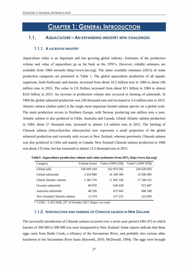
Table1: Aquaculture production volume and value (estimates from 2015, http://www.fao.org)
Category Volume (tons) Value (1000 US$) Value* (1000 NZ$)
Global (all) 106 004 184 162 974 582 236 639 093
Global salmonids 3 410 890 16 189 384 23 506 985
Global Atlantic salmon 2 381 576 11 945 146 17 344 351
Oceania salmonids 60 870 630 639 915 687
Australia salmonids 48 336 472 645 686 280
New Zealand Chinook salmon 12 474 157 233 215 093
* 1 US$ = 1.452 NZ$, 25th of October 2017 (https://xe.com)
1.1.2. INTRODUCTION AND FARMING OF CHINOOK SALMON IN NEW ZEALAND
The successful introduction of Chinook salmon occurred over a seven year period (1901-07) in which
batches of 300 000 to 500 000 ova were transported to New Zealand. Some reports indicate that these
eggs came from Battle Creek, a tributary of the Sacramento River, and probably also various other
hatcheries in the Sacramento River basin (Haworth, 2010; McDowall, 1994). The eggs were brought

CHAPTER 1: GENERAL INTRODUCTION
28
to the newly built hatchery on the Hakataramea River, a tributary of the Waitaki River in the South
Island. The first reports of returning mature Chinook salmon were in 1906 and returns of a couple of
hundred fish were reported in 1908. It was deducted at the time that these adult Chinook salmon
originated from the 1901-07 releases. In the following decade the species spread into the rivers on the
East Coast of the South Island of New Zealand (Quinn & Unwin, 1993) with currently major runs of
Chinook salmon present in all the major rivers on the East Coast, minor and erratic runs in the rivers
on the West Coast of the South Island and only occasional stray’s into the rivers of the North Island
(McDowall, 1994). The rivers supporting the largest populations of Chinook salmon in New Zealand
are the Waitaki, Rangitata, Rakaia and Waimakariri River. These rivers are primarily fed by snow and
ice melt from the Southern Alps flowing east to the South Pacific Ocean (Quinn et al., 2001).
Fishing for salmon in the rivers was part of the heritage and culture of the colonists who left their
home countries behind to come to New Zealand. In addition, Chinook salmon was a prized game fish
in California and was therefore selected for introduction in New Zealand. From 1900 until 1930 many
small hatcheries in New Zealand produced post-hatching and juvenile stage Chinook salmon to stock
rivers for anglers. From 1930 onwards these operations came under threat by the development of
hydroelectric dams, which destroyed many natural runs to the original spawning grounds up-river.
During the 1950’s a battle ensued between the government, who wanted to commercialise salmon
fishing, and the Acclimatisation Societies, who wanted to conserve salmon for sport fishing purposes.
In the 1960’s the Acclimatisation Societies and the Marine Department realised that more knowledge
was necessary to save the Chinook salmon from local extinction due to the development of dams and
the fishing in rivers and at sea. This led to the building of hatcheries as research facilities, for example
the Glenariffe Salmon Research Station and the Silverstream Hatchery. In the 1970’s a legislative
battle started between the Acclimatisation Societies and the government for the approval of salmon
and trout farming. Although in 1972 a salmon and trout farming bill was passed in parliament,
opposing parties continued to battle in the 80’s and 90’s. Today only salmon is allowed to be farmed.
By 1980 the Acclimation Societies recognised that farming salmon by ocean ranching methods would
increase the number of salmon in the rivers and therefore the ability to sport fish. The ocean ranching
farming method catches adult salmon in the rivers, fertilises the eggs and grows the embryos to
juvenile stages. The juvenile salmon are released in the streams, which go to sea to grow and the
adults are caught again when they return to the streams. A major issue with ocean ranching is the
salmon by-catch at sea. This caused the rapid decline of returning salmon to the rivers and the
marginal successes of ocean ranching methods. In 1983 a bill to legalise sea cage farming was
proposed. This led to the development of the current aquaculture companies farming Chinook salmon
in sea cages, amongst which New Zealand King Salmon is the largest with a production of about 8000
tons of salmon per year. A detailed history of the New Zealand salmon farming industry can be found
in the book: ‘Swimming Upstream’ by Jennifer Haworth (2010).
CHAPTER 1: GENERAL INTRODUCTION
29
1.1.3. CHALLENGES
1.1.3.1. An expanding industry
Although aquaculture is expanding worldwide, the industry also faces challenges. For example the
growth in production in Nordic countries such as Denmark, Faroe Islands, Finland, Iceland and
Sweden, was stagnant in 2010-2011. Changes in the natural environment, e.g. multiple years with
lower seawater temperatures, together with legislative changes that limit access to freshwater and
seawater sites, strict rules on waste water control and animal welfare can slow down the industry
(Badiola et al., 2012; Dalsgaard et al., 2013). Moreover, environmental and legislative changes will
become more relevant on a global scale in the future (Martins et al., 2010; Paisley et al., 2010). Easing
some of these constraints, recirculating aquaculture systems (RAS) allow for the industry to further
expand via land-based facilities and diminishing the need for extensive marine infrastructure. The
grow-out of sea stage juvenile and adult animals (= post-smolts) takes place in sea cages, while
traditionally the egg incubation and juvenile growth in freshwater (= pre-smolts) takes place in land
based systems such as ponds, raceways and lakes (Bergheim et al., 2009; Terjesen et al., 2009). The
RAS systems allow for incubation, juvenile growth and part of the adult grow-out stage to take place
on land. While pre-smolt stages were grown to a size range of 30 to 170 g before going to sea, the
RAS system allows to grow pre-smolt stages up to a size of 1000g before going to sea (Bergheim et
al., 2009; Dalsgaard et al., 2013). The use of RAS systems reduces the water consumption and
increases the control of waste water. Furthermore, the temperature and water quality parameters such
as dissolved oxygen, carbon dioxide, nitrogen-products, pH, salinity and light, can be perfectly
controlled allowing rearing conditions, growth rate and food utilisation to be optimised. The RAS
systems also allow for a better year round production of salmon because seasonal effects of sea water
temperature and light, and the time that the animal is in a sea cage, are reduced (Dalsgaard et al.,
2013). Welfare of farmed animals has become a major topic over recent years. A RAS system allows
for increased sustainability on the level of welfare issues (Badiola et al., 2012; Martins et al., 2010).
Although current RAS systems are still sensitive to technical failures (Badiola et al., 2012),
improvements in the engineering of RAS systems could provide a very stable environment, with
temperatures and other water quality parameters under full control in all seasons. Thus, increasing the
welfare of the farmed animal (Zhang et al., 2011).
1.1.3.2. Skeletal anomalies
Skeletal deformities present a major challenge to teleost aquaculture. The current knowledge of the
aetiology, ontogeny, development and types of normal and abnormal skeletal tissues in farmed
teleosts was reviewed by Boglione et al. (2013a; 2013b) and by Fjelldal et al. (2012) for Atlantic
salmon. These authors made two important conclusions: (i) the underlying cause of skeletal anomalies

CHAPTER 1: GENERAL INTRODUCTION
30
is multifactorial and (ii) the knowledge about how genetic and epigenetic factors cause skeletal
anomalies needs to be further researched and extended. Many different terms define skeletal
anomalies in scientific papers and books (Boglione et al., 2013b). However, throughout this thesis
mainly two terms will be used consistently, i.e. (i) malformation, when a skeletal structure has an
intrinsically abnormal development, and (ii) deformity, when a skeletal structure developed normally
but acquired an abnormal phenotype in a later life stage.
The oldest written and illustrated reports of external or visual deformities in caught fish date back to
the 1500s. Interestingly, salmon were the focus in two works by Pierre Belon (Belon du Mans, 1555;
Cenomani Bellonii, 1553), showing the hooked lower jaw of an adult salmon. We now know this is
not a deformity (Witten & Hall, 2002; Witten & Hall, 2003; Witten et al., 2005b). Therefore,
Rondelet (1555), who showed a pug-nosed juvenile or adult carp (not specified), was the first to
describe and illustrate a deformity. Reports of embryonic forms are much rarer and are only found
later in the literature, in the late 1800’s. Interestingly, the earliest accounts of malformations are again
in salmonids. Moreover, the ‘pug-nose’ is the phenotype that is described in post-hatch salmon and
trout (no species information) by Buckland (1877), Girdwoyn (1877) and Quatrefages (1888).
Skeletal deformities under farming conditions cause economic, biological and animal welfare issues.
Deformed animals are less likely to be accepted by the consumer (Fjelldal et al., 2012; Gjerde et al.,
2005). Normal looking animals with internal skeletal deformities may cause technical problems
during the filleting process (Gjerde et al., 2005). Rainbow trout (Oncorhynchus mykiss) was the first
species where skeletal anomalies were reported under farming conditions (Aulstad & Kittelsen, 1971).
However, it was only in the mid 1990’s that significant losses of farmed Atlantic salmon were
attributed to skeletal deformities (Vågsholm & Djupvik, 1998). In addition, certain skeletal
deformities can be present in high frequencies in externally normal animals (Gjerde et al., 2005). This
means that skeletal deformities are only noticed in the harvest stage of the animal’s life cycle.
Although all skeletal elements can be deformed, going from the lower jaw of ‘screamer’ deformities
and upper jaw of ‘pug-nose’ deformities, to deformed fin rays in the caudal fin, this thesis focusses on
the vertebral column. Three major categories of deformities of the vertebral column are distinguished
based on studies of wild and farmed salmon species, i.e. (i) vertebral axis deviations, (ii) vertebral
centra fusion and (iii) vertebral centrum deformity (Witten et al., 2009). These three major categories
were further refined into 20 deformity types based on observations in farmed Atlantic salmon.
Vertebral axis deviations include lordosis, kyphosis, and scoliosis. Vertebral centra fusions include a
varying degree of compression, elongation and complete fusion of the vertebral centra. Finally,
vertebral centrum deformities include radio-translucent and radio-dense centra, and centra symmetry
deviations. Most of the 20 deformity types identified by Witten et al. (2009) are vertebral centrum
deformities, with axis deviations (type 14, 15, 16) as a minor subset.

CHAPTER 1: GENERAL INTRODUCTION
31
Relatively few of the twenty deformities identified in Atlantic salmon were observed in Pacific
salmon. Gill and Fisk (1966) observed vertebral centra fusion, compression and vertical shift of
centra, and displacement of vertebral arches in chum (Oncorhynchus keta), pink (Oncorhynchus
gorbuscha) and sockeye salmon (Oncorhynchus nerka). They, however, did not consider displaced
associated elements as a deformity. Seymour (1959) observed mainly fusion and compression and an
occasional hyperdense vertebral centrum in four wild populations of Chinook salmon. Vertebral
column axis deviations, such as lordosis, kyphosis and scoliosis, are the main type of deformities
observed in free-living adult Chinook salmon in New Zealand. In addition, fusions, compression and
vertical shifts of vertebral centra are present but are about three times less frequent compared to axis
deviations (Davie et al., 2018). Similar deformities are observed in farmed Chinook salmon, with
vertebral column curvatures (Lordosis, Kyphosis and Scoliosis = LKS) most prevalent followed by
fusion, compression and vertical shift of vertebral centra. The deformity types can co-occur in a single
vertebral column and can occur over its entire length (Perrott et al., 2018). Of the 20 deformity types
described by Witten et al. (Witten et al., 2009), nine were very uncommon. These were types 4,
compression without X-structure, 9 elongation, 10 widely spaced and undersized, 11 pronounced
biconcave, 12 hyper-radiodense, 13 hyper-radiodense with flat endplates, 18 irregular internal
structures, 19 internal dorsal or ventral shift, 20 severe multiple malformations. Together these nine
deformity types made up four of the 327 deformities observed using the Witten system (Perrott et al.,
2018). Interestingly, the vertebral column curvatures mostly occur in late sea life stages of farmed
Chinook salmon, i.e. in the last three to nine months before harvest, and only rarely in pre-
smoltification fresh water stages. The LKS deformity has been associated with unilateral perivertebral
fibrosis of the vertical septum and adjacent muscle fibre bundles. The cause of the fibrosis and
whether the fibrosis causes LKS remains to be elucidated (Munday et al., 2016).
Based on observations made on X-ray, an overall deformity prevalence of 4.3% was reported in
farmed New Zealand Chinook salmon freshwater stages (Munday et al., 2018). Vertebral fusion was
the most important deformity type with a prevalence of 2.7% followed by vertebral compression and
vertical shift of the entire centrum, both with a prevalence of 1.3% (Munday et al., 2018). LKS
deformities (0.1%) were shown to be very rare in freshwater stage Chinook salmon (Munday et al.,
2018). For farmed New Zealand Chinook salmon at grading in sea and at harvest, respectively 0.9%
and 4.6% of the animals were reported to have visual deformities (Perrott et al., 2018). The overall
deformity prevalence at grading was reported to be 8.8% with vertebral fusion (5.6%) the most
common deformity type, followed by vertebral compression (3.0%), vertical shift of the entire
centrum (0.5%) and LKS (0.5%) (Perrott et al., 2018). At harvest the overall deformity prevalence
was reported to be 38.4%, a large increase compared to the prevalence at grading. An increase in the
prevalence of LKS (29.4%) and vertebral compression (22.0%) was responsible for most of the
increased overall deformity prevalence at harvest (Perrott et al., 2018). Also vertebral fusion (7.6%)

CHAPTER 1: GENERAL INTRODUCTION
32
was reported to be more prevalent at harvest. In contrast, vertical shift of the entire centrum had the
same prevalence (0.5%) at harvest compared to grading (Perrott et al., 2018).
Figure 1: Deformities in New Zealand Chinook salmon
All images are digital X-ray images, oriented with anterior to the left, posterior to the right, dorsal at the top and
ventral at the bottom. All scale bars are 1 cm. (A) Normal vertebral column in an adult animal. The left vertical
bar indicates the first vertebral body. The subsequent white bars indicate every 10th vertebral body. This
specimen has 65 vertebrae in total. (B) Entire vertebral column with a region of consistently compressed
vertebrae. The compressed vertebrae (position 18 to 37) are indicated by the horizontal white bar. (C) Unilateral
posterior (first vertebra) and anterior (second vertebra) compression (arrowhead) and fusion (block arrow) of
adjacent vertebrae. (D) Fused and remodelled vertebrae (block arrow). Three neural and haemal arches indicate
that three vertebrae are fused and remodelled. Compression is indicated by the white arrowhead, occurring
between a single and double-remodelled vertebral body. The white arrow indicates a vertically shifted vertebral

CHAPTER 1: GENERAL INTRODUCTION
33
centrum to the dorsal side. (E) Combination of lordosis (L) and kyphosis (K) with a vertically shifted vertebral
centrum to the ventral side. The line indicates compression (C) of three vertebrae at the position of the
downward bent. (F) Combination of lordosis (L), kyphosis (K) and scoliosis (S). Scoliosis is hard to observe on
a lateral X-ray, however in this image both the left (l) and right (r) part of the haemal arches are visible.
Whether or not all these deformity types are truly deformities rather than malformations is currently
unknown and represents a gap in the current knowledge. Early life stage malformations show a high
prevalence in marine fish farming (Boglione et al., 2013b), but a far lower prevalence in salmon
farming practices. This may explain why early life stage malformations in salmonids have not
received much attention in the past. In addition, the literature on early life stage malformation in
salmonids is sparse. Research on external malformations in salmonids increased after the Exxon
Valdez oil spill in Alaska. These studies focussed on the relation between level of toxins in the water
and prevalence of externally visible malformations (Finn, 2007; Johnson et al., 1998; Marty et al.,
1997; Ron et al., 2000). One study investigated the metabolic effects of pesticides in egg and post-
hatching stages of Chinook salmon (Viant et al., 2006). Deformities can occur in the three tissues that
build the early life stage vertebral column, i.e. the notochord, the cartilaginous precursors of the
neural and haemal arches and the early mineralised bone of the vertebral centrum and arches.
Categorising deformities based on these tissue types in the adult vertebral column is challenging. The
outer layer of the notochord, the notochord sheath, mineralises and appears to become bone.
However, the chordacentrum is mineralised type II collagen and is more closely related to mineralised
cartilage than to bone, which is mineralised type I collagen. The initially cartilaginous arches do
become mostly bone. Also, the observed deformities have been mainly categorised in the adult
vertebral column. Currently, knowledge about internal vertebral malformations in early life stages is
sparse and needs to be expanded. Insights into the aetiology of deformities in later life stages will
likely increase when more information is available about early malformations in salmonids.
1.2. TEMPERATURE: THE TOP EPIGENETIC FACTOR IN FISH
Temperature is one of the most important epigenetic factors affecting the physiology of most
Osteichthyes. Not surprisingly, the effects of temperature on fish were first studied in the 1800’s. The
biggest incentive for investigating temperature effects on the physiology of fish came with the
realisation that human activities had changed the temperature of water bodies (Belding, 1928). The
proliferative use of once-through cooling systems of steam electric power plants in the 60’s and 70’s
in the USA, resulted in the increase of temperature of the local water bodies due to the waste heat of
this industry. Legislation by the US government, which classified heat as a pollutant, aimed to reduce
the effect of human activity on local water bodies. In parallel, funds were made available for research
on the effect of increasing temperatures on the physiology of fish. The modern founders of fish
physiology related to temperature are F.E.J. Fry and J.R. Brett (Beitinger et al., 2000). Their work,

CHAPTER 1: GENERAL INTRODUCTION
34
and that of many others, from early 1900’s to the 1970’s, established a deeper understanding of the
multitude of effects temperature has on the physiology of temperate and coldwater teleosts (Brett,
1941; Brett, 1952; Brett, 1956; Brett, 1971; Brett et al., 1969; Fry, 1958; Fry et al., 1942; Fry et al.,
1946). Interestingly, research on effects of increased water temperature on fish life found that warmer
water in general was not lethal to fish. Rather, it was cold shock that killed fish (Beitinger et al.,
2000).
A large number of studies investigate the temperature optima of teleosts and their temperature
tolerance range. Temperature optima are often measured by survival rates of different life stages
(Beitinger & Bennett, 2000; Brett, 1956). However, the most dramatic effect temperature can have on
any aquatic life is killing the organism. The upper and lower lethal temperature limits of fish by either
observing actual death (incipient lethal temperature, ILT; Bliss & Stevens, 1937) or by observing loss
of equilibrium, i.e. loss of normal locomotion ('swimming belly-up'; critical thermal methodology,
CTM; Becker & Genoway, 1979) provides information about the temperature tolerance. The CTM
method allows estimating the lethal temperature limits without killing the fish and can therefore be
used for endangered fish species. The upper and lower lethal temperature limits are affected by the
acclimation temperature of the fish. This means that an animal acclimated to 20°C will have a higher
upper lethal temperature limit compared to an animal of the same species acclimated at 15°C. Lethal
temperature limits are expressed in a polygon where ‘critical thermal limit’ is plotted against
‘acclimation temperature’ (Fig. 2).
Since the effect of increased water temperature on cold water fish species was investigated in the 60’s
and 70’s, the cause of death of fish was expected to be warm water. In contrast, cold temperatures are
the most reported cause of temperature related death in fish. Four observations could explain the
discrepancy in number of reported ‘cold’ versus ‘heat’ deaths. First, fish increased their tolerance of
high temperatures more quickly than their tolerance of low temperatures (Brett, 1946; Davies, 1973;
Doudoroff, 1945). Second, fish lost heat tolerance more slowly than cold tolerance (Davenport &
Castle, 1895; Hathaway, 1928; Loeb & Wasteneys, 1912). Third, warm water temperatures increased
the metabolism of fish and therefore the activity levels. In contrast, cold water temperatures slowed
the fish metabolism down and caused lethargy. In addition, fish can acutely sense temperature
changes and use behaviour to avoid or escape unfavourable temperatures. The higher activity levels of
fish in warmer water allow a better avoidance of higher temperatures compared to colder temperatures
(Coutant, 1975; Neill & Magnuson, 1974; Richards, 1977). Fourth, upper temperature tolerances of
most fish were well above the ambient temperature they encountered in their natural environment
(Mundahl, 1990).

CHAPTER 1: GENERAL INTRODUCTION
35
Figure 2: The temperature polygon of Chinook salmon
Critical temperature (°C) on the y-axis is plotted against acclimation temperature (°C) on the x-axis. The
temperature polygon of adults is shown in blue, while the polygon for eggs and fry (post-hatch stages) is shown
in pink. The optimal temperature (OPT) for juveniles and adults is shown in green (between 12-13°C), while the
OPT for eggs and fry is shown in red (10°C). Further abbreviations: LLT, lower lethal temperature; ULT: upper
lethal temperature. The temperature polygons are based on data in Alderdice & Velsen (1978) and Brett (1952).
1.2.1. TEMPERATURE RANGE OF CHINOOK SALMON
Salmonids are stenothermal meaning that they have a narrow range of temperature optima (Beitinger
& Bennett, 2000). The temperature polygon of Chinook salmon is shown in Figure 2. The general
avoidance of temperatures above 15°C for all Oncorhynchus species, in spite of acclimation to this
level and to 20°C to 24°C, is marked. In addition, it seems that the range for a preferred freshwater
and seawater temperature observed among Pacific salmon frequently is 8°C to 10°C (Brett, 1952).
The optimal temperature for growth in Atlantic salmon smolts is 13°C in seawater, but growth still
occurs at temperatures up to 19°C to 20°C (Handeland et al., 2003). The seawater surface
temperatures in the Pacific Ocean native to Chinook salmon range from 9.9°C off the Adak Island
(Andreanof Islands) to 24°C in the San Diego Bay (https://www.nodc.noaa.gov). Sea water
temperatures are important for smoltification success of Pacific salmon. The likelihood of successful

CHAPTER 1: GENERAL INTRODUCTION
36
smoltification and survival after smoltification decreases rapidly with increasing temperatures in
rivers, estuaries and shore waters (McCullough, 1999).
The natural rivers in New Zealand have water temperatures from 8°C to 20°C
(https://www.niwa.co.nz). The water temperatures of New Zealand rivers cited in research articles
range between 6°C and 16°C (Davis & Unwin, 1989; Kinnison et al., 1998; Unwin, 1986; Unwin &
Glova, 1996; Unwin et al., 2000). Temperatures are likely to be lower in High Country Canterbury
rivers which are mainly glacial melt water fed (Quinn et al., 2001). Water temperatures flowing
through hatcheries are very stable, for example 11.8°C and 12.4°C for two hatcheries belonging to
New Zealand King Salmon Co Ltd The sea surface temperatures on the East coast of the South Island
range between 9°C and 19°C. (https://www.seatemperature.org). Winter temperature regimes in the
nearshore waters of the South Island’s east coast are between 8 and 10°C (Greig et al., 1988).
However, New Zealand summer temperatures are between 15 and 19°C. In the shallow waters of the
Marlborough Sounds, where the sea cages of farmed Chinook salmon are located, even higher
summer surface water temperatures (up to 23°C) have been measured (personal communication Mark
Preece). The temperature profiles of rivers in the South Island of New Zealand, and its surrounding
ocean, are ideal for Chinook salmon to thrive. However, shallow waters close to the shore line can
approach the upper lethal temperature limit.
1.2.2. USE OF TEMPERATURE IN AQUACULTURE
Temperature is a widely used tool to create both ideal husbandry strategies and strategies for year
round production (Fjelldal et al., 2012). In addition, aspects such as requirement of minerals, skeletal
development and development of deformities of the vertebral column are affected by temperature
(Boglione et al., 2013a; Boglione et al., 2013b; Dionísio et al., 2012; Takle et al., 2005).
Unsurprisingly, a large number of studies investigating the effects of temperature, including
temperature limits and temperature optima, exist for salmonids. APPENDIX A summarises some
temperature studies in salmonids. Most studies focus on survival, metabolic performance,
development, and growth performance. These studies provide knowledge to help improve farming
practices and to aid conservation efforts of these species in their native ranges.
1.2.2.1. Early stage temperatures in farming practice
Changing the temperature during incubation in farming practices can be used for several reasons.
Higher incubation temperatures will increase, while lower incubation temperatures will decrease the
developmental rate (Hayes et al., 1953). Increasing or decreasing the developmental rate will change
the time points at which certain developmental stages are reached (Mahon & Hoar, 1956; Murray &
Beacham, 1989). Thus, the time of hatching and time that the first feed stage is reached can be
shortened or prolonged (Crisp, 1981; Crisp, 1988; Jensen et al., 1989), which is useful in planning for

CHAPTER 1: GENERAL INTRODUCTION
37
harvesting salmon year round. The ‘wild’ adult salmon prior to maturation, when they are ready to
swim up-stream to their spawning grounds, is the life stage that an ocean ranch farmer aims to
harvest. In the wild, Chinook salmon return to the rivers for spawning from early spring to late
summer. If the life cycle of wild Chinook salmon is used, farmers would only be able to harvest over
spring and summer. However, if farmers can shorten or prolong the incubation time and early
freshwater stages, together with other husbandry changes in feeding regime and length of daylight
during grow-out stages, salmon will go to the sea water stage over different periods during the year.
Hence, the sea water grow-out stage will produce salmon ready to harvest throughout the year.
Gradually increasing temperature during incubation is also used to synchronise the hatching of cold
incubated eggs, after which the temperature is gradually lowered again. Although the effect of
incubation temperature has been investigated in Chinook, chum, coho (Oncorhynchus kisutch), pink
and sockeye salmon (Alderdice & Velsen, 1978; Beacham & Murray, 1990), it was unclear if the
increased temperature was the underlying direct cause of hatching. Alternatively, a reduced level of
oxygen in the incubation environment due to the temperature induced metabolic activity of the many
embryos may cause the animals to hatch (DiMichele & Powers, 1984; Dimichele & Taylor, 1980).
For example, a reduced level of oxygen at a constant 10°C caused premature hatching of chum
salmon embryos (Alderdice et al., 1958). While temperature is the main tool and beneficial from a
farm husbandry perspective, raising or lowering temperature too quickly in critically sensitive early
stages is likely to induce adverse effects.
1.2.2.2. Temperature sensitive early stages
The effect of temperature on the skeleton has been mostly observed as variation in countable elements
of the skeleton, such as vertebral bodies, gill rakers, branchiostegal rays and fin rays, i.e. meristic
variation. Meristic variation in the skeleton follows temperature clines occurring in the natural habitat.
The most famous of which is Jordan’s rule which relates meristic variation to a large North-South
latitudinal temperature scale (Jordan & Evermann, 1896). Meristic variation of skeletal elements in
salmonids has been used mostly to identify stocks or populations, but proved to be complex since
multiple skeletal elements need to be counted precisely (Beacham, 1990; Beacham et al., 1983;
Beacham & Murray, 1986; Landrum, 1966). Only the trends of several skeletal elements combined
gave significant results that could be used for clear stock identification. In addition, the counting of
skeletal elements needs to follow consistent protocols to be able to compare results between studies.
Using temperature experiments to study meristic variation in several teleost species, such as medaka
(Oryzias latipes), threespine stickleback (Gasterosteus aculeatus), sockeye, chum and pink salmon,
and rainbow trout brought several authors to the conclusion that the magnitude and duration of
exposure to changing temperatures are important in influencing skeletal variation (Batty et al., 1993;
Beacham & Murray, 1986; Kwain, 1975; Lindsey, 1988; Murray & Beacham, 1989; Seymour, 1959).

CHAPTER 1: GENERAL INTRODUCTION
38
Moreover, the developmental stages, at which temperatures are manipulated, affects meristic
variation. In salmonids, investigations of temperature sensitive stages related to meristic changes were
undertaken by Taning (1952) and Murray & Beacham (1989) in brown trout (Salmo trutta) and chum
salmon respectively. The temperature sensitive developmental stages related to meristic variation are
similar in both salmon species. The sensitive stages all occur before hatching and are: (i) the 16-cell
stage, (ii) the early blastula stage, (iii) the late blastula stage, (iv) at completion of epiboly, (v) at
completion of somitogenesis (also called initial eye pigmentation) and (vi) the eyed egg stage.
Positive or negative temperature changes of 6°C were found to either or both decrease or increase the
number of vertebrae and gill rakers (Murray & Beacham, 1989; Taning, 1952).
Temperature sensitive stages related to development of skeletal deformities were studied in Atlantic
salmon (Takle et al., 2005; Wargelius et al., 2005). In these studies heat and cold shocks were used at
specific developmental stages. Both authors found significant vertebral deformities in animals heat
shocked at the 20th to 25th somite stages. Takle et al. (2005) used a one hour heat shock of 8°C
(temperature increase) and a cold shock of 7°C (temperature decrease) at several early life stages, i.e.
before and during the somitogenesis phase. Although vertebral deformities were observed in juvenile
stages in almost all temperature treated groups (3-9%), including the control group at 8°C (7%),
animals heat shocked at the 25th somite stage and at the completion of somitogenesis showed a
significantly higher proportion of deformities. Cold shocked animals at the 45th somite stage and at
the completion of somitogenesis showed a higher proportion of vertebral deformities compared to the
control group. Wargelius et al. (2005) had a control group at 6°C and used a 6°C increase in
temperature for 24 hours as a heat shock. Although no specific proportions were given, external
malformations were observed in post-hatch stages. Juvenile specimens heat shocked at early
gastrulation showed 34% vertebral deformities, 31% when heat shocked at the 1st somite stage, 34%
when heat shocked at the 6th somite stage and 27% when heat shocked at the 15th to 20th somite stage.
The control group also had vertebral deformities (3%). The deformities observed were mainly
compression and fusions. Although temperature shocks are avoided in all forms of planned production
it is clear that salmon are vulnerable to the effects of temperature perturbations and that early
development is most temperature sensitive.
1.3. THE SALMON VERTEBRAL COLUMN
The vertebral column is a defining feature of vertebrates. The salmon vertebral column consists of
amphicoelous (biconcave) vertebrae (Fig. 3 A-B, D-E) and intervertebral ligaments. The vertebral
bodies have different functions within a single vertebral column. Attachment of the skull to the
vertebral column, protection of the neural cord, providing attachment for blood vessels, intestines,
swim bladder, reproductive organs and muscles, and transducing muscle forces are a few of their
many functions.

CHAPTER 1: GENERAL INTRODUCTION
39
Knowledge about the skeleton is mainly based on mammalian animal systems, where cartilage and
bone can be clearly separated in two tissue type categories. In the last decades it became clear that
teleost skeletal systems are not simply built of either cartilage or bone. Rather, cartilage and bone
represent two ends of a continuum of skeletal tissues (Witten & Hall, 2015; Witten et al., 2010). Both
cartilage and bone forming cells are controlled by a shared set of genes with sox9 and runx2 as
regulators for cartilage and bone respectively (Eames et al., 2004).
1.3.1. THE SALMON VERTEBRAL BODY
The salmon vertebral body is introduced based on the detailed descriptions of salmonid vertebral
bodies by Arratia & Schultze (1992). The vertebra depicted here (Fig. 3A-B, D-E) is found in late
juvenile to adult Chinook salmon. Although the schematic representations are original work (Fig. 3C,
F), the images and naming is adapted from Arratia & Schultze (1992). The combined chordacentrum,
autocentrum and the cancellous bone layers are further referred to as the vertebral centrum (Fig. 3C,
D-F). The cartilaginous bases of the associated elements, i.e. the basidorsals and basiventrals
respectively, and the bony part sticking out of the vertebral centrum are further referred to as
associated elements (Fig. 3A-C). The chordacentrum develops underneath the external elastic
membrane of the notochord sheath (Fig. 3F). The chordacentrum is the only structure of the vertebra
that develops within the notochord. All other structures of the vertebra develop perinotochordal, on
the outside of the external elastic membrane of the notochord sheath. The autocentrum, consisting of a
compact bone layer, develops on the outer surface of the external elastic membrane surrounding the
notochordally derived chordacentrum (Fig. 3F). The anterior and posterior edges of the autocentrum
are called the endplates (Fig. 3D). The compact bone layer of the autocentrum is observed as the
typical X-structure in X-ray images (Witten et al., 2009; Fig. 1). The arcocentra, also outside of the
notochord sheath but implanted at the level of the chordacentra, consist of cartilaginous parts and a
bony part. The cartilaginous parts are called the basidorsals and basiventrals (Gadow & Abbott, 1895)
which are the bases of the vertebral centrum associated elements (Fig. 3C, E). The bony part of the
arcocentrum forms the cancellous bone fused to the autocentrum (Fig. 3E), which forms the round
contour of the vertebral body (Fig. 3), and forms the bone of the associated elements (Fig. 3C). The
associated elements are the neural arches, parapophyses, ribs, haemal arches and structures of the
caudal fin skeleton.

CHAPTER 1: GENERAL INTRODUCTION
40
Figure 3: The Chinook salmon vertebral body
The images and schematics are the authors original work with naming adapted from Arratia & Schultze (1992).
(A-C) The entire caudal vertebral body (vertebral centrum and associated elements), scale bars of (A) and (B)
are 10 mm. (D-F) The vertebral centrum enlarged, scale bars of (D) and (E) are 5 mm. (A) The vertebra is
represented in a side view (anterior to the left and posterior to the right). The box indicates the enlarged image
(D). (B) Posterior view of the vertebra shown in (A). The box indicates the enlarged image (E). (C) A cross
section through the vertebral body. The neural arch and spine, and the haemal arch and spine are collectively
called the associated elements. The central part of the vertebral body to which the arches attach is called the
vertebral centrum. Bone is indicated in red, cartilage is indicated in blue. The black box indicates the enlarged
image (F). (D) Enlarged side view of the vertebral centrum [same orientation as (A)]. The vertebral endplates
(VE), the anterior and posterior part of the autocentrum and the cancellous bone (CB) are indicated. (E)
Enlarged posterior view of the vertebral centrum [same orientation as (B)]. The posterior vertebral endplate is
indicated by VE and the central canal by CC. (F) Enlarged image of the cross section of the vertebral centrum.
Notice that the chordacentrum lies underneath the external elastic membrane of the notochord sheath. All other
vertebral centrum layers developed perinotochordal and thus outside of the external elastic membrane.

CHAPTER 1: GENERAL INTRODUCTION
41
Deformities can occur in the three tissues that build the vertebral bodies and thus the vertebral
column. These tissues are the notochord, the cartilaginous precursors of the neural and haemal arches
and the early mineralised bone of the vertebral centrum and arches. Before vertebral deformities can
be correctly interpreted on a tissue level, an in-depth knowledge of the three tissue types is necessary.
Therefore the notochord, the cartilage structures associated with the axial skeleton as well as the bony
elements of the vertebral column per se are introduced below.
1.3.2. NOTOCHORD
The notochord is the key characteristic of the phylum Chordata, including lancelets
(Cephalochordata), tunicates and salps (Urochordata) and the craniates which, except for agnathans,
have vertebral columns made of vertebrae in addition to skulls. The structure was first described by
von Baer in chick embryos (von Baer, 1828). Alexander Kowalevsky (1867), a student of von Baer,
was the first to describe the development of the notochord in detail, based on observations made in
ascidians (tunicates) (Kowalevsky, 1867). Importantly, Kowalevksy (1867) suggested that the
ascidians were closely related to the vertebrates. This hypothesis gave rise to several scenarios and
theories homologising the notochord with ventral ‘chord’ structures in invertebrates such as annelids,
nemerteans and hemichordates and is a current on-going discussion (Annona et al., 2015).
The notochord in vertebrates arises from the dorsal organiser (Harland & Gerhart, 1997; Spemann &
Mangold, 1924). The equivalent of the dorsal organiser in teleosts is the embryonic shield
(Oppenheimer, 1936). The notochord anlage is formed through chordamesodermal cell movements
during gastrulation (Amacher & Kimmel, 1998; Fekany et al., 1999). Subsequently, through cell
division and intercalating cell movements of chordamesodermal cells a ‘single stack of coins’ like
notochord is formed (Grotmol et al., 2006; Kimmel et al., 1995). From these stacked cells two distinct
cell types differentiate, (i) the chordocytes or inner large vacuolated cells and (ii) the chordoblasts or
outer non-vacuolated epithelium-like cells that produce the notochord sheath (Coutinho et al., 2004;
Grotmol et al., 2003; Kryvi et al., 2017; Parsons et al., 2002; Stemple, 2005). Genes such as sneezy,
happy and dopey express coatomer complex components which were found to be important for the
vacuolisation of the chordocytes (Coutinho et al., 2004), while bashful, grumpy and sleepy express
laminin subunits important for the formation of the basement membrane of the notochord (Parsons et
al., 2002). In addition, an elegant study on zebrafish (Danio rerio) showed that the vacuoles in the
chordocytes are lysosome related organelles (LRO’s) (Ellis et al., 2013). Although the chordocytes
and chordoblasts appear to be two separate groups of cells with their own specific function,
chordoblasts have been shown to transform into chordocytes in the Atlantic salmon notochord (Kryvi
et al., 2017), which suggests that the notochord is a dynamic structure during the entire life cycle of
salmonids.

CHAPTER 1: GENERAL INTRODUCTION
42
The notochord has a dual function in embryonic and post-hatch bodies of vertebrates. It functions as
the main axial skeleton and as a midline signalling centre. In embryos and post-hatch stages, the
notochord is a hydrostatic rod to help locomotion (Satoh, 2003). For example, specific collagen
lamellae in the notochord sheath of Atlantic salmon provide torsional strength and are interconnected
to the surrounding connective tissue to direct forces from the muscles during swimming (Grotmol et
al., 2006). In juvenile and adult vertebral columns, the notochord transforms into the intervertebral
tissues (Wang, 2013). In the agnathans and some basal gnathostomes, such as dipnoans and sturgeons,
the embryonic notochord is retained throughout juvenile and adult life (Arratia et al., 2001). In
teleosts, although the chordocytes transform (Haga et al., 2009; Kryvi et al., 2017), the embryonic
notochord is retained in the centre of the vertebra to form a small central canal (Fig. 3D, E). In
addition, notochord is retained in the intervertebral spaces (Arratia et al., 2001; Grotmol et al., 2006;
Kryvi et al., 2017). In adult mammals, the nucleus pulposus is a remnant of the notochord and is
contained by an annulus fibrosus, and collectively better known as the intervertebral disc (Choi et al.,
2008; Pattappa et al., 2012). Although the notochord tissue in the intervertebral spaces in salmonids is
retained throughout life, the tissue structure changes over time in function of the forces elicited on the
vertebral column (Kryvi et al., 2017).
Importantly the notochord has a major role in the patterning of the vertebral column in teleosts
(Bensimon-Brito et al., 2012b; de Azevedo et al., 2012; Fleming et al., 2015; Grotmol et al., 2005).
However, the underlying mechanisms are less well understood. First, the notochord has an inherent
pattern that delineates the vertebral centrum anlage, i.e. the chordacentrum, representing the actual
vertebral centrum proper (Arratia et al., 2001; Fleming et al., 2004; Grotmol et al., 2003; Grotmol et
al., 2005; Nordvik et al., 2005; Wang et al., 2013). How the chordacentra pattern in the notochord
arises is currently unknown, but evidence suggests that the cells responsible for the formation of the
chordacentra have a notochord origin as laser ablation of notochord cells at specific repeated places
resulted in the loss of chordacentra in zebrafish (Fleming et al., 2004; Fleming et al., 2015). In
addition, the chordoblast cell-morphology changes and segmental expression of alkaline phosphatase
(ALP) was found to exist in the chordoblasts lining the future chordacentra in Atlantic salmon and
zebrafish (Bensimon-Brito et al., 2010; Fjelldal et al., 2005; Grotmol et al., 2003).
The notochord produces signalling molecules such as sonic hedgehog (SHH) that will signal to the
sclerotome (Fig. 4A) in the somites (Fleming et al., 2004; Fleming et al., 2001; Gilbert, 2000). The
somites are mesoderm derived and consist of two major multipotent cell populations. The sclerotome
will form the skeleton producing cells (chondroblasts and osteoblasts) while the derma-myotome will
form the dermis of the back and muscles inbetween the ribs, the deep back, the body wall, limb
muscles and the tongue (Gilbert, 2000). Interestingly, analysis of cell differentiation and biochemical
composition of extracellular material of notochord and cartilage showed that both tissues are related
(Cooper, 1965; Miller & Mathews, 1974). Modern molecular tools also confirmed that notochord

CHAPTER 1: GENERAL INTRODUCTION
43
tissue is closely related to cartilage based on shared expression of type II and IX collagen, aggrecan
and sox 9 (Domowicz et al., 1995; Grotmol et al., 2006; Ng et al., 1997; Zhao et al., 1997). However a
major difference between notochord and cartilage exists. In notochord cells, specifically the
chordocytes, hydrated materials are retained in large vacuoles to create hydrostatic pressure against
the thick sheath (Adams et al., 1990; Grotmol et al., 2006; Koehl et al., 2000). In contrast, cartilage
cells (chondroblasts and chondrocytes) secrete the hydrated materials into the extracellular matrix
(ECM) (Knudson & Knudson, 2001).

CHAPTER 1: GENERAL INTRODUCTION
44
Figure 4: Somites and sclerotome in teleosts
The schematic representations are the authors’ original work with naming adapted from Gilbert (2000) and
Morin-Kensicki et al. (2002). (A) Anterior view on one pair of teleost somites in their epithelial state. Sonic
hedgehog (SHH) signals from the notochord (NTC) to the somite induce the expression of pax 1, 9 genes
determining the sclerotomal fate of these cells. The region becoming sclerotome is shown in green. The
ectoderm (blue transparent layer) and the neural tube (NT, shown in blue) provide bone morphogenetic protein
(BMP) signals to the somites. BMP induces cells to become of derma-myotome (which is shown in red). The
notochord also secretes Noggin, a major BMP antagonist, which helps the ventral SHH signal to induce pax 1

CHAPTER 1: GENERAL INTRODUCTION
45
and pax 9 in future sclerotomal cells. (B) Same view as in (A). Sclerotomal cells (shown in green) in their
mesenchymal state have started migrating. The mesenchymal cells migrate upwards along the notochord and
neural tube and downwards along the somite to form the future neural and haemal arches, and to provide osteo-
progenitor cells which will ossify the perinotochordal vertebral centrum layers. The derma-myotome (shown in
red) has changed into a more chevron like shape but has still an epithelial phenotype. (C) Anterior-side view of
three somites (epithelial derma-myotome, shown in red) on the left side of the midline. The derma-myotome
segments on the right side were removed to be able to show the sclerotome cell (shown in green) migration. Cell
migration is shown by the arrows. Teleosts have leaky resegmentation, where sclerotome cells contribute to up
to three body segments. Migration of sclerotome cells along the notochord (shown by bottom arrow) was shown
in zebrafish (Morin-Kensicki et al., 2002).
1.3.3. CARTILAGE
Cartilage is best known as the text book mammalian hyaline cartilage and forms the basis of the
embryonic endoskeleton. However, within vertebrates, in particular teleosts, a large range of different
cartilaginous tissue types exist. Although some tissues intermediate between cartilage and bone are
considered transient or pathological in mammals (especially humans), such tissues are part of the
normal mature skeleton of teleosts and other actinopterygians (Hall & Witten, 2018; Witten et al.,
2009). This section focuses on the development of cartilage in the vertebral column of teleosts giving
rise to the associated elements, i.e. post-hatch neural and haemal arches, parapophyses, ribs and the
caudal fin skeleton. The cartilaginous bases of the neural and haemal arches and parapophyses, the
basidorsals and basiventrals, are the cartilaginous part of the arcocentra and remain cartilaginous until
adult stages.
The cartilaginous associated elements in the vertebral column are sclerotome derived. During
gastrulation, somitogenesis occurs in the embryo in an anterior to posterior direction. The number of
somites varies between species but is relatively fixed within each species (Richardson et al., 1998;
Ward & Mehta, 2014). The rate of somitogenesis can also vary within species (Schmidt & Starck,
2004; Schmidt & Starck, 2010). For example in zebrafish a pair of somites is formed about every 30
minutes (Schmidt & Starck, 2004; Schröter et al., 2008). Somites form through the interaction of
complex molecular pathways. The most widely accepted model of somitogenesis is the ‘clock and
wavefront’ mechanism (Cooke & Zeeman, 1976). Shortly after their formation, the mesenchymal
cells of the somites become epithelial through mesenchymal-epithelial transition (MET). Two cell
populations differentiate within the somite based on the signals the cells receive. The dermamyotome
is determined by BMP signalling from the neural tube and ectoderm (Fig. 4A). The sclerotome
differentiates upon receiving ventralising SHH and Noggin, which antagonises the dorsalising Bone
Morphogenetic Protein (BMP) signals. These signals from the notochord induce Pax1 (Paired box),
Pax9 and Mfh1 (Mesenchyme fork head) expression in the sclerotomal cells (Fan & Tessier-Lavigne,

CHAPTER 1: GENERAL INTRODUCTION
46
1994; Fleming et al., 2001; Furumoto et al., 1999; Hammerschmidt & McMahon, 1998; McMahon et
al., 1998; Peters et al., 1995; Pourquié et al., 1993). In amniotes the sclerotome is the ventromedial
portion of the somite (Fig. 4A). Interestingly, in zebrafish only a small group of cells found on the
anterior ventromedial edge of the somite represent the sclerotome (Morin-Kensicki & Eisen, 1997).
Upon receiving SHH signals from the notochord (Fig. 4A) the sclerotomal cell population will
undergo epithelial-mesenchymal transition (EMT). The EMT allows the sclerotomal cells to migrate
dorsally to surround the notochord and neural tube (Fig. 4B, C) (Morin-Kensicki & Eisen, 1997;
Morin-Kensicki et al., 2002). Already in 1855 (Remak, 1855) an offset between the vertebral segment
and the muscle segment was observed. Remak suggested that the vertebrae are formed by the
posterior and anterior halves of consecutive somites. This theory is now recognised as somite
resegmentation and was proven in later experimental work (Bagnall et al., 1988). How this
resegmentation happens is still largely unknown. Resegmentation allows each muscle to insert on two
consecutive vertebrae and allows the vertebral column to bend when muscles contract during
locomotion. Interestingly, in zebrafish the sclerotomal cells of a somite were observed to migrate to
the locations of a second and even third more posterior future vertebra (Fig. 4C). This observation
suggests there is ‘leaky resegmentation’ in zebrafish (Morin-Kensicki & Eisen, 1997; Morin-Kensicki
et al., 2002). Leaky resegmentation may be normal in teleosts in contrast to the one to one
resegmentation relationship observed in mammals (Witten & Hall, 2015).
In Atlantic salmon, the sclerotomal cells surrounding the notochord have initially a fibroblast
phenotype. The development of the post-hatch cartilaginous associated elements starts within the
myosepta with fibroblasts forming a mesenchymal condensation (Fig. 5A) which later develops into
hyaline cartilage (Fig. 5B) (Grotmol et al., 2006). Although remnants of the bases of the neural and
haemal arches, basidorsals and basiventrals respectively, remain cartilaginous in the adult stages, most
cartilage of the associated elements will be replaced by mineralised bone tissue during juvenile
growth stages of salmon (Fig. 5C) (Arratia & Schultze, 1992).
1.3.4. BONE
Osteogenesis (= ossification) or bone formation involves two steps. First, a scaffold or matrix is laid
down in the extracellular matrix, called ossification. Second, the bone matrix is fixed by the binding
of a mineral component (see below), called mineralisation. Calcification is a term better reserved for
processes that involve accumulations of calcium in the formation of shells in invertebrates. Bone in
the salmon vertebral body is formed via three types of ossification, i.e. perichondral, endochondral
and intramembranous ossification. The distinction between these types of ossification is based on
where the bone matrix is deposited. In perichondral ossifications, bone matrix is deposited on the
outside (peri = around) of a cartilaginous structure. All associated elements preformed in cartilage, are
initially perichondrally ossified. The cartilage of the associated elements is eventually replaced by

CHAPTER 1: GENERAL INTRODUCTION
47
bone via endochondral ossification. Here the cartilage is broken down and replaced by bone. There is
no cartilaginous precursor in intramembranous ossification. The chordacentrum mineralises within the
notochord sheath, while the autocentrum, the neural and haemal spines and vestigial skeletal
elements, such as ribs and uroneurals, form via intramembranous ossification.
Two types of bone occur in teleosts, i.e. acellular bone and cellular bone. Acellular bone has no cells
embedded within the bone tissue, while cells are embedded in the bone tissue in cellular bone. The
cells producing acellular bone are positioned on the bone tissue surface. Acellular bone was first
observed in teleost fish by Kölliker (1859). Acellular bone is a character of advanced teleosts and was
used as a character in phylogenetic studies (Maisey, 1988; Parenti, 1986; Smith & Hall, 1990). A
well-known example of a teleost with acellular bone in recent research is the model organism medaka.
Salmon, have cellular bone (Witten & Hall, 2002). The cells that produce bone are called osteoblasts.
Osteoblasts lay down the matrix (= scaffold) of the bone tissue called osteoid, which later mineralises
to form mature bone tissue. Once these cells are themselves surrounded by bone matrix they are called
osteocytes. The main component of the bone matrix is collagen type I. Non-collagenous components,
such as osteopontin, support bone cell survival. Osteonectin is important in bone mineralisation and
osteocalcin functions during bone remodelling (Hall, 2015a). The mineral component of bone in
vertebrates is hydroxyapatite (Ca10(PO4)6OH2) which is linked to the collagen matrix mediated by
osteonectin. Bone is resorbed by three cell types, multi-nucleated osteoclasts, mono-nucleated
osteoclasts and by osteocytes. Osteoclasts break down the mineral and matrix component of bone, a
process called resorption. Osteocytes only remove the mineral component from the bone tissue, called
osteocytic osteolysis. Together, the bone resorbing cells and bone producing cells form a highly active
tissue where this tissue turnover provides great potential to repair and remodel when damage occurs.
The bony layers of the vertebral centrum external to the notochord in salmon, i.e. the autocentrum and
the arcocentrum, are formed via intramembranous ossification. This perinotochordal bone is deposited
directly on the outside of the external elastic membrane of the notochord sheath at the level of the
chordacentrum (Fig. 5A) (Fleming et al., 2004; Grotmol et al., 2006; Inohaya et al., 2007). Three
classes of sclerotomal derived cells have been recognised during the early stages of vertebral body
formation (Fig. 5B, C). The first class of cells are non-mature osteoblasts scattered on the notochord
elastic external membrane, outside the chordacentrum. These cells express ALP and COL10α1 and
may contribute to perinotochordal ossification (Grotmol et al., 2005). The largest number of
sclerotomal cells, class two, are clustered in the intervertebral region and differentiate into fibroblasts
(Inohaya et al., 2007). Although these cells have no apparent osteoblast properties they secrete ECM
molecules outside of the notochord sheath. Class three sclerotomal cells are a population of mature
osteoblasts expressing osterix, an essential transcription factor for osteoblast differentiation
(Nakashima et al., 2002). These cells are found at the anterior and posterior rims of the autocentrum,
i.e. at the vertebral endplates, and were suggested to have differentiated from the class two cells in the

CHAPTER 1: GENERAL INTRODUCTION
48
intervertebral region (Inohaya et al., 2007; Renn & Winkler, 2009; Spoorendonk et al., 2008; Willems
et al., 2012). Class three cells also ossify the autocentrum and the bony part of the arcocentrum
(ossification of the arches and the cancellous bone, Fig. 3C, F). Salmon bone, formed by different
sclerotome cell types and different ossification types, is clearly complex but a dynamic tissue.
Figure 5: From notochord to vertebral body in salmonids (right)
The schematic representations are the authors’ original work adapted from Fleming et al. (2004), Grotmol et al.
(2005), Grotmol et al. (2006), Inohaya et al. (2007) and Wang (2013). (A-C) All figures have the same
orientation, with anterior to the left, posterior to the right, dorsal at the top and ventral at the bottom. The first
half shows a longitudinal section along the midline while the second half shows the entire external contour.
Corresponding associated elements on the ventral side are not shown. (A) Mesenchymal condensations (shown
in green) form the precursors of the basidorsals and cartilaginous neural arches [at ±400°d (degreedays) for
Atlantic salmon]. These condensations implant on the outside of the external elastic membrane (black line) of
the notochord sheath (see Fig. 3F, basidorsals and basiventrals). Sclerotomal cells are also scattered outside of
the notochord sheath. (B) The mesenchymal condensations have developed into cartilaginous neural arches.
Within the notochord sheath the chordacentra have mineralised and form closed rings (at ±750°d for Atlantic
salmon). Sclerotomal cells occur scattered on the outer surface of the arches and the notochord. Class I
sclerotomal cells occur perinotochordal at the level of the chordacentrum, while class II cells occur in the future
intervertebral regions. (C) The neural arches have ossified perichondrally except for the basidorsals which
remain cartilaginous. The perinotochordal autocentrum has also ossified. Sclerotomal cells and osteoblasts occur
scattered on the neural arch and the autocentrum. Class III cells occur at the anterior and posterior rims of the
autocentrum (indicated on the future posterior endplate), while class II cells (darker green) still occur in the
intervertebral region.
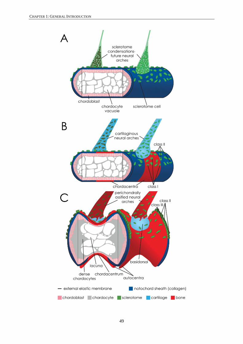
CHAPTER 1: GENERAL INTRODUCTION
49

CHAPTER 1: GENERAL INTRODUCTION
50
1.4. PACIFIC SALMON
“In spring the sea is filled with migrating fishes, some of them bound for the
mouths of great rivers, which they will ascend to deposit their spawn. Such
are the spring-run Chinook salmon coming in from the deep Pacific feeding
grounds to breast the rolling flood of the Columbia, the shad moving into the
Chesapeake and the Hudson and the Connecticut, the alewives seeking a
hundred coastal streams of New England, the salmon feeling their way to the
Penobscot and the Kennebec. For months or years these fish have known
only the vast spaces of ocean. Now the spring sea and the maturing of their
own bodies lead them back to the rivers of their birth.” In ‘The Sea Around
Us’ by Rachel Carson (1950)
Salmon are actinopterygian (ray-finned) fish and part of the infraclass Teleostei (from Greek complete
bone). The order Salmoniformes belongs to the superorder Protacanthopterygii. Salmonids may be the
best researched teleost fish family, but the phylogeny of the Salmoniformes clade remains debated
(Shedko et al., 2013). The Salmonidae family contains three main lineages, the subfamilies
Thymallinae, Coregoninae and Salmoninae. The Salmoninae count six genera, i.e. Hucho,
Brachymystax, Salmo (including Atlantic salmon), Parahucho, Oncorhynchus (the Pacific salmon
including Chinook salmon) and Salvelinus. The genus Oncorhynchus includes eight species of which
Chinook salmon (Oncorhynchus tshawytscha), commonly known as king salmon in New Zealand, is
the largest.
The distribution of native Pacific salmon species is the Northern part of the Pacific Ocean and in the
Arctic Ocean. Pacific salmon occur from the Laptev Sea North of Russia to the Beaufort Sea North of
Alaska in the Arctic Circle. The Southern-most border ranges from the Sea of Japan (35° north) to the
Californian Coast line (28° north). Although most species of Pacific salmon have an overlapping
native range, some species range very widely such as the Chum salmon, while others have a smaller
range such as masu salmon (Oncorhynchus masu), ranging only in the Western Pacific Ocean.
1.4.1. CHINOOK SALMON DISTRIBUTION AND LIFE CYCLE
Different synonyms for Chinook salmon, such as king salmon, spring salmon and Quinnat salmon are
used throughout the literature. The Chinook salmon native range starts 35° north in the West Pacific
around Japan, and spreads over the Kamchatka peninsula to the East Siberian sea (72° north). In the
East Pacific Chinook salmon range from the Bering Sea (57° north) in the North to the Californian
peninsula about 28° north (http://www.fao.org).

CHAPTER 1: GENERAL INTRODUCTION
51
Chinook salmon have an anadromous life cycle with reproduction in fresh water and juvenile to adult
growth in seawater (Bradford, 1995). Adult male and female Chinook salmon enter the fresh water
rivers typically in spring to late summer (cf. name spring salmon) but spawn mostly in autumn. The
life cycle starts as fertilised eggs (one of the largest teleost eggs of about 7 mm across) in the gravel
substrate of river beds. The eggs develop and alevins hatch from the eggs typically over the winter
period (Phillips & Wright, 2006). The alevins remain within the gravel substrate while they absorb
their yolk-sac. Once the yolk-sac is absorbed they are called fry and will emerge from between the
gravel. In slow growth environments (e.g., cold rivers at higher latitudes and/or higher elevation) the
juveniles tend to remain in streams, feeding chiefly on aquatic insects, for one year prior to seaward
migration in the following spring. This type of life history called ‘stream-type’ (Unwin, 1986; Unwin
& Lucas, 1993). Chinook salmon that migrate seaward in their first year, from spring through to early
fall, and often feeding in estuaries for weeks or months, are called ‘ocean-type’ (Quinn & Unwin,
1993; Taylor, 1990). Once juveniles move to the sea they are called smolts. The physiological process
by which juvenile salmon adapt to sea water is called smoltification. The smolts will form schools and
travel to the open ocean to feed on small marine crustaceans, squid and small fish such as suary,
anchovy and mackerel (Davis, 2003). After spending one to five years in the ocean the fully grown
salmon home to the river they exited at smoltification. The animals stop feeding before swimming up-
stream and start maturing once they start swimming up-stream. Some salmon swim more than a
thousand kilometers (Hinch & Rand, 1998). Remarkably, the adult males and females will return to
the exact place where they hatched (=homing or philopatry). Females select, prepare and guard nest
sites and males compete for access to mature females. The female excavates a redd (=nest) into the
gravel substrate of the river bottom by waving the pectoral fins and tail fin. The redd is an oval shaped
depression with a length that can range from 2.4 to 10 meters and a width that can range from 1.2 to
9.5 meters (Deverall et al., 1993). The female can build several redds and will in total deposit around
7000-10000 eggs which are fertilised and buried by the parents. The female will guard the nest site as
long as possible but will eventually die.
1.4.2. BATTLE CREEK CHINOOK SALMON
One native river system that deserves an introduction in this thesis is the Battle Creek watershed, a
tributary of the Sacramento River (California, USA). First, the spawning Chinook salmon adults in
this creek likely provided the progeny from which the entire current New Zealand stock is derived
(Haworth, 2010; McDowall, 1994; see SECTION 1.1.2). Second, Chinook salmon in this river system
currently arrive and spawn throughout the year, with major spring and winter runs. Runs occurring
during the four seasons are unique. The water temperatures in Battle Creek range from 11 to 20°C
(Ward & Kier, 1999). The local populations of salmonids in Battle Creek were declining due to
human impact, such as flood control, irrigation, hydro-electric power and domestic water supply. The

CHAPTER 1: GENERAL INTRODUCTION
52
unique year-round runs and the heritage value of this river system and its aquatic life were recognised
in the early 2000’s. A major government project by the U.S. Department of the Interior, Bureau of
Reclamation (Mid-Pacific Region) and California State Water Recourse Control Board proposed the
Battle Creek Salmon and Steelhead Restoration Project. This includes identifying problems that cause
salmon populations to decline, monitor populations and changing the hydro-geological outlay of
creeks and rivers to minimise the human impact and to allow adult salmon to go back to habitat that
was previously closed off. The full documentation of this project is available on the following site:
https://www.usbr.gov/mp/battlecreek/docs.html#deir.
1.5. GOALS AND OUTLINE OF THE THESIS
Chinook salmon have a well-studied biology, produce healthy fish for human consumption and
salmon farming has a well-established social license to operate. Environmental factors such as
temperature, oxygen, and light regime are adjusted to facilitate ideal farming conditions. Most
important of these is temperature which is manipulated to change the growth rate of farmed animals in
parts of the production cycle (Boglione et al., 2013a; Boglione et al., 2013b). For example,
temperatures are increased to induce fast growth. Yet, fast growth is frequently associated with
skeletal deformities (Fjelldal et al., 2012). Nutritional factors also affect deformity incidence
(Boglione et al., 2013b) and complex interactions between epigenetics (= environment, nutrition, etc.)
and genetics remain unelucidated.
Deformities of the axial skeleton are a severe and recurrent problem in salmon aquaculture. Vertebral
column deformities and other skeletal deformities of the skull and fins affect growth and performance
of the fish and lower the value of the fish product. Most of the limited data available about the
pathogenesis of deformities in Oncorhynchus species comes from Rainbow trout (Oncorhynchus
mykiss). A large volume of information is available for Atlantic salmon, because this species is the
primary farmed salmonid. However, Atlantic salmon is part of a different genus (Salmo) compared to
the Pacific salmon species (Oncorhynchus) making extrapolation of knowledge sometimes difficult.
Major classes of deformities, separately or in combination, present as a bending of the vertebral
column, as a shortening of the body axis and as deformities of vertebrae themselves. In addition,
skeletal deformities constitute a severe but rather poorly characterized problem for animal welfare
(Witten et al., 2006). Current knowledge suggests that vertebral malformations can start to develop
from early post-hatch life stages to late pre-harvesting stages (Fjelldal et al., 2012; Perrott et al.,
2018). Yet, how and when exactly malformations and deformities start to develop remains poorly
understood.
This research aims to help characterise skeletal deformities in early life stages of farmed Chinook
salmon and to help understand the aetiology of skeletal malformations. To do so, we test the

CHAPTER 1: GENERAL INTRODUCTION
53
hypothesis that incubation temperature affects the initiation and development of vertebral
malformations. Control of skeletal deformities in New Zealand Chinook salmon to within acceptably
low levels in commercial farm setting requires (a) identification of different skeletal anomalies, (b)
identification of sensitive life stages at which skeletal anomalies start, (c) an in-depth understanding
of the pathological processes underlying skeletal anomalies and (d) and in-depth understanding of
how epigenetic or genetic factors influence the malformation process (Boglione et al., 2013a; Witten
et al., 2009).
The current research focused on the following research tasks; outlined in CHAPTER 2:
1. DIAGNOSTIC IMAGING. Techniques such as whole-mount staining for cartilage and mineralized
bone and serial histological sectioning of different life stages were implemented. In addition
high-contrast and high-resolution X-ray imaging of the vertebral column of Chinook salmon
of different sizes was used.
2. IDENTIFICATION OF SENSITIVE LIFE STAGES. Patterns of vertebral mineralisation as well as the
development and progression of malformations were characterised. It is intuitive that
initiation of malformations would occur before they become grossly evident. Therefore the
task focussed on stages between hatching and first feed (= yolk sac absorbed and actively
searching for food).
3. IDENTIFICATION OF TEMPERATURE EFFECTS. The effect of constant temperature on vertebral
column development was investigated in post-hatch stages. In particular, the effect of
temperature on the vertebral column regionalisation, meristic variation and the rate of
malformation was of interest.
The temperature experiment provided the samples for each study of which the results are found in
CHAPTER 3-6. These Chapters are formatted as manuscripts and are either published or prepared for
submission to peer-reviewed journals. Post-hatch life stages were used to investigate if temperature
influences the rate of malformations occurring in early life stages (CHAPTER 3, ‘The external
phenotype-skeleton link in post-hatch farmed Chinook salmon’, Journal of Fish Diseases, 2017).
A close analysis of the juvenile vertebral column showed that six anatomical regions can be
recognised in the Chinook salmon. The implications of the observed regionalisation are discussed
from an evo-devo (evolutionary and developmental) perspective and provide evidence for an
alternative regionalisation pattern compared to mammalians (CHAPTER 4: ‘Vertebral column
regionalisation in juvenile Chinook salmon (Oncorhynchus tshawytscha)’, Journal of Anatomy,
2017). The vertebral column regions are temperature sensitive and show temperature dependent
meristic variation (CHAPTER 5: ‘Temperature sensitive regions of the Chinook salmon vertebral
column: Vestiges and meristic variation’, Journal of Morphology, 2018). Interestingly, the regional

CHAPTER 1: GENERAL INTRODUCTION
54
variation is masked by a similar total number of vertebrae in each temperature group. Temperature
also has a significant effect on the deformity prevalence of LKS, fusion, compression and vertical
shift in freshwater smoltification and seawater stage Chinook salmon (CHAPTER 6: ‘Lower
incubation and freshwater grow-out temperatures protects Chinook salmon from subsequent
vertebral deformities in seawater but not freshwater’, prepared for submission). High mortality
after seawater transfer was highlighted, as well as notable deformities in ‘runts’, a category of under-
performing animals with poorly understood reasons for retarded growth.
The thesis concludes with a general discussion (CHAPTER 7). Here, the results are integrated and
further discussed in a developmental context. Furthermore, practical implications of the results, and
the relationship of malformations and deformities are further explored. For example, constant
temperature may influence the proportions of occurring malformations and deformities but is unlikely
the direct cause of malformations. In addition, malformations in early life stages that survive in the
farming environment may foreshadow deformities observed later in life. Finally, some
recommendations for the salmon farming industry are made.
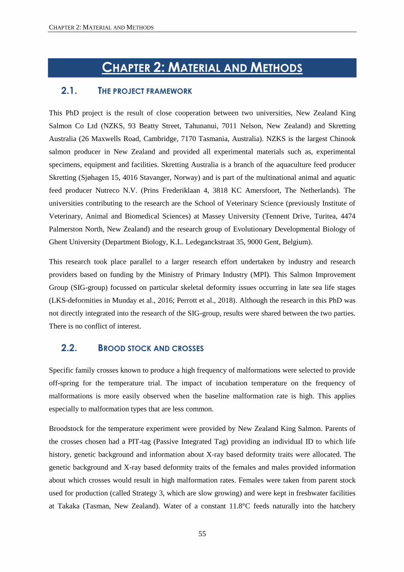
CHAPTER 2: MATERIAL AND METHODS
55
CHAPTER 2: MATERIAL AND METHODS
2.1. THE PROJECT FRAMEWORK
This PhD project is the result of close cooperation between two universities, New Zealand King
Salmon Co Ltd (NZKS, 93 Beatty Street, Tahunanui, 7011 Nelson, New Zealand) and Skretting
Australia (26 Maxwells Road, Cambridge, 7170 Tasmania, Australia). NZKS is the largest Chinook
salmon producer in New Zealand and provided all experimental materials such as, experimental
specimens, equipment and facilities. Skretting Australia is a branch of the aquaculture feed producer
Skretting (Sjøhagen 15, 4016 Stavanger, Norway) and is part of the multinational animal and aquatic
feed producer Nutreco N.V. (Prins Frederiklaan 4, 3818 KC Amersfoort, The Netherlands). The
universities contributing to the research are the School of Veterinary Science (previously Institute of
Veterinary, Animal and Biomedical Sciences) at Massey University (Tennent Drive, Turitea, 4474
Palmerston North, New Zealand) and the research group of Evolutionary Developmental Biology of
Ghent University (Department Biology, K.L. Ledeganckstraat 35, 9000 Gent, Belgium).
This research took place parallel to a larger research effort undertaken by industry and research
providers based on funding by the Ministry of Primary Industry (MPI). This Salmon Improvement
Group (SIG-group) focussed on particular skeletal deformity issues occurring in late sea life stages
(LKS-deformities in Munday et al., 2016; Perrott et al., 2018). Although the research in this PhD was
not directly integrated into the research of the SIG-group, results were shared between the two parties.
There is no conflict of interest.
2.2. BROOD STOCK AND CROSSES
Specific family crosses known to produce a high frequency of malformations were selected to provide
off-spring for the temperature trial. The impact of incubation temperature on the frequency of
malformations is more easily observed when the baseline malformation rate is high. This applies
especially to malformation types that are less common.
Broodstock for the temperature experiment were provided by New Zealand King Salmon. Parents of
the crosses chosen had a PIT-tag (Passive Integrated Tag) providing an individual ID to which life
history, genetic background and information about X-ray based deformity traits were allocated. The
genetic background and X-ray based deformity traits of the females and males provided information
about which crosses would result in high malformation rates. Females were taken from parent stock
used for production (called Strategy 3, which are slow growing) and were kept in freshwater facilities
at Takaka (Tasman, New Zealand). Water of a constant 11.8°C feeds naturally into the hatchery

CHAPTER 2: MATERIAL AND METHODS
56
facilities originating from upwelling freshwater springs (Te Waikoropupu Springs). Eggs from 12
females were obtained by stripping individuals following aquaculture standards. Eggs were gently
removed from the ovaries, washed and stored in a plastic bag together with the PIT-tag obtained from
the body cavity. Bagged eggs were placed in polystyrene boxes at 10°C for transportation by plane to
Tentburn Hatchery (Canterbury, New Zealand). These procedures were performed by NZKS staff and
are part of the normal reproduction procedures used to generate commercially grown animals.
Male specimens were sex-reversed males and therefore had a female genetic background. Juvenile
female specimens are treated with male hormone (17-α-methyltestosterone, Sigma-Aldrich, personal
communication Mike Anderson). Consequently, the gonads developed as testes instead of ovaries.
Male specimens, also Strategy 3 production salmon, were maintained at Tentburn hatchery
(Canterbury, New Zealand). Females with high X-ray based deformity traits were crossed with males
with low X-ray based deformity traits. Knowing the genetic background of the eggs of each stripped
female, the genetic background allocated to the PIT-tag of each male was used to devise a list (Table
2). This list was created by Fiona Hely (AbacusBio, 442 Moray Place, 9016 Dunedin, New Zealand).
The top six males best suited for the male x female crosses were selected for fertilising eggs. The PIT-
tag of twelve males was scanned and specimens were tested for milt (sperm) production (see Table 2).
The males are ready for reproduction when a white opaque fluid squirted from the anal pore after
applying light pressure on the abdominal cavity. Three males, including the top two males failed to
produce milt and one suggested male on the list was not present due to mortality.

CHAPTER 2: MATERIAL AND METHODS
57
Table 2: Male and females Chinook salmon: Males indicated in blue and all females with good egg
batches were used for crosses
Males PIT-tag no. Present on farm Milt producer
45379374 Yes No
29890286 No No
0A00147173 Yes Yes
107052590 Yes Yes
20581830 Yes Yes
107076810 Yes Yes
96577575 Yes Yes
0A00062039 Yes Yes
0A00134027 Yes Yes
29893351 Yes Yes
45573861 Yes No
29885588 Yes Yes
Females PIT-tag no. 100 eggs weight (g) Total clutch weight (g) Number of eggs
095812781 22.9 1964 8576
416363382 26.3 2076 7893
0A00152279 21.7 2042 9410
0A00154108 Bad eggs Bad eggs -
0A00145867 20.1 1772 8815
091108346 Not on list* Not on list* -
076892336 19.7 1430 7258
107039261 19.5 2296 11774
045578789 23.2 1942 8370
024265075 17.0 1858 10,929
021607021 24.7 2500 10,121
073379076 22.9 2300 10,043
* Specimens not on list provided by Abacus Bio Ltd (Dunedin, New Zealand)
Specimens with bad eggs and not on the list were not used for fertilisation
2.3. FERTILISATION
2.3.1. MILTING MALES
The six males selected from the list were caught with a landing net. Each salmon was held at the
caudal end (just anterior of the caudal fin) with the non-dominant hand while the head was kept under
water. Next, the male was lifted from the water and held against the body of the handler while placing
the dominant hand on the upper abdominal region (the testis are located ventrally of the kidneys lying

CHAPTER 2: MATERIAL AND METHODS
58
ventral of the vertebral column). To extrude the sperm (= milt) hard pressure was applied on the
abdominal cavity. The tail was gently moved left and right to increase the flow of sperm. Initially,
translucent fluid (urine) squirted from the anal pore. Care was taken not to collect the urine because
this will prematurely activate the sperm due to the reduced osmolality of the urine (Rosengrave et al.,
2009). The first part of the white opaque fluid was not collected, because remnants of urine could still
be present. Thereafter, the remaining milt was captured in a plastic container or bag labelled with the
PIT-tag number to identify the male. Great care must be taken in handling adult males since these are
not anaesthetised and can move vigorously while out of the water. After milting the males, milt
quality was confirmed under a microscope. A streak of milt was placed on a microscope glass and one
drop of coelomic fluid (1x concentration) was added. Coelomic fluid was made fresh and is based on
ovarian fluid (composition see Table 3). Coelomic fluid activates sperm (Billard, 1983; Galvano et al.,
2013; İnanan & Öğretmen, 2015), which was observed under the microscope as swirls occurring on
the edge of the streak. Sperm of all males had activated after adding coelomic fluid and was used for
the fertilisation process.
Table 3: Coelomic fluid recipe for 5L of 10x concentration
Chemical Concentration (mM) Molecular weight Volume (g)
NaCl 155 58.44 452.910
KCl 3.1 74.55 11.555
MgSO4*7H2O 1.3 264.48 16.021
CaCl2*2H2O 3.4 147.02 24.993
Tris Base 20 121.1 105.720
Tris HCl 20 157.56 20.010
Personal communication Seumas Walker
2.3.2. EGG PREPARATION AND FERTILISATION
The unfertilised eggs (called green eggs) of twelve females arrived at the Tentburn hatchery site in
plastic bags (20/05/2014). Each bag contained the egg clutch and PIT-tag of a single female. Eggs
were transferred to a plastic container and the egg quality was confirmed by softly pressing fingers
into the eggs. Good quality eggs feel smooth and silky. In contrast when eggs feel like hard solid tiny
balls (like a bowl of tiny marbles) the eggs are not suitable for further fertilisation. Of the twelve egg
clutches, one clutch had a PIT-tag which was not included on the list and one clutch had bad egg
quality (hard eggs). These eggs were not used for fertilisation. Next, the entire clutch was weighed.
Subsequently, 100 eggs were weighed. These two weights were used to estimate the number of eggs
in each clutch (see Table 2). To randomise the genetic background of the crosses and thus all
subsequent sampling strategies, all female gamete subsets were mixed prior to fertilisation
(randomised). One tenth of the weight of each egg clutch was pooled into ten containers (APPENDIX B)
and some coelomic fluid was added to keep the eggs moist. Subsequently, milt from the first three

CHAPTER 2: MATERIAL AND METHODS
59
males was divided over the first five containers of eggs with a transfer pipette (APPENDIX B, 1 ml milt
from each male per container, 3 ml milt per container in total, 1 ml milt per 672 grams of egg weight).
Milt from male four to six was divided over containers six to ten. Eggs and milt were gently mixed for
30 seconds with a gloved hand and were rested for three minutes for the fertilisation process to take
place. After three minutes the eggs were gently washed two to three times in water of the same
temperature of the eggs (10°C) by gently plunging a sieve with the eggs in consecutive bowls of
water. The fertilised eggs from each of the ten containers were randomly divided over two incubation
trays and their position in the incubation stack (Figure 6) also randomised. One tray received the acces
of eggs that were not divided over the trays. Newly fertilised eggs are highly sensitive and die when
rolled or moved too vigorously. Care was taken to avoid air bubbles that collect below the bottom
mesh and may move upwards through the incubation tray suddenly causing mortality of the entire
tray.
2.4. TEMPERATURE EXPERIMENT SET-UP
A temperature experiment with three constant temperatures, 4°C, 8°C and 12°C, during the incubation
and freshwater grow-out period was devised. Subsequently, specimens would be transferred to sea
cages at ambient temperatures. The experimental set-up, for incubation and freshwater grow-out, is
schematised in Figure 6A. Large temperature steps were chosen to increase the likelihood of finding a
significant impact of temperature on development. Bore water at a constant 12.4°C was pumped into
two tanks, an ambient water tank and chilled water tank. The chilled water tank was connected to a
chiller system cooling the water down to a range of 2.5 to 3.6°C. Water from each tank was pumped
to three automated electromagnetic valves which mixed ambient water with chilled water (Fig. 6A).
Each valve could be digitally set to provide a constant temperature (±0.1°C). Water mixed to the
correct temperature was fed into three partitioned incubation stacks via a flow meter. One incubation
stack per temperature had seven incubation trays. The upper part of the incubation stack contained
three incubation trays and the lower part contained four incubation trays. Water was fed into the top
tray of each partitioning of the incubation stack (Fig. 6A). An upper and lower partition of the
incubation stack was created to avoid factors that cause mass mortalities to cascade through the entire
stack (also called tank effect) because water passing through the eggs of one tray flowed into the next
tray. The trays are designed to provide upwelling water flow to the eggs which lie on a bottom mesh
(Fig. 6B, C). A top mesh prevents the eggs from flowing to the next tray. Water coming from the last
tray was drained and not recirculated. To protect the sensitive developing eggs and yolk-sac larvae
from light, the incubation stacks were covered with black plastic during the entire incubation period
until first feed stage was reached and individuals were moved to the 420 L black circular tanks (Fig.
6D, 23A). Water mixed to the correct temperature also fed these 420 L black circular tanks to

CHAPTER 2: MATERIAL AND METHODS
60
maintain juvenile salmon at their designated water temperature. Two tanks per temperature were
operated to avoid tank effects.
Monitoring the performance of the temperature set-up was crucial. The water level gauge in the
ambient water tank, and the water level gauge and thermometer in the chilled tank were connected to
electronic alarm systems (Fig. 6A, indicated by the electric symbol). Furthermore, the electromagnetic
valves, all pumps, and the chiller system were also connected to electronic alarm systems. Alarm
connected to thermometers such as in the chilled water tank, the electromagnetic valves and the
chiller system were triggered when the temperature exceeded the programmed range. Alarms
connected to pumps and the chiller compressor were also triggered when they failed or when a power
cut occurred. A manual override to restart the chiller compressor was installed due to the old age of
the chiller compressor. This manual override allowed for a reduction in restart procedures and reduced
the impact of a chiller failure on the temperature in the cold water tank. Reset programmes and
backup mechanisms for reserve pumps were in place to reduce the risk of a complete system failure.
A major temperature spike during the sensitive incubation phase of development would have resulted
in random and non-systematic errors. Temperature spikes would have created significant
interpretation difficulties and was to be avoided at all costs.

CHAPTER 2: MATERIAL AND METHODS
61
Figure 6: Temperature experiment set-up
The entire temperature experiment set-up is shown. The water flow is indicated by arrows in the blue pipes. The
valve under the filled blue pipe section could be switched to provide tanks with water of the same constant water
as in the incubation stacks. The glycol loop is shown with purple pipes and the chiller system is shown with red
outlines. All the components connected to black lines with an electricity symbol indicate that they were
connected to an alarm system. P stands for pump. (B) Top view of an incubation tray. The line indicates the
cross section shown in (C). Eggs of Chinook salmon were placed under the top mesh. Water flow is indicated
with blue arrows. The dotted arrow indicates were the water flows out of the tray and into the next tray below.
(C) Cross section at the middle line of the incubation tray. Eggs of Chinook salmon were placed in-between the
top and bottom mesh. The upwelling water flow is indicated by blue arrows. (D) Schematic representation
(cross section) of a 420 L incubation tank. Post-hatch stages were placed in this tank at first feed stage (±900°d,
fry stage) to grow-out to juveniles. Tanks were provided with the same constant temperature water as was
provided to the incubation stacks [see filled blue pipe in (A)]. The mesh in the bottom of the tank prevented fish
from escaping. The left outflow pipe valve was always open and was used to regulate the water level. The valve
of the right outflow pipe was opened during cleaning, which increased the outflow of water and flushed waste
feed from the bottom of the tank and outflow pipes.

CHAPTER 2: MATERIAL AND METHODS
62
The cold water pump (below the cold tank), the chiller circulation pump (above the sand filter), the
glycol pump (in the purple loop) and the chiller (Fig. 6A) failed due to both power cuts and electrical
failure. Only ambient water was pumped to the electromachnetic valves when the coldwater pump
failed. The thermostats were not able to supply correct temperature water and sent an alarm signal.
The coldwater pump was restarted by manual reset. When the chiller circulation pump failed, the
chiller automatically shut down and sent an alarm signal. The automatic chiller shutdown was to
avoid water in the water-glycol heat exchanger from freezing. A back-up chiller circulation pump was
manually started until the first pump could be fixed or restarted. When the glycol pump failed, the
chiller also automatically shut down and sent an alarm signal. The glycol pump was restarted by
manual reset. The water temperature slowly increased in the incubation stack when the chiller
circulation and glycol pumps, and/or the chiller failed because the water in the coldwater tank started
to rise in temperature (Colt & Maynard, 2017). However, the 3000 L cold reservoir used here, was
shown to only increase 1°C per 30 minutes (Colt & Maynard, 2017). Therefore, the failure of the
chiller circulation and glycol pumps and/or the chiller did not produce a temperature shock. In
addition, because the chiller system used here had a manual override restart procedure, it dramatically
reduced the time to restart compared to chiller systems with automatic reboot functions (Colt &
Maynard, 2017). Therefore, any chiller failure was resolved by a manual restart procedure and did not
register in the temperature logs (personal observation).
2.5. FISH MAINTENANCE
2.5.1. FLOW PROFILE AND TEMPERATURE MEASUREMENT
The entire experimental set-up, incubation trays and tanks, was a complete flow through system. After
fertilisation, the initial temperature was set at a constant 10°C for all three temperature groups. The
water flow rate was set at 9 L/min for the first day post fertilisation. On the second day post
fertilisation the flow rate was increased to 12 L/min and stayed at 12 L/min until smoltification.
Temperature and flow rate were manually checked every hour on the first two days post fertilisation.
On the third day the temperature was gradually adjusted to a constant 4°C, 8°C and 12°C for each
temperature group at a rate of 0.2°C per hour, with a 2°C adjustment per day.
During the incubation the temperatures were constantly monitored by automated temperature data
loggers which recorded the temperature every 15 minutes. The temperature data loggers were placed
in the top tray of the upper partition and the bottom tray of the lower partition of the incubation stack.
Temperature data was downloaded when first feed stage individuals were moved from incubation
trays to the 420 L circular black tanks and when the individuals were moved to ambient water in the
larger tanks. Temperature data of all temperature groups during the entire incubation period of the
4°C group is presented in Figure 7A, B, C. Irrespective of all the alarm and back-up systems in place,

CHAPTER 2: MATERIAL AND METHODS
63
temperature spikes due to pump and chiller compressor failures (see previous section) were still
observed. However, the temperature spikes were of a very short nature, i.e. between 15 minutes and
one hour, due to a quick response to each alarm and the use of back-up systems in place. The effects
of these temperature changes are discussed in section 7.1.1 TEMPERATURE – SHOCKS AS POTENTIAL
CAUSE OF MALFORMATIONS.

CHAPTER 2: MATERIAL AND METHODS
64
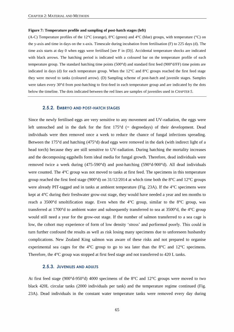
CHAPTER 2: MATERIAL AND METHODS
65
Figure 7: Temperature profile and sampling of post-hatch stages (left)
(A-C) Temperature profiles of the 12°C (orange), 8°C (green) and 4°C (blue) groups, with temperature (°C) on
the y-axis and time in days on the x-axis. Timescale during incubation from fertilisation (F) to 225 days (d). The
time axis starts at day 0 when eggs were fertilised [see F in (D)]. Accidental temperature shocks are indicated
with black arrows. The hatching period is indicated with a coloured bar on the temperature profile of each
temperature group. The standard hatching time points (500°d) and standard first feed (900°d/FF) time points are
indicated in days (d) for each temperature group. When the 12°C and 8°C groups reached the first feed stage
they were moved to tanks (coloured arrow). (D) Sampling scheme of post-hatch and juvenile stages. Samples
were taken every 30°d from post-hatching to first-feed in each temperature group and are indicated by the dots
below the timeline. The dots indicated between the red lines are samples of juveniles used in CHAPTER 5.
2.5.2. EMBRYO AND POST-HATCH STAGES
Since the newly fertilised eggs are very sensitive to any movement and UV-radiation, the eggs were
left untouched and in the dark for the first 175°d (= degreedays) of their development. Dead
individuals were then removed once a week to reduce the chance of fungal infections spreading.
Between the 175°d and hatching (475°d) dead eggs were removed in the dark (with indirect light of a
head torch) because they are still sensitive to UV-radiation. During hatching the mortality increases
and the decomposing eggshells form ideal media for fungal growth. Therefore, dead individuals were
removed twice a week during (475-590°d) and post-hatching (590°d-900°d). All dead individuals
were counted. The 4°C group was not moved to tanks at first feed. The specimens in this temperature
group reached the first feed stage (900°d) on 31/12/2014 at which time both the 8°C and 12°C groups
were already PIT-tagged and in tanks at ambient temperature (Fig. 23A). If the 4°C specimens were
kept at 4°C during their freshwater grow-out stage, they would have needed a year and ten months to
reach a 3500°d smoltification stage. Even when the 4°C group, similar to the 8°C group, was
transferred at 1700°d to ambient water and subsequently transferred to sea at 3500°d, the 4°C group
would still need a year for the grow-out stage. If the number of salmon transferred to a sea cage is
low, the cohort may experience of form of low density ‘stress’ and performed poorly. This could in
turn further confound the results as well as risk losing many specimens due to unforeseen husbandry
complications. New Zealand King salmon was aware of these risks and not prepared to organise
experimental sea cages for the 4°C group to go to sea later than the 8°C and 12°C specimens.
Therefore, the 4°C group was stopped at first feed stage and not transferred to 420 L tanks.
2.5.3. JUVENILES AND ADULTS
At first feed stage (900°d-950°d) 4000 specimens of the 8°C and 12°C groups were moved to two
black 420L circular tanks (2000 individuals per tank) and the temperature regime continued (Fig.
23A). Dead individuals in the constant water temperature tanks were removed every day during

CHAPTER 2: MATERIAL AND METHODS
66
cleaning of the tanks. The dead individuals were counted and checked for external malformations.
When cleaning tanks, care was taken to move slowly and deliberately, reducing the impact of stress
on the specimens in the tank. The second outflow valve is opened and the bottom mesh in the tank is
scrubbed and tapped to dislodge waste food and faeces. The nozzle of a water-blaster is placed on the
bottom mesh and water is carefully squirted through the mesh to dislodge and remove sludge from
underneath the mesh and from the outflow pipes. Next, water is also blasted in the second outflow
pipe to clean out extra sludge. The second outflow tap is closed for the water level to rise. The brush
and nozzle of the water-blaster is rinsed in a 3% Virkon®S (Antec International Ltd, DuPont, United
Kingdom) solution. Fish were fed intensively for the first ten days. After the initial ten days the
intensity of feeding per day was reduced and the size of the food pellet was increased. The feeding
schedule can be found in Table 4. The 8°C group showed abraded fins due to fin nipping and had a
fungal infection on the wounds of the fins. The fungal infection was successfully treated with 4.5 ppm
Halamid solution (1.8g/tank, Agrivantage, Axcentive Asia Pte Ltd, Singapore) for one hour in the
tank with a closed tap.
At 1400°d and 1530°d, 500 individuals were removed randomly in each temperature group to reduce
crowding stress, reduce the risk of infections and to allow for growth. Of the 500 culled individuals,
200 were weighed (grams) and measured (fork length in cm) (Table 12). At 2500°d (15/12/2014, 209
days post fertilisation), 500 individuals of the 12°C group were PIT-tagged and moved to two 1000 L
circular tanks (250 individuals/tank) at ambient water temperature (12.4°C) (Fig. 23A). On the same
day the temperature of the 8°C fish was gradually increased to 10°C, and then to 12°C on the next
day, at a rate of 0.2°C per hour. At 1700°d (17/12/2014, 211 days post fertilisation), 1000 individuals
of the 8°C group, now acclimated to 12°C were also PIT-tagged and moved to four 420 L black
circular tanks (250 individuals per tank) at ambient water temperature (12.4°C). During this grow out
stage another 50 random individuals were culled in each tank of both temperature groups to reduce
crowding in the tanks (Fig. 23A).
At 3348°d (29/04/2015, 344 days post fertilisation) 357 specimens of the 8°C and 376 specimens of
the 12°C (4177°d) groups were X-rayed using Kodak Industrex M100 Ready Pack II films (Kodak
Australia Pty Ltd, Victoria, Australia) (Fig. 23A, Table 12) and, after a week to recover from
radiography at Tentburn hatchery, were moved to experimental sea cages (6x6x6, 216 m3) on the
Ruakaka farm in the Marlborough Sounds (Ruakaka Bay, Queen Charlotte Sound). No specimens
were tested with a saltwater challenge to determine the freshwater smoltification status in order to
avoid reducing the number of smoltification-stage salmon that would go to seawater and available for
statistics. However, specimens were larger than the critical size for successful smoltification of
Chinook salmon in New Zealand (> 7.0 g) and entered seawater in late autumn (06/05/2015, Fig. 23,
Table 12) when Chinook salmon have a physiological smoltification window (Franklin, 1989). The
feeding schedule for sea stages can be found in Table 4. Consecutive digital X-rays/radiographs were

CHAPTER 2: MATERIAL AND METHODS
67
taken of PIT-tagged specimens from the 8°C and 12°C groups on three further occasions, six-months-
at-sea, 12-months-at-sea (Fig. 23B), at 15-months-at-see and at harvest (826 days post fertilisation).
While 339, 8°C and 271, 12°C specimens were transferred to sea cages many failed to thrive and were
culled when they were classified as runts or died. Following standard farming protocol, specimens
were classified as runts when they weighed less than a fifth of the average weight of the cohort. Runts
were euthanized with anaesthetic overdose of AquiS after they were X-rayed. Only twenty 8°C and
seven 12°C specimens survived until harvest stage.
Table 4: Feeding schedule from first feed to smoltification
Food size Food brand Frequency per day Time
Powder meal Str. 00, Golden Prima, Biomar Every 20 minutes 5 days FW
Crushed meal Crumble, Golden Prima, Biomar Every hour 5 days FW
Crushed/0.8 mm 0.8 mm GAP Semi-Float, Biomar 3 times per day 4 weeks FW
0.8/1.3 mm GAP Semi-Float, Biomar 3 times per day 3 weeks FW
1.3/1.5 mm GAP Semi-Float, Biomar 3 times per day 1 week FW
1.5 mm GAP Semi-Float, Biomar 3 times per day 6 weeks FW
2.0 mm GAP Semi-Float, Biomar 3 times per day 16 weeks FW
3.0 mm Quinnat GAP Immune Transfer Sink, Biomar Hand fed to satiation 24 weeks SW
4.0 mm Orient S 100 75 ppm Bulk, Skretting Hand fed to satiation 10 weeks SW
9.0 mm Orient HT 2000 40as 1050 kg, Skretting Hand fed to satiation 31 weeks SW
FW = freshwater, SW = seawater
2.6. SAMPLING
2.6.1. POST-HATCH STAGES
Three independent 50 egg samples from randomly selected trays were taken to determine fertilisation
rates. Fertilisation rates of 82-88% were observed. The first sample was taken after hatching (500°d).
From 500°d until 900°d, 200 randomly selected specimens from four different randomly selected
incubation trays were sampled every 30°d in each temperature group (Fig. 7D). Samples were fixed in
paraformaldehyde (PFA) based fixatives, which are ideal for histology because these fixatives cross-
link proteins and therefore mostly retain the inter- and intra-cellular structure of the tissue (Howat &
Wilson, 2014; Thavarajah et al., 2012). RNase free PFA was made by treating the demineralised
water with DEPC (diethyl pyrocarbonate, Sigma-Aldrich New Zealand Ltd, Auckland) and by using
autoclaved phosphate buffered saline solution (PBS) to dissolve the PFA. Fifty specimens were fixed
in 4% buffered paraformaldehyde (PFA), 100 specimens were fixed in 4% buffered RNase free PFA
and 50 specimens were fixed in buffered paraformaldehyde-lysine-periodate (PLP; McLean &
Nakane, 1974). Specimens fixed in 4% PFA and PLP were transferred gradually to 70% ethanol.
Specimens fixed in 4% RNase free PFA were gradually transferred either to 100% acetone or 100%

CHAPTER 2: MATERIAL AND METHODS
68
methanol and were kept at -20°C (see Table 5). Specimens were fixed in PLP because this was shown
to be a better fixative for embedding procedures aimed at electron microscopy. Specimens were fixed
in RNase free PFA to have the option to do whole mount in-situ hybridization, immunohistochemistry
and genotyping. Tissues were banked in a way to facilitate further research and depending on the
outcome of more descriptive studies informing the origin of malformations

CHAPTER 2: MATERIAL AND METHODS
Table 5: Fixing and storage solution schedule of a single sample of 200 specimens
No specimens Fixative Solution 1 Solution 2 Storage Temperature
Type Time (days) Type Time (days) Type Time (days)
50 4% PFA 3 30% ethanol 3 50% ethanol 3 70% ethanol Room temp
50 PLP 3 30% ethanol 3 50% ethanol 3 70% ethanol Room temp
50 4% PFA Rnase - 3 50% acetone 3 3 100% acetone -20°C
50 4% PFA Rnase - 3 30% methanol 3 70% methanol 3 100% methanol -20°C

CHAPTER 2: MATERIAL AND METHODS

CHAPTER 2: MATERIAL AND METHODS
71
2.6.2. JUVENILES
One hundred juvenile individuals were randomly sampled at 1400°d and 1530°d from both the 8°C
and 12°C groups, were fixed in 4% buffered PFA and were transferred gradually to 70% ethanol. The
fixing steps and transfer to 30%, 50% and 70% ethanol took seven days each.
2.7. RADIOLOGY
2.7.1. RADIOGRAPHY
The X-rays images were obtained using an Atomscope HF80/15+ portable X-ray Unit (Mikasa,
Tokyo). Freshwater smoltification-stage salmon were exposed to X-ray on Kodak Industrex M100
Ready Pack II films (Kodak Australia Pty Ltd, Victoria), while a 90-479 Sound Technologies Tru-DR
flat panel amorphous silicon digital radiographic receptor (DLC Australia Pty Ltd, Melbourne) was
used to obtain digital X-ray images of post-smoltification seawater stage salmon. The X-ray film was
placed at a distance from the emitter of 53.5 cm with the emitter set at 50 kV, 0.25 mA/sec and 2.1
seconds exposure time. For digital X-ray images the distance from the emitter to the digital receptor
was 50 cm in each case, with emitter settings at 80 kV and 0.45 mA/sec for fish at grading and at 80
kV and 0.60 mA/sec for harvest fish. Live fish at grading were anesthetised for radiography with 20
ppm Aqui-S (Aqui-S New Zealand Ltd, Aglionby St, Lower Hutt, New Zealand).
Exposed X-ray films were developed using Kodak Industrex single part developer (4x diluted in tap
water) and Kodak Industrex LO fixative (5x diluted in tap water). X-ray were developed at room
temperature (21°C) for seven minutes in the developer solution. The development process was
stopped by using a 3% acetic acid solution for 15 seconds. The films were fixed for four minutes in
the fixer solution. Subsequently, the X-ray films were vigorously rinsed in running tap water for 15
minutes and hung to dry. Developer and fixative were replenished according to manufacturer’s
recommendations.
2.7.2. ANALYSIS OF RADIOGRAPHS
Deformities were identified and scored on developed X-ray films that were placed on a light box
while deformities on digital X-ray images were identified and scored by viewing the image in
Photoshop software (see CHAPTER 6). Deformities were identified within vertebral column regions and
scored following the protocol described in Perrott et al. (2018). The vertebral column was subdivided
into four regions following Kacem et al. (1998): region 1 includes vertebrae 1 to 8 (V1-V8), region 2
includes vertebrae 9 to 31 (V9-V31), region 3 includes vertebrae 31 to 50 (V31-V50) and regions 4
includes vertebrae 51 to 63+ (V51-V63+). Variation in the number of vertebrae is present in region 4
and therefore the 63+ indication is used. The regions suggested by Kacem et al. (1998) have been well

CHAPTER 2: MATERIAL AND METHODS
72
established for the analysis of X-rays of the salmon vertebral column. Results obtained can therefore
be more easily compared with other research when these regions are used.
Vertebral deformities were recognised based on the Witten et al. (2009) classification system of 20
types of vertebral deformities. A combination of lordosis, kyphosis and scoliosis (LKS; Witten et al.,
2009, type 14, 15, 16), fusion (Witten et al., 2009, type 6, 7, 8), compression of vertebrae and or
intervertebral spaces (Witten et al., 2009, type 1, 2, 3, 4, 5, further referred to as compression), and
vertical shift within the entire vertebral centrum (Witten et al., 2009, type 17) were recognised and
could be easily compared to other studies investigating deformities in farmed Chinook salmon. In
addition to recognising the deformities, a severity score of 1 to 3 was assigned to each deformity. The
score is based on the number of vertebrae involved in the deformity and the angle of the curvature in
lordosis and kyphosis. Assessment of the severity of scoliosis was more subjective when it was not
present in combination with lordosis and kyphosis.
Severity 1: 1 to 2 vertebrae or 0 °to 20°
Severity 2: 2 to 5 vertebrae or 20° to 40°
Severity 3: 5+ vertebrae or >40°
The absence or presence of each deformity was scored per region by 0 or a 1, absent or present
respectively. A parallel dataset was generated where the present deformities were given a severity
score (1 to 3). The scoring protocol, its development and validation has been published in Perrott et al.
(2018).
2.8. WHOLE MOUNT CLEARING AND STAINING
Alizarin red S and Alcian blue staining protocols were adapted from Taylor and Van Dyke (1985) and
can be found in APPENDIX D. All specimens were weighed, measured and photographed (Nikon
D7000 body, AF-S Micro NIKKOR 60 mm 1:2.8G ED lens). Subsequently the specimens stained for
mineralised bone with Alizarin red S were gutted and the epidermis was removed. Specimens were
rinsed in tap water and placed in acetone to remove fat to increase the clarity of tissues after clearing
steps. Next, specimens were bleached, rinsed and placed in a neutralisation buffer. Specimens were
stained in an Alizarin red S solution (0.5 mg/ml in 1% KOH), rinsed and cleared stepwise in
glycerol/KOH solutions. A graded glycerol solution series was used to bring specimens to 100%
glycerol for storage.
Specimens stained for cartilage with Alcian blue were gutted but the epidermis was not removed
because it provides support for tissues during the trypsin treatment. Specimens were rinsed in tap
water and fat was removed by placing the specimens in a 70% acetone solution. Subsequently,
specimens were dehydrated in a graded ethanol series and stained in an Alcian blue 8GX solution (15

CHAPTER 2: MATERIAL AND METHODS
73
mg/ml) at pH 1.5-2. Next, specimens were rehydrated in a graded ethanol series, rinsed and treated
with trypsin dissolved in the neutralisation buffer. Specimens were cleared in graded glycerol/KOH
solutions and brought to 100% glycerol for storage with a graded glycerol series. The epidermis of
specimens was removed for imaging.
The Alizarin red S whole mount stained and cleared close to first feed specimens were analysed for
the presence of early malformations in chordacentra and mineralised associated elements (arches) (see
CHAPTER 3). Juvenile specimens whole mount stained with Alizarin red S and cleared were analysed
for vertebral morphology, vertebral meristic characters and vertebral column regionalisation (see
CHAPTER 4 AND 5). Alcian blue whole mount stained and cleared post-hatch specimens were analysed
for the presence of notochord malformations and malformation of the associated elements (see
CHAPTER 3). Specimens were examined under stereomicroscopes (Leica M80 and AXIOzoom.V16,
Zeiss) and images were taken with mid- and top-mounted cameras (Leica IC80 HD and AxioCam
MRc, Zeiss). Images were processed in Photoshop CS5.
2.9. HISTOLOGY
To obtain a thorough understanding of tissue morphology, including early malformation processes in
post-hatch stages, and of the juvenile vertebrae along the vertebral column, extensive efforts have
been made to obtain the best quality histology. Serial sectioning was applied to all the tissues
embedded, although the excess tissue was always trimmed. All specimens for histology were
weighed, measured and photographed. For CHAPTER 3, three normal post-hatch and three normal first
feed specimens per temperature group (4°C, 8°C and 12°C) were embedded (= 18 blocks). Also for
CHAPTER 3, two specimens of each external malformation phenotype per temperature group were
embedded, except for the rarer malformations such as tail malformation and bent neck for which one
specimen per temperature group was embedded (= 42 blocks). Both for normal as for malformed
phenotypes, the entire specimens were embedded to cut parasagittal sections. The excess tissue, until
the first head of a rib observed under a binocular microscope with oblique light, was trimmed before
serial sections from the entire vertebral column were taken. For CHAPTER 4 juvenile specimens, the
regions of interest were dissected out of the body of the specimens. Both at 1400°d and 1530°d, for
both 8°C and 12°C specimens, one tissue block for the rib-bearing region and one tissue block for the
transitional/caudal region were embedded for transverse sections (= 4 blocks). Although serial
sections were made, the transverse orientation meant that a greater number of sections were necessary
to observe multiple vertebrae. Therefore, both at 1400°d and 1530°d, for both 8°C and 12°C
specimens, two tissue blocks per region of interest, i.e. postcranial, rib-bearing, transverse/caudal and
preural/ural, were embedded for parasagittal sections (= 16 blocks). Again excess tissue was trimmed
before making serial sections of the left half of the vertebral column.

CHAPTER 2: MATERIAL AND METHODS
74
All specimens and dissected tissue blocks were placed overnight to 24 hours in acetone to remove fat.
Subsequently specimens and tissue blocks were rehydrated and decalcified in Morse solution [Morse,
1945; 22.5% formic acid (CH2O2, Merck KGaA, Darmstadt, Germany) and 10% sodium citrate
(C6H5Na3O7*2H2O, Merck KGaA, Darmstadt, Germany)]. Specimens and tissue blocks were rinsed,
processed overnight (APPENDIX C) and embedded in paraffin. Tissue was either embedded to make
parasagittal or transverse 4 μm sections on a Leica RM 2235 or Prosan MICROM HM360 microtome.
Sections were mounted on glass slides (Grale HDS, Trajan Scientific Australia Pty Ltd, Victoria,
Australia) and heated at 60°C to fix tissue sections to the glass slides. Sections were observed under a
microscope fitted with DIC light (differential interference contrast) to determine which sections to
stain, and therefore to reduce the cost and to reduce the number of sections for specific staining
methods. Sections were stained with Haematoxylin and eosin (adapted from; Gill et al., 1974; see
APPENDIX E), Masson’s trichrome, Verhoeff-Van Gieson, AB-PAS (adapted from; Culling, 1974a;
Culling, 1974b; see APPENDIX E) and Heidenhains AZAN trichrome (adapted from; Böck & Romeis,
1989; see APPENDIX E) methods to highlight collagen and elastin. Sections were mounted in
Entellan® (Merck KGaA, Darmstadt, Germany) or DPX (Sigma-Aldrich Chemie Gmbh, Munich,
Germany) and cover-slipped (Leica CV 5030). Sections were examined under a Zeiss Axiophot or
Zeiss Axio Imager Z1 microscope and images were taken with top mounted cameras (Olympus DP72
and Zeiss AxioCam 503 color).
2.10. CONTRIBUTIONS
Although the eggs were stripped from the females by New Zealand King Salmon (NZKS) staff,
following standard farming procedures, the males were milted and the eggs fertilised by Adelbert De
Clercq under supervision of NZKS-staff (Michael ‘Big Mike’ Anderson). During the incubation
period, Adelbert De Clercq responded to the alarms from equipment failures and power cuts. Dead
specimens were removed, samples were taken, and first feed stages of the 8°C and 12°C groups were
moved to tanks and fed by Adelbert De Clercq. The 8°C and 12°C specimens in tanks were further
fed and the 4°C specimens sampled by Solenne Metayer, a French exchange student. Specimens were
sampled and culled from the 8°C and 12°C group by Adelbert De Clercq and Solenne Metayer.
Freshwater smoltification-stage specimens of the 8°C and 12°C group were X-rayed on the Tentburn
hatchery by Matthew Perrott, Ben Wybourne, Bailey Lovett and NZKS-staff. The salmon were
transported to sea cages by NZKS-staff in cooperation with TNL (TNL International Ltd, Hornby,
Christchurch). The sea stages of the 8°C and 12°C group were X-rayed and harvested by Bailey
Lovett, Seumas Walker and NZKS-staff.

75
CHAPTER 3: THE EXTERNAL PHENOTYPE-SKELETON LINK IN
POST-HATCH FARMED CHINOOK SALMON
A. De Clercq(1, 3)
M.R. Perrott(1)
P.S. Davie(1)
M.A. Preece(2)
A. Huysseune(3)
P.E. Witten(1, 3)
1School of Veterinary Sciences, Massey University, Palmerston North, New Zealand
2New Zealand King Salmon, 93 Beatty Street, Nelson 7011, New Zealand
3Evolutionary Developmental Biology, Ghent University, Ghent, Belgium
De Clercq A, Perrott MR, Davie PS, Preece MA, Huysseune A, Witten PE (2017a) The external
phenotype-skeleton link in post-hatch farmed Chinook salmon (Oncorhynchus tshawytscha). Journal
of Fish Diseases, 41, 511-527

CHAPTER 3: THE EXTERNAL PHENOTYPE-SKELETON LINK IN POST-HATCH FARMED CHINOOK SALMON
76
3.1. ABSTRACT
Skeletal deformities in farmed fish are a recurrent problem. External malformations are easily
recognised but there is little information on how external malformations relate to malformations of the
axial skeleton: the external phenotype-skeleton link. Here this link is studied in post-hatch to first-feed
life stages of Chinook salmon (Oncorhynchus tshawytscha) raised at 4°C, 8°C and 12°C. Specimens
were whole mount stained for cartilage and bone, and analysed by histology.
In all temperature groups, externally normal specimens can have internal malformations,
predominantly fused vertebral centra. Conversely, externally malformed fish usually display internal
malformations. Externally curled animals typically have malformed haemal and neural arches.
External malformations affecting a single region (tail malformation and bent neck) relate to
malformed notochords and early fusion of fused vertebral centra. The frequencies of internal
malformations in both externally normal and malformed specimens show a U-shaped response, with
lowest frequency in 8°C specimens. The fused vertebral centra that occur in externally normal
specimens represent a malformation that can be contained and could be carried through into harvest
size animals. This study highlights the relationship between external phenotype and axial skeleton and
may help to set the framework for the early identification of skeletal malformations on fish farms.
3.1.1. KEYWORDS
Atlantic salmon, early diagnosis, skeleton, temperature, zebrafish
3.2. INTRODUCTION
Skeletal deformities are a recurrent problem in farmed teleost species. The occurrence and causative
factors of skeletal deformities have been reviewed for teleosts in general (Boglione et al., 2013a;
Boglione et al., 2013b) and salmonids in particular (Fjelldal et al., 2012). However, salmonid
literature focuses on juvenile and adult stages, emphasising that deformities can start to develop at
juvenile or even adult life stages (Fjelldal et al., 2009a; Witten et al., 2005a). Yet, common
deformities such as fusions and compressions might develop earlier, during embryonic stages or
shortly after hatching. Early malformations might develop further and aggravate in juvenile stages or,
conversely, might be contained with restoration of a functionally stable phenotype (Witten et al.,
2006). Therefore research on early life stages can provide critical information that can help to
understand the aetiology of malformations later in life (Koumoundouros, 2010).
Malformations in marine teleosts are extremely diverse and have been extensively studied (Boglione
et al., 2013a; Boglione et al., 2013b; Jezierska et al., 2009; Rosenthal & Alderdice, 1976). For
salmonids, most reports about malformations in early life stages describe external morphological

CHAPTER 3: THE EXTERNAL PHENOTYPE-SKELETON LINK IN POST-HATCH FARMED CHINOOK SALMON
77
variations. Twinning, pug headedness, lower jaw malformations and axial deviations (lordosis,
kyphosis and scoliosis) have been described (Eriksen et al., 2006; Fjelldal et al., 2016; Johnson et al.,
1998; Leduc, 1978; Mahrosh et al., 2014; Quatrefages, 1888; Yamamoto et al., 1996). Few reports
link external phenotypes with deformities of internal structures. One example is a study on pink
salmon (Oncorhynchus gorbuscha, Walbaum), which describes histological changes related to
external eye malformations (Marty et al., 1997). Another study on lake trout (Salvelinus namaycush,
Walbaum) reports retinal degeneration and necrosis caused by the exposure to a dioxin (Spitsbergen
et al., 1991). Such examples provide insights into the early onset of malformations that can lead to
deformities later in life. Here, we focus on the relationship between external phenotype and internal
malformations of the axial skeleton in farmed New Zealand Chinook salmon (Oncorhynchus
tshawytscha, Walbaum).
The teleost skeleton consists of many different types of cartilage and bone (Witten & Hall, 2015;
Witten et al., 2010). The early structures that build up the vertebral column, are the notochord, the
vertebral centra and their associated elements (Arratia et al., 2001). The tissues that constitute these
skeletal structures, i.e. notochord, cartilage and bone, are liable to alterations under farming
conditions (Boglione et al., 2013a). In teleosts the notochord is a permanent, continuous structure,
while it is reduced to the nucleus pulposus in the mammalian intervertebral disc (Corallo et al., 2013;
Stemple, 2005). The notochord functions as the mechanical support for the embryonic body axis and
as a midline signalling centre (Anderson et al., 2007; Stemple, 2005; Wang, 2013). Vacuolated
notochord cells (chordocytes) are surrounded by chordoblasts, which produce the notochord sheath
(Kryvi et al., 2017; Stemple, 2005). The anlagen of the teleost vertebral centra arise by segmented
mineralisation of the notochord sheath (Huxley, 1859; Kölliker, 1859), designated as chordacentra
(Arratia et al., 2001). Elements associated with the vertebral centra are parapophyses, ribs, neural and
haemal arches, neural and haemal spines, and the caudal fin skeletal elements (Arratia et al., 2001).
Except neural and haemal spines which ossify intramembranously, associated elements derive from
the somites (sclerotome), and in salmonids develop as cell-rich hyaline cartilaginous anlagen before
perichondral and endochondral ossification starts (Arratia et al., 2001; de Azevedo et al., 2012; De
Clercq et al., 2017b; Grotmol et al., 2003; Huxley, 1859; Kölliker, 1859; Schaeffer, 1967).
Temperature stands out among the many factors that influence skeletal development (Boglione et al.,
2013a; Boglione et al., 2013b). Using optimal temperatures during fertilisation, embryonic and post
hatching stages, is critical for normal development. At early stages temperature affects developmental
rate (Alderdice & Velsen, 1978; Beacham & Murray, 1989; Hamor & Garside, 1976), mortality
(Beacham & Murray, 1987b; Murray & Beacham, 1986; Tang et al., 1987), success of hatching
(Beacham & Murray, 1987a; Hamor & Garside, 1976; Hayes et al., 1953) and also normal skeletal
development (Cloutier et al., 2010; Fuiman et al., 1998; Grünbaum & Cloutier, 2010;

CHAPTER 3: THE EXTERNAL PHENOTYPE-SKELETON LINK IN POST-HATCH FARMED CHINOOK SALMON
78
Koumoundouros et al., 2009; Koumoundouros et al., 2001). Elevating the temperature accelerates
normal development but also increases the risk of abnormal development (Sfakianakis et al., 2004;
Takle et al., 2005; Wargelius et al., 2005; Ytteborg et al., 2010a; Ytteborg et al., 2010b; Ytteborg et
al., 2010c). While optimum temperatures for incubation of Chinook salmon are 8 to 10°C (Alderdice
& Velsen, 1978; Brett, 1952), temperatures used in hatchery operations are usually above the
optimum to accelerate growth (Fjelldal et al., 2012), i.e. 12°C in case of New Zealand Chinook
salmon.
Here, the external phenotype-anatomy link is addressed through the study of post-hatch larvae raised
until first feed at three different temperatures. Specifically this study asks (i) if malformation of
internal skeletal elements occurs in externally normal specimens, (ii) if specimens with external
malformations always have internally malformed skeletal structures, (iii) which skeletal structures are
malformed (notochord, chordacentra or associated elements) and (iv) how malformations are
influenced by rearing temperature.
3.3. MATERIAL AND METHODS
3.3.1. EXPERIMENTAL SET-UP AND SAMPLES
Eggs of ten females were randomised and fertilised with sperm of six males. Before hatching eggs
were kept at a constant 10°C (temperature at stripping) and were fertilised at 10°C, according to the
established farming practices (APPENDIX B). Specific family crosses known to produce a high
frequency of external malformations were selected to provide off-spring (Gjerde et al., 2005). The
impact of incubation temperature on the frequency of malformations is more easily observed when the
baseline malformation rate is high. This applies especially to malformation types that are less
frequent. Eggs were placed in incubation trays (±4000 eggs/tray). Initially the incubation trays were
supplied with a constant 10°C water (until 3 days post-fertilisation = 30°d, degreedays). Subsequently
the temperature was adjusted (0.2°C per hour) to constant 4, 8 and 12°C rearing temperatures. To
protect the light sensitive developing eggs and larvae, the incubation stacks were covered from the
light during the entire incubation period (fertilisation to first-feed stage).
Dead specimens were removed twice a week and not further analysed. During the hatching period
dead specimens were removed every two days. Mortality from fertilisation to first feed stage was
64%, 29% and 23% for the 4, 8 and 12°C groups respectively. The mortality in the 8 and 4°C was
higher than in the 12°C group due to mortality events of almost all eggs in one incubation tray in the
8°C group and two trays in the 4°C group. These mortality events occurred because the eggs were
moved by air bubbles percolating through the eggs during the sensitive stage (from 0°d to 180°d).
Eggs of these trays were not further sampled to avoid confounding of results. From the end of the
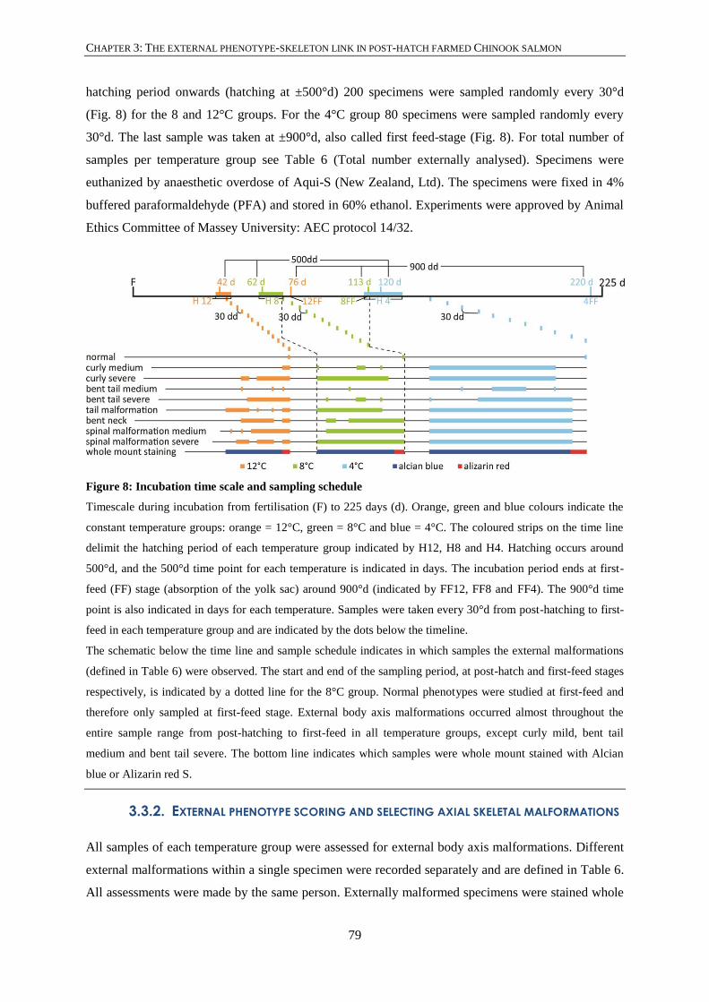
CHAPTER 3: THE EXTERNAL PHENOTYPE-SKELETON LINK IN POST-HATCH FARMED CHINOOK SALMON
79
hatching period onwards (hatching at ±500°d) 200 specimens were sampled randomly every 30°d
(Fig. 8) for the 8 and 12°C groups. For the 4°C group 80 specimens were sampled randomly every
30°d. The last sample was taken at ±900°d, also called first feed-stage (Fig. 8). For total number of
samples per temperature group see Table 6 (Total number externally analysed). Specimens were
euthanized by anaesthetic overdose of Aqui-S (New Zealand, Ltd). The specimens were fixed in 4%
buffered paraformaldehyde (PFA) and stored in 60% ethanol. Experiments were approved by Animal
Ethics Committee of Massey University: AEC protocol 14/32.
Figure 8: Incubation time scale and sampling schedule
Timescale during incubation from fertilisation (F) to 225 days (d). Orange, green and blue colours indicate the
constant temperature groups: orange = 12°C, green = 8°C and blue = 4°C. The coloured strips on the time line
delimit the hatching period of each temperature group indicated by H12, H8 and H4. Hatching occurs around
500°d, and the 500°d time point for each temperature is indicated in days. The incubation period ends at first-
feed (FF) stage (absorption of the yolk sac) around 900°d (indicated by FF12, FF8 and FF4). The 900°d time
point is also indicated in days for each temperature. Samples were taken every 30°d from post-hatching to first-
feed in each temperature group and are indicated by the dots below the timeline.
The schematic below the time line and sample schedule indicates in which samples the external malformations
(defined in Table 6) were observed. The start and end of the sampling period, at post-hatch and first-feed stages
respectively, is indicated by a dotted line for the 8°C group. Normal phenotypes were studied at first-feed and
therefore only sampled at first-feed stage. External body axis malformations occurred almost throughout the
entire sample range from post-hatching to first-feed in all temperature groups, except curly mild, bent tail
medium and bent tail severe. The bottom line indicates which samples were whole mount stained with Alcian
blue or Alizarin red S.
3.3.2. EXTERNAL PHENOTYPE SCORING AND SELECTING AXIAL SKELETAL MALFORMATIONS
All samples of each temperature group were assessed for external body axis malformations. Different
external malformations within a single specimen were recorded separately and are defined in Table 6.
All assessments were made by the same person. Externally malformed specimens were stained whole

CHAPTER 3: THE EXTERNAL PHENOTYPE-SKELETON LINK IN POST-HATCH FARMED CHINOOK SALMON
80
mount together with externally normal specimens (Table 7). Normal external phenotype specimens
were selected at first feed stage and were stained with Alizarin red S for mineralised tissues (mostly
bone) (Fig. 8). Malformed specimens were stained with Alcian blue except for the last two sampling
points, which were stained with Alizarin red S due to the start of mineralisation of the skeleton (Fig.
8).
Table 6: External body axis malformation phenotypes in post-hatch to first feed stage Chinook salmon:
percentages based on total number of specimens sampled per temperature group (samples taken every
30°d from post-hatch to first-feed and a definition for each malformation. If co-occurring within a single
specimen, different external malformations were recorded separately.
Malformation type 4°C
number
(%)
8°C
number
(%)
12°C
number
(%)
Definition Reference
Curly medium 25
(6.5)
6
(0.5)
5
(0.4)
Medium lordosis or kyphosis over the
length of the entire body, specimen
having the shape of half a circle
Mahrosh et
al. (2014)
Curly severe 54
(14.1)
66
(5.4)
46
(3.3)
Very severe lordosis or kyphosis over
the entire length of the body, ranging
from the head touching the caudal fin
to a complete spiral of the body
(coiled tail)
Mahrosh et
al. (2014)
Bent tail medium 9
(2.3)
1
(0.1)
4
(0.3)
Separate lordosis, kyphosis or
scoliosis of the caudal region
Eriksen et
al. (2006)
Bent tail severe 1
(0.3)
6
(0.5)
12
(0.9)
Severe separate lordosis, kyphosis or
scoliosis of the caudal region, e.g.
caudal region forming a single coil
Eriksen et
al. (2006)
Tail malformation 6
(1.6)
16
(1.3)
24
(1.8)
Heavily malformed preural and ural
region, ranging from stunted
development to absence of caudal fin
Eriksen et
al.
(2006),
Marty et al.
(1997)
Bent neck 47
(12.3)
38
(3.1)
34
(2.6)
A bend in the postcranial region of the
specimens body, includes ‘star gazer’
phenotype
Boglione et
al. (2013a)
Spinal
malformation
medium
62
(16.2)
83
(6.8)
55
(4.2)
A combination of lordosis, kyphosis
and scoliosis along the length of the
specimens body in mild forms
Marty et al.
(1997)
Spinal
malformation
severe
52
(11.0)
106
(8.7)
62
(4.7)
A combination of lordosis, kyphosis
and scoliosis along the length of the
specimens’ body in severe forms
Marty et al.
(1997)

CHAPTER 3: THE EXTERNAL PHENOTYPE-SKELETON LINK IN POST-HATCH FARMED CHINOOK SALMON
81
Total number of
specimens analysed
externally
383 1227 1313
3.3.3. WHOLE MOUNT STAINING AND ANALYSING INTERNAL MALFORMATIONS
Table 7 shows the number of larvae stained with Alcian blue 8GX (Acros organics, New Jersey,
USA) for cartilage and stained for mineralised bone with Alizarin red S (Sigma-Aldrich, Steinheim
am Albuch, Germany). The staining protocols were adapted from Taylor and Van Dyke (1985).
Briefly, specimens for Alcian blue were rinsed and stained for 48 hrs in [15mg/ml] Alcian blue
solution in 20% acetic acid/80% ethanol (pH = 1.5-2). Subsequently specimens were rehydrated,
rinsed and cleared in a solution of [0.5 mg/ml] trypsin in 30% saturated borax (di-sodium tetraborate,
Na2B4O7.10H2O) (pH = 9.4). Specimens were further cleared in a graded glycerol/KOH series,
dehydrated in a graded glycerol series and stored in 100% glycerol. Specimens for Alizarin red S were
rehydrated, bleached (3% H2O2 in 1% KOH, 1-1.5 hrs.), rinsed and placed for 24 hrs in a 30%
saturated borax solution. Specimens were stained overnight in a [0.5 mg/ml] solution of Alizarin red S
in 1% KOH. Specimens were cleared in a graded glycerol/KOH series, dehydrated in a graded
glycerol series and stored in 100% glycerol. Specimens were analysed with a Zeiss Axiozoom.V16
microscope equipped with an AxioCam MRc, top mount camera (Carl Zeiss Microscopy,
Oberkochen, Germany). Adobe Illustrator CS5 was used to create schematic representations.
Internal malformations were grouped into three phenotypes (Table 8): (i) notochord related
malformations, (ii) fusions of chordacentra and (iii) malformations related to associated elements
(further referred to as arches). When fusion of chordacentra occurred as a consequence of a
malformed notochord (see white asterisk Fig. 12C), the malformation was recognised as a notochord
malformation. Malformations of neural and haemal arches, ribs, neural and haemal spines of preural
vertebrae and caudal fin skeletal elements (stegural, epurals, uroneurals and hypurals) were
considered as malformations related to associated elements. Analysis and interpretation of vertebral
centra follows Bensimon-Brito et al. (2012b).
Table 7: Number of post-hatch (PH) to first-feed (FF) externally normal and externally malformed fish
stained with Alizarin red S (AR) and Alcian blue 8GX (AB)
External phenotype Stain Life stages sampled 4°C 8°C 12°C
Normal AR FF 50 50 50
Malformed AB From PH to FF 37 84 83
Malformed AR From PH to FF 9 9 34
Total number of malformed 46 93 117

CHAPTER 3: THE EXTERNAL PHENOTYPE-SKELETON LINK IN POST-HATCH FARMED CHINOOK SALMON
82
3.3.4. HISTOLOGY
In total, 18 specimens with externally normal phenotype were selected, three post-hatch and three
first-feed specimens per temperature group. In total, 64 specimens with externally malformed
phenotypes were selected. For each externally malformed phenotype, three specimens were selected
in the 8°C and 12°C temperature groups. In the 4°C temperature group two specimens for each
externally malformed phenotype were selected due to lower sample numbers. Before processing the
specimens were placed for 24 hours in acetone to remove fat, rinsed and subsequently decalcified for
48 hours in Morse solution (Morse, 1945) [22.5% formic acid (CH2O2, Merck KGaA, Darmstadt,
Germany) and 10% sodium citrate (C6H5Na3O7*2 H2O, Merck KGaA, Darmstadt, Germany)]. Finally,
the specimens were rinsed and embedded in paraffin. Parasagittal sections were cut at 4 μm with a
Leica RM 2235 microtome (Leica Microsystems, Wetzlar, Germany). Sections were stained with
haematoxylin and eosin (protocol adapted from; Gill et al., 1974), Heidenhain’s Azan trichrome,
Masson’s Trichrome and Verhoeff-Van Gieson methods to highlight collagen and elastin (Böck &
Romeis, 1989; Culling, 1974a; Culling, 1974b). Sections were analysed with bright field under a
Zeiss Axio Imager 0.Z.1 fitted with an Axiocam 503 colour top mount camera.
3.4. RESULTS
3.4.1. INTERNAL VERSUS EXTERNAL MORPHOLOGICAL PHENOTYPE
Table 8: Number and percentage (%) of specimens with internally normal and malformed phenotype for
all external phenotypes. If co-occurring within a single specimen, different internal malformations were
recorded separately. Associated elements = predominantly arches
External phenotype Related figure Internal phenotype 4°C
(%)
8°C
(%)
12°C
(%)
Normal * Figure 9A No internal malformations 30
(60)
42
(84)
38
(76)
Internal malformations 20
(40)
8
(16)
12
(24)
All external malformations * Figure 9B No internal malformation 10
(22)
47
(51)
57
(49)
Internal malformations 36
(78)
46
(49)
60
(51)
Normal Figure 10A Total number of specimens 50 50 50
Notochord 6
(12)
0
(0)
1
(2)
Chordacentra fusion 16
(32)
8
(16)
8
(16)

CHAPTER 3: THE EXTERNAL PHENOTYPE-SKELETON LINK IN POST-HATCH FARMED CHINOOK SALMON
83
Associated elements 6
(12)
1
(2)
6
(12)
Curly medium Figure 10B Total number of specimens 7 13 16
Notochord 0
(0)
0
(0)
1
(6)
Chordacentra fusion 0
(0)
3
(23)
3
(19)
Associated elements 4
(57)
6
(46)
4
(25)
Curly severe Figure 10C Total number of specimens 6 14 20
Notochord 1
(17)
3
(21)
6
(30)
Chordacentra fusion 1
(17)
2
(12)
5
(25)
Associated elements 3
(50)
5
(36)
6
(30)
Bent tail medium Figure 10D Total number of specimens 4 8 13
Notochord 2
(50)
3
(38)
2
(15)
Chordacentra fusion 1
(25)
0
(0)
0
(0)
Associated elements 4
(100)
3
(38)
4
(31)
Bent tail severe Figure 10E Total number of specimens 5 5 9
Notochord 1
(20)
0
(0)
0
(0)
Chordacentra fusion 1
(20)
1
(20)
2
(22)
Associated elements 3
(60)
3
(60)
2
(22)
Tail malformation Figure 10F Total number of specimens 5 10 5
Notochord 4
(80)
6
(60)
1
(20)
Chordacentra fusion 3
(60)
4
(40)
2
(40)
Associated elements 1
(20)
5
(50)
2
(40)
Bent neck Figure 10G Total number of specimens 5 13 13
Notochord 5
(100)
6
(46)
8
(62)

CHAPTER 3: THE EXTERNAL PHENOTYPE-SKELETON LINK IN POST-HATCH FARMED CHINOOK SALMON
84
Chordacentra fusion 2
(40)
3
(23)
7
(54)
Associated elements 2
(40)
4
(31)
4
(31)
Spinal malformation medium Figure 10H Total number of specimens 7 16 19
Notochord 2
(29)
3
(19)
1
(5)
Chordacentra fusion 1
(14)
0
(0)
1
(5)
Associated elements 3
(43)
2
(13)
6
(32)
Spinal malformation severe Figure 10I Total number of specimens 7 14 22
Notochord 5
(71)
3
(21)
9
(41)
Chordacentra fusion 1
(14)
2
(14)
8
(36)
Associated elements 5
(71)
3
(21)
7
(32)
* Total number of normal and malformed specimens in Table 7.
3.4.1.1. NORMAL EXTERNAL PHENOTYPE
Alizarin red S staining of first-feed stage specimens with normal external phenotype revealed internal
malformations in all three temperature groups (Fig. 9A, Table 8), with highest prevalence (40%) in
the 4°C group and lowest (16%) in the 8°C group. Fusion of chordacentra is the most frequent
malformation in specimens of all temperature groups, but its prevalence decreases in each temperature
group from 4 to 8°C (Fig. 10A, Table 8). Malformation of the associated elements is the second most
common phenotype in 4, 8 and 12°C specimens but has very low prevalence in the 8°C specimens.
Malformation of the notochord is only present in 4 and 12°C specimens, with highest prevalence in
the 4°C specimens.
3.4.1.2. MALFORMED BODY AXIS EXTERNAL PHENOTYPES
The definition of each external body axis malformation phenotype, and the number and percentages
per temperature group, are presented in Table 6. External body axis malformations are most common
in 4°C specimens, except bent tail severe and tail malformation which shows the highest percentage in
12°C specimens (Table 6). Bent tail medium and tail malformation are least common in 8°C
specimens (Table 6). In all temperature groups externally malformed specimens occur that have no
apparent skeletal malformations (Table 8).

CHAPTER 3: THE EXTERNAL PHENOTYPE-SKELETON LINK IN POST-HATCH FARMED CHINOOK SALMON
85
In almost more than 50% of the specimens, externally malformed fish displayed internal
malformations as well (Table 8). The 4°C group showed the highest percentage (78%) of internally
malformed specimens (Fig. 9C) and the 8°C group the lowest percentage (49%, Fig. 9B). In the 12°C
group, 51.3% of the externally malformed specimens showed internal malformations (Fig. 9B).
Below, internal malformations related to rearing temperature are briefly described for each of the
external malformation phenotypes.
Figure 9: Proportion of specimens with internal malformations for externally normal specimens (A) and
externally malformed specimens (B)
3.4.1.3. CURLY MEDIUM
In specimens with curly medium external phenotype, most internal malformations affect the
associated elements, but the prevalence decreases in each temperature group from 4 to 12°C. Fusion
of chordacentra occurs in 8 and 12°C specimens, while malformation of the notochord only appears in
12°C specimens (Fig. 10B).
3.4.1.4. CURLY SEVERE
In curly severe specimens the prevalence of malformed associated elements decreases in each
temperature group from 4 to 12°C, as with curly medium. Malformations of the notochord are the
second most common but increase in each temperature group from 4 to 12°C. Frequency of
chordacentra fusions is lowest in 8°C specimens (Fig. 10C).
3.4.1.5. BENT TAIL MEDIUM
All 4°C specimens have malformed associated elements. The frequency of both associated elements
and notochord malformations decreases in each temperature group from 4 to 12°C. Fusion of
chordacentra is only present in 4°C specimens (Fig. 10D).

CHAPTER 3: THE EXTERNAL PHENOTYPE-SKELETON LINK IN POST-HATCH FARMED CHINOOK SALMON
86
3.4.1.6. BENT TAIL SEVERE
In 12°C specimens, malformations of associated elements and fusion of chordacentra show equal
prevalence. The 4 and 8°C specimens show equal prevalence of fused chordacentra and associated
elements. Malformations of the notochord are only observed in 4°C specimens (Fig. 10E).
3.4.1.7. TAIL MALFORMATION
Malformation of the notochord and fusion of chordacentra both decrease in each temperature group
from 4 to 12°C. Chordacentra fusions are most prevalent in 4°C specimens and show the lowest
prevalence in 12°C specimens. In 12°C specimens fusion of chordacentra and malformation of
associated elements show an equal prevalence (Fig. 10F).
3.4.1.8. BENT NECK
All specimens in the 4°C group have a malformed notochord; 4°C specimens furthermore show an
equal prevalence of chordacentra fusions and malformed associated elements. Specimens of the 8°C
group show the lowest frequency of notochord malformations and chordacentra fusions (Fig. 10G).
3.4.1.9. SPINAL MALFORMATION MEDIUM
Prevalence of malformations of the notochord decreases in each temperature group from 4 to 12°C.
Associated elements are least affected by malformation in 8°C specimens. Fusion of chordacentra is
most prevalent in 4°C specimens while absent in 8°C specimens (Fig. 10H).
3.4.1.10. SPINAL MALFORMATION SEVERE
Prevalence of malformations of the notochord and associated elements is highest in 4°C specimens
and lowest in 8°C specimens. Prevalence of fusion of chordacentra is highest in 12°C specimens
compared to 4°C and 8°C specimens (Fig. 10I).

CHAPTER 3: THE EXTERNAL PHENOTYPE-SKELETON LINK IN POST-HATCH FARMED CHINOOK SALMON
87
Figure 10: Proportion of internal malformation types per temperature for each of the external body axis
phenotypes (normal and malformed) based on Table 8 (associated elements = arches)
3.4.2. AXIAL SKELETAL STRUCTURES
The elements of the axial skeleton (notochord, chordacentra and associated elements) are described
based on Alizarin red S and Alcian blue whole mount stained specimens and serial sections, from
post-hatch stage to first-feed stage.
3.4.2.1. NOTOCHORD
Two different types of notochord malformation occur in specimens of all temperature groups and
often within a single malformed notochord (Fig. 11A). The most common malformation is a partial or
complete pinching, where the external elastic membrane of the notochord sheath folds inward into the
notochord. The inward fold ranges from a shallow pinch (Fig. 11A) to a completely pinched-off
notochord (Fig. 11A, C-D). A pinch in the notochord often (but not necessarily) causes the notochord
to either slightly bend or even make a severe deviation of the axis. When a shallow pinch occurs, the
cartilaginous base of the anterior or posterior neural or haemal arch extends into the depression in the
notochord sheath (Fig. 11A, D). When a deep depression occurs, the cartilaginous bases of the neural
or haemal arches anterior and posterior of the pinch site fuse and extend into the deep pinch (Fig.
11B, F). Associated elements (neural and haemal arches, parapophyses and ribs) remain separate at
the level of shallow or deep pinching sites. When the notochord is completely pinched-off, each of the
two notochord ends is lined by external elastic membrane (Fig. 11A, C-D). The two external elastic
membranes are closely apposed (Fig. 11A, D) or separated from each other (Fig. 11A, C). In the latter
case, the cartilaginous bases of the neural and haemal arches fuse and fill the space between the

CHAPTER 3: THE EXTERNAL PHENOTYPE-SKELETON LINK IN POST-HATCH FARMED CHINOOK SALMON
88
separated notochord ends (Fig. 11A, I). The associated elements (neural and haemal arches,
parapophyses and ribs) are often fused at the level of separated notochord ends (Fig. 11I).

CHAPTER 3: THE EXTERNAL PHENOTYPE-SKELETON LINK IN POST-HATCH FARMED CHINOOK SALMON
89
Figure 11: Notochord malformations (left)
All images use following orientation: anterior = left, posterior = right, dorsal = up and ventral = down. (A)
Schematic of notochord malformation types based on Azan stained sections. Two types of notochord
malformations exist. The first type (shown anterior of the dashed line) is characterised by folding of the external
elastic membrane resulting in partial or complete pinching of the notochord. The depression in the external
elastic membrane of a shallow pinch is filled with cartilage of the bases of neural and haemal arches. At the
level of a complete pinching site, where the notochord ends remain closely apposed, cartilage of the fused bases
of neural arches (indicated by the asterisk) fills a shallow depression in the external elastic membrane. When
two notochord ends, each lined by external elastic membrane, are separated at a complete pinching site, the
space in-between the notochord ends is filled with cartilage of the fused bases of neural (BD) and haemal (BV)
arches. The second type of notochord malformation (shown posterior of the dashed line) is characterised by
hyperplasia of chordoblasts resulting in partial or full septa and scar tissue. Scar tissue contained within the
notochord replaces chordocytes. B-E. Alcian blue whole mount stained and cleared specimens. Scale bars = 250
μm. (B) Notochord pinched partially along the ventral side. The cartilaginous bases of the haemal arches
positioned at the pinch site fuse and extend into the shallow depression of the external elastic membrane
(asterisk). (C) Completely pinched notochord with two notochord ends separated (arrows). Each notochord end
is lined by external elastic membrane. Cartilaginous bases of neural and haemal arches fuse and invade the
space in-between the notochord ends (white asterisks). (D) Completely pinched notochord with the two
notochord ends closely apposed to each other (arrowheads). A shallow depression at the pinch site on the ventral
side is filled with cartilage of the base of the haemal arch (black arrow). (E) Scar tissue (SC) inside the
notochord stains with Alcian blue. Ventrally the notochord is pinched at the level of the scar tissue. The
cartilaginous bases of adjacent haemal arches are fused (white asterisk). (F-I) Parasagittal sections of notochord
malformations. (F) Partial deep pinch along the ventral side of the notochord combined with hyperplastic
chordoblasts starting to form scar tissue (SC). Cartilage (asterisk) fills the space of the inward fold of the
external elastic membrane (EEM). Highly polarised (cylindrical) chordoblasts (cb) line the notochord sheath
(nsh). Scale bar = 100 μm. (G) Hyperplastic chordoblasts (cb) produce collagenous notochord sheath material
forming into scar tissue (SC). The notochord sheath (nsh) and external elastic membrane are interrupted by
extruding scar tissue. Abbreviations: BD, cartilaginous base of neural arch; BV, cartilaginous base of haemal
arch. Scale bar = 100 μm. (H) Advanced formation of scar tissue (SC) compared to (G). The scar tissue has
replaced the chordocytes. The scar tissue is contained within the notochord sheath (nsh) and external elastic
membrane (EEM). Abbreviations: BD, cartilaginous base of neural arch; BV, cartilaginous base of haemal arch.
Scale bar = 100 μm. (I) Pinching malformations and hyperplasia of chordoblasts occurring in a single notochord.
The arrowhead indicates a sharp pinch along the ventral side of the notochord. Neural arches (NA) fuse as a
likely consequence of notochord (nch) malformations. The cartilaginous bases of neural arches (BD) fuse to the
cartilaginous bases of haemal arches (BV) where the notochord is heavily pinched to the right side. The
cartilage of fused bases fills the space created by the inward fold of the external elastic membrane. The arrow
indicates a full septum. The neural arches (NA) dorsal of the septum are unaffected. Scale bar = 250 μm.

CHAPTER 3: THE EXTERNAL PHENOTYPE-SKELETON LINK IN POST-HATCH FARMED CHINOOK SALMON
90
The second type of malformation of the notochord is dislocation of chordoblasts forming partial or
complete septa and scar tissue (Fig. 11A, E-I). In contrast to pinching, the external elastic membrane
of the notochord does not fold inwards when septa are present (Fig. 11A, I). These septa are lined by
polarised (cylindrical) chordoblasts suggesting that these cells are actively producing notochord
sheath material (Fig. 11A, I). The associated elements are not affected at the level of the notochord
septa (Fig. 11A, I). The notochord is narrowed, shallowly pinched (Fig. 11A, E, H) or deeply pinched
(Fig. 11F) at the level of the scar tissue and slightly bent or severely deflected. The scar tissue is
contained within the notochord thereby replacing the chordocytes (Fig. 11A, H), or bulges out from
the notochord, breaking through the external elastic membrane (Fig. 11G). Small chordocytes and
highly polarised (indicated by asterisks in Fig. 11G) or flattened (indicated by arrows in Fig. 11H)
chordoblasts line the scar tissue. While neural and haemal arches remain separate at the level of scar
tissue, their cartilaginous bases often fuse (Fig. 11E). Septa and pinching malformations can occur at
any point of the notochord contour. When pinching and dislocation of chordoblasts occur together
(Fig. 4I), the notochord is folded like a concertina.
3.4.2.2. CHORDACENTRA
Fused chordacentra in Alcian blue stained specimens were identified if there was also the fusion of
the cartilaginous bases of the neural or haemal arches. In Alizarin red S stained specimens, three
patterns of fusions are observed when chordacentra start mineralising. First, the mineralisation of two
or more adjacent chordacentra is continuous on the ventral side (Fig. 12A, B). Second, the
chordacentra are fused on the dorsal side while remaining separate on their ventral side (Fig. 12A, C).
This is often observed in preural vertebrae and co-occurs with separate haemal arches but fused
haemal spine. Third, adjacent chordacentra are fused but no longer separately recognisable. The
ventral side of the mineralisation has the width of two chordacentra (Fig. 12A). Each of these three
fusion patterns can co-occur with neural and haemal arches that remain separated or that are fused at
the level of their cartilaginous bases. In parasagittal sections chordacentrum fusions can be recognised
before mineralisation of chordacentra. The notochord sheath is thicker in the future intervertebral
regions and thinner where mineralisation of the notochord sheath (chordacentrum) will occur (Fig.
12D). In a normal notochord the intervertebral regions lie opposite to each other on the dorsal and
ventral side (Fig. 12A). In case of fusion of chordacentra, this pattern is disturbed (Fig. 12A, E).
3.4.2.3. ASSOCIATED ELEMENTS
The most commonly observed malformation of the associated elements is misalignment along the
notochord (Fig. 12F) and fusions of adjacent elements (Fig. 12G-I). Misaligned elements can be
distinguished when the cartilaginous bases of neural or haemal arches insert in an alternating pattern
left and right on the notochord (Fig. 12F). The distal ends of these neural and haemal arches are also

CHAPTER 3: THE EXTERNAL PHENOTYPE-SKELETON LINK IN POST-HATCH FARMED CHINOOK SALMON
91
misaligned (Fig. 12F). Fusion of associated elements can occur in all positions along their proximal-
distal axis (Fig. 12G-I). Associated elements can develop as small stunted cartilaginous elements
(already mineralised in Fig. 12I) or are bent anteriorly or more posteriorly, but this occurs to a lesser
degree compared to misalignments and fusions.

CHAPTER 3: THE EXTERNAL PHENOTYPE-SKELETON LINK IN POST-HATCH FARMED CHINOOK SALMON
92
Figure 12: Chordacentra fusions and associated elements (= arches) malformations
All panel images use following orientation: anterior = left, posterior = right, dorsal = up and ventral = down. (A)
Schematic of different chordacentra fusion patterns. From left to right, (i) chordacentra fused on the ventral side
and (ii) dorsal side with the fusing chordacentra remaining visible, and (iii) fusion of chordacentra on the ventral

CHAPTER 3: THE EXTERNAL PHENOTYPE-SKELETON LINK IN POST-HATCH FARMED CHINOOK SALMON
93
side but no longer separately recognisable. The latter is characterised by the wide ventral side of the
chordacentrum. Fusion of the cartilaginous haemal arch bases (BV) can occur. The second part of the schematic
shows how fused chordacentra are recognised in parasagittal sections. The notochord sheath is thicker in
intervertebral regions and thinner in chordacentra. On the dorsal side, the normal situation is presented, where
chordacentra alternate with intervertebral regions. On the ventral side, the alternating pattern is disturbed by
fused chordacentra. Abbreviations: BD, cartilaginous base of neural arch; HA, haemal arch; NA, neural arch.
(B-C) Alizarin red S stained specimens showing fused chordacentra. Scale bars = 250 μm. (B) Fusion of
chordacentra on the ventral side with the fusing chordacentra remaining visible. The mineralisation of the
notochord sheath between the two chordacentra on the ventral side is continuous (arrowheads). (C) Fusion of
chordacentra on the dorsal side. The fusing chordacentra remain visible on the ventral side due to a gap in the
mineralisation (block arrow). The white asterisk indicates a chordacentra fusion due to a notochord
malformation (pinch along the ventral side) and is therefore recognised as a notochord malformation. (D-E)
Parasagittal sections of the notochord showing normal and fused chordacentra. Scale bars = 100 μm. (D) A
normal intervertebral region (IV) and mineralised chordacentrum (CHC). The chordoblasts (cb) in the
intervertebral region are polarised (cylindrical), while the chordoblasts along the chordacentrum are flattened.
The notochord sheath (nsh) is thicker in the intervertebral region and thinner in the chordacentrum. (E) On the
dorsal side, a normal alternation of intervertebral region (IV) with mineralised chordacentra (CHC) can be
recognised. On the ventral side, this pattern is disturbed by a fused chordacentrum spanning two adjacent
chordacentra and the in-between intervertebral region. (F-I) Associated elements malformations: (F-H) Alcian
blue stained. (I) Alizarin red S stained. Scale bars = 250 μm. (F) Misalignment of neural arches. The
cartilaginous bases of neural arches have alternating positions on the left (arrowhead) and right side (block
arrow) of the notochord. The distal ends of the neural arches are also misaligned (arrows). (G) Fusion of
associated elements in the caudal fin skeleton. The rounded arrowhead indicates the distal fusion of two adjacent
preural haemal arches. The asterisk indicates the proximal fusion of hypurals 2 and 3. (H) Medial fusion of two
adjacent post-cranial neural arches (block arrow). (I) The block arrow indicates the medial fusion of two
adjacent caudal haemal arches. The line arrow indicates the stunted development of a haemal arch.
3.5. DISCUSSION
This study aimed to link external body axis malformations in post-hatch Chinook salmon with internal
malformations of axial skeletal structures, i.e. the notochord, the chordacentra and the associated
elements (predominantly arches). In addition the effect of temperature on malformations is
investigated. In all temperature groups, externally normal specimens with internal malformations
showed mainly chordacentra fusions. Interestingly, specimens in all temperature groups with external
malformations did not always display internal skeletal malformations. Yet, most animals’ external
malformations related to internal malformations.
A coherent diagnosing system for spinal malformations in salmonids is a prerequisite for linking a
particular spinal malformation to a particular cause. Knowledge about causes will eventually enable

CHAPTER 3: THE EXTERNAL PHENOTYPE-SKELETON LINK IN POST-HATCH FARMED CHINOOK SALMON
94
us to control and to avoid spinal malformations in farmed salmonids (Witten et al., 2009). The current
study distinguishes between eight different external body axis malformations, including two severity
classes in curly, bent tail and spinal malformations. The two severity classes can be distinguished
based on the angle of the body axis curvature. The diagnosed external phenotypes (including normal)
in the present study are represented in Figure 13. In addition this study looked more closely at the
internal malformations, and found two types of notochord malformations (pinching and hyperplasia of
chordoblasts), three patterns of chordacentra fusions and malformations of associated elements such
as misalignment, fusion or stunted development.
Figure 13: Diagnosing external phenotypes
All specimens represented are placed on their right side with anterior to the left and posterior to the right. A
dorsal view is represented of the SMM and SMS specimens. Specimens range from post-hatch to first feed
stages. Each specimen in this figure shows only one external malformation. However, multiple external
malformations can occur in a single specimen. N, first feed externally normal specimen; TM, tail malformation,
arrow indicates malformed tail and tail fin; BN, bent neck, arrow indicates a postcranial kyphosis; CM, curly
medium; CS, curly severe, the kyphosis is so severe the body forms a spiral shape; BTM, bent tail medium,
arrow indicates a bend (lordosis) in the caudal region of <40°; BTS, bent tail severe, arrow indicates a bend
(lordosis) in the caudal region of >40°; SMM, spinal malformation medium, arrowheads indicate scoliosis of
<40°; SMS, spinal malformation severe, arrowheads indicate scoliosis of >40° except the minor scoliosis
occurring in the caudal region. Yolk sac indicated by asterisks. Scale bars: 5 mm.
Interestingly, the main internal malformation in externally normal specimens is chordacentra fusion.
Fusion of caudal vertebral centra is a normal part of the urostyle formation in teleosts such as
salmonids (Arratia & Schultze, 1992; Schultze & Arratia, 1989; Witten et al., 2009) and cyprinids
(Bensimon-Brito et al., 2010; Bensimon-Brito et al., 2012a). Fusion of caudal centra in salmonids
occurs in late juvenile stages where vertebral centrum structures external to the notochord sheath fuse

CHAPTER 3: THE EXTERNAL PHENOTYPE-SKELETON LINK IN POST-HATCH FARMED CHINOOK SALMON
95
(Arratia & Schultze, 1992; Schultze & Arratia, 1988). Fusion in zebrafish (Danio rerio, Hamilton)
occurs also within the notochord sheath by continuous mineralisation between adjacent chordacentra
(Bensimon-Brito et al., 2012b). In gilthead sea bream (Sparus aurata, Linnaeus) pathological fusion
of vertebral centra can occur in early life stages at the level of chordacentra, as is the case during
zebrafish urostyle formation (Loizides et al., 2013). Fusions of chordacentra in the present study are
assigned to two aetiologies, (i) fusion by continuous mineralisation between two or more centra, and
(ii) fusion as a result of a notochord malformation such as pinching. How the pattern of chordacentra
is established is currently unknown, however the notochord is thought to play an intrinsic part in the
pattern formation (Fleming et al., 2004; Fleming et al., 2015). For example, expression of alkaline
phosphatase (ALP) was detected in chordoblasts lining the mineralisation zones of Atlantic salmon
(Salmo salar, Linnaeus) and zebrafish notochord (Bensimon-Brito et al., 2012b; Grotmol et al., 2005).
Furthermore, the morphology of chordoblasts in Atlantic salmon is different in mineralising and non-
mineralising zones of the notochord sheath (Grotmol et al., 2003; Grotmol et al., 2005). The most
recent evidence for a notochord driven mineralisation of the notochord sheath comes from studies on
medaka (Oryzias latipes, Temminck & Schlegel) osterix/sp7 mutants. These animals have defective
osteoblasts and bone formation is disrupted, yet the segmented mineralisation of the notochord sheath
is unaffected (Yu et al., 2017). Possibly the early fusion of chordacentra is caused by a change in the
behaviour of the chordoblasts that produce the notochord sheath.
Notochord malformations in this study occurred mostly in specimens with severe external
malformations, and therefore, severe notochord malformation may cause external malformation.
However, they also occurred in animals with less severe external malformations. Pinching of the
notochord was observed in green sturgeon (Acipencer medirostris, Ayres) at yolk-sac depletion
(Linares-Casenave et al., 2013). Shallow pinching, irregularities in the notochord sheath and
distortions of the tip of the notochord were also observed in gilthead sea bream larvae
(Koumoundouros et al., 1997; Santamaría et al., 1994). Andrades et al. (1996) showed a complete
pinch of the notochord with each notochord end lined by notochord sheath. Connective tissue filled
the space between the notochord ends. Interestingly, the observation in gilthead sea bream was made
in an early stage where cartilaginous associated elements are not yet present (Andrades et al., 1996;
Faustino & Power, 1998). The connective tissue between the notochord ends in the gilthead sea bream
may be replaced by cartilage later in development. In cases of severe notochord malformation with
dislocated chordoblasts and scar tissue the normal internal anatomy of the notochord is changed.
Alternatively, severe notochord malformations occurring in a single site may not cause severe
external malformations. Loizides et al. (2013) suggested that ectopic production of notochord sheath
material, i.e. scar tissue, prevents mineralisation of the notochord sheath. In contrast, mineralisation of
the notochord sheath and indications of mineralising ectopic sheath material (Fig. 11H) within the
scar tissue was observed in the present study. Scar tissue could develop into a fusion if mineralisation
CHAPTER 3: THE EXTERNAL PHENOTYPE-SKELETON LINK IN POST-HATCH FARMED CHINOOK SALMON
96
continues throughout post-hatch development. A fusion of two or three vertebral bodies can stabilise
and remodel into a normal functioning vertebral centrum (Witten et al., 2006).
In the present study, notochord malformations, fusion of chordacentra and malformation of associated
elements occurred separately in some specimens, confirming that the chordacentra and the associated
elements represent two developmental modules (de Azevedo et al., 2012; Detwiler & Holtzer, 1956;
Hall, 1977; Holtzer, 1951; Strudel, 1953a; Strudel, 1953b; To et al., 2015; Witten et al., 2007).
Pathological malformations of associated elements (= arches) along the vertebral column, such as
duplications, fusion, bending, dislocation and shortening, have been mainly observed in marine
teleosts (Boglione et al., 2013b; Gavaia et al., 2002; Sfakianakis et al., 2004). In post-hatch Chinook
salmon similar malformations of the associated elements are observed, with misalignment of the
arches as the main malformation phenotype. Malformations of the associated elements are likely
related to abnormal somite development. For example, left-right misalignment of somites occurs in
retinoic acid deficient mouse, chick and zebrafish embryos (Kawakami et al., 2005; Vermot &
Pourquie, 2005) and Notch1 mutant zebrafish (Conlon et al., 1995). Misaligned somites may cause the
associated elements (neural and haemal arches and parapophyses and ribs) to misalign along the
notochord. Neural and haemal arches are malformed and fused in fused somite mutant zebrafish
(Fleming et al., 2004; van Eeden et al., 1996). Also, fusion of associated elements has been observed
in the caudal fin (preural and ural regions) of salmonids and zebrafish (Arratia & Schultze, 1992;
Bensimon-Brito et al., 2012a) and confirms the results of this study. These fusions are likely part of
the natural variation present in the teleost caudal fin skeleton (Arratia et al., 2001; Bensimon-Brito et
al., 2010; Witten & Hall, 2015).
In addition to malformation of associated elements, abnormal somite development causes
malformations of muscles (Uji et al., 2015; van Eeden et al., 1996). Abnormal muscle development
could explain why specimens with external body axis malformations sometimes displayed normal
skeletal structures in the present study (e.g. curly phenotype). Incubation temperatures are known to
have immediate and long lasting effects on muscle development in teleosts (Finn, 2007; Johnston,
2006; Johnston et al., 2003). In Atlantic salmon, a higher temperature caused a higher rate of
hypertrophy (increase in volume) compared to hyperplasia (increase in number) of muscle fibres
(Johnston & McLay, 1997; Stickland et al., 1988). Temperature driven muscle plasticity was
suggested as a possible underlying cause for haemal lordosis in European sea bass (Dicentrarchus
labrax, Linnaeus) (Boglione et al., 2013b; Koumoundouros et al., 2009). Interestingly, when first feed
stage Atlantic salmon incubated in 5°C water were moved to 10°C water muscle recovery and catch-
up growth were observed (Nathanailides et al., 1995). This suggests that specimens with a normal
axial skeleton and minor body axis malformations can recover during juvenile growth. Recovery from
minor malformation phenotypes has also been suggested to occur in marine teleosts (Boglione et al.,
2013b; Rosenthal & Alderdice, 1976).

CHAPTER 3: THE EXTERNAL PHENOTYPE-SKELETON LINK IN POST-HATCH FARMED CHINOOK SALMON
97
In the present study the lowest frequency of 16% vertebral malformations occurred in the 8°C group.
This can be considered as a high frequency. Certain year classes of adult Atlantic salmon also showed
high frequencies (15.6% to 25.2%) of vertebral deformities (Gjerde et al., 2005). Although the
heritability of vertebral anomalies in Atlantic salmon is low, parental animals with high frequencies of
vertebral deformities produce off-spring with similar frequencies of vertebral deformities (Gjerde et
al., 2005; Sullivan et al., 2007b). In addition, it seems that the heritability of vertebral deformities
increases when there is a failure in husbandry practice, e.g. an unusually low or high incubation
temperature. Moreover, this induces unusually high frequencies of skeletal deformities (Kause et al.,
2007). The family crosses producing high frequencies of skeletal malformations together with the
temperatures chosen in the present study may explain the high frequencies of vertebral malformations
in all temperature groups. Different frequencies of both external and internal malformations were
observed in the three temperature groups indicating that temperature affects the malformation rate in
the present study.
Constantly elevated incubation temperatures during early life stages have been used to induce
vertebral column anomalies in salmonids (Boglione et al., 2013b). A U-shaped response in the
frequency of morphological malformations was found in brown trout (Salmo trutta, Linnaeus) reared
at five different incubation temperatures, with the lowest frequency occurring at 8°C (Réalis-Doyelle
et al., 2016). The frequencies of internal malformations in externally normal and malformed
specimens in the present study also showed a U-shaped response, with lowest frequencies in 8°C.
Chinook salmon were successfully transferred from Californian (6-16°C) waters to New Zealand
Rivers (Kinnison et al., 1998; McDowall, 1994; Unwin & Glova, 1996) and are therefore acclimated
to warmer temperatures. Perhaps it is not surprising that specimens incubated at the lowest
temperature, 4°C, showed the highest incidence of external and internal malformations. The highest
frequency of malformations (20%) was also observed in Arctic charr (Salvelinus alpinus, Linnaeus)
reared at a constant 2.3°C (Jeuthe et al., 2013). Furthermore, Embody (1934) observed a high level of
mortality in brook trout (Salvelinus fontinalis, Mitchill), lake trout, brown trout and rainbow trout
(Oncorhynchus mykiss, Walbaum) incubated below 6°C. In contrast, very low mortalities were
observed for brown trout and Arctic charr incubated at 2°C. However, high mortality occurred at
incubation temperatures below 6°C for grayling (Thymallys thymallus, Linnaeus) and Danube salmon
(Hucho hucho, Linnaeus) (Jungwirth & Winkler, 1984). Trends in mortality are similar for five
species of Pacific salmon. While coho salmon and sockeye salmon survive incubation at 4°C well
(Beacham & Murray, 1990; Murray & McPhail, 1988), Chinook salmon and chum salmon
(Oncorhyunchus keta, Walbaum) show high mortality when incubated at 4°C (Beacham & Murray,
1985a; Beacham & Murray, 1985b; Beacham & Murray, 1987b; Murray & McPhail, 1988). Pink
salmon does not survive incubation at 4°C (Beacham & Murray, 1990; Murray & Beacham, 1986;
Murray & McPhail, 1988). In addition, the studies in Pacific salmon observed the following trends
CHAPTER 3: THE EXTERNAL PHENOTYPE-SKELETON LINK IN POST-HATCH FARMED CHINOOK SALMON
98
during incubation in cold temperatures: (i) the highest mortality occurred before the completion of
epiboly, (ii) higher mortality occurred before hatching and (iii) the mortality of alevins is lower in
cold incubation temperatures compared to higher incubation temperatures. These trends in mortality,
specifically lower mortality of alevins in cold temperatures, could explain why higher frequencies of
malformations were observed in the 4°C group in the present study. Malformed embryos are more
likely to die in the egg stages at 4°C, however, once hatched, malformed alevins are more likely to
survive and therefore are more frequent in the 4°C group.
The mortality and malformations observed in previous studies and the present study suggests that
constant cold incubation temperatures elicit more stress affecting normal development compared to
warmer temperatures such as 12°C. Heat shock proteins (HSP) assist in stabilising and repairing
altered proteins due to thermal stress (Basu et al., 2002; Iwama et al., 1998; Kiang & Tsokos, 1998).
In Atlantic salmon, the presence of HSP70 mRNA was shown to increase after both warm and cold
thermal stress (Takle et al., 2005). A perturbation of the HSP mechanism may underlie the increased
frequency of malformations in 4°C incubated Chinook salmon. The hydrostatic pressure of the
vacuolated chordocytes against the notochord sheath provides the sturdiness of the notochord
(Grotmol et al., 2006; Kryvi et al., 2017). The vacuolisation and myogenesis of muscle fibres start at a
similar time point in trout (Killeen et al., 1999). However, while the myogenesis of muscle fibres was
not delayed, the start of vacuolisation was found to be delayed in trout embryos incubated at 2°C
(Killeen et al., 1999). Therefore, the muscle fibres which developed before the vacuolisation of the
notochord may elicit damage to the notochord sheath resulting in notochord malformations. This
could explain the higher frequency of malformations in the 4°C group in the present study.
The question as to when the pathological process(es) resulting in fusion start, remains to be answered.
Studies using temperature induced skeletal deformities in Atlantic salmon have also examined the
internal skeletal phenotype but did so in juvenile stages. Two studies used temperature shock to
induce malformations in early stages and examined the deformities in juvenile parr stage animals
(Takle et al., 2005; Wargelius et al., 2005). Ønsrud et al. (2004a) used two constant temperature
regimes in combination with different vitamin A levels during the incubation period to investiagate
the effect of temperature and vitamin A on vertebral column deformities in juvenile parr and pre- and
post-smoltification stages. Also, two constant temperature regimens during incubation were used to
study the expression levels of osteogenic and chondrogenic markers during fusion in juvenile parr and
pre-smolt stages (Ytteborg et al., 2010a; Ytteborg et al., 2010b; Ytteborg et al., 2010c). The
phenotypes of the vertebral deformities observed in these experiments are early, intermediate or final
stages of the deformities (Ytteborg et al., 2010b; Ytteborg et al., 2010c). In addition, fused vertebral
centra have been reported to develop normally before the deformation process starts (Fjelldal et al.,
2007a; Nordvik et al., 2005; Witten et al., 2009). In the present study, fusion of chordacentra was
observed to be present in early life stages. The literature reporting on temperature experiments as well

CHAPTER 3: THE EXTERNAL PHENOTYPE-SKELETON LINK IN POST-HATCH FARMED CHINOOK SALMON
99
as the current study suggests that vertebral anomalies can start at completely different life stages.
Importantly, this investigation demonstrates how distinct internal malformations can be characterised
in the embryonic/post-hatch life stages of Chinook salmon. Investigation of the ontogeny and
progression of compression and fusion in Atlantic salmon has previously led to the suggestion that
vertebral anomalies may arise as early as embryonic stages (Kvellestad et al., 2000; Witten et al.,
2006; Ytteborg et al., 2010a).
3.6. CONCLUSION
Although malformation frequencies were high due to specific family crosses, a temperature effect was
observed. Results obtained in the current study suggest that incubation and early rearing at a constant
8°C is preferable to rearing at 12°C and indicates that rearing at 4°C results in unacceptable
frequencies of body axis malformation (see Table 6). Interestingly, externally normal specimens
showed fusions of vertebral anlagen which can grow up to harvest stages. The study shows that
external malformations of the body axis are usually linked to internal malformations of the vertebral
column. Externally curled specimens typically have malformed arches. Malformations affecting a
single region relate to malformed notochords and fusions of vertebral centrum anlagen. Spinal
malformations (medium and severe) relate to malformed notochords and arches. Apparent differences
in the type and rate of malformations between the 4, 8 and 12°C groups provides the framework for
future investigations. Specifically, it will be necessary to develop conditions in which externally
normal specimens have at most mild internal malformations allowing the fish to grow beyond and
stabilise early life malformations.
3.7. ACKNOWLEDGEMENTS
The authors thank New Zealand King Salmon and Skretting Australia for funding and supporting the
experiments. Specifically, the authors would like to acknowledge Solenne Metayer for technical
assistance during the temperature experiment and Mieke Soenens for histological work.

CHAPTER 3: THE EXTERNAL PHENOTYPE-SKELETON LINK IN POST-HATCH FARMED CHINOOK SALMON
100

101
CHAPTER 4: VERTEBRAL COLUMN REGIONALISATION IN
CHINOOK SALMON (ONCORHYNCHUS TSHAWYTSCHA)
A. De Clercq(1, 4)
M.R. Perrott(1)
P. S. Davie(1)
M.A. Preece(2)
B. Wybourne(3)
N. Ruff(3)
A. Huysseune(4)
P.E. Witten(1, 4)
1School of Veterinary Sciences, Massey University, Palmerston North, New Zealand
2New Zealand King Salmon, 93 Beatty Street, Nelson 7011, New Zealand
3Skretting Australia, PO Box 117, Rosny Park, Tasmania, Australia
4Evolutionary Developmental Biology, Ghent University, Ghent, Belgium
De Clercq A, Perrott MR, Davie PS, Preece MA, Wybourne B, Ruff N, Huysseune A, Witten PE
(2017b) Vertebral column regionalisation in Chinook salmon, Oncorhynchus tshawytscha. Journal of
Anatomy, 231, 500-514

CHAPTER 4: VERTEBRAL COLUMN REGIONALISATION IN CHINOOK SALMON (ONCORHYNCHUS TSHAWYTSCHA)
102
4.1. ABSTRACT
Teleost vertebral centra are often similar in size and shape but vertebral associated elements, i.e.,
neural arches, haemal arches and ribs, show regional differences. Here we examine how presence,
absence and specific anatomical and histological characters of vertebral centra associated elements
can be used to define vertebral column regions in juvenile Chinook salmon (Oncorhynchus
tshawytscha). To investigate if the presence of regions within the vertebral column is independent of
temperature, animals raised at 8 and 12°C were studied at 1400 and 1530 degreedays, in the
freshwater phase of the life cycle. Anatomy and composition of the skeletal tissues of the vertebral
column were analysed using alizarin red S whole mount staining and histological sections. Six
regions, termed I to VI, are recognised in the vertebral column of specimens of both temperature
groups. Postcranial vertebrae (region I) carry neural arches and parapophyses but lack ribs.
Abdominal vertebrae (region II) carry neural arches and ribs that articulate with parapophyses.
Elastic- and fibrohyaline cartilage and Sharpey’s fibres connect the bone of the parapophyses to the
bone of the ribs. In the transitional region (III) vertebrae carry neural arches and parapophyses change
stepwise into haemal arches. Ribs decrease in size, anterior to posterior. Vestigial ribs remain attached
to the haemal arches with Sharpey’s fibres. Caudal vertebrae (region IV) carry neural and haemal
arches and spines. Basidorsals and basiventrals are small and surrounded by cancellous bone. Preural
vertebrae (region V) carry neural and haemal arches with modified neural and haemal spines to
support the caudal fin. Ural vertebrae (region VI) carry hypurals and epurals that represent modified
haemal and neural arches and spines respectively. The postcranial and transitional vertebrae and their
respective characters are usually recognised but should be considered as regions within the vertebral
column of teleosts because of their distinctive morphological characters. While the number of
vertebrae within each region can vary, each of the six regions are recognised in specimens of both
temperature groups. This refined identification of regionalisation in the vertebral column of Chinook
salmon can help to address evolutionary developmental and functional questions and to support
applied research into this farmed species.
4.1.1. KEYWORDS
Chinook salmon, Atlantic salmon, zebrafish, postcranial region, transitional region, vertebral column
4.2. INTRODUCTION
The teleost vertebral column is a complex structure comprising building blocks (vertebral centra) of
relatively similar size and shape in most species. There is agreement about the presence of three major
anterior-posterior regions within the vertebral column. Region one comprises the vertebral bodies
from the occipital region of the cranium to the last rib-bearing vertebral body. Region two consists of

CHAPTER 4: VERTEBRAL COLUMN REGIONALISATION IN CHINOOK SALMON (ONCORHYNCHUS TSHAWYTSCHA)
103
haemal arch-bearing vertebral bodies. Region three is characterised by the vertebral bodies of the
caudal fin complex. Different anatomical terms have been used for these regions (Arratia et al., 2001;
Bird & Mabee, 2003; Deschamps et al., 2008; Nybelin, 1963; Sallan, 2012). Terms used in the
literature for region one are precaudal, abdominal, truncal or prehaemal. In this region some authors
distinguish also postcranial (Deschamps et al., 2008) or cervical vertebral bodies (Fjelldal et al., 2013;
Ford, 1937; Holley, 2007; Morin-Kensicki et al., 2002). Terms used for region two are caudal or
haemal. Terms used for vertebrae of region three are postcaudal or ural (Britz & Conway, 2009;
Cubbage & Mabee, 1996; Deschamps et al., 2008; Koumoundouros, 2010; Nybelin, 1963; Sallan,
2012). Several authors have recognised a region with transitional vertebral bodies between region one
and two (Balfour & Parker, 1882; Bird & Mabee, 2003; Holley, 2007; Jawad et al., 2013; Nowroozi et
al., 2012) but little or no attention has been paid to this region so far.
Teleosts have different elements associated with the vertebral centra, such as parapophyses, ribs and
neural and haemal arches, of very different sizes and shapes (Arratia et al., 2001). The types of
associated elements, as well as their development and homology have been extensively described and
discussed in Balfour and Parker (1882), Gadow and Abbott (1895) and Arratia et al. (1992; 2001).
Some authors have used the associated elements attached to the vertebral centra as characteristics to
characterise the regions of the vertebral column. For Oncorhynchus species for example, Gill and Fisk
(1966) subdivided the vertebral column of sockeye, pink and chum salmon (O. nerka, O. gorbuscha,
O. keta respectively) into an abdominal and caudal region. In this system the presence of the first
haemal arch defines the first caudal vertebra. In comparison, Seymour (1959) suggests a
regionalisation of the vertebral column of Chinook salmon (O. tshawytscha) in which the presence of
the first haemal spine defines the beginning of the caudal region. Other characteristics that display
anterior to posterior alterations are skeletal tissue components and how the vertebrae-associated
elements are connected to centra. Together these characters provide insight into regionalised vertebral
column anatomy.
Regionalisation of the vertebral column is important from a functional, evolutionary and applied
perspective. Which regions are recognised largely depends on the research approach used. To
compare the structural and functional components of the vertebral column within teleosts (e.g.
swimming modes of different fish species), a clear distinction of regions of the vertebral column is
necessary. Ramzu and Meunier (1999) used morphometric characters of vertebral centra along the
axis of rainbow trout (Oncorhynchus mykiss) to distinguish several functional regions within the
vertebral column related to mode of swimming. In another approach, regions are recognised and
interpreted based on elongation of fish throughout evolution. For example, the change in ratio of
abdominal to caudal vertebrae is interpreted on an evolutionary time scale (Ward & Brainerd, 2007)
and subdivides the vertebral column only into major anterior and posterior regions. A study
investigating the meaning of the presence of vestiges, rudiments and fusions in the evolution of the

CHAPTER 4: VERTEBRAL COLUMN REGIONALISATION IN CHINOOK SALMON (ONCORHYNCHUS TSHAWYTSCHA)
104
caudal fin skeleton of zebrafish (Bensimon-Brito et al., 2012a) focuses only on the posteriormost part
of the axial skeleton. Understanding of aetiologies that cause deformities of the vertebral column in
farmed fish is another application for a detailed vertebral column subdivision. Compression of
vertebral centra in Atlantic salmon (Salmo salar) occurs in defined, and functionally relevant regions
such as the caudal and ural regions (Fjelldal et al., 2009a; Grini et al., 2011; Wargelius et al., 2005).
The occurrence of deformed vertebrae in the caudal and ural regions affects the swimming behaviour
of farmed salmon (Fjelldal et al., 2012). Severely deformed fish constitute a problem for animal
welfare and for animal growth (Fjelldal et al., 2012). A refined identification of vertebral column
regions integrating different sets of morphometric, anatomical and molecular characteristics could
help to better understand the normal development and development of deformities within the vertebral
column of farmed salmonids and could provide better descriptions of how deformities progress
through freshwater and marine life stages (Fjelldal et al., 2012; Grini et al., 2011; Witten et al., 2006).
Molecular studies have found that subdivision of most parts of the mammalian vertebral column
correspond to boundaries of Hox-gene expression (Asher et al., 2011; Burke et al., 1995; Galis, 1999).
However, Hox-genes appear to be only partly responsible for the regionalisation of the vertebral
column in zebrafish, i.e. the Hox-genes anterior expression boundaries are concentrated in the
anteriormost part of the vertebral column (Holley, 2007; Morin-Kensicki et al., 2002).
Here the vertebral column of juvenile Chinook salmon, Oncorhynchus tshawytscha is studied. The
species was introduced to New Zealand between 1901-1907 for game fishing (McDowall, 1994).
Chinook salmon established a wild population on the east coast of New Zealand's South Island by
1915 and became a species of interest for commercial salmon farming in 1970 (Unwin & Glova,
1996). River water temperatures in New Zealand range from 6°C to 16°C (Davis & Unwin, 1989;
Kinnison et al., 1998; Unwin & Glova, 1996; Unwin et al., 2000) with preferred incubation
temperatures of Chinook salmon ranging from a minimum 3°C to a maximum 12°C and optimum
temperature range from 8°C to 10°C (Alderdice & Velsen, 1978; Brett, 1952). Temperatures near the
optimum and high end of the incubation range are more likely used in farming conditions to induce
fast growth (Fjelldal et al., 2012). This study asks how the presence or absence, and specific
anatomical and histological characters of vertebral centrum associated elements, can be used to define
different regions in the vertebral column of Chinook salmon. To this end, region-specific changes of
vertebral centrum associated elements along the vertebral column are characterised. Furthermore, to
assess whether the characters of the regions within the vertebral column are temperature dependent,
we use specimens raised at two temperatures (8°C and 12°C). This information is used to establish (i)
six different vertebral types characteristic for six different regions and (ii) four vertebral subtypes.

CHAPTER 4: VERTEBRAL COLUMN REGIONALISATION IN CHINOOK SALMON (ONCORHYNCHUS TSHAWYTSCHA)
105
4.3. MATERIAL AND METHODS
4.3.1. FISH MAINTENANCE AND SAMPLING
Approximately 60 000 Chinook salmon eggs obtained from 10 females were randomly divided over
10 batches (APPENDIX B). The batches were artificially fertilized (20/05/2014, APPENDIX B) with milt
obtained from 6 males (brood stock provided by New Zealand King Salmon Co Ltd, Nelson, New
Zealand). Fertilised eggs were placed into two incubation stacks. Each incubation stack (7 incubation
trays/stack, ± 4000 eggs/incubation tray) was initially supplied with water at a constant 10°C from
fertilisation to 3 days post-fertilisation (= 30°d, degreedays). Subsequently the temperature was
gradually adjusted over 24 hours to constant 8°C and 12°C rearing temperatures. Dead individuals
were removed once a week during incubation period before hatching (175°d-475°d), every two days
during the hatching period (475°d-590°d) and twice a week after hatching (590°d-900°d) to avoid
fungal infections and reduce mortalities. At first feed stage 4000 fish from the 8°C (933°d) and 12°C
(943°d) groups were transferred to four 420 L circular tanks (two tanks per temperature). The fish
were fed manually to satiation every hour during the day. Fish were randomly sampled at 1400°d and
1530°d for each temperature and euthanized by anaesthetic overdose of Aqui-S (New Zealand, Ltd).
At sampling 50 specimens were taken from each of the four tanks, fixed in 4% buffered
paraformaldehyde (PFA) and stored in 60% ethanol.
4.3.2. WHOLE MOUNT STAINING
In total 120 fish were stained for mineralised bone with Alizarin red S (N = 30 at 1400°d and N = 30
at 1530°d for 8 and 12°C groups) following an adapted protocol of Taylor and Van Dyke (1985).
Briefly, after weighing and measuring fork length, specimens were rehydrated, bleached (1%
KOH/3% H2O2, 1-1.5 hrs.), rinsed in demineralised water and placed for 24 hours in a 70% saturated
borax solution (di-sodium tetraborate). Fish were stained overnight in a 0.5 mg/ml Alizarin red S/1%
KOH solution and rinsed in demineralised water. Specimens were cleared in graded glycerol/KOH
series, dehydrated in graded glycerol series and brought to 100% glycerol storage solution. Skeletal
structures were analysed using a stereomicroscope (Leica M80).
4.3.3. HISTOLOGY
Four juvenile specimens were used for histology. Two specimens of 1400°d (8°C and 12°C, resp.) and
two specimens of 1530°d (8°C and 12°C, resp.) were used. The 1400°d specimens weighed 1.38 and
3.09 grams and had fork lengths (FL) of 46.36 and 62.26 mm, respectively. The 1530°d specimens
weighed 1.61 and 5.88 grams and measured 57.28 and 75.32 mm, respectively. Tissue blocks of the
postcranial/abdominal region, the transitional region and the entire caudal fin were processed for

CHAPTER 4: VERTEBRAL COLUMN REGIONALISATION IN CHINOOK SALMON (ONCORHYNCHUS TSHAWYTSCHA)
106
paraffin embedding and sectioning. Before processing the tissues were placed overnight in acetone to
remove fat.
All tissue samples were rinsed in demineralised water for 24 hours and subsequently decalcified for
48 hours in Morse solution (Morse, 1945) [22.5% formic acid (CH2O2, Merck KGaA, Darmstadt,
Germany)] and 10% sodium citrate (C6H5Na3O7*2 H2O, Merck KGaA, Darmstadt, Germany) and
rinsed again, processed overnight and embedded in paraffin. The tissue blocks were either embedded
to make parasagittal or transverse sections of 4 μm on a Leica RM 2235 microtome. Sections were
stained with haematoxylin and eosin (adapted from; Gill et al., 1974), Masson’s Trichrome and
Verhoeff-Van Gieson methods to highlight collagen and elastin (Culling, 1974a; Culling, 1974b).
Sections were analysed with bright field under a Zeiss Axiophot microscope. Images were taken with
an Olympus DP72 top mount camera. Illustrator CS5 was used to draw schematics.
4.3.4. TERMINOLOGY
Structures of the vertebral centrum and the vertebral centrum associated elements are defined in Table
9, following Schultze and Arratia (2013) and other authors listed in the table. Cartilages of the
vertebral centra and associated elements were defined following the classification of Witten et al.
(2010). The types of bone were defined following Hall (Hall, 2015a; p.4) and Sharpey’s fibres
following Hall (Hall, 2015b; p.140) and Witten and Hall (2002; 2003; 2015). Finally, following Hall
(2003) a vestige is defined as a remnant of an ancestral feature that persists in adults, and a rudiment
as an incomplete transitory structure that is only found in embryos.

CHAPTER 4: VERTEBRAL COLUMN REGIONALISATION IN CHINOOK SALMON (ONCORHYNCHUS TSHAWYTSCHA)
Table 9: Definition of terms used
Term Definition Reference
Basidorsal The dorsally positioned cartilaginous part of the neural arch resting with the base on the external
elastic membrane of the notochordal sheath.
Gadow and Abbott (1895)
Basiventral The ventrally positioned cartilaginous part of the haemal arch resting with the base on the external
elastic membrane of the notochordal sheath.
Gadow and Abbott (1895)
Cancellous bone Consists of different combinations of curved plates with holes of various sizes, and a latticework of
rods of a variety of lengths and cross-sectional diameters. Trabecula is an individual structural
element of cancellous bone tissue, whether plate-like or rod-like.
Parfitt (1988), Whitehouse (1974)
Epicentral Intermuscular bone ligamentously connected to the cancellous layer of the vertebral centum. Bird and Mabee (2003), Patterson
and Johnson (1995)
Epineural Mineralised intermuscular tendon. Danos and Ward (2012)
Epural Modified neural arch of ural vertebra. Schultze and Arratia (2013)
Hypural Modified haemal arch or spine of ural vertebra supporting caudal fin rays. Stiassny (2000)
Parapophysis Bony structure providing attachment to muscles and articulation for ribs. Owen (1848)
Post-zygapophysis Dorsal (neural) or ventral (haemal) extension from the anterior end of a neural or haemal arch. It
articulates with the pre-zygapophysis of the preceding centrum.
Bird and Mabee (2003)
Preural vertebrae Carry slightly modified haemal and neural arches that support caudal fin rays. Haemal arches enclose
the caudal artery and vein.
Arratia and Schultze (1992),
Nybelin (1963), Sanger and
McCune (2002)
Pre-zygapophysis Dorsal (neural) or ventral (haemal) extensions from the anterior end of a neural or haemal arch. They
articulate with the post-zygapophyses of the preceding centrum.
Bird and Mabee (2003)
Rib Ventral rib, pleurapophysis or haemapophysis are synonyms. Rod-like bony structure with a head
that articulates with the parapophysis.
Bird and Mabee (2003), Britz and
Bartsch (2003), Gadow and Abbott
(1895), Owen, (1848)

CHAPTER 4: VERTEBRAL COLUMN REGIONALISATION IN CHINOOK SALMON (ONCORHYNCHUS TSHAWYTSCHA)
Stegural A modified uroneural positioned dorsally of preural 2 and 1 and ural 1 and 2. Arratia and Schultze (1992)
Ural vertebrae Carry modified neural arches and spines, and modified haemal arches. Schultze and Arratia (2013)
Uroneural Modified neural spine of ural vertebrae supporting caudal fin rays. Schultze and Arratia (2013)

CHAPTER 4: VERTEBRAL COLUMN REGIONALISATION IN CHINOOK SALMON (ONCORHYNCHUS TSHAWYTSCHA)
109
4.4. RESULTS
The same six regions, termed region I to VI, can be distinguished in the vertebral column in juvenile
Chinook salmon (N = 120) of the two temperature groups: the postcranial, abdominal, transitional,
caudal, preural and ural region (Table 10, Fig. 14-17). Temperature does not affect the anatomical
subdivision of the regions or the total number of vertebrae (V) ranging from 62 to 68 in both
temperature groups. The skeletal structures that show variation are briefly summed up at the end of
each section describing the regions. Temperature has possibly a small effect on the number of
vertebrae in different regions. A number of morphological characters are common for vertebrae in all
regions. Each of these six regions is characterised by vertebrae with a specific array of associated
elements (Table 10). Numbers of vertebrae within each region display minor variations. Vertebral
centra in all regions have two basidorsals on the dorsal side and two basiventrals on the ventral side
(Fig. 14). Basidorsals and basiventrals are composed of cell-rich hyaline cartilage and connect directly
to the bone of the neural and haemal arches except in regions V and VI. All vertebral centra carry
neural arches and neural spines dorsally, except in region VI (Fig. 14). Epineurals are attached to the
base of neural arches of region I and II. At the level of the base of the neural and haemal arch, pre-
and post-zygapophyses are present with specific shapes in regions I-II and III-IV. Ribs are present in
regions II and III and are preformed in cartilage, except in region III (subtype IIId). Cartilaginous
basidorsals and basiventrals are perichondrally ossifying except in regions V and VI. Vertebral centra
associated elements such as parapophyses, ribs, neural arches of type I-IV and haemal arches of type
IV vertebrae are completely ossified. Remnants of the cartilaginous precursor are still present in some
parapophyses and ribs of type I, II and III vertebrae. The neural spines of type I-IV and haemal spines
of type IV vertebrae are intramembranous ossifications. The cartilaginous neural and haemal arches
and modified spines of type V vertebrae are perichondrally ossifying. The stegural, epurals and
uroneurals dorsally and hypurals ventrally in region VI are perichondrally ossifying.
The description below for each of the six regions is based on a typical specimen with 64 vertebral
centra.

CHAPTER 4: VERTEBRAL COLUMN REGIONALISATION IN CHINOOK SALMON (ONCORHYNCHUS TSHAWYTSCHA)
110
Table 10: Definition of the vertebral column regions and types of vertebrae (+ indicates additional
structure, > indicates fused structures)
Vertebral Type Definition
Type I Postcranial vertebrae: vertebral centrum + rudimentary or complete parapophyses
+ epineurals
Type II Abdominal vertebrae: vertebral centrum + parapophyses + ribs
+ epineurals
Type III Transitional vertebrae: four different subtypes
Type IIIa Vertebral centrum > open haemal arch (ventrally extended parapophyses) + ribs
Type IIIb Vertebral centrum > closed haemal arch + ribs
Type IIIc Vertebral centrum > closed haemal arch > haemal spine + ribs
Type IIId Vertebral centrum > closed haemal arch > haemal spine + rudimentary ribs
Type IV Caudal vertebrae: vertebral centrum > neural arch > neural spine
> haemal arch > haemal spine
Type V Preural vertebrae: vertebral centrum + neural arch > modified neural spine
+ haemal arch > modified haemal spine
Type VI Ural vertebrae: vertebral centrum + stegural + epurals + uronurals
+ hypurals
4.4.1. POSTCRANIAL VERTEBRAE
Postcranial vertebrae (two in total, V1-2) carry epineurals dorsally and parapophyses on the ventral
side, and lack ribs (Table 10, Fig. 14).
Epineurals are attached to the base of the neural arches (not shown) and plate-like pre- and post-
zygapophyses are present anterior and posterior of the base of the neural arches (indicated by arrows,
Fig. 14). The first postcranial vertebra has a ventrolateral vestigial parapophysis consisting of a small
cartilaginous element (Fig. 16A) recessed into the cancellous bone of the vertebral centrum. The
second postcranial vertebra has a basiventral and parapophysis (Fig. 16B-C) similar to the basiventral-
parapophysis configuration of abdominal vertebrae (Fig. 14). Possible variations include fusion of the
first vertebra to the skull (basioccipital) and an additional postcranial vertebral centrum (V3).

CHAPTER 4: VERTEBRAL COLUMN REGIONALISATION IN CHINOOK SALMON (ONCORHYNCHUS TSHAWYTSCHA)
111
Figure 14: Schematic representation of vertebral centra and associated elements per vertebra type (Type
I-VI)
Simplified scheme of vertebral centra types and vertebral centra associated elements in the six regions of the
vertebral column of juvenile Chinook salmon (± 70 mm). Colours used for different tissue types are indicated in
the key. Type I = postcranial, Type II = abdominal, Type III = transitional, Type IV = caudal, Type V = preural
and Type VI = ural. Abbreviations: ant S, anterior spine, BD, basidorsal, BV, basiventral, E, epural, HA, haemal
arch, HS, haemal spine, Hy, hypural, NA, neural arch, NS, neural spine, pap, parapophysis, pre-zyg, pre-
zygapophysis, pst-zyg, post-zygapophysis, R, rib, ST, stegural. Arrows on type I and II point to pre- and post-
zygapophyses.

CHAPTER 4: VERTEBRAL COLUMN REGIONALISATION IN CHINOOK SALMON (ONCORHYNCHUS TSHAWYTSCHA)
112
4.4.2. ABDOMINAL VERTEBRAE
Abdominal vertebrae (V3-26) are rib-bearing and carry epineurals dorsally and parapophyses
ventrally (Table 10, Fig. 14).
The rib of the first abdominal vertebra is half the length of the ribs of more posterior abdominal
vertebral bodies (Fig. 15A). The abdominal vertebrae carry epineurals attached to the base of the
neural arches (not shown). Like in postcranial vertebrae, plate-like pre- and post-zygapophyses are
observed anterior and posterior of the bases of the neural arches (indicated by arrows, Fig. 14, 15B,
17A). Ventrally, abdominal vertebrae have basiventrals and parapophyses (Fig. 14, 15B, 17A). The
cartilaginous part of the rib head is attached to the cartilages of the parapophyses by a cartilaginous
joint containing two types of cartilage, elastic/cell-rich and fibro/cell-rich cartilage (Fig. 15B, 17A-E).
The bone adjacent to the cartilage of the rib head is connected to the bone of the parapophysis by
collagen fibre rich tissue and Sharpey’s fibres which envelope the cartilaginous joint (Fig. 17A-C).
The number of abdominal vertebrae can vary.
4.4.3. TRANSITIONAL VERTEBRAE
The transitional region (V 27-36, Table 10, Fig. 14, 15B) shows a stepwise anterior to posterior
change from vertebrae with parapophyses and ribs anteriorly, over vertebrae with open haemal arches
and ribs, to haemal arch and haemal spine-bearing vertebrae with vestigial ribs posteriorly. Anteriorly
in the transitional region, the bases of the neural arches fuse progressively to the cancellous bone of
the vertebral centra (Fig. 15B). Where fusion of the neural arch to the vertebral centrum is complete,
the pre- and post-zygapophyses at the base of the neural arch have now a smaller, sharp cone-like
shape (cf. Type IV vertebrae, Fig. 15B). The basidorsals gradually reduce in size anterior to posterior
in transitional vertebrae. Each transitional vertebral subtype can vary in number. We have therefore
distinguished four subtypes.
Subtype IIIa transitional vertebrae (V27-28) have parapophyses and ribs (Table 10, Fig. 15B).
Dorsally the base of the neural arch is fusing to the cancellous bone of the vertebral centrum.
Ventrally, the anterior part of the parapophysis gradually lengthens and extends ventrally (Table 10,
Fig. 15B). In the anteriormost type IIIa vertebra the head of the rib is attached to the parapophysis
with a cartilaginous joint (similar to abdominal vertebrae). In the posteriormost type IIIa centrum, the
cartilaginous joint is lost and the rib is positioned posteroventrally of the parapophysis. The rib
remains connected with collagen rich tissue and Sharpey’s fibres (Fig. 15B).
Subtype IIIb vertebrae (V29-32) have haemal arches and ribs (Table 10, Fig. 15B). Dorsally the base
of the neural arch is now completely fused to the cancellous bone of the vertebral centrum. The
ventrally extending parapophysis represents an open haemal arch, which increases in length. The base
of the open haemal arches fuses to the cancellous bone of the vertebral centrum. The left and right
part of the haemal arches fuse distally via an intramembranous bony bridge. The head of each rib

CHAPTER 4: VERTEBRAL COLUMN REGIONALISATION IN CHINOOK SALMON (ONCORHYNCHUS TSHAWYTSCHA)
113
remains connected posteriorly with collagen rich tissue and Sharpey’s fibres (Fig. 17D-E) but
gradually lies more ventrally relative to the haemal arches (Fig. 15B). The size of the ribs decreases
antero-posteriorly (Fig. 15A).
Subtype IIIc vertebrae (V33-36) have haemal arches, haemal spines and ribs (Table 10, Fig. 14, 15B).
A gradually longer haemal spine is fused to the distal bony bridge of the closed haemal arch. The ribs
further decrease in size posteriorly.
Subtype IIId vertebrae (V35-36) have haemal arches, haemal spines and vestigial ribs (Table 10, Fig.
15B). Haemal pre- and post-zygapophyses and a full-sized haemal arch and spine are present. The
vestigial ribs are often positioned more ventrally compared to the ribs in type IIIc vertebrae and
remain connected to the haemal arch with collagen rich tissue and Sharpey’s fibres (Fig. 15B).
Vestigial ribs can have the shape of a miniature rib or be reduced to a small sliver of bone (Fig. 15B).
Left-right asymmetry can occur in the shape, size and number of vestigial ribs.

CHAPTER 4: VERTEBRAL COLUMN REGIONALISATION IN CHINOOK SALMON (ONCORHYNCHUS TSHAWYTSCHA)
114
Figure 15: Chinook salmon vertebral column regionalisation, the transitional region and the caudal fin
endoskeleton
(A) Simplified scheme of juvenile Chinook salmon vertebral column indicating six types of vertebrae (see Table
10). The transitional region (plus two abdominal vertebrae anteriorly and two caudal vertebrae posteriorly) and
the caudal fin endoskeleton (preural and ural vertebral centra and associated elements) are highlighted in the
black boxes and presented in more detail in (B) and (C), respectively. Images of areas enclosed in boxes labelled
16D, 16E and 16G are presented in Figure 16. Orientation of all drawings follows the anterior-posterior (A-P)
and dorsal-ventral (D-V) axis indicated in the key. (B) Schematic representation of a typical transitional region
based on mean number of vertebrae of 120 alizarin red stained specimens. Dorsally, no distinction is made
between the neural arches and neural spines, which are fused to each other. The anteriormost vertebral centrum
in the transitional region has a parapophysis that shows only small differences compared to parapophyses of the
last abdominal (Type II) vertebrae. The ribs in these transitional vertebrae are attached in a similar way as on
abdominal vertebrae (Type IIIa). Anteriorly, the parapophyses extend ventrally and form an open haemal arch
(Type IIIa, Table 10). The ribs are loosely attached to the haemal arches and are in a more postero-vental
position. In Type IIIb haemal arches are closed and the bases fuse to the cancellous bone of the vertebral centra.
The bases of the neural arches similarly fuse with the cancellous bone of the vertebral centrum. Type IIIc
CHAPTER 4: VERTEBRAL COLUMN REGIONALISATION IN CHINOOK SALMON (ONCORHYNCHUS TSHAWYTSCHA)
115
vertebrae have an additional haemal spine which grows longer in each more posterior vertebral centrum. The rib
size remains more or less the same. The ribs become vestigial in Type IIId (Table 10) vertebrae, and neural and
haemal pre- and post-zygapophyses become apparent. Abbreviations: (BD) basidorsal, (BV) basiventral. (C)
Schematic representation of the caudal fin endoskeleton, including one caudal vertebra (Type IV), preural
vertebrae (PU, Type V), ural vertebrae (Type VI), and their respective associated elements. The cartilage of the
basidorsals (BD) of the preural vertebrae is continuous with the cartilage of the neural arches (e.g. NA PU 4);
likewise, the cartilage of the basiventrals (BV) is continuous with the haemal arches (e.g. HA PU 4), neural
spines (e.g. NS PU 4) and haemal spines (e.g. HS PU 2). The stegural (ST) is positioned dorsolateral of the base
of the neural spine of preural 2 (dashed line), dorsolateral of preural 1 and dorsal of urals 1 and 2, reaching
posteriorly to a level opposite the anterior edge of hypural 6. The three epurals (E1-3) are positioned dorsal of
the dermal bony outgrowth (dashed line) of the stegural. A cartilaginous plate is positioned dorsal of epurals 2
and 3 (*). A small opisthural cartilage (opc) is present at the tip of the notochord. Ventrally, the haemal spine of
preural 2 (HS PU 2), the parhypural (PH) and hypural 1 carry proximally a cartilaginous anterior spine (ant S).
Posterior of the hypurals (Hy) a fibrocartilaginous plate (cpl) is present. One or two rudimentary hyaline
cartilages can be present in the fibrocartilaginous plate.

CHAPTER 4: VERTEBRAL COLUMN REGIONALISATION IN CHINOOK SALMON (ONCORHYNCHUS TSHAWYTSCHA)
116

CHAPTER 4: VERTEBRAL COLUMN REGIONALISATION IN CHINOOK SALMON (ONCORHYNCHUS TSHAWYTSCHA)
117
Figure 16: Postcranial vertebrae and the caudal fin endoskeleton: Key characteristics (left)
(A-G) = Parasagittal sections from regions indicated in Fig. 15C. All sections are oriented with dorsal to top
(anterior to the left for parasagittal sections). (A-C) Postcranial vertebrae (Type I) have an elastic cartilaginous
band in the basidorsals (BD, indicated by *), similar to abdominal vertebrae (Type II). Ventrally, the recessed
parapophysis (arrow) and cartilaginous element are visible in vertebra 1 (V1). The parapophysis on vertebra 2
(V2) is indicated by a black arrowhead as a small sliver of bone in this plane of sectioning. The magnified
images in (B) and (C) are indicated in the black boxes. (B) The basidorsal of V2 with the elastic cartilage band
indicated by a white *. (C) The basiventral of V2 with the parapophysis indicated by the black arrowhead (A) =
Masson’s Trichrome, (B-C) = Verhoeff-Van Gieson. Scale bars = 100 μm. (D-G) The caudal fin endoskeleton:
(D) Section through the caudal fin endoskeleton left of the midline of the specimen (see Fig. 15C). Not all
structures are visible in this image due to the plane of sectioning. Dorsally, the basidorsals (BD) of preural (PU)
5-4 and ural 1 (U1) are visible. The neural spines of preurals 3 (NS PU 3) and 2, the stegural (ST) and the
proximal part of the epurals (E) are visible. Ventrally, the basiventrals (BV) of preural 5-1 and ural 1 are visible.
The bony bases of the haemal arches of preurals 5 (arrow) and 4 are fused to the basiventrals. A part of the arch
and the distal part of the haemal spine of preural 3 (HS PU 3), the haemal spine of preural 2 (HS PU 2) and the
parhypural (PH) are visible. Hypural 1 (Hy 1) and the basal and distal part of hypural 2 (Hy 2) are positioned
ventrally of ural 1. Proximally, the anterior spine (ant S) of the preural haemal spines and hypural 1 are
sectioned. Distally, the cartilage of hypural 1 and 2 is continuous (*) and the distal part of hypural 3 (Hy 3) is
visible. Positioned distally from hypural 2 and 3, rudimentary cartilages (black arrowheads) embedded in the
fibrocartilaginous plate can be observed. Haematoxylin and eosin. Scale bar = 200 μm. (E) Fused basiventrals of
hypural 1 (Hy 1) and 2 (Hy 2) with each basiventral showing a proliferation zone. Haematoxylin and eosin.
Scale bar = 200 μm. (F) Distally, the cartilage of hypurals 1 and 2 (Hy 1 and 2) are continuous (indicated by *).
Masson’s Trichrome. Scale bar = 100 μm. (G) Ural 2 (U2) and the bony base of hypural 4 (Hy 4). The bony
base of hypural 4 is connected to ural 2 with collagen fibre rich tissue and Sharpey’s fibres (black arrows).
Haematoxylin and eosin. Scale bar = 50 μm.

CHAPTER 4: VERTEBRAL COLUMN REGIONALISATION IN CHINOOK SALMON (ONCORHYNCHUS TSHAWYTSCHA)
118
Figure 17: Connection of ribs and arches to the abdominal, transitional and caudal vertebral centra
(A-C) = Parasagittal sections from region indicated in Fig. 15B. All sections are oriented with dorsal to the top
(anterior to the left for parasagittal sections). (A) Section through an abdominal vertebral centrum and
associated elements. Dorsally, the anterior bony outgrowth at the base of the neural arch (arrow), the neural arch
itself (NA) and the base of the neural arch (BNA) is visible. The basidorsal (BD) is fused to the base of the
neural arch and an elastic cartilaginous band (indicated by *; stained darker) is positioned ventrally within the
basidorsal. Ventrally the basiventral (BV) attaches to the mineralised notochord sheath (indicated by arrowhead)
of the notochord and is fused to the parapophysis (pap) and distally joined with a rib (R). Scale bar = 100 μm.
(B-C) The cartilage of the rib (R) is attached to the parapophysis (pap) with a cartilaginous joint. Elastic/cell-
rich cartilage (white arrow) connects the hyaline cartilage of the rib to the hyaline cartilage of the parapophysis
centrally. Fibro/cell-rich cartilage is positioned closely apposed to the elastic/cell-rich cartilage and Sharpey’s
fibres (black arrows) connect the bone of the rib to the bone of the parapophysis and line the joint. The
magnified region in (C) is indicated in the black box. (B-C) = Verhoeff-Van Gieson. Scale bar (B) = 100 μm,
(C) = 50 μm. (D-E) Sharpey’s fibres (black arrows) connecting the rib (R) and the haemal arch [HA in (E)] in
the transitional region (Type IIIb) are anchored both in the bone of the rib and the bone of the haemal arch. The
section is at the level of the intramembranous bony bridge (indicated by arrowhead) connecting the left and right
part of the haemal arch. The magnified region in (E) is indicated in the black box. (D) = Masson’s Trichrome,

CHAPTER 4: VERTEBRAL COLUMN REGIONALISATION IN CHINOOK SALMON (ONCORHYNCHUS TSHAWYTSCHA)
119
(E) = Verhoeff-Van Gieson. Scale bar (D) = 100 μm, (E) = 50 μm. (F-G) The basidorsals (BD) and basiventrals
(BV) in caudal vertebrae are completely surrounded by the cancellous bone (CB) of the vertebral centrum. The
base of the left neural arch (NA) and haemal arch (HA) are fused to the cancellous bone of the vertebral
centrum. The magnified region in (G) is indicated in the black box. (F-G) = Haematoxylin and eosin. Scale bar
= (F) = 100 μm, (G) = 50 μm.
4.4.4. CAUDAL VERTEBRAE
Caudal vertebrae (V37-58) have neural arches and neural spines dorsally, and haemal arches and
haemal spines ventrally. Neural and haemal pre- and post-zygapophyses are present (Table 10, Fig.
14, 15B).
The basidorsals and basiventrals of caudal vertebrae are small and completely surrounded by
cancellous bone of the vertebral centrum (Fig. 15B, 17F-G). The bases of the neural and haemal
arches are solidly fused to the cancellous bone of the vertebral centrum. The number of caudal
vertebrae can vary.
4.4.5. PREURAL VERTEBRAE
Preural vertebrae (V59-62, Table 10, Fig. 14, 15C) carry neural and haemal arches and modified
spines to support caudal fin rays (Fig. 15A, C).
Preurals 4-2 have large basidorsals which are continuous with the cartilaginous neural arches (Fig. 14,
15C). The anterior part of the stegural is positioned directly dorsolateral of the basidorsals of preurals
2 and 1. The basidorsal of preural 1 is not fused to a neural arch (Fig. 15C). Ventrally, the
cartilaginous haemal arches of preurals 5-2 are continuous with large cartilaginous basiventrals.
Similarly, the cartilaginous parhypural on preural 1 is continuous with the basiventral and a semi-
circular proliferation zone is visible (indicated by dashed line, Fig. 15C). The preural haemal spines
and the parhypural have anterior and posterior intramembranous bony outgrowths and are
interconnected with collagen fibre rich tissue and Sharpey’s fibres. The haemal spine of preural 2 and
the parhypural extend proximally into a perichondrally ossified anterior process. The anterior process
has a distinct anterior cartilaginous proliferation zone (indicated by dashed line, Fig. 15C, 16D).
Distal to of the haemal spines of preurals 3-1 a cartilaginous structure can be present (indicated by *,
Fig. 15C). Possible variations include a varying number of preural vertebrae, the fusion of preural
vertebrae and the presence (and left-right asymmetry) of a vestigial neural arch on preural 1. The
neural arch of the anteriormost preural vertebra can be fused to the cancellous bone of the vertebral
centrum. The cartilaginous structure distally of the haemal spines of preurals 3-1 can vary in shape
and size.

CHAPTER 4: VERTEBRAL COLUMN REGIONALISATION IN CHINOOK SALMON (ONCORHYNCHUS TSHAWYTSCHA)
120
4.4.6. URAL VERTEBRAE
The ural vertebrae (V63-64, Table 10) carry the stegural, epurals and uroneurals dorsally and hypurals
ventrally. These neural and haemal elements are modified to support caudal fin rays (Fig. 15A, C).
Ural 1, but not ural 2, has a basidorsal (Fig. 15C). The medial and posterior part of the stegural is
positioned immediately dorsal of the basidorsal of ural 1 and of ural 2 respectively. The medio-distal
intramembranous bony outgrowth of the stegural overlaps with the proximal part of three epurals (Fig.
15C). Distally of epural 2 and 3 a cartilaginous structure is present (indicated by *, Fig. 15C).
Uroneural 1 is positioned posteriorly of epural 3 and partly overlaps the dorsal end of the stegural.
Uroneural 2 is positioned dorsally of uroneural 1 (Fig. 15C). Ventrally, ural 1 has a single very large
basiventral, fused with the basiventrals of preural 1 through cartilaginous protrusions, which show a
proliferation zone (indicated by dashed line, Fig. 15C). Within the basiventral of ural 1 itself, two
semi-circular proliferation zones are present (indicated by dashed lines in Fig. 15C and 16D,
magnified in Fig. 16E). The cartilage of these two zones is continuous with the cartilage of hypural 1
and 2 respectively (Fig. 15C, 16D). The bases of hypurals 1 and 2 are both fused to the basiventral of
ural 1. Proximally, hypural 1 has a perichondrally ossified cartilaginous anterior process (Fig. 15C,
16D). Distally, the cartilage of hypurals 1 and 2 is continuous (Fig. 15C, 16D-F). Anterior and
posterior intramembranous bony outgrowths are present on hypurals 1-3. The proximal part of
hypural 3 is positioned at the level of the intervertebral region of ural 1 and 2. The proximal
cartilaginous part of hypural 3 is connected to the proximal cartilaginous part of hypural 2 and the
intervertebral region with elastin fibres. The perichondral bone at the base is connected with
Sharpey’s fibres to ural 2 (Fig. 15C). Ural 2 has no basiventral and the perichondrally ossified bases
of hypurals 4 and 5 are connected with Sharpey’s fibres to the bone of ural 2 (Fig. 15C, 16G).
Hypural 6 is connected with collagen fibre rich tissue and Sharpey’s fibres to hypural 5 (Fig. 15C).
Possible variations include extra ural vertebral centra posterior of ural 2, the absence of epural 3
and/or presence of uroneural 3 and the presence of a seventh hypural. The shapes and sizes of the
cartilaginous structure distally of epurals 2-3 can vary. Rudimentary hyaline cell-rich cartilages (one
or two) can be present within the cartilaginous plate (Fig. 15C) and are positioned at the level of the
gap between hypural 2 and 3.
4.5. DISCUSSION
Irrespective at which of the two temperatures specimens were raised, postcranial, abdominal,
transitional, caudal, preural and ural regions were recognised within the vertebral column of Chinook
salmon, based on distinct anatomical characters of vertebral centra and their associated elements. The
recognition of the transitional region is new as a separate region in-between the abdominal and caudal
region.

CHAPTER 4: VERTEBRAL COLUMN REGIONALISATION IN CHINOOK SALMON (ONCORHYNCHUS TSHAWYTSCHA)
121
If one applies the new system of the recognition of six vertebral column regions to the salmonid
vertebral column, in which anomalies frequently occur, most deformities are present in the posterior
abdominal (Fjelldal & Hansen, 2010; Fjelldal et al., 2007a; Sullivan et al., 2007b) and caudal regions
(Grini et al., 2011; Wargelius et al., 2005). Although the total number of vertebral centra of specimens
in each temperature group is not related to temperature, the number of centra varies in each region. If
variation in the number of vertebrae is related to temperature is currently investigated. The transitional
region could be more temperature sensitive, e.g. showing variation in the number of skeletal elements
such as vertebral centra and vestigial ribs related to rearing temperature. The variation of vertebrae
within the transitional and other regions is likely related to variation of somite numbers in each
region. This is supported by evidence found in threespine stickleback (Gasterosteus aculeatus), where
the variation in the caudal, preural and ural regions is due to modulation of somite formation (Ahn &
Gibson, 1999a; Lindsey, 1962). Alternatively, a homeotic shift could cause regional specific variation
and was suggested as an underlying mechanism in actinopterygians (Ward & Brainerd, 2007; Ward &
Mehta, 2010). Either variation of somite number or homeotic shift could cause regional variation in
Chinook salmon, but also a synergistic effect of both mechanisms can drive regional variation (Ahn &
Gibson, 1999b). In contrast, Ahn and Gibson (1999c) suggest that segmentation and axial
regionalisation in teleosts are two separate developmental processes and is supported by evidence
found in amniotes (Gomez & Pourquié, 2009; Müller et al., 2010). However, in the anterior part of the
mammalian vertebral column a strong coupling between the number of vertebrae and the vertebral
identity (vertebral column regions) appears evident. Occurring variation of skeletal elements such as
vestigial ribs in the transitional region and, epurals and uroneurals in the caudal fin could be used as
an indicator for developmental stress and stress caused by husbandry practices in fish farming
(Allenbach, 2011; Leary & Allendorf, 1989; Leary et al., 1985; Parsons, 1992).
Vertebral centra have been previously recognised as “cervical” in dipnoans and teleosts. Postcranial
vertebrae were recognised among other on the basis of lack of ribs in the Dipnoi Neoceratodus
forsteri (Queensland lungfish; Johanson et al., 2005) and in the teleosts Gadus morhua (Atlantic cod)
and Danio rerio (zebrafish) (Fjelldal et al., 2013; Holley, 2007; Morin-Kensicki et al., 2002). The first
postcranial vertebra is known to fuse to the basioccipital in teleosts and is part of natural variation
(Johnson & Britz, 2010). Moreover, fusion of vertebral centra to the base of the skull (occipital
region) and to the urostyle (fusion of ural vertebrae) is a common non-pathological phenomenon in
osteichthyans and occurs both in farmed and wild specimens (Arratia & Schultze, 1992; Arratia et al.,
2001; Britz & Johnson, 2005; Johanson et al., 2005; Witten et al., 2009). The posterior end of the
postcranial region co-aligns with the anterior expression boundary of the Hoxc-6 in zebrafish (Burke
et al., 1995; Holley, 2007; Morin-Kensicki et al., 2002). Whether the posteriormost postcranial
centrum coincides with the anterior expression boundary of Hoxc-6 in Chinook salmon is currently
unknown. To our knowledge transitional centra have never been recognised as a separate region in the

CHAPTER 4: VERTEBRAL COLUMN REGIONALISATION IN CHINOOK SALMON (ONCORHYNCHUS TSHAWYTSCHA)
122
literature before. Yet there are clearly recognisable anatomical characters that distinguish transitional
vertebrae from abdominal vertebrae and caudal vertebrae and that support their identification as a
separate transitional region: the presence of a haemal arch and ribs on the same vertebral centrum.
Although a separate transitional region has never been defined before, vertebrae with transitional
morphological characters have been described in basal actinopterygians of the genus Lepidosteus
(Balfour & Parker, 1882) and in several teleost species, such as in Atlantic salmon, zebrafish, onion
trevally (Carangoides caeruleopinnatus) and striped bass (Morone saxatilis) (Bird & Mabee, 2003;
Götte, 1879; Holley, 2007; Jawad, 2015; Nowroozi et al., 2012). Transitional vertebrae are distinctive
due to the presence and the anatomical shape of the parapophyses. For example, neoscopelid fishes
have two series of parapophyses, i.e. a lateral and ventral series (Miyashita & Fujita, 2000). The
parapophyses on the anterior transitional vertebrae of the onion trevally are lobe shaped, while long
flange-like parapophyses are present on posterior transitional vertebrae (Jawad, 2015). In frigate tuna
(Auxias thazard) the parapophyses on vertebral centra 8 to 12 have blade-like shapes with posteriorly
bent distal ends (Jawad et al., 2013). Furthermore, parapophyses can have distinctive functions. For
example, in black mackerel (Scombrolabrax heterolepis) the parapophyses bulge dorsolaterally to
form hemispherical pockets or bullae. These bullae open ventrally to allow evaginations of the gas
bladder to fit inside the pockets (Bond & Uyeno, 1981). In Atlantic salmon and zebrafish the
parapophyses are connected with a bony bridge to form the haemal canal (Ford, 1937). Parapophyses
in transitional region of Chinook salmon have likely a similar function.
A vertebral column consisting of five regions (cervical, thoracic, lumbar, sacral and caudal) is often
considered typical for tetrapods. Interestingly, a tetrapod-like regionalisation was recognised in the
early actinopterygian Tarrasius problematicus and could thus be an ancestral character of
osteichthyans (Sallan, 2012). A vertebral column with distinct regions is also present in teleosts,
however the morphology of the vertebral centrum and associated elements likely presents a highly
derived state. For example the transitional region where haemal arches and ribs co-occur could be a
derived character according to Sallan (2012), who discussed that the tetrapod sacral region is
anteriorised in teleosts causing ribs and haemal arches to be present on a single vertebral centum. It is
tempting to suggest that the ‘lumbar’ and/or ‘sacral’ regions in Tarrasius problematicus are
comparable to the transitional region described here. Specifically, the position of the first haemal arch
(cf. Type IIIb vertebrae in Chinook salmon) and co-occurrence of haemal arches and ribs are common
to O. tshawytscha and T. problematicus. Moreover, the stepwise anterior to posterior change of
parapophyses into haemal arches is also observed in other actinopterygians and even sarcopterygians
(Balfour & Parker, 1882; Jawad et al., 2013; Nowroozi et al., 2012; Sallan, 2012).
Although regions, such as postcranial, abdominal, transitional, caudal, preural and ural, in the
actinopterygian vertebral column have been described, the literature is still dominated by the use of a
dichotomous subdivision: an anterior and posterior region, often referred to as abdominal and caudal

CHAPTER 4: VERTEBRAL COLUMN REGIONALISATION IN CHINOOK SALMON (ONCORHYNCHUS TSHAWYTSCHA)
123
respectively (Gill & Fisk, 1966; Grande & Bemis, 1998; Seymour, 1959). The anterior region consists
of postcranial (Type I), abdominal (Type II) and transitional vertebrae (Type III) and the posterior
region of caudal (Type IV), preural (Type V) and ural vertebrae (Type VI). Initially the abdominal-
caudal subdivision of the vertebral column was based on anatomical characters. For example, in the
vertebral column of Oncorhynchus spp. the first caudal vertebral body was recognised as the vertebral
centrum with a closed haemal arch and median spine as associated elements (Canagaratnam, 1959;
Clothier, 1950; Gill & Fisk, 1966; Seymour, 1959). Such a definition would include our transitional
region in the anterior region. Recently the abdominal-caudal subdivision gained support in
evolutionary developmental studies, investigating which mechanisms underlie vertebral column
subdivision. Ward and Brainerd (2007) hypothesised that the actinopterygian vertebral column
contains two axial modules. Several studies on development and evolution of body shape and
elongation of fish have collected evidence in favour of a separate evolution of the anterior and
posterior axial modules (Maxwell & Wilson, 2013; Schilling & Long Jr, 2014; Ward & Brainerd,
2007; Ward & Mehta, 2010; Ward & Mehta, 2014). For example most actinopterygian fish evolve
elongated bodies mainly by adding vertebral centra rather than by increasing the length of individual
vertebrae. The increase in number of vertebral centra occurs either in the abdominal or in the caudal
region. Moreover, when vertebral centra are added in the abdominal region little change occurs in the
caudal region and vice versa. Ward and Metha (2010) suggested that a homeotic shift could change
the number of vertebral centra in the anterior or posterior vertebral column regions. Such an identity
switch could be caused by changes in Hox-gene expression patterns. In mammalian vertebral columns
the Hox-genes anterior expression boundaries correspond to sharp changes in vertebral morphology.
However, the transitional region in Chinook salmon shows a gradual anterior to posterior change of
the parapophyses to haemal arches and the regression of ribs to vestigial ribs. The gradual change of
structures in the transitional region challenges the correspondence of Hox-genes expression patterns to
sharp morphological boundaries. Only the Hoxc-6 expression co-aligns with the postcranial-
abdominal border in zebrafish. In contrast, the Hoxd12a anterior expression boundary is located in the
middle of the transitional region and is not co-aligned with any anatomical boundary (Burke et al.,
1995; Holley, 2007; Morin-Kensicki et al., 2002; Ward & Brainerd, 2007). Except for the Hoxc-6
anterior expression boundary, there is currently no strong support for co-alignment of Hox-gene
expression domains and vertebral column regions in actinopterygians. Regionalisation of the anterior
part of the actinopterygian vertebral column might be controlled by a different, and currently
unknown, mechanism that facilitates homeotic shifts and meristic changes (Ward & Mehta, 2010).
Also, a combination of homeotic and meristic mechanisms could underlie axial variation in teleost
species. Studies on the threespine stickleback species complex support a synergistic effect of both
mechanisms on axial variation (Ahn & Gibson, 1999a; Ahn & Gibson, 1999b; Ahn & Gibson, 1999c).

CHAPTER 4: VERTEBRAL COLUMN REGIONALISATION IN CHINOOK SALMON (ONCORHYNCHUS TSHAWYTSCHA)
124
Examples of exceptions to the anterior and posterior axial modules suggested by Ward and Brainerd
(2007) exist in literature. The position of the anus and the anterior insertion point of the anal fin
(Mabee et al., 2002) have been associated with the border of the abdominal and caudal axial modules
(Maxwell & Wilson, 2013). However, in a study on the vertebral column of ‘ambush predator’ type
teleosts it was suggested that the abdominal/caudal regional border dissociates from the anal fin
insertion point. This represents a first example of an exception of the anterior and posterior axial
modules. Specifically, the position of the anal fin moves posterior and consequently up to nine haemal
arch bearing vertebrae are positioned anterior of the anal fin (Maxwell & Wilson, 2013). A second
example is represented by the Weberian apparatus in zebrafish (Bird & Mabee, 2003), a modification
of the five anterior postcranial vertebral centra, that evolved in the abdominal region. The postcranial
vertebral centra and connected elements comprising the Weberian apparatus show marked growth rate
differences compared to other vertebral centra in Ostariophysi (Bird & Hernandez, 2009) supporting
the idea about the separate evolution of this region of the vertebral column. The dissociation of the
position of the anal fin in ‘ambush predators’ and the different growth rate of the Weberian apparatus
in Ostariophysi both show that more than two anterior-posterior modules can exist in the vertebral
column of teleosts.
Another way of explaining the regionalisation of the vertebral column in teleosts and other vertebrates
is to look at centra and associated elements (neural and haemal arches) as separate developmental
modules (de Azevedo et al., 2012; Detwiler & Holtzer, 1956; Hall, 1977; Holtzer, 1951; Strudel,
1953a; Strudel, 1953b; To et al., 2015; Witten et al., 2007). The development of the teleost vertebral
centrum starts with the mineralisation of the notochord sheath without the presence of a cartilaginous
precursor. Arratia et al. (2001) designates the teleost vertebral centrum anlage as a chordacentrum.
Conversely, the arches are somitic (sclerotome) derived and develop from cartilage precursors
(Arratia et al., 2001; de Azevedo et al., 2012; Grotmol et al., 2003; Huxley, 1859; Kölliker, 1859;
Schaeffer, 1967). Investigation of ‘fused somite’ mutant zebrafish shows that the pattern of neural and
haemal arches is disrupted yet the vertebral centra develop normally (Fleming et al., 2004). A recent
study on medaka (Oryzias latipes) shows that the ablation of somite-derived osteoblasts has no
influence on the mineralisation of vertebral centra within the notochord sheath (Yu et al., 2017).
Furthermore, in farmed Atlantic salmon the neural and haemal arches stay separate when vertebral
centra fuse (Witten et al., 2006). These studies show the separation of vertebral centra and associated
elements and support the idea that centra and associated elements are developmental modules.
Interestingly, also for the Chinook salmon vertebral column the associated elements rather than the
vertebral centra themselves are the anatomical structures that determine the regional identity of the
centrum and its associated elements.
In conclusion, six anatomical regions, including a new transitional region, represent a refined
regionalisation of the vertebral column of juvenile Chinook salmon. The recognition of this refined

CHAPTER 4: VERTEBRAL COLUMN REGIONALISATION IN CHINOOK SALMON (ONCORHYNCHUS TSHAWYTSCHA)
125
regionalisation is based on the associated elements of vertebral centra rather than the centra
themselves. Studying vertebrate taxa using this refined identification of regionalisation can help to
address evolutionary developmental and functional questions related to the gnathostome vertebral
column. Understanding of such fundamental questions supports applied research into farmed fish
species like Chinook salmon.
4.6. ACKNOWLEDGEMENTS
The authors would like to thank New Zealand King Salmon and Skretting Australia for funding and
support during the experimental procedures. We would like to acknowledge Evelyn Lupton and
Saritha Gils for histological work.
4.7. AUTHOR CONTRIBUTIONS (IN CREDIT TERMS)
A. De Clercq: Conceptualisation, methodology, investigation, writing – original draft preparation, and
visualisation
M.R. Perrott: Conceptualisation, methodology
P.S. Davie: Conceptualisation, review and editing of manuscript
M.A. Preece: Resources and approval of manuscript (extra term)
B. Wybourne: Conceptualisation, methodology
N. Ruff: review and editing of manuscript
A. Huysseune: review and editing of manuscript
P.E. Witten: Conceptualisation, methodology, review and editing of manuscript

CHAPTER 4: VERTEBRAL COLUMN REGIONALISATION IN CHINOOK SALMON (ONCORHYNCHUS TSHAWYTSCHA)
126

127
CHAPTER 5: TEMPERATURE SENSITIVE REGIONS OF THE
CHINOOK SALMON VERTEBRAL COLUMN. VESTIGES AND
MERISTIC VARIATION
A. De Clercq(1, 4)
M.R. Perrott(1)
P.S. Davie(1)
M.A. Preece(2)
M.A.G. Owen(3)
A. Huysseune(4)
P.E. Witten(1, 4)
1School of Veterinary Sciences, Massey University, Palmerston North, New Zealand
2New Zealand King Salmon, 93 Beatty Street, Nelson 7011, New Zealand
3Skretting Aquaculture Research Centre, PO BOX 48, N-4001 Stavanger, Norway
4Evolutionary Developmental Biology, Ghent University, Ghent, Belgium
De Clercq A, Perrott MR, Davie PS, Preece MA, Owen MAG, Huysseune A, Witten PE (2018).
Temperature sensitive regions of the Chinook salmon vertebral column. Vestiges and Meristic
variation. Journal of Morphology, DOI: 10.1002/jmor.20871

CHAPTER 5: TEMPERATURE SENSITIVE REGIONS OF THE CHINOOK SALMON VERTEBRAL COLUMN. VESTIGES AND
MERISTIC VARIATION
128
5.1. ABSTRACT
Variation of vertebral centra numbers is common in vertebrates. Likewise the number of associated
elements such as ribs and neural and haemal arches can vary and affect all regions of the vertebral
column. In mammals, only the number of cervical vertebrae is invariable. Variation of total vertebral
centra numbers is well documented in teleost fish, often related to temperature. Less information is
available about which part of the vertebral column and which associated elements are liable to
variation. Here variation in number of vertebral centra and associated elements is studied in Chinook
salmon in six distinct anatomical regions. Animals are raised at 8°C and 12°C to ask if vertebral
centrum numbers, the pattern and the frequency of variation in particular regions are temperature
dependent. No significant difference concerning the total number of vertebrae was found but regional
differences occurred between the 8°C and 12°C groups. Twelve specimens out of sixty of the 12°C
group had three postcranial vertebrae compared to only one specimen in the 8°C group. The number
of transitional vertebrae is significantly different in 8°C and 12°C specimens. Fewer transitional
vertebrae occur in more anterior positions in 8°C specimens. Most specimens of both temperature
groups had two ural centra, however 17 specimens out of sixty of the 12°C group had up to five ural
centra. Specimens of the 12°C group show more variation in the presence of the vestigial ribs
associated with transitional vertebrae. Clearly, the postcranial, transitional and ural regions are
temperature sensitive. This study shows that non-significant differences in the total number of
vertebrae can mask significant regional variation. Variation of vertebral numbers could be the
consequence of loss or gain of vertebral centra and/or a change in the identity of the associated
element on the vertebral centrum.
5.1.1. KEYWORDS
Atlantic salmon, regions, transitional, vertebrae, zebrafish
5.2. INTRODUCTION
Observing variations in the vertebral body numbers Darwin emphasised that, “it seems to be a rule
that when a part or organ is repeated many times in the same individual the number is variable”
(Darwin, 1859). The variation of discrete morphological features, such as scales, fin rays or vertebrae,
is defined as meristic variation (Fowler, 1970) and has been studied in many teleost species. Several
epigenetic factors are related to meristic variation, such as oxygen concentrations, salinity, light
regime and temperature during incubation (Brander, 1979; Canagaratnam, 1959; Fowler, 1970;
Gabriel, 1944; Garside, 1966a; Jordan & Evermann, 1896; McDowall, 2008; Tester, 1938; Witten &
Hall, 2015).

CHAPTER 5: TEMPERATURE SENSITIVE REGIONS OF THE CHINOOK SALMON VERTEBRAL COLUMN. VESTIGES AND
MERISTIC VARIATION
129
A well-recognised temperature effect on meristic variation of vertebrae is known as Jordan’s rule,
stipulating a geographical tendency for populations of the same fish species from higher latitudes to
have an increased number of vertebral centra (Jordan & Evermann, 1896). Indeed temperature is
considered as a main epigenetic factor that influences teleost development (Beitinger et al., 2000;
Brett, 1972). It is frequently manipulated in fish farming practices to influence growth rates (Fjelldal
et al., 2012) and in laboratory studies to investigate meristic variation (Beacham, 1990; Beacham &
Murray, 1986; Fowler, 1970; Gabriel, 1944; Hubbs, 1922; Sfakianakis et al., 2011; Taning, 1952).
Apart from inducing meristic variation, temperature has been reported to affect a large number of
parameters, such as embryonic developmental rate and hatching time (Alderdice & Velsen, 1978;
Beacham & Murray, 1990; Boyd et al., 2010; Ojanguren & Braña, 2003; Tang et al., 1987), somatic
growth rate and animal size (Beacham & Murray, 1990; Georgakopoulou et al., 2010), requirement
for minerals (Boglione et al., 2013a) and skeletal growth (Dionísio et al., 2012).
Embody (1934) was the first to carefully review and study the relationship between temperature and
development of salmonid eggs. The relationship between temperature and development is often
expressed as degreedays or daydegrees. A degreeday is the average amount of thermal energy an
ectothermal organism accumulates on a single day (Chezik et al., 2013). Degreedays are used in
salmon farming practices to classify developmental stages (Bergheim et al., 2009; Gjedrem &
Gunnes, 1978; Grotmol et al., 2005; Nordvik et al., 2005) so that developmental stages of fish reared
at different temperatures can be compared. Although degreedays provides an approximate measure of
development as a function of temperature and time, this metric does not reveal the effect of
temperature on development directly. For example, development in specimens with equal degreeday
values might have progressed differently within the measured time period resulting in a different
length. Thus, for zebrafish (Danio rerio) length has been suggested to be a better predictor of
development (Bird & Mabee, 2003). Another attempt for a more accurate estimation of early
development as a function of temperature and time is Gorodilov`s Tau-somite (τs) unit. This unit
represents the time it takes to develop one pair of somites at a given temperature. If salmonids
develop at a certain temperature the time interval during somite formation is relatively equal and
therefore τs is a relatively good estimation (Gorodilov, 1996; Gorodilov, 1995; Gorodilov &
Melnikova, 2003). However, to estimate τs accurately a strictly constant temperature during
development is necessary. The rate of somitogenesis (a constant rate of 2.4 somites per hour) was
calculated for zebrafish developing at a constant 27.4°C (Schmidt & Starck, 2004). The direct effects
of temperature on development are not easy to measure. Meristic characters, on the other hand, are
easily observable and quantifiable, and are also affected by temperature (Fowler, 1970).
Studying meristic variation of vertebral centra in salmonids has provided insights into how
temperature affects early freshwater stages, since the number of vertebral centra is established early in
development (Fowler, 1970). For example, chum salmon (Oncorhynchus keta) and sockeye salmon

CHAPTER 5: TEMPERATURE SENSITIVE REGIONS OF THE CHINOOK SALMON VERTEBRAL COLUMN. VESTIGES AND
MERISTIC VARIATION
130
(Oncorhynchus nerka) have the highest number of vertebrae and other meristic characters such as gill
rakers, reared at cold water temperatures (Beacham, 1985; Beacham & Murray, 1986). Likewise,
rainbow trout (Oncorhynchus mykiss) have a higher number of vertebrae, gill rakers and fin rays when
reared at lower temperatures (Kwain, 1975; Lindsey et al., 1984).
Studies that addressed regional meristic variation in teleosts have subdivided the vertebral column
into just an anterior and posterior part. Ward and Brainerd (2007) examined 867 actinopterygian and
elasmobranch species and showed that vertebral numbers, when compared between species, change
independently in the anterior (abdominal) and posterior (caudal) part of the vertebral column. In
threespine stickleback (Gasterosteus aculeatus), females and males have the same number of total
vertebrae but females have more abdominal vertebrae and males more caudal vertebrae (Aguirre et
al., 2014). Since these studies subdivide the vertebral column into just two major regions, abdominal
and caudal, there is no information on potential temperature-induced meristic variation within these
broadly defined regions. It is however known that zebrafish and other teleosts show a high degree of
variation in regions of the vertebral column that support the caudal fin, i.e. the ural region (Bensimon-
Brito et al., 2012a).
Here meristic variation is studied within the vertebral column of farmed Chinook salmon, raised at
8°C. The results are compared with specimens raised at 12°C. The usage of 8°C is a standard farming
practice (Alderdice & Velsen, 1978; Brett, 1952). The second temperature, 12°C, is considered to be a
high incubation temperature for Chinook salmon eggs. Although in some teleost species a negative
linear relation between temperature and number of vertebrae was shown (Jordan & Evermann, 1896),
a U-shaped response with the lowest number of meristic characters related to the median temperature
has been shown in other teleost species (Fowler, 1970). Notably, this U-shaped response was also
shown to occur in salmonids by Seymour (1959) and Taning (1952). A wider span of temperature in
Chinook salmon could therefore result in a U-shaped response of meristic characters, such as number
of vertebrae, resulting in animals incubated in colder and warmer temperatures having similar
variation. Therefore, a wider temperature span could reduce the differentiation of meristic characters
between specimens rather than increase it. Based on observations for these two temperatures, this
study asks which regions are particularly temperature sensitive. Attention is paid to variation in
vestigial skeletal elements, such as the vestigial ribs in the transitional region and the vestigial neural
arches and spine in the preural and ural regions.

CHAPTER 5: TEMPERATURE SENSITIVE REGIONS OF THE CHINOOK SALMON VERTEBRAL COLUMN. VESTIGES AND
MERISTIC VARIATION
131
5.3. MATERIAL AND METHODS
5.3.1. SAMPLES
Juvenile Chinook salmon (Oncorhynchus tshawytscha, Walbaum 1792) from farmed New Zealand
stocks were raised from eggs under strictly controlled temperature conditions (started at 20/05/2014).
Briefly, eggs were stripped from broodstock at 10°C, fertilised (APPENDIX B) and placed in incubation
trays supplied with water of a constant 10°C until 3 days post-fertilisation (= 30°d, degreedays).
Keeping the eggs three days at 10°C to settle in the incubation trays is standard farming practice.
Subsequently temperature was gradually changed (0.2°C per hour) to a constant 8°C and 12°C to
avoid temperature shock effects. At first-feed stage, fish from the 8°C (933°d) and 12°C (943°d)
groups were transferred to 420L tanks and further maintained at 8°C and 12°C. Specimens from both
temperature groups were randomly sampled at 1400°d and 1530°d and euthanized by anaesthetic
overdose (Aqui-S, New Zealand Ltd, Lower Hutt, New Zealand). The specimens were fixed in 4%
buffered paraformaldehyde (PFA) and stored in 60% ethanol. Experiments were approved by Animal
Ethics Committee of Massey University: AEC protocol 14/32.
5.3.2. SYSTEM OF VERTEBRAL COLUMN SUBDIVISION
This study subdivides the vertebral column into six regions (region I-IV) to analyse the effect of
temperature on meristic characters. These regions have been described in detail previously (De Clercq
et al., 2017b), and are shown in Figure 18. Briefly, the postcranial (I) region comprises the first
vertebrae that lack ribs. The abdominal (II) region is characterised by rib-bearing vertebrae. The
transitional (III) region is subdivided into four smaller regions (subtype IIIa-d) defined by the
stepwise change of parapophyses into haemal arches carrying a haemal spine. Subtype IIIa
transitional vertebrae have ventrally extended parapophyses. Subtype IIIb vertebrae have haemal
arches, while subtype IIIc have haemal arches and stepwise longer haemal spines. Finally, subtype
IIId vertebrae have haemal arches and full sized haemal spines. Ribs are associated with each
transitional vertebral subtype and change stepwise from full sized ribs of subtype IIIa vertebrae into
vestigial ribs of subtype IIId. A vestige is defined as a persisting remnant of an ancestral feature (Hall,
2003). The caudal (IV) region has fused haemal arches, carrying haemal spines. In the preural (V)
region the neural and haemal spines are modified to support caudal fin rays. The ural (VI) vertebrae
carry hypurals and epurals also supporting caudal fin rays.

CHAPTER 5: TEMPERATURE SENSITIVE REGIONS OF THE CHINOOK SALMON VERTEBRAL COLUMN. VESTIGES AND
MERISTIC VARIATION
132
Figure 18: Regionalisation of the Chinook vertebral column
Schematic representation of the vertebral column regions in Chinook salmon described in De Clercq et al.
(2017b). The subtype transitional vertebrae are abbreviated with IIIa, IIIb, IIIc and IIId. The preural and ural
regions have been enlarged to show the caudal fin skeletal elements in more detail. The purple lines indicate the
boarders of the preural and ural regions. Abbreviations: E, epurals; Hy 1, hypural 1; Hy 2, hypural 2; Hy 3,
hypural 3; Hy 4, hypural 4; Hy 5, hypural 5; Hy 6, hypural 6; PHy, parhypural; ST, stegural; UN, uroneurals;
VNA/S, vestigial neural arch/spine.
5.3.3. WHOLE MOUNT STAINING AND COUNTING VERTEBRAE
In total, 120 fish were stained for mineralized tissues with Alizarin red S (protocol adapted from
Taylor & Van Dyke, 1985). For each temperature group, 30 fish from 1400°d and 30 fish from
1530°d were grouped, resulting in N = 60 for each temperature group. The range and average of the
total lengths and weights of specimens at 1400°d and 1530°d are represented in Table 11. For whole
mount staining, specimens were rehydrated, bleached (1% KOH/3% H2O2, 1-1.5 hrs), rinsed in
demineralised water and placed for 24 hrs in a 70% saturated borax solution (di-sodium tetraborate).
Specimens were stained overnight in a 0.5 mg/ml Alizarin red S/1% KOH solution and subsequently
rinsed in demineralised water. Specimens were cleared in a graded glycerol/KOH series, dehydrated
in a graded glycerol series and stored in 100% glycerol. Specimens were placed on their right side.
The total number of vertebral centra (both with and without ural centra) was counted using a
stereomicroscope (Leica M80) and the centra assigned to regions. When fused vertebrae were
observed the number of associated elements (neural arches/spines, ribs or haemal arches/spines) were
counted to assess the original number of centra (Witten et al., 2006). When a postcranial vertebral
centrum was fused to the basioccipital, it was counted as a separate vertebra (Johnson & Britz, 2010).
Likewise, the vestigial ribs of subtype IIId transitional vertebrae, the vestigial neural arch and spine
on the second preural vertebra, the epurals and uroneurals were counted. A vertebral anlage, also
designated as chordacentrum, is defined as a ring shaped mineralisation within the notochord sheath
(Arratia et al., 2001). Illustrator CS5 was used to draw schematic representations.

CHAPTER 5: TEMPERATURE SENSITIVE REGIONS OF THE CHINOOK SALMON VERTEBRAL COLUMN. VESTIGES AND
MERISTIC VARIATION
133
Table 11: Total length (TL) in millimetres and weight in grams of samples (N = 60) analysed at 1400°d
and 1530°d (SD = standard deviation)
8°C 12°C
1400°d TL (mm) Weight (g) TL (mm) Weight (g)
Range 40.15-59.15 0.49-2.07 40.45-66.60 0.68-3.50
Average (±SD) 51.30 ±5.38 1.24 ±0.44 58.10 ±7.66 2.11 ±0.80
1530°d
Range 48.20-70.25 0.57-3.86 53.55-81.45 1.76-6.29
Average (±SD) 59.05 ±6.78 1.75 ±1.02 69.40 ±6.99 3.97 ±1.27
5.3.4. STATISTICS
For each temperature group, the median number of vertebrae for the total spine, and for each region
separately, was calculated. Medians were compared with the non-parametric Kruskall-Wallis rank
sum test to account for the non-continuous, discreet nature of the count data generated from the
number of vertebral centra in the specimens. The data was not normally distributed and a Poisson
regression was not utilized due to the low number of samples available. An alpha value (P) of less
than 0.05 was considered statistically significant. Statistics were calculated using RStudio Team
(2015, Integrated Development for R, RStudio, Inc., Boston).
5.4. RESULTS
Specimens raised both at 8°C and 12°C showed variation in the number of vertebrae in each region. In
addition, 12°C specimens showed different variation compared to 8°C specimens. To compare and
illustrate how extensive regional meristic variation between 8°C and 12°C specimens is, an 8°C
specimen with a total of 64 vertebrae and a 12°C specimen with a total of 66 vertebrae are represented
in Figure 19.
5.4.1. TOTAL NUMBER OF VERTEBRAE
The total number of vertebrae in the 8°C group ranged from 62 to 67 (median = 64) while in the 12°C
group it ranged from 63 to 68 vertebrae (median = 65). Total number of vertebrae in 8°C specimens
was not significantly different from 12°C specimens (p-value = 0.128) (Figure 20a). The number of
vertebral centra in 8°C specimens, not counting ural centra, ranged from 60 to 65, with a median of 62
vertebrae (Figure 20b). The number of vertebrae in 12°C specimens, not counting ural centra, ranged
from 61 to 65 with a median of 63 vertebrae (Figure 20b), and was not significantly different from
8°C specimens (p-value = 0.377).

CHAPTER 5: TEMPERATURE SENSITIVE REGIONS OF THE CHINOOK SALMON VERTEBRAL COLUMN. VESTIGES AND
MERISTIC VARIATION
134
Figure 19: Comparison of the vertebral column regions in an 8°C and 12°C specimen
The 8°C group is represented by a juvenile Chinook salmon with 64 vertebrae (green outlay) and the 12°C
group is represented by a juvenile Chinook salmon with 66 vertebrae (orange outlay). The black dotted lines
denote the vertebral column regions and the blue dotted lines denote the transitional vertebrae subtypes. The
arrow in the bottom specimens indicates the vertebral centrum magnified in figure 21. Ural centra that develop
posterior of the two ural centra depicted in the 8°C specimen and the vestigial skeletal elements are shown in
red. Abbreviations: E, epural; Hy, hypurals; ST, stegural; UN, uroneurals; VNA/S, vestigial neural arch/spine.
5.4.2. POSTCRANIAL VERTEBRAE
The median number of postcranial vertebrae in the 8°C specimens was two. Only one specimen had
three postcranial vertebrae. The median number of postcranial vertebrae in the 12°C specimens was
also two. However, 12 specimens had three postcranial vertebrae (Figure 20c). The number of
postcranial vertebrae was not significantly different between the 8°C and 12°C specimens (p-value =
0.617).
5.4.3. ABDOMINAL VERTEBRAE
In the 8°C specimens the number of abdominal vertebrae ranged from 22 to 26 with a median of 24
vertebrae. The number of abdominal vertebrae in the 12°C specimens had the same range (22 to 26
vertebrae) and median (24 vertebrae) as in 8°C specimens. The number of abdominal vertebrae was
not significantly different between the 8°C and 12°C specimens (p-value = 0.743).

CHAPTER 5: TEMPERATURE SENSITIVE REGIONS OF THE CHINOOK SALMON VERTEBRAL COLUMN. VESTIGES AND
MERISTIC VARIATION
135
5.4.4. TRANSITIONAL VERTEBRAE
The number of transitional vertebral centra in 12°C specimens was significantly different from 8°C
specimens (p-value = 0.025). The distribution of vertebrae showed distinctive peaks with 10 and 11
transitional vertebrae for the 8°C and 12°C groups, respectively (Figure 20d). In the 8°C group, the
number of transitional vertebrae ranged from 8 to 12 with a median of 10 vertebrae (Figure 20d).
Specimens of the 12°C group had a median of 11 transitional vertebrae, and ranged from 9 to 12
vertebrae. A greater proportion of 8°C specimens had transitional vertebrae in more anterior positions
(vertebrae 25-28) compared to the 12°C specimens (Figure 20e). A lower proportion of 8°C
specimens had transitional vertebrae in more posterior positions (vertebrae 35-38) compared to the
12°C specimens.
More subtype IIIa transitional vertebrae were observed in specimens of the 8°C group compared to
specimens of the 12°C group, but the number of subtype IIIa vertebrae was not significantly different
(p-value = 0.052). The number of subtype IIIa transitional vertebrae ranged from 1 to 5 in both the
8°C and 12°C groups. However the median number of subtype IIIa transitional vertebrae was 3 and
2.5 in the 8°C and 12°C groups, respectively.
The number of subtype IIIb vertebrae ranged from 3 to 6 in the 8°C specimens and from 2 to 6 in
12°C specimens. The median was 4.5 and 5 for the 8°C and 12°C specimens, respectively (p-value =
0.458). The number of subtype IIIc vertebrae ranged from 1 to 3 in the 8°C specimens and from 0 to 4
in 12°C specimens. The median is 1.5 and 1 for 8°C and 12°C specimens, respectively (p-value =
0.124). The number of subtype IIId vertebrae ranged from 0 to 3 in the 8°C specimens and from 0 to 3
in 12°C specimens. The median was 1 and 2 for the 8°C and 12°C specimens, respectively (p-value =
0.366). A greater proportion of 12°C specimens had subtype IIId transitional vertebrae in more
posterior positions (vertebrae 35 to 38).
5.4.5. CAUDAL VERTEBRAE
The number of caudal vertebrae ranged from 18 to 23 with a median of 22 vertebrae in the 8°C
specimens. The 12°C specimens had the same range (18 to 23), but a median of 21 vertebrae. A
greater proportion of 8°C specimens had more caudal vertebrae in more anterior positions (vertebrae
35-38). The number of caudal vertebrae was not significantly different between the 8°C and 12°C
specimens (p-value = 0.455).
5.4.6. PREURAL VERTEBRAE
The number of preural vertebrae ranged from 4 to 5 with a median of 5 vertebrae in the 8°C
specimens. The number of preural vertebrae in the 12°C specimens ranged from 4 to 6 with the same

CHAPTER 5: TEMPERATURE SENSITIVE REGIONS OF THE CHINOOK SALMON VERTEBRAL COLUMN. VESTIGES AND
MERISTIC VARIATION
136
median number of vertebrae as the 8°C specimens. The number of preural vertebrae was not
significantly different between the 8°C and 12°C specimens (p-value = 0.131).
5.4.7. URAL VERTEBRAE
Nearly all specimens (N = 59) in the 8°C group had two ural vertebrae with only one specimen having
a third ural vertebral anlage (i.e. chordacentrum) (Figure 20f). The number of ural vertebrae ranged
from 2 to 5 in 12°C specimens. Extra vertebral anlagen (i.e. chordacentra) in addition to the two ural
vertebrae were observed in 17 specimens: 13 specimens had three ural centra, three specimens had 4
ural centra (cf. Figure 19) and one specimen had 5 ural centra. Although the distribution of ural
vertebrae of 12°C specimens was skewed to the right (Figure 20f), the median of 2 vertebrae was the
same for both the 8 and 12°C groups. Also, the number of ural vertebrae was not significantly
different between the 8°C and 12°C specimens (p-value = NA (not applicable); due to skewed
distribution in the 12°C group the p-value cannot be calculated).
Figure 20: Comparison of the number of vertebral centra between specimens of the 8°C and 12°C groups
Grey bars depict the 8°C group and black bars the 12°C group. The y-axis shows number of specimens, note
that in (c), (e) and (f) the range is 0 to 60 specimens. X-axis shows the number of vertebrae except in (e) where
the x-axis shows the vertebral centrum position of transitional vertebrae, e.g. vertebra 25 shows the number of
specimens in the 8°C (grey) and 12°C (black) with a transitional vertebral centrum in this position along the

CHAPTER 5: TEMPERATURE SENSITIVE REGIONS OF THE CHINOOK SALMON VERTEBRAL COLUMN. VESTIGES AND
MERISTIC VARIATION
137
vertebral column. Arrowheads in (d) show the significantly different medians of transitional vertebrae in the 8°C
group (10) and 12°C group (11). Arrows and asterisks in (e) show a greater proportion of 8°C specimens have
transitional vertebral centra in a more anterior position of the vertebral column.
5.4.8. VESTIGES
As in Salmo salar (Patterson & Johnson, 1995) the ribs of subtype IIIa to IIIc transitional vertebrae
are preformed in cartilage and subsequently ossify perichondrally (Figure 21a, b). The vestigial ribs in
subtype IIId transitional vertebrae are formed by intramembranous ossification within the myosepta
tissue and are sometimes only a speck of bone (Figure 21b, c, d). Variation in the number of these
vestigial ribs was observed as presence or absence of elements on the left and right side of the body
(Figure 22). Also the position and the size of the elements varied (Figure 22). In the 8°C group, 29
specimens showed variation of the vestigial rib number on the left and right side. The vestigial ribs
associated with subtype IIId vertebrae showed a higher level of variation in 12°C specimens, where
37 specimens showed variation in the number on the left and right side.
In the caudal fin, a number of skeletal elements showed variation: the presence or absence of the
vestigial neural arch and spine on preural two, fusion of epurals and variation of the number of
uroneurals. Variation in the presence of the vestigial neural arch and spine, fusions of epurals and
number of uroneurals on the left and right side was very similar in 8°C and 12°C specimens. The
vestigial neural arch and spine were present in 36 specimens of the 8°C group, fusion of epurals
occurred in 13 specimens and 17 specimens showed variation in the number of uroneurals on the left
and right side. The vestigial neural arch and spine were present in 35 specimens of the 12°C group,
fusion of epurals occurred in 11 specimens and 18 specimens showed variation in the number of
uroneurals on the left and right side.

CHAPTER 5: TEMPERATURE SENSITIVE REGIONS OF THE CHINOOK SALMON VERTEBRAL COLUMN. VESTIGES AND
MERISTIC VARIATION
138
Figure 21: The transitional region, vestigial ribs and specks of bone in situ
All images show Alizarin red whole mount cleared and stained Chinook salmon oriented with anterior to the
left, posterior to the right. Scale bars of (a) and (b) are 200 μm and for (c) and (d) 50 μm. The entire transitional
region is shown in (a), with anteriorly an abdominal vertebra and posteriorly caudal vertebrae. Subtype
transitional vertebrae are indicated, with the shown specimen having 3 type IIIa, 5 type IIIb, 1 type IIIc and 2
type IIId vertebrae. The box in (a) indicates the vestigial ribs of type IIId transitional vertebrae shown enlarged
in (b). The arrows in (b) indicate the vestigial ribs on the left side of the specimen. The arrowheads in (c) and (d)
show the vestigial ribs on the left side while the line arrows show the vestigial ribs on the right side of the
specimens. The dorsal-most vestigial rib on the left side in (c) and the vestigial ribs in (d) are spherical specks of
bone. The ventral most vestigial ribs in (c) are elongated specks of bone.

CHAPTER 5: TEMPERATURE SENSITIVE REGIONS OF THE CHINOOK SALMON VERTEBRAL COLUMN. VESTIGES AND
MERISTIC VARIATION
139
Figure 22: Schematised caudal view of posterior-most transitional vertebral centrum
Variation occurs mainly in the left-right presence or absence of the vestigial ribs, but also in rib size (shown in
the `size` section). The presence of spherical vestigial specks of bone can vary in combination with ribs (shown
in the `specks` and `size and specks` sections). Vestigial specks of bone also occur without vestigial ribs and
vary in number, location and shape (shown in red in bottom row). The shape of vestigial specks varies from
spherical to elongated. The orientation is indicated in the top right of the figure: L = left, R = right. The
observed variations of vestigial ribs and specks in Figure 21 are indicated by (b) = Figure 21b, (c) = Figure 21c,
(d) = Figure 21d.
5.5. DISCUSSION
In this study variation of the total number of vertebral centra was observed within each temperature
group, but was not significantly different when compared between the two temperature groups. A
similar total number of vertebrae however may mask significant differences in the number of
transitional vertebrae as well as variation in the postcranial and ural regions. These differences would

CHAPTER 5: TEMPERATURE SENSITIVE REGIONS OF THE CHINOOK SALMON VERTEBRAL COLUMN. VESTIGES AND
MERISTIC VARIATION
140
go unnoticed if only total vertebral count was compared between the two temperature groups.
Importantly, all regions of the vertebral column of Chinook salmon in the 8 and 12°C groups show
meristic variation.
Variation of total number of vertebrae (not counting ural vertebrae) has been related to temperature
and has been studied in salmonids (Beacham, 1985; Beacham & Murray, 1986; Fleming, 2013; Fraser
et al., 2015; Kwain, 1975; Lindsey et al., 1984), threespine stickleback (Ahn & Gibson, 1999a;
Lindsey, 1962), paradise fish (Macropodus opercularis; Lindsey, 1954) and zebrafish (Ackerly &
Ward, 2016; Sfakianakis et al., 2011). In the current study, the total vertebral body number ranges
from 62 to 68. Seymour (1959) reported the range for the total number of vertebrae (not counting ural
centra) to be from 63 to 70 in Chinook salmon from the Sacramento River (California, USA), stock
from which the New Zealand Chinook salmon originated in the early 1900’s (McDowall, 1994).
Given similar water temperatures, the difference in total vertebrae range (lower in current study)
could be due to factors other than water temperature.
Sacramento River Chinook salmon were found to have a higher level of genetic variation (more
alleles per locus, polymorphic loci and greater mean heterozygosity) than New Zealand Chinook
salmon (Quinn & Unwin, 1993). A higher level of microsatellite variation was also found in Chinook
salmon from Battle Creek, a tributary to the Sacramento River (Kinnison, 1999). These observations
indicate a modest bottleneck effect may have occurred in New Zealand Chinook salmon. However,
Quinn et al. (2001) argued that the bottleneck effect was not great enough to prevent phenotypic
divergence among the New Zealand populations.
Life history traits such as average age, mean length and weight at maturity, timing of entry into fresh
water and arrival at spawning grounds were shown to differ between populations living in New
Zealand rivers and the native Sacramento River (Quinn & Unwin, 1993). This indicates that Chinook
salmon are, phenotypically and genetically, locally adapted and therefore different from their source
populations in California (Quinn & Unwin, 1993).
An apparent difference between Chinook salmon investigated by Seymour (1959) and those reported
here is that the fish in current study were propagated from farmed stocks. It was previously suggested
that rearing conditions could produce changes in meristic variations of skeletal structures (Boglione et
al., 2001). For example, the number of vertebrae, and dorsal, pectoral, anal and lower principal caudal
fin rays was reported to show higher meristic variation in hatchery-reared compared to wild-caught
juveniles of gilthead sea bream (Sparus aurata; Boglione et al., 2001). A study in rainbow trout
showed that increased domestication levels reduced the total number of vertebrae and the number of
dorsal and anal pterygiophores and rays (Pulcini et al., 2015). Zebrafish maintained at different
temperatures but with a similar total number of vertebrae show variation in caudal, and to a lesser
degree in abdominal, vertebrae (Ferreri et al., 2000). In wild and laboratory reared zebrafish the

CHAPTER 5: TEMPERATURE SENSITIVE REGIONS OF THE CHINOOK SALMON VERTEBRAL COLUMN. VESTIGES AND
MERISTIC VARIATION
141
number of transitional vertebrae showed significant differences (Ferreri et al., 2000). Wild zebrafish
(30°C) were shown to have a higher proportion of specimens with 0 to 1 transitional vertebrae, while
reared zebrafish (27.5°C) had a higher proportion of specimens with 1 to 2 transitional vertebrae
(Ferreri et al., 2000). This observation, with fewer transitional vertebrae in warmer water is in contrast
to what is observed in Chinook salmon in the present study, where fewer transitional vertebrae
occurred in the colder 8°C group specimens.
Variation in the total and regional number of vertebrae can be seen as phenotypic plasticity, that is, a
single genotype expressing different phenotypes in response to the environment (Pigliucci, 2001). For
example, in threespine stickleback, the vertebrae between the second dorsal spine pterygiophore and
first anal fin pterygiophore were found to be most variable with respect to temperature and salinity
(Lindsey, 1962). Interestingly these vertebrae have parapophyses changing stepwise from anterior to
posterior into haemal arches, recently defined as transitional vertebrae (De Clercq et al., 2017b).
Important to note is that this region corresponds to the only region found in the present study to show
significant variation. Meristic variation of vertebrae in salmonids can also relate to the genetic
background of different populations (Daisei et al., 2008; Vernon, 1957). Since the current study
investigates animals with a homogenous genetic background and it focuses on an environmental
factor, i.e. temperature. However, it is hard to speculate about the effects of the genetic background on
the present observations. Moreover, an interaction between genotype and environment can be
expected, in line with a suggestion made by Beacham (1985) and Beacham & Murray (1987a). These
authors suggested that natural selection produced stocks of sockeye and chum salmon in the rivers of
the North American West Coast (USA, Canada and Alaska) that are adapted to the different
environments and that a genotype x environment interaction underlies the observed meristic variation
of vertebrae, gill rakers and branchiostegal rays.
The vertebral centra anlagen in teleosts are formed as a result of the early metameric mineralisation of
the notochord sheath, called the chordacentra (Arratia et al., 2001; Bensimon-Brito et al., 2012b;
Grotmol et al., 2003; Grotmol et al., 2005; Huxley, 1859; Kölliker, 1859). There is increasing
evidence that this process is independent from sclerotome-derived osteoblasts (Fleming et al., 2004;
Fleming et al., 2015; Lleras Forero et al., 2018; Pogoda et al., 2018; Yu et al., 2017). In addition, the
number of vertebrae can be modified only in early embryogenesis (Lindsey & Ali, 1965). Taning
(1952) described three temperature sensitive time periods in sea trout, i.e. (i) the late blastula stage,
(ii) the closure of blastopore (at the end of epiboly) and (iii) the time period when the last somites
form. Murray and Beacham (1989) found that moving chum salmon specimens to colder water
temperatures at the completion of epiboly stage and initial eye pigmentation time points can change
the number of vertebral centra.
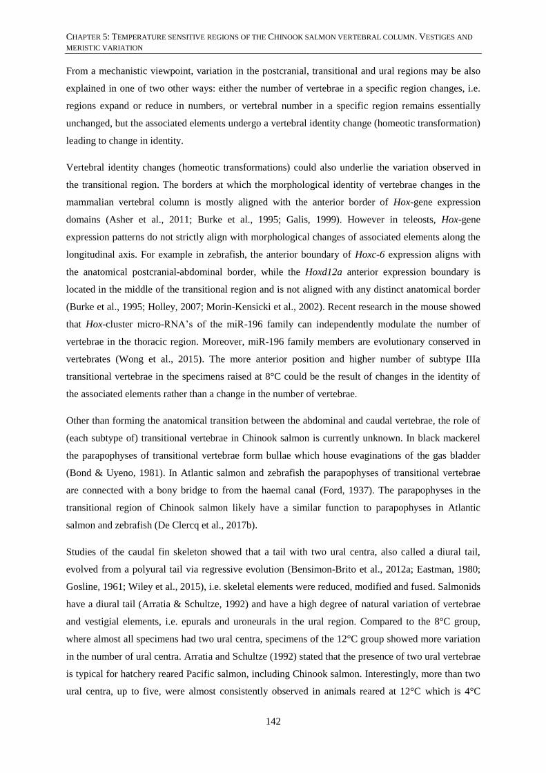
CHAPTER 5: TEMPERATURE SENSITIVE REGIONS OF THE CHINOOK SALMON VERTEBRAL COLUMN. VESTIGES AND
MERISTIC VARIATION
142
From a mechanistic viewpoint, variation in the postcranial, transitional and ural regions may be also
explained in one of two other ways: either the number of vertebrae in a specific region changes, i.e.
regions expand or reduce in numbers, or vertebral number in a specific region remains essentially
unchanged, but the associated elements undergo a vertebral identity change (homeotic transformation)
leading to change in identity.
Vertebral identity changes (homeotic transformations) could also underlie the variation observed in
the transitional region. The borders at which the morphological identity of vertebrae changes in the
mammalian vertebral column is mostly aligned with the anterior border of Hox-gene expression
domains (Asher et al., 2011; Burke et al., 1995; Galis, 1999). However in teleosts, Hox-gene
expression patterns do not strictly align with morphological changes of associated elements along the
longitudinal axis. For example in zebrafish, the anterior boundary of Hoxc-6 expression aligns with
the anatomical postcranial-abdominal border, while the Hoxd12a anterior expression boundary is
located in the middle of the transitional region and is not aligned with any distinct anatomical border
(Burke et al., 1995; Holley, 2007; Morin-Kensicki et al., 2002). Recent research in the mouse showed
that Hox-cluster micro-RNA’s of the miR-196 family can independently modulate the number of
vertebrae in the thoracic region. Moreover, miR-196 family members are evolutionary conserved in
vertebrates (Wong et al., 2015). The more anterior position and higher number of subtype IIIa
transitional vertebrae in the specimens raised at 8°C could be the result of changes in the identity of
the associated elements rather than a change in the number of vertebrae.
Other than forming the anatomical transition between the abdominal and caudal vertebrae, the role of
(each subtype of) transitional vertebrae in Chinook salmon is currently unknown. In black mackerel
the parapophyses of transitional vertebrae form bullae which house evaginations of the gas bladder
(Bond & Uyeno, 1981). In Atlantic salmon and zebrafish the parapophyses of transitional vertebrae
are connected with a bony bridge to from the haemal canal (Ford, 1937). The parapophyses in the
transitional region of Chinook salmon likely have a similar function to parapophyses in Atlantic
salmon and zebrafish (De Clercq et al., 2017b).
Studies of the caudal fin skeleton showed that a tail with two ural centra, also called a diural tail,
evolved from a polyural tail via regressive evolution (Bensimon-Brito et al., 2012a; Eastman, 1980;
Gosline, 1961; Wiley et al., 2015), i.e. skeletal elements were reduced, modified and fused. Salmonids
have a diural tail (Arratia & Schultze, 1992) and have a high degree of natural variation of vertebrae
and vestigial elements, i.e. epurals and uroneurals in the ural region. Compared to the 8°C group,
where almost all specimens had two ural centra, specimens of the 12°C group showed more variation
in the number of ural centra. Arratia and Schultze (1992) stated that the presence of two ural vertebrae
is typical for hatchery reared Pacific salmon, including Chinook salmon. Interestingly, more than two
ural centra, up to five, were almost consistently observed in animals reared at 12°C which is 4°C
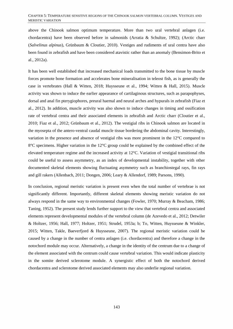
CHAPTER 5: TEMPERATURE SENSITIVE REGIONS OF THE CHINOOK SALMON VERTEBRAL COLUMN. VESTIGES AND
MERISTIC VARIATION
143
above the Chinook salmon optimum temperature. More than two ural vertebral anlagen (i.e.
chordacentra) have been observed before in salmonids (Arratia & Schultze, 1992); (Arctic charr
(Salvelinus alpinus), Grünbaum & Cloutier, 2010). Vestiges and rudiments of ural centra have also
been found in zebrafish and have been considered atavistic rather than an anomaly (Bensimon-Brito et
al., 2012a).
It has been well established that increased mechanical loads transmitted to the bone tissue by muscle
forces promote bone formation and accelerates bone mineralisation in teleost fish, as is generally the
case in vertebrates (Hall & Witten, 2018; Huysseune et al., 1994; Witten & Hall, 2015). Muscle
activity was shown to induce the earlier appearance of cartilaginous structures, such as parapophyses,
dorsal and anal fin pterygiophores, preural haemal and neural arches and hypurals in zebrafish (Fiaz et
al., 2012). In addition, muscle activity was also shown to induce changes in timing and ossification
rate of vertebral centra and their associated elements in zebrafish and Arctic charr (Cloutier et al.,
2010; Fiaz et al., 2012; Grünbaum et al., 2012). The vestigial ribs in Chinook salmon are located in
the myosepta of the antero-ventral caudal muscle tissue bordering the abdominal cavity. Interestingly,
variation in the presence and absence of vestigial ribs was more prominent in the 12°C compared to
8°C specimens. Higher variation in the 12°C group could be explained by the combined effect of the
elevated temperature regime and the increased activity at 12°C. Variation of vestigial transitional ribs
could be useful to assess asymmetry, as an index of developmental instability, together with other
documented skeletal elements showing fluctuating asymmetry such as branchiostegal rays, fin rays
and gill rakers (Allenbach, 2011; Dongen, 2006; Leary & Allendorf, 1989; Parsons, 1990).
In conclusion, regional meristic variation is present even when the total number of vertebrae is not
significantly different. Importantly, different skeletal elements showing meristic variation do not
always respond in the same way to environmental changes (Fowler, 1970; Murray & Beacham, 1986;
Taning, 1952). The present study lends further support to the view that vertebral centra and associated
elements represent developmental modules of the vertebral column (de Azevedo et al., 2012; Detwiler
& Holtzer, 1956; Hall, 1977; Holtzer, 1951; Strudel, 1953a; b; To, Witten, Huysseune & Winkler,
2015; Witten, Takle, Baeverfjord & Huysseune, 2007). The regional meristic variation could be
caused by a change in the number of centra anlagen (i.e. chordacentra) and therefore a change in the
notochord module may occur. Alternatively, a change in the identity of the centrum due to a change of
the element associated with the centrum could cause vertebral variation. This would indicate plasticity
in the somite derived sclerotome module. A synergistic effect of both the notochord derived
chordacentra and sclerotome derived associated elements may also underlie regional variation.

CHAPTER 5: TEMPERATURE SENSITIVE REGIONS OF THE CHINOOK SALMON VERTEBRAL COLUMN. VESTIGES AND
MERISTIC VARIATION
144
5.6. ACKNOWLEDGEMENTS
The authors would like to thank New Zealand King Salmon and Skretting Australia for funding and
support during the experimental procedures. Specifically we would like to acknowledge Nicole ruff
and Ben Wybourne.
5.7. AUTHOR CONTRIBUTIONS (IN CREDIT TERMS)
A. De Clercq: Conceptualisation, methodology, investigation, writing – original draft preparation, and
visualisation
M.R. Perrott: Conceptualisation, writing – review and editing, supervision and funding acquisition
P.S. Davie: Conceptualisation, supervision and funding acquisition
M.A. Preece: Resources and methodology
M.A.G. Owen: Formal analysis, writing – review and editing
A. Huysseune: Writing – review and editing, supervision and funding acquisition
P.E. Witten: Conceptualisation, writing – review and editing, supervision and funding acquisition

CHAPTER 6: LOWER INCUBATION AND FRESHWATER GROW-OUT TEMPERATURES PROTECTS CHINOOK SALMON
FROM SUBSEQUENT VERTEBRAL DEFORMITIES IN SEAWATER BUT NOT IN FRESHWATER.
145
CHAPTER 6: LOWER INCUBATION AND FRESHWATER
GROW-OUT TEMPERATURES PROTECTS CHINOOK SALMON
FROM SUBSEQUENT VERTEBRAL DEFORMITIES IN SEAWATER
BUT NOT IN FRESHWATER
A. De Clercq(1, 2)
1School of Veterinary Science, Massey University, Palmerston North, New Zealand
2Evolutionary Developmental Biology, Ghent University, Ghent, Belgium
Requested chapter from examiner and in preparation for publication

CHAPTER 6: LOWER INCUBATION AND FRESHWATER GROW-OUT TEMPERATURES PROTECTS CHINOOK SALMON
FROM SUBSEQUENT VERTEBRAL DEFORMITIES IN SEAWATER BUT NOT IN FRESHWATER.
146
6.1. ABSTRACT
Vertebral anomalies are a severe and recurrent problem in both Atlantic and Chinook salmon farming.
Temperature is often manipulated in salmon farming to influence growth rates and to obtain a year-
round production. However, temperature is also a risk factor for vertebral deformities. In salmonids,
vertebral deformities can develop de novo in every life stage, both in freshwater and in seawater
growth stages. Here PIT-tagged Chinook salmon, of crosses that produced off-spring with high
deformity rates, were incubated and grown in freshwater at a constant 8°C and 12°C before being
transferred to sea cages. Salmon were X-rayed at three life stages; the freshwater smoltification stage,
the six-months-at-sea-stage and the 12-months-at-sea stage. Radiographs of salmon were evaluated
for the presence of four deformity categories: a combination of lordosis, kyphosis and scoliosis
(=LKS), fusion, compression and/or reduced intervertebral space, and vertical shift. Deformities were
also scored from one (mild) to three (severe). The mortality in both temperature groups was highest
between the freshwater smoltification and six-months-at-sea stages. The 8°C group had a higher
prevalence of deformities and average severity score in the freshwater stage but a lower prevalence of
deformities and average severity score in the seawater stages compared to the 12°C group. LKS was
not present in freshwater stage specimens of either temperature group. Specimens that were culled six
months after seawater entry had significantly more deformities and LKS in the 12°C group only. Both
temperature groups had a significantly higher prevalence of deformities and LKS after the 12-months-
at-sea stage compared to after six-months-at-sea. In the current study we highlight that the mortality is
high after the seawater transfer stage, and that deformity is also notable in runts, a category of under-
performing animals with poorly understood reasons for retarded growth. These phenomena should be
better studied since the causes of mortality at this stage are still unknown. Here we also show that
incubation and freshwater rearing temperatures can influence the deformity prevalence in subsequent
seawater stages.
6.1.1. KEYWORDS
Smoltification, seawater stage, deformities, Chinook salmon, Atlantic salmon
6.2. INTRODUCTION
Vertebral anomalies are a severe and recurrent problem in salmon farming (Fjelldal et al., 2012). It
has been established that the underlying cause of vertebral anomalies is multifactorial (Boglione et al.,
2013a; Boglione et al., 2013b; Fjelldal et al., 2012), i.e. a plethora of genetic and epigenetic factors
which often interact with each other, underlie vertebral anomalies. Husbandry factors such as diet,
light, temperature, and vaccination are some of the most frequently manipulated factors in farming
conditions (Boglione et al., 2013b; Fjelldal et al., 2012). Often several of these factors are changed

CHAPTER 6: LOWER INCUBATION AND FRESHWATER GROW-OUT TEMPERATURES PROTECTS CHINOOK SALMON
FROM SUBSEQUENT VERTEBRAL DEFORMITIES IN SEAWATER BUT NOT IN FRESHWATER.
147
simultaneously to obtain fast or slow growing salmon, a necessity for a year-round production. For
example, a combination of more frequent feeding, longer light photoperiods and higher rearing
temperatures will lead to faster growth of freshwater juvenile stages. Faster growing salmon go into
seawater earlier in the year than slower growing salmon and therefore can be harvested earlier in the
year. Importantly, changes to specifications in the diet (Ørnsrud et al., 2004a; Ørnsrud et al., 2004b),
photoperiod (Fjelldal et al., 2004; Fjelldal et al., 2016), and fast growth juvenile production strategies
due to elevated temperatures (Fjelldal & Hansen, 2010; Fjelldal et al., 2006), are all factors known to
induce vertebral anomalies. Because several factors are manipulated at once, it is hard to ascertain
which factor is responsible for causing vertebral anomalies. In contrast, a single factor, i.e. the lack of
dietary phosphorous, could be linked to a single vertebral deformity phenotype (Witten type 10),
undersized vertebrae with increased intervertebral spaces (Witten et al., 2009), in Atlantic salmon
(Salmo salar) that were four months in seawater (Witten et al., 2016). Moreover, many one-factor
drivers for skeletal deformity have been mitigated via improvements to the diet.
Among the many environmental factors, temperature stands out as one of the master factors in fish
biology (Beitinger et al., 2000). Temperature influences developmental rate (Beacham & Murray,
1989), mortality (Beacham & Murray, 1987a; Murray & Beacham, 1986), success of hatching
(Hamor & Garside, 1976; Hayes et al., 1953) and normal skeletal development (Koumoundouros et
al., 2001) in early stages of development. Also in later stages of development temperature is important
for normal skeletal development (Cloutier et al., 2010; Grünbaum & Cloutier, 2010). Elevated rearing
temperature reduces the production time in salmon farming but increases the risk of abnormal
vertebral development (Fjelldal et al., 2006; Grini et al., 2011; Ytteborg et al., 2010a; Ytteborg et al.,
2010b) and can induce a wide variety of vertebral deformity phenotypes (Witten et al., 2009). Fast
growing Chinook salmon (Oncorhynchus tshawytscha) eggs and freshwater juveniles in New Zealand
are incubated at ~12°C, close to the upper temperature limit of 13°C for early incubation (Brett,
1952). This upper temperature limit, and lower temperature limit, in Chinook salmon was determined
by lethal temperature experiment on wild caught and laboratory reared salmon of different
populations along North-South latitude acclimated to different temperatures (Alderdice & Velsen,
1978; Brett, 1952). The same experiments showed that the preferred temperature range for incubation
and freshwater grow-out of Chinook salmon was between 8°C and 10°C (Alderdice & Velsen, 1978;
Brett, 1952).
Vertebral deformities in salmonids are often diagnosed and studied using radiography (Fjelldal et al.,
2012; Gill & Fisk, 1966; Witten et al., 2009; Witten et al., 2016). Based on radiographic images of
Atlantic salmon vertebral columns, vertebral deformity phenotypes have been classified into 20 types
(Witten et al., 2009). Within the 20 types of vertebral deformities a further subdivision into three
major categories can be made, i.e. (i) vertebral column curvatures, (ii) vertebral centra compressions
and fusions, and (iii) vertebral centrum deformities (Witten et al., 2009). Vertebral axis deviations

CHAPTER 6: LOWER INCUBATION AND FRESHWATER GROW-OUT TEMPERATURES PROTECTS CHINOOK SALMON
FROM SUBSEQUENT VERTEBRAL DEFORMITIES IN SEAWATER BUT NOT IN FRESHWATER.
148
include lordosis (ventral bending), kyphosis (dorsal bending) and scoliosis (sideways bending).
Vertebral centra compressions and fusions include varying degrees of compression, fusions and/or
complete fusions along the antero-posterior axis of the vertebrae. Vertebral centrum deformities
include radio-translucent and radio-dense centra, and deviations from the autocentrum symmetry
within the vertebral centrum. Vertebral deformities such as fusion and compression are most common
in Atlantic salmon (Fjelldal et al., 2012; Witten et al., 2005a; Witten et al., 2006), while vertebral
column curvatures have been observed but are less frequent (Fjelldal et al., 2004; Gil Martens et al.,
2012; Sullivan et al., 2007b). In Chinook salmon, fusion was reported as the most common vertebral
deformity in freshwater juveniles (Seymour, 1959). In other Pacific salmon species, i.e. sockeye, pink
and chum salmon (O. nerka, O. gorbuscha and O. keta, respectively) a varying degree of compression
and fusion was reported in juvenile and adult life stages (Gill & Fisk, 1966).
It has been stated in literature that vertebral deformities can develop at any salmonid life stage (De
Clercq et al., 2017a; Fjelldal et al., 2012; Witten et al., 2006). Temperature has been found to be a risk
factor in both early life stages, where elevated temperature and temperature shocks induce a range of
notochord and vertebral malformations (De Clercq et al., 2017a; Takle et al., 2005; Wargelius et al.,
2005), and late life stages (Fjelldal et al., 2006; Grini et al., 2011). More recently in studies of
Chinook salmon the development of certain vertebral deformities appeared to be life-stage dependent.
While the commonly occurring vertebral deformities such as fusion, compression and vertical shift of
the entire vertebral centrum can develop both in freshwater pre-smoltification and seawater post-
smoltification stages, vertebral column curvatures only appear to develop in late seawater stages
(Munday et al., 2016; Munday et al., 2018; Perrott et al., 2018).
In the current study we ask if hatchery rearing at 12°C predisposes Chinook salmon to higher
deformity rates overall, whether or not specific deformity phenotypes are more prevalent and if
salmon are more prone to deformity at certain life stages, compared to specimens raised at a more
optimum temperature of 8°C. The deformity types considered in the current study have been
previously described in Chinook salmon by Perrott et al. (2018), i.e. LKS (a combination of lordosis,
kyphosis and scoliosis), fusions, compression and/or reduced intervertebral spaces and vertical shift of
the entire vertebral centrum. The juvenile and adult life stages considered here span pre-smoltification
(~12 months rearing in freshwater), and post-smoltification, a period of known transition and stress.
Specimens were followed and evaluated after six months and 12 months of exposure to a seawater
husbandry environment.

CHAPTER 6: LOWER INCUBATION AND FRESHWATER GROW-OUT TEMPERATURES PROTECTS CHINOOK SALMON
FROM SUBSEQUENT VERTEBRAL DEFORMITIES IN SEAWATER BUT NOT IN FRESHWATER.
149
6.3. MATERIAL AND METHODS
6.3.1. BROODSTOCK
Commercial slow growing Chinook salmon adults were used to set up crosses that produced offspring
with high deformity rates (Table 2, SECTION 2.2). Broodstock were kept at a constant 11.8°C (Takaka
Hatchery, Takaka, New Zealand). Females with high X-ray based deformity rates were crossed with
sex-reversed males (juvenile females treated with 17-α-methyltestosterone, Sigma-Aldrich) with low
X-ray based deformity. Adults which provided offspring with high deformity rates were chosen to
increase the chance of less frequent deformities occurring in the experimental specimens. Eggs from
ten females were fertilised with milt from six males at 10°C. The gametes of both the females and
males were randomised to avoid any effect of the genetic background of specific crosses confounding
the constant temperature experiment (APPENDIX B).
6.3.2. EXPERIMENT
To avoid aberrant effects of potential temperature shocks after fertilisation, eggs were placed in
incubation trays (±4000 eggs/tray) and were supplied with water at a constant 10°C until 3 days post-
fertilisation. Subsequently, the temperature was adjusted (0.2°C per hour) to a constant 8°C and 12°C
for rearing. Dead specimens were removed every two days during the hatching period and twice a
week before and after the hatching period until specimens reached first feed stage. At first feed stage
(±900°d) 4000 specimens of each temperature group, i.e. the 8°C and 12°C groups, were moved to
two black 420 L circular tanks (2000 specimens per tank) (Fig. 23). The 8°C and 12°C temperature
regimes were continued in the tanks. Once dead individuals were removed, the tanks were carefully
cleaned every day to reduce the impact of stress on the specimens. The removal of waste feed from
the bottom of the tank and the filter mesh was to avoid infections. At 1400°d (cull 1) and 1500°d (cull
2), 250 individuals were randomly removed from each tank in order to reduce crowding stress, allow
for growth and to further reduce the risk of infections (Table 12, Fig. 23). One hundred of the 250
culled specimens of each tank were measured and weighed. A fungal infection of the fins was
successfully treated for 1 hour (water flow taps closed) with a 4.5 ppm Halamid solution (1.8g/tank,
Agrivantage, Axcentive Asia Pte Ltd, Singapore) before PIT-tagging. Due to fin nipping (biting)
some specimens had abraded and sometimes infected tail fins at the end of the freshwater stage, but
remained untreated before transfer to seawater.
At 2500°d, 500 specimens of the 12°C group were PIT-tagged (Avi-Tag 162e, Avid Identification
Systems Inc, Norco, CA, USA) and moved to 1000 L circular tanks (250 salmon/tank) at ambient
temperature (12.4°C, Table 12, Fig. 23) When the 12°C specimens were transferred to bigger tanks
the temperature of the 8°C specimens was raised to 12°C over two days (0.2°C per hour during the

CHAPTER 6: LOWER INCUBATION AND FRESHWATER GROW-OUT TEMPERATURES PROTECTS CHINOOK SALMON
FROM SUBSEQUENT VERTEBRAL DEFORMITIES IN SEAWATER BUT NOT IN FRESHWATER.
150
day). Subsequently, 1000 specimens of the 8°C group at 1700°d, now acclimated to 12°C were also
PIT-tagged and moved to four 420 L black circular tanks (250 specimens/tank) at ambient 12.4°C
(Table 12, Fig. 23). Another 50 specimens per tank were randomly removed to allow for growth and
reduce crowding stress after PIT-tagging (Fig. 23). Smoltification-stage salmon (based on size and
age) from both temperature groups, 343 specimens of the 8°C and 269 specimens of the 12°C group,
were transported at the same time to an experimental sea cage (6x6x6, 216 m3) at Ruakaka farm in the
Marlborough Sounds (Ruakaka Bay, Queen Charlotte Sound). No specimens were tested with a
saltwater challenge to determine the freshwater smoltification status in order to avoid reducing the
number of animals that would go to seawater and available for statistics. However, specimens were
larger than the critical size for successful smoltification of Chinook salmon in New Zealand (> 7.0 g,
Table 12) and entered seawater in late autumn (06/05/2015, Table 12, Fig. 23) when Chinook salmon
have a physiological smoltification window (Franklin, 1989). Specimens that were classified as
‘runts’ (animals that failed to thrive, weigh less than one fifth of the average weight of the cohort) at
the six-months-in-sea X-ray event were culled and euthanized with anaesthetic overdose (AquiS,
AquiS New Zealand Ltd, Lower Hutt, New Zealand).
Smoltification-stage Chinook salmon were X-rayed prior to transportation to sea cages, 357
specimens of the 8°C (3348°d) and 376 specimens of the 12°C (4177°d) (Table 12, Fig. 23).
Specimens were anaesthetised for radiography with 20 ppm AquiS and placed on their right side on
the film (Kodak Industrex M100 Ready Pack II, Kodak Australia Pty Ltd, Victoria) or on the digital
receptor. For films, the X-ray emitter was placed on a distance of 53.5 cm and set at 50 kV, 0.25
mA/sec and 2.1 seconds exposure time. Exposed X-ray films were developed at 21°C for seven
minutes, using Kodak Industrex developer and Kodak Industrex LO fixative following manufacturers
recommendations. After a week to recover from radiography, the salmon were moved to the
experimental sea cage. Digital X-rays were captured using an amorphous silicon digital receptor (90-
479 Sound Technologies Tru-DR, DLC Australia Pty Ltd, Melbourne) with the digital receptor focus
distance of 50 cm and X-ray settings of 80 kV and 0.45 mA/sec. Individually identified salmon from
each temperature group were radiographed at six months and 12 months following seawater transfer
(Table 12, Fig. 23), and at harvest (15 months). The X-ray source for both film and digital
radiography was an Atomscope HF80/15+ Portable Unit (Mikasa, Tokyo).
Figure 23: Schematised flow diagram of the 8°C and 12°C specimens from freshwater to seawater.
(below)
(A) The representative timeline is shown by a black line between the 12°C group trajectory (top, orange) and the
8°C group trajectory (middle, green). At the left of both temperature group trajectories an incubation stack is
shown, from which 4000 specimens were moved to tanks (2000 per tank) at first feed (900°d). First feed is
indicated on the timeline as the first grey vertical bar. The °d-stage and event is given below the tanks of the
freshwater stages. Grey vertical bars on the time line indicate the time points of these stages (see Table 12).

CHAPTER 6: LOWER INCUBATION AND FRESHWATER GROW-OUT TEMPERATURES PROTECTS CHINOOK SALMON
FROM SUBSEQUENT VERTEBRAL DEFORMITIES IN SEAWATER BUT NOT IN FRESHWATER.
151
Tanks containing water with ambient temperature are indicated in blue. The number of specimens at each stage
is shown in the tanks, underneath the volume of the tank. The stages where specimens were euthanized or died
(mortalities) are indicated in red. The exact number of mortalities, if known, is indicated above the arrows in the
freshwater stages (see also Table 13). Smolt-stage animals (smoltification-stage) of both temperature groups
were transported together (truck) to an experimental sea cage (see also B, barge and truck). (B) The timeline
now runs above the sea cages starting at the transfer date. The number of specimens of each temperature group
that were transferred is repeated (indicated by ‘transported’). The repeatedly measured specimens are indicated
by ‘repeated measure’ (above arrow), in first instance specimens that survived to six-months-at-sea and in
second instance specimens that survived to 12-months-at-sea. The number of animals in the sea cage at the two
X-ray events (i.e. six-months-at-sea and 12-months-at-sea) is shown in bold letters. The number of mortalities
(red) is indicated above and below the arrows in the seawater stages (see also Table 13). The number of runts is
indicated between the two sea cages after six-months-at-sea.

CHAPTER 6: LOWER INCUBATION AND FRESHWATER GROW-OUT TEMPERATURES PROTECTS CHINOOK SALMON FROM SUBSEQUENT VERTEBRAL DEFORMITIES IN SEAWATER BUT NOT IN
FRESHWATER.

CHAPTER 6: LOWER INCUBATION AND FRESHWATER GROW-OUT TEMPERATURES PROTECTS CHINOOK SALMON FROM SUBSEQUENT VERTEBRAL DEFORMITIES IN SEAWATER BUT NOT IN
FRESHWATER.

CHAPTER 6: LOWER INCUBATION AND FRESHWATER GROW-OUT TEMPERATURES PROTECTS CHINOOK SALMON
FROM SUBSEQUENT VERTEBRAL DEFORMITIES IN SEAWATER BUT NOT IN FRESHWATER.
154
Table 12: Summary data of Chinook salmon life stages during the constant temperature experiment in
freshwater and in sea.
°d Temperature Date Days Event Fork length (cm) Weight (g)
0 8°C and 12°C 20/05/14 0 Fertilisation
1400 8°C 11/11/14 175 Cull 1 4.5 (±0.04) 1.5 (±0.03)
1400 12°C 13/09/14 116 Cull 1 5.7 (±0.03) 2.2 (±0.04)
1500 8°C 27/11/14 191 Cull 2 5.4 (±0.04) 1.7 (±0.05)
1500 12°C 25/09/14 128 Cull 2 6.9 (±0.04) 4.0 (±0.06)
1700 8°C 17/12/14 211 PIT-tagging
2500 12°C 15/12/14 209
3348 8°C 29/04/15 344 X-ray (film)
16.8 (±0.05) 64.1 (±0.54)
4177 12°C 21.1 (±0.06) 151.6 (±1.53)
3435 8°C 06/05/15 351 Transfer
4264 12°C
5543 8°C 23/10/15 521
6-months-sea
X-ray (digital)
27.7 (±0.67) 407.1 (±24.86)
6372 12°C 30.6 (±0.88) 501.6 (±43.44)
7849 8°C 26/04/16 707
12-months-sea
X-ray (digital)
44.3 (±0.68) 1229.0 (±65.60)
8678 12°C 44.4 (±0.74) 1064.4 (±64.18)
Developmental stage in degreedays (°d). The “Event” column indicates what happened at the
corresponding age. The average (±standard error) of the fork length (cm) and weight (g) is given
for the cull 1, cull 2, freshwater X-ray, six-months and 12-months-at-sea time points. The three X-
ray datasets that were available for analyses are shown in blue.
6.3.3. FEEDING REGIME
First feed specimens of the 8°C and 12°C groups were fed intensively for the first ten days, five days
with powder meal (Str. 00, Golden Prima, Biomar) and five days with crushed meal (Crumble,
Golden Prima, Biomar). Subsequently, during the freshwater grow-out period, the growing animals
were fed three times a day with food pellets of increasing size (GAP Semi-Float, Biomar). After
transfer to the sea cage, specimens were hand fed to satiation with commercially available salmon
feeds, the first six months with 3.0 mm size pellets comprising 48% protein, 21% oil and 0.9%
available P; the subsequent ten weeks with 4.0 mm size pellets comprising 50% protein, 22 % oil and
0.8% available P; finally with 9.0 mm pellets until harvest comprising 39% protein, 26% oil and 0.6%
available P.
6.3.4. DATA COLLECTION AND STATISTICS
Deformities were identified within vertebral column regions and scored following the procedure in
Perrott et al. (2018). Deformities were identified and scored on developed X-ray film that was placed

CHAPTER 6: LOWER INCUBATION AND FRESHWATER GROW-OUT TEMPERATURES PROTECTS CHINOOK SALMON
FROM SUBSEQUENT VERTEBRAL DEFORMITIES IN SEAWATER BUT NOT IN FRESHWATER.
155
on a light box while deformities in digital X-ray images were identified and scored by viewing the
image in Photoshop software.
The vertebral column was subdivided into four regions following Kacem et al. (1998): region 1
includes vertebrae 1 to 8 (V1-V8), region 2 includes vertebrae 9 to 31 (V9-V31), region 3 includes
vertebrae 31 to 50 (V31-V50) and region 4 includes vertebrae 51 to 63+ (V51-V63+). Variation in the
number of vertebrae is present in region 4 and therefore the 63+ indication is used.
Vertebral deformities were recognised based on Witten et al. (2009) classification of 20 types of
vertebral deformities. In the present study, a combination of lordosis, kyphosis and scoliosis (Witten
et al., 2009, type 14, 15, 16), fusion (Witten et al., 2009, type 6, 7, 8), compression of vertebrae and or
intervertebral spaces (Witten et al., 2009, type 1, 2, 3, 4, 5, further referred to as compression), and
vertical shift within the entire vertebral centrum (Witten et al., 2009, type 17) were recognised. These
specific deformity types were chosen because they can be compared to other studies investigating
deformities in farmed Chinook salmon.
In addition to recognising the deformities, a severity score of 1 to 3 was assigned to each deformity.
The score is based on the number of vertebrae involved in the deformity and the angle of the
curvature in lordosis and kyphosis. Assessment of the severity of scoliosis was more subjective when
it was not present in combination with lordosis and kyphosis.
Severity 1: 1 to 2 vertebrae or 0 °to 20°
Severity 2: 2 to 5 vertebrae or 20° to 40°
Severity 3: 5+ vertebrae or >40°
The absence or presence of each deformity was scored per region by 0 or a 1, absent or present
respectively. For total deformity, a score of 1 was allocated when the specimen had a deformity of any
type in any region, even when different types of deformities in multiple regions along the vertebral
column occurred. In addition, a parallel dataset was generated in which each of the present
deformities were given a severity score (1 to 3). All statistical analyses were performed in R-studio
Team (2015, R-studio Inc., Boston). Figures were constructed with ggplot2 package (R-studio) and
Photoshop CS5.
Fork length and weight were measured at two freshwater culling events (cull 1 and cull 2), at the pre-
transfer X-ray event and at the sea water X-ray events at six and 12 months at sea (Table 12). The
average fork length and weight of the 8°C and 12°C specimens were tested for significant differences
with the non-parametric Wilcoxon rank sum test. This was due to the fact that the data were not
normally distributed and that the variances were unequal.

CHAPTER 6: LOWER INCUBATION AND FRESHWATER GROW-OUT TEMPERATURES PROTECTS CHINOOK SALMON
FROM SUBSEQUENT VERTEBRAL DEFORMITIES IN SEAWATER BUT NOT IN FRESHWATER.
156
Two datasets from the freshwater smoltification-stage X-ray scoring were constructed. The first
dataset contained the scoring of all smoltification-stage salmon (dataset 1). The second dataset
contained only the scoring of smoltification-stage salmon that survived to six-months-at-sea (dataset
2). A general linear model (glm, outcome variable binomial, Fisher exact method) was used to test for
statistically significant differences between the proportion of deformities (prevalence) of the 8°C and
12°C specimens at freshwater smoltification-stage (dataset 1 and dataset 2), and at six-months-at-sea
(all specimens). To increase the robustness of the glm-model the fork length and weights of each
individual were added as variables to the model. Fork length and weights of a number of freshwater
smoltification-stage salmon were missing due to a random set of specimens that had no PIT-tag.
Multiple imputation (5 iterations, 50 times, assuming missing data completely at random) on the fork
length and weight variables in the freshwater dataset was applied (mice package) to fill the missing
data points. For the six-months-at-sea dataset, a culling variable (whether or not a specimen was
removed after X-ray, ‘runts’) was also added. Because a low prevalence was observed for fusion,
compression and vertical shift in the seawater datasets, resulting in 2x2 contingency tables with low
values, the Firth logistic regression (logistf package) was used to test for significant differences in the
prevalence of deformities between the 8°C and 12°C groups on all datasets. The statistical output
values reported in the results are based on the Firth logistic regression except when indicated
otherwise. The Wald test indicates the fit of model. To test for significant differences in the
prevalence of total deformity proportion and LKS proportion from the freshwater to the 12-month-at-
sea stage (see repeated measure Fig. 23), a conditional logistic regression for matched paired binary
data (survival package) was used.
The severity score data for the freshwater smoltification-stage (dataset 1 and dataset 2), and at six-
months-at-sea (all specimens) was not distributed normally and had unequal variances. The average of
the score data was tested for significant differences between the 8°C and 12°C groups with the non-
parametric Wilcoxon test. The average score was calculated on the sum of scores, for total deformity:
Σi(score ith deformity type (Σi(score jth region)))/number of specimens
The average score for each deformity type:
Σi(score jth region)/number of specimens

CHAPTER 6: LOWER INCUBATION AND FRESHWATER GROW-OUT TEMPERATURES PROTECTS CHINOOK SALMON
FROM SUBSEQUENT VERTEBRAL DEFORMITIES IN SEAWATER BUT NOT IN FRESHWATER.
157
Table 13. Attrition of Chinook salmon specimens raised at a constant 8°C and 12°C available for X-ray
exposure at different life stages.
Life stage X-rayed No PIT-tag Mortality Culled
Pre-seawater transfer 8°C 357 5 238 (225 + 13)
Pre-seawater transfer 12°C 376 40 270 (205+65)
6-months-at-sea 8°C 114 45 39
6-months-at-sea 12°C 68 20 31
12-months-at-sea 8°C 30 10 (8 + 2)
12-months-at-sea 12°C 17 10 (7 + 3)
15-months-at-sea 8°C 20
15-months-at-sea 12°C 7
The “Mortality” column shows the total of specimens that died (died between life
stages + died at time of X-ray). The “Culled” columns shows the specimens
removed due to growth arrest (runting).
6.4. RESULTS
6.4.1. ATTRITION FROM FRESHWATER SMOLTIFICATION TO 15-MONTHS-AT-SEA STAGE
The highest attrition occurred in the 12°C group where only 1.9% of the specimens survived,
compared to 5.6% in the 8°C group, from the freshwater smoltification-stage until 15 months at sea
(harvest, Table 13). More specimens in the 12°C group (40) compared to the 8°C group (5) lacked a
PIT-tag and were culled. Also in the 12°C group more specimens (65) died during the X-ray event
compared to the 8°C group (13). The culling of specimens due to an arrest in growth (runting) only
occurred at the six-months-at-sea X-ray event, with 39 specimens culled in the 8°C group and 31 in
the 12°C group. The largest number of mortalities occurred between the X-ray time points in both
temperature groups, especially between the freshwater X-ray time point and the six-months-at-sea X-
ray time point, with a larger attrition rate in the 12°C group (71.8%), compared to the 8°C group
(66.7%). Due to high mortality rates in specimens of both temperature groups, only a few animals
were left to X-ray at 15-months-at-sea and at harvest (Table 13). No biologically meaningful
statistical analyses could be made with these very small sample sizes. Thus, the 15-months sample
point is not included in this report.
6.4.2. LENGTH AND WEIGHT
Although specimens of the 12°C group had a higher average fork length and weight compared to the
8°C group in freshwater, their average fork length and weight appeared to be no longer different at six
months in seawater (Fig. 24, APPENDIX C). In freshwater, the average (±SE) fork length and weight of
200 specimens during both culling events (cull 1 and cull 2) are given in Table 12. Specimens raised
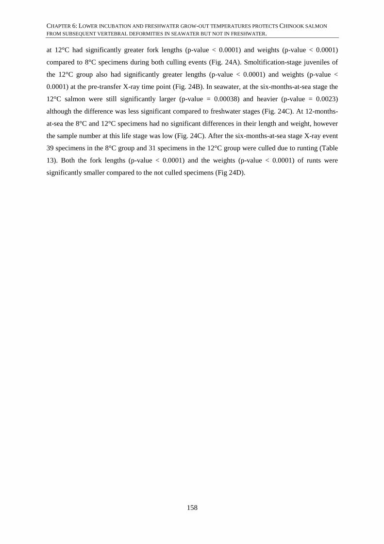
CHAPTER 6: LOWER INCUBATION AND FRESHWATER GROW-OUT TEMPERATURES PROTECTS CHINOOK SALMON
FROM SUBSEQUENT VERTEBRAL DEFORMITIES IN SEAWATER BUT NOT IN FRESHWATER.
158
at 12°C had significantly greater fork lengths (p-value < 0.0001) and weights (p-value < 0.0001)
compared to 8°C specimens during both culling events (Fig. 24A). Smoltification-stage juveniles of
the 12°C group also had significantly greater lengths (p-value < 0.0001) and weights (p-value <
0.0001) at the pre-transfer X-ray time point (Fig. 24B). In seawater, at the six-months-at-sea stage the
12°C salmon were still significantly larger (p-value = 0.00038) and heavier (p-value = 0.0023)
although the difference was less significant compared to freshwater stages (Fig. 24C). At 12-months-
at-sea the 8°C and 12°C specimens had no significant differences in their length and weight, however
the sample number at this life stage was low (Fig. 24C). After the six-months-at-sea stage X-ray event
39 specimens in the 8°C group and 31 specimens in the 12°C group were culled due to runting (Table
13). Both the fork lengths (p-value < 0.0001) and the weights (p-value < 0.0001) of runts were
significantly smaller compared to the not culled specimens (Fig 24D).

CHAPTER 6: LOWER INCUBATION AND FRESHWATER GROW-OUT TEMPERATURES PROTECTS CHINOOK SALMON
FROM SUBSEQUENT VERTEBRAL DEFORMITIES IN SEAWATER BUT NOT IN FRESHWATER.
159
Figure 24: Comparison of fork length and weight of the 8°C and 12°C groups at each life stage.
Boxplot of fork length in centimetre and weight in gram for (A) cull 1 (1400°d, Table 12) and cull 2 (1530°d,
Table 12), (B) freshwater smoltification-stage (indicated as ‘at smoltification stage’), (C) six- and 12-months-at-
sea. The 8°C group is coloured in dark green and the 12°C group in orange. Asterisks indicate significant
differences. (D) culled (runts) and not culled (Ncull) animals.
6.4.3. PREVALENCE OF DEFORMITIES
6.4.3.1. FRESHWATER SMOLTIFICATION-STAGE SPECIMENS (TABLE 14, FIG. 25A)
Before transferring the Chinook salmon from fresh water to seawater, the 8°C (357) and 12°C (376)
specimens were X-rayed. Temperature was found to be a statistically significant predictor of
deformity outcome (glm, 2947df, p-value < 0.0001). The total deformity prevalence was significantly
higher in specimens of the 8°C group (glm, 2947df, p-value < 0.0001), compared to specimens of the
12°C group. Specimens of the 8°C group had a higher but non-significant prevalence of fusion,

CHAPTER 6: LOWER INCUBATION AND FRESHWATER GROW-OUT TEMPERATURES PROTECTS CHINOOK SALMON
FROM SUBSEQUENT VERTEBRAL DEFORMITIES IN SEAWATER BUT NOT IN FRESHWATER.
160
compression and vertical shift compared to specimens of the 12°C group. No LKS was present in
freshwater stage Chinook salmon of either temperature group.
Figure 25: Prevalence and average severity score of deformities in Chinook salmon from the 8°C and
12°C groups.
Freshwater smoltification-stage (A, D), six-months-at-sea (6m) stage (B, E) and freshwater specimens that
survived to six-months-at-sea (C, F, see Fig. 23 repeated measure). The prevalence plots show the percentage of
deformity (y-axis). The average score plots show the average severity score (y-axis). Error bars indicate
standard error (based on average score). The number of specimens in each temperature group is indicated in the
legend of each bar plot. Asterisks indicate significant differences. Abbreviations: Total = total deformity (any
deformity type in any region), LKS = lordosis, kyphosis, scoliosis, F = fusion, C = compression, VS = vertical
shift, SW6 = six-months-at-sea.
6.4.3.2. SIX MONTHS IN SEAWATER SPECIMENS (TABLE 14, FIG. 25B, 26A, B)
At six months in seawater, 114 specimens of the 8°C group and 69 specimens of the 12°C group were
X-rayed. Temperature (p-value < 0.0001) and ‘runting’ (p-value < 0.0001) were found to be
statistically significant predictors for deformity outcome (Wald test 6df, p-value = < 0.0001).
Specimens of the 12°C group had a higher prevalence of total deformity and of each deformity type,
including LKS, compared to the 8°C group (Fig. 25A). Total deformity (p-value < 0.0001), LKS (p-
value < 0.0001) and vertical shift (p-value = 0.032) were significantly more prevalent in specimens of
the 12°C group compared to the 8°C group.
Culled specimens of the 8°C group (Fig. 26A) had a higher prevalence of total deformity and LKS,
and a lower prevalence of fusion. Compression and vertical shift deformity types occurred only in
specimens that were in the not-culled category. The prevalence of deformity types between culled and
not-culled specimens was not significantly different.
Culled specimens of the 12°C group (Fig. 26B) had a significantly higher prevalence of total
deformity (p-value < 0.0001) and LKS (p-value < 0.0001). Fusion and compression also had a higher

CHAPTER 6: LOWER INCUBATION AND FRESHWATER GROW-OUT TEMPERATURES PROTECTS CHINOOK SALMON
FROM SUBSEQUENT VERTEBRAL DEFORMITIES IN SEAWATER BUT NOT IN FRESHWATER.
161
prevalence in culled specimens but this was not statistically significant. Vertical shift occurred only in
culled specimens.
Figure 26: Deformity prevalence and average severity score in not-culled versus culled specimens.
Specimens of the 8°C (A, C) and 12°C groups (B, D) at the six-months-at-sea (6m) stage. Specimens were
culled due to ‘runting’. The prevalence plots show the percentage of deformity (y-axis). The average severity
score plots are also shown on the y-axis. Error bars indicate standard error (based on average score). The 8°C
group contained 114 specimens, while the 12°C group contained 68 specimens. Asterisks indicate significant
differences. Abbreviations: Total = total deformity (any deformity type in any region), LKS = lordosis,
kyphosis, scoliosis, F = fusion, C = compression, VS = vertical shift.
6.4.3.3. FRESHWATER SMOLTIFICATION-STAGE SALMON THAT SURVIVED TO SIX-
MONTHS-AT-SEA (TABLE 14, FIG. 25C)
Both temperature (p-value < 0.0001) and weight (p-value < 0.0001) were found to be statistically
significant predictors of deformity outcome (Wald test 6df, p-value < 0.0001) in this subset of
freshwater specimens of both temperature groups. Specimens of the 8°C group had a lower prevalence
of fusion, a higher total deformity prevalence and a higher prevalence of compression and vertical
shift compared to 12°C group. The differences in prevalence were not statistically significant for any
deformity type.
6.4.3.4. PREVALENCE OF TOTAL DEFORMITY AND LKS FROM FRESHWATER TO 12-
MONTHS-AT-SEA IN REPEATEDLY MEASURED SPECIMENS (TABLE 16, FIG. 27A)
Only total deformity and LKS were considered when comparing prevalence between life stages
because only these deformity types were present in specimens, 30 in the 8°C group and 17 in the 12°C
group, that lived in every life stage (see repeated measure Fig. 23). In these specimens, the 12°C
group had no deformity of any type at the freshwater smoltification-stage (Fig. 27A). The prevalence

CHAPTER 6: LOWER INCUBATION AND FRESHWATER GROW-OUT TEMPERATURES PROTECTS CHINOOK SALMON
FROM SUBSEQUENT VERTEBRAL DEFORMITIES IN SEAWATER BUT NOT IN FRESHWATER.
162
of total deformities was lower in repeatedly measured specimens of the 12°C group at six-months-at-
sea but higher at 12-months-at-sea. LKS did not occur in freshwater (Fig. 27B, D). The prevalence of
LKS was higher in specimens of the 12°C group at both six-months- and 12-months-at-sea.
Temperature was not a significant predictor of either total deformity and LKS. The incidence
(prevalence at different life stages) both for total deformity and LKS increased significantly from six-
months-at-sea to 12-months-at-sea (p-value < 0.0001, Wald test 3df, p-value < 0.0001). LKS went
from a low prevalence at six-months-at-sea to a high prevalence at 12-months-at-sea in specimens of
both temperature groups.
Figure 27: Prevalence and severity score of total deformity (A, C) and LKS (B, D) compared between
specimens of the 8°C and 12°C groups repeatedly measured and X-rayed at three different life stages
The prevalence plots show the percentage of deformity (y-axis). The average score plots show the average
severity score (y-axis). Error bars indicate standard error (based on average score). The number of specimens
included in each life stage was determined by the number of specimens which survived to 12-months-at-sea (see
repeated measure Fig. 23), i.e. 30 specimens for the 8°C group and 17 specimens for the 12°C group (indicated
in the plot legend). The asterisks connected by lines at the top indicate the significance level between six-
months- and 12-months-at-sea stages (*** p-value < 0.001). The asterisk in D above the SW-12 bars indicates
the significance level of the average severity score between the temperature groups (* p-value < 0.05).
Abbreviations: FW = freshwater smoltification-stage, SW-6 = repeated measure of the same specimens at the
six-months-at-sea stage, SW-12 = repeated measure of the same specimens at the 12-months-at-sea-stage.
6.4.4. CULLED SPECIMENS AT SIX MONTHS IN SEAWATER (FIG. 28)
At the six-months-at-sea grading time point, specimens were culled based on their weight, i.e. less
than one fifth of the average weight of the cohort. The culled animals were also significantly shorter
compared to the not-culled specimens (Fig. 24D). Based on X-ray, culled specimens had either, (i) no
vertebral deformities at the freshwater smoltification-stage but vertebral deformities after six months
in seawater (Fig. 28A, A’), or (ii) no vertebral deformities both at the smoltification-stage and after

CHAPTER 6: LOWER INCUBATION AND FRESHWATER GROW-OUT TEMPERATURES PROTECTS CHINOOK SALMON
FROM SUBSEQUENT VERTEBRAL DEFORMITIES IN SEAWATER BUT NOT IN FRESHWATER.
163
six months in seawater (Fig. 28B, B’), or (iii) vertebral deformities both at the smoltification-stage
and after six months in seawater (Fig. 28C, C’). If specimens that were culled at six-months-at-sea
had vertebral deformities at the smoltification-stage, then the deformities present at the six-months-at-
sea stage were either stable, had progressed or had arisen de novo, i.e. extra deformities developed
(Fig. 28C, C’).
Figure 28: X-rays of culled specimens of the 12°C group.
(A-C) Freshwater smoltification-stage scanned X-ray exposures on film. Specimens in A and B had no vertebral
deformities, while the specimen in C has fusion, compression of postcranial and abdominal vertebrae (R1, R2)
and lordosis in the abdominal region (R2) as indicated by the white bracket. (A’-C’) Digital X-ray of the same
specimens as in A-C at six-months-at-sea (grading). The specimen in A’ has developed fusion in postcranial
vertebrae (R1, arrowhead) after transfer to seawater because pre-transfer no fusion was visible (A). Although the
fusion, compression and lordosis complex (bracket) stayed stable in specimen C’, an extra compression (in
combination with hyper-density, arrow) has developed after transfer to seawater because this was not present
pre-seawater transfer (C). All scale bars are 1 cm.
6.4.5. DEFORMITY SEVERITY SCORES
6.4.5.1. FRESHWATER SMOLTIFICATION-STAGE SPECIMENS (TABLE 15, FIG. 25D)
Specimens of the 8°C group had a significantly higher average severity score for total deformity (p-
value = 0.0011) and vertical shift (p-value < 0.0001) compared to the 12°C group. For compression,
specimens of the 8°C group had a higher average severity score and for fusion a lower average
severity score compared to the 12°C group, however not significantly.
6.4.5.2. SIX MONTHS IN SEAWATER SPECIMENS (TABLE 15, FIG. 25E, 26C, D)
At six months in seawater, specimens of the 12°C group had a significantly higher average severity
score for total deformity (p-value = 0.00023) and LKS (p-value < 0.0001), and a non-significantly
higher average severity score for compression compared to the 8°C group (Fig. 25E). For fusion,
specimens of the 12°C group had a lower average severity score and nearly equal average severity
score for vertical shift compared to the 8°C group.

CHAPTER 6: LOWER INCUBATION AND FRESHWATER GROW-OUT TEMPERATURES PROTECTS CHINOOK SALMON
FROM SUBSEQUENT VERTEBRAL DEFORMITIES IN SEAWATER BUT NOT IN FRESHWATER.
164
Culled specimens of the 8°C group had a higher average severity score for total deformity and LKS
(Fig. 26C). For the fusion deformity type the average severity score was lower in culled animals. The
differences in average severity score were not significant.
Culled specimens of the 12°C group had a significantly higher average severity score for total
deformity (p-value < 0.0001) and LKS (p-value < 0.0001) and a higher average severity score for
fusion and compression (Fig. 26D) that was not significant. Vertical shift only occurred in culled
animals.
6.4.5.3. FRESHWATER SMOLTIFICATION-STAGE SALMON THAT SURVIVED TO SIX-
MONTHS-AT-SEA (TABLE 15, FIG. 25F)
Specimens of the 12°C group in this subset of freshwater smoltification-stage juveniles had a higher
average severity score for each deformity type compared to the 8°C group, but none of the differences
were significant.
6.4.5.4. TOTAL DEFORMITY AND LKS FROM FRESHWATER TO 12-MONTHS-AT-SEA IN
REPEATEDLY MEASURED SPECIMENS (TABLE 17, FIG. 27C, D)
The average severity score for total deformity was equal for the repeatedly measured specimens of the
8°C group at the freshwater smoltification-stage and at six-months-at-sea (Fig. 27C). Both at six-
month-at-sea and 12-months-at-sea the average severity score of total deformity was higher for the
repeatedly measured specimens of the 8°C group compared to the 12°C group. No significant
differences between the temperature groups were found. For LKS, the average severity score at six-
months-at-sea was slightly higher but not significant in specimens of the 8°C group compared to the
12°C group (Fig. 27D). At 12-months-at-sea the average severity score was significantly higher in
specimens of the 12°C group (p-value = 0.033, indicated by single asterisks). Both for total deformity
(p-value < 0.001) and LKS (p-value < 0.001) the average severity score increased significantly from
six-months-at-sea to 12-months-at-sea in both temperature groups.

CHAPTER 6: LOWER INCUBATION AND FRESHWATER GROW-OUT TEMPERATURES PROTECTS CHINOOK SALMON FROM SUBSEQUENT VERTEBRAL DEFORMITIES IN SEAWATER BUT
NOT IN FRESHWATER.
Table 14. The prevalence (proportion in % (±standard error)) of deformities in Chinook salmon at different life stages.
Group Temperature/cull Total LKS Fusion Compression Vertical shift
Smoltification-stage 8°C (357) 16.3 (±1.95) 0 4.7 (±1.12) 6.1 (±1.26) 10.5 (±1.62)
12°C (376) 7.7 (±1.38) 0 4.0 (±1.01) 4.0 (±1.01) 3.2 (±0.91)
6-months-at-sea 8°C (114) 13.2 (±3.19) 8.8 (±2.67) 3.5 (±1.74) 4.4 (±1.93) 0.9 (±0.88)
12°C (69) 39.1 (±6.00) 33.3 (±5.80) 4.3 (±4.30) 7.2 (±3.30) 2.9 (±2.10)
6-months-at-sea 8°C specimens (114) No cull (75) 10.7 (±3.59) 5.3 (±2.61) 4.0 (±2.28) 6.7 (±2.90) 1.3 (±1.33)
Cull (39) 17.9 (±6.23) 15.4 (±5.85) 2.6 (±2.56) 0 0
6-months-at-sea 12°C specimens (69) No cull (38) 18.2 (±2.16) 10.8 (±1.71) 2.7 (±0.90) 5.4 (±1.25) 0
Cull (31) 62.5 (±8.70) 59.4 (±8.82) 6.3 (±4.35) 9.4 (±5.24) 6.3 (±4.35)
Freshwater specimens that survived to 6-months-at-sea 8°C (114) 15.8 (±3.43) 0 3.5 (±1.73) 7.9 (±2.54) 8.8 (±2.66)
12°C (69) 13.0 (±4.08) 0 8.7 (±3.42) 4.3 (±2.47) 5.8 (±2.83)
Number of specimens analysed are given between bracket in both the “Group” and “Temperature/cull” columns. The standard error is given in
percentage between brackets in the deformity type columns. The “Total” column shows the prevalence of any type of deformity or multiple deformities
that may occur in a single specimen.

CHAPTER 6: LOWER INCUBATION AND FRESHWATER GROW-OUT TEMPERATURES PROTECTS CHINOOK SALMON FROM SUBSEQUENT VERTEBRAL DEFORMITIES IN SEAWATER BUT
NOT IN FRESHWATER.
Table 15. The average severity score (±standard error) of deformities in Chinook salmon at different life stages.
Group Temperature/cull Total LKS Fusion Compression Vertical shift
Smoltification-stage 8°C (357) 0.33 (±0.06) 0 0.09 (±0.03) 0.12 (±0.03) 0.12 (±0.02)
12°C (376) 0.27 (±0.08) 0 0.12 (±0.04) 0.11 (±0.03) 0.05 (±0.01)
6-months-at-sea 8°C (114) 0.46 (±0.15) 0.23 (±0.08) 0.11 (±0.07) 0.08 (±0.04) 0.04 (±0.04)
12°C (69) 1.16 (±0.28) 0.80 (±0.16) 0.09 (±0.07) 0.23 (±0.14) 0.04 (±0.04)
6-months-at-sea 8°C specimens (114) No cull (75) 0.40 (±0.19) 0.07 (±0.03) 0.15 (±0.11) 0.12 (±0.06) 0.07 (±0.05)
Cull (39) 0.56 (±0.22) 0.54 (±0.22) 0.03 (±0.03) 0 0
6-months-at-sea 12°C specimens (69) No cull (38) 0.30 (±0.12) 0.19 (±0.10) 0.03 (±0.03) 0.08 (±0.06) 0
Cull (31) 2.16 (±0.55) 1.50 (±0.28) 0.16 (±0.16) 0.41 (±0.28) 0.09 (±0.09)
Freshwater specimens that survived to 6-months-at-sea 8°C (114) 0.34 (±0.11) 0 0.10 (±0.06) 0.14 (±0.06) 0.11 (±0.03)
12°C (69) 0.61 (±0.34) 0 0.30 (±0.16) 0.19 (±0.14) 0.12 (±0.06)
Number of specimens analysed are given between brackets in both the “Group” and “Temperature/cull” columns. The standard error is given between
brackets in the deformity type columns. The average severity score in the “Total” column is based on the sum of scores of each deformity type in each
vertebral column region. The average severity score in the deformity type columns is based on the sum of scores of each vertebral column region.

CHAPTER 6: LOWER INCUBATION AND FRESHWATER GROW-OUT TEMPERATURES PROTECTS CHINOOK SALMON
FROM SUBSEQUENT VERTEBRAL DEFORMITIES IN SEAWATER BUT NOT IN FRESHWATER.
167
Table 16. Incidence of total deformity and LKS, prevalence (±standard error) in %, of Chinook salmon at
consecutive life stages.
Deformity type Temperature Smoltification-stage 6-months-at-sea 12-months-at-sea
Total 8°C (30) 16.7 (±6.92) 20.0 (±7.43) 76.7 (±7.85)
12°C (17) 0 11.1 (±7.83) 88.9 (±7.62)
LKS 8°C (30) 0 3.3 (±3.33) 73.3 (±8.21)
12°C (17) 0 5.6 (±5.72) 8.89 (±7.62)
The number of specimens per temperature group is indicated between brackets in the
“Temperature” column.
Table 17. The average severity score (±standard error) of total deformity and LKS of Chinook salmon at
consecutive life stages.
Deformity type Temperature Smoltification-stage 6-months-at-sea 12-months-at-sea
Total 8°C (30) 0.70 (±0.40) 0.80 (±0.46) 2.23 (±0.59)
12°C (17) 0 0.18 (±0.12) 1.72 (±0.40)
LKS 8°C (30) 0 0.07 (±0.07) 1.13 (±0.17)
12°C (17) 0 0.06 (±0.06) 1.50 (±0.32)
The number of specimens per temperature group is indicated between brackets in the
“Temperature” column.
6.4.6. COMPARING PREVALENCE (PROPORTIONS) WITH AVERAGE SEVERITY SCORE
Although prevalence and average severity score represented largely the same results, some differences
were noticeable. For example, although not significant, the prevalence of fusion was higher but the
average severity score lower in freshwater smoltification-stage specimens of the 8°C group compared
to the 12°C group (Table 18, Fig. 25A, D). At six-months-at-sea the deformity prevalence and average
severity score showed the same pattern for the deformity types of both temperature groups except for
fusion that was more prevalent, but less severe, in the 12°C group compared to the 8°C group (Table
18, Fig. 25B, E). For the freshwater smoltification-stage specimens that survived to six-months-at-sea
(repeated measure Fig. 23, Fig. 25C, F) the differences between deformity prevalence and average
severity score were most noticeable. While specimens of the 8°C group had a higher prevalence of
total deformity, they showed a lower average severity score for total deformity compared to the 12°C
group, i.e. a reversal of the results. A similar reversal of results was present for compression and
vertical shift. Also a reversal of the results was present between the prevalence of total deformity at
12-months-at-sea compared to the average severity score at 12-months-at-sea (Fig. 27A, C). No
pronounced differences between the prevalence and average severity score pattern of culled and not-
culled specimens of both temperature groups at six-months-at-sea were observed (Fig. 26).

CHAPTER 6: LOWER INCUBATION AND FRESHWATER GROW-OUT TEMPERATURES PROTECTS CHINOOK SALMON
FROM SUBSEQUENT VERTEBRAL DEFORMITIES IN SEAWATER BUT NOT IN FRESHWATER.
168
Table 18. Comparing prevalence (proportion) with average severity score
Group Deformity type 8°C 12°C
Freshwater smoltification Fusion 4.7 > 4.0 Prevalence
0.09 < 0.12 Severity score
Six-months-at-sea Fusion 3.5 < 4.3 Prevalence
0.11 > 0.09 Severity score
Freshwater specimens that survived to six-months-at-sea
Total 15.8 > 13.0 Prevalence
0.34 < 0.61 Severity score
Compression 7.9 > 4.3 Prevalence
0.14 < 0.19 Severity score
Vertical shift 8.8 > 5.8 Prevalence
0.11 < 0.12 Severity score
12-months-at-sea Total 76.7 < 88.9 Prevalence
2.23 > 1.72 Severity score
6.5. DISCUSSION
6.5.1. MORTALITY
The substantial attrition, 91.6% loss in the 8°C group and 95.5% in the 12°C group from freshwater
smoltification to 12-months-at-sea stage (Table 13), makes direct comparison of some deformity types
at different time points very difficult based on numbers. The number of specimens available for
comparison of deformities between life stages, determined by the number of salmon surviving to 12-
months-at-sea (see repeated measure Fig. 23), was very low in both temperature groups, i.e. 30
specimens in the 8°C group and 17 specimens in the 12°C group. In addition, the 8°C and 12°C
groups experienced different stresses during the later phase of growth in freshwater. The 8°C group
underwent treatment for fungal infections during the earlier stages of freshwater growth, while the
12°C group had fungal infections of the tailfin due to fin nipping at the end of the freshwater growth
phase. Experiments with the anaesthetic AquiS on bluegill (Lepomis macrochirus), channel catfish
(Ictalurus punctatus), lake trout (Salvelinus namaycush), rainbow trout (Oncorhynchus mykiss),
walleye (Sander vitreus) and yellow perch (Perca flavescens) showed that induction to sedation time
and recovery time decreased with decreasing temperature (Stehly & Gingerich, 2001). These results
show that induction and recovery timing needs to be followed closely when animals acclimated to
warm water are anaesthetised to minimise stress. The specimens reared in 12°C may be more
vulnerable to anaesthetic and therefore handling stress. Indeed, 65 specimens of the 12°C group died
at X-ray compared to 13 specimens in the 8°C group (Table 13). The results from the current study
are therefore interpreted cautiously.

CHAPTER 6: LOWER INCUBATION AND FRESHWATER GROW-OUT TEMPERATURES PROTECTS CHINOOK SALMON
FROM SUBSEQUENT VERTEBRAL DEFORMITIES IN SEAWATER BUT NOT IN FRESHWATER.
169
The most notable period of attrition for both temperature groups occurred after seawater entry, i.e.
225 specimens (63%) in the 8°C group and 205 specimens (55%) in the 12°C group (Table 13). Also
in commercially grown Chinook salmon the highest attrition occurs after seawater transfer, with a loss
of 63% at grading (four-months-at-sea; Perrott et al., 2018). Survival of smoltification-stage
salmonids in the seawater environment depends on the degree of smoltification (Clarke & Shelbourn,
1985; Mahnken Conrad & Waknitz, 1979). Freshwater rearing temperatures and time of transfer have
been stated in literature to be the most important factors regulating seawater adaptability
(McCullough, 1999). It has been reported for Chinook salmon that the maximum freshwater rearing
temperature for successful smoltification is 12°C (Wedemeyer et al., 1980). If indeed 12°C is close to
the maximum temperature for successful smoltification, specimens reared at 8°C could be expected to
adapt better to the marine environment and therefore have a lower attrition rate. Yet in the current
study the 8°C group showed higher attrition rate after seawater transfer compared to the 12°C group.
The smoltification success of 8°C specimens may have been reduced because they were moved to
ambient rearing temperatures four months before seawater transfer.
Seawater temperatures subsequent to the smoltification process are also important for smoltification
success (McCullough, 1999). For Chinook salmon, the temperature of seawater at entry should be a
few degrees colder than the freshwater temperature, with the highest smoltification success occurring
when freshwater at 13.8°C is followed by seawater at 10.2°C (Clarke & Shelbourn, 1985). The
temperatures reported in Clarke and Shelbourn’s research are from coastal seawaters around river
mouths in Canada. However, the coastal seawater around New Zealand is known to be warmer, up to
19°C (https://www.seatemperature.org). Moreover, coastal surface water temperatures can rise to
23°C (personal communication Mark Preece). In experiments where two seawater temperatures were
tested in relation to smoltification success of sockeye and Chinook salmon, 13°C was found to be
preferable compared to 19°C for both species (Franklin, 1989). It appears that New Zealand Chinook
salmon prefer cool seawater but are adapted to higher seawater temperatures than Chinook salmon
populations that occur in Canada. Smoltification success is also affected by other stress factors such
as transportation from hatcheries or predation in main river stems in the wild (Price & Schreck, 2003).
Clearly, smoltification is an essential life stage that has to be negotiated successfully for salmonids to
thrive well in seawater.
The large scale attrition after seawater transfer in Chinook salmon is often caused by a ‘failure to
thrive’, called ‘runting’ by farm staff, and characterised by a growth arrest or significantly slower
growth of a subset of salmon in a cohort. The ‘runting’ observed in cultured New Zealand Chinook
salmon is not unique. In cultured coho salmon (Oncorhynchus kisutch) two phenotypes of ‘runt’-like
salmon have been described. The first type was called ‘stunt’, early freshwater juvenile salmon (parr-
stage) that were transported to seawater too early and did not go through the endocrinological changes
necessary for successful smoltification (Clarke & Nagahama, 1977; Duan et al., 1995; Folmar et al.,

CHAPTER 6: LOWER INCUBATION AND FRESHWATER GROW-OUT TEMPERATURES PROTECTS CHINOOK SALMON
FROM SUBSEQUENT VERTEBRAL DEFORMITIES IN SEAWATER BUT NOT IN FRESHWATER.
170
1982). The fate of ‘stunt’ juveniles in seawater was always death (Folmar et al., 1982). The second
type was called ‘parr-revertant’, freshwater smoltification-stage juveniles that were transported to
seawater too early or too late and underwent desmoltification (Folmar et al., 1982). Often the larger
freshwater smoltification-stage juveniles went through desmoltification, i.e. they reacquired parr-
marks but retained many characteristics of smoltification-stage salmon, and went through
smoltification the following year (Folmar et al., 1982). In contrast, the smaller ‘parr-revertant’ salmon
performed badly in the early stages of seawater and eventually died (Folmar et al., 1982), similar to
the Chinook salmon ‘runts’ in the current study. Notably, the ‘runts’ do not reacquire parr-marks and
remain ‘silver’ (personal observation).
The endocrinological changes in ‘stunt’ and ‘parr-revertant’ coho salmon are well described (Clarke
& Nagahama, 1977; Duan et al., 1995; Folmar et al., 1982; Gray et al., 1990). In contrast, the
endocrinological status of Chinook salmon ‘runts’ is unknown. However, the phenotypic differences
between ‘stunt’ and ‘parr-revertant’ coho salmon, and ‘runt’ Chinook salmon in New Zealand, may
indicate that different mechanisms are involved in cohorts which fail to thrive. Also, no skeletal
abnormalities were mentioned in the phenotype of coho salmon ‘stunt’ or ‘parr-revertant’ animals,
while skeletal abnormalities are obvious in Chinook salmon ‘runts’. In the current study, at the six-
months-at-sea stage 39 specimens (34% of X-rayed specimens at six months) of the 8°C group and 31
(46% of X-rayed specimens at six months) specimens of the 12°C group were culled because of
runting (Table 13). Also in commercially farmed Chinook salmon, 73 individuals were classified as
‘runts’ during a grading event (four-months-at-sea; Perrott et al., 2018). It would be interesting to
investigate the endocrinological status of Chinook salmon shortly after transfer to seawater in a
systematic way. Currently, it is unknown if ‘runts’ fail to start feeding, or start and subsequently stop
feeding at sea. Mitigation of excess early attrition in seawater would avoid both unnecessary wastage
of feed and co-morbidities that add to the ethical cost of salmon farming and may also shed light on
de novo deformities, for example LKS. The early seawater stages are critical and appear to represent a
poorly understood period of development in commercial New Zealand Chinook farming operations.
This study highlights a future potential research focus for Chinook salmon farmers.
6.5.2. LENGTH AND WEIGHT
The fork length and weight differed significantly between specimens of the 8°C and 12°C groups at
almost all life stages except at 12-months-at-sea, with 12°C specimens being longer and heavier at
respective life stages (Table 12, Fig. 24) due to faster growth in the warmer rearing conditions. At six-
months-at-sea the difference in fork length and weight was less marked between the two temperature
groups but still significant. It appears that the 8°C group was catching up in length and weight to the
12°C group (Fig. 24C). At 12-months-at-sea, the 8°C specimens had a higher average weight
compared to 12°C specimens, although not significant (Table 12, Fig. 24C). This could indicate that

CHAPTER 6: LOWER INCUBATION AND FRESHWATER GROW-OUT TEMPERATURES PROTECTS CHINOOK SALMON
FROM SUBSEQUENT VERTEBRAL DEFORMITIES IN SEAWATER BUT NOT IN FRESHWATER.
171
by harvest, even if specimens at early and juvenile life stages are reared in cooler freshwater
temperatures, they catch up in length and weight to specimens initially reared in warmer freshwater
temperatures. However, the number of specimens available for comparison at 12-months-at-sea was
very low (30 specimens in 8°C and 17 specimens in 12°C groups). As a result the following
discussion is mostly limited to the individuals that survived to six months in seawater.
Compared to commercially grown Chinook salmon, which had an average weight of 432 grams at
four-months-at-sea and 3700 grams at 13-months-at-sea (Perrott et al., 2018), the Chinook salmon in
the current study had lower average weights at similar stages (Table 12). Specifically, at the 12-
months-at-sea stage the Chinook salmon in the current study weighed on average much less compared
to commercially grown salmon, i.e. 1229 grams for the 8°C group and 1064 for the 12°C group (Table
12) compared to 3700 grams for commercially grown salmon (Perrott et al., 2018). The low weight of
the specimens in both temperature groups at 12-months-at-sea could be explained by their high
deformity rate, especially the high prevalence of LKS at this life stage. Deformed Chinook salmon
have been reported to weigh significantly less compared to normal salmon at harvest (Perrott et al.,
2018). In a study reporting on the occurrence of perivertebral fibrosis associated with LKS in Chinook
salmon, the specimens with LKS had a significantly lower weight compared to specimens without
vertebral deformities (Munday et al., 2018). For example, Chinook salmon with LKS weighed on
average 1210 grams, similar to the specimens in the 8°C group at 12-months-at-sea in the current
study (Table 12), compared to Chinook salmon without LKS weighing 3325 grams (Munday et al.,
2016). Although no significant differences in fork length and weight could be found between
specimens of the 8°C and 12°C groups at 12-months-at-sea, the presence of vertebral deformities,
particularly LKS, has a detrimental effect on the growth of seawater stage Chinook salmon.
The weight cut-off for culling runts at grading was reported as one fifth of the overall average weight
of the cohort (Perrott et al., 2018). In the current study the runts that were culled had a significantly
smaller average fork length and weight in both temperature groups (Fig. 24D). Although the average
weight of the runts in the 8°C group was less than the one fifth of the overall average weight cut-off
value, the runts of the 12°C had a greater average weight than the one fifth cut-off value. The greater
weight in the 12°C runts was because farm staff also treated severely deformed salmon as runt, since
these animals were unlikely to thrive.
6.5.3. DEFORMITY PREVALENCE
Temperature was found to be a significant predictor of deformity outcome both in freshwater
smoltification-stage and in seawater life stages. This is in agreement with previous reports for Atlantic
salmon stating that temperature is a risk factor for vertebral deformities (Fjelldal et al., 2012; Fjelldal
et al., 2006; Fraser et al., 2015; Grini et al., 2011; Ørnsrud et al., 2004a). Although in the current

CHAPTER 6: LOWER INCUBATION AND FRESHWATER GROW-OUT TEMPERATURES PROTECTS CHINOOK SALMON
FROM SUBSEQUENT VERTEBRAL DEFORMITIES IN SEAWATER BUT NOT IN FRESHWATER.
172
study parent crosses were made to obtain offspring with high deformity rates, all possible measures
were taken to have no background genetic effect. These results are likely to show a direct effect of
rearing temperature on vertebral deformity rates. The caveats due to early rearing stressors and a very
high rate of attrition remain. While, the prevalence of deformities in each temperature group is
difficult to directly compare with commercially grown Chinook salmon, comparisons are possible and
some consistent trends are worth nothing.
The crosses aimed for offspring with high deformity rates, may also explain why the total deformity
and separate deformity type prevalence reported in the current study is higher compared to other
reports in Chinook salmon. An overall prevalence of 4.3% has been reported for freshwater
smoltification-stage Chinook salmon (Munday et al., 2018), which is lower than to the 7.7% and
16.3% respectively for the 8°C and 12°C groups reported here (Table 14). While fusion, at 2.5%, has
been reported to be the most common vertebral deformity in commercially farmed smoltification-
stage juveniles (Munday et al., 2018), fusion, compression and vertical shift have a prevalence of
about 4% in the current study. Vertical shift, especially in the 8°C group (10.5%, Table 14), has a
surprisingly high prevalence in the current study, compared to the 1.3% in commercially grown
smoltification-stage juveniles (Munday et al., 2018). No LKS was observed in the freshwater stage
salmon in the current study, which is in concordance to the very rare presence of LKS deformity in
farmed freshwater Chinook salmon, with a reported five cases out of 3736 analysed freshwater
juveniles (Munday et al., 2018).
An overall deformity prevalence of 8.8% has been reported in Chinook salmon that were four months
in seawater (Perrott et al., 2018). While this is lower than the total deformity prevalence reported here,
13.2% and 39.1% respectively for the 8°C and 12°C groups (Table 14), the Chinook salmon in the
current study were six months in seawater. Fusion was also the most common deformity (5.6%) after
four months in seawater for Chinook salmon reported by Perrott et al. (2018), which is higher than the
3.5% and 4.3% fusion in the 8°C and 12°C groups, respectively, observed here (Table 14).
Compression and vertical shift show a higher prevalence in the 8°C and 12°C groups (Table 14)
compared to the farmed Chinook salmon that were four-months-at-sea (3.0% and 0.5% respectively;
Perrott et al., 2018). However, the prevalence of compression and vertical shift in the current study
(Table 14) includes the observations on the runts that were culled after X-raying, where the reported
prevalence of compression and vertical shift in Perrott et al. (2018) excludes the runts. Nonetheless,
compression and vertical shift still had a higher prevalence in not-culled specimens of the 8°C and
12°C groups, except for vertical shift that was not present in the not-culled specimens of the 12°C
group. Moreover, counter-intuitively compression and vertical shift was not present in culled
specimens of the 8°C group.

CHAPTER 6: LOWER INCUBATION AND FRESHWATER GROW-OUT TEMPERATURES PROTECTS CHINOOK SALMON
FROM SUBSEQUENT VERTEBRAL DEFORMITIES IN SEAWATER BUT NOT IN FRESHWATER.
173
One major difference with the Chinook salmon that were four-months-at-sea (Perrott et al., 2018), is
the very low prevalence of LKS (0.5%) in comparison to the respectively 8.8% and 33.3% prevalence
of LKS in specimens of the 8°C and 12°C group reported here. Also at 12-months-at-sea total
deformity and LKS prevalence were higher in the current study (Table 16) compared to commercially
grown Chinook salmon (38.4% for overall deformity prevalence and 29.4% for LKS; Perrott et al.,
2018). The exact time of onset of LKS in seawater stage Chinook salmon remains unknown but
Perrott et al. (2018) discussed that LKS starts to develop within nine months before harvest. The
Chinook salmon in the current study were harvested after 15 month in seawater meaning that,
following the time of onset suggested for LKS by Perrott et al. (2018), LKS should only start
developing after nine months in seawater. However, in the current study a high prevalence of LKS
was already observed at six-months-at-sea. In order to be so readily observable at six-months-at-sea
the onset period would certainly be earlier.
The LKS deformity in late seawater stage Chinook salmon is suggested to be a secondary bone
deformity as a reaction to asymmetric muscle forces caused by unilateral fibrosis in the perivertebral
muscle (Munday et al., 2016). Therefore, the onset of the bone phenotype, i.e. vertebral column
curvature (=LKS), is likely dependent on the onset of the muscle fibrosis. The apparent earlier
development of LKS in salmon in the current study could be due to different sampling times after
seawater transfer, assuming that unilateral perivertebral fibrosis underlies LKS also in the current
study. If changes in the muscle leading to fibrosis that cause LKS and fibrosis takes time to develop
then LKS may not have been easily detected after four months in seawater (Perrott et al., 2018). The
current study, with LKS established at the six-months-at-sea stage, suggests that either LKS was
initiated between four and six months after seawater transfer or that initiation was at an earlier stage
and requires more than four months to become detectable by X-ray. Since the exact cause of the
muscle fibrosis remains unknown (Munday et al., 2016), there is room to specifically add hypotheses
which would fit with a driver for soft tissue changes soon after salmon are introduced to the seawater
environment. Since many specimens of both temperature groups died after seawater transfer, the
surviving specimens may carry evidence of the stresses felt during the freshwater-seawater transition
period. That 70-90% of the specimens had LKS at 12-months-at-sea suggests that the specimens of
both temperature groups were highly vulnerable to deformity from a combination of genetic and
epigenetic factors such as the rearing temperature, grow-out husbandry and, transition to and on-going
stresses in seawater. Thus, it could be hypothesised that the onset of fibrosis, and therefore LKS, may
be associated to how well Chinook salmon transfer from freshwater to seawater. This study suggests
that transition to seawater (optimisation of smoltification) and management of the early sea stage
should be further investigated.
The prevalence of deformity types was consistently higher in 8°C freshwater specimens (Fig. 25A)
but consistently lower in this group at six-months-at-sea (Fig. 25B). These results seem to imply that

CHAPTER 6: LOWER INCUBATION AND FRESHWATER GROW-OUT TEMPERATURES PROTECTS CHINOOK SALMON
FROM SUBSEQUENT VERTEBRAL DEFORMITIES IN SEAWATER BUT NOT IN FRESHWATER.
174
incubation at a constant 8°C protects Chinook salmon against the development of deformities in
subsequent seawater stages but, with the exception of fusion deformities, not during the freshwater
stages. Incubation at 12°C appears especially detrimental for the development of LKS in seawater
stages. The prevalence of fusion, compression and vertical shift was reduced in the six months at
seawater stage (Fig. 25B) compared to the freshwater stage (Fig. 25A).
The prevalence of vertical shift in the freshwater specimens that survived to six-months-at-sea (see
repeated measure Fig. 23) of the 8°C group (Fig. 25C) was heavily reduced by the six-months-at-sea
stage (Fig. 25B). This reduction in prevalence may be explained in more than one way. First, vertical
shift could become integral to the LKS deformity that occurs at six-months-at-sea, such that a mild
vertical shift deformity may no longer be observable at six-months-at-sea. Second, similar to fusion
and compression (Gil Martens et al., 2005), a vertical shift of the entire vertebral centrum is likely the
consequence of aberrations in the intervertebral space (notochord tissue) or the vertebral ligament that
connects adjacent vertebrae. The intervertebral tissue was described to be dynamic throughout the life
cycle of Atlantic salmon (Kryvi et al., 2017). During the freshwater juvenile stages of Atlantic salmon
the intervertebral space consists of dense almost un-vacuolated chordocytes. In smoltification and
seawater stage Atlantic salmon, proliferating chordoblasts give rise to a new population of vacuolated
chordocytes in the intervertebral regions (Kryvi et al., 2017; Nordvik et al., 2005). The intervertebral
tissue changes morphology during the smoltification process in Atlantic salmon, which could also
happen in Chinook salmon. Assuming a vertical shift remains stable from the point of detection, either
an allometric or isometric growth pattern, together with changes in the morphology of the
intervertebral space, would allow this deformity phenotype to revert toward a normal radiographic
phenotype. Furthermore, the average severity score of vertical shift in the freshwater specimens that
survived to six months is low (Fig. 25F). The presence of one or two vertically shifted vertebrae is
more likely to be resolved compared to a situation involving severe vertical shift deformities.
Under farming conditions, and after approximately six-months-at-sea, salmon that are
underperforming in their growth are removed. This ‘grading for runt removal’ ensures that cohorts
remain largely uniform and that stocking densities can be evaluated and adjusted. Farmed Chinook
salmon that are removed at grading have not been systematically investigated or reported on for the
presence of skeletal deformities identifiable using X-rays. The types of vertebral column deformities
that are present in farmed Chinook salmon ‘runts’ are likely to be the same as those previously
described (Perrott et al., 2018) but their prevalence is largely unknown. While all types of deformities
were present in culled specimens of the 12°C group, only LKS and fusion were present in the culled
8°C group. More importantly, all deformity types were more prevalent in culled specimens from the
12°C group compared to the 8°C group, with significantly more LKS in culled specimens of the 12°C
group. Although a clear difference was observed in the prevalence of LKS not-culled/culled between
the two temperature groups, it has to be noted that the prevalence of deformities could be high due to

CHAPTER 6: LOWER INCUBATION AND FRESHWATER GROW-OUT TEMPERATURES PROTECTS CHINOOK SALMON
FROM SUBSEQUENT VERTEBRAL DEFORMITIES IN SEAWATER BUT NOT IN FRESHWATER.
175
specific parental crosses and maybe also because of stresses during the freshwater-seawater transfer
period. Interpretation of the current results should be made carefully because the crosses used to
create experimental deformity are not used in real farming practices. The observations are nonetheless
of potential importance since they provide preliminary data on the prevalence of deformities in
runting salmon and further highlight the importance of successful smoltification for further seawater
growth.
6.5.4. PREVALENCE AND SCORE COMPARED
Except for a few instances, the average severity score reflected the prevalence results. In freshwater
smoltification-stage, specimens in the 8°C group had more fusions (Fig. 25A), but of lesser severity
(Fig. 25D), compared to the 12°C group. At six-months-at-sea the exact reverse situation was found
for fusions, in that 8°C specimens had fewer fusions but of a higher severity. The difference in
prevalence and average severity score was most clear in the subset of freshwater specimens that
survived to six-month-at-sea (repeated measure Fig. 23, Fig. 25C, F). Total deformity, compression
and vertical shift were more prevalent in specimens of the 8°C group (Fig. 25C) but had a lower
severity (Fig. 25F), compared to the 12°C group. At 12-months-at-sea, the total deformity prevalence
was lower in specimens of the 8°C group (Fig. 26A), but more severe (Fig. 26C), compared to the
12°C group.
The prevalence and average severity score results in general mirrored each other within temperature
groups. Yet, care must be taken to interpret either prevalence or a measure of severity in context of
farming conditions. For example, about 5% of the freshwater smoltification-stage specimens of the
8°C group had fusions (Table 14). Although a 5% deformity prevalence for fusions would be
considered high for farming standards, the average severity score for fusion in freshwater was low, i.e.
a value of 0.09 (Table 15). This means that specimens with fusions have on average only a few
vertebrae involved in the deformity. A few fused vertebrae can remodel into a larger vertebra with a
near similar function as a normal vertebra (Witten et al., 2006), which likely has a low impact on the
welfare of the animal, growth performance and harvesting process. Indeed, only Chinook salmon with
LKS had a significantly lower weight (slower growth) compared to salmon with fusions,
compressions and vertical shift deformities (Munday et al., 2016; Perrott et al., 2018). A 5%
prevalence of fusion with low severity may therefore be acceptable for a salmon farmer. Importantly,
results about the prevalence of deformities are best represented alongside a measure of the severity of
the deformity so that a salmon farmer can correctly interpret the impact of a certain deformity on the
growth and welfare of the production stock.
In conclusion, this study clearly observed a temperature effect on deformity prevalence in New
Zealand Chinook salmon. Results obtained in the current study suggest that incubation and freshwater

CHAPTER 6: LOWER INCUBATION AND FRESHWATER GROW-OUT TEMPERATURES PROTECTS CHINOOK SALMON
FROM SUBSEQUENT VERTEBRAL DEFORMITIES IN SEAWATER BUT NOT IN FRESHWATER.
176
grow-out temperatures of 8°C are preferable to rearing at 12°C and ‘protect’ Chinook salmon from
subsequent vertebral deformities in seawater but not freshwater stages. This study also shows that
when either prevalence or severity scores are reported, interpretation of the results should be made
carefully. It is best to report the prevalence and severity scores together. Finally, this study highlights
that the mortality is high in the life stages after seawater transfer and that deformity is also notable in
runts. Importantly, mortality after seawater transfer and deformities in runts were identified as
important future research focuses for Chinook salmon farmers.
6.6. ACKNOWLEDGEMENTS
I would like to thank New Zealand King Salmon (NZKS) and Skretting Australia for funding. In
particular, I would like to thank NZKS and my supervisors for enabling a carefully designed and
logistically challenging longitudinal study in which we investigate the effect/s of rearing temperature
on the entire production cycle of Chinook salmon. Further specific thanks are due to Ben Wybourne
(Skretting), NZKS staff and members of the Salmon Improvement group who contributed to taking X-
rays, animal and sample transport and the feeding of salmon during seawater stages. Special thanks
also to Bailey Lovett for taking and analysing X-rays of sea stages and providing the information
about feeding regime in sea stages.

CHAPTER 7: GENERAL DISCUSSION
177
CHAPTER 7: GENERAL DISCUSSION
7.1. EARLY LIFE MALFORMATIONS – INCUBATION TEMPERATURE
Results in CHAPTER 3 showed that internal malformations, such as notochord malformations,
chordacentra fusions and malformations of the associated elements can occur early in the post-hatch
stages of farmed Chinook salmon. Moreover, the proportions of these internal malformations were
linked to incubation temperature, with fewest malformations occurring in the 8°C group, followed by
the 12°C group and 4°C group. Although the experimental methods in this thesis aimed for an
absolute constant temperature experiment, perturbations in the temperature profile occurred due to
power cuts and electrical failure of pumps and the chiller system. A temperature change of at least
2°C occurring over a one hour period can be considered as a temperature shock (Takle et al., 2005;
Wargelius et al., 2005). The temperature increases that occurred in the main experiment of the thesis
are represented in Table 19. The temperature profile is also indicated in Figure 29. Only five
temperature increases occurred during the entire incubation period across all temperature groups, with
one temperature increase in the 12°C group, three in the 8°C group and four in the 4°C group (Fig.
29A-C). Although most temperature increases observed were more than 2°C (Table 19), they were of
a very short nature, i.e. between 15 minutes and one hour (see section 2.5.1 FLOW PROFILE AND
TEMPERATURE MEASUREMENT). Therefore these temperature changes may not have been severe
enough to cause developmental aberrations. In addition, it makes the interpretation of the effect of the
various temperature changes on development difficult. Moreover, because the experiment did not aim
to dissect the effect of temperature shock on skeletal development, the temperature changes were not
discussed in the results chapters. However, the possible implications of the temperature increases can
be discussed here.
Temperature, as the causative factor for vertebral deformities, was shown to affect early temperature
sensitive stages during the development of Atlantic salmon (Takle et al., 2005; Wargelius et al.,
2005). The critical stages observed were the late blastula stage, the early gastrulation stage, the
complete epiboly stage (other terms used are blastopore closure, the germ ring together with the
embryo have enclosed the entire egg yolk, or 15th to 20th somite stage), the completion of
somitogenesis (another term used is initial eye pigmentation) and the eyed egg stage (another term is
complete eye pigmentation). In addition to the effect of constant temperature on early development,
the observation of temperature changes during critical stages may have influenced the development
and may have confounded the observed differences in CHAPTER 3.
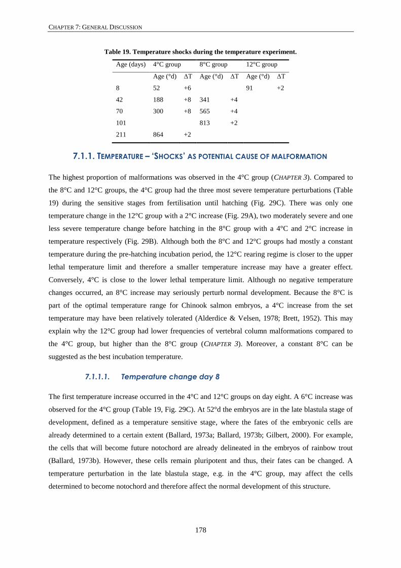
CHAPTER 7: GENERAL DISCUSSION
178
Table 19. Temperature shocks during the temperature experiment.
Age (days) 4°C group 8°C group 12°C group
Age (°d) ΔT Age (°d) ΔT Age (°d) ΔT
8 52 +6 91 +2
42 188 +8 341 +4
70 300 +8 565 +4
101 813 +2
211 864 +2
7.1.1. TEMPERATURE – ‘SHOCKS’ AS POTENTIAL CAUSE OF MALFORMATION
The highest proportion of malformations was observed in the 4°C group (CHAPTER 3). Compared to
the 8°C and 12°C groups, the 4°C group had the three most severe temperature perturbations (Table
19) during the sensitive stages from fertilisation until hatching (Fig. 29C). There was only one
temperature change in the 12°C group with a 2°C increase (Fig. 29A), two moderately severe and one
less severe temperature change before hatching in the 8°C group with a 4°C and 2°C increase in
temperature respectively (Fig. 29B). Although both the 8°C and 12°C groups had mostly a constant
temperature during the pre-hatching incubation period, the 12°C rearing regime is closer to the upper
lethal temperature limit and therefore a smaller temperature increase may have a greater effect.
Conversely, 4°C is close to the lower lethal temperature limit. Although no negative temperature
changes occurred, an 8°C increase may seriously perturb normal development. Because the 8°C is
part of the optimal temperature range for Chinook salmon embryos, a 4°C increase from the set
temperature may have been relatively tolerated (Alderdice & Velsen, 1978; Brett, 1952). This may
explain why the 12°C group had lower frequencies of vertebral column malformations compared to
the 4°C group, but higher than the 8°C group (CHAPTER 3). Moreover, a constant 8°C can be
suggested as the best incubation temperature.
7.1.1.1. Temperature change day 8
The first temperature increase occurred in the 4°C and 12°C groups on day eight. A 6°C increase was
observed for the 4°C group (Table 19, Fig. 29C). At 52°d the embryos are in the late blastula stage of
development, defined as a temperature sensitive stage, where the fates of the embryonic cells are
already determined to a certain extent (Ballard, 1973a; Ballard, 1973b; Gilbert, 2000). For example,
the cells that will become future notochord are already delineated in the embryos of rainbow trout
(Ballard, 1973b). However, these cells remain pluripotent and thus, their fates can be changed. A
temperature perturbation in the late blastula stage, e.g. in the 4°C group, may affect the cells
determined to become notochord and therefore affect the normal development of this structure.

CHAPTER 7: GENERAL DISCUSSION
179
Also on day eight, a 2°C increase was observed for the 12°C group (Fig. 29A). At 91°d the embryos
are in the gastrulation stage, between the 6th and 10th somite stages (Gorodilov, 1996; Gorodilov &
Melnikova, 2003). At gastrulation stage, the three germ layers (ectoderm, endoderm and mesoderm)
are formed from which the mesoderm will eventually form the notochord and somites. High levels of
cell movement and cell mixing occurs during early gastrulation (Ballard, 1973a; Ballard, 1973c;
Gilbert, 2000). Fortunately for the group closest to the upper lethal temperature limit (12°C), a 2°C
increase is the only temperature shock that occurred. This relatively small temperature increase of
short duration is unlikely to have had the same impact as the 8°C temperature increase that occurred
in the 4°C group.
7.1.1.2. Temperature change day 42
In the 4°C group a severe temperature change with an increase in temperature of 8°C occurred (Table
19, Fig. 29C). At 188°d the embryos have reached the completion of somitogenesis stage. The
maximum somite number in Chinook salmon (72-73) differs from the maximum somite number of
Atlantic salmon (66-68) (Gorodilov & Melnikova, 2003). Interestingly, in Pacific salmon species such
as Chinook, cherry (Oncorhynchus masu) and pink salmon, the complete epiboly stage corresponds to
the presence of 56-62 pairs of somites (Gorodilov & Melnikova, 2003) and thus is close to the
completion of somitogenesis stage time point. Completion of somitogenesis corresponds to the
appearance of pigmentation in the eye. First eye pigmentation was recognised in Atlantic salmon to be
around 186°d to 189°d, in coho salmon around 200°d and in Chinook salmon around 211°d (Boyd et
al., 2010; Gorodilov, 1996; Zeitoun & Tack, 1974). A temperature perturbation at 188°d in post-hatch
Chinook salmon may impact the normal formation of the posterior somites. Abnormal somite
formation later in development may lead to malformed associated elements as was observed in post-
hatch Chinook salmon (CHAPTER 3).
In the 8°C group, also on day 42, a temperature increase of 4°C was observed (Table 19, Fig. 29B). At
341°d the embryos have reached the appropriately named eyed egg stage. At this stage the eye is
completely covered by pigment and can be observed through the eggshell. This stage is also the final
temperature sensitive stage of the pre-hatching developmental period. Although the eyed egg stage is
reached at 236°d to 239°d in Atlantic salmon (Gorodilov, 1996), pigmentation of the eye was not
observed until 298°d in Chinook salmon (Boyd et al., 2010), closer to the temperature shock at 341°d
in the 8°C group. In Atlantic salmon the notochord has reached its full length and is vacuolised up to
somite 25 to 26 at this stage (Gorodilov, 1996). In contrast, the notochord has just reached full length
after the eyed egg stage in coho salmon (Zeitoun & Tack, 1974). A temperature change at this stage
could perturb the start or on-going notochord vacuolisation process. Improper vacuolisation of the
notochord could lead to kinking and pinching of the notochord, malformations that have been
observed in post-hatch Chinook salmon (CHAPTER 3).

CHAPTER 7: GENERAL DISCUSSION
180
Interestingly, in all temperature groups visually malformed specimens occurred without apparent
internal skeletal malformations (CHAPTER 3). During the eyed egg stage, the myotomes develop and
the first developing myofibres can be distinguished (Zeitoun & Tack, 1974). Early muscle
development is influenced by incubation temperature (Campos et al., 2013; Johnston & McLay, 1997;
Johnston et al., 1999; Macqueen et al., 2008; Stickland et al., 1988). Hypertrophied muscles (few but
large volume muscle cells) because of high incubation temperatures could cause post-hatch salmon
stages to experience excess of asymmetric forces on their notochord. This could likely result in the
bending of the notochord and could explain why a proportion of visually malformed post-hatch
Chinook salmon had no skeletal malformations (CHAPTER 3). The bending of the notochord would be
due to anomalies in muscle, or other soft tissue, development rather than vertebral malformations per
se.
7.1.1.3. Temperature change day 70
A third temperature change occurred on day 70 in the 4°C and 8°C groups. An 8°C increase occurred
in the 4°C group and a 4°C increase in the 8°C group (Table 19, Fig. 29B, C). At 300°d the 4°C group
experienced a temperature increase at a similar stage as the 8°C group did on day 42, i.e. the eyed egg
stage (341°d). The mesenchymal condensations that will form the associated elements (see Fig. 5A) in
Atlantic salmon start forming before 420°d (Grotmol et al., 2006), between 300°d and 390°d
(Grotmol et al., 2003). Although in general the rate of development of Chinook salmon is slower than
that of Atlantic salmon (Gorodilov & Melnikova, 2003) it is possible that the migration of
mesenchymal cell (sclerotome cells, see Fig. 4) could be perturbed. Also the condensation process
proper could be perturbed later in development. The mesenchymal cell migration and condensation
process is likely to vary between individuals of the same temperature group and therefore these events
could occur over a 90°d to 100°d developmental time period. Malformed associated elements, as
observed in post-hatch stage Chinook salmon (CHAPTER 3), could have arisen due to a temperature
shock.
At day 70, the 8°C group was at a 565°d stage, i.e. during or after hatching. The notochord at hatching
stage has reached the full length of the embryo and vacuolisation of the chordocytes has taken place
throughout the notochord. The notochord will keep increasing in length and diameter from hatching to
first feed (Grotmol et al., 2006). Shortly after hatching (around 500°d), a shift in the spatial
arrangement of the collagen type II fibrils in the notochord sheath occurs (Grotmol et al., 2006). The
fibrils shift from a concentric orientation to a helical orientation along the axis of the notochord.
Several of these helical layers, shifting from left- to right-handed coils will later be embedded in the
chordacentra, which are not yet present at hatching. It was suggested that the collagenous coils
increase the flexural stiffness and elastic recoil which are necessary during locomotion of post-hatch
stages (Grotmol et al., 2006). A temperature change could perturb the process of correctly orienting

CHAPTER 7: GENERAL DISCUSSION
181
the collagen fibrils and reduce the strength and resistance of the notochord to kinking under load. A
bent and kinked notochord was particularly a feature of post-hatch Chinook salmon tail malformation
and bent neck phenotypes in which high proportions of notochord malformations, including pinching,
were observed (CHAPTER 3). Also, in bent tail medium, spinal malformation medium and spinal
malformations severe phenotypes notochord malformations were the second most common internal
malformations (CHAPTER 3).
7.1.1.4. Temperature changes day 101 and 211
The fourth temperature increase only occurred in the 8°C group at 813°d (Table 19, Fig. 29B) while
the fifth temperature increase only occurred in the 4°C group at 864°d (Table 19, Fig. 29C). Both
these temperature changes are close to the 900°d first feed stage which is not considered as a
temperature sensitive stage. Close to the first feed stage the skeletal elements of the caudal fin and the
chordacentra start mineralising (personal observation). Temperature perturbations may interfere with
the normal mineralisation of the chordacentra and lead to chordacentra fusions. Temperature has been
shown to have no significant effect on the sequence of mineralisation but increases the phenotypic
variation in timing of the start of mineralisation (Cloutier et al., 2010; Mabee et al., 2000). In addition,
temperature modulates to a small degree where mineralisation starts (Cloutier et al., 2010; Mabee et
al., 2000). Atlantic salmon incubated at high temperatures showed the downregulation of matrix
metalloproteinase (MMP) 9 and MMP13 (Ytteborg et al., 2010b). These MMP’s are important
proteases that help break down the cartilage matrix during ossification (Engsig et al., 2000; Vu et al.,
1998). Moreover, the absence of MMP’s in the cartilage matrix delays endochondral ossification
(Stickens et al., 2004). Although, chordacentra fusion was the most common internal malformation
observed in visually normal post-hatch Chinook salmon (CHAPTER 3), the start of ossification of the
associated elements could be delayed by high incubation temperatures resulting in ossification defects
of the cartilaginous elements. Irregular ossification of associated elements, especially caudal fin
skeletal elements which experience forces during swimming, may expose these skeletal elements to
deformation processes in early juvenile stages (>1000°d).

CHAPTER 7: GENERAL DISCUSSION
182
Figure 29: Temperature shocks during the incubation period of Chinook salmon
Graphic representation of the temperature profiles of the 12°C (A, orange), 8°C (B, green) and 4°C (C, blue)
groups, with temperature (°C) on the y-axis and time in days on the x-axis. Timescale runs from incubation (day
0) to day 225. Each accidental temperature shock is indicated with a black arrow and on which day the
temperature shock occurred. The day of the expected hatching time at 500°d is indicated by a vertical line. The
expected day the specimens reach first feed (FF) at 900°d is also indicated with a vertical line. The specimens of
the 12°C (A) and 8°C (B) were transferred to tanks (indicated by arrow) after first feed stage.

CHAPTER 7: GENERAL DISCUSSION
183
7.1.1.5. Temperature change – Proposed mechanisms
Scar tissue was one of the notochord malformations observed in post-hatch Chinook salmon
(CHAPTER 3). In this malformation, chordocytes could be damaged as a consequence of temperature
perturbations. A positive temperature change increases the activity levels of fish. For example,
salmonids are more active in warmer water and show avoidance behaviour towards high sublethal and
lethal temperatures (Beitinger et al., 2000; Graham, 1949). The embryos prior to hatching also show
activity inside the egg (Johnston et al., 1999; Knight, 1963; Proctor et al., 1980), which is higher in
water with warmer temperatures (personal observation). A temperature increase could have
momentarily increased the number of movements made by the embryo inside the egg. Erratic
movements due to high temperature could have caused notochord lesions, ultimately resulting in scar
tissue in Chinook salmon (CHAPTER 3). This process is likely initiated by the collapse and rupture of
chordocytes when excess mechanical force occurred over a small area. At a cellular level, the plasma
membrane and its associated caveolae must directly absorb cell stretching by forces on the plasma
membrane and thus protect the notochord against damaging deformation. Caveolae (illustrated in
APPENDIX F) are small plasma membrane pits (60-80 nm) providing a reserve of plasma membrane
for when the cell is stretched. Hence, the cells are protected against rupturing (Bastiani & Parton,
2010; Garcia et al., 2017; Lim et al., 2017). When the eggshells of zebrafish caveolae mutants
(cavin1b-/-) were bleached, they became harder necessitating increased movements of the embryo to
hatch, a higher number of notochord lesions were observed compared to the control group (Lim et al.,
2017). In contrast, when the zebrafish embryos were anaesthetised, reducing the movements in the
egg, and dechorionated, a lower number of notochord lesions were observed compared to the control
group (Lim et al., 2017). These observations showed that notochord lesions developed by the
movement of embryos before hatching. A similar mechanism that causes notochord lesions in
zebrafish may happen in Chinook salmon. Movements of pre-hatch stage or hatch stage Chinook
salmon could have been increased due to a positive temperature change and may have caused
notochord lesions, where chordocytes collapse and consequently scar tissue develops. Therefore, a
temperature shock on day 70 at 565°d for the 8°C group, could have increased the rate of notochord
lesions.
The pinching and kinking of the notochord sheath was another notochord malformation observed in
post-hatch Chinook salmon (CHAPTER 3). At deep pinching sites and at the level of scar tissue the
collagenous middle layer of the notochord appeared thinner and contorted (Fig. 11F, G; CHAPTER 3).
A temperature increase could cause the proto-collagens, which are produced in the endoplasmic
reticulum (ER) and exported to the notochord sheath, to fold incorrectly. These proteins remain in the
ER because they cannot be exported to the Golgi-complex, and causes ER stress (Mori, 2009; Walter
& Ron, 2011). The ER stress will induce an unfolded protein response (UPR) response (illustrated in

CHAPTER 7: GENERAL DISCUSSION
184
APPENDIX F) which results in the up-regulation of heat shock protein (HSP) chaperones that help to
correctly fold proteins. The HSP family members were reported to be present in high levels in the
notochord of which HSP 70 operates in the ER (Sitia & Braakman, 2003). The heat shock protein was
shown to be up-regulated in Atlantic salmon post-hatch stages receiving a heat shock (Takle et al.,
2005). Mutations in the UPR response regulators Atf6α/β and Bbf2h7 in medaka and feelgood
(Creb3l2) in zebrafish (APPENDIX F) cause an incorrect UPR response and have been reported to show
abnormally developed notochords (Ishikawa et al., 2013; Ishikawa et al., 2017; Melville et al., 2011).
The notochord in these mutants appears bent and kinked, similar to what is observed in Chinook
salmon post-hatch stages (CHAPTER 3). In the Bbf2h7 medaka mutant the chordoblasts were reported
to appear detached from the basement membrane of the notochord. Also the chordoblasts in post-
hatch stage Chinook salmon in the present study appeared dislocated at the level of deep pinching
sites and scar tissue (CHAPTER 3). Malformed associated elements observed in the post-hatch stages
(CHAPTER 3) could develop by a similar mechanism or be affected by bent and pinched notochord
malformations. Also in the cartilage cells, incorrectly folded proto-collagens due to a heat shock
remain in the ER and are not transported to the extracellular matrix. The Atf6α/β and Bbf2h7 medaka
and feelgood (Creb3l2) zebrafish mutants were indeed reported to have abnormal cartilage formation
in the skull (Ishikawa et al., 2013; Ishikawa et al., 2017; Melville et al., 2011). The cartilaginous skull
bones appeared thin and underdeveloped with little extracellular matrix present between the cartilage
cells, suggesting that the collagens that build the cartilage matrix are not transported to the
extracellular space. Thus a temperature increase may cause both malformations of the notochord and
associated elements and may explain the co-occurrence of these internal malformations observed in
post-hatch Chinook salmon phenotypes such as curly severe, bent tail medium and spinal
malformation severe (CHAPTER 3).
7.1.2. TEMPERATURE – A MODULATOR OF DEVELOPMENT
Six regions were characterised in the juvenile Chinook salmon vertebral column (CHAPTER 4). A
detailed regionalisation of the vertebral column is necessary to investigate regional variation of
skeletal elements such as vertebrae, ribs and vestigial elements. Regional meristic variation of skeletal
structures, such as vertebrae, vestigial ribs in the transitional region, the vestigial neural arch and
spine on preural 2 and the uroneurals was observed in the vertebral column of juvenile Chinook
salmon (CHAPTER 5). Although a temperature shock at critical stages can cause malformation of the
vertebral column, a temperature change can modulate its normal development. For example, meristic
variation of vertebrae, fin rays, branchiostegal rays and gill rakers was observed when populations of
the Pacific salmon species living at different latitudes were compared (Murray & Beacham, 1989;
Taning, 1952). The variation of vertebrae in teleost populations occurring along environmental
latitudinal lines, of which temperature variation is most important, was stipulated as Jordan’s rule

CHAPTER 7: GENERAL DISCUSSION
185
(Jordan & Evermann, 1896). Research in zebrafish and Atlantic salmon found that the rate of
somitogenesis is changed by incubation temperature and slows down near the caudal end (Gorodilov,
2004; Holley, 2006; Schröter et al., 2008). For example, zebrafish embryos raised in higher
temperatures (30°C) had more somites compared to embryos raised in colder temperatures (27°C)
(Schmidt & Starck, 2010). Furthermore, Gorodilov (2004) and Moriyama et al. (2012) observed the
presence of pseudo-somites in the posteriormost end of the caudal region in coho salmon and medaka.
These observations indicate that the processes forming the last few somites are less well controlled
and may therefore be more prone to perturbations. Somites may fuse, could regress or extra somites
may develop into vestigial skeletal structures (Witten & Hall, 2015). The presence and variation in
occurrence of vestigial elements has been shown in the zebrafish caudal fin (Bensimon-Brito et al.,
2012a) and in Chinook salmon in the present study (CHAPTER 5).
The vestigial ribs in the transitional region, the vestigial neural arch on preural 2 and the uroneurals in
Chinook salmon are bilateral paired skeletal elements, and variation in their left and right presence
was observed (CHAPTER 5). Elevated constant temperatures, such as 12°C for Chinook salmon, can
cause chronic stress, which can be observed as variation in the symmetry of bilateral skeletal elements
such as fin rays, branchiostegal rays and gill rakers (Allenbach, 2011; Dongen, 2006; Leary &
Allendorf, 1989; Leary et al., 1985; Van Valen, 1962). The deviation of symmetry in these skeletal
elements is mostly assigned as fluctuating asymmetry, defined as the equal random variation of a
character on the left and right side of the individual (Leary & Allendorf, 1989; Leary et al., 1985; Van
Valen, 1962). Fluctuating asymmetry in the number of paired fin rays, scales, gill rakers and
branchiostegal rays was studied in chum salmon, sockeye salmon and coho salmon (Beacham, 1985;
Beacham, 1990; Campbell et al., 1998). Also teeth are part of the skeleton and in zebrafish variation
in the asymmetry of the dentition on the fifth ceratobranchial was observed (Woltmann et al., 2018).
Significant asymmetry in skeletal structures could be used as an indicator for unstable development
(Allenbach, 2011), which occurs in non-ideal rearing environments. However, consistent observation
of several skeletal elements needs to be taken into account to make precise interpretations of the
observed asymmetry. The value of using asymmetry of skeletal structures as an indicator for abnormal
development is still heavily debated (Allenbach, 2011). Although in juvenile Chinook salmon,
significant variation in the number of vertebrae was observed between the 8°C and 12°C groups, the
number of vestigial ribs, the arch on preural 2, and the uroneurals between the two temperature groups
was very similar (CHAPTER 5). The meristic variations of the vertebrae observed in the present study
could be a consequence of the accidental temperature shocks that occurred during incubation, rather
than the consequence of a constant elevated temperature (e.g. 12°C). The lack of variation in vestigial
skeletal elements could be explained by the absence of accidental temperature shocks during their
development in late first feed stages. Alternatively, 12°C may not have been high enough to cause
chronic developmental stress and therefore significant variation of paired vestigial skeletal elements.

CHAPTER 7: GENERAL DISCUSSION
186
7.2. EARLY LIFE MALFORMATIONS – A GENETIC BACKGROUND
The results in CHAPTER 3 show a link between the visual phenotype and the malformed skeletal
structures for the first time in a salmon species. Although visual phenotypes, such as curly medium
and severe, bent tail, lordosis, kyphosis and scoliosis also have been observed in early stages of
Atlantic salmon, chum salmon, trout and brook trout (Eriksen et al., 2006; Johnson et al., 1998;
Leduc, 1978; Mahrosh et al., 2014; Quatrefages, 1888; Yamamoto et al., 1996), malformation of the
underlying skeletal structures, such as notochord, chordacentra and associated elements have not been
reported in salmonids. The specific crosses between parental Chinook salmon used in the present
study (APPENDIX B) were chosen to produce offspring with more malformations and many different
types of vertebral column malformation. This means that the malformed phenotypes of post-hatch
Chinook salmon in the present study, apart from being caused by differences in the incubation
temperature, may also have a genetic basis and may be the result of mutations. Malformations of the
notochord, chordacentra and associated elements have been observed in several different zebrafish
and medaka mutants which have malfunctions in the cellular vesicular transport mechanisms and the
caveolae (summarised in APPENDIX F). Where in the cell, and which cellular components the
mutations affect in the chordocytes, the chordoblasts and the notochord sheath, are shown in
APPENDIX F. Salmonids share these universally conserved intracellular mechanisms for post-
translational transport of proteins. Below, malformation phenotypes observed in post-hatch stage
Chinook salmon in CHAPTER 3 are compared and discussed with malformation phenotypes observed
in mutant zebrafish and medaka. It is emphasised here that hypotheses of potential mechanisms are
only discussed in light of possible future research areas.
At shallow pinching sites, the post-hatch Chinook salmon notochord sheath was bent but was
otherwise unaffected by contortions, indicating that only the chordocytes were affected (CHAPTER 3).
The vacuoles in chordocytes were collapsed and the notochord appeared kinked in the Cltca zebrafish
mutant, which have clathrin coated vesicles with an impaired function (Ellis et al., 2013). Clathrin
coated vesicles are important in the protein transport from the plasma membrane to intracellular
vesicles such as lysosomes (APPENDIX F). The vacuole in chordocytes was shown to be a lysosome
related organelle (LRO) and therefore clathrin mediated vesicle transport was suggested to be
important in the maintenance of the vacuole (Ellis et al., 2013). In the sneezy, happy and dopey
zebrafish mutants the COP I vesicle coat components are truncated and impair correct COP I
mediated transport functions (APPENDIX F). In these mutants vacuoles in the chordocytes fail to form
(Coutinho et al., 2004; Odenthal et al., 1996; Stemple, 2005). The collapse of a few chordocytes in the
post-hatch Chinook salmon notochord could explain a shallow pinching of the notochord sheath
(CHAPTER 3).

CHAPTER 7: GENERAL DISCUSSION
187
At deep pinching sites and at the level of scar tissue in post-hatch Chinook salmon, the notochord
sheath and external elastic membrane was thinner, contorted and perturbed (Fig. 11F-H; CHAPTER 3).
A thinner and fragmented elastin membrane and a change in the notochord sheath collagen
architecture were observed in fusions in parr stage Atlantic salmon (Ytteborg et al., 2010a). The
collagenous middle layer of the notochord in sneezy, happy and dopey COP I mutants was observed to
be thinner or even absent resulting in a bent notochord (Coutinho et al., 2004; Odenthal et al., 1996;
Stemple, 2005). In zebrafish laminin mutants, bashful, grumpy and sleepy, the formation of the basal
lamina is perturbed (Parsons et al., 2002; Stemple, 2005). A correctly formed basement membrane is
necessary for cell differentiation, attachment and function (Bornstein & Sage, 2002; Corallo et al.,
2013; Coutinho et al., 2004; Parsons et al., 2002). If the chordoblasts cannot attach properly to the
basement membrane, the production of the collagenous sheath material is likely also compromised.
Grotmol et al. (2006) concluded that normal notochord sheath organisation is dependent on an intact
basal lamina. Indeed, in the bashful, grumpy and sleepy mutants, the formation of a perturbed basal
lamina resulted in the disorganisation of the collagen in the notochord sheath and of elastin layers of
the external elastic membrane (Parsons et al., 2002; Stemple, 2005). Displaced and hyperplastic
chordoblasts were also observed at deep pinching sites and at the level of scar tissue in post-hatch
Chinook salmon notochords (CHAPTER 3), where the notochord sheath and external elastic membrane
appeared disorganised.
Kinking of the notochord as was observed in in post-hatch Chinook salmon (CHAPTER 3). Mutations
in the emilin3a and emilin3b could cause a malformed notochord sheath and result in a bent
notochord. These genes were found to have an important role in organising the collagen II fibres in
the medial layer of the zebrafish notochord (Corallo et al., 2013). Mutant fish showed disorganised
collagen II fibres in the medial layer and a shortened and bent notochord. However, the basement
membrane and outer notochord sheath layer were unaffected (Corallo et al., 2013).
Chordacentra fusions were observed mostly in externally normal specimens but also in externally
malformed post-hatch Chinook salmon (Fig. 10; CHAPTER 3). The organisation of the collagen fibres
in the notochord sheath has been shown to be important for the correct mineralisation of chordacentra,
which starts in the collagen below the external elastic membrane in Atlantic salmon (Grotmol et al.,
2006). Incorrectly organised collagen fibres in the notochord sheath could explain the occurrence of
chordacentra fusions observed in Chinook salmon post-hatch stages (CHAPTER 3). The vbi (vertebra
imperfect) medaka mutant has a mutation in the SEC24D component of the COP II coated vesicles
(APPENDIX F). In this mutant the mineralisation of chordacentra is delayed and irregular. Adjacent
chordacentra fused ventrally, and the neural and haemal arches can develop malformed (Ohisa et al.,
2010). The zebrafish mutant bulldog has also mutation in the SEC24D COP II component (APPENDIX
F). This mutant exhibits craniofacial malformations, defects in chondrocyte maturation and delayed
mineralisation (Sarmah et al., 2010). In post-hatch Chinook salmon ventrally fused chordacentra (Fig.

CHAPTER 7: GENERAL DISCUSSION
188
12A-C), irregular mineralisation of chordacentra (Fig. 12I) and malformed arches (Fig. 12F-H) were
observed (CHAPTER 3) similar to the internal vertebral column phenotypes of the vbi and bulldog
mutants. The high baseline number of malformations occurring in the offspring of the selected crosses
in the present research may be the consequence of the genetic background and accidental temperature
shocks. However, the differences in proportions of malformations observed between the three
temperature groups (CHAPTER 3) is more likely a consequence of the three different constant
incubation temperatures rather than genetic background or accidental temperature shocks.
7.3. EARLY LIFE MALFORMATIONS FORESHADOW LATE LIFE DEFORMITIES
The results in CHAPTER 3 showed that notochord malformations, vertebral fusions and malformations
of the associated elements can already be present in post-hatch stages of Chinook salmon that are
visually malformed. The results in the current research thus confirm that malformations of the
vertebral column can occur in early life stages of salmon. Early life stage malformations must be
sublethal and carried through the freshwater and seawater stages in order to be present as later life
stage deformities. When malformations in the vertebral column of salmonids first appear and which
malformations of the vertebral column occur in early life stages represented a gap in the current
knowledge. In contrast, late and final stage deformities, such as compressions, fusions and bending of
the vertebral column (lordosis, kyphosis and scoliosis) are well studied in farmed freshwater pre-
smoltification stage salmonids (Fjelldal et al., 2007a; Grini et al., 2011; Kvellestad et al., 2000;
Munday et al., 2018; Sullivan et al., 2007a; Takle et al., 2005) and in both wild and farmed adult
salmonids (Deschamps et al., 2009; Deschamps et al., 2008; Fjelldal et al., 2009a; Fjelldal et al.,
2009b; Fjelldal et al., 2007b; Gil Martens et al., 2005; Munday et al., 2016; Perrott et al., 2018;
Seymour, 1959; Sullivan et al., 2007b; Witten et al., 2009). Importantly, Kvellestad et al. (2000),
Fjelldal et al. (2012) and Witten et al. (2005a) have suggested that deformities such as compression
and fusions observed in juvenile or adult life stages could have developed very early in life. However,
the phenotype of later deformities may mimic or be different from early life stage malformations,
which potentially foreshadow later deformities.
Most of the severely malformed (externally and internally) animals will not survive past the first feed
stage. Additionally, salmon farmers remove malformed individuals and aim to grow visually normal
salmon. Surprisingly, visually normal specimens in each temperature group, investigated in CHAPTER
3, showed internal malformation of the vertebral column. The main internal malformation observed in
normal phenotype post-hatch Chinook salmon was fusion of chordacentra. The chordacentrum is the
foundation of the vertebral centrum (Arratia et al., 2001) with the perinotochordal autocentrum and
bony arcocentrum forming around and connected to the chordacentrum. Therefore, the autocentra and
the bony arcocentra of adjacent vertebrae are likely to fuse during growth when their underlying
chordacentra are fused in early developmental stages. The cartilaginous arcocentra, i.e. the basidorsals

CHAPTER 7: GENERAL DISCUSSION
189
and basiventrals, remain separate since fused vertebrae have two neural and haemal arches. Indeed,
fused chordacentra with separate basidorsals and basiventrals have been observed in post-hatch
Chinook salmon (Fig. 12A; CHAPTER 3). In addition, early fusions do not affect the visual appearance
of the animal and are likely survivable. Fusions that originate in post-hatch stages could therefore be
carried through to the harvest stages. It appears that a deformity, such as a fusion, in late life stages
can already exist in early life stages. This answers the question: “Are some late life deformities
foreshadowed by early life stage malformations?”
While vertebral fusions can start to develop in salmonid post-hatch stages (CHAPTER 3), they can also
start to develop in both juvenile pre-smoltification stages and adult post-smoltification stages (Fjelldal
et al., 2012; Munday et al., 2018; Perrott et al., 2018). However, the exact moment when
malformations and deformities start to develop remains to be elucidated. Understanding when
malformations and deformities develop may help identify malformation and deformity prone stages.
Once these sensitive stages are identified changes in the husbandry strategies could be made to
prevent the development of malformations and deformities. Suggested measures could be a change in
incubation temperature, changing the timing of grading events and changes in the nutrition. Whether
or not deformities observed in juvenile and adult stages are early life stage malformations that were
carried through the development and growth of the animals is unknown. The experiment in the current
research aimed to follow early life stage specimens, reared at two different incubation and freshwater
grow-out temperatures, into their juvenile and adult life stages. The results would link malformations
in early life stages to deformities observed in late life stages. However, due to runting (= stop in
growth) in early sea stages, and malnutrition and mortality events in later seawater stages, the current
project failed to directly link early malformations to deformities that are observed in juvenile and
adult stages. However, results in CHAPTER 6 do show that early incubation and freshwater grow-out
temperatures significantly affect the prevalence of deformities in freshwater versus seawater stages.
Moreover, it appears that compared to 12°C, incubating at a cooler 8°C protects Chinook salmon
against the development of deformities, especially LKS, in seawater stages. Together, the results in
CHAPTER 3 AND 6 show that a changing of the incubation temperature can indeed be useful to reduce
the number of malformations and deformities that occur in farmed Chinook salmon.
Currently there is information about the skeletal phenotype of early malformations in Chinook salmon
(CHAPTER 3) and information about skeletal phenotype of late and final stage deformities in Chinook
salmon (Perrott et al., 2018) and Atlantic salmon (Witten et al., 2009; Witten et al., 2016).
Information about the start or early stages of deformities is currently still lacking. Linking
malformations to deformities is difficult but careful hypotheses can be postulated. For example,
Witten et al. (2006) proposed that two scenarios can occur during deformation of vertebral centra
based only on X-ray data of juvenile and adult Atlantic salmon. First, the containment scenario occurs
when two or three vertebrae progress from fusion, in which two or three vertebrae can be recognised,

CHAPTER 7: GENERAL DISCUSSION
190
to a single larger vertebral centrum typically recognised by the presence of two or three neural and
haemal associated elements. It was suggested that this ‘new’ larger vertebral centrum had a close to
normal function (Witten et al., 2006). Second, is the aggravation scenario, where over time adjacent
vertebral centra become sequentially involved in the vertebral deformity. Now, two new scenarios,
which link early malformations observed in post-hatch Chinook salmon (CHAPTER 3) to late
deformities observed in X-ray data in Atlantic and Chinook salmon (Perrott et al., 2018; Witten et al.,
2009) can be postulated. These scenarios are a true containment scenario and a stabilisation scenario.

CHAPTER 7: GENERAL DISCUSSION
191
7.3.1. THE CONTAINMENT SCENARIO – ‘SET IN BONE’
Figure 30 shows a hypothesised containment scenario of a pinched notochord observed in post-hatch
Chinook salmon (Fig. 11A; CHAPTER 3) into an ‘internal dorsal or ventral shift’ deformity (type 19;
Witten et al., 2009). Currently, a containment scenario is considered when two fused vertebral centra
are remodelled into one functional vertebral body (Witten et al., 2009; Witten et al., 2006). Still, the
shape of the internal structures of a vertebral centrum, i.e. the chordacentrum, could be retained from
post-hatch to adult life stages. The basidorsals of the neural associated elements are fused at the level
of the pinch (Fig. 30A) as was observed in post-hatch Chinook salmon (Fig. 11B; CHAPTER 3). The
chordacentra start to mineralise within the notochord sheath on the ventral side of the notochord. The
mineralisation progresses and the chordacentra become ventrally fused at the pinching site (Fig. 30B)
similar to observations in Chinook salmon (Fig. 12A, B; CHAPTER 3). The fusion between the adjacent
mineralised chordacentra at the pinching site has further progressed when the chordacentra form
dorsally closed rings (Fig. 30C). Subsequently, the perinotochordal autocentra start to ossify and fuse
to likely form a single autocentrum (Fig. 30D). The pinch of the notochord is now ‘set in bone’ with
the fused chordacentra retaining the pinched shape of the notochord sheath. The autocentrum
ossifying around the pinch likely further contains the shape (indicated by blue dotted line Fig. 30D).
The cancellous bone of the arcocentrum ossifies around the autocentrum (Arratia & Schultze, 1992),
firmly connecting the double arches to the autocentrum (only neural arches depicted Fig. 30E). On X-
ray images, the compact bone of the perinotochordal autocentra is visible as the characteristic X-
shapes in vertebrae of salmonids (Witten et al., 2009). The autocentrum covers the notochord sheath.
Thus, if the chordacentrum (within the notochord sheath) is mineralised in the shape of the pinched
notochord sheath, the perinotochordal autocentrum will have the shape of the pinched notochord. Due
to appositional bone growth of the bony arcocentrum the overall contour of the vertebral centrum is
normal even though the autocentrum forming around a pinch may appear on X-ray images as a
deformity type 19 observed by Witten et al. (2009). The internal X-structure of the vertebral body is
affected, meaning the centrum of the X is in a more dorsal or ventral position. Although type 19
deformities were not reported in Chinook salmon previously (Perrott et al., 2018), they have been
subsequently seen in juvenile and adult Chinook salmon (personal observation).

CHAPTER 7: GENERAL DISCUSSION
192

CHAPTER 7: GENERAL DISCUSSION
193
Figure 30: A proposed containment scenario
Schematic representation of the different developmental steps: a pinch in the notochord develops into a type 19
deformity (internal dorsal or ventral shift), where the internal X-structure of the vertebral body is shifted
dorsally or ventrally (Witten et al., 2009). All images are oriented with anterior to the left, posterior to the right,
dorsal at the top and ventral at the bottom. Associated elements are only shown on the dorsal side to better
illustrate the layers on the ventral side. (A-C) first feed stages (850°d-900°d). (D) Juvenile parr stage (1000-
1400°d). (E-F) Juvenile pre- and post-smoltification and adult stages (>1500°d). The internal structure of the
adult vertebral body follows the shape of the external elastic membrane (indicated in solid black and dotted blue
lines) which retains its shape throughout development because the chordacentra (A-C), develops first as a
mineralisation of the notochord sheath, followed by bone deposition onto the mineralised notochord sheath and
its outer elastic membrane, the autocentra (D) and bony arcocentra (E). All bony layers have been given the
same colour (red) because they are mineralised, although essentially chordacentra are mineralised type II
collagen and autocentra and bony arcocentra are mineralised type I collagen. (A) Basidorsals fuse at the level of
the pinch and space created by the pinch is filled with cartilage. On the ventral side the chordacentra (within the
notochord sheath) start to mineralise. (B) The adjacent chordacentra at the level of the pinch fuse ventrally
during their mineralisation. (C) The chordacentra have formed closed mineralised rings within the notochord
sheath. (D) The autocentra have ossified around the external elastic membrane fixing the shape of the pinch in
place. The neural arches have perichondrally ossified. (E) The arcocentra have ossified around and connected to
the autocentra. Appositional growth of the arcocentra (more growth on the dorsal side compared to the ventral
side) results in the normal external contour of the vertebral centra. The yellow lines mark the vertebral
endplates. (F) X-ray of adult Chinook salmon. The white X-structure apparent on the X-ray are the vertebral
autocentra. The course of the internal external elastic membrane (dorsal side) in the vertebral centra is indicated
with a blue line. The internal X-structure of the middle vertebral centrum is shifted ventrally. Notice the
occurrence of a double arch on the middle vertebral centrum (indicated by white arrows). This vertebra is a
fusion product of two vertebrae.
7.3.2. THE STABILISATION SCENARIO
A combination of notochord pinching, bending, dislocation and hyperplasia of chordoblasts and scar
tissue was one of the severe internal malformation phenotypes observed in post-hatch Chinook
salmon (Fig. 11I; CHAPTER 3). This resulted in the marked collapse of the notochord towards a
concertina shape and a shorter body axis (personal observation). The basidorsals and basiventrals are
fused in many places which could be the consequence of a restriction in space along the notochord.
Inside the notochord, the chordocytes, chordoblasts and notochord sheath are affected. At the level of
a notochord lesion, scar tissue replaces the chordocytes and the chordoblast monolayer is dislocated.
The dislocated chordoblasts produce ectopic notochord sheath material at the scar tissue site
(CHAPTER 3). The notochord sheath is folded, pinched and the external elastic membrane can be
disrupted. A proposed contour and internal structure for the collapsed notochord malformation are
schematically represented in Figure 31A (contour, based on Fig. 11A) and Figure 31B (internal

CHAPTER 7: GENERAL DISCUSSION
194
structure, based on Fig. 11I). Similar to the observations in Chinook salmon (CHAPTER 3), a collapse
of the notochord with pinching of the notochord sheath and the occurrence of scar tissue was observed
in a new type of vertebral compression and fusion in the caudal region of gilthead sea bream (Sparus
aurata) (Loizides et al., 2013).
A collapsed and severely malformed notochord likely provides less mechanical support and must in
some way be stabilised. The malformed notochord in post-hatch Chinook salmon could have been
stabilised by the cartilage of the fused basidorsals and basiventrals lining the notochord (Fig. 31A, B).
In post-hatch Chinook salmon (CHAPTER 3), the cartilage of the fused basidorsals and basiventrals
filled the spaces where the notochord was folded inwards, folded towards the left or right or folded
towards the dorsal or ventral side of the midline axis (Fig. 31A, B). This is shown histologically on a
parasagittal section of a collapsed notochord in Figure 11I. How this collapsed but stabilised
notochord further develops is currently unknown. Zebrafish with mutations in the caveolae
components, resulting in severe scar tissue lesions in the notochord, survived to adult stages (Garcia et
al., 2017; Lim et al., 2017). Moreover, although scar tissue in the notochord impaired the swimming
capacity of early post-hatch zebrafish stages, normal locomotor capacity was observed by 7 dpf when
vertebral centra started to form (Lim et al., 2017). Mineralisation of chordacentra was observed in the
severely malformed notochord of post-hatch Chinook salmon (CHAPTER 3). This result suggests that
post-hatch Chinook salmon with a collapsed notochord and shorter body axis could be able to swim
when chordacentra form. When the malformed salmon can swim, it can feed and survive under
farming conditions. The survival of these malformed post-hatch Chinook salmon until pre-
smoltification stages also means that the perinotochordal autocentra and bony arcocentra develop. It is
conceivable that, similar to basidorsals and basiventrals which likely develop fused due to a shortened
notochord, adjacent chordacentra fuse during mineralisation due to restriction in space. Once
chordacentra are mineralised, the perinotochordal bone starts to ossify. The ossification of this
perinotochordal bone likely further stabilises the mechanically weaker notochord. If chordacentra
develop fused in an abnormally shaped notochord, both the internal structure and the perinotochordal
vertebral centra can be expected to develop heavily compressed and contorted. The progression of the
collapsed notochord into a large fusion centre could result in the known external phenotype
‘pumpkin-seed’ in Chinook salmon. An example of a pumpkin-seed phenotype is shown in Figure
31C and D (C = visual appearance, D = X-ray image). Scar tissue observed in CHAPTER 3 (Fig. 11E,
G, H) can also occur in a single site and likely forms a weak part of the notochord. Thus, the
stabilisation of the notochord could also occur locally rather than over the entire vertebral column
length. The scar tissue site can be as small as the length of two chordacentra. The adjacent
chordacentra and perinotochordal vertebral centra likely fuse during growth to further stabilise the
notochord resulting in vertebral fusion that in later life stages may be remodelled to a new larger unit.

CHAPTER 7: GENERAL DISCUSSION
195
Figure 31: Stabilisation scenario of a heavily malformed notochord
(A-D) All images are oriented with anterior to the left, posterior to the right, dorsal at the top and ventral at the
bottom. (A) Schematic representation of the outside contour of a collapsed notochord in post-hatch stage
Chinook salmon (500°d to 800°d). The notochord external elastic membrane (black solid and dotted line) is
pinched and folded inwards to separate two notochord ends. Similar to the basidorsals and basiventrals the
arches remain separate or are fused. On the far right the basidorsals are fused to the basiventrals. (B) A
longitudinal section of the notochord in (A). Internally the notochord shows partial and complete septa (arrow)
and scar tissue. The arrowhead shows a pinch of the external elastic membrane. (C) Pre-smoltification stage
juvenile Chinook salmon with external ‘pumpkin-seed’ phenotype. (D) X-ray of the specimen shown in (C). The
external shape is indicated in white lines. Notice the fusion centre in the abdominal region. The fusion of the
vertebrae could be the consequence of a collapsed notochord or a mixture of early perturbations that could not
be contained but that eventually were stabilised.

CHAPTER 7: GENERAL DISCUSSION
196
7.4. RECOMMENDATIONS AND PERSPECTIVES
7.4.1. THE BEST INCUBATION TEMPERATURE IS A CONSTANT 8°C
Results from CHAPTER 3 showed that the lowest frequency of internal malformations occurred in the
constant 8°C temperature group. Results in CHAPTER 5 showed that juvenile Chinook salmon reared at
this temperature have more vertebral centra in the caudal region of the vertebral column, which is
possibly advantageous for swimming and likely improves the ability to feed. Also, specimens of the
8°C group showed less variation in the left-right presence of skeletal elements indicating that
development may be more stable at 8°C (Allenbach, 2011; Leary & Allendorf, 1989). Although in
CHAPTER 6, freshwater smoltification-stage salmon of the 8°C group had a significantly higher
prevalence of total deformities, this group had a significantly lower prevalence of total deformities
and LKS at the six-months-at-sea-stage. Moreover, fewer specimens were classified as runts in the
8°C group and had a lower prevalence of total deformities and LKS compared to the 12°C group.
Also, the 8°C group had a lower attrition rate during and between each life stage compared to the
12°C group. Therefore, it appears that incubation at 8°C and a period of growth in fresh water at 8°C
protects Chinook salmon against deformities in early sea stages. Given the greater number of
unexpected temperature changes in the 4°C and 8°C groups and the superior outcomes for the 8°C
group, it is likely that 8°C is the best incubation temperature for farmed salmon in New Zealand. It
may be that the actual difference between the 8°C and 12°C groups is underrepresented as the 12°C
group had the fewest temperatures changes.
In Chinook salmon, 8°C is at the lower end of the optimal temperature range for embryos and post-
hatch stages, from 8°C to 10°C (Alderdice & Velsen, 1978; Brett, 1952). The optimal temperature
range reported by these authors, is based on experiments investigating developmental rates, and
mortality and survival rates related to upper and lower lethal temperatures in Pacific salmon egg
stages. In the current study, early mortality was only reported briefly in CHAPTER 3 and developmental
and growth rates were not reported. Condition factor, another important measure of animal quality
and welfare for salmon farmers (Jones et al., 1999; Segner et al., 2012), was also not reported. Early
mortality, developmental rates, growth rates and condition factor related to temperature in farmed
Chinook salmon should be further investigated as datasets built on these measures could corroborate
that 8°C is indeed close to an optimal incubation temperature. However, only three temperatures were
tested here due to the cost of the large-scale experiment. Although clearer differences in results were
likely obtained between the temperature groups due to the large temperature steps, the temperatures
in-between 4°C, 8°C and 12°C would have to be tested in future experiments to refine the trend
indicated in this thesis. For example, repeating the 8°C incubation and adding smaller temperature
steps on either side of 8°C. Since the existing optimal temperature range for Chinook salmon

CHAPTER 7: GENERAL DISCUSSION
197
incubation is between 8°C and 10°C, the best results with respect to mortality rates, developmental
rates, condition factor and malformation rates, may in theory be obtained at any temperature between
8°C and 10°C. On the other hand, optimum temperature ranges for each of these metrics may be
slightly different from one another. Finding significant differences for these measures, when using
smaller temperature increments of ±1°C or < 1°C may be challenging.
For Chinook salmon, an incubation temperature of 8°C may be ideal in experimental conditions
(Alderdice & Velsen, 1978; Brett, 1952; current research), but may not be feasible in farming
operations. Due to the need for year round harvest, salmon farmers often influence husbandry factors
such as temperature, light regime and feeding regime to grow salmon faster or slower. Although light
and feeding regime can be changed in the seawater environment (Fjelldal et al., 2009b; Fjelldal et al.,
2005), temperature can only be easily influenced in the freshwater environment. The differentiation
between fast and slow growing salmon is more easily made by changing freshwater husbandry factors
which, in case of temperature, is likely to be a change to being above or below the optimal range for
robust skeletal development and growth. An example of how the industry manipulates temperature
and influences development, based on personal observations, is given below: The cohort of salmon
that was harvested in the first part of the year, and thus the faster growing group, was fertilised at
10°C and brought to 12°C for incubation and during the freshwater grow out stages. This cohort
received a ‘16 hours light-8 hours dark’ cycle and was fed frequently during the day with automated
hoppers. The cohort of salmon that was harvested in the second part of the year (slow growing group)
was also fertilised at 10°C but instead brought to 6°C for the incubation period until first feed stage.
Hatching for this group was synchronised by gradually raising the temperature to 12°C and then
returning it to 6°C. These animals had a natural day-night cycle and were fed less frequently
compared to the fast growing cohort. Thus, both the fast and slow growing groups had substantial
periods of early development at temperatures not considered to be optimal for rearing Chinook
salmon. Especially the temperature rise from 6°C (below optimal) to 12°C (above optimal) could be
considered stressful for early stages and may result in deformities that develop in seawater life stages
(CHAPTER 6).
Freshwater grow-out facilities provide space for early juveniles stages to swim. Sustained swimming
occurring in the freshwater grow-out system will increase the bone mineral content and osteocyte
density, as was observed for Atlantic salmon and rainbow trout (Deschamps et al., 2009; Totland et
al., 2011). Healthy bone, defined as a well-balanced deposition of bone by osteoblasts and resorption
by osteoclasts, decreases the risk of developing deformities during fast growth in freshwater stages.
However, for healthy bone formation to be sustained throughout the farm life cycle, the chordacentra
as the template of the vertebrae and the cartilaginous precursors of the neural and haemal arches, i.e.
the post-hatch stage associated elements, need to be perfectly formed in the pre- and post-hatch stages

CHAPTER 7: GENERAL DISCUSSION
198
of the salmon. The requirements for commercially dictated growth rates and the use of temperature
manipulation to achieve these results puts normal developmental processes at risk.
One thing that has become clear is the strong likelihood that to form a normal vertebral column and in
order to avoid as many malformations and deformities possible, low to moderate developmental rates
are preferred. Close to ideal development is largely driven by constant temperatures, and ideally no
fluctuations or temperature manipulations for synchronised hatching. In order to farm efficiently,
aquaculture must find a balance between close to ideal development and optimal growth for
commercial targets. These goals are likely to remain in conflict and a healthy dialogue that weighs
developmental science, aquaculture commerce and aquaculture social licence is critical to maintain. It
is likely that performance in seawater and freshwater is linked (CHAPTER 6) but that these linkages are
not well understood. Communication between the freshwater and seawater operations is therefore
equally, if not more, critical to maintain. Only through robust monitoring and commitment to track
performance of cohorts that experience different husbandry strategies during the early stages of
development will further clues of ‘optimal’ rearing identified.
7.4.2. KNOWLEDGE OF POST-HATCH SALMONID STAGES NEEDS TO BE INCREASED
As was shown in CHAPTER 3, the study of early malformations is useful, providing knowledge about
how early malformations develop, how they progress and about their regional occurrence. The
deformities of salmonids in later life stages, especially harvest stages, represent a cost to the farmer
(Fjelldal et al., 2012). However, studying early malformations can help prevent the presence of
deformities later in life and therefore make the farming process more efficient. In addition,
malformations and deformities represent challenges from an ethical and animal welfare perspective.
Decreasing early malformations that potentially foreshadow late life deformities, thus decreasing
deformities at harvest, will improve animal welfare. Improving farmed animal welfare is of concern to
the consumer, who has become more aware of ethical and animal welfare issues. In turn the salmon
farmers will have to focus more resources on maintaining social license and animal welfare.
The importance of accurate studies of skeletal structures in early life stages of teleosts, in order to
recognise and identify skeletal abnormalities, has been recognised for a long time (McMurrich, 1883).
Still, the lack of research and knowledge of post-hatch salmonid stages is remarkable. Boglione et al.
(2013a) noted that a frequency of about 20% severely malformed marine farmed teleosts at the end of
the hatchery phase can be considered as a good, but a rare result. In contrast, the frequency of
malformations at the end of the freshwater stage in salmon farming is lower compared to marine
teleost farming in general. Therefore, early malformations have never been a major issue for salmon
farmers and may explain why studies on early malformations in salmonids are limited. However, the
results in CHAPTER 3 clearly show that early skeletal malformations are present even if they are not

CHAPTER 7: GENERAL DISCUSSION
199
obvious. Internal malformations that are externally invisible may represent a good research target to
help avoid such malformations becoming costly vertebral deformities in harvest stages.
7.4.3. SALMON AS MODEL ORGANISM
Scar tissue observed in CHAPTER 3 looks very similar to scar tissue (notochord lesions) observed in
zebrafish (Garcia et al., 2017; Lim et al., 2017). In addition, scar tissue in post-hatch stage Chinook
salmon looks remarkably similar to chordoma tissue. A chordoma is a rare, but aggressive notochord
cell tumour that can occur in the bones of the human skeleton (Choi et al., 2008; Kitamura et al.,
2013; Presneau et al., 2011; Sun et al., 2015). Zebrafish with an HRASV12 mutation had notochords
with histological characteristics that were highly similar to the chordomas observed in human
patients. In the zebrafish mutant, cells were found to be plump, had focally prominent nuclear
pleomorphism and hyperchromasia, and grew in nests and cords with both a focal and a solid pattern.
The tumor matrix was found to be variable, ranging from dense eosinophilic to myxoid with
vacuolated spaces, which is also typical of human chordoma. Large cells with a vacuolated cytoplasm
were observed in zebrafish, which is also reminiscent of physaliferous cells of human chordomas
(Burger et al., 2014). Several markers that are used to diagnose chordoma in humans, i.e. Brachyury,
Cytokeratin, pErk and pS6, were immunohistochemically shown to be present in zebrafish chordoma
tissue (Burger et al., 2014). However, the HRAS mutation in zebrafish has not been reported in
humans and the chordoma in zebrafish has a rapid onset (3 dpf onwards) compared to the slow
growing human tumor, which often takes 5 years for symptoms to appear (Burger et al., 2014). It is
possible that the notochord scar tissue observed in Chinook salmon could be used to study chordoma
cancer in humans.
The use of fish as a model organism is not a new concept (Witten et al., 2017). Studies on bone
diseases, such as Osteogenesis Imperfecta, Cranio-lenticulo-sutural dysplasia and Spondyloepiphysial
dysplasia tarda, have been undertaken in zebrafish and medaka (Gistelinck et al., 2016a; Gistelinck et
al., 2018; Gistelinck et al., 2016b; Kondylis et al., 2009; Lang et al., 2006; Ohisa et al., 2010).
Although Chinook and Atlantic salmon do not have translucent eggs, embryos and post-hatch stages
or a reasonably fast life cycle, their eggs are large and are produced in high numbers per single
female. In addition, the early developmental stages of Chinook and Atlantic salmon are larger than
zebrafish stages and may be easier to handle in that specimens can be cleared and whole mount
stained. Techniques, such as whole mount staining and clearing with Alcian blue and Alizarin red
used in the present research, are well established. In addition, compared to the acellular bone in the
model organism medaka, the bones of salmonids are composed of cellular bone and provide another
advantage when comparisons and extrapolations to human bone physiology studies are required. Early
stages of Chinook salmon could therefore represent a useful research organism for:

CHAPTER 7: GENERAL DISCUSSION
200
(i) aquaculture purposes to gather knowledge about vertebral malformations and deformities
(CHAPTER 3, 6)
(ii) fundamental studies in the field of evo-devo to gather knowledge about the development
and evolution of the vertebral column (CHAPTERS 4, 5)
(iii) potentially studying human skeletal diseases

CHAPTER 7: REFERENCES
201
CHAPTER 8: REFERENCES
Ackerly KL, Ward AB (2016) How temperature-induced variation in musculoskeletal anatomy
affects escape performance and survival of zebrafish (Danio rerio). Journal of Experimental
Zoology Part A: Ecological Genetics and Physiology, 325, 25-40.
Adams DS, Keller R, Koehl MA (1990) The mechanics of notochord elongation, straightening and
stiffening in the embryo of Xenopus laevis. Development, 110, 115-130.
Aguirre WE, Walker K, Gideon S (2014) Tinkering with the axial skeleton: Vertebral number
variation in ecologically divergent threespine stickleback populations. Biological Journal of
the Linnean Society, 113, 204-219.
Ahn D, Gibson G (1999a) Axial variation in the threespine stickleback: Genetic and environmental
factors. Evolution & Development, 1, 100-112.
Ahn D, Gibson G (1999b) Axial variation in the threespine stickleback: Relationship to Hox gene
expression. Development Genes and Evolution, 209, 473-481.
Ahn D, Gibson G (1999c) Expression patterns of threespine stickleback Hox genes and insights into
the evolution of the vertebrate body axis. Development Genes and Evolution, 209, 482-494.
Alderdice D, Velsen F (1978) Relation between temperature and incubation time for eggs of Chinook
salmon (Oncorhynchus tshawytscha). Journal of the Fisheries Research Board of Canada,
35, 69-75.
Alderdice DF, Wickett WP, Brett JR (1958) Some effects of temporary exposure to low dissolved
oxygen levels on Pacific salmon eggs. Journal of the Fisheries Research Board of Canada,
15, 229-250.
Allenbach DM (2011) Fluctuating asymmetry and exogenous stress in fishes: A review. Reviews in
Fish Biololgy and Fisheries, 21, 355-376.
Altimiras J, Axelsoon M, Claireaux G, Lefrançois C, Mercier C, Farrell AP (2002) Cardiorespiratory
status of triploid brown trout during swimming at two acclimation temperatures. Journal of
Fish Biology, 60, 102-116.
Amacher SL, Kimmel CB (1998) Promoting notochord fate and repressing muscle development in
zebrafish axial mesoderm. Development, 125, 1397-1406.
Anderson C, Bartlett SJ, Gansner JM, Wilson D, He L, Gitlin JD, Kelsh RN, Dowden J (2007)
Chemical genetics suggests a critical role for lysyl oxidase in zebrafish notochord
morphogenesis. Molecular Biosystems, 3, 51-59.
Andrades JA, Becerra J, Fernández-Llebrez P (1996) Skeletal deformities in larval, juvenile and adult
stages of cultured gilthead sea bream (Sparus aurata L.). Aquaculture, 141, 1-11.
Annona G, Holland ND, D’Aniello S (2015) Evolution of the notochord. EvoDevo, 6, 30-43.

CHAPTER 7: REFERENCES
202
Arratia G, Schultze H-P (1992) Reevaluation of the caudal skeleton of certain actinopterygian fishes:
III. Salmonidae. Homologization of caudal skeletal structures. Journal of Morphology, 214,
187-249.
Arratia G, Schultze H-P, Casciotta J (2001) Vertebral column and associated elements in dipnoans
and comparison with other fishes: Development and homology. Journal of Morphology, 250,
101-172.
Asher RJ, Lin KH, Kardjilov N, Hautier L (2011) Variability and constraint in the mammalian
vertebral column. Journal of Evolutionary Biology, 24, 1080-1090.
Atkins ME, Benfey TJ (2008) Effect of acclimation temperature on routine metabolic rate in triploid
salmonids. Comparative Biochemistry and Physiology Part A: Molecular & Integrative
Physiology, 149, 157-161.
Aulstad D, Kittelsen A (1971) Abnormal body curvatures of rainbow trout (Salmo gairdneri) inbred
fry. Journal of the Fisheries Research Board of Canada, 28, 1918-1920.
Austreng E, Storebakken T, Åsgård T (1987) Growth rate estimates for cultured Atlantic salmon and
rainbow trout. Aquaculture, 60, 157-160.
Badiola M, Mendiola D, Bostock J (2012) Recirculating Aquaculture Systems (RAS) analysis: Main
issues on management and future challenges. Aquacultural Engineering, 51, 26-35.
Bagnall KM, Higgins SJ, Sanders EJ (1988) The contribution made by a single somite to the vertebral
column: Experimental evidence in support of resegmentation using the chick-quail chimaera
model. Development, 103, 69-85.
Balfour FM, Parker WN (1882) On the structure and development of Lepidosteus. Philosophical
Transactions of the Royal Society of London, 173, 359-442.
Ballard WW (1973a) Morphogenetic movements in Salmo gairdneri Richardson. Journal of
Experimental Zoology, 184, 27-48.
Ballard WW (1973b) A new fate map for Salmo gairdneri. Journal of Experimental Zoology, 184, 49-
73.
Ballard WW (1973c) Normal embryonic stages for salmonid fishes, based on Salmo gairdneri
(Richardson) and Salvelinus fontinalis (Mitchill). Journal of Experimental Zoology, 184, 7-
25.
Banks JL, Fowler LG, Elliott JW (1971) Effects of rearing temperature on growth, body form, and
hematology of fall Chinook fingerlings. The Progressive Fish-Culturist, 33, 20-26.
Bastiani M, Parton RG (2010) Caveolae at a glance. Journal of Cell Science, 123, 3831-3836.
Basu N, Todgham AE, Ackerman PA, Bibeau MR, Nakano K, Schulte PM, Iwama GK (2002) Heat
shock protein genes and their functional significance in fish. Gene, 295, 173-183.
Batty RS, Blaxter JHS, Fretwell K (1993) Effect of temperature on the escape responses of larval
herring, Clupea harengus. Marine Biology, 115, 523-528.

CHAPTER 7: REFERENCES
203
Beacham T (1985) Variation in number of vertebrae and gill rakers of sockeye salmon, Oncorhynchus
nerka, in North America. Environmental Biology of Fishes, 14, 97-105.
Beacham TD (1990) A genetic analysis of meristic and morphometric variation in chum salmon
(Oncorhynchus keta) at three different temperatures. Canadian Journal of Zoology, 68, 225-
229.
Beacham TD, Gould A, Stefanson A, Station PB (1983) Size, age, meristics, and morphometrics of
chum salmon returning to southern British Columbia during 1981-1982. pp. 1-37. Nanaimo,
British Columbia Department of Fisheries and Oceans, Fisheries Research Branch, Pacific
Biological Station.
Beacham TD, Murray CB (1985a) Effect of female size, egg size, and water temperature on
developmental biology of chum salmon (Oncorhynchus keta) from the Nitinat River, British
Columbia. Canadian Journal of Fisheries and Aquatic Sciences, 42, 1755-1765.
Beacham TD, Murray CB (1985b) Variation in length and body depth of pink salmon (Oncorhynchus
gorbuscha) and chum salmon (O. keta) in Southern British Columbia. Canadian Journal of
Fisheries and Aquatic Sciences, 42, 312-319.
Beacham TD, Murray CB (1986) The effect of spawning time and incubation temperature on meristic
variation in chum salmon (Oncorhynchus keta). Canadian Journal of Zoology, 64, 45-48.
Beacham TD, Murray CB (1987a) Adaptive variation in body size, age, morphology, egg size, and
developmental biology of chum salmon (Oncorhynchus keta) in British Columbia. Canadian
Journal of Fisheries and Aquatic Sciences, 44, 244-261.
Beacham TD, Murray CB (1987b) Effects of transferring pink (Oncorhynchus gorbuscha) and chum
(Oncorhynchus keta) salmon embryos at different developmental stages to a low incubation
temperature. Canadian Journal of Zoology, 65, 96-105.
Beacham TD, Murray CB (1988) Variation in developmental biology of pink salmon (Oncorhynchus
gorbuscha) in British Columbia. Canadian Journal of Zoology, 66, 2634-2648.
Beacham TD, Murray CB (1989) Variation in developmental biology of sockeye salmon
(Oncorhynchus nerka) and Chinook salmon (O. tshawytscha) in British Columbia. Canadian
Journal of Zoology, 67, 2081-2089.
Beacham TD, Murray CB (1990) Temperature, egg size, and development of embryos and alevins of
five species of Pacific salmon: A comparative analysis. Transactions of the American
Fisheries Society, 119, 927-945.
Beacham TD, Varnavskaya NV (1991) Effect of parental heterozygosity on pink salmon
(Oncorhynchus gorbuscha) embryonic and alevin survival and development at extreme
temperatures. Canadian Journal of Zoology, 69, 2485-2489.
Becker CD, Genoway RG (1979) Evaluation of the critical thermal maximum for determining thermal
tolerance of freshwater fish. Environmental Biology of Fishes, 4, 245-256.

CHAPTER 7: REFERENCES
204
Beitinger T, Bennett W (2000) Quantification of the role of acclimation temperature in temperature
tolerance of fishes. Environmental Biology of Fishes, 58, 277-288.
Beitinger T, Bennett W, McCauley R (2000) Temperature tolerances of North American freshwater
fishes exposed to dynamic changes in temperature. Environmental Biology of Fishes, 58, 237-
275.
Belding DL (1928) Water temperature and fish life. Transactions of the American Fisheries Society,
58, 98-105.
Belon du Mans P (1555) La nature et diversité des poissons, Avec leurs pourtraicts, representez au
plus pres du naturel, Charles Estienne, Imprimeur ordinaire du Roy, A Monseigneur le
Reverindiss Cardinal de Chastillon, Paris.
Bendiksen EÅ, Berg OK, Jobling M, Arnesen AM, Måsøval K (2003) Digestibility, growth and
nutrient utilisation of Atlantic salmon parr (Salmo salar L.) in relation to temperature, feed fat
content and oil source. Aquaculture, 224, 283-299.
Benfey T, McCabe L, Pepin P (1997) Critical thermal maxima of diploid and triploid brook charr,
Salvelinus fontinalis. Environmental Biology of Fishes, 49, 259-264.
Benfey TJ, Sutterlin AM (1984) Triploidy induced by heat shock and hydrostatic pressure in
landlocked Atlantic salmon (Salmo salar L.). Aquaculture, 36, 359-367.
Bensimon-Brito A, Cancela ML, Huysseune A, Witten PE (2010) The zebrafish (Danio rerio) caudal
complex – A model to study vertebral body fusion. Journal of Applied Ichthyology, 26, 235-
238.
Bensimon-Brito A, Cancela ML, Huysseune A, Witten PE (2012a) Vestiges, rudiments and fusion
events: The zebrafish caudal fin endoskeleton in an evo-devo perspective. Evolution &
Development, 14, 116-127.
Bensimon-Brito A, Cardeira J, Cancela ML, Huysseune A, Witten PE (2012b) Distinct patterns of
notochord mineralization in zebrafish coincide with the localization of Osteocalcin isoform 1
during early vertebral centra formation. BMC Developmental Biology, 12, 28-42.
Bergheim A, Drengstig A, Ulgenes Y, Fivelstad S (2009) Production of Atlantic salmon smolts in
Europe—Current characteristics and future trends. Aquacultural Engineering, 41, 46-52.
Billard R (1983) Spermiogenesis in the rainbow trout (Salmo gairdneri). Cell and Tissue Research,
233, 265-284.
Bird NC, Hernandez LP (2009) Building an evolutionary innovation: Differential growth in the
modified vertebral elements of the zebrafish Weberian apparatus. Zoology, 112, 97-112.
Bird NC, Mabee PM (2003) Developmental morphology of the axial skeleton of the zebrafish, Danio
rerio (Ostariophysi: Cyprinidae). Developmental Dynamics, 228, 337-357.
Bliss CI, Stevens WL (1937) The calculation of the time-mortality curve. Annals of Applied Biology,
24, 815-852.
Böck P, Romeis B (1989) Mikroskopische Technik. Urban und Schwarzenberg, München.

CHAPTER 7: REFERENCES
205
Boglione C, Gagliardi F, Scardi M, Cataudella S (2001) Skeletal descriptors and quality assessment in
larvae and post-larvae of wild-caught and hatchery-reared gilthead sea bream (Sparus aurata
L. 1758). Aquaculture, 192, 1-22.
Boglione C, Gavaia P, Koumoundouros G, Gisbert E, Moren M, Fontagné S, Witten PE (2013a)
Skeletal anomalies in reared European fish larvae and juveniles. Part 1: Normal and
anomalous skeletogenic processes. Reviews in Aquaculture, 5, S99-S120.
Boglione C, Gisbert E, Gavaia P, Witten PE, Moren M, Fontagné S, Koumoundouros G (2013b)
Skeletal anomalies in reared European fish larvae and juveniles. Part 2: Main typologies,
occurrences and causative factors. Reviews in Aquaculture, 5, S121-S167.
Bond CE, Uyeno T (1981) Remarkable changes in the vertebrae of perciform fish Scombrolabrax
with notes on its anatomy and systematics. Japanese Journal of Ichthyology, 28, 259-262.
Bornstein P, Sage EH (2002) Matricellular proteins: Extracellular modulators of cell function.
Current Opinion in Cell Biology, 14, 608-616.
Boyd JW, Oldenburg EW, McMichael GA (2010) Color photographic index of fall Chinook salmon
embryonic development and accumulated thermal units. PLoS ONE, 5, 1-10.
Bradford MJ (1995) Comparative review of Pacific salmon survival rates. Canadian Journal of
Fisheries and Aquatic Sciences, 52, 1327-1338.
Brander K (1979) The relationship between vertebral number and water temperature in cod. Journal
du Conseil, 38, 286-292.
Brett J (1967) Swimming performance of sockeye salmon (Oncorhynchus nerka) in relation to fatigue
time and temperature. Journal of the Fisheries Board of Canada, 24, 1731-1741.
Brett JR (1941) Tempering versus acclimation in the planting of speckled trout. Transactions of the
American Fisheries Society, 70, 397-403.
Brett JR (1946) Rate of gain of heat-tolerance in goldfish (Carassius auratus). Publications of the
Ontario Fisheries Research Laboratory, 64, 9-28.
Brett JR (1952) Temperature tolerance in young Pacific salmon, genus Oncorhynchus. Journal of the
Fisheries Research Board of Canada, 9, 265-323.
Brett JR (1956) Some principles in the thermal requirements of fishes. The Quarterly Review of
Biology, 31, 75-87.
Brett JR (1964) The respiratory metabolism and swimming performance of young sockeye salmon.
Journal of the Fisheries Research Board of Canada, 21, 1183-1226.
Brett JR (1971) Energetic responses of salmon to temperature. A study of some thermal relations in
the physiology and freshwater ecology of sockeye salmon (Oncorhynchus nerka). American
Zoologist, 11, 99-113.
Brett JR (1972) The metabolic demand for oxygen in fish, particularly salmonids, and a comparison
with other vertebrates. Respiration Physiology, 14, 151-170.

CHAPTER 7: REFERENCES
206
Brett JR, Shelbourn JE, Shoop CT (1969) Growth rate and body composition of fingerling sockeye
salmon, Oncorhynchus nerka, in relation to temperature and ration size. Journal of the
Fisheries Research Board of Canada, 26, 2363-2394.
Britz R, Bartsch P (2003) The myth of dorsal ribs in gnathostome vertebrates. Proceedings of the
Royal Society of London B: Biological Sciences, 270, 1-4.
Britz R, Conway KW (2009) Osteology of Paedocypris, a miniature and highly developmentally
truncated fish (Teleostei: Ostariophysi: Cyprinidae). Journal of Morphology, 270, 389-412.
Buckland F (1877) Snub-nosed Salmon. Forest and Stream, VIII, 96.
Burger A, Vasilyev A, Tomar R, Selig MK, Nielsen GP, Peterson RT, Drummond IA, Haber DA
(2014) A zebrafish model of chordoma initiated by notochord-driven expression of
HRASV12. Disease Models & Mechanisms, 7, 907-913.
Burke AC, Nelson CE, Morgan BA, Tabin C (1995) Hox genes and the evolution of vertebrate axial
morphology. Development, 121, 333-346.
Campbell WB (2003) Assessing developmental errors in branchiostegal rays as indicators of chronic
stress in two species of Pacific salmon. Canadian Journal of Zoology, 81, 1876-1884.
Campbell WB, Emlen JM, Hershberger WK (1998) Thermally induced chronic developmental stress
in coho salmon: Integrating measures of mortality, early growth, and developmental
instability. Oikos, 81, 398-410.
Campos C, Valente LMP, Conceição LEC, Engrola S, Fernandes JMO (2013) Temperature affects
methylation of the myogenin putative promoter, its expression and muscle cellularity in
Senegalese sole larvae. Epigenetics, 8, 389-397.
Canagaratnam P (1959) The influence of light intensities and durations during early development on
meristic variation in some salmonids. In Department of Zoology, pp. 1-124. Vancouver:
Univeristy of British Columbia.
Carson R (1950) The Sea Around Us, Oxford University Press, New york.
Cenomani Bellonii P (1553) De Aquatilibus, Libri duo Cum eiconibus ad viuam ipsorum effigiem,
quoad eius fieri potuit expressis, Apud Carolum Stephanum, Typographum Regium, Ad
amplissimum Cardinalem Castillionoeum Parisiis (Paris).
Chezik KA, Lester NP, Venturelli PA (2013) Fish growth and degree-days I: Selecting a base
temperature for a within-population study. Canadian Journal of Fisheries and Aquatic
Sciences, 71, 47-55.
Choi K-S, Cohn MJ, Harfe BD (2008) Identification of nucleus pulposus precursor cells and
notochordal remnants in the mouse: Implications for disk degeneration and chordoma
formation. Developmental Dynamics, 237, 3953-3958.

CHAPTER 7: REFERENCES
207
Clarke WC, Nagahama Y (1977) Effect of premature transfer to sea water on growth and morphology
of the pituitary, thyroid, pancreas, and interrenal in juvenile coho salmon (Oncorhynchus
kisutch). Canadian Journal of Zoology, 55, 1620-1630.
Clarke WC, Shelbourn JE (1985) Growth and development of seawater adaptability by juvenile fall
Chinook salmon (Oncorhynchus tshawytscha) in relation to temperature. Aquaculture, 45, 21-
31.
Clarke WC, Shelbourn JE (1986) Delayed photoperiod produces more uniform growth and greater
seawater adaptability in underyearling coho salmon (Oncorhynchus kisutch). Aquaculture, 56,
287-299.
Clarke WC, Shelbourn JE, Brett JR (1981) Effect of artificial photoperiod cycles, temperature, and
salinity on growth and smolting in underyearling coho (Oncorhynchus kisutch), Chinook (O.
tshawytscha), and sockeye (O. nerka) salmon. Aquaculture, 22, 105-116.
Clothier CR (1950) A key to some Southern California fishes: Based on vertebral characters,
California State Print Office, California.
Cloutier R, Caron A, Grünbaum T, Le François NR (2010) Effect of water velocity on the timing of
skeletogenesis in the Arctic charr, Salvelinus alpinus (Salmoniformes: Teleostei): An
empirical case of developmental plasticity. International Journal of Zoology, 2010, 1-15.
Colt J, Maynard D (2017) Impacts of chiller failure on temperature change in isolation incubators for
salmonids. Aquacultural Engineering, 76, 20-33.
Combs BD (1965) Effect of temperature on the development of salmon eggs. The Progressive Fish-
Culturist, 27, 134-137.
Combs BD, Burrows RE (1957) Threshold temperatures for the normal development of Chinook
salmon eggs. The Progressive Fish-Culturist, 19, 3-6.
Conlon RA, Reaume AG, Rossant J (1995) Notch1 is required for the coordinate segmentation of
somites. Development, 121, 1533-1545.
Cooke J, Zeeman EC (1976) A clock and wavefront model for control of the number of repeated
structures during animal morphogenesis. Journal of Theoretical Biology, 58, 455-476.
Cooper GW (1965) Induction of somite chondrogenesis by cartilage and notochord: A correlation
between inductive activity and specific stages of cytodifferentiation. Developmental Biology,
12, 185-212.
Corallo D, Schiavinato A, Trapani V, Moro E, Argenton F, Bonaldo P (2013) Emilin3 is required for
notochord sheath integrity and interacts with Scube2 to regulate notochord-derived Hedgehog
signals. Development, 140, 4594-4601.
Coutant C (1975) Temperature selection by fish - A factor in power-plant impact assessments. In
Environmental effects of cooling systems at nuclear power plants (ed Agency IAE), pp. 575-
597. Oslo: International Atomic Energy Agency, Vienna.

CHAPTER 7: REFERENCES
208
Coutinho P, Parsons MJ, Thomas KA, Hirst EMA, Saúde L, Campos I, Williams PH, Stemple DL
(2004) Differential requirements for COPI transport during vertebrate early development.
Developmental Cell, 7, 547-558.
Crisp DT (1981) A desk study of the relationship between temperature and hatching time for the eggs
of five species of salmonid fishes. Freshwater Biology, 11, 361-368.
Crisp DT (1988) Prediction, from temperature, of eyeing, hatching and ‘swim-up’ times for salmonid
embryos. Freshwater Biology, 19, 41-48.
Cubbage CC, Mabee PM (1996) Development of the cranium and paired fins in the zebrafish Danio
rerio (Ostariophysi, Cyprinidae). Journal of Morphology, 229, 121-160.
Culling CFA (1974a) Chapter 14 - Carbohydrates. In Handbook of Histopathological and
Histochemical Techniques (Third Edition) (ed Culling CFA), pp. 259-314. Wellington:
Butterworth-Heinemann.
Culling CFA (1974b) Chapter 19 - Tissues Requiring Special Treatment or Techniques. In Handbook
of Histopathological and Histochemical Techniques (Third Edition) (ed Culling CFA), pp.
405-466. Wellington: Butterworth-Heinemann.
Daisei A, Sh-ichi M, Nobuhisa K, Masamichi N (2008) Estimation of heritability and genetic
correlation of number of abdominal and caudal vertebrae in masu salmon. Fisheries Science,
74, 293-298.
Dalsgaard J, Lund I, Thorarinsdottir R, Drengstig A, Arvonen K, Pedersen PB (2013) Farming
different species in RAS in Nordic countries: Current status and future perspectives.
Aquacultural Engineering, 53, 2-13.
Danos N, Ward AB (2012) The homology and origins of intermuscular bones in fishes: Phylogenetic
or biomechanical determinants? Biological Journal of the Linnean Society, 106, 607-622.
Darwin C (1859) The Origin of Species, The Harvard Classics, P F Collier and Son, New York.
Davenport CB, Castle WE (1895) Studies in morphogenesis, III. On the acclimatization of organisms
to high temperatures. Archiv für Entwicklungsmechanik der Organismen, 2, 227-249.
Davie PS, Walker SP, Perrott MR, Symonds JE, Preece M, De Clercq A, Munday JS (2018) Vertebral
abnormalities in free-living Chinook salmon (Oncorhynchus tshawytscha, Walbaum) in New
Zealand. New Zealand Journal of Marine and Freshwater Research, 52, 444-456.
Davies WD (1973) Rates of temperature acclimation for hatchery reared striped bass fry and
fingerlings. The Progressive Fish-Culturist, 35, 214-217.
Davis NCD (2003) Feeding ecology of Pacific salmon (Oncorhynchus spp.) in the central North
Pacific Ocean and central Bering Sea, 1991-2000. In Faculty of Fisheries, pp. 1-206.
Hakodate, Hokkaido, Japan: Hokkaido University.

CHAPTER 7: REFERENCES
209
Davis SF, Unwin MJ (1989) Freshwater life history of Chinook salmon (Oncorhynchus tshawytscha)
in the Rangitata River catchment, New Zealand. New Zealand Journal of Marine and
Freshwater Research, 23, 311-319.
de Azevedo TP, Witten PE, Huysseune A, Bensimon-Brito A, Winkler C, To TT, Palmeirim I (2012)
Interrelationship and modularity of notochord and somites: A comparative view on zebrafish
and chicken vertebral body development. Journal of Applied Ichthyology, 28, 316-319.
De Clercq A, Perrott MR, Davie PS, Preece MA, Huysseune A, Witten PE (2017a) The external
phenotype–skeleton link in post-hatch farmed Chinook salmon (Oncorhynchus tshawytscha).
Journal of Fish Diseases, 41, 511-527.
De Clercq A, Perrott MR, Davie PS, Preece MA, Owen MAG, Huysseune A, Witten PE (2018)
Temperature sensitive regions of the Chinook salmon vertebral column. Vestiges and meristic
variation. Journal of Morphology, DOI:10.1002/jmor.20871, 1-11.
De Clercq A, Perrott MR, Davie PS, Preece MA, Wybourne B, Ruff N, Huysseune A, Witten PE
(2017b) Vertebral column regionalisation in Chinook salmon, Oncorhynchus tshawytscha.
Journal of Anatomy, 231, 500-514.
Deschamps M-H, Labbé L, Baloche S, Fouchereau-Péron M, Dufour S, Sire J-Y (2009) Sustained
exercise improves vertebral histomorphometry and modulates hormonal levels in rainbow
trout. Aquaculture, 296, 337-346.
Deschamps MH, Kacem A, Ventura R, Courty G, Haffray P, Meunier FJ, Sire JY (2008) Assessment
of “discreet” vertebral abnormalities, bone mineralization and bone compactness in farmed
rainbow trout. Aquaculture, 279, 11-17.
Detwiler SR, Holtzer H (1956) The developmental dependence of the vertebral column upon the
spinal cord in the urodeles. Journal of Experimental Zoology, 132, 299-310.
Deverall KR, Kelso JRM, James GD (1993) Redd characteristics and implications for survival of
Chinook salmon (Oncorhynchus tshawytscha) embryos in the Waitaki River, New Zealand.
New Zealand Journal of Marine and Freshwater Research, 27, 437-444.
Diaz NF, Iturra P, Veloso A, Estay F, Colihueque N (1993) Physiological factors affecting triploid
production in rainbow trout, Oncorhynchus mykiss. Aquaculture, 114, 33-40.
DiMichele L, Powers DA (1984) The relationship between oxygen consumption rate and hatching in
Fundulus heteroclitus. Physiological Zoology, 57, 46-51.
Dimichele L, Taylor MH (1980) The environmental control of hatching in Fundulus heteroclitus.
Journal of Experimental Zoology, 214, 181-187.
Dionísio G, Campos C, Valente LMP, Conceição LEC, Cancela ML, Gavaia PJ (2012) Effect of egg
incubation temperature on the occurrence of skeletal deformities in Solea senegalensis.
Journal of Applied Ichthyology, 28, 471-476.

CHAPTER 7: REFERENCES
210
Domowicz M, Li H, Hennig A, Henry J, Vertel BM, Schwartz NB (1995) The biochemically and
immunologically distinct CSPG of notochord is a product of the aggrecan gene.
Developmental Biology, 171, 655-664.
Donaldson LR, Foster FJ (1941) Experimental study of the effect of various water temperatures on the
growth, food utilization, and mortality rates of fingerling sockeye salmon. Transactions of the
American Fisheries Society, 70, 339-346.
Dongen SV (2006) Fluctuating asymmetry and developmental instability in evolutionary biology:
Past, present and future. Journal of Evolutionary Biology, 19, 1727-1743.
Doudoroff P (1945) The resistance and acclimatization of marine fishes to temperature changes. II.
Experiments with Fundulus and Atherinops. The Biological Bulletin, 88, 194-206.
Duan C, Plisetskaya EM, Dickhoff WW (1995) Expression of insulin-like growth factor I in normally
and abnormally developing coho salmon (Oncorhynchus kisutch). Endocrinology, 136, 446-
452.
Dwyer WP, Piper RG (1987) Atlantic salmon growth efficiency as affected by temperature. The
Progressive Fish-Culturist, 49, 57-59.
Eames BF, Sharpe PT, Helms JA (2004) Hierarchy revealed in the specification of three skeletal fates
by Sox9 and Runx2. Developmental Biology, 274, 188-200.
Eastman JT (1980) The caudal skeletons of catostomid fishes. The American Midland Naturalist, 103,
133-148.
Eddy RM (1971) The influence of dissolved oxygen concentration and temperature on the survival
and growth of Chinook salmon embryos and fry. In Fisheries & Wildlife, pp. 1-45. Corvallis,
Oregon: Oregon State University.
Edeling MA, Smith C, Owen D (2006) Life of a clathrin coat: Insights from clathrin and AP
structures. Nature Reviews Molecular Cell Biology, 7, 32-44.
Edsall TA, Colby PJ (1970) Temperature tolerance of young-of-the-year cisco, Coregonus artedii.
Transactions of the American Fisheries Society, 99, 526-531.
Eliason EJ, Clark TD, Hague MJ, Hanson LM, Gallagher ZS, Jeffries KM, Gale MK, Patterson DA,
Hinch SG, Farrell AP (2011) Differences in thermal tolerance among sockeye salmon
populations. Science, 332, 109-112.
Ellis K, Bagwell J, Bagnat M (2013) Notochord vacuoles are lysosome-related organelles that
function in axis and spine morphogenesis. The Journal of Cell Biology, 200, 667-679.
Embody GC (1934) Relation of temperature to the incubation periods of eggs of four species of trout.
Transactions of the American Fisheries Society, 64, 281-292.
Engsig MT, Chen Q-J, Vu TH, Pedersen A-C, Therkidsen B, Lund LR, Henriksen K, Lenhard T,
Foged NT, Werb Z, Delaissé J-M (2000) Matrix metalloproteinase 9 and vascular endothelial

CHAPTER 7: REFERENCES
211
growth gactor are essential for osteoclast recruitment into developing long bones. The Journal
of Cell Biology, 151, 879-890.
Eriksen MS, Bakken M, Espmark Å, Braastad BO, Salte R (2006) Prespawning stress in farmed
Atlantic salmon Salmo salar: Maternal cortisol exposure and hyperthermia during embryonic
development affect offspring survival, growth and incidence of malformations. Journal of
Fish Biology, 69, 114-129.
Fan C-M, Tessier-Lavigne M (1994) Patterning of mammalian somites by surface ectoderm and
notochord: Evidence for sclerotome induction by a hedgehog homolog. Cell, 79, 1175-1186.
Faustino M, Power DM (1998) Development of osteological structures in the sea bream: Vertebral
column and caudal fin complex. Journal of Fish Biology, 52, 11-22.
Fekany K, Yamanaka Y, Leung T, Sirotkin HI, Topczewski J, Gates MA, Hibi M, Renucci A,
Stemple D, Radbill A, Schier AF, Driever W, Hirano T, Talbot WS, Solnica-Krezel L (1999)
The zebrafish bozozok locus encodes Dharma, a homeodomain protein essential for induction
of gastrula organizer and dorsoanterior embryonic structures. Development, 126, 1427-1438.
Ferreri F, Nicolais C, Boglione C, Bmertoline B (2000) Skeletal characterization of wild and reared
zebrafish: Anomalies and meristic characters. Journal of Fish Biology, 56, 1115-1128.
Fiaz AW, Léon-Kloosterziel KM, Gort G, Schulte-Merker S, van Leeuwen JL, Kranenbarg S (2012)
Swim-training changes the spatio-temporal dynamics of skeletogenesis in zebrafish larvae
(Danio rerio). PLoS ONE, 7, 1-13.
Finn RN (2007) The physiology and toxicology of salmonid eggs and larvae in relation to water
quality criteria. Aquatic Toxicology, 81, 337-354.
Fjelldal PG, Glover KA, Skaala Ø, Imsland A, Hansen TJ (2009a) Vertebral body mineralization and
deformities in cultured Atlantic salmon (Salmo salar L.): Effects of genetics and off-season
smolt production. Aquaculture, 296, 36-44.
Fjelldal PG, Grotmol S, Kryvi H, Gjerdet NR, Taranger GL, Hansen T, Porter MJR, Totland GK
(2004) Pinealectomy induces malformation of the spine and reduces the mechanical strength
of the vertebrae in Atlantic salmon, Salmo salar. Journal of Pineal Research, 36, 132-139.
Fjelldal PG, Hansen T (2010) Vertebral deformities in triploid Atlantic salmon (Salmo salar L.)
underyearling smolts. Aquaculture, 309, 131-136.
Fjelldal PG, Hansen T, Breck O, Ørnsrud R, Lock EJ, Waagbø R, Wargelius A, Eckhard Witten P
(2012) Vertebral deformities in farmed Atlantic salmon (Salmo salar L.) – Etiology and
pathology. Journal of Applied Ichthyology, 28, 433-440.
Fjelldal PG, Hansen T, Breck O, Sandvik R, Waagbø R, Berg A, Ørnsrud R (2009b) Supplementation
of dietary minerals during the early seawater phase increase vertebral strength and reduce the
prevalence of vertebral deformities in fast-growing under-yearling Atlantic salmon (Salmo
salar L.) smolt. Aquaculture Nutrition, 15, 366-378.

CHAPTER 7: REFERENCES
212
Fjelldal PG, Hansen TJ, Berg AE (2007a) A radiological study on the development of vertebral
deformities in cultured Atlantic salmon (Salmo salar L.). Aquaculture, 273, 721-728.
Fjelldal PG, Lock E-J, Grotmol S, Totland GK, Nordgarden U, Flik G, Hansen T (2006) Impact of
smolt production strategy on vertebral growth and mineralisation during smoltification and
the early seawater phase in Atlantic salmon (Salmo salar, L.). Aquaculture, 261, 715-728.
Fjelldal PG, Nordgarden U, Berg A, Grotmol S, Totland GK, Wargelius A, Hansen T (2005)
Vertebrae of the trunk and tail display different growth rates in response to photoperiod in
Atlantic salmon, Salmo salar L., post-smolts. Aquaculture, 250, 516-524.
Fjelldal PG, Nordgarden U, Hansen T (2007b) The mineral content affects vertebral morphology in
underyearling smolt of Atlantic salmon (Salmo salar L.). Aquaculture, 270, 231-239.
Fjelldal PG, Solberg MF, Hansen T, Vågseth T, Glover KA, Kryvi H (2016) Salmonid fish: Model
organisms to study cardiovascular morphogenesis in conjoined twins? BMC Developmental
Biology, 16, 1-16.
Fjelldal PG, Totland GK, Hansen T, Kryvi H, Wang X, Søndergaard JL, Grotmol S (2013) Regional
changes in vertebra morphology during ontogeny reflect the life history of Atlantic cod
(Gadus morhua L.). Journal of Anatomy, 222, 615-624.
Fleming A, Keynes R, Tannahill D (2004) A central role for the notochord in vertebral patterning.
Development, 131, 873-880.
Fleming A, Keynes RJ, Tannahill D (2001) The role of the notochord in vertebral column formation.
Journal of Anatomy, 199, 177-180.
Fleming A, Kishida MG, Kimmel CB, Keynes RJ (2015) Building the backbone: The development
and evolution of vertebral patterning. Development, 142, 1733-1744.
Fleming M (2013) Deformity prevalence and meristic characteristics in Atlantic salmon: The effect of
ploidy, incubation temperature and hybridization. In Centre of Ecological and Evolutionary
Synthesis, Department of Bioscience, pp. 1-35. Oslo: University of Oslo.
Folmar LC, Dickhoff WW, Mahnken CVW, Waknitz FW (1982) Stunting and parr-reversion during
smoltification of coho salmon (Oncorhynchus kisutch). Aquaculture, 28, 91-104.
Ford E (1937) Vertebral variation in teleostean fishes. Journal of the Marine Biological Association of
the United Kingdom, 22, 1-60.
Fowler JA (1970) Control of vertebral number in teleosts - An embryological problem. The Quarterly
Review of Biology, 45, 148-167.
Franklin CE (1989) Physiological stress, smoltification and seawater adaptation in New Zealand's
sockeye and Quinnat salmon. In Department of Zoology, pp. 1-145. University of Canterbury:
Univeristy of Canterbury.
Fraser TWK, Hansen T, Fleming MS, Fjelldal PG (2015) The prevalence of vertebral deformities is
increased with higher egg incubation temperatures and triploidy in Atlantic salmon Salmo
salar L. Journal of Fish Diseases, 38, 75-89.

CHAPTER 7: REFERENCES
213
Fry F (1958) Temperature compensation. Annual Review of Physiology, 20, 207-224.
Fry F, Brett J, Clawson G (1942) Lethal limits of temperature for young goldfish. Revue Canadienne
De Biology, 1, 50-56.
Fry F, Hart J, Walker K (1946) Lethal temperature relations for a sample of young speckled trout,
Salvelinus fontinalis. University of Toronto Studies, Biological Series, Publications of the
Ontario Fisheries Research Laboratory, 54, 9-35.
Fuiman LA, Poling KR, Higgs DM (1998) Quantifying developmental progress for comparative
studies of larval fishes. Copeia, 1998, 602-611.
Furumoto T-a, Miura N, Akasaka T, Mizutani-Koseki Y, Sudo H, Fukuda K, Maekawa M, Yuasa S,
Fu Y, Moriya H, Taniguchi M, Imai K, Dahl E, Balling R, Pavlova M, Gossler A, Koseki H
(1999) Notochord-dependent expression of MFH1 and PAX1 cooperates to maintain the
proliferation of sclerotome cells during the vertebral column development. Developmental
Biology, 210, 15-29.
Gabriel ML (1944) Factors affecting the number and form of vertebrae in Fundulus heteroclitus.
Journal of Experimental Zoology, 95, 105-147.
Gadow H, Abbott EC (1895) On the evolution of the vertebral column of fishes. Philosophical
Transactions of the Royal Society B: Biological Sciences, 186, 163-221.
Galbreath P, Adams N, Sherrill LWI, Martin T (2006) Thermal tolerance of diploid versus triploid
rainbow trout and brook trout assessed by time to chronic lethal maximum. Environmental
Biology of Fishes, 75, 183-193.
Galis F (1999) Why do almost all mammals have seven cervical vertebrae? Developmental
constraints, Hox genes, and cancer. Journal of Experimental Zoology, 285, 19-26.
Galvano PM, Johnson K, Wilson CC, Pitcher TE, Butts IAE (2013) Ovarian fluid influences sperm
performance in lake trout, Salvelinus namaycush. Reproductive Biology, 13, 172-175.
Garcia J, Bagwell J, Njaine B, Norman J, Levic DS, Wopat S, Miller SE, Liu X, Locasale JW,
Stainier DYR, Bagnat M (2017) Sheath cell invasion and trans-differentiation repair
mechanical damage caused by loss of caveolae in the zebrafish notochord. Current Biology,
27, 1982-1989.
Garside ET (1959) Some effects of oxygen in relation to temperature on the development of lake trout
embryos. Canadian Journal of Zoology, 37, 689-698.
Garside ET (1966a) Developmental rate and vertebral number in salmonids. Journal of the Fisheries
Research Board of Canada, 23, 1537-1551.
Garside ET (1966b) Effects of oxygen in relation to temperature on the development of embryos of
brook rout and rainbow trout. Journal of the Fisheries Research Board of Canada, 23, 1121-
1134.

CHAPTER 7: REFERENCES
214
Gavaia PJ, Dinis MT, Cancela ML (2002) Osteological development and abnormalities of the
vertebral column and caudal skeleton in larval and juvenile stages of hatchery-reared Senegal
sole (Solea senegalensis). Aquaculture, 211, 305-323.
Georgakopoulou E, Katharios P, Divanach P, Koumoundouros G (2010) Effect of temperature on the
development of skeletal deformities in Gilthead seabream (Sparus aurata Linnaeus, 1758).
Aquaculture, 308, 13-19.
Gil Martens L, Fjelldal PG, Lock EJ, Wargelius A, Wergeland H, Witten PE, Hansen T, Waagbø R,
Ørnsrud R (2012) Dietary phosphorus does not reduce the risk for spinal deformities in a
model of adjuvant-induced inflammation in Atlantic salmon (Salmo salar) postsmolts.
Aquaculture Nutrition, 18, 12-20.
Gil Martens L, Obach A, Ritchie G, Witten PE (2005) Analysis of a short tail type farmed Atlantic
salmon (Salmo salar). Fish Veterinary Journal, 8, 71-79.
Gilbert SF (2000) Developmental Biology, Sinauer Associates, Inc, Sunderland, Massachusetts, USA.
Gill CD, Fisk DM (1966) Vertebral abnormalities in sockeye, pink, and chum salmon. Transactions of
the American Fisheries Society, 95, 177-182.
Gill G, Frost J, Miller K (1974) A new formula for a half-oxidized hematoxylin solution that neither
overstains nor requires differentiation. Acta Cytologica, 18, 300-311.
Girdwoyn M (1877) Pathology of fishes. Diseases, monstrosities, and anomalies of eggs and embryos.
Pamietnik Towarszystwa Nauk Scislych, Paryzu, IX, Pl. xi, fig. 112.
Gistelinck C, Gioia R, Gagliardi A, Tonelli F, Marchese L, Bianchi L, Landi C, Bini L, Huysseune A,
Witten PE, Staes A, Gevaert K, De Rocker N, Menten B, Malfait F, Leikin S, Carra S, Tenni
R, Rossi A, De Paepe A, Coucke P, Willaert A, Forlino A (2016a) Zebrafish collagen type I:
Molecular and biochemical characterization of the major dtructural protein in bone and skin.
Scientific Reports, 6, 1-13.
Gistelinck C, Kwon RY, Malfait F, Symoens S, Harris MP, Henke K, Hawkins MB, Fisher S, Sips P,
Guillemyn B, Bek JW, Vermassen P, De Saffel H, Witten PE, Weis M, De Paepe A, Eyre
DR, Willaert A, Coucke PJ (2018) Zebrafish type I collagen mutants faithfully recapitulate
human type I collagenopathies. Proceedings of the National Academy of Sciences, 115,
E8037-E8046.
Gistelinck C, Witten PE, Huysseune A, Symoens S, Malfait F, Larionova D, Simoens P, Dierick M,
Van Hoorebeke L, De Paepe A, Kwon RY, Weis M, Eyre DR, Willaert A, Coucke PJ (2016b)
Loss of type I collagen telopeptide lysyl hydroxylation causes musculoskeletal abnormalities
in a zebrafish model of Bruck Syndrome. Journal of bone and mineral research, 31, 1930-
1942.
Gjedrem T, Gunnes K (1978) Comparison of growth rate in Atlantic salmon, pink salmon, Arctic
charr, sea trout and rainbow trout under Norwegian farming conditions. Aquaculture, 13, 135-
141.
CHAPTER 7: REFERENCES
215
Gjerde B, Pante MJR, Baeverfjord G (2005) Genetic variation for a vertebral deformity in Atlantic
salmon (Salmo salar). Aquaculture, 244, 77-87.
Gomez C, Pourquié O (2009) Developmental control of segment numbers in vertebrates. Journal of
Experimental Zoology Part B: Molecular and Developmental Evolution, 312B, 533-544.
Gomez-Navarro N, Miller EA (2016) COP-coated vesicles. Current Biology, 26, 54-57.
Gorodilov Y (1996) Description of the early ontogeny of the Atlantic salmon, Salmo salar, with a
novel system of interval (state) identification. Environmental Biology of Fishes, 47, 109-127.
Gorodilov YN (1995) The relationship between the temperature and the duration of the
embryogenesis in vertebrates may be described by a logarithmic parabola of the second order.
Animal Biology, 4, 145-152.
Gorodilov YN (2004) Studies of temporal and spatial peculiarities of somitogenesis in fish embryos.
Russian Journal of Developmental Biology, 35, 92-105.
Gorodilov YN, Melnikova EL (2003) Comparison of early ontogenesis of Atlantic and Pacific salmon
species (genera Salmo and Oncorhynchus). In International Conference Atlantic Salmon:
Biology, Protection, and Breeding, pp. 121-133. Petrozavodsk, Russia.
Gosline WA (1961) The Perciform caudal skeleton. Copeia, 1961, 265-270.
Götte A (1879) Beiträge zur vergleichenden Morphologie des Skeletsystems der Wirbelthiere. Archiv
für mikroskopische Anatomie, 16, 117-152.
Graham J (1949) Some effects of temperature and oxygen pressure on the metabolism and activity of
the speckled trout, Salvelinus fontinalis. Canadian Journal of Research, 27, 270-288.
Grande L, Bemis WE (1998) A comprehensive phylogenetic study of amiid fishes (Amiidae) based on
comparative skeletal anatomy. An empirical search for interconnected patterns of natural
history. Journal of Vertebrate Paleontology, 18, 1-696.
Gray ES, Young G, Bern HA (1990) Radioreceptor assay for growth hormone in coho salmon
(Oncorhynchus kisutch) and its application to the study of stunting. Journal of Experimental
Zoology, 256, 290-296.
Greig MJ, Ridgway NM, Shakespeare BS (1988) Sea surface temperature variations at coastal sites
around New Zealand. New Zealand Journal of Marine and Freshwater Research, 22, 391-
400.
Grini A, Hansen T, Berg A, Wargelius A, Fjelldal PG (2011) The effect of water temperature on
vertebral deformities and vaccine-induced abdominal lesions in Atlantic salmon, Salmo salar
L. Journal of Fish Diseases, 34, 531-546.
Grotmol S, Kryvi H, Keynes R, Krossøy C, Nordvik K, Totland GK (2006) Stepwise enforcement of
the notochord and its intersection with the myoseptum: An evolutionary path leading to
development of the vertebra? Journal of Anatomy, 209, 339-357.

CHAPTER 7: REFERENCES
216
Grotmol S, Kryvi H, Nordvik K, Totland G (2003) Notochord segmentation may lay down the
pathway for the development of the vertebral bodies in the Atlantic salmon. Anatomy and
Embryology, 207, 263-272.
Grotmol S, Nordvik K, Kryvi H, Totland GK (2005) A segmental pattern of alkaline phosphatase
activity within the notochord coincides with the initial formation of the vertebral bodies.
Journal of Anatomy, 206, 427-436.
Grünbaum T, Cloutier R (2010) Ontogeny, variation, and homology in Salvelinus alpinus caudal
skeleton (Teleostei: Salmonidae). Journal of Morphology, 271, 12-24.
Grünbaum T, Cloutier R, Vincent B (2012) Dynamic skeletogenesis in fishes: Insight of exercise
training on developmental plasticity. Developmental Dynamics, 241, 1507-1524.
Gunnes K (1979) Survival and development of Atlantic salmon eggs and fry at three different
temperatures. Aquaculture, 16, 211-218.
Haga Y, Dominique V, III, Du S (2009) Analyzing notochord segmentation and intervertebral disc
formation using the twhh:gfp transgenic zebrafish model. Transgenic Research, 18, 669-683.
Hall BK (1977) Chondrogenesis of the somitic mesoderm, Springer-Verlag, Berlin Heidelberg.
Hall BK (2003) Descent with modification: The unity underlying homology and homoplasy as seen
through an analysis of development and evolution. Biological Reviews, 78, 409-433.
Hall BK (2015a) Chapter 1 - Vertebrate Skeletal Tissues. In Bones and Cartilage (Second Edition)
(ed Hall BK), pp. 3-16. San Diego: Academic Press.
Hall BK (2015b) Chapter 9 - Tendon Skeletogenesis and Sesamoids. In Bones and Cartilage (Second
Edition) (ed Hall BK), pp. 137-149. San Diego: Academic Press.
Hall BK, Witten PE (2018) Plasticity and variation of skeletal cells and tissues and the evolutionary
development of actinopterygian fishes. In Evolution and Development of Fishes (eds
Johanson Z, Underwood C, Richter M), pp. In press. Cambridge: Cambridge University
Press.
Hammerschmidt M, McMahon AP (1998) The effect of pertussis toxin on zebrafish development: A
possible role for inhibitory G-proteins in Hedgehog signaling. Developmental Biology, 194,
166-171.
Hamor T, Garside ET (1976) Developmental rates of embryos of Atlantic salmon, Salmo salar L., in
response to various levels of temperature, dissolved oxygen, and water exchange. Canadian
Journal of Zoology, 54, 1912-1917.
Hamor T, Garside ET (1977) Size relations and yolk utilization in embryonated ova and alevins of
Atlantic salmon Salmo salar L. in various combinations of temperature and dissolved oxygen.
Canadian Journal of Zoology, 55, 1892-1898.

CHAPTER 7: REFERENCES
217
Hamor T, Garside ET (1979) Hourly and total oxygen consumption by ova of Atlantic salmon, Salmo
salar L., during embryogenesis, at two temperatures and three levels of dissolved oxygen.
Canadian Journal of Zoology, 57, 1196-1200.
Handeland SO, Björnsson, Arnesen AM, Stefansson SO (2003) Seawater adaptation and growth of
post-smolt Atlantic salmon (Salmo salar) of wild and farmed strains. Aquaculture, 220, 367-
384.
Handeland SO, Imsland AK, Stefansson SO (2008) The effect of temperature and fish size on growth,
feed intake, food conversion efficiency and stomach evacuation rate of Atlantic salmon post-
smolts. Aquaculture, 283, 36-42.
Harland R, Gerhart J (1997) Formation and function of the Spemann's organizer. Annual Review of
Cell and Developmental Biology, 13, 611-667.
Hathaway ES (1928) Quantitative study of the changes produced by acclimatization in the tolerance
of high temperatures by fishes and amphibians. Bulletin of the U.S. Fish Commission, 43,
169-192.
Haworth J (2010) Swimming Upstream: How Salmon Farming Developed in New Zealand, Wily
Publications, Christchurch, New Zealand.
Hayes FR, Pelluet D (1945) The effect of temperature on the growth and efficiency of yolk
conversion in the salmon embryo. Canadian Journal of Research, 23d, 7-15.
Hayes FR, Pelluet D, Gorham E (1953) Some effects of temperature on the embryonic development
of the salmon (Salmo salar). Canadian Journal of Zoology, 31, 42-51.
Heath AG, Hughes GM (1973) Cardiovascular and respiratory changes during heat stress in rainbow
trout (Salmo gairdneri). Journal of Experimental Biology, 59, 323-338.
Heath DD, Devlin RH, Heath JW, Iwama GK (1994) Genetic, environmental and interaction effects
on the incidence of jacking in Oncorhynchus tshawytscha (Chinook salmon). Heredity, 72,
146-154.
Heggberget TG, Wallace JC (1984) Incubation of the eggs of Atlantic salmon, Salmo salar, at low
temperatures. Canadian Journal of Fisheries and Aquatic Sciences, 41, 389-391.
Heming TA (1982) Effects of temperature on utilization of yolk by Chinook salmon (Oncorhynchus
tshawytscha) eggs and alevins. Canadian Journal of Fisheries and Aquatic Sciences, 39, 184-
190.
Heming TA, McInerney JE, Alderdice DF (1982) Effect of temperature on initial feeding in alevins of
Chinook salmon (Oncorhynchus tshawytscha). Canadian Journal of Fisheries and Aquatic
Sciences, 39, 1554-1562.
Hevrøy EM, Waagbø R, Torstensen BE, Takle H, Stubhaug I, Jørgensen SM, Torgersen T, Tvenning
L, Susort S, Breck O, Hansen T (2012) Ghrelin is involved in voluntary anorexia in Atlantic
salmon raised at elevated sea temperatures. General and Comparative Endocrinology, 175,
118-134.

CHAPTER 7: REFERENCES
218
Hinch SG, Rand PS (1998) Swim speeds and energy use of upriver-migrating sockeye salmon
(Oncorhynchus nerka): Role of local environment and fish characteristics. Canadian Journal
of Fisheries and Aquatic Sciences, 55, 1821-1831.
Hokanson KEF, Kleiner CF, Thorslund TW (1977) Effects of constant temperatures and diel
temperature fluctuations on specific growth and mortality rates and yield of juvenile rainbow
trout, Salmo gairdneri. Journal of the Fisheries Research Board of Canada, 34, 639-648.
Holley SA (2006) Anterior–posterior differences in vertebrate segments: Specification of trunk and
tail somites in the zebrafish blastula. Genes & Development, 20, 1831-1837.
Holley SA (2007) The genetics and embryology of zebrafish metamerism. Developmental Dynamics,
236, 1422-1449.
Holtzer H (1951) Morphogenetic influence of the spinal cord on the axial skeleton and musculature.
The Anatomical Record, 109, 373-374.
Howat WJ, Wilson BA (2014) Tissue fixation and the effect of molecular fixatives on downstream
staining procedures. Methods, 70, 12-19.
Hubbs CL (1922) Variations in the number of vertebrae and other meristic characters of fishes
correlated with the temperature of water during development. The American Naturalist, 56,
360-372.
Huntsman AG (1942) Death of salmon and trout with high temperature. Journal of the Fisheries
Research Board of Canada, 5c, 485-501.
Huxley TH (1859) Observations on the development of some parts of the skeleton of fishes. Quarterly
Journal of Microscopical Science, 7, 33-46.
Huysseune A, Sire JY, Meunier FJ (1994) Comparative study of lower pharyngeal jaw structure in
two phenotypes of Astatoreochromis alluaudi (Teleostei: Cichlidae). Journal of Morphology,
221, 25-43.
https://www.fao.org/fishery/species/2933/en
https://www.fao.org/fishery/statistics/global-aquaculture-production/en
https://www.niwa.co.nz/freshwater-and-estuaries/freshwater-and-estuaries-update/freshwater-update-
33-may-2010/water-temperature-and-clarity
https://www.nodc.noaa.gov/dsdt/cwtg/spac_tmap.html
https://www.seatemperature.org/australia-pacific/new-zealand/
https://xe.com/currencycharts/?from=USD&to=NZD&view=1Y
Hyndman CA, Kieffer JD, Benfey TJ (2003) Physiology and survival of triploid brook trout following
exhaustive exercise in warm water. Aquaculture, 221, 629-643.
İnanan BE, Öğretmen F (2015) Determination of differences in the biochemical properties of sperm
activating and non-activating ovarian fluids and their influences on sperm motility in rainbow
trout (Oncorhynchus mykiss). Aquaculture, 448, 539-544.

CHAPTER 7: REFERENCES
219
Inohaya K, Takano Y, Kudo A (2007) The teleost intervertebral region acts as a growth center of the
centrum: In vivo visualization of osteoblasts and their progenitors in transgenic fish.
Developmental Dynamics, 236, 3031-3046.
Irving L, Black EC, Safford V (1941) The influence of temperature upon the combination of oxygen
with the blood of trout. The Biological Bulletin, 80, 1-17.
Ishikawa T, Okada T, Ishikawa-Fujiwara T, Todo T, Kamei Y, Shigenobu S, Tanaka M, Saito TL,
Yoshimura J, Morishita S, Toyoda A, Sakaki Y, Taniguchi Y, Takeda S, Mori K (2013)
ATF6α/β-Mediated adjustment of ER chaperone levels is essential for development of the
notochord in medaka fish. Molecular Biology of the Cell, 24, 1387-1395.
Ishikawa T, Toyama T, Nakamura Y, Tamada K, Shimizu H, Ninagawa S, Okada T, Kamei Y,
Ishikawa-Fujiwara T, Todo T, Aoyama E, Takigawa M, Harada A, Mori K (2017) UPR
Transducer BBF2H7 allows export of type II collagen in a cargo- and developmental stage–
specific manner. The Journal of Cell Biology, 216, 1761-1774.
Iwama G, Thomas P, Forsyth R, Vijayan M (1998) Heat shock protein expression in fish. Reviews in
Fish Biology and Fisheries, 8, 35-56.
Jawad LA (2015) Study of the vertebral column of the onion trevally, Carangoides caeruleopinnatus
(Teleostei: Carangidae) collected from the Sea of Oman. Italian Journal of Zoology, 82, 41-
47.
Jawad LA, Al-Hassani L, Al-Kharusi LH (2013) On the morphology of the vertebral column of the
Frigate tuna, Auxis thazard (Lacepedea, 1800)(Family: Scombridae). Acta Musei Nationalis
Pragae, 69, 101-105.
Jensen AJ, Johnsen BO, Saksgård L (1989) Temperature requirements in Atlantic salmon (Salmo
salar), brown trout (Salmo trutta), and Arctic charr (Salvelinus alpinus) from hatching to
initial feeding compared with geographic distribution. Canadian Journal of Fisheries and
Aquatic Sciences, 46, 786-789.
Jeuthe H, Brännäs E, Nilsson J (2013) Effects of egg size, maternal age and temperature on egg,
viability of farmed Arctic charr. Aquaculture, 408, 70-77.
Jezierska B, Ługowska K, Witeska M (2009) The effects of heavy metals on embryonic development
of fish (a review). Fish Physiology and Biochemistry, 35, 625-640.
Johanson Z, Sutija M, Joss J (2005) Regionalization of axial skeleton in the lungfish Neoceratodus
forsteri (Dipnoi). Journal of Experimental Biology part B: Molecular and Developmental
Evolution, 304B, 229-237.
Johnson GD, Britz R (2010) Occipito-vertebral fusion in actinopterygians: Conjecture, myth and
reality. Part 2: Teleosts. In Origin and Phylogenetic Interrelationships of Teleosts. (eds
Nelson JS, Schultze H-P, Wilson MVH), pp. 111-121. München: Verlag Dr. Friederich Pfeil.

CHAPTER 7: REFERENCES
220
Johnson R, Tietge J, Jensen K, Fernandez J, Linnum A, Lothenbach D, Holcombe G, Cook P, Christ
S, Lattier D, Gordon D (1998) Toxicity of 2,3,7,8-tetrachlorodibenzo-p-dioxin to early life
stage brook trout (Salvelinus fontinalis) following parental dietary exposure. Environmental
Toxicology and Chemistry, 17, 2408-2421.
Johnson SL (1988) The effects of the 1983 El Niño on Oregon's coho (Oncorhynchus kisutch) and
Chinook (O. tshawytscha) salmon. Fisheries Research, 6, 105-123.
Johnston IA (2006) Environment and plasticity of myogenesis in teleost fish. Journal of Experimental
Biology, 209, 2249-2264.
Johnston IA, Manthri S, Alderson R, Smart A, Campbell P, Nickell D, Robertson B, Paxton CGM,
Burt ML (2003) Freshwater environment affects growth rate and muscle fibre recruitment in
seawater stages of Atlantic salmon (Salmo salar L.). Journal of Experimental Biology, 206,
1337-1351.
Johnston IA, McLay HA (1997) Temperature and family effects on muscle cellularity at hatch and
first feeding in Atlantic salmon (Salmo salar L.). Canadian Journal of Zoology, 75, 64-74.
Johnston IA, Strugnell G, McCracken ML, Johnstone R (1999) Muscle growth and development in
normal-sex-ratio and all-female diploid and triploid Atlantic salmon. Journal of Experimental
Biology, 202, 99-115.
Jones RE, Petrell RJ, Pauly D (1999) Using modified length–weight relationships to assess the
condition of fish. Aquacultural Engineering, 20, 261-276.
Jordan DS, Evermann BW (1896) A check-list of the fishes and fishlike vertebrates of North and
Middle America, US Government Printing Office, Washington.
Jungwirth M, Winkler H (1984) The temperature dependence of embryonic development of grayling
(Thymallus thymallus), Danube salmon (Hucho hucho), Arctic charr (Salvelinus alpinus) and
brown trout (Salmo trutta fario). Aquaculture, 38, 315-327.
Kacem A, Meunier FJ, Baglinière JL (1998) A quantitative study of morphological and histological
changes in the skeleton of Salmo salar during its anadromous migration. Journal of Fish
Biology, 53, 1096-1109.
Kause A, Ritola O, Paananen T (2007) Changes in the expression of genetic characteristics across
cohorts in skeletal deformations of farmed salmonids. Genetics, Selection, Evolution : GSE,
39, 529-543.
Kawakami Y, Raya A, Raya RM, Rodriguez-Esteban C, Belmonte JCI (2005) Retinoic acid signalling
links left-right asymmetric patterning and bilaterally symmetric somitogenesis in the
zebrafish embryo. Nature, 435, 165-171.
Kiang JG, Tsokos GC (1998) Heat shock protein 70 kDa: Molecular biology, biochemistry, and
physiology. Pharmacology & Therapeutics, 80, 183-201.

CHAPTER 7: REFERENCES
221
Killeen JR, McLay HA, Johnston IA (1999) Temperature and neuromuscular development in embryos
of the trout (Salmo trutta L.). Comparative Biochemistry and Physiology Part A: Molecular
& Integrative Physiology, 122, 53-64.
Kimmel CB, Ballard WW, Kimmel SR, Ullmann B, Schilling TF (1995) Stages of embryonic
development of the zebrafish. Developmental Dynamics, 203, 253-310.
Kinnison MT (1999) Life history divergence and population structure of New Zealand Chinook
salmon: A study of contemporary microevolution. In Graduate School, pp. 6-165. Seattle,
Washington: University of Washington.
Kinnison MT, Unwin MJ, Hershberger WK, Quinn TP (1998) Egg size, fecundity, and development
rate of two introduced New Zealand Chinook salmon (Oncorhynchus tshawytscha)
populations. Canadian Journal of Fisheries and Aquatic Sciences, 55, 1946-1953.
Kitamura Y, Sasaki H, Kimura T, Miwa T, Takahashi S, Kawase T, Yoshida K (2013) Molecular and
clinical risk factors for recurrence of skull base chordomas: Gain on chromosome 2p,
expression of brachyury, and lack of irradiation negatively correlate zith patient prognosis.
Journal of Neuropathology & Experimental Neurology, 79, 816-823.
Knight AE (1963) The embryonic and larval development of the rainbow trout. Transactions of the
American Fisheries Society, 92, 344-355.
Knudson CB, Knudson W (2001) Cartilage proteoglycans. Seminars in Cell & Developmental
Biology, 12, 69-78.
Koehl MAR, Quillin KJ, Pell CA (2000) Mechanical design of fiber-wound hydraulic skeletons: The
stiffening and straightening of embryonic notochords. American Zoologist, 40, 28-041.
Kölliker A (1859) On the structure of the chorda dorsalis of the plagiostomes and some other fishes,
and on the relation of its proper sheath to the development of the vertebrae. Proceedings of
the Royal Society of London, 10, 214-222.
Kondylis V, Pizette S, Rabouille C (2009) The early secretory pathway in development: A tale of
proteins and mRNAs. Seminars in Cell & Developmental Biology, 20, 817-827.
Koumoundouros G (2010) Morpho-anatomical abnormalities in Mediterranean marine aquaculture. In
Recent Advances in Aquaculture Research (ed Koumoundouros G), pp. 125-148. Kerala,
India: Transworld Research Network
Koumoundouros G, Ashton C, Sfakianakis DG, Divanach P, Kentouri M, Anthwal N, Stickland NC
(2009) Thermally induced phenotypic plasticity of swimming performance in European sea
bass Dicentrarchus labrax juveniles. Journal of Fish Biology, 74, 1309-1322.
Koumoundouros G, Divanach P, Anezaki L, Kentouri M (2001) Temperature-induced ontogenetic
plasticity in sea bass (Dicentrarchus labrax). Marine Biology, 139, 817-830.
Koumoundouros G, Gagliardi F, Divanach P, Boglione C, Cataudella S, Kentouri M (1997) Normal
and abnormal osteological development of caudal fin in Sparus aurata L. fry. Aquaculture,
149, 215-226.

CHAPTER 7: REFERENCES
222
Kowalevsky A (1867) Entwicklungsgeschichte des Amphioxus lanceolatus. Imperial Academy of
Science Sint Petersburg (Ser VII), 11, 1-17.
Kryvi H, Rusten I, Fjelldal PG, Nordvik K, Totland GK, Karlsen T, Wiig H, Long JH (2017) The
notochord in Atlantic salmon (Salmo salar L.) undergoes profound morphological and
mechanical changes during development. Journal of Anatomy, 231, 639-654.
Kullgren A, Jutfelt F, Fontanillas R, Sundell K, Samuelsson L, Wiklander K, Kling P, Koppe W,
Larsson DGJ, Björnsson BT, Jönsson E (2013) The impact of temperature on the metabolome
and endocrine metabolic signals in Atlantic salmon (Salmo salar). Comparative Biochemistry
and Physiology Part A: Molecular & Integrative Physiology, 164, 44-53.
Kvellestad A, Høie S, Thorud K, Tørud B, Lyngøy A (2000) Platyspondyly and shortness of vertebral
column in farmed Atlantic salmon Salmo salar in Norway-description and interpretation of
pathologic changes. Diseases of Aquatic Organisms, 39, 97-108.
Kwain W-h (1975) Embryonic development, early growth, and meristic variation in rainbow trout
(Salmo gairdneri) exposed to combinations of light Intensity and temperature. Journal of the
Fisheries Research Board of Canada, 32, 397-402.
Lahnsteiner F (2012) Thermotolerance of brown trout, Salmo trutta, gametes and embryos to
increased water temperatures. Journal of Applied Ichthyology, 28, 745-751.
Landrum B (1966) Bilateral asymmetry in paired meristic characters of Pacific salmon. Pacific
Science, 20, 193-202.
Lang MR, Lapierre LA, Frotscher M, Goldenring JR, Knapik EW (2006) Secretory COPII coat
component Sec23a is essential for craniofacial chondrocyte maturation. Nature Genetics, 38,
1198-1203.
Leary RF, Allendorf FW (1989) Fluctuating asymmetry as an indicator of stress: Implications for
conservation biology. Trends in Ecology & Evolution, 4, 214-217.
Leary RF, Allendorf FW, Knudsen KL (1985) Inheritance of meristic variation and the evolution of
developmental stability in rainbow trout. Evolution, 39, 308-314.
Leduc G (1978) Deleterious effects of cyanide on early life stages of Atlantic salmon (Salmo salar).
Journal of the Fisheries Research Board of Canada, 35, 166-174.
Lee CG, Farrell AP, Lotto A, MacNutt MJ, Hinch SG, Healey MC (2003) The effect of temperature
on swimming performance and oxygen consumption in adult sockeye (Oncorhynchus nerka)
and coho (O. kisutch) salmon stocks. Journal of Experimental Biology, 206, 3239-3251.
Levanduski MJ, Beck JC, Seeb JE (1990) Optimal thermal shocks for induced diploid gynogenesis in
Chinook salmon (Oncorhynchus tshawytscha). Aquaculture, 90, 239-250.
Lim Y-W, Lo HP, Ferguson C, Martel N, Giacomotto J, Gomez GA, Yap AS, Hall TE, Parton RG
(2017) Caveolae protect notochord cells against catastrophic mechanical failure during
development. Current Biology, 27, 1968-1981.

CHAPTER 7: REFERENCES
223
Linares-Casenave J, Werner I, Van Eenennaam JP, Doroshov SI (2013) Temperature stress induces
notochord abnormalities and heat shock proteins expression in larval green sturgeon
(Acipenser medirostris Ayres 1854). Journal of Applied Ichthyology, 29, 958-967.
Lindsey CC (1954) Temperature-controlled meristic variation in the paradise fish Macropodus
operculars (L.). Canadian Journal of Zoology, 32, 87-98.
Lindsey CC (1962) Experimental study of meristic variation in a population of threespine
sticklebacks, Gasterosteus aculeatus. Canadian Journal of Zoology, 40, 271-312.
Lindsey CC (1988) Three factors controlling meristic variation. Fish Physiology, 11, 197-274.
Lindsey CC, Ali MY (1965) The effect of alternating temperature on vertebral count in medaka
(Oryzias latipes). Canadian Journal of Zoology, 43, 99-104.
Lindsey CC, Brett AM, Swain DP, Arnason AN (1984) Responses of vertebral numbers in rainbow
trout to temperature changes during development. Canadian Journal of Zoology, 62, 391-396.
Lleras Forero L, Narayanan R, Huitema LFA, VanBergen M, Apschner A, Peterson-Maduro J,
Logister I, Valentin G, Morelli LG, Oates A, Schulte-Merker S (2018) Segmentation of the
zebrafish axial skeleton relies on notochord sheath cells and not on the segmentation clock.
eLife, 7, [Epub ahead of print].
Loeb J, Wasteneys H (1912) On the adaptation of fish (Fundulus) to higher temperatures. Journal of
Experimental Zoology, 12, 543-557.
Loizides M, Georgiou AN, Somarakis S, Witten PE, Koumoundouros G (2013) A new type of
lordosis and vertebral body compression in Gilthead sea bream, Sparus aurata L.: Aetiology,
anatomy and consequences for survival. Journal of Fish Diseases, 37, 949-957.
Mabee PM, Crotwell PL, Bird NC, Burke AC (2002) Evolution of median fin modules in the axial
skeleton of fishes. Journal of Experimental Biology, 294, 77-90.
Mabee PM, Olmstead KL, Cubbage CC (2000) An experimental study of intraspecific variation,
developmental timing, and heterochrony in fishes. Evolution, 54, 2091-2106.
MacGregor R, MacCrimmon H (1977) Evidence of genetic and environmental influences on meristic
variation in the rainbow trout, Salmo gairdneri Richardson. Environmental Biology of Fishes,
2, 25-33.
Macqueen DJ, Robb DHF, Olsen T, Melstveit L, Paxton CGM, Johnston IA (2008) Temperature until
the ‘eyed stage’ of embryogenesis programmes the growth trajectory and muscle phenotype
of adult Atlantic salmon. Biology Letters, 4, 294-298.
Mahnken Conrad VW, Waknitz FW (1979) Factors affecting growth and survival of coho salmon
(Oncorhynchus kisutch) and Chinook salmon (O. tshawytscha) in saltwater net-pens in Puget
Sound. Proceedings of the World Mariculture Society, 10, 280-305.
Mahon EF, Hoar WS (1956) The early development of the chum salmon, Oncorhynchus keta
(Walbaum). Journal of Morphology, 98, 1-47.
CHAPTER 7: REFERENCES
224
Mahrosh U, Kleiven M, Meland S, Rosseland BO, Salbu B, Teien H-C (2014) Toxicity of road
deicing salt (NaCl) and copper (Cu) to fertilization and early developmental stages of Atlantic
salmon (Salmo salar). Journal of Hazardous Materials, 280, 331-339.
Maisey JG (1988) Phylogeny of early vertebrate skeletal induction and ossification patterns.
Evolutionary Biology, 22, 1-36.
Martins CIM, Eding EH, Verdegem MCJ, Heinsbroek LTN, Schneider O, Blancheton JP, d’Orbcastel
ER, Verreth JAJ (2010) New developments in recirculating aquaculture systems in Europe: A
perspective on environmental sustainability. Aquacultural Engineering, 43, 83-93.
Martins EG, Hinch SG, Patterson DA, Hague MJ, Cooke SJ, Miller KM, Lapointe MF, English KK,
Farrell AP (2011) Effects of river temperature and climate warming on stock-specific survival
of adult migrating Fraser River sockeye salmon (Oncorhynchus nerka). Global Change
Biology, 17, 99-114.
Marty GD, Heintz RA, Hinton DE (1997) Histology and teratology of pink salmon larvae near the
time of emergence from gravel substrate in the laboratory. Canadian Journal of Zoology, 75,
978-988.
Maxwell EE, Wilson LA (2013) Regionalization of the axial skeleton in the ‘ambush predator’ guild –
Are there developmental rules underlying body shape evolution in ray-finned fishes? BMC
Evolutionary Biology, 13, 1-17.
McCullough DA (1999) A review on the synthesis of effects of alterations to the water temperature
regime on freshwater life stages of salmonids, with special reference to Chinook salmon. (eds
Rieman B, Thurow R, Beacham TD, Bumgarner J), pp. 1-252. Seatle, Washington, USA:
Columbia River Inter-Tribal Fish Commission.
McDowall RM (1994) The origins of New Zealand's Chinook salmon, Oncorhynchus tshawytscha.
Marine Fisheries Review, 56, 1-7.
McDowall RM (2008) Jordan’s and other ecogeographical rules, and the vertebral number in fishes.
Journal of Biogeography, 35, 501-508.
McLean IW, Nakane PK (1974) Periodate-lysine-paraformaldehyde fixative a new fixative for
immunoelectron microscopy. Journal of Histochemistry & Cytochemistry, 22, 1077-1083.
McMahon JA, Takada S, Zimmerman LB, Fan C-M, Harland RM, McMahon AP (1998) Noggin-
mediated antagonism of BMP signaling is required for growth and patterning of the neural
tube and somite. Genes & Development, 12, 1438-1452.
McMurrich JP (1883) On the osteology and development of Syngnathus peckianus (Storer). In A
Treatise On Comparative Embryology (ed Balfour FM), pp. 623. London: MacMillan and Co.
Melville DB, Montero-Balaguer M, Levic DS, Bradley K, Smith JR, Hatzopoulos AK, Knapik EW
(2011) The feelgood mutation in zebrafish dysregulates COPII-dependent secretion of select
extracellular matrix proteins in skeletal morphogenesis. Disease Models & Mechanisms, 4,
763-776.

CHAPTER 7: REFERENCES
225
Mercier C, Axelsson M, Imbert N, Claireaux G, Lefrançois C, Altimiras J, Farrell AP (2002) In vitro
cardiac performance in triploid brown trout at two acclimation temperatures. Journal of Fish
Biology, 60, 117-133.
Miller EJ, Mathews MB (1974) Characterization of notochord collagen as a cartilage-type collagen.
Biochemical and Biophysical Research Communications, 60, 424-430.
Miyashita T, Fujita K (2000) Two series of parapophyses in neoscopelid fishes (Teleostei:
Myctophiformes). Ichthyological Research, 47, 143-148.
Mori K (2009) Signalling pathways in the unfolded protein response: Development from yeast to
mammals. The Journal of Biochemistry, 146, 743-750.
Morin-Kensicki EM, Eisen JS (1997) Sclerotome development and peripheral nervous system
segmentation in embryonic zebrafish. Development, 124, 159-167.
Morin-Kensicki EM, Melancon E, Eisen JS (2002) Segmental relationship between somites and
vertebral column in zebrafish. Development, 129, 3851-3860.
Moriyama Y, Kawanishi T, Nakamura R, Tsukahara T, Sumiyama K, Suster Maximiliano L,
Kawakami K, Toyoda A, Fujiyama A, Yasuoka Y, Nagao Y, Sawatari E, Shimizu A,
Wakamatsu Y, Hibi M, Taira M, Okabe M, Naruse K, Hashimoto H, Shimada A, Takeda H
(2012) The medaka zic1/zic4 mutant provides molecular insights into teleost caudal fin
evolution. Current Biology, 22, 601-607.
Morse A (1945) Formic acid-sodium citrate decalcification and butyl alcohol dehydration of teeth and
bones for sectioning in paraffin. Journal of Dental Research, 24, 143-153.
Mottley CM (1933) The effect of temperature during development on the number of scales in the
Kamloops trout, Salmo kamloops Jordan. Contributions to Canadian Biology and Fisheries,
8, 253-263.
Müller J, Scheyer TM, Head JJ, Barrett PM, Werneburg I, Ericson PGP, Pol D, Sánchez-Villagra MR
(2010) Homeotic effects, somitogenesis and the evolution of vertebral numbers in recent and
fossil amniotes. Proceedings of the National Academy of Sciences, 107, 2118-2123.
Mundahl ND (1990) Heat death of fish in shrinking stream pools. The American Midland Naturalist,
123, 40-46.
Munday JS, Perrott MR, Symonds JE, Walker SP, Lovett B, Preece MA, Davie PS (2016) Unilateral
perivertebral fibrosis associated with lordosis, kyphosis and scoliosis (LKS) in farmed
Chinook salmon in New Zealand. Diseases of Aquatic Organisms, 121, 211-221.
Munday JS, Perrott MR, Symonds JE, Walker SP, Preece MA, Davie PS (2018) Prevalence of spinal
abnormalities in Chinook salmon smolt and influence of early rearing temperature and growth
rates. Journal of Fish Diseases, 41, 1111-1116.
Murray CB (1980) Some effects of temperature on zygote and alevin survival, rate of development
and size at hatching and emergence of Pacific salmon and rainbow trout. In Department of
Zoology). Vancouver: University of British Columbia.
CHAPTER 7: REFERENCES
226
Murray CB, Beacham TD (1986) Effect of varying temperature regimes on the development of pink
salmon (Oncorhynchus gorbuscha) eggs and alevins. Canadian Journal of Zoology, 64, 670-
676.
Murray CB, Beacham TD (1987) The development of Chinook (Oncorhynchus tshawytscha) and
chum salmon (Onchorhynchus keta) embryos and alevins under varying temperature regimes.
Canadian Journal of Zoology, 65, 2672-2681.
Murray CB, Beacham TD (1989) Responses of meristic characters in chum salmon (Oncorhynchus
keta) to temperature changes during development. Canadian Journal of Zoology, 67, 596-600.
Murray CB, McPhail JD (1988) Effect of incubation temperature on the development of five species
of Pacific salmon (Oncorhynchus) embryos and alevins. Canadian Journal of Zoology, 66,
266-273.
Myers JM, Hershberger WK (1991) Early growth and survival of heat-shocked and tetraploid-derived
triploid rainbow trout (Oncorhynchus mykiss). Aquaculture, 96, 97-107.
Nakashima K, Zhou X, Kunkel G, Zhang Z, Deng JM, Behringer RR, de Crombrugghe B (2002) The
novel zinc finger-containing transcription factor Osterix is required for osteoblast
differentiation and bone formation. Cell, 108, 17-29.
Nathanailides C, Stickland NC, Lopez-Albors O (1995) Influence of prehatch temperature on the
development of muscle cellularity in posthatch Atlantic salmon (Salmo salar). Canadian
Journal of Fisheries and Aquatic Sciences, 52, 675-680.
Neill WH, Magnuson JJ (1974) Distributional ecology and behavioral thermoregulation of fishes in
relation to heated effluent from a power plant at Lake Monona, Wisconsin. Transactions of
the American Fisheries Society, 103, 663-710.
Ng L-J, Wheatley S, Muscat GEO, Conway-Campbell J, Bowles J, Wright E, Bell DM, Tam PPL,
Cheah KSE, Koopman P (1997) SOX9 binds DNA, activates transcription, and coexpresses
with type II collagen during chondrogenesis in the mouse. Developmental Biology, 183, 108-
121.
Nicieza AG, Metcalfe NB (1997) Growth compensation in juvenile atlantic salmon: Responses to
depressed temperature and food availibility Ecology, 78, 2385-2400.
Nickel W, Brügger B, Wieland FT (2002) Vesicular transport: The core machinery of COPI
recruitment and budding. Journal of Cell Science, 115, 3235-3240.
Nordvik K, Kryvi H, Totland GK, Grotmol S (2005) The salmon vertebral body develops through
mineralization of two preformed tissues that are encompassed by two layers of bone. Journal
of Anatomy, 206, 103-114.
Nowroozi BN, Harper CJ, De Kegel B, Adriaens D, Brainerd EL (2012) Regional variation in
morphology of vertebral centra and intervertebral joints in striped bass, Morone saxatilis.
Journal of Morphology, 273, 441-452.

CHAPTER 7: REFERENCES
227
Nybelin O (1963) Zur morphologie und terminologie des Schwanzskelettes der Actinopterygier. Arkiv
för Zoologi, 15, 485-516.
Odenthal J, Haffter P, Vogelsang E, Brand M, van Eeden FJ, Furutani-Seiki M, Granato M,
Hammerschmidt M, Heisenberg CP, Jiang YJ, Kane DA, Kelsh RN, Mullins MC, Warga RM,
Allende ML, Weinberg ES, Nusslein-Volhard C (1996) Mutations affecting the formation of
the notochord in the zebrafish, Danio rerio. Development, 123, 103-115.
Ohisa S, Inohaya K, Takano Y, Kudo A (2010) sec24d encoding a component of COPII is essential
for vertebra formation, revealed by the analysis of the medaka mutant, vbi. Developmental
Biology, 342, 85-95.
Ojanguren A, Reyes-Gavilán F, Muñoz R (1999) Effects of temperature on growth and efficiency of
yolk utilisation in eggs and pre-feeding larval stages of Atlantic salmon. Aquaculture
International, 7, 81-87.
Ojanguren AF, Braña F (2003) Thermal dependence of embryonic growth and development in brown
trout. Journal of Fish Biology, 62, 580-590.
Ojolick EJ, Cusack R, Benfey TJ, Kerr SR (1995) Survival and growth of all-female diploid and
triploid rainbow trout (Oncorhynchus mykiss) reared at chronic high temperature.
Aquaculture, 131, 177-187.
Oliva-Teles A, Kaushik SJ (1990) Effect of temperature on utilization of endogenous energy reserves
during embryonic development of diploid and triploid rainbow trout (Salmo gairdneri R.).
Aquaculture, 84, 373-382.
Olson PA, Foster RF (1957) Temperature tolerance of eggs and young of Columbia river Chinook
salmon. Transactions of the American Fisheries Society, 85, 203-207.
Oppenheimer JM (1936) Transplantation experiments on developing teleosts (Fundulus and Perca).
Journal of Experimental Zoology, 72, 409-437.
Ørnsrud R, Gil L, Waagbø R (2004a) Teratogenicity of elevated egg incubation temperature and egg
vitamin A status in Atlantic salmon, Salmo salar L. Journal of Fish Diseases, 27, 213-223.
Ørnsrud R, Wargelius A, Sæle Ø, Pittman K, Waagbø R (2004b) Influence of egg vitamin A status
and egg incubation temperature on subsequent development of the early vertebral column in
Atlantic salmon fry. Journal of Fish Biology, 64, 399-417.
Owen R (1848) On the Archetype and homologies of the vertebrate skeleton, Richard & John E.
Taylor, Red Lion Court, Fleet street, London.
Paisley LG, Ariel E, Lyngstad T, Jónsson G, Vennerström P, Hellström A, Østergaard P (2010) An
overview of aquaculture in the Nordic countries. Journal of the World Aquaculture Society,
41, 1-17.
Parenti LR (1986) The phylogenetic significance of bone types in euteleost fishes. Zoological Journal
of the Linnean Society, 87, 37-51.

CHAPTER 7: REFERENCES
228
Parfitt AM (1988) Bone histomorphometry: Standardisation of nomenclature, symbols and units
(summary of proposed system). Bone, 9, 67-69.
Parsons MJ, Pollard SM, Saúde L, Feldman B, Coutinho P, Hirst EMA, Stemple DL (2002) Zebrafish
mutants identify an essential role for laminins in notochord formation. Development, 129,
313-3146.
Parsons PA (1990) Fluctuating asymmetry: An epigenetic measure of stress. Biological Reviews, 65,
131-145.
Parsons PA (1992) Fluctuating asymmetry: A biological monitor of environmental and genomic
stress. Heredity, 68, 361-364.
Pattappa G, Li Z, Peroglio M, Wismer N, Alini M, Grad S (2012) Diversity of intervertebral disc
cells: Phenotype and function. Journal of Anatomy, 221, 480-496.
Patterson C, Johnson GD (1995) The intermuscular bones and ligaments of teleostean fishes,
Smithsonian Institution Press, Washinton, DC.
Perrott MR, Symonds JE, Walker SP, Hely FS, Wybourne B, Preece MA, Davie PS (2018) Spinal
curvatures and onset of vertebral deformities in farmed Chinook salmon (Oncorhynchus
tshawytscha) in New Zealand. Journal of Applied Ichthyology, 34, 501-511.
Peters H, Doll U, Niessing J (1995) Differential expression of the chicken Pax-1 and Pax-9 Gene: In
situ hybridization and immunohistochemical analysis. Developmental Dynamics, 203, 1-16.
Peterson R, Spinney H, Sreedharan A (1977) Development of Atlantic salmon (Salmo salar) eggs and
alevins under varied temperature regimes. Journal of the Fisheries Board of Canada, 34, 31-
43.
Phillips B, Wright H (2006) COSEWIC assessment and status report on the Chinook salmon
Oncorhynchus tshawytscha (Okanagan population) in Canada. pp. 7-41. Ottawa, Canada:
Committee on the Status of Endangered Wildlife in Canada.
Pigliucci M (2001) Phenotypic plasticity: Beyond nature and nurture, Johns Hopkins University
Press, Baltimore, Maryland.
Pogoda H-M, Riedl-Quinkertz I, Löhr H, Waxman JS, Dale RM, Topczewski J, Schulte-Merker S,
Hammerschmidt M (2018) Direct activation of chordoblasts by retinoic acid is required for
segmented centra mineralization during zebrafish spine development. Development, DOI:
10.1242/dev.159418 [early view].
Pourquié O, Coltey M, Teillet MA, Ordahl C, Le Douarin NM (1993) Control of dorsoventral
patterning of somitic derivatives by notochord and floor plate. Proceedings of the National
Academy of Sciences U.S.A., 90, 5242-5246.
Presneau N, Shalaby A, Ye H, Pillay N, Halai D, Idowu B, Tirabosco R, Whitwell D, Jacques TS,
Kindblom L-G, Brüderlein S, Möller P, Leithner A, Liegl B, Amary FM, Athanasou NN,
Hogendoorn PCW, Mertens F, Szuhai K, Flanagan AM (2011) Role of the transcription factor

CHAPTER 7: REFERENCES
229
T (brachyury) in the pathogenesis of sporadic chordoma: A genetic and functional-based
study. The Journal of Pathology, 223, 327-335.
Price CS, Schreck CB (2003) Stress and saltwater-entry behavior of juvenile Chinook salmon
(Oncorhynchus tshawytscha): Conflicts in physiological motivation. Canadian Journal of
Fisheries and Aquatic Sciences, 60, 910-918.
Proctor C, Mosse PRL, Hudson RCL (1980) A histochemical and ultrastructural study of the
development of the propulsive musculature of the brown trout, Salmo trutta L., in relation to
its swimming behaviour. Journal of Fish Biology, 16, 309-329.
Pulcini D, Cataudella S, Boglione C, Russo T, Wheeler PA, Prestinicola L, Thorgaard GH (2015)
Testing the relationship between domestication and developmental instability in rainbow
trout, Oncorhynchus mykiss (Teleostei, Salmonidae). Biological Journal of the Linnean
Society, 114, 608-628.
Quatrefages AD (1888) Mémoires sur la monstruosité double, chez les poisons. Gauthier-Villars et
Fils, Société Philomatique de Paris.
Quinn T, Kinnison M, Unwin M (2001) Evolution of Chinook salmon (Oncorhynchus tshawytscha)
populations in New Zealand: Pattern, rate, and process. Genetica, 112, 493-513.
Quinn TP, Unwin MJ (1993) Variation in life history patterns among New Zealand Chinook salmon
(Oncorhynchus tshawytscha) populations. Canadian Journal of Fisheries and Aquatic
Sciences, 50, 1414-1421.
Ramzu M, Meunier FJ (1999) Descripteurs morphologiques de la zonation de la colonne vertébrale
chez la truite arc-en-ciel Oncorhynchus mykiss (Walbaum, 1792) (Teleostei, Salmoniforme).
Annales des Sciences Naturelles - Zoologie et Biologie Animale, 20, 87-97.
Réalis-Doyelle E, Pasquet A, De Charleroy D, Fontaine P, Teletchea F (2016) Strong effects of
temperature on the early life stages of a cold stenothermal fish species, brown trout (Salmo
trutta L.). PLoS ONE, 11, 1-17.
Refstie T, Stoss J, Donaldson EM (1982) Production of all female coho salmon (Oncorhynchus
kisutch) by diploid gynogenesis using irradiated sperm and cold shock. Aquaculture, 29, 67-
82.
Remak R (1855) Untersuchungen über die Entwickelung der Wirbelthiere, G. Reimer, Berlin.
Renn J, Winkler C (2009) Osterix-mCherry transgenic medaka for in vivo imaging of bone formation.
Developmental Dynamics, 238, 241-248.
Richards FP (1977) Temperature preference studies in environmental impact assessments: An
overview with procedural recommendations. Journal of the Fisheries Board of Canada, 34,
728-761.
Richardson MK, Allen SP, Wright GM, Raynaud A, Hanken J (1998) Somite number and vertebrate
evolution. Development, 125, 151.

CHAPTER 7: REFERENCES
230
Robinson MS (2015) Forty years of clathrin-coated vesicles. Traffic, 16, 1210-1238.
Ron AH, Stanley DR, Alex CW, Robert FB, Frank PT, John EJ, Jeffrey WS (2000) Delayed effects
on growth and marine survival of pink salmon Oncorhynchus gorbuscha after exposure to
crude oil during embryonic development. Marine Ecology Progress Series, 208, 205-216.
Rondeletii G (1555) Universae aquatilium Historiae pars altera, cum veris ipsorum Imaginibus,
Apud Matthiam Bonhomme, Lugduni (Lyon).
Rosengrave P, Taylor H, Montgomerie R, Metcalf V, McBride K, Gemmell NJ (2009) Chemical
composition of seminal and ovarian fluids of Chinook salmon (Oncorhynchus tshawytscha)
and their effects on sperm motility traits. Comparative Biochemistry and Physiology Part A:
Molecular & Integrative Physiology, 152, 123-129.
Rosenthal H, Alderdice DF (1976) Sublethal effects of environmental stressors, natural and
pollutional, on marine fish eggs and larvae. Journal of the Fisheries Research Board of
Canada, 33, 2047-2065.
Rungruangsak-Torrissen K, Pringle GM, Moss R, Houlihan DF (1998) Effects of varying rearing
temperatures on expression of different trypsin isozymes, feed conversion efficiency and
growth in Atlantic salmon (Salmo salar L.). Fish Physiology and Biochemistry, 19, 247-255.
Sadler SE, Friars GW, Ihssen PE (1986) The influence of temperature and genotype on the growth
rate of hatchery-reared salmonids. Canadian Journal of Animal Science, 66, 599-606.
Sallan LC (2012) Tetrapod-like axial regionalization in an early ray-finned fish. Proceedings of the
Royal Society of London B: Biological Sciences, 279, 3264-3271.
Sanger TJ, McCune AR (2002) Comparative osteology of the Danio (Cyprinidae: Ostariophysi) axial
skeleton with comments on Danio relationships based on molecules and morphology.
Zoological Journal of the Linnean Society, 135, 529-546.
Santamaría JA, Andrades JA, Herráez P, Fernández-Llebrez P, Becerra J (1994) Perinotochordal
connective sheet of gilthead sea bream larvae (Sparus aurata, L.) affected by axial
malformations: An histochemical and immunocytochemical study. The Anatomical Record,
240, 248-254.
Sarmah S, Barrallo-Gimeno A, Melville DB, Topczewski J, Solnica-Krezel L, Knapik EW (2010)
Sec24D-Dependent transport of extracellular matrix proteins is required for zebrafish skeletal
morphogenesis. PLoS ONE, 5, 1-14.
Sato M, Kondo T, Yoshinaka R, Ikeda S (1983) Effect of water temperature on the skeletal deformity
in ascorbic acid-deficient rainbow trout (Salmo gairdneri). Bulletin of the Japanese Society of
Scientific Fisheries, 49, 443-446.
Satoh N (2003) The ascidian tadpole larva: Comparative molecular development and genomics.
Nature Reviews Genetics, 4, 285-295.
Schaeffer B (1967) Osteichthyan vertebrae. Zoological Journal of the Linnean Society, 47, 185-195.

CHAPTER 7: REFERENCES
231
Schilling N, Long Jr JH (2014) Axial systems and their actuation: New twists on the ancient body of
craniates. Zoology, 117, 1-6.
Schmidt K, Starck JM (2004) Developmental variability during early embryonic development of
zebra fish, Danio rerio. Journal of Experimental Zoology Part B: Molecular and
Developmental Evolution, 302B, 446-457.
Schmidt K, Starck JM (2010) Developmental plasticity, modularity, and heterochrony during the
phylotypic stage of the zebra fish, Danio rerio. Journal of Experimental Zoology Part B:
Molecular and Developmental Evolution, 314B, 166-178.
Schröter C, Herrgen L, Cardona A, Brouhard GJ, Feldman B, Oates AC (2008) Dynamics of zebrafish
somitogenesis. Developmental Dynamics, 237, 545-553.
Schultze H-P, Arratia G (1989) The composition of the caudal skeleton of teleosts (Actinopterygii:
Osteichthyes). Zoological Journal of the Linnean Society, 97, 189-231.
Schultze H-P, Arratia G (2013) The caudal skeleton of basal teleosts, its conventions, and some of its
major evolutionary novelties in a temporal dimension. In Mesosoic fishes 5 - Global diversity
and evolution (eds Arratia G, Schultze H-P, Wilson MVH), pp. 187-246. München: G. Verlag
Dr. Friedrich Pfeil.
Schultze HP, Arratia G (1988) Reevaluation of the caudal skeleton of some actinopterygian fishes: II.
Hiodon, Elops, and Albula. Journal of Morphology, 195, 257-303.
Segner H, Sundh H, Buchmann K, Douxfils J, Sundell KS, Mathieu C, Ruane N, Jutfelt F, Toften H,
Vaughan L (2012) Health of farmed fish: Its relation to fish welfare and its utility as welfare
indicator. Fish Physiology and Biochemistry, 38, 85-105.
Seymour A (1959) Effects of temperature upon the formation of vertebrae and fin rays in young
Chinook salmon. Transactions of the American Fisheries Society, 88, 58-69.
Sfakianakis DG, Koumoundouros G, Divanach P, Kentouri M (2004) Osteological development of
the vertebral column and of the fins in Pagellus erythrinus (L. 1758). Temperature effect on
the developmental plasticity and morpho-anatomical abnormalities. Aquaculture, 232, 407-
424.
Sfakianakis DG, Leris I, Laggis A, Kentouri M (2011) The effect of rearing temperature on body
shape and meristic characters in zebrafish (Danio rerio) juveniles. Environmental Biology of
Fishes, 92, 197-206.
Shedko SV, Miroshnichenko IL, Nemkova GA (2013) Phylogeny of salmonids (Salmoniformes:
Salmonidae) and its molecular dating: Analysis of mtDNA data. Russian Journal of Genetics,
49, 623-637.
Sitia R, Braakman I (2003) Quality control in the endoplasmic reticulum protein factory. Nature, 426,
891-894.
Smith MM, Hall BK (1990) Development and evolutionary origins of vertebrate skeletogenic and
odontogenic tissues. Biological Reviews, 65, 277-373.

CHAPTER 7: REFERENCES
232
Solar II, Donaldson EM, Hunter GA (1984) Induction of triploidy in rainbow trout (Salmo gairdneri
Richardson) by heat shock, and investigation of early growth. Aquaculture, 42, 57-67.
Solbakken VA, Hansen T, Stefansson SO (1994) Effects of photoperiod and temperature on growth
and parr-smolt transformation in Atlantic salmon (Salmo salar L.) and subsequent
performance in seawater. Aquaculture, 121, 13-27.
Spemann H, Mangold H (1924) Über Induktion von Embryonalanlagen durch Implantation artfremder
Organisatoren. Archiv für mikroskopische Anatomie und Entwicklungsmechanik, 100, 599-
638.
Spitsbergen JM, Walker MK, Olson JR, Peterson RE (1991) Pathologic alterations in early life stages
of lake trout, Salvelinus namaycush, exposed to 2,3,7,8-tetrachlorodibenzo-p-dioxin as
fertilized eggs. Aquatic Toxicology, 19, 41-71.
Spoorendonk KM, Peterson-Maduro J, Renn J, Trowe T, Kranenbarg S, Winkler C, Schulte-Merker S
(2008) Retinoic acid and Cyp26b1 are critical regulators of osteogenesis in the axial skeleton.
Development, 135, 3765-3774.
Stehly GR, Gingerich WH (2001) Evaluation of AQUI-STM (efficacy and minimum toxic
concentration) as a fish anaesthetic/sedative for public aquaculture in the United States.
Aquaculture Research, 30, 365-372.
Stemple DL (2005) Structure and function of the notochord: An essential organ for chordate
development. Development, 132, 2503-2512.
Stiassny MLJ (2000) Skeletal system. In The laboratory fish (ed Ostrander GK), pp. 109-118. San
Diego, CA: Academic Press.
Stickens D, Behonick DJ, Ortega N, Heyer B, Hartenstein B, Yu Y, Fosang AJ, Schorpp-Kistner M,
Angel P, Werb Z (2004) Altered endochondral bone development in matrix
metalloproteinase13-deficient mice. Development, 131, 5883-5895.
Stickland NC, White RN, Mescall PE, Crook AR, Thorpe JE (1988) The effect of temperature on
myogenesis in embryonic development of the Atlantic salmon (Salmo salar L.). Anatomy and
Embryology, 178, 253-257.
Strudel G (1953a) Consequences de l' excision de troncons du tube nerveux sur la morphogène de l'
embryon de poulet et sur la différenciation de ses organes: Contribution a la gene de l'
orthosympathique. Annales des Sciences Naturelles-Zoolgie et Biologie Animale, 15, 251-
329.
Strudel G (1953b) Influence morphogenese du tube nerveux et de la corde sur la différenciation de la
colonne vertebrale. Comptes Rendus des Seances de la Societe de Biologie et des ses Filiales
(Paris), 47, 132-133.
Sullivan M, Guy DR, Roberts RJ, Manchester NJ (2007a) The aetiology of spinal deformity in
Atlantic salmon, Salmo salar L.: Influence of genetic factors on the frequency and severity in
freshwater stages. Journal of Fish Diseases, 30, 753-758.
CHAPTER 7: REFERENCES
233
Sullivan M, Hammond G, Roberts RJ, Manchester NJ (2007b) Spinal deformation in commercially
cultured Atlantic salmon, Salmo salar L.: A clinical and radiological study. Journal of Fish
Diseases, 30, 745-752.
Sun X, Hornicek F, Schwab JH (2015) Chordoma: An update on the pathophysiology and molecular
mechanisms. Current Reviews in Musculoskeletal Medicine, 8, 344-352.
Takle H, Baeverfjord G, Lunde M, Kolstad K, Andersen Ø (2005) The effect of heat and cold
exposure on HSP70 expression and development of deformities during embryogenesis of
Atlantic salmon (Salmo salar). Aquaculture, 249, 515-524.
Tang J, Bryant MD, Brannon EL (1987) Effect of temperature extremes on the mortality and
development rates of coho salmon embryos and alevins. The Progressive Fish-Culturist, 49,
167-174.
Taning ÅV (1952) Experimental study of meristic characters in fishes. Biological Reviews, 27, 169-
193.
Taylor EB (1990) Phenotypic correlates of life-history variation in juvenile Chinook salmon,
Oncorhynchus tshawytscha. Journal of Animal Ecology, 59, 455-468.
Taylor WR, Van Dyke G (1985) Revised procedures for staining and clearing small fishes and other
vertebrates for bone and cartilage study. Cybium, 9, 107-119.
Terjesen BF, Ulgenes Y, Fjæra SO, Summerfelt ST, Brunsvik P, Baeverfjord G, Nerland S, Takle H,
Norvik OC, Kittelsen A (2009) RAS research facility dimensioning and design: A special
case compared to planning production systems. In Proceedings of Aquacultural Engineering
Society’s Fourth Issues Forum, pp. 223-238. Roanoke, Virginia, USA.
Tester AL (1938) Variation in the mean vertebral count of herring (Clupea pallasii) with water
temperature. Journal du Conseil, 13, 71-75.
Thavarajah R, Mudimbaimannar VK, Elizabeth J, Rao UK, Ranganathan K (2012) Chemical and
physical basics of routine formaldehyde fixation. Journal of Oral and Maxillofacial
Pathology, 16, 400-405.
To TT, Witten PE, Huysseune A, Winkler C (2015) An adult osteopetrosis model in medaka reveals
the importance of osteoclast function for bone remodeling in teleost fish. Comparative
Biochemistry and Physiology Part C: Pharmacolgoy, Toxicology and Endocrinology, 178,
68-75.
Totland GK, Fjelldal PG, Kryvi H, Løkka G, Wargelius A, Sagstad A, Hansen T, Grotmol S (2011)
Sustained swimming increases the mineral content and osteocyte density of salmon vertebral
bone. Journal of Anatomy, 219, 490-501.
Uji S, Suzuki T, Iwasaki T, Teruya K, Hirasawa K, Shirakashi M, Onoue S, Yamashita Y, Tsuji M,
Tsuchihashi Y, Okuzawa K (2015) Effect of temperature, hypoxia and disinfection with
ozonated seawater during somitogenesis on muscular development of the trunk in larval
CHAPTER 7: REFERENCES
234
seven-band grouper, Epinephelus septemfasciatus (Thunberg). Aquaculture Research, 46,
2698-2706.
Unwin MJ (1986) Stream residence time, size characteristics, and migration patterns of juvenile
chinook salmon (Oncorhynchus tshawytscha) from a tributary of the Rakaia river, New
Zealand. New Zealand Journal of Marine and Freshwater Research, 20, 231-252.
Unwin MJ, Glova GJ (1996) Changes in life history parameters in a naturally spawning population of
Chinook salmon (Oncorhynchus tshawytscha) associated with releases of hatchery-reared
fish. Canadian Journal of Fisheries and Aquatic Sciences, 54, 1235-1245.
Unwin MJ, Lucas DH (1993) Scale Characteristics of Wild and Hatchery Chinook Salmon
(Oncorhynchus tshawytscha) in the Rakaia River, New Zealand, and Their Use in Stock
Identification. Canadian Journal of Fisheries and Aquatic Sciences, 50, 2475-2484.
Unwin MJ, Quinn TP, Kinnison MT, Boustead NC (2000) Divergence in juvenile growth and life
history in two recently colonized and partially isolated Chinook salmon populations. Journal
of Fish Biology, 57, 943-960.
Vacaru AM, Unlu G, Spitzner M, Mione M, Knapik EW, Sadler KC (2014) In vivo cell biology in
zebrafish – Providing insights into vertebrate development and disease. Journal of Cell
Science, 127, 485-495.
Vågsholm I, Djupvik HO (1998) Risk factors for spinal deformities in Atlantic salmon, Salmo salar
L. Journal of Fish Diseases, 21, 47-53.
van Eeden FJ, Granato M, Schach U, Brand M, Furutani-Seiki M, Haffter P, Hammerschmidt M,
Heisenberg CP, Jiang YJ, Kane DA, Kelsh RN, Mullins MC, Odenthal J, Warga RM, Allende
ML, Weinberg ES, Nusslein-Volhard C (1996) Mutations affecting somite formation and
patterning in the zebrafish, Danio rerio. Development, 123, 153-164.
Van Valen L (1962) A study of fluctuating asymmetry. Evolution, 16, 125-142.
Verhille C, Farrell AP (2012) The in vitro blood-O2 affinity of triploid rainbow trout Oncorhynchus
mykiss at different temperatures and CO2 tensions. Journal of Fish Biology, 81, 1124-1132.
Vermot J, Pourquie O (2005) Retinoic acid coordinates somitogenesis and left-right patterning in
vertebrate embryos. Nature, 435, 215-220.
Vernon EH (1957) Morphometric comparison of three races of kokanee (Oncorhynchus nerka) within
a large British Columbia Lake. Journal of the Fisheries Research Board of Canada, 14, 573-
598.
Viant MR, Pincetich CA, Tjeerdema RS (2006) Metabolic effects of dinoseb, diazinon and
esfenvalerate in eyed eggs and alevins of Chinook salmon (Oncorhynchus tshawytscha)
determined by 1H NMR metabolomics. Aquatic Toxicology, 77, 359-371.
Viant MR, Werner I, Rosenblum ES, Gantner AS, Tjeerdema RS, Johnson ML (2003) Correlation
between heat-shock protein induction and reduced metabolic condition in juvenile steelhead

CHAPTER 7: REFERENCES
235
trout (Oncorhynchus mykiss) chronically exposed to elevated temperature. Fish Physiology
and Biochemistry, 29, 159-171.
von Baer KE (1828) Über Entwickelungsgeschichte der Thiere, Beobachtung und Reflexion. Part I,
Bornträger, Köningsberg.
Vu TH, Shipley JM, Bergers G, Berger JE, Helms JA, Hanahan D, Shapiro SD, Senior RM, Werb Z
(1998) MMP-9/Gelatinase B is a key regulator of growth plate angiogenesis and apoptosis of
hypertrophic chondrocytes. Cell, 93, 411-422.
Wallace JC, Heggberget TG (1988) Incubation of eggs of Atlantic salmon (Salmo Salar) from
different Norwegian streams at temperatures below 1°C. Canadian Journal of Fisheries and
Aquatic Sciences, 45, 193-196.
Walter P, Ron D (2011) The unfolded protein response: From stress pathway to homeostatic
regulation. Science, 334, 1081-1086.
Wang S (2013) Notochord development in Atlantic salmon (Salmo salar L.): Exploring molecular
pathways and putative mechanism of segmentation. In Department of Biology, Norway:
University of Bergen.
Wang S, Kryvi H, Grotmol S, Wargelius A, Krossøy C, Epple M, Neues F, Furmanek T, Totland GK
(2013) Mineralization of the vertebral bodies in Atlantic salmon (Salmo salar L.) is initiated
segmentally in the form of hydroxyapatite crystal accretions in the notochord sheath. Journal
of Anatomy, 223, 159-170.
Ward AB, Brainerd EL (2007) Evolution of axial patterning in elongate fishes. Biological Journal of
the Linnean Society, 90, 97-116.
Ward AB, Mehta RS (2010) Axial elongation in fishes: Using morphological approaches to elucidate
developmental mechanisms in studying body shape. Integrative and Comparative Biology,
50, 1106-1119.
Ward AB, Mehta RS (2014) Differential occupation of axial morphospace. Zoology, 117, 70-76.
Ward MB, Kier WM (1999) Battle Creek Salmon and Steelhead Restoration Plan. (ed Group BCW).
Sausalito, California: Kier Associates.
Wargelius A, Fjelldal P, Hansen T (2005) Heat shock during early somitogenesis induces caudal
vertebral column defects in Atlantic salmon (Salmo salar). Development Genes and
Evolution, 215, 350-357.
Webb JH, McLay HA (1996) Variation in the time of spawning of Atlantic salmon (Salmo salar) and
its relationship to temperature in the Aberdeenshire Dee, Scotland. Canadian Journal of
Fisheries and Aquatic Sciences, 53, 2739-2744.
Wedemeyer G, Saunders RL, Clarke CW (1980) Environmental factors affecting smoltification and
early marine survival of anadromous salmonids. Marine Fisheries Review, 42, 1-14.

CHAPTER 7: REFERENCES
236
Whitehouse WJ (1974) The quantitative morphology of anisotropic trabecular bone. Journal of
Microscopy, 101, 153-168.
Wiley EO, Fuiten AM, Doosey MH, Lohman BK, Merkes C, Azuma M (2015) The caudal skeleton of
the zebrafish, Danio rerio, from a phylogenetic perspective: A polyural interpretation of
homologous structures. Copeia, 103, 740-750.
Willems B, Büttner A, Huysseune A, Renn J, Witten PE, Winkler C (2012) Conditional ablation of
osteoblasts in medaka. Developmental Biology, 364, 128-137.
Witten PE, Gil Martens L, Hall BK, Huysseune A, Obach A (2005a) Compressed vertebrae in
Atlantic salmon Salmo salar: Evidence for metaplastic chondrogenesis as a skeletogenic
response late in ontogeny. Diseases of Aquatic Organisms, 64, 237-246.
Witten PE, Gil Martens L, Huysseune A, Takle H, Hjelde K (2009) Towards a classification and an
understanding of developmental relationships of vertebral body malformations in Atlantic
salmon (Salmo salar L.). Aquaculture, 295, 6-14.
Witten PE, Hall BK (2002) Differentiation of kype skeletal tissues in the anadromous male Atlantic
Salmon (Salmo salar). International Journal of Developmental Biology, 46, 719-730.
Witten PE, Hall BK (2003) Seasonal changes in the lower jaw skeleton in male Atlantic salmon
(Salmo salar L.): Remodelling and regression of the kype after spawning. Journal of
Anatomy, 203, 435-450.
Witten PE, Hall BK (2015) Teleost skeletal plasticity: Modulation, adaptation, and remodelling.
Copeia, 103, 1-13.
Witten PE, Hall BK, Huysseune A (2005b) Are breeding teeth in Atlantic salmon a component of the
drastig alterations of the oral facial skeleton? Archives of Oral Biology, 50, 213-217.
Witten PE, Harris MP, Huysseune A, Winkler C (2017) Small teleost fish provide new insights into
human skeletal diseases. Methods in Cell Biology, 138, 321-346.
Witten PE, Huysseune A, Hall BK (2010) A practical approach for the identification of the many
cartilaginous tissues in teleost fish. Journal of Applied Ichthyology, 26, 257-262.
Witten PE, Obach A, Huysseune A, Baeverfjord G (2006) Vertebrae fusion in Atlantic salmon (Salmo
salar): Development, aggravation and pathways of containment. Aquaculture, 258, 164-172.
Witten PE, Owen MAG, Fontanillas R, Soenens M, McGurk C, Obach A (2016) A primary
phosphorus‐deficient skeletal phenotype in juvenile Atlantic salmon Salmo salar: The
uncoupling of bone formation and mineralization. Journal of Fish Biology, 88, 690-708.
Witten PE, Takle H, Baeverfjord G, Huysseune A (2007) Early and late vertebral fusion in Atlantic
salmon (Salmo salar): Vertebral bodies and arches as developmental modules and the lifelong
role of the notochord. Journal of Morphology, 268, 1150.

CHAPTER 7: REFERENCES
237
Woltmann I, Shkil FN, De Clercq A, Huysseune A, Witten PE (2018) Supernumerary teeth in the
pharyngeal dentition of slow developing zebrafish. Journal of Applied Ichthyology, 34, 455-
464.
Wong SFL, Agarwal V, Mansfield JH, Denans N, Schwartz MG, Prosser HM, Pourquié O, Bartel DP,
Tabin CJ, McGlinn E (2015) Independent regulation of vertebral number and vertebral
identity by microRNA-196 paralogs. Proceedings of the National Academy of Sciences, 112,
4884-4893.
Yamamoto TS, Kobayashi W, Kuramoto T (1996) Twin malformation induced in chum salmon eggs
by elevated water temperature, with a suggestion as to its mechanism. Canadian Journal of
Zoology, 74, 485-491.
Ytteborg E, Baeverfjord G, Torgersen J, Hjelde K, Takle H (2010a) Molecular pathology of vertebral
deformities in hyperthermic Atlantic salmon (Salmo salar). BMC Physiology, 10, 1-16.
Ytteborg E, Torgersen J, Baeverfjord G, Takle H (2010b) Morphological and molecular
characterization of developing vertebral fusions using a teleost model. BMC Physiology, 10,
1-15.
Ytteborg E, Torgersen JS, Pedersen ME, Baeverfjord G, Hannesson KO, Takle H (2010c)
Remodeling of the notochord during development of vertebral fusions in Atlantic salmon
(Salmo salar). Cell and Tissue Research, 342, 363-376.
Yu T, Graf M, Renn J, Schartl M, Larionova D, Huysseune A, Witten PE, Winkler C (2017) A
vertebrate specific and essential role for sp7/osterix in osteogenesis revealed by gene knock-
out in the teleost medaka. Development, 144, 265-271.
Zeitoun IH, Tack PI (1974) The embryology of the coho salmon, Oncorhynchus kisutch (Walbaum).
Transactions of the American Fisheries Society, 103, 371-375.
Zhang S-Y, Li G, Wu H-B, Liu X-G, Yao Y-H, Tao L, Liu H (2011) An integrated recirculating
aquaculture system (RAS) for land-based fish farming: The effects on water quality and fish
production. Aquacultural Engineering, 45, 93-102.
Zhao Q, Eberspaecher H, Lefebvre V, de Crombrugghe B (1997) Parallel expression of Sox9 and
Col2a1 in cells undergoing chondrogenesis. Developmental Dynamics, 209, 377-386.

CHAPTER 7: REFERENCES
238

APPENDIX A
APPENDIX A: TEMPERATURE RESEARCH IN SALMONIDS
Table 20: Publications researching the effect of temperature in salmonids (search terms “salmon” and “temperature”; reviews not included). Order of species
follows the phylogeny of Salmonidae of Shedko et al. (2013).
Species
(common
name)
Species (Latin
name)
Temperature used in
experiments
Other factors studied or associated with temperature Reference
Grayling Thymallus
thumallus
Temperature groups Embryonic development time and mortality Jungwirth & Winkler (1984)
Cisco Coregonus artedii Acclimation temperature
Acclimation temperature
Temperature tolerance
High and low lethal temperature limits
Beitinger & Bennett (2000)
Edsall & Colby (1970)
Danube
salmon /
Huchen
Hucho hucho Temperature groups Embryonic development time and mortality Jungwirth & Winkler (1984)
Atlantic
salmon
Salmo salar Acclimation temperature
Temperature range
Routine metabolic rate
Process of dying by temperature in rivers
Time of morphogenesis
Growth and efficiency of yolk conversion
Development time from hatching to first feed
Time to hatch in geographical locations
Model hatching time related to temperature
Muscle cellularity at hatching and post-hatching
Family effects on muscle cellularity at hatching and first feed
Trypsin expression, feed conversion efficiency and growth
Atkins & Benfey (2008)
Huntsman (1942)
Hayes et al. (1953)
Hayes & Pelluet (1945)
Jensen et al. (1989)
Webb & McLay (1996)
Crisp (1981)
Nathanailides et al. (1995)
Johnston & McLay (1997)
Rungruangsak-Torrissen et al.
(1998)

APPENDIX A
Constant temperature
Temperature groups
Temperature transfer
Temperature shock
Catch-up growth in juveniles
Growth rate
Fertilisation to hatching time in very cold water
Incubation time
Survival and development
Oxygen and water exchange rate related to developmental rate and survival
Oxygen consumption by embryos in the egg
Yolk utilisation related to dissolved oxygen
Developmental rate and efficiency of yolk utilisation in embryos and alevins
Myogenesis in embryonic development
Temperature until eyed egg stage related adult growth and muscle
phenotype
Teratogenicity (malformation rates) related to vitamin A in embryos and
alevin
Teratogenicity (malformation rates) related to vitamin A in fry
Metabolome and endocrine metabolic signals
Growth, condition factor and smoltification related to photoperiod
Appatite at higher temperature
Growth, feed intake, food conversion efficiency and evacuation rate of the
stomach
Growth rate and protein and fat digestibility at parr stage
Growth efficiency
Effect of vaccination lesions on vertebral deformity
Development and size of embryos and alevins
HSP70 expression and development of malformations in embryos
Malformations and expression of skeletal development markers
Nicieza & Metcalfe (1997)
Austreng et al. (1987)
Wallace & Heggberget (1988)
Heggberget & Wallace (1984)
Gunnes (1979)
Hamor & Garside (1976)
Hamor & Garside (1979)
Hamor & Garside (1977)
Ojanguren et al. (1999)
Stickland et al. (1988)
Macqueen et al. (2008)
Ørnsrud et al. (2004a)
Ørnsrud et al. (2004b)
Kullgren et al. (2013)
Solbakken et al. (1994)
Hevrøy et al. (2012)
Handeland et al. (2008)
Bendiksen et al. (2003)
Dwyer & Piper (1987)
Grini et al. (2011)
Peterson et al. (1977)
Takle et al. (2005)
Wargelius et al. (2005)

APPENDIX A
Heat shock for triploidisation Benfey & Sutterlin, (1984)
Brown trout Salmo trutta Acclimation temperature
Temperature range
Temperature groups
Temperature groups/
temperature shocks
Embryonic growth and development until hatching
Cardiorespiratory status during swimming
Incubation period
Development time from hatching to first feed
Model hatching time related to temperature
Blood oxygen affinity and CO2 effect
Embryonic development time and mortality
Developmental time, morphometrics and bioenergetics of embryos and fry
In vitro cardiac performance
Thermotolerance of gametes, embryos and alevins
Ojanguren & Braña (2003)
Altimiras et al. (2002)
Embody (1934)
Jensen et al. (1989)
Crisp (1981)
Irving et al. (1941)
Jungwirth & Winkler (1984)
Réalis-Doyelle et al. (2016)
Mercier et al. (2002)
Lahnsteiner (2012)
Rainbow
trout
Oncorhynchus
mykiss
Temperature range
Constant temperature
Temperature groups
Incubation period
Model hatching time related to temperature
Effect of ploidy on thermal tolerance
Blood oxygen affinity and CO2 effect
Blood oxygen affinity of triploids in different CO2 tension levels
Development of the number of scales
Growth rate
Cardiovascular and respiratory response to heat stress
Ammonia and urea nitrogen excretion and oxygen consumption in diploids
and triploids from fertilisation to fry stage
Survival and growth of all-female diploids and triploids
Zygote and alevin survival, and rate of development to hatching and of
alevin and fry
Temperature at stripping affects triploidisation and survival of triploid
embryos
Embody (1934)
Crisp (1981)
Galbreath et al. (2006)
Irving et al. (1941)
Verhille & Farrell (2012)
Mottley (1933)
Austreng et al. (1987)
Heath & Hughes (1973)
Oliva-Teles & Kaushik (1990)
Ojolick et al. (1995)
Murray (1980)
Diaz et al. (1993)

APPENDIX A
Temperature shock
Embryonic development rate, growth rate and meristic variation related to
light intensity
Oxygen related to temperature, effect on development
Oxygen, related to developmental rate and vertebral number
Effect of parentage on meristic variation
Specific growth rate, mortality rate and yield of juveniles
Ascorbic acid deficiency related to skeletal deformities
HSP expression, metabolic condition and growth rates of juveniles
Juvenile growth rate
Early growth and survival of tetraploid derived triploids
Optimisation of triploidisation
Kwain (1975)
Garside (1966a)
Garside (1966b)
MacGregor & MacCrimmon
(1977)
Hokanson et al. (1977)
Sato et al. (1983)
Viant et al. (2003)
Sadler et al. (1986)
Myers & Hershberger (1991)
Solar et al. (1984)
Chinook
salmon
Oncorhynchus
tshawytscha
Acclimation temperature
Critical temperature
Temperature range
Constant temperature
Constant temperature/
Temperature groups
Temperature tolerance
High and low lethal temperature limits
Incubation time
Model hatching time related to temperature
Temperature tolerance of eggs to fingerling stage
Growth rate in sea and development of seawater adaptability
Growth, body form and haematology
Developmental errors as indicator for chronic stress
Meristic variation
Effect of El Niño on average size and mortality
Embryo and alevin survival, hatching and mergence time and length and
weight of alevins and fry
Egg weight to weight and length of alevin at hatching and fry at emergence /
Length and weight of fry
Beitinger & Bennett (2000)
Brett (1952)
Alderdice & Velsen (1978)
Crisp (1981)
Olson & Foster (1957)
Clarke & Shelbourn (1985)
Banks et al. (1971)
Campbell (2003)
Seymour (1959)
Johnson (1988)
Murray & McPhail (1988)
Beacham & Murray (1990)
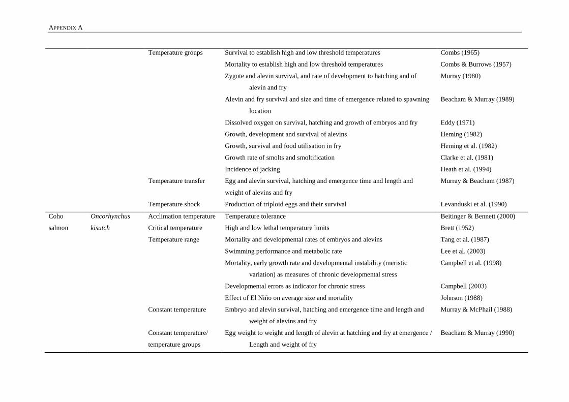
APPENDIX A
Temperature groups
Temperature transfer
Temperature shock
Survival to establish high and low threshold temperatures
Mortality to establish high and low threshold temperatures
Zygote and alevin survival, and rate of development to hatching and of
alevin and fry
Alevin and fry survival and size and time of emergence related to spawning
location
Dissolved oxygen on survival, hatching and growth of embryos and fry
Growth, development and survival of alevins
Growth, survival and food utilisation in fry
Growth rate of smolts and smoltification
Incidence of jacking
Egg and alevin survival, hatching and emergence time and length and
weight of alevins and fry
Production of triploid eggs and their survival
Combs (1965)
Combs & Burrows (1957)
Murray (1980)
Beacham & Murray (1989)
Eddy (1971)
Heming (1982)
Heming et al. (1982)
Clarke et al. (1981)
Heath et al. (1994)
Murray & Beacham (1987)
Levanduski et al. (1990)
Coho
salmon
Oncorhynchus
kisutch
Acclimation temperature
Critical temperature
Temperature range
Constant temperature
Constant temperature/
temperature groups
Temperature tolerance
High and low lethal temperature limits
Mortality and developmental rates of embryos and alevins
Swimming performance and metabolic rate
Mortality, early growth rate and developmental instability (meristic
variation) as measures of chronic developmental stress
Developmental errors as indicator for chronic stress
Effect of El Niño on average size and mortality
Embryo and alevin survival, hatching and emergence time and length and
weight of alevins and fry
Egg weight to weight and length of alevin at hatching and fry at emergence /
Length and weight of fry
Beitinger & Bennett (2000)
Brett (1952)
Tang et al. (1987)
Lee et al. (2003)
Campbell et al. (1998)
Campbell (2003)
Johnson (1988)
Murray & McPhail (1988)
Beacham & Murray (1990)
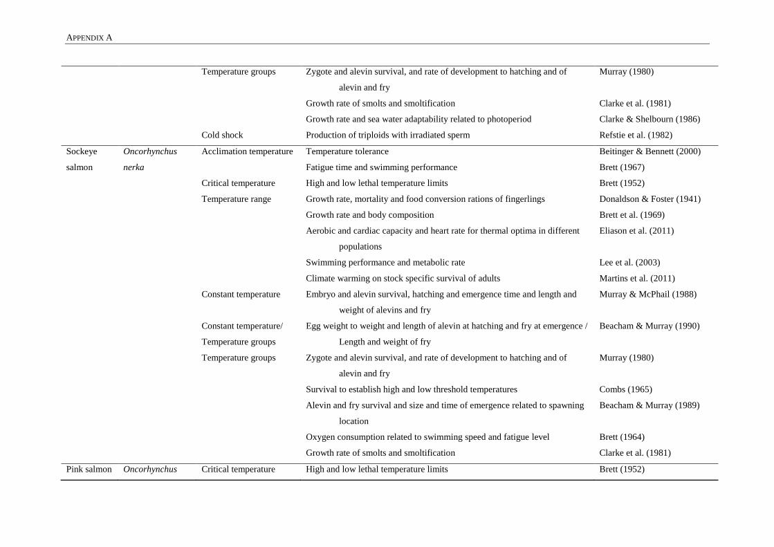
APPENDIX A
Temperature groups
Cold shock
Zygote and alevin survival, and rate of development to hatching and of
alevin and fry
Growth rate of smolts and smoltification
Growth rate and sea water adaptability related to photoperiod
Production of triploids with irradiated sperm
Murray (1980)
Clarke et al. (1981)
Clarke & Shelbourn (1986)
Refstie et al. (1982)
Sockeye
salmon
Oncorhynchus
nerka
Acclimation temperature
Critical temperature
Temperature range
Constant temperature
Constant temperature/
Temperature groups
Temperature groups
Temperature tolerance
Fatigue time and swimming performance
High and low lethal temperature limits
Growth rate, mortality and food conversion rations of fingerlings
Growth rate and body composition
Aerobic and cardiac capacity and heart rate for thermal optima in different
populations
Swimming performance and metabolic rate
Climate warming on stock specific survival of adults
Embryo and alevin survival, hatching and emergence time and length and
weight of alevins and fry
Egg weight to weight and length of alevin at hatching and fry at emergence /
Length and weight of fry
Zygote and alevin survival, and rate of development to hatching and of
alevin and fry
Survival to establish high and low threshold temperatures
Alevin and fry survival and size and time of emergence related to spawning
location
Oxygen consumption related to swimming speed and fatigue level
Growth rate of smolts and smoltification
Beitinger & Bennett (2000)
Brett (1967)
Brett (1952)
Donaldson & Foster (1941)
Brett et al. (1969)
Eliason et al. (2011)
Lee et al. (2003)
Martins et al. (2011)
Murray & McPhail (1988)
Beacham & Murray (1990)
Murray (1980)
Combs (1965)
Beacham & Murray (1989)
Brett (1964)
Clarke et al. (1981)
Pink salmon Oncorhynchus Critical temperature High and low lethal temperature limits Brett (1952)

APPENDIX A
gorbuscha Temperature range
Constant temperature
Constant temperature/
temperature groups
Temperature groups
Temperature transfer
Survival, growth, time of emergence related to spawning time
Embryo and alevin survival, hatching and emergence time and length and
weight of alevins and fry
Egg weight to weight and length of alevin at hatching and fry at emergence /
Length and weight of fry
Zygote and alevin survival, and rate of development to hatching and of
alevin and fry
Embryo and alevin survival, hatching time and time of emergence related to
heterozygosity
Survival of families, egg weight, length and weight of alevins and fry
Embryo survival
Egg and alevin survival, hatching and emergence time and length and
weight of alevins
Beacham & Murray, (1988)
Murray & McPhail, (1988)
Beacham & Murray, (1990)
Murray, (1980)
Beacham & Varnavskaya,
(1991)
Murray & Beacham, (1986)
Beacham & Murray, (1987b)
Murray & Beacham, (1986)
Chum
salmon
Oncorhynchus
keta
Acclimation temperature
Critical temperature
Constant temperature
Constant temperature/
temperature groups
Temperature groups
Temperature tolerance
High and low lethal temperature limits
Embryo and alevin survival, hatching and mergence time and length and
weight of alevins and fry
Egg weight to weight and length of alevin at hatching and fry at emergence /
Length and weight of fry
Zygote and alevin survival, and rate of development to hatching and of
alevin and fry
Female size and egg size on developmental biology
Survival of families
Spawning time and meristic variation
Meristic variations and fluctuating asymmetry
Occurrence of twinning malformation
Beitinger & Bennett (2000)
Brett (1952)
Murray & McPhail (1988)
Beacham & Murray (1990)
Murray (1980)
Beacham & Murray (1985a)
Beacham & Murray (1986)
Beacham & Murray (1986)
Beacham (1990)
Yamamoto et al. (1996)

APPENDIX A
Temperature transfer
Embryo survival
Meristic variation
Beacham & Murray (1987b)
Murray & Beacham (1987)
Brook charr
/ brook trout
Salvelinus
fontinalis
Acclimation temperature
Critical temperature
Temperature range
Constant temperature
Temperature groups
Temperature tolerance
Routine metabolic rate
Thermal maxima between diploid and triploid fish
Process of dying by temperature in rivers
Incubation period
Model hatching time related to temperature
Blood oxygen affinity and CO2 effect
Effect of ploidy on thermal tolerance
Survival related to exhaustive exercise in triploids
Oxygen related to temperature, effect on development
Oxygen, related to developmental rate and vertebral number
Oxygen uptake related to activity levels
Juvenile growth rate
Beitinger & Bennett (2000)
Atkins & Benfey (2008)
Benfey et al. (1997)
Huntsman (1942)
Embody (1934)
Crisp (1981)
Irving et al. (1941)
Galbreath et al. (2006)
Hyndman et al. (2003)
Garside (1966a)
Garside (1966b)
Graham (1949)
Sadler et al. (1986)
Lake trout Salvelinus
namaycush
Temperature range
Temperature groups
Incubation period
Oxygen related to temperature, effect on development
Juvenile growth rate
Embody (1934)
Garside (1959)
Sadler et al. (1986)
Arctic charr Salvelinus alpinus Temperature range
Temperature groups
Egg size and maternal age on egg viability at eyed egg stage
Embryonic development time and mortality
Jeuthe et al. (2013)
Jungwirth & Winkler (1984)

APPENDIX B
APPENDIX B: FERTILISATION SCHEDULE CHINOOK SALMON
Table 21: Fertilisation schedule of the temperature experiment with Chinook salmon.
Female Containers
Egg clutch C1 C2 C3 C4 C5 C6 C7 C8 C9 C10
F1 10% eggs
M1, M2, M3
10% eggs
M1, M2, M3
10% eggs
M1, M2, M3
10% eggs
M1, M2, M3
10% eggs
M1, M2, M3
10% eggs
M4, M5, M6
10% eggs
M4, M5, M6
10% eggs
M4, M5, M6
10% eggs
M4, M5, M6
10% eggs
M4, M5, M6
F2 10% eggs
M1, M2, M3
10% eggs
M1, M2, M3
10% eggs
M1, M2, M3
10% eggs
M1, M2, M3
10% eggs
M1, M2, M3
10% eggs
M4, M5, M6
10% eggs
M4, M5, M6
10% eggs
M4, M5, M6
10% eggs
M4, M5, M6
10% eggs
M4, M5, M6
F3 10% eggs
M1, M2, M3
10% eggs
M1, M2, M3
10% eggs
M1, M2, M3
10% eggs
M1, M2, M3
10% eggs
M1, M2, M3
10% eggs
M4, M5, M6
10% eggs
M4, M5, M6
10% eggs
M4, M5, M6
10% eggs
M4, M5, M6
10% eggs
M4, M5, M6
F4 10% eggs
M1, M2, M3
10% eggs
M1, M2, M3
10% eggs
M1, M2, M3
10% eggs
M1, M2, M3
10% eggs
M1, M2, M3
10% eggs
M4, M5, M6
10% eggs
M4, M5, M6
10% eggs
M4, M5, M6
10% eggs
M4, M5, M6
10% eggs
M4, M5, M6
F5 10% eggs
M1, M2, M3
10% eggs
M1, M2, M3
10% eggs
M1, M2, M3
10% eggs
M1, M2, M3
10% eggs
M1, M2, M3
10% eggs
M4, M5, M6
10% eggs
M4, M5, M6
10% eggs
M4, M5, M6
10% eggs
M4, M5, M6
10% eggs
M4, M5, M6
F6 10% eggs
M1, M2, M3
10% eggs
M1, M2, M3
10% eggs
M1, M2, M3
10% eggs
M1, M2, M3
10% eggs
M1, M2, M3
10% eggs
M4, M5, M6
10% eggs
M4, M5, M6
10% eggs
M4, M5, M6
10% eggs
M4, M5, M6
10% eggs
M4, M5, M6
F7 10% eggs
M1, M2, M3
10% eggs
M1, M2, M3
10% eggs
M1, M2, M3
10% eggs
M1, M2, M3
10% eggs
M1, M2, M3
10% eggs
M4, M5, M6
10% eggs
M4, M5, M6
10% eggs
M4, M5, M6
10% eggs
M4, M5, M6
10% eggs
M4, M5, M6
F8 10% eggs
M1, M2, M3
10% eggs
M1, M2, M3
10% eggs
M1, M2, M3
10% eggs
M1, M2, M3
10% eggs
M1, M2, M3
10% eggs
M4, M5, M6
10% eggs
M4, M5, M6
10% eggs
M4, M5, M6
10% eggs
M4, M5, M6
10% eggs
M4, M5, M6
F9 10% eggs
M1, M2, M3
10% eggs
M1, M2, M3
10% eggs
M1, M2, M3
10% eggs
M1, M2, M3
10% eggs
M1, M2, M3
10% eggs
M4, M5, M6
10% eggs
M4, M5, M6
10% eggs
M4, M5, M6
10% eggs
M4, M5, M6
10% eggs
M4, M5, M6

APPENDIX B
F10 10% eggs
M1, M2, M3
10% eggs
M1, M2, M3
10% eggs
M1, M2, M3
10% eggs
M1, M2, M3
10% eggs
M1, M2, M3
10% eggs
M4, M5, M6
10% eggs
M4, M5, M6
10% eggs
M4, M5, M6
10% eggs
M4, M5, M6
10% eggs
M4, M5, M6
Trays Split over
Two trays
Split over
Two trays
Split over
Two trays
Split over
Two trays
Split over
Two trays
Split over
Two trays
Split over
Two trays
Split over
Two trays
Split over
Two trays
Split over
Two trays
The eggs of each female were divided over 10 containers, with 10% of the total egg weight per container (indicated by 10% eggs). The first five containers were fertilised
with milt from the first three males (light orange), while container six to ten were fertilised with milt from male four to six (light blue). Each container received 1 ml milt
from each male, e.g. M1, M2, M3 stands for 1 ml milt of male 1, 1 ml of male 2 and 1 ml of male 3 were added to that container. Male four to six are indicated as M4, M5,
M6.

APPENDIX C
249
APPENDIX C: FORK LENGTH AND WEIGHT AT DIFFERENT
LIFE STAGES
Figure 32: Fork length and weight of the 8°C and 12°C groups at different life stages.
Boxplot of fork length (A) in centimetres and weight (B) in grams for cull 1 (1400°d, Table 12), cull 2 (1530°d,
Table 12), freshwater smoltification-stage (indicated as ‘smolt’), six-months-at-sea and 12-months-at-sea. The
8°C group is coloured in dark green and the 12°C group in orange.

APPENDIX C
250

APPENDIX D
APPENDIX D: WHOLE-MOUNT STAINING PROTOCOLS
Table 22: Alizarin red S staining protocol, adapted from Taylor and Van Dyke (1985)
Step Solution Post-Hatch First Feed Juvenile
Remove epithelia and
guts
Epithelium and guts removed in first feed stage specimens and juveniles - - -
Rinse Tap water 24 hrs 24 hrs 24 hrs
Remove fat 70-100% acetone solution (70% for post-hatch and first feed stage specimens) 12 hrs 24 hrs 48 hrs
Bleach pigment 1% KOH/3% H2O2 20 min 1hr 1:15 hrs
Rinse Demineralised water 30 min 30 min 30 min
Neutralisation Neutralisation buffer: 30% saturated borax (Na2B4O7 sodium tetraborate)/70% demineralised water (pH
9.4)
12 hrs 12 hrs 12 hrs
Bone staining Alizarin red solutions: 0.5 mg/ml alizarin red S in 1% KOH solutions 12 hrs 24 hrs 48 hrs
Rinse Demineralised water 30 min 1-3 hrs 5 hrs
Clearing
20% glycerol in a 1% KOH solution 5 days 5 days 10 days
40% glycerol in a 1% KOH solution 5 days 5 days 10 days
60% glycerol solution 5 days 10 days 21 days
80% glycerol solution 5 days 10 days 21 days
90% glycerol solution 5 days 10 days 1 month
95% glycerol solution 5 days 20 days 1 month
Storage 100% glycerol - - -

APPENDIX D
Table 23: Alcian Blue 8GX staining protocol, adapted from Taylor and Van Dyke (1985)
Step Solution Post-Hatch First Feed
Rinse Tap water 24 hrs 24 hrs
Remove fat 70-100% acetone solution (70% for post-hatch and first feed stage specimens) 12 hrs 24 hrs
Dehydration
30% ethanol solution 48 hrs 48 hrs
50% ethanol solution 48 hrs 48 hrs
70% ethanol solution 24 hrs 24 hrs
Cartilage staining 15mg/ml Alcian blue in 20% acetic acid (100%) and 80% ethanol (100%), allow the solution to stabilise for two days
before measuring and adjusting pH (pH ≤ 2.2 or 1.8 optimal)
48 hrs 48 hrs
Rehydration
70% ethanol solution 24 hrs 24 hrs
50% ethanol solution 24 hrs 24 hrs
30% ethanol solution 24 hrs 24 hrs
15% ethanol solution 24 hrs 24 hrs
Rinse Demineralised water 12 hrs 12 hrs
Neutralisation and
clearing
Neutralisation buffer: 0.5mg/ml trypsin in 30% saturated borax (Na2B4O7 sodium tetra borate)/70% demineralised
water (pH 9.4)
8 hrs 24 hrs
Clearing
20% glycerol in a 1% KOH solution 12 hrs 5 days
40% glycerol solution 5 days 5 days
60% glycerol solution 5 days 10 days
80% glycerol solution 5 days 10 days
90% glycerol solution 5 days 10 days
95% glycerol solution 5 days 20 days
Storage 100% glycerol - -

APPENDIX E
253
APPENDIX E: HISTOLOGICAL PROTOCOLS
Table 24: Processing for paraffin embedding
Step Solution Time (hours) Temperature Pressure
Dehydration
Ethanol 70% 0:30 Ambient
Ethanol 95% 1:00 40°C
Ethanol 100% 1:15 40°C
Ethanol 100% 1:15 40°C
Ethanol 100% 1:15 40°C
50% ethanol/50% xylene 1:20 40°C
Xylene 100% 0:45 40°C
Xylene 100% 0:45 40°C Vacuum
Replacing with paraffin
Paraffin 1:20 60°C Vacuum
Paraffin 1:20 60°C Vacuum
Paraffin 1:20 60°C Vacuum
Table 25: Haematoxylin and eosin staining protocol, adapted from (Gill et al., 1974)
Step Solution Time (minutes)
Paraffin removal
Xylene 100% 3:00
Xylene 100% 2:00
Ethanol 100% 1:00
Rehydration
Ethanol 100% 0:30
Ethanol 70% 1:30
Rinse (demineralised water) 1:00
Staining
Gill’s Haematoxylin 5:00
Rinse (demineralised water) 0:30
Scott’s tap water 0:30
Rinse (demineralised water) 1:00
Eoxin/Phloxine 2:00
Rinse (demineralised water) 0:30
Dehydration
Ethanol 70% 0:10
Ethanol 95% 0:15
Ethanol 100% 0:45
Ethanol 100% 1:00
Xylene 100% 1:00
Xylene 100% 1:00

APPENDIX E
254
Table 26: Masson’s trichrome staining protocol, adapted from (Culling, 1974b)
Step Solution Time (minutes)
Paraffin removal
Xylene 100% 3:00
Xylene 100% 2:00
Ethanol 100% 1:00
Rehydration
Ethanol 100% 0:30
Ethanol 70% 1:30
Rinse (demineralised water) 1:00
Post fixation Mordant in Bouin’s fixative Overnight
Stain
Rinse (demineralised water) 3:00
Celestine blue 10:00
Rinse (demineralised water) 0:10
Mayer’s Haematoxylin 10:00
Rinse (demineralised water) 0:10
Scott’s tap water (differentiate nuclei) 2:00
Rinse (demineralised water) 2:00
Beiberich scarlet-acid fuschsin 2:00
Rinse (demineralised water) 0:30
Phosphotungstic acid 5% 15:00
Rinse (demineralised water) 0:30
Light green solution 1:00
Rinse (demineralised water) 4 dips + drain
Blot dry on filter paper (removal excess stain) -
Glacial acetic acid 1% 4 dips + drain
Blot dry on filter paper (removal excess stain) -
Dehydration
Ethanol 95% 10 dips + drain
Ethanol 100% 10 dips + drain
Ethanol 100% 10 dips + drain
Xylene 100% 10 dips + drain
Xylene 100% 10 dips + drain
Xylene 100% 10 dips + drain

APPENDIX E
255
Table 27: Verhoeff-Van Gieson staining protocol, adapted from (Culling, 1974b)
Step Solution Time (minutes)
Paraffin removal
Xylene 100% 2:00
Xylene 100% 2:00
Ethanol 100% 1:00
Rehydration
Ethanol 100% 1:00
Ethanol 95% 1:00
Ethanol 70% 1:00
Rinse (demineralised water) 1:00
Staining
Verhoeff’s working solution (20 ml alcoholic haematoxylin + 8 ml
10% ferric chloride + 8 ml Verhoeff’s iodine)
15:00
Rinse (demineralised water) 0:30
2% ferric chloride (differentiate elastin fibres) 1:00 + check
Rinse (demineralised water) 0:30
Rinse in ethanol 95% (removal iodine residue) 0:05
Rinse (demineralised water) 5:00
Van Gieson stain (counterstain) 3:00
Blot dry on filter paper (removal excess stain) -
Dehydration
Ethanol 95% 10 dips + drain
Ethanol 100% 10 dips + drain
Ethanol 100% 10 dips + drain
Xylene 100% 10 dips + drain
Xylene 100% 10 dips + drain
Xylene 100% 10 dips + drain
Table 28: AB-PAS (Alcian Blue-Periodic Acid Shiff) staining protocol, adapted from (Culling, 1974a)
Step Solution Time (minutes)
Paraffin removal
Xylene 100% 2:00
Xylene 100% 2:00
Ethanol 100% 1:00
Rehydration
Ethanol 100% 1:00
Ethanol 95% 1:00
Ethanol 70% 1:00
Rinse (demineralised water) 0:30
Staining
1% Alcian blue (pH 2.5) 10:00
Rinse (demineralised water) 0:10
0.5% periodic acid (oxidise) 5:00
Rinse (demineralised water) 5:00
Shiff’s reagent (toxic!) 10:00
Rinse (demineralised water) 10:00

APPENDIX E
256
Gill’s Haematoxylin (counterstain) 0:30
Rinse (demineralised water) 0:30
Scott’s tap water 0:30
Rinse (demineralised water) 1:00
Dehydration
Ethanol 70% 10 dips + drain
Ethanol 95% 10 dips + drain
Ethanol 100% 10 dips + drain
Ethanol 100% 10 dips + drain
Xylene 100% 10 dips + drain
Xylene 100% 10 dips + drain
Xylene 100% 10 dips + drain
Table 29: Heidenhains AZAN trichrome staining protocol (Böck & Romeis, 1989)
Step Solution Time (hours)
Paraffin removal
Ultraclear 100% 00:05
Ultraclear 100% 00:05
Ethanol 96% 00:04
Ethanol 96% 00:04
Staining
Aniline alcohol (0.1% anilin in 95% ethanol) 00:05
Rinse (demineralised water) 00:00:10
Azocarmine 00:45
Rinse (demineralised water) 00:00:10
Aniline alcohol (0.1% anilin in 95% ethanol) 00:05
Acetic acid alcohol 1% 00:01
Rinse (demineralised water) 00:01
Phosphotungstic acid 5% 03:00
Rinse (demineralised water) 00:00:10
Aniline blue-Orange G 01:00
Rinse (demineralised water) 00:00:10
Dehydration
Ethanol 96% 00:02
Ethanol 96% 00:03
Isopropanol 00:03
Ultraclear 100% 00:03
Ultraclear 100% 00:03

APPENDIX F
APPENDIX F: MUTATIONS OF CELLULAR VESICULAR TRANSPORT
Table 30: Mutations affecting the normal chordocyte and chordoblast vesicular transport functions in zebrafish and medaka
Component (s) Function Mutation (s) Species Effects Reference(s)
Unfolded Protein
Response (UPR)
Folding proteins correctly in the ER lumen and
evacuating proteins correctly when ER-stress occurs.
This occurs when ER bulges due to the presence of to
many proteins. Transcriptional regulation of COPII
coat components (SEC23/SEC24/SAR1, SEC13/SEC
31, TANGO, SEDLIN, KLHL12, CUL3).
Atf6α/β
mutants
Medaka ER-stress occurs
Reduced induction of ER
chaperones
Chaperones: HSP90, GRP94,
Lectins, HSP70, HSP40 family
members
Ishikawa et al.
(2013)
Sitia &
Braakman
(2003)
Bbf2h7 is a transducer of all COP II components
(SEC23/SEC24/SAR1, SEC13/SEC31, TANGO,
SEDLIN, KLHL12, CUL3) and is similar to the
feelgood mutant in zebrafish.
Bbf2h7
mutants
Medaka Cartilage in skull under develops
Short tail
Bended notochord
Incomplete bone formation
Detachment of chordoblasts from
basal lamina
ER-stress due to build-up of
proteins in the ER
No collagen type II secretion
No collagen type IV secretion
Downregulation of all COPII coat
components
Ishikawa et al.
(2017)
Is similar to Bbf2h7 mutant in medaka. However,
Creb3l2 only regulates a subset of COP II components,
i.e. SEC23A/B and SEC24D.
Feelgood
(Creb3l2)
mutant
Zebrafish Cartilage in skull under develops
Short tail
Bended notochord
Melville et al.
(2011)
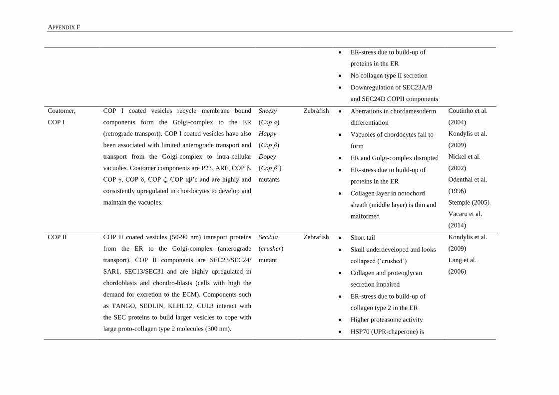
APPENDIX F
ER-stress due to build-up of
proteins in the ER
No collagen type II secretion
Downregulation of SEC23A/B
and SEC24D COPII components
Coatomer,
COP I
COP I coated vesicles recycle membrane bound
components form the Golgi-complex to the ER
(retrograde transport). COP I coated vesicles have also
been associated with limited anterograde transport and
transport from the Golgi-complex to intra-cellular
vacuoles. Coatomer components are P23, ARF, COP β,
COP γ, COP δ, COP ζ, COP αβ’ε and are highly and
consistently upregulated in chordocytes to develop and
maintain the vacuoles.
Sneezy
(Cop α)
Happy
(Cop β)
Dopey
(Cop β’)
mutants
Zebrafish Aberrations in chordamesoderm
differentiation
Vacuoles of chordocytes fail to
form
ER and Golgi-complex disrupted
ER-stress due to build-up of
proteins in the ER
Collagen layer in notochord
sheath (middle layer) is thin and
malformed
Coutinho et al.
(2004)
Kondylis et al.
(2009)
Nickel et al.
(2002)
Odenthal et al.
(1996)
Stemple (2005)
Vacaru et al.
(2014)
COP II COP II coated vesicles (50-90 nm) transport proteins
from the ER to the Golgi-complex (anterograde
transport). COP II components are SEC23/SEC24/
SAR1, SEC13/SEC31 and are highly upregulated in
chordoblasts and chondro-blasts (cells with high the
demand for excretion to the ECM). Components such
as TANGO, SEDLIN, KLHL12, CUL3 interact with
the SEC proteins to build larger vesicles to cope with
large proto-collagen type 2 molecules (300 nm).
Sec23a
(crusher)
mutant
Zebrafish Short tail
Skull underdeveloped and looks
collapsed (‘crushed’)
Collagen and proteoglycan
secretion impaired
ER-stress due to build-up of
collagen type 2 in the ER
Higher proteasome activity
HSP70 (UPR-chaperone) is
Kondylis et al.
(2009)
Lang et al.
(2006)

APPENDIX F
upregulated
= vbi mutant in medaka Sec24d
(bulldog)
mutant
Zebrafish Short tail
Skull underdeveloped
Bended notochord
Impaired chondrocyte maturation
ER-stress due to build-up of
collagen type 2 in the ER of
chondrocytes
UPR components are upregulated
Melville et al.
(2011)
Sarmah et al.
(2010)
= bulldog mutant in zebrafish Vbi (Vertebra
imperfecta)
mutant
Medaka Centrum mineralisation delayed
Centrum mineralisation is
irregular
Chordacentra fuse ventrally
Neural and haemal arches
malformed
Irregular migration and position
of osteoblasts lining the
notochord
ER-stress due to build-up of
collagen type 2 in the ER of
chondrocytes
HSP47 (UPR-chaperone) is
upregulated
Ohisa et al.
(2010)
Clathrin coat
complex
Clathrin coated vesicles transport membrane bound
proteins and enzymes from the Golgi-complex to the
Cltca mutant Zebrafish Bended in notochord
Vertebral centra form in shape of
Ellis et al.
(2013)

APPENDIX F
plasma membrane. These vesicles also transport
extracellular signals to the Golgi complex or to other
intra-cellular vesicles.
bended notochord
Vacuoles in the chordocytes
collapse
Caveolae Plasma membrane pits of 60-80 nm which are ‘cup’-
shaped important in cellular signalling pathways
(extra-cellular to intra-cellular signalling), endocytosis,
cellular attachment and detachment, fatty acid
regulation of the plasma membrane, mechanosensing
and mechano-protection of cells. Caveolae are present
in adipose cells and muscle cells. Highest number of
Caveolae was recorded in zebrafish chordocytes.
Components are Caveolin 1, 2, 3 and Cavin-1.
Caveolin 1
(Cav1)
Caveolin 3
(Cav3)
Cavin-1b
mutants
Zebrafish Shorter fish
Compromised swimming
Upon mechanical stress
Notochord lesions where most
mechanical stress
Reduction in number of caveolae
Aberrant morphology of caveolae
Delamination of chordocyte
Collapse and fragmentation of
vacuole
Cell rupture
Release of dense material in the
ECM
Bastiani &
Parton (2010)
Garcia et al.
(2017)
Lim et al.
(2017)

APPENDIX F

APPENDIX F

APPENDIX F
Figure 33: Mutations in cellular vesicular transport mechanisms in chordocytes
Schematic representation of two adjacent chordocytes (only part of the cell is shown). The top row shows the cells, the middle row the vesicular transport components and the
bottom row shows the mutations that occur in the respective vesicular transport components (text in red). Within the cell, from left to right, the nucleus, the endoplasmic
reticulum (ER), the Golgi-complex and a clathrin coated pit are shown. In the bottom part of the cell the vacuole (blue) is shown. The second cell only shows a vacuole, since
the largest part of the chordocyte cell is filled with a vacuole. Caveolae pits are represented in the cell plasma membrane. The COP II dependent transport is indicated by
purple arrows, while COP I dependent transport is indicated by green arrows. Clathrin coated vesicle transport is indicated by black arrows. Red crosses show which
pathways or components (caveolae) fail when a mutation occurs. A side view of all transport vesicle components is shown, except for the clathrin-triskelion which represents
a top view. Caveolae, (dark blue), COP I , COPII (purple) and Clathrin (black) components are adapted from Bastiani and Parton (2010), Nickel et al. (2002), Gomez-
Navarro and Miller (2016), Edeling et al. (2006), Robinson (2015) respectively. The two bent black lines underneath the COP I and COP II components represents the plasma
membrane phospholipid bilayer of the transport vesicle. The red cross on the clathrin-triskelion shows where the Cltca mutation forms a truncated protein (Edeling et al.,
2006).

APPENDIX F

APPENDIX F
Figure 34: Mutations in cellular vesicular transport mechanisms in chordoblasts
Schematic representation of a chordoblast adjacent to the notochord sheath (only part of the cell is shown). The top row shows the cell, the middle row the vesicular transport
components and the bottom row shows the mutations that occur in the respective vesicular transport components (text in red). Within the cell, from left to right, the nucleus,
the endoplasmic reticulum (ER), the Golgi-complex and a clathrin coated pit are shown. To the far right the notochord sheath is shown, with the basement membrane directly
opposed to the cell plasma membrane, the collagenous middle layer and the external elastic membrane (EEM) on the outside of the notochord sheath. The COP II dependent
transport is indicated by purple arrows, while COP I dependent transport is indicated by green arrows. Clathrin coated vesicle transport is indicated by black arrows. Red
crosses show which pathways fail when a mutation occurs. Black lightning bolts indicate where in the ER the unfolded protein response (UPR) is elicited by ER-stress. A
side view of all transport vesicle components is shown. COP I , COPII (purple), COP II-enlarged (purple) and Clathrin (black) components are adapted from Nickel et al.
(2002), Gomez-Navarro and Miller (2016), Ishikawa et al. (2017), Edeling et al. (2006) and Robinson (2015) respectively. The two bent black lines underneath the COP I and
COP II components represents the plasma membrane phospholipid bilayer of the transport vesicle. The black bolt next to Creb3l2 and Bbf2h7 indicates that these mutations
are involved in the unfolded protein response. The red cross on the clathrin-triskelion shows where the Cltca mutation forms a truncated protein (Edeling et al., 2006).

APPENDIX F

APPENDIX G
267
APPENDIX G: DRC 16 – V3 STATEMENT OF
CONTRIBUTION

APPENDIX G
268

APPENDIX G
269

APPENDIX G
270