Exogenous endothelial cells as accelerators of hematopoietic reconstitution
Upload
independentCategory
view
0download
0
Exogenous Mineralization of Cell-Seeded and UnseededCollagen-Chitosan Hydrogels using Modified Culture Medium
Rameshwar R. Rao, Alex Jiao, David H. Kohn, and Jan P. Stegemann*
Department of Biomedical Engineering, University of Michigan, Ann Arbor
AbstractInduced biomineralization of materials has been employed as a strategy to increase integrationwith host tissue, and more recently as method to control cell function in tissue engineering.However, mineralization is typically performed in the absence of cells, since hypertonic solutionsthat lack the nutrients and culture components required for maintenance of cell viability are oftenused. In the present study, we exposed fibroblast-seeded 3D collagen-chitosan hydrogels to adefined culture medium modified to have specific concentrations of ions involved inbiomineralization. Modified medium caused a significant increase in calcium deposition incollagen-chitosan gels, relative to constructs incubated in standard medium, though serumsupplementation attenuated mineral deposition. Collagen-chitosan constructs became opaque overthree days of mineralization in modified DMEM, in contrast to translucent control gels incubatedin standard DMEM. Histological staining confirmed increased levels of mineral in the treatedconstructs. Rheological characterization showed that both the storage and loss moduli increasedsignificantly in mineralized materials. Mineralization of fibroblast-seeded constructs resulted indecreased cell viability and proliferation rate over three days of incubation in modified medium,but the cell population remained over 75% viable and regained its proliferative potential afterrescue in standard culture medium. The ability to mineralize protein matrices in the presence ofcells could be useful in creating mechanically stable tissue constructs, as well as to study theeffects of the tissue microenvironment on cell function.
IntroductionThere is a clear need for materials and methods to improve bone healing outcomes,particularly in cases of large defects and non-unions. The natural healing response is oftennot adequate to obtain full repair, and in such cases strategies to augment bone regenerationcan be applied. Autografts and allografts are currently used clinically, but are hampered byissues of tissue availability and consistency [1]. The generic tissue engineering approach isto combine cells, biomaterials, and growth factors in a controlled fashion to create livingmaterials that can replace damaged tissue and/or enhance regeneration. In the case of bonetissue, a wide range of strategies have been employed, using a variety of cell types,materials, and biochemical factors[2].
© 2012 Acta Materialia Inc. Published by Elsevier Ltd. All rights reserved.*Corresponding Author: Jan P. Stegemann, Department of Biomedical Engineering, University of Michigan, 1101 Beal Ave., AnnArbor, MI 48109, Tel: 734-764-8313, Fax: 734-647-4834, [email protected]'s Disclaimer: This is a PDF file of an unedited manuscript that has been accepted for publication. As a service to ourcustomers we are providing this early version of the manuscript. The manuscript will undergo copyediting, typesetting, and review ofthe resulting proof before it is published in its final citable form. Please note that during the production process errors may bediscovered which could affect the content, and all legal disclaimers that apply to the journal pertain.
NIH Public AccessAuthor ManuscriptActa Biomater. Author manuscript; available in PMC 2013 April 1.
Published in final edited form as:Acta Biomater. 2012 April ; 8(4): 1560–1565. doi:10.1016/j.actbio.2012.01.001.
NIH
-PA Author Manuscript
NIH
-PA Author Manuscript
NIH
-PA Author Manuscript

One strategy for potentiating the bone healing response is to use materials that have beenexogenously mineralized using defined ionic solutions. Simulated body fluid (SBF) is asolution formulated with ion concentrations similar to blood plasma, which mineralizes thesurfaces and pore walls of both natural[3–7] and synthetic[8–10] scaffolds if thermodynamicconditions are appropriate. Kokubo et. al[11] first described that soaking a biomaterial inSBF leads to the ex vivo formation of a bone-like apatite coating, and later studies showedthat such coatings can be both osteoconductive[10] and osteoinductive[13], and canfacilitate the regeneration of bone [12, 14]. SBF-induced mineralization has been furtherexamined as a method for controlling osteoconductivity [5, 9, 15], as well as for protein [13,16] and gene delivery [17, 18]. Taken together, this body of work has shown that SBF canbe a useful tool to modify biomaterials for bone tissue engineering applications.
Previous studies using SBF to modify material scaffolds have been performed in the absenceof cells, since the high ionic concentrations and lack of nutrients in SBF are not conducive tothe maintenance of cell growth. However, cell-seeded materials have been proposed for anumber of orthopaedic applications. Natural biomaterial hydrogels are of interest in suchcases due to their ability to mimic the natural extracellular matrix [19] and provide tissue-specific cues to enhance cell attachment and stem cell differentiation [20]. Directencapsulation of cells during gel formation can be used to facilitate homogenous celldistribution in hydrogels. Numerous natural polymers including collagen [3], chitosan [21],and composite matrices [22–25] have been employed to engineer tissues and have shownpromise in bone regeneration[26]. A drawback of natural hydrogel materials is that theyoften lack mechanical strength and represent only the protein component of the native bonetissue. Mineralization of such matrices has been pursued as a strategy to improve theirmechanical properties and more closely mimic the native matrix[6], however the cellularcomponent is typically not included during the mineralization process.
In the present study, we mineralized 3D hydrogels using a modified culture medium thatcombined the ionic constituents of SBF with the nutrients, vitamins, and amino acids neededto maintain cell viability. The model tissue constructs consisted of fibroblast cells embeddedin collagen-chitosan hydrogel matrices developed previously in our laboratory[24, 26].Fibroblasts were used as a model cell type to examine the feasibility of mineralization in thepresence of cells, since this non-mineralizing cell type allowed us to isolate the effects ofmineralization to the medium alone. Both unseeded and fibroblast-seeded hydrogels wereexposed to mineralizing solutions that were formulated to induce biomineralization whilealso supporting cell growth, and the effects of such treatment on mineral content,mechanical properties, and cellular viability were determined. Our primary goal was todemonstrate that mineralization of protein-based hydrogels is possible in the presence ofcells. The ability to mineralize cell-seeded protein matrices could be useful in creatingmechanically stable and osteogenic tissue constructs, as well as in studying the process ofbiomineralization.
2. Materials and Methods2.1 Media formulations
The composition of the mineralization medium was based on previously studied simulatedbody fluid (SBF) formulations, with modifications to enhance both mineralization and theability to support cell growth. The base medium was Dulbecco’s modified Eagle medium(DMEM; high glucose, Invitrogen, Carlsbad, CA), which was supplemented with ionic salts.Table 1 shows the ion concentrations of relevant biological fluids and mineralizing media.The main augmentation to the modified medium formulation was a 4-fold increase incalcium (Ca2+) and phosphate (PO4
3−) in order to promote biomineralization, and an
Rao et al. Page 2
Acta Biomater. Author manuscript; available in PMC 2013 April 1.
NIH
-PA Author Manuscript
NIH
-PA Author Manuscript
NIH
-PA Author Manuscript

increased carbonate (HCO3−) level to provide buffering capacity. These modifications are
further discussed in the Results and Discussion section.
The modified medium (mDMEM) formulation was prepared by adding salts directly toDMEM to achieve final concentrations of 141 mM NaCl, 5.3 mM KCl, 10 mM CaCl2·H2O,1.0 mM MgCl2, 8.4 mM NaHCO3, 0.8 mM MgSO4, and 4.0 mM KH2PO4. The mediumwas prepared at 25°C and titrated to a pH of 7.4. In experiments examining the effect ofserum, mDMEM and control DMEM were supplemented with varying concentrations offetal bovine serum (FBS; Invitrogen) and 1% penicillin/streptomycin (P/S; Invitrogen). Asper previous protocols, mDMEM was changed every 12 hours to avoid precipitation in theculture solution and DMEM was changed every 3 days. In subsequent mineralizationexperiments using cells, the mineralization medium (MM) used was mDMEM supplementedwith 2% FBS. The control medium (DM) was DMEM supplemented with 10% FBS.
2.2 Collagen-chitosan Gel FabricationCollagen-chitosan gel composites were formed through a β-GP induced mechanism aspreviously described[24]. Briefly, 4.0 mg/ml bovine Type I collagen (MP Biomedicals,Solon, OH) was dissolved in 0.02 N acetic acid (Sigma) and was mixed with 2.0 wt%chitosan (93% DDA; Biosyntech, Quebec, Canada) dissolved in 0.1 N acetic acid at a massratio of 50/50 collagen/chitosan. Beta-glycerophosphate (β-GP) and glyoxal were added asphysical and chemical cross-linkers, respectively, at concentrations of 7.0wt% β-GP and 0.5mM glyoxal. A 400 µl aliquot of the pre-gelled mixture was injected into a well of a 24-wellplate to create a disk-shaped construct with diameter of 1.5 cm. Gelation was then initiatedby incubation of the mixture at 37°C for 30 min. Gels were washed three times in phosphatebuffered saline (PBS; Invitrogen) for 10 min to remove excess β-GP prior to use.
2.3 Calcium QuantificationCalcium deposition on acellular gels after 3 days incubation in either modified medium orDMEM containing 0, 2, 5, 10% FBS was quantified using an orthocresolphthalein complex-one (OCPC) method as previously described [27]. Briefly, collagen-chitosan gels werewashed three times in PBS for 10 minutes and frozen at days 0, 1, and 3. Samples were thendigested in 0.5 ml of 1.0 N acetic acid overnight. Ten microliters of the dissolved solutionwas then incubated at 10 min at 25°C with 300 µl of a working solution consisting of 0.05mg/ml of OCPC solution and ethanolamine/boricacid/8-hydroxyquinoline buffer (Sigma).Samples were read spectrophotometrically at 575 nm. Calcium values were quantified via astandard curve prepared from 0.0 to 100 µg/ml.
2.4 Gel Morphology and Von Kossa stainingAcellular gel morphology was examined 3 days after incubation in mineralization medium(MM = mDMEM + 2% FBS) and in control medium (DM = DMEM + 10% FBS). Gelswere washed three times in PBS for 10 min each and then transferred to a 12-well plate forimaging using a standard CCD camera in manual mode with a constant exposure setting.
For von Kossa staining, acellular gels were washed three times in PBS for 10 min and thenplaced in zinc-buffered formalin (Anatech LTD, Battle Creek, MI) for 30 minutes, followedby immersion in 70% ethanol (Fisher Scientific, Pittsburgh, PA). Samples werecryosectioned into the top, middle (300 µm from the top face), and bottom face at theHistology Core Facility at the University of Michigan Dental School. Gels were stained withvon Kossa reagent, embedded in paraffin and then mounted on slides. Images were taken at4X magnification using an Olympus IX15 Microscope system (Olympus America, CenterValley, PA) and stitched together using Metamorph Premier software (Molecular Devices,Sunnyvale, CA). Images were quantified using ImageJ software (National Institute of
Rao et al. Page 3
Acta Biomater. Author manuscript; available in PMC 2013 April 1.
NIH
-PA Author Manuscript
NIH
-PA Author Manuscript
NIH
-PA Author Manuscript

Health, Bethesda, MD) using thresholding and color discrimination to define positivestaining. The ratio of the stained to unstained area was used to determine the fraction of thesample that was positively stained in each sample.
2.5 Gel RheologyAcellular collagen-chitosanconstructs incubated in MM or DM for 3 days were washed threetimes in PBS for 10 min each and then evaluated by gel rheometry using an AR-G2rheometer (TA Instruments, New Castle, DE). Gels were loaded on to a Peltier stagepreheated to 37°C. A 8 mm steel parallel plate was used with a gap height of 1500 microns.A strain sweep was performed over the course of 45 minutes with strain rates from 0.1 to100% and a constant frequency of 1 radian/second. Reported values were taken over thelinear range of the samples.
2.6 Cell Culture and AssaysPrior to gel fabrication, human neonatal dermal fibroblasts (hFb; Lonza Inc., Walkersville,MD) were cultured in DMEM containing 10% FBS and 1% PS. For cell assays, hFb wereused at passage 7–9 and placed directly into the solution of collagen and chitosan at aconcentration of 1.0 × 10⁶ cells/ml prior to gelation, to allow for homogenous encapsulationwithin formed gels. Previous studies have shown that cells survive the encapsulation process[24, 26], and hFB were used in this study as a model cell type to isolate the effects ofmineralization to the modified media solution.
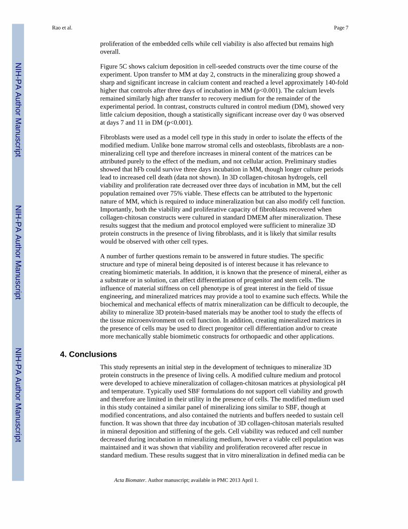
Cell-seeded constructs in the MM condition were cultured for 1 day in DM, 3 days in MM,and then 7 days in DM for a recovery period. Samples were collected at days 0, 1, 2, 4, 5, 7,11 corresponding to initial conditions, 1 day in DM, 1 day in MM, 3 days in MM, 1 dayrecovery in DM, 3 days recovery in DM, and 7 days recovery in DM (Figure 1). Gels werewashed three times in PBS for 10 min when switching between culture medium. Cell-seededgels cultured in DM only were used as controls and samples were collected at correspondingtimepoints.
To evaluate toxicity of the MM media, cell viability was examined using a vital stain kit(Live/Dead®, Molecular Probes, Eugene, OR). Constructs were washed three times insterile PBS and incubated at 37°C for 45 min in a solution containing 4.0 µm calcein-AMand 4.0 µm ethidium homodimer-1 in PBS. Gels were then washed again in PBS andimaged using a laser scanning confocal microscope (Olympus FluoView 500 LaserScanning Confocal Microscope, Olympus). Image scans were captured at a horizontal plane150 µm above the bottom surface of the gel and quantified using ImageJ software.
To quantify cell number during the mineralization period, DNA was extracted in 4.0 Mguanidine hydrochloride solution and measured using a commercially available DNA assay(PicoGreen kit, Invitrogen). Calcium deposited on the constructs during the mineralizationperiod was also measured using the OCPC assay described above.
2.7 Statistical AnalysisOne-way ANOVA testing with Tukey's post hoc analysis was used to analyze the effect ofFBS concentration over time on calcium deposition in acellular hydrogels. Student's T-testwas used to assess the significance of the fractional area of von Kossa staining, rheologicaldata, cell viability, DNA content, and calcium deposition in cell-seeded hydrogels treatedwith mineralizing medium, compared to control medium. One-way ANOVA with Tukey'spost hoc analysis was used to analyze the effect of medium type over time on DNA contentin cellular hydrogels. Statistical significance was set at p < 0.05. Numerical values are
Rao et al. Page 4
Acta Biomater. Author manuscript; available in PMC 2013 April 1.
NIH
-PA Author Manuscript
NIH
-PA Author Manuscript
NIH
-PA Author Manuscript

presented as mean +/− standard error of the mean (SEM). N = 4 for each assay, and errorbars on graphs represent the standard error of the mean.
3. Results and Discussion3.1 Rationale for media formulations
This work demonstrated the ability to mineralize protein biomaterials and change theirproperties both in the presence and absence of embedded cells. Mineralization techniquestypically employ simulated body fluid (SBF), however, the lack of nutrients, vitamins,amino acids, and glucose prevent these media from being used for cell culture. DMEM is acommonly used medium for cell culture that can lead to the precipitation of mineral noduleson hydroxyapatite and tricalcium phosphate scaffolds in normal cell cultureenvironments[28]. Therefore we used DMEM as a base medium and supplemented it withthe specific salts to enhance the mineralization process. Table 1 shows the ionconcentrations of SBF, DMEM, and mDMEM, as well as blood plasma for reference. Themineralizing DMEM(mDMEM) used in this study was formulated to have specific ionicconcentrations aimed at maximizing mineral deposition[28]. mDMEM containedconcentrations of sodium (Na+), magnesium (Mg2+), and sulfate (SO4
2−) similar toconventional SBF. Calcium (Ca2+) and phosphate (PO4
3−) are the primary ions required forbiomineralization, and were therefore added at concentrations approximately 4-fold higherthan conventional SBF to promote rapid mineralization of substrates. The carbonate(HCO3
−) level was maximized to serve as a pH buffer in the media to allow culture in a CO2incubator, though this anion may be associated with decreased mineralization. Each of thesesolutions was adjusted to physiological pH to provide an appropriate environment for cellculture.
3.2 Effect of serum on calcium depositionSerum is a necessary supplement in cell culture medium because it contains growth factorsthat maintain cell viability and growth [28, 29]. However, in some systems serum proteinscan detrimentally effect mineralization by delaying or inhibiting deposition [13, 30, 31]. Toinvestigate the effect of serum on calcium deposition in collagen-chitosan materials,acellular hydrogel constructs were exposed to mineralizing DMEM supplemented with 0, 2,5, and 10% FBS. Gels incubated in control DMEM with 0, 2, 5, 10% FBS served ascontrols. Figure 2 shows that calcium deposition increased significantly (p<0.01) from day 0in all samples incubated in mineralizing DMEM by day 1, and that mineralization continuedto increase to day 3 in culture. In contrast, samples exposed to control DMEM showed noevidence of calcium deposition regardless of serum content or timepoint. In mDMEMsamples, the presence of serum had no effect on the degree of calcium deposition at the day1 timepoint, but by day 3 serum-supplemented samples showed decreased mineraldeposition, relative to the sample with no serum (p<0.01). There was no significantdifference between 2, 5 and 10% serum. These data show that FBS had an inhibitory effecton calcium deposition, but that mineralization did occur in the presence of serum proteins.Because of the need for at least a low level of serum for cell maintenance, mineralizationstudies conducted with cells were performed using mDMEM supplemented with 2% FBS asthe mineralization medium (MM). The control medium (DM) was standard DMEMsupplemented with 10% FBS, which is widely used for cell culture.
3.3 Mineralization of collagen-chitosan materialsFigure 3 shows data on mineralization and mechanical properties of acellular collagen-chitosan matrices incubated in MM and DM for three days. Histological evaluation of VonKossa stained sections taken at the top, middle, and bottom of the constructs showed cleardifferences in degree of phosphate deposition between mineralizing and control conditions
Rao et al. Page 5
Acta Biomater. Author manuscript; available in PMC 2013 April 1.
NIH
-PA Author Manuscript
NIH
-PA Author Manuscript
NIH
-PA Author Manuscript

(Fig. 3A). After three days in incubation, constructs in DM remained translucent, where asthose in MM were opaque, and dark staining for calcium was more prominent in MMsamples (Fig. 3A inset). Quantification of relative phosphate staining showed that hydrogelsin MM exhibited significantly higher phosphate levels at the top and middle of theconstructs, though staining in the bottom section was not statistically different compared toconstructs in DM. These results support the finding that MM induces mineralization, butshow that mineral deposition was not evenly distributed through the material.
Figure 3B shows the storage (G’) and loss (G”) modulus from acellular collagen-chitosangels exposed to DM or MM for three days. Relative to control gels, mineralized gelsexhibited a 3-fold increase in stiffness as reflected by the storage modulus (p<0.05). Theloss modulus also increased significantly (p<0.001) in the MM condition, as compared to theDM condition. These data show that the mechanical properties of hydrogel constructs can beaugmented by mineralization even over relatively short time periods. SBF treatment canaffect the properties of scaffolds, though the effects can vary depending on the distributionof the mineral throughout the scaffolding material [8, 18]. Increasing the stiffness andtoughness of protein hydrogel materials is desirable in order to allow implantation ofconstructs, particularly in load-bearing applications. A stiffer matrix may also attenuate thecell-mediated matrix remodeling that occurs in 3D protein hydrogels [32], and may directthe phenotype of progenitor cells toward an osteogenic phenotype [33].
3.4 Effect of mineralization on cell functionViability data for hFb embedded in 3D collagen-chitosan materials are shown in Figure 4and Figure 5A. Green staining indicates the cytoplasm of living cells, whereas red-stainednuclei identify dead cells. The culture protocol is shown schematically by the arrows inFigure 4. Control constructs were cultured in DM for 11 days, whereas mineralizedconstructs were cultured in DM for one day, followed by three days of incubation in MM,and then 7 days of culture recovery in DM. All samples were imaged and assayed for cellfunction at days 1, 2, 4, 5, 7, and 11. After one day of culture in DM, cells began to spreadthrough the collagen-chitosan matrix (Fig. 4A,4B) and exhibited very high viability (>90%).Constructs subsequently exposed to MM showed significantly decreased viability (p<0.05)over the incubation period in MM (days 2 through 4, Fig. 4D, 4F), but viability was stillhigh (~80%). The morphology of cells incubated in MM was more rounded than the controlcells, which were highly stellate and exhibited viability over 90% (Fig. 4C, 4E). Uponreintroduction into standard medium, cells in the mineralized constructs recovered a spreadmorphology (Fig. 4H, 4J, 4L) and viability returned to the same level as in controlconstructs by day 11 (>90%).
The DNA content of hydrogel constructs was used as a measure of cell number and is shownin Figure 5B. Cell number increased steadily and significantly (p<0.001) over time in theDM condition, resulting in an approximately 5-fold increase by day 11. Constructs in themineralization group showed an initial rise in DNA content, which subsequently droppedsignificantly upon incubation in MM from days 2 through 4 (p<0.0001). Similar to cellviability, cell number in mineralized constructs recovered after transfer to DM, although theDNA content did not recover to the same levels as controls. The cell viability and cellnumber data provide different but complementary information on cell “health” in theconstructs. A lack of cell proliferation would lead to a decrease in overall cell number overtime, though viability observed as a snapshot at any given time can still be high, as observedin MM at the day 1 and 2 time points. Cell death is suggested by the data at the day 4 and 5time points, since both viability and cell number were decreased. In contrast, when cells areproliferating then cell number will increase and viability can also be high. This correspondsto the trends observed in DM. Taken together these data suggest that incubation inmineralizing medium significantly reduces, and perhaps completely inhibits, the
Rao et al. Page 6
Acta Biomater. Author manuscript; available in PMC 2013 April 1.
NIH
-PA Author Manuscript
NIH
-PA Author Manuscript
NIH
-PA Author Manuscript
proliferation of the embedded cells while cell viability is also affected but remains highoverall.
Figure 5C shows calcium deposition in cell-seeded constructs over the time course of theexperiment. Upon transfer to MM at day 2, constructs in the mineralizing group showed asharp and significant increase in calcium content and reached a level approximately 140-foldhigher that controls after three days of incubation in MM (p<0.001). The calcium levelsremained similarly high after transfer to recovery medium for the remainder of theexperimental period. In contrast, constructs cultured in control medium (DM), showed verylittle calcium deposition, though a statistically significant increase over day 0 was observedat days 7 and 11 in DM (p<0.001).
Fibroblasts were used as a model cell type in this study in order to isolate the effects of themodified medium. Unlike bone marrow stromal cells and osteoblasts, fibroblasts are a non-mineralizing cell type and therefore increases in mineral content of the matrices can beattributed purely to the effect of the medium, and not cellular action. Preliminary studiesshowed that hFb could survive three days incubation in MM, though longer culture periodslead to increased cell death (data not shown). In 3D collagen-chitosan hydrogels, cellviability and proliferation rate decreased over three days of incubation in MM, but the cellpopulation remained over 75% viable. These effects can be attributed to the hypertonicnature of MM, which is required to induce mineralization but can also modify cell function.Importantly, both the viability and proliferative capacity of fibroblasts recovered whencollagen-chitosan constructs were cultured in standard DMEM after mineralization. Theseresults suggest that the medium and protocol employed were sufficient to mineralize 3Dprotein constructs in the presence of living fibroblasts, and it is likely that similar resultswould be observed with other cell types.
A number of further questions remain to be answered in future studies. The specificstructure and type of mineral being deposited is of interest because it has relevance tocreating biomimetic materials. In addition, it is known that the presence of mineral, either asa substrate or in solution, can affect differentiation of progenitor and stem cells. Theinfluence of material stiffness on cell phenotype is of great interest in the field of tissueengineering, and mineralized matrices may provide a tool to examine such effects. While thebiochemical and mechanical effects of matrix mineralization can be difficult to decouple, theability to mineralize 3D protein-based materials may be another tool to study the effects ofthe tissue microenvironment on cell function. In addition, creating mineralized matrices inthe presence of cells may be used to direct progenitor cell differentiation and/or to createmore mechanically stable biomimetic constructs for orthopaedic and other applications.
4. ConclusionsThis study represents an initial step in the development of techniques to mineralize 3Dprotein constructs in the presence of living cells. A modified culture medium and protocolwere developed to achieve mineralization of collagen-chitosan matrices at physiological pHand temperature. Typically used SBF formulations do not support cell viability and growthand therefore are limited in their utility in the presence of cells. The modified medium usedin this study contained a similar panel of mineralizing ions similar to SBF, though atmodified concentrations, and also contained the nutrients and buffers needed to sustain cellfunction. It was shown that three day incubation of 3D collagen-chitosan materials resultedin mineral deposition and stiffening of the gels. Cell viability was reduced and cell numberdecreased during incubation in mineralizing medium, however a viable cell population wasmaintained and it was shown that viability and proliferation recovered after rescue instandard medium. These results suggest that in vitro mineralization in defined media can be
Rao et al. Page 7
Acta Biomater. Author manuscript; available in PMC 2013 April 1.
NIH
-PA Author Manuscript
NIH
-PA Author Manuscript
NIH
-PA Author Manuscript

used to modulate the composition and properties of engineered tissues in the presence ofliving cells.
References1. Mikos AG, Herring SW, Ochareon P, Elisseeff J, Lu HH, Kandel R, et al. Engineering Complex
Tissues. Tissue Engineering. 2006; 12:3307–3339. [PubMed: 17518671]2. Porter JR, Ruckh TT, Popat KC. Bone tissue engineering: A review in bone biomimetics and drug
delivery strategies. Biotechnology Progress. 2009; 25:1539–1560. [PubMed: 19824042]3. Al-Munajjed AA, et al. Development of a biomimetic collagen-hydroxyapatite scaffold for bone
tissue engineering using a SBF immersion technique. Journal of Biomedical Materials ResearchPart B: Applied Biomaterials. 2009; 90B:584–591.
4. Suárez-González D, Barnhart K, Saito E, Vanderby R, Hollister SJ, Murphy WL. Controllednucleation of hydroxyapatite on alginate scaffolds for stem cell-based bone tissue engineering.Journal of Biomedical Materials Research Part A. 2010; 95A:222–234.
5. Jayasuriya AC, Kibbe S. Rapid biomineralization of chitosan microparticles to apply in boneregeneration. Journal of Materials Science: Materials in Medicine. 2009; 21:393–398. [PubMed:19756963]
6. Gkioni K, Leeuwenburgh SCG, Douglas TEL, Mikos AG, Jansen JA. Mineralization of Hydrogelsfor Bone Regeneration. Tissue Engineering Part B: Reviews. 2010; 16:577–585. [PubMed:20735319]
7. Liu X, Smith LA, Hu J, Ma PX. Biomimetic nanofibrous gelatin/apatite composite scaffolds forbone tissue engineering. Biomaterials. 2009; 30:2252–2258. [PubMed: 19152974]
8. Davis HE, Rao RR, He J, Leach JK. Biomimetic scaffolds fabricated from apatite-coated polymermicrospheres. Journal of Biomedical Materials Research Part A. 2009; 90A:1021–1031. [PubMed:18655148]
9. Kretlow JD, Mikos AG. Review: Mineralization of Synthetic Polymer Scaffolds for Bone TissueEngineering. Tissue Engineering. 2007; 13:927–938. [PubMed: 17430090]
10. Murphy WL, Kohn DH, Mooney DJ. Growth of a continuous bonelike mineral within porouspoly(lactide-co-glycolide) scaffolds in vitro. Journal of Biomedical Materials Research Part A.1999; 50:50–58.
11. Kokubo T, Kushitani H, Sakka S, Kitsugi T, Yamamuro T. Solutions able to reproduce in vivosurface-structure changes in bioactive glass-ceramic A-W. Journal of Biomedical MaterialsResearch. 1990; 24:721–734. [PubMed: 2361964]
12. Kohn, DH.; Shin, K.; SI, H.; Jayasuriya, AC.; Leonova, EV.; Rosello, RA., et al. Self-assembledmineral scaffolds as model systems for biomineralization and tissue engineering. Toronto:University of Toronto Press; 2005.
13. Luong LN, Hong SI, Patel RJ, Outslay ME, Kohn DH. Spatial control of protein withinbiomimetically nucleated mineral. Biomaterials. 2006; 27:1175–1186. [PubMed: 16137760]
14. Murphy W, Hsiong S, Richardson T, Simmons C, Mooney D. Effects of a bone-like mineral filmon phenotype of adult human mesenchymal stem cells in vitro. Biomaterials. 2005; 26:303–310.[PubMed: 15262472]
15. Ohtsuki C, Kamitakahara M, Miyazaki T. Coating bone-like apatite onto organic substrates usingsolutions mimicking body fluid. Journal of Tissue Engineering and Regenerative Medicine. 2007;1:33–38. [PubMed: 18038390]
16. Yang HS, La W-G, Bhang SH, Lee T-J, Lee M, Kim B-S. Apatite-Coated Collagen Scaffold forBone Morphogenetic Protein-2 Delivery. Tissue Engineering Part A. 2011
17. Luong LN, McFalls KM, Kohn DH. Gene delivery via DNA incorporation within a biomimeticapatite coating. Biomaterials. 2009; 30:6996–7004. [PubMed: 19775750]
18. Rao RR, He J, Leach JK. Biomineralized composite substrates increase gene expression withnonviral delivery. Journal of Biomedical Materials Research Part A. 2010; 94:344–354. [PubMed:20186740]
19. Ma PX. Biomimetic materials for tissue engineering. Advanced Drug Delivery Reviews. 2008;60:184–198. [PubMed: 18045729]
Rao et al. Page 8
Acta Biomater. Author manuscript; available in PMC 2013 April 1.
NIH
-PA Author Manuscript
NIH
-PA Author Manuscript
NIH
-PA Author Manuscript

20. Lund AW, Yener B, Stegemann J, Plopper GE. The Natural and Engineered 3D Microenvironmentas a Regulatory Cue During Stem Cell Fate Determination. Tissue Engineering: Part B. 2009;15:371–380.
21. Bhat A, Dreifke MB, Kandimalla Y, Gomez C, Ebraheim NA, Jayasuriya AC. Evaluation of cross-linked chitosan microparticles for bone regeneration. Journal of Tissue Engineering andRegenerative Medicine. 2010; 4:532–542. [PubMed: 20872740]
22. Batorsky A, Liao J, Lund AW, Plopper GE, Stegemann JP. Encapsulation of adult humanmesenchymal stem cells within collagen-agarose microenvironments. Biotechnology andBioengineering. 2005; 92:492–500. [PubMed: 16080186]
23. Arpornmaeklong P, Pripatnanont P, Suwatwirote N. Properties of chitosan–collagen sponges andosteogenic differentiation of rat-bone-marrow stromal cells. International Journal of Oral andMaxillofacial Surgery. 2008; 37:357–366. [PubMed: 18272341]
24. Wang L, Stegemann JP. Glyoxal cross linking of cell-seeded chitosan/collagen hydrogels for boneregeneration. Acta Biomaterialia. 2011; 7:2410–2417. [PubMed: 21345389]
25. Cummings CL, Gawlitta D, Nerem RM, Stegemann JP. Properties of engineered vascularconstructs made from collagen, fibrin, and collagen–fibrin mixtures. Biomaterials. 2004; 25:3699–3706. [PubMed: 15020145]
26. Wang L, Stegemann JP. Thermogelling chitosan and collagen composite hydrogels initiated withβ-glycerophosphate for bone tissue engineering. Biomaterials. 2010; 31:3976–3985. [PubMed:20170955]
27. Wang L, Singh M, Bonewald LF, Detamore MS. Signalling strategies for osteogenicdifferentiation of human umbilical cord mesenchymal stromal cells for 3D bone tissueengineering. Journal of Tissue Engineering and Regenerative Medicine. 2009; 3:398–404.[PubMed: 19434662]
28. Lee JTY, et al. Cell culture medium as an alternative to conventional simulated body fluid. ActaBiomaterialia. 2011
29. Tan EML, Uitto J, Bauer EA, Eisen AZ. Human Skin Fibroblasts in Culture: Procollagen Synthesisin the Presence of Sera from Normal Human Subjects and from Patients with Dermal Fibroses.The Journal of Investigative Dermatology. 1981; 76:462–467. [PubMed: 7240793]
30. Combes C, Rey C, Freche M. In vitro crystallization of ostacalcium phosphate on type I collagen:influence of serum albumin. Journal of Materials Science: Materials in Medicine. 1999; 10:153–160. [PubMed: 15348163]
31. Juhasz JA, Best SM, Auffret AD, Bonfield W. Biological control of apatite growth in simulatedbody fluid and human blood serum. Journal of Materials Science: Materials in Medicine. 2007;19:1823–1829. [PubMed: 18157508]
32. Hong H, McCullough CM, Stegemann JP. The role of ERK signaling in protein hydrogelremodeling by vascular smooth muscle cells. Biomaterials. 2007; 28:3824–3833. [PubMed:17544501]
33. Parekh SH, Chatterjee K, Lin-Gibson S, Moore NM, Cicerone MT, Young MF, Simon CG Jr.Modulus-driven differentiation of marrow stromal cells in 3D scaffolds that is independent ofmyosin-based cytoskeletal tension. Biomaterials. 2011; 32:2256–2264. [PubMed: 21176956]
Rao et al. Page 9
Acta Biomater. Author manuscript; available in PMC 2013 April 1.
NIH
-PA Author Manuscript
NIH
-PA Author Manuscript
NIH
-PA Author Manuscript
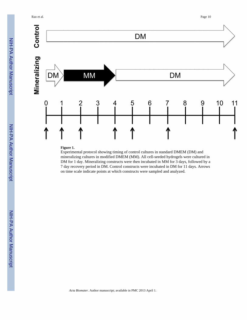
Figure 1.Experimental protocol showing timing of control cultures in standard DMEM (DM) andmineralizing cultures in modified DMEM (MM). All cell-seeded hydrogels were cultured inDM for 1 day. Mineralizing constructs were then incubated in MM for 3 days, followed by a7 day recovery period in DM. Control constructs were incubated in DM for 11 days. Arrowson time scale indicate points at which constructs were sampled and analyzed.
Rao et al. Page 10
Acta Biomater. Author manuscript; available in PMC 2013 April 1.
NIH
-PA Author Manuscript
NIH
-PA Author Manuscript
NIH
-PA Author Manuscript

Figure 2.Calcium deposition in collagen-chitosan gels through incubation in mineralizing DMEM(mDMEM). (*) denotes p < 0.05 from the groups containing FBS. Values are mean +/−standard error, n = 4.
Rao et al. Page 11
Acta Biomater. Author manuscript; available in PMC 2013 April 1.
NIH
-PA Author Manuscript
NIH
-PA Author Manuscript
NIH
-PA Author Manuscript
Rao et al. Page 12
Acta Biomater. Author manuscript; available in PMC 2013 April 1.
NIH
-PA Author Manuscript
NIH
-PA Author Manuscript
NIH
-PA Author Manuscript

Figure 3.A) Quantification of calcium deposition in different regions of collagen-chitosan constructsusing histological staining for calcium (n=4 at each location). Inset shows control andmineralized collagen-constructs (top row, scale bar = 1 mm) and representative histologysections (bottom row, scale bar = 500 µm). B) Storage (G’) and loss (G”) moduli for controland mineralized collagen-chitosan constructs after three days of culture (n=4, * denotes p <0.05 from the control DM group).
Rao et al. Page 13
Acta Biomater. Author manuscript; available in PMC 2013 April 1.
NIH
-PA Author Manuscript
NIH
-PA Author Manuscript
NIH
-PA Author Manuscript

Figure 4.Confocal micrographs of fibroblast-seeded collagen-chitosan gels cultured in controlmedium (A, C, E, G, I, K) or under mineralizing conditions (B, D, F, H, J, L). Cells arestained so that the cytoplasm of living cells is green and the nuclei of dead cells are red.Arrows at sides show culture protocol. Scale bar = 200 µm.
Rao et al. Page 14
Acta Biomater. Author manuscript; available in PMC 2013 April 1.
NIH
-PA Author Manuscript
NIH
-PA Author Manuscript
NIH
-PA Author Manuscript

Figure 5.Quantification of A) cell viability (n=4), B) DNA content (n=4), and C) calcium content(n=4) of collagen-chitosan constructs cultured in control and mineralizing medium. (#denotes statistical significance from day 0 controls; * denotes statistical significance fromDM controls; ^ denotes statistical significance from day 7)
Rao et al. Page 15
Acta Biomater. Author manuscript; available in PMC 2013 April 1.
NIH
-PA Author Manuscript
NIH
-PA Author Manuscript
NIH
-PA Author Manuscript

NIH
-PA Author Manuscript
NIH
-PA Author Manuscript
NIH
-PA Author Manuscript
Rao et al. Page 16
Tabl
e 1
Med
ia F
orm
ulat
ion
Ion
Con
cent
ratio
ns (m
M)
Na+
K+
Ca2+
Mg2+
HC
O3−
SO42−
HPO
32−pH
Blo
od P
lasm
a14
23.
6–5.
52.
1–2.
61.
027
1.0
0.65
–1.4
57.
2–7.
4
Sim
ulat
ed B
ody
Flui
d (S
BF)
141
4.0
2.5
1.0
4.2
0.5
1.0
7.4
Dul
becc
o’s m
odifi
ed E
agle
’s M
ediu
m (D
ME
M)
110
5.3
1.8
020
0.8
0.9
7.4
Mod
ified
DM
EM
(mD
ME
M)
141
5.3
101.
08.
40.
84.
07.
4
Acta Biomater. Author manuscript; available in PMC 2013 April 1.
Copyright © 2022 FDOKUMEN