Characterisation of Necator americanus excretory/secretory ...
Upload
independentCategory
view
4download
0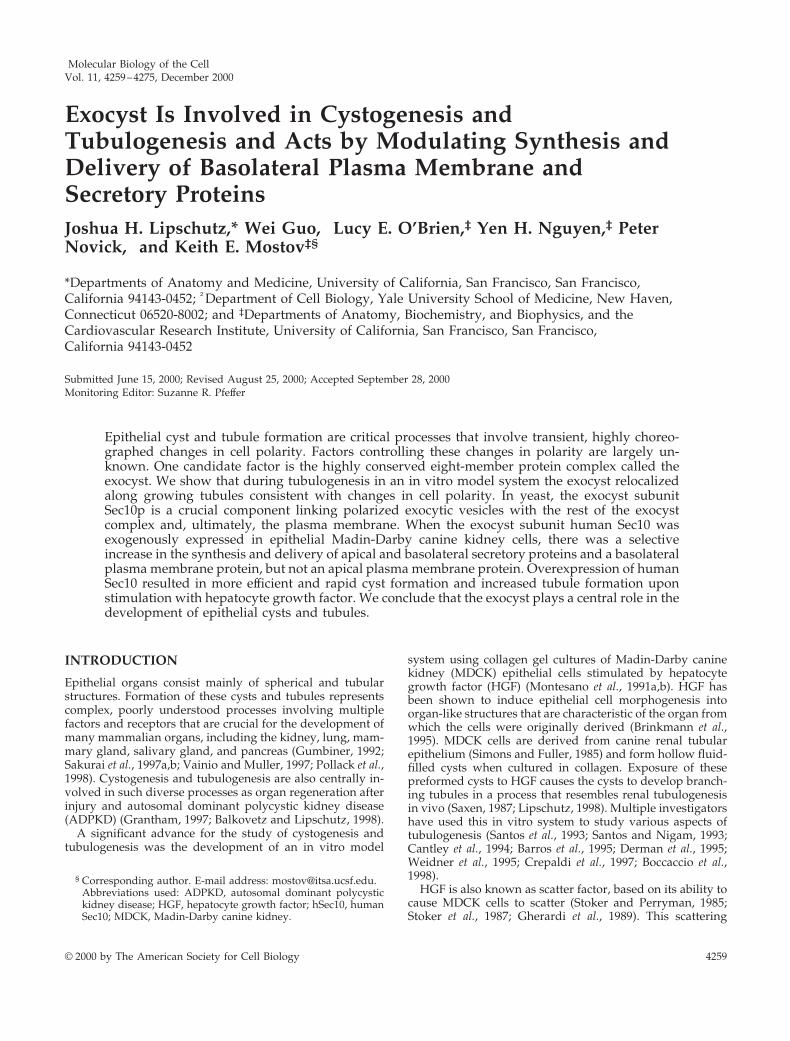
Molecular Biology of the CellVol. 11, 4259–4275, December 2000
Exocyst Is Involved in Cystogenesis andTubulogenesis and Acts by Modulating Synthesis andDelivery of Basolateral Plasma Membrane andSecretory ProteinsJoshua H. Lipschutz,* Wei Guo,† Lucy E. O’Brien,‡ Yen H. Nguyen,‡ PeterNovick,† and Keith E. Mostov‡§
*Departments of Anatomy and Medicine, University of California, San Francisco, San Francisco,California 94143-0452; †Department of Cell Biology, Yale University School of Medicine, New Haven,Connecticut 06520-8002; and ‡Departments of Anatomy, Biochemistry, and Biophysics, and theCardiovascular Research Institute, University of California, San Francisco, San Francisco,California 94143-0452
Submitted June 15, 2000; Revised August 25, 2000; Accepted September 28, 2000Monitoring Editor: Suzanne R. Pfeffer
Epithelial cyst and tubule formation are critical processes that involve transient, highly choreo-graphed changes in cell polarity. Factors controlling these changes in polarity are largely un-known. One candidate factor is the highly conserved eight-member protein complex called theexocyst. We show that during tubulogenesis in an in vitro model system the exocyst relocalizedalong growing tubules consistent with changes in cell polarity. In yeast, the exocyst subunitSec10p is a crucial component linking polarized exocytic vesicles with the rest of the exocystcomplex and, ultimately, the plasma membrane. When the exocyst subunit human Sec10 wasexogenously expressed in epithelial Madin-Darby canine kidney cells, there was a selectiveincrease in the synthesis and delivery of apical and basolateral secretory proteins and a basolateralplasma membrane protein, but not an apical plasma membrane protein. Overexpression of humanSec10 resulted in more efficient and rapid cyst formation and increased tubule formation uponstimulation with hepatocyte growth factor. We conclude that the exocyst plays a central role in thedevelopment of epithelial cysts and tubules.
INTRODUCTION
Epithelial organs consist mainly of spherical and tubularstructures. Formation of these cysts and tubules representscomplex, poorly understood processes involving multiplefactors and receptors that are crucial for the development ofmany mammalian organs, including the kidney, lung, mam-mary gland, salivary gland, and pancreas (Gumbiner, 1992;Sakurai et al., 1997a,b; Vainio and Muller, 1997; Pollack et al.,1998). Cystogenesis and tubulogenesis are also centrally in-volved in such diverse processes as organ regeneration afterinjury and autosomal dominant polycystic kidney disease(ADPKD) (Grantham, 1997; Balkovetz and Lipschutz, 1998).
A significant advance for the study of cystogenesis andtubulogenesis was the development of an in vitro model
system using collagen gel cultures of Madin-Darby caninekidney (MDCK) epithelial cells stimulated by hepatocytegrowth factor (HGF) (Montesano et al., 1991a,b). HGF hasbeen shown to induce epithelial cell morphogenesis intoorgan-like structures that are characteristic of the organ fromwhich the cells were originally derived (Brinkmann et al.,1995). MDCK cells are derived from canine renal tubularepithelium (Simons and Fuller, 1985) and form hollow fluid-filled cysts when cultured in collagen. Exposure of thesepreformed cysts to HGF causes the cysts to develop branch-ing tubules in a process that resembles renal tubulogenesisin vivo (Saxen, 1987; Lipschutz, 1998). Multiple investigatorshave used this in vitro system to study various aspects oftubulogenesis (Santos et al., 1993; Santos and Nigam, 1993;Cantley et al., 1994; Barros et al., 1995; Derman et al., 1995;Weidner et al., 1995; Crepaldi et al., 1997; Boccaccio et al.,1998).
HGF is also known as scatter factor, based on its ability tocause MDCK cells to scatter (Stoker and Perryman, 1985;Stoker et al., 1987; Gherardi et al., 1989). This scattering
§ Corresponding author. E-mail address: [email protected] used: ADPKD, autosomal dominant polycystickidney disease; HGF, hepatocyte growth factor; hSec10, humanSec10; MDCK, Madin-Darby canine kidney.
© 2000 by The American Society for Cell Biology 4259
activity originally led to the proposal of a two-step dissoci-ation/reassociation model of tubulogenesis in which MDCKcells detach from the cyst, lose polarity, and migrate out assingle cells into the collagen matrix. The migrating singlecells were then proposed to coalesce and reassemble intomulticellular structures that form tubules of polarized cells(Thiery and Boyer, 1992). However, we recently showed thatthis model was incorrect and that cells in the cyst, whenexposed to HGF, initially sent out long extensions of theirbasolateral surface. Cells then migrated out to form shortchains of cells that lacked apico-basolateral polarity andwere surrounded by basolateral surface (Pollack et al., 1998).During this process, cell–cell contacts were differentiallyregulated. E-cadherin was randomly distributed around thecell surface, desmoplakins I/II accumulated intracellularly,and the tight junction protein ZO-1 remained localized atsites of cell–cell contact. Further migration and division ledto cords of cells, with nascent lumina appearing between thecells as they began to repolarize, eventually forming maturetubules (Pollack et al., 1998). Our results indicated that tran-sient loss and restoration of cell polarity was a crucial com-ponent of tubulogenesis. Factors regulating this physiolog-ical modulation of polarity during tubule formation are notknown. One candidate factor is the eight-subunit exocystcomplex, six of whose member proteins were discoveredthrough studies of polarized secretion in yeast (Novick et al.,1980).
The budding yeast Saccharomyces cerevisiae represents asimple model organism in which to study polarized secre-tion and exocytosis. Exocytosis in yeast occurs at distinctplasma membrane subdomains, which vary with the cellcycle. Unbudded cells polarize secretion to the new bud sitebefore bud emergence and continue to direct secretion to thetip of the emerging bud (Tkacz and Lampen, 1972; Farkas etal., 1974; Field and Schekman, 1980). When the daughter cellis about two-thirds the size of the mother cell, secretionbecomes isotropic over the surface of the daughter cell,whereas very little growth or secretion occurs in the mothercell. After nuclear division, the secretory pathway orients tothe mother-bud neck, resulting in cytokinesis and septation(Byers, 1981). The exocyst is a determinant of polarizedsecretion throughout the yeast life cycle and Sec3p, a subunitof the exocyst, likely represents the spatial landmark thatdefines the sites of exocytosis (Novick et al., 1980; Finger etal., 1998). The exocyst is a 750-kDa complex comprised ofSec3p, Sec5p, Sec6p, Sec8p, Sec10p, Sec15p, Exo70p, andExo84p (Terbush et al., 1996; Guo et al., 1999a). In yeast,Sec10p and Sec15p exist as a subcomplex that acts as abridge between the rab GTPase Sec4p on the vesicle and therest of the exocyst and, ultimately, the plasma membrane(Guo et al., 1999b). Furthermore, in yeast overexpression ofthe carboxy terminal domain of Sec10p resulted in an en-larged elongated bud, whereas overexpression of the aminoterminal two-thirds of Sec10p inhibited exocytosis (Roth etal., 1998). Overexpression of full-length sec10 in yeast had nodiscernible effect, though that may be due to the fact thatsec10 overexpression with the GAL promoter did not reacha phenotypic threshold level (Guo and Novick, unpublishedobservations).
In yeast, the exocyst acts by targeting secretory vesicles tothe sites of exocytosis. In mammalian epithelial cells thetight junction acts as a physical barrier between the apical
and basolateral plasma membranes (Yeaman et al., 1999). Itwas hypothesized as early as 1980 that polarized vesiclescontaining proteins destined for the plasma membranes arefirst targeted to the area around the tight junction (Louvard,1980). In fully polarized epithelial cells, the mammalianexocyst (also called the Sec6/8 complex) localized to thetight junction, suggesting a role in membrane traffic (Grind-staff et al., 1998). Highly conserved mammalian homologuesof all eight yeast proteins have been identified (Hsu et al.,1998). In isolated nonpolarized MDCK cells the exocyst waslargely intracellular, in early contact cells the exocyst local-ized to the area of cell–cell contact, and, as polarity becameestablished, the exocyst relocalized to the tight junction. Instreptolysin-O-permeabilized MDCK cells, antibodies toSec8p inhibited delivery of a basolateral plasma membraneprotein, the exogenously expressed low-density lipoproteinreceptor, from the trans-Golgi network to the basolateralsurface but did not affect the delivery of an exogenouslyexpressed apical plasma membrane protein, p75NTR, dem-onstrating a role for the mammalian exocyst in polarizedmembrane traffic (Grindstaff et al., 1998). Here we haveinvestigated the role of the exocyst in epithelial cyst andtubule formation.
MATERIALS AND METHODS
Cystogenesis and TubulogenesisMDCK type II cells were maintained in minimum essential medium(MEM) supplemented with 5% fetal calf serum. For growth of cellsin collagen gels, MDCK cells were trypsinized and triturated into asingle-cell suspension of 2 3 104 cells/ml in a type I collagensolution as described previously (Pollack et al., 1998). Cells in sus-pension were plated onto 10-mm filters (0.02–0.2-mm pore size;Nunc, Naperville, IL) and the collagen was allowed to gel beforeaddition of medium. Medium was changed every 4 d. After 10 d,conditioned medium containing HGF from MRC5 human lungfibroblasts (ATCC CCL171) was added to the cultures.
Immunofluorescence, Confocal, and ElectronMicroscopy (EM)Cells in collagen gel were rinsed in phosphate-buffered saline andfixed for 30 min with 4% paraformaldehyde after digesting in col-lagenase (100 U/ml; Sigma, St. Louis, MO) for 10 min at 37°C aspreviously described (Pollack et al., 1998). Nonspecific binding siteswere blocked and the cells permeabilized by using 0.7% fish skingelatin and 0.025% saponin. Samples were placed in medium con-taining primary antibody at the following concentrations: Sec6 1:100(StressGen, Victoria, Canada), rSec8 1:100 (StressGen), Alexa 488and 594 phalloidin 1:50 (Molecular Probes, Eugene, OR), humanSec10 (hSec10) (1:100) (Guo et al., 1997). After extensive washing, thesamples were incubated in blocking buffer containing Alexa 488 or594-conjugated secondary antibody, 1:200 dilution (MolecularProbes). Cells were postfixed with 4% paraformaldehyde andmounted. Confocal images were collected by using a krypton-argonlaser (Bio-Rad 1024). For EM, filter-grown cells were fixed in asolution containing 2% glutaraldehyde, 0.8% paraformaldehyde,and 0.1 M cacodylate. The cells were stained with osmium andimidazole as previously described (Thiery et al., 1995), dehydrated,embedded in resin, sectioned, and imaged (Zeiss 10CA). For endo-cytic labeling of vesicles, filter-grown cells were exposed to apicaland basolateral media containing 1 mg/ml horseradish peroxidase(Sigma) for 10 min. Cells were then lightly fixed (in a solutioncontaining 1.5% glutaraldehyde, 0.1 M cacodylate, and 1% sucrose),washed with buffer (0.1 M cacodylate, 3% sucrose), incubated with
J.H. Lipschutz et al.
Molecular Biology of the Cell4260
peroxidase substrate (10 ml of 0.05 M Tris, pH 7.6, 5 mg of diaminobenzidine, 5% sucrose) followed by 1% H2O2, dehydrated, embed-ded in resin, and sectioned as described above.
TransfectionhSec10 was subcloned and placed into PCDNA3 (Invitrogen, SanDiego, CA) after addition of a C-terminal myc epitope tag. Theplasmid was transfected into MDCK type II cells by using thecalcium-phosphate precipitation method (Sambrook et al., 1989;Breitfeld et al., 1989). Clones were selected by resistance to G418 andwere kept under selection for subsequent experiments. The control(empty plasmid transfected) cells were a pooled line (i.e. nonclonal)to avoid clonal variation. Similar results were obtained with indi-vidual clonal control lines (Lipschutz and Mostov, unpublishedobservations). Clones expressing hSec10p-myc were identified byWestern blot with 9E10 anti-myc antibody (Santa Cruz Biotechnol-ogy, Santa Cruz, CA) at 1:1000 dilution, goat anti-mouse HRP(Jackson ImmunoResearch, West Grove, PA) as the secondary anti-body, and enhanced chemiluminescence (ECL) (NEN, Boston, MA).Equal amounts of total protein were assayed as determined by thebicinchoninic protein assay (Pierce, Rockford, IL), Ponceau Redstaining, and Western blotting with antibody (1:1000) against cyto-solic proteins p42 and p44 MAP Kinase (New England Biolabs,Beverly, MA).
Western Blot and ImmunoprecipitationCells were lysed in 0.5% SDS lysis buffer (0.5% SDS, 100 mM NaCl,50 mM tetraethylammonium-Cl, pH 8.1, 5 mM EDTA, 0.2% Tra-sylol, and 0.02% NaN3) and prepared in standard manner (Breitfeldet al., 1989). For immunoprecipitation, 4 ml of protein A-sepharosebeads coupled to 8 mg of anti-rSec8 antibody (StressGen) or 12 mg of9E10 antibody (anti-myc) conjugated to agarose (Santa Cruz Bio-technology) were incubated overnight with the cell lysate (afteraddition of equal volumes of 2.5% Triton X-100 in 100 mM tetra-ethylammonium buffer, pH 8). The immunoprecipitated proteinswere eluted and separated by SDS-PAGE and the proteins trans-ferred onto nitrocellulose. The protein bands were detected byincubations with the appropriate primary antibody, followed bygoat anti-mouse HRP (Jackson ImmunoResearch) as the secondaryantibody, and ECL (NEN). Quantification of bands was performedon scanned images by using the IPlab Gel program (Signal Analyt-ics).
Quantitation of Association of Sec8p and hSec10pby CoimmunoprecipitationhSec10 overexpressing cells grown to confluency on a 10-cm dishwere lysed in 1 ml of NP-40 lysis buffer (Calbiochem, La Jolla, CA)(1% NP-40, 125 mM NaCl, and 20 mM HEPES, pH 7.4). Foursequential rounds of immunoprecipitation were performed usingantibody against Sec8p (14G1; Stressgen). Anti-Sec8 (8 mg) antibodyand 4 ml of protein A-sepharose beads were added. After incubationat 4°C for 4–12 h the beads were pelleted by low-speed centrifuga-tion and the supernatant was transferred to a new tube. The beadswere washed five times in NP-40 lysis buffer. The next round ofimmunoprecipitation was performed on the supernatant, by addingthe same amount of anti-Sec8 antibody and beads, and repeating theincubation. After the fourth round of immunoprecipitation withantibody to Sec8p, a fifth and final round was performed with 9E10antibody to the myc epitope tag. The 9E10 immunoprecipitationwas performed by adding 12 mg of 9E10 antibody (anti-myc) con-jugated to agarose (Santa Cruz Biotechnology) and incubating for12 h at 4°C. The washed beads from all five immunoprecipitateswere analyzed by SDS-PAGE, blotted, and the blot probed withboth antibody to Sec8p and antibody to myc and visualized withgoat anti-mouse HRP (as the secondary antibody) and ECL. Inten-sities of all bands were quantitated by using the IPlab Gel program
(Signal Analytics, Vienna, VA). The total amount of Sec8p immu-noprecipitated in all five immunoprecipitates was taken as 100%,and of this 81% was found in the sum of rounds 1 to 4. Therefore,the first four rounds were sufficient to immunoprecipitate the vastmajority of Sec8p. We then asked what percentage of the hSec10pwas associated with the Sec8p immunoprecipitated during the firstfour rounds. The total hSec10p was taken as the sum of the hSec10bands in all five rounds of immunoprecipitation. The fraction ofhSec10p that associates with Sec8p was taken as the sum of hSec10pthat coimmunoprecipitated with the Sec8p in the first four rounds ofimmunoprecipitation with anti-Sec8p antibody, divided by the totalhSec10p. Note that a minority (35%) of hSec10p coimmunoprecipi-tated with the Sec8p in the first four rounds, and most hSec10p(65%) only immunoprecipitated with the antibody to its myc tag inthe fifth round.
To perform the reciprocal experiment, that is, to determine whatfraction of Sec8p could be coimmunoprecipitated with hSec10p, wefirst immunoprecipitated a duplicate cell lysate with four rounds ofantibody to the myc tag on the hSec10. A fifth and final round wasthen performed by using antibody to Sec8p (all immunoprecipita-tion conditions were as described above except 12 mg of anti-Sec8antibody, an amount sufficient to immunoprecipitate all the endog-enous canine Sec8 in a 10-cm plate of confluent cells, was used). Thefirst four rounds were sufficient to immunoprecipitate 86% of thehSec10p. We then determined the fraction of Sec8p that was immu-noprecipitated with the hSec10p. The total Sec8p was taken as thesum of all five rounds. The fraction of Sec8p that associates withhSec10p was taken as the sum of Sec8p in rounds 1 to 4, divided bythe total Sec8p. Note unlike the experiment described above, themajority of endogenous canine Sec8p (65%) coimmunoprecipitatedwith the hSec10p in the first four rounds, and only 35% was immu-noprecipitated with the antibody to Sec8p in the fifth round.
Synthesis, Secretion, and Surface Delivery AssaysCells from a confluent 10-cm plastic dish were trypsinized and 5%of the cells were seeded per 12-mm (0.4-mm pore size) filter (Costar,Cambridge, MA). The cells were grown for 5–6 d with fresh me-dium added daily. The cells were washed with phosphate-bufferedsaline and starved for 15 min in MEM lacking methionine. Cellswere then labeled by exposing the basolateral surface to a 25-mldrop of starvation medium containing 4 ml of [35S]methionine (31.4mCi/ul; NEN) for 20 min. For synthesis assays, after pulsing with[35S]methionine for 20 min, cells were lysed in 0.5% SDS, equalvolumes of 2.5% Triton X-100 were added, and immunoprecipita-tion was performed by using antibody against gp80 (a kind giftfrom D. Sabatini, New York University, NY), E-Cadherin (a kind giftfrom W.J. Nelson, Stanford, CA), or gp135 (a generous gift from G.Ojakian, State University of New York, NY).
For secretion assays, the cells were labeled as described above,washed extensively, and MEM was added (0.3 ml apically and 0.5ml basolaterally) and collected at the time points noted. The filterswere grown in triplicate for each experiment, and each experimentwas repeated at least three times.
Surface delivery assays of E-Cadherin and gp135 were performedas described above except cells were pulsed with [35S]methioninefor 20 min, washed extensively, allowed to chase for 60 min, andSulfo-NHS-Biotin (EZ-Link; Pierce) was added at 0.5 mg/ml. Im-munoprecipitation with antibody against E-Cadherin and gp135was performed. The antibody–beads–antigen complex was thenboiled and the supernatant was reprecipitated with streptavidinbeads and the remaining proteins run on an SDS-PAGE gel andanalyzed with a phosphorimager (Molecular Dynamics). Modifiedfrom Le Bivic et al. (1990).
Transcytosis AssayCells expressed transfected rabbit polymeric immunoglobulin re-ceptor. Briefly, cells were cultured as described above and exposed
Exocyst in Cyst and Tubule Formation
Vol. 11, December 2000 4261

at the basolateral surface to a 25-ml drop of medium containing 4 mlof 125I-IgA. The 125I-IgA was allowed to internalize for 10 min andfresh medium was added apically and basolaterally. The mediumwas collected at various time points and the fractions were quanti-fied in a gamma-counter as previously described (Luton et al., 1998).The filters were grown in triplicate for each assay, and each assaywas repeated three times.
Glycerol GradientContact-naive control and MDCK cells overexpressing hSec10 werehomogenized in a sucrose buffer [20 mM HEPES-KOH, pH 8, 90mM KOAc, 2 mM Mg(OAc)2, and 250 mM sucrose] by repeatedpassage through a 27-gauge needle. The postnuclear supernatantwas centrifuged at 15,000 3 g for 10 min. The resulting supernatantwas fractionated in a linear 22.5–36% (wt/wt) glycerol gradient bycentrifugation at 80,000 3 g for 16 h as previously described (Tinget al., 1995; Grindstaff et al., 1998). Fractions (110 ml) from 1.2 ml oftotal gradient volume were collected. Proteins in each fraction wereseparated by SDS-PAGE and transferred to nitrocellulose mem-branes for immunoblotting with antibody specific for Sec8p andmyc. In parallel, glycerol gradients were centrifuged containingglobular protein standards with known sedimentation coefficients:bovine serum albumin (4.3S), b-amylase (11.2S), and thyroglobulin(19.2S).
StatisticsThe sizes of cysts, the number of tubules per cyst, the synthesis ofgp80, and the synthesis and surface delivery of E-Cadherin andgp135 were summarized by medians and the statistical significanceassessed by the Mann-Whitney nonparametric test.
RESULTS
Exocyst Localizes to the Tight Junction andRelocalizes during TubulogenesisWe have previously shown that during HGF-induced for-mation of tubules from MDCK cells grown as cysts, the cellsgo through a dramatic sequence of changes in polarity andshape (Pollack et al., 1998). To investigate the possible role ofthe exocyst in these changes, we began by localizing theexocyst during this process. In MDCK cells grown as amonolayer on a filter support, the exocyst localized to theregion of the tight junction (Grindstaff et al., 1998). We firstlocalized the exocyst in non-HGF–treated cysts, where theMDCK cells formed a polarized monolayer surrounding alumen. Although the polarity of cells in a collagen gel cyst isgenerally thought to closely resemble that of filter-grown
cells, proteins such as galectin-3 have been shown to besecreted apically in monolayers (Lindstedt et al., 1993; Sato etal., 1993) but basolaterally in cysts (Bao and Hughes, 1995,1999). Compared with growth on a porous plastic filter,growth in an extracellular matrix gel is more likely to re-semble the environment experienced by cells in vivo. Giventhe differences between filter-grown and cyst-grown cells, itwas important to first examine the localization of the exocystin cells grown as cysts.
We found that the exocyst localized to the tight junction inpolarized MDCK cell cysts and colocalized with the tightjunction marker ZO-1 (Figures 1 and 2, a–c). In addition tothe tight junction staining, there was also diffuse cytoplas-mic staining. This could represent intracellular exocyst, be-cause even in fully polarized MDCK cells, previous investi-gators have shown that only ;70% of Sec6p and Sec8pmigrated near the top of an Opti-Prep gradient with theplasma membrane protein E-cadherin, suggesting 30% re-mained cytosolic. This can be contrasted to nonpolarizedMDCK cells in which .90% of Sec6p and Sec8p migrated toa position near the bottom of the gradient and was presum-ably cytosolic (Grindstaff et al., 1998). We cannot, however,rule out the possibility that at least some of the cytoplasmicstaining represented nonspecific background staining. Itshould be noted that diffuse background staining is oftensomewhat higher in a collagen gel system, due to the diffi-culties in thoroughly blocking and washing through thethick collagen gel, though this problem varies with the an-tibody used (e.g., the antibody to ZO-1 is clean even in cysts)(Pollack et al., 1997, 1998).
During tubulogenesis cells undergo a series of steps ofphysiologic remodeling of cell polarity and shape. We in-vestigated whether during tubulogenesis, changes in exo-cyst localization, corresponding to changes in cell polarity,would be seen. We have previously shown that in the firststage of HGF-induced tubule formation, the extension stage,basolateral extensions of individual cells protrude into thesurrounding collagen matrix whereas the cells remain at-tached to the cyst. Cell polarity and cell junctions, however,are maintained (Pollack et al., 1998). In this stage a substan-tial amount of the exocyst was found in the basolateralextension, and often seemed to be concentrated at the base ofthe extension. This may be analogous to cytoplasmic exocystin contact-naive cells (Grindstaff et al., 1998). However, someof the exocyst staining remained at the region of the tightjunction (Figure 2, d–f).
Figure 1. Sec8p partially localizesat the tight junction in cysts. Thisfigure is a confocal section throughthe middle of a mature, fully polar-ized cyst formed by MDCK cellsgrown for 10 d in a collagen matrix.Antibody against Sec8p (a) and an-tibody against the tight junctionprotein ZO-1 (b) show colocaliza-tion as demonstrated by the yellowcolor in the merged panel (c). Sec 8,green; ZO-1, red. Bar, 10 mm.
J.H. Lipschutz et al.
Molecular Biology of the Cell4262

During a later stage, cells form cords that are groups ofcells, two or three cells thick, that project away from, whileretaining contact with, the cyst. These cords do not containvisible lumens, though nascent lumens may be forming atregions of cell–cell contact. We costained all of our samplesfor polymerized actin by using Alexa 594 phalloidin. Corti-cal actin underlies the entire plasma membrane; however,actin staining is particularly strong underneath apical sur-faces and at regions of cell–cell contact that are likely to form
future apical surfaces during the later stages of tubulogen-esis. We have found the pattern of actin staining to beparticularly helpful for visualizing the reformation of polar-ized cell structures during tubulogenesis (Pollack et al.,1998). In the cord stage, the exocyst was expressed betweenareas of cell–cell contact, as shown by the arrow (Figure 2g)and the corresponding actin staining (Figure 2h). This seemsanalogous to a previous study showing that the exocystwent from a largely cytoplasmic state in contact-naive
Figure 2. Sec8p relocalizes during tubulogenesis. (a and b) Fluid-filled cyst formed by MDCK cells grown for 10 d in a collagen gel. Stainingis seen at the area of the tight junction (arrowhead) by using anti-Sec8 antibody (a). Sec8p colocalized with the tight junction protein ZO-1(Figure 1). Concurrent staining of actin with phalloidin was performed (b). (c) Merge of a and b. (d–l) Fluid-filled cysts formed by MDCKcells grown for 10 d in collagen and stimulated for 24 h with conditioned medium containing HGF. The exocyst can be seen relocalizing alongthe growing tubules in a pattern consistent with the changes in polarity that occur as tubules form. (d–f) Extension stage of tubulogenesis.Sec8p is seen relocalizing into the extension (d), in association with actin staining (e). (g–i) Cord stage of tubulogenesis. Arrow in g indicatesstaining at the region of cell–cell contact in the cord. This region may become the boundary of a new lumen, as shown by the intense actinstaining in this region (h). (j–l) Nascent tubule in the final stage of tubulogenesis. Arrow in j shows two vertical lines of Sec8p stainingoutlining the boundary of the lumen. Intense actin staining in a broad band in k underlies the apical surface surrounding this lumen. (f, i,and l) Merge of d and e, g and h, and j and k shows that the relocalizing exocyst closely surrounds, but does not precisely overlap the actinin the projections of the nascent tubules as indicated by lack of yellow in the merged panels. Sec 8, green; actin, red. Bar, 10 mm.
Exocyst in Cyst and Tubule Formation
Vol. 11, December 2000 4263

MDCK cells to localization at the area of cell–cell contact aspolarity was initiated in early contact MDCK cells grown intwo-dimensional cultures on filters (Grindstaff et al., 1998).
During the final stage of tubulogenesis small lumens be-gin to appear along the length of the developing tubule.Tubule maturation then occurs with the individual lumenscoalescing, enlarging, and becoming continuous with thelumen of the cyst. Apical and basolateral membranes of cellsof the tubule become clearly polarized and the arrangementof cell junctions that normally is found in polarized epithe-lial cells is restored (Pollack et al., 1998). The exocyst can beseen relocalizing during this final stage of tubulogenesis(Figure 2, j and k). A nascent lumen between cells, sectionedlongitudinally, is indicated by the elongated band of strongactin staining, which is characteristically subjacent to theapical surface (Figure 2k). Two thin lines of Sec8 staining(arrow in Figure 2j) bracket this actin staining. The relocal-ization of the exocyst during tubulogenesis is highly sugges-tive of the redirection of delivery of new membrane andsecretory products to the growing extensions and tubulesduring the physiologic remodeling of cell shape and polaritythat occurs throughout the tubulogenic process. This relo-calization is strikingly similar to the way in which the exo-cyst is involved in redirecting exocytic vesicles to differentregions of the plasma membrane during the yeast cell cycle.
The exocyst surrounded, but did not overlap the actinextending into the tubular processes. Note the green lines ofSec8 staining (arrows in Figure 2, g and j) surrounding thethicker red actin staining (Figure 2, i and l). Studies from
yeast demonstrate a strong connection between the exocystand the actin cytoskeleton (Ayscough et al., 1997; Finger andNovick, 1998). Although there are no reports to date that theexocyst and actin cytoskeleton interact in mammalian cells,there is increasing evidence that the actin and microtubulecytoskeletons cooperate in membrane traffic (Goode et al.,2000). It is therefore plausible that the interactions of theexocyst with the actin cytoskeleton are important in direc-tional vesicular traffic in MDCK cells.
A Portion of Overexpressed hSec10-myc Associateswith the Rest of the Exocyst ComplexOnly the localizations of Sec6p and Sec8p have previouslybeen reported in MDCK cells (Grindstaff et al., 1998). Weexamined the localization of endogenous canine Sec10p inMDCK cells. A significant portion of endogenous canineSec10p (Figure 3) colocalized with ZO-1 at the tight junction.However, staining with the antibody to Sec10p had abroader distribution than staining for ZO-1, which washighly localized to the tight junction (Figure 3, TJ row). SomeSec10p staining was also intracellular, mainly in a diffusecytosolic pattern (though there may be some unidentifiedpuncta). We did not see any of this staining with nonim-mune antibodies (Lipschutz and Mostov, unpublished ob-servations). However, because we did not have control cellslacking Sec10p, we could not rule out the possibility thatsome of this staining was nonspecific background.
Figure 3. Sec10p partially localizes atthe tight junction. Antibody againstSec10p and antibody against the tightjunction marker ZO-1 demonstrated co-localization (yellow in merged panels) atthe tight junction in MDCK cell monolay-ers grown for 7 d on filters. Ap, apical; TJ,tight junction; Bl, basolateral. Sec 10,green; ZO-1, red. Bar, 10 mm.
J.H. Lipschutz et al.
Molecular Biology of the Cell4264

To better understand the role and localization of the exo-cyst in MDCK cells, we used the approach of overexpressingone subunit. We chose Sec10 because the Sec10 subunit hasbeen shown to be part of a separate subcomplex consistingof Sec10p and Sec15p, and perturbation of Sec10 function inyeast has specific effects on polarized vesicular delivery(Roth et al., 1998; Guo et al., 1999b). We recently clonedhSec10 (Guo et al., 1997). Full-length hSec10 was transfectedinto MDCK cells and clones expressing hSec10 were se-lected. Three clones, expressing differing levels of hSec10,were chosen for further study (Figure 4a). In the followingexperiments all three clones showed similar phenotypes,and, in all the cases where quantitative data are presented,the differences from control cells transfected with vectoralone were statistically significant. There seemed to be arough, though not absolute, correlation between the level ofhSec10 overexpression and the magnitude of the effects seen;however, given the small number of hSec10 overexpressingclones examined thoroughly, we cannot make any definitivestatements. We estimate the degree of expression of trans-fected hSec10p over endogenous canine Sec10p to be ap-proximately three- to fivefold in the clone expressing thehighest amount of hSec10 (Lipschutz and Mostov, unpub-lished observations).
To circumvent the possible background staining problemand to investigate the distribution of the transfected hSec10pwithout the fluorescence signal from endogenous canineSec10p, we stained cells for the myc epitope present on thetransfected hSec10p (Figure 4b). Immunofluoresence stain-ing with antibody against the myc epitope tag showed trans-fected hSec10p mainly at the tight junction region, whichwas identified by costaining for ZO-1 (Lipschutz andMostov, unpublished observations). Some hSec10p wasagain present intracellularly, mostly diffusely in the cytosol(Figure 4b). The level of background staining with controlcells was very low, so we are confident that this signal wasdue to the transfected hSec10p. Therefore, Figures 3 and 4taken together suggest that both endogenous Sec10p and thetransfected hSec10p have significant localization to the tightjunction region, as well as some intracellular localization.
At steady state, in polarized cells grown on filters therewas no difference in the levels of Sec6p and Sec8p, permicrogram of total protein, in hSec10 transfected cells com-pared with control cells (Figure 4c) and Sec6p and Sec8pcontinued to localize at the tight junction in hSec10 overex-pressing cells (Lipschutz and Mostov, unpublished observa-tions).
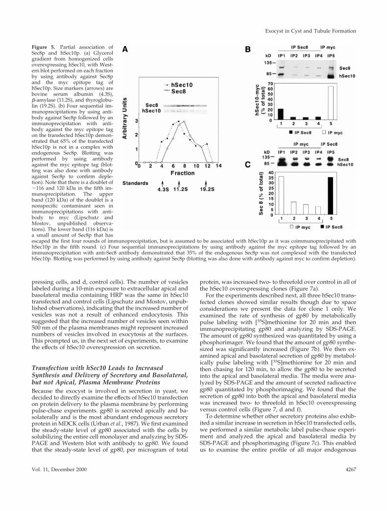
Previous investigators have shown that the eight-memberexocyst complex sedimented at ;17S. Although Sec6p sedi-mented as a reasonably symmetric peak centered around17S, Sec8p was shown to have a much broader distribution,including a wide shoulder sedimenting around ;11S, whichmay represent a subset of proteins in a partially assembledor disassembled complex (Grindstaff et al., 1998). We foundthat the sedimentation of hSec10p largely paralleled that ofendogenous Sec8p. Specifically, 72% of the overexpressedhSec10p sedimented with the same velocity as 90% of theendogenous Sec8p (Figure 5a). Of course, this result does notestablish that any or all of the hSec10p that sediments at thesame rate is truly physically associated with the Sec8p.Moreover, hSec10p had an overall distribution that wasslightly shifted to a slower sedimentation, suggesting that
some of the hSec10p may be in a smaller complex(es) orform(s) than Sec8p.
To determine how much of the hSec10p was associatedwith the endogenous Sec8p, we used a coimmunoprecipita-tion protocol, which is explained in detail in MATERIALSAND METHODS. We immunoprecipitated Sec8p by usinganti-Sec8 antibody. We found that four consecutive roundsof immunoprecipitation were sufficient to immunoprecipi-tate 81% of the Sec8p. A fifth and final round of immuno-precipitation was performed with antibody against the mycepitope tag. All of the immunoprecipitates were analyzed bySDS-PAGE and Western blot by using antibodies againstSec8p and the myc epitope tag of hSec10p. This procedureallowed us to accurately quantitate the amount of hSec10pthat coimmunoprecipitated with Sec8p. Of the total hSec10pprecipitated, 65% remained after Sec8p depletion (Figure 5b,M), suggesting the majority of transfected hSec10p was notcomplexed with the rest of the exocyst (Figure 5b). This is asexpected, since the transfected hSec10p is predicted to be inexcess over the endogenous Sec8p.
In the reciprocal experiment, we immunoprecipitatedhSec10p via its myc tag. Four rounds of immunoprecipita-tion with antibody to the myc tag on hSec10p were sufficientto immunoprecipitate 86% of the hSec10p. A fifth and finalround of immunoprecipitation was performed with anti-body to Sec8p. Of the total Sec8p precipitated, only 35%remained after hSec10p depletion (Figure 5c, f), indicatingthat the majority of endogenous canine Sec8p was com-plexed with the overexpressed hSec10p.
Taken together, these data show that ;35% of the over-expressed hSec10p associates with the Sec8p complex. Itshould be kept in mind that dissociation may occur duringthe lengthy sequential immunoprecipitations, which mayresult in an underestimation of the true degree of associa-tion. This association could be the mechanism by which theeffects of overexpressed hSec10, described below, are medi-ated; however, it is certainly possible that free hSec10p isacting independently of the complex containing Sec8p. Wewere unable to express the carboxy- or amino-terminal do-mains of hSec10 in either a stable or an inducible system inMDCK cells, most likely due to their extreme toxicity.
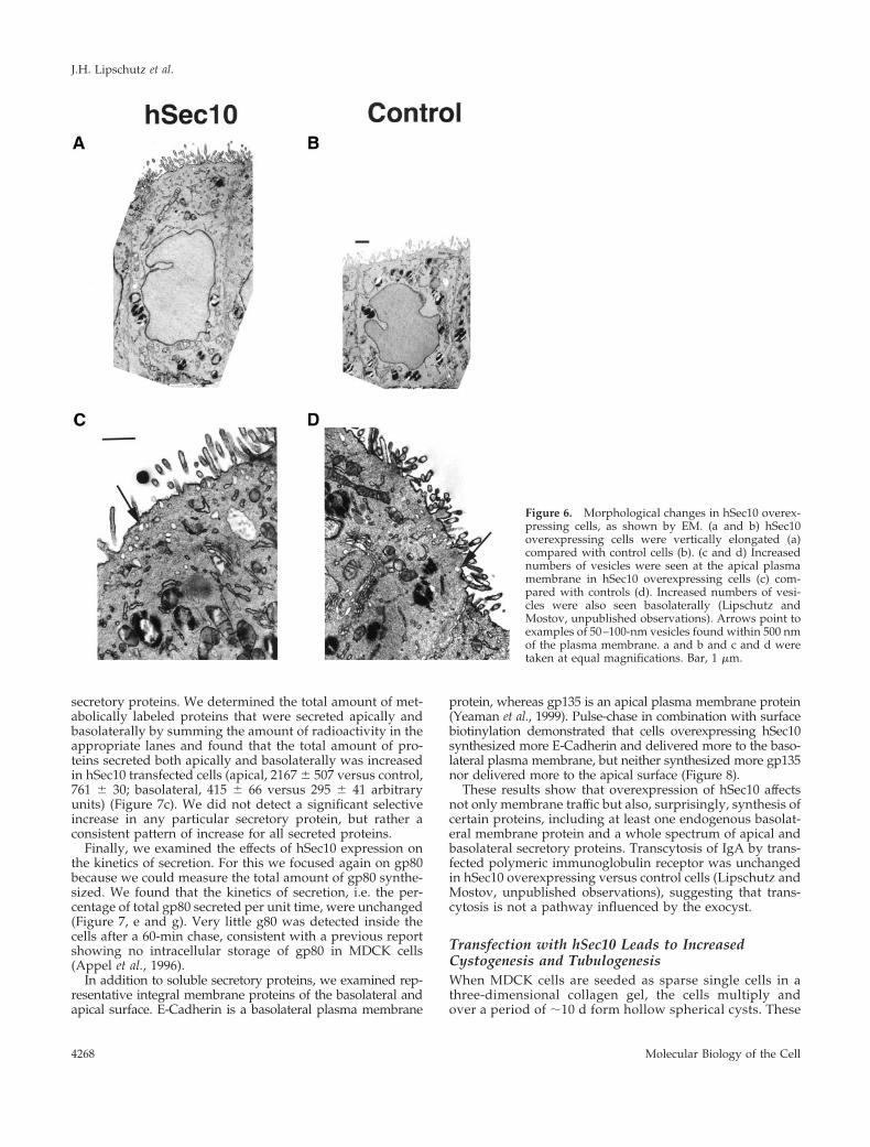
Transfection with hSec10 Leads to Changes inCell MorphologyWhen grown as a monolayer on filters, the cells overexpress-ing hSec10 were noted to be significantly taller in compari-son with control cells both by confocal (13.9 6 1.0 versus10.1 6 0.7 mm, as measured from the tight junction to thebasal surface) and EM (Figure 6a, hSec10 overexpressingcells, and b, control cells). The number of cells growing persurface area of Transwell filter substrate, however, was un-changed (Lipschutz and Mostov, unpublished observa-tions). Consistent with this, the mean diameter of hSec10overexpressing and control cells appeared similar on EM(43.9 6 9.4 mm/section versus 40.6 6 10.5 mm/section),suggesting that cells are taller but not wider.
By EM the number of 50–100-nm vesicles seen within 500nm of the plasma membranes was increased in the hSec10transfected versus control cells both apically (0.305 6 0.143versus 0.050 6 0.055 vesicles/mm of plasma membrane) andbasolaterally (0.164 6 0.067 versus 0.038 6 0.033 vesi-cles/mm of plasma membrane) (Figure 6c, hSec10 overex-
Exocyst in Cyst and Tubule Formation
Vol. 11, December 2000 4265

Figure 4. hSec10-myc partially lo-calizes at the tight junction and thelevels of Sec6p and Sec8p are un-changed in hSec10 overexpressingcells. (a) Western blot by using an-tibody against the myc epitope tagdemonstrated three hSec10 express-ing clones. C, control. (b) Immuno-fluorescent staining by using anti-body against the myc epitope tag inhSec10 overexpressing cells showstransfected hSec10p localized to theplasma membrane at the level of thetight junction (the level of the tightjunction was determined bycostaining with ZO-1; Lipschutzand Mostov, unpublished observa-tions). No myc staining is evident inthe control cells. (c) Western blotdemonstrates equal amounts ofSec6p and Sec8p protein per micro-gram of total protein in Sec10 over-expressing and control cells. Ap,apical; TJ, tight junction; Bl, basolat-eral. Bar, 10 mm.
J.H. Lipschutz et al.
Molecular Biology of the Cell4266
pressing cells, and d, control cells). The number of vesicleslabeled during a 10-min exposure to extracellular apical andbasolateral media containing HRP was the same in hSec10transfected and control cells (Lipschutz and Mostov, unpub-lished observations), indicating that the increased number ofvesicles was not a result of enhanced endocytosis. Thissuggested that the increased number of vesicles seen within500 nm of the plasma membranes might represent increasednumbers of vesicles involved in exocytosis at the surfaces.This prompted us, in the next set of experiments, to examinethe effects of hSec10 overexpression on secretion.
Transfection with hSec10 Leads to IncreasedSynthesis and Delivery of Secretory and Basolateral,but not Apical, Plasma Membrane ProteinsBecause the exocyst is involved in secretion in yeast, wedecided to directly examine the effects of hSec10 transfectionon protein delivery to the plasma membrane by performingpulse-chase experiments. gp80 is secreted apically and ba-solaterally and is the most abundant endogenous secretoryprotein in MDCK cells (Urban et al., 1987). We first examinedthe steady-state level of gp80 associated with the cells bysolubilizing the entire cell monolayer and analyzing by SDS-PAGE and Western blot with antibody to gp80. We foundthat the steady-state level of gp80, per microgram of total
protein, was increased two- to threefold over control in all ofthe hSec10 overexpressing clones (Figure 7a).
For the experiments described next, all three hSec10 trans-fected clones showed similar results though due to spaceconsiderations we present the data for clone 1 only. Weexamined the rate of synthesis of gp80 by metabolicallypulse labeling with [35S]methionine for 20 min and thenimmunoprecipitating gp80 and analyzing by SDS-PAGE.The amount of gp80 synthesized was quantitated by using aphosphorimager. We found that the amount of gp80 synthe-sized was significantly increased (Figure 7b). We then ex-amined apical and basolateral secretion of gp80 by metabol-ically pulse labeling with [35S]methionine for 20 min andthen chasing for 120 min, to allow the gp80 to be secretedinto the apical and basolateral media. The media were ana-lyzed by SDS-PAGE and the amount of secreted radioactivegp80 quantitated by phosphorimaging. We found that thesecretion of gp80 into both the apical and basolateral mediawas increased two- to threefold in hSec10 overexpressingversus control cells (Figure 7, d and f).
To determine whether other secretory proteins also exhib-ited a similar increase in secretion in hSec10 transfected cells,we performed a similar metabolic label pulse-chase experi-ment and analyzed the apical and basolateral media bySDS-PAGE and phosphorimaging (Figure 7c). This enabledus to examine the entire profile of all major endogenous
Figure 5. Partial association ofSec8p and hSec10p. (a) Glycerolgradient from homogenized cellsoverexpressing hSec10, with West-ern blot performed on each fractionby using antibody against Sec8pand the myc epitope tag ofhSec10p. Size markers (arrows) arebovine serum albumin (4.3S),b-amylase (11.2S), and thyroglobu-lin (19.2S). (b) Four sequential im-munoprecipitations by using anti-body against Sec8p followed by animmunoprecipitation with anti-body against the myc epitope tagon the transfected hSec10p demon-strated that 65% of the transfectedhSec10p is not in a complex withendogenous Sec8p. Blotting wasperformed by using antibodyagainst the myc epitope tag (blot-ting was also done with antibodyagainst Sec8p to confirm deple-tion). Note that there is a doublet of;116 and 120 kDa in the fifth im-munoprecipitation. The upperband (120 kDa) of the doublet is anonspecific contaminant seen inimmunoprecipitations with anti-body to myc (Lipschutz andMostov, unpublished observa-tions). The lower band (116 kDa) isa small amount of Sec8p that hasescaped the first four rounds of immunoprecipitation, but is assumed to be associated with hSec10p as it was coimmunoprecipitated withhSec10p in the fifth round. (c) Four sequential immunoprecipitations by using antibody against the myc epitope tag followed by animmunoprecipitation with anti-Sec8 antibody demonstrated that 35% of the endogenous Sec8p was not complexed with the transfectedhSec10p. Blotting was performed by using antibody against Sec8p (blotting was also done with antibody against myc to confirm depletion).
Exocyst in Cyst and Tubule Formation
Vol. 11, December 2000 4267
secretory proteins. We determined the total amount of met-abolically labeled proteins that were secreted apically andbasolaterally by summing the amount of radioactivity in theappropriate lanes and found that the total amount of pro-teins secreted both apically and basolaterally was increasedin hSec10 transfected cells (apical, 2167 6 507 versus control,761 6 30; basolateral, 415 6 66 versus 295 6 41 arbitraryunits) (Figure 7c). We did not detect a significant selectiveincrease in any particular secretory protein, but rather aconsistent pattern of increase for all secreted proteins.
Finally, we examined the effects of hSec10 expression onthe kinetics of secretion. For this we focused again on gp80because we could measure the total amount of gp80 synthe-sized. We found that the kinetics of secretion, i.e. the per-centage of total gp80 secreted per unit time, were unchanged(Figure 7, e and g). Very little g80 was detected inside thecells after a 60-min chase, consistent with a previous reportshowing no intracellular storage of gp80 in MDCK cells(Appel et al., 1996).
In addition to soluble secretory proteins, we examined rep-resentative integral membrane proteins of the basolateral andapical surface. E-Cadherin is a basolateral plasma membrane
protein, whereas gp135 is an apical plasma membrane protein(Yeaman et al., 1999). Pulse-chase in combination with surfacebiotinylation demonstrated that cells overexpressing hSec10synthesized more E-Cadherin and delivered more to the baso-lateral plasma membrane, but neither synthesized more gp135nor delivered more to the apical surface (Figure 8).
These results show that overexpression of hSec10 affectsnot only membrane traffic but also, surprisingly, synthesis ofcertain proteins, including at least one endogenous basolat-eral membrane protein and a whole spectrum of apical andbasolateral secretory proteins. Transcytosis of IgA by trans-fected polymeric immunoglobulin receptor was unchangedin hSec10 overexpressing versus control cells (Lipschutz andMostov, unpublished observations), suggesting that trans-cytosis is not a pathway influenced by the exocyst.
Transfection with hSec10 Leads to IncreasedCystogenesis and TubulogenesisWhen MDCK cells are seeded as sparse single cells in athree-dimensional collagen gel, the cells multiply andover a period of ;10 d form hollow spherical cysts. These
Figure 6. Morphological changes in hSec10 overex-pressing cells, as shown by EM. (a and b) hSec10overexpressing cells were vertically elongated (a)compared with control cells (b). (c and d) Increasednumbers of vesicles were seen at the apical plasmamembrane in hSec10 overexpressing cells (c) com-pared with controls (d). Increased numbers of vesi-cles were also seen basolaterally (Lipschutz andMostov, unpublished observations). Arrows point toexamples of 50–100-nm vesicles found within 500 nmof the plasma membrane. a and b and c and d weretaken at equal magnifications. Bar, 1 mm.
J.H. Lipschutz et al.
Molecular Biology of the Cell4268

cysts are polarized with the apical surface facing thefluid-filled interior. This is a model for the formation ofcysts, which are one of the basic building blocks of higherorder organizations of epithelial cells used in organogen-esis. We wished to investigate the role of the exocyst inthis process. When hSec10 overexpressing cells wereplated at very low density in collagen gels (Montesano et
al., 1991a,b) they formed cysts more efficiently and at asignificantly greater rate than did control cells (Figure 9,a– c). All three clones of hSec10 overexpressing cell cystswere larger and had a greater diameter than control cellcysts (i.e., clone 1, median diameter 90 versus 70 mm, p ,0.0001), though the cell number was unchanged (Lips-chutz and Mostov, unpublished observations). The larger
Figure 7. Synthesis and deliveryof secretory proteins in hSec10overexpressing cells. (a) Westernblot by using antibody againstgp80 demonstrates increasedsteady-state levels of gp80 in threehSec10 transfected clones (1, 2, 3)compared with control cells (C).(b–g) Synthesis and secretion as-says were performed by pulse andpulse-chase with [35S]methionine.(b) hSec10 overexpressing and con-trol cells were metabolically pulselabeled for 20 min with [35S]methi-onine and the amount of newlysynthesized gp80 was measured.(c) Other secretory proteins werealso increased in hSec10 overex-pressing versus control cells. Cellswere pulse labeled for 20 min with[35S]methionine, and chased for1 h. Then the apical (A) and baso-lateral (B) media were collectedand aliquots run on SDS-PAGEand analyzed by phosphorimag-ing. The entire gel is shown and theposition of gp80 indicated by thearrow. We show this experiment intriplicate to demonstrate that eventhough there is some variability,there is a general consistency withrespect to the increase in apical andbasolateral protein secretion acrossa broad spectrum of secretory pro-teins. (d and f) hSec10 overexpress-ing and control cells were metabol-ically pulse labeled for 20 min. Theapical and basolateral media werecollected for the indicated time in-tervals during the subsequentchase. Aliquots were analyzed bySDS-10% PAGE and radioactivityassociated with gp80 was deter-mined by using a phosphorimager.Total secretion of gp80 was in-creased both apically (d) and baso-laterally (f). (e and g) Data in d andf were replotted in e and g, respec-tively, to emphasize the kinetics ofsecretion, i.e., the fraction of thetotal gp80 secreted as a function oftime. Data for each condition werenormalized, so that the total gp80secreted into the apical (e) or baso-lateral (g) media by the end of the120-min time course was taken as100%. The cumulative secretion at each time point was then plotted. These results indicate that although the amounts of gp80 secretedapically and basolaterally were increased by hSec10 transfection, the kinetics of secretion, measured as cumulative percent of total over time,was unchanged.
Exocyst in Cyst and Tubule Formation
Vol. 11, December 2000 4269

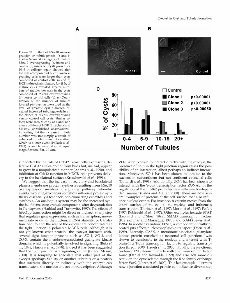
cyst size is consistent with the taller cell size measuredpreviously (Figure 6, a and b).
When MDCK cell cysts grown in collagen as describedabove are then exposed to HGF, the cysts form branchingtubules that extend out from the cyst. This is a model for theformation of tubules, another of the basic types of higherorder organization of epithelial cells used in organogenesis.To investigate the possible role of the exocyst in tubulogen-esis, we studied this process. After 48 h of HGF stimulation,we quantitated the extent of tubulogenesis by counting thenumber of visible tubules at the area of greatest cyst diam-eter. Cysts were grouped into bins containing 0–4, 5–9,10–19, and 201 tubules per cyst. Most remarkably, wheninduced by HGF, all three clones of the cysts composed ofhSec10 overexpressing cells formed significantly more tu-bules per individual cyst compared with controls (Figure 10,a–c).
DISCUSSION
We report two principal findings, both of which are surpris-ing. First, we show that the exocyst is centrally involved incystogenesis and tubulogenesis. We found that the exocystrelocalized during tubulogenesis coincident with changes inpolarity. Transfection of the hSec10 component of the exo-cyst led to a gain of function phenotype that included moreefficient and rapid cyst formation and increased tubulogen-esis upon stimulation with HGF. Second, this gain of func-tion also included the unexpected increase in synthesis of atleast one basolateral plasma membrane protein, as well as awhole array of both apical and basolateral secretory pro-teins.
In mice, a mutation in an exocyst subunit (Sec8) resultedin early embryonic lethality, which is understandable giventhe importance of the exocyst to the exocytic machinery(Friedrich et al., 1997; Hsu et al., 1998). Unlike the Sec8 genedisruption, the relocalization of the exocyst during tubulo-genesis, the accelerated formation and increased diameter ofcysts overexpressing hSec10, and the increased number oftubules with hSec10 overexpression indicate a more specific
and unexpected involvement of the exocyst in cystogenesisand tubulogenesis. Cysts and tubules represent two of thebasic building blocks of higher order organizations of epi-thelial cells used in organogenesis and the specific role of theexocyst offers an important new insight into these vital, yetpoorly understood, processes.
Basolateral extensions represent the first stage of tubulo-genesis (Pollack et al., 1998). It seems probable that theseextensions are dependent on the synthesis and delivery ofsecretory and plasma membrane proteins to the basolateralsurface in a manner analogous to cell locomotion likelydepending on exocytosis and recycling at the leading edgeof a crawling fibroblast (Bretscher and Aguado-Velasco,1998). This could be the mechanism for increased tubulo-genesis seen with hSec10 transfection. The relocalization ofthe exocyst during tubulogenesis is reminiscent of the de-velopmentally programmed delivery of new membrane pro-teins to very specific locations during epithelial formation inthe Drosophila embryo (Lecuit and Wieschaus, 2000). Theenhanced cystogenesis in the hSec10 overexpressing cellcysts could be a direct result of increased apical secretioninto the cyst lumen, and/or increased synthesis of basolat-eral plasma membrane proteins. It is interesting to note thatthe yeast exocyst is involved in transport of vesicles to thebud and in MDCK cells the exocyst is involved in transportof vesicles to the basolateral surface. The analogy of therelationship of the bud to the basolateral surface is further
Figure 9. Effect of hSec10 overexpression on cystogenesis. (a and b)After 7 d of growth in a collagen matrix, cysts composed of hSec10transfected cells (a) were mature, while cysts composed of control cells(b) were incompletely formed (confocal microscopy, cysts were stainedfor actin with phalloidin). (c) Quantitation demonstrated the increasedrate and efficiency of mature cyst formation, as noted in a and b, inhSec10 overexpressing compared with control cell cysts. a and b weretaken at equal magnification. Bar, 30 mm.
Figure 8. Synthesis and delivery of plasma membrane proteins inhSec10 overexpressing cells. In contrast to gp135, E-Cadherin syn-thesis and delivery were increased in hSec10 overexpressing versuscontrol cells. The p values were the result of at least three experi-ments and were determined by the Mann-Whitney nonparametrictest. Units are arbitrary phosphorimager units. NS, nonsignificant.Sec10 (f), control (M).
J.H. Lipschutz et al.
Molecular Biology of the Cell4270
supported by the role of Cdc42. Yeast cells expressing de-fective CDC42 alleles do not form buds but, instead, appearto grow in a nonpolarized manner (Adams et al., 1990), andinhibition of Cdc42 function in MDCK cells prevents deliv-ery to the basolateral surface (Kroschewski et al., 1999).
We suggest that the increase in secretory and basolateralplasma membrane protein synthesis resulting from hSec10overexpression involves a signaling pathway wherebyevents involving exocytosis somehow influence protein syn-thesis, essentially a feedback loop connecting exocytosis andsynthesis. An analogous system may be the increased syn-thesis of dense core granule components after degranulationin Tetrahymena (Haddad and Turkewitz, 1997). The effects ofhSec10p transfection might be direct or indirect at any stepthat regulates gene expression, such as transcription, move-ment into or out of the nucleus, mRNA stability, or transla-tion. Sec10p and the rest of the exocyst are concentrated atthe tight junction in polarized MDCK cells. Although it isnot yet known what proteins the exocyst interacts with,several tight junction proteins such as ZO-1, ZO-2, andZO-3, contain the membrane-associated guanylate kinasedomain, which is potentially involved in signaling (Butz etal., 1998; Haskins et al., 1998). Indeed it has been suggestedthat the tight junction is a signaling center (Li and Mrsny,2000). It is tempting to speculate that either part of theexocyst (perhaps Sec10p or another subunit) or a proteinthat interacts directly or indirectly with the exocyst cantranslocate to the nucleus and act on transcription. Although
ZO-1 is not known to interact directly with the exocyst, thepresence of both in the tight junction region raises the pos-sibility of an interaction, albeit perhaps an indirect interac-tion. Moreover, ZO-1 has been shown to localize to thenucleus in subconfluent but not confluent epithelial cells(Gottardi et al., 1996). Additionally, ZO-1 has been shown tointeract with the Y-box transcription factor ZONAB, in theregulation of the ErbB-2 promoter in a cell-density–depen-dent manner (Balda and Matter, 2000). There are now sev-eral examples of proteins at the cell surface that also influ-ence nuclear events. For instance, b-catenin moves from thelateral surface of the cell to the nucleus and influencestranscription (Korinek et al., 1997; Morin et al., 1997; Peifer,1997; Rubinfeld et al., 1997). Other examples include STAT(Leonard and O’Shea, 1998), SMAD transcription factors(Kretzschmar and Massague, 1998), and c-Abl (Lewis et al.,1996). In another variation, EPS15, a component of clathrin-coated pits affects nucleocytoplasmic transport (Doria et al.,1999). Recently, CASK, a membrane-associated guanylatekinase protein enriched at neuronal cell junctions, wasshown to translocate to the nucleus and interact with T-brain-1, a T-box transcription factor, to regulate transcrip-tion (Bredt, 2000; Hsueh et al., 2000). Finally, the junctionalprotein p120 catenin interacts with the transcription factorKaiso (Daniel and Reynolds, 1999) and also acts more di-rectly on the cytoskeleton through the Rho family exchangefactor Vav2 (Noren et al., 2000). This last example illustrateshow a junction-associated protein can influence diverse sig-
Figure 10. Effect of hSec10 overex-pression on tubulogenesis. (a and b,insets) Nomarski imaging of maturehSec10 overexpressing (a, inset) andcontrol (b, inset) cell cysts grown for10 d in collagen again showed thatthe cysts composed of hSec10 overex-pressing cells were larger than cystscomposed of control cells. (a and b)HGF-induced stimulation, for 48 h, ofmature cysts revealed greater num-bers of tubules per cyst in the cystscomposed of hSec10 overexpressing(a) versus control cells (b). (c) Quan-titation of the number of tubulesformed per cyst, as measured at thelevel of greatest cyst diameter, re-vealed increased tubulogenesis in allthe clones of hSec10 overexpressingversus control cell cysts. Similar ef-fects were seen as early as 6 and 12 hafter addition of HGF (Lipschutz andMostov, unpublished observations),indicating that the increase in tubulenumber was not simply a result ofenhanced tubular lumen formation,which is a later event (Pollack et al.,1998). a and b were taken at equalmagnification. Bar, 30 mm.
Exocyst in Cyst and Tubule Formation
Vol. 11, December 2000 4271
naling pathways in the cell, which may also be the case forSec10p.
Our data, as well as that of Grindstaff et al. (1998) suggestthat there may be at least two pathways to the apical surface.One pathway carries at least certain apical membrane pro-teins such as gp135 and exogenously expressed p75NTR andthe exocyst does not seem to play a role in this pathway.Another pathway involves a broad array of secretory pro-teins and this pathway does involve the exocyst, at least inthat overexpression of hSec10 increases their synthesis. Thisis consistent with other recent evidence showing multiplepathways from the trans-Golgi network to the apical surfaceof MDCK cells (Mostov et al., 2000; Orzech et al., 2000).
The effects of hSec10 overexpression on cyst and tubuleformation are reminiscent of ADPKD. ADPKD is one of themost common potentially lethal genetic disorders in hu-mans, inherited as a dominant trait, and results in cystic andtubular overgrowth, which leads to destruction of the nor-mal kidney architecture and renal failure (Grantham, 1997).In ADPKD there are gross abnormalities in cell polarity inthe cells lining the gigantic cystic expansions of the tubules(Wilson et al., 1991; Avner et al., 1992; Gabow, 1993). Forexample, there is reversed polarity of Na1-K1-ATPase, withmislocalization to the apical plasma membrane in ADPKDepithelia (Wilson et al., 1991). Although the PKD1 and PKD2genes (Consortium, 1995; Mochizuki et al., 1996), whichwhen mutated are responsible for the vast majority of casesof ADPKD, have been identified, their function, role in cys-togenesis and tubulogenesis, and downstream effectors re-main largely unknown (Arnould et al., 1999). In ADPKD,cysts develop from renal tubular epithelial cells (Grantham,1997). MDCK cells are also derived from renal tubular epi-thelium (Simons and Fuller, 1985). Recent work demon-strates exocyst abnormalities in ADPKD cells. In normalkidney cells both Sec6p and Sec8p were localized in closeapposition to the tight junction protein occludin. In contrast,both proteins were depleted from the ADPKD cell lateralmembranes and appeared diffusely dispersed throughoutthe cytoplasm (Charron et al., 2000). This is analogous to thecytoplasmic localization of Sec6p and Sec8p in contact-naiveMDCK cells (Grindstaff et al., 1998), suggesting that thedefect in ADPKD cells lies in their response to cell–cellcontact.
It is worthwhile to compare our results to recent progressin analysis of tubulogenesis in genetically tractable organ-isms. The trachea of Drosophila has been well studied and anumber of genes and proteins have been identified thatcontrol morphogenesis of this structure (Metzger and Kras-now, 1999). In both Drosophila trachea and the renal systemof Caenorhabditis elegans, a number of mutants have beenisolated that affect the diameter of tubules, though most ofthese genes have yet to be cloned (Buechner et al., 1999;Beitel and Krasnow, 2000). The phenotype of some of thesemutants is similar to the increase in cyst diameter that weobserve upon overexpression of hSec10.
Our results provide an entirely new perspective with re-spect to the exocyst; indicating that hSec10 functions, di-rectly or indirectly, in cellular processes far beyond its pre-viously known role in directing vesicular traffic. Theseresults suggest that there are previously unsuspected signal-ing or other pathways connecting membrane traffic, proteinsynthesis, and, especially, the vital, yet poorly understood
process of morphogenesis of higher order epithelial struc-tures, such as cysts and tubules. Understanding the mecha-nisms underlying these newly discovered connections willbe the subject of further investigations. Moreover, we canspeculate that components of this signaling pathway may behomologous to components that control cyst and tubulemorphogenesis in Drosophila and C. elegans.
ACKNOWLEDGMENTS
We thank S. Chapin and F. Luton for their helpful discussion andreading of this manuscript. P. DeCamilli is acknowledged for helpin cloning hSec10. S. Huling and the University of California, SanFrancisco, Liver Center are gratefully acknowledged for their assis-tance with electron microscopy. R. Bacallao and A. Wandinger-Nessare acknowledged for sending us their manuscript before publica-tion. Finally, we thank W.J. Nelson, K. Grindstaff, and C. Yeamanfor their helpful discussion and sharing of unpublished data beforepublication. This study was supported by National Institutes ofHealth and DAMD (DAMD-17-97-1-7249) grants to K.E.M, andNational Institutes of Health, a National Kidney Foundation YoungInvestigator Award, and a Northern California National KidneyFoundation grant to J.H.L.
REFERENCES
Adams, A.E., Johnson, D.I., Longnecker, R.M., Sloat, B.F., and Prin-gle, J.R. (1990). CDC42 and CDC43, two additional genes involvedin budding and the establishment of cell polarity in the yeast Sac-charomyces cerevisiae. J. Cell Biol. 111, 131–142.
Appel, D., Pilarsky, C., Graichen, R., and Koch-Brandt, C. (1996).Sorting of gp80 (GPIII, clusterin), a marker protein for constitutiveapical secretion in Madin-Darby canine kidney (MDCK) cells, intothe regulated pathway in the pheochromocytoma cell line PC12.Eur. J. Cell Biol. 70, 142–149.
Arnould, T., Sellin, L., Benzing, T., Tsiokas, L., Cohen, H.T., Kim, E.,and Walz, G. (1999). Cellular activation triggered by the autosomaldominant polycystic kidney disease gene product PKD2. Mol. Biol.Cell 19, 3423–3434.
Avner, E.D., Sweeney, W.E., Jr., and Nelson, W.J. (1992). Abnormalsodium pump distribution during renal tubulogenesis in congenitalmurine polycystic kidney disease. Proc. Natl. Acad. Sci. USA 89,7447–7451.
Ayscough, K.R., Stryker, J., Pokala, N., Sanders, M., Crews, P., andDrubin, D.G. (1997). High rates of actin filament turnover in bud-ding yeast and roles for actin in establishment and maintenance ofcell polarity revealed using the actin inhibitor latrunculin-A. J. CellBiol. 137, 399–416.
Balda, M.S., and Matter, K. (2000). The tight junction protein ZO-1and an interacting transcription factor regulate ErbB-2 expression.EMBO J. 19, 2024–2033.
Balkovetz, D.F., and Lipschutz, J.H. (1998). Hepatocyte growth fac-tor and the kidney: it’s not just for the liver. Int. Rev. Cytol. 186,225–260.
Bao, Q., and Hughes, R.C. (1995). Galectin-3 expression and effect oncyst enlargement and tubulogenesis in kidney epithelial MDCKcells cultured in three-dimensional matrices in vitro. J. Cell Sci. 108,2791–2800.
Bao, Q., and Hughes, R.C. (1999). Galectin-3 and polarized growthwithin collagen gels of wild-type and ricin-resistant MDCK renalepithelial cells. Glycobiology 9, 489–495.
Barros, E.J., Santos, O.F., Matsumoto, K., Nakamura, T., and Nigam,S.K. (1995). Differential tubulogenic and branching morphogenetic
J.H. Lipschutz et al.
Molecular Biology of the Cell4272

activities of growth factors: implications for epithelial tissue devel-opment. Proc. Natl. Acad. Sci. USA 92, 4412–4416.
Beitel, G.J., and Krasnow, M.A. (2000). Genetic control of epithelialtube size in the Drosophila tracheal system. Development 127, 3271–3282.
Boccaccio, C., Ando, C., Tamagnone, L., Bardelli, A., Michieli, P.,Battistini, C., and Comoglio, P.M. (1998). Induction of epithelialtubules by growth factor HGF depends on the STAT pathway.Nature 391, 285–288.
Bredt, D.S. (2000). Reeling CASK into the nucleus. Nature 404,241–242.
Breitfeld, P., Casanova, J.E., Harris, J.M., Simister, N.E., and Mostov,K.E. (1989). Expression and analysis of the polymeric immunoglob-ulin receptor. Methods Cell Biol. 32, 329–337.
Bretscher, M.S., and Aguado-Velasco, C. (1998). Membrane trafficduring cell locomotion. Curr. Opin. Cell Biol. 10, 537–541.
Brinkmann, V., Foroutan, H., Sachs, M., Weidner, K.M., and Birch-meier, W. (1995). Hepatocyte growth factor/scatter factor induces avariety of tissue-specific morphogenic programs in epithelial cells.J. Cell Biol. 131, 1573–1586.
Buechner, M., Hall, D.H., Bhatt, H., and Hedgecock, E.M. (1999).Cystic canal mutants in Caenorhabditis elegans are defective in theapical membrane domain of the renal (excretory) cell. Dev. Biol. 214,227–241.
Butz, S., Okamoto, T., and Sudhoff, T.C. (1998). A tripartite proteincomplex with the potential to couple synaptic vesicle exocytosis tocell adhesion in brain. Cell 94, 773–782.
Byers, B. (1981). Cytology of the yeast life cycle. In: MolecularBiology of the Yeast Saccharomyces: Life Cycle and Inheritance, eds.J.N. Strathern, E.W. Jones, and J.R. Broach), Cold Spring Harbor,NY: Cold Spring Harbor Laboratory, 59–96.
Cantley, L.G., Barros, E.J., Gandhi, M., Rauchman, M., and Nigam,S.K. (1994). Regulation of mitogenesis, motogenesis, and tubulogen-esis by hepatocyte growth factor in renal collecting duct cells. Am. J.Physiol. 267, F271–F280.
Charron, A.J., Nakamura, S., Bacallao, R., and Wandinger-Ness, A.(2000). Compromised cytoarchitecture and polarized trafficking inautosomal dominant polycystic kidney disease cells. J. Cell Biol. 149,111–124.
Consortium, T.I.P.D. (1995). Polycystic kidney disease: the completestructure of the PKD1 gene and its protein. Cell 81, 289–298.
Crepaldi, T., Gautreau, A., Comoglio, P.M., Louvard, D., and Arpin,M. (1997). Ezrin is an effector of hepatocyte growth factor-mediatedmigration and morphogenesis in epithelial cells. J. Cell Biol. 138,423–434.
Daniel, J., and Reynolds, A.B. (1999). The catenin p120ctn interactswith Kaiso, a novel BTB/POZ domain zinc finger transcriptionfactor. Mol. Biol. Cell 19, 3614–3623.
Derman, M.P., Cunha, M.J., Barros, E.J., Nigam, S.K., and Cantley,L.G. (1995). HGF-mediated chemotaxis and tubulogenesis requireactivation of the phosphatidylinositol 3-kinase. Am. J. Physiol. 268,F1211–F1217.
Doria, M., Salcini, A.E., Colombo, E., Parslow, T.G., Pelicci, P.G.,and Di Fiore, P.P. (1999). The Eps15 homology (EH) domain-basedinteraction between Eps15 and Hrb connects the molecular machin-ery of endocytosis to that of nucleocytosolic transport. J. Cell Biol.147, 1379–1384.
Farkas, V., Kovarik, J., Kosinova, A., and Bauer, S. (1974). Autora-diographic study of mannan incorporation into the growing cellwall of Saccharomyces cerevisiae. J. Bacteriol. 117, 265–269.
Field, C., and Schekman, R. (1980). Localized secretion of acidphosphatase reflects the pattern of cell surface growth in Saccharo-myces cerevisiae. J. Cell Biol. 86, 123–128.
Finger, F.P., Hughes, T.E., and Novick, P. (1998). Sec3p is a spatiallandmark for polarized secretion in budding yeast. Cell 92, 559–571.
Finger, F.P., and Novick, P. (1998). Spatial regulation of exocytosis:lessons from yeast. J. Cell Biol. 142, 609–612.
Friedrich, G.A., Hildebrand, J.D., and Soriano, P. (1997). The secre-tory protein Sec8 is required for paraxial mesoderm formation in themouse. Dev. Biol. 192, 364–374.
Gabow, P.A. (1993). Autosomal dominant polycystic kidney dis-ease. N. Engl. J. Med. 329, 332–340.
Gherardi, E., Gray, J., Stoker, M., Perryman, M., and Furlong, R.(1989). Purification of scatter factor, a fibroblast-derived basic pro-tein that modulates epithelial interactions and movement. Proc.Natl. Acad. Sci. USA 86, 5844–5848.
Goode, B.L., Drubin, D.G., and Barnes, G. (2000). Functional coop-eration between the microtubule and actin cytoskeleton. Curr. Opin.Cell Biol. 12, 63–71.
Gottardi, C.J., Arpin, M., Fanning, A.S., and Louvard, D. (1996). Thejunction-associated protein, zonula occludens-1, localizes to the nu-cleus before the maturation and during the remodeling of cell-cellcontacts. Proc. Natl. Acad. Sci. USA 93, 10779–10784.
Grantham, J.J. (1997). Mechanisms of progression in autosomaldominant polycystic kidney disease. Kidney Int. 52, S93–S97.
Grindstaff, K.K., Yeaman, C., Anandasabapathy, N., Hsu, S., Rodri-guez-Boulan, R., Scheller, R.H., and Nelson, W.J. (1998). Sec6/8complex is recruited to cell-cell contacts and specifies transportvesicle delivery to the basal-lateral membrane in epithelial cells. Cell93, 731–740.
Gumbiner, B.M. (1992). Epithelial morphogenesis [comment]. Cell69, 385–387.
Guo, W., Grant, A., and Novick, P. (1999a). Exo84p is an exocystprotein essential for secretion. J. Biol. Chem. 274, 23558–23564.
Guo, W., Roth, D., Gatti, E., De Camilli, P., and Novick, P. (1997).Identification and characterization of homologues of the Exocystcomponent Sec10p. FEBS Lett. 404, 135–139.
Guo, W., Roth, D., Walch-Solimena, C., and Novick, P. (1999b). Theexocyst is an effector for Sec4p, targeting secretory vesicles to sites ofexocytosis. EMBO J. 18, 1071–1080.
Haddad, A., and Turkewitz, A.P. (1997). Analysis of exocytosismutants indicates close coupling between regulated secretion andtranscription activation in Tetrahymena. Proc. Natl. Acad. Sci. USA94, 10675–10680.
Haskins, J., Gu, L., Wittchen, E.S., Hibbard, J., and Stevenson, B.R.(1998). Zo-3, a novel member of the MAGUK protein family foundat the tight junction, interacts with zo-1 and occludin. J. Cell Biol.141, 199–208.
Hsu, S., Hazuka, C.D., Roth, R., Foletti, D.L., Heuser, J., and Scheller,R.H. (1998). Subunit composition, protein interactions, and struc-tures of the mammalian brain sec6/8 complex and septin filaments.Neuron 20, 1111–1122.
Hsueh, Y., Wang, T., Yang, F., and Sheng, M. (2000). Nuclear trans-location and transcription by the membrane-associated guanylatekinase CASK/LIN-2. Nature 404, 298–302.
Korinek, V., Barker, N., Morin, P.J., Wichen, D.V., Weger, R.D.,Kinzler, K.W., Vogelstein, B., and Clevers, H. (1997). Constitutivetranscriptional activation by a b-catenin-Tcf complex in APC2/2colon carcinoma. Science 275, 1784–1787.
Exocyst in Cyst and Tubule Formation
Vol. 11, December 2000 4273

Kretzschmar, M., and Massague, J. (1998). SMADS: mediators andregulators of TGF-beta signaling. Curr. Opin. Genet. Dev. 8, 103–111.
Kroschewski, R., Hall, A., and Mellman, I. (1999). Cdc42 controlssecretory and endocytic transport to the basolateral plasma mem-brane of MDCK cells. Nat. Cell Biol. 1, 8–13.
Le Bivic, A., Sambuy, Y., Mostov, K., and Rodriguez-Boulan, E.(1990). Vectorial targeting of an endogenous apical membrane sia-loglycoprotein and uvomorulin in MDCK cells. J. Cell Biol. 110,1533–1539.
Lecuit, T., and Wieschaus, E. (2000). Polarized insertion of newmembrane from a cytoplasmic reservior during cleavage of theDrosophila embryo. J. Cell Biol. 150, 849–860.
Leonard, W.J., and O’Shea, J.J. (1998). JAKS and STATS: biologicalimplications. Annu. Rev. Immunol. 16, 293–322.
Lewis, J.M., Baskaran, R., Taagepera, S., Schwarzt, M.A., and Wang,J.Y. (1996). Integrin regulation of c-Abl tyrosine kinase activity andcytoplasmic-nuclear transport. Proc. Natl. Acad. Sci. USA 93,15174–15179.
Li, D., and Mrsny, R.J. (2000). Oncogenic Raf-1 disrupts epithelialtight junctions via downregulation of occludin. J. Cell Biol. 148,791–800.
Lindstedt, R., Apodaca, G., Barondes, S.H., Mostov, K.E., and Lef-fler, H. (1993). Apical secretion of a cytosolic protein by Madin-Darby canine kidney cells. Evidence for polarized release of anendogenous lectin by a nonclassical secretory pathway. J. Biol.Chem. 268, 11750–11757.
Lipschutz, J.H. (1998). The molecular development of the kidney: areview of the results of gene disruption studies. Am. J. Kidney Dis.31, 383–397.
Louvard, D. (1980). Apical membrane aminopeptidase appears atsite of cell-cell contact in cultured kidney epithelial cells. Proc. Natl.Acad. Sci. USA 77, 4132–4136.
Luton, F., Cardone, M.H., Zhang, M., and Mostov, K.E. (1998). Roleof tyrosine phosphorylation in ligand-induced regulation of trans-cytosis of the polymeric Ig receptor. Mol. Biol. Cell 9, 1787–1802.
Metzger, R.J., and Krasnow, M.A. (1999). Genetic control of branch-ing morphogenesis. Science 284, 1635–1639.
Mochizuki, T., Wu, G., Hayashi, T., Xenophontos, S.L., Veldhuisen,B., Saris, J.J., Reynolds, D.M., Cai, Y., Gabow, P.A., Pierides, A.,Kimberling, W.J., Breuning, M.H., Deltas, C.C., Peters, D.J.M., andSomlo, S. (1996). PKD2, a gene for polycystic kidney disease thatencodes an integral membrane protein. Science 272, 1339–1342.
Montesano, R., Matsumoto, K., Nakamura, T., and Orci, L. (1991a).Identification of a fibroblast-derived epithelial morphogen as hepa-tocyte growth factor. Cell 67, 901–908.
Montesano, R., Schaller, G., and Orci, L. (1991b). Induction of epi-thelial tubular morphogenesis in vitro by fibroblast-derived solublefactors. Cell 66, 697–711.
Morin, P.J., Sparks, A.B., Korinek, V., Barker, N., Clevers, H., Vo-gelstein, B., and Kinzler, K.W. (1997). Activation of b-catenin-Tcfsignaling in colon cancer by mutations in b-catenin or APC. Science275, 1787–1790.
Mostov, K.E., Verges, M., and Altschuler, Y. (2000). Membranetraffic in polarized epithelial cells. Curr. Opin. Cell Biol. 12, 483–490.
Noren, N.K., Liu, B.P., Burridge, K., and Kreft, B. (2000). p120catenin regulates the actin cytoskeleton via Rho family GTPases.J. Cell Biol. 150, 567–579.
Novick, P., Field, C., and Schekman, R. (1980). Identification of 23complementation groups required for post-translational events inthe yeast secretory pathway. Cell 21, 205–221.
Orzech, E., Cohen, S., Weiss, A., and Aroeti, B. (2000). Interactionsbetween the exocytic and endocytic pathways in polarized MadinDarby canine kidney cells. J. Biol. Chem. 275, 15207–15219.
Peifer, M. (1997). b-Catenin as oncogene: the smoking gun. Science275, 1752–1753.
Pollack, A.L., Barth, A.I.M., Altschuler, Y., Nelson, W.J., andMostov, K.E. (1997). Dynamics of b-catenin interactions with APCprotein regulate epithelial tubulogenesis. J. Cell Biol. 137, 1651–1662.
Pollack, A.L., Runyan, R.B., and Mostov, K.E. (1998). Morphogeneticmechanisms of epithelial tubulogenesis: MDCK cell polarity is tran-siently rearranged without loss of cell-cell contact during scatterfactor/hepatocyte growth factor-induced tubulogenesis. Dev. Biol.204, 64–79.
Roth, D., Guo, W., and Novick, P. (1998). Dominant negative allelesof SEC10 reveal distinct domains involved in secretion, and mor-phogenesis in yeast. Mol. Biol. Cell 9, 1725–1739.
Rubinfeld, B., Robbins, P., El-Gamil, M., Albert, I., Porfiri, E., andPolakis, P. (1997). Stabilization of b-catenin by genetic defects inmelanoma cell lines. Science 275, 1790–1792.
Sakurai, H., Barros, E.J., Tsukamoto, T., Barasch, J., and Nigam, S.K.(1997a). An in vitro tubulogenesis system using cell lines derivedfrom the embryonic kidney shows dependence on multiple solublegrowth factors. Proc. Natl. Acad. Sci. USA 94, 6279–6284.
Sakurai, H., Tsukamoto, T., Kjelsberg, C.A., Cantley, L.G., andNigam, S.K. (1997b). EGF receptor ligands are a large fraction of invitro branching morphogens secreted by embryonic kidney. Am. J.Physiol. 273, F463–F472.
Sambrook, J., Fritsch, E.F., and Maniatis, T. (1989). Expression ofcloned genes in cultured mammalian cells. In: Molecular Cloning: ALaboratory Manual, eds. J. Sambrook, E.F. Fritsch, and T. Maniatis),Cold Spring Harbor, New York: Cold Spring Harbor LaboratoryPress, 16.30–16.36.
Santos, O.F., Moura, L.A., Rosen, E.M., and Nigam, S.K. (1993).Modulation of HGF-induced tubulogenesis and branching by mul-tiple phosphorylation mechanisms. Dev. Biol. 159, 535–548.
Santos, O.F., and Nigam, S.K. (1993). HGF-induced tubulogenesisand branching of epithelial cells is modulated by extracellular ma-trix and TGF-beta. Dev. Biol. 160, 293–302.
Sato, S., Burdett, I., and Hughes, R.C. (1993). Secretion of the babyhamster kidney 30-kDa galactose-binding lectin from polarized andnonpolarized cells: a pathway independent of the endoplasmic re-ticulum-Golgi complex. Exp. Cell Res. 207, 8–18.
Saxen, L. (1987). Organogenesis of the Kidney. Cambridge: Cam-bridge University Press.
Simons, K., and Fuller, S.D. (1985). Cell surface polarity in epithelia.Annu. Rev. Cell Biol. 1, 295–340.
Stoker, M., Gherardi, E., Perryman, M., and Gray, J. (1987). Scatterfactor is a fibroblast-derived modulator of epithelial cell mobility.Nature 327, 239–242.
Stoker, M., and Perryman, M. (1985). An epithelial scatter factorreleased by embryo fibroblasts. J. Cell Sci. 77, 209–223.
Terbush, D.R., Maurice, T., Roth, D., and Novick, P. (1996). Theexocyst is a multiprotein complex required for exocytosis in Saccha-romyces cerevisiae. EMBO J. 15, 6483–6494.
Thiery, G., Bernier, J., and Bergeron, M. (1995). A simple techniquefor staining of cell membranes with imidazole and osmium tetrox-ide. J. Histocytochem. 43, 1079–1084.
Thiery, J.P., and Boyer, B. (1992). The junction between cytokinesand cell adhesion. Curr. Opin. Cell Biol. 4, 782–792.
Ting, A.E., Hazuka, C.D., Hsu, S.C., Kirk, M.D., Bean, A.J., andScheller, R.H. (1995). rSec6 and rSec8, mammalian homologs of
J.H. Lipschutz et al.
Molecular Biology of the Cell4274
yeast proteins essential for secretion. Proc. Natl. Acad. Sci. USA 92,9613–9617.
Tkacz, J.S., and Lampen, J.O. (1972). Wall replication in Saccharomy-ces species: use of fluorescein-conjugated concanavalin A to revealthe site of mannan insertion. J. Gen. Microbiol. 72, 243–247.
Urban, J., Parczyk, K., Leutz, A., Kayne, M., and Kondor-Koch, C.(1987). Constitutive apical secretion of an 80-kd sulfated glycopro-tein complex in the polarized epithelial Madin Darby canine kidneycell line. J. Cell Biol. 105, 2735–2743.
Vainio, S., and Muller, U. (1997). Inductive tissue interactions, cellsignaling, and the control of kidney organogenesis. Cell 90, 975–978.
Weidner, K.M., Sachs, M., Riethmacher, D., and Birchmeier, W.(1995). Mutation of juxtamembrane tyrosine residue 1001 sup-presses loss-of-function mutations of the met receptor in epithelialcells. Proc. Natl. Acad. Sci. USA 92, 2597–2601.
Wilson, P.D., Sherwood, A.C., Palla, K., Du, J., Watson, R., andNorman, J.T. (1991). Reversed polarity of Na1-K1-ATPase: mislo-calization to apical plasma membranes in polycystic kidney diseaseepithelia. Am. J. Physiol. 260, F420–F430.
Yeaman, C., Grindstaff, K.K., and Nelson, W.J. (1999). New perspec-tives on mechanisms involved in generating cell polarity. Physiol.Rev. 79, 73–98.
Exocyst in Cyst and Tubule Formation
Vol. 11, December 2000 4275
Copyright © 2022 FDOKUMEN