Research Recommendations for the American Telemedicine Association
Upload
independentCategory
view
0download
0
Journal of
REHABILITATIONMEDICINE
ISSN 1650-1977
Official journal of the– International Society of Physical and Rehabilitation Medicine (ISPRM)– UEMS European Board of Physical and Rehabilitation Medicine (EBPRM)– European Academy of Rehabilitation Medicine (EARM)
Published in association with the– European Society of Physical and Rehabilitation Medicine
Rehabilitation InformationT
www.medicaljournals.se/jrm
Vol. 41. No. 12October 2009
Special issue
the application of robotics in the functional motor recovery
of the paretic upper limb
articles from the workshop held in september 5–6, 2008 in crotone, italy
all correspondence concerning manuscripts, editorial matters and subscription should be addressed to:Editorial Manager: Mrs Agneta Andersson, Editorial assistants: Hanna Bergström, [email protected]@medicaljournals.se Jorunn Hartman, [email protected] postal address: see inside back cover Webmaster: Johanna Hartman, [email protected]
publication information: Journal of Rehabilitation Medicine (ISSN 1650-1977) volume 41 comprises ten regular issues published in January, February, March, April, May, June, July, September, October and November. Each issue comprises at least 96 pages.
Subscription rates for volume 41:- for institutions: paper incl. electronic access: EUR 430, electronic only: EUR 370- for individuals: paper incl. electronic access: EUR 175, electronic only: EUR 145- for the organizations mentioned above: paper incl. electronic access: EUR 75, electronic only: EUR 50
indexing: Journal of Rehabilitation Medicine is indexed in Index Medicus/MEDLINE, Excerpta Medica/EMBASE, Biological Abstracts, Current Contents/Clinical Practice, Allied and Alternative Medicine Database (AMED), Applied Social Sciences Index & Abstracts, Ergonomic Abstracts, Psychological Abstracts PsycINFO, PSYCLIT DATABASE, Cumulative Index to Nursing & Allied Health Literature (CINAHL), Developmental Medicine and Child Neurology, Exceptional Child Education Resources, Periodicals Scanned and Abstracted: Life Sciences Collection, Faxon Finder, Focus On Sports Science & Medicine, Research Alert, SCISEARCH, SportSearch.
Journal of rehabilitation medicineJournal of Rehabilitation Medicine is an international peer-review journal published in English with ten regular issues per year. It is owned by a Swedish nonprofit organization: Foundation for Rehabilitation Information. Journal of Rehabilita-tion Medicine was former called Scandinavian Journal of Rehabilitation Medicine, which was founded by Olle Höök in 1968. The name was changed to Journal of Rehabilitation Medicine in 2001.Journal of Rehabilitation Medicine aims to be a leading worldwide forum for research in physical and rehabilitation medicine, aiming to increase knowledge in evidence-based clinical rehabilitation. Contributions from all parts of the world and from differ-ent professions in rehabilitation are encouraged. Original articles, Reviews (including Educational reviews), Special reports, Short communications, Case reports, and Letters to the Editor are published. Clinical studies on rehabilitation in various patients groups, within neurological and musculoskeletal as well as in other relevant rehabilitation areas, reports on physical and behavioural treat-ment methodology, including rehabilitation technology, development and analysis of methodology for outcome measurements, epidemiological studies on disability in relation to rehabilitation, and studies on vocational and socio-medical aspects of rehabilita-tion will be considered for publication. The journal emphasizes the need for randomized controlled studies of various rehabilitation interventions, the use of the International Classification of Functioning, Disability and Health (ICF) as a background for reports when appropriate, and the use of modern psychometric methodology in treating and reporting data from ordinal scales.
editor-in-chiefGunnar Grimby, Göteborg, Sweden
associate editorsKristian Borg, Stockholm, Sweden Franco Franchignoni, Veruno, ItalyBjörn Gerdle, Linköping, SwedenKenneth Ottenbacher, Galveston, USAHenk Stam, Rotterdam, The NetherlandsGerold Stucki, Nottwil, Switzerland
editorial boardOlavi Airaksinen, Kuopio, FinlandMasami Akai, Saitama, JapanJari Arokoski, Kuopio, FinlandAniko Bartfai, Stockholm, SverigeLinamara R. Battistella, Sao Paulo, BrazilFin Biering-Sørensen, Copenhagen, DenmarkJörgen Borg, Uppsala, SwedenIan Cameron, Sydney, AustraliaAnne Chamberlain, Leeds, United KingdomAlain Delarque, Marseilles, FranceJean-Pierre Didier, Dijon, FranceJan Ekholm, Stockholm, SwedenVeronika Fialka-Moser, Vienna, AustriaAnne Fisher, Umeå, SwedenCharles-Albert Gobelet, Sion, SwitzerlandMartin Grabois, Houston, USACristoph Gutenbrunner, Hannover, GermanyKenji Hachisuka, Kitakyushu, JapanAndrew Haig, Michigan, USAKarin Harms-Ringdahl, Stockholm, SwedenTessa Hart, Philadelphia, USAMarta Imamura, São Paulo, Brasil
Alan Jette, Boston, USAJan Lexell, Lund, SwedenJianan Li, Nanjing, ChinaLeonard S.W. Li, Hong KongMeigen Liu, Tokyo, JapanCrt Marincek, Ljubljana, SloveniaNancy Mayo, Montreal, CanadaFrans Nollet, Amsterdam, The NetherlandsChang-il Park, Seoul, KoreaMichael Quittan, Vienna, AustriaCarol Richards, Quebec, CanadaBengt Sjölund, Copenhagen, DenmarkJohan Stanghelle, Oslo, NorwayKatharina Stibrant Sunnerhagen, Göteborg, SwedenSimon F.T. Tang, Tao-Yuan, TaiwanAlan Tennant, Leeds, United KingdomLuigi Tesio, Milan, ItalyLynne Turner-Stokes, Middlesex, United KingdomGuy Vanderstraeten, Gent, BelgiumMaobin Wang, Beijing, ChinaAnthony B. Ward, Stoke on Trent, United KingdomGünes Yavuzer, Ankara, Turkey
contact persons for the organizations:International Society of Physical Rehabilitation Medicine (ISPRM): Franco Franchignoni, Veruno, ItalyUEMS European Board of Physical and Rehabilitation Medicine: Guy Vanderstraeten, Gent, BelgiumEuropean Academy of Rehabilitation Medicine: Gustaaf Lankhorst, Amsterdam, The NetherlandsEuropean Society of Physical and Rehabilitation Medicine: Elena Milkova Ilieva, Plovdiv, Bulgaria and Calogero Foti, Rome, Italy
J Rehabil Med 2009; 41: 949–1020
J Rehabil Med 41© Foundation of Rehabilitation Information. ISSN 1650-1977
the application of robotics in the functional motor recovery
of the paretic upper limbarticles from the workshop held in september 5–6, 2008 in crotone, italy
Lucia F. Lucca, MD, Enrico Castelli, MD and Walter G. Sannita, MD, editors

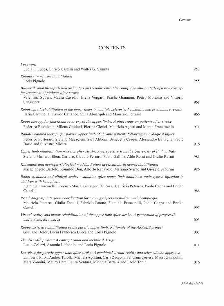
Contents
ForewordLucia F. Lucca, Enrico Castelli and Walter G. Sannita 953
Robotics in neuro-rehabilitationLoris Pignolo 955
Bilateral robot therapy based on haptics and reinforcement learning: Feasibility study of a new concept for treatment of patients after stroke
Valentina Squeri, Maura Casadio, Elena Vergaro, Psiche Giannoni, Pietro Morasso and Vittorio Sanguineti 961
Robot-based rehabilitation of the upper limbs in multiple sclerosis: Feasibility and preliminary resultsIlaria Carpinella, Davide Cattaneo, Suha Abuarqub and Maurizio Ferrarin 966
Robot therapy for functional recovery of the upper limbs: A pilot study on patients after strokeFederica Bovolenta, Milena Goldoni, Pierina Clerici, Maurizio Agosti and Marco Franceschin 971
Robot-mediated therapy for paretic upper limb of chronic patients following neurological injuryFederico Posteraro, Stefano Mazzoleni, Sara Aliboni, Benedetta Cesqui, Alessandro Battaglia, Paolo Dario and Silvestro Micera 976
Upper limb rehabilitation robotics after stroke: A perspective from the University of Padua, ItalyStefano Masiero, Elena Carraro, Claudio Ferraro, Paolo Gallina, Aldo Rossi and Giulio Rosati 981
Kinematic and neurophysiological models: Future applications in neurorehabilitationMichelangelo Bartolo, Romildo Don, Alberto Ranavolo, Mariano Serrao and Giorgio Sandrini 986
Robot-mediated and clinical scales evaluation after upper limb botulinum toxin type A injection in children with hemiplegia
Flaminia Frascarelli, Lorenzo Masia, Giuseppe Di Rosa, Maurizio Petrarca, Paolo Cappa and Enrico Castelli 988
Reach-to-grasp interjoint coordination for moving object in children with hemiplegiaMaurizio Petrarca, Giulia Zanelli, Fabrizio Patanè, Flaminia Frascarelli, Paolo Cappa and Enrico Castelli 995
Virtual reality and motor rehabilitation of the upper limb after stroke: A generation of progress?Lucia Francesca Lucca 1003
Robot-assisted rehabilitation of the paretic upper limb: Rationale of the ARAMIS projectGiuliano Dolce, Lucia Francesca Lucca and Loris Pignolo 1007
The ARAMIS project: A concept robot and technical designLucio Colizzi, Antonio Lidonnici and Loris Pignolo 1011
Exercises for paretic upper limb after stroke: A combined virtual-reality and telemedicine approachLamberto Piron, Andrea Turolla, Michela Agostini, Carla Zucconi, Feliciana Cortese, Mauro Zampolini, Mara Zannini, Mauro Dam, Laura Ventura, Michela Battauz and Paolo Tonin 1016
CONTENTS
J Rehabil Med 41

J Rehabil Med 2009; 41: 953
J Rehabil Med 41© 2009 The Authors. doi: 10.2340/16501977-0433Journal Compilation © 2009 Foundation of Rehabilitation Information. ISSN 1650-1977
An estimated 30–60% of adult patients after stroke do not achieve satisfactory motor recovery of the upper limb despite intensive rehabilitation. Motor re-organization in adults also depends on substantial contributions from the undamaged motor cortex, with functional inhibition by the unaffected arm that has become dominant – a limitation that neuro-rehabilitation should counterbalance after stroke as well as in other pathological conditions (e.g. multiple sclerosis) and in children.
Innovative technologies, such as advanced robotics and virtual reality, have proven appli-cable in neuro-rehabilitation, and their use in the treatment of the paretic upper limb appears promising. The available evidence supports applicability. However, research on efficacy has thus far been unsystematic, and the advantages of robotic-supported rehabilitation compared with conventional treatments remain, to a relevant extent, undocumented. More importantly, a comprehensive scientific rationale and pathophysiological understanding of the mechanisms underlying recovery (with or without robot assistance) remain to be devised.
The applicability of novel technologies depends on efficacy and cost-benefit ratio as much as it requires scientific background, expertise and communication to be shared by professionals and scientists from different fields. In this respect, the knowledge of bio-engineers and reha-bilitators need to be integrated for the robotic implements to be usable in neuro-rehabilitation. The patient’s needs and the training goals are central to the development of machine-human interfaces. Design, research and programming for robotics application in neuro-rehabilitation can benefit from captology and develop interactive computing products purported to change people’s attitude and/or behavior. The approach would also enhance the patients’ commitment to training and expand rehabilitation beyond the mere, often partial and usually compensatory, recovery of motor function. The approach looks promising, and research in this field is due.
A workshop on “The application of robotics in the functional motor recovery of the paretic upper limb” was held in Crotone, Italy, on 5–6 September 2008, with contributions from the major neuro-rehabilitation centres in the country and participation of leading scientists in neuroscience. The objectives of the workshop were to characterize by technology and rationale of development the robots and virtual reality systems available today for neuro-rehabilitation, focus attention on the methodological and applicative problems, promote multidisciplinary interaction and collaboration. It is our hope that the workshop and its proceedings will help share the relevant information on the issue and promote further research. With such an achieve-ment, the workshop would be successful beyond the duties and purposes of a scientific event. Thanks are due to Institute S. Anna – Research in Advanced Neuro-rehabilitation (RAN) for the successful organization, financial support, and publication of this special issue.
Lucia F. Lucca, MD1, Enrico Castelli, MD2 and Walter G. Sannita, MD3
From the 1Institute S. Anna – RAN, Crotone, 2Pediatric Neuro-Rehabilitation Division, Children’s Hospital “Bambino Gesù” IRCCS, Rome and 3Department of Motor Science and
Rehabilitation, University of Genova, Genova, Italy
FOREWORD

special report
J Rehabil Med 2009; 41: 955–960
J Rehabil Med 41© 2009 The Authors. doi: 10.2340/16501977-0434Journal Compilation © 2009 Foundation of Rehabilitation Information. ISSN 1650-1977
Objective: the aims of this study were to review robot-assist-ed motor and functional rehabilitation of the upper limb in patients with stroke and to outline possible clinical applica-tions of robotics in neuro-rehabilitation.Methods: available active systems, with actuators driving the paretic arm, were sub-classified by scientific rationale and mechatronic structure as exoskeletons or operational-type machines (manipulators). applicative studies were compared for indication of efficacy.Results and conclusion: clinical and biomechanical evidence available to date suggests substantial efficacy of robot- assisted neuro-rehabilitation in the recovery of the paretic arm after stroke, enabling longer dedicated training sessions with no additional work for the therapist. further investi-gation of large samples of patients is required to define the relationship between disability and residual function, to pro-vide shared criteria of evaluation of disability/outcome and protocols of rehabilitation, and to identify the expected fu-ture role and application of robotics in neuro-rehabilitation. Key words: robot therapy, rehabilitation, stroke, upper limb.J Rehabil Med 2009; 41: 955–960
Correspondence address: Loris Pignolo, RAN – Research on Advanced Neuro-rehabilitation S. Anna Institute, Via Siris, IT-88900 Crotone, Italy. E-mail: [email protected] March 16, 2009; accepted June 23, 2009
INTRODUCTION
“A robot is a re-programmable, multi-functional, manipula-tor designed to move material, parts, or specialized devices through variable programmed motions for the performance of a task.” Robotics Industry Association (~1980)
“Robotics is the intelligent connection of perception to action.” Michael Brady (~1985)
“Robotics is the science and technology of the design of mechatronic systems capable of generating and controlling motion and force.” Paolo Dario (~2000)
The neuro-rehabilitation procedures now in use vary in ra-tionale and strategy, with no evidence of differences in their therapeutic efficacy (1, 2).
Training needs to be intensive and prolonged (3, 4); exercises are poorly replicated, and the end-point is difficult for patients to anticipate (5), which may affect patients’ drive and com-
mitment. Disabilities, residual motor function and efficacy of treatment cannot be quantified reliably (6), as semi-quantitative evaluation scales are the only established methods to assess motor function and its changes. Each therapist can treat only a single subject at a time, with low effectiveness/costs ratio. In this context, robotic devices (7) appear to be suitable for application under certain conditions and modalities, allowing us to:• individually adjust the rehabilitative training protocol with
due accuracy, replication, and congruity with residual motor function and treatment targets (8);
• quantitatively assess baseline conditions and monitor changes during training;
• acquire knowledge on motor re-organization in hemiplegic subjects (9); and
• extend application with reduced costs by means of rehabilita-tive protocols performed at home under remote control, with access also made possible to patients who are technology illiterate (7).
Interacting robots and humans compensate reciprocally for their intrinsic limitations while benefitting from peculiar advantages. Robots allow reliable quantitative measures of physical properties over a wide range of variation (10, 11), with levels of speed, accuracy, power and endurance over time that are unachievable by humans. Reliability in the execution of repetitive tasks is high. In contrast, robots lack the flexibility and adaptation, code-independent communication, high-level information processing, and detection of and responsiveness to weak and otherwise undetected significant sensory inputs that characterize humans (Table I).
A robotic system traditionally comprises 5 major compo-nents, namely: • a mechanical structure with degrees of freedom consistent
with the tasks to be executed; • joint-controlling actuators, either electric or pneumatic (for
loads in the tens of Newtons), or hydraulic for loads in the range of thousands of Newtons;
• designated ambient, i.e. the space within reach of the robotic device(s);
• sequence(s) of tasks to be executed as detailed by the system computer in suitable language;
• a computer generating the signals that control the robot joints consistent with a priori information on the tasks to be executed and knowledge on actual and previous operative conditions and environment.
ROBOTICS IN NEURO-REHABILITATION
Loris Pignolo, EngFrom the S. Anna Institute and RAN – Research on Advanced Neuro-rehabilitation, Crotone, Italy
956 L. Pignolo
Electromechanical systems, known as mechatronic systems, result from the evolution of robotics and are peculiarly suited for application in neuro-rehabilitation. These are devices or systems with highly flexible mechanic structures working in the external world and their main implements embedded in the structure itself, including:• actuators;• source(s) of energy;• proprioceptive and exteroceptive sensors providing informa-
tion on the machine functional status and interaction with environment;
• computer single chips processing the signals transmitted by the sensors and instructing the motor controllers;
• man/machine interface(s) receiving information/instructions from users (either the therapist or the patient) and providing online feedback.
Robots can compensate for the patient’s inadequate strength or motor control at speeds individually calibrated on the residual motor functions (12, 13), while continuous feedback provides the patient with subjective perception of improvement (14).
These characteristics make robotics a potential support in the rehabilitation domain for both trainers and patients, whose role remains central to the process (15). A variety of sensory, motor and cognitive inputs (16) is needed and can be provided for the system to be operative. These include the patient’s subjective control of voluntary movements, (surface) somatosensory in-puts, proprioceptive static and dynamic information, pertinent visual information (17) (e.g. in virtual reality or computer games settings), motivation, perception of achievement and reward. In this perspective, motor performance is expected to improve in speed and precision of movement thanks to the repetition of calibrated and replicable exercises in intensive training programmes (18).
The evidence supports application of robotics in neuro-rehabilitation at virtually any level of motor impairment and irrespective of the time-lapse after stroke (19), although early
treatment results in earlier and better recovery. Working pro-tocols associated with constraint-induced movement therapy procedures, virtual reality or computer games are possible.
ROBOTICS IN NEURO-REHABILITATION
The field of robotics for neuro-rehabilitation has developed in parallel with robots for industrial use (20), with greater focus on the treatment of the paretic upper limb after stroke. An orthesis with 4 degrees of freedom, Case Manipulator (21), developed in the USA in 1960 was followed by the Rancho Los Amigos Manipulator (with 7 degrees of freedom; 1962) (21), and the Seamone and Schmeisser system (1974) (22). Two prototypes were developed in Europe in the 1970s, notably the German Heidelberg Manipulator (a multi-task robotic arm with 5 degrees of freedom and pneumatic end-effectors controlled by the therapist) (23) and the French Spartacus (designed to provide patients who have severe injury of the spine and spinal cord with tele-manipulators) (24).
Several projects have developed from these prototypes in the following 2 decades. Among these are: • Manus Project (Hoensbroek Institute for Rehabilitation, The
Netherlands, 1984), a manipulator with 5 degrees of free-dom for disabled clerks; a development of the rehabilitation robotics designed for research has been sold commercially by Exact Dynamics since 1990 (25);
• Master Project (French Atomic Energy Commission, Fon-tenay aux Roses, Saclay and Siege, France, 1985), making use of the RTX robot developed in the UK by the Universal Machine Intelligence Ltd, with a cost/performance balance that assured a significant place in the market (26);
• DeVAR (Desktop Vocational Assistive Robot) (van Der Loos, Palo Alto VA Administration, Palo Alto, CA, USA, 1989), implemented from the industrial robot Puma 260 (27);
• Regenesis Workstation Robot (Neil Squire Foundation, Van-couver, Canada, 1988), with 6 degrees of freedom (28);
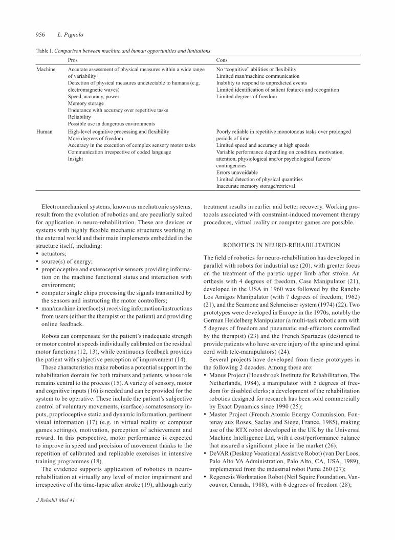
Table I. Comparison between machine and human opportunities and limitations
Pros Cons
Machine Accurate assessment of physical measures within a wide range of variabilityDetection of physical measures undetectable to humans (e.g. electromagnetic waves)Speed, accuracy, powerMemory storageEndurance with accuracy over repetitive tasksReliabilityPossible use in dangerous environments
No “cognitive” abilities or flexibilityLimited man/machine communicationInability to respond to unpredicted eventsLimited identification of salient features and recognition Limited degrees of freedom
Human High-level cognitive processing and flexibilityMore degrees of freedomAccuracy in the execution of complex sensory motor tasksCommunication irrespective of coded languageInsight
Poorly reliable in repetitive monotonous tasks over prolonged periods of timeLimited speed and accuracy at high speedsVariable performance depending on condition, motivation, attention, physiological and/or psychological factors/contingenciesErrors unavoidableLimited detection of physical quantitiesInaccurate memory storage/retrieval
J Rehabil Med 41
957Robotics in neuro-rehabilitation
• RTX Robot Arm (Universal Machine Intelligence LTD, Oxford, UK, 1986): 38% of robotic systems in use for rehabilitation training in 1989 had been implemented from the RTX (29);
• Handy 1 (Keele University, Keele SteffordShire, UK, 1987), a popular device implemented from the robotic arm Cyber 310 with 5 degrees of freedom (30);
• MoVAR (Mobile Vocational Assitive Robot, Stanford Uni-versity, Palo Alto, CA, USA, 1986) (31);
• Hadar WorkPlace Adaptations (Samhall-Hadar, Malmö, Sweden, 1988) (32);
• MIT Manus (Massachusetts Institute of Technology, Cam-bridge, USA, 1991), possibly the most seminal system developed thus far, widely marketed under the trade name In-Motion Shoulder- Elbow Robot (8).
RATIONALE, METHODOLOGIES AND EFFICACY
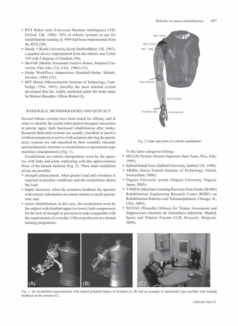
Several robotic systems have been tested for efficacy and in order to identify the useful robot/patient/therapist interaction in paretic upper limb functional rehabilitation after stroke. Research-dedicated systems are usually classified as passive (without actuators) or active (with actuators driving the paretic arm); systems are sub-classified by their scientific rationale and mechatronic structure as exoskeletons or operational-type machines (manipulators) (Fig. 1).
Exoskeletons are robotic manipulators worn by the opera-tor, with links and joints replicating with due approximation those of the human skeleton (Fig. 2). Three main modalities of use are possible:• strength enhancement, when greater load and resistance is
required in peculiar conditions and the exoskeleton shares the load;
• haptic functions, when the actuators feedback the operator with sensory information on remote motion or tactile percep-tion; and
• motor rehabilitation; in this case, the exoskeleton worn by the subject with disabled upper (or lower) limb compensates for the lack of strength or precision in tasks compatible with the requirements of everyday’s life or profession in a formal training programme.
To the latter categories belong:• MULOS System (Scuola Superiore Sant’Anna, Pisa, Italy,
1994); • Salford Rehab Exos (Salford University, Salford, UK, 1999);• ARMin (Swiss Federal Institute of Technology, Zürich,
Switzerland, 2006);• Nagoya University system (Nagoya University, Nagoya,
Japan, 2003);• T-WREX (Machines Assisting Recovery from Stroke (MARS)
Rehabilitation Engineering Research Center (RERC) on Rehabilitation Robotics and Telemanipulation, Chicago, IL, USA, 2004);
• WOTAS (Wearable Orthosis for Tremor Assessment and Suppression) (Instituto de Automática Industrial, Madrid, Spain and Hôpital Erasme ULB, Brussels, Belgium, 2006);
Fig. 1. An exoskeleton representation with related potential degree of freedom (A, B) and an example of operational type machine with training feedback on the monitor (C).
Fig. 2. Links and joints of a robotic manipulator.
J Rehabil Med 41
958 L. Pignolo
• MULOS (Motorized Upper Limb Orthotic System) (Centre for Rehabilitation and Engineering Studies, Newcastle, UK, 2001);
• MAHI Exos (Rice University, Houston, TX, USA, 2003);• L-Exos (Ligth Exoskeleton) (Scuola. Superiore Sant’Anna,
Pisa, Italy, 2007);• the Maryland-Georgetown-Army (MGA) Exoskeleton
(Georgetown University, Washington, DC, USA, 2005);• ARMOR Exoskeleton (University of Maryland, College
Park, MD and Georgetown University, Washington, DC, USA, 2007);
• 7 degree of freedom (DOF) Upper Limb Exoskeleton (Uni-versity of Washington, Washington, DC, USA, 2003).Exoskeletons offer greater DOF numbers up to 7 active DOF,
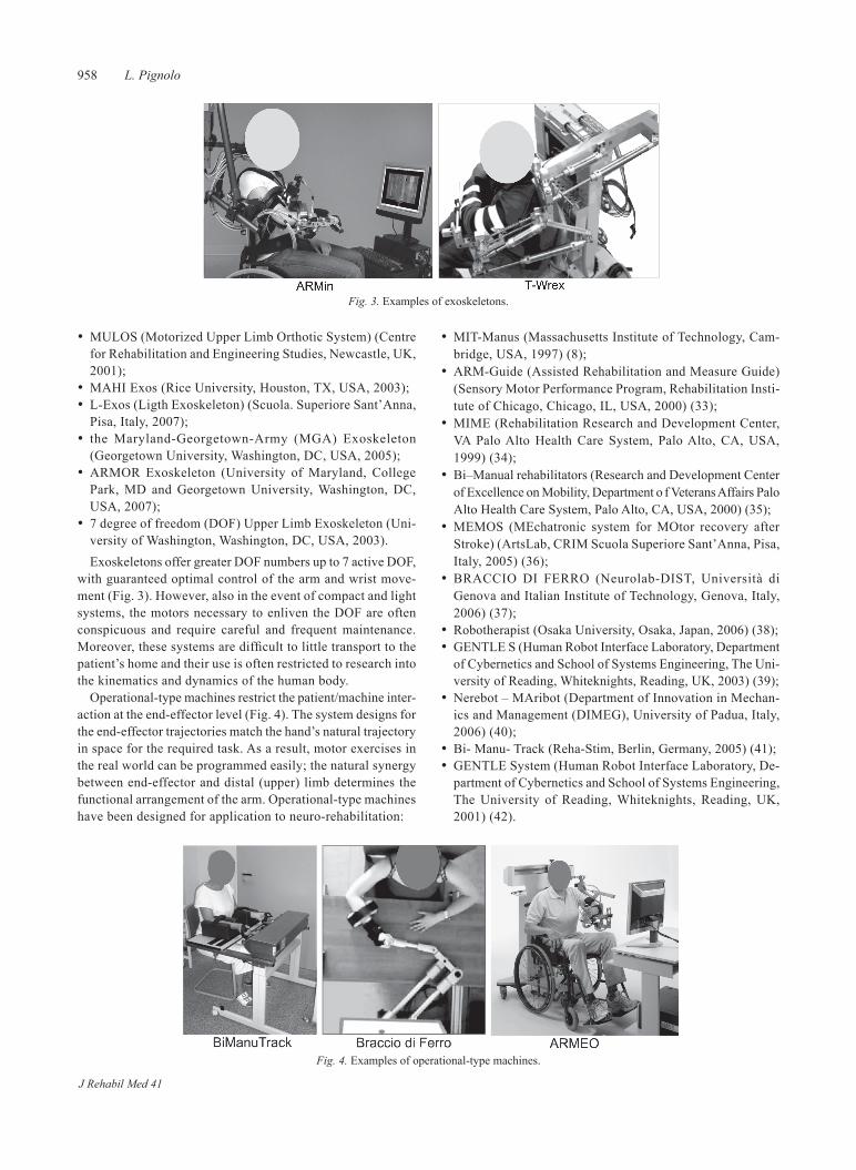
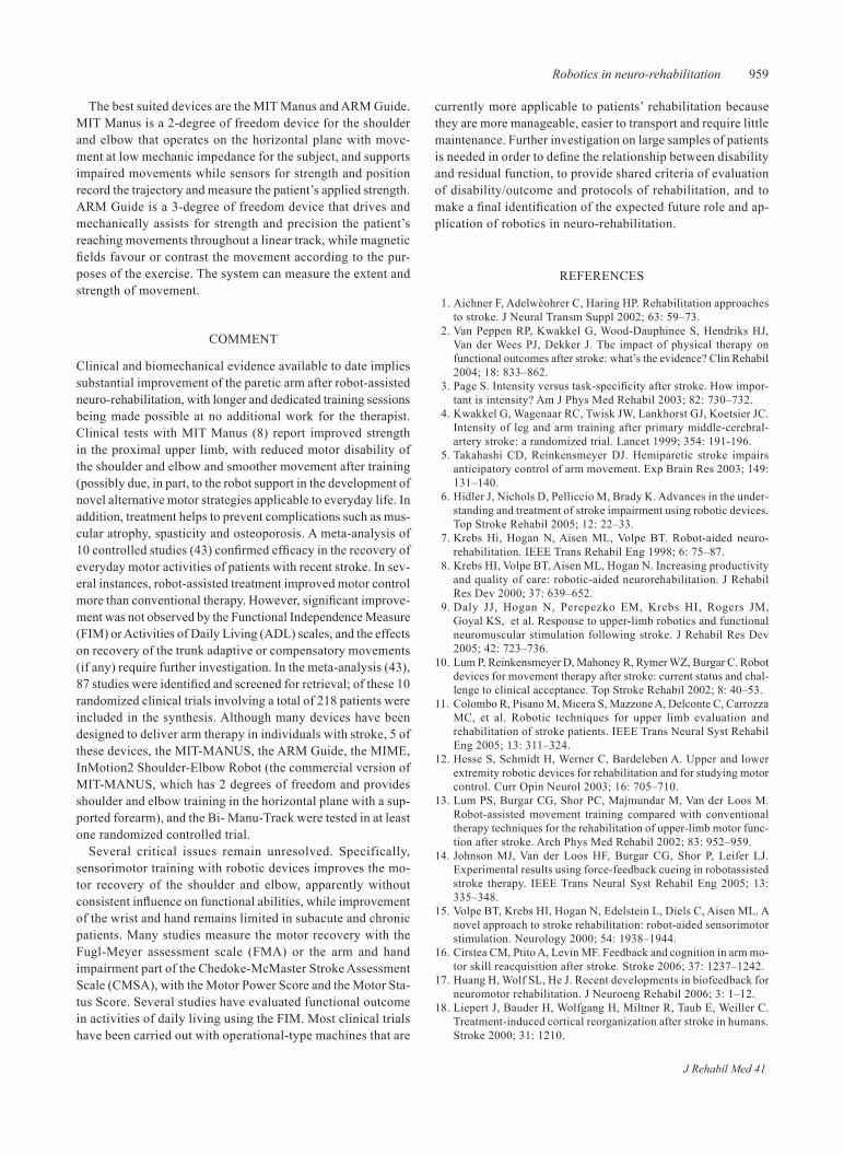
with guaranteed optimal control of the arm and wrist move-ment (Fig. 3). However, also in the event of compact and light systems, the motors necessary to enliven the DOF are often conspicuous and require careful and frequent maintenance. Moreover, these systems are difficult to little transport to the patient’s home and their use is often restricted to research into the kinematics and dynamics of the human body.
Operational-type machines restrict the patient/machine inter-action at the end-effector level (Fig. 4). The system designs for the end-effector trajectories match the hand’s natural trajectory in space for the required task. As a result, motor exercises in the real world can be programmed easily; the natural synergy between end-effector and distal (upper) limb determines the functional arrangement of the arm. Operational-type machines have been designed for application to neuro-rehabilitation:
• MIT-Manus (Massachusetts Institute of Technology, Cam-bridge, USA, 1997) (8);
• ARM-Guide (Assisted Rehabilitation and Measure Guide) (Sensory Motor Performance Program, Rehabilitation Insti-tute of Chicago, Chicago, IL, USA, 2000) (33);
• MIME (Rehabilitation Research and Development Center, VA Palo Alto Health Care System, Palo Alto, CA, USA, 1999) (34);
• Bi–Manual rehabilitators (Research and Development Center of Excellence on Mobility, Department o f Veterans Affairs Palo Alto Health Care System, Palo Alto, CA, USA, 2000) (35);
• MEMOS (MEchatronic system for MOtor recovery after Stroke) (ArtsLab, CRIM Scuola Superiore Sant’Anna, Pisa, Italy, 2005) (36);
• BRACCIO DI FERRO (Neurolab-DIST, Università di Genova and Italian Institute of Technology, Genova, Italy, 2006) (37);
• Robotherapist (Osaka University, Osaka, Japan, 2006) (38);• GENTLE S (Human Robot Interface Laboratory, Department
of Cybernetics and School of Systems Engineering, The Uni-versity of Reading, Whiteknights, Reading, UK, 2003) (39);
• Nerebot – MAribot (Department of Innovation in Mechan-ics and Management (DIMEG), University of Padua, Italy, 2006) (40);
• Bi- Manu- Track (Reha-Stim, Berlin, Germany, 2005) (41);• GENTLE System (Human Robot Interface Laboratory, De-
partment of Cybernetics and School of Systems Engineering, The University of Reading, Whiteknights, Reading, UK, 2001) (42).
Fig. 4. Examples of operational-type machines.
Fig. 3. Examples of exoskeletons.
J Rehabil Med 41
959Robotics in neuro-rehabilitation
The best suited devices are the MIT Manus and ARM Guide. MIT Manus is a 2-degree of freedom device for the shoulder and elbow that operates on the horizontal plane with move-ment at low mechanic impedance for the subject, and supports impaired movements while sensors for strength and position record the trajectory and measure the patient’s applied strength. ARM Guide is a 3-degree of freedom device that drives and mechanically assists for strength and precision the patient’s reaching movements throughout a linear track, while magnetic fields favour or contrast the movement according to the pur-poses of the exercise. The system can measure the extent and strength of movement.
COMMENT
Clinical and biomechanical evidence available to date implies substantial improvement of the paretic arm after robot-assisted neuro-rehabilitation, with longer and dedicated training sessions being made possible at no additional work for the therapist. Clinical tests with MIT Manus (8) report improved strength in the proximal upper limb, with reduced motor disability of the shoulder and elbow and smoother movement after training (possibly due, in part, to the robot support in the development of novel alternative motor strategies applicable to everyday life. In addition, treatment helps to prevent complications such as mus-cular atrophy, spasticity and osteoporosis. A meta-analysis of 10 controlled studies (43) confirmed efficacy in the recovery of everyday motor activities of patients with recent stroke. In sev-eral instances, robot-assisted treatment improved motor control more than conventional therapy. However, significant improve-ment was not observed by the Functional Independence Measure (FIM) or Activities of Daily Living (ADL) scales, and the effects on recovery of the trunk adaptive or compensatory movements (if any) require further investigation. In the meta-analysis (43), 87 studies were identified and screened for retrieval; of these 10 randomized clinical trials involving a total of 218 patients were included in the synthesis. Although many devices have been designed to deliver arm therapy in individuals with stroke, 5 of these devices, the MIT-MANUS, the ARM Guide, the MIME, InMotion2 Shoulder-Elbow Robot (the commercial version of MIT-MANUS, which has 2 degrees of freedom and provides shoulder and elbow training in the horizontal plane with a sup-ported forearm), and the Bi- Manu-Track were tested in at least one randomized controlled trial.
Several critical issues remain unresolved. Specifically, sensorimotor training with robotic devices improves the mo-tor recovery of the shoulder and elbow, apparently without consistent influence on functional abilities, while improvement of the wrist and hand remains limited in subacute and chronic patients. Many studies measure the motor recovery with the Fugl-Meyer assessment scale (FMA) or the arm and hand impairment part of the Chedoke-McMaster Stroke Assessment Scale (CMSA), with the Motor Power Score and the Motor Sta-tus Score. Several studies have evaluated functional outcome in activities of daily living using the FIM. Most clinical trials have been carried out with operational-type machines that are
currently more applicable to patients’ rehabilitation because they are more manageable, easier to transport and require little maintenance. Further investigation on large samples of patients is needed in order to define the relationship between disability and residual function, to provide shared criteria of evaluation of disability/outcome and protocols of rehabilitation, and to make a final identification of the expected future role and ap-plication of robotics in neuro-rehabilitation.
REFERENCES
Aichner F, Adelwèohrer C, Haring HP. Rehabilitation approaches 1. to stroke. J Neural Transm Suppl 2002; 63: 59–73.Van Peppen RP, Kwakkel G, Wood-Dauphinee S, Hendriks HJ, 2. Van der Wees PJ, Dekker J. The impact of physical therapy on functional outcomes after stroke: what’s the evidence? Clin Rehabil 2004; 18: 833–862.Page S. Intensity versus task-specificity after stroke. How impor-3. tant is intensity? Am J Phys Med Rehabil 2003; 82: 730–732.Kwakkel G, Wagenaar RC, Twisk JW, Lankhorst GJ, Koetsier JC. 4. Intensity of leg and arm training after primary middle-cerebral-artery stroke: a randomized trial. Lancet 1999; 354: 191-196.Takahashi CD, Reinkensmeyer DJ. Hemiparetic stroke impairs 5. anticipatory control of arm movement. Exp Brain Res 2003; 149: 131–140.Hidler J, Nichols D, Pelliccio M, Brady K. Advances in the under-6. standing and treatment of stroke impairment using robotic devices. Top Stroke Rehabil 2005; 12: 22–33.Krebs Hi, Hogan N, Aisen ML, Volpe BT. Robot-aided neuro-7. rehabilitation. IEEE Trans Rehabil Eng 1998; 6: 75–87.Krebs HI, Volpe BT, Aisen ML, Hogan N. Increasing productivity 8. and quality of care: robotic-aided neurorehabilitation. J Rehabil Res Dev 2000; 37: 639–652.Daly JJ, Hogan N, Perepezko EM, Krebs HI, Rogers JM, 9. Goyal KS, et al. Response to upper-limb robotics and functional neuromuscular stimulation following stroke. J Rehabil Res Dev 2005; 42: 723–736.Lum P, Reinkensmeyer D, Mahoney R, Rymer WZ, Burgar C. Robot 10. devices for movement therapy after stroke: current status and chal-lenge to clinical acceptance. Top Stroke Rehabil 2002; 8: 40–53.Colombo R, Pisano M, Micera S, 11. Mazzone A, Delconte C, Carrozza MC, et al. Robotic techniques for upper limb evaluation and rehabilitation of stroke patients. IEEE Trans Neural Syst Rehabil Eng 2005; 13: 311–324.Hesse S, Schmidt H, Werner C, Bardeleben A. Upper and lower 12. extremity robotic devices for rehabilitation and for studying motor control. Curr Opin Neurol 2003; 16: 705–710.Lum PS, Burgar CG, Shor PC, Majmundar M, Van der Loos M. 13. Robot-assisted movement training compared with conventional therapy techniques for the rehabilitation of upper-limb motor func-tion after stroke. Arch Phys Med Rehabil 2002; 83: 952–959.Johnson MJ, Van der Loos HF, Burgar CG, Shor P, Leifer LJ. 14. Experimental results using force-feedback cueing in robotassisted stroke therapy. IEEE Trans Neural Syst Rehabil Eng 2005; 13: 335–348.Volpe BT, Krebs HI, Hogan N, Edelstein L, Diels C, Aisen ML. A 15. novel approach to stroke rehabilitation: robot-aided sensorimotor stimulation. Neurology 2000; 54: 1938–1944.Cirstea CM, Ptito A, Levin MF. Feedback and cognition in arm mo-16. tor skill reacquisition after stroke. Stroke 2006; 37: 1237–1242. Huang H, Wolf SL, He J. Recent developments in biofeedback for 17. neuromotor rehabilitation. J Neuroeng Rehabil 2006; 3: 1–12.Liepert J, Bauder H, Wolfgang H, Miltner R, Taub E, Weiller C. 18. Treatment-induced cortical reorganization after stroke in humans. Stroke 2000; 31: 1210.
J Rehabil Med 41
960 L. Pignolo
Prange GB, Jannink MA, Groothuis-Oudshoorn CG, Hermens 19. HJ, Ijzerman MJ. Systematic review of the effect of robot-aided therapy on recovery of the hemiparetic arm after stroke. J Rehabil Res Dev 2006; 43: 171–184.Garcia E, Jimenez MA, Gonzalez P, Armada M. The evolution 20. of robotic research. IEEE Robotics and automation magazine. 2007; 14: 90-103.Kim Y, Cook AM. Manipulation and Mobility Aids. In: Webster 21. JG, editor. Electronic devices for rehabilitation. London: Chap-man and Hall; 1985.Seamone W, Schmeisser G. Evaluation of the JHU/APL Robot 22. Arm Workstation. In: Foulds R, editor. Interactive Robotic Aids. World Rehabilitation Fund Monograph #37. New York: World Rehabilitation Fund; 1986.Roesler H, Küppers HJ, Schmalenbach E. The medical manipula-23. tor and its adapted environment: A system for the rehabilitation of severely handicapped. Proc International Conference on Tele-manipulators for the Physically Handicapped. France: Institut de Recherche d’Informatique et d’Automatique, 1978.Kwee HH, Tramblay M, Barbier R. First Experimentation of the 24. spartacus telethesis in a clinical environment. Paraplegia 1983; 21: 275.Kwee HH, Duimel JJ. The MANUS wheelchair-borne manipula-25. tor: System review and first results. Proc. International Advanced Robotics Programme Workshop on Domestic and Medical & Healthcare Robotics, Newcastle, UK, 1989.Detriche JM, Lesigne B, Bernard T. Development of a workstation 26. for handicapped people including the robotized system master. Proc. International Conference On Rehabilitation Robotics, Atlanta, GA, USA 1991.Van der Loos M, Hammel J, Schwandt D, Lees D, Leifer LJ, 27. Perkash I. Design and evaluation of a vocational desktop robot. Proc. Rehabilitation Engineering and Assistive technology Society of North America 12th Annual Conference, New Orleans LA, USA, 1989.Fengler M, Cameron WM. Clinical testing of a low cost robotic 28. arm for the severely physically disabled. Proc. First International Workshop on Robotic Applications in Medical and Healthcare, Ottawa, ON, Canada, 1988.Jones T. RAID – Towards greater independence in the office and home 29. environment. Proc. International Conference on Rehabilitation Robot-ics (ICORR), Stanford, Palo Alto, CA, USA, 1999, p. 201–206.Topping M. Handy 1, A robotic aid to independence for severely 30. disabled people. In: Mokhtari M, editor. Integration of assistive technology in the information age. Amsterdam: IOS Press; 2001, p. 142–147.Van der Loos M, Michalowski S, Leifer L. Design of an omni-31.
directional mobile robot as a manipulation aid for the severely disabled. In: Foulds R, editor. Interactive Robotic Aids. World Rehabilitation Fund Monograph #37. New York: World Rehabili-tation Fund; 1986.Holmberg L. The installation of a robotized workstation at Sam-32. hall-HADAR. ECART 2 – Proceedings 1993, p. 29.2Reinkensmeyer DJ, Kahn LE, Averbuch M, McKenna-Cole AN, 33. Schmit BD, Rymer WZ. Understanding and treating arm movement impairment after chronic brain injury: progress with the ARM Guide. J Rehabil Res Dev 2000; 37: 653–662.Lum PS, Burgar CG, Shor PC. Evidence for improved muscle 34. activation patterns after retraining of reaching movements with the MIME robotic system in subjects with post-stroke hemiparesis. IEEE Trans Neural Syst Rehabil Eng 2004; 12: 186–194.Lum SP, Lehman SL, Reinkensmeyer D. The bimanual lifting 35. rehabilitator: an adaptive machine for therapy of stroke patients. Journal of Rehabilitation Engineering 1995; 3: 166–174.Micera S, Carozza MC, Guglielmelli E, Cappiello G, Zaccone F, 36. Freschi C, et al. A simple robotic system for neurorehabilitation. Auton Robot 2005; 19: 271–284.Casadio M, Morasso P, Sanguineti V, Arricchiello V. Braccio di 37. ferro: A new haptic workstation for neuromotor rehabilitation. Technology healthcare 2006; 13: 1–20.Furusho J, Kikuchi T, Oda K, Ohyama Y, Morita T, Shichi N, et al. 38. A 6-DOF rehabilitation support system for upper limbs including wrists “Robotherapist” with physical therapy. 10th International Conference on Rehabilitation Robotics. Noordwijk, The Nether-lands, 2007, p. 304–309.Amirabdollahian, F, Loureiro R, Driessen B, Harwin W. Error 39. correction movement for machine assisted stroke rehabilitation In: Mokhtari M, editor. Integration of assistive technology in the information age. Amsterdam: IOS Press; 2001, p. 60–65.Fanin C, Gallina P, Rossi A, Zanatta U, Masiero S. NeReBot: a wire 40. based robot for neurorehabilitation. Proceedings of the IEEE 8th International conference on rehabilitation roboticsICORR 2003, Dejeon Republic of Korea, April 2003.Hesse S, Werner C, Pohl M, Rueckriem S, Mehrhoz J, Lingnau 41. ML. Computerized arm training improves the motor control of the severely affected arm after stoke. Stroke 2005; 36: 1960–1966.Harwin W, Loureiro R, Amirabdollahian F, Taylor M, Johnson G, 42. Stokes E, et al. The Gentle/s project: a new method for delivering neurorehabilitation. In: Marincek C, Bühler C, Knops H, Andrich R, editors. Assistive technology – added value to the quality of life AAATE’01. Amsterdam: IOS Press; 2001; p. 36–41.Kwakkel G, Boudewijn KJ, Krebs HI. Effects of robot-assisted 43. therapy on upper limb recovery after stroke: A systematic review. Neurorehabil Neural Repair 2008; 22: 111–121
J Rehabil Med 41
oriGinal report
J Rehabil Med 2009; 41: 961–965
J Rehabil Med 41© 2009 The Authors. doi: 10.2340/16501977-0400Journal Compilation © 2009 Foundation of Rehabilitation Information. ISSN 1650-1977
Objective: to carry out a preliminary feasibility study of a new concept of robot therapy for severely impaired patients after stroke. Design: a haptic manipulandum connected to a bar that can rotate freely while providing a measure of the rotation angle. the controller combines a bilateral reaching task with the task of balancing the action of the 2 arms. reinforcement is given to the subject in 2 forms: audio-visual and haptic by means of adaptable force fields. Patients: four highly paretic patients with chronic stroke (fugl-meyer score less than 15). Methods: the training cycle consisted of 5 sessions over a pe-riod of 2 weeks. each session (45 min) was divided in blocks of 10 pairs of forward/backward movements. performance was determined by evaluating the number of successful movements per session, the session-by-session decrease in the assistive field, the mean reaching time, and the mean stopping field. Results: all subjects could understand the task, appreciated it and improved their performance during training. the reaching movements became smoother and quicker; balance errors and the magnitude of the resisting field were consist-ently reduced. Conclusion: bilateral robot therapy is a promising tech-nique, provided that it self-adapts to the patient’s perform-ance. formal clinical trials should address this point. Key words: rehabilitation, robotics, stroke, touch perception, re-inforcement, learning. J Rehabil Med 2009; 41: 961–965
Correspondence address: Valentina Squeri, Istituto Italiano di Tecnologia, Via Morego 30, IT-16163 Genova, Italy. E-mail: [email protected] March 16, 2009; accepted May 19, 2009
INTRODUCTION
Over the past years evidence has mounted regarding the capac-ity of the central nervous system (CNS) to alter its structure and function throughout all sorts of life experiences, including injuries to the CNS, in a complex network of interacting processes (1–4). Animal models of focal brain injuries suggest that behaviour is probably the most powerful modulator of post-injury recovery (5,
6): thus, beyond the initial critical period of self-repair (7), the principal process responsible for functional recovery is the use-dependent reorganization of neural mechanisms made possible by neural plasticity (8). Moreover, imaging data suggest that circuitry in motor cortices on both sides of the brain is modified during recovery (9), and this has lead to the concept that bilateral move-ment permits inter-hemispheric facilitation of the limbs (10).
This is the main motivation for the design of robotic or me-chatronic devices that aim at bilateral training of the normal and the paretic arm. Early prototypes of bilateral trainers were developed at the VA Palo Alto Center (11), based mainly on the so-called mirror image movement enabler concept (MIME) in which a robot manipulator applied forces to the paretic arm dur-ing goal-directed movements, keeping it in mirror-symmetry with the unaffected arm whose position was monitored by a position digitizer. Simple, low-cost bilateral arm trainers have also been developed and tested. Bilateral Arm Training, Auditory-Cued (BATRAC) is an example of such systems: it is a one degree of freedom custom-made mechanical arm trainer (12) that allows auditory cued patients to move two unyoked T-grips forward and backward in a parallel or alternate fash-ion. Another system in the same category is Reha-Slide (13), which allows unilateral or bilateral training of up to 3 degrees of freedom of the shoulder, elbow and wrist.
These bilateral trainers are aimed in particular at severely impaired patients who cannot carry out full extension reaching movements with the paretic limb without suitable assistance and thus are not eligible for conventional treatment approaches, including the promising constraint-induced movement therapy (14). However, in the previously mentioned bilateral arm train-ers, movements of the paretic arm are activated in a passive way, using the unaffected arm as the “primus movens” in order to overcome the inability of the paretic limb to carry out the prescribed movements.
In this paper, we propose an alternative concept: to use the robot as “primus movens” and combine the bilateral reaching task with the task of balancing the action of the 2 arms, ac-cording to a reinforcement learning paradigm. In this way the relationship between the 2 limbs is not of the master-slave type and the patient is strongly motivated to balance and co-ordinate the activation of the 2 limbs. This new bilateral training concept was implemented by means of a simple mechanical extension
BILATERAL ROBOT THERAPY BASED ON HAPTICS AND REINFORCEMENT LEARNING: FEASIBILITY STUDY OF A NEW CONCEPT FOR TREATMENT OF
PATIENTS AFTER STROKE
Valentina Squeri, Eng1*, Maura Casadio, PhD1*, Elena Vergaro, Eng2*, Psiche Giannoni, PT3, Pietro Morasso, Eng1,2 and Vittorio Sanguineti, PhD1,2
From the 1Italian Institute of Technology, 2Neurolab, DIST, University of Genova and 3ART Education and Rehabilitation Center, Genova, Italy. *Valentina Squeri, Maura Casadio and Elena Vergaro contributed equally to this study.
962 V. Squeri et al.
of the haptic robot Braccio di Ferro (BdF) (15) and an original haptic interaction scheme. The mechanical extension consists of a bar connected to the end-effector of the robot. The bar can rotate freely and the corresponding rotation angle is measured by a coaxial rotation sensor. The subject holds 2 handles at the 2 ends of the bar and is required to balance the forces applied by the 2 hands in such a way to reach a target and, at the same time, maintain the bar at a prescribed angle. The reinforcement learning scheme is expressed by means of suitable force fields that adapt to the patient’s performance. The feasibility of this training concept was tested with a preliminary clinical study that yielded promising results with 4 severely impaired patients. The approach can be adapted easily to any haptic robot that, as BdF or MIT-Manus (16), allows bi-directional human-robot interac-tion and the fine control of the interaction forces.
METHODSExperimental apparatusThe robot, BdF, is a planar manipulandum with 2 degrees of freedom, designed at the University of Genoa (15). Its most relevant features are: (i) large planar workspace (80 × 40 cm ellipse); (ii) rigid mechanical structure with direct drive of 2 brushless motors, designed in order to have low intrinsic mechanical impedance at the end-effector; (iii) large available force at the handle (continuous force > 50 N; peak force > 200 N); (iv) impedance control scheme that allows a bi-directional, smooth haptic interaction between the robot and the patient. Low mechanical imped-ance means that when the robot controller is off the subject perceives a virtually weightless, frictionless, and noiseless manipulandum. This also significantly improves the safety of the robot.
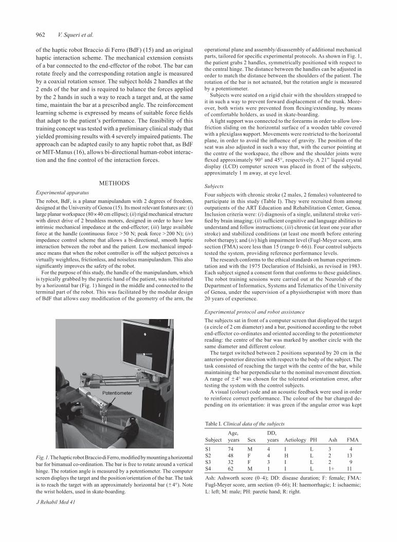
For the purpose of this study, the handle of the manipulandum, which is typically grabbed by the paretic hand of the patient, was substituted by a horizontal bar (Fig. 1) hinged in the middle and connected to the terminal part of the robot. This was facilitated by the modular design of BdF that allows easy modification of the geometry of the arm, the
operational plane and assembly/disassembly of additional mechanical parts, tailored for specific experimental protocols. As shown in Fig. 1, the patient grabs 2 handles, symmetrically positioned with respect to the central hinge. The distance between the handles can be adjusted in order to match the distance between the shoulders of the patient. The rotation of the bar is not actuated, but the rotation angle is measured by a potentiometer.
Subjects were seated on a rigid chair with the shoulders strapped to it in such a way to prevent forward displacement of the trunk. More-over, both wrists were prevented from flexing/extending, by means of comfortable holders, as used in skate-boarding.
A light support was connected to the forearms in order to allow low-friction sliding on the horizontal surface of a wooden table covered with a plexiglass support. Movements were restricted to the horizontal plane, in order to avoid the influence of gravity. The position of the seat was also adjusted in such a way that, with the cursor pointing at the centre of the workspace, the elbow and the shoulder joints were flexed approximately 90° and 45°, respectively. A 21” liquid crystal display (LCD) computer screen was placed in front of the subjects, approximately 1 m away, at eye level.
SubjectsFour subjects with chronic stroke (2 males, 2 females) volunteered to participate in this study (Table I). They were recruited from among outpatients of the ART Education and Rehabilitation Center, Genoa. Inclusion criteria were: (i) diagnosis of a single, unilateral stroke veri-fied by brain imaging; (ii) sufficient cognitive and language abilities to understand and follow instructions; (iii) chronic (at least one year after stroke) and stabilized conditions (at least one month before entering robot therapy); and (iv) high impairment level (Fugl-Meyer score, arm section (FMA) score less than 15 (range 0–66)). Four control subjects tested the system, providing reference performance levels.
The research conforms to the ethical standards on human experimen-tation and with the 1975 Declaration of Helsinki, as revised in 1983. Each subject signed a consent form that conforms to these guidelines. The robot training sessions were carried out at the Neurolab of the Department of Informatics, Systems and Telematics of the University of Genoa, under the supervision of a physiotherapist with more than 20 years of experience.
Experimental protocol and robot assistanceThe subjects sat in front of a computer screen that displayed the target (a circle of 2 cm diameter) and a bar, positioned according to the robot end-effector co-ordinates and oriented according to the potentiometer reading: the centre of the bar was marked by another circle with the same diameter and different colour.
The target switched between 2 positions separated by 20 cm in the anterior-posterior direction with respect to the body of the subject. The task consisted of reaching the target with the centre of the bar, while maintaining the bar perpendicular to the nominal movement direction. A range of ± 4° was chosen for the tolerated orientation error, after testing the system with the control subjects.
A visual (colour) code and an acoustic feedback were used in order to reinforce correct performance. The colour of the bar changed de-pending on its orientation: it was green if the angular error was kept
Fig. 1. The haptic robot Braccio di Ferro, modified by mounting a horizontal bar for bimanual co-ordination. The bar is free to rotate around a vertical hinge. The rotation angle is measured by a potentiometer. The computer screen displays the target and the position/orientation of the bar. The task is to reach the target with an approximately horizontal bar (± 4°). Note the wrist holders, used in skate-boarding.
Table I. Clinical data of the subjects
SubjectAge, years Sex
DD, years Aetiology PH Ash FMA
S1 74 M 4 I L 3 4S2 48 F 4 H L 2 13S3 32 F 3 I L 2 9S4 62 M 1 I L 1+ 11
Ash: Ashworth score (0–4); DD: disease duration; F: female; FMA: Fugl-Meyer score, arm section (0–66); H: haemorrhagic; I: ischaemic; L: left; M: male; PH: paretic hand; R: right.
J Rehabil Med 41
963Bilateral robot treatment of stroke patients
inside the prescribed range and it became red when the error became larger. Moreover, an unpleasant sound signalled that the orientation error was outside the threshold and a pleasant sound marked that the target was reached.
As soon as a subject reached a target, that target was switched off and the other target was activated, thus inducing a sequence of forward/backward movements that became quicker and quicker as performance improved.
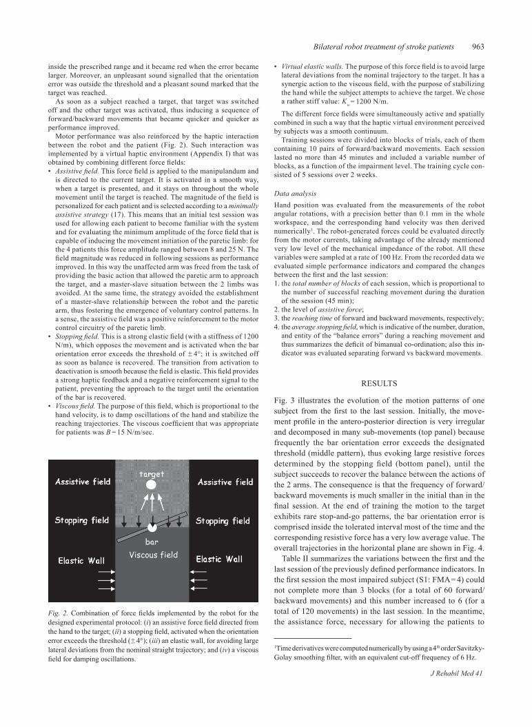
Motor performance was also reinforced by the haptic interaction between the robot and the patient (Fig. 2). Such interaction was implemented by a virtual haptic environment (Appendix I) that was obtained by combining different force fields: • Assistivefield.This force field is applied to the manipulandum and
is directed to the current target. It is activated in a smooth way, when a target is presented, and it stays on throughout the whole movement until the target is reached. The magnitude of the field is personalized for each patient and is selected according to a minimally assistive strategy (17). This means that an initial test session was used for allowing each patient to become familiar with the system and for evaluating the minimum amplitude of the force field that is capable of inducing the movement initiation of the paretic limb: for the 4 patients this force amplitude ranged between 8 and 25 N. The field magnitude was reduced in following sessions as performance improved. In this way the unaffected arm was freed from the task of providing the basic action that allowed the paretic arm to approach the target, and a master-slave situation between the 2 limbs was avoided. At the same time, the strategy avoided the establishment of a master-slave relationship between the robot and the paretic arm, thus fostering the emergence of voluntary control patterns. In a sense, the assistive field was a positive reinforcement to the motor control circuitry of the paretic limb.
• Stoppingfield.This is a strong elastic field (with a stiffness of 1200 N/m), which opposes the movement and is activated when the bar orientation error exceeds the threshold of ± 4°; it is switched off as soon as balance is recovered. The transition from activation to deactivation is smooth because the field is elastic. This field provides a strong haptic feedback and a negative reinforcement signal to the patient, preventing the approach to the target until the orientation of the bar is recovered.
• Viscousfield.The purpose of this field, which is proportional to the hand velocity, is to damp oscillations of the hand and stabilize the reaching trajectories. The viscous coefficient that was appropriate for patients was B = 15 N/m/sec.
• Virtual elastic walls. The purpose of this force field is to avoid large lateral deviations from the nominal trajectory to the target. It has a synergic action to the viscous field, with the purpose of stabilizing the hand while the subject attempts to achieve the target. We chose a rather stiff value: Kw = 1200 N/m.
The different force fields were simultaneously active and spatially combined in such a way that the haptic virtual environment perceived by subjects was a smooth continuum.
Training sessions were divided into blocks of trials, each of them containing 10 pairs of forward/backward movements. Each session lasted no more than 45 minutes and included a variable number of blocks, as a function of the impairment level. The training cycle con-sisted of 5 sessions over 2 weeks.
Data analysisHand position was evaluated from the measurements of the robot angular rotations, with a precision better than 0.1 mm in the whole workspace, and the corresponding hand velocity was then derived numerically1. The robot-generated forces could be evaluated directly from the motor currents, taking advantage of the already mentioned very low level of the mechanical impedance of the robot. All these variables were sampled at a rate of 100 Hz. From the recorded data we evaluated simple performance indicators and compared the changes between the first and the last session:1. the total number of blocks of each session, which is proportional to
the number of successful reaching movement during the duration of the session (45 min);
2. the level of assistive force;3. the reaching time of forward and backward movements, respectively;4. the averagestoppingfield, which is indicative of the number, duration,
and entity of the “balance errors” during a reaching movement and thus summarizes the deficit of bimanual co-ordination; also this in-dicator was evaluated separating forward vs backward movements.
RESULTS
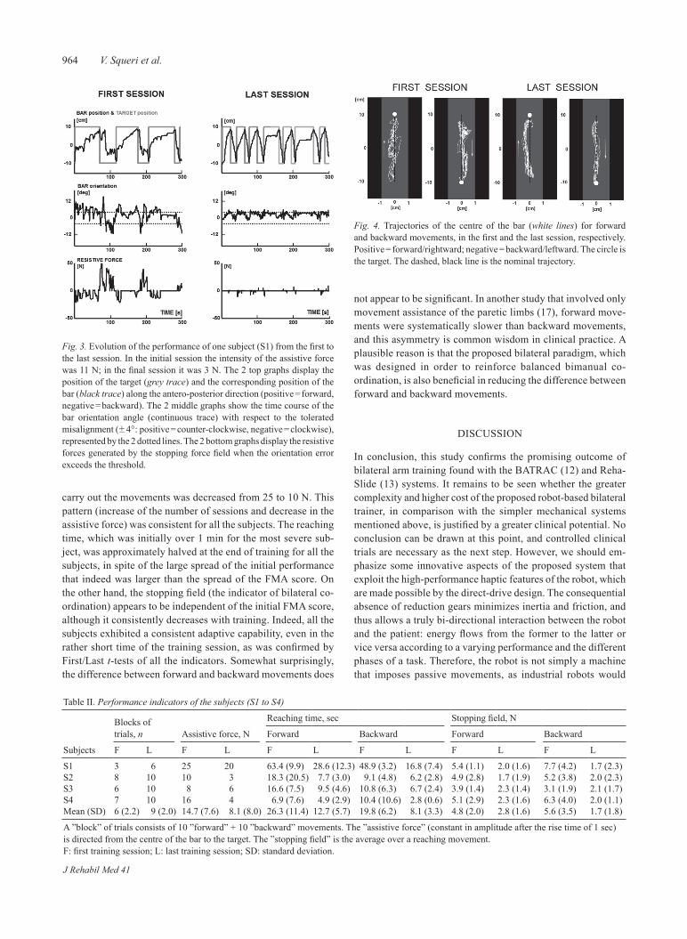
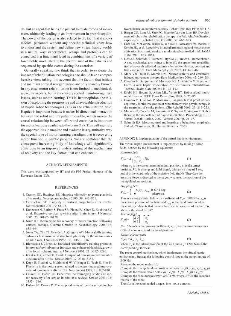
Fig. 3 illustrates the evolution of the motion patterns of one subject from the first to the last session. Initially, the move-ment profile in the antero-posterior direction is very irregular and decomposed in many sub-movements (top panel) because frequently the bar orientation error exceeds the designated threshold (middle pattern), thus evoking large resistive forces determined by the stopping field (bottom panel), until the subject succeeds to recover the balance between the actions of the 2 arms. The consequence is that the frequency of forward/backward movements is much smaller in the initial than in the final session. At the end of training the motion to the target exhibits rare stop-and-go patterns, the bar orientation error is comprised inside the tolerated interval most of the time and the corresponding resistive force has a very low average value. The overall trajectories in the horizontal plane are shown in Fig. 4.
Table II summarizes the variations between the first and the last session of the previously defined performance indicators. In the first session the most impaired subject (S1: FMA = 4) could not complete more than 3 blocks (for a total of 60 forward/backward movements) and this number increased to 6 (for a total of 120 movements) in the last session. In the meantime, the assistance force, necessary for allowing the patients to
1Time derivatives were computed numerically by using a 4th order Savitzky-Golay smoothing filter, with an equivalent cut-off frequency of 6 Hz.
Fig. 2. Combination of force fields implemented by the robot for the designed experimental protocol: (i) an assistive force field directed from the hand to the target; (ii) a stopping field, activated when the orientation error exceeds the threshold (± 4°); (iii) an elastic wall, for avoiding large lateral deviations from the nominal straight trajectory; and (iv) a viscous field for damping oscillations.
J Rehabil Med 41
964 V. Squeri et al.
carry out the movements was decreased from 25 to 10 N. This pattern (increase of the number of sessions and decrease in the assistive force) was consistent for all the subjects. The reaching time, which was initially over 1 min for the most severe sub-ject, was approximately halved at the end of training for all the subjects, in spite of the large spread of the initial performance that indeed was larger than the spread of the FMA score. On the other hand, the stopping field (the indicator of bilateral co-ordination) appears to be independent of the initial FMA score, although it consistently decreases with training. Indeed, all the subjects exhibited a consistent adaptive capability, even in the rather short time of the training session, as was confirmed by First/Last t-tests of all the indicators. Somewhat surprisingly, the difference between forward and backward movements does
not appear to be significant. In another study that involved only movement assistance of the paretic limbs (17), forward move-ments were systematically slower than backward movements, and this asymmetry is common wisdom in clinical practice. A plausible reason is that the proposed bilateral paradigm, which was designed in order to reinforce balanced bimanual co-ordination, is also beneficial in reducing the difference between forward and backward movements.
DISCUSSION
In conclusion, this study confirms the promising outcome of bilateral arm training found with the BATRAC (12) and Reha-Slide (13) systems. It remains to be seen whether the greater complexity and higher cost of the proposed robot-based bilateral trainer, in comparison with the simpler mechanical systems mentioned above, is justified by a greater clinical potential. No conclusion can be drawn at this point, and controlled clinical trials are necessary as the next step. However, we should em-phasize some innovative aspects of the proposed system that exploit the high-performance haptic features of the robot, which are made possible by the direct-drive design. The consequential absence of reduction gears minimizes inertia and friction, and thus allows a truly bi-directional interaction between the robot and the patient: energy flows from the former to the latter or vice versa according to a varying performance and the different phases of a task. Therefore, the robot is not simply a machine that imposes passive movements, as industrial robots would
Fig. 3. Evolution of the performance of one subject (S1) from the first to the last session. In the initial session the intensity of the assistive force was 11 N; in the final session it was 3 N. The 2 top graphs display the position of the target (grey trace) and the corresponding position of the bar (black trace) along the antero-posterior direction (positive = forward, negative = backward). The 2 middle graphs show the time course of the bar orientation angle (continuous trace) with respect to the tolerated misalignment (± 4°: positive = counter-clockwise, negative = clockwise), represented by the 2 dotted lines. The 2 bottom graphs display the resistive forces generated by the stopping force field when the orientation error exceeds the threshold.
Fig. 4. Trajectories of the centre of the bar (white lines) for forward and backward movements, in the first and the last session, respectively. Positive = forward/rightward; negative = backward/leftward. The circle is the target. The dashed, black line is the nominal trajectory.
Table II. Performance indicators of the subjects (S1 to S4)
Subjects
Blocks of trials, n Assistive force, N
Reaching time, sec Stopping field, N
Forward Backward Forward Backward
F L F L F L F L F L F L
S1 3 6 25 20 63.4 (9.9) 28.6 (12.3) 48.9 (3.2) 16.8 (7.4) 5.4 (1.1) 2.0 (1.6) 7.7 (4.2) 1.7 (2.3)S2 8 10 10 3 18.3 (20.5) 7.7 (3.0) 9.1 (4.8) 6.2 (2.8) 4.9 (2.8) 1.7 (1.9) 5.2 (3.8) 2.0 (2.3)S3 6 10 8 6 16.6 (7.5) 9.5 (4.6) 10.8 (6.3) 6.7 (2.4) 3.9 (1.4) 2.3 (1.4) 3.1 (1.9) 2.1 (1.7)S4 7 10 16 4 6.9 (7.6) 4.9 (2.9) 10.4 (10.6) 2.8 (0.6) 5.1 (2.9) 2.3 (1.6) 6.3 (4.0) 2.0 (1.1)Mean (SD) 6 (2.2) 9 (2.0) 14.7 (7.6) 8.1 (8.0) 26.3 (11.4) 12.7 (5.7) 19.8 (6.2) 8.1 (3.3) 4.8 (2.0) 2.8 (1.6) 5.6 (3.5) 1.7 (1.8)
A ”block” of trials consists of 10 ”forward” + 10 ”backward” movements. The ”assistive force” (constant in amplitude after the rise time of 1 sec) is directed from the centre of the bar to the target. The ”stopping field” is the average over a reaching movement. F: first training session; L: last training session; SD: standard deviation.
J Rehabil Med 41
965Bilateral robot treatment of stroke patients
do, but an agent that helps the patient to relate force and move-ment, ultimately leading to an improvement in proprioception. The power of the design is also related to the fact that it allows medical personnel without any specific technical know-how to understand the system and define new virtual haptic worlds in a natural way: experimental set-ups and protocols can be conceived at a functional level as combinations of a variety of force fields, modulated by the performance of the patients and sequenced by specific events during the exercises.
Generally speaking, we think that in order to evaluate the impact of rehabilitation technologies one should take a compre-hensive view, taking into account that the factors that initiate and maintain cortical reorganization are only scarcely known. In any case, motor rehabilitation is not limited to mechanical/muscular aspects, but is also deeply rooted in motor-cognitive issues, such as motor learning. This is, in our opinion, the mis-sion of exploiting the progressive and unavoidable introduction of haptic robot technologies (18) in the rehabilitation field. Haptics is important because it makes bi-directional interaction between the robot and the patient possible, which makes the causal relationship between effort and error that is important for motor learning available to the brain (19). This will multiply the opportunities to monitor and evaluate in a quantitative way the special type of motor learning paradigm that is recovering motor function in paretic patients. We are confident that the consequent increasing body of knowledge will significantly contribute to an improved understanding of the mechanisms of recovery and the key factors that can enhance it.
ACKNOWLEDGEMENTS
This work was supported by IIT and the FP7 Project Humour of the European Union (EU).
REFERENCES
Cramer SC, Bastings EP. Mapping clinically relevant plasticity 1. after stroke. Neuropharmacology 2000; 39: 842–851.Carmichael ST. Plasticity of cortical projections after Stroke. 2. Neuroscientist 2003; 9: 64–75.Dancause N, Barbay S, Frost SB, Plautz EJ, Chen D, Zoubina EV, 3. et al. Extensive cortical rewiring after brain injury. J Neurosci 2005; 25: 10167–10179.Nudo RJ. Mechanisms for recovery of motor function following 4. cortical damage. Current Opinion in Neurobiology 2006; 16: 638–644.Jones TA, Chu CJ, Grande LA, Gregory AD. Motor skills training 5. enhances lesion-induced structural plasticity in the motor cortex of adult rats. J Neurosci 1999; 19: 10153–10163.Biernaskie J, Corbett D. Enriched rehabilitative training promotes 6. improved forelimb motor function and enhanced dendritic growth after focal ischemic injury. J Neurosci 2001; 21: 5272–5280.Kwakkel G, Kollen B, Twisk J. Impact of time on improvement of 7. outcome after stroke. Stroke 2006; 37: 2348–2353.Kopp B, Kunkel A, Muhlnickel W, Villringer K, Taub E, Flor H. 8. Plasticity in the motor system related to therapy -induced improve-ment of movements after stroke. Neuroreport 1999; 10: 807-810.Calautti C, Baron JC. Functional neuroimaging studies of mo-9. tor recovery after stroke in adults: a review. Stroke 2003; 34: 1553–1566.Parlow SE, Dewey D. The temporal locus of transfer of training be-10.
tween hands: an interference study. Behav Brain Res 1991; 46: 1–8.Burgar CG, Lum PS, Shor PC, Machiel Van der Loos HF. Develop-11. ment of robots for rehabilitation therapy: the Palo Alto VA/Stanford experience. J Rehabil Res Dev 2000; 37: 663–673.Luft AR, McCombe-Waller S, Whitall J, Forrester LW, Macko R, 12. Sorkin JD, et al. Repetitive bilateral arm training and motor cortex activation in chronic stroke. a randomized controlled trial. JAMA 2004; 292: 1853–1861.Hesse S, Schmidt H, Werner C, Rybski C, Puzich U, Bardeleben A. 13. A new mechanical arm trainer to intensify the upper limb rehabilita-tion of severely affected patients after stroke: design, concept and first case series. Eura Medicophysics 2007; 43: 463–468.Mark VW, Taub E, Morris DM. Neuroplasticity and constraint-14. induced movement therapy. Eura Medicophys 2006; 42: 269–284.Casadio M, Sanguineti V, Morasso PG, Arrichiello V. Braccio di 15. Ferro: a new haptic workstation for neuromotor rehabilitation. Technol Health Care 2006; 14: 123–142.Krebs HI, Hogan N, Aisen ML, Volpe BT. Robot aided neuro-16. rehabilitation. IEEE Trans Rehab Eng 1998; 6: 75–87.Casadio M, Giannoni P, Morasso P, Sanguineti V. A proof of con-17. cept study for the integration of robot therapy with physiotherapy in the treatment of stroke patient. Clin Rehabil 2009; 23: 217–228.Morasso P, Casadio M, Sanguineti V, Squeri V, Vergaro E. Robot 18. therapy: the importance of haptic interaction. Proceedings IEEE Virtual Rehabilitation, 2007, Venice; 2007, p. 70–77.Schmidt RA. Motor control and learning: a behavioral emphasis. 19. 2nd ed. Champaign, IL: Human Kinetics; 2005.
APPENDIX I. Implementation of the virtual haptic environment.
The virtual haptic environment is implemented by mixing 4 force fields, defined by the following equations:Assistivefield
Fa(t) = AyT –yH T(t) (1)yT– yH
where yH is the current manipulandum position, yT is the target position, R(t) is a ramp and hold signal, with a rise time of 1 sec, and A is the amplitude of the assistive field (in N). Therefore the assistive force is directed to the target, whatever the position of the manipulandum position.Stoppingfield
Fs(t) ={–KS (yH–ystop) if E > 4 deg (2)0 otherwise
This is a strong elastic field with a stiffness of KS = 1200 N/m: yH is the current position of the hand and ystop is the hand position when the controller detects that the absolute orientation error of the bar E is above a threshold of ± 4°. Viscousfield
FV(t) = B 0 x•H (3)0 B y•HB = 15 N/m/s is the viscous coefficient; x•H, y
•
H are the time derivatives of the 2 components of the hand position.Virtual elastic wallsFW(t) = KW (xH–xW) (4)
where xW is the lateral position of the wall and KW = 1200 N/m is the corresponding stiffness.
The robot control mechanism, which implements the virtual haptic environment, iterates the following control loop at the sampling rate of 1000 Hz:Measure the robot angles ϑ(t);Compute the manipulandum position and speed xH(t), yH(t), x•H(t), y•H (t)Compute the overall force field F(t) = Fa(t) + FS(t) + FV(t) + FW(t);Compute the robot torques τ(t) = J(ϑ)T F(t), where J(ϑ) is the Jacobian matrix of the robot.Transform the commanded torques into motor currents.
J Rehabil Med 41
J Rehabil Med 41
oriGinal report
J Rehabil Med 2009; 41: 966–970
© 2009 The Authors. doi: 10.2340/16501977-0401Journal Compilation © 2009 Foundation of Rehabilitation Information. ISSN 1650-1977
Objective: to make a preliminary evaluation of the feasibi-lity of a robot-based rehabilitation protocol for the improve-ment of upper limb motor co-ordination in a group of pa-tients with multiple sclerosis.Patients and methods: seven patients with multiple sclero-sis underwent a training protocol of 8 sessions. During each session patients performed reaching movements toward virtual targets presented on a screen, by moving the handle of a robot, which generated resistive and disturbing forces. each subject was evaluated before and after the treatment by means of clinical and instrumental tests.Results: After the 8-session treatment, all patients signifi-cantly improved the velocity, linearity and smoothness of their reaching movements. moreover, this amelioration was also present in other kinds of movement, not executed dur-ing the sessions. results on the nine-hole peg test showed a clinically relevant improvement in the treated arm of 4 out of 7 patients, suggesting also a transfer of the therapy effect to tasks more related to activities of daily living.Conclusion: the preliminary results of this pilot study sug-gest that robot therapy can be applied to patients with mul-tiple sclerosis in a clinical setting and may be beneficial for reduction of the upper limb motor co-ordination deficit.Key words: multiple sclerosis, upper extremity, rehabilitation, robotics. J Rehabil Med 2009; 41: 966–970
Correspondence address: Ilaria Carpinella, Polo Tecnologico, Fond. Don C. Gnocchi Onlus, Via Capecelatro 66, IT-20148, Milan, Italy. E-mail: [email protected] March 16, 2009; accepted May 19, 2009
INTRODUCTION
Multiple sclerosis (MS) is a neurodegenerative, demyelinat-ing disease that affects mostly young and middle-aged people (1). Two of the most disabling symptoms of MS are ataxia (2) and tremor (3). Motor rehabilitation has been proved to be effective in reducing the disability of subjects with MS (4), but no data regarding specific effects on the upper limbs are available. It is known that when the alteration in upper limb motor co-ordination occurs during the disease progress, it greatly affects the performance of many activities of daily liv-ing (5). Clinical and magnetic resonance imaging studies have
demonstrated that defective motor co-ordination typical of MS is correlated with lesions in the brainstem and the cerebellum (3, 6) and that this anomaly depends on the alteration of the anticipatory (feed-forward) control, in which the motor com-mands required for a desired movement are pre-programmed (7). A study by Patton & Mussa-Ivaldi (8) has demonstrated that healthy subjects exposed to a force that perturbs their arm movements are able to adapt to this dynamic field and recover their original movements by cancelling the perturbation using a pre-programmed pattern of forces. Moreover, this motor learn-ing mechanism based on the feed-forward control component, has been demonstrated to be completely lost in subjects with cerebellar degeneration (9, 10), but to be still present, although impaired (11), in subjects with MS, both in the early stages (12) and in more advanced phases of the disease (13). On the basis of these considerations, a rehabilitative exercise that trains this anticipatory component of motor control through motor learn-ing and force field adaptation, would seem appropriate for the improvement of upper limb co-ordination and the reduction of disability in subjects with MS.
Robot devices, which are increasingly used in the rehabilita-tion treatment of subjects after stroke (14), therefore may also be good candidates for neuromotor rehabilitation of subjects with MS, as they allow the design of personalized training protocols based on the application of force fields otherwise not achievable, and simultaneously permit quantitative measure-ment of the motor performances during training.
In the present study we designed an experimental protocol of robot therapy, which combines both quantitative evaluation of motor performance and a training exercise for the neuromotor rehabilitation of the upper limbs in subjects with MS. The aim of this pilot study was to make a preliminary evaluation of the feasibility of robot therapy in MS.
METHODSSubjectsSeven subjects with MS [4 women and 3 men, mean age 46.0 years (standard deviation (SD) 11.8), Expanded Disability Status Scale (EDSS) (15): 4.5–6.5] and 9 healthy control subjects (mean age 41.0 years (SD 13)) participated in the study. All subjects signed an informed consent to the protocol. Inclusion criteria were: clinically or laboratory definite relapsing-remitting, primary or secondary progres-sive MS on the basis of McDonald criteria (16); Nine-Hole Peg Test (9HPT) (17) score between 30 and 180 sec; EDSS ≤ 7.5; Mini-Mental
ROBOT-BASED REHABILITATION OF THE UPPER LIMBS IN MULTIPLE SCLEROSIS: FEASIBILITY AND PRELIMINARY RESULTS
Ilaria Carpinella, Eng, MSc1, Davide Cattaneo, PT2, Suha Abuarqub, Eng, PhD1 and Maurizio Ferrarin, Eng, PhD1
From the 1Biomedical Technology Department and 2LaRiCE: Gait and Balance Disorders Laboratory, Department of Neurorehabilitation, Found. Don C. Gnocchi Onlus, IRCCS, Milan, Italy
967Robot-based rehabilitation of the upper limbs in MS
State Examination (18) > 24; Ashworth scale (19) < 2. Subjects were excluded if they had reduced and not amendable visual acuity or ocular motility, which interfered with the execution of the reaching task with the robot. Table I shows the demographic and clinical data of the participating subjects with MS.
Experimental equipmentThe apparatus consisted of a planar robotic manipulandum with 2 de-grees of freedom (Braccio di Ferro). The device, designed by Casadio et al. (20) is capable of delivering different kinds of forces (up to 25 N continuous) on the end-effector, which are then perceived by the subject’s hand grasping the handle. The robot can be programmed in order to design either resistive, assistive and/or perturbing force fields, which, in turn, can help or disturb the subject during the execution of movements of the upper limb.
Task descriptionThe subjects sat on a chair, with their trunk restrained by means of suitable holders, and grasped the robot handle with the hand of the most affected side. Each subject performed centre-out reaching movements, starting from the same central position towards targets presented in 2 directions (45° and 135° with respect to the horizontal axis, respectively). The amplitude of the nominal trajectory from the centre to the target was 26 cm. Both target and cursor were displayed on a 19’’ liquid crystal display (LCD) screen placed at a distance of approximately 1 m from the subjects. The position of the robot’s end-effector in the workspace was shown continuously on the monitor as a yellow circle with a radius of 1 cm, while targets, represented by green circles with a radius of 1 cm, were displayed on the screen in a random order. Subjects were allowed to look at the screen.
Rehabilitation protocol designThe rehabilitation protocol was composed of 3 main phases: (i) pre-treatment evaluation; (ii) robot-based treatment (8 sessions); and (iii) post-treatment evaluation.
In the pre- and post-treatment phases, subjects with MS underwent clinical evaluations; in particular 9HPT score and Tremor Severity Scale (21) score were used as outcome measures. The subjects with MS were then required to perform a test by means of the manipulan-dum, which consisted in tracking of a figure-of-8 shape (length ~1 m) displayed on the screen, in both the clockwise and anticlockwise directions. This test, used as a “transfer test”, was administered in order to evaluate whether the possible improvement related to the reaching movements executed during the training sessions (see below) could also be transferred to another kind of movement. Pre- and post-treatment evaluations were administered respectively the day before the first session and the day after the last session of the treatment.
The treatment phase was composed of 8 sessions, once per day, 5 days per week. Each treatment session consisted of 200 reaching movements, organized as suggested by Casadio et al. (12):
• Baseline (20 movements): no forces were applied on the end-effector, as a daily familiarization for the subject with the task.
• Baseline RF-Resistive Force (20 movements): the manipulandum generated a position-dependent resistive force Fr proportional (K = 50 N/m) to the distance between the actual position of the end-effector and the central position and directed along the line that connected the target and the central position.Baseline phases had the purpose of establishing a background level
of performance. • Training (120 movements): the manipulandum generated both the
resistive force Fr and a perturbing, velocity-dependent force Fv perpendicular to the instantaneous movement direction of the handle and proportional (B = 30 Ns/m) to the hand speed.
• Washout RF (20 movements): the manipulandum generated only the resistive force Fr.
• Washout (20 movements): no forces were applied.Washout phases had the purpose of detecting the short-term daily
effect of the training phases on the reaching performance.
Data elaborationHandle co-ordinates (x, y) were sampled at 100 Hz and low-pass filtered using a sixth-order Savitzky-Golay filter (12) with a 200 msec window and a cut-off frequency of approximately 9 Hz. The same filter was used to estimate the first and the third time derivatives to obtain the movement velocity and the linear jerk.
Data related to the reaching exercises were subdivided into single trajectories, corresponding to each reaching movement from the centre to a target. Then, for each trajectory, the following 3 parameters were extracted: (i) trajectory duration (sec): time needed to complete the reaching of one target; (ii) jerk metric (1/sec2): jerk magnitude aver-aged over the single trajectory and normalized with respect to the peak speed. Jerk metric was used as an indicator of the smoothness of the trajectory: the smaller the jerk metric the smoother the movement; (iii) lateral deviation: largest distance of the actual trajectory from the nominal trajectory (straight line connecting the centre and a target), normalized with respect to the nominal trajectory. This parameter represented the hand-path deviation from linearity.
From data related to the tracking of the figure-of-8 shape, instead, the following parameters were computed: (i) tracking duration (sec): time needed to track the figure-of-8 shape; (ii) tracking error (cm): mean distance of the actual tracking trajectory with respect to the nominal trajectory; (iii) jerk metric (1/sec2): mean jerk magnitude normalized with respect to the mean tracking velocity.
StatisticsData related to reaching tasks were averaged for each subject and for each session. Taking into account the small sample tested, data were analysed using non-parametric tests. In particular, differences among the 8 sessions were analysed by means of Friedman test (Ft) for multiple dependent samples, while comparison between pre- and
Table I. Demographic and clinical data of participating patients with multiple sclerosis (MS)
PatientAge, years/ sex MS type
Disease duration, years
Most evident symptom (upper limb) EDSS
Dominant hand Treated hand
P1 63/F Sec prog 23 Clumsiness 6 R RP2 37/F Relap rem 14 Tremor 6 R RP3 60/F Sec prog 29 Clumsiness 6 R LP4 32/F Relap rem 1 Clumsiness 5 R RP5 37/M Sec prog 17 Weakness 6 R RP6 45/M Prim prog 16 Clumsiness 4.5 R RP7 48/M Sec prog 13 Weakness 6.5 R R
EDSS: Expanded Disability Status Scale; F: female; L: left; M: male; Prim prog: primary progressive; R: right; Relap rem: relapsing-remitting; Sec prog: secondary progressive.
J Rehabil Med 41
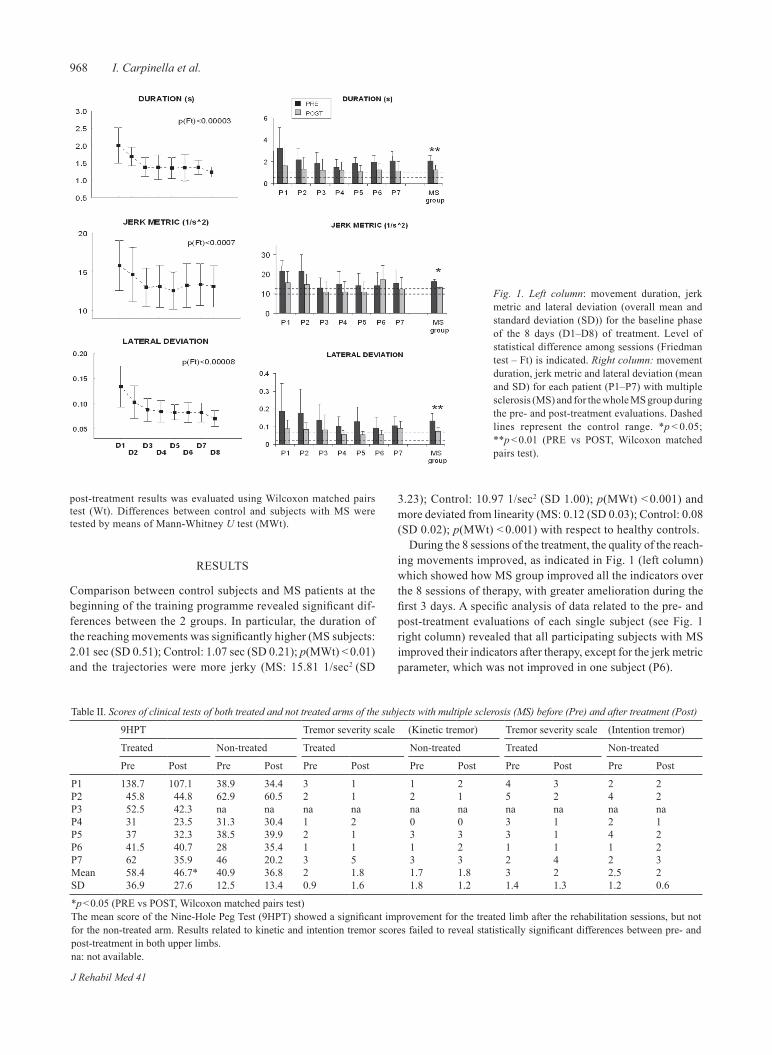
968 I. Carpinella et al.
post-treatment results was evaluated using Wilcoxon matched pairs test (Wt). Differences between control and subjects with MS were tested by means of Mann-Whitney U test (MWt).
RESULTS
Comparison between control subjects and MS patients at the beginning of the training programme revealed significant dif-ferences between the 2 groups. In particular, the duration of the reaching movements was significantly higher (MS subjects: 2.01 sec (SD 0.51); Control: 1.07 sec (SD 0.21); p(MWt) < 0.01) and the trajectories were more jerky (MS: 15.81 1/sec2 (SD
3.23); Control: 10.97 1/sec2 (SD 1.00); p(MWt) < 0.001) and more deviated from linearity (MS: 0.12 (SD 0.03); Control: 0.08 (SD 0.02); p(MWt) < 0.001) with respect to healthy controls.
During the 8 sessions of the treatment, the quality of the reach-ing movements improved, as indicated in Fig. 1 (left column) which showed how MS group improved all the indicators over the 8 sessions of therapy, with greater amelioration during the first 3 days. A specific analysis of data related to the pre- and post-treatment evaluations of each single subject (see Fig. 1 right column) revealed that all participating subjects with MS improved their indicators after therapy, except for the jerk metric parameter, which was not improved in one subject (P6).
Fig. 1. Left column: movement duration, jerk metric and lateral deviation (overall mean and standard deviation (SD)) for the baseline phase of the 8 days (D1–D8) of treatment. Level of statistical difference among sessions (Friedman test – Ft) is indicated. Right column: movement duration, jerk metric and lateral deviation (mean and SD) for each patient (P1–P7) with multiple sclerosis (MS) and for the whole MS group during the pre- and post-treatment evaluations. Dashed lines represent the control range. *p < 0.05; **p < 0.01 (PRE vs POST, Wilcoxon matched pairs test).
Table II. Scores of clinical tests of both treated and not treated arms of the subjects with multiple sclerosis (MS) before (Pre) and after treatment (Post)
9HPT Tremor severity scale (Kinetic tremor) Tremor severity scale (Intention tremor)
Treated Non-treated Treated Non-treated Treated Non-treated
Pre Post Pre Post Pre Post Pre Post Pre Post Pre Post
P1 138.7 107.1 38.9 34.4 3 1 1 2 4 3 2 2P2 45.8 44.8 62.9 60.5 2 1 2 1 5 2 4 2P3 52.5 42.3 na na na na na na na na na naP4 31 23.5 31.3 30.4 1 2 0 0 3 1 2 1P5 37 32.3 38.5 39.9 2 1 3 3 3 1 4 2P6 41.5 40.7 28 35.4 1 1 1 2 1 1 1 2P7 62 35.9 46 20.2 3 5 3 3 2 4 2 3Mean 58.4 46.7* 40.9 36.8 2 1.8 1.7 1.8 3 2 2.5 2SD 36.9 27.6 12.5 13.4 0.9 1.6 1.8 1.2 1.4 1.3 1.2 0.6
*p < 0.05 (PRE vs POST, Wilcoxon matched pairs test)The mean score of the Nine-Hole Peg Test (9HPT) showed a significant improvement for the treated limb after the rehabilitation sessions, but not for the non-treated arm. Results related to kinetic and intention tremor scores failed to reveal statistically significant differences between pre- and post-treatment in both upper limbs.na: not available.
J Rehabil Med 41
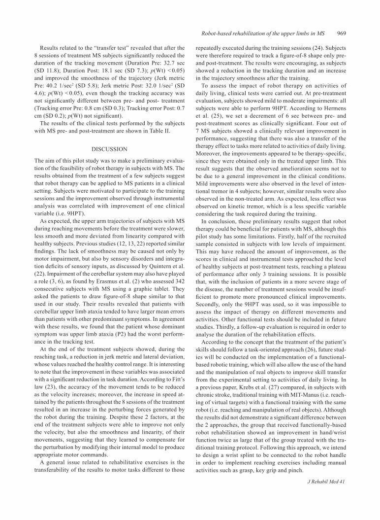
969Robot-based rehabilitation of the upper limbs in MS
Results related to the “transfer test” revealed that after the 8 sessions of treatment MS subjects significantly reduced the duration of the tracking movement (Duration Pre: 32.7 sec (SD 11.8); Duration Post: 18.1 sec (SD 7.3); p(Wt) < 0.05) and improved the smoothness of the trajectory (Jerk metric Pre: 40.2 1/sec2 (SD 5.8); Jerk metric Post: 32.0 1/sec2 (SD 4.6); p(Wt) < 0.05), even though the tracking accuracy was not significantly different between pre- and post- treatment (Tracking error Pre: 0.8 cm (SD 0.3); Tracking error Post: 0.7 cm (SD 0.2); p(Wt) not significant).
The results of the clinical tests performed by the subjects with MS pre- and post-treatment are shown in Table II.
DISCUSSION
The aim of this pilot study was to make a preliminary evalua-tion of the feasibility of robot therapy in subjects with MS. The results obtained from the treatment of a few subjects suggest that robot therapy can be applied to MS patients in a clinical setting. Subjects were motivated to participate to the training sessions and the improvement observed through instrumental analysis was correlated with improvement of one clinical variable (i.e. 9HPT).
As expected, the upper arm trajectories of subjects with MS during reaching movements before the treatment were slower, less smooth and more deviated from linearity compared with healthy subjects. Previous studies (12, 13, 22) reported similar findings. The lack of smoothness may be caused not only by motor impairment, but also by sensory disorders and integra-tion deficits of sensory inputs, as discussed by Quintern et al. (22). Impairment of the cerebellar system may also have played a role (3, 6), as found by Erasmus et al. (2) who assessed 342 consecutive subjects with MS using a graphic tablet. They asked the patients to draw figure-of-8 shape similar to that used in our study. Their results revealed that patients with cerebellar upper limb ataxia tended to have larger mean errors than patients with other predominant symptoms. In agreement with these results, we found that the patient whose dominant symptom was upper limb ataxia (P2) had the worst perform-ance in the tracking test.
At the end of the treatment subjects showed, during the reaching task, a reduction in jerk metric and lateral deviation, whose values reached the healthy control range. It is interesting to note that the improvement in these variables was associated with a significant reduction in task duration. According to Fitt’s law (23), the accuracy of the movement tends to be reduced as the velocity increases; moreover, the increase in speed at-tained by the patients throughout the 8 sessions of the treatment resulted in an increase in the perturbing forces generated by the robot during the training. Despite these 2 factors, at the end of the treatment subjects were able to improve not only the velocity, but also the smoothness and linearity, of their movements, suggesting that they learned to compensate for the perturbation by modifying their internal model to produce appropriate motor commands.
A general issue related to rehabilitative exercises is the transferability of the results to motor tasks different to those
repeatedly executed during the training sessions (24). Subjects were therefore required to track a figure-of-8 shape only pre- and post-treatment. The results were encouraging, as subjects showed a reduction in the tracking duration and an increase in the trajectory smoothness after the training.
To assess the impact of robot therapy on activities of daily living, clinical tests were carried out. At pre-treatment evaluation, subjects showed mild to moderate impairments: all subjects were able to perform 9HPT. According to Hermens et al. (25), we set a decrement of 6 sec between pre- and post-treatment scores as clinically significant. Four out of 7 MS subjects showed a clinically relevant improvement in performance, suggesting that there was also a transfer of the therapy effect to tasks more related to activities of daily living. Moreover, the improvements appeared to be therapy-specific, since they were obtained only in the treated upper limb. This result suggests that the observed amelioration seems not to be due to a general improvement in the clinical conditions. Mild improvements were also observed in the level of inten-tional tremor in 4 subjects; however, similar results were also observed in the non-treated arm. As expected, less effect was observed on kinetic tremor, which is a less specific variable considering the task required during the training.
In conclusion, these preliminary results suggest that robot therapy could be beneficial for patients with MS, although this pilot study has some limitations. Firstly, half of the recruited sample consisted in subjects with low levels of impairment. This may have reduced the amount of improvement, as the scores in clinical and instrumental tests approached the level of healthy subjects at post-treatment tests, reaching a plateau of performance after only 3 training sessions. It is possible that, with the inclusion of patients in a more severe stage of the disease, the number of treatment sessions would be insuf-ficient to promote more pronounced clinical improvements. Secondly, only the 9HPT was used, so it was impossible to assess the impact of therapy on different movements and activities. Other functional tests should be included in future studies. Thirdly, a follow-up evaluation is required in order to analyse the duration of the rehabilitation effects.
According to the concept that the treatment of the patient’s skills should follow a task-oriented approach (26), future stud-ies will be conducted on the implementation of a functional-based robotic training, which will also allow the use of the hand and the manipulation of real objects to improve skill transfer from the experimental setting to activities of daily living. In a previous paper, Krebs et al. (27) compared, in subjects with chronic stroke, traditional training with MIT-Manus (i.e. reach-ing of virtual targets) with a functional training with the same robot (i.e. reaching and manipulation of real objects). Although the results did not demonstrate a significant difference between the 2 approaches, the group that received functionally-based robot rehabilitation showed an improvement in hand/wrist function twice as large that of the group treated with the tra-ditional training protocol. Following this approach, we intend to design a wrist splint to be connected to the robot handle in order to implement reaching exercises including manual activities such as grasp, key grip and pinch.
J Rehabil Med 41
970 I. Carpinella et al.
REFERENCES
Confavreux C, Aimard G, Devic M. Course and prognosis of 1. multiple sclerosis assessed by the computerized data processing of 349 patients. Brain 1980; 103: 281–300.Erasmus LP, Sarno S, Albrecht H, Schwecht M, Pollmann W, Konig 2. N. Measurement of ataxic symptoms with a graphic tablet: standard values in controls and validity in Multiple Sclerosis patients. J Neurosci Methods 2001; 108: 25–37.Feys P, Maes F, Nuttin B, Helsen W, Malfait V, Nagels G, et al. Re-3. lationship between multiple sclerosis intention tremor severity and lesion load in the brainstem. Neuroreport 2005; 16: 1379–1382.Khan F, Turner-Stokes L, Ng L, Kilpatrick T. Multidisciplinary 4. rehabilitation for adults with multiple sclerosis. Cochrane Database Syst Rev 2007; (2): CD006036.O’Hara L, Cadbury H, De SL, Ide L. Evaluation of the effective-5. ness of professionally guided self-care for people with multiple sclerosis living in the community: a randomized controlled trial. Clin Rehabil 2002; 16: 119–128.Nakashima I, Fujihara K, Okita N, Takase S, Itoyama Y. Clini-6. cal and MRI study of brain stem and cerebellar involvement in Japanese patients with multiple sclerosis. J Neurol Neurosurg Psychiatry 1999; 67: 153–157.Sanguineti V, Morasso PG, Baratto L, Brichetto G, Luigi MG, 7. Solaro C. Cerebellar ataxia: quantitative assessment and cybernetic interpretation. Hum Mov Sci 2003; 22: 189–205.Patton JL, Mussa-Ivaldi FA. Robot-assisted adaptive training: 8. custom force fields for teaching movement patterns. IEEE Trans Biomed Eng 2004; 51: 636–646.Maschke M, Gomez CM, Ebner TJ, Konczak J. Hereditary cer-9. ebellar ataxia progressively impairs force adaptation during goal-directed arm movements. J Neurophysiol 2004; 91: 230–238.Smith MA, Shadmehr R. Intact ability to learn internal models of 10. arm dynamics in Huntington’s disease but not cerebellar degenera-tion. J Neurophysiol 2005; 93: 2809–2821.Leocani L, Comi E, Annovazzi P, Rovaris M, Rossi P, Cursi M 11. et al. Impaired short-term motor learning in multiple sclerosis: evidence from virtual reality. Neurorehabil Neural Repair 2007; 21: 273–278.Casadio M, Sanguineti V, Morasso P, Solaro C. Abnormal sen-12. sorimotor control, but intact force field adaptation, in multiple sclerosis subjects with no clinical disability. Mult Scler 2008; 14: 330–342.Squeri V, Vergaro E, Brichetto G, Casadio M, Morasso PG, Solaro 13. C, Sanguineti V. Adaptive robot training in the rehabilitation of incoordination in multiple sclerosis: a pilot study. Proceedings
of the 10th International Conference on Rehabilitation Robotics; 2007 June 12–15; Noordwijk, The Netherlands.Prange GB, Jannink MJ, Groothuis-Oudshoorn CG, Hermens 14. HJ, IJzerman MJ. Systematic review of the effect of robot-aided therapy on recovery of the hemiparetic arm after stroke. J Rehabil Res Dev 2006; 43: 171–184.Kurtzke JF. Rating neurologic impairment in multiple sclerosis: 15. an expanded disability status scale (EDSS). Neurology 1983; 33: 1444–1452.McDonald WI, Compston A, Edan G, Goodkin D, Hartung HP, 16. Lublin FD et al. Recommended diagnostic criteria for multiple sclerosis: guidelines from the International Panel on the diagnosis of multiple sclerosis. Ann Neurol 2001; 50: 121–127.Kellor M, Frost J, Silberberg N, Iversen I, Cummings R. Hand 17. strength and dexterity. Am J Occup Ther 1971; 25: 77–83.Pfeiffer E. A short portable mental status questionnaire for the as-18. sessment of organic brain deficit in elderly patients. J Am Geriatr Soc 1975; 23: 433–441.Bohannon RW, Smith MB. Interrater reliability of a modified 19. Ashworth scale of muscle spasticity. Phys Ther 1987; 67: 206– 207.Casadio M, Sanguineti V, Morasso PG, Arrichiello V. Braccio di 20. Ferro: a new haptic workstation for neuromotor rehabilitation. Technol Health Care 2006; 14: 123–142.Bain PG, Findley LJ, Atchison P, Behari M, Vidailhet M, Gresty 21. M et al. Assessing tremor severity. J Neurol Neurosurg Psychiatry 1993; 56: 868–873.Quintern J, Immisch I, Albrecht H, Pollmann W, Glasauer S, 22. Straube A. Influence of visual and proprioceptive afferences on upper limb ataxia in patients with multiple sclerosis. J Neurol Sci 1999; 163: 61–69.Fitts PM. The information capacity of the human motor system 23. in controlling the amplitude of movement. 1954. J Exp Psychol Gen 1992; 121: 262–269.Shumway-Cook A, Woollacott M. Motor control: theory and 24. practical application. Baltimore, MD: Lippincott Williams & Wilkins; 1995. Hermens H, Huijgen B, Giacomozzi C, Ilsbroukx S, Macellari 25. V, Prats E et al. Clinical assessment of the HELLODOC tele-rehabilitation service. Ann Ist Super Sanita 2008; 44: 154–163.Carr J, Shepherd R. A motor relearning programme for stroke. 2nd 26. edn. Rockville, MD: Aspen Systems; 1987.Krebs HI, Mernoff S, Fasoli SE, Hughes R, Stein J, Hogan N. A 27. comparison of functional and impairment-based robotic training in severe to moderate chronic stroke: a pilot study. NeuroRehabilita-tion 2008; 23: 81–87.
J Rehabil Med 41
oriGinal report
J Rehabil Med 2009; 41: 971–975
J Rehabil Med 41© 2009 The Authors. doi: 10.2340/16501977-0402Journal Compilation © 2009 Foundation of Rehabilitation Information. ISSN 1650-1977
Objective: to verify the possibility of administering robot-aided therapy for the upper limbs in patients after stroke; to evaluate patients’ degree of acceptance and compliance with the treatment; to establish if the treatment has an effect on motor impairment and functional outcome.Design: Quasi-experimental, uncontrolled study.Subjects: fourteen patients with chronic hemiparesis after stroke.Methods: patients were treated with a robotic system for the upper limbs (reoGotm; motorika medical ltd, israel). sub-jects performed the following assessment, at the start (t0), at the end of treatment (t1), and at the follow-up performed one month after the end of treatment (t2): fugl-meyer test (fm) for upper limbs; strength evaluation; ashworth scale; visual analogue scale (vas) for pain; frenchay arm test (fat); box and block test (b&b); functional independence measure (fimtm); abilhanD Questionnaire; timed up and Go test (tuG); euro-Quality of life questionnaire and; a vas for treatment satisfaction were administered to the subjects. Results: total scores of fm, b&b, fat and fimtm showed a statistically significant improvement from T0 and T1 (FM p < 0.002, b&b p < 0.012, fat p < 0.023, fimtm p < 0.007) and from t0 and t2 (fm p < 0.003, b&b p < 0.011, fat p < 0.024, fim p < 0.027). No statistically significant differences were found between evaluations at t1 and t2 (fm p < 0.595, b&b p < 0.491, fat p < 0.317, fim p < 0.180). Conclusion: the sample was capable of completing the treat-ment and demonstrated good participant satisfaction. this pilot study led to the finding of a clinical improvement and excellent patient compliance. it can be hypothesized that the results are robot-dependent and that they were learned and then maintained. however, the study is limited in that a con-trol group was not used. as such, it is desirable to continue this study with a control group, as well as by designing a prospective longitudinal randomized controlled trial study.Key words: stroke, upper limbs, robotics. J Rehabil Med 2009; 41: 971–975
Correspondence address: Federica Bovolenta, Medicine Rehabilitation, NOCSAE Hospital AUSL of Modena, via P.Giardini 1355, IT-41100 Modena, Italy. E-mail: federica. [email protected]
Submitted March 16, 2009; accepted May 19, 2009
INTRODUCTION
Stroke is the main cause of disability in industrialized coun-tries, with a significant impact on individual, family, and societal healthcare. As such, any form of treatment that in-creases the functional recovery of patients after stroke could significantly reduce the physical, emotional, and financial load that this condition carries for the sufferers, their families and society in general (1–3).
Therefore, as far as functional recovery is concerned, vari-ous longitudinal studies have shown that the upper limbs are involved at the onset of disease in 85% of cases, and that they remain non-functional 6 months after the acute event in 30–66% of cases, while only 5–20% of cases present complete functional recovery (4). Consequently, over the last few years, rehabilitative medicine has encouraged research in an attempt to identify the modalities, time-frames and proper motiva-tions of the rehabilitative intervention: attempting to identify predictive indicators of outcomes in the acute phases (4, 5); and seeking to understand the neurophysiological mechanisms underlying functional recovery (6, 7), in order to plan the most incisive therapeutic interventions.
Within the scope of this research, it has emerged that the proposed exercise must be intensive and specific in order for treatment to be effective (3); in addition, treatment must be repetitive, functional and motivating (8, 9), so as to bring about an increase in performance, as well as learning, acquisition and generalization (10).
These requirements seem to be satisfied by robotic devices for rehabilitation (11), both in terms of clinical results as well as in terms of positive effects on healthcare costs and increased efficiency. The use of robotic devices for rehabilitation of the upper limbs shows various advantages, including: a large number of patients that can benefit from robotic therapy, due to the flexibility of robotic systems; excellent acceptance of the therapy among patients; and, finally, that the therapy can be performed by the patient under the supervision of a physio-therapist. Recent technological advances have made it possible to develop robotic instruments capable of performing a safe and intensive rehabilitative intervention. Robotic therapy can be developed in different directions in order to reduce motor impairment and increase functional recovery, even in patients affected by moderate/severe impairments, following injuries
ROBOT THERAPY FOR FUNCTIONAL RECOVERY OF THE UPPER LIMBS: A PILOT STUDY ON PATIENTS AFTER STROKE
Federica Bovolenta, MD1, Milena Goldoni, PT1, Pierina Clerici, PT1, Maurizio Agosti, PT2 and Marco Franceschini, MD3
From the 1Medicine Rehabilitation NOCSAE Hospital AUSL of Modena, Modena, 2Geriatrics and Rehabilitation Department, University Hospital of Parma, Parma and 3IRCCS San Raffaele Pisana, Rome, Italy
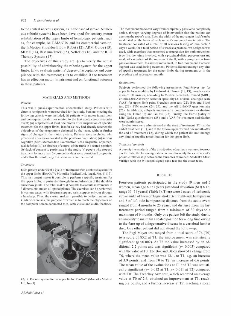
972 F. Bovolenta et al.
to the central nervous system, as in the case of stroke. Numer-ous robotic systems have been developed for sensory-motor rehabilitation of the upper limbs of hemiplegic patients, such as, for example, MIT-MANUS and its commercial version, the InMotion Shoulder-Elbow Robot (12), ARM-Guide (13), MIME (14), BiManu-Track (15), NeReBot (16), and the REO Therapy System (17).
The objectives of this study are: (i) to verify the actual possibility of administering the robotic system for the upper limbs; (ii) to evaluate patients’ degree of acceptance and com-pliance with the treatment; (iii) to establish if the treatment has an effect on motor impairment and on functional outcome in these patients.
MATERIALS AND METHODSPatientsThis was a quasi-experimental, uncontrolled study. Patients with chronic hemiparesis were recruited for the study. Persons meeting the following criteria were included: (i) patients with motor impairment and consequent disabilities related to the first acute cerebrovascular event; (ii) outpatients at least one month after suspension of specific treatment for the upper limbs, insofar as they had already reached the objectives of the programme designed by the team, without further signs of changes in the motor picture. Patients were excluded who presented: (i) a lesion located in the posterior circulation; (ii) serious cognitive (Mini-Mental State Examination < 24), linguistic, or percep-tual deficits; (iii) an absence of control of the trunk in a seated position; (iv) lack of consent to participate in the study; (v) people who stopped treatment for more than 5 consecutive days were considered drop-outs; under this threshold, any lost sessions were recovered.
TreatmentEach patient underwent a cycle of treatment with a robotic system for the upper limbs (ReoGoTM; Motorika Medical Ltd, Israel, Fig. 1) (17). This instrument makes it possible to perform a specific treatment for the upper limbs, in particular through the mobilization of the shoulder and elbow joints. The robot makes it possible to execute movements in 3 dimensions and on all spatial planes. The exercises can be performed in various ways: with forearm support, wrist support only, or through a handgrip. Thus, the system makes it possible to perform numerous kinds of exercises, the purpose of which is to reach the objectives on the computer screen connected to it, with visual and audio feedback.
The movement mode can vary from completely passive to completely active, through varying degrees of intervention that the patient can exert on the robot’s arm. Even the width of the movement itself can be modulated on the basis of each subject’s unique characteristics. The treatment consisted of a total of 20 sessions lasting 45 min each, 5 days a week, for a total period of 4 weeks; a protocol we designed was used, with exercises that presented a progression for both movement type (i.e. the joints involved, with a proximal-distal progression) and mode of execution of the movement itself, with a progression from passive movement, to assisted movement, to free movement. Forearm support was used during treatment. Patients did not undergo any kind of specific treatment for the upper limbs during treatment or in the preceding and subsequent month.
EvaluationsSubjects performed the following assessment: Fugl-Meyer test for upper limbs as modified by Lindmark & Hamrin (18, 19); muscle evalu-ation of 10 muscles, according to Medical Research Council (MRC) criteria (20); Ashworth scale for spasticity (21); visual analogue scale (VAS) for upper limb pain; Frenchay Arm test (22); Box and Block test (23); FIM motor (24, 25); and the ABILHAND questionnaire (26). In addition, subjects underwent a comprehensive evaluation using the Timed Up and Go test (27). Finally, the Euro-Quality of Life (QoL) questionnaire (28) and a VAS for treatment satisfaction were administered.
Evaluations were administered at the start of treatment (T0), at the end of treatment (T1), and at the follow-up performed one month after the end of treatment (T2), during which the patient did not undergo any kind of specific rehabilitation for the upper limb.
Statistical analysisA descriptive analysis of the distribution of patients was used to proc-ess the data; the following tests were used to verify the existence of a possible relationship between the variables examined: Student’s t-test, verified with the Wilcoxon signed-rank test and the exact tests.
RESULTS
Fourteen patients participated in the study (9 men and 5 women, mean age 60.57 years (standard deviation (SD) 8.18, range 35–71 years)) (Table I). There were 9 cases of ischaemic stroke and 5 of haemorrhagic stroke; 6 of right-side hemiparesis and 8 of left-side hemiparesis; distance from the acute event ranged from 4 months to 25 years; and distance from the last treatment period ranged from a minimum of 30 days to a maximum of 6 months. Only one patient left the study, due to an inability to maintain a seated position for a long time owing to the flare-up of a degenerative disease in a vertebral lumbar disc. One other patient did not attend the follow-up.
The Fugl-Meyer test ranged from a total score of 76 (T0) to a score of 85.2 at T1; the improvement was statistically significant (p < 0.002). At T2 the value increased by an ad-ditional 2.2 points and was significant (p < 0.003) compared with the value at T0. The Box and Block showed a change from T0, where the mean value was 13.1, to T1, e.g. an increase of 3.9 points, and from T0 to T2, an increase of 6.6 points. The mean value of the evaluations at T1 and T2 was statisti-cally significant (p < 0.012 at T1; p < 0.011 at T2) compared with T0. The Frenchay Arm test, which recorded an average value at T0 of 2.6, obtained an improvement at T1, reach-ing 3.2 points, and a further increase at T2, reaching a mean
Fig. 1. Robotic system for the upper limbs: ReoGoTM (Motorika Medical Ltd, Israel).
J Rehabil Med 41
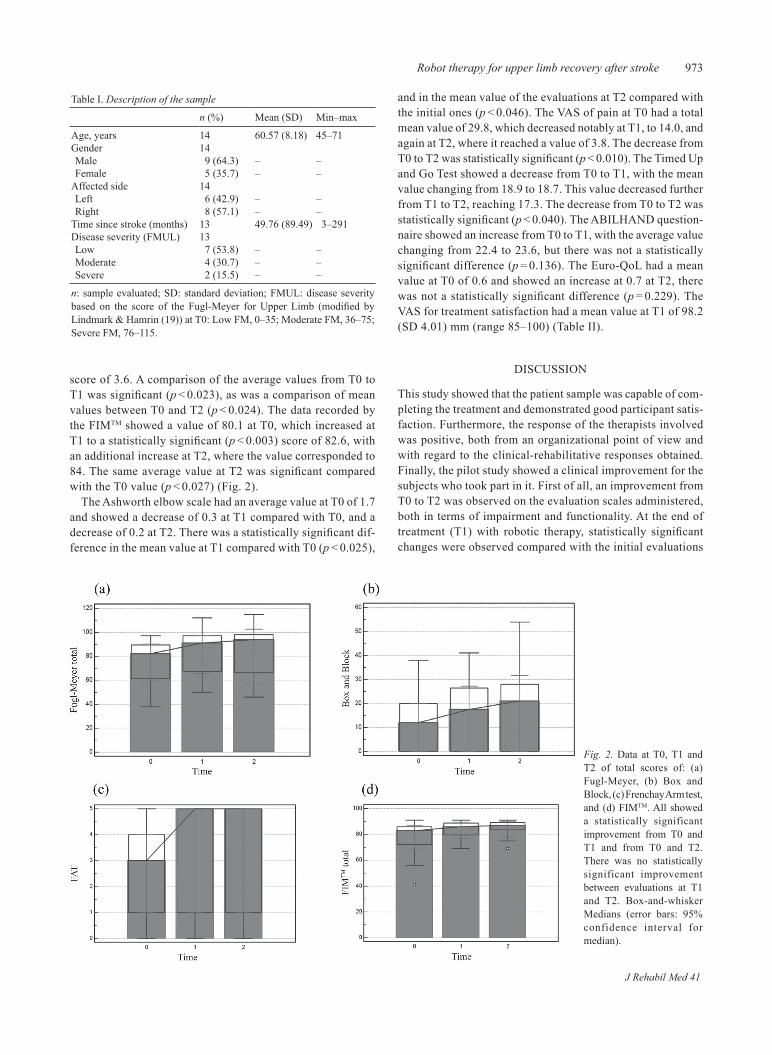
973Robot therapy for upper limb recovery after stroke
score of 3.6. A comparison of the average values from T0 to T1 was significant (p < 0.023), as was a comparison of mean values between T0 and T2 (p < 0.024). The data recorded by the FIMTM showed a value of 80.1 at T0, which increased at T1 to a statistically significant (p < 0.003) score of 82.6, with an additional increase at T2, where the value corresponded to 84. The same average value at T2 was significant compared with the T0 value (p < 0.027) (Fig. 2).
The Ashworth elbow scale had an average value at T0 of 1.7 and showed a decrease of 0.3 at T1 compared with T0, and a decrease of 0.2 at T2. There was a statistically significant dif-ference in the mean value at T1 compared with T0 (p < 0.025),
and in the mean value of the evaluations at T2 compared with the initial ones (p < 0.046). The VAS of pain at T0 had a total mean value of 29.8, which decreased notably at T1, to 14.0, and again at T2, where it reached a value of 3.8. The decrease from T0 to T2 was statistically significant (p < 0.010). The Timed Up and Go Test showed a decrease from T0 to T1, with the mean value changing from 18.9 to 18.7. This value decreased further from T1 to T2, reaching 17.3. The decrease from T0 to T2 was statistically significant (p < 0.040). The ABILHAND question-naire showed an increase from T0 to T1, with the average value changing from 22.4 to 23.6, but there was not a statistically significant difference (p = 0.136). The Euro-QoL had a mean value at T0 of 0.6 and showed an increase at 0.7 at T2, there was not a statistically significant difference (p = 0.229). The VAS for treatment satisfaction had a mean value at T1 of 98.2 (SD 4.01) mm (range 85–100) (Table II).
DISCUSSION
This study showed that the patient sample was capable of com-pleting the treatment and demonstrated good participant satis-faction. Furthermore, the response of the therapists involved was positive, both from an organizational point of view and with regard to the clinical-rehabilitative responses obtained. Finally, the pilot study showed a clinical improvement for the subjects who took part in it. First of all, an improvement from T0 to T2 was observed on the evaluation scales administered, both in terms of impairment and functionality. At the end of treatment (T1) with robotic therapy, statistically significant changes were observed compared with the initial evaluations
Fig. 2. Data at T0, T1 and T2 of total scores of: (a) Fugl-Meyer, (b) Box and Block, (c) Frenchay Arm test, and (d) FIMTM. All showed a statistically significant improvement from T0 and T1 and from T0 and T2. There was no statistically significant improvement between evaluations at T1 and T2. Box-and-whisker Medians (error bars: 95% confidence interval for median).
Table I. Description of the sample
n (%) Mean (SD) Min–max
Age, years 14 60.57 (8.18) 45–71Gender 14Male 9 (64.3) – –Female 5 (35.7) – –
Affected side 14Left 6 (42.9) – –Right 8 (57.1) – –
Time since stroke (months) 13 49.76 (89.49) 3–291Disease severity (FMUL) 13Low 7 (53.8) – –Moderate 4 (30.7) – –Severe 2 (15.5) – –
n: sample evaluated; SD: standard deviation; FMUL: disease severity based on the score of the Fugl-Meyer for Upper Limb (modified by Lindmark & Hamrin (19)) at T0: Low FM, 0–35; Moderate FM, 36–75; Severe FM, 76–115.
J Rehabil Med 41
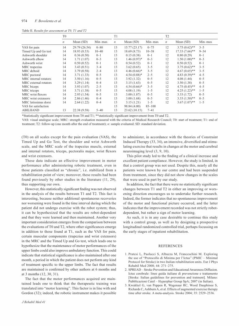
974 F. Bovolenta et al.
(T0) on all scales except for the pain evaluation (VAS), the Timed Up and Go Test, the shoulder and wrist Ashworth scale, and the MRC scale of the trapezius muscle, external and internal rotators, triceps, pectoralis major, wrist flexors and wrist extensors.
These data indicate an effective improvement in motor performance after administering robotic treatment, even in those patients classified as “chronic”, i.e. stabilized from a rehabilitation point of view; moreover, these results had been found previously by other studies in the literature (11, 29), thus supporting our own.
However, this statistically significant finding was not observed in the analysis of the results between T1 and T2. This fact is interesting, because neither additional spontaneous recoveries nor worsening were found in the time interval during which the patient did not undergo exercise with the robot system; thus, it can be hypothesized that the results are robot-dependent and that they were learned and then maintained. Another very important consideration emerges from the comparison between the evaluations of T0 and T2, where other significances emerge in addition to those found at T1, such as the VAS for pain, certain muscular components (trapezius and wrist extensors) in the MRC and the Timed Up and Go test, which leads one to hypothesize that the maintenance of motor performances of the upper limbs could also improve ambulatory function. This could indicate that statistical significance is also maintained after one month, a period in which the patient does not perform any kind of treatment specific to the upper limbs. The fact that results are maintained is confirmed by other authors at 6 months and at 3 months (12, 30, 31).
The fact that the motor performances acquired are main-tained leads one to think that the therapeutic training was translated into “motor learning”. This factor is in line with and Gordon (32); indeed, the robotic instrument makes it possible
to administer, in accordance with the theories of Constraint Induced Therapy (33, 34), an intensive, diversified and stimu-lating exercise that results in changes at the motor and cerebral neuroimaging level (3, 9, 10).
This pilot study led to the finding of a clinical increase and excellent patient compliance. However, the study is limited, in that a control group was not used. Despite this, nearly all the patients were known by our centre and had been suspended from treatment, since they did not show changes in the scales that were used in part by our study.
In addition, the fact that there were no statistically significant changes between T1 and T2 in either an improving or wors-ening direction encourages us to undertake further research. Indeed, the former indicates that no spontaneous improvement of the motor and functional picture occurred, and the latter indicates that the improvement recorded was not strictly robot-dependent, but rather a sign of motor learning.
As such, it is in any case desirable to continue this study with a control group, as well as by designing a prospective longitudinal randomized controlled trial, perhaps focussing on the early stages of inpatient rehabilitation.
REFERENCES
Pratesi L, Paolucci S, Albuzza M, Franceschini M. Exploring 1. the use of “Protocollo di Minima per l’Ictus” (PMIC – Minimal Protocol for Stroke) in two Italian rehabilitation units. Eur J Phys Rehabil Med 2008; 44: 271–275.SPREAD – Stroke Prevention and Educational Awareness Diffusion. 2. Ictus cerebrale: linee guida italiane di prevenzione e trattamento [Stroke: Italian guidelines for prevention and tratment]. Milano: Pubblicazioni Catel – Hyperphar Group SpA; 2007 (in Italian). Kwakkel G, van Peppen R, Wagenaar RC, Wood Dauphinee S, 3. Richards C, Ashburn A, et al. Effects of augmented exercise therapy time after stroke. A meta-analysis. Stroke 2004; 35: 2529–2536.
Table II. Results for assessment at T0, T1 and T2
T0 T1 T2
n Mean (SD) Min–max n Mean (SD) Min–max n Mean (SD) Min–max
VAS for pain 14 29.79 (26.54) 0–80 13 15.77 (23.17) 0–75 12 3.75 (0.62)** 3–5Timed Up and Go test 14 18.93 (8.53) 10–40 13 18.69 (8.73) 10–38 12 17.33 (7.66)** 9–34Ashworth shoulder 14 0.36 (0.50) 0–1 13 0.15 (0.38) 0–1 12 0.80 (0.29) 0–1Ashworth elbow 14 1.71 (1.07) 0–3 13 1.46 (0.97)* 0–3 12 1.50 (1.00)** 0–3Ashworth wrist 14 0.50 (0.52) 0–1 13 0.54 (0.52) 0–1 12 0.50 (0.52) 0–1MRC trapezius 14 3.43 (0.51) 3–4 13 3.62 (0.65) 3–5 12 3.75 (0.62)** 3–5MRC deltoid 14 3.79 (0.58) 2–4 13 4.46 (0.66)* 3–5 12 4.67 (0.65)** 3–5MRC pectoral 14 3.71 (1.33) 0–5 13 4.54 (0.88)* 2–5 12 4.83 (0.39)** 4–5MRC internal rotators 14 3.50 (1.16) 0–5 13 3.92 (1.32) 0–5 12 4.08 (1.44) 0–5MRC external rotators 14 3.29 (1.14) 0–4 13 3.15 (1.63) 0–5 12 3.50 (1.38) 0–5MRC biceps 14 3.93 (1.07) 2–5 13 4.54 (0.66)* 3–5 12 4.75 (0.45)** 4–5MRC triceps 14 3.71 (1.38) 0–5 13 4.08 (1.19) 1–5 12 4.25 (1.22)** 1–5MRC wrist flexors 14 2.93 (1.54) 0–5 13 3.00 (1.87) 0–5 12 3.33 (1.72) 0–5MRC wrist extensors 14 2.86 (1.46) 0–4 13 3.00 (1.68) 0–5 12 3.33 (1.50)** 0–5MRC latissimus dorsi 14 2.64 (1.22) 0–4 13 3.15 (1.21) 1–5 12 3.67 (1.07)** 1–5VAS for satisfaction 13 98.84 (4.00) 85–100ABILHAND 13 22.38 (9.58) 5–40 13 23.62 (10.15) 7–41
*Statistically significant improvement from T0 and T1; **statistically significant improvement from T0 and T2.VAS: visual analogue scale; MRC: strength evaluation measured with the criteria of Medical Research Council; T0: start of treatment; T1: end of treatment; T2: follow-up (one month after the end of treatment); n: sample evaluated; SD: standard deviation.
J Rehabil Med 41
975Robot therapy for upper limb recovery after stroke
Kwakkel G, Kollen BJ, van der Grond J, Prevo AJH. Probability 4. of regaining dexterity in the flaccid upper limb. Impact of sever-ity of paresis and time since onset in acute stroke. Stroke 2003; 34: 2181-86. Wagner JM, Lang CE, Sahrmann SA, Edwards DF, Dromerick 5. AW. Sensorimotor impairments and reaching performance in subjects with poststroke hemiparesis during the first few months of recovery. Phys Ther 2007; 87: 751–765.Nudo RJ. Postinfarct cortical plasticity and behavioral recovery. 6. Stroke 2007; 38: 840-845.Prabhakaran S, Zarahn E, Riley C, Speizer A, Chong JY, Lazar 7. RM, et al. Inter-individual variability in the capacity for motor recovery after ischemic stroke. Neurorehabil Neural Repair 2008; 22: 64–71.Yavuzer G, Senel A, Atay MB, Stam HJ. “Playstation eyetoy 8. games” improve upper extremity-related motor functioning in subacute stroke: a randomized controlled clinical trial. Eur J Phys Rehabil Med 2008; 44: 237–244.van Peppen RP, Kwakkel G, Wood-Dauphinee S, Hendriks HJ, 9. van der Wees PJ, Dekker J. The impact of physical therapy on functional outcomes after stroke: what’s the evidence? Clin Rehabil 2004; 18: 833–862.Krakauer J. Motor learning: its relevance to stroke recovery and 10. neurorehabilitation. Curr Opin Neurol 2006; 19: 84–90.Kwakkel G, Kollen BJ, Krebs HI. Effects of robot-assisted therapy 11. on upper limb recovery after stroke: a systematic review. Neurore-habil Neural Repair 2008; 22: 111–121.Volpe BT, Lynch D, Rykman-Berland A, Ferraro M, Galgano M 12. Hogan N, et al. Intensive sensorimotor arm training mediated by therapist or robot improves hemiparesis in patients with chronic stroke. Neurorehabil Neural Repair 2008; 22: 305–310.Kahn LE, Zygman ML, Rymer WZ, Reinkensmeyer DJ. Robot-13. assisted reaching exercise promotes arm movement recovery in chronic hemiparetic stroke: a randomized controlled pilot study. J Neuroeng Rehabil 2006; 3: 12–24.Lum P, Burgar CG, Van der Loos M, Shor PC, Majmundar M, 14. Yap R. MIME robotic device for upper-limb neurorehabilitation in subacute stroke subjects: a follow-up study. J Rehabil Res Dev 2006; 43: 631–642.Hesse S, Schmidt H, Werner C, Rybski C, Puzich C, Bardeleben 15. A. A new mechanical arm trainer to intensify the upper limb reha-bilitation after stroke: design, concept and first case series. Eura Medicophys 2007; 43: 463–468.Rosati G, Gallina P, Masiero S. Design, implementation and clini-16. cal tests of a wire-based robot for neurorehabilitation. IEEE Trans Neural Syst Rehabil Eng 2007; 15: 560–569. Treger I, Faran S, Ring H. Robot-assisted therapy for neuro-17. muscular training of sub-acute stroke patients. A feasibility study. Eur J Rehabil Med 2008; 44: 431–435.Fugl-Meyer AR, Jääskö L, Leyman I, Olsson S, Steglind S. The 18. post-stroke hemiplegic patient. 1. A method for evaluation of physi-cal performance. Scand J Rehabil Med 1975; 7: 13–31.Lindmark B, Hamrin E. Evaluation of functional capacity after 19. stroke as a basis for active intervention. Validation of a modified
chart for motor capacity assessment. Scand J Rehabil Med 1988; 20: 111–115.Medical Research Council. Aids to the examination of the periph-20. eral nervous system. Memorandum 45. London: Her Majesty’s Stationery Office; 1976. Ashworth B. preliminary trial of carisprodol in multiple sclerosis. 21. Practitioner 1964; 192: 540–542.Heller A, Wade DT, Wood VA, Sunderland A, Hewer RL, Ward 22. E. Arm function after stroke: measurement and recovery over the first three months. J Neurol Neurosurg Psychiatry 1987; 50: 714–719.Mathhiowetz V, Volland G, Kashman N, Weber K. Adult norms 23. for the box and block test for manual dexterity. Am J Occup Ther 1985; 39: 386–391. Tesio L, Granger CV, Perucca L, Franchignoni FP, Battaglia MA, 24. Russel CF. The FIM instrument in the United States and Italy: a comparative study. Am J Phys Med Rehabil 2002; 81: 168–176.Functional Indipendent Measure: versione italiana. Manuale d’uso 25. [Functional Independent Measure: Italian version. Users manual]. Ricerca Riabil 1992; Suppl 2: 1–44 (in Italian). Penta M, Tesio L, Arnould C, Zancan A, Thonnard JL. 26. The ABIL-HAND questionnaire as a measure of manual ability in chronic stroke patients: Rasch-based validation and relationship to upper limb impairment. Stroke 2001; 32: 1627–1634.Podsiadlo D, Richardson S. The timed “Up & Go”: a test of basic 27. functional mobility for frail elderly persons. J Am Geriatr Soc 1991; 39: 142–148.Dorman PJ, Waddell F, Slattery J, Dennis M, Sandercock P. Is 28. the Euroqol a valid measure of health-related quality of life after stroke? Stroke 1997; 28: 1876–1882.Dipietro L, Krebs HI, Fasoli SE, Volpe BT, Stein J, Becver C, 29. Hogan N. Changing motor synergies in chronic stroke. J Neuro-physiol 2007; 98: 757–768.Cordo P, Lutsep H, Cordo L, Wright W, Cacciatore T, Skoss R. 30. Assisted Movement with Enhanced Sensation (AMES): coupling motor and sensory to remediate motor deficits in chronic stroke patients. Neurorehabil Neural Repair 2009; 23: 67–77. Hesse S, Werner MA, Pohl M, Rueckriem PT, Mehrholz PT, Ling-31. nau MA. Computerized arm training improves the motor control of the severely affected arm after stroke. A single-blinded randomized trial in two centers. Stroke 2005; 36: 1960–1966.Gordon J. Assumption underlying physical therapy intervention. 32. Theoretical and historicalperspectives. In: Carr JH, Shepherd PB, editors. Movement science: Foundation for physical therapy in rehabilitation. 2nd ed. Rockville: Aspen Publisher Inc.; 1987, p. 1–31.Dursun N, Dursun E, Sade I, Çekmece Ç. Constraint induced 33. movement therapy: efficacy in a Turkish stroke patient population and evaluation by a new outcome measurement tool. Eur J Rehabil Med 2008 [Epub ahead of print]. Gauthier LV, Taub E, Perkins C, Ortmann M, Mark VW, Uswatte 34. G. Remodelling the brain. Plastic structural brain changes pro-duced by different motor therapies after stroke. Stroke 2008; 39: 1520–1525.
J Rehabil Med 41
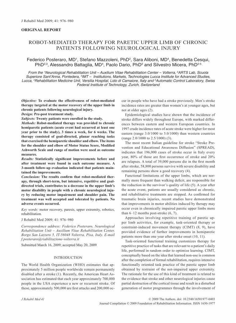
J Rehabil Med 41
oriGinal report
J Rehabil Med 2009; 41: 976–980
© 2009 The Authors. doi: 10.2340/16501977-0403Journal Compilation © 2009 Foundation of Rehabilitation Information. ISSN 1650-1977
Objective: to evaluate the effectiveness of robot-mediated therapy targeted at the motor recovery of the upper limb in chronic patients following neurological injury. Design: pre-post treatment study.Subjects: twenty patients were enrolled in the study. Methods: robot-mediated therapy was provided to chronic hemiparetic patients (acute event had occurred at least one year prior to the study), 3 times a week, for 6 weeks. the therapy consisted of goal-directed, planar reaching tasks that exercised the hemiparetic shoulder and elbow. the items for the shoulder and elbow of Motor Status Score, Modified ashworth scale and range of motion were used as outcome measures. Results: Statistically significant improvements before and after treatment were found in each outcome measure. a 3-month follow-up evaluation indicated that patients main-tained the improvements. Conclusion: The results confirm that robot-mediated ther-apy, through short-term, but intensive, repetitive and goal-directed trials, contributes to a decrease in the upper limb’s motor disability in people with a chronic neurological inju-ry by reducing motor impairment and shoulder pain. the treatment was well accepted and tolerated by patients. no adverse events occurred.Key words: motor recovery, paresis, upper extremity, robotics, rehabilitation. J Rehabil Med 2009; 41: 976–980
Correspondence address: Federico Posteraro, Neurological Rehabilitation Unit – Auxilium Vitae Rehabilitation Center, Borgo San Lazzero 5, IT-56048 Volterra, Pisa, Italy. E-mail [email protected] March 16, 2009; accepted May 20, 2009
INTRODUCTION
The World Health Organization (WHO) estimates that ap-proximately 5 million people worldwide remain permanently disabled after a stroke (1). Recently, the American Heart As-sociation has estimated that each year approximately 700,000 people in the USA experience a new or recurrent stroke. Of these, approximately 500,000 are first attacks and 200,000 oc-
cur in people who have had a stroke previously. Men’s stroke incidence rates are greater than women’s at younger ages, but not at older ages (2).
Epidemiological studies have shown that the incidence of stroke differs widely throughout Europe, with marked differ-ences between eastern and western European countries. In 1997 crude incidence rates of acute stroke were higher for most eastern (range 3.0/1000 to 5.0/1000) than western countries (range 2.0/1000 to 2.5/1000) (3).
The most recent Italian guideline for stroke “Stroke Pre-vention and Educational Awareness Diffusion” (SPREAD), indicates that 196,000 cases of stroke occur in Italy every year, 80% of those are first occurrence of stroke and 20% are relapses. A total of 39,000 persons die in the first month after stroke, 58,800 persons survive with severe disability and remaining persons show a good recovery (4).
Functional limitations of the upper limbs, which are nor-mally more frequent than walking deficit, are responsible for the reduction in the survivor’s quality of life (5). A year after the acute event, patients are usually considered as chronic, and rehabilitative treatments are stopped. As confirmed for traumatic brain injuries, recent studies have demonstrated that improvements in motor abilities induced by therapy may occur even in chronically impaired paretic upper limbs more than 6–12 months post-stroke (6, 7).
Approaches involving repetitive training of paretic up-per limb activities, for example, task-oriented therapy or constraint-induced movement therapy (CIMT) (8, 9), have provided evidence of further improvements in hemiparetic patients more than one year after stroke onset (10, 11).
Task-oriented functional training customizes therapy for repetitive practice of tasks that are relevant to a patient’s daily life, performed in random order to optimize learning. CIMT, conceptually based on the idea that learned non-use is common after the completion of formal rehabilitation, requires intensive functionally oriented task practice of the paretic upper limb obtained by restraint of the not-impaired upper extremity. The rationale for the use of this kind of treatment is related to the evidence that stroke and other neurological injuries cause partial destruction of the cortical tissue and result in a disturbed generation of motor programmes through the involvement of
ROBOT-MEDIATED THERAPY FOR PARETIC UPPER LIMB OF CHRONIC PATIENTS FOLLOWING NEUROLOGICAL INJURY
Federico Posteraro, MD1, Stefano Mazzoleni, PhD2, Sara Aliboni, MD4, Benedetta Cesqui, PhD2,3, Alessandro Battaglia, MD4, Paolo Dario, PhD2 and Silvestro Micera, PhD2-5
From the 1Neurological Rehabilitation Unit – Auxilium Vitae Rehabilitation Center – Volterra, 2ARTS Lab, Scuola Superiore Sant’Anna, Pontedera, 3IMT – Institutions, Markets, Technologies Lucca Institute for Advanced Studies,
Lucca, 4Rehabilitation Medicine Unit, Versilia Hospital, Lido di Camaiore, Italy and 5Automatic Control Laboratory, Swiss Federal Institute of Technology, Zurich, Switzerland
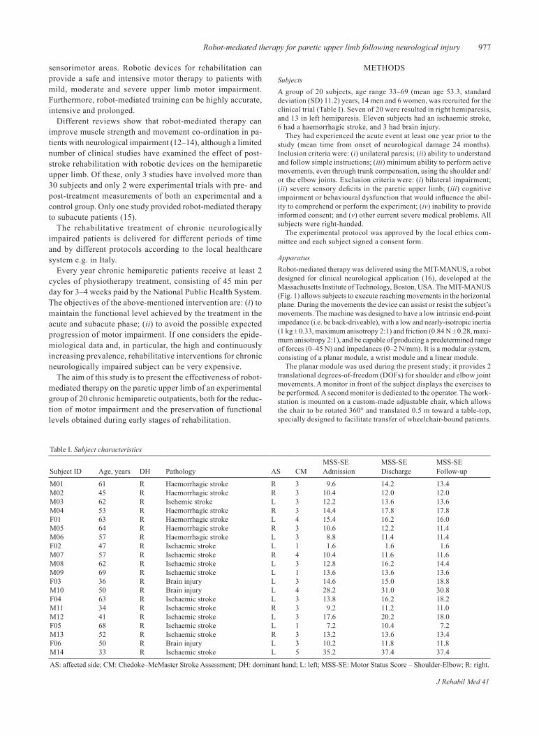
977Robot-mediated therapy for paretic upper limb following neurological injury
sensorimotor areas. Robotic devices for rehabilitation can provide a safe and intensive motor therapy to patients with mild, moderate and severe upper limb motor impairment. Furthermore, robot-mediated training can be highly accurate, intensive and prolonged.
Different reviews show that robot-mediated therapy can improve muscle strength and movement co-ordination in pa-tients with neurological impairment (12–14), although a limited number of clinical studies have examined the effect of post-stroke rehabilitation with robotic devices on the hemiparetic upper limb. Of these, only 3 studies have involved more than 30 subjects and only 2 were experimental trials with pre- and post-treatment measurements of both an experimental and a control group. Only one study provided robot-mediated therapy to subacute patients (15).
The rehabilitative treatment of chronic neurologically impaired patients is delivered for different periods of time and by different protocols according to the local healthcare system e.g. in Italy.
Every year chronic hemiparetic patients receive at least 2 cycles of physiotherapy treatment, consisting of 45 min per day for 3–4 weeks paid by the National Public Health System. The objectives of the above-mentioned intervention are: (i) to maintain the functional level achieved by the treatment in the acute and subacute phase; (ii) to avoid the possible expected progression of motor impairment. If one considers the epide-miological data and, in particular, the high and continuously increasing prevalence, rehabilitative interventions for chronic neurologically impaired subject can be very expensive.
The aim of this study is to present the effectiveness of robot-mediated therapy on the paretic upper limb of an experimental group of 20 chronic hemiparetic outpatients, both for the reduc-tion of motor impairment and the preservation of functional levels obtained during early stages of rehabilitation.
METHODSSubjectsA group of 20 subjects, age range 33–69 (mean age 53.3, standard deviation (SD) 11.2) years, 14 men and 6 women, was recruited for the clinical trial (Table I). Seven of 20 were resulted in right hemiparesis, and 13 in left hemiparesis. Eleven subjects had an ischaemic stroke, 6 had a haemorrhagic stroke, and 3 had brain injury.
They had experienced the acute event at least one year prior to the study (mean time from onset of neurological damage 24 months). Inclusion criteria were: (i) unilateral paresis; (ii) ability to understand and follow simple instructions; (iii) minimum ability to perform active movements, even through trunk compensation, using the shoulder and/or the elbow joints. Exclusion criteria were: (i) bilateral impairment; (ii) severe sensory deficits in the paretic upper limb; (iii) cognitive impairment or behavioural dysfunction that would influence the abil-ity to comprehend or perform the experiment; (iv) inability to provide informed consent; and (v) other current severe medical problems. All subjects were right-handed.
The experimental protocol was approved by the local ethics com-mittee and each subject signed a consent form.
ApparatusRobot-mediated therapy was delivered using the MIT-MANUS, a robot designed for clinical neurological application (16), developed at the Massachusetts Institute of Technology, Boston, USA. The MIT-MANUS (Fig. 1) allows subjects to execute reaching movements in the horizontal plane. During the movements the device can assist or resist the subject’s movements. The machine was designed to have a low intrinsic end-point impedance (i.e. be back-driveable), with a low and nearly-isotropic inertia (1 kg ± 0.33, maximum anisotropy 2:1) and friction (0.84 N ± 0.28, maxi-mum anisotropy 2:1), and be capable of producing a predetermined range of forces (0–45 N) and impedances (0–2 N/mm). It is a modular system, consisting of a planar module, a wrist module and a linear module.
The planar module was used during the present study; it provides 2 translational degrees-of-freedom (DOFs) for shoulder and elbow joint movements. A monitor in front of the subject displays the exercises to be performed. A second monitor is dedicated to the operator. The work-station is mounted on a custom-made adjustable chair, which allows the chair to be rotated 360° and translated 0.5 m toward a table-top, specially designed to facilitate transfer of wheelchair-bound patients.
Table I. Subject characteristics
Subject ID Age, years DH Pathology AS CMMSS-SEAdmission
MSS-SEDischarge
MSS-SEFollow-up
M01 61 R Haemorrhagic stroke R 3 9.6 14.2 13.4M02 45 R Haemorrhagic stroke R 3 10.4 12.0 12.0M03 62 R Ischemic stroke L 3 12.2 13.6 13.6M04 53 R Haemorrhagic stroke R 3 14.4 17.8 17.8F01 63 R Haemorrhagic stroke L 4 15.4 16.2 16.0M05 64 R Haemorrhagic stroke R 3 10.6 12.2 11.4M06 57 R Haemorrhagic stroke L 3 8.8 11.4 11.4F02 47 R Ischaemic stroke L 1 1.6 1.6 1.6M07 57 R Ischaemic stroke R 4 10.4 11.6 11.6M08 62 R Ischaemic stroke L 3 12.8 16.2 14.4M09 69 R Ischaemic stroke L 1 13.6 13.6 13.6F03 36 R Brain injury L 3 14.6 15.0 18.8M10 50 R Brain injury L 4 28.2 31.0 30.8F04 63 R Ischaemic stroke L 3 13.8 16.2 18.2M11 34 R Ischaemic stroke R 3 9.2 11.2 11.0M12 41 R Ischaemic stroke L 3 17.6 20.2 18.0F05 68 R Ischaemic stroke L 1 7.2 10.4 7.2M13 52 R Ischaemic stroke R 3 13.2 13.6 13.4F06 50 R Brain injury L 3 10.2 11.8 11.8M14 33 R Ischaemic stroke L 5 35.2 37.4 37.4
AS: affected side; CM: Chedoke–McMaster Stroke Assessment; DH: dominant hand; L: left; MSS-SE: Motor Status Score – Shoulder-Elbow; R: right.
J Rehabil Med 41
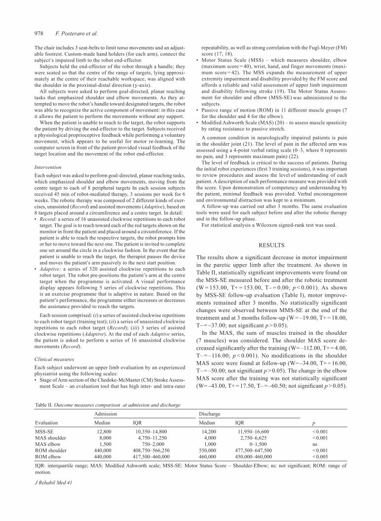
978 F. Posteraro et al.
The chair includes 3 seat-belts to limit torso movements and an adjust-able footrest. Custom-made hand holders (for each arm), connect the subject’s impaired limb to the robot end-effector.
Subjects held the end-effector of the robot through a handle; they were seated so that the centre of the range of targets, lying approxi-mately at the centre of their reachable workspace, was aligned with the shoulder in the proximal-distal direction (y-axis).
All subjects were asked to perform goal-directed, planar reaching tasks that emphasized shoulder and elbow movements. As they at-tempted to move the robot’s handle toward designated targets, the robot was able to recognize the active component of movement: in this case it allows the patient to perform the movements without any support.
When the patient is unable to reach to the target, the robot supports the patient by driving the end-effector to the target. Subjects received a physiological proprioceptive feedback while performing a voluntary movement, which appears to be useful for motor re-learning. The computer screen in front of the patient provided visual feedback of the target location and the movement of the robot end-effector.
InterventionEach subject was asked to perform goal-directed, planar reaching tasks, which emphasized shoulder and elbow movements, moving from the centre target to each of 8 peripheral targets In each session subjects received 45 min of robot-mediated therapy, 3 sessions per week for 6 weeks. The robotic therapy was composed of 2 different kinds of exer-cises, unassisted (Record) and assisted movements (Adaptive), based on 8 targets placed around a circumference and a centre target. In detail:• Record: a series of 16 unassisted clockwise repetitions to each robot
target. The goal is to reach toward each of the red targets shown on the monitor in front the patient and placed around a circumference. If the patient is able to reach the respective targets, the robot prompts him or her to move toward the next one. The patient is invited to complete one set around the circle in a clockwise fashion. In the event that the patient is unable to reach the target, the therapist pauses the device and moves the patient’s arm passively to the next start position.
• Adaptive: a series of 320 assisted clockwise repetitions to each robot target. The robot pre-positions the patient’s arm at the centre target when the programme is activated. A visual performance display appears following 5 series of clockwise repetitions. This is an exercise programme that is adaptive in nature. Based on the patient’s performance, the programme either increases or decreases the assistance provided to reach the targets.
Each session comprised: (i) a series of assisted clockwise repetitions to each robot target (training test); (ii) a series of unassisted clockwise repetitions to each robot target (Record); (iii) 3 series of assisted clockwise repetitions (Adaptive). At the end of each Adaptive series, the patient is asked to perform a series of 16 unassisted clockwise movements (Record).
Clinical measuresEach subject underwent an upper limb evaluation by an experienced physiatrist using the following scales: • Stage of Arm section of the Chedoke-McMaster (CM) Stroke Assess-
ment Scale – an evaluation tool that has high inter- and intra-rater
repeatability, as well as strong correlation with the Fugl-Meyer (FM) score (17, 18).
• Motor Status Scale (MSS) – which measures shoulder, elbow (maximum score = 40), wrist, hand, and finger movements (maxi-mum score = 42). The MSS expands the measurement of upper extremity impairment and disability provided by the FM score and affords a reliable and valid assessment of upper limb impairment and disability following stroke (19). The Motor Status Assess-ment for shoulder and elbow (MSS-SE) was administered to the subjects.
• Passive range of motion (ROM) in 11 different muscle groups (7 for the shoulder and 4 for the elbow).
• Modified Ashworth Scale (MAS) (20) – to assess muscle spasticity by rating resistance to passive stretch.
A common condition in neurologically impaired patients is pain in the shoulder joint (21). The level of pain in the affected arm was assessed using a 4-point verbal rating scale (0–3, where 0 represents no pain, and 3 represents maximum pain) (22).
The level of feedback is critical to the success of patients. During the initial robot experiences (first 3 training sessions), it was important to review procedures and assess the level of understanding of each patient. A description of each performance measure was provided with the score. Upon demonstration of competency and understanding by the patient, minimal feedback was provided. Verbal encouragement and environmental distraction was kept to a minimum.
A follow-up was carried out after 3 months. The same evaluation tools were used for each subject before and after the robotic therapy and in the follow-up phase.
For statistical analysis a Wilcoxon signed-rank test was used.
RESULTS
The results show a significant decrease in motor impairment in the paretic upper limb after the treatment. As shown in Table II, statistically significant improvements were found on the MSS-SE measured before and after the robotic treatment (W = 153.00, T+ = 153.00, T– = 0.00; p < 0.001). As shown by MSS-SE follow-up evaluation (Table I), motor improve-ments remained after 3 months. No statistically significant changes were observed between MMS-SE at the end of the treatment and at 3 months follow-up (W = –19.00, T+ = 18.00, T– = –37.00; not significant p > 0.05).
In the MAS, the sum of muscles trained in the shoulder (7 muscles) was considered. The shoulder MAS score de-creased significantly after the training (W= –112.00, T+ = 4.00, T– = –116.00; p < 0.001). No modifications in the shoulder MAS score were found at follow-up (W= –34.00, T+ = 16.00, T– = –50.00; not significant p > 0.05). The change in the elbow MAS score after the training was not statistically significant (W= –43.00, T+ = 17.50, T– = –60.50; not significant p > 0.05).
Table II. Outcome measures comparison at admission and discharge
Evaluation
Admission Discharge
pMedian IQR Median IQR
MSS-SE 12,800 10,350–14,800 14,200 11,950–16,600 < 0.001MAS shoulder 8,000 4,750–11,250 4,000 2,750–6,625 < 0.001MAS elbow 1,500 750–2,000 1,000 0–1,500 nsROM shoulder 440,000 408,750–566,250 550,000 477,500–647,500 < 0.001ROM elbow 440,000 417,500–460,000 460,000 450,000–460,000 < 0.005
IQR: interquartile range; MAS: Modified Ashworth scale; MSS-SE: Motor Status Score – Shoulder-Elbow; ns: not significant; ROM: range of motion.
J Rehabil Med 41
979Robot-mediated therapy for paretic upper limb following neurological injury
Passive ROM in the shoulder also increased in both groups. The results show that, for the shoulder, a statistically significant improvement was found (W = 134.00, T+ = 135.00, T– = –1.00; p < 0.001). At follow-up the passive ROM values of the shoulder showed a further statistically significant reduction in impairment (W = 91.00, T+ = 98.50, T– = –7.00; p < 0.05).
Elbow passive ROM showed a statistically significant improve-ment (W = 64.00, T+ = 65.00, T– = –1.00; p < 0.005).
At admission to the clinical trial, 5 subjects had shoulder pain. At the end of the robotic therapy they showed a reduction in the pain score. The scores of two subjects decreased from 1 to 0, and three from 2 to 1. No patients showed an increased score on the pain scale.
The robot-mediated therapy was well accepted and tolerated by the patients. No adverse events occurred during the study and no patient withdrew from therapy. At the end of treatment patients informally reported improved use of the impaired upper limb and some of them also reported an improvement in locomotion.
DISCUSSION
These results confirm the effectiveness of robot-mediated reha-bilitation therapy for chronic patients and support the hypothesis that improvements in motor abilities after a neurological injury can continue for more than one year after the acute event.
In the group of patients treated with the robotic system, MSS-SE score and ROM increased in the shoulder and el-bow, and MAS scores decreased and did not increase. These results do not support the hypothesis that active motor action in spastic patients may be responsible for increasing muscle tone, and do not imply any worsening in motor performance. In fact, most traditional rehabilitative methodologies, based on reflex inhibition (e.g. Bobath) (23), aim to reduce and limit spasticity and, in some cases (24), to delay execution of active movements, since they could be responsible for an increase in muscular tone and a worsening of spasticity.
According to these methodologies, active movements in-volving flexor muscles, such as shoulder adduction, shoulder intra-rotation and, in particular, elbow flexion, can induce an increase in muscle spasticity, thus resulting in a worsening of upper limb motor impairment.
A reduction in shoulder pain, the prevalence of which is very high in chronic hemiparetic subjects (21), is an additional advantage of robot-mediated therapy.
Patients with severe spasticity (CM = 3) also improved, and thus it is advised that this group of patients are recruited to robotic training.
Patients with flaccid hemiparesis (CM = 1) appear to display a rather lower improvement in the MMS-SE score after the training. This might imply that it is less effective to perform a specific intervention in the chronic phase compared with the addition of sensorimotor stimulation during the acute phase after a stroke (25).
The mechanisms involved in the improvement in motor performance after repetitive training are not known; the in-
duced modifications in impairment in chronic patients after neurological injury could be related to motor recovery and motor relearning.
It is well known that for motor learning and motor planning, humans have to perform voluntary movements. Proprioceptive, visual and tactile feedback is also important for motor learning: the first-mentioned being the most important.
The patients, including severely spastic subjects, have to perform voluntary movements as far as possible. If the sub-ject is unable to complete the movement the robot helps him or her to reach the desired target, thus providing an adequate proprioceptive feedback.
Robotic systems can provide some advantages in motor rehabilitation for people with chronic neurological injury involving the upper limb, by delivering an intensive and repeti-tive treatment. In each session the patient can perform almost 1000 goal-directed movements in approximately 45 min. Such treatments can be physically exhausting for a physiotherapist. A robotic system can contribute to the treatment being performed in a repetitive way and to supporting the trajectories with a high precision. Therefore, robotic therapy may be a substitute for traditional therapy, at least in chronic patients, for the preservation of functional recovery obtained during the initial period after an acute event. The large and increasing number of surviving neurologically impaired chronic patients requiring treatment suggests that robot-mediated therapy could also be useful in reducing costs to the healthcare system.
Furthermore, kinematic and kinetic data can be recorded and used to search for useful markers that can quantify the motor recovery process of each patient. Anyway, the search for such markers is not the objective of the present work, which is mainly focused on the clinical assessment.
It is noteworthy that such systems (using reprogrammable software code) are helpful in investigating the pathophysio-logical mechanisms underlying motor recovery following neurological damage.
As set out in the International Classification of Functioning, Disability and Health (ICF) terminology (26), future efforts should aim to maximize the improvement in the activity level and not only in the structure and body functioning.
ACKNOWLEDGEMENTS
The present work was partly funded by Fondazione Cassa di Risparmio di Lucca, Italy and Fondazione Cassa di Risparmio di Volterra, Italy.
Conflictofinterest:The authors have no conflicts of interest to declare, or any financial or other interest in the manufacturer or distributor of the device used in the present study.
REFERENCES
World Health Organization, The atlas of heart disease and stroke. 1. Available from: http://www.who.int/cardiovascular_diseases/resources/atlas/en/.American Heart Association, heart disease and stroke statistics – 2. 2007. Available from: http://www.americanheart.org/.
J Rehabil Med 41
980 F. Posteraro et al.
Brainin M, Bornstein N, Boysen G, Demarin V. Acute neurological 3. stroke care in Europe: results of the European Stroke Care Inven-tory. Eur J Neurol 2000; 7: 5–10.The Italian Guidelines for stroke prevention. Stroke Prevention 4. and Educational Awareness Diffusion (SPREAD). 4th ed. Milano: Pubblicazioni Catel – Hyperphar Group SpA; 2007.Nichols-Larsen DS, Clark PC, Zeringue A, Greenspan A, Blanton 5. S. Factors influencing stroke survivors’ quality of life during sub-acute recovery. Stroke 2005; 36: 1480–1484.Duncan PW, Goldstein LB, Matchar D, Divine GW, Feussner J. 6. Measurement of motor recovery after stroke. Stroke 1992; 23: 1084–1089.Hendricks HT, Van Limbeek J, Geurts AC, Zwarts MJ. Motor 7. recovery after stroke: a systematic review of the literature. Arch Phys Med Rehabil 2002; 83: 1629–1637.Miltner WL, Bauder H, Sommer M, Dettmers C, Taub E. Effects 8. of constraint induced movement therapy on patients with chronic motor deficits after stroke. Stroke 1999; 30: 586–592.Wolf SL, Winstein CJ, Miller JP, Taub E, Uswatte G, Morris 9. D, et al. Effect of constraint-induced movement therapy on up-per extremity function 3 to 9 months after stroke: the EXCITE randomized clinical trial. J Am Med Assoc 2006; 296: 2095–2104.Fasoli SE, Krebs HI, Stein J, Frontera WR, Hogan N. Effects of 10. robotic therapy on motor impairment and recovery in chronic stroke. Arch Phys Med Rehabil 2003; 84: 477–482.Duncan PW, Zorowitz R, Bates B, Choi JY, Glasberg JJ, Graham 11. GD, et al. Management of adult stroke rehabilitation care: a clinical practice guideline. Stroke 2005; 36: e100–e143.Barreca S, Wolf SL, Fasoli S, Bohannon R. Treatment interventions 12. for the paretic upper limb of stroke survivors: a critical review. Neurorehab Neural Repair 2003; 17: 220–226.Riener R, Nef T, Colombo G. Robot-aided neurorehabilitation of 13. the upper limb. Med Biol Eng Comput 2005; 43: 2–10.Micera S, Carrozza MC, Guglielmelli E, Cappiello G, Zaccone F, 14. Freschi C, et al. A simple robotic system for neurorehabilitation. J Autonomous Robots 2005; 19: 271–284.Prange GB, Jannink MJ, Groothuis-Oudshoorn CG, Hermens 15. HJ, Ijzerman MJ. Systematic review of the effect of robot-aided
therapy on recovery of the hemiparetic arm after stroke. J Rehabil Res Dev 2006; 43: 171–184.Krebs HI, Hogan N, Aisen ML, Volpe BT. Robot-aided neuro-16. rehabilitation. IEEE Trans Rehabil Eng 1998; 6: 75–87.Gowland C, Stratford P, Ward M, Moreland J. Torresin W, Van 17. Hullenaar S, et al. Measuring physical impairment and disability with the Chedoke-McMaster Stroke Assessment. Stroke 1993; 24: 58–63. Gowland C, Van Hullenaar S, Torrensin W, Moreland J, Vanspall B, 18. Barreca S, et al. Chedoke-Mc Master stroke assessment: develop-ment, validation and administration manual. Hamilton, Ontario: Chedoke-McMaster Hospitals and McMaster University; 1995. Ferraro M, Demaio JH, Krol J, Trudell C, Rannekleiv K, Edelstein 19. L, et al. Assessing the motor status score: a scale for the evaluation of upper limb motor outcomes in patients after stroke. Neurorehab Neural Re 2002; 16: 283–289. Bohannon RW, Smith MD. Interrater reliability of a modified Ash-20. worth scale of muscle spasticity. Phys Ther 1987; 67: 206–207.Chae J, Mascarenhas D, Yu DT, Kirsteins A, Elovic EP, Flanagan 21. SR, et al. Poststroke shoulder pain: its relationship to motor im-pairment, activity limitation, and quality of life. Arch Phys Med Rehabil 2007; 88: 298–301.Dawniew WW, Leatham PA, Rhind VM, Wright V, Branco J, 22. Anderson JA, et al. Studies with pain rating scales. Ann Rheum Dis 1978; 37: 378–381. Barnes MP, Johnson GR, editors. Upper motor neurone syndrome 23. and spasticity. Clinical management and neurophysiology. Cam-bridge: Cambridge University Press; 2001.Perfetti C. La rieducazione motoria dell’emiplegico [Motor re-24. habilitation for hemiparetic patients]. Milando: Ghedini Editore; 1979 (in Italian). Feys H, De Weerdt W, Verbeke G, Steck GC, Capiau C, Kiekens C, 25. et al. Early and repetitive stimulation of the arm can substantially improve the long-term outcome after stroke: a 5-year follow-up study of a randomized trial. Stroke 2004; 35: 924–929. Stucki G, Melvin J. The International Classification of Functioning, 26. Disability and Health (ICF): a unifying model for the conceptual description of the rehabilitation strategy. J Rehabil Med 2007; 39: 279–285.
J Rehabil Med 41
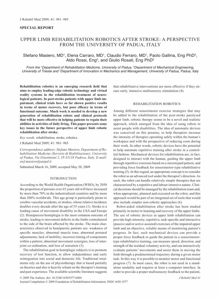
special report
J Rehabil Med 2009; 41: 981–985
J Rehabil Med 41© 2009 The Authors. doi: 10.2340/16501977-0404Journal Compilation © 2009 Foundation of Rehabilitation Information. ISSN 1650-1977
Rehabilitation robotics is an emerging research field that aims to employ leading-edge robotic technology and virtual reality systems in the rehabilitation treatment of neuro-logical patients. in post-stroke patients with upper limb im-pairment, clinical trials have so far shown positive results in terms of motor recovery, but poor efficacy in terms of functional outcome. much work is needed to develop a new generation of rehabilitation robots and clinical protocols that will be more effective in helping patients to regain their abilities in activities of daily living. this paper presents some key issues in the future perspective of upper limb robotic rehabilitation after stroke.Key words: rehabilitation, stroke, robotics. J Rehabil Med 2009; 41: 981–985
Correspondence address: Stefano Masiero, Department of Re-habilitation Medicine (Servizio di Riabilitazione), University of Padua, Via Giustiniani 2, IT-35128 Padova, Italy. E-mail: [email protected] March 16, 2009; accepted May 20, 2009
INTRODUCTION
According to the World Health Organization (WHO), by 2050 the proportion of persons over 65 years old will have increased by more than 70% in the industrialized countries and by more than 200% worldwide. This age group is particularly prone to cerebro-vascular accidents, or strokes, whose relative incidence doubles every decade after the age of 55 years (1). Stroke is a leading cause of movement disability in the USA and Europe (2). Hemiparesis/hemiplegia is the most common outcome of stroke, leading to movement deficits in the limbs contralateral to the side of the brain affected by the stroke. The main char-acteristics observed in hemiparetic patients are: weakness of specific muscles; abnormal muscle tone; abnormal postural adjustments; lack of mobility; incorrect timing of components within a pattern; abnormal movement synergies; loss of inter-joint co-ordination, and loss of sensation (3).
The rehabilitation goal in hemiplegic subjects is to promote recovery of lost function, to allow independence and early reintegration into social and domestic life. Traditional treat-ments rely on the use of physiotherapy that is based partially on theories and also is heavily reliant on the therapist’s training and past experience. The available scientific literature suggests
that rehabilitative interventions are more effective if they en-sure early, intensive multisensory stimulation (4).
REHABILITATION ROBOTICS
Among different sensorimotor exercise strategies that may be added to the rehabilitation of the post-stroke paralysed upper limb, robotic therapy seems to be a novel and realistic approach, which emerged from the idea of using robots to assist people with disabilities. The idea of automatic devices was conceived on this premise, to help therapists increase the intensity of therapies operating safely within the human’s workspace and with the prospective of reducing costs during their work. In other words, robotic devices have the potential to help automate repetitive training after stroke in a control-led fashion. Mechanical devices for rehabilitation are, in fact, designed to interact with the human, guiding the upper limb through repetitive exercises based on a stereotyped pattern, and providing force feedback for sensorimotor-type rehabilitative training (5). In this regard, an appropriate concept is to consider the robot as an advanced tool under the therapist’s direction. As such, the robot can handle relatively simple therapies that are characterized by a repetitive and labour-intensive nature. Clini-cal decisions should be managed by the rehabilitation team and, when appropriate, planned and executed on the robot, and this approach would be part of an integrated set of tools that would also include simpler non-robotic approaches (6).
Robot-aided rehabilitation after stroke has been studied primarily in motor re-learning and recovery of the upper limbs. The use of robotic devices in upper limb rehabilitation can provide high-intensity, repetitive, task-specific and interactive (passive and/or active-assisted) exercises of the impaired upper limb and an objective, reliable means of monitoring patient’s progress. In fact, such mechanical devices can provide a proper force feedback to guide the patient in a sensorimotor-type rehabilitative training, can measure speed, direction, and strength of the residual voluntary activity, and can interactively evaluate patients’ movements and assist them in moving the limb through a predetermined trajectory during a given motor task. In this way it is possible to monitor motor and functional progress (7). In most cases, the robot is not used in a stand-alone modality and requires at least a computer interface. In order to provide a proper multisensory feedback to the patient,
UPPER LIMB REHABILITATION ROBOTICS AFTER STROKE: A PERSPECTIVE FROM THE UNIVERSITY OF PADUA, ITALY
Stefano Masiero, MD1, Elena Carraro, MD1, Claudio Ferraro, MD1, Paolo Gallina, Eng PhD3, Aldo Rossi, Eng2, and Giulio Rosati, Eng PhD2
From the 1Department of Rehabilitation Medicine, University of Padua, 2Department of Mechanical Engineering, University of Trieste and 3Department of Innovation in Mechanics and Management, University of Padua, Padua, Italy
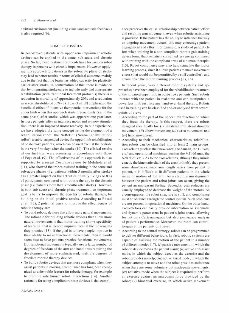
982 S. Masiero et al.
a virtual environment (including visual and acoustic feedback) is also required (6).
SOME KEY ISSUES
In post-stroke patients with upper arm impairment robotic devices can be applied in the acute, sub-acute and chronic phase. So far, most treatment protocols have focused on robot therapy in persons with chronic impairment. However, apply-ing this approach to patients in the sub-acute phase of stroke may lead to better results in terms of clinical outcome, mainly due to the fact that the brain has added capacity for plasticity earlier after stroke. In confirmation of this, there is evidence that by integrating stroke care to include early and appropriate rehabilitation (with traditional treatment protocols) there is a reduction in mortality of approximately 20% and a reduction in severe disability of 30% (8). Feys et al. (9) emphasized the beneficial effect of intensive therapeutic interventions for the upper limb when this approach starts precociously (i.e. in the acute phase) after stroke, which was apparent one year later. In these patients, after an intensive motor and sensory stimula-tion, there is an improved motor recovery. In our experience, we have adopted the same concept in the development of a rehabilitation robot: the NeReBot (Neuro-Rehabilitation-roBot), a cable-suspended device for upper limb rehabilitation of post-stroke patients, which can be used even at the bedside in the very first days after the stroke (10). The clinical results of our first trial were promising in accordance with those of Feys et al. (9). The effectiveness of this approach is also supported by a recent Cochrane review by Mehrholz et al. (11), who showed that robot-assisted training in the acute and sub-acute phases (i.e. patients within 3 months after stroke) has a greater impact on the activities of daily living (ADLs) of participants, compared with robotic therapy in the chronic phase (i.e. patients more than 3 months after stroke). However, in both sub-acute and chronic phase treatment, an important goal is to try to improve the benefits of robotic therapy, by building on the initial positive results. According to Rosati et al. (12), 2 potential ways to improve the effectiveness of robotic therapy are: • To build robotic devices that allow more natural movements.
The rationale for building robotic devices that allow more natural movements is that motor training shows specificity of learning; that is, people improve most at the movements they practice (13). If the goal is to have people improve in their ability to make functional movements, then it would seem best to have patients practice functional movements. But functional movements typically use a large number of degrees of freedom of the arm and hand, thus requiring the development of more sophisticated, multiple degrees of freedom robotic therapy devices.
• To build robotic devices that are more compliant when they assist patients in moving. Compliance has long been recog-nized as a desirable feature for robotic therapy, for example to promote safe human robot interactions (14). Another rationale for using compliant robotic devices is that compli-
ance preserves the causal relationship between patient effort and resulting arm movement, even when robotic assistance is provided. If the patient has the ability to influence the way an ongoing movement occurs, this may encourage patient engagement and effort. For example, a study of patient ef-fort when training in a non-compliant robotic gait training device found that the patient consumed less energy compared with training with the compliant arms of a human therapist (15). Robot compliance may also help stimulate the motor learning process, since it allows patients to make movement errors (that would not be permitted by a stiff controller), and errors drive the motor learning process (13, 16).
In recent years, very different robotic systems and ap-proaches have been employed for the rehabilitation treatment of the impaired upper limb in post-stroke patients. Such robots interact with the patient in real-time and can manipulate a powerless limb just like any hand-over-hand therapy. Robots used in training can be classified and/or analysed from several points of view:• According to the part of the upper limb function on which
they focus the therapy. In this respect, there are robots designed specifically for: (i) unilateral or bilateral shoulder movement; (ii) elbow movement; (iii) wrist movement; and (iv) hand movement.
• According to their mechanical characteristics, rehabilita-tion robots can be classified into at least 2 main groups: exoskeleton (such as the Pnew-wrex, the Arm-In, the L-Exos, etc.) and operational machines (such as the MIT-Manus, the NeReBot, etc.). As to the exoskeletons, although they mimic exactly the kinematic chain of the arm (or limb), they present some drawbacks: since arm length varies from patient to patient, it is difficult to fit different patients in the whole range of motion of the arm. As a result, a misalignment between the patient and robot joints can occur, giving the patient an unpleasant feeling. Secondly, gear reducers are usually employed to decrease the weight of the motors. As a consequence, the robot structure is stiff, and compliance must be obtained through the control system. Such problems are not present in operational machines. On the other hand, exoskeletons can easily provide information on kinematic and dynamic parameters in patient’s joint-space, allowing for not only Cartesian-space but also joint-space analysis of patient’s performance. Moreover, the robot can control torques at the patient-joint level.
• According to the control strategy, robots can be programmed to deliver different behaviours. In fact, robotic systems are capable of assisting the motion of the patient in a number of different modes (17): (i) passive movement, in which the robotic device moves the patient’s arm; (ii) active non-assist mode, in which the subject executes the exercise and the robot provides no help; (iii) active assist mode, in which the subject attempts to move and the robot provides assistance when there are some voluntary but inadequate movements; (iv) resistive mode when the subject is required to perform an exercise against an antagonist force provided by the robot; (v) bimanual exercise, in which active movement
J Rehabil Med 41
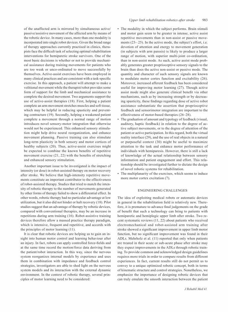
983Upper limb rehabilitation robotics after stroke
of the unaffected arm is mirrored by simultaneous active/passive/assistive movement of the affected arm by means of the robotic device. In many cases, more than one modality is incorporated into single robot devices. Given the broad range of therapy approaches currently practised in clinics, thera-pists face the difficult task of selecting optimal rehabilitation interventions for hemiparetic stroke survivors. One of the most basic decisions is whether or not to provide mechani-cal assistance during training movements for patients who are too weak or unco-ordinated to move successfully by themselves. Active-assist exercises have been employed in many clinical practices and are consistent with a task-specific exercise. In this approach, a patient will attempt to make a volitional movement while the therapist/robot provides some form of support for the limb and mechanical assistance to complete the desired movement. Two arguments support the use of active-assist therapies (18). First, helping a patient complete an arm movement stretches muscles and soft tissue, which may be helpful in reducing spasticity and prevent-ing contracture (19). Secondly, helping a weakened patient complete a movement through a normal range of motion introduces novel sensory-motor integration that otherwise would not be experienced. This enhanced sensory stimula-tion might help drive neural reorganization, and enhance movement planning. Passive training can also stimulate long-term plasticity in both sensory and motor cortices of healthy subjects (20). Thus, active-assist exercises might be expected to combine the known benefits of repetitive movement exercise (21, 22) with the benefits of stretching and enhanced sensory stimulation.
Another important issue to be investigated is the impact of intensity (or dose) in robot-assisted therapy on motor recovery after stroke. We believe that high-intensity repetitive move-ments constitute an important contributor to the effectiveness of robot-assisted therapy. Studies that tried to match the inten-sity of robotic therapy to the number of movements generated by other forms of therapy failed to show a differential effect. In other words, robotic therapy had no particular advantage at low utilization, but it also did not hinder or halt recovery (18). Pilot studies suggest that an advantage of therapy by robotic devices, compared with conventional therapies, may be an increase in repetitions during arm training (16). Robot-assistive training devices therefore allow a massed practice therapy paradigm, which is intensive, frequent and repetitive, and accords with the principles of motor learning (11).
It is clear that robotic devices are helping us to gain an in-sight into human motor control and learning behaviour after an injury. In fact, robots can apply controlled force-fields and at the same time record the motion/force data deriving from the patient/robot interaction. In this way, since the nervous system reorganizes internal models by experience and uses them in combination with impedance and feedback control strategies, investigators are able to shed light on the nervous system models and its interaction with the external dynamic environment. In the context of robotic therapy, several prin-ciples of motor learning need to be considered:
• The modality in which the subject performs. Brain stimuli and motor gain seem to be greater in intense, active assist repetitive movements than in non-assist or passive move-ments (23– 25). In the active mode, the subject’s effort, i.e. devotion of attention and energy to movement generation (in subjects with arm paresis) is likely to produce a larger range of motion, with superior multi-joint co-ordination, than in non-assist mode. As such, active assist mode prob-ably generates greater proprioceptive sensory signals to the brain than does the active non-assist or passive mode. The quantity and character of such sensory signals are known to modulate motor cortex function and excitability (26). Moreover, increased afferent feedback has been considered useful for improving motor learning (27). Though active assist mode might also generate clinical benefit via other mechanisms, such as by increasing strength or by decreas-ing spasticity, these findings regarding dose of active robot assistance substantiate the assertion that proprioceptive feedback and sensorimotor integration are important to the effectiveness of motor-based therapies (26–28).
• The graduation of amount and typology of feedback (visual, auditory, haptic feedback) in relation to the degree of ac-tive subject movements, or to the degree of attention of the patient or active participation. In this regard, both the virtual reality interface (29), and the use of real objects in a natural or purposeful context (30) might be useful to maximize attention to the task and enhance motor performance of individuals with hemiparesis. However, there is still a lack of knowledge of the actual relationship between sensory information and patient engagement and effort. This rela-tionship should be investigated further to dictate the design of novel robotic systems for rehabilitation.
• The multiplanarity of the exercises, which seems to induce more motor cortex excitation (7).
ENGINEERING CHALLENGES
The idea of exploiting medical robots or automatic devices in general in the rehabilitation field is relatively new. There-fore, it is premature to advance final judgements on the grade of benefit that such a technology can bring to patients with hemiparetic and hemiplegic upper limb after stroke. Two re-cent systematic reviews (11, 22) about patients who received electromechanical and robot-assisted arm training after stroke showed a significant improvement in upper limb motor function, but no significant improvement was found in their ADLs. Mehrholz et al. (11) reported that only when patients are treated in their acute or sub-acute phase after stroke may they expect improvements in the ADLs through robotic train-ing. To provide common and acknowledged design guidelines requires more trials in order to compare results from different experiences. In fact, current results still do not permit us to convey to a unique optimized robotic concept, both in terms of kinematic structure and control strategies. Nonetheless, we emphasize the importance of designing robotic devices that can truly emulate the smooth interaction between the patient
J Rehabil Med 41
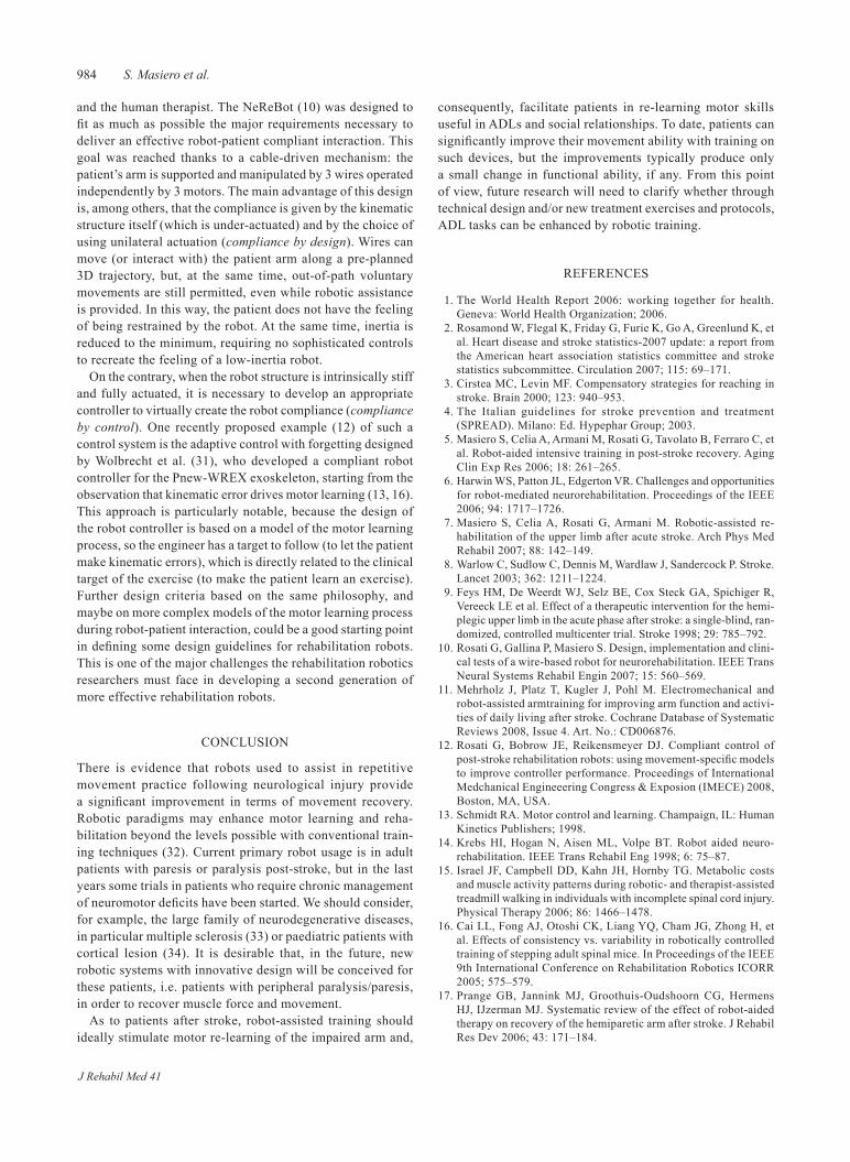
984 S. Masiero et al.
and the human therapist. The NeReBot (10) was designed to fit as much as possible the major requirements necessary to deliver an effective robot-patient compliant interaction. This goal was reached thanks to a cable-driven mechanism: the patient’s arm is supported and manipulated by 3 wires operated independently by 3 motors. The main advantage of this design is, among others, that the compliance is given by the kinematic structure itself (which is under-actuated) and by the choice of using unilateral actuation (compliance by design). Wires can move (or interact with) the patient arm along a pre-planned 3D trajectory, but, at the same time, out-of-path voluntary movements are still permitted, even while robotic assistance is provided. In this way, the patient does not have the feeling of being restrained by the robot. At the same time, inertia is reduced to the minimum, requiring no sophisticated controls to recreate the feeling of a low-inertia robot.
On the contrary, when the robot structure is intrinsically stiff and fully actuated, it is necessary to develop an appropriate controller to virtually create the robot compliance (compliance by control). One recently proposed example (12) of such a control system is the adaptive control with forgetting designed by Wolbrecht et al. (31), who developed a compliant robot controller for the Pnew-WREX exoskeleton, starting from the observation that kinematic error drives motor learning (13, 16). This approach is particularly notable, because the design of the robot controller is based on a model of the motor learning process, so the engineer has a target to follow (to let the patient make kinematic errors), which is directly related to the clinical target of the exercise (to make the patient learn an exercise). Further design criteria based on the same philosophy, and maybe on more complex models of the motor learning process during robot-patient interaction, could be a good starting point in defining some design guidelines for rehabilitation robots. This is one of the major challenges the rehabilitation robotics researchers must face in developing a second generation of more effective rehabilitation robots.
CONCLUSION
There is evidence that robots used to assist in repetitive movement practice following neurological injury provide a significant improvement in terms of movement recovery. Robotic paradigms may enhance motor learning and reha-bilitation beyond the levels possible with conventional train-ing techniques (32). Current primary robot usage is in adult patients with paresis or paralysis post-stroke, but in the last years some trials in patients who require chronic management of neuromotor deficits have been started. We should consider, for example, the large family of neurodegenerative diseases, in particular multiple sclerosis (33) or paediatric patients with cortical lesion (34). It is desirable that, in the future, new robotic systems with innovative design will be conceived for these patients, i.e. patients with peripheral paralysis/paresis, in order to recover muscle force and movement.
As to patients after stroke, robot-assisted training should ideally stimulate motor re-learning of the impaired arm and,
consequently, facilitate patients in re-learning motor skills useful in ADLs and social relationships. To date, patients can significantly improve their movement ability with training on such devices, but the improvements typically produce only a small change in functional ability, if any. From this point of view, future research will need to clarify whether through technical design and/or new treatment exercises and protocols, ADL tasks can be enhanced by robotic training.
REFERENCES
The World Health Report 2006: working together for health. 1. Geneva: World Health Organization; 2006.Rosamond W, Flegal K, Friday G, Furie K, Go A, Greenlund K, et 2. al. Heart disease and stroke statistics-2007 update: a report from the American heart association statistics committee and stroke statistics subcommittee. Circulation 2007; 115: 69–171.Cirstea MC, Levin MF. Compensatory strategies for reaching in 3. stroke. Brain 2000; 123: 940–953.The Italian guidelines for stroke prevention and treatment 4. (SPREAD). Milano: Ed. Hypephar Group; 2003. Masiero S, Celia A, Armani M, Rosati G, Tavolato B, Ferraro C, et 5. al. Robot-aided intensive training in post-stroke recovery. Aging Clin Exp Res 2006; 18: 261–265.Harwin WS, Patton JL, Edgerton VR. Challenges and opportunities 6. for robot-mediated neurorehabilitation. Proceedings of the IEEE 2006; 94: 1717–1726.Masiero S, Celia A, Rosati G, Armani M. Robotic-assisted re-7. habilitation of the upper limb after acute stroke. Arch Phys Med Rehabil 2007; 88: 142–149.Warlow C, Sudlow C, Dennis M, Wardlaw J, Sandercock P. Stroke. 8. Lancet 2003; 362: 1211–1224.Feys HM, De Weerdt WJ, Selz BE, Cox Steck GA, Spichiger R, 9. Vereeck LE et al. Effect of a therapeutic intervention for the hemi-plegic upper limb in the acute phase after stroke: a single-blind, ran-domized, controlled multicenter trial. Stroke 1998; 29: 785–792.Rosati G, Gallina P, Masiero S. Design, implementation and clini-10. cal tests of a wire-based robot for neurorehabilitation. IEEE Trans Neural Systems Rehabil Engin 2007; 15: 560–569.Mehrholz J, Platz T, Kugler J, Pohl M. Electromechanical and 11. robot-assisted armtraining for improving arm function and activi-ties of daily living after stroke. Cochrane Database of Systematic Reviews 2008, Issue 4. Art. No.: CD006876. Rosati G, Bobrow JE, Reikensmeyer DJ. Compliant control of 12. post-stroke rehabilitation robots: using movement-specific models to improve controller performance. Proceedings of International Medchanical Engineeering Congress & Exposion (IMECE) 2008, Boston, MA, USA.Schmidt RA. Motor control and learning. Champaign, IL: Human 13. Kinetics Publishers; 1998. Krebs HI, Hogan N, Aisen ML, Volpe BT. Robot aided neuro-14. rehabilitation. IEEE Trans Rehabil Eng 1998; 6: 75–87.Israel JF, Campbell DD, Kahn JH, Hornby TG. Metabolic costs 15. and muscle activity patterns during robotic- and therapist-assisted treadmill walking in individuals with incomplete spinal cord injury. Physical Therapy 2006; 86: 1466–1478.Cai LL, Fong AJ, Otoshi CK, Liang YQ, Cham JG, Zhong H, et 16. al. Effects of consistency vs. variability in robotically controlled training of stepping adult spinal mice. In Proceedings of the IEEE 9th International Conference on Rehabilitation Robotics ICORR 2005; 575–579.Prange GB, Jannink MJ, Groothuis-Oudshoorn CG, Hermens 17. HJ, IJzerman MJ. Systematic review of the effect of robot-aided therapy on recovery of the hemiparetic arm after stroke. J Rehabil Res Dev 2006; 43: 171–184.
J Rehabil Med 41
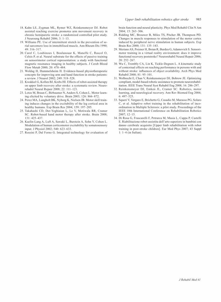
985Upper limb rehabilitation robotics after stroke
Kahn LE, Zygman ML, Rymer WZ, Reinkensmeyer DJ. Robot 18. assisted reaching exercise promotes arm movement recovery in chronic hemiparetic stroke: a randomized controlled pilot study. J Neuroeng Rehabil 2006; 3: 1–13.Williams PE. Use of intermittent stretch in the prevention of se-19. rial sarcomere loss in immobilised muscle. Ann Rheum Dis 1990; 49: 316–317.Carel C, Loubinoux I, Boulanouar K, Manelfe C, Rascol O, 20. Celsis P, et al. Neural substrate for the effects of passive training on sensorimotor cortical representation: a study with functional magnetic resonance imaging in healthy subjects. J Cereb Blood Flow Metab 2000; 20: 478–484.Woldag H, Hummelsheim H. Evidence-based physiotherapeutic 21. concepts for improving arm and hand function in stroke patients: a review. J Neurol 2002; 249: 518–528. Kwakkel G, Kollen BJ, Krebs HI. Effects of robot-assisted therapy 22. on upper limb recovery after stroke: a systematic review. Neuro-rehabil Neural Repair 2008; 22: 111–121.Lotze M, Braun C, Birbaumer N, Anders S, Cohen L. Motor learn-23. ing elicited by voluntary drive. Brain 2003; 126: 866–872.Perez MA, Lungholt BK, Nyborg K, Nielsen JB. Motor skill train-24. ing induces changes in the excitability of the leg cortical area in healthy humans. Exp Brain Res 2004; 159: 197–205.Takahashi CD, Der-Yeghiaian L, Le V, Motiwala RR, Cramer 25. SC. Robot-based hand motor therapy after stroke. Brain 2008; 131: 425–437.Kaelin-Lang A, Luft A, Sawaki L, Burstein A, Sohn Y, Cohen L. 26. Modulation of human corticomotor excitability by somatosensory input. J Physiol 2002; 540: 623–633.Rossini P, Dal Forno G. Integrated technology for evaluation of 27.
brain function and neural plasticity. Phys Med Rehabil Clin N Am 2004; 15: 263–306.Ridding MC, Brouwer B, Miles TS, Pitcher JB, Thompson PD. 28. Changes in muscle responses to stimulation of the motor cortex induced by peripheral nerve stimulation in human subjects. Exp Brain Res 2000; 131: 135–143.Merians AS, Poizner H, Boian R, Burdea G, Adamovich S. Sensori-29. motor training in a virtual reality environment: does it improve functional recovery poststroke? Neurorehabil Neural Repair 2006; 20: 252–267.Wu C, Trombly CA, Lin K, Tickle-Degnen L. A kinematic study 30. of contextual effects on reaching performance in persons with and without stroke: influences of object availability. Arch Phys Med Rehabil 2000; 81: 95–101.Wolbrecht E, Chan V, Reinkensmeyer DJ, Bobrow JE. Optimizing 31. compliant, model-based robotic assistance to promote neurorehabil-itation. IEEE Trans Neural Syst Rehabil Eng 2008; 16: 286–297. Reinkensmeyer DJ, Emken JL, Cramer SC. Robotics, motor 32. learning, and neurological recovery. Ann Rev Biomed Eng 2004; 6: 497–525.Squeri V, Vergaro E, Brichetto G, Casadio M, Marasso PG, Solaro 33. C, et al. Adaptive robot training in the rehabilitation of inco-ordination in Multiple Sclerosis: a pilot study. Proceedings of the IEEE 10th International Conference on Rehabilitation Robotics 2007; 12–15.Di Rosa G, Frascarelli F, Petrarca M, Masia L, Cappa P, Castelli 34. E. Riabilitazione robot-assistita dell’arto superiore in bambini con danno cerebrale acquisito [Upper limb rehabilitation with robot training in post-stroke children]. Eur Med Phys 2007; 43 Suppl 1: 1–4 (in Italian).
J Rehabil Med 41
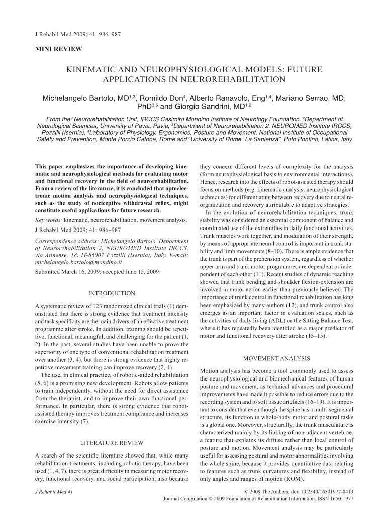
© 2009 The Authors. doi: 10.2340/16501977-0413Journal Compilation © 2009 Foundation of Rehabilitation Information. ISSN 1650-1977
J Rehabil Med 41
mini revieW
J Rehabil Med 2009; 41: 986–987
this paper emphasizes the importance of developing kine-matic and neurophysiological methods for evaluating motor and functional recovery in the field of neurorehabilitation. from a review of the literature, it is concluded that optoelec-tronic motion analysis and neurophysiological techniques, such as the study of nociceptive withdrawal reflex, might constitute useful applications for future research.Key words: kinematic, neurorehabilitation, movement analysis.J Rehabil Med 2009; 41: 986–987
Correspondence address: Michelangelo Bartolo, Department of Neurorehabilitation 2, NEUROMED Institute IRCCS, via Atinense, 18, IT-86007 Pozzilli (Isernia), Italy. E-mail: [email protected] March 16, 2009; accepted June 15, 2009
INTRODUCTION
A systematic review of 123 randomized clinical trials (1) dem-onstrated that there is strong evidence that treatment intensity and task specificity are the main drivers of an effective treatment programme after stroke. In addition, training should be repeti-tive, functional, meaningful, and challenging for the patient (1, 2). In the past, several studies have been unable to prove the superiority of one type of conventional rehabilitation treatment over another (3, 4), but there is strong evidence that highly re-petitive movement training can improve recovery (2, 4).
The use, in clinical practice, of robotic-aided rehabilitation (5, 6) is a promising new development. Robots allow patients to train independently, without the need for direct assistance from the therapist, and to improve their own functional per-formance. In particular, there is strong evidence that robot-assisted therapy improves treatment compliance and increases exercise intensity (7).
LITERATURE REVIEW
A search of the scientific literature showed that, while many rehabilitation treatments, including robotic therapy, have been used (1, 4, 7), there is great difficulty in measuring motor recov-ery, functional recovery, and social participation, also because
they concern different levels of complexity for the analysis (form neurophysiological basis to environmental interactions). Hence, research into the effects of robot-assisted therapy should focus on methods (e.g. kinematic analysis, neurophysiological techniques) for differentiating between recovery due to neural re-organization and recovery attributable to adaptive strategies.
In the evolution of neurorehabilitation techniques, trunk stability was considered an essential component of balance and coordinated use of the extremities in daily functional activities. Trunk muscles work together, and modulation of their strength, by means of appropriate neural control is important in trunk sta-bility and limb movements (8–10). There is ample evidence that the trunk is part of the prehension system, regardless of whether upper arm and trunk motor programmes are dependent or inde-pendent of each other (11). Recent studies of dynamic reaching showed that trunk bending and shoulder flexion-extension are involved in motor action earlier than previously believed. The importance of trunk control in functional rehabilitation has long been emphasized by many authors (12), and trunk control also emerges as an important factor in evaluation scales, such as the activities of daily living (ADL) or the Sitting Balance Test, where it has repeatedly been identified as a major predictor of motor and functional recovery after stroke (13–15).
MOVEMENT ANALYSIS
Motion analysis has become a tool commonly used to assess the neurophysiological and biomechanical features of human posture and movement, as technical advances and procedural improvements have made it possible to reduce errors due to the recording system and to soft tissue artefacts (16–19). It is impor-tant to consider that even though the spine has a multi-segmental structure, its function in whole-body motor and postural tasks is a global one. Moreover, structurally, the trunk musculature is characterized mainly by its linking of non-adjacent vertebrae, a feature that explains its diffuse rather than local control of posture and motion. Movement analysis may be particularly useful for assessing postural and motor abnormalities involving the whole spine, because it provides quantitative data relating to features such as trunk curvatures and flexibility, instead of only angles and ranges of motion (ROM).
KINEMATIC AND NEUROPHYSIOLOGICAL MODELS: FUTURE APPLICATIONS IN NEUROREHABILITATION
Michelangelo Bartolo, MD1,3, Romildo Don4, Alberto Ranavolo, Eng1,4, Mariano Serrao, MD, PhD3,5 and Giorgio Sandrini, MD1,2
From the 1Neurorehabilitation Unit, IRCCS Casimiro Mondino Institute of Neurology Foundation, 2Department of Neurological Sciences, University of Pavia, Pavia, 3Department of Neurorehabilitation 2, NEUROMED Institute IRCCS,
Pozzilli (Isernia), 4Laboratory of Physiology, Ergonomics, Posture and Movement, National Institute of Occupational Safety and Prevention, Monte Porzio Catone, Rome and 5University of Rome “La Sapienza”, Polo Pontino, Latina, Italy
987Kinematic and neurophysiological models in neurorehabilitation
For this reason, the development of global models for assessing the whole spine regarded as a deformable body should be inte-grated with ones used for the lower (20) and upper (21) limbs.
NOCICEPTIVE WITHDRAWAL REFLEX
Kinematic methods and neurophysiological techniques, such as the nociceptive withdrawal reflex (NWR) could be employed to evaluate aspects of motor and functional recovery after rehabilitative intervention.
The NWR is a defensive response by which a limb is with-drawn from a painful stimulus by activating a complex neural network located in the spinal cord, which involves different muscles (22). The study of NWR has been used for examining changes in spinal cord function during rhythmic lower limb movements in humans (22). The NWR is easily recorded in several limb muscles as a clear and stable electromyography (EMG) response after painful electrical stimulation of sev-eral nerves. Although the flexion synergy evoked by painful stimuli serves a primarily protective function, various studies have shown that the NWR also fulfils a more complex motor function. Hand motor function is particularly important in humans for reaching and grasping, as well as for exploring and manipulating objects, and arm and hand movements are under more complex neural control than leg and foot movements.
Because the inter-neural network mediating NWR responses is included in the descending motor pathways, it could be hypo thesized that studying the NWR during movement in hemi-paretic patients might furnish pathophysiological information possibly useful for the planning of rehabilitation treatment. Although the flexion synergy evoked by painful stimuli serves a primarily protective function, various studies have shown that the NWR also fulfils a more complex motor function. The few studies that have investigated spinal reflexes during rhythmic upper limb movements have shown, in some muscles, a phase-dependent modulation of the kind observed in the lower limbs during walking. However, these studies considered cutaneous-muscular reflexes evoked by moderate, non-painful stimulation and evaluated during active or passive rhythmic cyclical movements constrained by a hydraulic ergometer (22). Therefore, data on the modulation of spinal reflexes after painful stimulation during arm movements are currently lacking. Study of the modulation of the NWR during voluntary movements of the upper limb (e.g. reaching and grasping, exploring and manipulating objects) may broaden understanding of the spinal mechanism involved in this complex motor function.
CONCLUSION
Kinematic and neurophysiological techniques, such as the study of NWR for the upper limb, represent methods to produce repeatable measurements. As already reported by a number of scientific papers (7, 23), these methods could be useful for evaluating the effects and efficacy of rehabilitation treatments, particularly robotic rehabilitation programmes, and should constitute useful applications for future research.
REFERENCES
van Peppen RP, Kwakkel G, Wood-Dauphinee S, Hendriks HJ, 1. Van der Wees PJ, Dekker J. The impact of physical therapy on functional outcomes after stroke: what’s the evidence? Clin Rehabil 2004; 18: 833–862.Kwakkel G, Kollen BJ, Lindeman E. Understanding the pattern of 2. functional recovery after stroke: facts and theories. Restor Neurol Neurosci 2004; 22: 281–299.Logigian MK, Samuels MA, Falconer J, Zagar R. Clinical exercise tri-3. al for stroke subjects. Arch Phys Med Rehabil 1983; 64: 364–367.Butefisch C, Humelsheim H, Denzler P, Mauritz KH. Repetitive train-4. ing of isolated movements improves the outcome of motor rehabilita-tion of the centrally paretic hand. J Neurol Sci 1995; 130: 56–68.Krebs HI, Volpe BT, Aisen ML, Hogan N. Increasing productivity 5. and quality of care: robotic-aided neurorehabilitation. J Rehabil Res Dev 2000; 37: 639–652.Burgar CG, Lum PS, Shor PC, Van der Loos HFM. Development 6. of robots for rehabilitation therapy: the Palo Alto VA/Stanford experience. J Rehabil Res Dev 2000; 37: 663–673.Kwakkel G, Kollen BJ, Krebs HI. Effects of robot-assisted therapy 7. on upper limb recovery after stroke: a systematic review. Neuro-rehabil Neural Repair 2008; 22: 111–121.Cholewicki J, Panjabi MM, Khachatryan A. Stabilizing function 8. of trunk flexor-extensor muscles around a neutral spine posture. Spine 1997: 22: 2207–2212.Hodges PW, Richardson CA. Relationship between limb movement 9. speed and associated contraction of the trunk muscles. Ergonomics 1997; 40: 1220–1230.Ebenbichler GR, Oddsson LI, Kollmitzer J, Erim Z. Sensory-motor 10. control of the lower back: implications for rehabilitation. Med Sci Sports Exerc 2001; 33: 1889–1898.Saling, M, Stelmach, GE, Mescheriakov, S, Berger, M. Prehension 11. with trunk assisted reaching. Behav Brain Res 1996; 80: 153–160.Dickstein R, Sheffi S, Markovici E. Anticipatory postural adjust-12. ment in selected trunk mucles in post stroke hemiparetic patients. Arch Phys Med Rehabil 2004; 85: 228–234.Wade DT, Skilbeck CE, Hewer RL. Predicting Barthel ADL score 13. at 6 months after an acute stroke. Arch Phys Med Rehabil 1983; 64: 24–28.Kwakkel G, Wagenaar RC, Kollen BJ, Lankhorst GJ. Predicting 14. disability in stroke: a critical review of the literature. Age Ageing 1996; 25: 479–489.Franchignoni FP, Tesio L, Ricupero C, Martino MT. Trunk control 15. test as an early predictor of stroke rehabilitation outcome. Stroke 1997; 28: 1382–1385.Cappozzo A, Della Croce U, Leardini A, Chiari L. Human move-16. ment analysis using stereophotogrammetry. Part 1: Theoretical background. Gait Posture 2005: 21; 186–196.Chiari L, Della Croce U, Leardini A, Cappozzo A. Human move-17. ment analysis using stereophotogrammetry. Part 2: instrumental errors. Gait Posture 2005: 21; 197–211.Della Croce U, Leardini A, Chiari L, Cappozzo A. Human move-18. ment analysis using stereophotogrammetry. Part 4. Assessment of anatomical landmark mislocation and its effects on joint kinemat-ics. Gait Posture 2005: 21; 226–237. Leardini A, Chiari L, Della Croce U, Cappozzo A. Human movement 19. analysis using stereophotogrammetry. Part 3: Soft tissue artifact as-sessment and compensation. Gait Posture 2005: 21; 212–225.Davis RB, Ounpuu S, Tyburski D, Gage JR. A gait analysis data 20. collection and reduction technique. Hum Movement Sci 1991: 10; 575–587.Rab G, Petuskey K, Bagley A. A method for determination of upper 21. extremity kinematics. Gait Posture 2002; 15: 113–119.Sandrini G, Serrao M, Rossi P, Romaniello A, Cruccu G, Willer 22. JC. The lower limb flexion reflex in humans. Prog Neurobiol 2005; 77: 353–395.Serrao M, Pierelli F, Don R, Ranavolo A, Cacchio A, Currà A, 23. et al. Kinematic and electromyographic study of the nociceptive withdrawal reflex in the upper limbs during rest and movement. J Neurosci 2006; 26: 3505–3513.
J Rehabil Med 41
J Rehabil Med 41
oriGinal report
J Rehabil Med 2009; 41: 988–994
© 2009 The Authors. doi: 10.2340/16501977-0412Journal Compilation © 2009 Foundation of Rehabilitation Information. ISSN 1650-1977
Objective: the aim of this pilot study was to examine changes in different aspects of impairment, including spasticity in the upper limbs, of hemiplegic children following botulinum toxin type a intervention. progress was assessed using stand-ard clinical measurements and a robotic device.Design: pre-post multiple baseline.Subjects: six children with hemiplegia.Methods: botulinium toxin type a injections were adminis-tered into the affected upper limb muscles. outcomes were evaluated before and one month after the injection. outcome assessments included: Melbourne Scale, Modified Ashworth scale (mas) and passive range of motion. furthermore, a robotic device was employed as an evaluation tool.Results: patients treated with botulinum toxin type a had significantly greater reduction in spasticity (MAS, p < 0.01), which explains an improvement in upper limb function and quality movement measured with the melbourne scale (p < 0.01). these improvements are consistent with robot-based evaluation results that showed statistically significant changes (p < 0.01) following botulinum toxin type a injections.Conclusion: the upper limb performs a wide variety of move-ments. the multi-joint nature of the task during the robot-mediated evaluation required active control of joint interac-tion forces. there was good correlation between clinical scales and robotic evaluation. hence the robot-mediated assessment may be used as an additional tool to quantify the degree of motor improvement after botulinum toxin type a injections. Key words: robotics, botulinum toxin, child, muscle spasticity, upper extremity. J Rehabil Med 2009; 41: 988–994
Correspondence address: Flaminia Frascarelli, Pediatric Neuro-Rehabilitation Division, Children’s Hospital “Bambino Gesù” IRCCS, Via Torre di Palidoro, IT-00050 Passoscuro, (Fiumicino),Rome,Italy.E-mail:[email protected] March 16, 2009; accepted June 15, 2009
INTRODUCTION
Muscle spasticity of the limbs is a common clinical sign in chil-dren with acquired or congenital neurological disorders (ND).
Lance’s (1) definition of spasticity is: “a motor disorder characterized by a velocity- dependent increase in tonic stretch reflexes with exaggerated tendon jerk, resulting from hyper-excitability of the stretch reflex, as one component of the upper motor neuron syndrome”. Spasticity is characterized by a number of motor disorders that include positive and nega-tive symptoms and that characterize children affected by ND. Positive symptoms include spasticity, hypertonia, increased muscle stiffness and excessive co-contraction between agonist and antagonist muscles. Negative symptoms include muscle weakness, abnormal postural adjustments, abnormal motor synergies and inter-joint incoordination (2–5). Children af-fected by hemiparesis during reaching movements showed an unstructured inter-joint coordination between elbow and shoulder from the middle to the end of the reaching act. In particular, in mid-reach, they had difficulties in coordinating elbow flexion with shoulder horizontal adduction, in going from flexion to extension of the elbow and in coordinating these with shoulder movement (6, 7).
Goal-directed movement in hemiparetic children is char-acterized by decreased movement speed, smoothness and co-ordination, and abnormal muscle synergy. Deficits in voluntary control of movement, together with presence of spasticity, have been associated with disorders in the organization of segmental reflex activity (7).
In the last few years botulinum toxin type A (BoNT-A) has been widely used in the management of spasticity in children with acquired or congenital brain injury in order to reduce hypertonicity and improve functional outcomes (8, 9) enhanc-ing motor skill development. The reduction in spasticity after BoNT-A injection seems to be useful in the management of muscle contracture and bone deformity (10, 11), improving the joint range of motion, muscle coordination, and motor function with long-lasting effectiveness from 2 to 4 months (12–14). Several studies (15, 16) assess these changes by using conventional clinical scales that may, however, be insufficient to quantify the functional improvement in the limb after the injection (17). Quantitative and qualitative descriptions of motor synergy changes after a brain injury are poor overall and this hampers understanding of the process underlying the recovery of motor function (18–20). Robotic devices provide
ROBOT-MEDIATED AND CLINICAL SCALES EVALUATION AFTER UPPER LIMB BOTULINUM TOXIN TYPE A INJECTION IN CHILDREN WITH
HEMIPLEGIA
Flaminia Frascarelli, MD1,4, Lorenzo Masia, PhD2, Giuseppe Di Rosa, MD1, Maurizio Petrarca, MD1, Paolo Cappa, Full Prof3 and Enrico Castelli, MD1
From the 1Pediatric Neuro-Rehabilitation Division, Children’s Hospital “Bambino Gesù” IRCCS, Rome, 2Robotics Brain and Cognitive Science Department, Italian Institute of Technology, Genova, 3Department of Mechanics and Aero-
nautics, and 4Department of Physical Medicine and Rehabilitation, “Sapienza” University of Rome, Rome, Italy
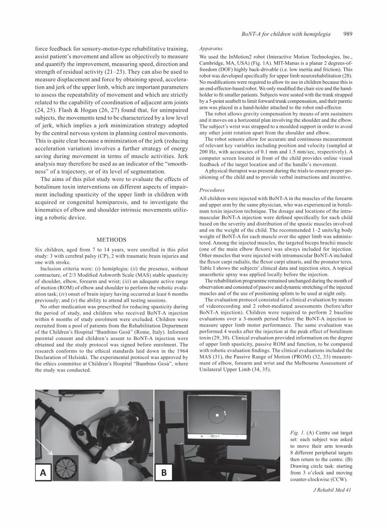
989BoNT-A for children with hemiplegia
force feedback for sensory-motor-type rehabilitative training, assist patient’s movement and allow us objectively to measure and quantify the improvement, measuring speed, direction and strength of residual activity (21–23). They can also be used to measure displacement and force by obtaining speed, accelera-tion and jerk of the upper limb, which are important parameters to assess the repeatability of movement and which are strictly related to the capability of coordination of adjacent arm joints (24, 25). Flash & Hogan (26, 27) found that, for unimpaired subjects, the movements tend to be characterized by a low level of jerk, which implies a jerk minimization strategy adopted by the central nervous system in planning control movements. This is quite clear because a minimization of the jerk (reducing acceleration variation) involves a further strategy of energy saving during movement in terms of muscle activities. Jerk analysis may therefore be used as an indicator of the “smooth-ness” of a trajectory, or of its level of segmentation.
The aims of this pilot study were to evaluate the effects of botulinum toxin interventions on different aspects of impair-ment including spasticity of the upper limb in children with acquired or congenital hemiparesis, and to investigate the kinematics of elbow and shoulder intrinsic movements utiliz-ing a robotic device.
METHODSSix children, aged from 7 to 14 years, were enrolled in this pilot study: 3 with cerebral palsy (CP), 2 with traumatic brain injuries and one with stroke.
Inclusion criteria were: (i) hemiplegia; (ii) the presence, without contracture, of 2/3 Modified Ashworth Scale (MAS) stable spasticity of shoulder, elbow, forearm and wrist; (iii) an adequate active range of motion (ROM) of elbow and shoulder to perform the robotic evalu-ation task; (iv) onset of brain injury having occurred at least 6 months previously; and (v) the ability to attend all testing sessions.
No other medication was prescribed for reducing spasticity during the period of study, and children who received BoNT-A injection within 6 months of study enrolment were excluded. Children were recruited from a pool of patients from the Rehabilitation Department of the Children’s Hospital “Bambino Gesù” (Rome, Italy). Informed parental consent and children’s assent to BoNT-A injection were obtained and the study protocol was signed before enrolment. The research conforms to the ethical standards laid down in the 1964 Declaration of Helsinki. The experimental protocol was approved by the ethics committee at Children’s Hospital “Bambino Gesù”, where the study was conducted.
ApparatusWe used the InMotion2 robot (Interactive Motion Technologies, Inc., Cambridge, MA, USA) (Fig. 1A). MIT-Manus is a planar 2 degrees-of-freedom (DOF) highly back-drivable (i.e. low inertia and friction). This robot was developed specifically for upper limb neurorehabilitation (28). No modifications were required to allow its use in children because this is an end-effector-based robot. We only modified the chair size and the hand-holder to fit smaller patients. Subjects were seated with the trunk strapped by a 5-point seatbelt to limit forward trunk compensation, and their paretic arm was placed in a hand-holder attached to the robot end-effector.
The robot allows gravity compensation by means of arm sustainers and it moves on a horizontal plan involving the shoulder and the elbow. The subject’s wrist was strapped to a moulded support in order to avoid any other joint rotation apart from the shoulder and elbow.
The robot sensors allow for accurate and continuous measurement of relevant key variables including position and velocity (sampled at 200 Hz, with accuracies of 0.1 mm and 1.5 mm/sec, respectively). A computer screen located in front of the child provides online visual feedback of the target location and of the handle’s movement.
A physical therapist was present during the trials to ensure proper po-sitioning of the child and to provide verbal instructions and incentive.
Procedures All children were injected with BoNT-A in the muscles of the forearm and upper arm by the same physician, who was experienced in botuli-num toxin injection technique. The dosage and locations of the intra-muscular BoNT-A injection were defined specifically for each child based on the severity and distribution of the spastic muscles involved and on the weight of the child. The recommended 1–2 units/kg body weight of BoNT-A for each muscle over the upper limb was adminis-tered. Among the injected muscles, the targeted biceps brachii muscle (one of the main elbow flexors) was always included for injection. Other muscles that were injected with intramuscular BoNT-A included the flexor carpi radialis, the flexor carpi ulnaris, and the pronator teres. Table I shows the subjects’ clinical data and injection sites. A topical anaesthetic spray was applied locally before the injection.
The rehabilitation programme remained unchanged during the month of observation and consisted of passive and dynamic stretching of the injected muscles and of the use of positioning splints to be used at night only.
The evaluation protocol consisted of a clinical evaluation by means of videorecording and 2 robot-mediated assessments (before/after BoNT-A injection). Children were required to perform 2 baseline evaluations over a 3-month period before the BoNT-A injection to measure upper limb motor performance. The same evaluation was performed 4 weeks after the injection at the peak effect of botulinum toxin (29, 30). Clinical evaluation provided information on the degree of upper limb spasticity, passive ROM and function, to be compared with robotic evaluation findings. The clinical evaluations included the MAS (31), the Passive Range of Motion (PROM) (32, 33) measure-ment of elbow, forearm and wrist and the Melbourne Assessment of Unilateral Upper Limb (34, 35).
Fig. 1. (A) Centre out target set: each subject was asked to move their arm towards 8 different peripheral targets then return to the centre. (B) Drawing circle task: starting from 3 o’clock and moving counter-clockwise (CCW).
J Rehabil Med 41
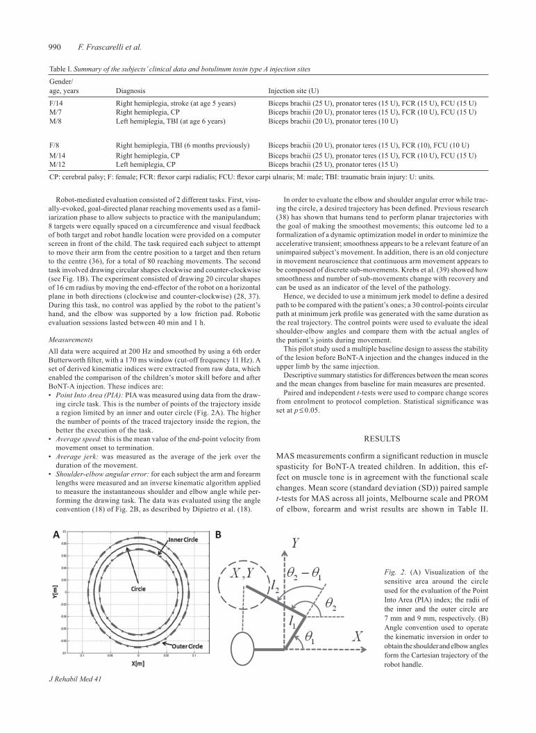
990 F. Frascarelli et al.
Robot-mediated evaluation consisted of 2 different tasks. First, visu-ally-evoked, goal-directed planar reaching movements used as a famil-iarization phase to allow subjects to practice with the manipulandum; 8 targets were equally spaced on a circumference and visual feedback of both target and robot handle location were provided on a computer screen in front of the child. The task required each subject to attempt to move their arm from the centre position to a target and then return to the centre (36), for a total of 80 reaching movements. The second task involved drawing circular shapes clockwise and counter-clockwise (see Fig. 1B). The experiment consisted of drawing 20 circular shapes of 16 cm radius by moving the end-effector of the robot on a horizontal plane in both directions (clockwise and counter-clockwise) (28, 37). During this task, no control was applied by the robot to the patient’s hand, and the elbow was supported by a low friction pad. Robotic evaluation sessions lasted between 40 min and 1 h.
Measurements All data were acquired at 200 Hz and smoothed by using a 6th order Butterworth filter, with a 170 ms window (cut-off frequency 11 Hz). A set of derived kinematic indices were extracted from raw data, which enabled the comparison of the children’s motor skill before and after BoNT-A injection. These indices are:• Point Into Area (PIA): PIA was measured using data from the draw-
ing circle task. This is the number of points of the trajectory inside a region limited by an inner and outer circle (Fig. 2A). The higher the number of points of the traced trajectory inside the region, the better the execution of the task.
• Average speed: this is the mean value of the end-point velocity from movement onset to termination.
• Average jerk: was measured as the average of the jerk over the duration of the movement.
• Shoulder-elbow angular error: for each subject the arm and forearm lengths were measured and an inverse kinematic algorithm applied to measure the instantaneous shoulder and elbow angle while per-forming the drawing task. The data was evaluated using the angle convention (18) of Fig. 2B, as described by Dipietro et al. (18).
In order to evaluate the elbow and shoulder angular error while trac-ing the circle, a desired trajectory has been defined. Previous research (38) has shown that humans tend to perform planar trajectories with the goal of making the smoothest movements; this outcome led to a formalization of a dynamic optimization model in order to minimize the accelerative transient; smoothness appears to be a relevant feature of an unimpaired subject’s movement. In addition, there is an old conjecture in movement neuroscience that continuous arm movement appears to be composed of discrete sub-movements. Krebs et al. (39) showed how smoothness and number of sub-movements change with recovery and can be used as an indicator of the level of the pathology.
Hence, we decided to use a minimum jerk model to define a desired path to be compared with the patient’s ones; a 30 control-points circular path at minimum jerk profile was generated with the same duration as the real trajectory. The control points were used to evaluate the ideal shoulder-elbow angles and compare them with the actual angles of the patient’s joints during movement.
This pilot study used a multiple baseline design to assess the stability of the lesion before BoNT-A injection and the changes induced in the upper limb by the same injection.
Descriptive summary statistics for differences between the mean scores and the mean changes from baseline for main measures are presented.
Paired and independent t-tests were used to compare change scores from enrolment to protocol completion. Statistical significance was set at p ≤ 0.05.
RESULTS
MAS measurements confirm a significant reduction in muscle spasticity for BoNT-A treated children. In addition, this ef-fect on muscle tone is in agreement with the functional scale changes. Mean score (standard deviation (SD)) paired sample t-tests for MAS across all joints, Melbourne scale and PROM of elbow, forearm and wrist results are shown in Table II.
Table I. Summary of the subjects’ clinical data and botulinum toxin type A injection sites
Gender/ age, years Diagnosis Injection site (U)
F/14 Right hemiplegia, stroke (at age 5 years) Biceps brachii (25 U), pronator teres (15 U), FCR (15 U), FCU (15 U)M/7 Right hemiplegia, CP Biceps brachii (20 U), pronator teres (15 U), FCR (10 U), FCU (15 U)M/8 Left hemiplegia, TBI (at age 6 years) Biceps brachii (20 U), pronator teres (10 U)
F/8 Right hemiplegia, TBI (6 months previously) Biceps brachii (20 U), pronator teres (15 U), FCR (10), FCU (10 U)M/14 Right hemiplegia, CP Biceps brachii (25 U), pronator teres (15 U), FCR (10 U), FCU (15 U)M/12 Left hemiplegia, CP Biceps brachii (25 U), pronator teres (15 U)
CP: cerebral palsy; F: female; FCR: flexor carpi radialis; FCU: flexor carpi ulnaris; M: male; TBI: traumatic brain injury: U: units.
Fig. 2. (A) Visualization of the sensitive area around the circle used for the evaluation of the Point Into Area (PIA) index; the radii of the inner and the outer circle are 7 mm and 9 mm, respectively. (B) Angle convention used to operate the kinematic inversion in order to obtain the shoulder and elbow angles form the Cartesian trajectory of the robot handle.
J Rehabil Med 41
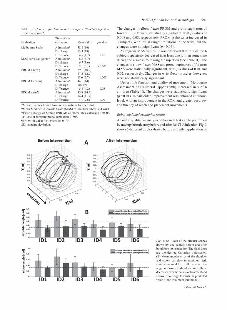
991BoNT-A for children with hemiplegia
The changes in elbow flexor PROM and prono-supinators of forearm PROM were statistically significant, with p-values of 0.008 and 0.03, respectively. PROM at the wrist increased in 3 subjects, with initial range limitations in the wrist, but the changes were not significant (p = 0.09).
As regards MAS values, it was observed that in 5 of the 6 subjects spasticity decreased in at least one joint at some time during the 4 weeks following the injection (see Table II). The changes in elbow flexor MAS and prono-supinators of forearm MAS were statistically significant, with p-values of 0.01 and 0.02, respectively. Changes in wrist flexor muscles, however, were not statistically significant.
Upper limb function and quality of movement (Melbourne Assessment of Unilateral Upper Limb) increased in 5 of 6 children (Table II). The changes were statistically significant (p = 0.01). In particular, improvement was obtained at elbow-level, with an improvement in the ROM and greater accuracy and fluency of reach and placement movements.
Robot-mediated evaluation resultsAn initial qualitative analysis of the circle task can be performed by tracing the trajectory before and after BoNT-A injection. Fig. 3 shows 5 different circles drawn before and after application of
Table II. Before vs after botulinum toxin type A (BoNT-A) injection: scale scores (n = 6)
EvaluationTime of the evaluation Mean (SD) p-value
Melbourne Scale Admission* 58.8 (16)Discharge 63.3 (18)Difference 4.5 (1.7) 0.01
MAS across all joints† Admission* 8.8 (3.7)Discharge 6.7 (3.6)Difference 2.1 (0.1) < 0.001
PROM elbow‡ Admission* 29.1 (10.2)Discharge 17.5 (12.9)Difference 11.6 (2.7) 0.008
PROM forearm§ Admission* 44.1 (14)Discharge 50 (19)Difference 5.8 (4.2) 0.03
PROM wrist¶ Admission* 35.8 (14.4)Discharge 34.4 (11.7)Difference 4.1 (2.6) 0.09
*Mean of scores from 2 baseline evaluations for each child. †Mean Modified Ashworth Scale (MAS) of shoulder elbow and wrist. ‡Passive Range of Motion (PROM) of elbow: flex-extension 150–0°. §PROM of forearm: prono-supination 0–80°. ¶PROM of wrist: flex-extension 0–70°.SD: standard deviation.
Fig. 3. (A) Plots of the circular shapes drawn by one subject before and after botulinum toxin injection. The black lines are the desired Cartesian trajectories. (B) Mean angular error of the shoulder and elbow correlate to minimum jerk simulation model. In all patients, the angular error of shoulder and elbow decreases over the course of treatment and seems to converge towards the predicted value of the minimum jerk model.
J Rehabil Med 41
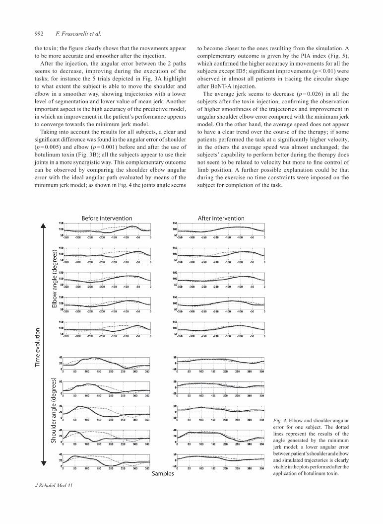
992 F. Frascarelli et al.
the toxin; the figure clearly shows that the movements appear to be more accurate and smoother after the injection.
After the injection, the angular error between the 2 paths seems to decrease, improving during the execution of the tasks; for instance the 5 trials depicted in Fig. 3A highlight to what extent the subject is able to move the shoulder and elbow in a smoother way, showing trajectories with a lower level of segmentation and lower value of mean jerk. Another important aspect is the high accuracy of the predictive model, in which an improvement in the patient’s performance appears to converge towards the minimum jerk model.
Taking into account the results for all subjects, a clear and significant difference was found in the angular error of shoulder (p = 0.005) and elbow (p = 0.001) before and after the use of botulinum toxin (Fig. 3B); all the subjects appear to use their joints in a more synergistic way. This complementary outcome can be observed by comparing the shoulder elbow angular error with the ideal angular path evaluated by means of the minimum jerk model; as shown in Fig. 4 the joints angle seems
to become closer to the ones resulting from the simulation. A complementary outcome is given by the PIA index (Fig. 5), which confirmed the higher accuracy in movements for all the subjects except ID5; significant improvements (p < 0.01) were observed in almost all patients in tracing the circular shape after BoNT-A injection.
The average jerk seems to decrease (p = 0.026) in all the subjects after the toxin injection, confirming the observation of higher smoothness of the trajectories and improvement in angular shoulder elbow error compared with the minimum jerk model. On the other hand, the average speed does not appear to have a clear trend over the course of the therapy; if some patients performed the task at a significantly higher velocity, in the others the average speed was almost unchanged; the subjects’ capability to perform better during the therapy does not seem to be related to velocity but more to fine control of limb position. A further possible explanation could be that during the exercise no time constraints were imposed on the subject for completion of the task.
Fig. 4. Elbow and shoulder angular error for one subject. The dotted lines represent the results of the angle generated by the minimum jerk model; a lower angular error between patient’s shoulder and elbow and simulated trajectories is clearly visible in the plots performed after the application of botulinum toxin.
J Rehabil Med 41
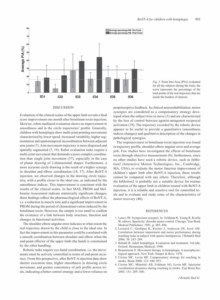
993BoNT-A for children with hemiplegia
DISCUSSION
Evaluation of the clinical scales of the upper limb reveals a final score improvement one month after botulinum toxin injection; likewise, robot-mediated evaluation shows an improvement in smoothness and in the circle trajectories’ profile. Generally, children with hemiplegia show multi-joint pointing movements characterized by lower speed, increased variability, higher seg-mentation and spatiotemporal incoordination between adjacent arm joints (7). Arm movement trajectory is more dispersed and spatially segmented (7, 19). Robot evaluation tasks require a multi-joint movement that demands a more complex coordina-tion than single joint movement (37), especially in the case of planar drawing of 2-dimensional shapes. Furthermore, a more accurate circle drawing is the result of a higher synergy in shoulder and elbow coordination (18, 37). After BoNT-A injection, we observed changes in the drawing circle trajec-tory, with a profile closer to the ideal one, as indicated by the smoothness indices. This improvement is consistent with the results of the clinical scales. In fact MAS, PROM and Mel-bourne Assessment indicate statistically significant changes; these findings reflect the pharmacological effects of BoNT-A, i.e. a reduction in muscle tone and a significant improvement in PROM during the period of chemodenervation induced by the botulinum toxin. However, the sample is too small to confirm the existence of a link between body structure, function and changes in functional activities.
The shoulder-elbow angular error indicates to what extent the real trajectory drawn by the child is close to the ideal one. In fact the improvement in this parameter could be correlated with a smooth coordination between shoulder and elbow when the end-point effector of the upper limb (the hand) is constrained by the robot handling.
Robotic tasks require eye-hand coordination, i.e. the move-ments must be actively controlled in terms of end-point accu-racy. From this perspective, after BoNT-A injection data show shorter execution time, higher smoothness in drawing circle movement, and greater consistency of jerk profile across tri-als, indicating a better control strategy and a lower reliance on
proprioceptive feedback. In clinical neurorehabilitation, motor synergies are considered as a compensatory strategy deve-loped when the subject tries to move (3) and are characterized by the loss of control between agonist-antagonist reciprocal activation (19). The trajectory recorded by the robotic device appears to be useful to provide a quantitative (smoothness indices changes) and qualitative description of the changes in pathological synergies.
The responsiveness to botulinum toxin injection was found in trajectory profile, shoulder-elbow angular error and average jerk. Few studies have investigated the effects of botulinum toxin through objective measurement (8); furthermore, since no other studies have used a robotic device, such as InMo-tion2 (Interactive Motion Technologies, Inc., Cambridge, MA, USA), to evaluate the motor function improvement of children’s upper limb after BoNT-A injection, these results cannot be compared with any others. Therefore, although the InMotion2 is probably not a suitable device for routine evaluation of the upper limb in children treated with BoNT-A injection, it is a reliable and sensitive tool for controlled tri-als and to evaluate and study some of the characteristics of motor recovery (40).
REFERENCES
Lance JW. Symposium synopsis. In: Feldman R, Young R, Koella 1. W, editors. Spasticity: disorder motor control. Chicago: Year Book Medical Publishers; 1981, p. 485–494.Leonard C, Gardipee K, Koontz J, Anderson JH, Scott AW. 2. Correlation between impairment and motor performance during reaching tasks in subject with spastic hemiparesis. J Rehabil Med 2006; 38: 243–249.Bobath B. Adult hemiplegia. Evaluation and treatment. 3rd edn. 3. Oxford: Heinemann Medical; 1990.Brunnstrom S. Movement therapy in hemiplegia. A neurophysio-4. logical approach. New York: Harper & Row; 1970.Cirstea MC, Levin MF. Compensatory strategy for reaching in 5. stroke. Brain 2000; 123: 940–953.Cirstea MC, Mitnitski AB, Feldman AG, Levin MF. Interjoint 6. coordination dynamics during reaching in stroke. Exp Brain Res 2003; 151: 289–300.
Fig. 5. Point Into Area (PIA) evaluated for all the subjects during the trials; the score represents the percentage of the total points of the real trajectory that are inside the borders of interest.
J Rehabil Med 41
994 F. Frascarelli et al.
Levin FM. Interjoint coordination during pointing movement is 7. disrupted in spastic hemiparesis. Brain 1996; 119: 281–293.Wallen M, O’Flaherty SJ, Waugh MC. Functional outcomes of 8. intramuscular botulinum toxin type A and occupational therapy in the upper limbs of children with cerebral palsy: a randomized controlled trial. Arch Phys Med Rehabil 2007; 88: 1–10.Wasiak J, Hoare B, Wallen M. Botulinum toxin A as an adjunct to 9. treatment in the management of the upper limb in children with spastic cerebral palsy. Cochrane Database Syst Rev 2004. Hurvitz EA, Conti GE, Brown SH. Changes in movement char-10. acteristics of the spastic upper extremity after botulinum toxin injection. Arch Phys Med Rehabil 2003; 84: 444–454.Boyd RN, Graham HK. Objective measurement of clinical findings 11. in the use of botulinum toxin type A for the management of children with cerebral palsy. Eur J Neurol 1999; 6: 23–35.Lee HM, Chen JJ, Wu YN, Wang YL, Huang SC, Piotrkiewicz 12. M. Time course analysis of the effects of botulinum toxin type a on elbow spasticity based on biomechanic and electromyographic parameters. Arch Phys Med Rehabil 2008; 89: 692–699.Wong V, Ng A, Sit P. Open-label study of botulinum toxin for 13. upper limb spasticity in cerebral palsy. J Child Neurol 2002; 17: 132–142.Hoare B, Imms C. Upper limb injection of botulinum toxin-A in 14. children with cerebral palsy: a critical review of the literature and clinical implications for occupational therapist. Am J Occ Ther 2004; 58: 389–397.Simpson DM, Alexander DN, O’Brien CF, Taglieti M, Asward 15. AS, Leon JM, et al. Botulinum toxin type A in the treatment of upper extremity spasticity: a randomized, double-blind, placebo-controlled trial. Neurology 1996; 46: 1306–1310. Hurvitz EA, Conti GE, Flansburg EL, Brown SH. Motor control 16. testing of upper limb function after botulinum toxin injection: a case study. Arch Phys Med Rehabil 2000; 81: 1408–1415.Condliffe EG, Clark DG, Patten C. Reliability of elbow stretch 17. reflex assessment in chronic post-stroke hemiparesis. Clin Neuro-physiol 2005; 116: 1870–1878.Dipietro L, Krebs HI, Fasoli SE, Volpe BT, Stein J, Bever C, et al. 18. Changing motor synergies in chronic stroke. J Neurophysiology 2007; 98: 757–768.Levin MF, Selles RW, Verheul MH, Meijer MG. Deficits in the 19. coordination of agonist and antagonist muscles in stroke pa-tients: implications for normal motor control. Brain Res 2000; 2: 352–369.Lee HM, Huang YZ, Chen JJ, Hwang IS. Quantitative analysis of 20. the velocity related pathophysiology of spasticity and rigidity in the elbow flexor. J Neurol Neurosurg Psychiatry 2002; 72: 621–629.Lum PS, Burgar CG, Peggy PC, Majmundar M, Van der Loos M. 21. Robot-assisted movement training compared with conventional therapy techniques for the rehabilitation of upper-limb motor func-tion after stroke. Arch Phys Med Rehabil 2002; 83: 952–959.Liebermann DG, Buchman AS, Franks MI. Enhancement of motor 22. rehabilitation through the use of information technologies. Clin Biomech 2006; 21: 8–20.Masiero S, Celia A, Rosati G, Armani M. Robotic-assisted re-23.
habilitation of the upper limb after acute stroke. Arch Phys Med Rehabil 2007; 88: 142–149.Chang JJ, Tung WL, Wu WL, Huang MH, Su FC. Effects of robot-24. aided bilateral force-induced isokinetic arm training combined with conventional rehabilitation on arm motor function in patients with chronic stroke. Arch Phys Med Rehabil 2007; 88: 1332–1338.Ramos E, Latash MP, Hurvitz EA. Quantification of upper extrem-25. ity function using kinematic analysis. Arch Phys Med Rehabil 1997; 78: 491–496.Hogan N. Adaptive control of mechanical impedance by coac-26. tivation of antagonist muscles. Trans Automatic Control 1984; AC-29: 681–690.Flash T, Hogan N The coordination of arm movements: an ex-27. perimentally confirmed mathematical model. J Neurosci 1985; 5: 1688–1703. Krebs HI, Hogan N, Aisen ML, Volpe BT. Robot aided neuro-28. rehabilitation. IEEE Trans Rehabil Eng 1998; 6: 75–87.Ward AB. Spasticity treatment with botulinum toxin. J Neural 29. Transm 2008; 115: 607–616.Dolly JO, Aoki K. The structure and mode of action of botulinum 30. toxins. Eur J Neurol 2006; 4: 1–9.Bohannon RW, Smith MD. Interrater reliability of the Modi-31. fied Ashworth Scale of muscle spasticity. Phys Ther 1987; 67: 206–207.De Jong LD, Nieuwboer A, Aufdemkampe G. The hemiplegic arm: 32. interrater reliability and concurrent validity of passive range of motion measurements. Disabil Rehabil 2007; 29: 1442–1448. Gajdosik RL, Bohannon RW. Clinical measurement of range of 33. motion: review of goniometry emphasizing reliability and validity. Phys Ther 1987; 67: 1867–1872.Bourke-Taylor H. Melbourne Assessment of Unilateral Upper Limb 34. Function: construct validity and correlation with the Pediatric Evaluation of Disability Inventory. Develop Med Child Neurol 2003; 45: 92–96. Randall M, Carlin JB, Chondros P, Reddihough D. Reliability of 35. the Melbourne Assessment of Unilateral Upper Limb Function. Develop Med Child Neurol 2001; 43: 761–767.Krebs HI, Brashers-Krug T, Rauch SL, Savage CR, Hogan N, 36. Rubin RH, et al. Robot-aided functional imaging: application to a motor learning study. Human Brain Mapping; John Wiley & Sons; 1998; 6, p. 59–72.Laquaniti F, Ferrigno G, Pedotti A, Soetching JF, Terzuolo C. 37. Changes in spatial scale on drawing and handwriting: kinematic contributions by proximal and distal joints. J Neurosci 1987; 7: 819–828. Hogan N. An organizing principle for a class of voluntary move-38. ments. J Neurosci 1984; 4: 2745–2754. Krebs HI, Aisen ML, Volpe BT, Hogan N. Quantization of continu-39. ous arm movements in humans with brain injury. Proc Natl Acad Sci USA 1999; 96: 4645–4649.Frascarelli F, Masia L, Di Rosa G, Cappa P, Petrarca M, Castelli 40. E, Krebs HI. The impact of robotic rehabilitation in children with acquired or congenital movement disorders. Eur J Phys Rehabil Med 2009; 45: 135–141.
J Rehabil Med 41
oriGinal report
J Rehabil Med 2009; 41: 995–1002
J Rehabil Med 41© 2009 The Authors. doi: 10.2340/16501977-0436Journal Compilation © 2009 Foundation of Rehabilitation Information. ISSN 1650-1977
Objective: to evaluate interjoint coordination in children with hemiplegia as they reach to grasp objects, in both static and dynamic conditions. an ad hoc robotic device was used to study the dynamic condition.Design: observational study.Patients: six children with hemiplegia and 6 young adults.Methods: Kinematics of the trunk and arm were studied us-ing an optoelectronic system. in the dynamic condition the target object, a cup, was moved by the robotic device along clockwise and counterclockwise circular trajectories.Results: two main strategies were used to study the onset and offset of shoulder and elbow movements and their maxi-mum velocities. The hand velocity profile was bell-shaped in the static condition and compatible with ramp movements for the more affected side in the dynamic condition. the time to object contact was higher for the more affected side in the dynamic condition. the temporal coordination index illus-trated an immature and less flexible behaviour in children’s reaching in all the examined conditions. Conclusion: Study of the hand velocity profiles, the time to object contact and the temporal coordination index high-lighted, first, the dependence of upper limb interjoint coor-dination on task, context, residual resources and individual solution, and secondly, the sensory-motor deficit character-istics of the children’s more affected side during dynamic reaching, raising the prospect of a promising training con-text in children with hemiplegia.Key words: reaching, hemiplegia, children, moving target, inter-joint coordination.J Rehabil Med 2009; 41: 995–1002
Correspondence address: Maurizio Petrarca, Movement and Robotic Laboratory, Division of Paediatric Rehabilitation, Children’s Hospital “Bambino Gesù” IRCCS, Via Torre di Palidoro s.n.c., IT-00050 Passoscuro (Fiumicino) Rome, Italy. E-mail: [email protected] March 3, 2009; accepted July 8, 2009
INTRODUCTION
During activities of daily living, the upper limbs are involved in numerous and complex tasks in relationship with objects, persons and the environment. The visuo-motor integration of prehension has been analysed and divided into sub-compo-
nents, such as reaching, grasping, manipulation, arm transport with or without handling objects, and release (1, 2).
Many studies have described the reaching and grasping movements of subjects with or without disabilities, in both adults and children; however, a review of the literature is dif-ficult due to both the different tasks and contexts studied and the different movement analysis methods used (3–13). Some studies have described the influence of the task and context on the reaching of children diagnosed with cerebral palsy and the relevance of this to the rehabilitation of upper limb function (3, 7). To complicate movement analysis further, the subject and/or the object target could be in motion, so that we can observe his or her relative motion. In this context, reaching control must be of the predictive or pro-active type, and it is based on spatial and temporal features, depending on task demands. In the published literature, this complexity inherent to the reaching task in children with hemiplegia has not been fully examined, even though this could provide useful information for rehabilitative training. Thus, a brief analysis of the main reaching characteristics and physiology in dynamic condi-tions seems useful in order to define the procedure adopted in the present study and the design chosen for the novel ad hoc robotic system used for moving the target.
The predictive characteristics of dynamic reaching imply 2 main consequences. First, when an object moves towards us, it moves along its projection cone on the retina, generating a retinal image that grows as the object nears and shrinks as the object moves away (14, 15). When an object moves at constant velocity, the hand-object time-to-contact, or tau margin (15), is used to select the movement start with respect to the task demands. If the object moves with acceleration, it is necessary to integrate visual information with previous experience (13). Secondly, reaching for a stationary or moving object requires non-linear coordination between the elbow and the shoulder (3, 16). Since reaching is mainly a ballistic movement, it has been hypothesized that it depends on feed-forward strategies based on the interaction among body, objects and force fields (17–19) exerted by the internal simulator for the action (20).
Taking into account the above-mentioned reaching character-istics, our training experiences for children with cerebral palsy were in agreement with the effectiveness of reaching training based on moving objects. Thus, we decided to develop an ad hoc 3 degree of freedom (DOF) apparatus in order to impose
REACH-TO-GRASP INTERJOINT COORDINATION FOR MOVING OBJECTS IN CHILDREN WITH HEMIPLEGIA
Maurizio Petrarca, MD1, Giulia Zanelli, PhD2, Fabrizio Patanè, PhD1,2, Flaminia Frascarelli, MD1, Paolo Cappa, MD1,2 and Enrico Castelli, MD1
From the 1Paediatric Neuro-Rehabilitation Division, Children’s Hospital “Bambino Gesù” IRCCS, 2Department of Mechanics and Aeronautics, “Sapienza” University of Rome, Rome, Italy
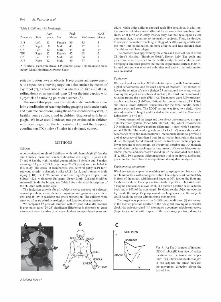
996 M. Petrarca et al.
settable motion laws on objects. It represents an improvement with respect to: a moving target on a flat surface by means of x-y robot (7), a small cube with 4 wheels (i.e. like a small car) rolling down on an inclined ramp (21) or the intercepting with a joystick of a moving point on a screen (8).
The aim of this paper was to study shoulder and elbow inter-joint coordination of reaching during grasping tasks under static and dynamic conditions, using the above-mentioned system, in healthy young subjects and in children diagnosed with hemi-plegia. We have used 2 indexes not yet evaluated in children with hemiplegia, i.e. the tau variable (15) and the temporal coordination (TC) index (3), also in a dynamic context.
METHODS
SubjectsA convenience sample of 6 children with mild hemiplegia (2 females and 4 males, mean and standard deviation (SD) age: 12 years (SD 3) and 6 healthy right-handed young adults (1 female and 5 males, mean age 23 years (SD 1), age range 22–24 years) were included in this study. The cause of hemiparesis was cerebral palsy (CP) for 3 subjects, arterial ischaemic stroke (AIS) for 2, and traumatic brain injury (TBI) for 1. We administered the Fugl-Meyer Upper Limb Subtest (22), Melbourne Unilateral Upper Limb (23) and Modified Ashworth Scale for biceps; see Table I for a detailed description of the children with hemiplegia.
The inclusion criteria for all subjects were: absence of seizures; arousal problem; visual deficits; cognitive and gross sensorial defi-cits; and ability in reaching and gross prehension. The children were enrolled after standard neurological and functional examinations.
We compared 12-year-old children with 23-year-old adults, because in previous studies (24, 25) significant differences in the reach-to-grasp movement were found only between children younger than 6 years and
adults, while older children showed adult-like behaviour. In addition, the enrolled children were affected by an event that involved both sides, or at birth or in early infancy they had not developed a clear dominant side, in contrast to the healthy subjects. Thus, we decided to compare the mature reaching strategy of healthy young adults with the inter-limb coordination on more affected and less affected sides of children with hemiplegia.
The protocol was approved by the ethics and medical board of the Children’s Hospital “Bambino Gesù”, Rome, Italy. The goals and procedure were explained to the healthy subjects and children with hemiplegia and their parents before the experiment started; their in-formed consent was obtained only after oral and written information was presented.
Equipment We developed an ad hoc 3DOF robotic system, with 3 miniaturized digital servomotors, one for each degree of freedom. Two motors al-lowed the rotation of a stick (height 25 cm) around the x- and y-axes, moving the object on a spherical surface; the third motor imposed a rotation around the z-axis (Fig. 1A). The motors were fully program-mable via software (LabView, National Instruments, Austin, TX, USA) and they allowed different trajectories for the robot handle, with a smooth start and stop. The 3DOF system was fixed to a desk, which was adjustable in height and located in the centre of the Movement Laboratory (14 × 7 m2).
The movements of the target and the subject were measured using an optoelectronic system (Vicon MX, Oxford, UK), which recorded the 3D position of reflective markers (diameter 14 mm) with 8 cameras, set at 120 Hz. The working volume (1 × 1 × 1 m3) was calibrated in accordance with the manufacturer’s recommendations to provide a global accuracy of less than 1 mm. In particular, in all trials, the same skilled therapist placed 10 markers on: the trunk (one on the upper and lower portions of the sternum, on 7th cervical vertebra and 10th thoracic vertebra) and on the reaching arm (one on each of the shoulder, external elbow, internal and external wrist and the 3rd metacarpal of each hand) (Fig. 1B.). Two cameras videotaped each trial in the frontal and lateral plane, to facilitate clinical interpretation during data analysis.
Experimental conditionsWe chose a paper cup as the reaching and grasping target, because this is a familiar task with ecological value. The subjects sat comfortably in front of the target, with hips and knees at 90°, feet on the floor and hands on the desk. The cup was fixed to the top of the robot stick with a magnet and located at eye-level, in a median position relative to the body and at 80% of the arm length. By doing so, the object trajectories lay inside the subject’s peripersonal reaching space, i.e. the subjects could reach the object without trunk movements.
The target was presented in 3 different conditions: (i) stationary, in the median position relative to the body; (ii) moving on a circular clockwise trajectory; and (iii) moving on a counterclockwise trajectory (trajectory centred with respect to the stationary position, diameter
Table I. Children with hemiplegia
Diagnosis SideAge, years Sex
Fugl-Meyer Melbourne
MAS biceps
AIS Left 13 Female 30 55 3CP Right 8 Male 43 77 0CP Left 12 Male 44 78 1+TBI Right 12 Female 42 68 2CP Left 17 Male 50 64 1AIS Right 12 Male 49 77 1
AIS: arterial ischaemic stroke; CP: cerebral palsy; TBI: traumatic brain injury; MAS: Modified Ashworth Scale.
Fig. 1. (A) The 3 degrees of freedom (3DOF) robot. (B) Rear view of marker locations on the trunk and upper limbs. (C) Elbow and shoulder angles in one subject; the arrow indicates the movement direction along the dashed line.
J Rehabil Med 41
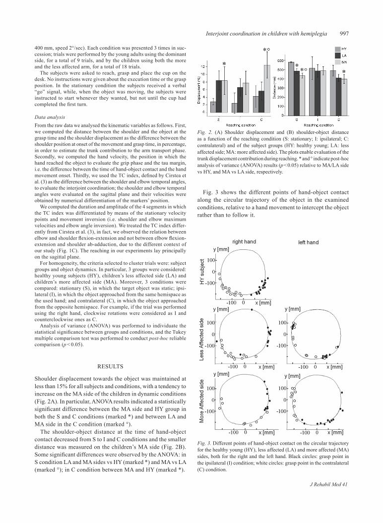
997Interjoint coordination in children with hemiplegia
400 mm, speed 2°/sec). Each condition was presented 3 times in suc-cession; trials were performed by the young adults using the dominant side, for a total of 9 trials, and by the children using both the more and the less affected arm, for a total of 18 trials.
The subjects were asked to reach, grasp and place the cup on the desk. No instructions were given about the execution time or the grasp position. In the stationary condition the subjects received a verbal “go” signal, while, when the object was moving, the subjects were instructed to start whenever they wanted, but not until the cup had completed the first turn.
Data analysisFrom the raw data we analysed the kinematic variables as follows. First, we computed the distance between the shoulder and the object at the grasp time and the shoulder displacement as the difference between the shoulder position at onset of the movement and grasp time, in percentage, in order to estimate the trunk contribution to the arm transport phase. Secondly, we computed the hand velocity, the position in which the hand reached the object to evaluate the grip phase and the tau margin, i.e. the difference between the time of hand-object contact and the hand movement onset. Thirdly, we used the TC index, defined by Cirstea et al. (3) as the difference between the shoulder and elbow temporal angles, to evaluate the interjoint coordination; the shoulder and elbow temporal angles were evaluated on the sagittal plane and their velocities were obtained by numerical differentiation of the markers’ position.
We computed the duration and amplitude of the 4 segments in which the TC index was differentiated by means of the stationary velocity points and movement inversion (i.e. shoulder and elbow maximum velocities and elbow angle inversion). We treated the TC index differ-ently from Cirstea et al. (3), in fact, we observed the relation between elbow and shoulder flexion-extension and not between elbow flexion-extension and shoulder ab-adduction, due to the different context of our study (Fig. 1C). The reaching in our experiments lay principally on the sagittal plane.
For homogeneity, the criteria selected to cluster trials were: subject groups and object dynamics. In particular, 3 groups were considered: healthy young subjects (HY), children’s less affected side (LA) and children’s more affected side (MA). Moreover, 3 conditions were compared: stationary (S), in which the target object was static; ipsi-lateral (I), in which the object approached from the same hemispace as the used hand; and contralateral (C), in which the object approached from the opposite hemispace. For example, if the trial was performed using the right hand, clockwise rotations were considered as I and counterclockwise ones as C.
Analysis of variance (ANOVA) was performed to individuate the statistical significance between groups and conditions, and the Tukey multiple comparison test was performed to conduct post-hoc reliable comparison (p < 0.05).
RESULTS
Shoulder displacement towards the object was maintained at less than 15% for all subjects and conditions, with a tendency to increase on the MA side of the children in dynamic conditions (Fig. 2A). In particular, ANOVA results indicated a statistically significant difference between the MA side and HY group in both the S and C conditions (marked *) and between LA and MA side in the C condition (marked °).
The shoulder-object distance at the time of hand-object contact decreased from S to I and C conditions and the smaller distance was measured on the children’s MA side (Fig. 2B). Some significant differences were observed by the ANOVA: in S condition LA and MA sides vs HY (marked *) and MA vs LA (marked °); in C condition between MA and HY (marked *).
Fig. 3 shows the different points of hand-object contact along the circular trajectory of the object in the examined conditions, relative to a hand movement to intercept the object rather than to follow it.
Fig. 2. (A) Shoulder displacement and (B) shoulder-object distance as a function of the reaching condition (S: stationary; I: ipsilateral; C: contralateral) and of the subject groups (HY: healthy young; LA: less affected side; MA: more affected side). The plots enable evaluation of the trunk displacement contribution during reaching. * and ° indicate post-hoc analysis of variance (ANOVA) results (p < 0.05) relative to MA/LA side vs HY, and MA vs LA side, respectively.
Fig. 3. Different points of hand-object contact on the circular trajectory for the healthy young (HY), less affected (LA) and more affected (MA) sides, both for the right and the left hand. Black circles: grasp point in the ipsilateral (I) condition; white circles: grasp point in the contralateral (C) condition.
J Rehabil Med 41
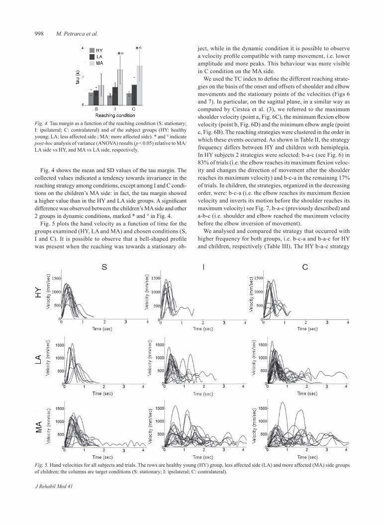
998 M. Petrarca et al.
Fig. 4 shows the mean and SD values of the tau margin. The collected values indicated a tendency towards invariance in the reaching strategy among conditions, except among I and C condi-tions on the children’s MA side: in fact, the tau margin showed a higher value than in the HY and LA side groups. A significant difference was observed between the children’s MA side and other 2 groups in dynamic conditions, marked * and ° in Fig. 4.
Fig. 5 plots the hand velocity as a function of time for the groups examined (HY, LA and MA) and chosen conditions (S, I and C). It is possible to observe that a bell-shaped profile was present when the reaching was towards a stationary ob-
ject, while in the dynamic condition it is possible to observe a velocity profile compatible with ramp movement, i.e. lower amplitude and more peaks. This behaviour was more visible in C condition on the MA side.
We used the TC index to define the different reaching strate-gies on the basis of the onset and offsets of shoulder and elbow movements and the stationary points of the velocities (Figs 6 and 7). In particular, on the sagittal plane, in a similar way as computed by Cirstea et al. (3), we referred to the maximum shoulder velocity (point a, Fig. 6C), the minimum flexion elbow velocity (point b, Fig. 6D) and the minimum elbow angle (point c, Fig. 6B). The reaching strategies were clustered in the order in which these events occurred. As shown in Table II, the strategy frequency differs between HY and children with hemiplegia. In HY subjects 2 strategies were selected: b-a-c (see Fig. 6) in 83% of trials (i.e. the elbow reaches its maximum flexion veloc-ity and changes the direction of movement after the shoulder reaches its maximum velocity) and b-c-a in the remaining 17% of trials. In children, the strategies, organized in the decreasing order, were: b-c-a (i.e. the elbow reaches its maximum flexion velocity and inverts its motion before the shoulder reaches its maximum velocity) see Fig. 7, b-a-c (previously described) and a-b-c (i.e. shoulder and elbow reached the maximum velocity before the elbow inversion of movement).
We analysed and compared the strategy that occurred with higher frequency for both groups, i.e. b-c-a and b-a-c for HY and children, respectively (Table III). The HY b-a-c strategy
Fig. 5. Hand velocities for all subjects and trials. The rows are healthy young (HY) group, less affected side (LA) and more affected (MA) side groups of children; the columns are target conditions (S: stationary; I: ipsilateral; C: contralateral).
Fig. 4. Tau margin as a function of the reaching condition (S: stationary; I: ipsilateral; C: contralateral) and of the subject groups (HY: healthy young; LA: less affected side ; MA: more affected side). * and ° indicate post-hoc analysis of variance (ANOVA) results (p < 0.05) relative to MA/LA side vs HY, and MA vs LA side, respectively.
J Rehabil Med 41
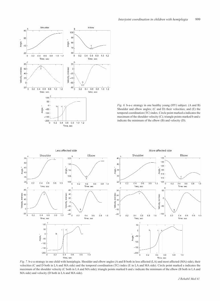
999Interjoint coordination in children with hemiplegia
Fig. 6. b-a-c strategy in one healthy young (HY) subject. (A and B) Shoulder and elbow angles; (C and D) their velocities; and (E) the temporal coordination (TC) index. Circle point marked a indicates the maximum of the shoulder velocity (C); triangle points marked b and c indicate the minimum of the elbow (B) and velocity (D).
Fig. 7. b-c-a strategy in one child with hemiplegia. Shoulder and elbow angles (A and B both in less affected (LA) and most affected (MA) side), their velocities (C and D both in LA and MA side) and the temporal coordination (TC) index (E in LA and MA side). Circle point marked a indicates the maximum of the shoulder velocity (C both in LA and MA side); triangle points marked b and c indicate the minimum of the elbow (B both in LA and MA side) and velocity (D both in LA and MA side).
J Rehabil Med 41
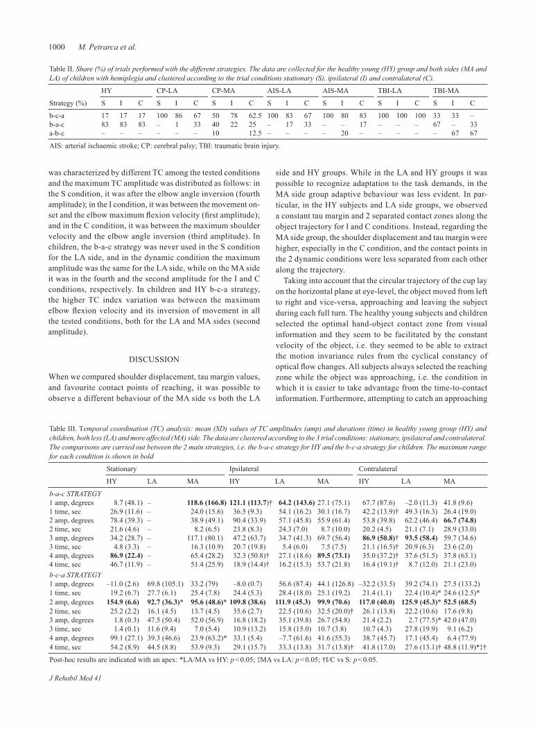
1000 M. Petrarca et al.
was characterized by different TC among the tested conditions and the maximum TC amplitude was distributed as follows: in the S condition, it was after the elbow angle inversion (fourth amplitude); in the I condition, it was between the movement on-set and the elbow maximum flexion velocity (first amplitude); and in the C condition, it was between the maximum shoulder velocity and the elbow angle inversion (third amplitude). In children, the b-a-c strategy was never used in the S condition for the LA side, and in the dynamic condition the maximum amplitude was the same for the LA side, while on the MA side it was in the fourth and the second amplitude for the I and C conditions, respectively. In children and HY b-c-a strategy, the higher TC index variation was between the maximum elbow flexion velocity and its inversion of movement in all the tested conditions, both for the LA and MA sides (second amplitude).
DISCUSSION
When we compared shoulder displacement, tau margin values, and favourite contact points of reaching, it was possible to observe a different behaviour of the MA side vs both the LA
side and HY groups. While in the LA and HY groups it was possible to recognize adaptation to the task demands, in the MA side group adaptive behaviour was less evident. In par-ticular, in the HY subjects and LA side groups, we observed a constant tau margin and 2 separated contact zones along the object trajectory for I and C conditions. Instead, regarding the MA side group, the shoulder displacement and tau margin were higher, especially in the C condition, and the contact points in the 2 dynamic conditions were less separated from each other along the trajectory.
Taking into account that the circular trajectory of the cup lay on the horizontal plane at eye-level, the object moved from left to right and vice-versa, approaching and leaving the subject during each full turn. The healthy young subjects and children selected the optimal hand-object contact zone from visual information and they seem to be facilitated by the constant velocity of the object, i.e. they seemed to be able to extract the motion invariance rules from the cyclical constancy of optical flow changes. All subjects always selected the reaching zone while the object was approaching, i.e. the condition in which it is easier to take advantage from the time-to-contact information. Furthermore, attempting to catch an approaching
Table III. Temporal coordination (TC) analysis: mean (SD) values of TC amplitudes (amp) and durations (time) in healthy young group (HY) and children, both less (LA) and more affected (MA) side. The data are clustered according to the 3 trial conditions: stationary, ipsilateral and contralateral. The comparisons are carried out between the 2 main strategies, i.e. the b-a-c strategy for HY and the b-c-a strategy for children. The maximum range for each condition is shown in bold
Stationary Ipsilateral Contralateral
HY LA MA HY LA MA HY LA MA
b-a-c STRATEGY1 amp, degrees 8.7 (48.1) – 118.6 (166.8) 121.1 (113.7)† 64.2 (143.6) 27.1 (75.1) 67.7 (87.6) –2.0 (11.3) 41.8 (9.6)1 time, sec 26.9 (11.6) – 24.0 (15.6) 36.5 (9.3) 54.1 (16.2) 30.1 (16.7) 42.2 (13.9)† 49.3 (16.3) 26.4 (19.0)2 amp, degrees 78.4 (39.3) – 38.9 (49.1) 90.4 (33.9) 57.1 (45.8) 55.9 (61.4) 53.8 (39.8) 62.2 (46.4) 66.7 (74.8)2 time, sec 21.6 (4.6) – 8.2 (6.5) 23.8 (8.3) 24.3 (7.0) 8.7 (10.0) 20.2 (4.5) 21.1 (7.1) 28.9 (33.0)3 amp, degrees 34.2 (28.7) – 117.1 (80.1) 47.2 (63.7) 34.7 (41.3) 69.7 (56.4) 86.9 (50.8)† 93.5 (58.4) 59.7 (34.6)3 time, sec 4.8 (3.3) – 16.3 (10.9) 20.7 (19.8) 5.4 (6.0) 7.5 (7.5) 21.1 (16.5)† 20.9 (6.3) 23.6 (2.0)4 amp, degrees 86.9 (22.4) – 65.4 (28.2) 32.3 (50.8)† 27.1 (18.6) 89.5 (73.1) 35.0 (37.2)† 37.6 (51.5) 37.8 (63.1)4 time, sec 46.7 (11.9) – 51.4 (25.9) 18.9 (14.4)† 16.2 (15.3) 53.7 (21.8) 16.4 (19.1)† 8.7 (12.0) 21.1 (23.0)b-c-a STRATEGY1 amp, degrees –11.0 (2.6) 69.8 (105.1) 33.2 (79) –8.0 (0.7) 56.6 (87.4) 44.1 (126.8) –32.2 (33.5) 39.2 (74.1) 27.5 (133.2)1 time, sec 19.2 (6.7) 27.7 (6.1) 25.4 (7.8) 24.4 (5.3) 28.4 (18.0) 25.1 (19.2) 21.4 (1.1) 22.4 (10.4)* 24.6 (12.5)*2 amp, degrees 154.9 (6.6) 92.7 (36.3)* 95.6 (48.6)* 109.8 (38.6) 111.9 (45.3) 99.9 (70.6) 117.0 (40.0) 125.9 (45.3)* 52.5 (68.5)2 time, sec 25.2 (2.2) 16.1 (4.5) 13.7 (4.5) 35.6 (2.7) 22.5 (10.6) 32.5 (20.0)† 26.1 (13.8) 22.2 (10.6) 17.6 (9.8)3 amp, degrees 1.8 (0.3) 47.5 (50.4) 52.0 (56.9) 16.8 (18.2) 35.1 (39.8) 26.7 (54.8) 21.4 (2.2) 2.7 (77.5)* 42.0 (47.0)3 time, sec 1.4 (0.1) 11.6 (9.4) 7.0 (5.4) 10.9 (13.2) 15.8 (15.0) 10.7 (3.8) 10.7 (4.3) 27.8 (19.9) 9.1 (6.2)4 amp, degrees 99.1 (27.1) 39.3 (46.6) 23.9 (63.2)* 33.1 (5.4) –7.7 (61.6) 41.6 (55.3) 38.7 (45.7) 17.1 (45.4) 6.4 (77.9)4 time, sec 54.2 (8.9) 44.5 (8.8) 53.9 (9.3) 29.1 (15.7) 33.3 (13.8) 31.7 (13.8)† 41.8 (17.0) 27.6 (13.1)† 48.8 (11.9)*‡†
Post-hoc results are indicated with an apex: *LA/MA vs HY: p < 0.05; ‡MA vs LA: p < 0.05; †I/C vs S: p < 0.05.
Table II. Share (%) of trials performed with the different strategies. The data are collected for the healthy young (HY) group and both sides (MA and LA) of children with hemiplegia and clustered according to the trial conditions stationary (S), ipsilateral (I) and contralateral (C).
Strategy (%)
HY CP-LA CP-MA AIS-LA AIS-MA TBI-LA TBI-MA
S I C S I C S I C S I C S I C S I C S I C
b-c-a 17 17 17 100 86 67 50 78 62.5 100 83 67 100 80 83 100 100 100 33 33 –b-a-c 83 83 83 – 1 33 40 22 25 – 17 33 – – 17 – – – 67 – 33a-b-c – – – – – – 10 12.5 – – – – 20 – – – – – 67 67
AIS: arterial ischaemic stroke; CP: cerebral palsy; TBI: traumatic brain injury.
J Rehabil Med 41
1001Interjoint coordination in children with hemiplegia
object assured a higher grade of success than attempting to catch a leaving object, because over- or under-estimation of the object’s eventual position led only to a different accelera-tion impact. The above-mentioned findings confirm that the reaching start is visually guided by object position in the S condition and by extracting object motion invariance in dy-namic conditions (i.e. I and C).
Moreover, the HY and the LA side groups showed a bell-shaped hand velocity profile, i.e. a ballistic movement of the hand towards the reaching target. In contrast, the children’s MA side group exhibited a ramp hand velocity profile, i.e. the sub-jects produced low velocities and continuous adjustments.
Study of the TC parameters provided more detailed in-formation on the shoulder-elbow coordination than the task demands. During the reaching tasks the shoulder executed a flexion movement, driving the arm towards the object, while the elbow first flexed in order to gain clearance from the table and then extended towards the target. The selected strategies and TC index showed great variability among patients and conditions, as reported in Tables II and III.
When the HY group executed their favourite strategy (b-a-c), the shoulder reached the maximum velocity before the elbow inverted its movement, while, when the children executed their favourite strategy (b-c-a), the shoulder reached the maximum velocity after the elbow inverted its movement. Thus, children selected a simpler rule of joint co-variation, completing elbow flexion first and then moving both shoulder and elbow in exten-sion. This difference from the healthy young adults could be attributed to the children’s sensory motor deficit, considering the less complex coordination required by the b-c-a strategy, as documented by: (i) the differences in maximum amplitude of TC indexes; (ii) the greater trunk displacement; and, (iii) the greater variability in the hand velocity profile for the MA side.
The healthy young subjects started the reaching at the same time with respect to the action goal for all the examined conditions. In dynamic conditions, they selected 2 differ-ent contact zones for the 2 rotation directions, and different shapes in the TC index for the 3 conditions, i.e. they showed an anticipatory motor control taking into account the object motion characteristics. Our results are in agreement with the hypothesis that the reaching start is visually guided, while the arm movement is driven by proprioceptive information and previous experiences.
The children with hemiplegia seem to be able to realize the same anticipatory strategy, but they selected a simpler coor-dination between elbow and shoulder, moving both joints in acceleration and deceleration with more invariance through the conditions than did the healthy young subjects. Unlike Cirstea et al. (3), who found a lack of coordination between elbow and shoulder from the middle to the end of the reach in adults with hemiplegia, we found a lack of coordination at the beginning of the reach. However, while the task selected by Cirstea et al. (3) required an inversion of shoulder and elbow coordination in late reach, in our task this is required in early reach.
Thus, interjoint coordination seems to be constrained by task (i.e. to catch the target), context (i.e. dynamic conditions or
the need to achieve safe table clearance), and system’s residual resources (i.e. sensory motor deficit, muscles and soft tissue characteristics and previous experience). From a global exami-nation of the collected data, it is difficult to assess whether the movement dynamic is controlled by an internal model (13), or by an internal simulator (20), or by a local attractor of dynamic balancing structured on previous experiences and tuned by means of the ongoing sensory motor information. This last option implies a continuous control that needs an integrated control variable, neither strictly efferent or afferent, as the λ proposed by Feldman & Levin (26), which comprises sensory and motor aspects, central and peripheral aspects and muscle properties. Consequently, the MA side limitation and variabil-ity could be attributed to the sensory-motor deficit during hand transportation towards the object. However, the demonstrated variability of the TC index and the kinetic/kinematic variables (i.e. the speed profile, tau margin and contact points with mov-ing objects) seems useful to detect the adaptive strategy with respect to the tasks’ demands and the sensory motor deficit in children with mild hemiplegia.
The perspective of training in dynamic conditions seems to be useful to meliorate the reaching adaptability to the tasks and contexts, as proposed by Schenk et al. (7). The observed differences in the interjoint coordination may agree with the consideration of Latash et al. (27) on the motor equivalence phenomena, which is related to the problem of the degrees of freedom redundancy in driving a multijoint arm towards a target, i.e. synergies link joints in flexible binding that is task dependent and activated by a simple timing signal. If further studies confirm our findings, that children with hemiplegia have difficulty in planning ongoing inversion of the interjoint coordination depending on the children’s available resources, task and context, training could be based just on the modulation of task and context in order to improve children’s resources for reaching tasks. In conclusion, children with hemiplegia cannot be considered as a homogeneous group and therefore it is important to personalize training, as recommended by Rönnquist & Rösblad (6); furthermore, from the perspective of the present study, dynamic training should be personalized with respect to the individual interjoint coordination limita-tion observed.
Finally, further studies are required in order to overcome the main limitations of the present study. The number of en-rolled subjects should be increased, the control group should be age-matched, patients should be grouped according to age, specific diagnosis and severity, and different tasks and contexts should be tested.
ACKNOWLEDGEMENTS
The authors are grateful to the children and young subjects who gener-ously gave their time to assist with this research. The authors thank the Scientific Direction of the Children’s Hospital ‘’Bambino Gesu`’’ IRCCS for financial support and the ‘’Pegaso Association’’ for supporting the routine clinical activity of the Movement and Robotic Laboratory. This project was partially supported by a research grant from the Italian Health Ministry (grant “Pilot study on a novel typology of medical devices: robotic systems for rehabilitation and tele-rehabilitation”).
J Rehabil Med 41
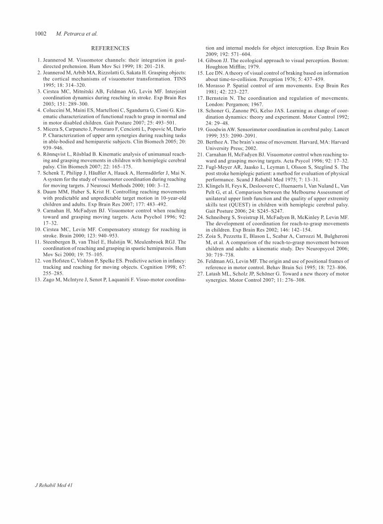
1002 M. Petrarca et al.
REFERENCES
Jeannerod M. Visuomotor channels: their integration in goal-1. directed prehension. Hum Mov Sci 1999; 18: 201–218.Jeannerod M, Arbib MA, Rizzolatti G, Sakata H. Grasping objects: 2. the cortical mechanisms of visuomotor transformation. TINS 1995; 18: 314–320.Cirstea MC, Mitnitski AB, Feldman AG, Levin MF. Interjoint 3. coordination dynamics during reaching in stroke. Exp Brain Res 2003; 151: 289–300.Coluccini M, Maini ES, Martelloni C, Sgandurra G, Cioni G. Kin-4. ematic characterization of functional reach to grasp in normal and in motor disabled children. Gait Posture 2007; 25: 493–501.Micera S, Carpaneto J, Posteraro F, Cenciotti L, Popovic M, Dario 5. P. Characterization of upper arm synergies during reaching tasks in able-bodied and hemiparetic subjects. Clin Biomech 2005; 20: 939–946.Rönnqvist L, Rösblad B. Kinematic analysis of unimanual reach-6. ing and grasping movements in children with hemiplegic cerebral palsy. Clin Biomech 2007; 22: 165–175.Schenk T, Philipp J, Häußler A, Hauck A, Hermsdörfer J, Mai N. 7. A system for the study of visuomotor coordination during reaching for moving targets. J Neurosci Methods 2000; 100: 3–12.Daum MM, Huber S, Krist H. Controlling reaching movements 8. with predictable and unpredictable target motion in 10-year-old children and adults. Exp Brain Res 2007; 177: 483–492.Carnahan H, McFadyen BJ. Visuomotor control when reaching 9. toward and grasping moving targets. Acta Psychol 1996; 92: 17–32.Cirstea MC, Levin MF. Compensatory strategy for reaching in 10. stroke. Brain 2000; 123: 940–953.Steenbergen B, van Thiel E, Hulstijn W, Meulenbroek RGJ. The 11. coordination of reaching and grasping in spastic hemiparesis. Hum Mov Sci 2000; 19: 75–105.von Hofsten C, Vishton P, Spelke ES. Predictive action in infancy: 12. tracking and reaching for moving objects. Cognition 1998; 67: 255–285.Zago M, McIntyre J, Senot P, Laquaniti F. Visuo-motor coordina-13.
tion and internal models for object interception. Exp Brain Res 2009; 192: 571–604.Gibson JJ. The ecological approach to visual perception. Boston: 14. Houghton Mifflin; 1979.Lee DN. A theory of visual control of braking based on information 15. about time-to-collision. Perception 1976; 5: 437–459.Morasso P. Spatial control of arm movements. Exp Brain Res 16. 1981; 42: 223–227.Bernstein N. The coordination and regulation of movements. 17. London: Pergamon; 1967.Schoner G, Zanone PG, Kelso JAS. Learning as change of coor-18. dination dynamics: theory and experiment. Motor Control 1992; 24: 29–48.Goodwin AW. Sensorimotor coordination in cerebral palsy. Lancet 19. 1999; 353: 2090–2091.Berthoz A. The brain’s sense of movement. Harvard, MA: Harvard 20. University Press; 2002. Carnahan H, McFadyen BJ. Visuomotor control when reaching to-21. ward and grasping moving targets. Acta Psycol 1996; 92: 17–32.Fugl-Meyer AR, Jaasko L, Leyman I, Olsson S, Steglind S. The 22. post stroke hemiplegic patient: a method for evaluation of physical performance. Scand J Rehabil Med 1975; 7: 13–31.Klingels H, Feys K, Desloovere C, Huenaerts I, Van Nuland L, Van 23. Pelt G, et al. Comparison between the Melbourne Assessment of unilateral upper limb function and the quality of upper extremity skills test (QUEST) in children with hemiplegic cerebral palsy. Gait Posture 2006; 24: S245–S247.Schneiberg S, Sveistrup H, McFadyen B, McKinley P, Levin MF. 24. The development of coordination for reach-to-grasp movements in children. Exp Brain Res 2002; 146: 142–154.Zoia S, Pezzetta E, Blason L, Scabar A, Carrozzi M, Bulgheroni 25. M, et al. A comparison of the reach-to-grasp movement between children and adults: a kinematic study. Dev Neuropsycol 2006; 30: 719–738.Feldman AG, Levin MF. The origin and use of positional frames of 26. reference in motor control. Behav Brain Sci 1995; 18: 723–806. Latash ML, Scholz JP, Schöner G. Toward a new theory of motor 27. synergies. Motor Control 2007; 11: 276–308.
J Rehabil Med 41
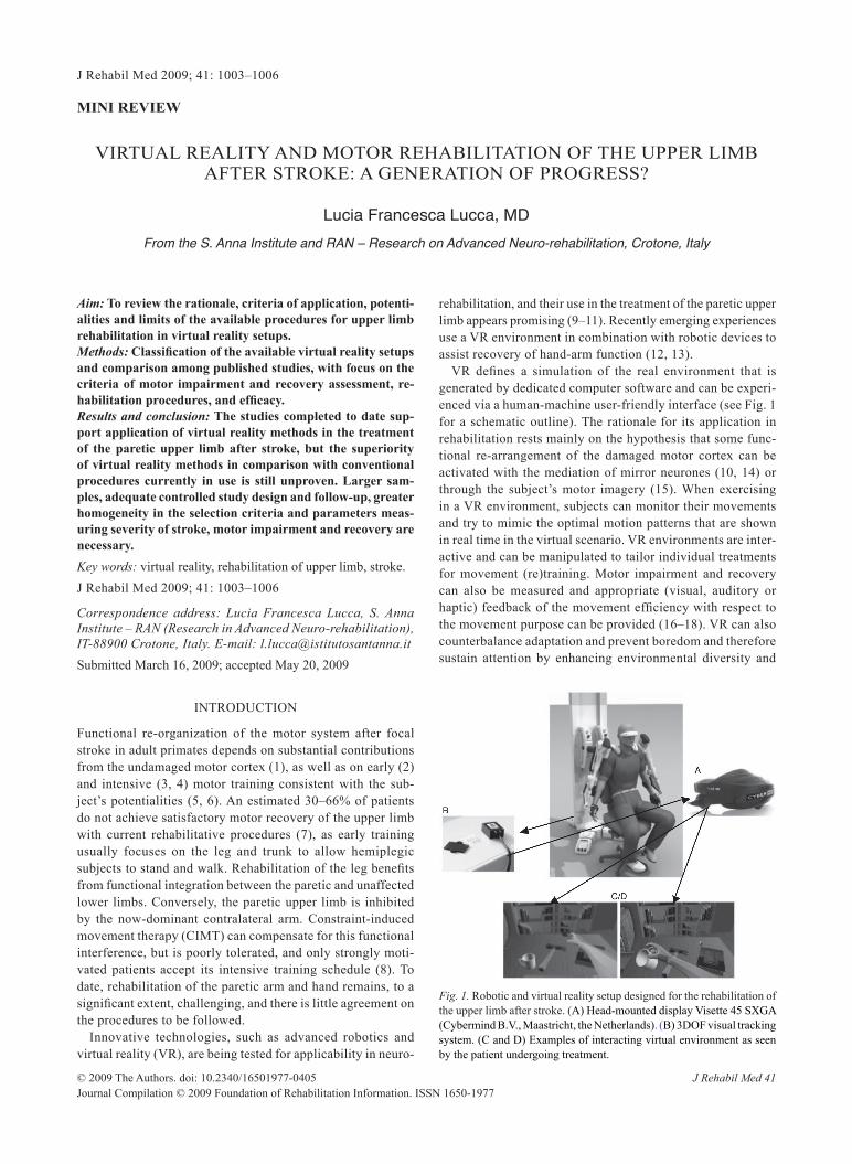
mini revieW
J Rehabil Med 2009; 41: 1003–1006
J Rehabil Med 41© 2009 The Authors. doi: 10.2340/16501977-0405Journal Compilation © 2009 Foundation of Rehabilitation Information. ISSN 1650-1977
Aim: to review the rationale, criteria of application, potenti-alities and limits of the available procedures for upper limb rehabilitation in virtual reality setups.Methods: Classification of the available virtual reality setups and comparison among published studies, with focus on the criteria of motor impairment and recovery assessment, re-habilitation procedures, and efficacy.Results and conclusion: the studies completed to date sup-port application of virtual reality methods in the treatment of the paretic upper limb after stroke, but the superiority of virtual reality methods in comparison with conventional procedures currently in use is still unproven. larger sam-ples, adequate controlled study design and follow-up, greater homogeneity in the selection criteria and parameters meas-uring severity of stroke, motor impairment and recovery are necessary.Key words: virtual reality, rehabilitation of upper limb, stroke.J Rehabil Med 2009; 41: 1003–1006
Correspondence address: Lucia Francesca Lucca, S. Anna Institute – RAN (Research in Advanced Neuro-rehabilitation), IT-88900 Crotone, Italy. E-mail: [email protected] March 16, 2009; accepted May 20, 2009
INTRODUCTION
Functional re-organization of the motor system after focal stroke in adult primates depends on substantial contributions from the undamaged motor cortex (1), as well as on early (2) and intensive (3, 4) motor training consistent with the sub-ject’s potentialities (5, 6). An estimated 30–66% of patients do not achieve satisfactory motor recovery of the upper limb with current rehabilitative procedures (7), as early training usually focuses on the leg and trunk to allow hemiplegic subjects to stand and walk. Rehabilitation of the leg benefits from functional integration between the paretic and unaffected lower limbs. Conversely, the paretic upper limb is inhibited by the now-dominant contralateral arm. Constraint-induced movement therapy (CIMT) can compensate for this functional interference, but is poorly tolerated, and only strongly moti-vated patients accept its intensive training schedule (8). To date, rehabilitation of the paretic arm and hand remains, to a significant extent, challenging, and there is little agreement on the procedures to be followed.
Innovative technologies, such as advanced robotics and virtual reality (VR), are being tested for applicability in neuro-
rehabilitation, and their use in the treatment of the paretic upper limb appears promising (9–11). Recently emerging experiences use a VR environment in combination with robotic devices to assist recovery of hand-arm function (12, 13).
VR defines a simulation of the real environment that is generated by dedicated computer software and can be experi-enced via a human-machine user-friendly interface (see Fig. 1 for a schematic outline). The rationale for its application in rehabilitation rests mainly on the hypothesis that some func-tional re-arrangement of the damaged motor cortex can be activated with the mediation of mirror neurones (10, 14) or through the subject’s motor imagery (15). When exercising in a VR environment, subjects can monitor their movements and try to mimic the optimal motion patterns that are shown in real time in the virtual scenario. VR environments are inter-active and can be manipulated to tailor individual treatments for movement (re)training. Motor impairment and recovery can also be measured and appropriate (visual, auditory or haptic) feedback of the movement efficiency with respect to the movement purpose can be provided (16–18). VR can also counterbalance adaptation and prevent boredom and therefore sustain attention by enhancing environmental diversity and
VIRTUAL REALITY AND MOTOR REHABILITATION OF THE UPPER LIMB AFTER STROKE: A GENERATION OF PROGRESS?
Lucia Francesca Lucca, MDFrom the S. Anna Institute and RAN – Research on Advanced Neuro-rehabilitation, Crotone, Italy
Fig. 1. Robotic and virtual reality setup designed for the rehabilitation of the upper limb after stroke. (A) Head-mounted display Visette 45 SXGA (Cybermind B.V., Maastricht, the Netherlands). (B) 3DOF visual tracking system. (C and D) Examples of interacting virtual environment as seen by the patient undergoing treatment.
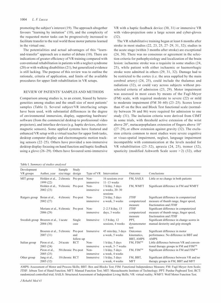
1004 L. F. Lucca
promoting the subject’s interest (19). The approach altogether favours “learning by imitation” (10), and the complexity of the requested motor tasks can be progressively increased to facilitate transfer to the real world those motor patterns learned in the virtual one.
The potentialities and actual advantages of this “learn-and-transfer” approach are a matter of debate (10). There are indications of greater efficiency of VR training compared with conventional rehabilitation in patients with a neglect syndrome (20) or with walking disabilities (21), but generalized evidence is still lacking. The purpose of this review was to outline the rationale, criteria of application, and limits of the available procedures for upper limb rehabilitation in VR setups.
REVIEW OF PATIENTS’ SAMPLES AND METHODS
Comparison among studies is, to an extent, biased by hetero-geneities among studies and the small size of most patients’ samples (Table I). Several subject/VR interfacing setups have been used, with substantial differences in the degree of environmental immersion, display, supporting hardware/software (from the commercial desktop to professional video projectors), and interface devices (e.g. haptic devices, electro-magnetic sensors). Some applied systems have featured and enhanced VR setup with a virtual teacher for upper limb tasks, desktop computer display and electromagnetic motion track-ing sensors (22–25). Others have provided a non-immersive desktop display focusing on hand function and haptic feedback using a glove (26–29). Others have favoured semi-immersive
VR with a haptic feedback device (30, 31) or immersive VR with video-projection onto a large screen and cyber-gloves (32).
The VR rehabilitative training began at least 6 months after stroke in most studies (22, 23, 25, 27–29, 31, 32); studies in the acute stage (within 3 months after stroke) are exceptional (24, 30). There was no consensus or agreement in the selec-tion criteria for pathophysiology and localization of the brain lesion: ischaemic stroke was a requisite in some studies (24, 25), while patient with either ischaemic or haemorrhagic stroke were admitted in others (29, 31, 32). Damage had to be restricted to the cortex (i.e. the area supplied by the main cerebral artery) (24, 25), could include the thalamus and radiations (32), or could vary across subjects without pre-selected criteria of admission (23, 29). Motor impairment was assessed in most cases by means of the Fugl-Meyer (FM) scale, with required moderate to severe (22) or mild to moderate impairment (FM 30–60) (23–25). Scores lower than 45 on the Box and Block Test functional scale (normal-ity between 56 and 86) were required for admission to one study (31). The inclusion criteria were derived from CIMT in some trials, with threshold active extension of the wrist above 20°, metacarpophalanx extension of fingers above 10° (27–29), or elbow extension against gravity (32). The exclu-sion criteria common to most studies were severe cognitive or visuo-spatial impairment, neglect, language impairment incompatible with communication at the levels needed for VR rehabilitation (23–32), apraxia (24, 25), tremor (32), spasticity (modified Ashworth Scale score > 2) (32), other
Table I. Summary of studies analysed
Development VR groups Author, year
Sample size/stage
Study design Type of VR Intervention Outcome Conclusions
MIT group Holden et al., 1999 (22)
2/chronic Pre-post Non-immersive
16 sessions over 11–13 weeks
FM, SAILS Little or no change in both patients
Holden et al., 2002 (23)
9/chronic Pre-post Non-immersive
1 h/day, 3 days a weeks, 20–30 sessions
FM, WMFT Significant difference in FM and WMFT
Rutgers group Boian et al., 2002 (27)
4/chronic Pre-post Non-immersive
2 h/day, 5 days a week, 3 weeks
JTHF computerized measure
Significant difference in computerized measure of thumb range, finger speed, fractionation and JTHF
Merians et al., 2006 (29)
8/chronic Pre-post Non-immersive
2–2.5 h/day, 13 days, 3 weeks
JTHF computerized measure
Significant difference in computerized measure of thumb range, finger speed, fractionation and JTHF
Swedish group Broeren et al., 2004 (30)
1/acute Single case
Immersive 1.5 h/day, 12 sessions, 4 weeks
PPT, dynamometer test
Significant difference in change scores in manual dexterity and grip strength
Broeren et al., 2007 (31)
5/chronic Pre-post and follow-up
Immersive 45 min/day, 3 days a week, 5 weeks
Outcomes kinematics, BBT, AMPS
Significant difference in motor performance. No difference in BBT and AMPS
Italian group Piron et al., 2003 (24)
24/acute RCT Non-immersive
1 h/day, 5 days a week, 5–7 weeks
FM, FIMTM Little difference between VR and con ven-tional therapy groups in FM and FIMTM
Piron et al., 2005 (25)
50/chronic Pre-post Non-immersive
1 h/day, 5 days a week, 4 weeks
FM, FIMTM Significant difference in FM and FIMTM
Other group Jang et al., 2005 (32)
10/chronic RCT Immersive 1 h/day, 5 days a week, 4 weeks
FM, BBT, MFT
Significant difference between VR and no therapy groups in FM, BBT and MFT
AMPS: Assessment of Motor and Process Skills; BBT: Box and Blocks Test; FIM: Functional Independence Measure; FM: Fugl-Meyer Arm Scale; JTHF: Jebsen Test of Hand Function; MFT: Manual Function Test; MIT: Massachusetts Institute of Technology; PPT: Purdue Pegboard Test; RCT: randomized controlled trial; SAILS: Structured Assessment of Independent Living Skills; VR: virtual reality; WMFT: Wolf Motor Function Test.
J Rehabil Med 41
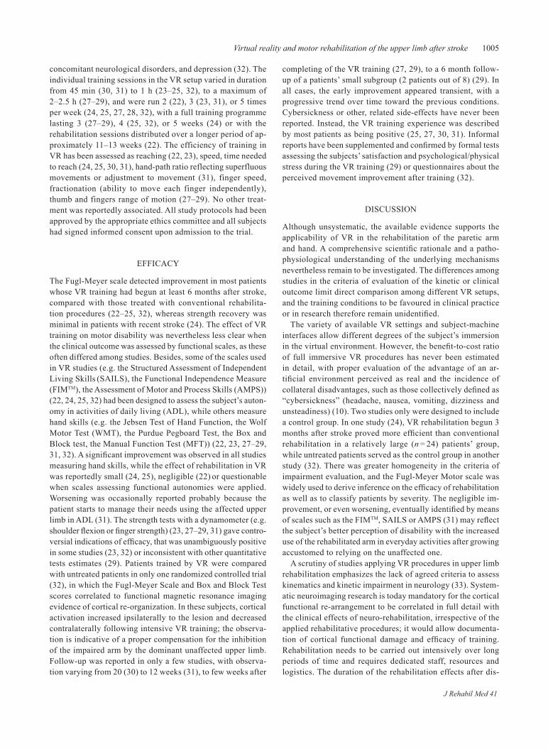
1005Virtual reality and motor rehabilitation of the upper limb after stroke
concomitant neurological disorders, and depression (32). The individual training sessions in the VR setup varied in duration from 45 min (30, 31) to 1 h (23–25, 32), to a maximum of 2–2.5 h (27–29), and were run 2 (22), 3 (23, 31), or 5 times per week (24, 25, 27, 28, 32), with a full training programme lasting 3 (27–29), 4 (25, 32), or 5 weeks (24) or with the rehabilitation sessions distributed over a longer period of ap-proximately 11–13 weeks (22). The efficiency of training in VR has been assessed as reaching (22, 23), speed, time needed to reach (24, 25, 30, 31), hand-path ratio reflecting superfluous movements or adjustment to movement (31), finger speed, fractionation (ability to move each finger independently), thumb and fingers range of motion (27–29). No other treat-ment was reportedly associated. All study protocols had been approved by the appropriate ethics committee and all subjects had signed informed consent upon admission to the trial.
EFFICACY
The Fugl-Meyer scale detected improvement in most patients whose VR training had begun at least 6 months after stroke, compared with those treated with conventional rehabilita-tion procedures (22–25, 32), whereas strength recovery was minimal in patients with recent stroke (24). The effect of VR training on motor disability was nevertheless less clear when the clinical outcome was assessed by functional scales, as these often differed among studies. Besides, some of the scales used in VR studies (e.g. the Structured Assessment of Independent Living Skills (SAILS), the Functional Independence Measure (FIMTM), the Assessment of Motor and Process Skills (AMPS)) (22, 24, 25, 32) had been designed to assess the subject’s auton-omy in activities of daily living (ADL), while others measure hand skills (e.g. the Jebsen Test of Hand Function, the Wolf Motor Test (WMT), the Purdue Pegboard Test, the Box and Block test, the Manual Function Test (MFT)) (22, 23, 27–29, 31, 32). A significant improvement was observed in all studies measuring hand skills, while the effect of rehabilitation in VR was reportedly small (24, 25), negligible (22) or questionable when scales assessing functional autonomies were applied. Worsening was occasionally reported probably because the patient starts to manage their needs using the affected upper limb in ADL (31). The strength tests with a dynamometer (e.g. shoulder flexion or finger strength) (23, 27–29, 31) gave contro-versial indications of efficacy, that was unambiguously positive in some studies (23, 32) or inconsistent with other quantitative tests estimates (29). Patients trained by VR were compared with untreated patients in only one randomized controlled trial (32), in which the Fugl-Meyer Scale and Box and Block Test scores correlated to functional magnetic resonance imaging evidence of cortical re-organization. In these subjects, cortical activation increased ipsilaterally to the lesion and decreased contralaterally following intensive VR training; the observa-tion is indicative of a proper compensation for the inhibition of the impaired arm by the dominant unaffected upper limb. Follow-up was reported in only a few studies, with observa-tion varying from 20 (30) to 12 weeks (31), to few weeks after
completing of the VR training (27, 29), to a 6 month follow-up of a patients’ small subgroup (2 patients out of 8) (29). In all cases, the early improvement appeared transient, with a progressive trend over time toward the previous conditions. Cybersickness or other, related side-effects have never been reported. Instead, the VR training experience was described by most patients as being positive (25, 27, 30, 31). Informal reports have been supplemented and confirmed by formal tests assessing the subjects’ satisfaction and psychological/physical stress during the VR training (29) or questionnaires about the perceived movement improvement after training (32).
DISCUSSION
Although unsystematic, the available evidence supports the applicability of VR in the rehabilitation of the paretic arm and hand. A comprehensive scientific rationale and a patho-physiological understanding of the underlying mechanisms nevertheless remain to be investigated. The differences among studies in the criteria of evaluation of the kinetic or clinical outcome limit direct comparison among different VR setups, and the training conditions to be favoured in clinical practice or in research therefore remain unidentified.
The variety of available VR settings and subject-machine interfaces allow different degrees of the subject’s immersion in the virtual environment. However, the benefit-to-cost ratio of full immersive VR procedures has never been estimated in detail, with proper evaluation of the advantage of an ar-tificial environment perceived as real and the incidence of collateral disadvantages, such as those collectively defined as “cybersickness” (headache, nausea, vomiting, dizziness and unsteadiness) (10). Two studies only were designed to include a control group. In one study (24), VR rehabilitation begun 3 months after stroke proved more efficient than conventional rehabilitation in a relatively large (n = 24) patients’ group, while untreated patients served as the control group in another study (32). There was greater homogeneity in the criteria of impairment evaluation, and the Fugl-Meyer Motor scale was widely used to derive inference on the efficacy of rehabilitation as well as to classify patients by severity. The negligible im-provement, or even worsening, eventually identified by means of scales such as the FIMTM, SAILS or AMPS (31) may reflect the subject’s better perception of disability with the increased use of the rehabilitated arm in everyday activities after growing accustomed to relying on the unaffected one.
A scrutiny of studies applying VR procedures in upper limb rehabilitation emphasizes the lack of agreed criteria to assess kinematics and kinetic impairment in neurology (33). System-atic neuroimaging research is today mandatory for the cortical functional re-arrangement to be correlated in full detail with the clinical effects of neuro-rehabilitation, irrespective of the applied rehabilitative procedures; it would allow documenta-tion of cortical functional damage and efficacy of training. Rehabilitation needs to be carried out intensively over long periods of time and requires dedicated staff, resources and logistics. The duration of the rehabilitation effects after dis-
J Rehabil Med 41
1006 L. F. Lucca
continuing VR training is crucial and should be determined in controlled follow-up studies, which also remain unsystematic to date (29–31). This discrepancy contrasts with the increased availability of advanced technologies and the need for reliable criteria to help define cost/benefit ratios and priorities in private and public health facilities. In general, the scenario would motivate research to achieve widespread application with reduced costs, possibly by making home rehabilitation under remote control a realistic option and by extending the use of VR to people who are computer- or technologically- illiterate (35–37). In this respect, basing on the potentialities of this approach, the lack of the long-term efficacy of VR rehabilita-tion procedures could challenge physicians, physiotherapists and bio-engineers.
REFERENCES
Nudo RJ. Functional and structural plasticity in motor cortex: 1. implications for stroke recovery. Phys Med Rehabil Clin N Am 2003; 14: 57–76.Wade T, Wood VA, Langton Hewer R. Recovery after stroke – the 2. first three months. J Neurol Neurosurg Psych 1985; 48: 7–13.Kwakkel G, Wagenaar RC, Twisk JW, Lankhorst GJ, Koetsier JC. 3. Intensity of leg and arm training after primary middle-cerebral-artery stroke: a randomized trial. Lancet 1999; 354: 191–196.Page S. Intensity versus task-specificity after stroke. How impor-4. tant is intensity? Am J Phys Med Rehabil 2003; 82: 730–732.Liepert J, Bauder H, Miltner WHR, Taub E, Weiller C. Treatment-5. induced cortical reorganization after stroke in humans. Stroke 2002; 31: 1210–1216.Bayona NA, Bitensky J, Salter K, Teasell R. The role of task-6. specific training in rehabilitation therapies. Topics Stroke Rehab 2005; 12: 58–65.Truelsen T, Bonita R. Stroke in developing countries: a continuing 7. challenge. Stroke Rev 2003; 7: 61–66.Page SJ, Levine P, Leonard C. Modified constraint-induced move-8. ment therapy in acute stroke: a randomized controlled pilot study. Neurorehabil Neural Repair 2005; 19: 27–32.Sveistrup H. Motor rehabilitation using virtual reality. J Neuro-9. engineering Rehabil 2004; 1: 10.Holden MK.10. Virtual enviroments for motor rehabilitation: review. Cyberpsychol Behav 2005; 8: 187-211.Rose FD, Broocks BM, Rizzo AA. Virtual reality in brain damage 11. rehabilitation: review. Cyberpsychol Behav 2005; 8: 241–262.Patton J, Dawe G, Scharver C, Mussa-Ivaldi F, Kenyon R. Robotics 12. and virtual reality: a perfect marriage for motor control research and rehabilitation. Assist Technol 2006; 18: 181–195.Adamovich S, Fluet GG, Merians AS, Mathai A, Qiu Q. Recovery 13. of hand function in virtual reality: training hemiparetic hand and arm together or separately. Conf Proc IEEE Eng Med Biol Soc 2008: 2008: 3475–3478.Eng K, Sierkierka E, Pyk P, Chvrier E, Hauser Y, Cameirao M, et 14. al. Interactive visuo-moto therapy system for stroke rehabilitation. Med Biol Eng Comput 2007; 45: 901–907.Gaggioli A, Meneghini A, Morganti F, Alcaniz M, Riva G. A strat-15. egy for computer-assisted mental practice in stroke rehabilitation. Neurorehabil Neural Repair 2006; 20: 503–507.Todorov E, Shadmehr R, Bizzi E. Augmented feedback presented 16. in a virtual environment accelerates learning of a difficult motor task. J Motor Behav 1997; 29: 147–158.Huang H, Wolf SL, He J. Recent developments in biofeedback for 17. neuromotor rehabilitation. J NeuroEng Rehab 2006; 3: 1–12.
Cirstea CM, Ptito A, Levin MF. Feedback and cognition in arm mo-18. tor skill reacquisition after stroke. Stroke 2006; 37: 1237–1242.Weiss PL, Bialik P, Kizony K. Virtual reality provides leisure 19. time opportunities for young adults with physical and intellectual disabilities. Cyberpsychol Behav 2003; 6: 335–342.Webster JS, McFarland PT, Rappaport LJ, Morrill B, Roades LA, 20. Abadee PS. Computer-assisted training for improving wheelchair mobility in unilateral neglect patients. Arch Phys Med Rehabil 2001; 82: 769–775.Jaffe DL, Brown DA, Pierson-Carey CD, Buckley EL, Lew HL. 21. Stepping over obstacles to improve walking in individuals with post-stroke hemiplegia. J Rehabil Res Dev 2004; 41: 283–292. Holden M, Todorov E, Callahan J, Bizzi E. Virtual environment 22. training improves motor performance in two patients with stroke: case report. Neurol Report 1999; 23: 57–67. Holden MK, Dyar T. Virtual environment training: a new tool for 23. rehabilitation. Neurol Report 2002; 26: 62–71.Piron L, Tonin P, Atzori AM, Zucconi C, Massaro C, Trivello E, 24. Dam M. The augmented-feedback rehabilitation technique facili-tates the arm motor recovery in patients after a recent stroke. Stud Health Technol Inform 2003; 94: 265–267.Piron L, Tonin P, Piccione F, Iaia V, Trivello E, Dam M. Virtual 25. environment training therapy for arm motor rehabilitation. Pres-ence 2005; 14: 732–740.Burdea G, Deshpande S, Langrana N, Gomez D, Liu B. A virtual-26. reality-based system for hand diagnosis and rehabilitation. Presence-Teleoperators Virtual Environ 1997; 6: 229–248.Boian R, Sharma A, Han C, Merians AS, Burdea G, Adamovich 27. S, et al. Virtual reality-based post-stroke hand rehabilitation. Stud Health Technol Inform 2002; 85: 64–70.Deutsch JE, Merians AS, Adamovich S, Poizner H, Burdea GC. 28. Development and application of virtual reality technology to im-prove hand use and gait of individuals post-stroke. Restor Neurol Neurosc 2004; 22: 371–386.Merians A, Poizner H, Boian R, Burdea G, Adamovich S. Sensori-29. motor training in a virtual reality environment: does it improve functional recovery post-stroke? Neurorehabil Neural Repair 2006; 20: 252–267.Broeren J, Rydmark M, Sunnerhagen K. Virtual reality and haptics 30. as a training device for movement rehabilitation after stroke: a single case-study. Arch Phys Med Rehabil 2004; 85: 1247–1250.Broeren J, Rydmark M, Bjorkdahl A, Sunnerhagen K. Assesssment 31. and training in a 3-dimensional virtual environment with haptics: a report on 5 cases of motor rehabilitation in the chronic stage after stroke. Neurorehabil Neural Repair 2007; 21: 180–189.Jang SH, You SH, Hallett M, Cho YW, Park CM, Cho SH, et 32. al. Cortical reorganization and associated functional motor re-covery after virtual reality in patients with chronic stroke: an experimenter-blind preliminary study. Arch Phys Med Rehabil 2005; 86: 2218–2223.Broeren J, Sunnerhagen K, Rydmark M. A kinematic analysis of a 33. haptic handheld stylus in a virtual environment: a study in healthy subjects. J Neuroeng Rehabil 2007; 4: 1–8.Holden MK, Dyar T, Callahan J, Schwamm L, Bizzi E. Quantita-34. tive assessment of motor generalization in the real world following training in a virtual environment in patients with stroke. Neurol Report 2001; 25: 129–130. Holden MK, Dyar TA, Schwamm L, Bizzi E. Virtual-environment 35. based telerehabilitation in patients with stroke. Presence: Tele-operat Virtual Environ 2005; 14: 214–233. Piron L, Tonin P, Trivello E, Battistin L, Dam M. Motor tele-36. rehabilitation in post-stroke patients. Med Inform Internet Med 2004; 29: 119–125.Kuttuva M, Boian R, Merians A, Burdea G, Bouzit M, Lewis J, 37. Fensterheim D. The Rutgers Arm, a rehabilitation system in a virtu-al reality: a pilot study. Cyberpsychol Behav 2006; 9: 148–151.
J Rehabil Med 41
special report
J Rehabil Med 2009; 41: 1007–1010
J Rehabil Med 41© 2009 The Authors. doi: 10.2340/16501977-0406Journal Compilation © 2009 Foundation of Rehabilitation Information. ISSN 1650-1977
robot aramis (automatic recovery arm motility in-tegrated system) is intended to provide the therapist with novel and time/cost-efficient approaches to the rehabilitation of the paretic upper limb after stroke. the system has been designed and implemented based on common experience in rehabilitation and will provide a robot–patient interaction compensating for some intrinsic limitations of traditional treatments. rationale, technical characteristics and applica-tion are described in detail here.Key words: robot assisted rehabilitation, paresis upper limb, stroke.J Rehabil Med 2009; 41: 1007–1010
Correspondence address: Giuliano Dolce, RAN – Research on Advanced Neuro-rehabilitation, S. Anna Institute, Via Siris – IT-88900 Crotone, Italy. E-mail: [email protected] March 16, 2009; accepted May 25, 2009
INTRODUCTION
The outcome for patients with motor impairment after stroke has improved significantly over recent decades with the in-creasing resources and advanced rehabilitation procedures available in developed countries (1–3). Early admission to, and treatment in, dedicated units is crucial for rehabilitation and is favoured by healthcare policies, restricting in time both the permanence in emergency care units and rehabilitation in hospital (4, 5). In the rehabilitation of inpatients, priority is therefore usually given to posture and walking (6, 7), in order to achieve a greater level of independence in activities of daily living (ADL). Treatment of the upper limb is usually postponed, and recovery of its movement and motor control is often incomplete.
Detailed knowledge of the pathophysiological mechanisms regulating the motility and recovery of the paretic arm is still lacking. Ad hoc approaches are therefore mandatory for a useful rehabilitation protocol to be devised and for recovery to occur, with requirements that are, to a relevant extent, de-termined by the peculiar motor organization of the arm and shoulder (8, 9). In addition, adequate tools are needed to test the adequacy and usefulness of any rehabilitation procedure over a wide range of adaptation conditions. Two major strate-gies, constraint-induced movement therapy (CIMT) (10), and robot-supported rehabilitation (11, 12), have been developed in recent years.
THE ARAMIS PROJECT: A RATIONALEThere are significant functional links between the trunk and lower limbs. Locomotion after paresis becomes possible also due to the early re-organization of brain control of the trunk, often observed as early as 3–4 weeks or less after brain injury (13). Clinical experience indicates that the unaffected lower limb can vicariate the contralateral paretic leg, and this functional tutoring makes locomotion, if not walking, possible (14).
The upper limbs appear, by contrast, to be largely independ-ent of each other. Correct movement would otherwise become impossible when spontaneous motor recovery is interfered with by poorly tractable algo-dystrophic syndromes, dislocation, or intractable pain at the glenohumeral capsule not prevented by early counter-measures. The arms and hands compete with each other to a significant extent and the unaffected upper limb usually takes over, thus excluding the paretic one when bilat-eral engagement and co-ordination are required for complex motor operations to be carried out. The proximal, but not the distal, upper limb portion receives both ipsi- and contra-lateral inputs from the brain (15). Very early in extra-uterine life, motor control lateralizes to become peculiarly dependent on the contralateral hemisphere motor organization; although functionally silent in normal conditions, ipsilateral control is nevertheless maintained in part. Brain plasticity (16, 17) allows a post-lesional functional re-arrangement to develop and mediates in motor recovery no matter how complete. This process is possible and usually occurs in the 3–4 months after diaschisis, with the potentiality for recovery decreasing over time depending on the lesion and the individual motor organiza-tion before brain insult (18). The spontaneous re-arrangement is not driven by functional or evolutionary rules and can lead to unfit patterns responsible for, for example, spasticity, hypotonia or pathological synergies.
In principle, the crural and brachial functional roles in the recovery of the upper and lower limbs should not differ to a significant extent, yet inadequate recovery has markedly dif-ferent effects. The main functional and evolutionary purpose of the arm is to drive the hand in the subject’s own personal space under visual control mediated by the mirror neurone system (19). The functional recovery of the fingers is of limited help when the hand cannot be moved in the competing space with precision and reliability (20). The roles of the shoulder and elbow in recovery are crucial (21, 22); with proximal-to-distal spontaneous recuperation, hand motor recovery is not
ROBOT-ASSISTED REHABILITATION OF THE PARETIC UPPER LIMB: RATIONALE OF THE ARAMIS PROJECT
Giuliano Dolce, MD, Lucia Francesca Lucca, MD and Loris Pignolo, EngFrom the S. Anna Institute and RAN – Research on Advanced Neuro-rehabilitation, Crotone, Italy
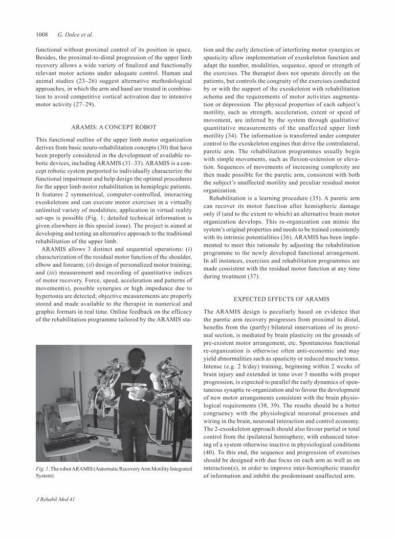
1008 G. Dolce et al.
functional without proximal control of its position in space. Besides, the proximal-to-distal progression of the upper limb recovery allows a wide variety of finalized and functionally relevant motor actions under adequate control. Human and animal studies (23–26) suggest alternative methodological approaches, in which the arm and hand are treated in combina-tion to avoid competitive cortical activation due to intensive motor activity (27–29).
ARAMIS: A CONCEPT ROBOT
This functional outline of the upper limb motor organization derives from basic neuro-rehabilitation concepts (30) that have been properly considered in the development of available ro-botic devices, including ARAMIS (31–33). ARAMIS is a con-cept robotic system purported to individually characterize the functional impairment and help design the optimal procedures for the upper limb motor rehabilitation in hemiplegic patients. It features 2 symmetrical, computer-controlled, interacting exoskeletons and can execute motor exercises in a virtually unlimited variety of modalities; application in virtual reality set-ups is possible (Fig. 1; detailed technical information is given elsewhere in this special issue). The project is aimed at developing and testing an alternative approach to the traditional rehabilitation of the upper limb.
ARAMIS allows 3 distinct and sequential operations: (i) characterization of the residual motor function of the shoulder, elbow and forearm; (ii) design of personalized motor training; and (iii) measurement and recording of quantitative indices of motor recovery. Force, speed, acceleration and patterns of movement(s), possible synergies or high impedance due to hypertonia are detected; objective measurements are properly stored and made available to the therapist in numerical and graphic formats in real time. Online feedback on the efficacy of the rehabilitation programme tailored by the ARAMIS sta-
tion and the early detection of interfering motor synergies or spasticity allow implementation of exoskeleton function and adapt the number, modalities, sequence, speed or strength of the exercises. The therapist does not operate directly on the patients, but controls the congruity of the exercises conducted by or with the support of the exoskeleton with rehabilitation schema and the requirements of motor activities augmenta-tion or depression. The physical properties of each subject’s motility, such as strength, acceleration, extent or speed of movement, are inferred by the system through qualitative/quantitative measurements of the unaffected upper limb motility (34). The information is transferred under computer control to the exoskeleton engines that drive the contralateral, paretic arm. The rehabilitation programmes usually begin with simple movements, such as flexion-extension or eleva-tion. Sequences of movements of increasing complexity are then made possible for the paretic arm, consistent with both the subject’s unaffected motility and peculiar residual motor organization.
Rehabilitation is a learning procedure (35). A paretic arm can recover its motor function after hemispheric damage only if (and to the extent to which) an alternative brain motor organization develops. This re-organization can mimic the system’s original properties and needs to be trained consistently with its intrinsic potentialities (36). ARAMIS has been imple-mented to meet this rationale by adjusting the rehabilitation programme to the newly developed functional arrangement. In all instances, exercises and rehabilitation programmes are made consistent with the residual motor function at any time during treatment (37).
EXPECTED EFFECTS OF ARAMIS
The ARAMIS design is peculiarly based on evidence that the paretic arm recovery progresses from proximal to distal, benefits from the (partly) bilateral innervations of its proxi-mal section, is mediated by brain plasticity on the grounds of pre-existent motor arrangement, etc. Spontaneous functional re-organization is otherwise often anti-economic and may yield abnormalities such as spasticity or reduced muscle tonus. Intense (e.g. 2 h/day) training, beginning within 2 weeks of brain injury and extended in time over 3 months with proper progression, is expected to parallel the early dynamics of spon-taneous synaptic re-organization and to favour the development of new motor arrangements consistent with the brain physio-logical requirements (38, 39). The results should be a better congruency with the physiological neuronal processes and wiring in the brain, neuronal interaction and control economy. The 2-exoskeleton approach should also favour partial or total control from the ipsilateral hemisphere, with enhanced tutor-ing of a system otherwise inactive in physiological conditions (40). To this end, the sequence and progression of exercises should be designed with due focus on each arm as well as on interaction(s), in order to improve inter-hemispheric transfer of information and inhibit the predominant unaffected arm.
Fig. 1. The robot ARAMIS (Automatic Recovery Arm Motility Integrated System).
J Rehabil Med 41
1009Robot-assisted rehabilitation of the upper limb: ARAMIS project
VALIDATION OF ARAMIS
Further investigation on large patients’ samples is required for validation. The advantages of ARAMIS in the quantita-tive characterization of the motor disability, residual function and outcome would help provide shared criteria of evaluation and protocols of rehabilitation, to a final identification of the expected future role and applicability of robotics in neuro-rehabilitation. A study protocol has been approved by the eth-ics committee and the National Governmental Agencies. Two groups of subjects with hemiparesis due to stroke that occurred, respectively, less than 3 months, or more than 6 months, previ-ously, with age ranging from 18 to 70 years will be admitted to the study. Exclusion criteria will be: implanted pace-maker derivations, aphasia or cognitive impairment not compatible with collaboration, pregnancy, and epilepsy. Systemic or local pharmacological therapies preventing or treating spasticity will not be allowed during the study. Subjects with stroke that occurred less than 3 months earlier will be treated by both conventional rehabilitative methods and treatment controlled by ARAMIS (2 × 45-min sessions/day for a maximum period of 6 weeks), while those with stroke that occurred more than 6 months earlier will be treated only by ARAMIS-controlled training procedures.
ACKNOWLEDGEMENTS
ARAMIS (Automatic Recovery Arm Motility Integrated System) is a project carried out by the S. Anna Institute and RAN – Research on Advanced Neuro-rehabilitation (Crotone), in collaboration with CETMA (Brindisi), S&D srl (Crotone), DEIS (University of Calabria, Cosenza), Synapsys srl (Livorno, Italy). The prototype is part of the research project MIMERICA, which is supported by governmental funding.
REFERENCES
Truelsen T, Bonita R. Stroke in developing countries: a continuing 1. challenge. Stroke Review 2003; 7: 61–66.Malouin F, Richards CL, McFadyen B, Doyon J. New perspec-2. tives of locomotor rehabilitation after stroke. Med Sci (Paris) 2003; 19: 994–998.Kwakkel G, Kollen B, Lindeman E. Understanding the pattern of 3. functional recovery after stroke: facts and theories. Restorative Neurol Neurosci 2004; 22: 281–299.Wade T, Wood VA, Langton Hewer R. Recovery after stroke – the 4. first three months. J Neurol Neurosurg Psych 1985; 48: 7–13.Page S. Intensity versus task-specificity after stroke. How impor-5. tant is intensity? Am J Phys Med Rehabil 2003; 82: 730–732.Foley NC, Teasell RW, Bhogal SK, Doherty T, Speechley MR. The 6. efficacy of stroke rehabilitation: a qualitative review. Top Stroke Rehabil 2003; 10: 1–18.Aichner F, Adelwèohrer C, Haring HP. Rehabilitation approaches 7. to stroke. J Neural Transm Suppl 2002; 63: 59–73.Jang SH, Kim YH, Cho SH, Chang Y, Lee ZI, Ha JS. Cortical 8. reorganization associated with motor recovery in hemiparetic stroke patients. Neuroreport 2003; 14: 1305–1310.Stein J. Motor recovery strategies after stroke. Topics Stroke 9. Rehabil 2004; 11: 12–22.Page SJ, Levine P, Leonard C. Modified constraint-induced move-10. ment therapy in acute stroke: a randomized controlled pilot study. Neurorehabil Neural Repair 2005; 19: 27–32.
Krebs HI, Hogan N, Aisen ML, Volpe BT. Robot-aided neurore-11. habilitation. IEEE Trans Rehabil Eng 1998; 6: 75–87.Kwakkel G, Kollen BJ, Krebs HI. Effects of robot-assisted therapy 12. on upper limb recovery after stroke: a systematic review. Neurore-habil Neural Repair 2008; 22: 111–121.Levin MF, Cirstea MC, Michaelsen SM, Roby-Brami A. Use of 13. trunk for reaching targets placed within and beyond the reach in adult hemiparesis. Exp Brain Res 2002; 143: 171–180.Ustinova KI, Goussev VM, Balasubramaniam R, Levin MF. Dis-14. ruption of coordination between arm, trunk, and center of pressure displacement in patients with hemiparesis. Motor Control 2004; 8: 139–159.Hassler R, Rolle Y, Streicher H. Pathophisiologie der Bewustsein-15. tatigkeit. Stuttgart: Georg Thieme; 1972, p. 1–12.Nudo RJ. Functional and structural plasticity in motor cortex: 16. implications for stroke recovery. Phys Med Rehabil Clin N Am 2003; 14: 57–76.Teasell R, Bayona NA, Bitensky J. Plasticity and reorganization of 17. the brain post stroke. Top Stroke Rehabil 2005; 12: 11–26.Katrak P, Bowring G, Conroy P, Chilvers M, Poulos R, McNeil 18. D. Predicting upper limb recovery after stroke: the place of early shoulder and hand movement. Arch Phys Med Rehabil 1998; 79: 758–761.Rizzolatti G, Craighero L. The mirror neuron system. Annu Rev 19. Neurosci 2004; 27: 169–192.Archambault P, Pigeon P, Feldman AG, Levin MF. Recruitment 20. and sequencing of different degrees of freedom during pointing movements involving the trunk in healthy and hemiparetic subjects. Exp Brain Res 1999; 126: 55–67.Michaelsen SM, Jacobs S, Roby-Brami A, Levin MF. Compensa-21. tion for distal impairments of grasping in adults with hemiparesis. Exp Brain Res 2004; 157: 162–173.Michaelsen SM, Dannenbaum R, Levin MF. Task-specific training 22. with trunk restraint on arm recovery in stroke: randomized control trial. Stroke 2006; 37: 186–192.Pascual-Leone A, Grafman J, Hallett M. Modulation of cortical 23. motor output maps during development of implicit and explicit knowledge. Science 1994; 263: 1287–1289.Karni A, Meyer G, Jezzard P, Adams MM, Turner R, Ungerleider 24. LG. Functional MRI evidence for adult motor cortex plasticity during motor skill learning. Nature 1995; 377: 155–158.Nudo RJ, Milliken GW, Jenkins WM, Merzenick MM. Use- 25. dependent alterations of movement representations in primary motor cortex of adult squirrel monkeys. J Neurosci 1996; 16: 785–807.Hlustik P, Solodkin A, Noll DC, Small SL. Cortical plasticity dur-26. ing three-week motor skill learning. J Clin Neurophysiol 2004; 21: 180–191.Merzenich, MM, DeCharms RC Neural representations, experience 27. and change. In: Llinas R, Churchland P, editors. The mind–brain continuum. Boston, MA: MIT Press; 1996.Nudo RJ, Milliken GW, Jenkins WM, Merzenick MM. Reor-28. ganization of movement representations in primary motor cortex following focal ischemic infarcts in adult squirrel monkeys. J Neurophysiol 1996; 75: 2144–2149.Muellbacher W, Richards C, Ziemann U, Wittenberg G, Wels D, 29. Boroojerdi B et al. Improving and function in chronic stroke. Arch Neurol 2002; 59: 1278–1282.Lennon S, Baxter D, Ashburn A. Physiotherapy based on the 30. Bobath concept in stroke rehabilitation: a survey within the UK. Disabil Rehabil 2001; 23: 254–262.Mayr A, Mina S, Köchl G, Kronreif G, Saltuari L. A new driven 31. orthosis for the upper extremity (ARMOR): preliminary results. Neurorehabil Neural Repair 2006; 20; 51.Nef T, Mihelj M, Colombo G, Riener R. ARMin32. – Robot for rehabilitation of the upper extremities. IEEE International Con-ference on Robotics and Automation, Orlando: ICRA, 2006, p. 3152–3157.
J Rehabil Med 41
1010 G. Dolce et al.
Carignan C, Liszka M, Roderick S. Design of an arm exoskeleton 33. with scapula motion for shoulder rehabilitation. Advanced Robot-ics 2005; 18: 524–531.Ada L, Canning C, Dwyer T. Effect of muscle length on strength 34. and dexterity after stroke. Clin Rehabil 2000; 14: 55–61.Winstein CJ, Merians AS, Sullivan KJ. Motor learning after unilat-35. eral brain damage. Neuropsychologia 1999; 37: 975–987.Bayona NA, Bitensky J, Salter K, Teasell R. Plasticity and re-36. organization of the uninjured brain. Top Stroke Rehabil 2005; 12: 1–10.Schaechter JD. Motor rehabilitation and brain plasticity after 37.
hemiparetic stroke. Prog Neurobiol 2004; 73: 61–72. Calautti C, Baron J. Functional neuroimaging studies of mo-38. tor recovery after stroke in adults: a review. Stroke 2003; 34: 1553–1566.Cramer SC, Nelles G, Schaechter JD, Kaplan JD, Finkelstein SP, 39. Rosen BR. A functional MRI study of three motor tasks in the evaluation of stroke recovery. Neural Rehabil Neural Rep 2001; 15: 1–8.Dong Y, Dobkin B, Cen S, Wu AD, Winstein CJ. Motor cortex 40. activation during treatment may predict therapeutic gains in paretic hand function after stroke. Stroke 2006; 37: 1552–1555.
J Rehabil Med 41
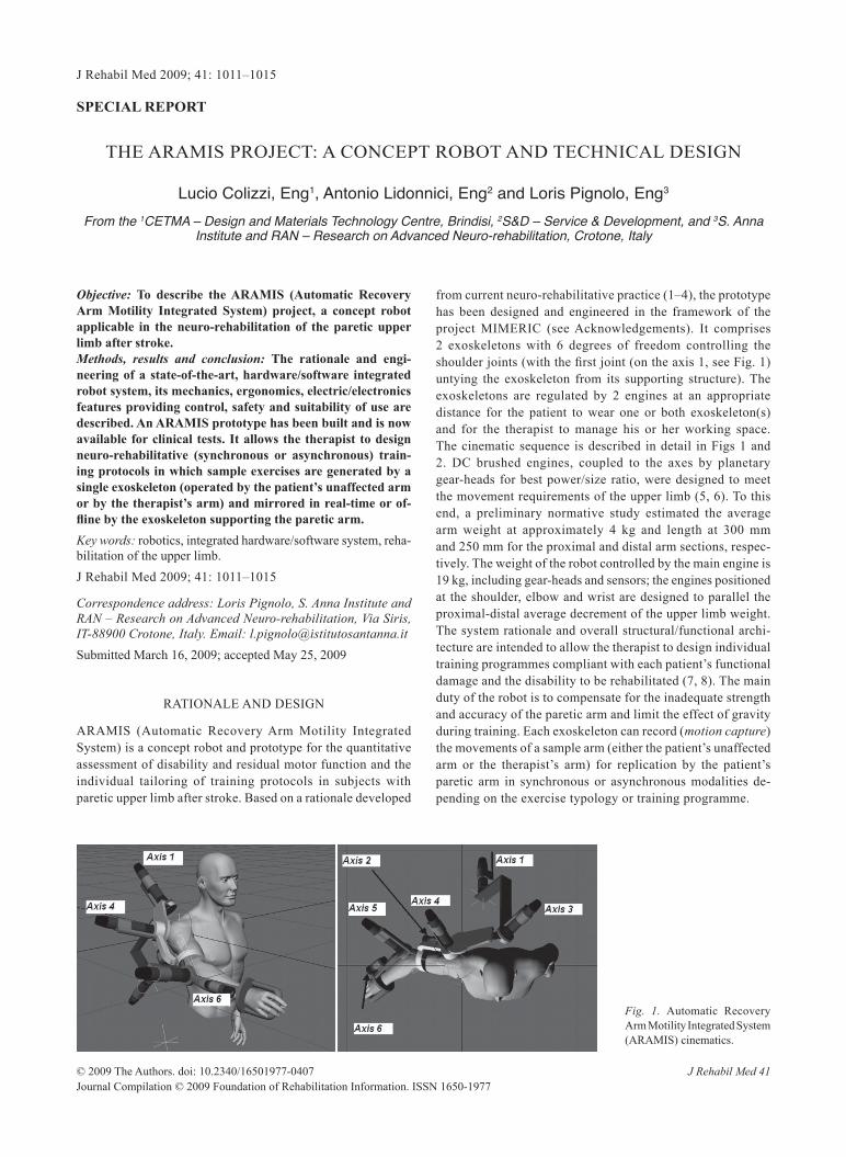
special report
J Rehabil Med 2009; 41: 1011–1015
J Rehabil Med 41© 2009 The Authors. doi: 10.2340/16501977-0407Journal Compilation © 2009 Foundation of Rehabilitation Information. ISSN 1650-1977
Objective: to describe the aramis (automatic recovery arm motility integrated system) project, a concept robot applicable in the neuro-rehabilitation of the paretic upper limb after stroke.Methods, results and conclusion: the rationale and engi-neering of a state-of-the-art, hardware/software integrated robot system, its mechanics, ergonomics, electric/electronics features providing control, safety and suitability of use are described. an aramis prototype has been built and is now available for clinical tests. it allows the therapist to design neuro-rehabilitative (synchronous or asynchronous) train-ing protocols in which sample exercises are generated by a single exoskeleton (operated by the patient’s unaffected arm or by the therapist’s arm) and mirrored in real-time or of-fline by the exoskeleton supporting the paretic arm. Key words: robotics, integrated hardware/software system, reha-bilitation of the upper limb. J Rehabil Med 2009; 41: 1011–1015
Correspondence address: Loris Pignolo, S. Anna Institute and RAN – Research on Advanced Neuro-rehabilitation, Via Siris, IT-88900 Crotone, Italy. Email: [email protected] March 16, 2009; accepted May 25, 2009
RATIONALE AND DESIGN
ARAMIS (Automatic Recovery Arm Motility Integrated System) is a concept robot and prototype for the quantitative assessment of disability and residual motor function and the individual tailoring of training protocols in subjects with paretic upper limb after stroke. Based on a rationale developed
from current neuro-rehabilitative practice (1–4), the prototype has been designed and engineered in the framework of the project MIMERIC (see Acknowledgements). It comprises 2 exoskeletons with 6 degrees of freedom controlling the shoulder joints (with the first joint (on the axis 1, see Fig. 1) untying the exoskeleton from its supporting structure). The exoskeletons are regulated by 2 engines at an appropriate distance for the patient to wear one or both exoskeleton(s) and for the therapist to manage his or her working space. The cinematic sequence is described in detail in Figs 1 and 2. DC brushed engines, coupled to the axes by planetary gear-heads for best power/size ratio, were designed to meet the movement requirements of the upper limb (5, 6). To this end, a preliminary normative study estimated the average arm weight at approximately 4 kg and length at 300 mm and 250 mm for the proximal and distal arm sections, respec-tively. The weight of the robot controlled by the main engine is 19 kg, including gear-heads and sensors; the engines positioned at the shoulder, elbow and wrist are designed to parallel the proximal-distal average decrement of the upper limb weight. The system rationale and overall structural/functional archi-tecture are intended to allow the therapist to design individual training programmes compliant with each patient’s functional damage and the disability to be rehabilitated (7, 8). The main duty of the robot is to compensate for the inadequate strength and accuracy of the paretic arm and limit the effect of gravity during training. Each exoskeleton can record (motion capture) the movements of a sample arm (either the patient’s unaffected arm or the therapist’s arm) for replication by the patient’s paretic arm in synchronous or asynchronous modalities de-pending on the exercise typology or training programme.
THE ARAMIS PROJECT: A CONCEPT ROBOT AND TECHNICAL DESIGN
Lucio Colizzi, Eng1, Antonio Lidonnici, Eng2 and Loris Pignolo, Eng3
From the 1CETMA – Design and Materials Technology Centre, Brindisi, 2S&D – Service & Development, and 3S. Anna Institute and RAN – Research on Advanced Neuro-rehabilitation, Crotone, Italy
Fig. 1. Automatic Recovery Arm Motility Integrated System (ARAMIS) cinematics.
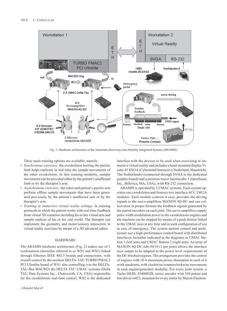
1012 L. Colizzi et al.
Three main training options are available, namely: • Synchronous exercises: the exoskeleton hosting the paretic
limb helps replicate in real time the sample movements of the other exoskeleton. In this training modality, sample movements can be provided either by the patient’s unaffected limb or by the therapist’s arm.
• Asynchronous exercises: the robot and patient’s paretic arm perform offline sample movements that have been gener-ated previously by the patient’s unaffected arm or by the therapist’s arm.
• Training in immersive virtual reality settings, in training protocols in which the patient works with real-time feedback from virtual 3D scenarios including his or her virtual arm and sample replicas of his or her real world. The therapist can implement the geometry and motor/sensory interaction in virtual reality exercises by means of a 3D advanced editor.
HARDWARE
The ARAMIS hardware architecture (Fig. 2) makes use of 2 workstations (hereafter referred to as WS1 and WS2) linked through Ethernet IEEE 802.3 boards and connections, with overall control by the resident DELTA-TAU TURBO PMAC2 PCI Ultralite board of WS1 also controlling (via the DELTA-TAU Bus MACRO) the DELTA TAU UMAC systems (Delta TAU Data Systems Inc., Chatsworth, CA, USA) responsible for the exoskeletons real-time control. WS2 is the dedicated
interface with the devices to be used when exercising in im-mersive virtual reality and includes a head-mounted display Vi-sette 45 SXGA (Cybermind Interactive Nederland, Maastricht, The Netherlands) (connected through SVGA to the dedicated graphic board) and a position-tracer Inertiacube 3 (InterSense Inc., Billerica, MA, USA), with RS-232 connection.
ARAMIS is operated by 2 UMAC systems. Each system op-erates one exoskeleton and features two interface ACC-24E2A modules. Each module controls 4 axes, provides the driving signals to the servo-amplifiers MAXON 4Q-DC and can col-lect/store in proper formats the feedback signals generated by the paired encoders on each joint. The servo-amplifiers supply pulse width modulation power to the exoskeletons engines and the machine can be stopped by means of a push-button linked to the UMAC axes at any time and in each configuration of use in case of emergency. The system motion control and archi-tecture use a high-performance control board with distributed interfaces, hereafter indicated in the diagrams as UMAC Sta-tion 1 (left arm) and UMAC Station 2 (right arm). An array of MAXON 4Q-DC Ads-50/10 (1 per joint) allows the interface axes output to be adapted to the power level requirements of the DC brushed engines. The arrangement provides the control of engines with 10 A maximum power absorption in each of 4 work quadrants, with clockwise/counterclockwise movements in each engine/generator modality. For every joint system, a Tacho HEDL 5540HEDL series encoder with 550 pulses and line driver rs422, mounted for every motor by Maxon Electron-
Fig. 2. Hardware architecture of the Automatic Recovery Arm Motility Integrated System (ARAMIS).
J Rehabil Med 41
1013ARAMIS project
ics (Kansas City, US), is placed upstream of the shaft motor gear system, while an encoder KS2304-240x10 is integrated downstream. The virtual reality hardware includes a helmet with stereoscopic HMD Visette 45 SXGA and an Inertiacube 3 tracer of the head movement mechanically linked to the HMD. Inertiacube 3 is a hybrid 3-degree of freedom tracing device with accelerometers, magnetometers, angular speed detectors, and algorithm combining data to provide information about heading, pitch and roll in the 3 degrees of freedom.
The UMAC system firmware for handling and control can process data and signals and drives the ARAMIS engines in real time. Dedicated, advanced software interacts with the Turbo PMAC2 Ultralite control board, generates its working para meters and is fed back with relevant information on the ongo-ing training session. The operator sets the working parameters through the ARAMIS Manager software; these are processed by the firmware into distinct control routines that also evalu-ate the feedback information from the exoskeleton’s sensors and feed forward the UMAC devices with signals instructing the exoskeleton’s motor patterns. The distribution of the gear-heads (1 per joint) does not allow the exoskeleton to move unless engine-driven and a servo-arm has been implemented to link the exoskeleton to the arm. A system with encoder and opposed springs is positioned downstream of each driveshaft and linked out of phase to the main encoder controlling the engine; this allows the exoskeleton to follow the movements initiated by the patient or therapist.
SOFTWARE
The dedicated software has been designed on the basis of the logics and requirements of the neuro-rehabilitative processes that ARAMIS is intended to support (9, 10). Specifically: • Baseline registration of each patient by demographics,
clinical condition, actual motor disability, and expected recovery.
• Individual design of robot-supported rehabilitative protocols.• Neuro-rehabilitation, with monitoring of the training effects
and acquisition/storing of the relevant information on changes in the trainee’s motor organization during rehabilitation.
• Up-/downgrading of the training protocols consistent with the achieved recovery or unexpected contingencies (clinical changes, medical problems, side-effects, etc.).
• Offline data analyses.• Feedback information of potential use in the patient’s further
training.• Re-calibration of the training protocols and changes in the reha-
bilitative strategies congruent with the obtained information.
Four dedicated software modules have been implemented for full system control, with a comprehensive system architecture featuring advance-control connections between framework software, located in WS1 and WS2, logic connections with front-end hardware.• ARAMIS Manager: the main framework module for new
patients’ registration, access to the patients’ database and rel-
evant information needed to plan and carry on the early phases of the rehabilitative protocol. In addition, ARAMIS Manager provides quantitative feedback information and allows control of the exoskeletons and the virtual reality hardware consistent with the training protocol and modalities.
• EXERCISE Builder: the module allowing implementation of virtual 3D scenarios for the patient to interact with when trained in virtual reality settings.
• HMD Interface is activated when required during virtual reality training. The module implements a 3D rendering engine with 2 input devices and monitors in real time the position in space of the patient’s arm and HMD-connected sensors.
• POSIS: dedicated software for the analysis of bio-mechanical information obtained by monitoring the training sessions through a 3D player and signal processing descriptors of the patient’s motor performance. This tool of the ARAMIS framework is crucial when analysing the early effects of reha-bilitation and upgrading the training protocols/modalities.
The contributions of the software modules at different phases of the rehabilitation procedure are outlined in Fig. 3.
DISCUSSION
Robots allow reliable quantitative measurement of physical properties over a wide range of variation, with speed, accuracy, power reliability and endurance over time, and repetitive task conditions that are not achievable by humans. Virtual reality is expected to contribute further (11, 12) to this process. The ARAMIS overall active/passive architecture and exoskeleton multiple-option use in different functional paradigms are expected to compensate, at least in part, for the functional competition between the paretic and unaffected arm, and to promote interaction. The approach should improve the out-come of robot-assisted neuro-rehabilitation compared with conventional training strategies. The purpose of the ARAMIS project is to provide the therapist with a flexible designer of exercises, i.e. a series of software tools able to adapt the ma-chine performance to precise, possibly peculiar, rehabilitation needs. ARAMIS can be used to design training protocols and exercises without predetermined or coded restrictions. The therapist can define a sequence of training movements based on any rehabilitation rationale or methodology by selecting move-ment, speed and acceleration, with high-accuracy definition in space of the 3D target objects and trajectories with which each patient is requested to comply. The complexity of each exercise and of the training protocol can be increased gradually; to this end, visual stimuli can be calibrated according to the trainee’s needs and therapist’s strategies before presentation to the patients during training in virtual reality settings. Training protocols, sequence of exercises, the physical characters of each exercise and the patient’s errors or improvement during treatment are coded and stored in the database for re-use in the same subject’s treatment or to train subjects sharing clinical conditions, disability, and/or training protocols.
J Rehabil Med 41
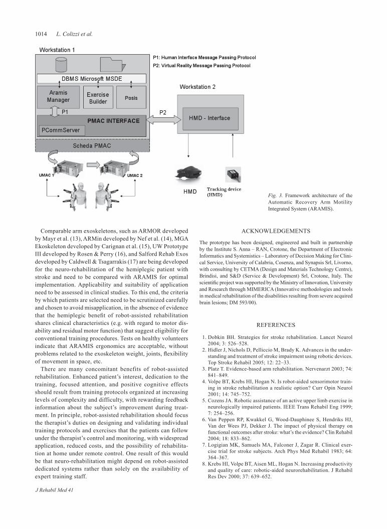
1014 L. Colizzi et al.
Comparable arm exoskeletons, such as ARMOR developed by Mayr et al. (13), ARMin developed by Nef et al. (14), MGA Ekoskeleton developed by Carignan et al. (15), UW Prototype III developed by Rosen & Perry (16), and Salford Rehab Exos developed by Caldwell & Tsagarrakis (17) are being developed for the neuro-rehabilitation of the hemiplegic patient with stroke and need to be compared with ARAMIS for optimal implementation. Applicability and suitability of application need to be assessed in clinical studies. To this end, the criteria by which patients are selected need to be scrutinized carefully and chosen to avoid misapplication, in the absence of evidence that the hemiplegic benefit of robot-assisted rehabilitation shares clinical characteristics (e.g. with regard to motor dis-ability and residual motor function) that suggest eligibility for conventional training procedures. Tests on healthy volunteers indicate that ARAMIS ergonomics are acceptable, without problems related to the exoskeleton weight, joints, flexibility of movement in space, etc.
There are many concomitant benefits of robot-assisted rehabilitation. Enhanced patient’s interest, dedication to the training, focused attention, and positive cognitive effects should result from training protocols organized at increasing levels of complexity and difficulty, with rewarding feedback information about the subject’s improvement during treat-ment. In principle, robot-assisted rehabilitation should focus the therapist’s duties on designing and validating individual training protocols and exercises that the patients can follow under the therapist’s control and monitoring, with widespread application, reduced costs, and the possibility of rehabilita-tion at home under remote control. One result of this would be that neuro-rehabilitation might depend on robot-assisted dedicated systems rather than solely on the availability of expert training staff.
ACKNOWLEDGEMENTS
The prototype has been designed, engineered and built in partnership by the Institute S. Anna – RAN, Crotone, the Department of Electronic Informatics and Systemistics – Laboratory of Decision Making for Clini-cal Service, University of Calabria, Cosenza, and Synapsis Srl, Livorno, with consulting by CETMA (Design and Materials Technology Centre), Brindisi, and S&D (Service & Development) Srl, Crotone, Italy. The scientific project was supported by the Ministry of Innovation, University and Research through MIMERICA (Innovative methodologies and tools in medical rehabilitation of the disabilities resulting from severe acquired brain lesions; DM 593/00).
REFERENCES
Dobkin BH. Strategies for stroke rehabilitation. Lancet Neurol 1. 2004; 3: 526–528.Hidler J, Nichols D, Pelliccio M, Brady K. Advances in the under-2. standing and treatment of stroke impairment using robotic devices. Top Stroke Rehabil 2005; 12: 22–33. Platz T. Evidence-based arm rehabilitation. Nervenarzt 2003; 74: 3. 841–849.Volpe BT, Krebs HI, Hogan N. Is robot-aided sensorimotor train-4. ing in stroke rehabilitation a realistic option? Curr Opin Neurol 2001; 14: 745–752.Cozens JA. Robotic assistance of an active upper limb exercise in 5. neurologically impaired patients. IEEE Trans Rehabil Eng 1999; 7: 254–256.Van Peppen RP, Kwakkel G, Wood-Dauphinee S, Hendriks HJ, 6. Van der Wees PJ, Dekker J. The impact of physical therapy on functional outcomes after stroke: what’s the evidence? Clin Rehabil 2004; 18: 833–862.Logigian MK, Samuels MA, Falconer J, Zagar R. Clinical exer-7. cise trial for stroke subjects. Arch Phys Med Rehabil 1983; 64: 364–367.Krebs HI, Volpe BT, Aisen ML, Hogan N. Increasing productivity 8. and quality of care: robotic-aided neurorehabilitation. J Rehabil Res Dev 2000; 37: 639–652.
Fig. 3. Framework architecture of the Automatic Recovery Arm Motility Integrated System (ARAMIS).
J Rehabil Med 41
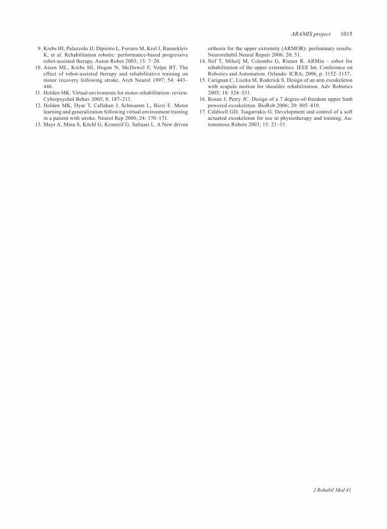
1015ARAMIS project
Krebs HI, Palazzolo JJ, Dipietro L, Ferraro M, Krol J, Rannekleiv 9. K, et al. Rehabilitation robotic: performance-based progressive robot-assisted therapy. Auton Robot 2003; 15: 7–20. Aisen ML, Krebs HI, Hogan N, McDowel F, Volpe BT. The 10. effect of robot-assisted therapy and rehabilitative training on motor recovery following stroke. Arch Neurol 1997; 54: 443–446.Holden MK. Virtual enviroments for motor rehabilitation: review. 11. Cyberpsychol Behav 2005; 8: 187–211.Holden MK, Dyar T, Callahan J, Schwamm L, Bizzi E. Motor 12. learning and generalization following virtual environment training in a patient with stroke. Neurol Rep 2000; 24: 170–171. Mayr A, Mina S, Köchl G, Kronreif G, Saltuari L. A New driven 13.
orthosis for the upper extremity (ARMOR): preliminary results. Neurorehabil Neural Repair 2006; 20; 51.Nef T, Mihelj M, Colombo G, Riener R. ARMin – robot for 14. rehabilitation of the upper extremities. IEEE Int. Conference on Robotics and Automation. Orlando: ICRA; 2006, p. 3152–3157.Carignan C, Liszka M, Roderick S. Design of an arm exoskeleton 15. with scapula motion for shoulder rehabilitation. Adv Robotics 2005; 18: 524–531.Rosen J, Perry JC. Design of a 7 degree-of-freedom upper limb 16. powered exoskeleton. BioRob 2006; 20: 805–810.Caldwell GD, Tsagarrakis G. Development and control of a soft 17. actuated exoskeleton for use in physiotherapy and training. Au-tonomous Robots 2003; 15: 21–33.
J Rehabil Med 41
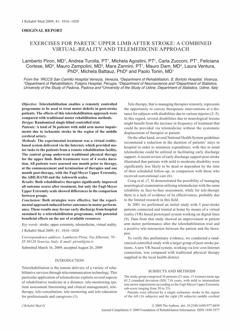
J Rehabil Med 41
oriGinal report
J Rehabil Med 2009; 41: 1016–1020
© 2009 The Authors. doi: 10.2340/16501977-0459Journal Compilation © 2009 Foundation of Rehabilitation Information. ISSN 1650-1977
Objective: telerehabilitation enables a remotely controlled programme to be used to treat motor deficits in post-stroke patients. the effects of this telerehabilitation approach were compared with traditional motor rehabilitation methods.Design: randomized single-blind controlled trial.Patients: a total of 36 patients with mild arm motor impair-ments due to ischaemic stroke in the region of the middle cerebral artery.Methods: the experimental treatment was a virtual reality-based system delivered via the internet, which provided mo-tor tasks to the patients from a remote rehabilitation facility. the control group underwent traditional physical therapy for the upper limb. both treatments were of 4 weeks dura-tion. all patients were assessed one month prior to therapy, at the commencement and termination of therapies and one month post-therapy, with the fugl-meyer upper extremity, the abilhanD and the ashworth scales.Results: Both rehabilitative therapies significantly improved all outcome scores after treatment, but only the fugl-meyer upper extremity scale showed differences in the comparison between groups.Conclusion: both strategies were effective, but the experi-mental approach induced better outcomes in motor perform-ance. these results may favour early discharge from hospital sustained by a telerehabilitation programme, with potential beneficial effects on the use of available resources.Key words: stroke, upper extremity, telemedicine, virtual reality.J Rehabil Med 2009; 41: 1016–1020
Correspondence address: Lamberto Piron, Via Alberoni, 70 – IT-30126 Venezia, Italy. E-mail: [email protected] March 16, 2009; accepted August 26, 2009
INTRODUCTION
Telerehabilitation is the remote delivery of a variety of reha-bilitative services through telecommunication technology. This particular application of telemedicine exploits several aspects of rehabilitative medicine at a distance: tele-monitoring (pa-tient assessment functioning and clinical management), tele-therapy, tele-consultation, tele-mentoring and tele-education for professionals and caregivers (1).
Tele-therapy, that is managing therapies remotely, represents the opportunity to convey therapeutic interventions at a dis-tance for subjects with disabilities due to various injuries (2–5). In this regard, several disabilities due to neurological lesions might benefit from the increase in frequency of treatment that could be provided via telemedicine without the systematic displacement of therapist or patient.
On the other hand, several National Health System guidelines recommend a reduction in the duration of patients’ stays in hospital in order to minimize expenditure; with this in mind telemedicine could be utilized in facilitating early discharge support. A recent review of early discharge support post-stroke illustrated that patients with mild to moderate disability were significantly less likely to be dead or dependent by the time of their scheduled follow-up, in comparison with those who received conventional care (6).
Craig et al. (7, 8) demonstrated the possibility of managing neurological examination utilizing telemedicine with the same reliability as face-to-face assessment, while for tele-therapy there is a lack of evidence of its effectiveness, probably due to the limited research in this field.
In 2001 we performed an initial study with 5 post-stroke patients connected and treated at home by means of a virtual reality (VR) based prototypal system working on digital lines (9). Data from that study showed an improvement in patient arm motor performance after the telerehabilitation trial and a positive tele-interaction between the patient and the thera-pist.
To verify this preliminary evidence, we conducted a rand-omized controlled study with a larger group of post-stroke pa-tients. A new VR-based system, working via low-cost Internet connection, was compared with traditional physical therapy supplied in the local health-district.
SUBJECTS AND METHODSThe study group comprised 36 patients (21 men, 15 women) mean age 65.2 (standard deviation (SD) 7.8) years, with mild to intermediate arm motor impairment (according to the Fugl-Meyer Upper Extremity sub-score ranging from 30 to 55).
Patients were affected by a single ischaemic stroke in the region of the left (16 subjects) and the right (20 subjects) middle cerebral
EXERCISES FOR PARETIC UPPER LIMB AFTER STROKE: A COMBINED VIRTUAL-REALITY AND TELEMEDICINE APPROACH
Lamberto Piron, MD1, Andrea Turolla, PT1, Michela Agostini, PT1, Carla Zucconi, PT1, Feliciana Cortese, MD2, Mauro Zampolini, MD3, Mara Zannini, PT1, Mauro Dam, MD4, Laura Ventura,
PhD5, Michela Battauz, PhD6 and Paolo Tonin, MD1
From the 1IRCCS San Camillo Hospital Venezia, Venezia, 2Department of Rehabilitation, S. Bortolo Hospital, Vicenza, 3Department of Rehabilitation, Foligno Hospital, Perugia, 4Department of Neuroscience and 5Department of Statistics, University of the Study of Padova, Padova and 6University of the Study of Udine, Department of Statistics, Udine, Italy
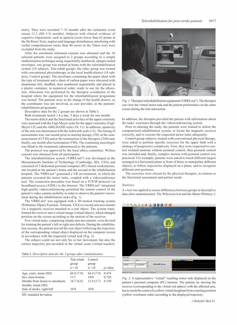
1017Telerehabilitation for post-stroke patients
artery. They were recruited 7–32 months after the ischaemic event (mean 13.3 (SD 5.5) months). Subjects with clinical evidence of cognitive impairment, such as apraxia (score lower than 62 points at the De Renzi Test), neglect and language disturbances interfering with verbal comprehension (more than 40 errors in the Token test) were excluded from the study.
After the enrolment informed consent was obtained and the 36 selected patients were assigned to 2 groups according to a simple randomization technique using sequentially numbered, opaque sealed envelopes: one group was treated at home with the telerehabilitation system (18 subjects, Tele-rehab group), the other group was treated with conventional physiotherapy in the local health-district (18 sub-jects, Control group). The envelopes containing the paper sheet with the type of treatment and a sheet of carbon paper were obscured with aluminium foil, shuffled, then numbered sequentially and placed in a plastic container, in numerical order, ready to use for the alloca-tion. Allocation was performed by the therapist coordinator of the hospital where the equipment for the telerehabilitation programme was hosted. The patients were in the charge of the health district, so the coordinator was not involved, as care provider, in the patients’ rehabilitation programme.
Descriptive data for the 2 groups are shown in Table I.Both treatments lasted 1 h a day, 5 days a week for one month.The motor deficit and the functional activities of the upper extremity
were assessed with the Fugl-Meyer scale for the upper extremity (Fugl-Meyer UE) and the ABILHAND scale (10, 11). In addition, spasticity of the arm was determined with the Ashworth scale (12). The timing of assessments was: one month prior to starting therapy (T0), at the com-mencement of (T30) and at the termination of the therapies (T60) and, finally, one month after termination (T90). The examining neurologist was blind to the treatments administered to the patients.
The protocol was approved by the local ethics committee. Written consent was obtained from all participants.
The telerehabilitation system (VRRS.net®) was developed at the Massachusetts Institute of Technology (Cambridge, MA, USA) and consisted of 2 dedicated personal computer (PC)-based workstations, one located at the patient’s home and the second at the rehabilitation hospital. The VRRS.net® generated a VR environment, in which the patients executed the motor tasks, coupled with a videoconference tool. The connection procedure was based on a TCP/IP protocol via broadband access (ADSL) to the Internet. The VRRS.net® integrated high-quality videoconferencing permitted the remote control of the patient’s video-camera mobility in order to observe the patient’s move-ment during the rehabilitation tasks (Fig. 1).
The VRRS.net® was equipped with a 3D motion tracking system (Polhemus 3Space Fastrack, Vermont, USA) to record arm movements via a magnetic receiver attached to a real object. The system trans-formed the receiver into a virtual image (virtual object), which changed position on the screen according to the motion of the receiver.
Five virtual tasks, comprising simple arm movements, were devised for training the patient’s left or right arm deficits. During the rehabilita-tion session, the patient moved the real object following the trajectory of the corresponding virtual object displayed on the computer screen in accordance with the requested virtual task (Fig. 2).
The subject could see not only his or her movement, but also the correct trajectory pre-recorded in the virtual scene (virtual teacher).
In addition, the therapist provided the patient with information about the tasks’ exactness through the videoconferencing system.
Prior to entering the study, the patients were trained to utilize the computerized rehabilitation system, to locate the magnetic receiver correctly, and to execute the requested motor tasks adequately.
Control group subjects, treated with conventional physical therapy, were asked to perform specific exercises for the upper limb with a strategy of progressive complexity. First, they were requested to con-trol isolated motions without postural control, then postural control was included and, finally, complex motion with postural control was practiced. For example, patients were asked to touch different targets arranged in a horizontal plane in front of them; to manipulate different objects; to follow trajectories displayed on a plane; and to recognize different arm positions.
The exercises were chosen by the physical therapist, in relation to the functional assessment and patient needs.
StatisticsA t-test was applied to assess differences between groups in descriptive data after randomization. The Wilcoxon test and the Mann-Whitney U
Table I. Descriptive data for the 2 groups after randomization
Tele-rehab group n = 18
Control groupn = 18 p-value
Age, years, mean (SD) 66.0 (7.9) 64.4 (7.9) 0.474Sex, men/women 11/7 10/8 0.720Months from lesion to enrolment, months, mean (SD)
14.7 (6.6) 11.9 (3.7) 0.150
Side of stroke, right/left 10/8 10/8
SD: standard deviation.
Fig. 1. Therapist telerehabilitation equipment (VRRS.net®). The therapist can view the virtual motor task and the patient performance on the same screen during the tele-interaction.
Fig. 2. A representative “virtual” reaching motor task displayed on the patient’s personal computer (PC) monitor. The patient, by moving the receiver (corresponding to the virtual red sphere) with the affected arm, has to reach the centre of a yellow virtual doughnut from a starting position (yellow wireframe cube) according to the displayed trajectory.
J Rehabil Med 41
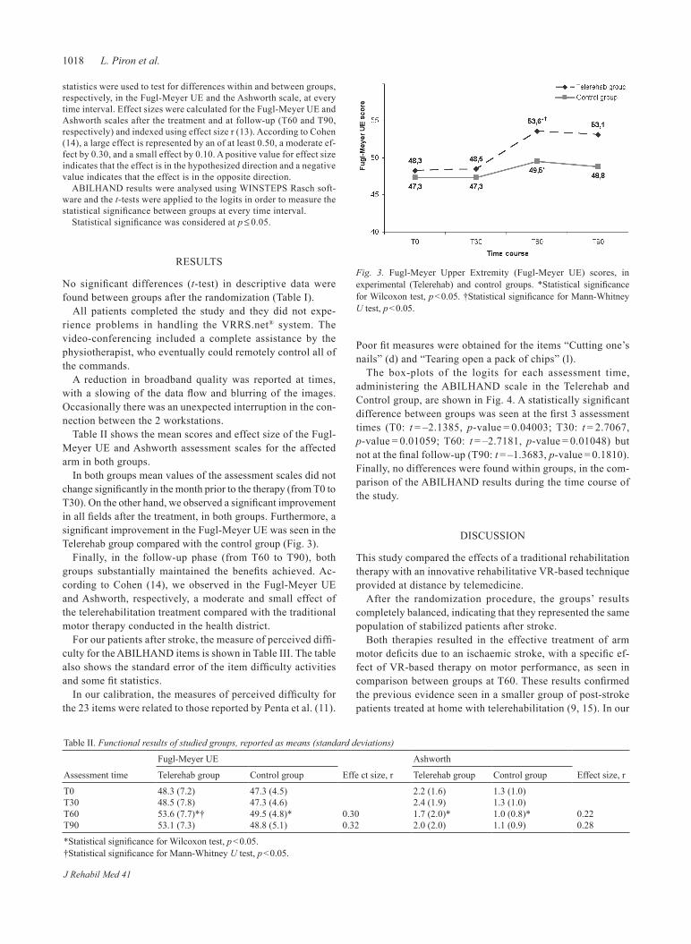
1018 L. Piron et al.
statistics were used to test for differences within and between groups, respectively, in the Fugl-Meyer UE and the Ashworth scale, at every time interval. Effect sizes were calculated for the Fugl-Meyer UE and Ashworth scales after the treatment and at follow-up (T60 and T90, respectively) and indexed using effect size r (13). According to Cohen (14), a large effect is represented by an of at least 0.50, a moderate ef-fect by 0.30, and a small effect by 0.10. A positive value for effect size indicates that the effect is in the hypothesized direction and a negative value indicates that the effect is in the opposite direction.
ABILHAND results were analysed using WINSTEPS Rasch soft-ware and the t-tests were applied to the logits in order to measure the statistical significance between groups at every time interval.
Statistical significance was considered at p ≤ 0.05.
RESULTS
No significant differences (t-test) in descriptive data were found between groups after the randomization (Table I).
All patients completed the study and they did not expe-rience problems in handling the VRRS.net® system. The video-conferenc ing included a complete assistance by the physiotherapist, who eventually could remotely control all of the commands.
A reduction in broadband quality was reported at times, with a slowing of the data flow and blurring of the images. Occasionally there was an unexpected interruption in the con-nection between the 2 workstations.
Table II shows the mean scores and effect size of the Fugl-Meyer UE and Ashworth assessment scales for the affected arm in both groups.
In both groups mean values of the assessment scales did not change significantly in the month prior to the therapy (from T0 to T30). On the other hand, we observed a significant improvement in all fields after the treatment, in both groups. Furthermore, a significant improvement in the Fugl-Meyer UE was seen in the Telerehab group compared with the control group (Fig. 3).
Finally, in the follow-up phase (from T60 to T90), both groups substantially maintained the benefits achieved. Ac-cording to Cohen (14), we observed in the Fugl-Meyer UE and Ashworth, respectively, a moderate and small effect of the telerehabilitation treatment compared with the traditional motor therapy conducted in the health district.
For our patients after stroke, the measure of perceived diffi-culty for the ABILHAND items is shown in Table III. The table also shows the standard error of the item difficulty activities and some fit statistics.
In our calibration, the measures of perceived difficulty for the 23 items were related to those reported by Penta et al. (11).
Poor fit measures were obtained for the items “Cutting one’s nails” (d) and “Tearing open a pack of chips” (l).
The box-plots of the logits for each assessment time, administering the ABILHAND scale in the Telerehab and Control group, are shown in Fig. 4. A statistically significant difference between groups was seen at the first 3 assessment times (T0: t = –2.1385, p-value = 0.04003; T30: t = 2.7067, p-value = 0.01059; T60: t = –2.7181, p-value = 0.01048) but not at the final follow-up (T90: t = –1.3683, p-value = 0.1810). Finally, no differences were found within groups, in the com-parison of the ABILHAND results during the time course of the study.
DISCUSSION
This study compared the effects of a traditional rehabilitation therapy with an innovative rehabilitative VR-based technique provided at distance by telemedicine.
After the randomization procedure, the groups’ results completely balanced, indicating that they represented the same population of stabilized patients after stroke.
Both therapies resulted in the effective treatment of arm motor deficits due to an ischaemic stroke, with a specific ef-fect of VR-based therapy on motor performance, as seen in comparison between groups at T60. These results confirmed the previous evidence seen in a smaller group of post-stroke patients treated at home with telerehabilitation (9, 15). In our
Table II. Functional results of studied groups, reported as means (standard deviations)
Assessment time
Fugl-Meyer UE
Effe ct size, r
Ashworth
Effect size, rTelerehab group Control group Telerehab group Control group
T0 48.3 (7.2) 47.3 (4.5) 2.2 (1.6) 1.3 (1.0)T30 48.5 (7.8) 47.3 (4.6) 2.4 (1.9) 1.3 (1.0)T60 53.6 (7.7)*† 49.5 (4.8)* 0.30 1.7 (2.0)* 1.0 (0.8)* 0.22T90 53.1 (7.3) 48.8 (5.1) 0.32 2.0 (2.0) 1.1 (0.9) 0.28
*Statistical significance for Wilcoxon test, p < 0.05.†Statistical significance for Mann-Whitney U test, p < 0.05.
Fig. 3. Fugl-Meyer Upper Extremity (Fugl-Meyer UE) scores, in experimental (Telerehab) and control groups. *Statistical significance for Wilcoxon test, p < 0.05. †Statistical significance for Mann-Whitney U test, p < 0.05.
J Rehabil Med 41
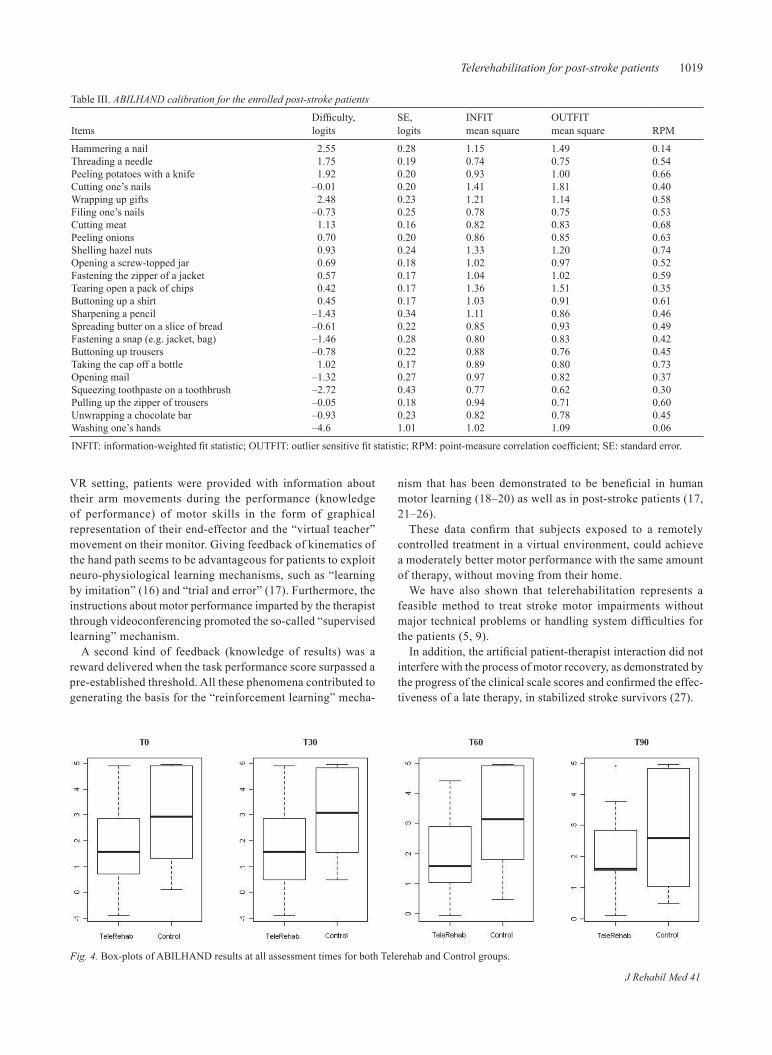
1019Telerehabilitation for post-stroke patients
VR setting, patients were provided with information about their arm movements during the performance (knowledge of performance) of motor skills in the form of graphical representation of their end-effector and the “virtual teacher” movement on their monitor. Giving feedback of kinematics of the hand path seems to be advantageous for patients to exploit neuro-physiological learning mechanisms, such as “learning by imitation” (16) and “trial and error” (17). Furthermore, the instructions about motor performance imparted by the therapist through videoconferencing promoted the so-called “supervised learning” mechanism.
A second kind of feedback (knowledge of results) was a reward delivered when the task performance score surpassed a pre-established threshold. All these phenomena contributed to generating the basis for the “reinforcement learning” mecha-
nism that has been demonstrated to be beneficial in human motor learning (18–20) as well as in post-stroke patients (17, 21–26).
These data confirm that subjects exposed to a remotely controlled treatment in a virtual environment, could achieve a moderately better motor performance with the same amount of therapy, without moving from their home.
We have also shown that telerehabilitation represents a feasible method to treat stroke motor impairments without major technical problems or handling system difficulties for the patients (5, 9).
In addition, the artificial patient-therapist interaction did not interfere with the process of motor recovery, as demonstrated by the progress of the clinical scale scores and confirmed the effec-tiveness of a late therapy, in stabilized stroke survivors (27).
Table III. ABILHAND calibration for the enrolled post-stroke patients
ItemsDifficulty,logits
SE,logits
INFITmean square
OUTFITmean square RPM
Hammering a nail 2.55 0.28 1.15 1.49 0.14Threading a needle 1.75 0.19 0.74 0.75 0.54Peeling potatoes with a knife 1.92 0.20 0.93 1.00 0.66Cutting one’s nails –0.01 0.20 1.41 1.81 0.40Wrapping up gifts 2.48 0.23 1.21 1.14 0.58Filing one’s nails –0.73 0.25 0.78 0.75 0.53Cutting meat 1.13 0.16 0.82 0.83 0.68Peeling onions 0.70 0.20 0.86 0.85 0.63Shelling hazel nuts 0.93 0.24 1.33 1.20 0.74Opening a screw-topped jar 0.69 0.18 1.02 0.97 0.52Fastening the zipper of a jacket 0.57 0.17 1.04 1.02 0.59Tearing open a pack of chips 0.42 0.17 1.36 1.51 0.35Buttoning up a shirt 0.45 0.17 1.03 0.91 0.61Sharpening a pencil –1.43 0.34 1.11 0.86 0.46Spreading butter on a slice of bread –0.61 0.22 0.85 0.93 0.49Fastening a snap (e.g. jacket, bag) –1.46 0.28 0.80 0.83 0.42Buttoning up trousers –0.78 0.22 0.88 0.76 0.45Taking the cap off a bottle 1.02 0.17 0.89 0.80 0.73Opening mail –1.32 0.27 0.97 0.82 0.37Squeezing toothpaste on a toothbrush –2.72 0.43 0.77 0.62 0.30Pulling up the zipper of trousers –0.05 0.18 0.94 0.71 0.60Unwrapping a chocolate bar –0.93 0.23 0.82 0.78 0.45Washing one’s hands –4.6 1.01 1.02 1.09 0.06
INFIT: information-weighted fit statistic; OUTFIT: outlier sensitive fit statistic; RPM: point-measure correlation coefficient; SE: standard error.
Fig. 4. Box-plots of ABILHAND results at all assessment times for both Telerehab and Control groups.
J Rehabil Med 41
1020 L. Piron et al.
Both groups retained all the outcome values 30 days after the termination of treatment, indicating that both strategies induce changes in motor behaviour that endure with time.
These observations may underestimate the effect of the physical presence of the therapist and may reinforce the hy-pothesis that adequate feedback and the supply of real-time therapist interaction may represent the key factors in the proc-esses of motor recovery.
In both groups, the subjective perceived manual ability showed a constant, although small, improvement during the whole time course of the study, with maintenance at the follow-up, as indicated by the analysis of the reported answers in the ABILHAND scale. These results, together with the improve-ment in the Fugl-Meyer UE, demonstrate how the continuity of care bettered objective and subjective outcomes in discharged and stabilized stroke patients.
In conclusion, the results of this study indicate that mild to intermediate post-stroke patients may undergo a telerehabili-tation programme so improve their motor deficits. This fact may favour an early discharge from hospital and a subsequent rehabilitative intervention at home, which do not compromise clinical outcomes after a stroke and may have beneficial effects on quality of life.
Further research is necessary to evaluate the cost-effective-ness of this type of approach in telemedicine.
REFERENCES
Forducey PG, Ruwe WD, Dawson SJ, Scheideman-Miller C, 1. McDonald NB, Hantla MR. Using telerehabilitation to promote TBI recovery and transfer of knowledge. NeuroRehabil 2003; 18: 103–111.Burns RB, Crislip D, Daviou P, Temkin A, Vesmarovich S, Anshutz 2. J. Using telerehabilitation to support assistive technology. Assistive Technol 1998; 10: 126–133.Reinkensmeyer DJ, Pang CT, Nessler JA, Painter CC. Web-based 3. telerehabilitation for the upper extremity after stroke. IEEE Trans Neural Syst Rehabil Eng 2002; 10: 102–108.Burdea G, Popescu V, Henz V, Colbert K. Virtual reality based 4. orthopedic telerehabilitation. IEEE Trans Neural Syst Rehabil Eng 2000; 8: 430–432.Piron L, Tonin P, Atzori AM, Zanotti E, Trivello E, Dam M. Virtual 5. environment system for motor tele-rehabilitation. In: Westwood JD, Hoffman HF, Robb RA, Stredney D, editors. Medicine meets virtual reality 2002. Amsterdam: IOS Press; 2002, p. 355–361.Langhorne P, Widen-Holmqvist L. Early support discharge after 6. stroke. J Rehabil Med 2007; 39: 103–108.Craig JJ, McConville JP, Patterson VH, Wootton R. Neurological 7. examination is possible using telemedicine. J Telemed Telecare 1999; 5: 177–181.Craig J, Patterson V, Russell C, Wootton R. Interactive videocon-8.
sultation is a feasible method for neurological in-patient assess-ment. Eur J Neurol 2000; 7: 699–702.Piron L, Tonin P, Trivello E, Battistin L, Dam M. Motor tele-9. rehabilitation in post-stroke patients. Med Inform Internet Med 2004; 29: 119–125.Fugl-Meyer R, Jaasko L, Leyman L, Olsson S, Steglind S. The 10. post-stroke hemiplegic patient. A method for evaluation of physical performance. Scand J Rehab Med 1975; 7: 13–31.Penta M, Tesio L, Arnould C, Zancan A, Thonnard JL. The ABIL-11. HAND Questionnaire as a measure of manual ability in chronic stroke patients Rasch-based validation and relationship to upper limb impairment. Stroke 2001; 32: 1627–1634.Bohannon RW, Smith MB. Interrater reliability of a modified Ash-12. worth scale of muscle spasticity. Phys Ther 1987; 67: 206–207.Rosenthal R, Rosnow RL Essentials of behavioral research: 13. methods and data analysis. 2nd edn. New York, NY: McGraw-Hill; 1991.Cohen J, editor. Statistical power analysis for the behavioral 14. sciences. 2nd ed. Hillsdale, NJ: Lawrence Erlbaum Associates; 1988.Piron L, Tonin P, Cortese F, Zampolini M, Piccione F, Agostini M, 15. et al. Post-stroke arm motor telerehabilitation web-based. Proceed-ings of IEEE 5th International Workshop Virtual Rehabilitation. New Jersey: IET (IEEE) Press; 2006, p. 145–148. Russon AE. Learning by imitation: a hierarchical approach. Behav 16. Brain Sci 1998; 21: 667–684.Piron L, Tonin P, Piccione F, Iaia V, Trivello E, Dam M. Virtual 17. environment training therapy for arm motor rehabilitation. Pres-ence 2005; 6: 732–740.Winstein CJ. Knowledge of results and motor learning implications 18. for physical therapy. Phys Ther 1991; 71: 140–149.Young DE, Schmidt RA. Augmented kinematic feedback for motor 19. learning. J Mot Behav 1992; 24: 261–273.Todorov E, Shadmehr R, Bizzi E. Augmented feedback presented 20. in a virtual environment accelerates learning of a difficult motor task. J Motor Behav 1997; 29: 147–158.Winstein CJ, Pohl PS, Cardinale C, Green A, Scholtz L, Waters 21. CS. Learning a partial-weight-bearing skill: effectiveness of two forms of feedback. Phys Ther 1996; 76: 985–993.Cirstea CM, Ptito A, Levin MF. Feedback and cognition in arm mo-22. tor skill reacquisition after stroke. Stroke 2006; 37: 1237–1242.Woldag H, Hummelsheim H. Evidence-based physiotherapeutic 23. concepts for improving arm and hand function in stroke patients: a review. J Neurol 2002; 249: 518–528.Piron L, Tonin P, Atzori AM, Zucconi C, Massaro C, Trivello E, et 24. al. The augmented-feedback rehabilitation technique facilitates the arm motor recovery in patients after a recent stroke. Stud Health Technol Inform 2003; 94: 265–267.You SH, Jang SH, Kim YH, Hallett M, Ahn SH, Kwon YH, et 25. al. Virtual reality-induced cortical reorganization and associated locomotor recovery in chronic stroke: an experimenter-blind randomized study. Stroke 2005; 36: 1166–1171. Holden MK. Virtual environments for motor rehabilitation: review. 26. Cyberpsychol Behav 2005; 8: 187–211.Page SJ, Gater DR, Bach-Y-Rita P. Reconsidering the motor 27. recovery plateau in stroke rehabilitation. Arch Phys Med Rehabil 2004; 85: 1377–1381.
J Rehabil Med 41
information and instructions to the authors – Journal of rehabilitation medicine(extended instructions are available at http://www.medicaljournals.se/jrm)
preparing for submissionSubmission of a manuscript is held to imply that it has not previously been published and is not otherwise submitted for publication, except as an abstract (which in that case has to be stated).
Authorship. All persons designated as authors must participate suf-ficiently in the work to take public responsibility in its contents. For criteria for authors see “Uniform requirements for manuscripts submitted to biomedical journals” Ann Intern Med 1997; 126: 36–47, or JAMA 1997; 277: 927–34. The full document is available at www.icmje.org. If 5 authors or more, a statement on each author’s contribution must be described in the accompanying letter.
Length and number of copies. Articles should not exceed 9 printed pages (approx. 25 double-spaced manuscript pages incl. the reference list). Additional pages must be paid for by the authors at a rate of USD160, Euro 140 per page.
Submission can be made online at http://www.medicaljournals.se/jrm. (A complete copy of the manuscript, including tables and figures, accompanied by a submission letter can also be sent via ordinary mail if the internet option is not possible. In that case, please also submit a floppy disc with the complete manuscript). For further information please contact the Editorial office.
Language. All papers should be written in English. It is essential that the language is irreproachable.
Conflict of interest and funding. Authors are responsible for recognising and disclosing financial and other conflicts of interest that might bias their work. They should acknowledge in the manuscript all financial support for the work and other financial or personal connections to the work.
Ethics and consent. When reporting studies on human subjects, indicate whether the procedures followed have been approved by an ethic com-mittee (in accordance with ethical standards on human experimentation and with the Helsinki Declaration of 1975, as revised in 1983). Do not use patients’ names, initials, or hospital numbers, especially in illustrative material. Identifying information should not be published in written de-scriptions, photographs, and pedigrees unless the information is essential for scientific purposes and the patient (or parent or guardian) gives writ-ten informed consent for publication. Informed consent for this purpose requires that the patient be shown the manuscript to be published.
Copyright. It is a condition of publication that author vest copyright in their articles, including abstracts, in Foundation of Rehabilitation Medi-cine (Journal of Rehabilitation Medicine). Another option is to sign “An exclusive licence to publish” and maintain the ownership of copyright. For further information regarding copyright please visit http:// www.medicaljournals.se/jrm (Instructions to authors). To view the ‘Copyright Transfer Frequently Asked Questions’ please visit http://www.tandf.co.uk/journals/copyright.asp.
arrangement of manuscriptManuscripts should be typewritten, double-spaced throughout, on one side of the paper, with ordinary margins. Papers should normally be di-vided into Title page, Abstract, Introduction (including clear statement of the aim of the study), Methods, Results, Discussion, and References.
Title page: A separate title page is necessary and should beara) the title of the article,b) names of the authors,c) the institutions of origin,d) a short title, and the corresponding author’s name, address, telefax number and e-mail.
Abstract: Journal of Rehabilitation Medicine requires structured ab-stracts for all original articles. The following headlines should be used when appropriate: Objective, Design, Subjects/Patients, Methods, Results and Conclusion. The total number of words should not exceed 200. A list of from 3–8 key-words or terms from Medical Subject Headings (Bethesda, Md. 1966) suitable for indexing terms should be typed at the bottom of the abstract page.Below the abstract: Title of journal; The principal author’s complete address, including e-mail.
Statistics. Describe statistical methods with enough detail to enable a knowledgeable reader with access to the original data to verify the reported results. When possible, quantify findings and present them with appropriate indicators of measurement error or uncertainty (such as confidence intervals). Avoid relying solely on statistical hypothesis testing, such as the use of p-values, which fails to convey important quantitative information. Calculations of power should be performed when appropriate. For analysis of reliability use adequate methods. Statistical methods for data from rating scales must take account of the rank-invariant properties of ordinal data. The median level and the quartiles are appropriate measure for describing the distribution of ordinal data. Discuss the eligibility of experimental subjects. For randomised controlled trials (RCT) the authors should follow the guidelines of report-ing RCT given by CONSORT (Begg et al. JAMA 996;276:7-9). When analyzing several outcome variables and/or investigating the relationships between many variables consider to use multivariate statistical methods. Describe the methods for and success of any blinding of observations. Report losses to observation (such as dropouts from a clinical trial). References for the design of the study and statistical methods should be to standard works when possible (with pages stated) rather than to papers in which the designs or methods were originally reported. Specify any general-use computer programs used.
References should be numbered consecutively in the order in which they are first mentioned in the text. Identify references in text by Arabic numerals in parenthesis as follows: “as shown by Smith (5)”:... if two authors; “Smith & Jones (6)”;... if more than two authors; “Smith et al. (7)”. The style of references must follow the Vancouver system and for the abbreviations of journal titles; please consult the List of Journals Indexed in Index Medicus, published annually as a list in the January issue of Index Medicus, also accessible at www.nlm.nih.gov.).
Reference to periodicals: Shah S, Vanclay F, Cooper B. Improving the sensitivity of the Barthel Index for stroke rehabilitation. J Clin Epidemiol 1989; 42: 709–710.
Reference to books: Keith RA, Granger CV, Hamilton BB, Sherwin FS. The functional independence measure: a new tool for rehabilitation. In: Eisenberg MG, Grzesiak RC, editors. Advances in clinical rehabilita-tion. New York: Springer Publishing; 1987, p. 6–18.
For journal article in electronic format use the following style: Morse SS. Factors in the emergence of infectious diseases. Emerg Infect Dis [serial online] 1995 Jan–Mar [cited 1996 Jun 5]; 1(1): [24 screens]. Available from: URL: http://www.cdc.gov\ncidod\EID\eid.htm
Tables and figures: Tables are to be numbered consecutively with roman numerals. Each table should be typed on a separate sheet and should have a descriptive heading which is self-explanatory.
All illustrations should be considered as figures. Each graph, drawing, or photograph should be numbered in sequence with Arabic numerals. All figures should have legends, listed on a separate sheet. Each figure should be identified by the name of the journal (JRM), the author’s name, and the figure number. Line drawing and lettering should be large enough to sustain photo-reduction.
proofs and offprintsPage proofs are sent directly from the printing office to the corresponding author, via e-mail as pdf-files. It is the authors full responsibility to read and check the proofs against the manuscript. The proof should be signed and returned to the Editorial Office within four days by airmail.
Offprints may be ordered on the offprint order form accompanying the page proof.
supplementsLengthy papers may be published as supplements, the full cost being borne by the author. Supplements are subject to editorial revision before publication.
Address of the Editorial OfficeJournal of Rehabilitation MedicineTrädgårdsgatan 14, SE-753 09, Uppsala, SwedenPhone: +46 18 580 50 90E-mail: [email protected]
contentsLucca, L.F., Castelli, E. & Sannita, W.G.: Foreword 953Pignolo, L.: Robotics in neuro-rehabilitation 955Squeri, V., Casadio, M., Vergaro, E., Giannoni, P., Morasso, P. & Sanguineti, V.: Bilateral robot therapy based on haptics and reinforcement learning: Feasibility study of a new concept for treatment of patients after stroke 961Carpinella, I., Cattaneo, D., Abuarqub, S. & Ferrarin, M.: Robot-based rehabilitation of the upper limbs in multiple sclerosis: Feasibility and preliminary results 966Bovolenta, F., Goldoni, M., Clerici, P., Agosti, M. & Franceschini, M.: Robot therapy for functional recovery of the upper limbs: A pilot study on patients after stroke 971Posteraro, F., Mazzoleni, S., Aliboni, S., Cesqui, B., Battaglia, A., Dario, P. & Micera, S.: Robot-mediated therapy for paretic upper limb of chronic patients following neurological injury 976Masiero, S., Carraro, E., Ferraro, C., Gallina, P., Rossi, A. & Rosati, G.: Upper limb rehabilitation robotics after stroke: A perspective from the University of Padua, Italy 981Bartolo, M., Don, R., Ranavolo, A., Serrao, M. & Sandrini, G.: Kinematic and neurophysiological models: Future applications in neurorehabilitation 986Frascarelli, F., Masia, L., Di Rosa, G., Petrarca, M., Cappa, P. & Castelli, E.: Robot-mediated and clinical scales evaluation after upper limb botulinum toxin type A injection in children with hemiplegia 988Petrarca, M., Zanelli, G., Patanè, F., Frascarelli, F., Cappa, P. & Castelli, E.: Reach-to-grasp interjoint coordination for moving object in children with hemiplegia 995Lucca, L.F.: Virtual reality and motor rehabilitation of the upper limb after stroke: A generation of progress? 1003
Dolce, G., Lucca, L.F. & Pignolo, L.: Robot-assisted rehabilitation of the paretic upper limb: Rationale of the ARAMIS project 1007Colizzi, L., Lidonnici, A. & Pignolo, L.: The ARAMIS project: A concept robot and technical design 1011
Piron, L., Turolla, A., Agostini, M., Zucconi, C., Cortese, F., Zampolini, M., Zannini, M., Dam, M., Ventura, L., Battauz, M. & Tonin, P.: Exercises for paretic upper limb after stroke: A combined virtual-reality and telemedicine approach 1016
supplementsfor further information see our website http://jrm.medicaljournals.se
no. 44, 2004. Stucki, G. & Grimby, G. (Eds.): ICF core sets for chronic conditions (144 pp.).
no. 45, 2007. Gutenbrunner, C., Ward, A.B. & Chamberlain M.A. (Eds.): White book on physical and rehabilitation medicine in Europe (48 pp.).
no. 46, 2008. Abstract book: Asian Oceania Conference of Physical and Rehabilitation Medicine, May 16–19, 2008, Nanjing, China (176 pp.).
no. 47, 2008. Abstract book: 16th European Congres of Physical and Rehabilitation Medicine, June 3–6, 2008, Brugge, Belgium (303 pp.).
Copyright © 2022 FDOKUMEN