Examining Children - docet
31
Examining Children
-
Upload
khangminh22 -
Category
Documents
-
view
4 -
download
0
Transcript of Examining Children - docet
Examining Children
Page 2 of 31 © DOCET
2
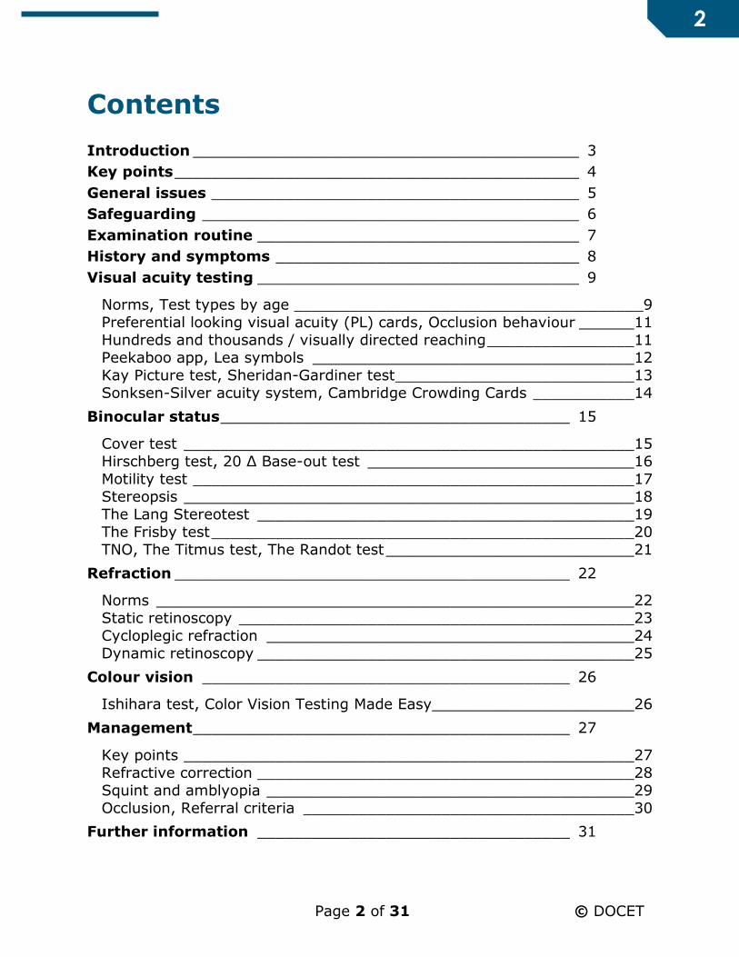
Contents
Introduction __________________________________________ 3
Key points ____________________________________________ 4
General issues ________________________________________ 5
Safeguarding _________________________________________ 6
Examination routine ___________________________________ 7
History and symptoms _________________________________ 8
Visual acuity testing ___________________________________ 9
Norms, Test types by age ______________________________________9
Preferential looking visual acuity (PL) cards, Occlusion behaviour ______11
Hundreds and thousands / visually directed reaching ________________11
Peekaboo app, Lea symbols ___________________________________12
Kay Picture test, Sheridan-Gardiner test __________________________13
Sonksen-Silver acuity system, Cambridge Crowding Cards ___________14
Binocular status ______________________________________ 15
Cover test _________________________________________________15
Hirschberg test, 20 Δ Base-out test _____________________________16
Motility test ________________________________________________17
Stereopsis _________________________________________________18
The Lang Stereotest _________________________________________19
The Frisby test ______________________________________________20
TNO, The Titmus test, The Randot test ___________________________21
Refraction ___________________________________________ 22
Norms ____________________________________________________22
Static retinoscopy ___________________________________________23
Cycloplegic refraction ________________________________________24
Dynamic retinoscopy _________________________________________25
Colour vision ________________________________________ 26
Ishihara test, Color Vision Testing Made Easy______________________26
Management _________________________________________ 27
Key points _________________________________________________27
Refractive correction _________________________________________28
Squint and amblyopia ________________________________________29
Occlusion, Referral criteria ____________________________________30
Further information __________________________________ 31
Page 3 of 31 © DOCET
3
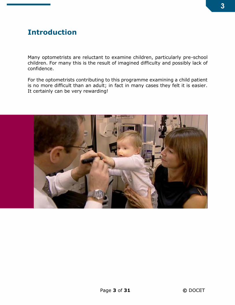
Introduction
Many optometrists are reluctant to examine children, particularly pre-school
children. For many this is the result of imagined difficulty and possibly lack of
confidence.
For the optometrists contributing to this programme examining a child patient
is no more difficult than an adult; in fact in many cases they felt it is easier.
It certainly can be very rewarding!
Page 4 of 31 © DOCET
4
Key points Examining children is NOT more difficult than examining adults
Create a child friendly environment:
o A low table and chairs for the waiting room
o Have a supply of toys, colouring materials etc.
Take a pragmatic approach
You may not be able to do every test at one visit:
o You may not be able to do retinoscopy
o You may not be able to do a subjective
o You may not be able to do a cover test
Abandon tests quickly if they are not successful and move on
A series of short visits may be more productive than one long one
Do not be afraid to re-book.
You must learn to trust your observations
Rely on objective not subjective findings
Results will vary – especially retinoscopy and cover test findings.
Absolute accuracy is not the key goal
0.50 D sphere errors or a small cyl not corrected is not important.
The structure of your routine needs to be fluid and adaptable
Base it on the presenting history Begin with something achievable, ie cover test or stereopsis.
Managing the parental expectations may be the hardest part
Address parents’ concerns:
o Why have you come?
o Are you worried?
o What are you worried about?
Don’t be dismissive.
Communicate with the child at their level
Talk to child NOT parents
Address child by name frequently Praise the child, don’t criticise.
Above all: MAKE IT FUN!
Page 5 of 31 © DOCET
5
General issues
Always bear in mind that to the child an optometric practice is an unfamiliar
and threatening environment. Much can be done to minimise the threatening
nature by adapting the practice to take into account the concerns a child
patient might have. If possible, adapt an area to be child friendly: bright
cheerful colours, furniture such as tables and chairs at a child’s height, toys,
books and drawing materials will all help to put the child at ease.
It is also important to communicate with the child on their level using language
that they will understand, but without being patronising. Talk to the child, rather than the parents, addressing them by their name frequently.
If in the consulting room the child is wary and intimidated, sitting the child,
particularly a pre-school child, on the parents lap may help to minimise the
threatening nature of the practice environment, the consulting room
equipment and indeed the optometrist themselves. A whole examination can
be conducted in this way.
Making the examination fun is in many respects the key to success. If you are
able to, try and turn the examination and its individual elements into a game.
The use of toys in the examination itself, as fixation objects for example, will
help foster this fun element.
Normal development of vision can only be established by comparing the
performance of a particular patient with the expected level of development for
a child of that age. The refractive findings, level of visual acuity and binocular
vision must be compared to the expected level of achievement for the child’s
peer group to establish normality or otherwise.
Page 6 of 31 © DOCET
6
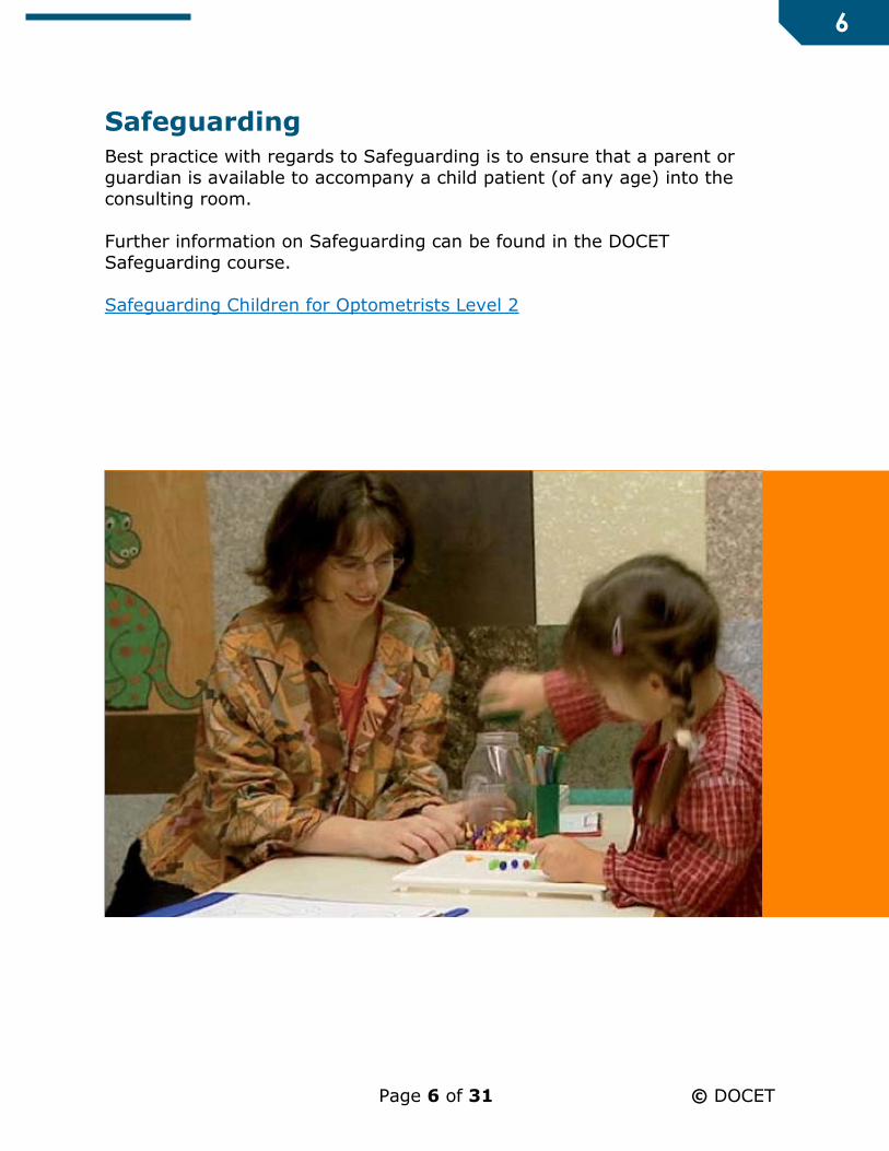
Safeguarding Best practice with regards to Safeguarding is to ensure that a parent or
guardian is available to accompany a child patient (of any age) into the
consulting room.
Further information on Safeguarding can be found in the DOCET
Safeguarding course.
Safeguarding Children for Optometrists Level 2
Page 7 of 31 © DOCET
7
Examination routine
Don’t expect your examination of a child patient to mimic the routine you
would use on an adult. Your routine needs to be flexible and adaptable and
you should be prepared to abandon a particular test if it is not successful and
move on to something else, or use an alternative technique. Indeed the order
in which tests are conducted might seem quite illogical. The only important
rule is that you do the tests that you believe are necessary; the order in which
they are done does not matter.
Base the structure of your routine on the presentation and the history of each individual patient.
It is important not to be frustrated or concerned about minor variations or
perceived lack of accuracy in the results of clinical investigations. It is
important, though, to have confidence and to be able to trust your own
observations. Absolute accuracy in the examination of a child patient is not
critical. A sphere which is 0.50 D out, or failure to correct a small cylinder, is
not going to affect the visual development of the child. It should be expected
that findings will fluctuate, particularly retinoscopy and cover test results.
Don’t expect to necessarily get all tests completed on one single visit.
Depending on the level of cooperation and alertness of the child this may not
be possible. Split the routine into smaller, more manageable elements that the child can cope with and re-book for another day to complete your
examination.
It will help the routine to run smoothly if the child has a success rather than
a failure early on. For that reason it may be better to pick something like the
cover test to begin rather than checking vision or visual acuity first. Any child
over 36 months should have stereopsis, so that may be a good place to start.
It is particularly important to be positive and encouraging to the child even if
they do not perform a test well, praising rather than criticising them. However
it is not necessary to pretend that every test has a successful result and
showing failure is acceptable. Do end the examination on a positive note, however, and finish with a success, even if it is necessary to return to a test
completed earlier. Finishing on a line of letters on the chart that you know the
child can see and praising the result works well and is a good standby.
Page 8 of 31 © DOCET
8
History and symptoms
History and symptoms will inevitably come from the parents who will be
expressing their own concerns about their child’s vision. The “symptoms” will
be largely based on parental observation of the child’s behaviour to give clues
to the state of the vision. So it is important to establish a few basic questions:
• Why have you brought your child in to be examined?
• Are you worried about your child’s vision?
• What are you worried specifically about?
Never underestimate or dismiss a parents concerns or observations; they are
rarely wrong.
It is helpful to ask questions relevant to a child patient and the symptoms they are likely to experience. For example, children rarely complain of symptoms
such as headaches and won’t experience diplopia.
Always bear in mind any risk factors that might apply and in particular relevant
family history, such as high refractive error and squint. Always obtain as much
information about an absent parent as diplomatically as possible, bearing in
mind there might be a relationship with the child but not the other parent.
Specific elements of the child’s history and the mother’s pregnancy may be of
great importance. Maternal illness during pregnancy, prematurity at birth, a
difficult or assisted delivery may have relevance for the development of the
child’s visual system.
Page 9 of 31 © DOCET
9
Visual acuity testing
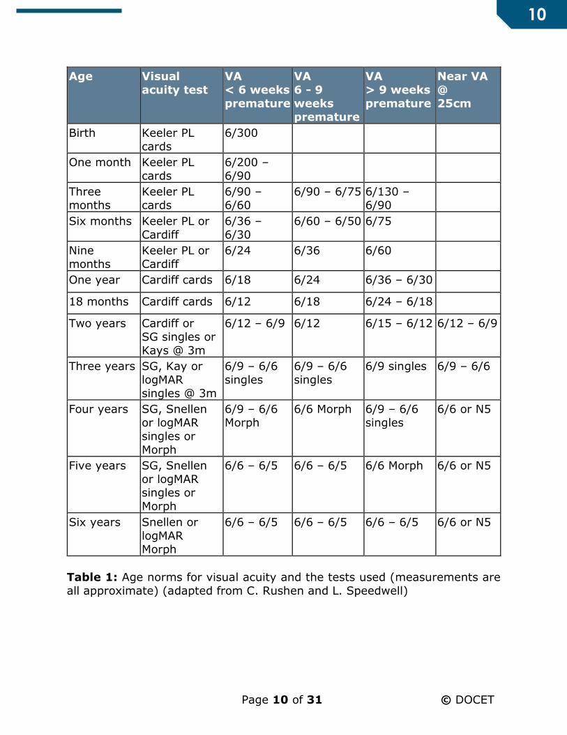
Norms
Table 1 below gives average expected visual acuity measures for children from
birth to six years of age. In young children visual acuity values tend to vary
depending which type of test is used.
The following reference provides an additional guide to testing the visual
acuity of young children: Saunders, K. (2010) Testing Visual Acuity of Young
Children: An Evidence-based Guide for Optometrists. Optometry in Practice,
11(4), 161-168.
Test types by age
Birth to 12 months
Preferential Looking Cards (Keeler Acuity Cards) – the best
Occlusion behaviour
Hundreds and thousands
Visually directed reaching
Peekaboo App by Glasgow Centre for Ophthalmic Research
12 months to 2.5 years
Cardiff Cards
Hundreds and thousands
Peekaboo App by Glasgow Centre for Ophthalmic Research
2.5 years onwards
Lea symbols
Kay pictures
Sheridan-Gardiner
Sonksen-Silver
Cambridge Crowding Cards
Crowded Kay pictures LogMAR acuity cards
Snellen Letters
Page 10 of 31 © DOCET
10
Age Visual
acuity test
VA
< 6 weeks
premature
VA
6 - 9
weeks
premature
VA
> 9 weeks
premature
Near VA
@
25cm
Birth Keeler PL
cards
6/300
One month Keeler PL
cards
6/200 –
6/90
Three
months
Keeler PL
cards
6/90 –
6/60
6/90 – 6/75 6/130 –
6/90
Six months Keeler PL or
Cardiff
6/36 –
6/30
6/60 – 6/50 6/75
Nine
months
Keeler PL or
Cardiff
6/24 6/36 6/60
One year Cardiff cards 6/18 6/24 6/36 – 6/30
18 months Cardiff cards 6/12 6/18 6/24 – 6/18
Two years Cardiff or
SG singles or
Kays @ 3m
6/12 – 6/9 6/12 6/15 – 6/12 6/12 – 6/9
Three years SG, Kay or
logMAR
singles @ 3m
6/9 – 6/6
singles
6/9 – 6/6
singles
6/9 singles 6/9 – 6/6
Four years SG, Snellen
or logMAR
singles or
Morph
6/9 – 6/6
Morph
6/6 Morph 6/9 – 6/6
singles
6/6 or N5
Five years SG, Snellen
or logMAR
singles or
Morph
6/6 – 6/5 6/6 – 6/5 6/6 Morph 6/6 or N5
Six years Snellen or
logMAR
Morph
6/6 – 6/5 6/6 – 6/5 6/6 – 6/5 6/6 or N5
Table 1: Age norms for visual acuity and the tests used (measurements are
all approximate) (adapted from C. Rushen and L. Speedwell)
Page 11 of 31 © DOCET
11
Preferential looking visual acuity (PL) cards
Although cooperation is known to decline noticeably around the age of 12
months, forced-choice preferential looking (PL) is regarded as a quick and
sensitive indicator of monocular visual deficit in children under 1 year.
This test uses a series of rectangular cards with a patch of square-wave
gratings of various spatial frequencies on one side and an equal sized patch
of equal luminance in plain grey on the other. The cards are presented unseen
by the optometrist and there is a peephole halfway between the two patches
through which the optometrist watches the infant. A judgement must be made
if it is fixating to the right or left (the forced-choice).
The test works on the basis that a child will prefer to look at an object with
visual interest, ie the grating, rather than a plain field of the same luminance.
When the grating is too narrow to be differentiated the child will gaze
randomly at one side or the other. The card with the highest spatial frequency
expected to be seen in accordance with the infant's age is presented to each eye, preferably twice, for a definite response. The visual acuity is estimated
as the highest spatial frequency the child is believed to be able to see. The
test seems to be equally effective if the patches are presented vertically so a
variation of horizontal and vertical presentations helps to maintain the infant's
interest.
Occlusion behaviour
From about three months of age, a child will object if an eye with better vision
is covered while they are looking at something of interest. Gross loss of vision
in one eye is unlikely if the baby appears equally happy and able with either
eye covered.
Hundreds and thousands / visually directed reaching
Small cake decorations (100s & 1000s) held in the palm of the hand (or
mother's hand) can be used to gain attention in the over six month olds. At
nine months the baby may prod the decorations and at one year old attempt
to pick them up. The cake decorations themselves should not however be
regarded as a reliable measure of visual acuity: the average decoration held
at 33 cm is roughly equivalent to 6/60 and when held at 25 cm, equivalent to
6/150.

Page 12 of 31 © DOCET
12
Peekaboo app
This app was released in 2016 and uses a preferential looking technique on a
tablet device. When compared to Keeler Acuity Cards, results were similar
(reference see below). However, currently (Oct 2020) this app is not available
to download.
Reference: Livingstone I, Butler L, Misanjo E et al.(2019) Testing pediatric
acuity with an iPad: validation of ‘‘Peekaboo Vision’’ in Malawi and the UK.
Trans Vis Sci Tech. 8(1):8
Lea symbols
The Lea symbols are a means of testing visual acuity in children aged two to
four years old.
Described originally in 1980 they were devised by Lea Hyvärinen, a Finnish
paediatric ophthalmologist. The symbols used were selected after a long
period of research and trialing to conform to the following key principles:
• The test symbols are simple shapes familiar to small children
• They blur equally
• They are calibrated against Landolt C, the international standard
reference optotype
• The spaces between optotypes are equal to the width of the optotypes
• The distance between the test lines is equal to the height of the lower
line.
Page 13 of 31 © DOCET
13
The test is made up of a combination of the Lea shapes, a square, a circle, a
house, and an apple (heart), and may be applied as a line test or single
symbols at near as well as far distances.
The test is easy to apply and well accepted by children. The near test is
contained on a reading card with large reference symbols printed at the bottom so the child can match the shape of the test symbol if they are unable
or unwilling to name it. Visual acuity for distance is measured with the chart
held at three metres. If that distance is too great for a young child, testing
can be performed at two metres. In older children measurements can be made
at distances up to six metres if required.
Kay Picture test
The test comprises of a series of symbols, the component parts of which
conform to the Snellen principle of subtending 1 minute of arc, but the overall
size of the picture subtends 10 minutes of arc. The pictures are of common
objects that should be known to a child and the test is based on the child recognising and naming the object, although there are matching cards
available for very shy children. It is effective and useful for children aged 2-3
years.
The test is available as either single pictures or crowding in LogMAR format
for use at 3 metres to measure acuity from 1.0 – 0.1, or a standard Snellen
format with single pictures for use at 3 or 6 metres to measure acuity from
3/3 (6/6) – 3/30 (6/60). Where possible, the crowded, logMAR version should
be used.
Recognition booklets can also be supplied and add to the usefulness of the
test, as they can be loaned to a parent for practice in naming the pictures and
having one eye occluded at home.
Sheridan-Gardiner test
In the Sheridan-Gardiner test single letters are displayed singly at either 3 or
6 metres and the child points to the matching letter on a key card. The test’s
main drawback is that it measures single letter visual acuity giving a
misleadingly high result for children with amblyopia.
Page 14 of 31 © DOCET
14
Sonksen-Silver acuity system
The Sonksen-Silver Acuity System uses the Sheridan-Gardiner letter selection
but presented in a line with standardised spacing to introduce crowding and
remove the single letter advantage for amblyopes. The letters are matched on
a key card in the same way as the Sheridan-Gardiner test.
Cambridge Crowding Cards
This is another test for use at 3m or 6m, designed to elicit the crowding phenomenon. The Sheridan Gardiner selection of letters is used and the child
has to identify the letter which is surrounded by four others. The matching
letters can be arranged so that there is no confusing resemblance to the letters
displayed on the test card; this method is said to be more accurate than linear
testing.
Although both the Sonksen-Silver acuity system and the Cambridge Crowding
Cards are better than single letter presentation, they are difficult to administer
without pointing, thus to some extent isolating the required letter.

Page 15 of 31 © DOCET
15
Binocular status
Cover test
The cover test is one of the simplest objective tests to perform and yet
potentially the most informative. It has the potential to give information about
the type, size and control of a deviation, the likely binocular function present
and the probable involvement of extraocular muscle anomalies. In order to
maximise this potential though, it is vital that the child can be encouraged to
fixate a suitable object appropriate for their age.
Suitable fixation targets can be many and varied. For a younger child a pen
torch, retinoscope or ophthalmoscope light may be suitable, or a small,
brightly coloured, interesting toy may be used. As accommodation must be
suitably stimulated, any fixation target should preferably have some visual
interest. For an older child a small graphical object recognisable to the patient,
such as a cartoon character, “budgie stick” etc might be more suitable.
Page 16 of 31 © DOCET
16
Hirschberg test
This is a gross test of ocular alignment based on a subjective assessment of
the position of the corneal reflex: the first Purkinje image. A pen torch is
normally held in front of the child, close to, and in the same plane as, the
examiners eye. In an orthophoric infant the corneal reflex is just slightly nasal to the centre of each cornea due to angle lambda. If, however, the child has
a marked squint then the apparent position of the reflex relative to the centre
of the pupil will be displaced: towards the temporal side in the case of
esotropia and towards the nasal side in the case of exotropia.
The following rule of thumb may be applied for any age of patient:
1 millimetre = 22 prism dioptres
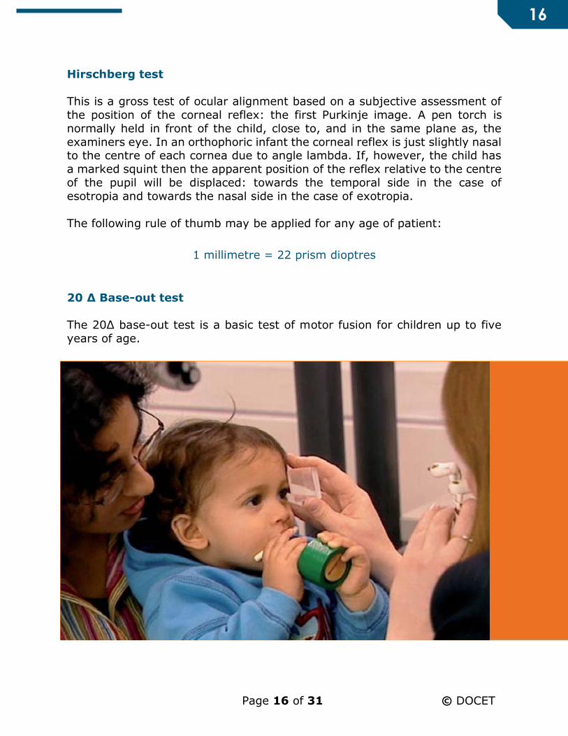
20 Δ Base-out test
The 20Δ base-out test is a basic test of motor fusion for children up to five
years of age.
Page 17 of 31 © DOCET
17
The presence of fusion is a sign that some degree of binocular vision has
developed. A base-out prism is held before one eye while the child looks at a
suitable fixation target. The eye behind the prism should adduct rapidly to
restore normal fusion and quickly abduct again on removal of the prism. The
speed of the fusion movements is a simple guide to the quality of the
binocularity.
The following prisms are suitable:
Six months of age 10Δ base-out
12 – 18 months of age 15Δ base-out
Over 18 months of age 20Δ base out
Other possible responses are:
No movement – implies no fusion or lack of attention
Slow to overcome the prism or slow to recover after the prism has been
removed – implies poor fusion.
By the age of five years, a child should cooperate with the measurement of
the prism fusion range using a prism bar. The normal range for near is 35 Δ
– 45 Δ base-out to 12 Δ – 8 Δ base-in.
Motility test
Together with the cover test, the motility test is a key assessment when
examining a child patient. It will give information about the range of ocular movements and whether they are concomitant or incomitant.
As with the cover test, success in motility testing for a younger child patient
is heavily dependent on them being offered an interesting and stimulating
fixation target. A wide variety of toys can be adapted for use as a motility
target if they are reasonably small, have an interesting and stimulating
appearance and/or are brightly coloured. It may help to attract or maintain
the child’s attention to have a toy that flashes, lights up, squeaks or has some
other form of auditory stimulus.
Page 18 of 31 © DOCET
18
In very young children it may not be possible to explain the nature of the test
and what you expect of them to the child. In these situations, move the child
rather than the stimulus; the so cold "swinging infant" procedure. In this
technique the target is kept stationary and the child is rotated. It will be
necessary for the parent, by gentle holding, to prevent the child from turning
their head as they rotate them. If they have been successfully encouraged to direct their attention to the stimulus they will maintain their fixation as they
are rotated and their ocular motility assessed.
Stereopsis
A high degree of stereopsis is proof of bifoveal fixation and good binocular
vision, but a poor degree or absence of stereopsis is not necessarily associated
with poor vision or poor binocular function. In practice, good stereopsis
provides the examiner with confidence that a binocular vision anomaly is not
present, whereas poor stereopsis requires careful assessment to establish the
probable cause.
Stereopsis is known to be present in infants as young as four months of age,
but in practice it may be difficult to demonstrate any stereopsis in children
under one year of age. Table 3 below gives average expected stereo acuity
measures for children. Stereo acuity values tend to vary depending which type
of test is used.
Age Stereo (secs of arc)
Birth
One month
Three months
Six months 600 Frisby
Nine months 300 Frisby
One year 210 – 170 Frisby
18 months 170 – 150 Frisby
Two years 100 – 85 Frisby
Three years 85 – 55 Frisby
Four years 40 – 30 Frisby
Five years 30 – 20 Frisby
Six years 10 – 5 Frisby
Table 3: Age norms for stereo acuity and the tests used (measurements are
all approximate) (adapted from A. Grounds by C. Rushen and L. Speedwell)
Page 19 of 31 © DOCET
19
The Lang test and the Frisby screening test are designed to produce a
behavioural response: the child attempts to reach out and grasp the object,
and may provide a result in some children as young as six to 12 months. In
older children a variety of tests are available.
Stereopsis tests are available in a variety of designs and produce a three
dimensional object in a variety of different ways. Most tests use simple
geometrical shapes as test objects presented against a random patterned
background. It seems to be unavoidable that stereo tests produce monocular
clues of depth to some degree; precautions may need to be taken in the
application of the tests to ensure that these clues are minimised. The following
tests are in common use:
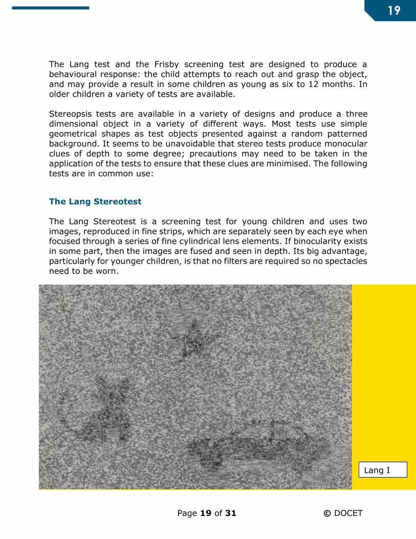
The Lang Stereotest
The Lang Stereotest is a screening test for young children and uses two
images, reproduced in fine strips, which are separately seen by each eye when focused through a series of fine cylindrical lens elements. If binocularity exists
in some part, then the images are fused and seen in depth. Its big advantage,
particularly for younger children, is that no filters are required so no spectacles
need to be worn.
Lang I
Page 20 of 31 © DOCET
20
The test is available in two forms. The Lang I test measures stereopsis at
1,200, 600 and 550 seconds of arc and the Lang II test, which is finer,
measures 600, 400 and 200 seconds of arc. The Lang II test also has an object
(star) which is always visible, even with only one eye.
Answers can be checked by holding the test vertically, where stereo clues disappear, or by holding the test upside down when the object pattern is
inverted.
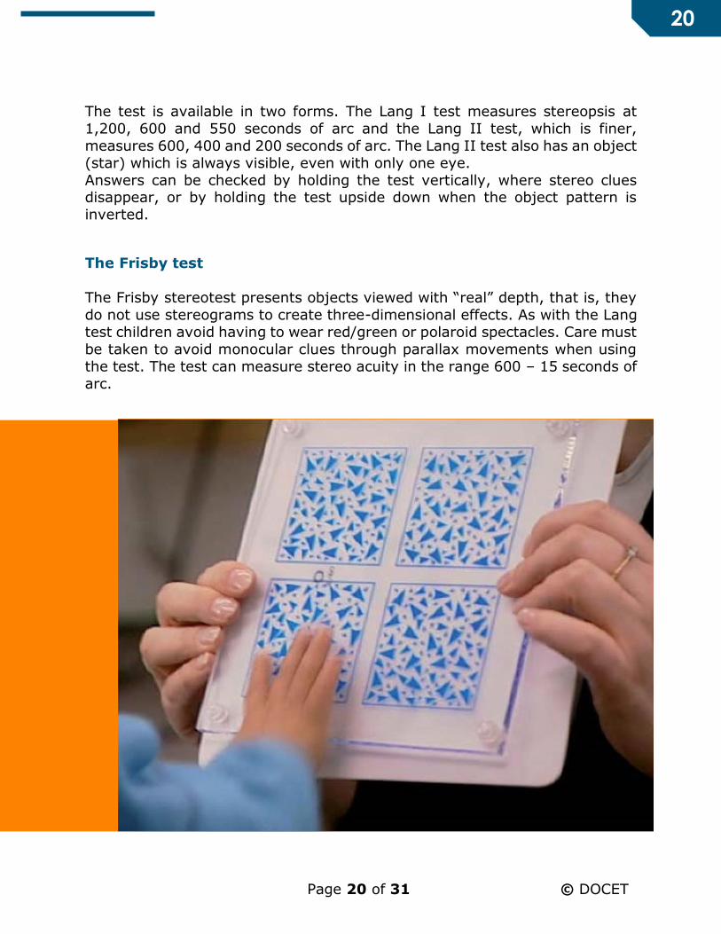
The Frisby test
The Frisby stereotest presents objects viewed with “real” depth, that is, they
do not use stereograms to create three-dimensional effects. As with the Lang
test children avoid having to wear red/green or polaroid spectacles. Care must
be taken to avoid monocular clues through parallax movements when using
the test. The test can measure stereo acuity in the range 600 – 15 seconds of
arc.
Page 21 of 31 © DOCET
21
The Frisby test is available in a screening version designed for younger
children and infants. It presents a three-dimensional object field, together
with a flat image side by side in a preferential looking format. In this case a
spontaneous pointing or looking responses can be observed to establish that stereopsis is present.
TNO
The TNO test is a random-dot type of stereotest requiring the wearing of red
and green glasses. It comprises seven test plates and comes in an adult and
children’s version. It can assess stereo acuity down to 15 seconds of arc.
The Titmus test
This is perhaps the most familiar stereo test because of it use of a large three
dimensional fly to elicit a response in the vast majority of patients. It uses a crossed Polaroid visor to achieve stereopsis and in part cartoon characters to
make the test more appealing to younger children. It suffers because of
noticeable monocular clues which may be difficult to disguise. The Titmus test
will assess stereo acuity from a gross 3552 – 700 for the fly and 800 – 40
seconds of arc for the graded tests.
The Randot test
The Randot test is in effect a modified Titmus test and is based on the same
polarised design. It uses a random pattern background to remove many of the
monocular clues present in that test.
Page 22 of 31 © DOCET
22
Refraction
Norms
Refractive error in infants is predominantly hypermetropic; there are very
few myopes. Table 4 lists average refractive errors throughout early
childhood. As you will see from the table over the first two years of life the
majority of this hypermetropic refractive error has disappeared and by four
years of age it has more or less gone completely. This process is known as
emmetropisation. A number of factors can disrupt the emmetropisation
process and care may be needed in the management of refractive error in
young children in order to avoid this.
Age Average Rx
Birth
One month
Three months +3.00
Six months +2.50
Nine months +2.25
One year +2.00
18 months +1.50
Two years +1.25
Three years +1.00
Four years +0.50
Five years +0.50
Six years +0.50
Table 4: Age norms for refractive error (measurements are all approximate).
(Adapted from A. Grounds by C. Rushen and L. Speedwell)
In addition to spherical refractive error astigmatism of around 1 – 2 dioptres is common.
As with other areas of examining children described in this text, the
assessment of refractive error will by necessity have to rely heavily on various
objective retinoscopy techniques and the use of measures to control
accommodation.
Page 23 of 31 © DOCET
23
Static retinoscopy
This is the term used to describe distance fixation retinoscopy where the
patient’s accommodation is relaxed. This may be difficult to achieve in very
young children, and the use of an assistant to encourage the child to maintain
fixation can be very valuable. As with the motility test flashing or squeaking toys can be very useful.
It is likely that for many children it is the retinoscope light itself which is the
most interesting object and it may be very difficult to stop them looking at it
rather than your intended target. If you are refracting an infant then this may
be used to your advantage by adopting the Mohindra technique.
The Mohindra technique uses the retinoscope (held at 50cm) as the fixation
target. Providing all other light sources in the examination room are
extinguished, the child will fixate the retinoscope light but their
accommodation will be relaxed. It is advised that the correction factor applied
(to take account of the relaxed accommodation), should be adapted depending on the age of the patient. A correction factor of -1.25D for adults,
-1.00D for children older than 2 years and 0.75D for those under this age,
should be added to the final ret result.
With infants and very young children a trial frame is impractical and
retinoscopy should be done by holding trial lenses in front of the patient’s
eyes. It may be easier to refract using spherical lenses only rather than
attempt to hold a sphere and a cyl together.
Page 24 of 31 © DOCET
24
Cycloplegic refraction
Cycloplegic refraction is an essential tool in the examination of children,
although the application of the drops can be challenging for the child, the
parents and the optometrist. Whilst it might be argued that for a cooperative
child with good acuity, normal oculomotor balance, good stereopsis, no relevant family history and normal accommodation a cycloplegic examination
may not be needed, with certain groups of children, a cyclopegic refraction
should always be considered.
These are:
Children with unexplained poor VA
Children with poor stereopsis
Children with an esophoria or manifest esotropia
Children with over or underactive accommodation
Children of parents with high hypermetropia or squint.
Various steps can be taken to minimise the discomfort and ensure that the
cycloplegic can be instilled effectively:
Proxymetacaine: Some practitioners advocate the use of Proxymetacaine 0.5%, immediately before the cycloplegic agent.
Anaesthetising the cornea has been shown to reduce discomfort as the
subsequent drops are instilled and the local anaesthetic can facilitate
absorption of the cycloplegic agent. However, if the child objects to the
first set of drops, the second set may be even harder to administer!
Instilling on the eyelids: In cases where the child is reluctant to open
their eyes to allow instillation of the cycloplegic agent, it is possible to
drop it onto the closed eyelids. Enough of the drug will find its way
through the closed lids for it to be effective.
For most children confidence and decisiveness are all that is necessary in order to effectively instill a cycloplegic.
For the vast majority of child patients Cyclopentolate 1.0% is the cycloplegic
of choice. It is generally well tolerated, produces effective cycloplegia in most
cases, requires no tonus allowance and is available in single dose preservative
free modality.
Page 25 of 31 © DOCET
25
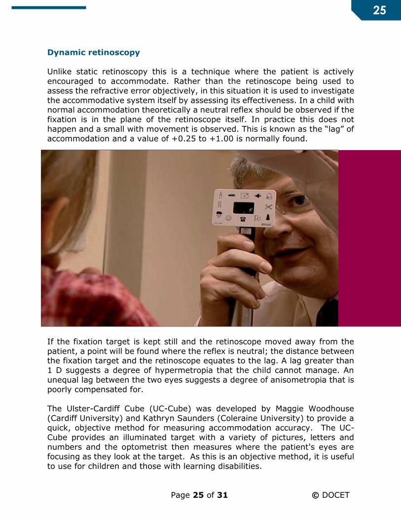
Dynamic retinoscopy
Unlike static retinoscopy this is a technique where the patient is actively
encouraged to accommodate. Rather than the retinoscope being used to
assess the refractive error objectively, in this situation it is used to investigate
the accommodative system itself by assessing its effectiveness. In a child with normal accommodation theoretically a neutral reflex should be observed if the
fixation is in the plane of the retinoscope itself. In practice this does not
happen and a small with movement is observed. This is known as the “lag” of
accommodation and a value of +0.25 to +1.00 is normally found.
If the fixation target is kept still and the retinoscope moved away from the
patient, a point will be found where the reflex is neutral; the distance between the fixation target and the retinoscope equates to the lag. A lag greater than
1 D suggests a degree of hypermetropia that the child cannot manage. An
unequal lag between the two eyes suggests a degree of anisometropia that is
poorly compensated for.
The Ulster-Cardiff Cube (UC-Cube) was developed by Maggie Woodhouse
(Cardiff University) and Kathryn Saunders (Coleraine University) to provide a
quick, objective method for measuring accommodation accuracy. The UC-
Cube provides an illuminated target with a variety of pictures, letters and
numbers and the optometrist then measures where the patient's eyes are
focusing as they look at the target. As this is an objective method, it is useful
to use for children and those with learning disabilities.
Page 26 of 31 © DOCET
26
Colour vision Congenital colour vision defects are present in around 8% of boys and around
0.5% of girls. In most cases the defect causes no appreciable handicap in
normal life. It is of value for a child patient and its parent to know if a defect
is present so that certain decisions – particularly career related decisions –
can be approached in an informed way.
Ishihara test
This pseudoisochromatic plate test is the most commonly used of all colour
vision tests. It is intended to be used in northern daylight at a viewing distance of 75 centimetres. The test will identify protan and deutan defects, but is very
sensitive and can only very crudely quantify a defect. Although the majority
of the plates rely on numeracy skills there are a number where finger tracing
of a winding line can be utilised.
Color Vision Testing Made Easy
Color Vision Testing Made Easy is a 14 plate pseudoisochromatic test that uses
common objects such as a dog, balloon, car and boat and symbols such as
squares, circles and stars, rather than numbers or letters. This makes the test
simple to use on young children as well as people with learning disabilities. It
was designed by US optometrist Terrace Waggoner and is intended for
children aged 3 to 5 years, although it can be successfully used on older and younger age groups.
The test is quick and easy to administer and is divided into two parts. Part I
uses two of the symbols, and each of these cards is designed so that a colour
deficient person can always see at least one of them and therefore does not
get discouraged or self-conscious. This device also allows the tester to verify
that the child understands the test and is cooperating. Part II uses the objects
for matching or tracing with very young children.
The test strategy allows a child to be scored and a quantifiable result is
obtained. The response patterns of the normal and colour deficient child are
clear-cut so that a diagnosis can be made with a high degree of confidence.
Page 27 of 31 © DOCET
27
Management
Key points
The hardest part of managing the child patient may be managing parental
expectations.
Parents are often told to do something but are not given an explanation of
why. Parents find this is very frustrating and it has an adverse effect on
compliance.
All members of the optometric team should be confident that they have the appropriate skills and expertise before managing any child.
Refractive error plays a significant part in the aetiology and
management of strabismus and/or amblyopia. Children with these
conditions should have regular refractions (normally this will be
approximately at least once a year) with fundus examinations
undertaken as appropriate.
Strabismus may be indicative of wider ocular or neurological pathology
and in best practice should be referred for ophthalmic and orthoptic
assessment.
Many children with anisometropic amblyopia can be managed by optometrists in the community. The improvement of vision in the
amblyopic eye with the use of spectacles alone should be monitored
regularly over a six-month period. The child will require referral to an
ophthalmologist if:
o There is no improvement on two consecutive visits during this
period, and
o The vision is still below normal or
o Vision improvement is not sustained.
Page 28 of 31 © DOCET
28
Refractive correction
The following Guidelines for Prescribing have been drawn up by Dr Margaret
Woodhouse of the Department of Optometry & Vision Sciences at Cardiff
University and are widely thought to represent current good practice.
NB Although evidence based, these represent Margaret’s personal opinion and
are published for guidance only. Other practitioners may adopt different
criteria.
Children of any age
Consider prescribing in cases of:
Extreme refractive errors for age
Strabismus and/or amblyopia
Persistent anisometropia (over 1.00D seen on at least two visits three
months apart). Depending on other factors, such as the level of
refractive error in the “better” eye, prescribing the inter-ocular difference only may be acceptable.
Children under two years of age
Monitor refractive error only.
Children over two years of age
Consider prescribing in cases of:
Significant refractive error that is not decreasing.
What is a significant refractive error (in children over two years)?
Hypermetropia of +3.00D or greater
Prescribe reduced by 1.00D if no BV anomalies
Myopia of –0.75 or greater
Since very young children are interested mainly in near, prescription not
needed
Prescribe full amount when child begins to need clear distance vision
Astigmatism (in the absence of hypermetropia/myopia) of 2.50D or
greater.

Page 29 of 31 © DOCET
29
Children with Down’s syndrome and cerebral palsy (and other disabilities)
Are less likely to emmetropise, so consider prescribing for refractive
errors earlier
Are likely to have poor accommodative responses, so DO NOT reduce
hypermetropic prescription Are likely to benefit from bifocals or other style of near prescription.
Squint and amblyopia
Several risk factors can be analysed to predict the possible development of
squint and/or amblyopia and help plan a management strategy.
Family history of squint, amblyopia and high refractive error, prematurity and
low birth weight are significant risk factors.
David Stidwell offers the following guidance:
The onset of strabismus (and therefore strabismic amblyopia) has an
age distribution very similar to the critical period, ie most children
develop tropias between six months and five years, with a peak at 21,
30 and 30 months for non-accommodative, partially and fully
accommodative esotropia respectively.
Refraction over +2.00 in the better eye, anisometropia over +1.50, and
a pre-existing marked heterophoria (or any combination of these) are
predictable risk factors.
Bilateral ametropia over +6.00 will produce bilateral amblyopia rather
than strabismus.
Monocular ametropia with the better eye under one dioptre will produce
straight anisometropic amblyopia.
The higher the refractive error and the higher the anisometropia the
earlier the strabismus and/or amblyopia will occur, so:
o R +2.00DS L +6.00DS will produce both anisometropic amblyopia
starting from two months old and
o An accommodative strabismus starting from three or four years old.

Page 30 of 31 © DOCET
30
Occlusion
Whilst initial management for amblyopia with a refractive component is
usually by way of correcting the refractive error, in some cases, occlusion may
also be needed. The visual acuity at first examination and compliance with
occlusion are now thought to be the main predictors of the visual outcome in children prescribed occlusion for amblyopia. Hours of occlusion and age at first
visit do not seem to be associated with better outcomes.
In general, occlusion therapy should be part-time, to avoid the risk of inducing
deprivational amblyopia in the “good” eye, and should be effective in all cases.
Patching for two to three hours per day, preferably combined with detailed
visual tasking such as drawing or computer and video games, has been shown
to be as effective as longer periods and avoids many of the pitfalls of occlusion
therapy and helps to improve compliance.
Detailed instruction, together with the reasoning for the therapy, should be
given to the child and their parents.
Referral criteria
If you do not feel you have the skills and expertise to manage the child appropriately then refer.
Large angle squints may require surgery and should be referred.
If referring a child with squint, consider the length of time to first
appointment and the effect this may have on the degree of amblyopia.
Consider interim occlusion therapy.
Refer anisometropic amblyopia when, on two consecutive visits during
the first six months of refractive correction:
o No VA improvement is shown
o The VA remains below an acceptable level o An early VA improvement is not sustained.
Page 31 of 31 © DOCET
31
Further information
The College of Optometrists
https://www.college-optometrists.org/
The Association of Optometrists
https://www.aop.org.uk/
Lea Symbols
http://www.lea-test.fi
Kay Picture Test http://www.kaypictures.co.uk
Color Vision Testing Made Easy
http://colorvisiontesting.com/color5.htm
Acknowledgements
DOCET would like to thank the following for their help and assistance in the
production of this Distance Learning Project:
Paul Adler
Lynne Weddell