Evolution of the vertebrate bone matrix: An expression analysis of the network forming collagen...
11
Evolution of the Vertebrate Bone Matrix: An Expression Analysis of the Network Forming Collagen Paralogues in Amphibian Osteoblasts DANIEL ALDEA 1 , PATRICIA HANNA 1 , DAVID MUNOZ 1 , JAVIER ESPINOZA 1 , MARCELA TORREJON 2 , LAURENT SACHS 3 , NICOLAS BUISINE 3 , SILVAN OULION 4 , HECTOR ESCRIVA 4 , AND SYLVAIN MARCELLINI 1 * 1 Laboratorio de Desarrollo y Evolución, Departamento de Biología Celular, Facultad de Ciencias Biológicas, Universidad de Concepción, Barrio Universitario s/n, Concepción, Chile 2 Departamento de Bioquímica y Biología Molecular, Facultad de Ciencias Biológicas, Universidad de Concepción, Barrio Universitario s/n, Concepción, Chile 3 Muséum National d'Histoire Naturelle, Département Régulation Développement et Diversité Moléculaire, UMR7221 CNRS, Evolution des Régulations Endocriniennes, Paris cedex 05, France 4 Centre National de la Recherche Scientifique, Unité Mixte de Recherche 7232, Université Pierre et Marie Curie Paris 06, Observatoire Océanologique, Banyuls‐sur‐Mer, France ABSTRACT The emergence of vertebrates is closely associated to the evolution of mineralized bone tissue. However, the molecular basis underlying the origin and subsequent diversification of the skeletal mineralized matrix is still poorly understood. One efficient way to tackle this issue is to compare the expression, between vertebrate species, of osteoblastic genes coding for bone matrix proteins. In this work, we have focused on the evolution of the network forming collagen family which contains the Col8a1, Col8a2, and Col10a1 genes. Both phylogeny and synteny reveal that these three paralogues are vertebrate‐specific and derive from two independent duplications in the vertebrate lineage. To shed light on the evolution of this family, we have analyzed the osteoblastic expression of the network forming collagens in endochondral and intramembraneous skeletal elements of the amphibian Xenopus tropicalis. Remarkably, we find that amphibian osteoblasts express Col10a1,a gene strongly expressed in osteoblasts in actinopterygians but not in amniotes. In addition, while Col8a1 is known to be robustly expressed in mammalian osteoblasts, the expression levels of its amphibian orthologue are dramatically reduced. Our work reveals that while a skeletal expression of network forming collagen members is widespread throughout vertebrates, osteoblasts from divergent vertebrate lineages express different combinations of network forming collagen paralogues. J. Exp. Zool. (Mol. Dev. Evol.) 9999B: 1–11, 2013. © 2013 Wiley Periodicals, Inc. How to cite this article: Aldea D, Hanna P, Munoz D, Espinoza J, Torrejon M, Sachs L, Buisine N, Oulion S, Escriva H, Marcellini S. 2013. Evolution of the vertebrate bone matrix: An expression analysis of the network forming collagen paralogues in amphibian osteoblasts. J. Exp. Zool. (Mol. Dev. Evol.) 9999:1–11. J. Exp. Zool. (Mol. Dev. Evol.) 9999B:1–11, 2013 RESEARCH ARTICLE © 2013 WILEY PERIODICALS, INC.
Transcript of Evolution of the vertebrate bone matrix: An expression analysis of the network forming collagen...

Evolution of the Vertebrate BoneMatrix: An Expression Analysis ofthe Network Forming CollagenParalogues in AmphibianOsteoblastsDANIEL ALDEA1, PATRICIA HANNA1,DAVID MUNOZ1, JAVIER ESPINOZA1,MARCELA TORREJON2, LAURENT SACHS3,NICOLAS BUISINE3, SILVAN OULION4,HECTOR ESCRIVA4, AND SYLVAIN MARCELLINI1*1Laboratorio de Desarrollo y Evolución, Departamento de Biología Celular, Facultad de CienciasBiológicas, Universidad de Concepción, Barrio Universitario s/n, Concepción, Chile2Departamento de Bioquímica y Biología Molecular, Facultad de Ciencias Biológicas,Universidad de Concepción, Barrio Universitario s/n, Concepción, Chile3Muséum National d'Histoire Naturelle, Département Régulation Développement et DiversitéMoléculaire, UMR7221 CNRS, Evolution des Régulations Endocriniennes, Paris cedex 05, France4Centre National de la Recherche Scientifique, Unité Mixte de Recherche 7232, UniversitéPierre et Marie Curie Paris 06, Observatoire Océanologique, Banyuls‐sur‐Mer, France
ABSTRACT The emergence of vertebrates is closely associated to the evolution of mineralized bone tissue.However, the molecular basis underlying the origin and subsequent diversification of the skeletalmineralized matrix is still poorly understood. One efficient way to tackle this issue is to compare theexpression, between vertebrate species, of osteoblastic genes coding for bone matrix proteins. Inthis work, we have focused on the evolution of the network forming collagen family which containsthe Col8a1, Col8a2, and Col10a1 genes. Both phylogeny and synteny reveal that these threeparalogues are vertebrate‐specific and derive from two independent duplications in the vertebratelineage. To shed light on the evolution of this family, we have analyzed the osteoblastic expressionof the network forming collagens in endochondral and intramembraneous skeletal elements of theamphibian Xenopus tropicalis. Remarkably, we find that amphibian osteoblasts express Col10a1, agene strongly expressed in osteoblasts in actinopterygians but not in amniotes. In addition, whileCol8a1 is known to be robustly expressed in mammalian osteoblasts, the expression levels of itsamphibian orthologue are dramatically reduced. Our work reveals that while a skeletal expressionof network forming collagen members is widespread throughout vertebrates, osteoblasts fromdivergent vertebrate lineages express different combinations of network forming collagenparalogues. J. Exp. Zool. (Mol. Dev. Evol.) 9999B: 1–11, 2013. © 2013 Wiley Periodicals, Inc.
How to cite this article: Aldea D, Hanna P, Munoz D, Espinoza J, Torrejon M, Sachs L, Buisine N,Oulion S, Escriva H, Marcellini S. 2013. Evolution of the vertebrate bone matrix: An expressionanalysis of the network forming collagen paralogues in amphibian osteoblasts. J. Exp. Zool. (Mol.Dev. Evol.) 9999:1–11.
J. Exp. Zool.(Mol. Dev. Evol.)9999B:1–11, 2013
RESEARCH ARTICLE
© 2013 WILEY PERIODICALS, INC.

The mineralized skeleton represents an evolutionary novelty thathas played a pivotal role for the adaptation of vertebrate animals to avariety of ecological niches (Hall, 2005). There are severalcomplementary ways to tackle the problem of bone evolution. Forinstance, regulatory and developmental changes have beenassociated to morphological modifications of the vertebrate body,including jaw shape, digit identity, vertebrate number, and fin size(Albertson et al., 2005; Chan et al., 2010; Di‐Poi et al., 2010; Tamuraet al., 2011). Another important issue in the field of bone Evo‐Devoconcerns the emergence and diversification of specialized cell types,such as chondrocytes (cartilagemaking‐cells) and osteoblasts (bone‐making cells), able to secrete components of the skeletal matrix. Forinstance, the fibrillar collagen proteins play a key role in thenucleation of hydroxyapatite crystals during skeletal mineralization(Glimcher et al., '57), and constitute one such family whoseevolutionary history has been analyzed in exquisite detail. Thefibrillar collagen genes can be grouped into three clades (namedA, B,and C) that arose prior to the emergence of deuterostomes (Wada etal., 2006; Rychel and Swalla, 2007). Notochordal expressionrepresents a synapomorphy of chordates, as clade A orthologuesare expressed in the notochord of cephalochordates, ascidians, andvertebrates (Wada et al., 2006; Zhang and Cohn, 2006). Thevertebrate clade A members have expanded to four subfamiliesthrough two rounds of whole genome duplications (Zhang andCohn, 2008).One huge andheterogeneous family of collagenous proteinswhose
implication for skeletal development and evolution has largely beenoverlooked contains the members of the non‐fibrillar collagens(Heino, 2007; Kadler et al., 2007). Here, we will focus on one specificsubgroup of non‐fibrillar collagens that includes the Collagen 8a1(Col8a1), Collagen 8a2 (Col8a2), and Collagen 10a1 (Col10a1)proteins, that are commonly referred to as the network formingcollagen family. Indeed, Col8a1, Col8a2, and Col10a1 display highsequence similarity and share the unique ability to form hexagonalnetworks in the extracellular matrix, either as heterotrimers orhomotrimers (Stephan et al., 2004; Heino, 2007; Kadler et al., 2007).Functional studies have shown that the network forming
collagens are required for the correct morphogenesis of the tissues
in which they are expressed. First, the expression of Col8a1 andCol8a2 has been reported in the Descemet's membrane separatingcorneal endothelial cells from corneal stroma as well as bloodvessel walls (Kapoor et al., '86; Sinha et al., 2001; Plenzet al., 2003). Accordingly, mutations in Col8a1 or Col8a2 causeeye defects in humans and mice (Hopfer et al., 2005; Desronvilet al., 2010). Second, the zebrafish genome contains two Col8a1genes, one of which is expressed in the notochord and jawcartilages, while its paralogue is expressed in the eye (Gansner andGitlin, 2008). A mutation preventing trimerization of thenotochord‐specific Col8a1 paralogue causes severe axial defectsin the fish Danio rerio (Gansner and Gitlin, 2008). Third, inagreement with the cartilage‐specific phenotype of mouse knock‐out mutants and of human patients suffering from Schmidmetaphyseal chondrodysplasia, transcripts of the mammalianCol10a1 orthologues are detected in hypertrophic chondrocytes(Warman et al., '93; Kwan et al., '97; Kim et al., '99).The expression of Col10a1 represents an interesting case of
expression pattern evolution between skeletal cell types. This isbecause independent studies have shown that Col10a1 is stronglyexpressed in osteoblasts in a variety of actinopterygian species,but not in mammals (Warman et al., '93; Kwan et al., '97; Kimet al., '99; Avaron et al., 2006; Laue et al., 2008; Li et al., 2009;Albertson et al., 2010; Maye et al., 2011; Eames et al., 2012).However, the complete lack of data from amphibians limits ourability to polarize evolutionary change for this gene. In thisrespect, comparing the model organism Xenopus tropicalis toother vertebrate species will undoubtedly shed light on theevolution of the network forming collagen genes.In the present study, we aimed at clarifying the evolutionary
history of the network forming collagen genes, putting aparticular emphasis on the mineralized bone matrix secreted byosteoblasts. We examined the phylogenetic relationships andsynteny conservation between Col8a1, and Col8a2 and Col10a1,and found that these three paralogues are vertebrate‐specific andresult from the two rounds of genomic duplications that occurredbefore vertebrate diversification (Ohno, '70; Dehal andBoore, 2005). Using X. tropicalis as a representative amphibianspecies, we provide a comprehensive examination of transcriptdistribution of all three genes in endochondral and intra-membraneous bones and find that the osteoblastic expressionof the amphibian network forming collagen genes is more similarto actinopterygian fishes than to mammals. We propose thatnetwork forming collagens are part of themineralized bonematrixof all osteichthyans, although osteoblasts from distinct vertebratelineages express different combinations of paralogues.
MATERIALS AND METHODS
Phylogeny and Synteny AnalysesThe amino acid sequences of the vertebrate Col8a1, Col8a2, andCol10a1 orthologs were identified from the NCBI (http://www.ncbi.
Grant sponsor: FONDECYT Grant; grant number: 1110756; grant sponsor:Agence Nationale de la Recherche Grants; grant numbers: ANR‐2010‐BLAN‐1716 01, ANR‐2010‐BLAN‐1234 02; grant sponsor: ECOS‐CONICYT;grant number: C09B01; grant sponsor: “CRESCENDO” European IntegratedProject; grant number: LSHM‐CT‐2005‐018652.
The authors declare that they have no conflict of interest.�Correspondence to: Sylvain Marcellini, Laboratory of Development and
Evolution, Department of Cell Biology, University of Concepcion, Concep-cion, Chile. E‐mail: [email protected]
Received 20 October 2012; Revised 3 April 2013; Accepted 6 April 2013DOI: 10.1002/jez.b.22511Published online XX Month Year in Wiley Online Library
(wileyonlinelibrary.com).
2 ALDEA ET AL.
J. Exp. Zool. (Mol. Dev. Evol.)

nlm.nih.gov/) and the Ensembl (http://www.ensembl.org/) data-bases. Multiple amino acid sequences were aligned using MUSCLE(v3.7) (Edgar, 2004) and regions of ambiguous homology wereremoved manually, thereby restricting our dataset to the 835 aabelonging to the more conserved triple helical domain. Phylogenetictrees were reconstructed using both Bayesian and likelihoodmethods. In order to root the phylogenetic tree, collagens belongingto other subgroups such as multiplexins and fibrillar collagens wereused as outgroups (Fig. 1 and Supplementary Material S1).Bayesian phylogenetic inferences were conducted using
MrBayes(v3.2.1) (Ronquist et al., 2012). One million generationswere run for three Markov chains and sampled every 100generation. The initial 25% sampled trees were discarded as theburn‐in phase, and the remaining trees were used to calculateposterior probabilities (PP) in a 50% majority rule consensus tree.A maximum likelihood (ML) tree was also generated usingPHYML3.0 (Guindon and Gascuel, 2003) with a JTT model asproposed by ProtTest(v2.4) (Abascal et al., 2005). The robustness ofthe tree nodes was estimated by a bootstrap test (1,000 replicates).Trees were viewed in FigTree(v1.3.1) (http://tree.bio.ed.ac.uk/).For synteny analyses, genomic structure and chromosomal
organization of each collagen (Col8a1, Col8a2, and Col10a1) inMus musculus, Gallus gallus, X. tropicalis, D. rerio, andGasterosteus aculeatus (only for Col10a1) were compared usingNCBI and the Ensembl genome browsers.
Animal Facility and Sample PreparationsAdults are maintained at the University of Concepcion followingestablished protocols for X. tropicalis. Embryos and tadpoles wereobtained by natural mating and staged according to theNieuwkoop and Faber developmental table (Nieuwkoop andFaber, '67). Tadpoles were anesthetized with a solution of 200 mg/mL of tricaine (Sigma–Aldrich, St. Louis, MO, USA) before tissuedissection in 0.6X Leibovtiz 15 medium (Invitrogen, Paisley, UK).The Ethics Committee of the University of Concepcion (Con-cepcion, Chile) approved all experimental procedures carried outduring this study, which were performed following the guidelinesoutlined in the Biosafety and Bioethics Manual of the NationalCommission of Scientific and Technological Research (CONICYT,Chilean Government). Embryos and tissue samples were eitherfixed in MEMFA (1 mM MOPS, 2 mM EGTA, 1 mM MgSO4, 3.7%formaldehyde) for in situ hybridization or frozen in liquid nitrogenfor RNA preparation.
Expression AnalysesFor in situ hybridization probes, genomic regions correspondingto the last coding exon of the X. tropicalis Col8a1 (GenBankaccession number XM_002940988.1), Col8a2 (GenBank accessionnumber XM_002936225.1), and Col10a1 (Xenbase gene pagenumber 6468755) were amplified with the following primers:Col8a1‐Forward: 50‐TTC CAG GAC TTC CTG GAC TTA‐30; Col8a1‐Reverse: 50‐GTG GAC CAA TTA ATC CCT TGT‐30; Col8a2‐
Forward: 50‐ATC TGG TAT TGC TTC TAATGG‐30; Col8a2‐Reverse:50‐TTACCTGGTAAC CCATTAGGA‐30; Col10a1‐Forward: 50‐ATTCCA AAA GTA CCA GAG CAG‐30; Col10a1‐Reverse: 50‐AAC CATCAG AAC CTT GTT CTC‐30. Ten nanograms of genomic DNAwereused per PCR reaction with the KOD enzyme (Calbiochem,SanDiego, CA, USA) following the manufacturer's instructions.Three DNA fragments were amplified, corresponding to Col8a1(678 pb long), Col8a2 (714 bp long), Col10a1 (832 bp long),individually blunt‐cloned into pBluescript (Stratagene, La Jolla,CA, USA), and used as templates to synthesize Dig‐RNA probes.Probe synthesis, and in situ hybridization on whole mountembryos were carried out using standard protocols. A detailedprotocol for the in situ hybridization on paraffin sections isprovided as Supplementary Material S2. In situ hybridization onwhole mount embryos was performed as previously described(Maldonado‐Agurto et al., 2011), and the same protocol wasapplied to dissected stage 60 calvaria.RNA sequencing (RNA‐seq) was performed on total RNA
prepared from stage 53 hindlimbs and stage 60 calvaria (Wanget al., 2009). Specific libraries were prepared from poly‐ARNA andsequenced with the Illumina technology by the IGBMCMicroarrayand Sequencing platform, member of the France Genomiqueprogram. Reads were matched onto the reference genome withbowtie (Langmead et al., 2009). To measure RNA abundance,digital expression values were corrected for gene length andsequencing depth by using standard RKPM values (number ofreads per kilobase per million mapped reads) (Wang et al., 2009).Data visualization was carried out with gbrowse, based on RNAs‐seq density profiles normalized for the sequencing depth of eachlibrary. While RNA‐seq allows the characterization of geneexpression levels and exon mapping, it does not reveal theconnectivity between exons. Hence, in order to better associateRNA‐seq profiles to individual genes, we performed RNA Paired‐End‐diTag (RNA‐PET), which simultaneously captures the 50 and30 ends of entire transcripts, using total RNA prepared from stage53 hindlimbs (Ng et al., 2005; Fullwood et al., 2009). Specific RNA‐PET libraries were prepared from poly‐A RNA, sequenced with theSOLiD technology, and reads were subsequently matched onto thereference genome with Bioscope. A detailed procedure of RNA‐seqand RNA‐PET will be published elsewhere.
RT‐PCRRNA extraction was performed in TRIzol (Life Technologies,Carlsbad, CA, USA) following the manufacturer's instructions.Reverse transcription reactions were performed in 20 mL with1 mg of total RNA, 5 mM oligo dT, 0.5 mM dNTPs, 0.5 mL ofRNAsin (Promega, Madison, WI, USA) and 0.8 mL of M‐MLVreverse transcriptase (Promega). cDNA (0.5 mL) was used in 10 mLPCR reactions (GoTaq, Promega). The X. tropicalis cDNAs wereamplified with the following Col8a2 PCR primers (50–30): Exon1F50‐ATG TAAAAC TCT CCT CAG TGG‐30; Exon4R 50‐81114840 TCCATT AGA AGC AATACC AGA T‐30. For nested PCR, 0.5 mL of this
J. Exp. Zool. (Mol. Dev. Evol.)
EVOLUTION OF THE NETWORK FORMING COLLAGENS 3

Figure 1. Phylogeny of collagens 8a1, 8a2, and 10a1 in osteichthyans using a Bayesian phylogenetic approach. Col8a2 and Col10a1 are moreclosely related to each other than to the Col8a1 paralogues. Col15a1 and Col18a1 sequences are used as outgroups. Only posteriorprobabilities inferior to 1 are shown.
J. Exp. Zool. (Mol. Dev. Evol.)
4 ALDEA ET AL.

PCR reaction were used as a template in a second 10 mL PCRreaction using the aforementioned Exon4R primer in combinationwith the following primer: Exon1nestedF 50‐TGC AAG GTG CAAACA GTG AG‐30. The GenBank (http://www.ncbi.nlm.nih.gov/)accession number for the X. tropicalis Col8a2 cDNA sequenced inthis study is JQ693471. See Supplementary Material S3 for details.
RESULTS
Phylogeny and Conservation of Synteny of the Network FormingCollagen Family MembersWe first aimed to resolve the phylogenetic relationships of thenetwork forming collagen members (i.e., the relationships betweenCollagen 8 and 10 protein sequences). In spite of extensive searchesin complete genome sequences of amphioxus and tunicate (i.e.,Branchiostoma floridae and Ciona intestinalis) and in orthologydatabases we did not find any non‐vertebrate orthologue of thevertebrate network forming collagens. Hence, we focused our studyon the vertebrate genes. Our results show that the osteichthyanCollagen 8a1, 8a2, and 10a1 proteins are highly conserved andclearly cluster as a monophyletic group (Fig. 1 and SupplementaryMaterials S1 and S4). Node robustness (posterior probabilities andbootstrap values) within this monophyletic group suggests that theCol8a2 and Col10a1 paralogous groups are more closely related toeach other than to the Col8a1 paralogues (Fig. 1 and supplementarymaterial S1). As choosing an outgroup to root collagen trees is notstraightforward, we have performed a variety of phylogeneticanalyses using different outgroups, and have obtained similar resultsin all cases (Supplementary Material S1). Synteny is well conservedamong osteichthyans within each group of paralogues (Fig. 2). Forexample, Col8a1 orthologues are linked to Dcbld2 and Tmem30c;Col8a2 orthologues are linked to TrappC3, Adprhl2, and Tekt2; andCol10a1 orthologues are linked to Bet3l, Dse, Nt5dc1, and Frk(Fig. 2). Furthermore, during the teleost‐specific genomic duplication(3R), a duplication of Col10a1 occurred and led to two Col10a1 genecopies (Col10a1a and Col10a1b). Both copies have been maintainedin stickleback, whereas onlyCol10a1a is present in the zebrafish. Thesynteny study confirms the loss ofCol10a1b inD. rerio (Fig. 2). A lowconservation of synteny is found between the three network formingcollagen loci since only Col8a2 and Col10a1 genes are linked toTrappC3 and its paralogue Bet3l, respectively (See Fig. 2 andTurnbull et al., 2005). In summary, the network forming collagengenes form amonophyletic family, and both phylogeny and syntenysupport a closer relationship of the Col8a2 and Col10a1 paralogues.
Expression of the Network Forming Collagens in XenopusEndochondral BonesWe cloned regions of the large last coding exon for all threenetwork forming collagen genes from genomic DNA, andperformed in situ hybridization at different stages of X. tropicalishindlimb development (Fig. 3A–K). We compared expressionpatterns between stage 51 limb buds (Fig. 3A–D), that mainly
contain undifferentiated mesenchyme, and stage 58 legs(Fig. 3E–K) that display differentiated skeletal elements contain-ing both bone and cartilage (Miura et al., 2008; Espinozaet al., 2010). No signal was detected for Col8a1 at these stages(Fig. 3B,I). Col8a2 transcripts are present throughout the limb budat stage 51 (Fig. 3C), and in a variety of cell types at stage 58 suchas chondrocytes, osteoblasts lining the bone surface, theosteocytes embedded within the bone matrix, and striated musclefibers (Fig. 3F,J). Col10a1 is not detected at stage 51 (Fig. 3D), butdisplays a specific expression pattern in osteoblasts at stage 58(Fig. 3G,K). In addition, Col10a1 is expressed in the chondrocytes
Figure 2. Synteny of the network forming collagen genes. Top:Col8a1 is linked to Dcbld2 and Tmem30c in osteichthyans; and toFilip1l in amniotes. Middle: Col8a2 is linked to TrappC3 and Tekt2in osteichthyans; and to Adprhl2 in tetrapods. Bottom: Col10a1 islinked to Dse1, Nt5dc1, and Frk in osteichthyans, and to Bet3l intetrapods. Bet3l and TrappC3 are paralogues (light gray). Speciesnames, chromosomes (Chr.), and scaffolds are indicated on the left.
J. Exp. Zool. (Mol. Dev. Evol.)
EVOLUTION OF THE NETWORK FORMING COLLAGENS 5

Figure 3. Continued.
J. Exp. Zool. (Mol. Dev. Evol.)
6 ALDEA ET AL.
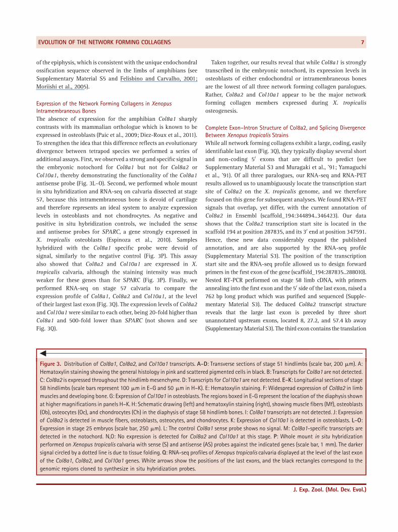
of the epiphysis, which is consistent with the unique endochondralossification sequence observed in the limbs of amphibians (seeSupplementary Material S5 and Felisbino and Carvalho, 2001;Moriishi et al., 2005).
Expression of the Network Forming Collagens in XenopusIntramembraneous BonesThe absence of expression for the amphibian Col8a1 sharplycontrasts with its mammalian orthologue which is known to beexpressed in osteoblasts (Paic et al., 2009; Diez‐Roux et al., 2011).To strengthen the idea that this difference reflects an evolutionarydivergence between tetrapod species we performed a series ofadditional assays. First, we observed a strong and specific signal inthe embryonic notochord for Col8a1 but not for Col8a2 orCol10a1, thereby demonstrating the functionality of the Col8a1antisense probe (Fig. 3L–O). Second, we performed whole mountin situ hybridization and RNA‐seq on calvaria dissected at stage57, because this intramembraneous bone is devoid of cartilageand therefore represents an ideal system to analyze expressionlevels in osteoblasts and not chondrocytes. As negative andpositive in situ hybridization controls, we included the senseand antisense probes for SPARC, a gene strongly expressed inX. tropicalis osteoblasts (Espinoza et al., 2010). Sampleshybridized with the Col8a1 specific probe were devoid ofsignal, similarly to the negative control (Fig. 3P). This assayalso showed that Col8a2 and Col10a1 are expressed in X.tropicalis calvaria, although the staining intensity was muchweaker for these genes than for SPARC (Fig. 3P). Finally, weperformed RNA‐seq on stage 57 calvaria to compare theexpression profile of Col8a1, Col8a2 and Col10a1, at the levelof their largest last exon (Fig. 3Q). The expression levels of Col8a2and Col10a1were similar to each other, being 20‐fold higher thanCol8a1 and 500‐fold lower than SPARC (not shown and seeFig. 3Q).
Taken together, our results reveal that while Col8a1 is stronglytranscribed in the embryonic notochord, its expression levels inosteoblasts of either endochondral or intramembraneous bonesare the lowest of all three network forming collagen paralogues.Rather, Col8a2 and Col10a1 appear to be the major networkforming collagen members expressed during X. tropicalisosteogenesis.
Complete Exon–Intron Structure of Col8a2, and Splicing DivergenceBetween Xenopus tropicalis StrainsWhile all network forming collagens exhibit a large, coding, easilyidentifiable last exon (Fig. 3Q), they typically display several shortand non‐coding 50 exons that are difficult to predict (seeSupplementary Material S3 and Muragaki et al., '91; Yamaguchiet al., '91). Of all three paralogues, our RNA‐seq and RNA‐PETresults allowed us to unambiguously locate the transcription startsite of Col8a2 on the X. tropicalis genome, and we thereforefocused on this gene for subsequent analyses. We found RNA‐PETsignals that overlap, yet differ, with the current annotation ofCol8a2 in Ensembl (scaffold_194:344894..346423). Our datashows that the Col8a2 transcription start site is located in thescaffold 194 at position 287835, and its 30 end at position 347591.Hence, these new data considerably expand the publishedannotation, and are also supported by the RNA‐seq profile(Supplementary Material S3). The position of the transcriptionstart site and the RNA‐seq profile allowed us to design forwardprimers in the first exon of the gene (scaffold_194:287835..288010).Nested RT‐PCR performed on stage 58 limb cDNA, with primersannealing into the first exon and the 50 side of the last exon, raised a762 bp long product which was purified and sequenced (Supple-mentary Material S3). The deduced Col8a2 transcript structurereveals that the large last exon is preceded by three shortunannotated upstream exons, located 8, 27.2, and 57.4 kb away(SupplementaryMaterial S3). The third exon contains the translation
3Figure 3. Distribution of Col8a1, Col8a2, and Col10a1 transcripts. A–D: Transverse sections of stage 51 hindlimbs (scale bar, 200 mm). A:Hematoxylin staining showing the general histology in pink and scattered pigmented cells in black. B: Transcripts for Col8a1 are not detected.C: Col8a2 is expressed throughout the hindlimbmesenchyme. D: Transcripts for Col10a1 are not detected. E–K: Longitudinal sections of stage58 hindlimbs (scale bars represent 100 mm in E–G and 50 mm in H–K). E: Hematoxylin staining. F: Widespread expression of Col8a2 in limbmuscles and developing bone. G: Expression of Col10a1 in osteoblasts. The regions boxed in E‐G represent the location of the diaphysis shownat higher magnifications in panels H–K. H: Schematic drawing (left) and hematoxylin staining (right), showing muscle fibers (Mf), osteoblasts(Ob), osteocytes (Oc), and chondrocytes (Ch) in the diaphysis of stage 58 hindlimb bones. I: Col8a1 transcripts are not detected. J: Expressionof Col8a2 is detected in muscle fibers, osteoblasts, osteocytes, and chondrocytes. K: Expression of Col10a1 is detected in osteoblasts. L–O:Expression in stage 25 embryos (scale bar, 250 mm). L: The control Col8a1 sense probe shows no signal. M: Col8a1‐specific transcripts aredetected in the notochord. N,O: No expression is detected for Col8a2 and Col10a1 at this stage. P: Whole mount in situ hybridizationperformed on Xenopus tropicalis calvaria with sense (S) and antisense (AS) probes against the indicated genes (scale bar, 1 mm). The darkersignal circled by a dotted line is due to tissue folding. Q: RNA‐seq profiles of Xenopus tropicalis calvaria displayed at the level of the last exonof the Col8a1, Col8a2, and Col10a1 genes. White arrows show the positions of the last exons, and the black rectangles correspond to thegenomic regions cloned to synthesize in situ hybridization probes.
J. Exp. Zool. (Mol. Dev. Evol.)
EVOLUTION OF THE NETWORK FORMING COLLAGENS 7

initiation codon and codes for 54 amino acids that are 57% identicalto the residues encoded by the mouse Col8a2 third exon(Supplementary Material S3).Interestingly, the sequence of the second exon obtained by RT‐
PCR does not overlap with the sequence of the second exondetected by RNA‐seq (Supplementary Material S3). Takingadvantage of a unique BsrI restriction site found exclusively inthe sequence obtained by RT‐PCR, we showed that the Col8a2transcripts from developing endochondral and intramembraneousbones (both before and after the onset of osteogenesis) share thesame second exon (Supplementary Material S3). Because RT‐PCRand RNA‐seq data were obtained from animals raised in differentlaboratories, it is possible that this discrepancy at the level of thesecond exon reflects a divergence of splice donor and acceptorsites between X. tropicalis strains.
DISCUSSIONAmongst the huge diversity of collagenous proteins found inanimals, the non‐fibrillar network forming collagens form adiscrete subgroup and display the unique property of being able toassemble as hexagonal networks in the extracellular matrix(Stephan et al., 2004; Heino, 2007; Kadler et al., 2007). Our datasupport a monophyly of the network forming collagen members,their presence uniquely within vertebrates, and an expansion ofthis family through two rounds of whole genome duplications(Ohno, '70; Dehal and Boore, 2005). Both phylogeny andconservation of synteny analyses support a closer relationshipbetween the Col8a2 and Col10a1 paralogues, which thereforeprobably arose from the same precursor gene during the lastduplication event. In this respect the current nomenclatureinaccurately reflects their phylogenetic relationships. We namethe ancestral gene Col8/10 and emphasize that additional workwith invertebrate deuterostomes, cyclostomes and chon-drichthyans will be required to clarify the timing of emergenceof the network forming collagen family.The requirement of a rigid column of cells to strengthen the
antero‐posterior axis of the swimming tadpole larvae of a putativechordate ancestor probably contributed to the recruitment ofextracellular proteins conferring specific mechanical properties tothe notochordal shaft. Hence, notochordal cells express fibrillarcollagens from all three clades (Wada et al., 2006; Zhang andCohn, 2006). Here, we find that, like in teleosts, the amphibianCol8a1 gene is strongly expressed in the notochord (Gansner andGitlin, 2008). Because we did not detect notochordal expressionfor Col8a2 and Col10a1, Col8a1 might be the principal networkforming collagen expressed in the notochord of osteichthyans,possibly explaining the strong axial phenotype observed inCol8a1 mutant zebrafish embryos (Gansner and Gitlin, 2008).In the course of this study, RT‐PCR and RNA‐seq detected a
different sequence for the second non‐coding exon of Col8a2(SupplementaryMaterial S3). This difference appears to be specificto different X. tropicalis strains, because RNA‐seq and RT‐PCRs
performed on various tissues confirmed the presence of a singleexon within each frog population. We therefore interpret ourresults as a consequence of established cis mutations between X.tropicalis strains that have modified the splice acceptor and donorsites of the second Col8a2 exon. Alternative splicing phenomenonis widespread between distantly related species, but also occursbetween human individuals and mouse subspecies, frequentlyaltering coding regions (Wang and Cooper, 2007; Harr andTurner, 2010; Keren et al., 2010). As the second Col8a2 exonappears to be non‐coding, assessing the functional relevance ofthis divergence is not straightforward, and might be aconsequence of neutral drift (Wagner, 2008).Based on phylogeny, synteny and expression, we propose an
evolutionarymodel for the skeletal expression of the network formingcollagen genes (Fig. 4). According to this scenario, the ancestral Col8/10 gene was expressed in bone and cartilage at the base of theosteichthyans. Below, we discuss how the expression of each of thenetwork forming collagen paralogues evolved following speciationand duplication events. First, our results show that the osteoblasticexpression of Col10a1 is shared between amphibians and
Figure 4. A model for the evolution of the network formingcollagens in osteichthyans. On the top and the left of the schemeare shown the phylogenetic relationships of the network formingcollagen genes and the groups of osteichthyans considered in thisstudy, respectively. We propose that the ancestral network formingcollagen gene (Col8/10) was robustly transcribed in osteoblasts(OB) and chondrocytes (CH), and that two rounds of vertebrate‐specific genomic duplications produced three paralogues. Weclassify expression levels of the actinopterygian (Act.), mammalian(Mam.), and amphibian (Amp.) orthologues as follows: detected(þ), not detected (�), or unknown (?).
J. Exp. Zool. (Mol. Dev. Evol.)
8 ALDEA ET AL.

actinopterygians and supports the concept that this feature representsa synapomorphy of osteichthyans which was secondarily lost in theamniote lineage (Kim et al., '99; Avaron et al., 2006; Laue et al., 2008;Li et al., 2009; Albertson et al., 2010; Maye et al., 2011; Eameset al., 2012). Second, Col8a1 represents another case of expressiondivergence between tetrapods. Indeed, while Col8a1 is expressed inmammalian osteoblasts (Paic et al., 2009; Diez‐Roux et al., 2011), weshow here that its osteoblastic expression levels are extremely low byin situ hybridization and RNA‐seq in endochondral and intra-membraneous bones of X. tropicalis. Hence, our results suggest adrastic reductionof the expression levels of this gene in the amphibianlineage. Finally, the Col8a2 paralogue displays a conservedosteoblastic expression between amphibians and mammals. Indeed,Col8a2 has been detected in purified mouse skeletal cells, where ittends to decrease as osteoblasts differentiate into osteocytes (Paicet al., 2009). This observation is consistent with our in situhybridization results showing a stronger signal in osteoblasts thanosteocytes (Fig. 3J). Future studies aiming at analyzing the expressionof both Col8a1 and Col8a2 in actinopterygian skeletal cells will berequired to unravel the evolutionary history of these genes.Considering that Col8a1, Col8a2, and Col10a1 proteins are
highly conserved (Supplementary Material S4) and are able toassemble into higher order structures as homotrimers or hetero-trimers (Stephan et al., 2004; Heino, 2007; Kadler et al., 2007), wepropose that a strong functional redundancy facilitated independentlosses of osteoblastic expression for these genes. Hence, whilenetwork forming collagens are components of the osteichthyanbone matrix, it appears that osteoblasts from distinct vertebratelineages express different combinations of paralogues.
ACKNOWLEDGMENTSWe are grateful to two anonymous reviewers whose constructivecomments improved the quality of the present work. Weacknowledge Ruth Chavez for technical support and FranciscoNualart for providing access to the histology facility.
LITERATURE CITEDAbascal F, Zardoya R, Posada D. 2005. ProtTest: selection of best‐fitmodels of protein evolution. Bioinformatics 21:2104–2105.
Albertson RC, Streelman JT, Kocher TD, Yelick PC. 2005. Integration andevolution of the cichlid mandible: the molecular basis of alternatefeeding strategies. Proc Natl Acad Sci USA 102:16287–16292.
Albertson RC, Yan YL, Titus TA, et al. 2010. Molecular pedomorphismunderlies craniofacial skeletal evolution in Antarctic notothenioidfishes. BMC Evol Biol 10:4.
Avaron F, Hoffman L, Guay D, Akimenko MA. 2006. Characterization oftwo new zebrafish members of the hedgehog family: atypicalexpression of a zebrafish indian hedgehog gene in skeletal elementsof both endochondral and dermal origins. Dev Dyn 235:478–489.
Chan YF, Marks ME, Jones FC, et al. 2010. Adaptive evolution of pelvicreduction in sticklebacks by recurrent deletion of a Pitx1 enhancer.Science 327:302–305.
Dehal P, Boore JL. 2005. Two rounds of whole genome duplication inthe ancestral vertebrate. PLoS Biol 3:e314.
Desronvil T, Logan‐Wyatt D, Abdrabou W, et al. 2010. Distribution ofCOL8A2 and COL8A1 gene variants in Caucasian primary open angleglaucoma patients with thin central corneal thickness. Mol Vis16:2185–2191.
Di‐Poi N, Montoya‐Burgos JI, Miller H, et al. 2010. Changes in Hoxgenes' structure and function during the evolution of the squamatebody plan. Nature 464:99–103.
Diez‐Roux G, Banfi S, Sultan M, et al. 2011. A high‐resolutionanatomical atlas of the transcriptome in the mouse embryo. PLoSBiol 9:e1000582.
Eames BF, Amores A, Yan YL, Postlethwait JH. 2012. Evolution of theosteoblast: skeletogenesis in gar and zebrafish. BMC Evol Biol 12:27.
Edgar RC. 2004. MUSCLE: multiple sequence alignment with highaccuracy and high throughput. Nucleic Acids Res 32:1792–1797.
Espinoza J, SanchezM, Sanchez A, et al. 2010. Two families of Xenopustropicalis skeletal genes display well‐conserved expression patternswith mammals in spite of their highly divergent regulatory regions.Evol Dev 12:541–551.
Felisbino SL, Carvalho HF. 2001. Growth cartilage calcification andformation of bone trabeculae are late and dissociated events in theendochondral ossification of Rana catesbeiana. Cell Tissue Res306:319–323.
Fullwood MJ, Wei CL, Liu ET, Ruan Y. 2009. Next‐generation DNAsequencing of paired‐end tags (PET) for transcriptome and genomeanalyses. Genome Res 19:521–532.
Gansner JM, Gitlin JD. 2008. Essential role for the alpha 1 chain of typeVIII collagen in zebrafish notochord formation. Dev Dyn 237:3715–3726.
Glimcher MJ, Hodge AJ, Schmitt OF. 1957. Macromolecularaggregation states in relation to mineralization: the collagen‐hidroxyapatite system as studied in vitro. Proc Natl Acad Sci USA43:860–867.
Guindon S, Gascuel O. 2003. A simple, fast, and accurate algorithm toestimate large phylogenies by maximum likelihood. Syst Biol52:696–704.
Hall BK. 2005. Bones and cartilage: developmental and evolutionarybiology. Academic Press. San Diego, CA: p 760.
Harr B, Turner LM. 2010. Genome‐wide analysis of alternative splicingevolution among Mus subspecies. Mol Ecol 19:228–239.
Heino J. 2007. The collagen family members as cell adhesion proteins.BioEssays 29:1001–1010.
Hopfer U, Fukai N, Hopfer H, et al. 2005. Targeted disruption of Col8a1and Col8a2 genes inmice leads to anterior segment abnormalities inthe eye. FASEB J 19:1232–1244.
Kadler KE, Baldock C, Bella J, Boot‐Handford RP. 2007. Collagens at aglance. J Cell Sci 120:1955–1958.
Kapoor R, Bornstein P, Sage EH. 1986. Type VIII collagen from bovineDescemet's membrane: structural characterization of a triple‐helical domain. Biochemistry 25:3930–3937.
J. Exp. Zool. (Mol. Dev. Evol.)
EVOLUTION OF THE NETWORK FORMING COLLAGENS 9
Keren H, Lev‐Maor G, Ast G. 2010. Alternative splicing and evolution:diversification, exon definition and function. Nat Rev Genet 11:345–355.
Kim IS, Otto F, Zabel B, Mundlos S. 1999. Regulation of chondrocytedifferentiation by Cbfa1. Mech Dev 80:159–170.
Kwan KM, Pang MK, Zhou S, et al. 1997. Abnormal compartmentali-zation of cartilage matrix components in mice lacking collagen X:implications for function. J Cell Biol 136:459–471.
Langmead B, Trapnell C, Pop M, Salzberg SL. 2009. Ultrafast andmemory‐efficient alignment of short DNA sequences to the humangenome. Genome Biol 10:R25.
Laue K, Janicke M, Plaster N, Sonntag C, Hammerschmidt M. 2008.Restriction of retinoic acid activity by Cyp26b1 is required for propertiming and patterning of osteogenesis during zebrafish develop-ment. Development 135:3775–3787.
Li N, Felber K, Elks P, Croucher P, Roehl HH. 2009. Tracking geneexpression during zebrafish osteoblast differentiation. Dev Dyn238:459–466.
Maldonado‐Agurto R, Toro G, Fuentealba J, et al. 2011. Cloning andspatiotemporal expression of RIC‐8 in Xenopus embryogenesis.Gene Expr Patterns 11:401–408.
Maye P, Fu Y, Butler DL, et al. 2011. Generation and characterization ofCol10a1‐mcherry reporter mice. Genesis 49:410–418.
Miura S, Hanaoka K, Togashi S. 2008. Skeletogenesis in Xenopustropicalis: characteristic bone development in an anuran amphibi-an. Bone 43:901–909.
Moriishi T, Shibata Y, Tsukazaki T, Yamaguchi A. 2005. Expressionprofile of Xenopus banded hedgehog, a homolog of mouse Indianhedgehog, is related to the late development of endochondralossification in Xenopus laevis. Biochem Biophys Res Commun328:867–873.
Muragaki Y, Jacenko O, Apte S, et al. 1991. The alpha 2(VIII) collagengene. A novel member of the short chain collagen family located onthe human chromosome 1. J Biol Chem 266:7721–7727.
Ng P, Wei CL, Sung WK, et al. 2005. Gene identification signature (GIS)analysis for transcriptome characterization and genome annota-tion. Nat Methods 2:105–111.
Nieuwkoop PD, Faber J. 1967. Normal table of Xenopus laevis (Daudin).The Netherlands: North Holland.
Ohno S. 1970. Evolution by gene duplication. Heidelberg, Germany:Springer‐Verlag.
Paic F, Igwe JC, Nori R, et al. 2009. Identification of differentiallyexpressed genes between osteoblasts and osteocytes. Bone 45:682–692.
Plenz GA, Deng MC, Robenek H, Volker W. 2003. Vascular collagens:spotlight on the role of type VIII collagen in atherogenesis.Atherosclerosis 166:1–11.
Ronquist F, Teslenko M, van der Mark P, et al. 2012. MrBayes 3.2:efficient Bayesian phylogenetic inference and model choice across alarge model space. Syst Biol 61:539–542.
Rychel AL, Swalla BJ. 2007. Development and evolution of chordatecartilage. J Exp Zool B Mol Dev Evol 308:325–335.
Sinha S, Kielty CM, Heagerty AM, Canfield AE, Shuttleworth CA. 2001.Upregulation of collagen VIII following porcine coronary arteryangioplasty is related to smooth muscle cell migration notangiogenesis. Int J Exp Pathol 82:295–302.
Stephan S, Sherratt MJ, Hodson N, Shuttleworth CA, Kielty CM. 2004.Expression and supramolecular assembly of recombinant alpha1(viii) and alpha2(viii) collagen homotrimers. J Biol Chem 279:21469–21477.
Tamura K, Nomura N, Seki R, Yonei‐Tamura S, Yokoyama H. 2011.Embryological evidence identifies wing digits in birds as digits 1, 2and 3. Science 331:753–757.
Turnbull AP, Kummel D, Prinz B, et al. 2005. Structure of palmitoylatedBET3: insights into TRAPP complex assembly and membranelocalization. EMBO J 24:875–884.
Wada H, Okuyama M, Satoh N, Zhang S. 2006. Molecular evolution offibrillar collagen in chordates, with implications for the evolution ofvertebrate skeletons and chordate phylogeny. Evol Dev 8:370–377.
Wagner A. 2008. Neutralism and selectionism: a network‐basedreconciliation. Nat Rev Genet 9:965–974.
Wang GS, Cooper TA. 2007. Splicing in disease: disruption of thesplicing code and the decoding machinery. Nat Rev Genet 8:749–761.
Wang Z, Gerstein M, Snyder M. 2009. RNA‐Seq: a revolutionary toolfor transcriptomics. Nat Rev Genet 10:57–63.
Warman ML, Abbott M, Apte SS, et al. 1993. A type X collagenmutation causes Schmid metaphyseal chondrodysplasia. Nat Genet5:79–82.
Yamaguchi N, Mayne R, Ninomiya Y. 1991. The alpha 1 (VIII) collagengene is homologous to the alpha 1 (X) collagen gene and contains alarge exon encoding the entire triple helical and carboxyl‐terminalnon‐triple helical domains of the alpha 1 (VIII) polypeptide. J BiolChem 266:4508–4513.
Zhang G, CohnMJ. 2006. Hagfish and lancelet fibrillar collagens revealthat type II collagen‐based cartilage evolved in stem vertebrates.Proc Natl Acad Sci USA 103:16829–16833.
Zhang G, Cohn MJ. 2008. Genome duplication and the origin of thevertebrate skeleton. Curr Opin Genet Dev 18:387–393.
SUPPORTING INFORMATIONAdditional Supporting Information may be found in the onlineversion of this article at the publisher's web‐site.
Figure S1. Phylogeny of collagens 8a1, 8a2, and 10a1 inosteichthyans. A: Maximum likelihood (ML) tree showing therelationships between Col8a1, Col8a2, and Col10a1. Col15a1 andCol18a1 sequences are used as outgroups. B–E: Phylogeny ofCollagens 8a1, 8a2, and 10a1 in osteichthyans using a Bayesianphylogenetic approach. Only posterior probabilities inferior to 1are shown. Outgroups are composed of members from the Col1a1(B), Col4a2 (C), Col5a1 (D), and Col15a1 (E) families. Note thatCol8a2 and Col10a1 are consistently more closely related to eachother than to the Col8a1 paralogues.
J. Exp. Zool. (Mol. Dev. Evol.)
10 ALDEA ET AL.

Figure S2. In situ hybridization protocol for paraffin sections ofXenopus tropicalis tadpole skeletal elements.
Figure S3. Exon–intron structure of the Col8a2 gene and splicingdivergence between Xenopus tropicalis strains. A–D: RNA‐seqsequences are shown in pink and RT‐PCR sequences are shown ingray. The genomic coordinates of Xenopus tropicalis scaffold 194are indicated on each side of the sequences. A: Localization of thefirst Col8a2 exon by RNA‐seq and RT‐PCR. The positions of theExon1F and Exon1nestedF primers are indicated (blue). B: RNA‐seq and RT‐PCR detected different sequences for the Col8a2second exon. Note that only the exon from the strain tested by RT‐PCR harbors a BsrI site (black). C: Localization of the third Col8a2exon by RNA‐seq and RT‐PCR. Only two residues at position336854 and 337017 (white) differ between the RT‐PCR sequenceand the reference genome. The translation initiation site isindicated in red, and the predicted amino acid sequence isshown below. Residues conserved with mouse are highlighted ingreen. D: The sequence shows the beginning of the fourth andlast Col8a2 exon, until the position of the Exon4R primerused for the RT‐PCR (yellow). For the sake of simplicity, the
predicted amino acid sequence is not shown. E: Schematicsummary of the Xenopus tropicalis Col8a2 structure deduced byRNA‐seq (pink) and RT‐PCR (gray). The size of exons (pb) andintrons (kb), the position of the BsrI restriction site, and of thetranslation start and stop codons are indicated. F: Nested RT‐PCRwas performed on hindlimbs and skull both before (NF 51–53) andafter (NF58) the differentiation of mature osteoblasts. The primerpairs (Exon1F � Exon4R) and (Exon1nestedF � Exon4R) wereused in the first and second PCR reactions, respectively. The PCRproduct was subjected to restriction digest, revealing the presence ofthe BsrI site in all cases. Control PCRs were also performed on RNAwhich was not subjected to reverse transcription (RT�). Thefragment sizes of the molecular weight marker are indicated on theleft (base pairs).
Figure S4. Alignment of vertebrate Collagen 8a1, 8a2, and 10a1protein sequences.
Figure S5. Chondrocytic expression of Col10a1 in the epiphysis ofstage 58 Xenopus tropicalis femoral bones. Arrows point at someof the Col10a1 expressing cells.
J. Exp. Zool. (Mol. Dev. Evol.)
EVOLUTION OF THE NETWORK FORMING COLLAGENS 11