Evidence for conformational changes in the yeast deoxyhypusine hydroxylase Lia1 upon iron...
20
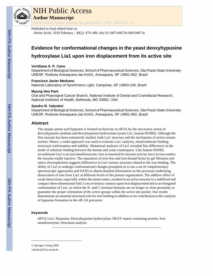
Evidence for conformational changes in the yeast deoxyhypusine hydroxylase Lia1 upon iron displacement from its active site Veridiana S. P. Cano Department of Biological Sciences, School of Pharmaceutical Sciences, São Paulo State University- UNESP, Rodovia Araraquara-Jaú Km01, Araraquara, SP 14801-902, Brazil Francisco Javier Medrano National Laboratory of Synchrotron Light, Campinas, SP 13803-100, Brazil Myung Hee Park Oral and Pharyngeal Cancer Branch, National Institute of Dental and Craniofacial Research, National Institutes of Health, Bethesda, MD 20892, USA Sandro R. Valentini Department of Biological Sciences, School of Pharmaceutical Sciences, São Paulo State University- UNESP, Rodovia Araraquara-Jaú Km01, Araraquara, SP 14801-902, Brazil Abstract The unique amino acid hypusine is formed exclusively in eIF5A by the successive action of deoxyhypusine synthase and deoxyhypusine hydroxylase (yeast Lia1, human DOHH). Although the first enzyme has been extensively studied, both Lia1 structure and the mechanism of action remain unclear. Hence, a multi-approach was used to evaluate Lia1 catalysis, metal/substrate binding, structural conformation and stability. Mutational analyses of Lia1 revealed fine differences in the mode of substrate binding between the human and yeast counterparts. Like human DOHH, recombinant Lia1 is an iron metalloenzyme. Iron is essential for enzyme activity since its loss renders the enzyme totally inactive. The separation of iron-free and iron-bound forms by gel filtration and native electrophoresis suggests differences in Lia1 tertiary structure related to the iron binding. The ability of Lia1 to undergo conformational changes prompted us to use a set of complementary spectroscopic approaches and SAXS to obtain detailed information on the processes underlying dissociation of iron from Lia1 at different levels of the protein organization. The additive effect of weak interactions, especially within the metal center, resulted in an active enzyme in a stabilized and compact three-dimensional fold. Loss of tertiary contacts upon iron displacement led to an elongated conformation of Lia1, in which the N- and C-terminal domains are no longer in close proximity to guarantee the proper orientation of the active groups within the active site pocket. Our results demonstrate an essential structural role for iron binding in addition to its contribution to the catalysis of hypusine formation in the eIF-5A precursor. Keywords eIF5A Lia1; Hypusine; Deoxyhypusine hydroxylase; HEAT-repeat containing protein; Iron metalloenzyme; Structural analysis © Springer-Verlag 2009 [email protected] . NIH Public Access Author Manuscript Amino Acids. Author manuscript; available in PMC 2010 July 12. Published in final edited form as: Amino Acids. 2010 February ; 38(2): 479–490. doi:10.1007/s00726-009-0407-8. NIH-PA Author Manuscript NIH-PA Author Manuscript NIH-PA Author Manuscript
-
Upload
independent -
Category
Documents
-
view
1 -
download
0
Transcript of Evidence for conformational changes in the yeast deoxyhypusine hydroxylase Lia1 upon iron...
Evidence for conformational changes in the yeast deoxyhypusinehydroxylase Lia1 upon iron displacement from its active site
Veridiana S. P. CanoDepartment of Biological Sciences, School of Pharmaceutical Sciences, São Paulo State University-UNESP, Rodovia Araraquara-Jaú Km01, Araraquara, SP 14801-902, Brazil
Francisco Javier MedranoNational Laboratory of Synchrotron Light, Campinas, SP 13803-100, Brazil
Myung Hee ParkOral and Pharyngeal Cancer Branch, National Institute of Dental and Craniofacial Research,National Institutes of Health, Bethesda, MD 20892, USA
Sandro R. ValentiniDepartment of Biological Sciences, School of Pharmaceutical Sciences, São Paulo State University-UNESP, Rodovia Araraquara-Jaú Km01, Araraquara, SP 14801-902, Brazil
AbstractThe unique amino acid hypusine is formed exclusively in eIF5A by the successive action ofdeoxyhypusine synthase and deoxyhypusine hydroxylase (yeast Lia1, human DOHH). Although thefirst enzyme has been extensively studied, both Lia1 structure and the mechanism of action remainunclear. Hence, a multi-approach was used to evaluate Lia1 catalysis, metal/substrate binding,structural conformation and stability. Mutational analyses of Lia1 revealed fine differences in themode of substrate binding between the human and yeast counterparts. Like human DOHH,recombinant Lia1 is an iron metalloenzyme. Iron is essential for enzyme activity since its loss rendersthe enzyme totally inactive. The separation of iron-free and iron-bound forms by gel filtration andnative electrophoresis suggests differences in Lia1 tertiary structure related to the iron binding. Theability of Lia1 to undergo conformational changes prompted us to use a set of complementaryspectroscopic approaches and SAXS to obtain detailed information on the processes underlyingdissociation of iron from Lia1 at different levels of the protein organization. The additive effect ofweak interactions, especially within the metal center, resulted in an active enzyme in a stabilized andcompact three-dimensional fold. Loss of tertiary contacts upon iron displacement led to an elongatedconformation of Lia1, in which the N- and C-terminal domains are no longer in close proximity toguarantee the proper orientation of the active groups within the active site pocket. Our resultsdemonstrate an essential structural role for iron binding in addition to its contribution to the catalysisof hypusine formation in the eIF-5A precursor.
KeywordseIF5A Lia1; Hypusine; Deoxyhypusine hydroxylase; HEAT-repeat containing protein; Ironmetalloenzyme; Structural analysis
© Springer-Verlag [email protected] .
NIH Public AccessAuthor ManuscriptAmino Acids. Author manuscript; available in PMC 2010 July 12.
Published in final edited form as:Amino Acids. 2010 February ; 38(2): 479–490. doi:10.1007/s00726-009-0407-8.
NIH
-PA Author Manuscript
NIH
-PA Author Manuscript
NIH
-PA Author Manuscript
IntroductionHypusine biosynthesis is a remarkable post-translational modification known to date to occurin only one single cellular protein, the putative eukaryotic translation initiation factor 5A(Cooper et al. 1983; Park et al. 1981). The unusual amino acid hypusine is generated in a two-step reaction process catalyzed consecutively by deoxyhypusine synthase (yeast Dys1/humanDHS) and deoxyhypusine hydroxylase (yeast Lia1/human DOHH) (Wolff et al. 2007). Thefirst enzyme mediates the NAD-dependent transfer of the 4-aminobutyl moiety of spermidineto the ε-amino group of one specific lysine residue (K51 in yeast protein) in the eIF5Aprecursor. Hydroxylation of the resulting deoxyhypusine intermediate by the second enzymeof the pathway leads to the formation of hypusine (Park 2006; Park et al. 1982). Hydroxylationnot only completes hypusine biosynthesis but also eIF5A maturation, thus rendering the proteinbiologically active (Abbruzzese et al. 1986; Park 2006).
The hypusine residue exerts a critical role for the protein function in vivo. Disruption ofdeoxyhypusine synthase gene (DYS1), as well as the single mutation of the lysine at the siteof hypusine formation in the eIF5A precursor, results in loss of cell viability in the yeastSaccharomyces cerevisiae (Sasaki et al. 1996; Schnier et al. 1991). Besides, an increase ofcells arrested in G1 is observed upon rapid depletion of eIF5A, or shift of conditional eIF5Amutants to the restrictive temperature (Kang and Hershey 1994; Zanelli and Valentini 2005).Despite the clear correlation observed between the inhibition of hypusine formation and arrestof cell-cycle progression in yeast (Kang and Hershey 1994; Zanelli and Valentini 2005) andin mammalian cells treated with inhibitors of DOHH (Hanauske-Abel et al. 1994), thebiological function of this intriguing protein still remains to be elucidated. Recently, thehypusine-dependent association of eIF5A with ribosomes actively engaged in translation hasbrought eIF5A back to the translation scenario (Jao and Chen 2006; Zanelli et al. 2006).Furthermore, a defect observed in polysomal profiles of temperature-sensitive mutantssupports a role for eIF5A in the elongation step of translation instead of initiation (Zanelli etal. 2006; Zanelli and Valentini 2007). Finally, a defect in protein synthesis was detected inyeast cells expressing mutated forms of human or yeast eIF5A as the primary consequence ofloss of protein function (Cano et al. 2008; Dias et al. 2008).
In an attempt to identify eIF5A-interacting proteins that would help us to understand its rolein the cell, a two-hybrid screen was performed, and the protein encoded by the geneYJR070C, named LIA1 (Ligand of eIF5A), was identified as an eIF5A cellular partner in S.cerevisiae (Thompson et al. 2003). It was subsequently shown that the gene LIA1 encodes theenzyme responsible for the final step of hypusination, deoxyhypusine hydroxylase (Park et al.2006).
Prediction studies of its secondary structure have revealed that Lia1 contains motifs known asHEAT-like repeats (Park et al. 2006; Thompson et al. 2003), which are found in a variety ofproteins, including the four proteins Huntingtin, Elongation factor 3, the PR65/A subunit ofprotein phosphatase 2A and TOR1, from which “HEAT” is derived. The consensus secondarystructure for an individual HEAT motif consists of a pair of anti-parallel α-helices separatedby a non-helical region. Normally, HEAT repeats appear in tandem arrays to form an elongatedmolecule characterized by a double layer of α-helices. Interestingly, a role for mediatingprotein–protein interactions has been suggested for these repeats (Groves et al. 1999).Structural modeling predicts that the human homologue protein DOHH consists of eight HEATrepeats in a symmetrical dyad (four HEAT repeats in each of the N- and C- terminal armsconnected by a variable loop). Two metal coordination sites have been identified at the fourconserved His-Glu motifs of human DOHH (Kim et al. 2006; Park et al. 2006). Conservationof the HEAT repeat structures and the potential metal coordination sites suggests that, likehuman DOHH, Lia1 also possesses an iron-dependent catalytic activity.
Cano et al. Page 2
Amino Acids. Author manuscript; available in PMC 2010 July 12.
NIH
-PA Author Manuscript
NIH
-PA Author Manuscript
NIH
-PA Author Manuscript
Although the human DOHH has been characterized for its iron requirement and substratebinding through extensive mutagenesis studies (Kang et al. 2007; Kim et al. 2006), little isknown about the structural aspects of Lia1 and its catalytic requirements. To better understandthe secondary and tertiary structures of this distinct hydroxylase in relation to its iron bindingand activity, we performed structural and functional analyses of the purified recombinant yeastLia1, by employing in-depth spectroscopic analyses and small angle X-ray scattering (SAXS),in addition to site-directed mutagenesis to dissect the structural components of the Lia1 activesite. The mutational analysis was carried out with ten proteins with ala-nine substitutions athighly conserved amino acid residues. In the spectroscopic analysis, we have exploited twostructural characteristics of Lia1: the distribution of HEAT repeats along the protein, in orderto follow circular dichroism signals, and the presence of a single tryptophan residue to evaluateits fluorescence properties. Finally, the conformational changes of Lia1 observed upondisruption of iron binding were analyzed by acrylamide quenching of tryptophan intrinsicfluorescence and by SAXS angle X-ray scattering (SAXS). Taken together, our data suggestfine differences in the coordination of iron and substrate at the active site between humanDOHH and yeast Lia1 and support the essential contribution of iron for Lia1 hydroxylaseactivity as well as its tertiary structure by maintenance of its active conformation.
Experimental proceduresPurification of yeast recombinant wild type and mutant Lia1 proteins
E. coli BL21(DE3) cells harboring the plasmids expressing wild-type or mutant forms of Lia1as GST fusion proteins were grown in 1 l of LB medium containing 100 μg/ml ampicillin.GST-Lia1 protein expression was induced when the culture reached 0.6 OD600nm by additionof 0.1 mM IPTG for 4 h at 37°C. Cells were harvested by centrifugation and suspended in 100ml of ice-cold Tris buffer (50 mM Tris–HCl at pH 7.5 and 1 mM DTT) containing an EDTA-free protease inhibitor cocktail. Cells were lysed using an ultrasonic processor and debris wasremoved by centrifugation at 20,000×g at 4°C for 45 min. GST-Lia1 present in the clarifiedsupernatant was then purified by affinity chromatography to a glutathione–Sepharose resinusing a FPLC system (GE Healthcare). After thrombin cleavage of GST-Lia1, free GST proteinwas eliminated by a second chromatographic step using the same resin. The purified Lia1protein was further subjected to molecular exclusion separation in a Superdex 200 gel filtrationcolumn. Fractions were grouped in pools and concentrated in centrifugal filter devices. Purifiedenzymes were then equilibrated in Tris buffer (50 mM Tris–HCl, pH 7.5, 1 mM DTT). Athrombin cleavage capture kit was used to release free mutant enzymes obtained from small-scale purifications.
Site-directed mutagenesisTen single mutants (H79A, E80A, H112A, E113A, E116A, E116D, H237A, E238A, H270A,E271A) of Lia1 from S. cerevisiae were generated at highly conserved amino acid residuesusing the QuickChange site-directed mutagenesis kit. pGEX-4T-1-LIA1 and pBTM116-LIA1(Thompson et al. 2003), encoding yeast wild type Lia1 as a GST and LexA fusion protein,respectively, were used as template for the PCR with primer sets designed for the desiredmutations. Each amino acid substitution was confirmed by DNA sequencing.
Circular dichroism spectroscopyCircular dichroism measurements were carried out on a JASCO J-810 spectropolarimeter,equipped with a Peltier-type temperature controller and a thermostated cell holder, interfacedwith a thermostatic bath. Far-UV spectra were recorded in a 0.1 cm path length quartz cell ata protein concentration of 0.15 mg/ml in 20 mM Tris–HCl pH 7.5. Five consecutive scans wereaccumulated and the average spectra stored. The data were corrected for the baselinecontribution of the buffer and the observed ellipticities were converted into the mean residue
Cano et al. Page 3
Amino Acids. Author manuscript; available in PMC 2010 July 12.
NIH
-PA Author Manuscript
NIH
-PA Author Manuscript
NIH
-PA Author Manuscript
ellipticities [θ] based on a mean molecular mass per residue of 112 Da. Thermal denaturationexperiments were performed by increasing the temperature from 20 to 95°C at 1°/min, allowingtemperature equilibration for 5 min before recording each spectrum. Tm represents thetemperature at the midpoint of the unfolding transition. (GdnHCl)-induced denaturation curvesat constant temperature were obtained by recording the CD signal at 222 nm for each sample.
Fluorescence spectroscopySteady-state fluorescence measurements were carried out in an Aminco BOWMAN series 2spectrofluorometer. Excitation and emission bandwidths were 8 and 16 nm, respectively.Tryptophan fluorescence was measured with an excitation wavelength of 295 nm and theemission spectra were recorded between 310 and 400 nm. All measurements were performedin 20 mM Tris–HCl, pH 7.5 at 25°C and at a protein concentration of 0.15 mg/ml. Stern–Volmerquenching was performed using 295 nm excitation and 342 nm emission. Fluorescenceintensities were corrected for dilution and contribution of the quenchers and plotted as the ratioof initial fluorescence intensity (Fo) to intensity (F) at each quencher concentration (Fo/F vs.[Q].) The slope determined by linear regression was the Stern–Volmer quenching constant,Ksv. Iodide stock solutions (KI 1 M) were made fresh before use, held on ice, and protectedfrom light.
Calculation of the apparent fractional extent of unfoldingThe observed CD or fluorescence signal at any given temperature or GdnHCl concentrationwas converted to apparent fractional extent of unfolding (Fapp) using the equation: Fapp =(Yobs − YU)/(YU − YN), where Yobs is the observed CD or fluorescence signal, and YN and YUrefer to the native and unfolded states, respectively. The values of YN and YU were obtainedby linear extrapolation of the temperature or perturbing agent dependence from the regionsbefore and after the transition.
Small angle X-ray scatteringSmall angle X-ray scattering experiments were performed at the D011A-SAXS1 beamline atthe National Laboratory of Synchrotron Light (Campinas, Brazil). Data were collected usinga MAR CCD 165 (MAR Research) area detector, and 10 min exposure time for eachmeasurement at room temperature. The wavelength used in the experiments was 1.488 Å. Thedistance between the sample and detector was 1,074.35 mm. Scattered intensities wererecorded in the angular range 0.0501 < q < 0.3085 Å−1, where q is the scattering vector(4πsinθ/λ), and 2θ and λ are the scattering angle and wavelength of the X-rays, respectively.Data were analyzed using the software package FIT2D. Intensity data were reduced to I(q)versus q using standard procedures to correct for incident beam intensity, sample absorptionand blank subtraction. The radius of gyration, Rg, was calculated using the entire scatteringcurve with the program GNOM (Semenyuk and Svergun 1991), which also provides thedistance distribution function P(r) and the maximum dimension of the scattering particles. Thelow-resolution model was calculated using the program DAMMIN in slow mode (Svergun1999). In order to increase the reliability of the results, the final models for the dummy atommodeling were obtained by a spatial average of six independent models, calculated with theprogram DAMAVER (Volkov and Svergun 2003).
ResultsLia1 is an iron metalloenzyme
The yeast recombinant protein Lia1 purified by affinity chromatography on glutathione–Sepharose resin was subjected to size-exclusion chromatography in order to evaluate its purity.The elution profile of Lia1 during gel filtration on Superdex 200 column (Fig. 1a) revealed a
Cano et al. Page 4
Amino Acids. Author manuscript; available in PMC 2010 July 12.
NIH
-PA Author Manuscript
NIH
-PA Author Manuscript
NIH
-PA Author Manuscript
small shoulder corresponding to the first four eluted fractions, in addition to a major peakcorresponding to the last five fractions. All fractions showed a single band of approximately36 kDa when analyzed by SDS-PAGE (Fig. 1b). However, when the same samples wereevaluated by non-denaturing electrophoresis (Fig. 1c), two different bands were clearlydistinguished, a diffuse upper band (dashed arrow), present in all fractions, and a better resolvedband (solid arrow), which migrates faster, present only in the last five fractions (fractions 32–36). The fractions were pooled into three different groups: group 1, fractions 28–31, displayingmainly the upper diffuse band; group 2, fractions 32 and 33 with a mixture of the two observedbands; and group 3, fractions 34–36, composed mainly of the lower band.
When Lia1 activity was measured in these three groups (Fig. 1d), the enzyme activity wasmarkedly reduced (nearly no enzymatic activity) in group 1, and was moderate in group 2 (35%of substrate deoxyhypusine converted to hypusine). On the other hand, purified Lia1 fromgroup 3 showed the highest activity with nearly 70% conversion of the substrate protein, eIF5A([3H]Dhp), to the hypusine form, eIF5A([3H]Hpu) (Fig. 1d). The enzyme activity wasevaluated as herein (Park et al. 1984). eIF5A([3H]Dhp) was prepared as described previously(Park et al. 2003).
Analysis of metal content was then performed by inductively coupled plasma-high resolutionmass spectrometry. Iron, out of nine metals analyzed (cadmium, cobalt, chromium, copper,iron, magnesium, manganese, nickel and zinc) was found to be the only metal associated withLia1 in the samples. Metal contents were 0.1, 0.3 and 1.2 mol of iron per mol of enzyme, forpool 1, 2 and 3, respectively (Fig. 1e). The essential nature of iron for Lia1 activity is evidentas loss of metal (in pool 1) results in a totally inactive enzyme. Taken together, these datasuggest that the lower band of the enzyme upon native gel represents the metal-boundholoenzyme, whereas the diffuse upper band corresponds to the inactive iron-free apoenzyme.The presence of both the apoenzyme and the holoenzyme in groups 2 and 3 may be due to agradual metal dissociation from Lia1 or due to its inability to obtain a full content of iron. Incontrast to the human enzyme, no reconstitution of the holoenzyme from apoenzyme wasobserved with addition of 0.2 mM ferrous ammonium sulfate or ferric chloride (data notshown). Separation of the Lia1 apoenzyme and the holoenzyme by gel filtration and by nativegel electrophoresis demonstrates that the apoenzyme has a larger hydrodynamic size than theholoenzyme.
Mapping amino acid residues critical for iron and/or substrate binding and catalysisIn order to identify amino acid residues of Lia1 active site that contribute to metal binding andcatalysis, a mutational analysis was undertaken by site-directed mutagenesis. Target residueswere selected at the four conserved His-Glu motifs (the predicted iron binding site) and anadditional highly conserved residue E116. Thus, ten mutant proteins were generated: H79A,E80A, H112A, E113A, E116A, E116D, H237A, E238A, H270A, E271A. All the eight mutantproteins with alanine substitutions at the conserved His-Glu motifs were completely inactivein deoxyhypusine hydroxylation (Fig. 2a). Interestingly, while the mutant E116A was inactive,substitution at the same residue with aspartate rendered low levels of enzyme activity (3% ofwild type). The metal content for six mutants (H79A, H112A, E113A, H237A, H270A andE271A) was very low (nearly zero), indicating that loss of iron binding contributes to enzymeinactivation. On the other hand, mutants E80A and E238A, despite the lack of enzymaticactivity, displayed a relatively high metal content (0.6 and 1.2 mol/mol, respectively) andE116A and E116D, a modestly reduced level of iron (0.4 mol/mol).
The activity and iron contents of the His-Glu site mutants of Lia1 are fairly similar to the patternobserved for the corresponding mutants of the human protein reported (Kang et al. 2007; Kimet al. 2006). Yet the involvement of the same six residues of the conserved His-Glu motifs(H79, H112, E113, H237, H270 and E271 for the yeast Lia1) in iron binding reveals the critical
Cano et al. Page 5
Amino Acids. Author manuscript; available in PMC 2010 July 12.
NIH
-PA Author Manuscript
NIH
-PA Author Manuscript
NIH
-PA Author Manuscript
importance of these set of amino acid residues for Lia1 catalysis. However, analysis by nativegel electrophoresis not only revealed some differences in mobility between the human mutantenzymes and the yeast counterparts, but also showed some disparities among the yeast mutants(Fig. 2b). The slow and diffused upper band of Lia1 wild type suggest heterogeneouspopulation of apoenzymes with different tertiary structures and varying degrees of opennessof the dyad arms. The fast moving tight band, in turn, suggests that the holoenzymes arehomogeneous in compact structures. Although all the iron deficient mutants of human DOHHbehaved like the human wild type apoenzyme upon native gel electrophoresis (Kim et al.2006), some iron deficient Lia1 mutants did not. Lia1 mutants H79A, H112A and E113Aresolved as a sharp band around the same position of the wild type apoenzyme (Fig. 2b),suggesting that these mutants have opened dyad arms, but are homogeneous in the degree ofopenness and tertiary structure. In contrast, E271A resolved as a much broader band than theapo form of the wild type Lia1, suggesting that two dyad arms are more flexible and free tomove in this mutant. Curiously, the two other iron binding deficient mutants, H237A andH270A also exhibited a smear of apoenzyme, but an additional fast moving band of higherresolution could be detected (Fig. 2b), suggesting that a portion of this mutant still retains acompact holoenzyme-like structure. E116A/D displayed both apo and holo forms of theenzyme with reduced holoenzyme band consistent with its reduced iron content. These resultsdemonstrate that the yeast Lia1 mutant proteins, in the absence of iron binding, displaystructural variations depending on the specific mutations.
Finally, to determine whether enzyme inactivity was a result of impaired substrate binding, thesame Lia1 mutants were generated as LexA-fusion proteins. Expression of two-hybrid reportergenes, HIS3 and LacZ, was used to monitor the interaction between eIF5A fused to Gal4activating domain and the mutant enzymes (Fig. 2c) (Thompson et al. 2003). E113A, E116A,E238A, H270A, E271A and E274A mutants did not grow in medium lacking histidine andalso were negative for β-galactosidase assay, suggesting loss of interaction with eIF5A. On theother hand, the mutant proteins H79A, E80A, H112A, E116D, H237A and E274D were ableto interact with eIF5A as they induced expression of the reporter genes and supported growthfully or partially in the absence of His. The finding that the E116D and E274D with Aspsubstitution are partially active in eIF5A binding whereas those with Ala substitutions areinactive, suggests the importance of these acidic residues of Lia1 in anchoring the substrateprotein. Thus, the data reveal that Glu residues E113, E116, E238, E271 and E274 are criticalfor anchoring eIF5A in the enzyme structure.
Structural characterization of Lia1 by spectroscopic techniquesPrediction studies of Lia1 secondary structure revealed the presence of motifs known as HEATrepeats (Thompson et al. 2003). These motifs are individually composed of a pair of α-helicesand seem to assemble in a repetitive fashion throughout the Lia1 protein. In this context, circulardichroism spectroscopy in the far-UV region (190–250 nm) was initially used to evaluate thebackbone conformation of Lia1. The CD spectrum of Lia1 at 25°C (Fig. 3a) showed two relativeminima at 208 and 222 nm, and a maximum around 192 nm. This spectrum is typical of proteinswith a high content of α-helices, and thus, confirms the structural prediction. In addition, anestimation of the secondary structure content using the program CDNN showed the presenceof 59.3% α-helix, 6.6% β-sheet, 15.7% turn and 22.5% remainder.
Lia1 contains a single tryptophan residue located at position 155. According to the structuralmodel of the human protein (Park et al. 2006), this residue is positioned in the fourth HEATrepeat in the vicinity of a variable loop, which connects the N- (HEAT repeats 1–4) and C-terminal (HEAT repeats 5–8) halves of this protein. The hinge loop is believed to be a regionof great flexibility, and thus, the single tryptophan W155 was used as a reporter group tomonitor structural properties of the yeast Lia1 enzyme. The tryptophan maximum emission
Cano et al. Page 6
Amino Acids. Author manuscript; available in PMC 2010 July 12.
NIH
-PA Author Manuscript
NIH
-PA Author Manuscript
NIH
-PA Author Manuscript
wavelengths (λmax) observed after excitation of Lia1 protein at 280 and 295 nm were,respectively, 339 and 342 nm (Fig. 3b). These values suggest a low exposure of the indole ringto an aqueous environment. As a means to determine the degree of exposure of this tryptophanresidue, quenching studies of Lia1 intrinsic fluorescence were carried out using collisionalquenchers. The Stern–Volmer constants (KSV) obtained for acrylamide and potassium iodide(KI) were 2.45 ± 0.04 and 2.99 ± 0.15 M−1, respectively. In contrast, the reported Stern–Volmerconstants for acrylamide and KI quenching of N-acetyl-tryptophan-amide (Ac-Trp-NH2), amodel compound for a completely solvent-exposed fluorophore, were 20.8 and 10.5 M−1,respectively. The low values of KSV observed for Lia1 quenching confirmed that its tryptophanresidue (W155) is buried in the native protein molecule, thus not readily accessible toacrylamide or iodide.
Effects of pH on Lia1 catalytic activity and tertiary structureAs pH can alter the ionization state of amino acid side chains, thereby affecting either thereactivity of catalytic groups or the conformation of the enzyme active site, we sought toinvestigate the effects of pH on Lia1 enzyme activity and tertiary structure. The purifiedrecombinant yeast enzyme was able to catalyze in vitro the conversion of eIF5A([3H]Dhp) toeIF5A([3H]Hpu) in a pH range from 6.0 to 10.0 (Fig. 4a). Although the protein was active inthis wide pH range, the maximum activity was observed at pH 7.5. Low pH values (<6.0)resulted in complete loss of activity, whereas at higher pH values (>8.0) the enzyme partiallyretained its activity. To evaluate whether loss of enzyme activity was related to dissociation ofiron, native gel electrophoresis was used to analyze the conformational state adopted by Lia1at different pH. As shown in Fig. 4b, at pH 2.0 and 3.0 the enzyme displayed only the upperband, characteristic of the apoenzyme. This is in accordance with the lack of activity at thesepH values. Conversely, the lower band was mainly detected at pH > 7.0, likely due to thepresence of iron in the active site of the holoenzyme. Although the two bands representing theapo- and the holo-forms are present at pH 4.0 and 5.0, no enzymatic activity was detected. Thelack of activity at these pH values might be a consequence of the low iron content combinedwith the protonation of the histidine and glutamate residues of the active site andconformational changes of Lia1 due to global changes in protonation status. Finally, at pH 6.0both forms coexisted and partial activity was observed.
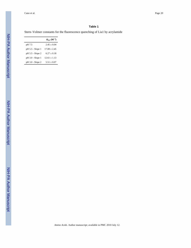
As shown earlier by collisional quenching studies with acrylamide and KI, the only tryptophanresidue present in Lia1 protein seems to be buried from the solvent. In order to determine ifthe structural changes are associated with iron disruption from the Lia1 active site, quenchingexperiments with acrylamide were carried out at pH 3.0, 5.5 and 7.5 (Fig. 4c). Fluorescenceintensities values at λmax were used to obtain Stern–Volmer plots and to calculate KSV (Table1). At low pH values, 5.5 and 3.0, quenching of Lia1 fluorescence proceeds as a biphasicprocess. For both pH values, the first phase showed an anomalous increase, in contrast to theKSV obtained for Lia1 at pH 7.5, since the KSV was close to that of free tryptophan. The KSVcalculated for the second stages of both pH values 3.0 and 5.5 were approximately 2 and 2.5times higher than the one obtained at pH 7.5 (Table 1). Thus, these results suggest thattryptophan accessibility, which was low at pH 7.5, increased upon disruption of compact Lia1structure as the pH was lowered.
Stability of Lia1 as judged by heat- and GdnHCl-induced unfoldingThe spectral features of Lia1 shown above suggest that changes in CD signals and fluorescencecan be used to characterize the global structural changes occurring during the unfolding of thisprotein. As the temperature was raised to 95°C, Lia1 lost the characteristics of an α-helicalprotein, giving rise to a different CD spectrum at each of the pH values analyzed (data notshown). At pH 7.5, both minima signals at 208 and 222 nm shifted to 204 and 220 nm,respectively; while the relative maximum at 192 nm decreased its intensity to nearly zero (Fig.
Cano et al. Page 7
Amino Acids. Author manuscript; available in PMC 2010 July 12.
NIH
-PA Author Manuscript
NIH
-PA Author Manuscript
NIH
-PA Author Manuscript

5a). However, temperature-induced changes of the ellipticity at pH 7.5 began at early stages(~35°C) and unfolding of the protein was completed once the temperature reached 65°C (Fig.5b). The detection of an inflexion point around 45°C might indicate the existence of a stableunfolding intermediate. The presence of an excess of iron (100 μM) in the solution seems tochange this point slightly (Fig. 5b). A significant decrease in protein stability under acidicconditions was observed (Fig. 5c), as reflected by the decrease of the midpoint of denaturation.Although at pH 5.5 Lia1 began to unfold at similar temperatures observed for higher pH values,the midpoint of denaturation was achieved 10°C lower (Fig. 5c), revealing the cooperativityof the unfolding profile under this pH. At the lowest pH studied (3.0) the effect was even moredrastic, since unfolding of the protein started at 25°C and was saturated at 55°C with theunfolding midpoint at approximately 30°C. At pH 9.6 the inflexion point detected at pH 7.5became more pronounced (Fig. 5c). These data reinforce the existence of a stable unfoldingintermediate.
GdnHCl-induced unfolding of Lia1 was monitored at pH 7.5 and 4.5 by following changes inintrinsic fluorescence of the tryptophan residue, reflected by fluorescence maximum intensityand center of mass, as well as CD signals (Fig. 5d). The changes occurring in Lia1 secondarystructure monitored at 222 nm showed a GdnHCl-concentration-dependent loss of ellipticityin both pH 7.5 and 4.5. The transition curves shown in Fig. 5d demonstrated that Lia1 at pH7.5 began to unfold with 1.0 M GdnHCl and complete unfolding of the protein was achievedin 4.5 M GdnHCl. In contrast, at pH 4.5 the midpoint of denaturation was achieved at lowerGdnHCl concentration. Similar to the thermal unfolding, in all the three profiles an inflexionpoint between 2.5 and 3 M GdnHCl was observed. Thus, the GdnHCl-induced transition curvesof the enzyme displayed a more complex unfolding pathway, suggesting the accumulation ofintermediate specie(s) in equilibrium with native and unfolded states. Taken together, the datademonstrate higher protein instability at lower pH, which can be correlated with loss of ironfrom its coordination sites.
Topology of Lia1 in its iron-free and iron-bound states by SAXSSmall angle scattering profiles, obtained for Lia1 wild-type at two different pH values (7.5 andcitrate 4.5) and for the mutant apoenzyme E113A are shown in Fig. 6 together with theircorresponding pair distance distribution functions. The asymmetric shape of the P(r) functionsrevealed that the protein is slightly elongated. The radius of gyration of Lia1 wild-type at pH7.5, calculated from the second moment of the P(r) function, was 24.17 ± 0.01 Å. In contrast,in the absence of iron the radii of gyration were 27.07 ± 0.04 Å (WT, pH 4.5) and 25.80 ± 0.02Å (E113A). These numbers are in good agreement with the hydrodynamic radius obtained bydynamic light scattering and indicate a more compact conformation of the iron-bound state.
A low-resolution model was calculated using a dummy atom modeling procedure (Svergun1999). The models obtained for Lia1 are shown in Fig. 7, together with the crystal structure ofthe protein YIBA from E. coli (PDB Id. 1OYZ), used for comparison. This protein has a HEAT-repeat folding and a molecular mass of 31.9 kDa, close to that of Lia1. The models are theaverage of six independent calculations in order to give the most probable structure (Volkovand Svergun 2003). The overall shape of iron-free of Lia1 (WT-pH 4.5 and E113A) is lesscompact and more elongated than that observed for the holoenzyme (WT-pH 7.5), suggestingthat both arms are further separated in the apoenzyme. These data are in accordance with thetryptophan fluorescence experiments and support the notion that Lia1 conformation isdependent on iron binding or release.
DiscussionLia1 catalyzes the final step of hypusine synthesis, the hydroxylation of deoxyhypusine ineIF5A. Our results demonstrate that Lia1 is a novel HEAT repeat containing metalloenzyme
Cano et al. Page 8
Amino Acids. Author manuscript; available in PMC 2010 July 12.
NIH
-PA Author Manuscript
NIH
-PA Author Manuscript
NIH
-PA Author Manuscript
with a structure distinct from that of other protein hydroxylases. The sequence of Lia1 is highlyconserved in all eukaryotes from fungi to human and its structure is predicted to consist ofeight HEAT repeats, with four repeats in symmetrical N- and C-terminal dyad arms, as wasmodeled for the human protein (Park et al. 2006). Mutational analyses of Lia1 reveal apparentdifferences in the binding of substrate protein at the active sites of yeast Lia1 and humanDOHH. Furthermore, we have carried out extensive structural analyses of Lia1 employingcomplementary spectroscopic techniques (CD and quenching of tryptophan fluorescence) andSAXS to obtain detailed information on the process of structural changes associated withthermal and chemical denaturation, low pH and loss of iron. The results indicate a dual role ofiron binding in Lia1, for catalysis as well as for structural integrity.
The yeast recombinant Lia1, like human DOHH, was shown to be an iron metalloenzymeconsisting of a mixture of apoenzyme (iron-free form) and holoenzyme (iron-bound form).The essential nature of this metal for enzyme activity was demonstrated by the fact that Lia1apoenzyme was totally devoid of activity. The identification and separation of apo- and holo-forms of the human enzyme (Kim et al. 2006) and Lia1 by molecular exclusion chromatographyand native electrophoresis suggest structural differences in the tertiary structure depending onthe presence or absence of metal. The super helical structure of DOHH is entirely differentfrom the beta-jellyroll structure, termed double stranded beta helix (DSBH), found in themajority of other protein hydroxylases in the family of the Fe(II)-and 2-oxoacid-dependentdioxygenases, e.g., prolyl and lysyl hydroxylases. While both DOHH and DSBH enzymes aredependent on molecular oxygen as a source of hydroxyl group, there are clear differences inreaction mechanisms. There is no requirement for 2-oxoacid for DOHH catalysis and no siteidentified for 2-oxoacid binding (Kim et al. 2006; Park et al. 2006). Moreover, DOHH has adi-iron active center, whereas DSBH enzymes have a mono-iron active center. The mode ofiron binding at the active site of human DOHH analyzed by Mössbauer spectroscopy (incollaboration with Dr. Lawrence Que, University of Minnesota, unpublished results) suggestsa reaction mechanism similar to that of methane monoamine oxidase hydroxylase (MMOH),which has a di-iron active center, coordinated by His and Glu/Asp residues contributed fromfour alpha helical bundles (Nordlund and Eklund 1995). Although a number of iron chelatorsand their conjugates have been shown to be effective inhibitors of human DOHH in cells andto cause cell cycle arrest at the G1/S boundary (Hanauske-Abel et al. 1994), no specificinhibitors are currently available. Thus, elucidation of the structure and reaction mechanismof DOHH will facilitate the design and development of specific inhibitors of the humanenzyme.
Substitution of any residue at the four conserved His-Glu motifs with Ala resulted in totalinactivation of Lia1 activity. Iron binding was diminished in six mutant enzymes, H79A,H112A, E113A, H237A, H270A and E271A, but not in E80A and E238A, suggestinginvolvement of H79, H112, E113, H237, H270 and E271 in coordination of iron. We testedthe ability of these eight mutant proteins and Ala and Asp substitution mutants at two additionalhighly conserved Glu sites, E116 and E274, close to the second and fourth His-Glu motifs, tobind its substrate in vivo by a two hybrid assay. In case of human DOHH mutants, substratebinding carried out in vitro by pull-down of human eIF5A(Dhp) by GST-human DOHHsuggested the importance of all Glu residues of the His-Glu motifs and the conserved Glyresidues neighboring the first and third His-Glu motifs (Kang et al. 2007). Although the twomethods used are different, the data are consistent in indicating the importance of the acidicGlu residues of Lia1 and DOHH active sites in the binding of eIF5A(Dhp) substrate. Obviously,the carboxyl side chains of these Glu residues (E113, E116, E238, E271 and E274) of Lia1must be important in anchoring not only the deoxyhypusine side chain, but also other basicamino acid residues of the hypusine site loop (S47KTGK51HGHAK56) containing thedeoxyhypusine residue. It is curious that E80 of Lia1 is not required for eIF5A binding in vivo,since E57 of human DOHH (corresponding to E80 of Lia1) was critical for eIF5A(Dhp)/
Cano et al. Page 9
Amino Acids. Author manuscript; available in PMC 2010 July 12.
NIH
-PA Author Manuscript
NIH
-PA Author Manuscript
NIH
-PA Author Manuscript
hDOHH binding in vitro (Kang et al. 2007). Furthermore, H270 of Lia1 appears to be importantfor substrate binding, whereas the matching residue in human DOHH (H240) is not. Thus,there seem to be fine differences in the mode of substrate binding between the human and theyeast enzymes, while both interactions remain to be extremely selective. Although His and Gluresidues of Lia1 active site that are critical for binding of iron and/or the protein substrate havebeen identified, we cannot yet assign the specific coordination sites for the two iron atoms,oxygen and the protein substrate.
The knowledge of Lia1 structural features is necessary not only to understand how this enzymeinteracts with its substrate, but also to elucidate the mechanism by which the deoxyhypusine-containing eIF5A intermediate is promptly converted to the mature hypusinated form. Ascatalytic metals have been shown to play structural roles in metalloenzymes, mainly conferringstability to the protein (Arnold and Zhang 1994), a set of experiments were designed in orderto investigate the possible role of the metal in the structure of this protein. Recombinant Lia1was sensitive to thermal or chemical unfolding. It began to unfold at relatively low temperatures(~35°C) and low chemical agent concentration (1 M GdnHCl), indicating a poor proteinstability. Thermal unfolding profiles showed the presence of more than one inflexion pointindicating the possible presence of more than two different states (native and unfolded) in thedenaturation process. The analysis of the circular dichroism and fluorescence data showeddifferent unfolding profiles for different parameters of the protein, indicating the existence ofintermediate states in the unfolding process. Removal of the iron from the recombinant proteinby either reducing the pH of the solution, below the pKa of the histidines that hold the metal,or addition of sodium citrate caused a drastic reduction in Lia1 stability against thermaltreatment or chemical agents. These data point to the important role of the metal atoms inmaintaining the structure of Lia1 by bridging the two dyad arms of the protein to result in aclosed, compact conformation. This role of iron is further supported by the increasedaccessibility of the tryptophan residue to collisional quenchers upon removal of bound metaland also by the models obtained from SAXS data.
The model obtained from SAXS data is compatible with the horseshoe model of the humanDOHH (Park et al. 2006) and shows that Lia1 exists as a monomer in solution. However, dueto the low resolution of SAXS measurements, our model does not rule out the possibility thatLia1 might assume another conformation. SAXS analyses of members of the karyopherin βfamily, nucleocytoplasmic transporters known to be constituted of 20 HEAT repeats, revealedthat they undergo large conformational changes upon ligand binding (Fukuhara et al. 2004).This is in agreement with the conformational differences between iron-bound and iron-freeLia1. Determination of crystal structures of Lia1 apo and holoenzymes and its complex withthe protein substrate will provide further information on the structural rearrangements that thisenzyme undergoes upon iron displacement from its active site, unveil the precise topology ofthe complex interactions and provide insights into the reaction mechanism of this uniqueprotein hydroxylase.
AcknowledgmentsWe thank Edith C. Wolff (NIDCR/NIH) and Cleslei F. Zanelli (UNESP) for helpful discussion and critical readingof our manuscript. This work was supported by grants to S. R. V. from Fundação de Amparo à Pesquisa do Estado deSão Paulo (FAPESP) and Conselho Nacional de Desenvolvimento Científico e Tecnológico (CNPq). We also thankFAPESP, CNPq and CAPES for fellowships awarded to V. S. P. C.
Abbreviations
eIF5A Eukaryotic initiation factor 5A
eIF5A(Dhp) Deoxyhypusine-containing intermediate
Cano et al. Page 10
Amino Acids. Author manuscript; available in PMC 2010 July 12.
NIH
-PA Author Manuscript
NIH
-PA Author Manuscript
NIH
-PA Author Manuscript
eIF5A(Hpu) Mature form of eIF5A containing hypusine
Dys1 Deoxyhypusine synthase
Lia1 Yeast deoxyhypusine hydroxylase
DOHH Human deoxyhypusine hydroxylase
YPD Rich medium containing glucose
DTT Dithiothreitol
WT Wild type
NAD Nicotinamide adenine nucleotide
CD Circular dichroism
SAXS Small-angle X-ray scattering
IPTG Isopropyl-beta-D-thiogalactopyranoside
GST Glutathione S-transferase
KSV Stern–Volmer constant
GdnHCl Guanidinium hydrochloride
ReferencesAbbruzzese A, Park MH, Folk JE. Deoxyhypusine hydroxylase from rat testis. Partial purification and
characterization. J Biol Chem 1986;261(7):3085–3089. [PubMed: 3949761]Arnold FH, Zhang JH. Metal-mediated protein stabilization. Trends Biotechnol 1994;12(5):189–192.
[PubMed: 7764902]Cano VS, Jeon GA, Johansson HE, Henderson CA, Park JH, Valentini SR, Hershey JW, Park MH.
Mutational analyses of human eIF5A-1—identification of amino acid residues critical for eIF5Aactivity and hypusine modification. FEBS J 2008;275(1):44–58. [PubMed: 18067580]
Cooper HL, Park MH, Folk JE, Safer B, Braverman R. Identification of the hypusine-containing proteinhy + as translation initiation factor eIF-4D. Proc Natl Acad Sci USA 1983;80(7):1854–1857. [PubMed:6403941]
Dias CA, Cano VS, Rangel SM, Apponi LH, Frigieri MC, Muniz JR, Garcia W, Park MH, Garratt RC,Zanelli CF, Valentini SR. Structural modeling and mutational analysis of yeast eukaryotic translationinitiation factor 5A reveal new critical residues and reinforce its involvement in protein synthesis.FEBS J 2008;275(8):1874–1888. [PubMed: 18341589]
Fukuhara N, Fernandez E, Ebert J, Conti E, Svergun D. Conformational variability of nucleo-cytoplasmictransport factors. J Biol Chem 2004;279(3):2176–2181. [PubMed: 14561738]
Groves MR, Hanlon N, Turowski P, Hemmings BA, Barford D. The structure of the protein phosphatase2A PR65/A subunit reveals the conformation of its 15 tandemly repeated HEAT motifs. Cell 1999;96(1):99–110. [PubMed: 9989501]
Hanauske-Abel HM, Park MH, Hanauske AR, Popowicz AM, Lalande M, Folk JE. Inhibition of the G1-S transition of the cell cycle by inhibitors of deoxyhypusine hydroxylation. Biochim Biophys Acta1994;1221(2):115–124. [PubMed: 8148388]
Jao DL, Chen KY. Tandem affinity purification revealed the hypusine-dependent binding of eukaryoticinitiation factor 5A to the translating 80S ribosomal complex. J Cell Biochem 2006;97(3):583–598.[PubMed: 16215987]
Kang HA, Hershey JW. Effect of initiation factor eIF-5A depletion on protein synthesis and proliferationof Saccharomyces cerevisiae. J Biol Chem 1994;269(6):3934–3940. [PubMed: 8307948]
Kang KR, Kim YS, Wolff EC, Park MH. Specificity of the deoxyhypusine hydroxylase-eukaryotictranslation initiation factor (eIF5A) interaction: identification of amino acid residues of the enzyme
Cano et al. Page 11
Amino Acids. Author manuscript; available in PMC 2010 July 12.
NIH
-PA Author Manuscript
NIH
-PA Author Manuscript
NIH
-PA Author Manuscript
required for binding of its substrate, deoxyhypusine-containing eIF5A. J Biol Chem 2007;282(11):8300–8308. [PubMed: 17213197]
Kim YS, Kang KR, Wolff EC, Bell JK, McPhie P, Park MH. Deoxyhypusine hydroxylase is a Fe(II)-dependent, HEAT-repeat enzyme. Identification of amino acid residues critical for Fe(II) bindingand catalysis [corrected]. J Biol Chem 2006;281(19):13217–13225. [PubMed: 16533814]
Nordlund P, Eklund H. Di-iron-carboxylate proteins. Curr Opin Struct Biol 1995;5(6):758–766.[PubMed: 8749363]
Park MH. The post-translational synthesis of a polyamine-derived amino acid, hypusine, in the eukaryotictranslation initiation factor 5A (eIF5A). J Biochem (Tokyo) 2006;139(2):161–169. [PubMed:16452303]
Park MH, Cooper HL, Folk JE. Identification of hypusine, an unusual amino acid, in a protein fromhuman lymphocytes and of spermidine as its biosynthetic precursor. Proc Natl Acad Sci USA 1981;78(5):2869–2873. [PubMed: 6789324]
Park MH, Cooper HL, Folk JE. The biosynthesis of protein-bound hypusine (N epsilon -(4-amino-2-hydroxybutyl)lysine). Lysine as the amino acid precursor and the intermediate role of deoxyhypusine(N epsilon -(4-aminobutyl)lysine). J Biol Chem 1982;257(12):7217–7222. [PubMed: 6806267]
Park MH, Liberato DJ, Yergey AL, Folk JE. The biosynthesis of hypusine (N epsilon-(4-amino-2-hydroxybutyl)lysine). Alignment of the butylamine segment and source of the secondary aminonitrogen. J Biol Chem 1984;259(19):12123–12127. [PubMed: 6434537]
Park JH, Wolff EC, Folk JE, Park MH. Reversal of the deoxyhypusine synthesis reaction. Generation ofspermidine or homospermidine from deoxyhypusine by deoxyhypusine synthase. J Biol Chem2003;278(35):32683–32691. [PubMed: 12788913]
Park JH, Aravind L, Wolff EC, Kaevel J, Kim YS, Park MH. Molecular cloning, expression, and structuralprediction of deoxyhypusine hydroxylase: a HEAT-repeat-containing metalloenzyme. Proc NatlAcad Sci USA 2006;103(1):51–56. [PubMed: 16371467]
Sasaki K, Abid MR, Miyazaki M. Deoxyhypusine synthase gene is essential for cell viability in the yeastSaccharomyces cerevisiae. FEBS Lett 1996;384(2):151–154. [PubMed: 8612813]
Schnier J, Schwelberger HG, Smit-McBride Z, Kang HA, Hershey JW. Translation initiation factor 5Aand its hypusine modification are essential for cell viability in the yeast Saccharomyces cerevisiae.Mol Cell Biol 1991;11(6):3105–3114. [PubMed: 1903841]
Semenyuk AV, Svergun DI. GNOM-a program package for small-angle scattering data processing. JAppl Crystallogr 1991;24:537–540.
Svergun DI. Restoring low resolution structure of biological macromolecules from solution scatteringusing simulated annealing. Biophys J 1999;76:2879–2886. [PubMed: 10354416]
Thompson GM, Cano VS, Valentini SR. Mapping eIF5A binding sites for Dys1 and Lia1: in vivo evidencefor regulation of eIF5A hypusination. FEBS Lett 2003;555(3):464–468. [PubMed: 14675757]
Volkov VV, Svergun DI. Uniqueness of ab initio shape determination in small-angle scattering. J ApplCrystallogr 2003;36:860–864.
Wolff EC, Kang KR, Kim YS, Park MH. Post-translational synthesis of hypusine: evolutionaryprogression and specificity of the hypusine modification. Amino Acids 2007;33(2):341–350.[PubMed: 17476569]
Zanelli CF, Valentini SR. Pkc1 acts through Zds1 and Gic1 to suppress growth and cell polarity defectsof a yeast eIF5A mutant. Genetics 2005;171(4):1571–1581. [PubMed: 16157662]
Zanelli CF, Valentini SR. Is there a role for eIF5A in translation? Amino Acids 2007;33(2):351–358.[PubMed: 17578650]
Zanelli CF, Maragno AL, Gregio AP, Komili S, Pandolfi JR, Mestriner CA, Lustri WR, Valentini SR.eIF5A binds to translational machinery components and affects translation in yeast. Biochem BiophysRes Commun 2006;348(4):1358–1366. [PubMed: 16914118]
Cano et al. Page 12
Amino Acids. Author manuscript; available in PMC 2010 July 12.
NIH
-PA Author Manuscript
NIH
-PA Author Manuscript
NIH
-PA Author Manuscript
Fig. 1.Biochemical characterization of recombinant wild type Lia1 protein. a Elution profile of Lia1recombinant protein during size exclusion chromatography on Superdex 200 column. b Allfractions corresponding to the peak generated in the chromatographic step were evaluated bySDS-PAGE. c Electrophoresis under non-denaturing conditions. Wild-type enzyme resolvedinto two major species: a compact, fast moving band (solid arrow) and a second more diffuse,slow moving band (dashed arrow). d In vitro hydroxylase activity assay. The purified proteinfractions were pooled into three different groups: Group 1 (28–31), Group 2 (32–33) and group3 (34–36). Each pool was assayed for deoxyhypusine hydroxylase activity and enzyme activityis presented as the amount of hypusine formed relative to the total radioactivity (hypusine +deoxyhypusine). e Analysis of iron content of each pool by inductively coupled high-plasmaresolution mass spectrometry. mAU milliabsorbance units, Hpu hypusine, Fr fractions
Cano et al. Page 13
Amino Acids. Author manuscript; available in PMC 2010 July 12.
NIH
-PA Author Manuscript
NIH
-PA Author Manuscript
NIH
-PA Author Manuscript
Fig. 2.Mutational and functional analyses of Lia1 protein. a In vitro hydroxylase activity assay ofwild-type and mutant recombinant Lia1 proteins. b Evaluation of the migration pattern byelectrophoresis under non-denaturing and denaturing conditions. c Mapping the amino acidresidues critical for Lia1 binding to eIF5A. Tenfold serial dilutions of the two-hybrid reporterstrain L40 containing both pBTM-LIA1, encoding wild-type or mutant proteins, and pACT-eIF5A were spotted onto −Leu/Trp (−LT) and −Leu/Trp/His (−LTH) plates and incubated at30°C for 2 days. For β-galactosidase assay, 4 μL of cell suspension were dropped onto anitrocellulose membrane laying on top of a −Leu/Trp plate and incubated 24 h at 30°C priorto the assay. The L40 strain transformed with the plasmids pBTM.LIA1/pACT (−),pBTM.TIF51A/pACT.LIA1 (+) or pBTM.TIF51A/pACT.DYS1 (++) was used as control
Cano et al. Page 14
Amino Acids. Author manuscript; available in PMC 2010 July 12.
NIH
-PA Author Manuscript
NIH
-PA Author Manuscript
NIH
-PA Author Manuscript
Fig. 3.Circular dichroism and tryptophan fluorescence of yeast recombinant Lia1. a Circulardichroism spectroscopy in the far-UV region (190–250 nm). b Lia1 intrinsic fluorescencecharacteristics. Emission spectra were obtained after excitation at 280 nm (full line) and 295nm (dashed line)
Cano et al. Page 15
Amino Acids. Author manuscript; available in PMC 2010 July 12.
NIH
-PA Author Manuscript
NIH
-PA Author Manuscript
NIH
-PA Author Manuscript
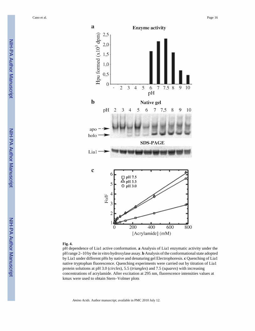
Fig. 4.pH dependence of Lia1 active conformation. a Analysis of Lia1 enzymatic activity under thepH range 2–10 by the in vitro hydroxylase assay. b Analysis of the conformational state adoptedby Lia1 under different pHs by native and denaturing gel Electrophoresis. c Quenching of Lia1native tryptophan fluorescence. Quenching experiments were carried out by titration of Lia1protein solutions at pH 3.0 (circles), 5.5 (triangles) and 7.5 (squares) with increasingconcentrations of acrylamide. After excitation at 295 nm, fluorescence intensities values atkmax were used to obtain Stern–Volmer plots
Cano et al. Page 16
Amino Acids. Author manuscript; available in PMC 2010 July 12.
NIH
-PA Author Manuscript
NIH
-PA Author Manuscript
NIH
-PA Author Manuscript
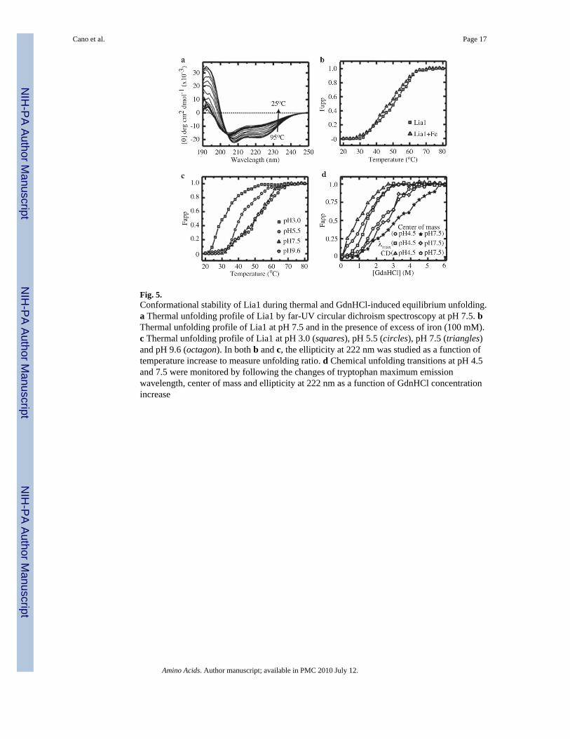
Fig. 5.Conformational stability of Lia1 during thermal and GdnHCl-induced equilibrium unfolding.a Thermal unfolding profile of Lia1 by far-UV circular dichroism spectroscopy at pH 7.5. bThermal unfolding profile of Lia1 at pH 7.5 and in the presence of excess of iron (100 mM).c Thermal unfolding profile of Lia1 at pH 3.0 (squares), pH 5.5 (circles), pH 7.5 (triangles)and pH 9.6 (octagon). In both b and c, the ellipticity at 222 nm was studied as a function oftemperature increase to measure unfolding ratio. d Chemical unfolding transitions at pH 4.5and 7.5 were monitored by following the changes of tryptophan maximum emissionwavelength, center of mass and ellipticity at 222 nm as a function of GdnHCl concentrationincrease
Cano et al. Page 17
Amino Acids. Author manuscript; available in PMC 2010 July 12.
NIH
-PA Author Manuscript
NIH
-PA Author Manuscript
NIH
-PA Author Manuscript
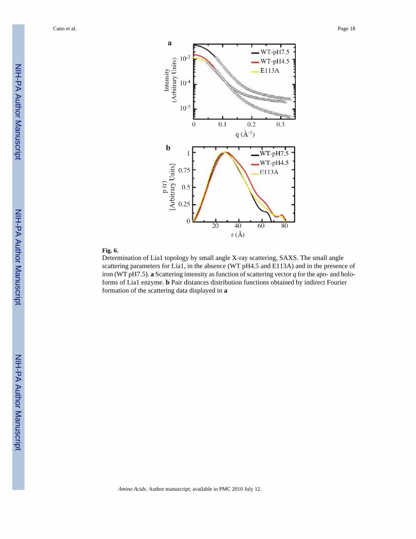
Fig. 6.Determination of Lia1 topology by small angle X-ray scattering, SAXS. The small anglescattering parameters for Lia1, in the absence (WT pH4.5 and E113A) and in the presence ofiron (WT pH7.5). a Scattering intensity as function of scattering vector q for the apo- and holo-forms of Lia1 enzyme. b Pair distances distribution functions obtained by indirect Fourierformation of the scattering data displayed in a
Cano et al. Page 18
Amino Acids. Author manuscript; available in PMC 2010 July 12.
NIH
-PA Author Manuscript
NIH
-PA Author Manuscript
NIH
-PA Author Manuscript
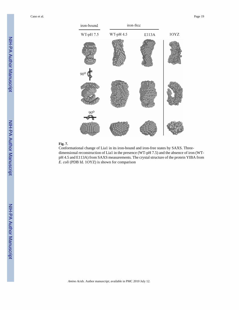
Fig. 7.Conformational change of Lia1 in its iron-bound and iron-free states by SAXS. Three-dimensional reconstruction of Lia1 in the presence (WT-pH 7.5) and the absence of iron (WT-pH 4.5 and E113A) from SAXS measurements. The crystal structure of the protein YIBA fromE. coli (PDB Id. 1OYZ) is shown for comparison
Cano et al. Page 19
Amino Acids. Author manuscript; available in PMC 2010 July 12.
NIH
-PA Author Manuscript
NIH
-PA Author Manuscript
NIH
-PA Author Manuscript
NIH
-PA Author Manuscript
NIH
-PA Author Manuscript
NIH
-PA Author Manuscript
Cano et al. Page 20
Table 1
Stern–Volmer constants for the fluorescence quenching of Lia1 by acrylamide
KSV (M−1)
pH 7.5 2.45 ± 0.04
pH 5.5 - Slope 1 17.08 ± 2.45
pH 5.5 - Slope 2 6.27 ± 0.18
pH 3.0 - Slope 1 12.61 ± 1.13
pH 3.0 - Slope 2 5.51 ± 0.07
Amino Acids. Author manuscript; available in PMC 2010 July 12.