Limiting Discovery of a Defendant's Wealth When Punitive ...
MOLECULAR AND CELLULAR BIOLOGY,0270-7306/00/$04.0010
Aug. 2000, p. 5690–5699 Vol. 20, No. 15
Copyright © 2000, American Society for Microbiology. All Rights Reserved.
Evidence for a Telomere-Independent “Clock” Limiting RASOncogene-Driven Proliferation of Human Thyroid
Epithelial CellsC. J. JONES,1 D. KIPLING,1 M. MORRIS,1 P. HEPBURN,1 J. SKINNER,1 A. BOUNACER,1 F. S. WYLLIE,1
M. IVAN,1 J. BARTEK,2 D. WYNFORD-THOMAS,1* AND J. A. BOND1
Cancer Research Campaign Laboratories, Department of Pathology, University of Wales College of Medicine, HeathPark, Cardiff CF14 4XN, United Kingdom,1 and Institute of Cancer Biology, Danish Cancer Society,
DK-2100 Copenhagen, Denmark2
Received 9 December 1999/Returned for modification 3 January 2000/Accepted 27 April 2000
An initiating role for RAS oncogene mutation in several epithelial cancers is supported by its high incidencein early-stage tumors and its ability to induce proliferation in the corresponding normal cells in vitro. Usingretroviral transduction of thyroid epithelial cells as a model we ask here: (i) how mutant RAS can inducelong-term proliferation in an epithelial cell in contrast to the premature senescence observed in fibroblasts;and (ii) what is the “clock” which eventually triggers spontaneous growth arrest even in epithelial clonesgenerated by mutant RAS. The early response to RAS activation in thyroid epithelial cells showed two featuresnot seen in fibroblasts: (i) a marked decrease in expression of the cyclin-dependent kinase inhibitor (CDKI)p27kip1 and (ii) the absence of any induction of p21waf1. When proliferation eventually ceased (after up to 20population doublings) this occurred despite undiminished expression of mutant RAS and was tightly corre-lated with a return to the initial high level of p27kip1 expression, together with the de novo appearance ofp16ink4a. Importantly, neither the CDKI changes nor the proliferative life span of RAS-induced epithelialclones was altered by induction of telomerase activity through forced expression of the catalytic subunit,hTERT, at levels sufficient to immortalize human fibroblasts. These data provide a basis for cell-type differ-ences in sensitivity to RAS-induced proliferation which may explain the corresponding tumor-type specificityof RAS mutation. They also show for the first time in a primary human cell model that a telomere-independentmechanism can limit not only physiological but also oncogene-driven proliferation, pointing therefore to atumour suppressor mechanism additional, or alternative, to the telomere clock.
RAS mutation occurs at high frequency in several humanepithelial tumor types, notably those of colon (8), pancreas (1),and thyroid (36, 58). Analyses of clinical samples indicate itsinvolvement at early (premalignant) stages and in pancreasand thyroid are consistent with a role as the initiating molec-ular event. In the case of thyroid, where a suitable cell culturemodel exists, this has been strongly supported by the results ofin vitro gene transfer experiments (7, 37). Whereas normalthyrocytes exhibit a very low proliferative rate (as is also thecase in the intact gland) and cease growing in even optimalculture conditions after less than 3 population doublings (PD),introduction of mutant RAS induces a dramatic proliferativeresponse, resulting in generation of clones whose final size (upto 107 cells) and well-differentiated phenotype are consistentwith those of a small benign thyroid tumor (adenoma) in vivo(7).
This prolonged clonal expansion contrasts sharply with themore widely studied effects of RAS mutation in primary fibro-blasts (39, 55, 73), in which proliferation is sharply limited byinduction of a premature senescence state. Nevertheless, evenin thyrocytes, proliferation does not continue indefinitely andeventually spontaneously ceases after 15 to 25 PD (7, 37),
terminating in a viable state of growth arrest, resembling rep-licative senescence. Spontaneous immortalization has neverbeen observed despite many hundreds of gene transfer exper-iments in our laboratory.
In vivo, the vast majority of thyroid adenomas also appear toreach a self-limiting quiescent end point. Such limitations intumor growth are often ascribed to insufficient ability to pro-mote new blood vessel formation and/or to invade surround-ing tissues. Importantly, however, the observation that arestriction similar to clonal expansion occurs even in tissueculture indicates, on the contrary, that a cell-intrinsic mech-anism which is independent of tissue architecture and ismost likely to be based on number of elapsed cell divisionsis responsible.
Interest in intrinsic proliferative life span barriers (PLBs) (4,52, 69) has been heightened recently by the demonstration thatone such cell division “clock” is based on the progressive short-ening of chromosome telomeres, which has been shown totrigger replicative senescence in a number of normal humancell types (3) by a pathway involving p53 (5, 21) and the cyclin-dependent kinase inhibitor (CDKI) p21Waf1 (11). Here wehave set out to examine the relationship between this mecha-nism and that responsible for limiting the proliferative re-sponse to activated RAS in thyroid epithelial cells. In strikingcontrast to the fibroblast paradigm, the results point to a te-lomere-independent clock operating through a distinctly dif-ferent set of CDKI changes, which may represent a novel classof PLB responsible for arresting oncogene-induced tumor de-velopment in some types of human epithelial cells at an early(premalignant) stage.
* Corresponding author. Mailing address: Cancer Research Cam-paign Laboratories, Department of Pathology, University of WalesCollege of Medicine, Heath Park, Cardiff CF14 4XN, United King-dom. Phone: 44 (029) 2074 2700. Fax: 44 (029) 2074 2704. E-mail:[email protected].
5690
MATERIALS AND METHODS
Cells and culture conditions. Primary monolayer cultures of follicular epithe-lial cells (.99% epithelial as judged by cytokeratin immunostaining) were pre-pared from surgical samples of normal thyroid tissue by protease digestion andmechanical disaggregation (66) and maintained in a 2:1:1 mixture of Dulbecco’smodified Eagle’s medium, Ham’s F-12 medium, and MCDB104 (all from LifeTechnologies, Paisley, United Kingdom) (7) supplemented with 10% fetal calfserum (Imperial Laboratories, London, United Kingdom). Normal human dip-loid fibroblasts (HCA2 cells, kindly provided by James Smith, Houston, Texas)were grown in Dulbecco’s modified Eagle’s medium (Life Technologies) supple-mented with 10% fetal calf serum (Imperial Laboratories). Senescence occurredat an estimated PD level of 65 to 70.
Retroviral vectors. Replication-defective amphotropic retroviral vectors en-coding the Val-12 mutant of human H-RAS (psi-CRIP-DOEJ) together with theneo gene for selection in G418 and the vector-only control (psi-CRIP-neo) wereused as previously described (7). To allow dual selection, we constructed retro-virus vectors for hTERT and HPV E7 based on pBABEpuro (46), which confersresistance to puromycin. pBABEpuro-hTERT has been described recently (68);pBABEpuro-E7 was constructed by PCR synthesis of the E7 open reading frametogether with a consensus upstream Kozak sequence and appropriate restrictionsites to allow ligation into the BamHI and EcoRI sites of pBABEpuro.
Retroviral gene transfer. Primary thyroid epithelial cells were plated at ;5 3105 per 60-mm-diameter dish and infected 2 days later with retrovirus-containingmedium from near-confluent producer cells, containing 8 mg of Polybrene per ml(7). Three days later, cells were passaged and maintained in medium with orwithout G418 (400 mg/ml) or puromycin (2.5 mg/ml) as indicated.
Assessment of DNA synthesis by BrdU incorporation. Cells were labeled byincubation in 10 mM bromodeoxyuridine (BrdU) for 1 h, following which nuclearincorporation was detected by immunoperoxidase immunocytochemistry as pre-viously described (4). The proportion of labeled nuclei (labeling index [LI]) wasdetermined from a count of .500 cells per datum point.
Analysis of mutant RAS expression by reverse transcription-PCR. Poly(A)1
RNA was extracted from normal monolayers or from pooled colonies generatedby retroviral vector DOEJ, using a Micro-FastTrack mRNA isolation kit (In-vitrogen Corp., San Diego, Calif.). A partial H-ras cDNA was synthesized usinga reverse transcription-PCR kit (Perkin-Elmer Cetus, Norwalk, Conn.) withprimer 59-TGGACGAATACGACCCCACT-39, located downstream of codon12. This was then amplified using this primer together with upstream primer59-CTGAGGAGCGATGACGGAAT-39 in a PCR mixture consisting of 30 cy-cles of 1-min denaturation at 94°C, 1-min annealing at 60°C, and 1-min extensionat 72°C, with an additional 4-min extension after the final cycle. A single 95-bpproduct was seen on gel electrophoresis. This was then sequenced using anABI-Prism cycle sequencing kit (PE-Applied Biosystems) with the downstreamprimer described above.
Detection of SAb-Gal activity. Endogenous senescence-associated mammalianb-galactosidase activity (SAb-Gal) (15) was assessed histochemically (7) usingX-Gal substrate (5-bromo-4-chloro-3-indolyl-b-D-galactopyranoside).
Immunocytochemical analysis. For p16ink4a, monolayers were fixed in ace-tone-methanol, 1:1 (10 min at 220°C), and a standard indirect immunoperoxi-dase procedure applied, using mouse monoclonal antibody DCS-50 (43) (Onco-gene Research Products), followed by peroxidase-conjugated rabbit anti-mouseimmunoglobulin (Ig) (Dako). A similar procedure was followed for H-RAS usingmonoclonal Y13-259 followed by swine-anti-rat Ig-peroxidase (Dako) and for thecorresponding rat control antibody (antipolyoma large T).
For p21waf1, p27kip1, and cyclin D1, cultures were fixed in 4% paraformalde-hyde (10 min; or 4 min in the case of cyclin D1) and then pretreated with 50 mMglycine (10 min), 0.2% Triton X-100 (10 min), and 0.3% H2O2 (3 min), andnonspecific binding was blocked with 2% horse serum (30 min). Anti-p21 (Clone6B6; Cambridge Bioscience, Cambridge, United Kingdom), anti-p27 (Transduc-tion Laboratories), or anti-cyclin D1 (DCS-6; Santa Cruz) mouse monoclonalantibodies were applied followed by the mouse-specific avidin-biotin-peroxidase(ABC) system (Novocastra).
For cyclin D3, cells were fixed in 2% paraformaldehyde and then permeabil-ized in methanol followed by 0.1% Triton X-100 as described (14). Detection wasby the ABC method using monoclonal antibody DCS-22 clone E8 (2) as theprimary antibody.
For all antigens, sites of antibody binding were visualized by the deposition ofbrown polymer following incubation in diaminobenzidine-hydrogen peroxidesolution.
Immunoblotting. Cells were lysed for 5 min at 4°C by 1% NP-40 in 150 mMNaCl, 50 mM Tris (pH 8.0), and 5 mM EDTA buffer, which contained 1 mMphenylmethylsulfonyl fluoride and 0.01 mg each of aprotinin and leupeptin perml. Protein samples (30 mg) were separated by sodium dodecyl sulfate–12%polyacrylamide gel electrophoresis and electroblotted to Transblot polyvinyli-dene difluoride membrane (Bio-Rad Labs, Hemel Hempstead, United King-dom). Anti-p16 antibody (as above) was applied, followed by goat anti-mouseIg-peroxidase conjugate and visualization by the ECL detection system (Amer-sham, Little Chalfont, United Kingdom). The filter was stained with India ink,and quantitation of the specific signal and the amount of protein loaded wasperformed using a Bio-Rad imaging densitometer running Molecular Analystsoftware.
TRF length analysis. Genomic DNA (1 mg), prepared as previously described(30), was digested with 10 U each of RsaI and HinfI and separated on 0.5%agarose Tris-borate-EDTA gels. Gels were denatured with 1.5 M NaCl–0.5 MNaOH (15 min), neutralized with 1.5 M NaCl–0.5 M Tris, pH 8.0 (10 min), andthen dried onto 3MM paper (Whatman) for 1 h at room temperature followedby 30 min at 60°C. Gels were then removed from the paper and hybridized at37°C overnight in 53 SSC (13 SSC is 0.15 M NaCl plus 0.015 M sodiumcitrate)–53 Denhardt’s–0.5 mM pyrophosphate–10 mM Na2HPO4 with a single-stranded DNA probe (CCCTAA)3 (500 ng/gel), which was end-labeled with[g-32P]ATP (3,000 Ci/mmol) by 10 U of T4 polynucleotide kinase (Amersham).After washing with three changes of 0.13 SSC at 37°C and two at room tem-perature, gels were wrapped in Saran and signals were detected using a STORMphosphorimager (Amersham Pharmacia Biotech) from which mean terminalrestriction fragment (TRF) length was calculated (35) using Molecular Analystsoftware (Bio-Rad).
Telomere repeat amplification protocol (TRAP assay). Cells (107 per 185 ml)were lysed in hypotonic-detergent buffer (1 mM Tris-HCl [pH 7.5], 1 mMMgCl2, 1 mM EGTA, 1 mM phenylmethylsulfonyl fluoride, 5 mM 2-mercapto-ethanol, 0.5% 3-[(3-cholamidopropyl)-dimethylammonio]-1-propanesulfonate(CHAPS), 10% glycerol) for 30 min on ice followed by centrifugation at100,000 3 g for 30 min at 4°C. Extracts (3,000 cell equivalents) were assayed withor without pretreatment for 10 min at 85°C (to abolish telomerase activity).
Telomerase activity was assayed according to the standard TRAP protocol(32) except that the wax barrier was avoided and Taq polymerase and the secondprimer, CX, were added to reaction mixtures prewarmed to 92°C followingelongation of the TS primer. Telomerase products were resolved in 10% poly-acrylamide gels and visualized by Sybr Gold staining and fluoroimaging, againusing a STORM system. Extracts of cell line 293 were used as a positive control(10). The incorporation of an internal standard (67) demonstrated that therewere no inhibitors of PCR detectable with the quantities of protein used.
RESULTS
Induction of thyrocyte proliferation by mutant RAS corre-lates with reduced nuclear p27kip1 expression. Normal humanthyroid epithelial cells in primary culture were stably trans-duced with an amphotropic retroviral vector encoding mutant(V12) H-RAS. As described previously (7), this results in thegeneration of rapidly growing colonies (approximately 50 perdish of 105 cells infected) which are clearly distinguishablefrom the surrounding uninfected monolayer by 7 to 10 daysafter infection. Normal (uninfected) thyroid cells cease prolif-erating after ,3 PD and, even prior to this, exhibit a very lowproliferative rate, which accounts for the low yield of trans-duced cells compared to that seen for example with fibroblastcell lines.
Using immunocytochemistry to permit analysis of small cellnumbers and to reveal changes in subcellular distribution, weinitially examined the expression of several cell cycle regulatorswhich have been implicated in RAS-induced proliferation inother models, namely cyclin D1, p21waf1, and p27kip1. We alsoincluded cyclin D3, which has been shown to play a particularlyimportant role in normal thyroid epithelial cells (14).
Normal thyrocytes, after 7 days in culture, expressed readilydetectable levels of cyclin D1, D3, (not shown), and p21 in themajority of nuclei (Fig. 1a); in particular nearly 100% of cellsexhibited strong nuclear immunostaining for p27 (Fig. 1a). Asexpected, the BrdU LI was extremely low (,1%) and nearly allcells were positive for SAb-Gal, which has been widely re-ported as a marker of replicative senescence in other cell types(15).
In colonies induced to proliferate by mutant RAS at theearliest time point analyzable (7 to 10 days) BrdU LI hadincreased to 36%, accompanied by a fall in the SAb-Gal indexto 7%. This proliferative response was associated with a slightreduction in the proportion of cells expressing detectable nu-clear p21, which failed to reach statistical significance, and anincrease in the proportion expressing cyclin D1 from ;70% tonearly 100% (Fig. 1a and c). Cyclin D3 expression remainedessentially unchanged at this (and subsequent) time point atbetween 50 and 60% of nuclei (not shown). The major changeobserved however was a marked fall in p27 expression, which
VOL. 20, 2000 TELOMERE-INDEPENDENT “CLOCK” AND PROLIFERATION 5691

was detectable in only 16% of cells (nuclei) expressing mutantRAS, compared to 95% in the surrounding normal monolayer(Fig. 1a and b).
To control for the possibility that the reduction in p27 con-
tent in early RAS-induced colonies might merely be a second-ary consequence of the much higher proportion of proliferat-ing cells, we also examined an analogous model in whichprimary thyrocytes are induced to proliferate by simian virus 40
FIG. 1. Expression of cell cycle regulators during the life span of thyroid epithelial clones induced to proliferate by mutant RAS. (a) Representative photomicro-graphs of normal thyroid epithelial cells (N) and colonies induced by mutant RAS at an early rapidly proliferating stage (RAS-E) and at a late stage at the end of theirproliferative life span (RAS-L). Expression of p21waf1, p27kip1, and p16ink4a and incorporation of BrdU were analyzed by immunocytochemistry (positivity indicatedby brown peroxidase reaction product); the senescence-associated marker SAb-Gal was assessed by X-Gal histochemistry (blue reaction product). In some panels,nuclei are lightly counterstained with hematoxylin to aid visualization. Note a reduction in nuclear p27 correlating with high BrdU labeling in RAS-E colonies and theincrease in p16 in RAS-L colonies (magnification 325). (b and c) Quantitative analyses of normal epithelial cells and colonies expressing mutant RAS at early (1 to2 weeks), middle (2 to 3 weeks), and late (.5 weeks) stages showing an inverse correlation between a proportion of nuclei containing detectable p27 (■) and aproportion in cell cycle S phase as shown by BrdU incorporation (F) and a direct correlation between p27 and SAb-Gal expression (Œ) note less-marked changes inproportions of nuclei containing detectable p21 (h) and cyclin D1 (E). Results are presented as means of .300 cells per datum point 6 standard errors of the mean(error bars). (d) Western blot-ECL analysis of p16ink4a expression (upper panel) in normal thyroid epithelial cells (N), RAS-induced colonies at an intermediate stageof their life span (RAS MID), and end-stage colonies (RAS L). The lower panel shows part of an India ink-stained filter to assess equality of protein loading. Figuresshow integrated optical density values obtained by scanning densitometry from which the relative increase in p16 signal corrected for total protein between RAS MIDand RAS L is calculated at 20-fold.
5692 JONES ET AL. MOL. CELL. BIOL.

T following infection with the retroviral vector psi-CRIP-SVU19 (6). Despite similar proliferation rates, and at similarsizes, colonies generated in this way failed to show any reduc-tion in p27 expression (data not shown).
Spontaneous cessation of RAS-induced thyrocyte prolifera-tion correlates with reexpression of nuclear p27 and de novoexpression of p16ink4a. RAS-induced growth ceased by 5 weekspostinfection at a final colony size varying from 104 to 106 cells.Colonies were analyzed as above at this and at intermediatetime points postinfection. Growth arrest was associated with,and explicable by, a decline in BrdU LI, which eventuallyreturned to normal levels (;1%), together with a correspond-ing restoration of SAb-Gal expression and a flattened, senes-cence-like morphology (Fig. 1a).
The changes in p21 and cyclin D1 expression seen in earlyRAS colonies reverted to near-normal in terminally arrestedcolonies; however, the time course showed an imperfect cor-relation with BrdU LI at intermediate time points (Fig. 1c). Incontrast, p27 expression showed a tight inverse correlationthroughout with BrdU LI and a direct correlation with SAb-Gal (Fig. 1b).
Given its established role in senescence in other cell types,we also examined expression of the CDK inhibitor p16ink4a.There was a clear increase from undetectable levels in normalcolonies to readily detectable levels in end-stage RAS colonies(Fig. 1a); however, the rather diffuse, predominantly cytoplas-mic, pattern of immunostaining obtained with antibodyDCS-50 was difficult to quantify microscopically. In this case,therefore, sufficient cells were collected to perform a limitedWestern blot analysis (Fig. 1d) which confirmed the absence ofsignal in normal cells and a 20-fold increase between prolifer-ating (early) and arrested (end-stage) RAS colonies.
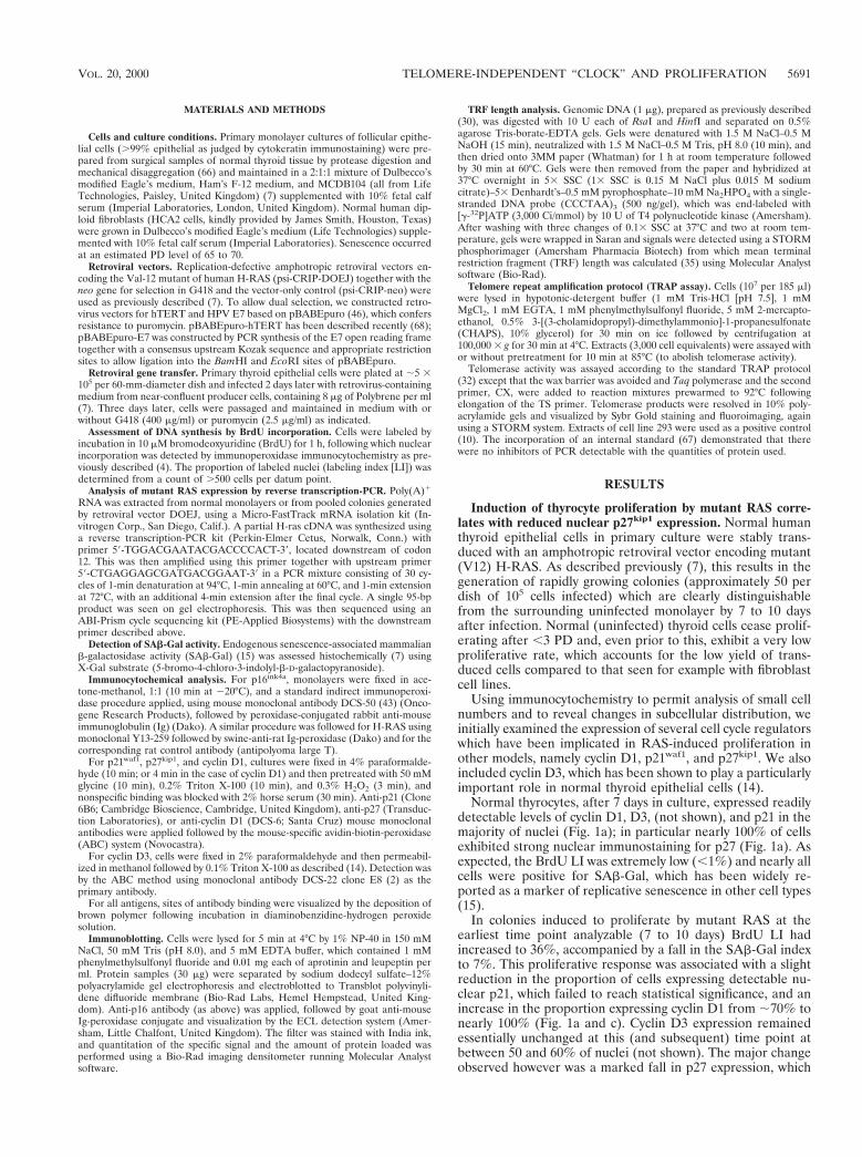
The possibility that the above changes were due simply todecreased mutant RAS expression in late-stage colonies, forexample through methylation of the retroviral promoter, wasexcluded by analysis at both protein (Fig. 2a to d) and mRNA(Fig. 2e) levels, which confirmed the persistent overexpressionof the mutant compared to the endogenous wild-type sequencein both early- and late-stage colonies.
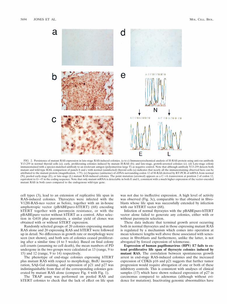
Cessation of RAS-induced thyrocyte proliferation is not de-pendent on telomere erosion. Mean telomere length, measuredas TRF size after HinfI/RsaI digestion of genomic DNA, variedfrom 9 to 11 kbp in different isolates of human thyrocytes at theend of their normal proliferative life span (Fig. 3).
Analysis of DNA from pooled RAS-induced colonies incomparison to the normal cells from which they were derivedshowed only variable, low-magnitude TRF shortening. In end-stage colonies this varied in different experiments from ;1.4kbp down to unresolvable differences (Fig. 3a and b). Further-more, the final mean TRF value (typically ;10 kbp) was alwaysmuch larger than that observed in normal human fibroblasts atthe end of their replicative life span (;7 kb in our hands) (Fig.3b).
To address the possibility that normal thyrocytes, unlikefibroblasts and most other primary cultures, might circumventtelomere erosion through physiological expression of telomer-ase, we assayed telomerase activity in lysates using the well-established in vitro TRAP assay (32). No activity could bedetected in any normal sample, nor in nearly all lysates ofpooled RAS colonies at both early and late stages (Fig. 3c).Only one sample (Thy.1 RAS-E [Fig. 3c]) out of more than 30analyzed was found to be positive, and this only at extremelyweak levels (,1% of the positive control 293 cell line).
Finally we asked whether stable induction of telomeraseactivity through forced expression of the catalytic sub-unit oftelomerase, hTERT, would, as in fibroblasts and several other
FIG. 1—Continued.
VOL. 20, 2000 TELOMERE-INDEPENDENT “CLOCK” AND PROLIFERATION 5693
cell types (3), lead to an extension of replicative life span inRAS-induced colonies. Thyrocytes were infected with theV12H-RAS-neo vector as before, together with an in-houseamphotropic vector (pBABEpuro-hTERT) (68) encodinghTERT together with puromycin resistance, or with thepBABEpuro vector without hTERT as a control. After selec-tion in G418 plus puromycin, a similar yield of clones wasobtained with or without hTERT expression.
Randomly selected groups of 20 colonies expressing mutantRAS alone and 20 expressing RAS and hTERT were followedup in detail. No differences in growth rate or morphology wereseen (not shown), and both sets of colonies ceased proliferat-ing after a similar time (4 to 5 weeks). Based on final colonycell counts (assuming no cell death), the mean numbers of PDundergone in the two groups were calculated as 13 (range, 9 to15) and 12 (range, 9 to 14) respectively.
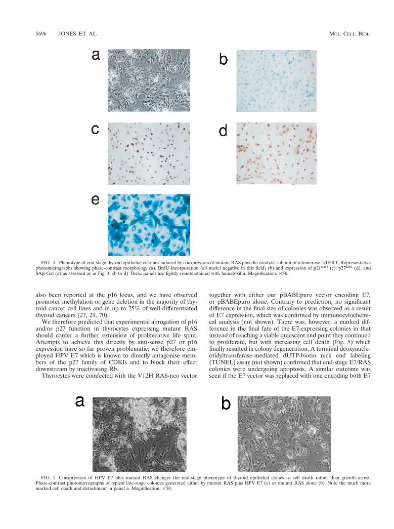
The phenotype of end-stage colonies expressing hTERTplus mutant RAS with respect to morphology, BrdU incorpo-ration, SAb-Gal staining, and expression of p21 and p27 wasindistinguishable from that of the corresponding colonies gen-erated by mutant RAS alone (compare Fig. 4 with Fig. 1).
The TRAP assay was performed on pooled RAS andhTERT colonies to check that the lack of effect on life span
was not due to ineffective expression. A high level of activitywas observed (Fig. 3c), comparable to that obtained in fibro-blasts whose life span was successfully extended by infectionwith our hTERT vector (68).
Infection of normal thyrocytes with the pBABEpuro-hTERTvector alone failed to generate any colonies, either with orwithout puromycin selection.
These data indicate that terminal growth arrest occurringboth in normal thyrocytes and in those expressing mutant RASis regulated by a mechanism which comes into operation atmean telomere lengths well above those associated with senes-cence in fibroblasts and furthermore, unlike the latter, is notabrogated by forced expression of telomerase.
Expression of human papillomavirus (HPV) E7 fails to ex-tend proliferative life span of thyrocyte colonies induced bymutant RAS. The correlation between spontaneous growtharrest in end-stage RAS-induced colonies and the increasedexpression of CDKIs p16 and p21 suggests that further tumorprogression would require abrogation of one or both of theseinhibitory controls. This is consistent with analyses of clinicalsamples (17) which have shown reduced expression of p27 incarcinomas compared to adenomas (although without evi-dence for mutation). Inactivating genomic abnormalities have
FIG. 2. Persistence of mutant RAS expression in late-stage RAS-induced colonies. (a to c) Immunocytochemical analysis of H-RAS protein using anti-ras antibodyY13-259 in normal thyroid cells (a); early, proliferating colonies induced by mutant H-RAS (b); and late-stage, growth-arrested colonies (c). (d) Late-stage colonyimmunostained with a species-matched antibody to an irrelevant antigen (polyomavirus large T) as negative control. Note that although antibody Y13-259 detects bothmutant and wild-type RAS, comparison of panels b and c with normal (uninfected) thyroid cells (a) indicates that nearly all the immunostaining observed here can beattributed to the mutant protein (magnification, 375). (e) Sequence (antisense) of cDNA surrounding codon 12 of H-RAS derived by RT-PCR of mRNA from normal(N), pooled early-stage (E), or late-stage (L) mutant RAS-induced colonies. The point mutation (arrowed) appears as a C3A transversion at position 2 of codon 12,equivalent to G3T in the coding sequence. Note that only mutant mRNA is detectable in both E and L, consistent with a much higher expression of the vector-encodedmutant RAS in both cases compared to the endogenous wild-type gene.
5694 JONES ET AL. MOL. CELL. BIOL.
FIG. 3. Analysis of telomere length and telomerase activity in thyroid cells expressing mutant RAS. (a) TRF analysis of HinfI/RsaI-digested genomic DNA fromnormal thyrocytes (N), and pools of early (proliferating) (RAS E) and late-stage (RAS L) RAS colonies derived from two separate human thyroid samples (Thy.1 andThy.2). Mean TRF values were calculated from densitometry data as described in text. (b) TRF analysis of normal (N) and late-stage (RAS L) RAS colonies derivedfrom a third thyroid sample (Thy.3), together with colonies expressing both mutant RAS and the catalytic subunit of human telomerase (RAS 1 hTERT). Senescenthuman fibroblasts (strain HCA2) are shown for comparison (HDF SEN). (c) Telomerase activity assessed by TRAP assay in normal cells (N), in pools of early coloniesexpressing mutant RAS (RAS E) and late-stage colonies expressing mutant RAS alone (RAS L) or with hTERT (RAS L 1 hTERT). Each sample is analyzed with(1) or without (2) prior heat treatment (85°C for 10 min). The immortal human epithelial cell line 293 is included as a positive control. The arrow indicates the positionof the internal telomerase amplification standard as a control for nonspecific PCR inhibition.
VOL. 20, 2000 TELOMERE-INDEPENDENT “CLOCK” AND PROLIFERATION 5695
also been reported at the p16 locus, and we have observedpromoter methylation or gene deletion in the majority of thy-roid cancer cell lines and in up to 25% of well-differentiatedthyroid cancers (27, 29, 70).
We therefore predicted that experimental abrogation of p16and/or p27 function in thyrocytes expressing mutant RASshould confer a further extension of proliferative life span.Attempts to achieve this directly by anti-sense p27 or p16expression have so far proven problematic; we therefore em-ployed HPV E7 which is known to directly antagonise mem-bers of the p27 family of CDKIs and to block their effectdownstream by inactivating Rb.
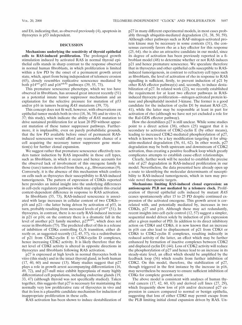
Thyrocytes were coinfected with the V12H RAS-neo vector
together with either our pBABEpuro vector encoding E7,or pBABEpuro alone. Contrary to prediction, no significantdifference in the final size of colonies was observed as a resultof E7 expression, which was confirmed by immunocytochemi-cal analysis (not shown). There was, however, a marked dif-ference in the final fate of the E7-expressing colonies in thatinstead of reaching a viable quiescent end point they continuedto proliferate, but with increasing cell death (Fig. 5) whichfinally resulted in colony degeneration. A terminal deoxynucle-otidyltransferase-mediated dUTP-biotin nick end labeling(TUNEL) assay (not shown) confirmed that end-stage E7/RAScolonies were undergoing apoptosis. A similar outcome wasseen if the E7 vector was replaced with one encoding both E7
FIG. 4. Phenotype of end-stage thyroid epithelial colonies induced by coexpression of mutant RAS plus the catalytic subunit of telomerase, hTERT. Representativephotomicrographs showing phase-contrast morphology (a), BrdU incorporation (all nuclei negative in this field) (b) and expression of p21waf1 (c), p27kip1 (d), andSAb-Gal (e) as assessed as in Fig. 1. (b to d) These panels are lightly counterstained with hematoxilin. Magnification, 350.
FIG. 5. Coexpression of HPV E7 plus mutant RAS changes the end-stage phenotype of thyroid epithelial clones to cell death rather than growth arrest.Phase-contrast photomicrographs of typical late-stage colonies generated either by mutant RAS plus HPV E7 (a) or mutant RAS alone (b). Note the much moremarked cell death and detachment in panel a. Magnification, 350.
5696 JONES ET AL. MOL. CELL. BIOL.
and E6, indicating that, as observed previously (4), apoptosis inthyrocytes is p53 independent.
DISCUSSION
Mechanisms underlying the sensitivity of thyroid epithelialcells to RAS-induced proliferation. The prolonged growthstimulation induced by activated RAS in normal thyroid epi-thelial cells stands in sharp contrast to the response observedin normal human fibroblasts, in which proliferation is limitedwithin a few PD by the onset of a permanent growth arreststate, which, apart from being independent of telomere erosion(65), closely resembles replicative senescence mediated byboth p14arf-p53 and p16ink4a pathways (39, 55, 73).
This premature senescence phenotype, which we too haveobserved in fibroblasts, has aroused great interest recently (51)as a potential innate tumor suppressor mechanism and anexplanation for the selective pressure for mutation of p53and/or p16 in tumors bearing RAS mutations (39, 73).
This concept does not fit well, however, with observations onepithelial tumor models both in vivo (18, 36, 58) and in vitro (4,37; this study), which indicate the ability of RAS mutation todrive sustained proliferation for at least 20 PD without appar-ent mutation of these tumor suppressor pathways. Further-more, it is implausible, even on purely probabilistic grounds,that the few PD available before onset of permanent RAS-induced senescence would afford any reasonable chance of acell acquiring the necessary tumor suppressor gene muta-tion(s) for further clonal expansion.
We suggest rather that premature senescence effectively ren-ders tumor induction by RAS impossible in those cell types,such as fibroblasts, in which it occurs and hence accounts forthe observed lack of involvement of this oncogene family inthose (rare) tumors derived from them, e.g., fibrosarcomas (9).Conversely, it is the absence of this mechanism which conferson cells such as thyrocytes their susceptibility to RAS-inducedtumorigenesis. The pattern of expression of CDKIs reportedhere provides an initial insight into the underlying differencesin cell-cycle regulatory pathways which may explain this crucialcontext-dependent difference in response to RAS activation.
In fibroblasts (55, 73), RAS-induced senescence is associ-ated with large increases in cellular content of two CDKIs—p16 and p21—the latter being driven by activation of p53, inturn, probably resulting from increased levels of p14arf (51). Inthyrocytes, in contrast, there is no early RAS-induced increasein p21 or p16; on the contrary there is a dramatic fall in thelevel of another p21 family member, p27KIP1, which does notoccur in fibroblasts (73). The predicted effect of this is a releaseof inhibition of CDKs controlling G1/S transition, either di-rectly or, as suggested recently (12, 45, 57), via a redistributionof p21 from CDK2-cyclin E to CDK4-cyclin D complexes,hence increasing CDK2 activity. It is likely therefore that thenet level of CDKI activity is altered in opposite directions inthyrocytes and fibroblasts following RAS activation.
p27 is expressed at high levels in normal thyrocytes both invitro (this study) and in the intact thyroid gland, in both human(17, 40, 60) and mouse (13). Many studies suggest that p27plays a role in growth arrest accompanying differentiation (16,49, 72), and p27-null mice exhibit hyperplasia of many highlydifferentiated cell populations, including endocrine glands (19,33, 47) (although thyroid was not specifically studied). Takentogether, this suggests that p27 is necessary for maintaining thenormally very low proliferative rate of thyrocytes in vivo andthat its loss is a plausible candidate mechanism for induction ofinappropriate proliferation in these cells.
RAS activation has been shown to induce destabilization of
p27 in many different experimental models, in most cases prob-ably through ubiquitin-mediated degradation (31, 38, 50, 59).Although other pathways such as RAF-mitogen-activated pro-tein kinase may be necessary in some contexts (31), the con-sensus currently favors rho as a key effector for this response(25, 64). rho is also an attractive candidate in our model, sinceits degree of activation has been previously reported in a fi-broblast model (48) to determine whether or not RAS inducesp21 and hence premature senescence. We speculate thereforethat in thyrocytes and other epithelial cells susceptible to RAS-induced tumorigenesis, in contrast to refractory cell types suchas fibroblasts, the level of activation of rho in response to RASsignalling is sufficient, firstly, to prevent induction of p21 byother RAS effector pathway(s) and, secondly, to induce desta-bilization of p27. In related work (22), we recently establishedthe requirement for at least two effector pathways in RAS-induced thyrocyte proliferation—mitogen-activated protein ki-nase and phosphatidyl inositol 3-kinase. The former is a goodcandidate for the induction of cyclin D1 by mutant RAS (25,41), while the latter may be responsible at least in part foractivation of rho (although we have not yet excluded a role forthe Ral-GDS effector pathway).
How rho destabilizes p27 is still unclear. While some studiespoint to a direct action (38), others (25) suggest that it issecondary to activation of CDK2-cyclin E (by other means),leading to increased CDK2-mediated phosphorylation of p27,which is known to be a key modification targeting it for ubiq-uitin-mediated degradation (56, 61, 62). In other words, p27degradation may be both upstream and downstream of CDK2activation, thus creating a positive feedback loop which greatlycomplicates attempts to establish cause-effect relationships.
Clearly, further work will be needed to establish the preciserole of p27 degradation in RAS-induced proliferation in ourmodel. Nevertheless, the data presented here already open upa route to identifying the molecular determinants of suscepti-bility to RAS-induced tumorigenesis, which in turn may pro-vide novel therapeutic targets.
Mechanisms limiting RAS-induced clonal expression: anantioncogenic PLB not mediated by a telomere clock. Prolif-eration of thyroid epithelial cells induced by mutant RASceases spontaneously after 15 to 25 PD despite continued ex-pression of the activated oncogene. This growth arrest is cor-related with, and potentially mediated by, increases in twoCDKIs, p27 and p16. Although these may act redundantly,recent insights into cell cycle control (12, 57) suggest a simpler,sequential model driven solely by induction of p16 expressionafter a given number of PD. In addition to its direct inhibitoryaction on CDK4 and CDK6, it is now known that an increasein p16 can also lead to displacement of p21 from CDK4 orCDK6 to CDK2-cyclin E complexes, resulting indirectly inreduced activity of the latter, an effect which may be furtherenhanced by formation of inactive complexes between CDK2and displaced cyclin D1 (44). Loss of CDK2 activity will reducethe phosphorylation of p27 and hence lead to an increase in itssteady-state level, an effect which should be amplified by thefeedback loop (56) which results from further inhibition ofCDK2. On this model, therefore, the stabilization of p27,though triggered in the first instance by the increase in p16,may nevertheless be necessary to ensure sufficient inhibition ofCDKs for complete growth arrest.
The above model is consistent with analyses of human thy-roid cancers (17, 42, 60, 63) and derived cell lines (27, 29),which frequently show loss of p16 and/or decreased p27 ex-pression in cancers compared to normal or benign epithelia,suggesting that loss of either CDKI may permit escape fromthe PLB limiting initial clonal expansion driven by RAS. Un-
VOL. 20, 2000 TELOMERE-INDEPENDENT “CLOCK” AND PROLIFERATION 5697
fortunately, we have not as yet been able to test this predictionexperimentally by direct manipulation of p27 or p16. A morecentral abrogation of this TSG pathway using HPV E7 was oflimited value, since although appearing to relieve proliferativearrest, net growth was offset by increasing cell death.
A key finding in the current work is the apparent indepen-dence of the above-mentioned PLB on telomere erosion, asshown most conclusively by the failure of experimentally in-duced telomerase activity to extend life span despite stabiliza-tion of telomere length. We must, therefore, postulate theexistence of a separate cell division counting mechanism, whichacts at least in part via induction of p16. Such a clock alsoseems to operate in controlling physiological (growth factorinduced) proliferative life span in a variety of situations, in-cluding primary fibroblasts in rodents (54) and, moreover, sev-eral specific types of human epithelium, including mammaryepithelial cells (20, 34), keratinocytes (34) and uroepithelialcells (53, 71) which undergo senescence after a similar range ofPD (10 to 30 PD). The originality of our observation is that itdemonstrates a crucial role for a p16-dependent, telomere-independent PLB in limiting not only physiological but alsooncogene-driven proliferation.
In all of these situations the major question which remains isthe link between replicative age (elapsed divisions) and induc-tion of p16 expression. Demethylation of promoter sequencesclearly plays a part in this (20, 26) and there is long-standingevidence for reduction in DNA methylation during replicativeageing (24); the mechanistic details of such a clock, however,and the role of potential trans-acting factors such as BMI-1(28) remain obscure. Given its potential importance as a nat-ural tumor suppressor mechanism alternative to telomere ero-sion, this promises to be an area of major basic and therapeuticsignificance.
ACKNOWLEDGMENTS
We thank the Cancer Research Campaign and the Medical Re-search Council for grant support.
We also thank Michele Haughton for primary cells and TheresaKing for manuscript and graphics preparation.
REFERENCES1. Almoguera, C., D. Shibata, K. Forrester, J. Martin, N. Arnheim, and M.
Perucho. 1988. Most human carcinomas of the exocrine pancreas containmutant c-K-ras genes. Cell 53:549–554.
2. Bartkova, J., J. Lukas, M. Strauss, and J. Bartek. 1998. Cyclin D3: require-ment for G1/S transition and high abundance in quiescent tissues suggest adual role in proliferation and differentiation. Oncogene 17:1027–1037.
3. Bodnar, A. G., M. Ouellette, M. Frolkis, S. E. Holt, C.-P. Chiu, G. B. Morin,C. B. Harley, J. W. Shay, S. Lichtsteiner, and W. E. Wright. 1998. Extensionof life-span by introduction of telomerase into normal human cells. Science279:349–352.
4. Bond, J., M. Haughton, J. Rowson, V. Gire, D. Wynford-Thomas, and F.Wyllie. 1999. Control of replicative life span in human cells: barriers to clonalexpansion intermediate between M1 senescence and M2 crisis. Mol. Cell.Biol. 19:3103–3114.
5. Bond, J. A., F. S. Wyllie, and D. Wynford-Thomas. 1994. Escape fromsenescence in human diploid fibroblasts induced directly by mutant p53.Oncogene 9:1885–1889.
6. Bond, J. A., G. O. Ness, J. Rowson, M. Ivan, D. White, and D. Wynford-Thomas. 1996. Spontaneous de-differentiation correlates with extended lifes-pan in transformed thyroid epithelial cells: an epigenetic mechanism oftumour progression. Int. J. Cancer 67:563–572.
7. Bond, J. A., F. S. Wyllie, J. Rowson, A. Radulescu, and D. Wynford-Thomas.1994. In vitro reconstruction of tumour initiation in a human epithelium.Oncogene 9:281–290.
8. Bos, J. L., E. R. Fearon, S. R. Hamilton, M. Verlaan-de Vries, J. H. vanBoom, A. J. van der Eb, and B. Vogelstein. 1987. Prevalence of ras genemutations in human colorectal cancers. Nature 327:293–297.
9. Bos, J. L. 1989. Ras oncogenes in human cancer: a review. Cancer Res.49:4682–4689.
10. Broccoli, D., J. W. Young, and T. de Lange. 1995. Telomerase activity innormal and malignant hematopoietic cells. Proc. Natl. Acad. Sci. USA 92:9082–9086.
11. Brown, J. P., W. Wenyi, and J. M. Sedivy. 1997. Bypass of senescence afterdisruption of p21CIP1/WAF1 gene in normal diploid human fibroblasts.Science 277:831–834.
12. Cheng, M., P. Olivier, J. A. Diehl, M. Fero, M. F. Roussel, J. M. Roberts, andC. J. Sherr. 1999. The p21(Cip1) and p27(Kip1) CDK ‘inhibitors’ are essen-tial activators of cyclin D-dependent kinases in murine fibroblasts. EMBO J.18:1571–1583.
13. Coppee, F., F. Depoortere, J. Bartek, C. Ledent, M. Parmentier, and J. E.Dumont. 1998. Differential patterns of cell cycle regulatory proteins expres-sion in transgenic models of thyroid tumours. Oncogene 17:631–641.
14. Depoortere, F., A. Van Keymeulen, J. Lukas, S. Costagliola, J. Bartkova,J. E. Dumont, J. Bartek, P. P. Roger, and S. Dremier. 1998. A requirementfor cyclin D3-cyclin-dependent kinase (cdk)-4 assembly in the cyclic adeno-sine monophosphate-dependent proliferation of thyrocytes. J. Cell Biol. 140:1427–1439.
15. Dimri, G. P., X. Lee, G. Basile, M. Acosta, G. Scott, C. Roskelley, E. E.Medrano, M. Linskens, I. Rubelj, O. Pereira-Smith, M. Peacocke, and J.Campisi. 1995. A biomarker that identifies senescent human cells in cultureand in aging skin in vivo. Proc. Natl. Acad. Sci. USA 92:9363–9367.
16. Durand, B., M. L. Fero, J. M. Roberts, and M. C. Raff. 1998. p27Kip1 altersthe response of cells to mitogen and is part of a cell-intrinsic timer thatarrests the cell cycle and initiates differentiation. Curr. Biol. 8:431–440.
17. Erickson, L. A., L. Jin, P. C. Wollan, G. B. Thompson, J. van Heerden, andR. V. Lloyd. 1998. Expression of p27kip1 and Ki-67 in benign and malignantthyroid tumors. Mod. Pathol. 11:169–174.
18. Fearon, E. R., and B. Vogelstein. 1990. A genetic model for colorectaltumorigenesis. Cell 61:759–767.
19. Fero, M. L., M. Rivkin, M. Tasch, P. Porter, C. E. Carow, E. Firpo, K.Polyak, L. H. Tsai, V. Broudy, R. M. Perlmutter, K. Kaushansky, and J. M.Roberts. 1996. A syndrome of multiorgan hyperplasia with features of gi-gantism, tumorigenesis, and female sterility in p27(Kip1)-deficient mice. Cell85:733–744.
20. Foster, S. A., D. J. Wong, M. T. Barrett, and D. A. Galloway. 1998. Inacti-vation of p16 in human mammary epithelial cells by CpG island methylation.Mol. Cell. Biol. 18:1793–1801.
21. Gire, V., and D. Wynford-Thomas. 1998. Reinitiation of DNA synthesis andcell division in senescent human fibroblasts by microinjection of anti-p53antibodies. Mol. Cell. Biol. 18:1611–1621.
22. Gire, V., C. J. Marshall, and D. Wynford-Thomas. 1999. Activation of mi-togen-activated protein kinase is necessary but not sufficient for proliferationof human thyroid epithelial cells induced by mutant Ras. Oncogene 18:4819–4832.
23. Hirai, A., S. Nakamura, Y. Noguchi, T. Yasuda, M. Kitagawa, I. Tatsuno, T.Oeda, K. Tahara, T. Terano, S. Narumiya, L. D. Kohn, and Y. Saito. 1997.Geranylgeranylated rho small GTPase(s) are essential for the degradation ofp27Kip1 and facilitate the progression from G1 to S phase in growth-stim-ulated rat FRTL-5 cells. J. Biol. Chem. 272:13–16.
24. Holliday, R. 1996. Endless quest. Bioessays 18:3–5.25. Hu, W., C. J. Bellone, and J. J. Baldassare. 1999. RhoA stimulates p27(Kip)
degradation through its regulation of cyclin E/CDK2 activity. J. Biol. Chem.274:3396–3401.
26. Huschtscha, L. I., J. R. Noble, A. A. Neumann, E. L. Moy, P. Barry, J. R.Melki, S. J. Clark, and R. R. Reddel. 1998. Loss of p16INK4 expression bymethylation is associated with lifespan extension of human mammary epi-thelial cells. Cancer Res. 58:3508–3512.
27. Ivan, M., D. Wynford-Thomas, and C. J. Jones. 1996. Abnormalities of thep16INK4a gene in thyroid cancer cell lines. Eur. J. Cancer 32:2369–2370.
28. Jacobs, J., K. Kieboom, S. Marino, R. de Pinho, and M. van Lohuizen. 1999.The oncogene and polycomb-group gene bmi-1 regulates cell proliferationand senescence through the ink4a locus. Nature 397:164–168.
29. Jones, C. J., J. J. Shaw, F. S. Wyllie, N. Gaillard, M. Schlumberger, and D.Wynford-Thomas. 1996. High frequency deletion of the tumour suppressorgene P16INK4a (MTS1) in human thyroid cancer cell lines. Mol. Cell.Endocrinol. 116:115–119.
30. Jones, C. J., A. Soley, J. W. Skinner, J. Gupta, M. F. Haughton, F. S. Wyllie,M. Schlumberger, S. Bacchetti, and D. Wynford-Thomas. 1998. Dissociationof telomere dynamics from telomerase activity in human thyroid cancer cells.Exp. Cell Res. 240:333–339.
31. Kawada, M., S. Yamagoe, Y. Murakami, K. Suzuki, S. Mizuno, and Y.Uehara. 1997. Induction of p27Kip1 degradation and anchorage indepen-dence by Ras through the MAP kinase signaling pathway. Oncogene 15:629–637.
32. Kim, N. W., M. A. Piatyszek, K. R. Prowse, C. B. Harley, M. D. West, P. L. C.Ho, G. M. Coviello, W. E. Wright, S. L. Weinrich, and J. W. Shay. 1994.Specific association of human telomerase activity with immortal cells andcancer. Science 266:2011–2015.
33. Kiyokawa, H., R. D. Kineman, K. O. Manova-Todorova, V. C. Soares, E. S.Hoffman, M. Ono, D. Khanam, A. C. Hayday, L. A. Frohman, and A. Koff.1996. Enhanced growth of mice lacking the cyclin-dependent kinase inhibitorfunction of p27(Kip1). Cell 85:721–732.
34. Kiyono, T., S. A. Foster, J. I. Koop, J. K. McDougall, D. A. Galloway, andA. J. Klingelhutz. 1998. Both Rb/p16INK4a inactivation and telomerase
5698 JONES ET AL. MOL. CELL. BIOL.
activity are required to immortalize human epithelial cells. Nature 396:84–88.
35. Kruk, P. A., N. J. Rampino, and V. A. Bohr. 1995. DNA damage and repairin telomeres: relation to aging. Proc. Natl. Acad. Sci. USA 92:258–262.
36. Lemoine, N. R., E. S. Mayall, F. S. Wyllie, E. D. Williams, M. Goyns, B.Stringer, and D. Wynford-Thomas. 1989. High frequency of ras oncogeneactivation in all stages of human thyroid tumorigenesis. Oncogene 4:159–164.
37. Lemoine, N. R., S. Staddon, J. Bond, F. S. Wyllie, J. J. Shaw, and D.Wynford-Thomas. 1990. Partial transformation of human thyroid epithelialcells by mutant Ha-ras oncogene. Oncogene 5:1833–1837.
38. Leone, G., J. DeGregori, R. Sears, L. Jakoi, and J. R. Nevins. 1997. Myc andRas collaborate in inducing accumulation of active cyclin E/Cdk2 and E2F.Nature 387:422–426.
39. Lin, A. W., M. Barradas, J. C. Stone, L. van Aelst, M. Serrano, and S. W.Lowe. 1998. Premature senescence involving p53 and p16 is activated inresponse to constitutive MEK/MAPK mitogenic signaling. Genes Dev. 12:3008–3019.
40. Lloyd, A. C. 1998. Ras versus cyclin-dependent kinase inhibitors. Curr. Opin.Genet. Dev 8:43–48.
41. Lloyd, R. V., L. Jin, X. Qian, and E. Kulig. 1997. Aberrant p27kip1 expres-sion in endocrine and other tumors. Am. J. Pathol. 150:401–407.
42. Lloyd, R. V., L. A. Erickson, L. Jin, E. Kulig, X. Qian, J. C. Cheville, andB. W. Scheithauer. 1999. p27kip1: a multifunctional cyclin-dependent kinaseinhibitor with prognostic significance in human cancers. Am. J. Pathol. 154:313–323.
43. Lukas, J., D. Parry, L. Aagaard, D. J. Mann, J. Bartkova, M. Strauss, G.Peters, and J. Bartek. 1995. Retinoblastoma-protein-dependent cell-cycleinhibition by the tumour suppressor p16. Nature 375:503–506.
44. Lukas, J., C. S. Sorensen, C. Lukas, E. Santoni-Rugiu, and J. Bartek. 1999.p16INK4a, but not constitutively active pRb, can impose a sustained G1arrest: molecular mechanisms and implications for oncogenesis. Oncogene18:3930–3935.
45. Mitra, J., C. Y. Dai, K. Somasundaram, W. S. El-Deiry, K. Satyamoorthy, M.Herlyn, and G. H. Enders. 1999. Induction of p21WAF1/CIP1 and inhibition ofCdk2 mediated by the tumor suppressor p16INK4a. Mol. Cell. Biol. 19:3916–3928.
46. Morgenstern, J. P., and H. Land. 1990. Advanced mammalian gene transfer:high titre retroviral vectors with multiple drug selection markers and acomplementary helper-free packaging cell line. Nucleic Acids Res. 18:3587–3596.
47. Nakayama, K., N. Ishida, M. Shirane, A. Inomata, T. Inoue, N. Shishido, I.Horii, and D. Y. Loh. 1996. Mice lacking p27(Kip1) display increased bodysize, multiple organ hyperplasia, retinal dysplasia, and pituitary tumors. Cell85:707–720.
48. Olson, M. F., H. F. Paterson, and C. J. Marshall. 1998. Signals from Ras andRho GTPases interact to regulate expression of p21Waf1/Cip1. Nature 394:295–299.
49. Onishi, T., and K. Hruska. 1997. Expression of p27Kip1 in osteoblast-likecells during differentiation with parathyroid hormone. Endocrinology 138:1995–2004.
50. Pagano, M., S. W. Tam, A. M. Theodoras, P. Beer-Romero, G. Del Sal, V.Chau, P. R. Yew, G. F. Draetta, and M. Rolfe. 1995. Role of the ubiquitin-proteasome pathway in regulating abundance of the cyclin-dependent kinaseinhibitor p27. Science 269:682–685.
51. Palmero, I., C. Pantoja, and M. Serrano. 1998. p19ARF links the tumoursuppressor p53 to Ras. Nature 395:125–126.
52. Reddel, R. 1998. A reassessment of the telomere hypothesis of senescence.Bioessays 20:977–984.
53. Reznikoff, C. A., T. R. Yeager, C. D. Belair, E. Savelieva, J. A. Puthenveettil,and W. M. Stadler. 1996. Elevated p16 at senescence and loss of 16 atimmortalization in human papillomavirus 16 E6, but not E7, transformedhuman uroepithelial cells. Cancer Res. 56:2886–2890.
54. Russo, I., A. R. Silver, A. P. Cuthbert, D. K. Griffin, D. A. Trott, and R. F.Newbold. 1998. A telomere-independent senescence mechanism is the solebarrier to Syrian hamster cell immortalization. Oncogene 17:3417–3426.
55. Serrano, M., A. W. Lin, M. E. McCurrach, D. Beach, and S. W. Lowe. 1997.Oncogenic ras provokes premature cell senescence associated with accumu-lation of p53 and p16INK4a. Cell 88:593–602.
56. Sheaff, R. J., M. Groudine, M. Gordon, J. M. Roberts, and B. E. Clurman.1997. Cyclin E-CDK2 is a regulator of p27Kip1. Genes Dev. 11:1464–1478.
57. Sherr, C. J., and J. M. Roberts. 1999. CDK inhibitors: positive and negativeregulators of G1-phase progression. Genes Dev. 13:1501–1512.
58. Suarez, H. G., J. A. du Villard, M. Severino, B. Caillou, M. Schlumberger, M.Tubiana, C. Parmentier, and R. Monier. 1990. Presence of mutations of allthree ras genes in human thyroid tumours. Oncogene 5:565–570.
59. Takuwa, N., and Y. Takuwa. 1997. Ras activity late in G1 phase required forp27kip1 downregulation, passage through the restriction point, and entry intoS phase in growth factor-stimulated NIH 3T3 fibroblasts. Mol. Cell. Biol.17:5348–5358.
60. Tallini, G., G. Garcia-Rostan, A. Herrero, D. Zelterman, G. Viale, S. Bosari,and M. L. Carcangiu. 1999. Downregulation of p27KIP1 and Ki67/Mib1labeling index support the classification of thyroid carcinoma into prognos-tically relevant categories. Am. J. Surg. Pathol. 23:678–685.
61. Tsvetkov, L. M., K. H. Yeh, S. J. Lee, H. Sun, and H. Zhang. 1999. p27(Kip1)ubiquitination and degradation is regulated by the SCF(Skp2) complexthrough phosphorylated Thr187 in p27. Curr. Biol. 9:661–664.
62. Vlach, J., S. Hennecke, and B. Amati. 1997. Phosphorylation-dependentdegradation of the cyclin-dependent kinase inhibitor p27. EMBO J. 16:5334–5344.
63. Wang, S., J. Wuu, L. Savas, N. Patwardhan, and A. Khan. 1998. The role ofcell cycle regulatory proteins, cyclin D1, cyclin E, and p27 in thyroid carci-nogenesis. Hum. Pathol. 29:1304–1309.
64. Weber, J. D., W. Hu, S. C. Jefcoat, Jr., D. M. Raben, J. J. Baldassare. 1997.Ras-stimulated extracellular signal-related kinase 1 and RhoA activities co-ordinate platelet-derived growth factor-induced G1 progression through theindependent regulation of cyclin D1 and p27. J. Biol. Chem. 272:32966–32971.
65. Wei, S., and J. M. Sedivy. 1999. Expression of catalytically active telomerasedoes not prevent premature senescence caused by overexpression of onco-genic Ha-Ras in normal human fibroblasts. Cancer Res. 59:1539–1543.
66. Williams, D. W., E. D. Williams, and D. Wynford-Thomas. 1988. Loss ofdependence in IGF-1 for proliferation of human thyroid adenoma cells.Br. J. Cancer 57:535–539.
67. Wright, W. E., J. W. Shay, and M. A. Piatyszek. 1995. Modifications of atelomeric repeat amplification protocol (TRAP) result in increased reliabil-ity, linearity and sensitivity. Nucleic Acids Res. 23:3794–3795.
68. Wyllie, F. S., C. J. Jones, J. W. Skinner, M. F. Haughton, C. Wallis, D.Wynford-Thomas, R. G. Faragher, and D. Kipling. 2000. Telomerase pre-vents the accelerated cell ageing of Werner syndrome fibroblasts. Nat.Genet. 24:16–17.
69. Wynford-Thomas, D. 1999. Cellular senescence and cancer. J. Pathol. 187:100–111.
70. Wynford-Thomas, D. 1999. Molecular basis of tumour initiation and pro-gression in the thyroid follicular cell: in vitro models, p. 225–238. In G.Thomas, A. Karaoglov, and E. D. Williams (ed.), Radiation and thyroidcancer. World Scientific, London, United Kingdom.
71. Yeager, T. R., S. DeVries, D. F. Jarrard, C. Kao, S. Y. Nakada, T. D. Moon,R. Bruskewitz, W. M. Stadler, L. F. Meisner, K. W. Gilchrist, M. A. Newton,F. M. Waldman, and C. A. Reznikoff. 1998. Overcoming cellular senescencein human cancer pathogenesis. Genes Dev. 12:163–174.
72. Zabludoff, S. D., M. Csete, R. Wagner, X. Yu, and B. J. Wold. 1998. p27Kip1is expressed transiently in developing myotomes and enhances myogenesis.Cell Growth Differ. 9:1–11.
73. Zhu, J., D. Woods, M. McMahon, and J. M. Bishop. 1998. Senescence ofhuman fibroblasts induced by oncogenic Raf. Genes Dev. 12:2997–3007.
VOL. 20, 2000 TELOMERE-INDEPENDENT “CLOCK” AND PROLIFERATION 5699
Copyright © 2022 FDOKUMEN