Evidence for a Proton-Protein Symport Mechanism in the Anthrax Toxin Channel
8
The Journal of General Physiology ARTICLE The Rockefeller University Press $30.00 J. Gen. Physiol. Vol. 133 No. 3 307–314 www.jgp.org/cgi/doi/10.1085/jgp.200810170 307 INTRODUCTION The toxin produced by Bacillus anthracis, the causative agent of anthrax, has proved to be a promising model system to study the molecular driving forces that govern protein translocation across biological membranes. It is composed of three separate monomeric proteins: pro- tective antigen (PA; 83 kD), named for its effectiveness in inducing protective antibody-mediated immunity against anthrax, and two enzymes, lethal factor (LF; 90 kD) and edema factor (EF; 89 kD). The three pro- teins may be produced recombinantly in soluble form and studied independently (for a review of anthrax toxin see Young and Collier, 2007). Toxin action is initi- ated by a self-assembly process. First, PA binds to a cell surface receptor, where it is activated by a furin family protease that cleaves off a 20-kD N-terminal fragment. The remaining 63-kD receptor-bound portion (PA 63 ) self-assembles into a ring-shaped homoheptamer, called D. Basilio and S.J. Juris contributed equally to this work. Correspondence to Daniel Basilio: [email protected] S.J. Juris’ s present address is Depts. of Biology and Chemistry, Central Michigan University, Mount Pleasant, MI 48859. Abbreviations used in this paper: EF, edema factor; LF, lethal factor; MTS-ACE, 2-aminocarbonyl-ethyl-methanethiosulfonate; MTS-ES, 2-sulfo- nato-ethyl-methanethiosulfonate; MTS-ET, 2-trimethylammonium-ethyl- methanethiosulfonate; PA, protective antigen; PEG, polyethylene-glycol; WT, wild-type. the prepore. The prepore may then form complexes with up to three molecules of EF and/or LF. These complexes are endocytosed and delivered to an acidic vesicle compartment, where the prepore undergoes an acidic pH-dependent conformational rearrangement to form a cation-selective, ion-conducting channel. The (PA 63 ) 7 channel spans the membrane as an extended, 14-stranded barrel (Benson et al., 1998; Nassi et al., 2002; Nguyen, 2004; Katayama et al., 2008) that serves as the conduit for LF and EF into the cytosol. Once in the cytosol, LF, a Zn 2+ protease, specifically cleaves and inactivates MAP kinase kinases (Pannifer et al., 2001), and EF, a Ca 2+ calmodulin–dependent adenylyl cyclase, increases cAMP levels (Drum et al., 2002); both activi- ties benefit invading B. anthracis by suppressing the host’s immune functions (Young and Collier, 2007). A model of the (PA 63 ) 7 14-strand barrel reveals that its lumen is 15-Å wide and is able to accommodate sec- ondary structure only as large as an -helix (Krantz et al., 2004). Translocation through the lumen thus re- quires the substrates to unfold. Thermodynamic analysis Evidence for a Proton–Protein Symport Mechanism in the Anthrax Toxin Channel Daniel Basilio, 1 Stephen J. Juris, 2 R. John Collier, 2 and Alan Finkelstein 1 1 Department of Physiology and Biophysics, and Department of Neuroscience, Albert Einstein College of Medicine, Bronx, NY 10461 2 Department of Microbiology and Molecular Genetics, Harvard Medical School, Boston, MA 02115 The toxin produced by Bacillus anthracis, the causative agent of anthrax, is composed of three proteins: a translo- case heptameric channel, (PA 63 ) 7 , formed from protective antigen (PA), which allows the other two proteins, lethal and edema factors (LF and EF), to translocate across a host cell’s endosomal membrane, disrupting cellular homeostasis. It has been shown that (PA 63 ) 7 incorporated into planar phospholipid bilayer membranes forms a channel capable of transporting LF and EF. Protein translocation through the channel is driven by a proton electrochemical potential gradient on a time scale of seconds. A paradoxical aspect of this is that although LF N (the N-terminal 263 residues of LF), on which most of our experiments were performed, has a net negative charge, it is driven through the channel by a cis-positive voltage. We have explained this by claiming that the (PA 63 ) 7 channel strongly disfavors the entry of negatively charged residues on proteins to be translocated, and hence the aspartates and glutamates on LF N enter protonated (i.e., neutralized). Therefore, the translocated species is positively charged. Upon exiting the channel, the protons that were picked up from the cis solution are released into the trans solution, thereby making this a proton–protein symporter. Here, we provide further evidence of such a mechanism by showing that if only one SO 3 , which is essentially not titratable, is introduced at most positions in LF N , through the reaction of an introduced cysteine residue at those positions with 2-sulfonato-ethyl-methanethio- sulfonate, voltage-driven LF N translocation is drastically inhibited. We also find that a site that disfavors the entry of negatively charged residues into the (PA 63 ) 7 channel resides at or near its -clamp, the ring of seven phenylala- nines near the channel’ s entrance. © 2009 Basilio et al. This article is distributed under the terms of an Attribution–Noncom- mercial–Share Alike–No Mirror Sites license for the first six months after the publication date (see http://www.jgp.org/misc/terms.shtml). After six months it is available under a Cre- ative Commons License (Attribution–Noncommercial–Share Alike 3.0 Unported license, as described at http://creativecommons.org/licenses/by-nc-sa/3.0/). on January 6, 2016 jgp.rupress.org Downloaded from Published February 9, 2009
Transcript of Evidence for a Proton-Protein Symport Mechanism in the Anthrax Toxin Channel

The
Jour
nal o
f G
ener
al P
hysi
olo
gy
A RT I C L E
The Rockefeller University Press $30.00J. Gen. Physiol. Vol. 133 No. 3 307–314www.jgp.org/cgi/doi/10.1085/jgp.200810170 307
I N T R O D U C T I O N
The toxin produced by Bacillus anthracis , the causative
agent of anthrax, has proved to be a promising model
system to study the molecular driving forces that govern
protein translocation across biological membranes. It is
composed of three separate monomeric proteins: pro-
tective antigen (PA; 83 kD), named for its effectiveness
in inducing protective antibody-mediated immunity
against anthrax, and two enzymes, lethal factor (LF;
90 kD) and edema factor (EF; 89 kD). The three pro-
teins may be produced recombinantly in soluble form
and studied independently (for a review of anthrax
toxin see Young and Collier, 2007 ). Toxin action is initi-
ated by a self-assembly process. First, PA binds to a cell
surface receptor, where it is activated by a furin family
protease that cleaves off a 20-kD N-terminal fragment.
The remaining 63-kD receptor-bound portion (PA 63 )
self-assembles into a ring-shaped homoheptamer, called
D. Basilio and S.J. Juris contributed equally to this work.
Correspondence to Daniel Basilio: d b s e y l e r @ a e c o m . y u . e d u
S.J. Juris ’ s present address is Depts. of Biology and Chemistry, Central
Michigan University, Mount Pleasant, MI 48859.
Abbreviations used in this paper: EF, edema factor; LF, lethal factor;
MTS-ACE, 2-aminocarbonyl-ethyl-methanethiosulfonate; MTS-ES, 2-sulfo-
nato-ethyl-methanethiosulfonate; MTS-ET, 2-trimethylammonium-ethyl-
methanethiosulfonate; PA, protective antigen; PEG, polyethylene-glycol;
WT, wild-type.
the prepore. The prepore may then form complexes
with up to three molecules of EF and/or LF. These
complexes are endocytosed and delivered to an acidic
vesicle compartment, where the prepore undergoes an
acidic pH-dependent conformational rearrangement to
form a cation-selective, ion-conducting channel. The
(PA 63 ) 7 channel spans the membrane as an extended,
14-stranded � barrel ( Benson et al., 1998 ; Nassi et al.,
2002 ; Nguyen, 2004 ; Katayama et al., 2008 ) that serves
as the conduit for LF and EF into the cytosol. Once in
the cytosol, LF, a Zn 2+ protease, specifi cally cleaves and
inactivates MAP kinase kinases ( Pannifer et al., 2001 ),
and EF, a Ca 2+ calmodulin – dependent adenylyl cyclase,
increases cAMP levels ( Drum et al., 2002 ); both activi-
ties benefi t invading B. anthracis by suppressing the
host ’ s immune functions ( Young and Collier, 2007 ).
A model of the (PA 63 ) 7 14-strand � barrel reveals that
its lumen is � 15- Å wide and is able to accommodate sec-
ondary structure only as large as an � -helix ( Krantz
et al., 2004 ). Translocation through the lumen thus re-
quires the substrates to unfold. Thermodynamic analysis
Evidence for a Proton – Protein Symport Mechanism in the Anthrax Toxin Channel
Daniel Basilio , 1 Stephen J. Juris , 2 R. John Collier , 2 and Alan Finkelstein 1
1 Department of Physiology and Biophysics, and Department of Neuroscience, Albert Einstein College of Medicine, Bronx, NY 10461
2 Department of Microbiology and Molecular Genetics, Harvard Medical School, Boston, MA 02115
The toxin produced by Bacillus anthracis , the causative agent of anthrax, is composed of three proteins: a translo-case heptameric channel, (PA 63 ) 7 , formed from protective antigen (PA), which allows the other two proteins, lethal and edema factors (LF and EF), to translocate across a host cell ’ s endosomal membrane, disrupting cellular homeostasis. It has been shown that (PA 63 ) 7 incorporated into planar phospholipid bilayer membranes forms a channel capable of transporting LF and EF. Protein translocation through the channel is driven by a proton electrochemical potential gradient on a time scale of seconds. A paradoxical aspect of this is that although LF N (the N-terminal 263 residues of LF), on which most of our experiments were performed, has a net negative charge, it is driven through the channel by a cis-positive voltage. We have explained this by claiming that the (PA 63 ) 7 channel strongly disfavors the entry of negatively charged residues on proteins to be translocated, and hence the aspartates and glutamates on LF N enter protonated (i.e., neutralized). Therefore, the translocated species is positively charged. Upon exiting the channel, the protons that were picked up from the cis solution are released into the trans solution, thereby making this a proton – protein symporter. Here, we provide further evidence of such a mechanism by showing that if only one SO 3
� , which is essentially not titratable, is introduced at most positions in LF N , through the reaction of an introduced cysteine residue at those positions with 2-sulfonato-ethyl-methanethio-sulfonate, voltage-driven LF N translocation is drastically inhibited. We also fi nd that a site that disfavors the entry of negatively charged residues into the (PA 63 ) 7 channel resides at or near its � -clamp, the ring of seven phenylala-nines near the channel ’ s entrance.
© 2009 Basilio et al. This article is distributed under the terms of an Attribution–Noncom-mercial–Share Alike–No Mirror Sites license for the fi rst six months after the publication date (see http://www.jgp.org/misc/terms.shtml). After six months it is available under a Cre-ative Commons License (Attribution–Noncommercial–Share Alike 3.0 Unported license, as described at http://creativecommons.org/licenses/by-nc-sa/3.0/).
on January 6, 2016jgp.rupress.org
Dow
nloaded from
Published February 9, 2009

308 Proton–Protein Symporter in the Anthrax Toxin Channel
sines] [ Bragg and Robertson, 1989 ]. If the pKs of the
side chains are the same as those of their respective free
amino acids, LF N bears a net negative charge of � � 7 at
pH 5.5 and � � 12 at pH 6.5, a symmetric pH at which
LF N translocation can still be driven by voltage [ Krantz
et al., 2006 ]. Thus, unless the average pKs of the aspar-
tates and glutamates on LF N are shifted 1.5 units or
more above their free amino acid values, LF N bears a
net negative charge at at least some of the pHs at which
its translocation has been studied.) The simplest mech-
anism to achieve a positive charge on the translocated
species is for the carboxyl groups of the aspartic and
glutamic acid residues to be largely neutralized, i.e.,
protonated. Thus, these carboxyls on LF N must pick up
protons from the cis solution at some point during their
entrance into the channel, and then discharge them
into the trans solution as they exit the channel. In other
words, the (PA 63 ) 7 channel functions as a proton – protein
symporter ( Krantz et al., 2006 ; Finkelstein, 2009 ). At all
times, the portion of LF N that lies within the channel
bears a net positive charge.
At pH 5.5, a carboxyl on LF N spends � 97% of its time
in its ionized, negatively charged form (assuming the
pKs of the aspartates and glutamates on LF N are approx-
imately the same as those of the free amino acids). Why,
then, should its minority, protonated neutral form be
of the conformational stabilities of the homologous
� 250-residue amino-terminal domains of LF and EF
(LF N and EF N , respectively) indicates that acidic condi-
tions encountered in the endosome are suffi cient to de-
stabilize the native structures of these proteins ( Krantz
et al., 2004 ). When added to planar lipid bilayer mem-
branes, (PA 63 ) 7 forms channels with a unitary conduc-
tance of � 55 pS in 100 mM KCl at pH 5.5 ( Krantz et al.,
2005 ); at pM concentrations, several thousand channels
can be incorporated into the membrane ( Blaustein
et al., 1989 ). It has been demonstrated that these chan-
nels are capable of translocating LF and EF across pla-
nar lipid bilayers ( Krantz et al., 2006 ), where the amino
terminus precedes the carboxy terminus into the pore
( Zhang et al., 2004a ), and the driving force comes from
the gradient of the electrochemical potential of H +
( Zhang et al., 2004b ; Krantz et al., 2006 ). However,
translocation is more complicated than acid-induced
unfolding, followed by electrophoresis through a pas-
sive pore ( Finkelstein, 2009 ). Both the second law of
thermodynamics and the laws of electrostatics require
that the translocated species have a net positive charge,
given that a cis-positive voltage drives translocation, yet
LF N has a net negative charge. (LF N contains 54 acidic
side chains [19 aspartates and 35 glutamates] and 47
basic side chains [5 arginines, 9 histidines, and 33 ly-
Figure 1. The primary sequence of LF N ( Bragg and Robertson, 1989 ) with its N-terminal Novagen His 6 -tag. The orange arrows mark the cysteine mutants investigated in this paper. Acidic residues (aspartates and glutamates) are colored in red, and basic residues (lysines, arginines, and histidines) are in blue. Above the sequence, the red sine waves designate � -helices, the yellow arrows are � -structure, the black lines mark neither � -helices nor � -structure, as determined from the crystal structure of LF (PDB entry 1JKY), and the gray line designates the unresolved unstructured region, residues 1 – 28.
on January 6, 2016jgp.rupress.org
Dow
nloaded from
Published February 9, 2009

Basilio et al. 309
ments. The membrane-forming solution was 3% diphytanoyl-phosphatidylcholine (Avanti Polar Lipids, Inc.) in n -decane, and membrane formation was monitored both visually and by capaci-tance ( � 500 pF). All experiments were performed under voltage clamp conditions with a Bilayer Clamp BC-525C (Warner Instru-ments). Voltages are those of the cis solution (to which protein was added) with respect to the trans solution, which was held at virtual ground. Current responses were fi ltered at 1 kHz by a low-pass eight-pole Bessel fi lter (Warner Instruments), recorded by computer via an analogue-to-digital converter (NI USB-6211; Na-tional Instruments) at 20 Hz, using IGOR NIDAQ Tools MX 1.0 and IGOR 6.0.3.1 (WaveMetrics Inc.), and confi rmed by a chart recorder (DMP-4B Physiograph; Narco Bio-Systems Inc.).
(PA 63 ) 7 Channel Formation, LF N Conductance Block, and Translocation After membrane formation, PA 63 prepore heptamer was added to the cis compartment (to a fi nal concentration of � 1 ng/ml [ � 2 pM]), which was held at a � � of +20 mV with respect to the trans compartment. After the conductance consequent to (PA 63 ) 7 chan-nel formation had reached a more-or-less constant level, LF N was added to the cis compartment (fi nal concentration, � 3 nM). The progress of LF N binding to (PA 63 ) 7 channels and blocking them was monitored by the continuous fall of conductance. In most experiments, > 95% of the conductance was blocked by LF N at +20 mV before translocation experiments were begun. After LF N conductance block of (PA 63 ) 7 channels was complete, excess li-gand was removed from the cis compartment by perfusion, using a BPS-2 Bilayer Perfusion System (Warner Instruments) coupled to a peristaltic pump (Buchler) at a rate of 10 ml/min. The ex-change of 10 volumes was accomplished in three and a half min-utes, while � � was held constant at +20 mV. After this, � � was stepped to +55 mV, and the rate of LF N translocation was deter-mined, as refl ected in the rate of conductance rise caused by LF N traversing the channel and exiting into the trans solution.
In Figs. 3 – 6 , what is plotted is the normalized conductance ver-sus time. With the exception of the MTS-ES – reacted LF N cysteine mutants that never reached a constant value, conductances were normalized to the level obtained after unblocking at +55 mV was completed. In general, this conductance level was ≥ 90% of the conductance level before blocking by LF N . For the MTS-ES – reacted LF N mutants, after the 55 mV was applied for the length of time indicated in the fi gures, the voltage was stepped for 30 s to � 80 mV, and then back to +20 mV. Negative voltages drive LF N out of the channel back to the cis side, so that the conductance immediately obtained at +20 mV (before the slow reblocking of the channels by still-attached LF N ) represents that of unblocked channels. Conductances were normalized to this level, which was ≥ 90% of the conductance level before blocking by LF N .
the one that enters the channel? It can be argued that
the channel ’ s signifi cant (although not ideal [ Blaustein
et al., 1989 ; Blaustein and Finkelstein, 1990 ]) cation se-
lectivity implies that it strongly disfavors entry of nega-
tively charged side chains into the channel. Here, we
provide further evidence for the importance of the pro-
tonation of the carboxyls on LF N by showing that if only
one SO 3 � , which is essentially not titratable, is intro-
duced at most positions in LF N (through the reaction of
an introduced cysteine residue at those positions with 2-
sulfonato-ethyl-methanethiosulfonate [MTS-ES]), LF N
translocation is drastically inhibited.
M AT E R I A L S A N D M E T H O D S
Protein Purifi cation and Labeling with MTS Reagents Wild-type (WT) PA (83 kD), its F427A mutant, LF N (residues 1 – 263 of LF, containing the N-terminal Novagen His 6 -tag), and its cys-teine mutants ( Fig. 1 ) were expressed recombinantly and purifi ed as described previously ( Krantz et al., 2004, 2005 ; Pimental et al., 2004 ; Zhang et al., 2004b ). The heptameric prepore form of PA 63 was prepared by nicking PA 83 with trypsin and purifying the PA 63 heptamer from the smaller 20-kD fragment using anion-exchange chromatography ( Cunningham et al., 2002 ). The MTS-labeling reaction of LF N cysteine mutants was performed by incubation of 100 μ l LF N (2 mg/ml) with 3 μ l of MTS reagents (20 μ g/ μ l; Toronto Research Chemicals) for 30 min at room tem-perature in degassed 150 mM NaCl and 20 mM NaPO 4 , pH 7.2. The reaction was stopped by dialyzing out the MTS reagents at 4 ° C. To confi rm that virtually all of the protein was labeled, MTS-reacted and unreacted protein were incubated with 600 μ M maleimide 5,000 polyethylene-glycol (PEG; Sigma-Aldrich) for 30 min at 30 ° C and then run on an SDS non-reducing acrylamide gel. The unreacted protein suffers a shift on the gel, whereas the MTS-reacted LF N is unaffected ( Fig. 2 ); WT LF N , which has no na-tive cysteines, is also unaffected (not depicted).
Planar Lipid Bilayers Bilayers were formed by the brush technique ( Mueller et al., 1963 ) across a 350- μ m diameter aperture in a Tefl on partition. Mem-branes separated two compartments of 3.5 ml containing symmet-ric buffered solutions of 100 mM KCl, 5 mM potassium succinate, and 1 mM EDTA, pH 5.5, which could be stirred by small mag-netic bars. Agar salt bridges (3 M KCl, 3% agar) linked Ag/AgCl electrodes in saturated KCl baths to the cis and trans compart-
Figure 2. Example of an SDS non-reducing acrylamide gel of an LF N cysteine mutant (I77C) reacted with MTS reagents, followed by incubation with maleimide 5,000 PEG. Lane 1, I77C; lane 2, I77C plus maleimide PEG; lane 3, I77C reacted with MTS-ES, followed by ma-leimide PEG; lane 4, I77C reacted with MTS-ET, followed by maleimide PEG; lane 5, I77C reacted with MTS-ACE, followed by maleimide PEG. Note the gel shift in lane 2 caused by the reaction of the maleimide PEG with the cyste-ine on I77C; the virtual absence of any such shift in lanes 3 – 5 indicates that almost all the I77C had reacted with the MTS reagent.
on January 6, 2016jgp.rupress.org
Dow
nloaded from
Published February 9, 2009

310 Proton–Protein Symporter in the Anthrax Toxin Channel
channel by cis-positive voltages ( Zhang et al., 2004b ).
We have argued that this can occur because the car-
boxyls on the aspartates and glutamates of LF N are,
for the most part, in their protonated form within the
channel ( Krantz et al., 2006 ), and that this is because
the channel strongly disfavors the entry into it of nega-
tive charges (anions) ( Finkelstein, 2009 ). If this is so,
one might expect that the introduction into LF N of es-
sentially non-titratable negative charges would have a
detrimental effect on its translocation. And indeed,
introducing only one SO 3 � at five arbitrarily chosen
positions within its sequence had a profound effect
on its rate of translocation. Namely, normally under
R E S U LT S
LF N translocation is driven by a gradient of the electro-
chemical potential of H + , which can be established by
either applying a Δ � or a Δ pH (or both) across the mem-
brane (see Finkelstein, 2009 ). Although pH-driven translo-
cation is physiologically more relevant than voltage-driven
translocation ( Krantz et al., 2006 ), our concern here is
strictly with the biophysics of voltage-driven translocation.
Effect of an Introduced SO 3 � on LF N Translocation Despite LF N having a net negative charge of approx-
imately � 7 at pH 5.5, it is driven through the (PA 63 ) 7
Figure 4. Partial recovery of the translocation rate of MTS-ES – reacted LF N (red) when positively charged lysines are fl anking the introduced SO 3
� . The experi-ments were performed as described in Fig. 3 . The rate of translocation of MTS-ACE – reacted LF N E126C, T199C, and N242C (black) was the same as that of WT LF N and the unreacted lysine mutants (not depicted). Note that one lysine adjacent to the SO 3
� (orange in E126C and N242C, and purple in T199C) increased the rate of translocation, and two lysines, one on either side of the SO 3
� (green in E126C), produced an even greater increase in the rate of translocation, although still considerably short of the rate of translocation of WT LF N .
Figure 3. The effect of introduc-ing one SO 3
� (by reacting an LF N cysteine mutant with MTS-ES) on the translocation rate of LF N . The panels show the normalized rise of conductance (a refl ection of translocation of LF N through the (PA 63 ) 7 channel) that occurred when, after the perfusion of the indicated MTS-reacted LF N cys-teine mutant out of the cis com-partment, the voltage was stepped from +20 to +55 mV. Note the very slow translocation of the MTS-ES – reacted LF N (red). In contrast, the translocation rates of the MTS-ET – reacted (blue) and the MTS-ACE – reacted LF N (black) were essentially the same as that of WT LF N (which is not shown in the panels for clarity), with a half-time of � 5 s.
on January 6, 2016jgp.rupress.org
Dow
nloaded from
Published February 9, 2009
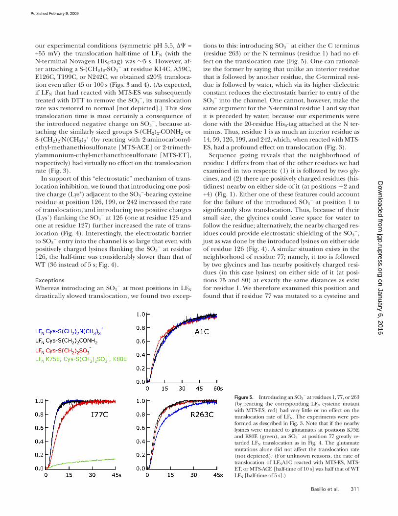
Basilio et al. 311
tions to this: introducing SO 3 � at either the C terminus
(residue 263) or the N terminus (residue 1) had no ef-
fect on the translocation rate ( Fig. 5 ). One can rational-
ize the former by saying that unlike an interior residue
that is followed by another residue, the C-terminal resi-
due is followed by water, which via its higher dielectric
constant reduces the electrostatic barrier to entry of the
SO 3 � into the channel. One cannot, however, make the
same argument for the N-terminal residue 1 and say that
it is preceded by water, because our experiments were
done with the 20-residue His 6 -tag attached at the N ter-
minus. Thus, residue 1 is as much an interior residue as
14, 59, 126, 199, and 242, which, when reacted with MTS-
ES, had a profound effect on translocation ( Fig. 3 ).
Sequence gazing reveals that the neighborhood of
residue 1 differs from that of the other residues we had
examined in two respects: (1) it is followed by two gly-
cines, and (2) there are positively charged residues (his-
tidines) nearby on either side of it (at positions � 2 and
+4) ( Fig. 1 ). Either one of these features could account
for the failure of the introduced SO 3 � at position 1 to
signifi cantly slow translocation. Thus, because of their
small size, the glycines could leave space for water to
follow the residue; alternatively, the nearby charged res-
idues could provide electrostatic shielding of the SO 3 � ,
just as was done by the introduced lysines on either side
of residue 126 ( Fig. 4 ). A similar situation exists in the
neighborhood of residue 77; namely, it too is followed
by two glycines and has nearby positively charged resi-
dues (in this case lysines) on either side of it (at posi-
tions 75 and 80) at exactly the same distances as exist
for residue 1. We therefore examined this position and
found that if residue 77 was mutated to a cysteine and
our experimental conditions (symmetric pH 5.5, Δ � =
+55 mV) the translocation half-time of LF N (with the
N-terminal Novagen His 6 -tag) was � 5 s. However, af-
ter attaching a S-(CH 2 ) 2 -SO 3 � at residue K14C, A59C,
E126C, T199C, or N242C, we obtained ≤ 20% transloca-
tion even after 45 or 100 s ( Figs. 3 and 4 ). (As expected,
if LF N that had reacted with MTS-ES was subsequently
treated with DTT to remove the SO 3 � , its translocation
rate was restored to normal [not depicted].) This slow
translocation time is most certainly a consequence of
the introduced negative charge on SO 3 � , because at-
taching the similarly sized groups S-(CH 2 ) 2 -CONH 2 or
S-(CH 2 ) 2 -N(CH 3 ) 3 + (by reacting with 2-aminocarbonyl-
ethyl-methanethiosulfonate [MTS-ACE] or 2-trimeth-
ylammonium-ethyl-methanethiosulfonate [MTS-ET],
respectively) had virtually no effect on the translocation
rate ( Fig. 3 ).
In support of this “ electrostatic ” mechanism of trans-
location inhibition, we found that introducing one posi-
tive charge (Lys + ) adjacent to the SO 3 � -bearing cysteine
residue at position 126, 199, or 242 increased the rate
of translocation, and introducing two positive charges
(Lys + ) fl anking the SO 3 � at 126 (one at residue 125 and
one at residue 127) further increased the rate of trans-
location ( Fig. 4 ). Interestingly, the electrostatic barrier
to SO 3 � entry into the channel is so large that even with
positively charged lysines fl anking the SO 3 � at residue
126, the half-time was considerably slower than that of
WT (36 instead of 5 s; Fig. 4 ).
Exceptions Whereas introducing an SO 3
� at most positions in LF N
drastically slowed translocation, we found two excep-
Figure 5. Introducing an SO 3 � at residues 1, 77, or 263
(by reacting the corresponding LF N cysteine mutant with MTS-ES; red) had very little or no effect on the translocation rate of LF N . The experiments were per-formed as described in Fig. 3 . Note that if the nearby lysines were mutated to glutamates at positions K75E and K80E (green), an SO 3
� at position 77 greatly re-tarded LF N translocation as in Fig. 4 . The glutamate mutations alone did not affect the translocation rate (not depicted). (For unknown reasons, the rate of translocation of LF N A1C reacted with MTS-ES, MTS-ET, or MTS-ACE [half-time of 10 s] was half that of WT LF N [half-time of 5 s].)
on January 6, 2016jgp.rupress.org
Dow
nloaded from
Published February 9, 2009

312 Proton–Protein Symporter in the Anthrax Toxin Channel
the � -clamp ( Krantz et al., 2005 ). If the phenylalanines
were mutated to alanines, protein translocation was
compromised ( Krantz et al., 2005 , 2006 ). When F427
was mutated to an alanine, the rate of voltage-driven
translocation decreased by about a factor of three, but,
strikingly, the rate of translocation of LF N with an at-
tached SO 3 � enormously increased. (A much larger de-
crease occurs in the rate of translocation driven by a pH
gradient [ Krantz et al., 2005 , 2006 ] and is probably a
consequence of the creation of a leakage pathway for
protons, thereby compromising the Δ pH across the
� -clamp.) This effect of the alanine mutation is clearly
seen in Fig. 6 A for LF N A59C; a similar effect was seen
for the other residue tested, LF N N242C (not depicted).
With either WT LF N or the neutral (CH 2 ) 2 -CONH 2 at-
tached to the LF N A59C cysteine, the half-time of trans-
location (at +55 mV) increased from � 5 to � 15 s,
whereas with SO 3 � attached to the cysteine, the half-
time decreased from a value > > 200 to � 30 s. Thus, the
mutation of F427 to an alanine has almost completely
removed a barrier to SO 3 � translocation. The effect of
the � -clamp on anion entry into the channel was also
manifested in the channel becoming less selective for
then reacted with MTS-ES, the translocation rate was
barely affected ( Fig. 5 ). It appears that of the two possi-
ble factors that we have speculated could be the cause
of this, the two glycines or electrostatic shielding, it is
the latter that accounts for this at position 77, because
when the lysines at positions 75 and 80 were mutated to
glutamates, the introduction of an SO 3 � at position 77
now severely slowed translocation ( Fig. 5 ). Unfortu-
nately, mutating the histidines at positions � 2 and +4 to
aspartates had no effect on the translocation of the
MTS-ES – reacted cysteine at position 1 (not depicted).
Thus, why an SO 3 � at position 1 does not slow transloca-
tion remains an unexplained exception.
A Site of Negative Charge Exclusion from the Channel Where within the channel is the negative charge exclu-
sion of SO 3 � occurring? The (PA 63 ) 7 channel is a mush-
room-like structure with a long stem and a cap that
contains the binding site for LF N ( Fig. 6 B ) ( Krantz
et al., 2005 ; Katayama et al., 2008 ). Near the junction of
the cap with the stem lies residue F427, which forms a
ring of seven phenylalanines that plays an important
role in protein translocation, and which we have dubbed
Figure 6. The effect of replacing phenylalanine with alanine at residue 427 of PA 63 on the rate of translocation of LF N . (A) The experi-ments were performed as described in Fig. 3 . For the WT channel, as shown in Fig. 3 and reproduced here, the introduction of an SO 3
� at residue 59 (red) drastically reduced the rate of LF N translocation from that of MTS-ACE – reacted LF N (black), which was the same as that of WT LF N (not depicted). Mutating the phenylalanine at residue 427 to alanine in the (PA 63 ) 7 channel dramatically increased the rate of translocation of LF N with an attached SO 3
� (orange) and decreased the rate of translocation of MTS-ACE – reacted LF N (gray), which was the same as that of WT LF N (not depicted). (B) Cartoon of the (PA 63 ) 7 channel with LF N being translocated through it. The constriction near the junction of the vestibule with the stem is the � -clamp, which is formed by seven F427s. We envision that for nega-tively charged residues (aspartates or glutamates) on LF N to get past the � -clamp, they must be neutralized; that is, they pick up protons from the cis solution. This proton is subsequently discharged into the trans solution after the residue has traversed the channel. If a non-titratable SO 3
� is introduced into LF N , it is retarded at the � -clamp. Mutation of F427 to alanine alters the constriction and now more readily allows a negatively charged group such as SO 3
� to get past it.
on January 6, 2016jgp.rupress.org
Dow
nloaded from
Published February 9, 2009

Basilio et al. 313
duction (by reacting single-site cysteine mutants with
MTS-ES) of only one, essentially non-titratable nega-
tive charge (SO 3 � ) at arbitrarily chosen positions in LF N
drastically reduced the rate of its translocation ( Fig. 3 ).
The electrostatic basis for this exclusion of SO 3 � is rein-
forced by our observation that it could be partially ame-
liorated by the placement of positive charges (lysines)
adjacent to it in the amino acid sequence ( Fig. 4 ).
The exceptions to the inhibitory effect on LF N trans-
location by an SO 3 � attached to the C terminus (residue
263) or to residue 77 can be interpreted in terms of the
electrostatic forces acting to exclude SO 3 � from the
channel. Thus, the failure of an SO 3 � at the C terminus
to reduce the translocation rate can be attributed to the
water molecules surrounding SO 3 � at this position,
which, because of their high dielectric constant, attenu-
ate the electrostatic repulsive forces acting to exclude
SO 3 � from the channel. An interior SO 3
� -attached resi-
due will in general not have suffi cient space around it to
admit many water molecules. The failure of an SO 3 � at
residue 77 to reduce translocation can be attributed to
electrostatic shielding of its charge by nearby positively
charged lysines at positions 75 and 80. Indeed, when
they were mutated to glutamates, the introduction of
an SO 3 � at position 77 drastically slowed translocation
( Fig. 5 ). This result, obtained by removing positive
charges nearby the SO 3 � and thereby slowing LF N trans-
location, nicely complements the results obtained by
adding positive charges nearby the SO 3 � , and thereby
accelerating LF N translocation ( Fig. 4 ). The one excep-
tion that remains unexplained is why the introduction
of an SO 3 � at position 1 (which is actually an interior
residue because it is preceded by a 20-residue His 6 -tag)
did not retard translocation.
We also identifi ed the � -clamp as a signifi cant factor
in the exclusion of SO 3 � from the channel. Thus, the
(PA 63 F427A) 7 channel translocated MTS-ES – reacted LF-
N A59C at least an order of magnitude faster than did the
WT (PA 63 ) 7 channel ( Fig. 6 A ), and its selectivity for K +
over Cl � was diminished from that of the WT channel
( Fig. 7 ). (The � -clamp is not the only site that acts to
exclude Cl � from the channel. One expects that the
rings of negative charge within the channel created by
residues D276, E302, E308, D315, D335, and E343
should participate in this process. And indeed, the triple-
mutant channel E302S/E308S/D315S was much less
selective for K + over Cl � than was the F427A channel [un-
published data], and, as expected, LF N translocation was
severely impaired [unpublished data].) Why the muta-
tion of F427 to an alanine results in a channel that is less
exclusionary of anions is not obvious. One possibility is
that the mutation causes a repositioning further away
from the entrance to the channel of the nearby nega-
tively charged aspartates at positions 425 and 426. It is
interesting to note that this loss in anion exclusion is
accompanied by a decrease in the translocation rate
K + over Cl � . We see in Fig. 7 that the (PA 63 F427A) 7 chan-
nel, although still cation selective, is less so than is the
WT channel.
D I S C U S S I O N
Because LF N translocation from the cis to the trans so-
lution through (PA 63 ) 7 channels is driven by cis-positive
voltages, even though LF N bears a net negative charge,
we have argued that LF N ’ s aspartates and glutamates
must be transported in their protonated (neutralized)
form (see Krantz et al., 2006 ; Finkelstein, 2009 ). That is,
negatively charged residues are largely excluded at
some point from entering the channel. The experi-
ments reported here support this argument in that
(with specifi c exceptions discussed below) the intro-
Figure 7. The effect of mutating F427 to an alanine on the ion selectivity of the (PA 63 ) 7 channel. The experiments were started with the membrane (diphytanoyl-phosphatidylcholine) separat-ing symmetric solutions of 100 mM KCl, 5 mM potassium succi-nate, and 1 mM EDTA, pH 5.5. After treating the membrane with either valinomycin (0.2 μ g/ml), WT (PA 63 ) 7 , or (PA 63 F427A) 7 , KCl was added in steps to the cis solution, and for each step the rever-sal potential (E rev ) was measured. Plotted in the fi gure is E rev ver-sus the activity ratio of KCl (a cis /a trans ). (Activity coeffi cients were obtained from Appendix 8.10, Table II in Robinson and Stokes [1965] , where we took the KCl concentration to be equal to the K + concentration. Approximately 10 mM of K + was contributed by K-succinate and K-EDTA.) We see in the fi gure that, as expected, the valinomycin-treated membrane (open circles) displayed ideal K + selectivity (continuous line). The selectivity of the WT (PA 63 ) 7 – treated membrane (fi lled circles) deviated somewhat from ideal cation selectivity, and that of the (PA 63 F427A) 7 – treated mem-brane (fi lled triangles) deviated even more so; that is, it was more permeable to Cl � than was the WT channel. The dotted lines are drawn to connect the points. (Applying inappropriately, as usual, the Goldman-Hodgkin-Katz equation to the data, we calculate that P K /P Cl is 21 and 7.6 for the WT (PA 63 ) 7 and (PA 63 F427A) 7 channels, respectively.)
on January 6, 2016jgp.rupress.org
Dow
nloaded from
Published February 9, 2009

314 Proton–Protein Symporter in the Anthrax Toxin Channel
Finkelstein , A. 2009 . Proton-coupled protein transport through
the anthrax toxin channel. Philos. Trans. R. Soc. Lond. B Biol. Sci. 364 : 209 – 215 .
Katayama , H. , B.E. Janowiak , M. Brzozowski , J. Juryck , S. Falke , E.P.
Gogol , R.J. Collier , and M.T. Fisher . 2008 . GroEL as a molecu-
lar scaffold for structural analysis of the anthrax toxin pore. Nat. Struct. Mol. Biol. 15 : 754 – 760 .
Krantz , B.A. , A.D. Trivedi , K. Cunningham , K.A. Christensen , and
R.J. Collier . 2004 . Acid-induced unfolding of the amino-terminal
domains of the lethal and edema factors of anthrax toxin. J. Mol. Biol. 344 : 739 – 756 .
Krantz , B.A. , R.A. Melnyk , S. Zhang , S.J. Juris , D.B. Lacy , Z. Wu ,
A. Finkelstein , and R.J. Collier . 2005 . A phenylalanine clamp
catalyzes protein translocation through the anthrax toxin pore.
Science . 309 : 777 – 781 .
Krantz , B.A. , A. Finkelstein , and R.J. Collier . 2006 . Protein translo-
cation through the anthrax toxin transmembrane pore is driven
by a proton gradient. J. Mol. Biol. 355 : 968 – 979 .
Mueller , P. , D.O. Rudin , H.T. Tien , and W.C. Westcott . 1963 .
Methods for the formation of single bimolecular lipid mem-
branes in aqueous solution. J. Phys. Chem. 67 : 534 – 335 .
Nassi , S. , R.J. Collier , and A. Finkelstein . 2002 . PA 63 channel of an-
thrax toxin: an extended beta-barrel. Biochemistry . 41 : 1445 – 1450 .
Nguyen , T.L. 2004 . Three-dimensional model of the pore form of
anthrax protective antigen. Structure and biological implications.
J. Biomol. Struct. Dyn. 22 : 253 – 265 .
Pannifer , A.D. , T.Y. Wong , R. Schwarzenbacher , M. Renatus , C.
Petosa , J. Bienkowska , D.B. Lacy , R.J. Collier , S. Park , S.H. Leppla ,
et al . 2001 . Crystal structure of the anthrax lethal factor. Nature . 414 : 229 – 233 .
Pimental , R.A. , K.A. Christensen , B.A. Krantz , and R.J. Collier .
2004 . Anthrax toxin complexes: heptameric protective antigen
can bind lethal factor and edema factor simultaneously. Biochem. Biophys. Res. Commun. 322 : 258 – 262 .
Robinson , R.A. , and R.H. Stokes . 1965 . Electrolyte Solutions.
Butterworth, London. 494 pp.
Young , J.A. , and R.J. Collier . 2007 . Anthrax toxin: receptor-binding,
internalization, pore formation, and translocation. Annu. Rev. Biochem. 76 : 243 – 265 .
Zhang , S. , A. Finkelstein , and R.J. Collier . 2004a . Evidence that
translocation of anthrax toxin ’ s lethal factor is initiated by entry
of its N terminus into the protective antigen channel. Proc. Natl. Acad. Sci. USA . 101 : 16756 – 16761 .
Zhang , S. , E. Udho , Z. Wu , R.J. Collier , and A. Finkelstein . 2004b .
Protein translocation through anthrax toxin channels formed in
planar lipid bilayers. Biophys. J. 87 : 3842 – 3849 .
of WT LF N ( Fig. 6 A ). This is to be expected, because in-
sofar as ionized glutamates and aspartates on LF N are al-
lowed to enter the channel, they will act to retard its
(positive) voltage-driven translocation. At this time, it is
not clear to what extent the slower voltage-driven trans-
location of LF N through the (PA 63 F427A) 7 channel is
a consequence of its allowing negatively charged gluta-
mates and aspartates to enter the channel, or to its ef-
fect on the unfolding of LF N ( Krantz et al., 2005 ).
We thank Dr. Paul Kienker for the GHK calculations and insight-ful comments, and Dr. Myles Akabas for critically reading this paper.
This work was supported by National Institutes of Health grants GM-29210 (to A. Finkelstein) and AI-22021 (to R.J. Collier).
Lawrence G. Palmer served as editor.
Submitted: 25 November 2008 Accepted: 15 January 2009
R E F E R E N C E S Benson , E.L. , P.D. Huynh , A. Finkelstein , and R.J. Collier . 1998 .
Identifi cation of residues lining the anthrax protective antigen
channel. Biochemistry . 37 : 3941 – 3948 .
Blaustein , R.O. , and A. Finkelstein . 1990 . Voltage-dependent block
of anthrax toxin channels in planar phospholipid bilayer mem-
branes by symmetric tetraalkylammonium ions. Effects on macro-
scopic conductance. J. Gen. Physiol. 96 : 905 – 919 .
Blaustein , R.O. , T.M. Koehler , R.J. Collier , and A. Finkelstein .
1989 . Anthrax toxin: channel-forming activity of protective an-
tigen in planar phospholipid bilayers. Proc. Natl. Acad. Sci. USA . 86 : 2209 – 2213 .
Bragg , T.S. , and D.L. Robertson . 1989 . Nucleotide sequence and
analysis of the lethal factor gene ( lef ) from Bacillus anthracis. Gene . 81 : 45 – 54 .
Cunningham , K. , D.B. Lacy , J. Mogridge , and R.J. Collier . 2002 .
Mapping the lethal factor and edema factor binding sites on
oligomeric anthrax protective antigen. Proc. Natl. Acad. Sci. USA . 99 : 7049 – 7053 .
Drum , C.L. , S.Z. Yan , J. Bard , Y.Q. Shen , D. Lu , S. Soelaiman , Z.
Grabarek , A. Bohm , and W.J. Tang . 2002 . Structural basis for the
activation of anthrax adenylyl cyclase exotoxin by calmodulin.
Nature . 415 : 396 – 402 .
on January 6, 2016jgp.rupress.org
Dow
nloaded from
Published February 9, 2009