Evaluation of Visual Screening in Prevention of Cervical and ...
147
RICHARD MUWONGE Evaluation of Visual Screening in Prevention of Cervical and Oral Cancer in India ACADEMIC DISSERTATION To be presented, with the permission of the Faculty of Medicine of the University of Tampere, for public discussion in the Auditorium of Tampere School of Public Health, Medisiinarinkatu 3, Tampere, on September 26th, 2008, at 12 o’clock. UNIVERSITY OF TAMPERE
-
Upload
khangminh22 -
Category
Documents
-
view
4 -
download
0
Transcript of Evaluation of Visual Screening in Prevention of Cervical and ...

RICHARD MUWONGE
Evaluation of Visual Screening in Preventionof Cervical and Oral Cancer in India
ACADEMIC DISSERTATIONTo be presented, with the permission of
the Faculty of Medicine of the University of Tampere,for public discussion in the Auditorium of
Tampere School of Public Health, Medisiinarinkatu 3,Tampere, on September 26th, 2008, at 12 o’clock.
U N I V E R S I T Y O F T A M P E R E

RICHARD MUWONGE
Evaluation of Visual Screening in Preventionof Cervical and Oral Cancer in India
A c t a U n i v e r s i t a t i s Ta m p e r e n s i s 1 3 4 5Ta m p e r e U n i v e r s i t y P r e s s
Ta m p e r e 2 0 0 8

University of Tampere, School of Public HealthFinlandInternational Agency for Research on Cancer, LyonFrance
Supervised by Reviewed byProfessor Risto Sankila Professor Markku KoskenvuoUniversity of Tampere University of HelsinkiFinland FinlandDr Rengaswamy Sankaranarayanan Professor emer. Aulikki NissinenInternational Agency for Research on University of KuopioCancer FinlandFrance
DistributionBookshop TAJU Tel. +358 3 3551 6055P.O. Box 617 Fax +358 3 3551 768533014 University of Tampere [email protected] www.uta.fi/taju http://granum.uta.fi
Cover design byJuha Siro
LayoutSirpa Randell
Acta Universitatis Tamperensis 1345 Acta Electronica Universitatis Tamperensis 760ISBN 978-951-44-7440-8 (print) ISBN 978-951-44-7441-5 (pdf)ISSN 1455-1616 ISSN 1456-954X http://acta.uta.fi
Tampereen Yliopistopaino Oy – Juvenes PrintTampere 2008

To my beloved family, Mum, Dad, Phyllis, George, John, Aggie, Loretta and Phillip


TAblE Of CONTENTS
LIST OF TABLES ...............................................................................................................viii
LIST OF FIGURES ............................................................................................................... ix
SUMMARY ........................................................................................................................... xi
LIST OF ORIGINAL ARTICLES ...................................................................................... xv
LIST OF ABBREvIATIONS ............................................................................................. xvi
1. INTRODUCTION ..........................................................................................................191.1 Basic concepts ......................................................................................................20
1.1.1 Primary prevention of cancer ................................................................201.1.2 Early detection of cancer ........................................................................20
1.1.2.1 Screening ..................................................................................211.1.2.2 Evaluation of screening programmes ..................................23
1.1.3 Palliative care............................................................................................261.1.4 Concept of risk and protective factors ..................................................26
1.2 Demographic profile of India ............................................................................261.3 Health care infrastructure in India ..................................................................291.4 Cancer registration in India ..............................................................................32
2. REvIEw OF THE LITERATURE ..............................................................................352.1 Cancer of the cervix uteri ..................................................................................38
2.1.1 Epidemiology of cervical cancer ............................................................392.1.1.1 The global scene ......................................................................392.1.1.2 India ......................................................................................... 40
2.1.2 Natural history .........................................................................................412.1.3 Risk factors............................................................................................... 422.1.4 Screening ...................................................................................................47
2.1.4.1 Screening methods for cervical neoplasia .......................... 482.1.5 Prevention by vaccination ......................................................................53
2.2 Cancer of the oral cavity ................................................................................... 542.2.1 Epidemiology of oral cancer ...................................................................55
2.2.1.1 The global scene ......................................................................552.2.1.2 India ..........................................................................................56
2.2.2 Oral cancer precursors ............................................................................572.2.3 Natural history .........................................................................................57

2.2.4 Risk factors................................................................................................582.2.5 Screening ...................................................................................................622.2.6 Primary prevention ................................................................................ 642.2.7 Results of treatment of precursors .........................................................65
3. AIMS OF THE STUDY ................................................................................................ 66
4. METHODS USED IN THE STUDY .......................................................................... 684.1 Data sources ........................................................................................................ 68
4.1.1 Cervical cancer screening cross-sectional studies carried out in Africa and India (Papers I and II) ............................................. 684.1.2 Cluster-randomized cervical cancer screening trial in Ambillikai, Dindigul District, Tamil Nadu, India (Paper III) ..........704.1.3 The Trivandrum oral cancer screening study in Kerala, India: a cluster-randomized controlled trial (Paper Iv) ....................724.1.4 A nested case control study from the Trivandrum oral cancer screening study in Kerala, India (Paper v) .............................74
4.2 Statistical methods used in analysis .................................................................75
5. RESULTS .........................................................................................................................825.1 Assessment of accuracies of tests for screening for cervical cancer and exploration of the sources of heterogeneity .............................................82
5.1.1 Accuracy of screening tests at CIN II or worse outcome ...................825.1.2 Summary of test accuracy of all screening tests for all categories of CIN .................................................................................... 845.1.3 Pooled relative accuracy of screening tests ......................................... 865.1.4 Influence of study characteristics on the sensitivity and specificity of screening tests .................................................................. 885.1.5 Influence of study characteristics on the diagnostic odds ratio using multivariate SROC regression analysis ............................ 88
5.2 Assessment of gain in test performance when two visual inspection screening techniques are combined in the detection of high grade CIN or worse ...............................................................................915.3 Evaluation of the effect of visual inspection screening of the cervix on cervical cancer incidence and mortality .................................925.4 Evaluation of the effect of oral visual inspection screening on oral cancer mortality ..........................................................................................94

5.5 Assessment of tobacco smoking, chewing and alcohol drinking, the major risk factors of cancer of the oral cavity ..........................................95
5.5.1 Effect of tobacco smoking on oral cancer risk.....................................955.5.2 Effect of tobacco chewing on oral cancer risk .................................... 965.5.3 Effect of alcohol drinking on oral cancer risk .....................................975.5.4 Attributable fractions for tobacco smoking, chewing and alcohol drinking on the risk of oral cancer ..........................................97
6. DISCUSSION ............................................................................................................... 1006.1 Methodological strengths of the study ..........................................................1016.2 Methodological limitations of the study ........................................................1046.3 Comparison of the study results with findings from other studies ...........107
6.3.1 Accuracy of screening tests ..................................................................1076.3.2 Gain in test performance from combining two visual inspection techniques of cervical cancer screening ..........................1096.3.3 Effect on cervical cancer incidence and mortality of screening by visual inspection with acetic acid ................................1106.3.4 Effect of oral visual inspection on oral cancer mortality ................1116.3.5 The three major risk factors of oral cancer in this study population ...............................................................................................112
7. CONCLUSIONS ...........................................................................................................114
ACKNOwLEDGEMENTS ...............................................................................................118
REFERENCES ................................................................................................................... 120
ORIGINAL PUBLICATIONS ..........................................................................................145

viii
RICHARD MUWONGE
lIST Of TAblES
Table 1.1 Achievements through the years 1951–2008 as a result of thepolicy initiatives of the National Health Policy -1983 ...............................................30
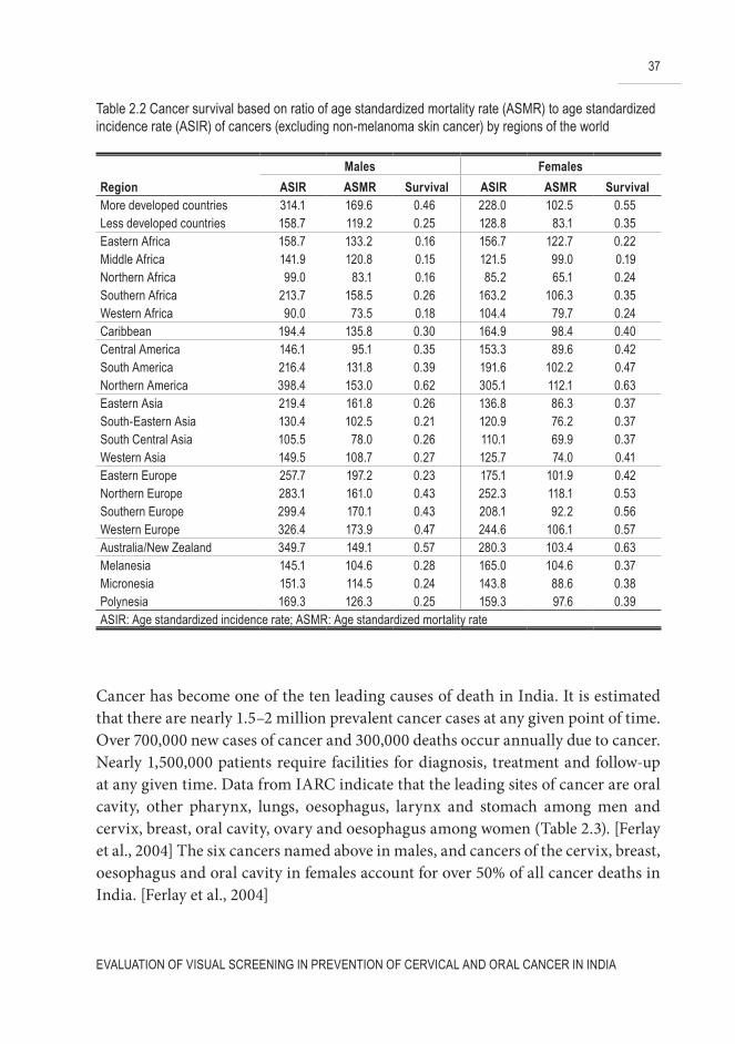
Table 2.1 Global cancer incidence and mortality ............................................................36Table 2.2 Cancer survival based on ratio of age standardized mortality rate
(ASMR) to age standardized incidence rate (ASIR) of cancers (excludingnon-melanoma skin cancer) by regions of the world ................................................37
Table 2.3 Cancer incidence in India in 2002 ....................................................................38Table 2.4 Terminology of cervical precancerous abnormalities ................................... 42Table 2.5 Regression, persistence and progression probabilities of CIN .................... 42Table 4.1 Tests assessed and the number of women tested in each centre ...................69Table 4.2 Cross-tabulation of screening test and reference standard results ..............75Table 4.3 Joint probabilities of pairs of test results for the single and the
combined test among the diseased and non-diseased ..............................................79Table 5.1 Number of women included, sensitivity and specificity values at
CIN II or worse disease outcome for the different cervical cancerscreening tests performed in the different cross-sectional study sites inAfrica and India ..............................................................................................................83
Table 5.2 Sensitivity and specificity of five screening tests for CIN1 or moresevere disease (CIN1+), CIN2+, CIN3+ and cancer; minimum, maximumand meta-analytically pooled measures ......................................................................85
Table 5.3 Multivariate logistic regression assessing the factors influencingvariation of the sensitivity and specificity at CIN2+ outcome.Non-significant effects are omitted .............................................................................89
Table 5.4 Results of the SROC regression analysis assessing the factorsinfluencing variation of the DOR at CIN2+ outcome. Non-significanteffects are omitted ......................................................................................................... 90
Table 5.5. Ratio (R*) of the number of additional false positives per additional true positive for outcome CIN 2-3+ at varying diseaseprevalence, assuming a zero trade-off .........................................................................92
Table 5.6 Overall and age-specific hazard ratio for incidence for cervicalcancers and for cervical cancer deaths ........................................................................93
Table. 5.7 Oral cancer incidence, stage distribution and mortality in arandomized control trial of oral cancer screening in TrivandrumDistrict, India ..................................................................................................................95
Table 5.8 Smoking and risk of oral cancer using incident cases (only males considered) ................................................................................................ 96

ix
EVAlUATION Of VISUAl SCREENING IN PREVENTION Of CERVICAl AND ORAl CANCER IN INDIA
Table 5.9 Chewing habit and risk of oral cancer using the incident casesby gender ..........................................................................................................................98
Table 5.10 Alcohol drinking and risk of oral cancer using the incident cases(only males considered) ................................................................................................ 99
Table 5.11 The adjusted population attributable fractions for smoking,chewing and alcohol drinking ..................................................................................... 99
lIST Of fIGURES
Figure 1.1 Determination of cut-off point for screen positivity ....................................22Figure 1.2 Age population pyramids of India and the United States, 2000 .................28Figure 2.1 Position of the cervix in relation to other female reproductive organs .....39Figure 2.2 Position of the oral cavity in relation to other head and neck organs .......55Figure 4.1 Cervical cancer screening cross-sectional study sites in
Africa and India ............................................................................................................. 68Figure 4.2 National map showing Dindigul District, Tamil Nadu State, India ..........71Figure 4.3 Map showing study clusters, Trivandrum District,
Kerala State, India ..........................................................................................................73Figure 4.4 Sensitivity and specificity for a) vIA alone and combined test and b)
vILI alone and combined test when disease outcome=CIN 2-3+. ......................... 80Figure 5.1 Relative sensitivity in a), c) and e) and relative specificity in b), d)
and f) at outcomes CIN2+ and CIN3+ ........................................................................87Figure 5.2 Cumulative incidence of cervical carcinoma a) overall, b) stage 2
or worse and c) cumulative mortality from cervical cancer in interventionand control groups ........................................................................................................94

x
RICHARD MUWONGE

xi
EVAlUATION Of VISUAl SCREENING IN PREVENTION Of CERVICAl AND ORAl CANCER IN INDIA
SUMMARy
India is the country with the greatest relative global burden of cancers of the cervix and oral cavity, and these two cancers form the biggest share of the cancer burden in the country. Because these two cancers are generally seen to pass through a preclinical detectable phase, screening for their precancers and providing appropriate treatment would be beneficial in the efforts to reduce the cancer burden in the country. Pap smear, which has been seen to be an effective cervical cancer screening technique in the developed world, is resource intensive, requiring a laboratory infrastructure, quality assurance for the different steps involved and a system to report the test results to women. For this reason, implementation of Pap smear screening in India, as in other low/medium resourced countries, has met challenges and difficulties, leading to the evaluation of alternative, simple, safe, acceptable, affordable and inexpensive visual inspection techniques for detecting cervical precancer lesions and preventing cervical cancer. Furthermore, oral visual inspection is an oral cancer screening method which is cheap, can be easily applied by a wide range of medical personnel and, hence, is suitable for India and other developing countries.
The main aim of this study was to assess the test performance and to evaluate the impact of visual inspection techniques when used in screening for cervical and oral cancer lesions to facilitate their use in cervical and oral cancer prevention programmes, and to contribute to the efforts in the prevention of cervical and oral cancers especially in low/medium resourced settings. The test performance of other cervical cancer screening methods is additionally explored to enable comparisons with the visual screening techniques. The additional value of a combination of two visual screening methods for detecting cervical neoplasia compared to a single test is likewise evaluated. The data used were from two large cluster-randomized trials carried out in India and a number of cross-sectional study sites mainly from India.
Between 1999 and 2003, the test performance of five cervical cancer screening methods, visual inspection with acetic acid (vIA), visual inspection with Lugol’s iodine (vILI), vIA with magnification (vIAM), conventional Pap smear and Human papilomavirus (HPv) testing, were simultaneously evaluated in more than 58,000 women aged 25 to 64 from eleven urban settings in India (6 centres) and five African countries (5 centres), using a common protocol. Different providers blind to the results from the other tests performed these tests. These studies were carried out by the International Agency for Research on Cancer (IARC) as part of the Alliance for Cervical Cancer Prevention (ACCP) supported by the Bill & Melinda Gates

xii
RICHARD MUWONGE
Foundation (Seattle, washington, USA) to advance cervical cancer prevention in low/medium resourced countries.
In order to evaluate whether a single lifetime vIA screening and treatment of detected cervical intraepithelial neoplacia (CIN) by cryotherapy and excision under field conditions, all provided by trained nurses, can lead to reduced cervical cancer incidence and mortality among women offered screening compared to a similar group of women receiving the existing standard health care, IARC in collaboration with the Christian Fellowship Community Health Centre (CFCHC), a rural hospital and a cancer centre in the Dindigul District of Tamil Nadu State in South India, organized a large randomized controlled trial involving about 80,000 women. In this trial, 57 clusters (49,300 women) were randomly allocated to the intervention group and 56 to the control group (31,000). Apparently healthy eligible women aged 30–59 years, with an intact uterus, no past history of cervical cancer, and living in the study clusters were enumerated and interviewed by female health workers to obtain sociodemographic and reproductive variables. All eligible women in both groups were educated about the prevention, early detection, and treatment of cervical cancer. women (31,300 in number) in the intervention group were then offered vIA, and vIA positive women were colposcopied. The nurse, during the same screening visit, took a punch biopsy in those with abnormalities in colposcopy followed by immediate treatment with cryotherapy, when appropriate. women with lesions not eligible for cryotherapy were referred for loop electrosurgical excision procedure (LEEP) and those with suspected invasive cancer were referred for further investigations and treatment. During seven years (2000–2006) from the beginning of screening, 167 invasive cervical cancer cases and 83 cervical cancer deaths accrued in the group of women offered vIA screening compared with 158 cases and 92 deaths and in the control group. This translated into a 25% reduction in the number of cervical cancer cases, a 24% reduction in the occurrence of advanced cervical cancers and a 35% reduction in the number of cervical cancer deaths among women offered vIA screening, all these reductions were statistically significant. Furthermore, the overall risk of death from any causes also declined significantly, by 13% in the vIA group. In conclusion, this trial showed that vIA screening could reduce the cervical cancer burden.
In a similar community-based cluster-randomized controlled oral cancer screening intervention trial, carried out by IARC in collaboration with the Regional Cancer Centre, Trivandrum, Kerala, India, 13 clusters called ‘panchayaths’ (municipal administrative units in rural areas of India, with total populations of 20,000–50,000), involving about 191,800 apparently healthy individuals of 35 years and above, with no past history of oral cancer were randomly allocated to two groups. Seven clusters

xiii
EVAlUATION Of VISUAl SCREENING IN PREVENTION Of CERVICAl AND ORAl CANCER IN INDIA
of the intervention group (with about 96,500 individuals) were randomized to receive three rounds of oral visual inspection at 3-year intervals provided by trained health workers during the period 1995–2004 in Trivandrum, South India, whereas 6 clusters randomized to the control group received the standard health care. The aim of this trial was to assess if oral visual screening would ultimately lead to a reduction in oral cancer mortality in the intervention group compared to the control group. A shift towards early stage at diagnosis (41% vs 23%) and a higher 5-year survival proportion (50% vs 34%) were observed in the screened population. A 21% reduction in oral cancer mortality was observed in the intervention group compared to the control group 9 years from the initiation of screening in this trial, which did not reach statistical significance. However, a statistically significant 33% reduction in mortality was observed among tobacco and/or alcohol users compared to similar control subjects. In summary, evidence from the Indian study shows that oral visual screening can reduce mortality in high-risk individuals. The cost-effectiveness of oral visual inspection is currently being addressed in the context of this trial.
In an effort to assess the effect of the three major risk factors, tobacco smoking, paan chewing and alcohol habits, on oral cancer incidence in Trivandrum, India, a nested case-control study was designed within the framework of the Trivandrum oral cancer screening trial. The analysis included all incident oral cancer cases diagnosed during the trial period. Five controls, matched for sex, age (±1 year), panchayaths, round of screening and response status (that is if they were interviewed or not at the particular round and at the previous round(s) for the cases diagnosed in the second and third screening rounds), were randomly selected for each case from all other participants not diagnosed with oral cancer during the trial period. Paan chewing was the strongest risk factor associated with oral cancer with increased risk effects observed in all categories of paan chewing. Big differences in risk estimates among men and women chewing paan were observed with a 3-fold increased risk of oral cancer for male chewers compared to an 11-fold increased risk among female chewers, both groups compared to their corresponding never chewers. Effects of chewing paan with or without tobacco on oral cancer risk were elevated for both sexes. A 2-fold increased risk of oral cancer was observed among male bidi smokers. Dose response relations were observed for the frequency and duration of chewing and alcohol drinking, as well as in duration of bidi smoking. These results further show that cessation of tobacco use and moderation of alcohol use in combination with early diagnosis remain the key elements in oral cancer prevention and control.
India, like many low and medium resourced countries, is hit hard by the burden of cervical and oral cancers. It has a limited health budget, cancer treatment facilities are not universally available and life-prolonging therapies are often unavailable.

xiv
RICHARD MUWONGE
Nevertheless, it is of great importance to prevent those cancers (such as cervical and oral cancer) that can be prevented. Based on the evidence discussed in this dissertation, specific priorities should be given to primary prevention initiatives aimed at taking action against tobacco and heavy alcohol consumption and concerted action through early detection, against cancers of the cervix and oral cavity.

xv
EVAlUATION Of VISUAl SCREENING IN PREVENTION Of CERVICAl AND ORAl CANCER IN INDIA
lIST Of ORIGINAl ARTIClES
This dissertation is based on the following original articles. Some results originally not reported in the articles are also presented.
I. Arbyn M, Sankaranarayanan R, Muwonge R, Keita N, Dolo A, Mbalawa CG, Nouhou H, Sakande B, wesley R, Somanathan T, Sharma A, Shastri S, Basu P. Pooled analysis of the accuracy of five cervical cancer screening tests assessed in eleven studies in Africa and India. Int J Cancer 2008; 123: 153–60.
II. Muwonge R, walter SD, wesley RS, Basu P, Shastri SS, Thara S, Mbalawa CG, Sankaranarayanan R. Assessing the gain in diagnostic performance when two visual inspection methods are combined for cervical cancer prevention. J Med Screen 2007; 14: 144–50.
III. Sankaranarayanan R, Esmy PO, Rajkumar R, Muwonge R, Swaminathan R, Shanthakumari S, Fayette JM, Cherian J. Effect of visual screening on cervical cancer incidence and mortality in Tamil Nadu, India: a cluster-randomized trial. Lancet 2007; 370: 398–406.
Iv. Sankaranarayanan R, Ramadas K, Thomas G, Muwonge R, Thara S, Mathew B, Rajan B. Effect of screening on oral cancer mortality in Kerala, India: a cluster-randomized controlled trial. Lancet 2005; 365: 1927–33.
v. Muwonge R, Ramadas K, Sankila R, Thara S, Thomas G, vinoda J, Sankaranarayanan R. Role of tobacco smoking, chewing and alcohol drinking in the risk of oral cancer in Trivandrum, India: A nested case-control design using incident cancer cases. Oral Oncol 2008; 44: 446–54.

xvi
RICHARD MUWONGE
lIST Of AbbREVIATIONS
ACCP Alliance for Cervical Cancer PreventionAIDS Acquired immunodeficiency syndromeASCUS Atypical squamous cells of undetermined significanceCBHI Central Bureau of Health IntelligenceCFCHC Christian Fellowship Community Health CentreCHC Community Health CentreCI Confidence intervalCIA Central Intelligence AgencyCIN Cervical intraepithelial neoplasiaCIS Carcinoma in situDNA Deoxyribonucleic acidDOR Diagnostic odds ratioDPCP Detectable preclinical phase DTP Diphtheria, tetanus and poliomyelitisEBv Espstein-Barr virusFP False positiveHBCR Hospital Based Cancer RegistryHHv Human herpesvirusHIv Human immunodeficiency virusHPv Human papiloma virusHSIL High-grade squamous intraepithelial lesionsHSv Herpes simplex virusIARC International Agency for Research on CancerICD-O International Classification of Diseases for OncologyIDB International Data BaseIMR Infant mortality rateICMR Indian Council of Medical Research LBC Liquid-based cytologyLEEP Loop electrosurgical excision procedureLR+ Positive likelihood ratioLR- Negative likelihood ratio

xvii
EVAlUATION Of VISUAl SCREENING IN PREVENTION Of CERVICAl AND ORAl CANCER IN INDIA
LSIL Low-grade squamous intraepithelial lesionMOHFw Ministry of Health & Family welfareNCCP National Cancer Control ProgrammesNCD Non-communicable diseasesNCRP National Cancer Registry ProgrammeNHP National Health PolicyNPv Negative predictive valueOR Odds ratioOSF Oral submucous fibrosisPBCR Population Based Cancer RegistryPHC Primary Health CentrePPv Positive predictive valuePRB Population Reference BureauRGI Registrar General of IndiaRHS Rural Health StatisticsRLU Relative light unitSC Sub-CentreSCJ Squamocolumnar junctionSROC Summary receiver operating characteristic SRS Sample Registration SystemTP True positiveUNDP United Nations Development ProgrammeUNPD United Nations Population DivisionUSA United States of AmericavIA visual inspection with acetic acidvIAM visual inspection with acetic acid using a magnifying glassvILI visual inspection with Lugol’s iodinewB world BankwHO world Health Organization

xviii
RICHARD MUWONGE
19
EVAlUATION Of VISUAl SCREENING IN PREVENTION Of CERVICAl AND ORAl CANCER IN INDIA
1. INTRODUCTION
In developing countries and newly industrializing regions such as Asia-Pacific, non-communicable diseases (NCD), such as cancer, cardiovascular diseases and diabetes are becoming major public health issues. [wHO, 2005] Driven mainly by the ageing population, cancer is becoming a major health problem for most countries. The International Agency for Research on Cancer (IARC) estimated 8.1 million new cancer cases [Parkin et al., 1999] and 5.2 million cancer deaths [Pisani et al., 1999] in 1990. The estimates of these figures increased to 10.9 million (5.0 million in more and 5.8 million in less developed countries), 6.7 million (2.7 million in more and 4.0 million in less developed countries), respectively for 2002 [Ferlay et al., 2004], with the malignant tumours responsible for 12% of the nearly 56 million deaths worldwide and expected to increase to 15 million by 2020. [Ferlay et al., 2004] About 60% of these new cases is expected to occur in less developed regions of the world, and cancer is emerging as a major public health problem in developing countries, matching its effect in industrialized nations. [Parkin, 2001; Stewart et al., 2003]
with this increasing burden, understanding, preventing and controlling malignant neoplasm is an urgent priority worldwide. In 2002, the world Health Organization (wHO) published ‘National Cancer Control Programmes (NCCP), policy and managerial guidelines’ which offer the most rational means of achieving a substantial degree of cancer control, even where resources are severely limited [wHO, 2002]. NCCP is a public health programme aiming to reduce cancer incidence and mortality and improve quality of life of cancer patients through systematic and equitable implementation of evidence-based strategies for prevention, early detection, diagnosis, treatment and palliation while making the best use of the available resources. It is based on current evidence suggesting that at least 1/3 of the new cases of cancer each year throughout the world are preventable by modifying risk factors (such as controlling tobacco and alcohol use, moderating diet, and immunizing against viral hepatitis B); early detection and effective treatment would permit further 1/3 of the deaths to be avoided where resources are available; while effective techniques permitting comprehensive pain relief and palliative care for improving the quality of life of the 1/3 more advanced cases (and their families). [wHO, 2002]. Establishing a comprehensive NCCP requires competent management

20
RICHARD MUWONGE
and the best use of available resources for planning, implementing and evaluating disease control strategies, tailored to the local socioeconomic and cultural context, as well as scientific knowledge and experience ranging from the complexities of intracellular molecular regulation to individual lifestyle choices.
Improved cancer control, to a substantial extent, depends on prevention strategies and early detection programmes, including information campaigns and population-based screening programmes. The success of early detection programmes relies on effective and optimal use of treatment possibilities. Even though the tumour biology is largely known, many years will probably elapse before cancer mortality can be significantly reduced through application of new cancer drugs and treatment principles. Hence, the aspects of controlling cancers, such as cervical and oral cancers, must be tackled in the context of systematic and comprehensive cancer control strategies, in which risk factor moderation campaigns and cancer screening programmes are integrated with other health programmes, rather than working in isolation.
1.1 basic concepts
1.1.1 Primary prevention of cancer
Primary prevention means eliminating or minimizing the exposure of individuals to the causes of cancer or increasing their resistance to them, leading to reduced individual susceptibility to the effects of such causes. It is this approach that offers the greatest public health potential and the most cost-effective long-term cancer control. This prevention strategy includes programmes such as tobacco control programmes used in the fight against tobacco-related cancers of the lung, oral cavity, larynx and oesophagus, and vaccination programmes such as the currently evaluated human papiloma virus (HPv) vaccination to reduce cervical cancer incidence.
1.1.2 Early detection of cancer
Early detection comprises early diagnosis in symptomatic populations and screening in asymptomatic, but at risk, populations. Screening of apparently healthy individuals may reveal cancer in early or precursor stages, when treatment may be most effective. Early detection is only successful when linked to effective treatment, which makes it possible to prevent the progression of the disease and its complications (including

21
EVAlUATION Of VISUAl SCREENING IN PREVENTION Of CERVICAl AND ORAl CANCER IN INDIA
deaths). Thus, a national cancer control programme should set up guidelines for integrating treatment resources with early detection programmes and provide therapeutic standards for the most import cancers in the country. [wHO, 2002] In low resourced regions, the development of national diagnostic and treatment guidelines should include minimum standards of care that promote rational use of existing resources and greater equity in access to treatment services. Since the cost of setting up and maintaining early detection, diagnostic and treatment facilities is high, they should preferably initially be concentrated in a few locations in a country to avoid draining the limited resources that are usually shared with other competing needs. Facilities can be expanded when additional resources become available.
with early detection, there is a greater chance that curative treatment will be successful, particularly for cancers of the breast, cervix, mouth, larynx, colon and rectum and skin. It is therefore critical that people are taught to recognize early warning signs of the disease, such as lumps, sores that fail to heal, abnormal bleeding, persistent indigestion, and chronic hoarseness, and that they are urged to seek prompt medical attention. This can be promoted in all countries by public health education campaigns and through training of primary health care workers.
1.1.2.1 Screening
Population screening, which is mass application of relatively simple and inexpensive tests to asymptomatic individuals to classify them as being likely or unlikely to have the disease, is one approach to early detection. Subjects with abnormal screening results are then subjected to conventional diagnostic procedures and, if necessary, given appropriate treatment. The ultimate objective of cancer screening programmes is reduction of mortality from the disease among the individuals screened.
For any cancer screening project to succeed the following criteria must be satisfied:
Detectable preclinical phase and early treatmentThe cancer must have a detectable preclinical phase (DPCP) during which early treatment results in lower mortality than treatment given later after symptoms develop. Cervical cancer, for example, develops from precancerous lesions, which take probably more than 10 years to progress to invasive cancer. These lesions, when detected by a screening test such as the Papanicolaou (Pap or conventional cytology) smear test and treated, usually have a better prognosis than if treatment begins after the cancer becomes invasive.

22
RICHARD MUWONGE
Suitable testA screening programme has to use a suitable test. The suitability of a test is considered by assessing its accuracy characteristics to assess that it is a valid test and assessing acceptability and the costs involved.
Test validityThe validity of the screening test can be expressed in terms of its sensitivity and specificity, the two measures used to determine the ability of the test to identify correctly the diseased and non-diseased individuals. In reality, there is always an overlap between the distributions of the screening test results in the disease free and diseased populations (Figure 1.1). This makes the location of the cut-off value to classify screening test results as positive or negative arbitrary.
A valid screening test should have both high sensitivity and high specificity. Sensitivity is the indicator of yield of cases (i.e. number of diseased cases identified by the programme), whereas specificity is an indicator of the number of false positive test results. As shown in Figure 1.1, in practice there is always a trade-off between these two measures. The ability of a screening test to detect as many true positives as possible (high sensitivity) can only be increased at the expense of an increase in
figure 1.1 Determination of cut-off point for screen positivity

23
EVAlUATION Of VISUAl SCREENING IN PREVENTION Of CERVICAl AND ORAl CANCER IN INDIA
the number of false positive screening test results (low specificity) and vice versa. A screening policy aiming at maximum sensitivity might lead to unacceptably low specificity, resulting in high costs from the referral of large numbers of false positives for further investigations and poor motivation of subjects to participate in subsequent screening examinations. [dos Santos Silva, 1999; wHO, 2002]
Test acceptability and costIn addition to a screening test having adequate validity, it should be low in cost, convenient, simple and as painless as possible, and should not cause any complications. A combination of these features would improve compliance, which is one of the key factors for a successful screening programme.
Suitable screening programmeScreening programmes should be undertaken only when their effectiveness has been demonstrated, when resources (personnel, equipment and so on) are sufficient to cover nearly all of the target group, when facilities exist for diagnostic and for therapeutic procedures and follow-up of those with abnormal results, and when the prevalence of the disease is high enough to justify the effort and costs of screening. The screening policy should specify precisely who is to be screened, at what age, at what frequency and with what test and whom to treat and with which treatment. [dos Santos Silva, 1999; Hakama et al., 1986; Soler et al., 2000] Taking the example of cervical cancer screening, in high resourced regions such as the USA, screening is recommended to begin not later than 21 years of age either with annual screening with conventional cervical cytology smear test or every two years with liquid-based cytology until age 30 years. After 30 years, screening may continue every 2–3 years for those women who have had three consecutive, adequate, negative/normal cytology results. [Smith, 2006] In developing countries, the best cervical cancer screening strategy might be to screen women at highest risk of the disease with maximum population coverage, even at infrequent screening intervals, or even only once in a lifetime. [Soler et al., 2000; wHO, 2002]
1.1.2.2 Evaluation of screening programmes
After establishing that a particular cancer is an important public health problem and valid screening test is available, it becomes necessary to evaluate the potential screening programme to assess whether it is worth introducing as a measure to control that particular cancer. This includes assessing the feasibility and cost-

24
RICHARD MUWONGE
effectiveness (low cost per case detected) of the screening programme. Regardless of how cost-effective the screening programme, its final goal of reducing morbidity and/or mortality from that particular cancer in the target population must be warranted.
Process measuresSince it can take many years for precancerous lesions to manifest as invasive cancers, it would take years after the beginning of a screening programme to be able assess its final objective of reduction in cancer morbidity and/or mortality. For this reason, the feasibility, acceptability and costs of the programme may be evaluated using process measures, which are related to the administrative and organizational aspects of the programme such as identification of the target population, number and proportion of participating in the screening, diagnosis and treatment facilities in the health system, number and proportion complying with the referral to these facilities, total costs, and costs per case detected.
In particular, the positive predictive value (PPv) of the screening test is a useful process measure, which gives the proportions of persons found to truly have the cancer in question after further diagnostic examination out of all those who had positive screening test results. A high PPv indicates that a large proportion of programme costs are actually being spent on the detection of the disease during its DPCP.
Effectiveness of reducing cancer mortalityIdentifying and treating precancerous disease does not have a public health value if it does not ultimately lead to reduction in cancer morbidity and/or mortality of those cases. Accurately estimating the effect of screening on cancer morbidity and mortality requires a follow-up period of large populations. Consequently, intermediate outcomes such as detection rates of precancerous lesions and stage distribution of cancer at diagnosis and case-fatality (survival) have been evaluated since they may be available in the early years of the screening programme. For example, down staging and lower case-fatality should be observed in screen-detected cancer cases than in the symptomatically diagnosed cases if the screening programme is successful.
However, there are serious limitations associated with the use of intermediate endpoints. This is because they suffer from four types of biases, namely, length bias, lead-time bias, over-diagnosis bias and selection bias. [Baker et al., 2002; dos Santos Silva, 1999]
25
EVAlUATION Of VISUAl SCREENING IN PREVENTION Of CERVICAl AND ORAl CANCER IN INDIA
Length biasThis type of bias occurs when screening over-represents less aggressive disease because it has a longer asymptomatic period and thus has a high likelihood of being detected by screening. On the other hand, fast growing tumours have a short asymptomatic period and are therefore less likely to be detected early by screening, especially if the screening interval is long, making them present as symptomatic cases. The screen-detected cases may be those with lesions with a more favourable prognosis, while cases with similar onset date but more rapid disease progression are detected by clinical symptoms. In this case, the screening programme will falsely appear to improve survival while the result merely reflects the detection of less aggressive disease through screening.
Lead-time biasSince screening is carried out in asymptomatic individuals, by default the time of diagnosis for every case detected by screening will be advanced by some amount (lead-time) compared to the time of diagnosis in the absence of screening. In this case, if survival is calculated from the date of diagnosis, screening will falsely appear to prolong survival, because of early detection, even if both screened and unscreened individuals would have survived for the same amount of time after the onset of the disease. In other words, detection of an asymptomatic cancer by screening starts the clock at a younger age so the survival time from screen detection is longer than the survival time from clinical detection, even if screening does not change age at death. This is referred to as lead-time bias. One of the ways in which the effect of this type of bias can be taken into account during the evaluation of the screening programme is to compare the mortality rates between the screened and unscreened groups. Alternatively, if the amount of lead-time is known, which in reality is very unlikely, it can be accounted for in the comparison of survival experience between the screen-detected and symptomatic cases.
Over-diagnosisThere is a possibility that many of the precancerous lesions detected by the screening programme would never have progressed to invasive cancer or death. Thus, the true benefit of screening by identifying pre-clinical lesions may be much smaller than is perceived.

26
RICHARD MUWONGE
Selection biasIndividuals who consent to be screened may differ from others in ways that are related to survival times, leading to selection bias.
1.1.3 Palliative care
Palliative care is aimed at improving the quality of life of those patients beyond curative treatment and their families who are affected with life-threatening disease. It also includes, besides the treatment, pain relief and consideration of other physical, psychological and spiritual problems. [dos Santos Silva, 1999; wHO, 2002]
1.1.4 Concept of risk and protective factors
Most cancers emerge due to the interaction of multiple factors ranging from an individual’s genetic characteristics to his/her lifestyle. Researchers of causes of cancer define the term risk factor as any individual or environmental factor that is related to the increased likelihood of developing that particular cancer. Factors associated with a decreased likelihood of a particular cancer are referred to as protective factors. Risk or protective factors are a matter of probability. They influence an individual’s likelihood of developing a disease. This does not necessarily mean that they cause the disease. Some individuals with one or more risk factors for a particular cancer never develop it, while others who have no known risk factors do develop the cancer.
Different cancers have different risk factors. For example, tobacco smoking is an important risk factor for lung and oral cancers, but not for skin cancer. On the other hand, exposure to ultraviolet light from the sun is a risk factor for skin but not for lung cancer. Some risk factors, such as lifestyle and environmental factors, can be moderated to change their effect on the risk of particular cancers. Other risk factors, especially demographic and genetic characteristics, cannot be modified. To establish the effect of a potential risk factor, epidemiological research is used with the ultimate goal of introducing and guiding disease prevention strategies.
1.2 Demographic profile of India
(Most of this section is based on information abstracted from the Population Reference Bureau’s world Population Data Sheet [PRB, 2008], the U.S. Bureau of Census International Data Base [IDB, 2008], the United Nations Population Division

27
EVAlUATION Of VISUAl SCREENING IN PREVENTION Of CERVICAl AND ORAl CANCER IN INDIA
[UNPD, 2007], the CIA’s The world Factbook [CIA, 2008], and the world Bank [wB, 2008].)
India, with a projected 2007 population of 1.13 billion people [Census of India 2001. 2007], is second only to China in population and is expected to surpass China’s population with 1.5 billion people by 2040. India reached a population of 1 billion at the beginning of 2000, almost three times its 1951 population of 361 million. India had rising rates of population growth from 1921, reaching a peak of 2.5% in 1981. In 2000, the rate was estimated to be 1.8%. By 2025, India may have more people than the entire developed world, including Japan. According to the 2001 population census, India had 532,223,090 males and 496,514,346 females, resulting in a sex ratio of 933 females per 1000 males. [Census of India 2001. 2007] The population density of India is one of the highest in the world at 325 persons per square km [Census of India 2001. 2007], ten times the density of the United States. Since 1881, censuses have been regularly conducted in India every 10 years.
India is divided into 28 states and 7 union territories. The majority of people live in rural areas, which form the biggest part of India. There has been a gradual shift of people to urban areas in the past few decades. The urban population increased from 19% of the total population in 1965 to 28% in 2000.
According to the 2001 census figures [Census of India 2001. 2007], Hindus comprised about 81% of the population followed by Muslims with 14%. The other minority religious groups include Christians, Sikhs, Buddhists, and Jains. Caste, class, and religion have often been sources of tension between different communities.
The total fertility rate has declined from 6 in 1947 to 3.3 in 2000. It is expected to decline further to the level of replacement by 2020. A major contributor has been the increase in the average age at marriage. In 1961, the average age at marriage for men was 22 years and 16 for women. By 1993, this had increased to 26.5 and 24.5 respectively.
Since independence, the Indian government has emphasized family planning through contraception use. In 2000, estimates indicated that 48% of (married) Indian women were using some method of contraception; 43% used a method of modern contraception. Among couples using any method of contraception, 67% of all use was female sterilization and 9% was male sterilization.
Improved control of diseases has resulted in lower death rates. The death rate per thousand population decreased from 26.6 in 1955 to 9 in 2000. The infant mortality rate (IMR, per thousand births) decreased from 96 in 1989 to 56 in 2005, comparable to the average of 60 for South Asia. [UNDP, 2008]
The decline in death rates since 1955 is largely due to control of major epidemics, in particular the successful malaria eradication programme in the 1970s and

28
RICHARD MUWONGE
the extensive childhood immunization programme. Government programmes in maternal and child health include vaccinations for Diphtheria, tetanus and poliomyelitis (DPT) and other childhood diseases and health care for women, especially expectant and nursing mothers.
Life expectancy at birth has increased for both males and females from 46 and 44 years respectively in 1965 to 62.3 and 63.9 respectively in 2003. As shown in Figure 1.2, India had a youthful population structure with 36% of its population below the
figure 1.2 Age population pyramids of India and the United States, 2000

29
EVAlUATION Of VISUAl SCREENING IN PREVENTION Of CERVICAl AND ORAl CANCER IN INDIA
age of 15 years, and only 4% above 65 in 2000. In the United States, the corresponding figures were 21% and 13%.
The literacy rate for India was 65% (75% for males and 54% for females) in the 2001 census, with the rate higher in urban than rural areas (80% versus 59%). Male literacy was significantly higher in both urban and rural areas. [Census of India 2001, 2007]
Income inequality is high in India. Thirty-five percent (350–400 million inhabitants) of the population was below the poverty line in 1994 and 75% those falling below the poverty line reside in rural areas. At the same time, India has the world’s largest middle class (300 million), which was virtually non-existent in 1947.
1.3 Health care infrastructure in India
India’s National Health Policy (NHP) was last formulated in 1983, and since then there have been marked changes in the determinant factors relating to the health sector. Some of the policy initiatives outlined in the NHP-1983 have yielded results, while in several other areas the outcome has not been as expected. [MOHFw India, 2002] The noteworthy initiatives under that policy were:
(i) A phased, time-bound programme for setting up a well-dispersed network of comprehensive primary health care services, linked with extension and health education, designed in the context of the ground reality that elementary health problems can be resolved by the people themselves;
(ii) Intermediation through ‘Health volunteers’ having appropriate knowledge, simple skills and requisite technologies;
(iii) Establishment of a well-worked out referral system to ensure that patient load at the higher levels of the hierarchy is not needlessly burdened by those who can be treated at the decentralized level;
(iv) An integrated net-work of evenly spread speciality and super-speciality services; encouragement of such facilities through private investments for patients who can pay, so that the drain on the Government’s facilities is limited to those entitled to free service.
Government initiatives in the public health sector have recorded some noteworthy successes over time. Smallpox and guinea worm disease have been eradicated from the country. Polio is on the verge of being eradicated. Leprosy, kala azar, and filariasis are likely to be eliminated in the near future. There has been a substantial drop in the total fertility rate and IMR. The success of the initiatives taken in the

30
RICHARD MUWONGE
public health field is reflected in the progressive improvement of many demographic, epidemiological and infrastructural indicators over time (Table 1.1). [MOF India, 2008; MOHFw India, 2002; RGI, 2007]
Table 1.1 Achievements through the years 1951–2008 as a result of the policy initiatives of the National Health Policy -1983
Indicator 1951 1981 2008
Demographic changeslife expectancy at birthCrude birth rate (/1000 population)Crude death rate (/1000 population)Infant mortality rate (/1000 live births)
36.740.8
25146
5433.912.5110
(RGI)(RGI)
63.223.5
7.556
(Mid 2003, RGI)(2006, RGI)(2006, RGI)(2005, UNDP)
Epidemiological shiftsMalaria (cases in millions)leprosy (cases/10,000 population)Smallpox (number of cases)Guinea worm (number of cases)Polio (number of cases)
7538.1
>44,88729,709
2.757.3
Eradicated>39,792
225
0.912.4
Eradicated214
September 2004March 2004
(December 2003)
Infrastructure (/ million population)SC/PHC/CHCDispensaries & hospitals (all)beds (private & public)Doctors (modern system)Nursing personnel
226
32517150
8434
833393211
15128
804581
1,303
(2006, RHS)(2006, NHP)(January 2002, CbHI)(2005, NHP)(2006)
CbHI: Central bureau of Health Intelligence; NHP: National Health Profile;RGI: Registrar General of India; RHS: Rural Health Statistics; SC/PHC/CHC: Sub Centres/Primary Health Centres/Community Health Centres; UNDP: United Nations Development Programme
while noting that the public health initiatives over the years have contributed significantly to the improvement of these health indicators, it is to be acknowledged that public health indicators and disease-burden statistics are the outcome of several complementary initiatives under the wider umbrella of the developmental sector, covering rural development, agriculture, food production, sanitation, drinking water supply, education, etc. Despite the impressive public health gains as revealed in the statistics in Table 1.1, there is no gain considering the fact that the morbidity and mortality levels in the country are still unacceptably high. These unsatisfactory health indices are, in turn, an indication of the limited success of the public health system in meeting the preventive and curative requirements of the general population.
The period after the announcement of NHP-1983 has not only seen the persistence of some communicable diseases such as malaria, tuberculosis, some common

31
EVAlUATION Of VISUAl SCREENING IN PREVENTION Of CERVICAl AND ORAl CANCER IN INDIA
water-borne infections (gastroenteritis, cholera, and some forms of hepatitis) and a new and extremely virulent communicable disease, HIv/AIDS, but also seen an increase in mortality from some non-communicable diseases like diabetes, cancer and cardiovascular diseases. The increase in life expectancy has increased the requirement for geriatric care. Similarly, the increasing burden of trauma cases is also a significant public health problem.
Another area of grave concern in the public health domain is the persistent incidence of macro and micro nutrient deficiencies, especially among women and children. In the vulnerable sub-category of women and the girl child, this has the multiplier effect through the birth of low birth weight babies and serious ramifications of the consequential mental and physical retarded growth.
In the health care sector, stagnant public spending on health (less than 1 percent of gross domestic product) places India among the bottom 20 percent of countries. Most low-income countries spend more than India, where current levels are far below what is needed to provide basic health care to the population. The bulk of public spending on primary health care has been spread too thinly to be fully effective, while the referral linkages to secondary care have suffered. As in other countries, preventive health services take a back seat to curative care.
Over the last five decades, India has built up a vast health infrastructure and manpower at primary, secondary and tertiary care in government, voluntary and private sectors. These institutions are manned by professionals and para-professionals trained in the medical colleges. Currently, private sector health services range from those provided by large corporate hospitals, smaller hospitals and nursing homes to clinics and dispensaries run by qualified personnel.
while there is a general shortage of medical personnel in the country, this shortfall impacts disproportionately on the less-developed and rural areas. No incentive system attempted so far has induced private medical personnel to go to such areas; and even in the public health sector the effort to deploy medical personnel in such under-served areas, has usually been a losing battle. In such a situation, the possibility needs to be examined of entrusting some limited public health functions to nurses, paramedics and other personnel from the extended health sector after providing them with adequate training.
India has a vast reserve of practitioners in the Indian systems of medicine and homoeopathy, who have undergone formal training in their own disciplines. The possibility of using such practitioners in the implementation of state/central government public health programmes in order to increase the outreach of basic health care in the country is addressed in the NHP-2002.

32
RICHARD MUWONGE
1.4 Cancer registration in India
At first sight, it may seem that cancer registration is a luxury that ought to occupy a lowly place in the priorities of the health services of a developing country, given the many competing demands from other important problems of communicable diseases, respiratory and gastrointestinal infections and malnutrition. Yet this would be a mistaken belief, firstly because cancer is already a significant health problem in developing countries, including India, and one that is likely to increase in future, and secondly because the presence of an adequate information system is an essential part of a cancer control strategy.
India lacks nationwide cancer registration and systematic death registration. Established in 1963, the Mumbai Cancer Registry has reliable data on cancer incidence since 1964. Three other established satellite registries with reliable data are those in Poona since 1972, Aurangabad since 1978 and Nagpur since 1980. These registries cover only a few urban centres in India, and hence cannot be used to extrapolate a nationwide estimate.
Considering the scantiness of cancer data and the magnitude of the cancer problem in India, the Indian Council of Medical Research (ICMR) initiated the National Cancer Registry Programme (NCRP) in 1982 with the following objectives:
1. Generate reliable data on the magnitude and patterns of cancer (morbidity, mortality, incidence).
2. Generate authentic data from Hospital Based Cancer Registries (HBCRs) on cancer patient care parameters, including diagnosis, extent of the disease, treatment and outcome, follow-up and survival which can be used to undertake clinical and epidemiological studies in the form of case control or cohort studies and other relative frequency data.
3. Provide a research base for developing appropriate strategies to aid in National Cancer Control Programme.
4. Develop human resources in cancer registration and epidemiology.
Data collection commenced from 1 January 1982 in three Population Based Cancer Registries (PBCRs) at Bangalore, Chennai and Mumbai, and three HBCRs in Chandigarh, Dibrugarh and Trivandrum. [MOHFw India, 2002] In order to extend the assessment of cancer patient care, HBCRs were also started at Bangalore, Chennai and Mumbai in 1984. From 1986 two more urban PBCRs were started in Delhi and Bhopal. For the first time in India, a PBCR was also started by the ICMR during the subsequent years (1987) in Barshi in the state of Maharashtra. The Trivandrum Regional Cancer Centre established another rural cancer registry at Karunagappally

33
EVAlUATION Of VISUAl SCREENING IN PREVENTION Of CERVICAl AND ORAl CANCER IN INDIA
in the state of Kerala in 1990 with funding from the Department of Atomic Energy, Mumbai. The PBCR in Trivandrum was also initiated in 1994 by the Regional Cancer Centre, Trivandrum in collaboration with the IARC, Lyon, France. Another PBCR was established in Kolkotta in 1997 in collaboration with IARC. Under the auspices of the NCRP-ICMR, six PBCRs have commenced functioning since January 2003. These are in Aizawl (covering Mizoram State), Dibrugarh (covering Dibrugarh District), Gangtok (covering Sikkim State), Guwahati (covering Kamrup District), Imphal (covering Manipur State) and in Silchar covering Silchar town. A PBCR has also been started at Ahmedabad to cover Ahmedabad rural district but no results are as yet available. The other PBCRs comprise those at Ahmedabad (urban), Ambillikai (rural), Aurangabad (urban), Nagpur (urban) and, Pune (urban). [NCRP, 2004]
The staffs of the registries visit hospitals on a routine basis and review the records in various departments including pathology, radiology, radiotherapy, inpatient wards and outpatient clinics to elicit the desired information on reported cancer cases. [Bobba et al. 2003] The hospitals include the main cancer hospitals and other general hospitals in both the government and private sector. All registries are required to register all malignant neoplasms coded as per the International Classification of Diseases for Oncology (ICD-O-2). [Percy et al., 1990]
In order to estimate the cancer burden in India at the national level, NCRP in collaboration with wHO, in 2002, started the ‘Atlas of Cancer in India’ project. The main objectives of this project are:
1. To obtain an overview of patterns of cancer in different parts of the country;
2. To calculate estimates of cancer incidence wherever feasible.
The overall aim of this cancer atlas project is to get to know the similarities and differences in patterns of cancer across the country in a relatively cost-effective way using recent advances in computer and information technology transmission. Knowing patterns of cancer across the country would provide important leads in undertaking aetiological research, in targeting cancer control measures and in examining clinical outcomes.
Since 1982, the cancer registries under the NCRP have provided an idea of the magnitude and pattern of cancer in selected urban centres and in a couple of rural pockets. However, wide areas of the population, particularly the rural areas, remain mostly uncovered and, therefore, the patterns of cancer in several urban centres and rural areas remain largely unknown. India is a vast country with populations having

34
RICHARD MUWONGE
varied cultures, customs and habits. The environment differs as do dietary habits and socio-economic status. Important differences exist in the lifestyles of the urban and rural populations. Geographic differences in patterns of cancer have already been observed among the different registries. Therefore, the information already available from all existing population and hospital registries under the NCRP is very important and crucial for the main objectives of the project.

35
EVAlUATION Of VISUAl SCREENING IN PREVENTION Of CERVICAl AND ORAl CANCER IN INDIA
2. REVIEW Of THE lITERATURE
worldwide, cancer claims 6.7 million lives annually. [Ferlay et al., 2004] In terms of incidence, Table 2.1 shows that the most common cancers worldwide (excluding non-melanoma skin cancer) are lung (12.4% of all cancers), breast (10.6%), colorectum (9.4%), stomach (8.6%), prostate (6.3%), liver (5.8%) and cervix uteri (4.5%). [Ferlay et al., 2004] For any disease, the ratio of mortality to incidence represents the approximate case fatality ratio for a given cancer; a figure of 0.7, for example, means that 70% of new cases will die (or conversely, that 30% will survive). There are regional differences in survival from the different types of cancers and cancers overall. Table 2.2 shows the estimates of survival based on the ratio of age-adjusted mortality and incidence in the different regions of the world giving an idea of the differences between regions. [Ferlay et al., 2004] In general, survival is better in the developed countries/areas than in developing areas, even for cancers of the cervix and cancers of the oral cavity, for which early detection and prevention programmes have been shown to be effective in cancer incidence and/or mortality reduction in the developed regions.

36
RICHARD MUWONGE
Tabl
e 2.
1 G
loba
l can
cer i
ncid
ence
and
mor
talit
y
Can
cer t
ype
Mal
esFe
mal
es
Inci
denc
eM
orta
lity
Inci
denc
eM
orta
lity
Cas
esC
RA
SRD
eath
sC
RA
SRC
ases
CR
ASR
Dea
ths
CR
ASR
blad
der
273,
858
8.8
10.1
108,
310
3.5
4.0
82,6
992.
72.
536
,699
1.2
1.1
brai
n, n
ervo
us s
yste
m10
8,22
13.
53.
780
,034
2.6
2.8
81,2
642.
62.
661
,616
2.0
2.0
brea
st1,
151,
298
37.4
37.4
410,
712
13.3
13.2
Cer
vix
uter
i49
3,24
316
.016
.227
3,50
58.
99.
0C
olon
and
rect
um55
0,46
517
.620
.127
8,44
68.
910
.247
2,68
715
.314
.625
0,53
28.
17.
6C
orpu
s ut
eri
198,
783
6.5
6.5
50,3
271.
61.
6H
odgk
in ly
mph
oma
38,2
181.
21.
214
,460
0.5
0.5
24,11
10.
80.
88,
352
0.3
0.3
Kapo
si s
arco
ma
--
- -
- -
--
- -
- -
Kid
ney
etc.
129,
223
4.1
4.7
62,6
962.
02.
379
,257
2.6
2.5
39,1
991.
31.
2la
rynx
139,
230
4.5
5.1
78,6
292.
52.
920
,011
0.6
0.6
11,3
270.
40.
4le
ukae
mia
171,
037
5.5
5.9
125,
142
4.0
4.3
129,
485
4.2
4.1
97,3
643.
23.
1li
ver
442,
119
14.1
15.7
416,
882
13.3
14.9
184,
043
6.0
5.8
181,
439
5.9
5.7
lung
965,
241
30.9
35.5
848,
132
27.1
31.2
386,
891
12.6
12.1
330,
786
10.7
10.3
Mel
anom
a of
ski
n79
,043
2.5
2.8
21,9
520.
70.
881
,134
2.6
2.6
18,8
290.
60.
6M
ultip
le m
yelo
ma
46,5
121.
51.
732
,696
1.0
1.2
39,1
921.
31.
229
,839
1.0
0.9
Nas
opha
rynx
55,7
961.
81.
934
,913
1.1
1.2
24,2
470.
80.
815
,419
0.5
0.5
Non
-Hod
gkin
lym
phom
a17
5,12
35.
66.
198
,865
3.2
3.5
125,
448
4.1
3.9
72,9
552.
42.
3O
esop
hagu
s31
5,39
410
.111
.526
1,16
28.
49.
614
6,72
34.
84.
712
4,73
04.
03.
9O
ral c
avity
175,
916
5.6
6.3
80,7
362.
62.
998
,373
3.2
3.2
46,7
231.
51.
5O
ther
pha
rynx
106,
219
3.4
3.8
67,9
642.
22.
524
,077
0.8
0.8
16,0
290.
50.
5O
vary
etc
.20
4,49
96.
66.
612
4,86
04.
14.
0Pa
ncre
as12
4,84
14.
04.
611
9,54
43.
84.
410
7,46
53.
53.
310
7,47
93.
53.
3Pr
osta
te67
9,02
321
.725
.322
1,00
27.1
8.2
Stom
ach
603,
419
19.3
22.0
446,
052
14.3
16.3
330,
518
10.7
10.3
254,
297
8.3
7.9
Test
is48
,613
1.6
1.5
8,87
80.
30.
3Th
yroi
d37
,424
1.2
1.3
11,2
970.
40.
410
3,58
93.
43.
324
,078
0.8
0.8
All
site
s bu
t ski
n5,
801,
839
185.
720
9.6
3,79
5,99
112
1.5
137.
75,
060,
657
164.
316
1.5
2,92
7,89
695
.192
.1C
R: C
rude
rate
; ASR
: Age
sta
ndar
dize
d ra
te
37
EVAlUATION Of VISUAl SCREENING IN PREVENTION Of CERVICAl AND ORAl CANCER IN INDIA
Cancer has become one of the ten leading causes of death in India. It is estimated that there are nearly 1.5–2 million prevalent cancer cases at any given point of time. Over 700,000 new cases of cancer and 300,000 deaths occur annually due to cancer. Nearly 1,500,000 patients require facilities for diagnosis, treatment and follow-up at any given time. Data from IARC indicate that the leading sites of cancer are oral cavity, other pharynx, lungs, oesophagus, larynx and stomach among men and cervix, breast, oral cavity, ovary and oesophagus among women (Table 2.3). [Ferlay et al., 2004] The six cancers named above in males, and cancers of the cervix, breast, oesophagus and oral cavity in females account for over 50% of all cancer deaths in India. [Ferlay et al., 2004]
Table 2.2 Cancer survival based on ratio of age standardized mortality rate (ASMR) to age standardized incidence rate (ASIR) of cancers (excluding non-melanoma skin cancer) by regions of the world
Males Females
Region ASIR ASMR Survival ASIR ASMR SurvivalMore developed countries 314.1 169.6 0.46 228.0 102.5 0.55less developed countries 158.7 119.2 0.25 128.8 83.1 0.35Eastern Africa 158.7 133.2 0.16 156.7 122.7 0.22Middle Africa 141.9 120.8 0.15 121.5 99.0 0.19Northern Africa 99.0 83.1 0.16 85.2 65.1 0.24Southern Africa 213.7 158.5 0.26 163.2 106.3 0.35Western Africa 90.0 73.5 0.18 104.4 79.7 0.24Caribbean 194.4 135.8 0.30 164.9 98.4 0.40Central America 146.1 95.1 0.35 153.3 89.6 0.42South America 216.4 131.8 0.39 191.6 102.2 0.47Northern America 398.4 153.0 0.62 305.1 112.1 0.63Eastern Asia 219.4 161.8 0.26 136.8 86.3 0.37South-Eastern Asia 130.4 102.5 0.21 120.9 76.2 0.37South Central Asia 105.5 78.0 0.26 110.1 69.9 0.37Western Asia 149.5 108.7 0.27 125.7 74.0 0.41Eastern Europe 257.7 197.2 0.23 175.1 101.9 0.42Northern Europe 283.1 161.0 0.43 252.3 118.1 0.53Southern Europe 299.4 170.1 0.43 208.1 92.2 0.56Western Europe 326.4 173.9 0.47 244.6 106.1 0.57Australia/New Zealand 349.7 149.1 0.57 280.3 103.4 0.63Melanesia 145.1 104.6 0.28 165.0 104.6 0.37Micronesia 151.3 114.5 0.24 143.8 88.6 0.38Polynesia 169.3 126.3 0.25 159.3 97.6 0.39ASIR: Age standardized incidence rate; ASMR: Age standardized mortality rate

38
RICHARD MUWONGE
2.1 Cancer of the cervix uteri
The uterine cervix is the small cylindrical neck that leads from the uterus, or womb, into the vagina (Figure 2.1). A knob of the cervix protrudes into the vagina and can be visualized on physical examination. Cell samples are taken from this part of the cervix for the Pap smear test, which is used to detect cancer cells or changes in cell structure that may lead to cancer. The most commonly detected changes are dysplasias, which are thought to be precursor conditions for carcinoma in situ (CIS) and invasive cancer of the cervix. However, many dysplasias regress over time, and the factors that lead to progression are unclear.
Table 2.3 Cancer incidence in India in 2002
Males Females Overall
Cancer site Cases Rank Cases Rank Cases Rankbladder 12,444 11 3,031 18 15,475 15brain, nervous system 12,150 12 7,530 12 19,680 13breast 82,951 2 82,951 2Cervix uteri 132,082 1 132,082 1Colon and rectum 19,508 7 13,555 6 33,063 8Corpus uteri 6,937 14 6,937 20Hodgkin lymphoma 5,039 15 2,155 20 7,194 19Kaposi sarcoma - -Kidney etc. 4,738 16 2,129 21 6,867 21larynx 24,216 5 3,157 17 27,373 9leukaemia 15,062 9 9,778 8 24,840 10liver 9,153 13 4,477 15 13,630 16lung 35,495 3 8,046 10 43,541 6Melanoma of skin 1,407 21 882 23 2,289 25Multiple myeloma 3,883 18 2,525 19 6,408 22Nasopharynx 2,258 20 1,150 22 3,408 23Non-Hodgkin lymphoma 13,900 10 7,389 13 21,289 11Oesophagus 29,652 4 20,805 5 50,457 4Oral cavity 52,008 1 30,906 3 82,914 3Other pharynx 38,542 2 7,793 11 46,335 5Ovary etc. 21,146 4 21,146 12Pancreas 5,711 14 3,506 16 9,217 18Prostate 16,789 8 16,789 14Stomach 22,650 6 11,743 7 34,393 7Testis 3,076 19 3,076 24Thyroid 4,361 17 8,686 9 13,047 17All sites but skin 404,309 447,592 851,901

39
EVAlUATION Of VISUAl SCREENING IN PREVENTION Of CERVICAl AND ORAl CANCER IN INDIA
2.1.1 Epidemiology of cervical cancer
2.1.1.1 The global scene
Cancer of the cervix uteri is the seventh commonest cancer overall and the second most frequent cancer in women worldwide. [Ferlay et al., 2004] It is a major cause of morbidity, mortality and premature death among middle-aged women in developing countries, who account for 80% of the annual estimated 493,000 new cases and 274,000 deaths worldwide. In these low resourced countries, cervical cancer accounts for 15% of female new cancer cases, with a cumulative risk before age 65 of 1.5%, whereas in developed countries, these proportions are far less with only 3.6% of new cancers and a cumulative risk (age 0 to 64) of 0.8%. If effective prevention interventions are not implemented, over 1 million women will suffer from it annually by the year 2030, leading to a greater disparity in risk and suffering in developing compared to developed nations, and increasing the social inequalities.
The highest incidence rates are observed in the developing world, such as in sub-Saharan Africa, Melanesia, Latin America and the Caribbean, South-Central and Southeast Asia, with age standardized (world) incidence rates ranging from 18.7 to 42.7 per 100,000. In more developed regions, these rates are generally lower than 14.5
figure 2.1 Position of the cervix in relation to other female reproductive organs
40
RICHARD MUWONGE
per 100,000. [Parkin et al., 2005] These lower incidence rates have, however, been realized after the introduction of screening programmes in the developed countries in the 1960s and 1970s. Before that, the incidence was similar to that of developing countries today in most of Europe, North America and Japan: [Gustafsson et al., 1997], estimated to be 38.0 per 100,000 in the Second National Cancer Survey of the United States, [Dorn et al., 1959] was 37.8 per 100, 000 in Hamburg, Germany, in 1960–62, 28.3 per 100,000 in Denmark in 1953–57 and 22.1 per 100,000 in Miyagi, Japan, in 1959–60. [Doll et al., 1966] The lowest rate of 0.4 per 100,000 has been reported in Ardabil, northwest Iran. [Sadjadi et al., 2003] very low rates are also observed in China (6.8 per 100,000) and western Asia (5.8 per 100,000). [Parkin et al., 2005]
Mortality rates are considerably lower than incidence in both developing and developed region but still the ratio of mortality to incidence is higher for the former (57%) than the latter (47%). [Ferlay et al., 2004] Survival rates are seen to vary between regions with quite good prognosis in regions with low-risk (survival obtained from case fatality ratio was 70% for USA, 66% for western Europe and 65% for Japan in 2002) and fair survival rates even in some developing regions (55% in South America and 58% in Thailand) where many cases present at relatively advanced stage. [Parkin et al., 2005] However poor survival proportions were observed in sub-Saharan Africa (21%). [Parkin et al., 2005]
2.1.1.2 India
India is a high-risk country for cervical cancer accounting for a quarter of the global burden of this cancer with 126,000 new cases and 71,000 deaths occurring annually. [Ferlay et al., 2004] Cervical cancer accounts for 30% of cancer in women, with a lifetime risk of about 2.5%. [Ferlay et al., 2004] The age-standardized incidence rate of cervical cancer during 1993–97 ranged between 11 and 66 per 100,000 women in different regions of India. [Parkin et al., 2002; Rajkumar et al., 2000; Sen et al., 2002] Although a slow and steady decline in incidence rates is observed in some urban populations, the risk is still high, particularly in rural areas, and the absolute number of cases is on the increase due to population growth.
The ratio of mortality to incidence (56%) is still higher than that observed in the developed region of the world (47%). [Ferlay et al., 2004] Five-year relative survival rates for different regions of India are reported to vary between 33% (observed in rural India) and 60%. [Gajalakshmi et al., 2000; Jayant et al., 1996; Nandakumar et al., 1998; Shanta et al., 1998; Yeole et al., 2004] These figures were lower than
41
EVAlUATION Of VISUAl SCREENING IN PREVENTION Of CERVICAl AND ORAl CANCER IN INDIA
those reported for Europe [Berrino et al., 2007] and USA. [Gatta et al., 2000; Sankaranarayanan et al., 1996] Elderly age and late stage at diagnosis are the main factors leading to the poor survival observed in the different parts of India. [Jayant et al., 1998; Shanta et al., 1998; Yeole et al., 2004]
2.1.2 Natural history
Invasive cervical cancers are usually preceded by a long phase of preinvasive disease. This preinvasive disease is microscopically assessed and characterized into a spectrum of progressive lesion severity ranging from cellular atypia to various grades of dysplasia or cervical intraepithelial neoplasia (CIN) before progression to invasive carcinoma. Using different terminology systems (Table 2.4), precursor lesions of the cervix are commonly classified into mild dysplasia or CIN I, moderate dysplasia or CIN II, and severe dysplasia or CIN III. However, newer terminology for precursor lesions of the cervix classifies them as squamous intraepithelial lesions (SILs), which are graded as low (combines flat condylomatous (HPv) changes and CIN I) and high (encompasses more advanced CIN such as CIN II and III). [Sellors et al., 2003] Studies have clearly shown that infection of the cervical epithelium with specific high-risk types of HPv plays a fundamental role in the development of cervical cancer and its precursor lesions and maintenance of malignant growth. HPv DNA has been detected in virtually all cervical cancer specimens [walboomers et al., 1999; zur Hausen, 1999] with HPv 16 having the dominating role followed to a lesser degree by HPv 18. [IARC, 2006] Most cervical abnormalities caused by HPv infection are unlikely to progress to high-grade SILs or cervical cancer, as most of them regress by themselves. The long timeframe between initial infection and evident disease indicates that other exogenous or endogenous cofactors, such as sexual reproductive factors, sexually transmitted diseases, nutritional deficiencies and fruits and genetic susceptibility, acting in conjunction with HPv may be necessary for the disease progression. [Sellors et al., 2003; Stewart et al., 2003] Spontaneous regression of CIN may also signify that a lot of women may not be exposed to these cofactors.
Studies addressing the natural history of CIN, with particular emphasis on disease regression, persistence and progression, have demonstrated that most low grade SILs regress to normal within relatively short periods or do not progress to severe lesions or invasive disease. [Holowaty et al., 1999; McIndoe et al., 1984; Melnikow et al., 1998; Mitchell et al., 1994; Nasiell et al., 1986; Ostor, 1993; Schlecht et al., 2003] On the other hand, high grade SILs have a greater likelihood of progressing to invasive cancer, though a proportion of such lesions also regress or persist. [Table

42
RICHARD MUWONGE
2.5; Ostor, 1993] The mean interval for progression from CIN to invasive cervical cancer appears to be 10 to 20 years.
Table 2.4 Terminology of cervical precancerous abnormalities
Common dysplasia terminology
Cervical intraepithelial neoplasia (CIN) system
Bethesda system
Unspecified cellular changes
Cellular atypia Atypical squamous cells of undetermined significance (ASCUS)
Mild dysplasia CIN I low-grade squamous intraepithelial lesions (lSIl)
Moderate dysplasiaSevere dysplasia/ carcinoma in situ (CIS)
CIN IICIN III (includes CIS)
High-grade squamous intraepithelial lesions (HSIl)
Table 2.5 Regression, persistence and progression probabilities of CIN
CIN Category Regression (%) Persistence (%) Progression to CIN III (%)
Progression to invasive cervical
cancer (%)I 57 32 11 1II 43 35 22 1.5III 32 56 - 12
Reference: [Ostor, 1993]
2.1.3 Risk factors
Infection with Human Papilloma virus (HPv) is a primary risk factor of cervical cancer. Other possible risk factors are tobacco smoking, sexual intercourse at an early age, multiple sexual partners, HIv, other sexually transmitted diseases (HSv-2 and Chlamydia trachomatis infection), long-term oral contraceptive use, low socioeconomic status, certain micronutrient deficiencies in vegetables and fruits and genetic susceptibility. [Sellors et al., 2003; Stewart et al., 2003]
HPVHPv infection, which is sexually transmitted, is the main risk factor for cervical cancer. Of the numerous HPv viruses, HPv-16 is the type most commonly found in precancerous and cancerous lesions, followed by HPv-18. In fact HPv-16 and 18, along with 11 other virus types, are responsible for 90% of HPv infections that result

43
EVAlUATION Of VISUAl SCREENING IN PREVENTION Of CERVICAl AND ORAl CANCER IN INDIA
in high grade SILs, severe changes in the cells lining the cervix, and cervical cancer. Other HPv types associated with CIN and cervical cancer are 31, 33, 35, 39, 45, 51, 52, 56, 58, 59 and 66 (with strong association) and 26, 68, 73 and 82 (with possible association). [zur Hausen, 2006] Persistent infection with one or more of the above oncogenic types is considered to be a necessary cause for cervical neoplasia [IARC, 1995]. Even though most sexually active women are exposed to HPv infection at least once in a lifetime, development of cervical neoplasia is not common.
AgeCancer of the cervix occurs most often in women over the age of 40. The rise in incidence of cancer of the cervix begins at ages 20–29, from which it increases rapidly to reach a peak at around ages 45–49 in European populations, but in developing countries the peak is often rather at a later (55–60) age. [Curado et al., 2007; Stewart et al., 2003]
Irregular or total lack of screeningCervical cancer is more common among women who are never screened or do not have regular Pap tests. [Bosch et al., 1992; Ferrera et al., 2000; Leyden et al., 2005; Nygard et al., 2002] Screening for cervical cancer helps to find precancerous cells, which, if appropriate treatment is given, almost always leads to prevention of invasive cancer.
Weakened immune system (the body’s natural defence system)women with HIv (the virus that causes AIDS) infection or who use medication that suppresses the immune system have a higher-than-average risk of developing cervical cancer. Higher risk of HPv infection and lower HPv clearance are observed in women infected with HIv than those not so infected. [Palefsky et al., 1999; Rowhani-Rahbar et al., 2007] Furthermore, the high prevalence, incidence and persistence/progression of SILs appear to be associated primarily with increased HPv persistence that may result from immunosuppression related to HIv infection. [Hawes et al., 2006; Six et al., 1998]
Number of sexual partnerswomen who have had many sexual partners have a higher-than-average risk of developing cervical cancer. [Biswas et al., 1997; Kjaer et al., 1992] Also, a woman who has had sexual intercourse with a man who has had many sexual partners may be at higher risk of developing cervical cancer. [Agarwal et al., 1993; Bosch et al., 1996;

44
RICHARD MUWONGE
Brinton et al., 1989; Buckley et al., 1981; Castellsague et al., 2002; Franceschi et al., 2003; Hammouda et al., 2005] In both cases, the risk of developing cervical cancer is higher because these women have a higher-than-average risk of HPv infection. However, in two studies from Denmark and Colombia, neither the presence of HPv DNA in the penis of husbands, nor the lifetime number of husband’s female sexual partners nor the lifetime number of female prostitutes as sexual partners was significantly associated with the risk of cervical cancer. [Kjaer et al., 1991; Munoz et al., 1996] This might indicate that HPv DNA detection in the penis of adult men is a poor reflection of lifetime exposure or of aetiologically relevant exposure to HPv.
SmokingIn a number of studies, cigarette smoking and exposure to passive smoking have been shown to increase the risk of cervical cancer, especially among long-term or high-intensity smokers [Slattery et al., 1989; winkelstein, Jr., 1990]. Furthermore, women with an HPv infection who smoke cigarettes have a higher risk of cervical cancer than women with HPv infection who do not smoke. [Hildesheim et al., 2001; Plummer et al., 2003] Tobacco-specific carcinogens and polycyclic aromatic hydrocarbons have been identified in the cervical mucus or epithelium of smokers [Melikian et al., 1999; Prokopczyk et al., 1997], but the biological mechanisms underlying the smoking-cervical cancer relationship have not been identified. These compounds can bind to and damage cellular DNA and may cooperate with HPv to produce malignant transformation. It is also possible that chronic inhalation of wood smoke could have an effect on the progression to cervical cancer, similar to that observed in smoking. [velema et al., 2002]
Long-term oral contraceptive use Prolonged use of birth control oral contraceptives (5 or more years) may increase the risk of cervical cancer in women both with or without HPv infection. [Appleby et al., 2007; Moreno et al., 2002; Smith et al., 2003] In an analysis of pooled data from 24 epidemiological studies, the risk of cervical cancer decline after use cessation, and by 10 or more years had returned to that of never users. [Appleby et al., 2007]
MultiparityStudies suggest that giving birth to many children may increase the risk of cervical cancer in women with higher risk estimates observed among women with HPv infection. [Ferrera et al., 2000; Franceschi et al., 2003; Hammouda et al., 2005; Hildesheim et al., 2001; Munoz et al., 2002] During pregnancy, oestrogens and

45
EVAlUATION Of VISUAl SCREENING IN PREVENTION Of CERVICAl AND ORAl CANCER IN INDIA
progesterone concentrations in the blood are known to increase progressively to reach the highest levels in the last weeks. [Singer, 1975] These hormonal changes are probably responsible for the alterations in the junction between the squamous and columnar epithelium (transformation zone) occurring during pregnancy. The columnar epithelium turns outwards onto the ectocervix (ectopy) more during the second and third trimesters. In addition, squamous metaplasia of the transformation zone increases during pregnancy reaching a maximum during the third trimester. [Singer, 1975] Moreover, in multiparous women, cervical ectopy increases with the number of full term pregnancies. [Autier et al., 1996] Hence, high parity may increase the risk of cervical carcinoma because the transformation zone is kept on the exocervix for several years, resulting in the direct exposure to HPv and other cofactors.
Early age of sexual debutIncreased risk is observed in women reporting early age at first sexual intercourse. [Bosch et al., 1992; Ferrera et al., 2000] Early onset of sexual activity is thought to be associated with high risk because, during puberty, cervical tissue undergoes a variety of changes that may make the area more vulnerable. [Biswas et al., 1997]
Other sexually transmitted diseasesConflicting results on the role of herpes simplex virus type 2 (HSv-2) infection in cervical carcinoma and its precursors have been observed in several studies. HSv-2 may act in conjunction with HPv infection to increase the risk of invasive cervical carcinoma [Hildesheim et al., 1991; Olsen et al., 1998; Smith et al., 2002a], although other evidence indicates no role of HSv-2 in cervical carcinogenesis. [Ferrera et al., 1997; Lehtinen et al., 2002] In a study carried out in Lebanon, combined HPv + Chlamydia trachomatis or HPv + HSv-1, but not HSv-2, infections were associated with a greater risk of developing cervical carcinoma. [Finan et al., 2006] By contrast, the majority of cervical cancer risk associated with HSv-2 was confined to HPv-negative tumours, indicating possible separate pathways to disease that may account for 5–10% of invasive cervical cancers. [Daling et al., 1996] Chlamydia trachomatis infection was likely to be a risk factor of cervical squamous cell carcinoma in a Nordic nested case-control study [Hakama et al., 2000], in a nested case-control study in Sweden [wallin et al., 2002], in a population-based case-control study in Seattle, USA [Madeleine et al., 2007], and in a case-control study using data from Brazil and the Philippines. [Smith et al., 2002b] These studies, taking into account the central role of HPv infection, found chlamydia trachomatis infection to be a possible cofactor of HPv in the aetiology of squamous cervical cancer. Its effect may

46
RICHARD MUWONGE
possibly modulate the host’s immunity and/or precipitate chronic inflammation as persistence of oncogenic HPv infections is shown to be more likely among women with a previous chlamydia trachomatis infection. [Silins et al., 2005] The antagonism in the joint effects of HPv 16 and chlamydia trachomatis observed in the Nordic study could possibly be explained by misclassification, selection bias or a true biological phenomenon with HPv6/11 and chlamydia trachomatis exposures antagonizing the carcinogenic effects of HPv16. Presence of antibodies to chlamydia trachomatis and male genital warts in husbands were identified as a risk factor for cervical neoplasia in their wives. [Kjaer et al., 1991; Munoz et al., 1996]
Low socioeconomic status and ethnicityIn the USA, black women continue to experience incidence rates that are nearly two times higher than those in whites. Racial differences are also evident in survival statistics; blacks have a 61 percent five-year relative survival rate compared with 72 percent for whites. [Ries et al., 2008] The racial differences may be due, in part, to the association of cervical cancer with the sexual and other behavioural characteristics of low socioeconomic status. [de Sanjose et al., 1996; de Sanjose et al., 1997] A woman having low education level and husband’s lack of schooling are determinants of the risk of cervical cancer. [Bosch et al., 1992; de Sanjose et al., 1996; Ferrera et al., 2000; Munoz et al., 1996] In the studies carried out in Colombia and Spain, women in the lower educational strata reported a significantly higher number of sexual partners, fewer Pap smears and had a higher prevalence of HPv DNA, while their husbands reported a greater number of sexual partners and contacts with prostitutes. [de Sanjose et al., 1996; Munoz et al., 1996]
Micronutrient deficienciesA systematic review of recent evidence classifying scientific evidence as convincing, probable, possible or insufficient indicated a probable protective effect of cervical neoplasia for folate, retinol and vitamin E and a possible protective effect for vegetables, vitamins C and B12, alpha-carotene, beta-carotene, lycopene, lutein/zeaxanthin and cryptoxanthin. [Garcia-Closas et al., 2005] On the other hand, a probable increase in risk of cervical neoplasia was associated with high blood homocysteine. [Garcia-Closas et al., 2005] Thus, currently, there is no convincing evidence for an association between diet and nutritional status and cervical carcinogenesis taking into account HPv infection. [Garcia-Closas et al., 2005; Potischman et al., 1996]

47
EVAlUATION Of VISUAl SCREENING IN PREVENTION Of CERVICAl AND ORAl CANCER IN INDIA
Genetic susceptibilityIn two studies conducted in Costa Rica and the United States, family history of cervical cancer in a first-degree relative was associated with increased risk of squamous cell carcinomas [Zelmanowicz et al., 2005] In the Costa Rican study, the effect persisted when the analysis was restricted to HPv-exposed individuals. These results are consistent with a role of host factors in the pathogenesis of squamous cell cervical cancer, although familial aggregation due to shared environmental exposures could not be ruled out in this study. A Swedish study demonstrated that a significant familial clustering of Swedish cases of cervical tumours was more likely to result from genetic rather than environmental factors. [Magnusson et al., 1999] Comparisons of mono- and dizygotic twins have also indicated a genetic contribution to cervical cancer in situ. [Ahlbom et al., 1997]
2.1.4 Screening
Cervical cancer is one of the cancers suitable for screening since it has a long DPCP during which CIN lesions can be detected before it manifests itself in a malignant form. The objective of cervical screening is to prevent invasive cervical cancer by detecting and treating women with high-grade CIN 2 and 3 lesions. when detected and appropriately treated, these lesions have a better prognosis than if treatment begins after the cancer becomes invasive.
By far the best established screening method for cervical cancer is the Papanicolaou (“Pap”) smear. Since 1949, population-based screening programmes using the Pap smear have been introduced in many developed countries. Indirect evidence based on time trends in the incidence of, or mortality due to, cervical cancer in relation to screening intensity and on the risk of cervical cancer in individuals in relation to their screening history has shown screening efficacy in these countries. However, in most developing countries including India, organized and effective population-based cervical cancer screening programmes have not yet been implemented due to several barriers, such as competing health care priorities, and limited, under-staffed, under-resourced and overstretched primary health care facilities [Sankaranarayanan et al., 2001]. Needless to say, cancer diagnostic, treatment and palliative care services are even more limited in many of these countries.

48
RICHARD MUWONGE
2.1.4.1 Screening methods for cervical neoplasia
Conventional cervical cytology, liquid-based cytology (LBC), HPv testing and visual screening after the application of acetic acid (vIA) or Lugol’s iodine (vILI) are the currently available tests for the early detection of CIN. The most widely used and evaluated screening test is conventional cytology. In recent years, because of the limitations of conventional cytology especially in the developing regions of the world, the other tests have been increasingly evaluated in different settings.
Conventional cervical cytologyCytology (Pap smear) screening entails collection of cervical cell samples from the cervical epithelium using a wooden spatula or a brush, preparation and fixation of the smear by a doctor or a nurse followed by staining, reading and reporting of the results by a cytotechnician and a cytopathologist. Cytology requires a laboratory infrastructure, with internal and external quality control measures to process slides and microscopy, and a system to communicate the results to the women. High quality training, continuing education, and proficiency testing of personnel are essential to ensure reliable and efficient testing.
Cytology has been shown to have a wide range of sensitivity and specificity to detect CIN 2 and 3 lesions, with these estimates ranging from 47–62% and 60–95% respectively in reviews of several studies. [Fahey et al., 1995; Nanda et al., 2000] In several cross-sectional studies in developing countries assessing the accuracy of cytology, values varied from 31–78% and 91–96%. [Sankaranarayanan et al., 2005a]
A marked reduction in the incidence of and mortality from (to the tune of 50 to 80%) cervical cancer after the introduction of large-scale population-based cytology screening programmes in developed countries of Europe, North America, Japan, Australia and New Zealand has been realized in the last five decades. [IARC, 2004a; Sankaranarayanan et al., 2001] Organized cytology screening with systematic call, recall, follow-up and surveillance systems have shown the greatest effect (e.g. Finland, Iceland).
Successful implementation of quality-assured cytology screening programmes in developing countries is fraught with challenges, considering the infrastructure for testing, trained personnel for reading, quality assurance and the organization required. Failure of cytology screening to reduce the cervical cancer burden by any great extent in Latin American countries such as Cuba, Brazil, Mexico, Peru, Colombia among others is mainly due to sub-optimal cytology testing, lack of quality assurance, poor coverage of women at risk and inadequate follow-up of screen-positive women with diagnosis and treatment. [Sankaranarayanan et al.,

49
EVAlUATION Of VISUAl SCREENING IN PREVENTION Of CERVICAl AND ORAl CANCER IN INDIA
2001] while poor quality cytology is a reflection of several challenges in providing quality assured testing, the lack of coverage for diagnosis and treatment is related to the inadequate health care infrastructure, human resources and programme logistics. Failure and the difficulties in organizing cytology screening in low- and medium-resourced countries have prompted the search for and evaluation of alternative screening tests such as vIA, vILI and HPv DNA testing and paradigms that require one, or two visits, to complete the screening and diagnosis/treatment processes. [IARC, 2004a; Sankaranarayanan et al., 2001; Sankaranarayanan et al., 2005a]
Liquid based cytologyLiquid-based cytology (LBC) relies on a uniform thin layer of cervical cells without debris prepared from processing a fluid medium containing the cervical cells. The advantages of LBC include an increased chance of a more representative and complete transfer of cervical cells from the sampling device to the slide and improved microscopic readability due to the elimination of problems such as poor fixation, air-drying artifact, uneven thickness of the cellular spread, debris due to blood and inflammatory cells, and overlapping of cells. Cell suspension remaining after the preparation of the smear may be used for additional testing procedures such as HPv testing. This is a more expensive test than conventional cytology and requires additional instrumentation to prepare the smears. LBC is reported to improve sample adequacy and to have better performance of cervical cytology, than with conventional cytology [Fremont-Smith et al., 2004; Klinkhamer et al., 2003; Nanda et al., 2000] Other studies have reported similar performance and/or a reduction in unsatisfactory smears using LBC compared with conventional cytology. [Cheung et al., 2003; Ronco et al., 2007] However, in one systematic review that included 56 primary studies, no evidence was found that liquid-based cytology reduced the proportion of unsatisfactory slides, or detected more high-grade lesions in high-quality studies, than conventional cytology. [Davey et al., 2006]
As with conventional cytology, it is not feasible to implement LBC in many low-resource settings. Although some countries have changed to LBC for cervical screening, controversy on its performance compared to conventional cytology persists. The impact of LBC on cancer incidence and mortality and its cost-effectiveness remains to be established.
HPV testingThe fact that cervical neoplasia are caused by persistent infection with oncogenic types of HPv has led to the evaluation of HPv testing as a primary screening

50
RICHARD MUWONGE
test for cervical neoplasia. HPv testing is the most objective and reproducible of all currently available cervical screening tests. The sensitivity of HPv testing in detecting CIN 2 and 3 lesions varied from 66 to 100% and the specificity varied from 62 to 96% in several cross-sectional studies. [Franco, 2003; Koliopoulos et al., 2007; Sankaranarayanan et al., 2005a] In randomized trials, the sensitivity of HPv test for the detection of CIN is greater than that of Pap testing. [Bulkmans et al., 2007; Mayrand et al., 2007; Naucler et al., 2007] In a two-round randomized controlled implementation trial in the Netherlands, in which women aged 29–56 years were randomly assigned to combined cytological and HPv DNA testing (intervention group) or to conventional cytological testing only (control group), there was a 70% significant increase in CIN 3 or worse lesions detected at baseline, a 55% significant decrease at round two, and a similar number of CIN 3 or worse lesions detected over the two rounds. [Bulkmans et al., 2007] This led to the conclusion that HPv DNA testing in cervical screening leads to earlier detection of CIN3 or worse lesions, which could permit an extension of the screening interval. In a similar Swedish randomized controlled trial involving women aged 32–38 years [Naucler et al., 2007], the proportion of women in the intervention group who were found to have CIN 2-3 or worse lesions at baseline was 51% greater than that in the control group. In this study, as in the Netherlands study, the proportion of women detected with these lesions was 42% less in the intervention group than in the control group at subsequent screening examinations. These two randomized controlled trials indicate that the addition of an HPv test to the Pap test to screen women for cervical cancer reduces the incidence of grade 2 or 3 CIN or invasive cancer detected by subsequent screening examinations.
Self-collected samples for testing of oncogenic HPv is a potential viable screening option that hold promise for women in under-resourced areas or those who are reluctant to participate in screening programmes, save for the limited evidence supporting it. [Stewart et al., 2007] Additional definitive research is needed to provide concrete evidence based information on the use of self-sampling for HPv DNA testing and its role in increasing screening rates, especially in women who are never or seldom screened.
Because repeated testing of women at risk for cervical neoplasia may not be viable in low-resource settings, HPv testing may provide an objective method of identifying and investing the limited resources on women at risk for disease [Sankaranarayanan et al., 2005a]. However, it is currently much more expensive (20–30 US$) than other screening tests making it unaffordable in such settings. Furthermore, it requires sophisticated laboratory infrastructure including testing equipment, storage facilities for samples and trained technicians, which further limits its feasibility
51
EVAlUATION Of VISUAl SCREENING IN PREVENTION Of CERVICAl AND ORAl CANCER IN INDIA
in these settings. Further developments in terms of less expensive testing and less sophisticated infrastructure and equipment are crucial to make HPv testing viable in low-resource settings. Currently, development of simple, affordable, rapid and accurate HPv testing methods for use in low- and medium-resource settings is underway.
In summary, HPv testing is substantially more sensitive for prevalent CIN 2 or worse lesions, but significantly less specific compared to Pap smear testing. It is unclear whether this gain signifies over diagnosis or protection against future high-grade CIN or cervical cancer. Additionally, it has not been established if screening with HPv testing compared to cytology will lead to a reduction in incidence of or mortality from invasive cervical cancer and a cluster-randomized trial is addressing this issue in India. [Sankaranarayanan et al., 2005b] Interim results from this trial showed similar detection rates of CIN 2 and 3 lesions among women screened by cytology or HPv testing. [Sankaranarayanan et al., 2005b] HPv testing reportedly does not add significant psychological distress when combined with cytology in routine primary cervical screening [Kitchener et al., 2007].
Visual inspection screeningIn recent years, visual inspection screening techniques have been evaluated in comparison with conventional cytology in a search for affordable, simple cervical screening tests. These screening techniques involve assessing the cervix with the naked eyes after application of dilute acetic acid (vIA) or Lugol’s iodine solution (vILI), the solution that makes most precancerous and early cancerous lesions visible. A range of personnel including doctors, nurses, midwives, and paramedical health workers can be rapidly trained on shorter training courses of 4–10 days in providing vIA or vILI [Blumenthal et al., 2005; Sankaranarayanan et al., 2003a] compared to the training of cytotechnicians (12–24 months). A wide range of teaching materials is now available for training personnel in carrying out vIA and/or vILI competently. [IARC, 2007; Mcintosh et al., 2001; Sankaranarayanan et al., 2003a] However, these visual tests are subjective and suffer from high false-positive rates and low to moderate specificity and reproducibility. Quality assurance procedures for vIA and vILI are yet to be standardized and constant monitoring and frequent re-training of test providers is required to ensure consistent high performance under field conditions.
visual inspection with acetic acid (vIA), also known as direct visual inspection, or as acetic acid test, or cervicoscopy involves naked eye inspection of the cervix using a bright torchlight or a halogen focus lamp, 1–2 minutes after the application of 3–5% acetic acid using a cotton swab or a spray. The test result is termed positive

52
RICHARD MUWONGE
when well-defined acetowhite areas close to the squamocolumnar junction (SCJ) or to the external os or on the entire cervix or a cervical growth turning acetowhite are observed. [Sankaranarayanan et al., 2003a] Results from vIA screening are immediate allowing diagnostic investigations and/or treatment in the same session as screening.
The sensitivity of vIA to detect CIN 2 and 3 lesions and invasive cervical cancer varied from 37% to 95% and the specificity varied from 49% to 97% in several cross-sectional studies in developing countries. [Sankaranarayanan et al., 2005a] The wide range in accuracy parameters of vIA in different studies draws attention to the subjective nature of the test, the varying competency of test providers, and the varying quality of reference standards used to establish the true positive disease. In studies where conventional cytology was concomitantly evaluated, the sensitivity of vIA was found to be higher than or similar to that of cytology, but had lower specificity. [Sankaranarayanan et al., 2005a] It appears that vIA has on average a sensitivity of 55% and specificity of 85% to detect high-grade CIN in experimental study settings. Studies evaluating screening using vIA with additional low-level magnification have not shown any additional improvement accuracy over and above that of naked eye inspection. [Sankaranarayanan et al., 2005a; Shastri et al., 2005]
Given the fact that one of the limitations to successful cytology screening programmes in low resourced regions is lack of compliance with diagnostic and treatment procedures, the immediate availability of the vIA test results offers an option of a ‘screen and treat’ or ‘single visit’ approach to ensure high compliance with treatment of screen-positive women. In this approach, the screen-positive women with no clinical evidence of invasive cancer and satisfying the criteria for ablative therapy, are immediately treated with cryotherapy, without confirmatory investigations such as colposcopy or histology. This avoids the inevitable loss to follow-up that occurs when women must be recalled following positive cytology or HPv tests. The safety, acceptability and the feasibility the single-visit approach of combining vIA and cryotherapy have been demonstrated in Ghana [Blumenthal et al., 2007], Guatemala [Mathers et al., 2005], South Africa [Denny et al., 2005] and rural Thailand. [Gaffikin et al., 2003] In the South African randomized controlled trial, vIA followed by cryotherapy resulted in a 37% and 46% lower prevalence of CIN 2-3 lesions at 6 and 12 month follow-up compared with a control group of delayed treatment. [Denny et al., 2005] Much higher declines occurred when HPv test-positive women were given immediate cryotherapy. It was concluded that both screen-and-treat approaches (vIA or HPv testing followed by immediate cryotherapy) are safe and result in a lower prevalence of high-grade cervical cancer precursor lesions compared with delayed evaluation at both 6 and 12 months.

53
EVAlUATION Of VISUAl SCREENING IN PREVENTION Of CERVICAl AND ORAl CANCER IN INDIA
The discussion of the efficacy and effectiveness of vIA screening in reducing cervical cancer incidence and mortality is a part of this dissertation.
visual inspection with Lugol’s iodine (vILI) involves naked eye examination of the cervix after application of Lugol’s iodine to identify mustard-yellow lesions in the transformation zone of the cervix. [Sankaranarayanan et al., 2003a] The test result is positive when a definite mustard-yellow area on the cervix close to the SCJ or the os or on a cervical growth is observed. The sensitivity of vILI varied between 44 and 92% and specificity between 75 and 85% in cross-sectional studies. [Sangwa-Lugoma et al., 2006; Sankaranarayanan et al., 2004a; Sankaranarayanan et al., 2005a; Sarian et al., 2005] Like vIA, vILI test results are reported immediately after the application of iodine.
2.1.5 Prevention by vaccination
while early detection of asymptomatic precancerous lesions by screening and their effective treatment lead to the prevention of invasive cervical cancer and premature death from it, the fact that cervical cancer is caused by persistent infection by one or more of the 15 oncogenic HPv types, with HPv types 16 and 18 causing 70% of cervical cancers, provides the exciting opportunity for prevention through vaccination. At present, monovalent (HPv 16), bivalent (HPv 16, 18) and quadrivalent (HPv 6, 11, 16, 18) HPv L1 virus-like particle (vLP) vaccines have been developed and evaluated in several studies. [Brown et al., 2004; Harper et al., 2004; Harper et al., 2006; Koutsky et al., 2002; Koutsky et al., 2006; Mao et al., 2006; Reisinger et al., 2007; The Future II study Group, 2007; villa et al., 2005; villa et al., 2006b; villa et al., 2006a] The results from these studies indicate that a regimen of three intramuscular injections of HPv vaccine offers HPv–naïve women a very high level of protection (~99%) from infections and CIN associated with the HPv types included in the vaccine. The vaccines were safe and well tolerated with relatively few side effects. [Brown et al., 2004; Harper et al., 2004; Harper et al., 2006; Koutsky et al., 2002; Koutsky et al., 2006; Mao et al., 2006; Reisinger et al., 2007; The Future II study Group, 2007; villa et al., 2005; villa et al., 2006b; villa et al., 2006a]
while HPv vaccination holds great promise for cervical cancer prevention, there are still several challenges that need to be resolved before it can be widely implemented in high-risk developing countries [Agosti et al., 2007]. These include: current high costs of the vaccines, affordability, feasibility, acceptability, logistics of vaccine delivery (in view of the need for three doses spread over 6 months, improved strategies and vaccine platforms to reach out to pre- or early-adolescent girls), long-

54
RICHARD MUWONGE
term immunogenicity and efficacy in preventing cervical neoplasia, cross-protection against HPv types not targeted by the vaccine antigens and the efficacy of different, more logistically feasible dose regimes in inducing and maintaining immunogenicity and long-term protection against cervical neoplasia. To initiate HPv vaccination in low- and medium-resource countries, vaccination costs should be dramatically reduced both by lowering the costs of vaccine and of vaccine delivery. Currently, the efficacy and safety of using a two- instead of a three-dose vaccine regime schedule over 6 months, resulting in a reduced frequency of vaccination, is being evaluated. This strategy may prove useful in reducing vaccine delivery costs. Additional studies are still required to establish the efficacy of the two-dose vaccine regime and to resolve issues related to long-term protection against cervical neoplasia, cross-protection, long-term safety and to determine future policies for screening of vaccinated cohorts.
while prophylactic vaccination is likely to provide important future health gains if vaccination is offered to girls before onset of sexual activity, cervical screening should still be continued for women, as the risk of being already infected with the oncogenic HPv types remains.
2.2 Cancer of the oral cavity
Oral cancer is part of a group of cancers called head and neck cancers. Oral cancer starts in the mouth, also called the oral cavity (Figure 2.2). The oral cavity includes the lips, the inside lining of the lips and cheeks (buccal mucosa), the teeth, the gums, the front two-thirds of the tongue, the floor of the mouth below the tongue, the bony roof of the mouth (hard palate), and the area behind the wisdom teeth (retromolar trigone).

55
EVAlUATION Of VISUAl SCREENING IN PREVENTION Of CERVICAl AND ORAl CANCER IN INDIA
Neoplasms of diverse cellular origin arise in the oral regions, including squamous cell carcinomas, nasopharyngeal carcinoma, lymphoma, mucosal melanoma, sarcomas, and salivary gland tumors. Squamous cell carcinomas and their variants constitute over 90% of oral malignancies. Some of the tumors have an apparent “precancerous” state. Leukoplakia and erythroplakia are two clinical lesions widely considered to be premalignant. The actual curative treatment modalities of oral cancer are usually surgery and radiotherapy (external or brachytherapy), with chemotherapy in advanced disease.
2.2.1 Epidemiology of oral cancer
2.2.1.1 The global scene
Oral cancer is the 12th most common cancer in the world in terms of number of cases, with about 274,000 new cases and 127,000 deaths per year. [Ferlay et al., 2004] Two thirds of the cases occur in developing countries and majority of cases are over the age of 40 years at the time of detection. Although it is known that the incidence increases with age, there has been a growing trend in recent years for oral
figure 2.2 Position of the oral cavity in relation to other head and neck organs
56
RICHARD MUWONGE
cancer to occur in young patients, especially in males. [Siriwardena et al., 2006] Most studies on oral cancer in young adults suggest that 4–6% of oral cancers now occur at ages younger than 40 years. [Llewellyn et al., 2001] There is a geographical variation in the site of the oral cavity affected showing the tongue and the lip to be the most commonly affected sites in the western world whereas in south Asia, where tobacco-chewing habits are widely practised, the commonest site affected is the buccal mucosa.
Population-based 5-year relative survival for patients with this type of cancers is approximately 30% in selected developing countries. [Sankaranarayanan et al., 1998a] Although 5-year survival for localized cancers exceeded 80% in the USA. [Greenlee et al., 2000], it was approximately 60% in selected developing countries. [Sankaranarayanan et al., 1998a] The poor overall survival reflects the advanced stage at diagnosis for the vast majority of these patients, as 5-year survival seldom exceeds 40% for patients with regional disease and 15% for those who have disease with distant metastasis. [Sankaranarayanan et al., 1998a]
2.2.1.2 India
India accounts for 30% of the world’s new cases of cancers of the oral cavity [Ferlay et al., 2004] and the highest incidence rates have been observed on the Indian sub-continent. Oral cancer is the most common cancer among men (52,000 new cases per year), third most common among women (31,000 new cases per year) and the second cause of cancer deaths (46,000 deaths per year) in India. [Ferlay et al., 2004] The high incidence of oral cancer in India has been attributed to tobacco chewing and smoking, and alcohol drinking. [Sankaranarayanan et al., 1989a; Sankaranarayanan et al., 1989b; Sankaranarayanan, 1990; Sankaranarayanan et al., 1990] The oral use of smokeless tobacco is very prevalent in India, in areas like Kerala its use being more common among women. [Reddy et al., 2004]
The most common site of oral cancers in India is the buccal mucosa, especially in the retromolar area. This is directly related to the chewing habit, as the betel quid is kept in the buccal pouch for many hours. Other sites are the tongue, gingival, hard palate, floor of the mouth and the lip. Oral cancer occurs in the hard palate in women in some areas of India because of the practice of reversed smoking, where the burning end is kept inside the mouth.
According to data from the Mumbai population-based cancer registry, the five-year relative survival for patients with oral cancer was approximately 40%. [Yeole

57
EVAlUATION Of VISUAl SCREENING IN PREVENTION Of CERVICAl AND ORAl CANCER IN INDIA
et al., 2003] Five-year observed survival was 59% for localised cancer and 16% for cancers with regional extension.
2.2.2 Oral cancer precursors
Oral cancer is often preceded by precancerous lesions such as leukoplakia, erythroplakia, lichen planus and submucous fibrosis. Oral leukoplakia, clinically categorized as homogeneous or non-homogeneous, refers to flat, predominantly white lesions in the lining of the mouth that cannot be characterized as any other disease. [Sankaranarayanan et al., 2002] Homogeneous leukoplakia is defined as white lesions with a uniformly flat, smooth, corrugated or wrinkled surface, whereas those which are white, or red and white with irregularly flat, nodular, or exophytic surfaces are termed non-homogeneous leukoplakia, with three subcategories (erythroleukoplakia, nodular lesions and verrucous lesions). Erythroplakia is used to denote velvety red, non-removable lesions in the oral mucosa and they often harbour early invasive cancers. Lichen planus of the erosive form presents with erythematous (red) areas that are ulcerated and uncomfortable. Oral submucous fibrosis (OSF) is characterized by recurrent inflammation and stiffness of the oral mucosa with progressive restriction in opening the mouth and protrusion of the tongue, as well as difficulty in eating, swallowing and phonation.
2.2.3 Natural history
The natural history of oral precancerous lesions is not as extensively documented as that of the precursors to cervical cancer. Thus, for example, it is not clear whether the different types of leukoplakia and erythroplakia represent independent disease entities or a continuum of progressive clinical phases similar to the different stages evident during the development of cervical intraepithelial neoplasia. Although only a small fraction of subjects with these lesions may progress to invasive cancer, around 20–80% of invasive cancers have been reported to have coexisting oral precancerous lesions. [Sankaranarayanan et al., 2002] In a follow-up study in India, other lesions such as homogeneous leukoplakias often preceded non-homogeneous leukoplakias that progressed to malignant lesions. [Gupta et al., 1989] In hospital-based studies, the reported range of malignant transformation rate for leukoplakia is 4.4–17.5%, whereas in population based studies the reported transformation rate range is 0.13–2.2% over several years. [Sankaranarayanan et al., 2002]. Some leukoplakias tend to

58
RICHARD MUWONGE
regress while others remain stable. The proportion of leukoplakias which regress has been reported to vary between 5 and 20% per year. However, it is difficult to establish to what extent these differences are due to variations in natural history as opposed to selection of cases.
No spontaneous regression is believed to occur in OSF. The transformation rates from OSF to malignancy are reported to range from 2 to 7.6% over a follow-up ranging from 4 to 17 years. [Sankaranarayanan et al., 2002]
The risk of malignant transformation varies by gender (higher in women), type and location of leukoplakia (higher with non-homogeneous types and those located on the tongue or the floor of the mouth), presence of candida albicans and presence of epithelial dysplasia. There is need for molecular markers to identify lesions with definite potential for malignant transformation.
2.2.4 Risk factors
Ninety percent of people with oral cancers use tobacco and drink alcohol. Other possible causes of oral cancer may include oral lesions, viruses, nutritional deficiencies and excessive sun exposure. Recently, areca nut, even without tobacco, has been classified as an oral carcinogen. [IARC, 2004b]
Tobacco useUse of tobacco in the form of smoking cigarettes, cigars, pipes, chewing tobacco and dipping snuff appears to play the major role in the development of oral cancer. Using the results from several studies assessing the relationship between cancer of the oral cavity and tobacco, the carcinogenic potential of tobacco was established. [IARC, 1986; IARC, 2004c] The tobacco forms used vary across the world, which in turn determines the most common affected site in the oral cavity. Forms of smoking mainly include cigarettes, cigars and bidi (a locally made cigarette containing 0.5gr of coarse tobacco dust rolled in a dried temburni leaf); the latter mainly used in South Asia and reported to be more hazardous than cigarette smoking. [Dikshit et al., 2000] In Europe, North America and Japan, tobacco smoking and alcohol account for 75% of oral cancers. Pipe smoking and reversed smoking are other less popular forms of smoking, which cause the palate to be the most affected cancer site. Smokeless tobacco habits, in which tobacco, areca nut and slaked lime are wrapped in a betel leaf (paan) and chewed for long hours while keeping the quid under the buccal pouch, are practised more in South and South-east Asia. For this reason, the most affected oral cancer site in these populations is the buccal mucosa. Areca nut

59
EVAlUATION Of VISUAl SCREENING IN PREVENTION Of CERVICAl AND ORAl CANCER IN INDIA
is carcinogenic to humans and the risk of oral cancer is increased by chewing paan without tobacco, although the risk is higher for paan containing tobacco. [IARC, 2004b; van wyk et al., 1993] In the Sudan, moist snuff, locally known as toombak, produced from fermented ground tobacco powder and mixed with an aqueous solution of sodium bicarbonate, seems to contain high levels of carcinogenic substance, [Idris et al., 1991; Idris et al., 1998] and is hence a major oral cancer risk factor. [Idris et al., 1994] The tobacco snuff used in the Scandinavian countries and North America is considered to be less carcinogenic. [Johnson, 2001]
There are dose-response relationships in frequency and duration of both tobacco smoking and use of smokeless tobacco with the risk of oral cancer, whereas smoking cessation serves to reduce the risk. [Balaram et al., 2002; Blot et al., 1988; Castellsague et al., 2004; Rodriguez et al., 2004] The excess risk of oral cancer from smoking almost disappears within 10 years of cessation. [IARC, 2004c]
Alcohol consumptionAlcohol is the second major risk factor for oral cancer with 75–80% of patients frequently consuming alcohol. For non-smokers, it is the most important risk factor. Above 30 grams of alcohol per day, the risk increases linearly by amount of alcohol consumed. [Rodriguez et al., 2004] People who both drink and smoke have a much higher risk of oral cancer than those using only alcohol or tobacco. [Blot, 1992] It is possible, however, that alcohol also interacts with other carcinogens in causing these cancers in tobacco abstainers.
Heavy drinkers and smokers are over 30 times more at risk compared to those abstaining from both products. The association between oral cancer and alcohol seem to depend on the total amount of ethanol ingested rather than the type of alcohol (beer, wine, spirits) consumed. [Boyle et al., 2003] However, even though the risk has been shown to increase linearly with increasing ethanol content, an independent effect of type of alcohol with spirit consumers having elevated risk estimates of developing oral cancer than drinkers of only wine or beer has been suggested. [Castellsague et al., 2004; Huang et al., 2003] Nevertheless, the most prevalent alcoholic beverage in each population tends to be the one with the highest risk. [Altieri et al., 2004]
It has also been suggested that alcohol consumption, especially among heavy users, may result in nutritional deficiencies and immunosuppression, which could increase susceptibility to cancer. [Blot, 1992] Furthermore, alcohol increases the permeability of the oral mucosa and enhances penetration of carcinogens.
The use of mouthwashes, particularly those with high alcoholic content, has also been investigated. [Carretero Pelaez et al., 2004; Elmore et al., 1995; winn

60
RICHARD MUWONGE
et al., 1991; winn et al., 2001] The increased risks seemed to be confined to users of mouthwash high in alcohol content, [Carretero Pelaez et al., 2004; winn et al., 1991] a result consistent with the elevated risks associated with drinking alcoholic beverages. Further research needs to be done to clarify the role of mouthwashes and the development of oral cancer.
Dietary and nutritional factorsDietary deficiencies, particularly of vitamin A (and related carotenoids), vitamin C, vitamin E, iron, selenium, folate, flavonoids and other trace elements have been linked to increased risk of oral cancer. [Bosetti et al., 2003; Key et al., 2004; Negri et al., 2000; Pelucchi et al., 2003; Rossi et al., 2007] Many studies have found that high fruit and vegetable intake was associated with significantly decreased risk of oral cancer. [Franceschi et al., 1999; Levi et al., 1998; Lissowska et al., 2003; Macfarlane et al., 1995] In addition, studies have suggested the risk of oral cancer to diminish with increasing body mass index (BMI). [Franceschi et al., 2001; Nieto et al., 2003] The effect of low BMI, however, tended to be weaker and non-significant among never smokers and never drinkers, indicating that leanness may be an early marker of some unidentified biological effect of smoking and/or of alcohol misuse, which may contribute to the prediction of cancer of the oral cavity. Further research is needed in this area to enable accumulation of conclusive evidence.
Chronic traumaChronic sores from ill-fitting dentures of sharp teeth are considered a potential risk factor for oral cancer. [Lockhart et al., 1998; Perez et al., 2005; Rosenquist et al., 2005; velly et al., 1998] Increased risk was observed even after adjusting for tobacco and alcohol use.
MateDrinking hot mate, a tea-like beverage brewed from dried leaves of the perennial tree, Ilex paraguarensis, was associated with increased risk of oral carcinogenesis. [Goldenberg, 2002; IARC, 1991]
Sun exposureExcessive exposure to solar irradiation is a major risk factor for cancer of the lip. [Pogoda et al., 1996] The vast majority of lip cancers occur on the lower lip and many patients have outdoor occupations where sun exposure is increased. Lip
61
EVAlUATION Of VISUAl SCREENING IN PREVENTION Of CERVICAl AND ORAl CANCER IN INDIA
cancer is three times more common in men than women, which may be an effect of occupation, smoking and sun exposure. [Perea-Milla et al., 2003]
ImmunosuppressionIncreased incidence of oral cancer is seen in immuno-compromised individuals. Carcinomas of the lip have been reported in a number of kidney transplant patients receiving immunosuppressive medication, [de visscher et al., 1997] and oral cancer has been reported in young AIDS patients. [Flaitz et al., 1995]
VirusesThe role of viruses such as HPv, human herpesvirus (HHv) and Espstein-Barr virus (EBv) in the aetiopathogenesis of oral carcinoma remains unclear. Because an increased risk of oral cancer in women with cervical cancer has been observed, a common risk factor other than smoking, such as HPv infection has been suggested with transmission of HPv via oral sex as one possibility. Although HPv prevalence among oral cancer cases was reported to be above 20% in some studies [Schwartz et al., 1998; Smith et al., 2004], the IARC multicentre study reported a prevalence of 3.9% (95%CI=2.5–5.3). [Herrero et al., 2003] It has been reported that infection with HPv16 increased the risk of cancer of the oral cavity and particularly oropharynx. [Herrero et al., 2003; Schwartz et al., 1998] The role of infection with Epstein-Barr virus and herpes simplex viruses remains uncertain.
Oral hygieneSeveral studies have concluded that poor oral hygiene is associated with risk of oral cancer. Oral cancer risk was inversely associated with several measures of oral hygiene such as frequency of tooth brushing and visits to a dentist, [Balaram et al., 2002; Lissowska et al., 2003; Moreno-Lopez et al., 2000; velly et al., 1998] and directly associated with number of missing teeth and the general oral condition evaluated according to the presence of tartar, decayed teeth or mucosal irritation. [Balaram et al., 2002; Garrote et al., 2001; Lissowska et al., 2003] It is, however, difficult to determine to what extent tobacco and alcohol account for the association between oral hygiene and oral cancer since they both have a strong direct effect on oral health and are highly correlated with poor hygiene.
Family history and genetic factorsIn a multi-centre case-control study conducted in Italy and Switzerland between 1992 and 2005, family history of oral and pharyngeal cancers in first-degree relatives

62
RICHARD MUWONGE
was found to be an independent strong determinant of these cancers. [Garavello et al., 2008] Previous evidence of familial and genetic susceptibility, however, does not appear to be a risk factor for oral cancer. [Das et al., 2002; Siriwardena et al., 2006] It is reasonable to assume a possible genetic background since not all tobacco users develop the cancer and some cancer patients to not have identifiable risk factors at all.
A genetic predisposition has been suggested for oral cancer risk. Studies have found that individuals with polymorphism in GSTM1 and CYP1A1 have a genetically higher risk of oral cancer particularly with low dose of cigarette smoking. [Sreelekha et al., 2001] Several other genetic alterations, including activation of proto-oncogenes such as cyclin D1, RAS, MYC, EGFR and inactivation of tumour suppressor genes, have been observed in patients with oral cancer. [Stewart et al., 2003]
2.2.5 Screening
The fact that most oral cancers arise from pre-existing lesions makes it amenable to screening. However, the suitability of screening programmes is globally still questionable in terms of cost-effectiveness and partially in terms of reduction in morbidity and mortality, as the incidence in most countries is low. [Patton, 2003] In regions such as South Asia, where oral cancer is the most common malignancy, such programmes could lead to effective early detection. Trained clinicians, nurses and auxiliary health workers can readily clinically detect both oral precancerous and early suspicious cancerous lesions after carefully assessing the mouth through systematic visual oral inspection and by palpation. [Sankaranarayanan, 1997] visual inspection of the oral cavity, mouth self-examination, toludine blue application, oral cytology and fluorescence imaging are the currently available early detection methods.
Oral visual inspectionOral visual inspection, a systematic naked eye visual inspection of the oral cavity and neck coupled with palpation of oral mucosa and neck, is the most evaluated, and readily applicable screening method. Palpation of the oral mucosa whenever suspicious lesions are encountered and routine inspection and palpation of the neck are integral components of the physical examination of the oral cavity. An oral visual examination carefully performed by doctors and/or trained health workers under adequate light can lead to early detection of cancer and its precursors. [Frenandez et al., 1995; Mashberg et al., 1984; Mathew et al., 1997; Mehta et al.,

63
EVAlUATION Of VISUAl SCREENING IN PREVENTION Of CERVICAl AND ORAl CANCER IN INDIA
1986; Sankaranarayanan, 1997; Sankaranarayanan et al., 2000; warnakulasuriya et al., 1984; warnakulasuriya et al., 1991] In several studies, the sensitivity of visual examination for detecting oral precancerous lesions and early asymptomatic oral cancers varied from 58 to 94% and the specificity from 76 to 98% [Mathew et al., 1997; Mehta et al., 1986; Rodrigues et al., 1998; Sankaranarayanan, 1997; Sankaranarayanan et al., 2005; warnakulasuriya et al., 1984; warnakulasuriya et al., 1991]. The proportion of screen positive test results among screened subjects ranged between 1.3 and 7.3% but the compliance to referral among screen-positive subjects was sub-optimal, ranging from 54 to 72%.
Visual inspection after toluidine blue stainingTolonium chloride (toluidine blue) dye has been used mainly as an adjunct for early detection of oral cancer in subjects with precancerous lesions, in order to provide better demarcation of sites of possible malignant and dysplastic changes for biopsy taking. [Martin et al., 1998; Missmann et al., 2006; Onofre et al., 2001] However, its acceptance as a potential oral cancer detection tool by the dental profession has on the whole been hesitant due to wide-ranging reports on its sensitivity and specificity. Few specified clinical settings have evaluated this test, largely among patients suspected of having malignant or precancerous oral lesions [Gupta et al., 2007; Martin et al., 1998; Mashberg, 1980; Onofre et al., 2001; Ram et al., 2005; Silverman S Jr et al., 1984; warnakulasuriya et al., 1996], reporting false negative and false positive rates ranging from 2 to 60% and 9–40% respectively. Its value as a primary screening test in the early detection of oral cancer has not yet been established. A recent study has suggested the use of the less expensive methylene blue staining as a screening tool for oral cancer in large, high-risk groups in place of toluidine blue, as its observed false negative and false positive rates fell within the range of those observed for toluidine blue. [Chen et al., 2007]
Mouth self-examinationSelf-screening for oral cancer or health education to promote mouth self-examination, especially in high-risk population groups has attracted very little attention. In a study in India assessing the feasibility of mouth self-examination, 36% of 22,000 subjects who were taught mouth self-examination reported actually having practised the test and in the 247 subjects visiting the clinic within two weeks of a promotion campaign, 89 oral precancerous lesions were detected and 7 oral cancers. [Mathew et al., 1995] There is a lack of information on long-term feasibility of and detection rates with self-screening in oral cancer detection.

64
RICHARD MUWONGE
Oral cytologyUnlike cervical cytology screening, screening by oral cytology has never attained the same recognition or efficacy and its role as a primary oral screening test is not yet established. A major challenge is the keratinisation of the oral epithelium to have an adequate number of cells collected and clear visibility of oral lesions needs to be established before a sample can be collected. High false negative rates for oral lesions for the test have been observed due to inadequate cellular smears and the subjective nature of interpretation. [Ogden et al., 1997; Silverman et al., 1977] New collection techniques using brush biopsy have reportedly improved the sensitivity (92.3%) and specificity (94.3%) for detection of oral cancer or dysplasia when applied to subjects with visually identifiable lesions [Scheifele et al., 2004; Sciubba, 1999]. Recently, liquid-based oral cytology has also been investigated and it was not only seen to enhance both sensitivity and specificity, but also enabled the collection of ‘accidental’ tissue fragments, utilized as microbiopsies for further investigation [Navone et al., 2007]
Fluorescence spectroscopy or imagingThe fluorescence spectroscopy technique uses the intensity and character of light emitted from the fluorescence to evaluate the physical and chemical properties of tissue. Autofluorescence, and 5-amino levulinic acid (5-ALA) induced protoporphyrin IX (PPIX) fluorescence can be recorded using a target integrating colour CCD camera [Betz et al., 2002]. Its usefulness as a screening tool remains to be ascertained.
Saliva based testsThe value of using genomic targets in saliva as an early detection approach in oral cancer is currently being investigated [Zimmermann et al., 2007].
2.2.6 Primary prevention
Since the most important risk factors of oral cancer such as tobacco smoking and alcohol drinking are known, primary prevention, through strategies such as health education messages aimed at reducing or eliminating these factors, is of paramount importance in the fight against the disease. Epidemiologic studies have identified smoking cessation, moderation of alcohol consumption, and increased consumption of fruits, and probably vegetables as three actions that could lead to the prevention of approximately three quarters of cases in western countries. Similar effects could

65
EVAlUATION Of VISUAl SCREENING IN PREVENTION Of CERVICAl AND ORAl CANCER IN INDIA
be brought about in developing countries through cessation of cigarette smoking and, where appropriate, betel quid chewing, and increased consumption of fruits and vegetables. The use of sunscreens and protective clothing would significantly reduce exposure to solar radiation and in turn lead to a reduction in lip cancers.
Control of tobacco and alcohol habits should be the major integral part of management of these lesions [Gupta et al., 1995], since no widely accepted guidelines for the specific management of these lesions are in place.
2.2.7 Results of treatment of precursors
Besides, unlike the management of cervical intraepithelial lesions, management of oral precursors is often challenging, as the results are far from satisfactory. [Tradati et al., 1997] In a Cochrane systematic review [Lodi et al., 2004] and in a non-randomized clinical trial in Denmark, [Holmstrup et al., 2006] it was observed that none of treatments were effective in preventing all leukoplakia from malignant transformation. It was further observed that treatments might be effective in the resolution of lesion, however relapses and adverse effects were common. [Lodi et al., 2004]

66
RICHARD MUWONGE
3. AIMS Of THE STUDy
India is the country with the largest proportion of global burden of cancers of the cervix and oral cavity and these two cancers form the biggest portion of the cancer burden in the country. Because these two cancers are generally seen to pass through a preclinical detectable phase, screening for their precancers and providing appropriate treatment would be beneficial in the efforts at reducing the cancer burden in the country. Pap smear, which has been seen to be an effective cervical cancer screening technique in the developed world, is resource-intensive, requiring a laboratory infrastructure, quality assurance for the different steps involved and a system to report the test results to women. For this reason, implementation of Pap smear screening in India, as in other low/medium resourced countries, has encountered challenges and difficulties, leading to the evaluation of alternative, simple, safe, acceptable, affordable and inexpensive visual inspection techniques in detecting CIN and preventing cervical cancer. Furthermore, oral visual inspection is an oral cancer screening method, which is cheap, is easily applicable by wide range of medical personnel and hence suitable for India and other developing countries.
The main aim of this study was to assess the test performance and to evaluate the impact of visual inspection techniques when used in screening for cervical and oral cancer lesions to facilitate their use in cervical and oral cancer prevention programmes, and to contribute to the efforts in the prevention of cervical and oral cancers especially in low/medium resourced settings. The test performance of other cervical cancer screening methods is additionally explored to allow for comparisons with the visual screening techniques. The added value of a combination of two visual screening methods for detecting cervical neoplasia is used compared to a single test is similarly evaluated. The data used were from two large cluster-randomized trials carried out in India and a number of cross-sectional study sites mainly from India.
In order to achieve this objective, several studies are summarized in this dissertation:
1. An assessment of the test accuracies of five cervical cancer screening tests and exploration of the sources of heterogeneity by assessing the association between test accuracies and individual and study characteristics

67
EVAlUATION Of VISUAl SCREENING IN PREVENTION Of CERVICAl AND ORAl CANCER IN INDIA
2. An investigation of whether screening for cervical precancerous lesions and cancer using a combination of two visual inspection techniques would result in significant gains in test performance in terms of detection of high grade CIN or worse compared to using a single screening test.
3. An evaluation of whether visual inspection screening of the cervix would ultimately lead to a reduction in both cervical cancer incidence and mortality.
4. An evaluation of whether visual inspection screening of the oral cavity would ultimately lead to a reduction in oral cancer mortality.
5. An assessment of the major risk factors of cancer of the oral cavity with the aim of strengthening the information base for use in public health education and promotion messages for prevention.

68
RICHARD MUWONGE
4. METHODS USED IN THE STUDy
4.1 Data sources
4.1.1 Cervical cancer screening cross-sectional studies carried out in Africa and India (Papers I and II)
Study population, test providers and testsBetween 1999 and 2003, the test performances of five cervical cancer screening methods were simultaneously evaluated in more than 58,000 women aged 25 to 64 from eleven urban settings (Figure 4.1) in India (6 centres) and five African countries (5 centres), using a common protocol. [Sankaranarayanan et al., 2004d; Sankaranarayanan et al., 2004b; Sankaranarayanan et al., 2004a; Sankaranarayanan et al., 2004c] Details of the tests assessed and the number of women tested in each centre are given in Table 4.1.
figure 4.1 Cervical cancer screening cross-sectional study sites in Africa and India

69
EVAlUATION Of VISUAl SCREENING IN PREVENTION Of CERVICAl AND ORAl CANCER IN INDIA
Table 4.1 Tests assessed and the number of women tested in each centre
Number Centre Country Tests evaluated Number of women tested
1 bamako Mali VIA, VIlI 5,552
2 brazzaville Congo VIA, VIlI 6,935
3 Conakry Guinea VIA, VIlI 8,627
4 Niamey Niger VIA, VIlI 2,534
5 Ouagadougou burkina faso VIA, VIlI 2,051
6 Calcutta 1 India VIA, VIAM, Pap smear, HPV testing 5,894
7 Calcutta 2 India VIA, VIlI, VIAM, HPV testingHPV testing 8,080
8 Jaipur India VIA, VIlI, Pap smear 5,786
9 Mumbai India VIA, VIlI, VIAM, Pap smear, HPV testing 4,004
10 Trivandrum 1 India VIA, VIlI, Pap smear 4,457
11 Trivandrum 2 India VIA, VIlI, Pap smear, HPV testing 4,759
Total 58,679
Test providers included trained female health workers with a variety of different educational qualifications: auxiliary nurse midwives, registered nurses, cyto-technicians, university graduates in science and arts subjects or high-school graduates. Multiple screening tests were applied independently on the same women by different examiners, who were blind to the results of the other tests.
The technicians and doctors involved in the study were trained and reoriented at the beginning of the study and retrained and assessed periodically during the course of the study. Internal and external quality control measures were introduced in the pathology laboratories. Laboratory procedures and manuals were reviewed.
Definition of positivity of the screening testsThe three visual inspection tests and HPv testing were graded as negative or positive. Positivity for vIA and vIAM was defined as presence of opaque, dense, well-defined aceto-white areas touching the squamo-columnar junction or close to the external os or presence of aceto-white growth, observed 1 minute after application of 4% acetic acid solution on the cervix; [Sankaranarayanan et al., 2003a] vILI positivity was defined as presence of mustard or saffron yellow lesions after application of Lugol’s iodine [Sankaranarayanan et al., 2003a]; and HPv testing result was considered positive when a signal with relative light unit (RLU) higher than one using controls that contained 1pg/mL of HPv DNA was obtained. [Lorincz, 1997; Sankaranarayanan et al., 2004b] The Pap smear was reported in four categories:

70
RICHARD MUWONGE
negative for neoplastic cellular changes, atypia of unspecified significance (ASCUS), low-grade (LSIL) and high-grade intra-epithelial lesion or worse (HSIL+).
Assessment and definition of the final disease statusFor the confirmation of the true disease status, all screened women were subsequently examined with colposcopy on the same day and punch biopsies were taken when a colposcopically suspect or abnormal lesion was identified. Colposcopists and histologists examining biopsies were blind with respect to the screening test results. The final disease status was defined using histopathological diagnosis or colposcopy diagnosis if no biopsy was taken or if it was inconclusive. This final outcome was categorised in five classes: normal or non-neoplastic changes, CIN I including HPv changes, CIN II, CIN III and invasive cancer. Patients with CIN or cancer were offered appropriate follow-up and treatment.
4.1.2 Cluster-randomized cervical cancer screening trial in Ambillikai, Dindigul District, Tamil Nadu, India (Paper III)
One hundred and fourteen clusters (panchayaths or municipal units) in seven sub districts of Dindigul District, Tamil Nadu State in South India (Figure 4.2) were randomly allocated either to an intervention group (57 clusters), to receive a single round of vIA screening by trained nurses, or to a control group to receive existing care (57 clusters). women in one control group cluster were not enumerated and not included in the study because of non-cooperation from the panchayath and village authorities, leaving 56 clusters in the control group.

71
EVAlUATION Of VISUAl SCREENING IN PREVENTION Of CERVICAl AND ORAl CANCER IN INDIA
Over 80,000 apparently healthy eligible women aged 30–59 years, with an intact uterus, no past history of cervical cancer, and living in the study clusters were enumerated and interviewed by female health workers to elicit socio-demographic and reproductive variables. All eligible women in both groups were educated about prevention, early detection, and treatment of cervical cancer. The screening period lasted 2000–2003.
Screen positivity and reference investigationsThe nurse offered screen-positive women immediate colposcopy. women with abnormal colposcopy were offered immediate cryotherapy, when appropriate, after punch biopsies were taken from them.
figure 4.2 National map showing Dindigul District, Tamil Nadu State, India
(Calcutta)

72
RICHARD MUWONGE
Study monitoringProcess measures such as participation in screening, diagnosis and treatment, screen-positivity, and positive predictive value of vIA for CIN and cervical cancer, were used to monitor the progress of the study. Additionally, internal and external quality control measures were used for both screening and diagnostic tests.
Definition and assessment of outcomesThe primary outcome measures were cervical cancer incidence and mortality. The staff of Dindigul district cancer registry, who were not part of the trial investigators, independently recorded the cervical cancer incident cases and deaths, using case-finding methods recommended by IARC and the International Association of Cancer Registries for cancer registration in developing countries. [Jensen et al., 1991] The cervical cancer incident cases and deaths accruing 2000–2006 were then linked with the trial database by the registry staff, screening project staff and trial investigators to enable classification as belonging to the intervention and control groups.
4.1.3 The Trivandrum oral cancer screening study in Kerala, India: a cluster-randomized controlled trial (Paper IV)
The Trivandrum oral cancer screening study, carried out during the period 1996–2004, was first described in two other articles. [Ramadas et al., 2003; Sankaranarayanan et al., 2000] Thirteen clusters (panchayaths or municipal administrative units) in the Trivandrum district, Kerala State, India (Figure 4.3) were randomized to two groups; seven to receive three rounds of oral visual screening by trained health workers at 3-year intervals, and six to a control group to receive standard care. Eligible participants were apparently healthy individuals of 35 years and above with no past history of oral cancer, living in the randomized clusters. Information on sociodemographic factors and personal habits was collected from eligible individuals in both groups. All participants were individually given health education messages aimed at preventing tobacco and preventing and reducing alcohol use.

73
EVAlUATION Of VISUAl SCREENING IN PREVENTION Of CERVICAl AND ORAl CANCER IN INDIA
Oral visual screeningTrained health workers undertook oral visual inspection in bright daylight and with the additional use of a flashlight. The findings were recorded as: normal or non-referable lesions (e.g. fissures in the tongue, aphthous ulcers, black patches, blanching), referable lesions that were suggestive of precancerous lesions (e.g. white lesions, ulcerated or nodular white lesions, verrucous lesions, red lesions, oral submucous fibrosis), or lesions suggestive of cancer (e.g. suspicious ulcers or growths).
Screen positivity and reference investigationsScreen positivity was defined as the presence of one or more of the referable lesions. Screen-positive individuals were referred to dentists and oncologists in specialized clinics for further reference investigations. Results from the doctors’ clinical examination of the oral cavity were recorded as normal, benign lesions, oral precancerous lesions (lichen planus, homogeneous leucoplakia, non-homogeneous leucoplakia, oral submucous fibrosis), or invasive cancer. Biopsy samples were taken from individuals with clinically confirmed oral precancerous lesions and cancers. The reference investigation for final diagnosis was clinical examination by doctors
figure 4.3 Map showing study clusters, Trivandrum District, Kerala State, India
74
RICHARD MUWONGE
or histology (or both). Oral leucoplakia cases were reviewed for surgical excision, which was undertaken whenever possible. [Pandey et al., 2001] Individuals with submucous fibrosis were treated symptomatically, and those with confirmed oral cancers were referred to treatment with surgery, radiotherapy, or chemotherapy. Screen-negative individuals and individuals with positive screens but showing no neoplasia were advised to attend repeat screening after 3 years.
Study monitoringTo monitor the progress of the study, process measures were used, including: proportion of those interviewed among the enumerated individuals in both groups; screening participation in the intervention group; screen positivity among screened individuals; and compliance with referral among (the) screen positives.
Definition and assessment of outcomesIntermediate outcome measures assessed were programme sensitivity (screen-detected oral cancer as a proportion of the total oral cancer cases diagnosed in the intervention group), positive predictive value (proportion of positive screening results with a reference diagnosis of precancer or oral cancer), case fatality (proportion of deaths in oral cancer cases), and survival of oral cancer patients in the screening and control groups. The final outcome measure was oral cancer mortality in the intervention and control groups. Information on the frequency of oral cancer cases and deaths in both intervention and control groups was obtained from the Trivandrum population-based cancer registry, hospital cancer registry of the Regional Cancer Centre, medical records departments of the local hospitals, histopathology registers of pathology laboratories, municipal death registers, and death records of churches and mosques. Information was also obtained during house visits and telephone enquiries. All cancer cases were either histologically confirmed or diagnosed by doctors.
4.1.4 A nested case control study from the Trivandrum oral cancer screening study in Kerala, India (Paper V)
A nested case-control study was conducted within the framework of the Trivandrum oral cancer screening study in Kerala, India described in the previous section. Cases were participants from both groups diagnosed with oral cancer during the study period, after their first interview. Five controls were randomly selected for each case from all other participants not diagnosed with oral cancer during the study

75
EVAlUATION Of VISUAl SCREENING IN PREVENTION Of CERVICAl AND ORAl CANCER IN INDIA
period. Controls for a particular case were selected from the non-cancer individuals enumerated in the same screening round in which the case was diagnosed. These controls were matched for sex, age (±1 year), panchayaths and response status (that is if they were interviewed or not at the particular round and at the previous round(s) for the cases diagnosed in the second and third screening rounds). For 12 cases for which enough controls with the above matching criteria could not be obtained, additional controls were selected matched for age (±2, ±3, ±4 or ±5 years) with all other matching variables remaining the same.
4.2 Statistical methods used in analysis
To assess the test accuracies of the cervical cancer screening tests (Papers I and II)The accuracy of vIA, vILI, vIAM, cytology and HPv testing was assessed by estimating the following parameters: sensitivity, specificity, positive predictive value (PPv) and negative predictive value (NPv) of the tests. This was done by first getting a crosstabulation of the screening test results (categorized into positive and negative) and the true disease status results (categorized into diseased and not diseased) (Table 4.2).
Table 4.2 Cross-tabulation of screening test and reference standard results
Test result
True disease status
Diseased Not Diseased
Test+ a b
Test- c d
a= true positives; b=false positives;c=false negatives; d=true negatives
Estimates of the parameters were then obtained using the formulas indicated below.
Sensitivity = a(a + c)
Specificity = d(b + d)

76
RICHARD MUWONGE
Using meta-analytical methods, sensitivity and specificity of the five tests and the ratio of the sensitivity and specificity of one of the test compared to the other tests were assessed for each category of CIN using random effect models, allowing for inter-setting heterogeneity. [Sharp et al., 1997; Sutton et al., 1998]
To explore the sources of heterogeneity (Paper I)Sources of heterogeneity were explored by assessing the association between test accuracies and individual and study characteristics. The influence of age, study centre, and time period on study outcomes was assessed using logistic regression and summary receiver operating characteristic (SROC) regression. [Moses et al., 1993] Age was aggregated into 5-year groups (restricted to women between 30 and 64), and study period by tertiles, using date of screening or chronological rank ID. Study period was considered as a proxy for accumulated experience of the test providers.
Logistic regression was used to assess the influence of study characteristics on each dichotomous diagnostic parameter (sensitivity and specificity) separately. By using SROC regression, the impact of these covariates simultaneously on sensitivity, specificity and diagnostic odds ratio (DOR, an overall accuracy measure that integrates sensitivity and specificity), was evaluated. The DOR, given by
defines the odds of a positive result among women with, for instance, CIN2+ to the odds of a positive test among women without CIN2+.
The coefficients of the linear SROC regression equation,
D = b0 + b1 * S
describe the relation between terms, D and S, the difference (D) and the sum (S), respectively, of the logits of the true and false positivity rates, where
PPV = a(a + b)
NPV = d(c + d)
DOR = =odds(sensitivity)odds(1 - specificity)
sensitivity/(1 - sensitivity)(1 - specificity)/specificity
S = ln + lnSensitivity(1 - Sensitivity)
⎛⎜⎝
⎞⎟⎠
(1 - Specificity)(Specificity)
⎛⎜⎝
⎞⎟⎠

77
EVAlUATION Of VISUAl SCREENING IN PREVENTION Of CERVICAl AND ORAl CANCER IN INDIA
and
when the coefficient of the S term (β1) in SROC regression is significantly different from zero, it indicates that there is change of accuracy due to varying degree of positivity of the screen test.
The three covariates were added in the linear model as indicated in the formula below, allowing for an explanation of the variation of sensitivity and specificity by study characteristics
D = b0 + b1 * S + b2 * Age + b3 * Period + b4 * Site
To assess gain in test performance (Paper II)The combined test was defined as testing with a single conventional vIA test [or vILI test], plus vILI [or vIA] used as an additional test. The aim was to assess the value of vILI [vIA] as an additional test, beyond the value of vIA alone [vILI alone]. The combined test was termed positive if either vIA or vILI had a positive result.
In addition to the estimation of sensitivity, specificity, PPv and NPv, the accuracy of the combined test compared to vIA alone or vILI alone was evaluated using the positive likelihood ratio (LR+) and negative likelihood ratio (LR-) and their 95%CI. [Macaskill et al., 2002] The formulae for these additional parameters are given below.
The odds of disease following a positive test are obtained by multiplying the prior odds of disease (λ) by LR+. Thus, the PPv can be obtained by
Similarly, the NPv can be is expressed as
D = ln - lnSensitivity(1 - Sensitivity)
⎛⎜⎝
⎞⎟⎠
(1 - Specificity)(Specificity)
⎛⎜⎝
⎞⎟⎠
= ln(DOR)
LR+ = Sensitivity(1 - Specificity)
LR- = 1 -Sensitivity(Specificity)
PPV = λLR+(1 + λLR+)

78
RICHARD MUWONGE
The PPv is the same as the prior-test probability of disease, and a positive test result has no diagnostic value when LR+ =1. Likewise, the NPv is the same as the prior-test probability of non-disease when LR− =1. The PPv increases when LR+ increases above 1, whereas the NPv increases when LR- decreases below 1.
If LR+ of the combined test (LR+comb) is greater than LR+ of the single test (LR+sing) and the 95% CI of (LR+comb) / (LR+sing) does not include 1, the combined test would be preferred. This is because, in this case, the use of the combined test significantly improves the LR+, which in turn means a significant increase in the PPv. Alternatively, if LR- of the combined test (LR-comb) is greater than LR- of the single test (LR-sing) and the 95% CI of (LR-comb) / (LR-sing) does not include 1, then we would prefer the single test. In such a case, the NPv is significantly increased as LR- is significantly improved (decreased) when the single test is used.
If one or both of LR+ and LR- do not improve significantly, there is then no clear choice between the single test and the combined test. In this situation, the decision to use the combined test [or not] will be influenced by the trade-off in the expected number of additional number of false positive (FP) results one is prepared to accept for each additional true positive (TP) detected, which in turn depends on the prevalence of disease in the study population. The formulae used for the calculation of trade-off (T) per person tested and the ratio (R always >0) of the number of additional false positives per additional true positive found and its 95%CI, as demonstrated by Macaskill, [Macaskill et al., 2002] are given below.
Among the diseased, the probability of each possible pair of test results is given by p+
jk = Pr(single test = j; combined test = k | D = +) where j and k represent the results for single test and combined test, respectively, j, k = -/+ and − = negative and + = positive. The probabilities corresponding to each pair of test results among the non-diseased are represented by p-
jk = Pr(single test = j; combined test = k | D = -). These two probabilities are estimated from the cross tabulation of the distribution of the single and combined test results among the diseased and non-diseased. From the estimates of the two probabilities, the joint probabilities π+
jk, and π-jk in Table
4.3 are then calculated.
NPV = 1(1 + λLR-)

79
EVAlUATION Of VISUAl SCREENING IN PREVENTION Of CERVICAl AND ORAl CANCER IN INDIA
Table 4.3 Joint probabilities of pairs of test results for the single and the combined test among the diseased and non-diseased
Disease D+ D-
Combined test Combined test
Single test + - + -
+ π+++[= p+
++ + p++-] 0 π-
++[= p-++ + p-
+-] 0- π+
-+[= p+-+] π+
--[= p+--] π-
-+[= p--+] π-
--[= p---]
The trade-off, T, is given by
where θ is the prevalence of the disease in the population.T=0 indicates equivalence of the two tests; T>0 implies that the combined test
would be preferred; and T<0 would lead to preference of the single test. The critical value of R, (R*), when T =0 is estimated as
with the corresponding asymptotic standard error of ln(R*) (SE(lnR*)), using the delta method, given by
where nD- is the number of non-diseased and nD+ is the number of diseased individuals.
By varying prevalence across a range of plausible values of the test accuracy parameters, one can assess whether the corresponding value of R* lies in an acceptable range. The choice of R* will depend on the added cost of the adjunct test and the utilities for treating a person with disease and treating a person without disease.
The gain in test performance was also evaluated using a simple graphical method (also using likelihood ratios, see Figure 4.1). [Biggerstaff, 2000] Figure 4.1a (4.1b) shows the accuracy of vIA (vILI) alone in Receiver Operating Characteristic (ROC) space, an alternative way to describe test accuracy. The rectangle in the upper right
T = Rθπ++ - (1 - θ)π -+,- -
R* =θπ+
+ --(1 - θ)π -+
SE(lnR*) = -1 - π -+ -1 - π ++-nD- π -+ -nD+ π ++
+⎛⎜⎝
⎞⎟⎠√

80
RICHARD MUWONGE
corner represents the area in which the sensitivity and specificity of a combined test must lie. The slope of the line from (0, 0) that passes through (1 - specificity, sensitivity) gives LR+ for the single test. Similarly, the slope of the line from (1, 1) that passes through (1 - specificity, sensitivity) gives LR- for the single test. These two lines divide the rectangle into three regions. The combined test would be preferred if its point (1 - specificity, sensitivity) falls in region c, or the single test would be preferred if the point falls in region s. In region t, a trade-off occurs and no clear choice would be made between the tests based purely on the likelihood ratios.
To evaluate the effect of visual inspection screening of the cervix on both cervical cancer incidence and mortality (Paper III)Intention-to-treat analysis was used in which all eligible women in the clusters randomized were considered irrespective of their participation in the interview or screening. Multivariate analysis of cancer incidence and mortality endpoints was carried out using Cox proportional hazards regression, taking into account cluster design and adjusting for age, education, marital status and parity.
Participation in screening and treatment, screen-positivity and stage distribution were calculated as proportions. For the calculation of incidence rates, the person-years of follow-up in both groups were calculated from the date of study entry of the
figure 4.4 Sensitivity and specificity for a) VIA alone and combined test and b) VIlI alone and combined test when disease outcome=CIN 2-3+.Key: The slope of the line passing through coordinate (0,0) is equal to the positive likelihood ratio of the single test. likewise, the slope of the line passing through coordinate (1,1) is equal to the negative likelihood ratio of the single test. VIA: Visual inspection with acetic acid. VIlI: Visual inspection with lugol’s iodine. CIN 2-3+: Cervical intraepithelial neoplasia grades 2 and 3 and cancer.

81
EVAlUATION Of VISUAl SCREENING IN PREVENTION Of CERVICAl AND ORAl CANCER IN INDIA
woman to the date of diagnosis, death, migration or last follow-up visit, whichever came first. For mortality rates, the person-years of follow-up were calculated from the date of study entry of the woman to the date of death, migration or last follow-up visit. The earliest date of entry was January 2000 and the latest date of exit was December 2006.
To evaluate the effect of visual inspection screening of the oral cavity on oral cancer mortality (Paper IV)Intention-to-treat analysis was employed and analysis was carried out using the cluster as the unit of analysis to consider clustering. The comparison of rate ratios was performed using the heuristic 95% confidence interval (CI) of the rate ratios. [Bennett et al., 2002]
Participation in screening, screen positivity, compliance for referral, stage distribution and case fatality were calculated as proportions and survival was computed by Kaplan-Meier analysis. [Kaplan et al., 1958] For the calculation of incidence and mortality rates among all eligible women, the number of person-years in the intervention and control groups was calculated from the date of study entry of the individual to 31 December 2004 or death.
To assess the effect of the major risk factors of cancer of the oral cavity (Paper V)The effects of paan chewing, tobacco smoking or alcohol drinking on the risk of oral cancer were estimated with odds ratios (ORs) and their 95% confidence interval (CIs), derived from conditional logistic regression analysis with adjustment for education, religion and the other two habits. Continuous variables such as years of chewing, smoking or drinking, and frequency of use were categorized by dividing the distributions among exposed controls into approximate tertiles. Trend tests for ordered variables were performed by assigning the score j to the jth exposure level of a categorical variable (where j = 1, 2, …) and treating it as a continuous predictor in conditional logistic regression. For the calculation of pack-years, the amount of tobacco was estimated as 1 gram per cigarette, 0.5 grams per bidi and 2 grams per other types. [Balaram et al., 2002; IARC, 1986]
Attributable fractions for each habit [Miettinen, 1974] and a combination of habits [Bruzzi et al., 1985] were obtained using ORs estimates from the conditional regression models. ORs estimates for a combination of two habits were obtained after adjusting for the third habit.

82
RICHARD MUWONGE
5. RESUlTS
5.1 Assessment of accuracies of tests for screening for cervical cancer and exploration of the sources of heterogeneity
The evaluation and comparison of test accuracies for the five screening modalities, vIA, vILI, vIAM, Pap smear and HPv testing, were carried out at disease outcomes of CIN I or worse (CIN1+), CIN II or worse (CIN2+), CIN III or worse (CIN3+) and cancer. Table 5.1 shows the number of women assessed, the sensitivity and specificity of the screening tests at the CIN2+ outcome performed in the 11 cross-sectional study sites in Africa (five sites) and India (six sites).
5.1.1 Accuracy of screening tests at CIN II or worse outcome
Test accuracy of VIA for CIN2+ outcomeThe overall sensitivity of vIA was 79.2% (95% CI=73.3-85.0%), varying between 61.5% (95% CI=53.5–69.0%), in Calcutta 1, and 91.1% (95% CI=85.7–94.9%) in Conakry. The overall specificity of vIA was 84.7% (95% CI=80.7–88.8%). The lowest specificity was observed in Ouagadougou (specificity=74.2%; 95% CI= 72.2–76.1%), and the highest specificity was found in Niamey (specificity=94.5%; 95% CI=93.5–95.3%) followed by that of Conakry (specificity=93.8% (95% CI=93.2–94.3%).
Test accuracy of VILI for CIN2+ outcomeThe overall sensitivity for vILI (91.2%; 95%CI=87.8–94.6%) was statistically significantly higher than for vIA. Among the study sites, the sensitivity of vILI was generally higher than vIA with the exception of Jaipur (87.5%; CI=76.8–83.2%) and Trivandrum 2 (80.2%; 95% CI=70.9–88.3%) where theses estimates were equal to those for vIA. On the other hand, the pooled specificity of vILI (84.5% [CI=81.3%–87.8%]) was not significantly different from that of vIA and the site specificities of vILI varied over a similar range as vIA, between 73.0 % and 91.6%.
Test accuracy of VIAM for CIN2+ outcomeThe pooled and all three site-specific estimates of both sensitivity and specificity for vIAM were similar to those observed for vIA.

83
EVAlUATION Of VISUAl SCREENING IN PREVENTION Of CERVICAl AND ORAl CANCER IN INDIA
Tabl
e 5.
1 N
umbe
r of w
omen
incl
uded
, sen
sitiv
ity a
nd s
peci
ficity
val
ues
at C
IN II
or w
orse
dis
ease
out
com
e fo
r the
diff
eren
t cer
vica
l can
cer s
cree
ning
test
s pe
rform
ed in
the
diffe
rent
cro
ss-s
ectio
nal s
tudy
site
s in
Afri
ca a
nd In
dia
Pap
smea
rPa
p sm
ear
VIA
VILI
VIA
M(C
ut-o
ff=A
SCU
S+)
(Cut
-off
=LSI
L+)
HPV
test
ing
Site
Cou
ntry
Num
ber o
f w
omen
SeSp
SeSp
SeSp
SeSp
SeSp
SeSp
bam
ako
Mal
i5,
552
0.79
30.
908
0.97
00.
895
--
--
--
--
braz
zavi
lleC
ongo
6,93
50.
805
0.76
60.
956
0.89
0-
--
--
--
-
Con
akry
Gui
nea
8,62
70.
911
0.93
80.
970
0.90
4-
--
--
--
-
Nia
mey
Nig
er2,
534
0.65
00.
945
0.90
00.
916
--
--
--
--
Oua
gado
ugou
burk
ina
faso
2,05
10.
900
0.74
20.
980
0.73
1-
--
--
--
-
Cal
cutta
1In
dia
5,89
40.
615
0.82
2-
-0.
646
0.83
30.
400
0.86
50.
329
0.92
70.
484
0.91
6
Cal
cutta
2In
dia
8,08
00.
732
0.89
30.
814
0.86
50.
732
0.89
3-
--
-0.
677
0.94
5
Jaip
urIn
dia
5,78
60.
875
0.75
10.
875
0.73
0-
-0.
333
0.93
20.
238
0.95
8-
-
Mum
bai
Indi
a4,
004
0.61
70.
881
0.74
10.
840
0.65
40.
860
0.67
10.
985
0.59
50.
991
0.65
20.
935
Triv
andr
um 1
Indi
a4,
457
0.88
60.
781
0.91
30.
811
--
0.81
90.
879
0.77
90.
886
--
Triv
andr
um 2
Indi
a4,
759
0.80
20.
890
0.80
20.
869
--
0.61
70.
976
0.61
70.
983
0.64
20.
946
Tota
l58
,679
0.79
20.
847
0.91
20.
845
0.67
00.
862
0.57
00.
928
0.51
20.
949
0.61
90.
936
Se =
Sen
sitiv
ity; S
p =
Spec
ifici
ty; A
SCU
S+ =
ASC
US
or w
orse
; lSI
l+ =
lSI
l or
wor
se
84
RICHARD MUWONGE
Test accuracy of cytology for CIN2+ outcomeThe pooled sensitivities of cytology at two cut-off points, ASCUS and LSIL were 57.0% (95% CI=37.6–76.3%) and 51.2% (95% CI=30.0–72.4%) respectively. The sensitivity varied widely among study sites between 33.3 and 81.9% at ASCUS, and between 23.8 and 77.9% at LSIL. The lowest values were observed in Jaipur and the highest in Trivandrum 1. All sensitivity estimates of cytology, except for Mumbai, were lower than those obtained for the other screening tests. On the other hand, all specificity values observed for cytology in the different sites were higher than those of other tests, except that of Calcutta 1 at ASCUS cut-off point. The overall specificity of cytology at ASCUS and LSIL was 92.8% (95% CI=88.7–96.8%) and 94.9% (95% CI=92.1–97.7%) respectively, with site specific specificity ranging from 86.5% in Calcutta 1 to 98.5% in Mumbai at ASCUS cut-off and from 88.6% in Trivandrum 1 to 99.1% in Mumbai at LSIL.
Test accuracy of HPV testing for CIN2+ outcomeIn general, the sensitivity estimates for HPv testing were higher than those for Pap smear but lower than those observed for the visual inspection methods. The observed pooled estimate was 61.9% (95% CI=56.2–67.7%) and the site-specific estimates ranged between 48.4% for Calcutta 1 and 67.7% for Calcutta 2. Conversely, the observed specificity values were better than those of the visual inspection screening modalities. The overall specificity of HPv testing was 93.6% (95% CI=92.4–94.8%), ranging from 91.6% in Calcutta1 to 94.6% in Trivandrum 2.
5.1.2 Summary of test accuracy of all screening tests for all categories of CIN
The sensitivity and specificity for all tests at the different outcomes are summarised in Table 5.2. The sensitivity rose substantially with increasing severity of outcome (>22% difference in sensitivity for CIN1+ and cancer), whereas the specificity decreased (≤3 % difference in specificity for CIN1+ and cancer). All accuracy measures showed statistically significant inter-study heterogeneity (p for Cochrane’s Q test <0.01) with the exception of the sensitivity of HPv testing for the outcomes of CIN2+, CIN3+ and cancer, which were statistically homogenous (p for Cochrane’s Q test >0.2).

85
EVAlUATION Of VISUAl SCREENING IN PREVENTION Of CERVICAl AND ORAl CANCER IN INDIA
Tabl
e 5.
2 Se
nsiti
vity
and
spe
cific
ity o
f fiv
e sc
reen
ing
test
s fo
r CIN
1 or
mor
e se
vere
dis
ease
(CIN
1+),
CIN
2+, C
IN3+
and
can
cer;
min
imum
, max
imum
and
m
eta-
anal
ytic
ally
poo
led
mea
sure
s
Test
Out
com
eTe
st c
ut-o
ffSe
nsiti
vity
Spec
ifici
ty
Min
Max
Pool
ed (9
5% C
I)M
inM
axPo
oled
(95%
CI)
VIA
CIN
1+
CIN
2+C
IN3+
Can
cer
Acet
owhi
te le
sion
s or
gr
owth
0.42
50.
650
0.58
30.
667
0.90
00.
911
0.94
61.
000
0.61
8 (0
.523
-0.7
13)
0.79
2 (0
.733
-0.8
50)
0.82
9 (0
.771
-0.8
87)
0.88
7 (0
.831
-0.9
43)
0.75
20.
742
0.73
80.
731
0.95
10.
945
0.94
30.
941
0.86
5 (0
.828
-0.9
01)
0.84
7 (0
.807
-0.8
88)
0.84
2 (0
.800
-0.8
83)
0.83
6 (0
.793
-0.8
80)
VIlI
CIN
1+
CIN
2+C
IN3+
Can
cer
Non
iodi
ne u
ptak
e ye
llow
ar
eas
or g
row
th0.
503
0.74
10.
729
0.66
7
0.94
10.
980
1.00
01.
000
0.73
7 (0
.630
-0.8
45)
0.91
2 (0
.878
-0.9
46)
0.93
8 (0
.906
-0.9
71)
0.95
7 (0
.918
-0.9
97)
0.74
10.
730
0.72
60.
719
0.92
80.
916
0.91
40.
911
0.86
6 (0
.834
-0.8
98)
0.84
5 (0
.813
-0.8
78)
0.83
8 (0
.805
-0.8
71)
0.83
2 (0
.798
-0.8
65)
VIA
MC
IN1+
CIN
2+C
IN3+
Can
cer
Acet
owhi
te le
sion
s or
gr
owth
0.42
50.
646
0.65
70.
763
0.68
40.
732
0.74
41.
000
0.58
5 (0
.432
-0.7
39)
0.67
0 (0
.618
-0.7
22)
0.68
2 (0
.618
-0.7
47)
0.82
6 (0
.677
-0.9
76)
0.86
40.
833
0.82
80.
824
0.90
10.
893
0.89
10.
889
0.88
1 (0
.858
-0.9
04)
0.86
2 (0
.824
-0.9
00)
0.85
9 (0
.820
-0.8
98)
0.85
5 (0
.815
-0.8
96)
Pap
smea
rC
IN1+
CIN
2+C
IN3+
Can
cer
ASC
US+
0.23
00.
333
0.35
60.
400
0.65
50.
819
0.96
41.
000
0.34
3 (0
.153
-0. 5
32)
0.57
0 (0
.376
-0. 7
63)
0.63
0 (0
.379
-0. 8
82)
0.72
5 (0
.549
-0. 9
00)
0.86
60.
865
0.86
30.
857
0.98
70.
985
0.98
20.
977
0.94
6 (0
.915
-0. 9
77)
0.92
8 (0
.887
-0. 9
68)
0.92
3 (0
.881
-0. 9
66)
0.91
8 (0
.875
-0. 9
62)
CIN
1+C
IN2+
CIN
3+C
ance
r
lSIl
+0.
172
0.23
80.
267
0.20
0
0.63
30.
779
0.89
31.
000
0.30
6 (0
.112-
0. 4
99)
0.51
2 (0
.300
-0. 7
24)
0.56
1 (0
.327
-0. 7
96)
0.65
1 (0
.432
-0. 8
71)
0.92
90.
886
0.87
30.
865
0.99
30.
991
0.98
80.
983
0.96
7 (0
.948
-0. 9
85)
0.94
9 (0
.921
-0. 9
77)
0.94
5 (0
.916
-0. 9
75)
0.94
1 (0
.910
-0. 9
71)
CIN
2+C
IN3+
Can
cer
HSI
l+0.
175
0.22
20.
200
0.61
70.
768
1.00
0
0.42
6 (0
.265
-0. 5
86)
0.51
6 (0
.320
-0. 7
11)
0.65
1 (0
.432
-0. 8
71)
0.97
70.
975
0.97
3
0.99
70.
997
0.99
5
0.99
3 (0
.988
-0. 9
97)
0.99
0 (0
.984
-0. 9
95)
0.98
5 (0
.978
-0. 9
93)
HC
2C
IN1+
CIN
2+C
IN3+
Can
cer
RlU
>10.
215
0.48
40.
623
0.61
5
0.33
70.
677
0.73
50.
857
0.26
6 (0
.215
-0.3
16)
0.61
9 (0
.562
-0.6
77)
0.68
4 (0
.615
-0.7
54)
0.72
1 (0
.603
-0.8
38)
0.92
20.
916
0.91
40.
911
0.95
10.
946
0.94
40.
940
0.94
0 (0
.929
-0.9
51)
0.93
6 (0
.924
-0.9
48)
0.93
4 (0
.922
-0.9
46)
0.93
0 (0
.918
-0.9
42)
VIA:
vis
ual i
nspe
ctio
n af
ter a
pplic
atio
n of
ace
tic a
cid;
VIl
I: vi
sual
insp
ectio
n af
ter a
pplic
atio
n of
lugo
l’s io
dine
, VIA
M: v
isua
l ins
pect
ion
afte
r app
licat
ion
of a
cetic
aci
d an
d us
ing
a m
agni
fyin
g lo
op; H
C2:
Hyb
rid-C
aptu
re 2
ass
ay; C
IN: c
ervi
cal i
ntra
-epi
thel
ial n
eopl
asia
; ASC
US:
aty
pica
l squ
amou
s ce
lls o
f uns
peci
fied
sign
ifica
nce;
lSI
l: lo
w g
rade
squ
amou
s in
traep
ithel
ial l
esio
n; H
SIl:
hig
h-gr
ade
intra
epith
elia
l les
ion;
RlU
: rel
ativ
e lig
ht u
nits
.

86
RICHARD MUWONGE
5.1.3 Pooled relative accuracy of screening tests
Figure 5.1 displays the relative sensitivity and specificity of the different screening modalities with respect to the prediction of CIN2+ or CIN3+.
Comparison with VIA (Figures 5.1 a and b)Compared to vIA, the sensitivity of vILI for CIN2+ and CIN3+ was 10.5% (95% CI=4.8–16.5%) and 7.4% (95% CI=4.3–10.6%) significantly higher respectively. The relative sensitivity of vILI was considerably higher than vIA in the African studies. The specificities of both tests were not statistically significantly different. The accuracy of vIAM was similar to that of vIA. The Pap smear had a significantly lower sensitivity than vIA for CIN2+ outcome, even at the lowest cytological cut-off of ASCUS+ (relative sensitivity=0.742; 95% CI=0.576–0.958), but also a significantly higher specificity, and this difference increased with the test threshold. The sensitivity of HPv testing was lower than that of vIA. However, this difference did not reach the level of statistical significance. In contrast, the specificity of HPv testing was 7% to 8% significantly higher than that of vIA.
Comparison with VILI (Figures 5.1 c and d)For all histological outcomes and cytological cut-offs, the Pap smear test was significantly less sensitive but more specific than vILI. Likewise, HPv testing had lower sensitivity than vILI, but this finding was only significant for CIN2+. Conversely, the specificity of the HPv test was significantly higher.
Comparison with HPV testing (Figures 5.1 e and f)The Pap smear test showed a lower sensitivity and a higher specificity than the HPv test. This difference in sensitivity, however, was never significant. On the other hand, there was a statistically significant difference in specificity when LSIL+ and HSIL+ were considered as cut-offs.

87
EVAlUATION Of VISUAl SCREENING IN PREVENTION Of CERVICAl AND ORAl CANCER IN INDIA
figure 5.1 Relative sensitivity in a), c) and e) and relative specificity in b), d) and f) at outcomes CIN2+ and CIN3+Key: CIN2+ = CIN II or worse; CIN3+ = CIN III or worse; the bars indicate the relative values and the lines give their 95% confidence intervals.

88
RICHARD MUWONGE
5.1.4 Influence of study characteristics on the sensitivity and specificity of screening tests
The effect of the 5-year age group (restricted to women between 30 and 64), the study phase (1, 2 or 3) or the study location on sensitivity and specificity was explored using logistic regression (Table 5.3). The sensitivity did not vary by age. A period effect was noticed for vIA only, with higher sensitivity in the third study phase. The sensitivity for all tests differed by study location. with the exception of the specificity of HPv testing that was not influenced by study phase, all other effects were significant when prediction of absence of disease (specificity) was explored.
5.1.5 Influence of study characteristics on the diagnostic odds ratio using multivariate SROC regression analysis
Table 5.4 shows results from the SROC regression analysis assessing the effect of the same covariates used in the previous sub-section on the DOR. There was no statistically significant variation of the DOR for the outcome of CIN2+ by age group. The DOR of vIA increased by study period and also varied significantly by setting. The DORs for vIA were also higher in the Calcutta 2 or Trivandrum 2 compared to Calcutta1 or Trivandrum 1 studies respectively, the settings in which the same providers were used and the second study started after the completion of the first. The DORs of vILI, vIAM and cytology at cut-off LSIL+ were significantly higher in the third period compared to the first, but there was no significant difference between the first and second period. There was a country effect for vILI with significantly elevated DOR estimates in Congo, Mali, Guinea and Niger compared to that of India. The DOR of vIAM and cytology varied significantly among the Indian settings where the tests were evaluated.
HPv testing was the only screening method for which accuracy did not vary by period. Nevertheless, a significant variation by setting was observed.
For the outcome CIN3+, SROC regressions showed similar results, except for HPv testing where there was no more significant setting effect.

89
EVAlUATION Of VISUAl SCREENING IN PREVENTION Of CERVICAl AND ORAl CANCER IN INDIA
Tabl
e 5.
3 M
ultiv
aria
te lo
gist
ic re
gres
sion
ass
essi
ng th
e fa
ctor
s in
fluen
cing
var
iatio
n of
the
sens
itivi
ty a
nd s
peci
ficity
at C
IN2+
out
com
e. N
on-s
igni
fican
t ef
fect
s ar
e om
itted
VIA
VILI
Pap
smea
r (cu
t-of
f=LS
IL+)
HPV
test
ing
Sens
itivi
tySp
ecifi
city
Sens
itivi
tySp
ecifi
city
Sens
itivi
tySp
ecifi
city
Sens
itivi
tySp
ecifi
city
Bas
ic (%
)75
.884
.897
.086
.716
.694
.848
.491
.3
RR
(95%
CI)
RR
(95%
CI)
RR
(95%
CI)
RR
(95%
CI)
RR
(95%
CI)
RR
(9
5% C
I)R
R(9
5% C
I)R
R(9
5% C
I)
Age
gro
up30
-34
35-3
940
-44
45-4
950
-54
55-5
960
-64
1.00
1.01
1.02
1.02
1.03
1.03
0.98
(1.0
0-1.
02)
(1.0
1-1.
03)
(1.0
1-1.
03)
(1.0
2-1.
04)
(1.0
1-1.
05)
(0.9
4-1.
02
1.00
0.99
1.00
1.01
1.01
1.01
0.92
(0.9
8-1.
00)
(0.9
9-1.
01)
(0.9
9-1.
02)
(1.0
0-1.
03)
(0.9
9-1.
02)
(0.8
7-0.
96)
1.00
0.46
1.01
0.96
1.01
1.00
1.07
(0.2
4-0.
90)
(0.8
2-1.
10)
(0.5
6-1.
09)
(0.6
5-1.
12)
(0.5
5-1.
12)
(0.6
1-1.
17)
1.00
0.98
0.97
0.96
0.94
0.95
0.88
(0.9
7-0.
99)
(0.9
5-0.
98)
(0.9
5-0.
98)
(0.9
2-0.
96)
(0.9
2-0.
97)
(0.8
3-0.
93)
1.00
1.01
1.03
1.02
1.01
1.00
0.97
(1.0
0-1.
02)
(1.0
1-1.
04)
(1.0
1-1.
03)
(0.9
9-1.
03)
(0.9
7-1.
02)
(0.9
0-1.
01)
Peri
od 1 2 3
1.00
1.03
1.14
(0.9
6-1.
10)
(1.0
8-1.
18)
1.00
1.09
1.09
(1.0
9-1.
10)
(1.0
8-1.
09)
1.00
1.04
1.04
(1.0
4-1.
05)
(1.0
4-1.
05)
1.00
1.03
1.03
(1.0
2-1.
03)
(1.0
2-1.
03)
Site ba
mak
obr
azza
ville
Cal
cutta
1C
alcu
tta 2
Con
akry
Jaip
urM
umba
iN
iam
eyO
uaga
doug
ouTr
ivan
drum
1Tr
ivan
drum
2
1.00
0.98
0.77
0.87
1.17
1.10
0.71
0.79
1.20
1.13
0.99
(0.8
5-1.
08)
(0.6
1-0.
92)
(0.6
7-1.
04)
(1.0
7-1.
24)
(0.8
9-1.
22)
(0.5
2-0.
90)
(0.4
7-1.
06)
(1.0
1-1.
28)
(1.0
0-1.
21)
(0.8
0-1.
13)
1.00
0.75
0.85
0.98
1.05
0.75
0.95
1.07
0.70
0.77
0.96
(0.7
2-0.
78)
(0.8
3-0.
88)
(0.9
6-1.
00)
(1.0
3-1.
06)
(0.7
2-0.
78)
(0.9
2-0.
97)
(1.0
5-1.
09)
(0.6
6-0.
74)
(0.7
4-0.
80)
(0.9
4-0.
99)
1.00
0.98
0.84
1.00
0.89
0.76
0.93
1.01
0.94
0.82
(0.9
0-1.
01)
(0.6
2-0.
96)
(0.9
3-1.
02)
(0.6
8-0.
98)
(0.5
1-0.
91)
(0.6
4-1.
01)
(0.8
6-1.
03)
(0.8
0-1.
00)
(0.6
0-0.
95)
1.00
1.00
0.96
1.01
0.79
0.93
1.03
0.75
0.89
0.96
(0.9
8-1.
01)
(0.9
4-0.
98)
(0.9
9-1.
02)
(0.7
6-0.
81)
(0.9
0-0.
95)
(1.0
1-1.
05)
(0.7
1-0.
79)
(0.8
6-0.
91)
(0.9
4-0.
98)
0.72
1.00
1.05 1.15
1.07
(0.4
3-1.
03)
(0.7
8-1.
14)
(1.0
7-1.
19)
(0.8
5-1.
15)
0.97
1.00
1.05
0.94
1.04
(0.9
6-0.
98)
(1.0
4-1.
05)
(0.9
2-0.
96)
(1.0
3-1.
04)
1.00 1.15
1.13
1.13
(1.0
2-1.
23)
(0.9
9-1.
22)
(1.0
0-1.
22)
1.00
1.03
1.02
1.03
(1.0
2-1.
04)
(1.0
0-1.
03)
(1.0
2-1.
04)

90
RICHARD MUWONGE
Table 5.4 Results of the SROC regression analysis assessing the factors influencing variation of the DOR at CIN2+ outcome. Non-significant effects are omitted
Term Coef. Std.er t P>t (95% CI)
VIA, reference site = BamakoSPeriod 2Period 3brazzavilleCalcutta 11Calcutta 22ConakryJaipurMumbaiNiameyOuagadougouTrivandrum 1Trivandrum 2Intercept
0.260.831.04
-1.22-1.51-0.940.98
-2.21-1.340.11
-1.34-1.71-0.503.24
0.050.160.160.370.370.410.430.500.370.540.630.420.440.30
4.755.056.69
-3.28-4.10-2.292.29
-4.39-3.650.20-2.14-4.02-1.12
10.70
0.000.000.000.000.000.030.020.000.000.850.040.000.260.00
0.150.500.74
-1.96-2.24-1.760.13
-3.20-2.07-0.97-2.59-2.55-1.372.64
0.371.151.35
-0.48-0.78-0.121.84-1.21-0.611.18
-0.10-0.860.383.85
VILI, reference country = Burkina FasoSPeriod 2Period 3CongoGuineaIndiaMaliNigerConstant
0.380.460.732.412.100.672.442.301.74
0.100.280.250.880.940.890.911.100.85
3.651.642.872.752.220.752.662.082.06
0.000.110.010.010.030.460.010.040.04
0.17-0.100.220.650.20-1.120.600.080.05
0.591.021.244.173.992.454.274.513.44
VIAM, reference site = Calcutta 1SPeriod 2Period 3Calcutta 2MumbaiConstant
0.110.460.590.63
-0.032.15
0.110.240.250.260.230.25
0.991.962.382.42-0.158.60
0.330.060.020.020.880.00
-0.12-0.020.080.10
-0.501.64
0.340.951.101.160.432.66
Pap smear at cutoff LSIL+ (reference site =Jaipur)SPeriod 2Period 3Calcutta 1MumbaiTrivandrum 1Trivandrum 2Constant
0.190.550.76-1.411.87
-0.391.593.31
0.100.280.280.450.470.510.460.51
1.911.922.68-3.103.97
-0.773.496.52
0.060.060.010.000.000.440.000.00
-0.01-0.020.19
-2.310.92-1.420.682.29
0.381.121.33
-0.502.810.632.514.33
HPV testing (reference site = Calcutta 1) SCalcutta 22MumbaiTrivandrum 2Constant
0.940.400.884.48
0.340.330.310.43
2.781.222.81
10.39
0.010.230.010.00
0.25-0.270.253.61
1.621.081.525.35
Coef.= coefficient; Std.er=standard error

91
EVAlUATION Of VISUAl SCREENING IN PREVENTION Of CERVICAl AND ORAl CANCER IN INDIA
5.2 Assessment of gain in test performance when two visual inspection screening techniques are combined in the detection of high grade CIN or worse
Figures 4.1a and 4.1b show that defining the disease outcome as CIN 2-3+, the point (1-specificity, sensitivity) of the combined test falls in the region t, where there is a trade-off and no clear preference (exists) between the combined test and either of the two single tests. Alternatively, when vIA was considered to be the conventional test, the observed ratio of the positive likelihood ratios for disease was 0.88 (95% CI=0.86–0.90) and the ratio of the negative likelihood ratios was 0.40 (95% CI=0.34–0.47). Neither confidence interval includes 1, and hence there is a significant decrease in both the positive and negative likelihood ratios. The decrease in LR+ favours the use of vIA alone while the decrease in LR- favours the use of the combined test. Similar results were obtained when vILI was considered to be the conventional test, with a ratio of positive likelihood ratios of 0.80 (95% CI=0.79–0.81) and a ratio of the negative likelihood ratios of 0.87 (95%CI=0.80–0.95).
Taken together, these results show that the combined test has a lower negative likelihood ratio than either of the single tests, but also a lower positive likelihood ratio. This implies that there is a trade-off in the possible use of the combined test, and therefore the expected relative numbers of additional true positive and false positive test results must be considered.
Table 5.5 presents the ratio of the number of additional FPs per additional TP at varying disease prevalences, assuming a trade-off (T) of zero. The value T=0 corresponds to equivalent performance of the single test and the combined test. This implies that at the trade-off point in our study population with disease prevalence approximately 2%, there would be about 16.0 (95% CI=13.6–18.8) additional FPs for each additional TP detected, using the CIN 2-3+ disease outcome, when vIA is the conventional test. This implies that, in a programme setting, one would prefer the use of vIA alone if the FP/TP ratio was 16.0 or higher. when vILI was taken as the conventional test, the estimate of FP/TP was much higher, at 121.1 (95%CI=75.4–194.6).

92
RICHARD MUWONGE
Table 5.5. Ratio (R*) of the number of additional false positives per additional true positive for outcome CIN 2-3+ at varying disease prevalence, assuming a zero trade-off
VIA with VILI as additional test VILI with VIA as additional test
Prevalence (%) R* (95% CI) R* (95% CI)0.6 54.2 (46.1-63.6) 409.5 (254.9-658.0)0.7 46.4 (39.5-54.4) 350.7 (218.2-563.4)0.8 40.5 (34.5-47.6) 306.5 (190.8-492.5)0.9 36.0 (30.7-42.3) 272.2 (169.4-437.3)1.0 32.4 (27.6-38.0) 244.7 (152.3-393.2)2.0 16.0 (13.6-18.8) 121.1 (75.4-194.6)3.0 10.6 (9.0-12.4) 79.9 (49.7-128.4)4.0 7.8 (6.7-9.2) 59.3 (36.9-95.3)5.0 6.2 (5.3-7.3) 47.0 (29.2-75.5)6.0 5.1 (4.4-6.0) 38.7 (24.1-62.2)7.0 4.3 (3.7-5.1) 32.8 (20.4-52.8)8.0 3.8 (3.2-4.4) 28.4 (17.7-45.7)
CIN 2-3+: Cervical intraepithelial neoplasia grades 2 and 3 and cancer. CI: Confidence interval.
5.3 Evaluation of the effect of visual inspection screening of the cervix on cervical cancer incidence and mortality
From a total of 49,311 women in 57 village clusters randomly allocated to be offered one round of vIA screening, 31,343 (63.6%) were screened during the period 2000–2003. Of the women screened, 3,088 (9.9%) were positive on vIA, and 1,874 were detected with CIN, three-fourths of whom received treatment. More than 90% of treated CIN cases were cured. [Sankaranarayanan et al., 2007] A total of 30,958 women in 56 clusters randomly allocated to the control group received health education on prevention of cervical cancer and how to seek screening services on their own in the course of routine health care services.
Seven years from the beginning of screening, 167 cervical cancer cases and 83 cervical cancer deaths occurred in the group of women offered vIA screening compared with 158 cases and 92 deaths and in the control group during the period 2000–2006, resulting in age standardized incident rates of 75.2 and 99.1 respectively and age standardized mortality rates of 39.6 and 56.7 respectively for the two groups. This translated into a 25% reduction in the number of cervical cancer cases, 24% reduction in the occurrence of advanced cervical cancers and a 35% reduction in the number of cervical cancer deaths among women offered vIA screening (Table 5.6). Significant screening benefits were observed more among the age group 30–39

93
EVAlUATION Of VISUAl SCREENING IN PREVENTION Of CERVICAl AND ORAl CANCER IN INDIA
for cervical cancer incidence and among age groups 30–39 and 40–49 for cervical cancer mortality outcome (Table 5.6). Moreover, the overall risk death from any causes also declined by 13% in the vIA group.
Cumulative cervical cancer incidence rate, stage 2 or worse cancer and cumulative mortality over time are given in Figure 5.2. There was no difference in incidence during the first year of follow-up in either group, with a higher cumulative incidence in the control group and the gap widened after one year of follow-up; the gap widened after three years of follow-up for stage 2 or worse disease. The gap between the two cumulative mortality curves widened after 5 years of follow-up.
Table 5.6 Overall and age-specific hazard ratio for incidence for cervical cancers and for cervical cancer deaths
Cervical cancer incidence
Cervical cancer death
Hazard ratio (95% CI) Hazard ratio (95% CI)Control group 1.00 1.00Intervention group (VIA)Overall
30-39 years40-49 years50-59 years
0.750.620.820.76
(0.59-0.95)(0.40-0.96)(0.55-1.24)(0.50-1.16)
0.650.340.550.99
(0.47-0.89)(0.18-0.66)(0.31-1.00)(0.58-1.66)
Test of interaction (group x age group) p-value 0.63 0.045CI=Confidence interval

94
RICHARD MUWONGE
5.4 Evaluation of the effect of oral visual inspection screening on oral cancer mortality
In a community-based cluster-randomized controlled oral cancer screening trial involving three rounds of oral visual inspection at 3-year intervals provided by trained health workers during the period 1995–2004 in Trivandrum, South India, a shift towards early stage at diagnosis (41% vs 23%) and a higher 5-year survival frequency (50% vs 34%) were observed in the screened population (Table 5.7). A 21% reduction in oral cancer mortality was observed in the intervention group compared to the control group 9 years from the initiation of screening in this study, which did not reach statistical significance. However, a statistically significant 33% reduction in mortality was observed among tobacco and/or alcohol users compared to similar control subjects (Table 5.7).
figure 5.2 Cumulative incidence of cervical carcinoma a) overall, b) stage 2 or worse and c) cumulative mortality from cervical cancer in intervention and control groups

95
EVAlUATION Of VISUAl SCREENING IN PREVENTION Of CERVICAl AND ORAl CANCER IN INDIA
Table. 5.7 Oral cancer incidence, stage distribution and mortality in a randomized control trial of oral cancer screening in Trivandrum District, India
Intervention group
Control group Rate ratio (95%CI)
OverallEligible individuals (number)Oral cancer cases (number)Stage I and II cancer cases (%)Oral cancer deaths (number)5-year survival (%)Oral cancer mortality rate (per 100,000)
96,517205
41775016
95,35615823873421 0.79 (0.51-1.22)
Among tobacco or alcohol users, or bothOral cancer deaths (number)Oral cancer mortality rate (per 100,000)
7030
8545 0.66 (0.45-0.95)
People with no habitsOral cancer deaths (number)Oral cancer mortality rate (per 100,000)
73
21 3.47 (0.12-96.51)
5.5 Assessment of tobacco smoking, chewing and alcohol drinking, the major risk factors of cancer of the oral cavity
During the screening period of the Trivandrum oral cancer screening study, 282 (163 males and 119 females) incident oral cancer cases were identified from both the intervention and control groups and used for the nested case-control study analysis. The intra-oral site distribution was buccal mucosa (143 [50.7%]); tongue (76 [27.0%]); gum (25 [8.9%]); palate (22 [7.8%]); floor of month (11 [3.4%]); and lip (5 [1.8%]).
5.5.1 Effect of tobacco smoking on oral cancer risk
The effect of tobacco smoking was assessed among males only because very few women (27) reported ever having smoked. Having ever smoked had no association with the risk of oral cancer in males after adjusting for chewing and alcohol drinking (Table 5.8). However, there was a significant increase in risk of oral cancer among smokers of bidi alone (OR=1.9, 95%CI=1.1–3.2) compared to never smokers. Moreover, when the analysis was restricted to smokers of bidi and never smokers, a dose response was observed in duration of bidi smoking (p=0.045).

96
RICHARD MUWONGE
Table 5.8 Smoking and risk of oral cancer using incident cases (only males considered)
Cases Controls Adjusteda
(n=163) (n=815) OR (95%CI) p for trendNever smoked b 55 335 1.0
SmokingEver smokedPastCurrently
1081494
48072
408
1.21.01.2
(0.8-1.8)(0.5-2.1)(0.8-1.8)
0.412 c
Type of cigarettes d
CigarettesbidiCigarettes + bidiOthers
194044
1
113129211
3
1.01.91.00.9
(0.6-1.9)(1.1-3.2)(0.6-1.7)(0.1-9.9)
Frequency (Times/day) d
1-1011-20>20
393233
170167118
1.31.01.6
(0.8-2.1)(0.6-1.7)(0.9-2.9)
0.263
Duration (years) d
<2020-3940+
195530
96232124
1.01.31.4
(0.5-2.0)(0.8-2.1)(0.8-2.5)
0.200
Pack years d
<2020-3940+
662810
29012239
1.21.41.3
(0.8-1.9)(0.8-2.4)(0.6-3.0)
0.461
a Adjusted for education, religion, chewing and alcohol drinking habits (both habits categorized into never and ever); b Reference category; c p for trend for never, past and current categories; d Numbers do not add up to total because of missing information; n: Total number; OR: Odds ratio; CI: Confidence interval
5.5.2 Effect of tobacco chewing on oral cancer risk
Significantly increased estimates of oral cancer risk were obtained in all categories of chewing (Table 5.9). Analysis stratified by gender showed that oral cancer risk estimates among females were higher than those observed in males in all categories of chewing (Table 5.9). The most elevated estimates were observed among past chewers (OR=5.9, 95%CI=3.0–11.7 for males and OR=39.0, 95%CI=15.0–101.8 for females), chewers of paan with tobacco (OR=3.4, 95%CI=2.2–5.2 for males and OR=11.8, 95%CI=6.0–23.3 for females), individuals who had chewed more than five times a day and those had those who had chewed for 20 year or more. An increased risk of oral cancer was still seen among those chewing paan without tobacco (borderline

97
EVAlUATION Of VISUAl SCREENING IN PREVENTION Of CERVICAl AND ORAl CANCER IN INDIA
significance for males with OR=3.3, 95%CI=0.9–12.0 and significant for females with OR=5.4, 95%CI=2.1–14.1).
5.5.3 Effect of alcohol drinking on oral cancer risk
Only one female from the controls reported alcohol use, hence its effect on oral cancer risk was assessed only among males. The observed increase oral cancer risk among the males who had ever consumed alcohol was not statistically significant after adjusting for tobacco smoking and chewing (OR=1.4, 95%CI=0.9–2.0). Dose responses were observed for both frequency (p for trend =0.050) and duration (p for trend =0.010) of drinking (Table 5.10).
5.5.4 Attributable fractions for tobacco smoking, chewing and alcohol drinking on the risk of oral cancer
The estimated attributable fractions in males having ever smoked, ever chewed or ever consumed alcohol were 9.0%, 42.6% and 12.2% respectively and 81.2% for having ever chewed in females (Table 5.11). The estimate increased to 62.0% for males engaging in all the three habits (Table 5.11).

98
RICHARD MUWONGE
Tabl
e 5.
9 C
hew
ing
habi
t and
risk
of o
ral c
ance
r usi
ng th
e in
cide
nt c
ases
by
gend
er
Mal
esFe
mal
esO
vera
ll
Cas
esC
ontr
ols
Adj
uste
d aC
ases
Con
trol
sA
djus
ted a
Cas
esC
ontr
ols
Adj
uste
d a
(n=1
63)
(n=8
15)
OR
(95%
CI)
(n=1
19)
(n=5
95)
OR
(95%
CI)
(n=2
82)
(n=1
410)
OR
(95%
CI)
Nev
er c
hew
edb
6456
11.
016
354
1.0
8091
51.
0
Che
win
gEv
er c
hew
edPa
stC
urre
ntly
p fo
r tre
nd c
99 21 78
254 32 222
3.1) 5.9
2.7
<0.0
01
(2.1
-4.6
(3.0
-11.
7)(1
.8-4
.2)
103 21 82
241 18 223
11.0
39.0 9.5
<0.0
01
(5.8
-20.
7)(1
5.0-
101.
8)(5
.0-1
8.0)
202 42 160
495 50 445
5.0
11.9 4.3
<0.0
01
(3.6
-6.9
)(7
.0-2
0.4)
(3.1
-6.1
)
Type
che
wed
d
Paan
with
out t
obac
coPa
an w
ith to
bacc
oA
reca
nut
/lim
e +T
obac
co
5 81 5
16 197 18
3.3
3.4
1.5
(0.9
-12.
0)(2
.2-5
.2)
(0.4
-5.0
)
8 85 4
28 186 11
5.4
11.8 9.1
(2.1
-14.
1)(6
.0-2
3.3)
(1.2
-67.
0)
13 166 9
44 383 29
3.5
5.4
2.4
(1.7
-7.1
)(3
.8-7
.7)
(0.9
-6.4
)
Freq
uenc
y (T
imes
/day
)d
1-5
6-10
>10
p fo
r tre
nd
28 39 24
99 77 54
2.0
4.5
4.0
<0.0
01
(1.2
-3.5
)(2
.7-7
.7)
(2.0
-7.8
)
74 23 4
188 33 3
8.5
10.3
18.8
<0.0
01
(4.2
-17.
6)(5
.1-2
0.8)
(8.5
-41.
6)
62 79 51
196
168 90
3.7
5.8
7.8
<0.0
01
(2.4
-5.5
)(3
.9-8
.7)
(4.8
-12.
7)
Dur
atio
n (Y
ears
)d
<20
20-3
940
+p
for t
rend
24 44 25
98 80 52
1.9
4.9
5.4
<0.0
01
(1.1
-3.3
)(2
.8-8
.5)
(2.7
-10.
8)
44 39 15
98 75 51
8.0
15.4 9.9
<0.0
01
(3.8
-16.
7)(7
.4-3
2.1)
(4.2
-23.
3)
55 87 49
182
149
123
3.4
7.5
6.5
<0.0
01
(2.2
-5.1
)(5
.0-1
1.4)
(3.9
-10.
8)
Swal
low
che
win
g to
bacc
o flu
idd
Che
win
g/no
sw
allo
win
gC
hew
ing/
swal
low
ing
88 519
8 323.
41.
2(2
.3-5
.2)
(0.4
-3.6
)94 2
218 1
11.0
25.3
(5.7
-21.
2)(6
.8-9
4.6)
182 12
416 39
5.2
4.0
(3.7
-7.3
)(1
.8-8
.8)
Kee
p ch
ewin
g to
bacc
o in
mou
th o
vern
ight
d
Che
win
g/do
n’t k
eep
Che
win
g/ke
epO
ccas
iona
lly k
eep
82 9 1
208 15 7
3.2
5.8
0.9
(2.1
-4.8
)(2
.0-1
6.7)
(0.1
-8.9
)
97 3 1
218 5 2
11.5
10.0
12.4
(6.0
-22.
4)(2
.1-4
8.0)
(1.0
-155
.2)
179 12 2
426 20 9
5.1
7.4
3.0
(3.6
-7.1
)(3
.1-1
7.5)
(0.6
-15.
4)a
Adju
sted
for e
duca
tion,
relig
ion,
sm
okin
g an
d al
coho
l drin
king
hab
its (b
oth
habi
ts c
ateg
oriz
ed in
to n
ever
and
eve
r) ;b
Ref
eren
ce c
ateg
ory;
c p fo
r tre
nd fo
r nev
er, p
ast a
nd c
urre
nt c
ateg
orie
s; d
Num
bers
do
not a
dd u
p to
tota
l bec
ause
of m
issi
ng in
form
atio
n;n:
Tot
al n
umbe
r; O
R: O
dds
ratio
; CI:
Con
fiden
ce in
terv
al

99
EVAlUATION Of VISUAl SCREENING IN PREVENTION Of CERVICAl AND ORAl CANCER IN INDIA
Table 5.10 Alcohol drinking and risk of oral cancer using the incident cases (only males considered)
Cases Controls Adjusteda
(n=163) (n=815) OR (95%CI) p for trendNever taken 74 508 1.0
AlcoholEver takenPastCurrently
892366
30779
228
1.41.31.4
(0.9-2.1)(0.7-2.4)(0.9-2.2)
0.152b
Liquor type c
ToddyArrackforeign liquorCombination of at least two
3169
48
63230
154
2.52.02.11.5
(0.6-10.9)(0.9-4.4)(0.9-5.2)(0.9-2.5)
Frequency (Days/week) c
1-34-7
1756
68154
1.51.7
(0.7-2.9)(1.0-2.7)
0.050
Duration (Years) c
<2020-3940+
223814
7612324
1.41.53.3
(0.7-2.6)(0.9-2.6)(1.4-7.7)
0.010
a Adjusted for education, religion, smoking and chewing habits (both habits categorized into never and ever);b p for trend for never, past and current categories; c Numbers do not add up to total because of missing information; n: Total number; OR: Odds ratio; CI: Confidence interval
Table 5.11 The adjusted population attributable fractions for smoking, chewing and alcohol drinking
Attributable fractions (%)
Factor Men Women
Smoking 9.0
Chewing 42.6 81.2
Alcohol drinking 12.2
Smoking and chewing 58.0
Smoking and alcohol drinking 26.9
Chewing and alcohol drinking 56.3
Smoking, chewing and alcohol 62.0

100
RICHARD MUWONGE
6. DISCUSSION
Despite the fact that the overall burden of communicable diseases has fallen somewhat since 1990, [Lopez et al., 1998] non-communicable diseases appeared to be sweeping the entire globe, with an increasing trend in developing countries. [Boutayeb, 2006] It is predicted that if this trend persists, by 2020, non-communicable diseases will account for 80% of the global burden of disease, causing seven out of every ten deaths in developing countries, compared with less than half today. [Boutayeb, 2006] Among the non-communicable diseases, special attention needs to be paid to cancers, cardiovascular diseases, diabetes and respiratory conditions such as asthma and chronic obstructive pulmonary disease. The majority of the global cancer burden has now shifted from high resource developed countries to medium and low resource countries. In India, cancer is one of the major areas of concern among non-communicable diseases. Cancers in India account for about 3.3% of the disease burden and about 9% of all deaths compared to 5.1% and 12.5% worldwide. [NCMH, 2005] If common risk factors for cancers, such as tobacco and alcohol consumption, continue to be more prevalent in India, these estimates will change. Fairly conservative assumptions show that the number of people living with cancers will rise in India by nearly one-quarter from 2001 to 2016. Nearly 1,000,000 new cases of cancer will be diagnosed in 2016, compared to about 800,000 in 2001 and nearly 670,000 people are expected to die of cancer in India in 2016. Effective cancer control measures and capacity building are essential to curb this trend. These measures involve developing programmes aimed at the reduction of cancer incidence and mortality. Depending on resources and competing health priorities, all steps must be taken to avoid those cancers that are avoidable; to treat those cancers that are treatable; to cure those cancers that are curable; and to provide palliation for those patients who need palliative care. In the case of India, since cancers of the cervix and oral cavity form a big part of the cancer burden (1/4 of annual cases), setting up primary and secondary prevention measures will be beneficial in the efforts of reducing this burden.

101
EVAlUATION Of VISUAl SCREENING IN PREVENTION Of CERVICAl AND ORAl CANCER IN INDIA
6.1 Methodological strengths of the study
A suitable screening test should not only be simple and safe but should also have a satisfactory accuracy, as measured in terms of its sensitivity, specificity, positive predictive and negative predictive values. These measures can be estimated without verification bias from studies in which all screened individuals, irrespective of their screen test results, have received the reference investigation to determine their true disease status. A study will suffer from this type of bias if only screen positive individuals or additionally a sample of screen negative persons receive the reference investigations, leading to an inflation of sensitivity estimates. In the cross sectional studies discussed in this dissertation, the reference standard consisted of histology or colposcopy if no histology result was available. All women, irrespective of the screening test result, underwent a colposcopic examination and biopsies were directed at those with colposcopic abnormalities, hence there was no verification bias.
In addition, the cross-sectional studies formed the largest study, following a common protocol, to assess simultaneously the test performance of five different cervical cancer screening tests. The test providers received the same training at the beginning of and re-training during the study period following training manuals developed by IARC. [Sankaranarayanan et al., 2003a; Sellors et al., 2003] This could have helped to reduce the inter-observer variability in these cross-sectional studies.
The cross-sectional studies also reflected the heterogeneous service delivery conditions which prevail in real programme settings, such as a large number of test providers with different educational backgrounds, a large number of colposcopists with different lengths of experience and a large number of pathologists and the varying levels of development of health services. Unlike the study locations in India with moderately developed health care services, the African study sites are in countries with some of the least developed health care systems in the world. Before this study, no cervical cancer screening programmes existed in any of the study centres in the African countries included in this study. Great effort was made to ensure that there was good quality colposcopy and histopathology reporting in these studies. The project provided the opportunity to train a core group of service providers in all the countries included in the study and to improve histopathology facilities and reporting, particularly at the African study sites.
vIA, vILI and colposcopy were independently carried out by different test providers blind to the outcome of the other tests. These measures ensured the independent assessment of the two screening tests. vILI was performed following the application of acetic acid and colposcopy for logistical reasons, since the

102
RICHARD MUWONGE
epithelial staining following iodine remains for a long time (up to 30–45 minutes) and introducing vILI early in the sequence of tests would have greatly prolonged the time needed to examine each participant, as the women need to wait for an additional hour for the iodine stain to disappear before acetic acid can be applied for colposcopy.
Meta-analytical methods were used to pool test accuracy measures from the different study sites. The sensitivity, specificity, predictive values of the five tests, and the ratio of the sensitivity and specificity of the screening tests were assessed using random effect models, which allows for inter-setting heterogeneity. [Sharp et al., 1997; Sutton et al., 1998] In addition, these analytical tools also allowed for the assessment of the effect of individual and study characteristics on the test accuracy parameters. [Sutton et al., 2000] In order to improve the performance of the screening tests, sources of variation need to be known and considered when setting up the cancer screening programmes.
The cross-sectional studies also made it possible to assess additional gain in performance when vIA and vILI are combined to detect pre-cancer lesions or cancer over and above the use of either tests alone. This evaluation of the tests can be approached in two ways. First, one can make a direct straightforward comparison to assess which one yields the best diagnostic performance using the two usual measures, sensitivity and specificity. [Sankaranarayanan et al., 2003b; Sankaranarayanan et al., 2004a; Shastri et al., 2005] But such direct comparisons are problematic when comparing the diagnostic performance of a combined test with one of its component tests since the combined test will have a higher sensitivity but a lower specificity than the conventional test. In this dissertation, the gain in performance of the combined test was alternatively evaluated using likelihood ratios, which take into account the trade-off in test performance in both diseased and disease-free populations. This is because the inherent trade-off between sensitivity and specificity does not necessarily lead to a trade-off between the positive predictive and negative predictive values of a test, which are the measures of clinical importance and should be taken into account in addition to sensitivity and specificity when comparing diagnostic tests. [Macaskill et al., 2002] The difficulty is that both PPv and NPv depend on the prevalence of the disease in the population and hence a prior knowledge of the prevalence is required to decide if a particular test should be used in a particular setting. Thus, likelihood ratios that depend less heavily on the disease prevalence are used in this analysis.
It is when asking questions about therapy or prevention that we need to avoid the non-experimental approaches, since these routinely lead to false positive conclusions about efficacy. [Sackett et al., 1996] Today, “evidence-based” medicine aims to

103
EVAlUATION Of VISUAl SCREENING IN PREVENTION Of CERVICAl AND ORAl CANCER IN INDIA
rationalize the medical decision-making process by taking into account, first and foremost, the results of controlled randomized clinical trials, which provide the highest level of evidence and are so much less likely to mislead us. [Jaillon, 2007; Sackett et al., 1996] The Ambillikai cervical cancer visual inspection screening study was a randomized control study in which village clusters were randomly allocated to either the intervention arm to receive vIA or to the control arm to receive the standard health care. The Trivandrum oral cancer screening study is another cluster-randomized trial in which clusters were allocated randomly to receive either oral visual inspection or the control group to receive the existing standard care. Both trials, being the first of their kind, provide respectively the best evidence concerning the efficacy of screening with vIA in the prevention of cervical cancer incidence and mortality and oral visual screening in the prevention of oral cancer mortality. Data from these studies are also being used to assess the cost effectiveness of the two visual screening methods compared to the standard care.
The two cluster randomized studies were undertaken in Dindigul District, India because of the high risk of cervical cancer [Franceschi et al., 2005; Rajkumar et al., 2000] and in Trivandrum District, India where there is a high risk of oral cancer [Parkin et al., 2002] and the availability of diagnostic and treatment facilities in both regions. Panchayaths or municipal units were randomized to minimise contamination between study groups. In the Dindigul study, a see + see and treat approach was used to minimize loss to follow-up of screen positive women for diagnostic and treatment procedures. Since registration of death is likely to be incomplete in rural India, additional measures such as collecting data from death registers in churches, mosques, by annual house visits and telephone inquiries in this study villages, as well as active cancer registration in the entire district were undertaken to ensure the accuracy and completeness of mortality assessment. Misclassification of cause of death is unlikely due to the very low risk of endometrial cancer in rural India and almost all cancer patients there die from cancers affecting them, given the advanced stages at presentation.
In practice, case-control studies are much more susceptible to various forms of bias, as discussed below, so that by many they are still considered inferior to cohort studies and therefore their usefulness in the process of causal inference is diminished relative to the cohort studies. The nested case-control design used in this study measured data on exposure and confounders before diagnosis of the disease, thus reducing potential recall bias and the temporal ambiguity usually inherent in case-control studies. In addition, cases and controls were drawn from the same cohort, decreasing the likelihood of selection bias in this study. This was different from earlier case-control studies carried out in India [Balaram et al., 2002;
104
RICHARD MUWONGE
Nandakumar et al., 1990; Sankaranarayanan et al., 1989a; Sankaranarayanan et al., 1989b; Sankaranarayanan et al., 1990; Znaor et al., 2003] that used hospital-based controls from non tobacco-related cancer patients, which might not be representative of the general population where the cases originate. Selection bias into the original Trivandrum oral cancer screening study cohort could not have happened since all eligible individuals were enumerated into the study regardless of whether they participated in screening or not. In general, participation rates in case-control studies are low and often different in cases and controls with a potential to create serious selection bias, especially if the exposure distribution is different between participants and non-participants. Non-participation among the cases and controls in this nested case-control study was completely avoided. This nested case-control study retained all the advantages of a cohort study. The additional limitations of case-control studies, such as differential misclassification (due to recall bias), were minimized. [Austin et al., 1994][Austin et al., 1994]
The literature to date shows that in India, no cohort or nested case-control study looking at the risk factors of oral cancer incidence has been published. However, a cohort study from India was published, looking at the effect of tobacco on oral cancer mortality. [Gupta et al., 2005] Elsewhere in the world, one cohort study investigating oral cancer incidence among women [Nordlund et al., 1997] and four other cohort studies [Chyou et al., 1995; Engeland et al., 1996; Gronbaek et al., 1998; Kjaerheim et al., 1998] similar to ours in design have been published, but because of the small numbers of cancer of the oral cavity, all four studies presented analyses combining all cancers of the aerodigestive tract.
6.2 Methodological limitations of the study
It is known that colposcopy followed by biopsy taken from colposcopically suspect lesions is not a perfect gold standard. [Gage et al., 2006; Jeronimo et al., 2005] In the cross-sectional studies there was strong correlation between all visual inspection methods and colposcopy results, given the fact that all these tests are based on visual manifestations, leading to confounding that favours agreement among the tests, which in turn might explain the high apparent accuracy (sensitivity and specificity) of the visual methods. The test sensitivity of colposcopy itself was not evaluated in the ACCP trials. Colposcopy is only approximately 70% sensitive for CIN 2+ in expert hands (on a good day) and this is a likely weakness in the studies in that the sensitivity of colposcopy is probably not so great, but this causes vIA or vILI performance to be over-estimated. In a Chinese study where multiple random

105
EVAlUATION Of VISUAl SCREENING IN PREVENTION Of CERVICAl AND ORAl CANCER IN INDIA
biopsies were taken from all women tested, Pretorius showed that the sensitivity of colposcopy-directed biopsy for CIN2+ in women with satisfactory colposcopy was only 57%. [Jeronimo et al., 2006] Pretorius later observed that it is possible that the sensitivity of vIA is overestimated if colposcopically directed biopsy and vIA miss similar small lesions. [Pretorius et al., 2004] Moreover, suboptimal blinding of gold standard verification in certain settings may have occurred, for instance, in Conakry, where outlying high sensitivity and specificity of vIA were observed. Furthermore, the histological interpretation of small punch biopsies is subjective. Over-interpretation of CIN lesions, which in fact were not CIN2+, but vIA or vILI positive and negative on HPv testing could explain the apparent high sensitivity of the former and low sensitivity of the latter. In a recent re-evaluation of a diagnostic study on cervical cancer screening tests conducted in Zimbabwe, including correction for gold standard misclassification yielded substantially higher estimates of the sensitivity of HPv testing and lower for vIA compared to original estimates based on colposcopy-based biopsies. [Pretorius et al., 2006] It seems plausible that gold standard misclassification was less evident in the Indian sites, where providers had more experience of carrying out both the screening and confirmatory tests than their African counterparts. It may be clear, for the future, that higher standards for disease confirmation are needed such as p16 immuno-staining of histological preparations, strict blinding of assessors, quality review by highly experienced colposcopists and histologists on random sub-samples, taking multiple random biopsies and, last but not least, robust statistical methods adjusting for misclassification and verification biases.
vIA always preceded vILI, so there could have been a probable order effect that might make it difficult to claim what vILI would have done without prior effect on cervical epithelium, that is, if there is something that makes iodine effect better. However, this effect is unlikely, as unpublished data from studies by the same researchers assessing the accuracy of vILI and vIA when vILI was performed first show test accuracy results similar to those obtained in the cross-sectional studies discussed in this dissertation.
In assessing the gain in performance of combining two visual screening tests of cervical cancer compared to a single test, using the likelihood ratios depends less heavily on prevalence of the disease than do PPv and NPv. However, because disease prevalence varies in different populations, generalizing these measures of test performance to populations with very different prevalences from that observed in this cross-sectional study should be done with caution.
There was an imbalance between the intervention and control groups of the Dindigul study in the number of eligible women analysed, with the intervention

106
RICHARD MUWONGE
group having more women than the control group due to some intervention clusters with relatively large populations, participation of women moving into the intervention clusters from elsewhere during the screening years (2000–2003) and of women who missed enumeration at the beginning owing to their unavailability at that time and the refusal of one control cluster to be enumerated. However, this imbalance did not in any way affect the results and conclusions drawn from the study because of the randomized design. Furthermore, when analysis was restricted to the 34,803 eligible women in the intervention and 30,770 eligible women in the control group who were enumerated in 2000, the first year of the study, all the results were similar to those presented in this dissertation obtained when all enumerated women in the intervention (49,311 eligible women) and control group (30,958 eligible women) were included.
Because of the very low risk of oral cancer in people with no tobacco or alcohol use, the Trivandrum oral cancer screening trial did not have enough statistical power to detect a significant decline in mortality in people with no hazardous habits who received screening even though such individuals constituted about half the eligible participants in the study. Additionally, no mortality reductions were observed among overall eligible individuals as well as in stratified groups of all men and all women. Oral visual screening was associated with a significant reduction in oral cancer mortality in tobacco or alcohol users, who were men, but not in their female counterparts. with continued follow-up and accrual of more events, a significant reduction in mortality might be seen in the future in high-risk women as well.
One of the limitations of the nested case-control study analysed in this dissertation might have been under-reporting of tobacco smoking and alcohol drinking habits, especially among women, which may have distorted the true associations between these factors and oral cancer risk. However, this was quite unlikely among the men given the magnitude and statistical significance of the associations and the internal consistence of the results (i.e. positive associations were found for intensity and duration).
Even though individuals who both drink and smoke have previously been seen to have a much higher risk of oral cancer than those with only one of these habits, [Blot 1992] the synergetic role of a combination of habits on oral cancer carcinogenesis could not be clearly assessed because of the small number of oral cancer cases analysed in the nest case-control study. In addition, since not all potential risk factors were adjusted for in the analysis in this study, residual confounding is always possible. However, given the strength of the associations and the refined statistical adjustments performed, this would need to be exerted by a risk factor very strongly related to both exposures of interest and to cancer status in order to explain the

107
EVAlUATION Of VISUAl SCREENING IN PREVENTION Of CERVICAl AND ORAl CANCER IN INDIA
strong reported associations. In this analysis, the most relevant risk factors reported in the literature were adjusted for. Further stratified analyses excluding cases and/or controls that could potentially distort the results (for tobacco chewing (using) cases and controls without the other two habits and for tobacco smoking, redefining the ever smokers’ category; data not shown) minimally altered the findings.
6.3 Comparison of the study results with findings from other studies
6.3.1 Accuracy of screening tests
In addition to being convenient, safe and acceptable for the target community members, a screening test should have good sensitivity and specificity (i.e. be able to discriminate well between early disease and non-disease) measured using cross-sectional studies with adequate sample size. [Mahe et al., 2005] The results from the cross-sectional studies discussed in this dissertation represent the largest experience so far on the test qualities of vIA and vILI with minimal verification bias permitting the evaluation of sources of variation of test characteristics using individual and study variables of interest. In these studies, it was observed that screening with vIA or vILI enables the detecting of the presence of cervical cancer and its precursors with an accuracy as good or even better than the standard Pap smear test or testing for the presence of high-risk HPv with HPv testing. However, the inter-study variation of vIA and vILI accuracy parameters was wide. Similar sensitivity (83%) and specificity (89%) for vIA were reported in a recent study in Mongolia [Elit etElit et al., 2006], in Brazil [Braganca et al., 2005] (sensitivity 72% and specificity 91%) and in Kenya [De vuyst et al., 2005] (sensitivity 73% and specificity 80%).De vuyst et al., 2005] (sensitivity 73% and specificity 80%). Studies by other researchers have shown similar test sensitivities but far lower test specificities [Bhatla et al., 2004; Cronje et al., 2003; Londhe et al., 1997; University of Zimbabwe/JHPIEGO, 1999] or test specificities similar to ours but with far lower sensitivities. [Cronje et al., 2001; Denny et al., 2000; Sarian et al., 2005] This inconsistency across studies reflects the substantial subjectivity among different providers in interpreting visual tests, a result of different levels of experience, training methods, monitoring and quality assurance. It also reflects the fact that visual inspection methods have low reproducibility. The accuracy of vIA, increased significantly by study phase. It also increased in Trivandrum 2 and Calcutta 2 compared to that in Trivandrum 1 and Calcutta 1, where the same teams did the examinations. These findings underline the need for experience, continuous training and supervision.

108
RICHARD MUWONGE
vILI was 10% more sensitive for detecting CIN2+ than vIA, but had the same specificity in the cross-sectional studies, thus appearing to be the preferred method to detect high-grade CIN in developing countries. Both the reported sensitivity and specificity estimates for vILI in two other studies were generally lower than those in this analysis. [Sangwa-Lugoma et al., 2006; Sarian et al., 2005] within these two studies still, the observed sensitivity of vILI was higher than that of vIA. More studies by different providers in different settings are required for the evaluation of vILI.
The results for HPv testing from the four Indian cross-sectional studies showed an unexpectedly low sensitivity (62%) for a high-grade CIN or worse. In all reported studies from other developing countries, where the accuracy of HPv testing was done without verification bias, a higher sensitivity exceeding 80% has been reported [Almonte et al., 2007; Belinson et al., 2001; De vuyst et al., 2005; womack et al., 2000; wright, Jr. et al., 2000] and a consistently higher sensitivity exceeding 90% has been reported in studies in the developed world. [Arbyn et al., 2006; Cuzick et al., 2006; Franco, 2003; Koliopoulos et al., 2007] Possible explanations for this low sensitivity in these Indian studies could be deterioration of the sample due to exposure at high temperature and/or misclassification of the outcome. A problem in laboratory testing is hardly likely, because of the high concordance in the quality control results between the Indian readers and those performed in a specialized virological French laboratory on a random subsample. [Sankaranarayanan et al., 2004b] Presence of other HPv types not included in the high-risk HPv DNA probe cocktail is another possibility, which hypothesis, however, is not supported by evidence from the recent HPv type distribution studies and case-control studies conducted India. [Franceschi et al., 2003; Franceschi et al., 2005] The narrow inter-study variation in HPv testing accuracy, predominantly non-significant, most probably reflects high reproducibility, independent of training or experience.
Conventional cervical cytology is the most commonly and widely used cervical screening test that has been mainly responsible for the early detection of cervical precancerous lesions and subsequent decline of invasive cervical cancer incidence and mortality in many developed regions of the world, where successful screening programmes have been introduced. [IARC, 2004a] However, cervical cytology screening programmes in developing countries such as Cuba, Brazil, Costa Rica, Mexico and other Latin American countries have been associated with no or minimal impact on disease burden. [Lazcano-Ponce et al., 1998; Robles et al., 1996; Sankaranarayanan et al., 2001] One of the main reasons for the lack of success, or the sub-optimal performance of cytology-based screening in less developed countries, was sub-optimal cytology testing. [Lazcano-Ponce et al., 1998; Sankaranarayanan

109
EVAlUATION Of VISUAl SCREENING IN PREVENTION Of CERVICAl AND ORAl CANCER IN INDIA
et al., 2001] For this reason, assessment of the test characteristics of cytology in different settings is of great importance, as they are useful in determining screening policy decisions. Among all tests evaluated in the Indian cross-sectional studies, cytology showed the lowest sensitivity, even at the lowest cytological cut-off (57% for CIN2+) and a high specificity (93% for CIN2+). These findings are consistent with the results of other published studies from developing countries. [Denny et al., 2000; Denny et al., 2002; Londhe et al., 1997; Salmeron et al., 2003; Sankaranarayanan et al., 1998b; University of Zimbabwe/JHPIEGO, 1999] All these studies but one (the study from Zimbabwe) [University of Zimbabwe/JHPIEGO, 1999] suffer from verification bias. In the studies with verification bias, [Denny et al., 2000; Denny et al., 2002; Londhe et al., 1997; Salmeron et al., 2003; Sankaranarayanan et al., 1998b] the sensitivity of Pap smear to detect CIN 2+ ranged 13–90% and the specificity 62–98%. In the study from Zimbabwe, [University of Zimbabwe/JHPIEGO, 1999] the sensitivity of cytology was 44.3% and the specificity was 90.2%. One recent review [Sankaranarayanan et al., 2005a] showed the sensitivity of Pap smear to range 31–78% and the specificity 91–96% in studies in developing countries. Other most recent systematic reviews of the accuracy of cervical cytology in the developed world, including mostly trials with verification bias, have shown that Pap smear has a wide range in sensitivity in detecting CIN 2 and 3 lesions ranging 47–62% and the specificity 60–96%. [Fahey et al., 1995; Nanda et al., 2000] These findings further confirm the inconsistence in test accuracy of cytology in low resource settings in which repeated cytology testing is difficult due to logistic problems.
6.3.2 Gain in test performance from combining two visual inspection techniques of cervical cancer screening
Since in programmes in low- and medium-resource settings repeat screening is prohibitive, identification and treating as many significant lesions [such as CIN 2-3] or early invasive cancer as possible in a single visit is critically important to prevent cervical cancer. By using two screening tests such as vIA and vILI in parallel, that is, when both tests are applied to women on the same visit, and when all women who have a positive result on either vIA or vILI are referred for colposcopy, the diagnostic yield of clinically important lesions inevitably increases compared to when either of the single tests is done. [Macaskill et al., 2002; Shastri et al., 2005] Using the cross-sectional studies, no clear difference in performance was observed between the combined test and either of the single tests. Hence a trade-off in expected additional true positives per additional false positive when combined testing had to

110
RICHARD MUWONGE
be considered. In a programme setting with a disease prevalence of 2%, similar to that observed in the cross-sectional studies, and when the single conventional test and the combined test were regarded as equivalent, one would prefer the use of vIA alone if the ratio of additional false positives per additional CIN 2+ lesion detected was 16.0 or higher. when vILI was taken as the conventional test, this estimate was much higher, at 121.1. These estimates are applicable to situations with similar high performance parameters for the two tests. The estimates would be higher in studies (such as that by Bhatla et al. [Bhatla et al., 2004]) with similar test sensitivities but far lower test specificities whereas lower estimates would be obtained in settings (like that of Sarian et al. [Sarian et al., 2005]) with similar test specificities but with far lower sensitivities, when either test is used as the conventional test.
The ratio of the number of additional false positives the policymakers would be prepared to accept for each additional true positive would depend on the extra cost of the additional tests and the utilities for treating a person without the disease. On the assumption that screening using vILI alone would cost about the same and using vIA cost estimates obtained by Legood et al. [Legood et al., 2005], the estimated total additional cost [per 1000 women] of testing with combined testing versus either single tests was about international $4100 [$23,702.68–$19,585.00].
6.3.3 Effect on cervical cancer incidence and mortality of screening by visual inspection with acetic acid
As mentioned previously, the ultimate aim of a cancer screening programme has to be reduction of cancer mortality. This IARC/CFCHC study carried out in Dindigul, India shows for the first time in a randomized trial that incorporating good training of health workers, nurses and doctors, and sustained quality assurance and monitoring, would lead to a significant reduction in cervical cancer burden in terms of both cancer incidence and mortality using a single round of vIA screening. Incidentally, in a randomized controlled trial in South Africa, vIA followed by immediate cryotherapy resulted in a 37% and 46% lower prevalence of CIN 2-3 lesions at 6 and 12-month follow-up compared with a control group of delayed evaluation. [Denny et al., 2005] Cost effectiveness studies based on data from India, Kenya, Peru, South Africa and Thailand indicate that the most cost-effective strategies for cervical screening are those approaches requiring the fewest visits, leading to improved follow-up testing and treatment. [Goldie et al., 2005; Mandelblatt et al., 2002] Screening women once in their lifetime, at the age of 35 years, with a one- or two-visit screening strategy involving vIA or HPv testing,

111
EVAlUATION Of VISUAl SCREENING IN PREVENTION Of CERVICAl AND ORAl CANCER IN INDIA
reduced the lifetime risk of cancer by approximately 25 to 36 percent, at a cost less than 500 dollars per year of life saved. [Goldie et al., 2005] Relative risk of cervical cancer declined by an additional 40 percent with two screenings at ages 35 and 40 years, resulting in a cost per year of life saved that was less than each country’s per capita gross domestic product, indicating that this would be a cost-effective approach. It is worth mentioning that in the Dindigul study, vIA screening was most effective in the younger age group with greatest reductions in hazard ratios observed in the age group 30–39 years. This is biologically plausible, since the transformation zone where cervical neoplasia occur is fully exposed on the ectocervix in young women, enabling vIA to detect the abnormalities.
6.3.4 Effect of oral visual inspection on oral cancer mortality
Similar to the situation for cervical cancer, evidence of the efficacy of oral visual inspection in the reduction of oral cancer mortality was needed from a randomized trial. The Trivandrum oral cancer screening trial showed an overall non-significant reduction in oral cancer deaths in the screening group compared to the control group nine years after initiation of screening. However, in users of tobacco or alcohol, or both, this value was significantly lower in the intervention group than among controls. Even though visual screening has been shown to detect early oral neoplasia if provided as part of routine medical care by health workers [Frenandez et al., 1995; Mashberg et al., 1984; Mathew et al., 1995; Mathew et al., 1996; Mathew et al., 1997; Mehta et al., 1986; warnakulasuriya et al., 1984; warnakulasuriya et al., 1991], prior to this trial, no definite evidence had (so far) indicated that organised and systematic population-based oral screening could reduce mortality from oral cancer. In these other studies, the sensitivity of oral visual inspection to detect lesions ranged from 57.7% to 61.4% and the specificity varied 98.6–98.8%. [Frenandez et al., 1995; Mashberg et al., 1984; Mathew et al., 1995; Mathew et al., 1996; Mathew et al., 1997; Mehta et al., 1986; warnakulasuriya et al., 1984; warnakulasuriya et al., 1991]
In a nation-wide oral cancer screening programme in Cuba initiated 1984, in which an annual oral examination of subjects aged 15 and above was done by dentists, the results were not definitive. Although the proportion of stage I cancers increased from 24% in 1983 to 49% in 1989, no reduction in oral cancer mortality was observed after the introduction of screening due to sub-optimal coverage of target populations both for participation and treatment. [Frenandez et al., 1995] A case-control study in the framework of that programme revealed a 33% significant reduction in the risk of advanced oral cancer. [Sankaranarayanan et al., 2002]

112
RICHARD MUWONGE
The programme was reorganised to cover subjects aged 30 years and above with oral visual inspection once in 3 years and with an improved referral pathway for diagnosis and treatment.
6.3.5 The three major risk factors of oral cancer in this study population
In the meantime, as effective oral screening programmes are being set up, primary prevention initiatives through health education messages are still required to step up oral cancer prevention strategies. These messages are usually supported by evidence-based information assessing the major risk factors in difference populations. The results from the nested case-control study presented in this dissertation show that the main oral cancer risk groups were those engaged in tobacco chewing, bidi smoking and heavy alcohol drinking.
Tobacco smokingSimilar results to the increased risk of cancer of the oral cavity among individuals smoking bidi were obtained in other studies. [Balaram et al., 2002; Nandakumar et al., 1990; Sankaranarayanan et al., 1989a; Sankaranarayanan et al., 1989b; Sankaranarayanan et al., 1990; Znaor et al., 2003] In this nested case-control study, as in some earlier studies in India, no elevated risk of oral cancer was found with smoking of cigarettes only [Balaram et al., 2002; Sankaranarayanan et al., 1989a; Sankaranarayanan et al., 1989b; Sankaranarayanan et al., 1990; Znaor et al., 2003] or combined bidi plus cigarette smoking. [Balaram et al., 2002; Sankaranarayanan et al., 1989b; Sankaranarayanan et al., 1990] However, some studies have reported increased effect on oral cancer risk as a result of cigarette and/or pipe smoking [Castellsague et al., 2004; Hayes et al., 1999; Nandakumar et al., 1990] or combined bidi plus cigarette smoking. [Nandakumar et al., 1990; Znaor et al., 2003] It is possible that the result in this nested case-control study is such because the most prevalent type of smoking is bidi not cigarettes. It might also indicate the qualitative difference between bidi and cigarette smoke due to the additional burning of the dried temburni leaf.
Chewing of paanJust as observed in other studies, [Balaram et al., 2002; Jayant et al., 1977; Nandakumar et al., 1990; Sankaranarayanan et al., 1989a; Sankaranarayanan et al., 1989b; Sankaranarayanan et al., 1990; Znaor et al., 2003] chewing of tobacco came out as the strongest risk factor of oral cancer in this nested case-control study. The risk estimates

113
EVAlUATION Of VISUAl SCREENING IN PREVENTION Of CERVICAl AND ORAl CANCER IN INDIA
observed were substantially higher in women than in men. This finding was likewise observed in two other studies, [Balaram et al., 2002; Nandakumar et al., 1990] but in the three studies carried out by Sankaranarayanan et al., [Sankaranarayanan et al., 1989a; Sankaranarayanan et al., 1989b; Sankaranarayanan et al., 1990] 20 years ago, no difference in OR estimates between sexes was found. In tobacco chewing, it is possible that females are more susceptible to oral damage, as has already been reported for alcohol drinking. [Franceschi et al., 1994; Hashibe et al., 2000]
As is the case with chewing of paan with tobacco, chewing of paan without tobacco in this nested case-control study was shown to be an independent risk factor for oral cancer as indicated in other studies. [Balaram et al., 2002; Jayant et al., 1977; Nandakumar et al., 1990; Sankaranarayanan et al., 1989a; Sankaranarayanan et al., 1989b; Sankaranarayanan et al., 1990; Znaor et al., 2003] Some of the most important carcinogens have been identified in tobacco. [Hoffmann et al., 1997] One of the major components of betel quid is the areca nut, which has recently been declared carcinogenic by IARC. [IARC, 2004b] Furthermore, a higher risk was seen in chewers who kept the quid overnight. These findings possibly explain in part why tobacco chewing emerged as a stronger risk factor than smoking since there is a direct and prolonged exposure of quid to the inside of the mouth. Tobacco smoking involves the inhaling of smoke, which may have less contact with the mouth and more contact with the throat and lungs than tobacco chewing. Past chewers of tobacco had a higher oral cancer risk than current chewers, but this is most likely artificial and due to ‘reverse causality’ – that is the tendency for some individuals who have developed symptoms of a life-threatening disease to quit chewing.
Alcohol drinkingAlcohol drinking was associated with a statistically non-significant elevated risk of oral cancer, a result consistent with evidence from other case-control studies. [Balaram et al., 2002; Sankaranarayanan et al., 1989b; Sankaranarayanan et al., 1990; Znaor et al., 2003] In addition, heavy drinking was associated with an increase in oral cancer risk, a finding similar to that reported in other three cohort studies of cancer of the upper aerodigestive tract. [Chyou et al., 1995; Gronbaek et al., 1998; Kjaerheim et al., 1998] However, the result observed in this nested case-control study may be an overestimation due the uneven loss of controls compared to cases due to missing information (8% cases versus 10% controls with missing information on frequency and duration of drinking).

114
RICHARD MUWONGE
7. CONClUSIONS
Implementation of cancer prevention and control programmes requires resources and the process should be based on reliable information from well-designed high quality studies. The best inferences for studying the efficacy of such prevention programmes, like cancer screening programmes, are obtained from randomized trials. However, before assessing the efficacy and effectiveness of a cancer screening test, assessment of its validity and reliability is usually required. This can be satisfactorily obtained from well-designed cross-sectional studies. Previously, no randomized control trials had been established either to study the effect of visual inspection of the cervix on cervical cancer incidence and mortality or to assess the effect of oral visual inspection on oral cancer mortality. This dissertation reports the results from the analyses of two large randomized control trials carried out in India. The first trial, the cervical cancer screening programme in Dindigul District which took place 2000–2003, aimed at assessing whether screening using a simple and cheap visual inspection technique using acetic acid would result in a reduction in both cervical cancer incidence and mortality. The second trial, the Trivandrun oral cancer screening trial carried out 1996–2004, aimed at studying whether oral visual inspection would ultimately lead to a reduction in oral cancer mortality. In addition, this dissertation evaluates and compares the test performance of five different cervical cancer screening methods and ascertains if there is a significant added value when two visual screening methods of the cervix (vIA and vILI) are combined compared to a single method. Hence, vital, previously lacking information that will help in the widespread setting up of cervical and oral cancer control programmes in India and other developing countries has been reported in this dissertation.
In addition to several advantages of vIA and vILI as screening tests in low-resource settings, such as being simple, inexpensive tests which do not require a sophisticated laboratory infrastructure and that the test providers can be trained in much shorter training periods (5–10 days) compared to the training of cytotechnicians (12–24 months), the immediate availability of the test result permits diagnostic procedures (colposcopy with or without biopsy) and treatment to be performed at the time of the screening visit, avoiding the inevitable loss to follow-up that occurs when women must be recalled following positive cytology or HPv tests. The results

115
EVAlUATION Of VISUAl SCREENING IN PREVENTION Of CERVICAl AND ORAl CANCER IN INDIA
from the cross-sectional studies discussed in this dissertation clearly demonstrate that both vIA and vILI can identify the majority of cases of HSIL, although the variation in sensitivity between the study centres illustrates the provider-dependent nature of both tests and the variation by study phase emphasizes the need for experience, continual training and supervision and internal and external quality control measures to be put in place. The high negative predictive values of both tests means that women who are test negative can be reassured with confidence that they are disease free.
Since the sensitivity of visual tests (particularly vILI or a combination of vIA and vILI) is high, repeated testing at short intervals as with other tests with low sensitivity, such as cytology, can be avoided. Since screening tests often cannot be repeated in programmes in low and medium resource settings, identification of significant lesions [such as CIN 2-3] or early invasive cancer is critically important to prevent cervical cancer. Given the extent of additional costs [per woman] of combining vIA and vILI estimated in the cross-sectional study, and where the objective is to cover as much of the target population with a once in a lifetime or less frequently repeated (e.g., at 10-year intervals), low-intensity screening, combining vIA and vILI would be a good and feasible option for cervical cancer prevention in India and other developing countries. This is especially true in settings already screening with vIA. However, in practice, one needs to consider not only the cost of combining the tests but also the convenience of conducting them simultaneously. Formal cost-effectiveness analyses need to be carried out assigning utilities and costs to decide if the ratio of additional false positives to additional true positives detected by use of the combined test is acceptable or not.
Setting up vIA and/or vILI widespread routine use in real-life settings entails a number of challenges. The specificity of vIA and vILI is still low compared to that of good quality conventional cytology. Some 15% of women will be false positives, due to low specificity (around 85%), and will require diagnostic workup (e.g., colposcopy/biopsy), or may receive treatment unnecessarily with about 3% of them having minor side effects and complications, [Sankaranarayanan et al., 2007] if screen-positive women are treated without diagnostic triage in a single-visit “screen and treat” approach. It remains to be seen if specificity can be improved without substantial loss of sensitivity by standardizing reporting categories and training strategies. Little information is available on how visual screening tests of cervical cancer will perform when introduced for use in routine real-life settings, given that most of the currently available information on the test performance of visual tests comes from clinical research settings. Since visual tests are essentially subjective tests, there is some concern regarding their reproducibility, particularly in routine

116
RICHARD MUWONGE
practice. A fair degree of agreement (agreement rate 64.5%, kappa value 0.38) was observed between the master trainer and test providers in our study using 36 cervical photographs after acetic acid application; the agreement rates varied 52.8 to 80.2% (range in kappa values: 0.15 to 0.65) among the centres of the cross-sectional study. In one recently completed study using photographs of acetic acid impregnated cervix, a moderate to substantial degree of agreement was observed among expert trainers in different study settings. [Sellors et al. 2002] Quality assurance of visual screening in field conditions also poses a major challenge. Close monitoring of test positivity and disease detection rates as well as periodic retraining are essential to maintain good standards of visual testing under field conditions. [Sankaranarayanan et al. 2004a]
with the convincing evidence that cervical cancer incidence and mortality can be reduced using a single round of vIA screening, comprehensive prevention and control programmes through the NCCP should integrate the routine use of vIA screening in both clinical and public health settings for cervical cancer prevention in India and other developing countries pending further improvements in HPv testing such as development and introduction of HPv DNA rapid tests like Fast HPv, assessment of long term immunogenicity and efficacy of HPv vaccination in preventing cervical neoplasia, reduced costs of these two methods and their widespread use. Routine teaching of vIA for medical students, nurses, health workers and doctors is advocated to facilitate its wide diffusion in clinical and community settings. Moreover, service delivery for vIA-positive women may involve colposcopic triage and biopsy where sufficient capacity exists or, in regions with limited capacity, a single-visit strategy involving cryotherapy without colposcopy or biopsy can be considered. [Blumenthal et al., 2007; Gaffikin et al., 2003; Mathers et al., 2005]
The findings presented in this dissertation give emphasis to public health initiatives in oral cancer control targeted to prevent smoking and chewing and/or prevent and reduce alcohol-drinking exposures. The public should be aware of the high risk of oral cancer attributed to chewing, bidi smoking as well as a combination of tobacco smoking, chewing and alcohol consumption. In addition to the primary prevention efforts through health education to reduce tobacco and alcohol use, organised routine oral visual screening, especially if restricted to high-risk individuals, is a worthwhile initiative for the control for oral cancer. Given the relatively poor survival rates of patients diagnosed with oral cancer, moderation or cessation of tobacco and alcohol use [Colditz et al., 2002] and early detection efforts remain the key elements in effectively preventing and controlling oral cancer.
India, like many low and medium resource countries, is hit hard by the burden of cervical and oral cancers. It has a limited health budget and a high background level of communicable disease. Cancer treatment facilities are not universally available and

117
EVAlUATION Of VISUAl SCREENING IN PREVENTION Of CERVICAl AND ORAl CANCER IN INDIA
life-extending therapies are often unavailable. Cancer and other chronic diseases, which are becoming more common, can cause devastating damage. Nevertheless, it is of great importance to prevent those cancers (such as cervical and oral cancer) that can be prevented. Based on the evidence discussed in this dissertation, specific priorities should be given to primary prevention initiatives aimed at taking action against tobacco and heavy alcohol consumption and concerted action through early detection, against cancers of the cervix and oral cavity.

118
RICHARD MUWONGE
ACKNOWlEDGEMENTS
I am sincerely grateful to Dr. R. Sankaranaranayan, Head of Screening Group, IARC for all the assistance and guidance he has given to me since the beginning of my career at IARC and throughout my PhD studies. I thank him for allowing me to use the studies presented in this dissertation and for his continual supervision, guidance and support. I am also extremely grateful to my supervisor, Prof. Risto Sankila, for effectively guiding me throughout my PhD studies and for the fact that through him I got the funding to start my PhD degree. I am thankful to Prof. Matti Hakama for the great help and guidance he rendered to me in writing up my articles. My thanks also go to Prof. Anssi Auvinen for his useful contribution to my PhD research protocol and teaching. I am extremely grateful to the reviewers, Prof. Aulikki Nissinen and Prof. Markku Koskenvuo for their valuable comments to enrich my dissertation.
I wish to express my sincere thanks to all the lecturers of Tampere School of Public Health (TSPH), especially Prof. Suvi virtanen, Prof. Pekka Jousilahti, Senior Asst. Prof. Arto Palmu, Senior Asst. Prof. Susanna Kautiainen, Asst. Prof. Miia Artama, Prof. Hannu Oja, Prof. Stephen walter, Dr. Marc Arbyn, Prof. Nick Fieller, Prof. Eero Pukkala, Prof. Paul Dickmann, Prof. Matti Lehtinen, Prof. Timo Hakulinen, Prof. Tony Chen, Prof. Per Ashorn, Prof. Ralf Reintjes, Lecturer Heini Huhtala, Asst. Prof. Klaus Nordhausen, Asst. Prof. Neill Booth and all other members of staff who helped me in my studies and research work. My sincere gratitude also goes to Ms. Catarina Ståhle-Nieminen, the International Coordinator, TSPH, for her continual assistance in all administrative issues every time I needed her assistance, especially during the final stages of PhD studies. My thanks also go to the previous coordinators Ms. Marika Yli-Arvela and Ms. Salla Lappi for all their help and assistance during the nine-month stay in Tampere.
I wish to express my sincere thanks to Ms. virginia Mattila for her quick and precise checking of the language fluency of my manuscript, Ms. Sirpa Randell for the technical editing, Ms. Leena Nikkari, Ms. Sari Orhanen and Ms. Hanna Saressalo in the Faculty of Medicine, for their help in official affairs, Ms. Outi Sisattö and Ms. Soile Levalahti in the library for their help in printing issues of my dissertation.
I thank The Finnish Cancer Society, Helsinki, for sponsoring me for the International Postgraduate Programme in Epidemiology (IPPE) at the University of Tampere. I also wish to thank the Doctoral Programs in Public Health (DPPH) for sponsoring my coursework through the IPPE programme. My special thanks go to Prof. Pekka Rissanen, Director, TSPH, Finland, for giving me the opportunity to study here and

119
EVAlUATION Of VISUAl SCREENING IN PREVENTION Of CERVICAl AND ORAl CANCER IN INDIA
also for teaching me at the School. I also thank IARC, Lyon, France, for funding my PhD research work.
I am very grateful to Dr. Peter Boyle, Director, IARC, for the advice and encouragement he has been giving throughout my studies. Special thanks go to Ms. Krittika Pitaksaringkarn, Screening Group, IARC, for not only helping me to create and edit the figures I presented but also for her frequent assistance and advice whenever I contacted her. I thank all the other staff of the Screening group, IARC, Dr. Catherine Sauvaget, Ms. Evelyn Bayle, Ms. Mary Renaud, Ms. Odile Bouvy, Mr. Jean-Marie Fayette, Mr. Eric Lucas and Dr. René Lambert, and the following members of other groups in IARC, Dr. Lawrence von Szen Karsa, Ms. Marie-Pascale Cottard, Ms. Asiedua Asante, Ms. Mariana Castillo Beltran and Ms. Marianna De Camargo Cancela for always being there for me. It has been a great pleasure and honour working with you all. I am grateful to Ms Sharon Grant and Ms. Latifa Bouanzi of IARC Library, whose constant help has enabled me to have access to many references that are quoted in this dissertation. My gratitude also goes to Ms. Sophie Sibert and Ms. Eve Elakroud at IARC, and Dr. Cedric Mahe for always being available for me whenever I need help and for making my stay in Lyon smooth. I am extremely grateful to Ms. Monika Moissonnier, IARC and Dr. Silvina Arrossi for their constant friendship, assistance and advice. I am thankful to Dr. Kunnambath Ramadas, Ms. Roshni Rames Krishnan and Ms. Jissa vinoda, Trivandrum, India and Dr. R. Swaminathan, Chennai, India, for their good collaboration and suggestions during the analysis of the study in Trivandrum.
I am extremely indebted to my family Mum and Dad, Maama and Taata Muwonge, Maama Loretta, George, John, Phyllis, Aggie, Loretta, Matia, Cindy, Philo, Monica, Francis, Loretta, Lorna and Phillip for their constant encouragement, support and love. I also thank my very good friend Simon Kasasa who has always been willing to listen and advise me, for all the help and encouragement he has offered me.
Many thanks to my IPPE and SOE colleagues, Arundati Char, Katerina Savchuk, Aline Kapeu, Jenny wu, Mangesh Pednekar, Felipe Castro, John Phuka, Aleksei Baburin, Ahmed Guled, as well as other students at TSPH. In addition, I thank so much my friends and workmates Sonia Garritano, Fabienne Lesueur, Catherine voegele and Jamil Ahmad. I had a great time with you all.
Richard Muwonge
Tampere, Finland, September 2008

120
RICHARD MUWONGE
REfERENCES
Agarwal SS, Sehgal A, Sardana S, Kumar A, Luthra UK. Role of male behavior in cervical carcinogenesis among women with one lifetime sexual partner. Cancer 1993; 72: 1666–69.
Agosti JM, Goldie SJ. Introducing HPv vaccine in developing countries – key challenges and issues. N Engl J Med 2007; 356: 1908–10.
Ahlbom A, Lichtenstein P, Malmstrom H, Feychting M, Hemminki K, Pedersen NL. Cancer in twins: genetic and nongenetic familial risk factors. J Natl Cancer Inst 1997; 89: 287–93.
Almonte M, Ferreccio C, winkler JL, Cuzick J, Tsu v, Robles S, Takahashi R, Sasieni P. Cervical screening by visual inspection, HPv testing, liquid-based and conventional cytology in Amazonian Peru. Int J Cancer 2007; 121: 796–802.
Altieri A, Bosetti C, Gallus S, Franceschi S, Dal ML, Talamini R, Levi F, Negri E, Rodriguez T, La vC. wine, beer and spirits and risk of oral and pharyngeal cancer: a case-control study from Italy and Switzerland. Oral Oncol 2004; 40: 904–09.
Appleby P, Beral v, Berrington de GA, Colin D, Franceschi S, Goodhill A, Green J, Peto J, Plummer M, Sweetland S. Cervical cancer and hormonal contraceptives: collaborative reanalysis of individual data for 16,573 women with cervical cancer and 35,509 women without cervical cancer from 24 epidemiological studies. Lancet 2007; 370: 1609–21.
Arbyn M, Tulunay G, Ozgul N, Yalvac S, verguts J, Poppe w, Sankaranarayanan R. European Union support for a Turkish reproductive health project to assess alternative cervical cancer screening methods in Sanliurfa (rural south-east Turkey). Eur J Cancer Prev 2006; 15: 552–53.
Austin H, Hill HA, Flanders wD, Greenberg RS. Limitations in the application of case-control methodology. Epidemiol Rev 1994; 16: 65–76.
Autier P, Coibion M, Huet F, Grivegnee AR. Transformation zone location and intraepithelial neoplasia of the cervix uteri. Br J Cancer 1996; 74: 488–90.
Baker SG, Kramer BS, Prorok PC. Statistical issues in randomized trials of cancer screening. BMC Med Res Methodol 2002; 2: 11.
Balaram P, Sridhar H, Rajkumar T, vaccarella S, Herrero R, Nandakumar A, Ravichandran K, Ramdas K, Sankaranarayanan R, Gajalakshmi v, Munoz N, Franceschi S. Oral cancer in southern India: the influence of smoking, drinking, paan-chewing and oral hygiene. Int J Cancer 2002; 98: 440–45.
121
EVAlUATION Of VISUAl SCREENING IN PREVENTION Of CERVICAl AND ORAl CANCER IN INDIA
Belinson J, Qiao YL, Pretorius R, Zhang wH, Elson P, Li L, Pan QJ, Fischer C, Lorincz A, Zahniser D. Shanxi Province Cervical Cancer Screening Study: a cross-sectional comparative trial of multiple techniques to detect cervical neoplasia. Gynecol Oncol 2001; 83: 439–44.
Bennett S, Parpia T, Hayes R, Cousens S. Methods for the analysis of incidence rates in cluster randomized trials. Int J Epidemiol 2002; 31: 839–46.
Berrino F, De AR, Sant M, Rosso S, Bielska-Lasota M, Coebergh Jw, Santaquilani M. Survival for eight major cancers and all cancers combined for European adults diagnosed in 1995–99: results of the EUROCARE-4 study. Lancet Oncol 2007; 8: 773–83.
Betz CS, Stepp H, Janda P, Arbogast S, Grevers G, Baumgartner R, Leunig A. A comparative study of normal inspection, autofluorescence and 5-ALA-induced PPIX fluorescence for oral cancer diagnosis. Int J Cancer 2002; 97: 245–52.
Bhatla N, Mukhopadhyay A, Joshi S, Kumar A, Kriplani A, Pandey RM, verma K. visual inspection for cervical cancer screening: evaluation by doctor versus paramedical worker. Indian J Cancer 2004; 41: 32–36.
Biggerstaff BJ. Comparing diagnostic tests: a simple graphic using likelihood ratios. StatStat Med 2000; 19: 649–63.
Biswas LN, Manna B, Maiti PK, Sengupta S. Sexual risk factors for cervical cancerSexual risk factors for cervical cancer among rural Indian women: a case-control study. Int J Epidemiol 1997; 26: 491–95.
Blot wJ. Alcohol and cancer. Cancer Res 1992; 52: 2119s–23s.Blot wJ, McLaughlin JK, winn DM, Austin DF, Greenberg RS, Preston-Martin S,
Bernstein L, Schoenberg JB, Stemhagen A, Fraumeni JF, Jr. Smoking and drinkingSmoking and drinking in relation to oral and pharyngeal cancer. Cancer Res 1988; 48: 3282–87.
Blumenthal PD, Gaffikin L, Deganus S, Lewis R, Emerson M, Adadevoh S. Cervical cancer prevention: safety, acceptability, and feasibility of a single-visit approach in Accra, Ghana. Am J Obstet Gynecol 2007; 196: 407–08.
Blumenthal PD, Lauterbach M, Sellors Jw, Sankaranarayanan R. Training for cervical cancer prevention programs in low-resource settings: focus on visual inspection with acetic acid and cryotherapy. Int J Gynaecol Obstet 2005; 89 Suppl 2: S30–S37.
Bobba R, Khan Y. Cancer in India – An Overview. GOR (Journal for the Medical, Pharmaceutical and Biotechnology Industries) 2003; 5: 93–96.
Bosch FX, Castellsague X, Munoz N, de SS, Ghaffari AM, Gonzalez LC, Gili M, Izarzugaza I, viladiu P, Navarro C, vergara A, Ascunce N, Guerrero E, Shah Kv. Male sexual behavior and human papillomavirus DNA: key risk factors for cervical cancer in Spain. J Natl Cancer Inst 1996; 88: 1060–67.
Bosch FX, Munoz N, de SS, Izarzugaza I, Gili M, viladiu P, Tormo MJ, Moreo P, Ascunce N, Gonzalez LC, . Risk factors for cervical cancer in Colombia and Spain. Int J Cancer 1992; 52: 750–58.

122
RICHARD MUWONGE
Bosetti C, Gallus S, Trichopoulou A, Talamini R, Franceschi S, Negri E, La vC. Influence of the Mediterranean diet on the risk of cancers of the upper aerodigestive tract. Cancer Epidemiol Biomarkers Prev 2003; 12: 1091–94.
Boutayeb A. The double burden of communicable and non-communicable diseases in developing countries. Trans R Soc Trop Med Hyg 2006; 100: 191–99.
Boyle P, Autier P, Bartelink H, Baselga J, Boffetta P, Burn J, Burns HJ, Christensen L, Denis L, Dicato M, Diehl v, DOLL R, Franceschi S, Gillis CR, Gray N, Griciute L, Hackshaw A, Kasler M, Kogevinas M, Kvinnsland S, La vC, Levi F, Mcvie JG, Maisonneuve P, Martin-Moreno JM, Bishop JN, Oleari F, Perrin P, Quinn M, Richards M, Ringborg U, Scully C, Siracka E, Storm H, Tubiana M, Tursz T, veronesi U, wald N, weber w, Zaridze DG, Zatonski w, zur HH. European Code Against Cancer and scientific justification: third version (2003). Ann Oncol 2003; 14: 973–1005.
Braganca JF, Derchain SF, Sarian LO, Messias da Silva SM, Labatte S, Zeferino LC. Aided visual inspection with acetic acid (vIA) and HPv detection as optional screening tools for cervical cancer and its precursor lesions. Clin Exp Obstet Gynecol 2005; 32: 225–29.
Brinton LA, Reeves wC, Brenes MM, Herrero R, Gaitan E, Tenorio F, de Britton RC, Garcia M, Rawls wE. The male factor in the etiology of cervical cancer among sexually monogamous women. Int J Cancer 1989; 44: 199–203.
Brown DR, Fife KH, wheeler CM, Koutsky LA, Lupinacci LM, Railkar R, Suhr G, Barr E, Dicello A, Li w, Smith JF, Tadesse A, Jansen KU. Early assessment of the efficacy of a human papillomavirus type 16 L1 virus-like particle vaccine. vaccine 2004; 22: 2936–42.
Bruzzi P, Green SB, Byar DP, Brinton LA, Schairer C. Estimating the population attributable risk for multiple risk factors using case-control data. Am J Epidemiol 1985; 122: 904–14.
Buckley JD, Harris Rw, Doll R, vessey MP, williams PT. Case-control study of the husbands of women with dysplasia or carcinoma of the cervix uteri. Lancet 1981; 2: 1010–15.
Bulkmans Nw, Berkhof J, Rozendaal L, van Kemenade FJ, Boeke AJ, Bulk S, voorhorst FJ, verheijen RH, van GK, Boon ME, Ruitinga w, van BM, Snijders PJ, Meijer CJ. Human papillomavirus DNA testing for the detection of cervical intraepithelial neoplasia grade 3 and cancer: 5-year follow-up of a randomised controlled implementation trial. Lancet 2007; 370: 1764–72.Lancet 2007; 370: 1764–72.
Carretero Pelaez MA, Esparza Gomez GC, Figuero RE, Cerero LR. Alcohol-containingAlcohol-containing mouthwashes and oral cancer. Critical analysis of literature. Med Oral 2004; 9: 120.
Castellsague X, Bosch FX, Munoz N, Meijer CJ, Shah Kv, de SS, Eluf-Neto J, Ngelangel CA, Chichareon S, Smith JS, Herrero R, Moreno v, Franceschi S. Male circumcision, penile human papillomavirus infection, and cervical cancer in female partners. N Engl J Med 2002; 346: 1105–12.

123
EVAlUATION Of VISUAl SCREENING IN PREVENTION Of CERVICAl AND ORAl CANCER IN INDIA
Castellsague X, Quintana MJ, Martinez MC, Nieto A, Sanchez MJ, Juan A, Monner A, Carrera M, Agudo A, Quer M, Munoz N, Herrero R, Franceschi S, Bosch FX. The role of type of tobacco and type of alcoholic beverage in oral carcinogenesis. Int J Cancer 2004; 108: 741–49.
Census of India 2001 Census of India 2001 Home Page: Office of the Registrar General & Census Commissioner Registrar General & Census Commissioner. http: //www.censusindia.gov.in/ 2007
Chen Yw, Lin JS, Fong JH, wang IK, Chou SJ, wu CH, Lui MT, Chang CS, Kao SY. Use of methylene blue as a diagnostic aid in early detection of oral cancer and precancerous lesions. Br J Oral Maxillofac Surg 2007; 45: 590–91.
Cheung AN, Szeto EF, Leung BS, Khoo US, Ng Aw. Liquid-based cytology and conventional cervical smears: a comparison study in an Asian screening population. Cancer 2003; 99: 331–35.
Chyou PH, Nomura AM, Stemmermann GN. Diet, alcohol, smoking and cancer of the upper aerodigestive tract: a prospective study among Hawaii Japanese men. Int J Cancer 1995; 60: 616–21.
CIA The world Factbook. Central Intelligence Agency (CIA) https: //www.cia.gov/library/publications/the-world-factbook/ 2008
Colditz GA, Samplin-Salgado M, Ryan CT, Dart H, Fisher L, Tokuda A, Rockhill B. Harvard report on cancer prevention, volume 5: fulfilling the potential for cancer prevention: policy approaches. Cancer Causes Control 2002; 13: 199–212.
Cronje HS, Cooreman BF, Beyer E, Bam RH, Middlecote BD, Divall PD. Screening for cervical neoplasia in a developing country utilizing cytology, cervicography and the acetic acid test. Int J Gynaecol Obstet 2001; 72: 151–57.
Cronje HS, Parham GP, Cooreman BF, de BA, Divall P, Bam RH. A comparison of four screening methods for cervical neoplasia in a developing country. Am J Obstet Gynecol 2003; 188: 395–400.
Curado MP, Edwards BK, Shin HR, Storm H, Ferlay J, Heanue M, Boyle P. Cancer Incidence in Five Continents vol IX. Report No.: 160, IARC Scientific Publication.Lyon 2007
Cuzick J, Clavel C, Petry KU, Meijer CJ, Hoyer H, Ratnam S, Szarewski A, Birembaut P, Kulasingam S, Sasieni P, Iftner T. Overview of the European and North American studies on HPv testing in primary cervical cancer screening. Int J Cancer 2006; 119: 1095–101.
Daling JR, Madeleine MM, McKnight B, Carter JJ, wipf GC, Ashley R, Schwartz SM, Beckmann AM, Hagensee ME, Mandelson MT, Galloway DA. The relationship of human papillomavirus-related cervical tumors to cigarette smoking, oral contraceptive use, and prior herpes simplex virus type 2 infection. Cancer Epidemiol Biomarkers Prev 1996; 5: 541–48.

124
RICHARD MUWONGE
Das BR, Nagpal JK. Understanding the biology of oral cancer. Med Sci Monit 2002; 8:Med Sci Monit 2002; 8: RA258-RA267.
Davey E, Barratt A, Irwig L, Chan SF, Macaskill P, Mannes P, Saville AM. Effect of study design and quality on unsatisfactory rates, cytology classifications, and accuracy in liquid-based versus conventional cervical cytology: a systematic review. LancetLancet 2006; 367: 122–32.
de Sanjose S, Bosch FX, Munoz N, Shah K. Social differences in sexual behaviour andSocial differences in sexual behaviour and cervical cancer. IARC Sci Publ 1997; 309–17.
de Sanjose S, Bosch FX, Munoz N, Tafur L, Gili M, Izarzugaza I, Izquierdo A, Navarro C, vergara A, Munoz MT, Ascunce N, Shah Kv. Socioeconomic differences in cervical cancer: two case-control studies in Colombia and Spain. Am J Public Health 1996; 86: 1532–38.
de visscher JG, Bouwes Bavinck JN, van dw, I. Squamous cell carcinoma of the lower lip in renal-transplant recipients. Report of six cases. Int J Oral Maxillofac Surg 1997; 26: 120–23.
De vuyst H, Claeys P, Njiru S, Muchiri L, Steyaert S, De Sutter P, van Marck E, Bwayo J, Temmerman M. Comparison of pap smear, visual inspection with acetic acid, human papillomavirus DNA-PCR testing and cervicography. Int J Gynaecol Obstet 2005; 89: 120–26.
Denny L, Kuhn L, De SM, Pollack AE, Dupree w, wright TC, Jr. Screen-and-treat approaches for cervical cancer prevention in low-resource settings: a randomized controlled trial. JAMA 2005; 294: 2173–81.
Denny L, Kuhn L, Pollack A, wainwright H, wright TC, Jr. Evaluation of alternative methods of cervical cancer screening for resource-poor settings. Cancer 2000; 89: 826–33.
Denny L, Kuhn L, Pollack A, wright TC, Jr. Direct visual inspection for cervical cancer screening: an analysis of factors influencing test performance. Cancer 2002; 94: 1699–707.
Dikshit RP, Kanhere S. Tobacco habits and risk of lung, oropharyngeal and oral cavity cancer: a population-based case-control study in Bhopal, India. Int J Epidemiol 2000; 29: 609–14.
Doll, R., Payne, P., and waterhouse, J. (eds). Cancer Incidence in Five Continents vol I. UICC, Geneva. Springer-verlag, Berlin 1966
Dorn FH, Cutler JS. Morbidity from cancer in the United States. Public Health Monogr 1959; 73: 1–207.
dos Santos Silva I. Chapter 16: Cancer prevention. In: dos Santos Silva I. Cancer Epidemiology: Principles and Methods. IARCPress, Lyon, France. 1999: 355–83.IARCPress, Lyon, France. 1999: 355–83.
Elit L, Baigal G, Tan J, Munkhtaivan A. Assessment of 2 cervical screening methodsAssessment of 2 cervical screening methods in Mongolia: cervical cytology and visual inspection with acetic acid. J Low Genit Tract Dis 2006; 10: 83–88.

125
EVAlUATION Of VISUAl SCREENING IN PREVENTION Of CERVICAl AND ORAl CANCER IN INDIA
Elmore JG, Horwitz RI. Oral cancer and mouthwash use: evaluation of the epidemiologic evidence. Otolaryngol Head Neck Surg 1995; 113: 253–61.
Engeland A, Andersen A, Haldorsen T, Tretli S. Smoking habits and risk of cancers other than lung cancer: 28 years’ follow-up of 26,000 Norwegian men and women. Cancer Causes Control 1996; 7: 497–506.
Fahey MT, Irwig L, Macaskill P. Meta-analysis of Pap test accuracy. Am J Epidemiol 1995; 141: 680–89.
Ferlay, J., Bray, F., Pisani, P., and Parkin, D. M. GLOBOCAN 2002: Cancer Incidence, Mortality and Prevalence worldwide. version 2.0. IARC CancerBase 5. http: //www-dep.iarc.fr/ 2004
Ferrera A, Baay MF, Herbrink P, Figueroa M, velema JP, Melchers wJ. A sero-epidemiological study of the relationship between sexually transmitted agents and cervical cancer in Honduras. Int J Cancer 1997; 73: 781–85.Int J Cancer 1997; 73: 781–85.
Ferrera A, velema JP, Figueroa M, Bulnes R, Toro LA, Claros JM, de Barahona O, Melchers wJ. Co-factors related to the causal relationship between human papillomavirusCo-factors related to the causal relationship between human papillomavirus and invasive cervical cancer in Honduras. Int J Epidemiol 2000; 29: 817–25.
Finan RR, Musharrafieh U, Almawi wY. Detection of Chlamydia trachomatis and herpes simplex virus type 1 or 2 in cervical samples in human papilloma virus (HPv)-positive and HPv-negative women. Clin Microbiol Infect 2006; 12: 927–30.
Flaitz CM, Nichols CM, dler-Storthz K, Hicks MJ. Intraoral squamous cell carcinoma in human immunodeficiency virus infection. A clinicopathologic study. Oral Surg Oral Med Oral Pathol Oral Radiol Endod 1995; 80: 55–62.
Franceschi S, Bidoli E, Negri E, Barbone F, La vC. Alcohol and cancers of the upper aerodigestive tract in men and women. Cancer Epidemiol Biomarkers Prev 1994; 3: 299–304.
Franceschi S, Dal Maso L, Levi F, Conti E, Talamini R, La vecchia C. Leanness as early marker of cancer of the oral cavity and pharynx. Ann Oncol 2001; 12: 331–36.Ann Oncol 2001; 12: 331–36.
Franceschi S, Favero A, Conti E, Talamini R, volpe R, Negri E, Barzan L, La vC. FoodFood groups, oils and butter, and cancer of the oral cavity and pharynx. Br J Cancer 1999; 80: 614–20.
Franceschi S, Rajkumar R, Snijders PJ, Arslan A, Mahe C, Plummer M, Sankaranarayanan R, Cherian J, Meijer CJ, weiderpass E. Papillomavirus infection in rural women in southern India. Br J Cancer 2005; 92: 601–06.
Franceschi S, Rajkumar T, vaccarella S, Gajalakshmi v, Sharmila A, Snijders PJ, Munoz N, Meijer CJ, Herrero R. Human papillomavirus and risk factors for cervical cancer in Chennai, India: a case-control study. Int J Cancer 2003; 107: 127–33.
Franco EL. Chapter 13: Primary screening of cervical cancer with human papillomavirus tests. J Natl Cancer Inst Monogr 2003; 89–96.

126
RICHARD MUWONGE
Fremont-Smith M, Marino J, Griffin B, Spencer L, Bolick D. Comparison of the SurePath liquid-based Papanicolaou smear with the conventional Papanicolaou smear in a multisite direct-to-vial study. Cancer 2004; 102: 269–79.Cancer 2004; 102: 269–79.
Frenandez GL, Sankaranarayanan R, Lence Anta JJ, Rodriguez SA, Maxwell PD. AnAn evaluation of the oral cancer control program in Cuba. Epidemiology 1995; 6: 428–31.
Gaffikin L, Blumenthal PD, Emerson M, Limpaphayom K. Safety, acceptability, and feasibility of a single-visit approach to cervical-cancer prevention in rural Thailand: a demonstration project. Lancet 2003; 361: 814–20.
Gage JC, Hanson vw, Abbey K, Dippery S, Gardner S, Kubota J, Schiffman M, Solomon D, Jeronimo J. Number of cervical biopsies and sensitivity of colposcopy. Obstet Gynecol 2006; 108: 264–72.
Gajalakshmi v, Rajaraman S, Shanta v. A survival study of cervical cancer in Chennai,A survival study of cervical cancer in Chennai, India. Indian J Cancer 2000; 37: 158–64.
Garavello w, Foschi R, Talamini R, La vC, Rossi M, Dal ML, Tavani A, Levi F, Barzan L, Ramazzotti v, Franceschi S, Negri E. Family history and the risk of oral and pharyngeal cancer. Int J Cancer 2008; 122: 1827–31.
Garcia-Closas R, Castellsague X, Bosch X, Gonzalez CA. The role of diet and nutrition in cervical carcinogenesis: a review of recent evidence. Int J Cancer 2005; 117: 629–37.
Garrote LF, Herrero R, Reyes RM, vaccarella S, Anta JL, Ferbeye L, Munoz N, Franceschi S. Risk factors for cancer of the oral cavity and oro-pharynx in Cuba. Br J Cancer 2001; 85: 46–54.
Gatta G, Capocaccia R, Coleman MP, Gloeckler Ries LA, Hakulinen T, Micheli A, Sant M, verdecchia A, Berrino F. Toward a comparison of survival in American and European cancer patients. Cancer 2000; 89: 893–900.
Goldenberg D. Mate: a risk factor for oral and oropharyngeal cancer. Oral Oncol 2002; 38: 646–49.
Goldie SJ, Gaffikin L, Goldhaber-Fiebert JD, Gordillo-Tobar A, Levin C, Mahe C, wright TC. Cost-effectiveness of cervical-cancer screening in five developing countries. N Engl J Med 2005; 353: 2158–68.
Greenlee RT, Murray T, Bolden S, wingo PA. Cancer statistics, 2000. CA Cancer J Clin 2000; 50: 7–33.
Gronbaek M, Becker U, Johansen D, Tonnesen H, Jensen G, Sorensen TI. PopulationPopulation based cohort study of the association between alcohol intake and cancer of the upper digestive tract. BMJ 1998; 317: 844–47.
Gupta A, Singh M, Ibrahim R, Mehrotra R. Utility of toluidine blue staining and brush biopsy in precancerous and cancerous oral lesions. Acta Cytol 2007; 51: 788–94.

127
EVAlUATION Of VISUAl SCREENING IN PREVENTION Of CERVICAl AND ORAl CANCER IN INDIA
Gupta PC, Bhonsle RB, Murti PR, Daftary DK, Mehta FS, Pindborg JJ. An epidemiologic assessment of cancer risk in oral precancerous lesions in India with special reference to nodular leukoplakia. Cancer 1989; 63: 2247–52.
Gupta PC, Murti PR, Bhonsle RB, Mehta FS, Pindborg JJ. Effect of cessation of tobacco use on the incidence of oral mucosal lesions in a 10-yr follow-up study of 12,212 users. Oral Dis 1995; 1: 54–58.Oral Dis 1995; 1: 54–58.
Gupta PC, Pednekar MS, Parkin DM, Sankaranarayanan R. Tobacco associatedTobacco associated mortality in Mumbai (Bombay) India. Results of the Bombay Cohort Study. Int J Epidemiol 2005; 34: 1395–402.
Gustafsson L, Ponten J, Bergstrom R, Adami HO. International incidence rates of invasive cervical cancer before cytological screening. Int J Cancer 1997; 71: 159–65.Int J Cancer 1997; 71: 159–65.
Hakama M, Luostarinen T, Hallmans G, Jellum E, Koskela P, Lehtinen M, Thoresen S, Youngman L, Hakulinen T. Joint effect of HPv16 with Chlamydia trachomatisJoint effect of HPv16 with Chlamydia trachomatis and smoking on risk of cervical cancer: antagonism or misclassification (Nordic countries). Cancer Causes Control 2000; 11: 783–90.
Hakama, M., Miller, A. B., and Day, N. E. (eds). Screening for Cancer of the Uterine Cervix (IARC Scientific Publications No. 76). International Agency for Research on Cancer (IARC). Lyon, France 1986
Hammouda D, Munoz N, Herrero R, Arslan A, Bouhadef A, Oublil M, Djedeat B, Fontaniere B, Snijders P, Meijer C, Franceschi S. Cervical carcinoma in Algiers, Algeria: human papillomavirus and lifestyle risk factors. Int J Cancer 2005; 113: 483–89.
Harper DM, Franco EL, wheeler C, Ferris DG, Jenkins D, Schuind A, Zahaf T, Innis B, Naud P, De Carvalho NS, Roteli-Martins CM, Teixeira J, Blatter MM, Korn AP, Quint w, Dubin G. Efficacy of a bivalent L1 virus-like particle vaccine in prevention of infection with human papillomavirus types 16 and 18 in young women: a randomised controlled trial. Lancet 2004; 364: 1757–65.
Harper DM, Franco EL, wheeler CM, Moscicki AB, Romanowski B, Roteli-Martins CM, Jenkins D, Schuind A, Costa Clemens SA, Dubin G. Sustained efficacy up to 4.5 years of a bivalent L1 virus-like particle vaccine against human papillomavirus types 16 and 18: follow-up from a randomised control trial. Lancet 2006; 367: 1247–55.
Hashibe M, Sankaranarayanan R, Thomas G, Kuruvilla B, Mathew B, Somanathan T, Parkin DM, Zhang ZF. Alcohol drinking, body mass index and the risk of oral leukoplakia in an Indian population. Int J Cancer 2000; 88: 129–34.
Hawes SE, Critchlow Cw, Sow PS, Toure P, N’Doye I, Diop A, Kuypers JM, Kasse AA, Kiviat NB. Incident high-grade squamous intraepithelial lesions in Senegalese women with and without human immunodeficiency virus type 1 (HIv-1) and HIv-2. J Natl Cancer Inst 2006; 98: 100–09.

128
RICHARD MUWONGE
Hayes RB, Bravo-Otero E, Kleinman Dv, Brown LM, Fraumeni JF, Jr., Harty LC, winn DM. Tobacco and alcohol use and oral cancer in Puerto Rico. Cancer Causes Control 1999; 10: 27–33.
Herrero R, Castellsague X, Pawlita M, Lissowska J, Kee F, Balaram P, Rajkumar T, Sridhar H, Rose B, Pintos J, Fernandez L, Idris A, Sanchez MJ, Nieto A, Talamini R, Tavani A, Bosch FX, Reidel U, Snijders PJ, Meijer CJ, viscidi R, Munoz N, Franceschi S. Human papillomavirus and oral cancer: the International Agency for Research on Cancer multicenter study. J Natl Cancer Inst 2003; 95: 1772–83.
Hildesheim A, Herrero R, Castle PE, wacholder S, Bratti MC, Sherman ME, Lorincz AT, Burk RD, Morales J, Rodriguez AC, Helgesen K, Alfaro M, Hutchinson M, Balmaceda I, Greenberg M, Schiffman M. HPv co-factors related to the development of cervical cancer: results from a population-based study in Costa Rica. Br J Cancer 2001; 84: 1219–26.
Hildesheim A, Mann v, Brinton LA, Szklo M, Reeves wC, Rawls wE. Herpes simplex virus type 2: a possible interaction with human papillomavirus types 16/18 in the development of invasive cervical cancer. Int J Cancer 1991; 49: 335–40.
Hoffmann D, Djordjevic Mv. Chemical composition and carcinogenicity of smokeless tobacco. Adv Dent Res 1997; 11: 322–29.
Holmstrup P, vedtofte P, Reibel J, Stoltze K. Long-term treatment outcome of oralLong-term treatment outcome of oral premalignant lesions. Oral Oncol 2006; 42: 461–74.
Holowaty P, Miller AB, Rohan T, To T. Natural history of dysplasia of the uterine cervix. J Natl Cancer Inst 1999; 91: 252–58.
Huang wY, winn DM, Brown LM, Gridley G, Bravo-Otero E, Diehl SR, Fraumeni JF, Jr., Hayes RB. Alcohol concentration and risk of oral cancer in Puerto Rico. Am J Epidemiol 2003; 157: 881–87.
IARC monographs on evaluation of the carcinogenic risks to humans: Tobacco smoking. 38 version. International Agency for Research on Cancer (IARC). IARCPress, Lyon, France 1986
IARC monographs on evaluation of the carcinogenic risks to humans: Coffee, tea, mate, methylxanthines, and methylglyoxal. 51 version. International Agency for Research on Cancer (IARC). IARCPress, Lyon, France 1991
IARC monographs on evaluation of the carcinogenic risks to humans: Human Papillomaviruses. 64 version. International Agency for Research on Cancer (IARC). IARCPress, Lyon, France 1995
IARC Handbooks on Cancer Prevention. vol 10. Cervix cancer screening. International Agency for Research on Cancer (IARC). IARCPress, Lyon, France 2004a
IARC monographs on evaluation of the carcinogenic risks to humans: Betel-quid and areca-nut chewing and some areca-nut derived nitrosamines. 85 version. International Agency for Research on Cancer (IARC). IARCPress, Lyon, France 2004b

129
EVAlUATION Of VISUAl SCREENING IN PREVENTION Of CERVICAl AND ORAl CANCER IN INDIA
IARC monographs on evaluation of the carcinogenic risks to humans: Tobacco smoke and involuntary smoking. 83 version. International Agency for Research on Cancer (IARC). IARCPress, Lyon, France 2004c
IARC monographs on evaluation of the carcinogenic risks to humans: Human Papillomaviruses. 90 version. International Agency for Research on Cancer (IARC). In press, Lyon, France 2006
IARC Screening Group website. International Agency for Research on Cancer (IARC) http: //screening.iarc.fr 2007
IDB International Data Base (IDB). U.S.Census Bureau http: //www.census.gov/ipc/www/idb/ 2008
Idris AM, Ibrahim SO, vasstrand EN, Johannessen AC, Lillehaug JR, Magnusson B, wallstrom M, Hirsch JM, Nilsen R. The Swedish snus and the Sudanese toombak: are they different? Oral Oncol 1998; 34: 558–66.
Idris AM, Nair J, Ohshima H, Friesen M, Brouet I, Faustman EM, Bartsch H. Unusually high levels of carcinogenic tobacco-specific nitrosamines in Sudan snuff (toombak). Carcinogenesis 1991; 12: 1115–18.
Idris AM, Prokopczyk B, Hoffmann D. Toombak: a major risk factor for cancer of the oral cavity in Sudan. Prev Med 1994; 23: 832–39.
Jaillon P. [Controlled randomized clinical trials]. Bull Acad Natl Med 2007; 191: 739–56.
Jayant K, Balakrishnan v, Sanghvi LD, Jussawalla DJ. Quantification of the role of smoking and chewing tobacco in oral, pharyngeal, and oesophageal cancers. Br J Cancer 1977; 35: 232–35.
Jayant K, Nene BM, Dinshaw KA, Budukh AM, Dale PS. Chapter 10: Survival from cervical cancer in Barshi registry, rural India. In: Sankaranarayanan R, Black RJ, Parkin DM. Cancer Survival in Developing Countries. IARC Scientific Publications No.145. International Agency for Research on Cancer (IARC), Lyon, France. 1998:1998: 69–77.
Jayant K, Rao RS, Nene BM, Dale PS, Nandakumar A. Improved survival in cervicalImproved survival in cervical cancer cases in a rural Indian population. Br J Cancer 1996; 74: 285–87.
Jensen, O. M., Parkin, D. M., Maclennan, R, Muir, C. S., and Skeet, R. G. (eds). Cancer Registration Principles and Methods. IARC Scientific Publications. No.95. International Agency for Research on Cancer (IARC). IARCPress, Lyon, France 1991
Jeronimo J, Morales O, Horna J, Pariona J, Manrique J, Rubinos J, Takahashi R. visual inspection with acetic acid for cervical cancer screening outside of low-resource settings. Rev Panam Salud Publica 2005; 17: 1–5.
Jeronimo J, Schiffman M. Colposcopy at a crossroads. Am J Obstet Gynecol 2006; 195: 349–53.

130
RICHARD MUWONGE
Johnson N. Tobacco use and oral cancer: a global perspective. J Dent Educ 2001; 65:J Dent Educ 2001; 65: 328–39.
Kaplan E, Meier P. Nonparametric estimation from incomplete observations. J Am StatNonparametric estimation from incomplete observations. J Am Stat Assoc 1958; 53: 407–81.
Key TJ, Schatzkin A, willett wC, Allen NE, Spencer EA, Travis RC. Diet, nutrition and the prevention of cancer. Public Health Nutr 2004; 7: 187–200.
Kitchener HC, Fletcher I, Roberts C, wheeler P, Almonte M, Maguire P. The psychosocial impact of human papillomavirus testing in primary cervical screening: a study within a randomized trial. Int J Gynecol Cancer 2007;Int J Gynecol Cancer 2007;
Kjaer SK, Dahl C, Engholm G, Bock JE, Lynge E, Jensen OM. Case-control study of riskCase-control study of risk factors for cervical neoplasia in Denmark. II. Role of sexual activity, reproductive factors, and venereal infections. Cancer Causes Control 1992; 3: 339–48.Cancer Causes Control 1992; 3: 339–48.
Kjaer SK, de villiers EM, Dahl C, Engholm G, Bock JE, vestergaard BF, Lynge E, Jensen OM. Case-control study of risk factors for cervical neoplasia in Denmark. I: Role ofCase-control study of risk factors for cervical neoplasia in Denmark. I: Role of the ”male factor” in women with one lifetime sexual partner. Int J Cancer 1991; 48: 39–44.
Kjaerheim K, Gaard M, Andersen A. The role of alcohol, tobacco, and dietary factors in upper aerogastric tract cancers: a prospective study of 10,900 Norwegian men. Cancer Causes Control 1998; 9: 99–108.
Klinkhamer PJ, Meerding wJ, Rosier PF, Hanselaar AG. Liquid-based cervical cytology. Cancer 2003; 99: 263–71.
Koliopoulos G, Arbyn M, Martin-Hirsch P, Kyrgiou M, Prendiville w, Paraskevaidis E. Diagnostic accuracy of human papillomavirus testing in primary cervical screening: a systematic review and meta-analysis of non-randomized studies. Gynecol Oncol 2007; 104: 232–46.
Koutsky LA, Ault KA, wheeler CM, Brown DR, Barr E, Alvarez FB, Chiacchierini LM, Jansen KU. A controlled trial of a human papillomavirus type 16 vaccine. N Engl J Med 2002; 347: 1645–51.
Koutsky LA, Harper DM. Chapter 13: Current findings from prophylactic HPv vaccine trials. vaccine 2006; 24 Suppl 3: S114–S121.
Lazcano-Ponce EC, Buiatti E, Najera-Aguilar P, onso-de-Ruiz P, Hernandez-Avila M. Evaluation model of the Mexican national program for early cervical cancer detection and proposals for a new approach. Cancer Causes Control 1998; 9: 241–51.
Legood R, Gray AM, Mahe C, wolstenholme J, Jayant K, Nene BM, Shastri SS, Malvi SG, Muwonge R, Budukh AM, Sankaranarayanan R. Screening for cervical cancer in India: How much will it cost? A trial based analysis of the cost per case detected. Int J Cancer 2005; 117: 981–87.
Lehtinen M, Koskela P, Jellum E, Bloigu A, Anttila T, Hallmans G, Luukkaala T, Thoresen S, Youngman L, Dillner J, Hakama M. Herpes simplex virus and risk of

131
EVAlUATION Of VISUAl SCREENING IN PREVENTION Of CERVICAl AND ORAl CANCER IN INDIA
cervical cancer: a longitudinal, nested case-control study in the Nordic countries. Am J Epidemiol 2002; 156: 687–92.
Levi F, Pasche C, La vC, Lucchini F, Franceschi S, Monnier P. Food groups and risk ofFood groups and risk of oral and pharyngeal cancer. Int J Cancer 1998; 77: 705–09.
Leyden wA, Manos MM, Geiger AM, weinmann S, Mouchawar J, Bischoff K, Yood MU, Gilbert J, Taplin SH. Cervical cancer in women with comprehensive health care access: attributable factors in the screening process. J Natl Cancer Inst 2005; 97: 675–83.
Lissowska J, Pilarska A, Pilarski P, Samolczyk-wanyura D, Piekarczyk J, Bardin-Mikollajczak A, Zatonski w, Herrero R, Munoz N, Franceschi S. Smoking, alcohol, diet, dentition and sexual practices in the epidemiology of oral cancer in Poland. Eur J Cancer Prev 2003; 12: 25–33.
Llewellyn CD, Johnson Nw, warnakulasuriya KA. Risk factors for squamous cell carcinoma of the oral cavity in young people – a comprehensive literature review. Oral Oncol 2001; 37: 401–18.
Lockhart PB, Norris CM, Jr., Pulliam C. Dental factors in the genesis of squamous cell carcinoma of the oral cavity. Oral Oncol 1998; 34: 133–39.Oral Oncol 1998; 34: 133–39.
Lodi G, Sardella A, Bez C, Demarosi F, Carrassi A. Interventions for treating oralInterventions for treating oral leukoplakia. Cochrane Database Syst Rev 2004; CD001829.
Londhe M, George SS, Seshadri L. Detection of CIN by naked eye visualization after application of acetic acid. Indian J Cancer 1997; 34: 88–91.
Lopez AD, Murray CC. The global burden of disease, 1990–2020. Nat Med 1998; 4: 1241–43.
Lorincz AT. Chapter 6: Methods of DNA hybridisation and their clinical applicability to human papillomavirus detection. In: Franco E, Monsonego J. New developments in cervical cancer screening and prevention. Blackwell Science Ltd, Oxford. 1997: 325–37.
Macaskill P, walter SD, Irwig L, Franco EL. Assessing the gain in diagnostic performance when combining two diagnostic tests. Stat Med 2002; 21: 2527–46.
Macfarlane GJ, Zheng T, Marshall JR, Boffetta P, Niu S, Brasure J, Merletti F, Boyle P. Alcohol, tobacco, diet and the risk of oral cancer: a pooled analysis of three case-control studies. Eur J Cancer B Oral Oncol 1995; 31B: 181–87.
Madeleine MM, Anttila T, Schwartz SM, Saikku P, Leinonen M, Carter JJ, wurscher M, Johnson LG, Galloway DA, Daling JR. Risk of cervical cancer associated with Chlamydia trachomatis antibodies by histology, HPv type and HPv cofactors. Int J Cancer 2007; 120: 650–55.
Magnusson PK, Sparen P, Gyllensten UB. Genetic link to cervical tumours. Nature 1999; 400: 29–30.

132
RICHARD MUWONGE
Mahe C, Gaffikin L. Screening test accuracy studies: how valid are our conclusions? Application to visual inspection methods for cervical screening. Cancer Causes Control 2005; 16: 657–66.
Mandelblatt JS, Lawrence wF, Gaffikin L, Limpahayom KK, Lumbiganon P, warakamin S, King J, Yi B, Ringers P, Blumenthal PD. Costs and benefits of different strategies to screen for cervical cancer in less-developed countries. J Natl Cancer Inst 2002; 94: 1469–83.
Mao C, Koutsky LA, Ault KA, wheeler CM, Brown DR, wiley DJ, Alvarez FB, Bautista OM, Jansen KU, Barr E. Efficacy of human papillomavirus-16 vaccine to prevent cervical intraepithelial neoplasia: a randomized controlled trial. Obstet Gynecol 2006; 107: 18–27.
Martin IC, Kerawala CJ, Reed M. The application of toluidine blue as a diagnostic adjunct in the detection of epithelial dysplasia. Oral Surg Oral Med Oral Pathol Oral Radiol Endod 1998; 85: 444–46.
Mashberg A. Reevaluation of toluidine blue application as a diagnostic adjunct in the detection of asymptomatic oral squamous carcinoma: a continuing prospective study of oral cancer III. Cancer 1980; 46: 758–63.
Mashberg A, Barsa P. Screening for oral and oropharyngeal squamous carcinomas. CA Cancer J Clin 1984; 34: 262–68.
Mathers LJ, wigton TR, Leonhardt JG. Screening for cervical neoplasia in an unselected rural Guatemalan population using direct visual inspection after acetic acid application: a pilot study. J Low Genit Tract Dis 2005; 9: 232–35.
Mathew B, Sankaranarayanan R, Sunilkumar KB, Kuruvila B, Pisani P, Nair MK. Reproducibility and validity of oral visual inspection by trained health workers in the detection of oral precancer and cancer. Br J Cancer 1997; 76: 390–94.
Mathew B, Sankaranarayanan R, wesley R, Nair MK. Evaluation of mouth self-examination in the control of oral cancer. Br J Cancer 1995; 71: 397–99.
Mathew B, wesley R, Dutt SC, Amma S, Sreekumar C. Cancer screening by local volunteers. world Health Forum 1996; 17: 377–78.
Mayrand MH, Duarte-Franco E, Rodrigues I, walter SD, Hanley J, Ferenczy A, Ratnam S, Coutlee F, Franco EL. Human papillomavirus DNA versus Papanicolaou screening tests for cervical cancer. N Engl J Med 2007; 357: 1579–88.
McIndoe wA, McLean MR, Jones Rw, Mullins PR. The invasive potential of carcinoma in situ of the cervix. Obstet Gynecol 1984; 64: 451–58.
Mcintosh, N., Blumenthal, P. D., and Blouse, A. (eds). Cervical Cancer Prevention Guidelines for Low-Resource Settings. JHPIEGO Technical Manual. JHPIEGO. 2001
Mehta FS, Gupta PC, Bhonsle RB, Murti PR, Daftary DK, Pindborg JJ. Detection of oral cancer using basic health workers in an area of high oral cancer incidence in India. Cancer Detect Prev 1986; 9: 219–25.

133
EVAlUATION Of VISUAl SCREENING IN PREVENTION Of CERVICAl AND ORAl CANCER IN INDIA
Melikian AA, Sun P, Prokopczyk B, El-Bayoumy K, Hoffmann D, wang X, waggoner S. Identification of benzo[a]pyrene metabolites in cervical mucus and DNA adducts in cervical tissues in humans by gas chromatography-mass spectrometry. Cancer Lett 1999; 146: 127–34.
Melnikow J, Nuovo J, willan AR, Chan BK, Howell LP. Natural history of cervical squamous intraepithelial lesions: a meta-analysis. Obstet Gynecol 1998; 92: 727–35.
Miettinen OS. Proportion of disease caused or prevented by a given exposure, trait or intervention. Am J Epidemiol 1974; 99: 325–32.
Missmann M, Jank S, Laimer K, Gassner R. A reason for the use of toluidine blue staining in the presurgical management of patients with oral squamous cell carcinomas. Oral Surg Oral Med Oral Pathol Oral Radiol Endod 2006; 102: 741–43.
Mitchell MF, Hittelman wN, Hong wK, Lotan R, Schottenfeld D. The natural history of cervical intraepithelial neoplasia: an argument for intermediate endpoint biomarkers. Cancer Epidemiol Biomarkers Prev 1994; 3: 619–26.
MOF India Union Budget & Economic Survey. Ministry of Finance (MOF), India http: //indiabudget.nic.in/ 2008
MOHFw India National Health policy 2002. Ministry of Health & Family welfare (MOHFw), India http: //mohfw.nic.in/depth.htm 2002
Moreno v, Bosch FX, Munoz N, Meijer CJ, Shah Kv, walboomers JM, Herrero R, Franceschi S. Effect of oral contraceptives on risk of cervical cancer in women with human papillomavirus infection: the IARC multicentric case-control study. Lancet 2002; 359: 1085–92.
Moreno-Lopez LA, Esparza-Gomez GC, Gonzalez-Navarro A, Cerero-Lapiedra R, Gonzalez-Hernandez MJ, Dominguez-Rojas v. Risk of oral cancer associated with tobacco smoking, alcohol consumption and oral hygiene: a case-control study in Madrid, Spain. Oral Oncol 2000; 36: 170–74.
Moses LE, Shapiro D, Littenberg B. Combining independent studies of a diagnostic test into a summary ROC curve: data-analytic approaches and some additional considerations. Stat Med 1993; 12: 1293–316.
Munoz N, Castellsague X, Bosch FX, Tafur L, de SS, Aristizabal N, Ghaffari AM, Shah Kv. Difficulty in elucidating the male role in cervical cancer in Colombia, a high-risk area for the disease. J Natl Cancer Inst 1996; 88: 1068–75.
Munoz N, Franceschi S, Bosetti C, Moreno v, Herrero R, Smith JS, Shah Kv, Meijer CJ, Bosch FX. Role of parity and human papillomavirus in cervical cancer: the IARC multicentric case-control study. Lancet 2002; 359: 1093–101.
Nanda K, McCrory DC, Myers ER, Bastian LA, Hasselblad v, Hickey JD, Matchar DB. Accuracy of the Papanicolaou test in screening for and follow-up of cervical cytologic abnormalities: a systematic review. Ann Intern Med 2000; 132: 810–19.

134
RICHARD MUWONGE
Nandakumar A, Anantha N, venugopal TC. Chapter 9: Population-based survival from breast and cervical cancer and lymphoreticular malignancies in Bangalore, India. In: Sankaranarayanan R, Black RJ, Parkin DM. Cancer Survival in Developing Countries. IARC Scientific Publications No.145. International Agency for Research on Cancer (IARC), Lyon, France. 1998: 61–68.
Nandakumar A, Thimmasetty KT, Sreeramareddy NM, venugopal TC, Rajanna, vinutha AT, Srinivas, Bhargava MK. A population-based case-control investigation on cancers of the oral cavity in Bangalore, India. Br J Cancer 1990; 62: 847–51.
Nasiell K, Roger v, Nasiell M. Behavior of mild cervical dysplasia during long-term follow-up. Obstet Gynecol 1986; 67: 665–69.
Naucler P, Ryd w, Tornberg S, Strand A, wadell G, Elfgren K, Radberg T, Strander B, Forslund O, Hansson BG, Rylander E, Dillner J. Human papillomavirus and Papanicolaou tests to screen for cervical cancer. N Engl J Med 2007; 357: 1589–97.
Navone R, Burlo P, Pich A, Pentenero M, Broccoletti R, Marsico A, Gandolfo S. The impact of liquid-based oral cytology on the diagnosis of oral squamous dysplasia and carcinoma. Cytopathology 2007; 18: 356–60.
NCMH. Burden of Disease in India. National Commission on Macroeconomics and Health (NCMH), Ministry of Health & Family welfare, India. Ministry of Health & Family welfare, India, New Delhi 2005
NCRP Development of an Atlas of Cancer in India. National Cancer Registry Programme (NCRP) http: //www.canceratlasindia.org/ 2004
Negri E, Franceschi S, Bosetti C, Levi F, Conti E, Parpinel M, La vC. SelectedSelected micronutrients and oral and pharyngeal cancer. Int J Cancer 2000; 86: 122–27.
Nieto A, Sanchez MJ, Martinez C, Castellsague X, Quintana MJ, Bosch X, Conde M, Munoz N, Herrero R, Franceschi S. Lifetime body mass index and risk of oral cavity and oropharyngeal cancer by smoking and drinking habits. Br J Cancer 2003; 89: 1667–71.
Nordlund LA, Carstensen JM, Pershagen G. Cancer incidence in female smokers: a 26-year follow-up. Int J Cancer 1997; 73: 625–28.
Nygard JF, Skare GB, Thoresen SO. The cervical cancer screening programme in Norway, 1992–2000: changes in Pap smear coverage and incidence of cervical cancer. J Med Screen 2002; 9: 86–91.
Ogden GR, Cowpe JG, wight AJ. Oral exfoliative cytology: review of methods of assessment. J Oral Pathol Med 1997; 26: 201–05.J Oral Pathol Med 1997; 26: 201–05.
Olsen AO, Orstavik I, Dillner J, vestergaard BF, Magnus P. Herpes simplex virusHerpes simplex virus and human papillomavirus in a population-based case-control study of cervical intraepithelial neoplasia grade II-III. APMIS 1998; 106: 417–24.
Onofre MA, Sposto MR, Navarro CM. Reliability of toluidine blue application in the detection of oral epithelial dysplasia and in situ and invasive squamous cell carcinomas. Oral Surg Oral Med Oral Pathol Oral Radiol Endod 2001; 91: 535–40.

135
EVAlUATION Of VISUAl SCREENING IN PREVENTION Of CERVICAl AND ORAl CANCER IN INDIA
Ostor AG. Natural history of cervical intraepithelial neoplasia: a critical review. Int J Gynecol Pathol 1993; 12: 186–92.
Palefsky JM, Minkoff H, Kalish LA, Levine A, Sacks HS, Garcia P, Young M, Melnick S, Miotti P, Burk R. Cervicovaginal human papillomavirus infection in human immunodeficiency virus-1 (HIv)-positive and high-risk HIv-negative women. J Natl Cancer Inst 1999; 91: 226–36.
Pandey M, Thomas G, Somanathan T, Sankaranarayanan R, Abraham EK, Jacob BJ, Mathew B, Trivandrum Oral Cancer Screening Study G. Evaluation of surgical excision of non-homogeneous oral leukoplakia in a screening intervention trial, Kerala, India. Oral Oncol 2001; 37: 103–09.
Parkin DM. Global cancer statistics in the year 2000. Lancet Oncol 2001; 2: 533–43.Parkin DM, Bray F, Ferlay J, Pisani P. Global cancer statistics, 2002. CA Cancer J Clin
2005; 55: 74–108.Parkin DM, Pisani P, Ferlay J. Estimates of the worldwide incidence of 25 major cancersEstimates of the worldwide incidence of 25 major cancers
in 1990. Int J Cancer 1999; 80: 827–41.Parkin, D. M., whelan, S. L., Ferlay, J., Teppo, L., and Thomas, D. B. (eds). Cancer
Incidence in Five Continents vol vIII. IARC Scientific Publication No.15. IARCPress, Lyon, France 2002
Patton LL. The effectiveness of community-based visual screening and utility of adjunctive diagnostic aids in the early detection of oral cancer. Oral Oncol 2003; 39:Oral Oncol 2003; 39: 708–23.
Pelucchi C, Talamini R, Negri E, Levi F, Conti E, Franceschi S, La vC. Folate intake andFolate intake and risk of oral and pharyngeal cancer. Ann Oncol 2003; 14: 1677–81.
Percy, C., van Holten, v., and Muir, C. International Classification of Diseases for Oncology (ICD-O). Geneva: 1990.
Perea-Milla LE, Minarro-Del Moral RM, Martinez-Garcia C, Zanetti R, Rosso S, Serrano S, Aneiros JF, Jimenez-Puente A, Redondo M. Lifestyles, environmental and phenotypic factors associated with lip cancer: a case-control study in southern Spain. Br J Cancer 2003; 88: 1702–07.
Perez MA, Raimondi AR, Itoiz ME. An experimental model to demonstrate the carcinogenic action of oral chronic traumatic ulcer. J Oral Pathol Med 2005; 34: 17–22.
Pisani P, Parkin DM, Bray F, Ferlay J. Estimates of the worldwide mortality from 25 cancers in 1990. Int J Cancer 1999; 83: 18–29.
Plummer M, Herrero R, Franceschi S, Meijer CJ, Snijders P, Bosch FX, de SS, Munoz N. Smoking and cervical cancer: pooled analysis of the IARC multi-centric case-control study. Cancer Causes Control 2003; 14: 805–14.
Pogoda JM, Preston-Martin S. Solar radiation, lip protection, and lip cancer risk in Los Angeles County women (California, United States). Cancer Causes Control 1996; 7: 458–63.

136
RICHARD MUWONGE
Potischman N, Brinton LA. Nutrition and cervical neoplasia. Cancer Causes Control 1996; 7: 113–26.
PRB world Population Data Sheet. Population Reference Bureau (PRB) http: //www.prb.org/ 2008
Pretorius RG, Kim RJ, Belinson JL, Elson P, Qiao YL. Inflation of sensitivity of cervical cancer screening tests secondary to correlated error in colposcopy. J Low Genit Tract Dis 2006; 10: 5–9.
Pretorius RG, Zhang wH, Belinson JL, Huang MN, wu LY, Zhang X, Qiao YL. Colposcopically directed biopsy, random cervical biopsy, and endocervical curettage in the diagnosis of cervical intraepithelial neoplasia II or worse. Am J Obstet Gynecol 2004; 191: 430–34.
Prokopczyk B, Cox JE, Hoffmann D, waggoner SE. Identification of tobacco-specific carcinogen in the cervical mucus of smokers and nonsmokers. J Natl Cancer Inst 1997; 89: 868–73.
Rajkumar R, Sankaranarayanan R, Esmi A, Jayaraman R, Cherian J, Parkin DM. Leads to cancer control based on cancer patterns in a rural population in South India. Cancer Causes Control 2000; 11: 433–39.
Ram S, Siar CH. Chemiluminescence as a diagnostic aid in the detection of oral cancer and potentially malignant epithelial lesions. Int J Oral Maxillofac Surg 2005; 34: 521–27.
Ramadas K, Sankaranarayanan R, Jacob BJ, Thomas G, Somanathan T, Mahe C, Pandey M, Abraham E, Najeeb S, Mathew B, Parkin DM, Nair MK. Interim results from a cluster randomized controlled oral cancer screening trial in Kerala, India. Oral Oncol 2003; 39: 580–88.
Reddy KS, Gupta PC. Report on Tobacco Control in India. Ministry of Health & Family welfare, Government of India, New Delhi, India 2004
Reisinger KS, Block SL, Lazcano-Ponce E, Samakoses R, Esser MT, Erick J, Puchalski D, Giacoletti KE, Sings HL, Lukac S, Alvarez FB, Barr E. Safety and persistent immunogenicity of a quadrivalent human papillomavirus types 6, 11, 16, 18 L1 virus-like particle vaccine in preadolescents and adolescents: a randomized controlled trial. Pediatr Infect Dis J 2007; 26: 201–09.
RGI Census of India. Registrar General & Census Commissioner, India http: //www.censusindia.gov.in/ 2007
Ries, L. A. G., Melbert, D., Krapcho, M., Stinchcomb, D. G., Howlader, N., Horner, M. J., Mariotto, A., Miller, B. A., Feuer, E. J., Altekruse, S. F., Lewis, D. R., Clegg, L., Eisner, M. P., Reichman, M., and Edwards, B. K. SEER Cancer Statistics Review, 1975–2005, National Cancer Institute. Bethesda, MD. Based on November 2007 SEER data submission, posted to the SEER web site http: //seer.cancer.gov/csr/1975_2005/, 2008

137
EVAlUATION Of VISUAl SCREENING IN PREVENTION Of CERVICAl AND ORAl CANCER IN INDIA
Robles SC, white F, Peruga A. Trends in cervical cancer mortality in the Americas. Bull Pan Am Health Organ 1996; 30: 290–301.
Rodriguez T, Altieri A, Chatenoud L, Gallus S, Bosetti C, Negri E, Franceschi S, Levi F, Talamini R, La vC. Risk factors for oral and pharyngeal cancer in young adults. Oral Oncol 2004; 40: 207–13.
Ronco G, Cuzick J, Pierotti P, Cariaggi MP, Dalla PP, Naldoni C, Ghiringhello B, Giorgi-Rossi P, Minucci D, Parisio F, Pojer A, Schiboni ML, Sintoni C, Zorzi M, Segnan N, Confortini M. Accuracy of liquid based versus conventional cytology: overall results of new technologies for cervical cancer screening: randomised controlled trial. BMJ 2007; 335: 28.
Rosenquist K, wennerberg J, Schildt EB, Bladstrom A, Goran HB, Andersson G. Oral status, oral infections and some lifestyle factors as risk factors for oral and oropharyngeal squamous cell carcinoma. A population-based case-control study in southern Sweden. Acta Otolaryngol 2005; 125: 1327–36.
Rossi M, Garavello w, Talamini R, Negri E, Bosetti C, Dal ML, Lagiou P, Tavani A, Polesel J, Barzan L, Ramazzotti v, Franceschi S, La vC. Flavonoids and the risk of oral and pharyngeal cancer: a case-control study from Italy. Cancer Epidemiol Biomarkers Prev 2007; 16: 1621–25.
Rowhani-Rahbar A, Hawes SE, Sow PS, Toure P, Feng Q, Dem A, Dembele B, Critchlow Cw, N’Doye I, Kiviat NB. The impact of HIv status and type on the clearance of human papillomavirus infection among Senegalese women. J Infect Dis 2007; 196: 887–94.
Sackett DL, Rosenberg wM, Gray JA, Haynes RB, Richardson wS. Evidence based medicine: what it is and what it isn’t. BMJ 1996; 312: 71–72.
Sadjadi A, Malekzadeh R, Derakhshan MH, Sepehr A, Nouraie M, Sotoudeh M, Yazdanbod A, Shokoohi B, Mashayekhi A, Arshi S, Majidpour A, Babaei M, Mosavi A, Mohagheghi MA, Alimohammadian M. Cancer occurrence in Ardabil: results of a population-based cancer registry from Iran. Int J Cancer 2003; 107: 113–18.
Salmeron J, Lazcano-Ponce E, Lorincz A, Hernandez M, Hernandez P, Leyva A, Uribe M, Manzanares H, Antunez A, Carmona E, Ronnett BM, Sherman ME, Bishai D, Ferris D, Flores Y, Yunes E, Shah Kv. Comparison of HPv-based assays with Papanicolaou smears for cervical cancer screening in Morelos State, Mexico. Cancer Causes Control 2003; 14: 505–12.
Sangwa-Lugoma G, Mahmud S, Nasr SH, Liaras J, Kayembe PK, Tozin RR, Drouin P, Lorincz A, Ferenczy A, Franco EL. visual inspection as a cervical cancer screening method in a primary health care setting in Africa. Int J Cancer 2006; 119: 1389–95.
Sankaranarayanan R. Oral cancer in India: an epidemiologic and clinical review. Oral Surg Oral Med Oral Pathol 1990; 69: 325–30.
Sankaranarayanan R. Health care auxiliaries in the detection and prevention of oral cancer. Oral Oncol 1997; 33: 149–54.

138
RICHARD MUWONGE
Sankaranarayanan R, Basu P, wesley RS, Mahe C, Keita N, Mbalawa CC, Sharma R, Dolo A, Shastri SS, Nacoulma M, Nayama M, Somanathan T, Lucas E, Muwonge R, Frappart L, Parkin DM. Accuracy of visual screening for cervical neoplasia: Results from an IARC multicentre study in India and Africa. Int J Cancer 2004a; 110: 907–13.
Sankaranarayanan, R., Black, R. J., and Parkin, D. M. Cancer survival in developing countries. New York: Oxford University Press. 1998a.
Sankaranarayanan R, Budukh AM, Rajkumar R. Effective screening programmes for cervical cancer in low- and middle-income developing countries. Bull world Health Organ 2001; 79: 954–62.
Sankaranarayanan R, Chatterji R, Shastri SS, wesley RS, Basu P, Mahe C, Muwonge R, Seigneurin D, Somanathan T, Roy C, Kelkar R, Chinoy R, Dinshaw K, Mandal R, Amin G, Goswami S, Pal S, Patil S, Dhakad N, Frappart L, Fontaniere B. Accuracy of human papillomavirus testing in primary screening of cervical neoplasia: results from a multicenter study in India. Int J Cancer 2004b; 112: 341–47.
Sankaranarayanan R, Duffy Sw, Day NE, Nair MK, Padmakumary G. A case-control investigation of cancer of the oral tongue and the floor of the mouth in southern India. Int J Cancer 1989a; 44: 617–21.
Sankaranarayanan R, Duffy Sw, Padmakumary G, Day NE, Krishan NM. Risk factors for cancer of the buccal and labial mucosa in Kerala, southern India. J Epidemiol Community Health 1990; 44: 286–92.
Sankaranarayanan R, Duffy Sw, Padmakumary G, Day NE, Padmanabhan TK. Tobacco chewing, alcohol and nasal snuff in cancer of the gingiva in Kerala, India. Br J Cancer 1989b; 60: 638–43.
Sankaranarayanan R, Gaffikin L, Jacob M, Sellors J, Robles S. A critical assessment of screening methods for cervical neoplasia. Int J Gynaecol Obstet 2005a; 89 Suppl 2: S4–S12.
Sankaranarayanan R, Mathew B, Jacob BJ, Thomas G, Somanathan T, Pisani P, Pandey M, Ramadas K, Najeeb K, Abraham E. Early findings from a community-based, cluster-randomized, controlled oral cancer screening trial in Kerala, India. The Trivandrum Oral Cancer Screening Study Group. Cancer 2000; 88: 664–73.
Sankaranarayanan R, Nene BM, Dinshaw KA, Mahe C, Jayant K, Shastri SS, Malvi SG, Chinoy R, Kelkar R, Budukh AM, Keskar v, Rajeshwarker R, Muwonge R, Kane S, Parkin DM. A cluster randomized controlled trial of visual, cytology and human papillomavirus screening for cancer of the cervix in rural India. Int J Cancer 2005b; 116: 617–23.
Sankaranarayanan R, Rajkumar R, Esmy PO, Fayette JM, Shanthakumary S, Frappart L, Thara S, Cherian J. Effectiveness, safety and acceptability of ‘see and treat’ with cryotherapy by nurses in a cervical screening study in India. Br J Cancer 2007; 96: 738–43.

139
EVAlUATION Of VISUAl SCREENING IN PREVENTION Of CERVICAl AND ORAl CANCER IN INDIA
Sankaranarayanan R, Shastri SS, Basu P, Mahe C, Mandal R, Amin G, Roy C, Muwonge R, Goswami S, Das P, Chinoy R, Frappart L, Patil S, Choudhury D, Mukherjee T, Dinshaw K. The role of low-level magnification in visual inspection with acetic acid for the early detection of cervical neoplasia. Cancer Detect Prev 2004c; 28: 345–51.Cancer Detect Prev 2004c; 28: 345–51.
Sankaranarayanan R, Somanathan T. Chapter 6: Upper Aerodigestive Tract. In: FrancoIn: Franco EL, Rohan TE. Cancer precursors. Springer, New York. 2002: 77–95.
Sankaranarayanan R, Swaminathan R, Black RJ. Global variations in cancer survival.Global variations in cancer survival. Study Group on Cancer Survival in Developing Countries. Cancer 1996; 78: 2461–64.
Sankaranarayanan R, Thara S, Sharma A, Roy C, Shastri S, Mahe C, Muwonge R, Fontaniere B. Accuracy of conventional cytology: results from a multicentre screening study in India. J Med Screen 2004d; 11: 77–84.
Sankaranarayanan, R. and wesley, R. (eds). A practical manual on visual screening for cervical neoplasia. International Agency for Research on Cancer (IARC). IARCPress, Lyon, France 2003a
Sankaranarayanan R, wesley R, Somanathan T, Dhakad N, Shyamalakumary B, Amma NS, Parkin DM, Nair MK. visual inspection of the uterine cervix after the application of acetic acid in the detection of cervical carcinoma and its precursors. Cancer 1998b; 83: 2150–56.
Sankaranarayanan R, wesley R, Thara S, Dhakad N, Chandralekha B, Sebastian P, Chithrathara K, Parkin DM, Nair MK. Test characteristics of visual inspection with 4% acetic acid (vIA) and Lugol’s iodine (vILI) in cervical cancer screening in Kerala, India. Int J Cancer 2003b; 106: 404–08.
Sarian LO, Derchain SF, Naud P, Roteli-Martins C, Longatto-Filho A, Tatti S, Branca M, Erzen M, Serpa-Hammes L, Matos J, Gontijo R, Braganca JF, Lima TP, Maeda MY, Lorincz A, Dores GB, Costa S, Syrjanen S, Syrjanen K. Evaluation of visual inspection with acetic acid (vIA), Lugol’s iodine (vILI), cervical cytology and HPv testing as cervical screening tools in Latin America. This report refers to partial results from the LAMS (Latin AMerican Screening) study. J Med Screen 2005; 12:J Med Screen 2005; 12: 142–49.
Scheifele C, Schmidt-westhausen AM, Dietrich T, Reichart PA. The sensitivity andThe sensitivity and specificity of the OralCDx technique: evaluation of 103 cases. Oral Oncol 2004; 40: 824–28.
Schlecht NF, Platt Rw, Duarte-Franco E, Costa MC, Sobrinho JP, Prado JC, Ferenczy A, Rohan TE, villa LL, Franco EL. Human papillomavirus infection and time to progression and regression of cervical intraepithelial neoplasia. J Natl Cancer Inst 2003; 95: 1336–43.
Schwartz SM, Daling JR, Doody DR, wipf GC, Carter JJ, Madeleine MM, Mao EJ, Fitzgibbons ED, Huang S, Beckmann AM, McDougall JK, Galloway DA. Oral cancer risk in relation to sexual history and evidence of human papillomavirus infection. J Natl Cancer Inst 1998; 90: 1626–36.

140
RICHARD MUWONGE
Sciubba JJ. Improving detection of precancerous and cancerous oral lesions. Computer-assisted analysis of the oral brush biopsy. U.S. Collaborative OralCDx Study Group. J Am Dent Assoc 1999; 130: 1445–57.
Sellors Jw, Jeronimo J, Sankaranarayanan R, wright TC, Howard M, Blumenthal PD. Assessment of the cervix after acetic acid wash: inter-rater agreement using photographs. Obstet Gynecol 2002; 99: 635–40.
Sellors, J. w. and Sankaranarayanan, R. Colposcopy and Treatment on Cervical Intraepithelial Neoplasia: A Beginners‘ Manual. Lyon: IARCPress. 2003.Lyon: IARCPress. 2003.
Sen U, Sankaranarayanan R, Mandal S, Ramanakumar Av, Parkin DM, Siddiqi M. Cancer patterns in eastern India: the first report of the Kolkata cancer registry. Int J Cancer 2002; 100: 86–91.
Shanta v, Gajalakshmi CK, Swaminathan R. Chapter 12: Cancer survival in Chennai (Madras), India. In: Sankaranarayanan R, Black RJ, Parkin DM. Cancer Survival in Developing Countries. IARC Scientific Publications No. 145. International Agency for Research on Cancer (IARC), Lyon, France. 1998: 89–100.
Sharp S, Sterne J. Meta Analysis. Stata Technical Bulletin 1997; 38: 9–14.Shastri SS, Dinshaw K, Amin G, Goswami S, Patil S, Chinoy R, Kane S, Kelkar R,
Muwonge R, Mahe C, Ajit D, Sankaranarayanan R. Concurrent evaluation of visual, cytological and HPv testing as screening methods for the early detection of cervical neoplasia in Mumbai, India. Bull world Health Organ 2005; 83: 186–94.
Silins I, Ryd w, Strand A, wadell G, Tornberg S, Hansson BG, wang X, Arnheim L, Dahl v, Bremell D, Persson K, Dillner J, Rylander E. Chlamydia trachomatis infection and persistence of human papillomavirus. Int J Cancer 2005; 116: 110–15.
Silverman S Jr, Migliorati C, Barbosa J. Toluidine blue staining in the detection of oralToluidine blue staining in the detection of oral precancerous and malignant lesions. Oral Surg Oral Med Oral Pathol 1984; 57: 379–82.
Silverman S, Bilimoria KF, Bhargava K, Mani NJ, Shah RA. Cytologic, histologic and clinical correlations of precancerous and cancerous oral lesions in 57,518 industrial workers of Gujarat, India. Acta Cytol 1977; 21: 196–98.
Singer A. The uterine cervix from adolescence to the menopause. Br J Obstet Gynaecol 1975; 82: 81–99.
Siriwardena BS, Tilakaratne A, Amaratunga EA, Tilakaratne wM. Demographic, aetiological and survival differences of oral squamous cell carcinoma in the young and the old in Sri Lanka. Oral Oncol 2006; 42: 831–36.
Six C, Heard I, Bergeron C, Orth G, Poveda JD, Zagury P, Cesbron P, Crenn-Hebert C, Pradinaud R, Sobesky M, Marty C, Babut ML, Malkin JE, Odier A, Fridmann S, Aubert JP, Brunet JB, de v, I. Comparative prevalence, incidence and short-term prognosis of cervical squamous intraepithelial lesions amongst HIv-positive and HIv-negative women. AIDS 1998; 12: 1047–56.

141
EVAlUATION Of VISUAl SCREENING IN PREVENTION Of CERVICAl AND ORAl CANCER IN INDIA
Slattery ML, Robison LM, Schuman KL, French TK, Abbott TM, Overall JC, Jr., Gardner Jw. Cigarette smoking and exposure to passive smoke are risk factors for cervical cancer. JAMA 1989; 261: 1593–98.
Smith EM, Ritchie JM, Summersgill KF, Hoffman HT, wang DH, Haugen TH, Turek LP. Human papillomavirus in oral exfoliated cells and risk of head and neck cancer. J Natl Cancer Inst 2004; 96: 449–55.
Smith JS, Green J, Berrington de GA, Appleby P, Peto J, Plummer M, Franceschi S, Beral v. Cervical cancer and use of hormonal contraceptives: a systematic review. Lancet 2003; 361: 1159–67.
Smith JS, Herrero R, Bosetti C, Munoz N, Bosch FX, Eluf-Neto J, Castellsague X, Meijer CJ, van den Brule AJ, Franceschi S, Ashley R. Herpes simplex virus-2 as a human papillomavirus cofactor in the etiology of invasive cervical cancer. J Natl Cancer Inst 2002a; 94: 1604–13.
Smith JS, Munoz N, Herrero R, Eluf-Neto J, Ngelangel C, Franceschi S, Bosch FX, walboomers JM, Peeling Rw. Evidence for Chlamydia trachomatis as a human papillomavirus cofactor in the etiology of invasive cervical cancer in Brazil and the Philippines. J Infect Dis 2002b; 185: 324–31.
Smith RA. Cancer screening in the USA. Journal of Medical Screening 2006; 13: 48–53.
Soler ME, Gaffikin L, Blumenthal PD. Cervical cancer screening in developing countries. Prim Care Update Ob Gyns 2000; 7: 118–23.
Sreelekha TT, Ramadas K, Pandey M, Thomas G, Nalinakumari KR, Pillai MR. Genetic polymorphism of CYP1A1, GSTM1 and GSTT1 genes in Indian oral cancer. Oral Oncol 2001; 37: 593–98.
Stewart Bw, Kleihues P. world Cancer Report. International Agency for Research on Cancer. IARCPress, Lyon, France 2003
Stewart DE, Gagliardi A, Johnston M, Howlett R, Barata P, Lewis N, Oliver T, Mai v. Self-collected samples for testing of oncogenic human papillomavirus: a systematic review. J Obstet Gynaecol Can 2007; 29: 817–28.
Sutton AJ, Abrams KR, Jones DR, Sheldon TA, Song F. Systematic reviews of trials and other studies. Health Technol Assess 1998; 2: 1–276.
Sutton, A. J., Abrams, K. R., Jones, D. R., Sheldon, T. A., and Song, F. Methods for Meta-Analysis in Medical Research. Chichester (UK): 2000.
The Future II study Group. Quadrivalent vaccine against human papillomavirus to prevent high-grade cervical lesions. N Engl J Med 2007; 356: 1915–27.
Tradati N, Grigolat R, Calabrese L, Costa L, Giugliano G, Morelli F, Scully C, Boyle P, Chiesa F. Oral leukoplakias: to treat or not? Oral Oncol 1997; 33: 317–21.
UNDP Human Development Report 2007/2008. United Nations Development Programme (UNDP) http: //hdr.undp.org/en/ 2008

142
RICHARD MUWONGE
University of Zimbabwe/JHPIEGO. visual inspection with acetic acid for cervical-cancer screening: test qualities in a primary-care setting. University of Zimbabwe/JHPIEGO Cervical Cancer Project. Lancet 1999; 353: 869–73.
UNPD Population and HIv/AIDS 2007. United Nations Population Division (UNPD) http: //www.un.org/esa/population/unpop.htm 2007
van wyk Cw, Stander I, Padayachee A, Grobler-Rabie AF. The areca nut chewing habit and oral squamous cell carcinoma in South African Indians. A retrospective study. S Afr Med J 1993; 83: 425–29.
velema JP, Ferrera A, Figueroa M, Bulnes R, Toro LA, de BO, Claros JM, Melchers wJ. Burning wood in the kitchen increases the risk of cervical neoplasia in HPv-infected women in Honduras. Int J Cancer 2002; 97: 536–41.
velly AM, Franco EL, Schlecht N, Pintos J, Kowalski LP, Oliveira Bv, Curado MP. Relationship between dental factors and risk of upper aerodigestive tract cancer. Oral Oncol 1998; 34: 284–91.
villa LL, Ault KA, Giuliano AR, Costa RL, Petta CA, Andrade RP, Brown DR, Ferenczy A, Harper DM, Koutsky LA, Kurman RJ, Lehtinen M, Malm C, Olsson SE, Ronnett BM, Skjeldestad FE, Steinwall M, Stoler MH, wheeler CM, Taddeo FJ, Yu J, Lupinacci L, Railkar R, Marchese R, Esser MT, Bryan J, Jansen KU, Sings HL, Tamms GM, Saah AJ, Barr E. Immunologic responses following administration of a vaccine targeting human papillomavirus Types 6, 11, 16, and 18. vaccine 2006a; 24: 5571–83.
villa LL, Costa RL, Petta CA, Andrade RP, Ault KA, Giuliano AR, wheeler CM, Koutsky LA, Malm C, Lehtinen M, Skjeldestad FE, Olsson SE, Steinwall M, Brown DR, Kurman RJ, Ronnett BM, Stoler MH, Ferenczy A, Harper DM, Tamms GM, Yu J, Lupinacci L, Railkar R, Taddeo FJ, Jansen KU, Esser MT, Sings HL, Saah AJ, Barr E. Prophylactic quadrivalent human papillomavirus (types 6, 11, 16, and 18) L1 virus-like particle vaccine in young women: a randomised double-blind placebo-controlled multicentre phase II efficacy trial. Lancet Oncol 2005; 6: 271–78.
villa LL, Costa RL, Petta CA, Andrade RP, Paavonen J, Iversen OE, Olsson SE, Hoye J, Steinwall M, Riis-Johannessen G, Andersson-Ellstrom A, Elfgren K, Krogh G, Lehtinen M, Malm C, Tamms GM, Giacoletti K, Lupinacci L, Railkar R, Taddeo FJ, Bryan J, Esser MT, Sings HL, Saah AJ, Barr E. High sustained efficacy of a prophylactic quadrivalent human papillomavirus types 6/11/16/18 L1 virus-like particle vaccine through 5 years of follow-up. Br J Cancer 2006b; 95: 1459–66.
walboomers JM, Jacobs Mv, Manos MM, Bosch FX, Kummer JA, Shah Kv, Snijders PJ, Peto J, Meijer CJ, Munoz N. Human papillomavirus is a necessary cause of invasive cervical cancer worldwide. J Pathol 1999; 189: 12–19.
wallin KL, wiklund F, Luostarinen T, Angstrom T, Anttila T, Bergman F, Hallmans G, Ikaheimo I, Koskela P, Lehtinen M, Stendahl U, Paavonen J, Dillner J. A population-based prospective study of Chlamydia trachomatis infection and cervical carcinoma. Int J Cancer 2002; 101: 371–74.

143
EVAlUATION Of VISUAl SCREENING IN PREVENTION Of CERVICAl AND ORAl CANCER IN INDIA
warnakulasuriya KA, Ekanayake AN, Sivayoham S, Stjernsward J, Pindborg JJ, Sobin LH, Perera KS. Utilization of primary health care workers for early detection of oral cancer and precancer cases in Sri Lanka. Bull world Health Organ 1984; 62: 243–50.
warnakulasuriya KA, Johnson Nw. Sensitivity and specificity of OraScan (R) toluidine blue mouthrinse in the detection of oral cancer and precancer. J Oral Pathol Med 1996; 25: 97–103.
warnakulasuriya KA, Nanayakkara BG. Reproducibility of an oral cancer and precancer detection program using a primary health care model in Sri Lanka. Cancer Detect Prev 1991; 15: 331–34.
wB India: Data, Projects and Research. The world Bank (wB) http: //www.worldbank.org/ 2008
wHO (eds). National cancer control programmes: policies and managerial guidelines. 2nd version. world Health Organization (wHO). Geneva, Switzerland 2002
wHO Building healthy communities and populations. world Health Organization (wHO) http: //www.wpro.who.int/about/key_policy_orientation/policy_04.htm 2005
winkelstein w, Jr. Smoking and cervical cancer-current status: a review. Am J Epidemiol 1990; 131: 945–57.
winn DM, Blot wJ, McLaughlin JK, Austin DF, Greenberg RS, Preston-Martin S, Schoenberg JB, Fraumeni JF, Jr. Mouthwash use and oral conditions in the risk of oral and pharyngeal cancer. Cancer Res 1991; 51: 3044–47.Cancer Res 1991; 51: 3044–47.
winn DM, Diehl SR, Brown LM, Harty LC, Bravo-Otero E, Fraumeni JF, Jr., Kleinman Dv, Hayes RB. Mouthwash in the etiology of oral cancer in Puerto Rico. CancerMouthwash in the etiology of oral cancer in Puerto Rico. Cancer Causes Control 2001; 12: 419–29.
womack SD, Chirenje ZM, Blumenthal PD, Gaffikin L, McGrath JA, Chipato T, Ngwalle E, Shah Kv. Evaluation of a human papillomavirus assay in cervical screening in Zimbabwe. BJOG 2000; 107: 33–38.
wright TC, Jr., Denny L, Kuhn L, Pollack A, Lorincz A. HPv DNA testing of self-collected vaginal samples compared with cytologic screening to detect cervical cancer. JAMA 2000; 283: 81–86.
Yeole BB, Kumar Av, Kurkure A, Sunny L. Population-based survival from cancers of breast, cervix and ovary in women in Mumbai, India. Asian Pac J Cancer Prev 2004;Asian Pac J Cancer Prev 2004; 5: 308–15.
Yeole BB, Ramanakumar Av, Sankaranarayanan R. Survival from oral cancer inSurvival from oral cancer in Mumbai (Bombay), India. Cancer Causes Control 2003; 14: 945–52.

144
RICHARD MUWONGE
Zelmanowicz AM, Schiffman M, Herrero R, Goldstein AM, Sherman ME, Burk RD, Gravitt P, viscidi R, Schwartz P, Barnes w, Mortel R, Silverberg SG, Buckland J, Hildesheim A. Family history as a co-factor for adenocarcinoma and squamous cell carcinoma of the uterine cervix: results from two studies conducted in Costa Rica and the United States. Int J Cancer 2005; 116: 599–605.
Zimmermann BG, Park NJ, wong DT. Genomic targets in saliva. Ann N Y Acad Sci 2007; 1098: 184–91.
Znaor A, Brennan P, Gajalakshmi v, Mathew A, Shanta v, varghese C, Boffetta P. Independent and combined effects of tobacco smoking, chewing and alcohol drinking on the risk of oral, pharyngeal and esophageal cancers in Indian men. Int J Cancer 2003; 105: 681–86.
zur Hausen H. viruses in human cancers. Eur J Cancer 1999; 35: 1878–85.zur Hausen, H. Infections Causing Human Cancer. weinheim, German: wiley-vCH.
2006.

ORIGINAl PUblICATIONS