Neither one-time negative screening tests nor negative colposcopy provides absolute reassurance...
18
Neither One-Time Negative Screening Tests nor Negative Colposcopy Provides Absolute Reassurance against Cervical Cancer Philip E. Castle, PhD, MPH 1,# , Ana C. Rodríguez, MD, MS 2 , Robert D. Burk, MD 3 , Rolando Herrero, MD, PhD 2 , Allan Hildesheim, PhD 1 , Diane Solomon, MD 4 , Mark E. Sherman, MD 1 , Jose Jeronimo, MD 1,5 , Mario Alfaro, MD 6 , Jorge Morales, MD 2 , Diego Guillén, MD 2 , Martha L. Hutchinson, MD 7 , Sholom Wacholder, PhD 1 , and Mark Schiffman, MD, MPH 1 1 Division of Cancer Epidemiology and Genetics, National Cancer Institute, NIH, Bethesda, MD, USA 2 Proyecto Epidemiológico Guanacaste, Fundación INCIENSA, San José, Costa Rica 3 Albert Einstein College of Medicine, NY, USA 4 Division of Cancer Prevention, National Cancer Institute, NIH, Bethesda, MD, USA 5 Program for Appropriate Technology in Health, Seattle, WA 6 Laboratorio Nacional de Citología, Caja Costarricense de Seguro Social, San Jose, Costa Rica 7 Department of Pathology and Laboratory Medicine, Women and Infants' Hospital of Rhode Island, Providence, RI USA Abstract A population sample of 10,049 women living in Guanacaste, Costa Rica was recruited into a natural history of human papillomavirus (HPV) and cervical neoplasia study in 1993–4. At the enrollment visit, we applied multiple state-of-the-art cervical cancer screening methods to detect prevalent cervical cancer and to prevent subsequent cervical cancers by the timely detection and treatment of precancerous lesions. Women were screened at enrollment with 3 kinds of cytology (often reviewed by more than one pathologist), visual inspection, and Cervicography. Any positive screening test led to colposcopic referral and biopsy and/or excisional treatment of CIN2 or worse. We retrospectively tested stored specimens with an early HPV test (Hybrid Capture Tube Test) and for >40 HPV genotypes using a research PCR assay. We followed women typically 5–7 years and some up to 11 years. Nonetheless, sixteen cases of invasive cervical cancer were diagnosed during follow-up. Six cancer cases were failures at enrollment to detect abnormalities by cytology screening; three of the six were also negative at enrollment by sensitive HPV DNA testing. Seven cancers represent failures of colposcopy to diagnose cancer or a precancerous lesion in screen- positive women. Finally, three cases arose despite attempted excisional treatment of precancerous lesions. Based on this evidence, we suggest that no current secondary cervical cancer prevention technologies applied once in a previously under-screened population is likely to be 100% efficacious in preventing incident diagnoses of invasive cervical cancer. # Corresponding author: Philip E. Castle, PhD, MPH, Division of Cancer Epidemiology and Genetics, National Cancer Institute, 6120 Executive Blvd. Room 5030, EPS MSC 7234, Bethesda, MD 20892-7234, Phone: 301-435-3976, Fax: 301-402-0916, [email protected]. NIH Public Access Author Manuscript Int J Cancer. Author manuscript; available in PMC 2010 October 1. Published in final edited form as: Int J Cancer. 2009 October 1; 125(7): 1649–1656. doi:10.1002/ijc.24525. NIH-PA Author Manuscript NIH-PA Author Manuscript NIH-PA Author Manuscript
Transcript of Neither one-time negative screening tests nor negative colposcopy provides absolute reassurance...

Neither One-Time Negative Screening Tests nor NegativeColposcopy Provides Absolute Reassurance against CervicalCancer
Philip E. Castle, PhD, MPH1,#, Ana C. Rodríguez, MD, MS2, Robert D. Burk, MD3, RolandoHerrero, MD, PhD2, Allan Hildesheim, PhD1, Diane Solomon, MD4, Mark E. Sherman, MD1,Jose Jeronimo, MD1,5, Mario Alfaro, MD6, Jorge Morales, MD2, Diego Guillén, MD2, MarthaL. Hutchinson, MD7, Sholom Wacholder, PhD1, and Mark Schiffman, MD, MPH1
1Division of Cancer Epidemiology and Genetics, National Cancer Institute, NIH, Bethesda, MD,USA2Proyecto Epidemiológico Guanacaste, Fundación INCIENSA, San José, Costa Rica3Albert Einstein College of Medicine, NY, USA4Division of Cancer Prevention, National Cancer Institute, NIH, Bethesda, MD, USA5Program for Appropriate Technology in Health, Seattle, WA6Laboratorio Nacional de Citología, Caja Costarricense de Seguro Social, San Jose, Costa Rica7Department of Pathology and Laboratory Medicine, Women and Infants' Hospital of RhodeIsland, Providence, RI USA
AbstractA population sample of 10,049 women living in Guanacaste, Costa Rica was recruited into anatural history of human papillomavirus (HPV) and cervical neoplasia study in 1993–4. At theenrollment visit, we applied multiple state-of-the-art cervical cancer screening methods to detectprevalent cervical cancer and to prevent subsequent cervical cancers by the timely detection andtreatment of precancerous lesions. Women were screened at enrollment with 3 kinds of cytology(often reviewed by more than one pathologist), visual inspection, and Cervicography. Any positivescreening test led to colposcopic referral and biopsy and/or excisional treatment of CIN2 or worse.We retrospectively tested stored specimens with an early HPV test (Hybrid Capture Tube Test)and for >40 HPV genotypes using a research PCR assay. We followed women typically 5–7 yearsand some up to 11 years. Nonetheless, sixteen cases of invasive cervical cancer were diagnosedduring follow-up. Six cancer cases were failures at enrollment to detect abnormalities by cytologyscreening; three of the six were also negative at enrollment by sensitive HPV DNA testing. Sevencancers represent failures of colposcopy to diagnose cancer or a precancerous lesion in screen-positive women. Finally, three cases arose despite attempted excisional treatment of precancerouslesions. Based on this evidence, we suggest that no current secondary cervical cancer preventiontechnologies applied once in a previously under-screened population is likely to be 100%efficacious in preventing incident diagnoses of invasive cervical cancer.
#Corresponding author: Philip E. Castle, PhD, MPH, Division of Cancer Epidemiology and Genetics, National Cancer Institute,6120 Executive Blvd. Room 5030, EPS MSC 7234, Bethesda, MD 20892-7234, Phone: 301-435-3976, Fax: 301-402-0916,[email protected].
NIH Public AccessAuthor ManuscriptInt J Cancer. Author manuscript; available in PMC 2010 October 1.
Published in final edited form as:Int J Cancer. 2009 October 1; 125(7): 1649–1656. doi:10.1002/ijc.24525.
NIH
-PA Author Manuscript
NIH
-PA Author Manuscript
NIH
-PA Author Manuscript

IntroductionIt is well-recognized that cytology-based programs reduce the burden of cervical cancer indeveloped countries. The incidence of cervical cancer has fallen by 50% or more sinceintroduction of Pap smear screening for cervical cancer detection in developed countries(1;2). However, a single cervical cytology test is insensitive for the detection of precancerand cancer of the cervix (3;4), and cytology testing must be repeated frequently to achieveprogrammatic effectiveness (1). A more efficient and accurate screen could extend the reachof screening to many regions in great need of cervical cancer prevention programs (5).
Based on the central role of persistent, carcinogenic human papillomavirus (HPV) incervical carcinogenesis, testing for carcinogenic HPV has been introduced recently intocervical cancer screening. Compared with cytology, carcinogenic HPV testing has proven tobe more reliable than cytology (6;7) and to have greater sensitivity for detection of cervicalprecancer (cervical intraepithelial neoplasia grade 3 [CIN3]) and cancer (4;8–11). In theU.S., carcinogenic HPV testing with cytology is approved for primary screening of womenaged 30 years and older (12), who are past the peak of self-limited infections (13). Womenaged 30 years and older who test negative for carcinogenic HPV and are cytologicallynormal are at an extremely low risk for incipient precancer and cancer for the subsequent 10years or more (14;15), permitting an extension of the screening interval to at least 3 years. Arecent publication by the International Agency for Research on Cancer, based on a meetingof experts, concluded that HPV testing is an acceptable alternative to Pap smears/cervicalcytology for cervical cancer screening (2).
The success of cytology screening and the advent of a more sensitive and reliable moleculartesting for HPV could lead to the perception that cervical cancer is entirely preventable bydetecting and treating all cases of CIN3 and CIN2 (equivocal precancer), especially whereresources are available to combine tests. Even if combinations of tests that are 100%sensitive for identifying ≥CIN3 are not perfectly specific. Because many women testpositive but do not have ≥CIN3, diagnosis by colposcopy and biopsy still has an importantrole in distinguishing those women with precancerous lesions from those with benign HPVinfections. However, the sensitivity of colposcopy and colposcopically-directed biopsies fordetection of precancerous and cancerous lesions, especially small precancerous lesions, hasrecently been questioned (16–18).
With the advent of HPV testing and liquid-based cytology, it is uncertain how sensitive wecan make a single round of screening and colposcopy referral especially for women withoutgood prior screening coverage. In the beginning of our decade-long cohort study inGuanacaste, Costa Rica, we hoped that we could provide nearly absolute reassurance againstcervical cancer by combining multiple state-of-the-art screening tests, colposcopy, biopsies,and excisional treatment of all lesions diagnosed as CIN2 or worse. While the screening diddetect and lead to treatment of many precancerous lesions at baseline and during follow-upand did identify women with early cancers at enrollment (19;20), some women who did notscreen positive developed cancer during follow-up. Here, we present clinical patterns ofincidently-detected cancer diagnoses in the context of multi-modality screening andcolposcopy.
MethodsStudy Population
The Proyecto Guanacaste Epidemiólogico (PEG) is a population-based cohort study begunin 1993 to study the natural history of HPV and cervical neoplasia and the performance ofalternative screening methods. Detailed methods of this NCI and local IRB-approved
Castle et al. Page 2
Int J Cancer. Author manuscript; available in PMC 2010 October 1.
NIH
-PA Author Manuscript
NIH
-PA Author Manuscript
NIH
-PA Author Manuscript

population-based study in Guanacaste, Costa Rica have been reported elsewhere (21;22).From a random sample of census tracts of this mainly rural population (240,000 inhabitants),11,742 potentially eligible subjects aged 18 years and older were identified, 10,738 womenwere eligible and invited to participate, and 10,049 women (94% of eligible women) agreedto visit one of our study clinics. After excluding 583 virgins, 291 women who either refusedor could not have a pelvic exam, 630 hysterectomized women, and 290 women who weretreated and censored for possible high-grade cervical neoplasia detected at enrollment, 8,255women were included in the main analytic cohort and followed for up to 11 years. Therewere 7,450 (90% of 8,255) women who had at least one follow-up visit.
At enrollment (21;22), women underwent a pelvic examination by a small team ofexperienced nurses; the nurses noted any women with a visual diagnosis of possible cancer.Cervical cells were collected using a Cervex broom. After preparation of a conventional Papsmear, the residual cells were placed into PreservCyt for semi-automated liquid-basedcytology (ThinPrep, Hologic Corporation (formerly Cytyc)), Boxborough, MA). Theconventional Pap was interpreted by both a Costa Rican expert cytopathologist and by aU.S. pathologist using a Food and Drug Administration (FDA)-approved automatedtechnology (PapNet) that located worst cells and cell clusters. ThinPrep slides were preparedinitially and read in the U.S., but the task of preparing slides was transferred gradually toCosta Rica after which time two readings (Costa Rica and U.S.) were produced. Thethreshold for abnormality was atypical squamous cells of undetermined significance (ASC)or worse (≥ASC). The cervix was sampled again using a Dacron swab and the cells wereplaced into specimen transport medium (STM; Qiagen (formerly Digene), Gaithersburg,MD, USA) for HPV testing. The STM specimen was initially tested using then-current,FDA-approved technology (Hybrid Capture Tube Test [HCT], Qiagen Corporation(formerly Digene), with a detection threshold of 10 pg HPV DNA/mL). Finally, Cervigrams(National Testing Laboratories, Fenton, MO) were taken and interpreted in the U.S., with areferral threshold of P1 (low-grade) or worse diagnosis. A standardized questionnaire ondemographics and cervical cancer risk factors was administered by an interviewer at eachvisit.
Women with any positive screening test except HCT were sent to colposcopy, where asingle gynecologic oncologist (trained in colposcopy in Costa Rica and the U.S., and trainedin LEEP in the U.S.) biopsied and/or treated for suspicious lesions. All women withhistologic CIN2 or worse (≥CIN2) were treated by excision of the cervical lesion. Womenwith preliminary evidence suggesting ≥CIN2, but without confirmatory histology receivedindividual care and referred for further follow-up by the national health care system. Theremaining women without evidence of ≥CIN2 were assigned to follow-up groups based ontheir perceived risk of developing high-grade precursors or cancer as informed by theirenrollment testing results or lifetime number of sexual partners, with more intensive follow-up (with screening every 6 or 12 months) assigned to those at the greatest risk, having 5 ormore sexual partners, or because they were selected in 2% random sample of the lower-riskpopulation as described in detail elsewhere (22).Otherwise, the lower-risk population wasscreened a second time during years 5–7 of follow-up, approximately 1/3 of this subgroupevaluated each year. Throughout the study, women with cytologic, visual, or Cervigramevidence of ≥CIN2 were referred to colposcopy. Follow-up was typically 5–7 yearsfollowing enrollment but some were seen up to 11 years either for safety reasons and/orrecruited to participate in ancillary studies of sub-populations.
At the last visit attended by 6,798 (82.4%) of the women in the cohort, the colposcopicreferral criteria, relaxed for additional safety, were as follows: 1) any ≥ASC cytologicinterpretation; 2) a positive Cervigram (P0 or P1) in either of the last two screening visits; or3) persistent infection by a carcinogenic HPV genotype (HPV16, 18, 31, 33, 35, 39, 45, 51,
Castle et al. Page 3
Int J Cancer. Author manuscript; available in PMC 2010 October 1.
NIH
-PA Author Manuscript
NIH
-PA Author Manuscript
NIH
-PA Author Manuscript
52, 56, 58, 59, 68) or 4) HPV 16 or HPV 18 at either of the last two screening visits. Finally,a 6.25% random sample of the cohort was referred for an exit colposcopy.
HPV DNA DetectionTo genotype the stored enrollment STM specimens retrospectively, we used a MY09/M11L1 degenerate primer PCR method for amplification of HPV DNA and PCR products weretyped using dot blot hybridization for 48 types as previous described (23;24). Replicatetesting was done on select specimens to adjudicate seemingly equivocal test results; a listingof results from HPV testing by PCR, HCT, and/or Hybrid Capture 2 (hc2; Qiagen,Corporation) by visit for each follow-up cancer case are presented in the SupplementalOnline Table.
PathologyHistologic specimens from biopsies, loop electrosurgical excision procedures (LEEPs), andhysterectomy underwent dual review by Costa Rican and U.S. pathologists; the finaldiagnosis was produced by an algorithm as previously described (22).
StatisticsBecause we could not distinguish between cancers present but missed by the enrollmentscreen and truly incident cancer, we refer to the cases diagnosed after enrollment as “follow-up” cases to reflect our uncertainty regarding the origins of the cancer. We compared thefollow-up cases of cancer to those cases diagnosed at enrollment and to the entire cohort ofwomen not diagnosed with cancer for select enrollment characteristics, using a Kruskal-Wallis test (continuous) or Fisher's exact test (categorical) to test pair-wise for statisticalsignificant (p< 0.05) differences. We primarily describe the enrollment characteristics ofthese three groups of participants because we hypothesized that it was the risk behaviors upto the time of enrollment that led to the development of cancer, and that the cancer or aprecancerous lesion with invasive potential was already present at enrollment whetherdetected or not.
ResultsEighteen cervical cancers, all squamous cell carcinomas, were diagnosed during follow-up.Sixteen cases were newly diagnosed during follow-up and are the focus of this report; twoother cases were excluded from this report because these cases appeared to be recurrent aschart review revealed that their original diagnosis of cancer occurred before enrollment.Thirteen cases were identified during follow-up in the cohort study and were confirmedhistologically upon review; three were identified through linkage of patient social securitynumber with the national cancer registry and were not confirmed histologically. Of the 16cases, 10 cases were diagnosed among the sub-cohort of women in active follow-up (17,811person-years or 5.6 cases per 10,000 person years) and 5 cases were diagnosed among thesub-cohort of women in passive follow-up (31,729 person-years or 1.6 cases per 10,000person-years). The other case was diagnosed subsequent to a CIN3 diagnosis at enrollment(and therefore was not assigned to a follow-up sub-cohort) (6.3 person-years). Six of 15cases for whom staging data were retrieved had stage 1A/AB, and five of 15 had died as ofJanuary of 2008.
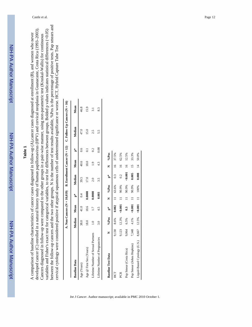
Women who were diagnosed with cervical cancer during follow-up were similar in theirenrollment age to women diagnosed with cancer at enrollment (n = 12) and women whowere never diagnosed with cancer (n = 10,019) (Table 1). However, these follow-up casestended to have earlier sexual debut, to have more sexual partners, and to be more parousthan women diagnosed with cancer at enrollment and women without cancer at enrollment.
Castle et al. Page 4
Int J Cancer. Author manuscript; available in PMC 2010 October 1.
NIH
-PA Author Manuscript
NIH
-PA Author Manuscript
NIH
-PA Author Manuscript

Cancer cases diagnosed during follow-up were less likely (non-significantly) thanenrollment cases to test positive for carcinogenic HPV by HCT (37.5% vs. 63.6%,respectively) and retrospectively by PCR for HPV (62.5% vs. 90.9%, respectively). Follow-up cancers were less likely (non-significantly) than enrollment cases to have positivecytology by either cytologic method or review of the cytology. There was a non-significantdifference in the age of diagnosis for follow-up cancer cases (median = 51.5 years, mean =52.2 years) versus the enrollment cancer cases (median = 39.5 years; mean = 40.6 years) (p= 0.06). There were no significant differences between cancer groups for the use of oralcontraceptives (ever/never) and smoking (ever/never). Interestingly, the age at sexual debutwas correlated with the age of diagnosis for the follow-up cases (Spearman ρ = 0.48, p =0.05), suggesting a relationship between the peak exposure to/prevalence of HPV thattypically occur shortly after sexual debut (19;25) and the timing of cancer diagnoses.
Details about follow-up time and follow-up visits are shown in Table 2. Follow-up cancercases and non-cases were followed for a similar duration and with similar number of visits.
The case histories of all 16 women (Patients A–P) diagnosed with cancer during follow-upare shown in Table 3; cases E, F, and M were identified through the national cancer registry.Five cases (excluding those who were treated during the enrollment phase) were passivelyfollowed (because there were no indicators of cervical abnormalities) and subsequentlydiagnosed with cancer at their exit visit 5–7 years later. Cases can be assigned to thefollowing causes: I) six failures (Patients A–F) to detect abnormalities by enrollmentcytology screening, three of which (Patients B, E, and F) also would have been completelymissed by HPV DNA detection had HPV DNA detection been used for screening; II) sevenfailures of diagnosis by colposcopy (Patients G–M); and III) three recurrences of diseaseafter excisional treatment for a diagnosis of a cervical precancerous lesions (Patients N–P);in two (Patient N and O) of the three patients , the LEEP was considered insufficient andmargins were taken.
HPV16 infections caused six follow-up cases (37.5%). Interestingly, another five cases(31.3%) were caused by HPV18 (n = 4, 25.0%) or HPV45 (n = 1, 6.3%), the HPV genotypemost closely related genetically to HPV18; 31.3% caused by HPV18 or HPV45 was non-significantly greater (p = 0.09, two-sided binomial test) than the expected percentage(17.4%) based on a recent meta-analysis of prevalent-detected cancers (26). By comparison,50% of the enrollment cancer cases were HPV16 positive and 25% were HPV18 or HPV45positive although the distribution was not significantly different from cases diagnosedduring follow-up (p = 0.9). One case (6.3%) was due to HPV52 infection while another(Patient D) was probably due to HPV33 infection although the enrollment test was negativeby PCR for this type. Three cases (Patient E, F, and J) tested HPV negative at all timepoints, despite repeated testing at each time point.
DiscussionDespite multiple screening modalities for detection, colposcopic evaluations, and treatmentof ≥CIN2, more than 1 out of 1000 women followed in the cohort was still diagnosed withcervical cancer during follow-up. We observed that, for varied reasons described here, oneround of even multi-modal screening (with rigorous follow-up of the screen positives andother high-risk participants) can not be used to take a region with previously poor screeningto nearly zero risk of subsequent invasive cancer. We effectively detected and treatedseveral hundred precancerous lesions (19;20), significantly lowering but not eliminating thecancer risk. In populations without a long history of widespread, effective screening, likeGuanacaste at the start of our project, the CIN3 lesions were likely to be more advanced thanthose lesions found in well-screened populations and perhaps have a greater potential for
Castle et al. Page 5
Int J Cancer. Author manuscript; available in PMC 2010 October 1.
NIH
-PA Author Manuscript
NIH
-PA Author Manuscript
NIH
-PA Author Manuscript

invasion (27;28) when missed. The median age of high-grade cervical neoplasia diagnosedat enrollment in Guanacaste was 34 years (29), approximately 5–10 years older than medianage in the U.S., while the median age of sexual debut of 18 years in Guanacaste (Table 1)(when the population becomes “at risk”) is similar to the estimated median age of 17–18years in the U. S. (30).
In addition to describing the failures (cancers) of the screening program, it would be usefulto estimate the effectiveness of the screening program by comparing the incidence rates ofcancer in the cohort to the expected rates without screening, based on the rates of cancer inthe other 5/6th of the Guanacaste population not included in the cohort study. We are,unfortunately, not able to estimate the effectiveness of the screening program of cancer bycomparing the incidence rates of cancer in the cohort to the expected rates withoutscreening. We observed an approximate two-fold higher rate of cancers found within thecohort (12 in 1/6th of the population) than in the remaining population (31 in 5/6th of thepopulation) based on the Guanacaste cancer registry during the enrollment phase. Thisparadoxical increase, rather than decrease, at enrollment within cohort is the consequence ofimproved screening and accelerated the detection of early cancers that would have beenmissed otherwise and only found later when they manifested symptoms. Thus, anycalculation of the rate ratio (observed versus expected) is an underestimate of theeffectiveness of the screening program: many cancers that could be prevented by screeningthe women not in the cohort would not be detected until after the end of cohort follow-upbecause their cancer diagnosis would be based on symptomatic manifestation, given thelimitations of community-based screening in Guanacaste. We note, for example, that themedian age of the screen-detected 12 cancer diagnoses within the cohort at enrollment wassignificantly younger than the median age of the 31 supplemental cases of cancer identifiedin the Guanacaste through the cancer registry(29) (39.5 years vs.59.0 years, respectively, p =0.004 [Kruskal-Wallis]).
We have also observed a secular trend of reduced incidence of cervical cancer over the timeperiod of the cohort (data not shown). This was presumably due to improved cytologicscreening, as we introduced U.S.-quality liquid-based cytologic screening to the Guanacasteregion, and timely management of the screen positives. We therefore were unable toestimate the expected number of cancers without intervention and the number of cancersaverted within the cohort study.
Notably, there were no cases of adenocarcinoma (95% CI = 0.0%–20.6%), the precancerouslesion for which (adenocarcinoma in situ) generally is believed to be more commonlymissed by screening programs than CIN3/CIS (31). It is unknown how commonadenocarcinoma is relative to squamous cell carcinoma in general population of Costa Ricabut presumably it is less so than in the U.S. and Europe, where its incidence is on the rise(32;33). Thus, it is uncertain whether finding no adenocarcinoma is unusual or not.
In retrospect, the most promising test for detection of cervical cancer would have been HPVPCR or an equivalently sensitive test, because in most but not all cancer cases, women wereDNA positive for a carcinogenic HPV genotype years before cancer was detected. The nextiteration of hybrid capture, hc2, resembles PCR testing in its clinical sensitivity (34) and isapproximately 10–20% more sensitive for the detection of ≥CIN3 than HCT in use at thetime of PEG enrollment (data not shown). As correlate of sensitivity, a negative HPV testtherefore provides the most reassurance against cancer of all one-time screening methods.Yet, HPV testing using a well-validated research PCR assay failed to test positive repeatedlyfor two cases (Patient F and J) even at the time of diagnosis and two others (Patient B andD) tested positive for HPV sporadically.
Castle et al. Page 6
Int J Cancer. Author manuscript; available in PMC 2010 October 1.
NIH
-PA Author Manuscript
NIH
-PA Author Manuscript
NIH
-PA Author Manuscript

Three limitations of our HPV screening bear mentioning. First, HCT, the predecessor andless sensitive version of the current FDA-approved test, hc2, was used to screen women atbaseline. Use of a more sensitive test at baseline might have improved the risk stratificationof the population, resulting in more women assigned to the active follow-up, and possiblydetecting and censoring women with CIN3 prior to invasion. We used the retrospectivetesting by a well-validated research PCR assay to simulate how a very analytically sensitivetest might have performed had it been available at the time of enrollment. Second, womenwere not routinely referred to colposcopy after testing positive for carcinogenic HPV byPCR, except during exit colposcopy where Patient J was referred to colposcopy due to theevidence of long-term HPV18 persistence that ultimately lead to cancer detection. Third, aDacron swab, rather than the currently standard of cervical brush and Ayre's spatula or acervical broom, was used to collect specimens for HPV testing. This choice may have led toless than optimal cervical sampling. As a consequence of the latter, the HPV testing used inthis study may not have performed as sensitively as the current standards of performance.
Liquid-based cytology, read very sensitively (and non-specifically) by an U.S. expertreviewer (M.L.H.)(34), was negative repeatedly for one case (Patient L) and was negativeuntil the time of cancer diagnosis for another (Patient D). Conventional cytology was lesssensitive (and more specific) (34), albeit within the range observed in other studies(4;11;35;36), as were the nurse impression of possible cancer and Cervicography interpretedin the U.S. We have observed endemic levels of unexplained cervical inflammation andcervicitis in this population (37) (unpublished observations), which could have obfuscatedprecancerous lesions from detection.
It is particular noteworthy that half the incidently-detected cancers had a colposcopicevaluation called normal within two years of the diagnosis, suggesting that colposcopicevaluation missed either CIN3 about to invade or cancer itself. Given the insensitivity ofcolposcopy (16;38;39), we cannot determine with certainty whether these cases representedtruly incident or prevalent cases of cancer missed at enrollment. Even when colposcopyfound evidence of an abnormality, it did not always result in taking a biopsy of the mostsevere disease with resultant appropriate treatment and clinical management, as is the caseof Patient G. In high-risk populations in which colposcopic evaluation is used for diagnosis,careful follow-up of colposcopically-evaluated women with <CIN2 is warranted if theresources permit. Without advances in colposcopy (17), it is unclear whether simplyincreasing the sensitivity of screening (e.g. including HPV testing) will lead to an equaldecrease in the incidence of cancer.
There were also several examples of failed treatments. During the study, three patients weretreated for precancerous lesions by LEEP, which has been shown to be 90–95% effective(40–42), prior to their cancer diagnosis. However, it is notable that these women were ages39, 41, and 48 at the time of LEEP, perhaps indicating that they had larger, more advancedprecancerous lesions in the cervical canal with a greater potential to invade (27) if thetreatment was incomplete and the margins positive (n.b. two were). Based on recentresearch, careful post-treatment surveillance and follow-up using HPV DNA detection forsensitive identification of recurrence/failed treatment (40;43) might be warranted these high-risk, post-treatment women.
While there were no significant differences in the HPV genotype distribution of the cancersfound at enrollment versus cancers found during follow-up, HPV18 and HPV45 togetherwere somewhat more common in follow-up cases than what is expected based on a recentmeta-analysis (26). Screening by cytology and diagnosis by colposcopy of HPV18-relatedprecancerous lesions may not be as effective as for HPV16-related lesions. Patient L is adidactic example of elevated risk due to a long-term persistent HPV18 infection that was
Castle et al. Page 7
Int J Cancer. Author manuscript; available in PMC 2010 October 1.
NIH
-PA Author Manuscript
NIH
-PA Author Manuscript
NIH
-PA Author Manuscript

largely missed by the classic screening methods for cervical cancer prevention, cytology andcolposcopy.
It is important to recognize that cases of cervical cancer can arise as the consequences of thefalse negative test results and diagnostic procedures and not necessarily from malfeasance oreven human error (44–49). Routine auditing of large cervical cancer programs will helpprovide the quality control to minimize these errors (44–49) but intrinsically there areinaccuracies in all medical procedures. Secondary cervical cancer prevention programsrequire 3 steps, screening, diagnosis, and treatment, each of which contributes to the error intimely detection and treatment of cervical precancerous lesions. Even with the possibleadvent of low-cost HPV testing for low-resource settings (5;50), cancers will occur becausethese inherent errors in each prevention step happen in the context of women withpotentially invasive precancerous lesions, as predicted from Bayes' Theorem, which says therisk of having cancer given a negative test is a function of probability of a false-negative testand the prevalence of the disease. A broader recognition that the most effective cervicalcancer prevention programs will have a very low but non-zero risk of cancer will help avoidthe tendencies to over-screen and over-treat women in a misguided effort to achieve a 100%efficacious program.
In summary, it seems unlikely that any negative test or combination of negative tests, evenincluding colposcopic evaluation of the cervix that has been considered the gold standard ofcervical cancer screening and diagnosis, can provide absolute reassurance against cancer inthis population or in any setting. A single good screening can vastly reduce the risk ofsubsequent cancer in previously unscreened women. Yet, the inherent errors of each step ofthe secondary prevention of cervical cancer in a population at an elevated risk will result in a“significant” residual risk of cancer.
Supplementary MaterialRefer to Web version on PubMed Central for supplementary material.
AcknowledgmentsNational Institutes of Health (N01-CP-21081, N01-CP-33061, N01-CP-40542, N01-CP-50535, N01-CP-81023,intramural program, CA78527 to RB). The Guanacaste cohort (design and conduct of the study, sample collection,management, analysis and interpretation of the data) for the enrollment and follow-up phases were supported by theNational Cancer Institute, National Institutes of Health, Department of Health and Human Services. This researchwas supported [in part] by the Intramural Research Program of the NIH/National Cancer Institute.
Reference List(1). Kitchener HC, Castle PE, Cox JT. Chapter 7: Achievements and limitations of cervical cytology
screening. Vaccine. Aug 21; 2006 24(Suppl 3):S63–70. S63–S70.(2). Cervix Cancer Screening. Vol. Volume 10. IARC Press; Lyon, France: 2005.(3). Cuzick J, Clavel C, Petry KU, Meijer CJ, Hoyer H, Ratnam S, Szarewski A, Birembaut P,
Kulasingam S, Sasieni P, Iftner T. Overview of the European and North American studies onHPV testing in primary cervical cancer screening. Int J Cancer. Apr 3.2006
(4). Arbyn M, Sasieni P, Meijer CJ, Clavel C, Koliopoulos G, Dillner J. Chapter 9: Clinicalapplications of HPV testing: A summary of meta-analyses. Vaccine. Aug 21; 2006 24(Suppl3):S78–89. S78–S89.
(5). Schiffman M, Castle PE, Jeronimo J, Rodriguez AC, Wacholder S. Human papillomavirus andcervical cancer. Lancet. Sep 8; 2007 370(9590):890–907. [PubMed: 17826171]
(6). Castle PE, Wheeler CM, Solomon D, Schiffman M, Peyton CL. Interlaboratory reliability ofHybrid Capture 2. Am J Clin Pathol. Aug; 2004 122(2):238–45. [PubMed: 15323141]
Castle et al. Page 8
Int J Cancer. Author manuscript; available in PMC 2010 October 1.
NIH
-PA Author Manuscript
NIH
-PA Author Manuscript
NIH
-PA Author Manuscript

(7). Carozzi FM, Del Mistro A, Confortini M, Sani C, Puliti D, Trevisan R, De Marco L, Tos AG,Girlando S, Palma PD, Pellegrini A, Schiboni ML, et al. Reproducibility of HPV DNA Testingby Hybrid Capture 2 in a Screening Setting. Am J Clin Pathol. Nov; 2005 124(5):716–21.[PubMed: 16203283]
(8). Mayrand MH, Duarte-Franco E, Rodrigues I, Walter SD, Hanley J, Ferenczy A, Ratnam S,Coutlee F, Franco EL. Human papillomavirus DNA versus Papanicolaou screening tests forcervical cancer. N Engl J Med. Oct 18; 2007 357(16):1579–88. [PubMed: 17942871]
(9). Naucler P, Ryd W, Tornberg S, Strand A, Wadell G, Elfgren K, Radberg T, Strander B, ForslundO, Hansson BG, Rylander E, Dillner J. Human papillomavirus and Papanicolaou tests to screenfor cervical cancer. N Engl J Med. Oct 18; 2007 357(16):1589–97. [PubMed: 17942872]
(10). Bulkmans N, Berkhof J, Rozendaal L, van KF, Boeke A, Bulk S, Voorhorst F, Verheijen R, vanGK, Boon M, Ruitinga W, van BM, et al. Human papillomavirus DNA testing for the detectionof cervical intraepithelial neoplasia grade 3 and cancer: 5-year follow-up of a randomisedcontrolled implementation trial. Lancet. Oct 3.2007
(11). Cuzick J, Clavel C, Petry KU, Meijer CJ, Hoyer H, Ratnam S, Szarewski A, Birembaut P,Kulasingam S, Sasieni P, Iftner T. Overview of the European and North American studies onHPV testing in primary cervical cancer screening. Int J Cancer. Sep 1; 2006 119(5):1095–101.[PubMed: 16586444]
(12). Wright TC Jr. Massad LS, Dunton CJ, Spitzer M, Wilkinson EJ, Solomon D. 2006 consensusguidelines for the management of women with abnormal cervical cancer screening tests. Am JObstet Gynecol. Oct; 2007 197(4):346–55. [PubMed: 17904957]
(13). Wright TC Jr. Schiffman M, Solomon D, Cox JT, Garcia F, Goldie S, Hatch K, Noller KL,Roach N, Runowicz C, Saslow D. Interim guidance for the use of human papillomavirus DNAtesting as an adjunct to cervical cytology for screening. Obstet Gynecol. Feb; 2004 103(2):304–9.[PubMed: 14754700]
(14). Sherman ME, Lorincz AT, Scott DR, Wacholder S, Castle PE, Glass AG, Mielzynska-Lohnas I,Rush BB, Schiffman M. Baseline cytology, human papillomavirus testing, and risk for cervicalneoplasia: a 10-year cohort analysis. J Natl Cancer Inst. Jan 1; 2003 95(1):46–52. [PubMed:12509400]
(15). Kjaer S, Hogdall E, Frederiksen K, Munk C, van den BA, Svare E, Meijer C, Lorincz A, Iftner T.The absolute risk of cervical abnormalities in high-risk human papillomavirus-positive,cytologically normal women over a 10-year period. Cancer Res. Nov 1; 2006 66(21):10630–6.[PubMed: 17062559]
(16). Gage JC, Hanson VW, Abbey K, Dippery S, Gardner S, Kubota J, Schiffman M, Solomon D,Jeronimo J. Number of cervical biopsies and sensitivity of colposcopy. Obstet Gynecol. Aug;2006 108(2):264–72. [PubMed: 16880294]
(17). Jeronimo J, Schiffman M. Colposcopy at a crossroads. Am J Obstet Gynecol. Aug; 2006 195(2):349–53. [PubMed: 16677597]
(18). Pretorius RG, Zhang WH, Belinson JL, Huang MN, Wu LY, Zhang X, Qiao YL. Colposcopicallydirected biopsy, random cervical biopsy, and endocervical curettage in the diagnosis of cervicalintraepithelial neoplasia II or worse. Am J Obstet Gynecol. Aug; 2004 191(2):430–4. [PubMed:15343217]
(19). Herrero R, Castle PE, Schiffman M, Bratti MC, Hildesheim A, Morales J, Alfaro M, ShermanME, Wacholder S, Chen S, Rodriguez AC, Burk RD. Epidemiologic profile of type-specifichuman papillomavirus infection and cervical neoplasia in guanacaste, costa rica. J Infect Dis. Jun1; 2005 191(11):1796–807. [PubMed: 15871111]
(20). Schiffman M, Herrero R, Desalle R, Hildesheim A, Wacholder S, Cecilia RA, Bratti MC,Sherman ME, Morales J, Guillen D, Alfaro M, Hutchinson M, et al. The carcinogenicity ofhuman papillomavirus types reflects viral evolution. Virology. Jun 20; 2005 337(1):76–84.[PubMed: 15914222]
(21). Herrero R, Schiffman MH, Bratti C, Hildesheim A, Balmaceda I, Sherman ME, Greenberg M,Cardenas F, Gomez V, Helgesen K, Morales J, Hutchinson M, et al. Design and methods of apopulation-based natural history study of cervical neoplasia in a rural province of Costa Rica: theGuanacaste Project. Rev Panam Salud Publica. May; 1997 1(5):362–75. [PubMed: 9180057]
Castle et al. Page 9
Int J Cancer. Author manuscript; available in PMC 2010 October 1.
NIH
-PA Author Manuscript
NIH
-PA Author Manuscript
NIH
-PA Author Manuscript

(22). Bratti MC, Rodriguez AC, Schiffman M, Hildesheim A, Morales J, Alfaro M, Guillen D,Hutchinson M, Sherman ME, Eklund C, Schussler J, Buckland J, et al. Description of a seven-year prospective study of human papillomavirus infection and cervical neoplasia among 10 000women in Guanacaste, Costa Rica. Rev Panam Salud Publica. Feb; 2004 15(2):75–89.Descripcion de un estudio prospectivo de siete anos sobre la infeccion por el virus del papilomahumano y el cancer cervicouterino en 10 000 mujeres de Guanacaste, Costa Rica. [PubMed:15030652]
(23). Castle PE, Schiffman M, Gravitt PE, Kendall H, Fishman S, Dong H, Hildesheim A, Herrero R,Bratti MC, Sherman ME, Lorincz A, Schussler JE, et al. Comparisons of HPV DNA detection byMY09/11 PCR methods. J Med Virol. Nov; 2002 68(3):417–23. [PubMed: 12226831]
(24). Qu W, Jiang G, Cruz Y, Chang CJ, Ho GY, Klein RS, Burk RD. PCR detection of humanpapillomavirus: comparison between MY09/MY11 and GP5+/GP6+ primer systems. J ClinMicrobiol. Jun; 1997 35(6):1304–10. [PubMed: 9163434]
(25). de Sanjose S, Diaz M, Castellsague X, Clifford G, Bruni L, Munoz N, Bosch FX. Worldwideprevalence and genotype distribution of cervical human papillomavirus DNA in women withnormal cytology: a meta-analysis. Lancet Infect Dis. Jul; 2007 7(7):453–9. [PubMed: 17597569]
(26). Smith JS, Lindsay L, Hoots B, Keys J, Franceschi S, Winer R, Clifford GM. Humanpapillomavirus type distribution in invasive cervical cancer and high-grade cervical lesions: ameta-analysis update. Int J Cancer. Aug 1; 2007 121(3):621–32. [PubMed: 17405118]
(27). Schiffman M, Rodriguez AC. Heterogeneity in CIN3 diagnosis. Lancet Oncol. May; 2008 9(5):404–6. [PubMed: 18452848]
(28). McCredie MR, Sharples KJ, Paul C, Baranyai J, Medley G, Jones RW, Skegg DC. Naturalhistory of cervical neoplasia and risk of invasive cancer in women with cervical intraepithelialneoplasia 3: a retrospective cohort study. Lancet Oncol. May; 2008 9(5):425–34. [PubMed:18407790]
(29). Herrero R, Hildesheim A, Bratti C, Sherman ME, Hutchinson M, Morales J, Balmaceda I,Greenberg MD, Alfaro M, Burk RD, Wacholder S, Plummer M, et al. Population-based study ofhuman papillomavirus infection and cervical neoplasia in rural Costa Rica. J Natl Cancer Inst.Mar 15; 2000 92(6):464–74. [PubMed: 10716964]
(30). Mosher WD, Chandra A, Jones J. Sexual behavior and selected health measures: men and women15-44 years of age, United States, 2002. Adv Data. Sep 15.2005 (362):1–55.
(31). Smith HO, Padilla LA. Adenocarcinoma in situ of the cervix: sensitivity of detection by cervicalsmear: will cytologic screening for adenocarcinoma in situ reduce incidence rates foradenocarcinoma. Cancer. Dec 25; 2002 96(6):319–22. [PubMed: 12478677]
(32). Bray F, Carstensen B, Moller H, Zappa M, Zakelj MP, Lawrence G, Hakama M, Weiderpass E.Incidence trends of adenocarcinoma of the cervix in 13 European countries. Cancer EpidemiolBiomarkers Prev. Sep; 2005 14(9):2191–9. [PubMed: 16172231]
(33). Wang SS, Sherman ME, Hildesheim A, Lacey JV Jr. Devesa S. Cervical adenocarcinoma andsquamous cell carcinoma incidence trends among white women and black women in the UnitedStates for 1976-2000. Cancer. Mar 1; 2004 100(5):1035–44. [PubMed: 14983500]
(34). Ferreccio C, Bratti MC, Sherman ME, Herrero R, Wacholder S, Hildesheim A, Burk RD,Hutchinson M, Alfaro M, Greenberg MD, Morales J, Rodriguez AC, et al. A comparison ofsingle and combined visual, cytologic, and virologic tests as screening strategies in a region athigh risk of cervical cancer. Cancer Epidemiol Biomarkers Prev. Sep; 2003 12(9):815–23.[PubMed: 14504189]
(35). Arbyn M, Bergeron C, Klinkhamer P, Martin-Hirsch P, Siebers AG, Bulten J. Liquid comparedwith conventional cervical cytology: a systematic review and meta-analysis. Obstet Gynecol. Jan;2008 111(1):167–77. [PubMed: 18165406]
(36). Nanda K, McCrory DC, Myers ER, Bastian LA, Hasselblad V, Hickey JD, Matchar DB.Accuracy of the Papanicolaou test in screening for and follow-up of cervical cytologicabnormalities: a systematic review. Ann Intern Med. May 16; 2000 132(10):810–9. [PubMed:10819705]
(37). Castle PE, Hillier SL, Rabe LK, Hildesheim A, Herrero R, Bratti MC, Sherman ME, Burk RD,Rodriguez AC, Alfaro M, Hutchinson ML, Morales J, et al. An association of cervicalinflammation with high-grade cervical neoplasia in women infected with oncogenic human
Castle et al. Page 10
Int J Cancer. Author manuscript; available in PMC 2010 October 1.
NIH
-PA Author Manuscript
NIH
-PA Author Manuscript
NIH
-PA Author Manuscript

papillomavirus (HPV). Cancer Epidemiol Biomarkers Prev. Oct; 2001 10(10):1021–7. [PubMed:11588127]
(38). Guido R, Schiffman M, Solomon D, Burke L. Postcolposcopy management strategies for womenreferred with low-grade squamous intraepithelial lesions or human papillomavirus DNA-positiveatypical squamous cells of undetermined significance: a two-year prospective study. Am J ObstetGynecol. Jun; 2003 188(6):1401–5. [PubMed: 12824969]
(39). Pretorius RG, Kim RJ, Belinson JL, Elson P, Qiao YL. Inflation of sensitivity of cervical cancerscreening tests secondary to correlated error in colposcopy. J Low Genit Tract Dis. Jan; 200610(1):5–9. [PubMed: 16378026]
(40). Kreimer AR, Guido RS, Solomon D, Schiffman M, Wacholder S, Jeronimo J, Wheeler CM,Castle PE. Human papillomavirus testing following loop electrosurgical excision procedureidentifies women at risk for posttreatment cervical intraepithelial neoplasia grade 2 or 3 disease.Cancer Epidemiol Biomarkers Prev. May; 2006 15(5):908–14. [PubMed: 16702369]
(41). Mitchell MF, Tortolero-Luna G, Cook E, Whittaker L, Rhodes-Morris H, Silva E. A randomizedclinical trial of cryotherapy, laser vaporization, and loop electrosurgical excision for treatment ofsquamous intraepithelial lesions of the cervix. Obstet Gynecol. Nov; 1998 92(5):737–44.[PubMed: 9794661]
(42). Alvarez RD, Helm CW, Edwards RP, Naumann RW, Partridge EE, Shingleton HM, McGee JA,Hall JB, Higgins RV, Malone JM Jr. Prospective randomized trial of LLETZ versus laserablation in patients with cervical intraepithelial neoplasia. Gynecol Oncol. Feb; 1994 52(2):175–9. [PubMed: 8314135]
(43). Arbyn M, Paraskevaidis E, Martin-Hirsch P, Prendiville W, Dillner J. Clinical utility of HPV-DNA detection: Triage of minor cervical lesions, follow-up of women treated for high-gradeCIN: An update of pooled evidence. Gynecol Oncol. Dec; 2005 99(3 Suppl):S7–S11. [PubMed:16154623]
(44). Andrae B, Kemetli L, Sparen P, Silfverdal L, Strander B, Ryd W, Dillner J, Tornberg S.Screening-preventable cervical cancer risks: evidence from a nationwide audit in Sweden. J NatlCancer Inst. May 7; 2008 100(9):622–9. [PubMed: 18445828]
(45). Cuzick J. Routine audit of large-scale cervical cancer screening programs. J Natl Cancer Inst.May 7; 2008 100(9):605–6. [PubMed: 18445817]
(46). Slater DN, Milner PC, Radley H. Audit of deaths from cervical cancer: proposal for an essentialcomponent of the National Screening Programme. J Clin Pathol. Jan; 1994 47(1):27–8. [PubMed:8132804]
(47). Sung HY, Kearney KA, Miller M, Kinney W, Sawaya GF, Hiatt RA. Papanicolaou smear historyand diagnosis of invasive cervical carcinoma among members of a large prepaid health plan.Cancer. May 15; 2000 88(10):2283–9. [PubMed: 10820350]
(48). Leyden WA, Manos MM, Geiger AM, Weinmann S, Mouchawar J, Bischoff K, Yood MU,Gilbert J, Taplin SH. Cervical cancer in women with comprehensive health care access:attributable factors in the screening process. J Natl Cancer Inst. May 4; 2005 97(9):675–83.[PubMed: 15870438]
(49). Nygard JF, Nygard M, Skare GB, Thoresen SO. Screening histories of women with CIN 2/3compared with women diagnosed with invasive cervical cancer: a retrospective analysis of theNorwegian Coordinated Cervical Cancer Screening Program. Cancer Causes Control. May; 200516(4):463–74. [PubMed: 15953989]
(50). Qiao YL, Sellors JW, Eder PS, Bao YP, Lim JM, Zhao FH, Weigl B, Zhang WH, Peck RB, Li L,Chen F, Pan QJ, et al. A new HPV-DNA test for cervical-cancer screening in developing regions:a cross-sectional study of clinical accuracy in rural China. Lancet Oncol. Oct; 2008 9(10):929–36. [PubMed: 18805733]
Castle et al. Page 11
Int J Cancer. Author manuscript; available in PMC 2010 October 1.
NIH
-PA Author Manuscript
NIH
-PA Author Manuscript
NIH
-PA Author Manuscript
NIH
-PA Author Manuscript
NIH
-PA Author Manuscript
NIH
-PA Author Manuscript
Castle et al. Page 12
Tabl
e 1
A c
ompa
rison
of b
asel
ine
char
acte
ristic
s of c
ance
r cas
es d
iagn
osed
in fo
llow
-up
(A),c
ance
r cas
es d
iagn
osed
at e
nrol
lmen
t (B
), an
d w
omen
who
nev
erde
velo
ped
canc
er (C
) enr
olle
d in
a n
atur
al h
isto
ry st
udy
of h
uman
pap
illom
aviru
s (H
PV) a
nd c
ervi
cal n
eopl
asia
in G
uana
cast
e, C
osta
Ric
a (1
993–
2003
).C
ance
rs d
iagn
osed
in fo
llow
-up
wer
e co
mpa
red
to th
e ot
her g
roup
s in
a pa
ir-w
ise
man
ner,
usin
g no
n-pa
ram
etric
test
(Kru
skal
-Wal
lis) f
or c
ontin
uous
varia
bles
and
Fis
her's
exa
ct te
st fo
r cat
egor
ical
var
iabl
es, t
o te
st fo
r diff
eren
ces b
etw
een
grou
ps. B
olde
d p
valu
es in
dica
tes s
tatis
tical
diff
eren
ces (
<0.0
5)be
twee
n th
e fo
llow
-up
canc
ers a
nd th
e tw
o ot
her g
roup
s. N
is th
e nu
mbe
r of t
est r
esul
ts a
vaila
ble,
%Po
s is t
he p
erce
ntag
e of
pos
itive
test
s. Pa
p sm
ears
and
cerv
ical
cyt
olog
y w
ere
cons
ider
ed p
ositi
ve if
aty
pica
l squ
amou
s cel
ls o
f und
eter
min
ed si
gnifi
canc
e or
wor
se. H
CT,
Hyb
rid C
aptu
re T
ube
Test
A. N
on-C
ance
rs (N
= 1
0,01
9)B
. Enr
ollm
ent C
ance
r (N
= 1
2)C
. Fol
low
-Up
Can
cers
(N =
16)
Bas
elin
e D
ata
Med
ian
Mea
np*
Med
ian
Mea
np*
Med
ian
Mea
n
Age
(Yea
rs)
38.0
41.0
0.4
39.5
40.6
0.6
47.0
46.8
Age
@ F
irst S
ex (Y
ears
)18
.018
.60.
0008
17.0
18.0
0.1
15.0
15.9
Life
time
Num
ber o
f Sex
ual P
artn
ers
1.0
1.7
0.00
082.
01.
90.
22.
53.
1
Life
time
Num
ber o
f Pre
gnan
cies
3.0
4.5
0.00
33.
54.
30.
085.
58.
3
Bas
elin
e T
est D
ata
N%
Pos
p*N
%Po
sp*
N%
Pos
HC
T9,
130
8.6%
0.00
211
63.6
%0.
316
37.5
%
PCR
9,12
312
.3%
<0.0
0111
90.9
%0.
216
62.5
%
Pap
Smea
r (C
osta
Ric
a)9,
064
6.5%
0.3
1190
.9%
<0.0
0116
12.5
%
Pap
Smea
r (Jo
hns H
opki
ns)
7,34
84.
1%<0
.001
1190
.9%
0.00
515
33.3
%
Liqu
id-B
ased
Cyt
olog
y (U
.S.)
8,66
712
.5%
<0.0
0111
100%
0.00
814
50.0
%
Int J Cancer. Author manuscript; available in PMC 2010 October 1.

NIH
-PA Author Manuscript
NIH
-PA Author Manuscript
NIH
-PA Author Manuscript
Castle et al. Page 13
Table 2
Summary of the follow-up of 16 women diagnosed with invasive cervical cancer during follow-up and 10,019women without cancer participating in the natural history study of human papillomavirus and cervicalneoplasia in Guanacaste, Costa Rica (1993–2003).
All Follow-Up Cancers Non-Cancers
n = 10,035 n = 13 n = 10,022
Follow-up time (Months)
Mean 71.3 88.9 71.3
Median 84.1 100.1 84.1
IQR 60.2–96.3 91.1–105.3 60.2–96.2
Range 0.0–141.7 21.1–131.0 0.0–141.7
Number of Follow-Up Visits
Mean 3.1 3.1 6.7
Median 1 1 6
IQR 1–5 1–5 5–8
Range 0–20 0–20 3–12
Int J Cancer. Author manuscript; available in PMC 2010 October 1.

NIH
-PA Author Manuscript
NIH
-PA Author Manuscript
NIH
-PA Author Manuscript
Castle et al. Page 14
Tabl
e 3
Med
ical
his
torie
s of 1
6 w
omen
who
wer
e di
agno
sed
with
inva
sive
cer
vica
l can
cer d
urin
g fo
llow
-up
whi
le p
artic
ipat
ing
in th
e na
tura
l his
tory
stud
y of
hum
an p
apill
omav
irus (
HPV
) and
cer
vica
l neo
plas
ia in
Gua
naca
ste,
Cos
ta R
ica
(199
3–20
03).
The
case
s his
tory
unt
il th
e fir
st h
isto
logi
c di
agno
sis o
f can
cer i
s pre
sent
ed. P
atie
nts A
, H, I
and
P re
porte
d re
spec
tivel
y on
ly 2
, 4, 2
, and
2 li
fetim
e nu
mbe
rs o
f sex
ual
partn
ers i
n th
eir f
ollo
w-u
p qu
estio
nnai
res.
Can
cer p
atie
nts E
, F, a
nd M
wer
e fo
und
thro
ugh
linka
ge w
ith th
e ca
ncer
regi
stry
.C
olpo
= c
olpo
scop
ic e
valu
atio
n; P
ost-c
olpo
= p
ost-c
olpo
scop
ic e
valu
atio
n; P
ap =
Pap
anic
olao
u sm
ear;
LBC
= li
quid
-bas
ed c
ytol
ogy;
CR
= C
osta
Ric
a; JH
U =
John
s Hop
kins
Pat
holo
gist
(M.E
.S.);
US
= U
.S.
path
olog
ist (
M.H
.L.);
LEE
P =
loop
ele
ctro
surg
ical
exc
isio
n pr
oced
ure;
Hys
t = h
yste
rect
omy;
CIN
= c
ervi
cal i
ntra
epith
elia
l neo
plas
ia; A
SCU
S =
atyp
ical
cel
ls o
f und
eter
min
ed si
gnifi
canc
e; L
SIL
= lo
w-g
rade
squa
mou
s int
raep
ithel
ial l
esio
n; H
SIL
= hi
gh-g
rade
squa
mou
s int
raep
ithel
ial l
esio
n; U
nsat
= u
nsat
isfa
ctor
y; #
Partn
ers =
life
time
num
ber o
f sex
ual p
artn
ers a
t enr
ollm
ent;
HC
T =
hybr
id c
aptu
re tu
be te
st re
sult;
AFS
= a
ge a
t firs
t sex
ual i
nter
cour
se; #
Partn
ers =
life
time
num
ber o
f sex
ual p
artn
ers a
t enr
ollm
ent
Patie
nt A
(Age
= 3
6; A
FS =
15;
#Pa
rtne
rs =
4)
Tim
e (M
onth
s)V
isit
Typ
ePa
p (C
R)
Pap
(JH
U)
LB
C (U
S)L
BC
(CR
)C
ervi
gram
(Rev
iew
)H
CT
HPV
(PC
R)
Col
poH
isto
logy
0.0
Scre
enN
orm
alN
orm
alN
orm
alN
orm
al (N
orm
al)†
Neg
ativ
e16
0.0
Scre
enA
SCU
SC
ance
rP3
-Can
cer (
Can
cer)
*16
;45
60.8
Col
poC
ance
rB
iops
y =
Can
cer
Patie
nt B
(Age
= 5
0; A
FS =
15;
#Pa
rtne
rs =
3)
Tim
e (M
onth
s)V
isit
Typ
ePa
p (C
R)
Pap
(JH
U)
LB
C (U
S)L
BC
(CR
)C
ervi
gram
(Rev
iew
)H
CT
HPV
(PC
R)
Col
poH
isto
logy
0.0
Scre
enN
orm
alN
orm
alN
orm
alN
orm
al (N
orm
al)†
Neg
ativ
eN
egat
ive
72.2
Scre
enA
SCU
SC
IN3#
Rea
ctiv
eN
orm
al (N
orm
al)*
18
84.1
Scre
enC
IN3
CIN
3N
orm
alM
issi
ng
85.8
Col
poH
SIL
LEEP
= C
ance
r
Patie
nt C
(Age
= 2
9; A
FS =
17;
#Pa
rtne
rs =
1)
Tim
e (M
onth
s)V
isit
Typ
ePa
p (C
R)
Pap
(JH
U)
LB
C (U
S)L
BC
(CR
)C
ervi
gram
(Rev
iew
)H
CT
HPV
(PC
R)
Col
poH
isto
logy
0.0
Scre
enR
eact
ive
Nor
mal
Inad
equa
teN
orm
alN
egat
ive
18
60.3
Scre
enR
eact
ive
ASC
US
P2-H
SIL*
18
63.5
Col
poH
SIL
LEEP
= C
ance
r
Patie
nt D
(Age
= 6
3; A
FS =
18;
#Pa
rtne
rs =
6)
Tim
e (M
onth
s)V
isit
Typ
ePa
p (C
R)
Pap
(JH
U)
LB
C (U
S)L
BC
(CR
)C
ervi
gram
(Rev
iew
)H
CT
HPV
(PC
R)
Col
poH
isto
logy
0.0
Scre
enN
orm
alN
orm
alN
orm
alN
orm
al‡
Neg
ativ
e51
;61
13.1
Scre
enN
orm
alN
orm
alN
orm
al‡
33
31.4
Scre
enR
eact
ive
Nor
mal
Nor
mal
‡61
44.3
Scre
enN
orm
alN
orm
alN
orm
al‡
Neg
ativ
e
Int J Cancer. Author manuscript; available in PMC 2010 October 1.

NIH
-PA Author Manuscript
NIH
-PA Author Manuscript
NIH
-PA Author Manuscript
Castle et al. Page 15Pa
tient
D (A
ge =
63;
AFS
= 1
8; #
Part
ners
= 6
)
Tim
e (M
onth
s)V
isit
Typ
ePa
p (C
R)
Pap
(JH
U)
LB
C (U
S)L
BC
(CR
)C
ervi
gram
(Rev
iew
)H
CT
HPV
(PC
R)
Col
poH
isto
logy
56.7
Scre
enN
orm
alN
orm
alN
orm
al‡
Neg
ativ
e
60.4
Scre
enN
orm
alN
orm
alN
orm
al‡
Neg
ativ
e
84.2
Scre
enC
ance
rC
IN3
ASC
US
Nor
mal
‡33
;35;
52;5
8
91.4
Col
poC
ance
rB
iops
y =
Can
cer
Patie
nt E
(Age
= 7
5; A
FS =
12;
#Pa
rtne
rs =
1)
Tim
e (M
onth
s)V
isit
Typ
ePa
p (C
R)
Pap
(JH
U)
LB
C (U
S)L
BC
(CR
)C
ervi
gram
(Rev
iew
)H
CT
HPV
(PC
R)
Col
poH
isto
logy
0.0
Scre
enN
orm
alN
orm
alN
orm
alN
orm
alN
egat
ive
Neg
ativ
e
132.
3C
olpo
Bio
psy
= C
ance
r
Patie
nt F
(Age
= 4
8; A
FS =
23;
#Pa
rtne
rs =
1)
Tim
e (M
onth
s)V
isit
Typ
ePa
p (C
R)
Pap
(JH
U)
LB
C (U
S)L
BC
(CR
)C
ervi
gram
(Rev
iew
)H
CT
HPV
(PC
R)
Col
poH
isto
logy
0.0
Scre
enR
eact
ive
Nor
mal
Nor
mal
Nor
mal
Neg
ativ
eN
egat
ive
72.3
Scre
enR
eact
ive
Nor
mal
Rea
ctiv
eN
orm
alN
egat
ive
73.6
Col
poB
iops
y =
Can
cer
Patie
nt G
(Age
= 2
3; A
FS =
14;
#Pa
rtne
rs =
2)
Tim
e (M
onth
s)V
isit
Typ
ePa
p (C
R)
Pap
(JH
U)
LB
C (U
S)L
BC
(CR
)C
ervi
gram
(Rev
iew
)H
CT
HPV
(PC
R)
Col
poH
isto
logy
0.0
Scre
enN
orm
alA
SCU
SA
SCU
SP1
-LSI
L(H
SIL)
‡Po
sitiv
e16
1.4
Col
poLS
ILB
iops
y =
CIN
1
18.4
Scre
enC
IN2
CIN
2M
issi
ngP1
-LSI
L(H
SIL)
‡16
21.8
Col
poH
SIL
Bio
psy
= C
IN1
23.9
Post
-col
poH
SIL
LEEP
= C
ance
r
Patie
nt H
(Age
= 6
4; A
FS =
15;
#Pa
rtne
rs =
5)
Tim
e (M
onth
s)V
isit
Typ
ePa
p (C
R)
Pap
(JH
U)
LB
C (U
S)L
BC
(CR
)C
ervi
gram
(Rev
iew
)H
CT
HPV
(PC
R)
Col
poH
isto
logy
0.0
Scre
enR
eact
ive
Nor
mal
ASC
US
Nor
mal
(Nor
mal
)‡Po
sitiv
e45
;51;
58;8
4
5.2
Col
po45
; 58
Nor
mal
19.6
Scre
enA
SCU
SC
ance
rC
ance
rP0
-Pro
babl
y N
orm
al*
45
21.2
Col
poC
ance
rB
iops
y =
Can
cer
Int J Cancer. Author manuscript; available in PMC 2010 October 1.

NIH
-PA Author Manuscript
NIH
-PA Author Manuscript
NIH
-PA Author Manuscript
Castle et al. Page 16Pa
tient
I (A
ge =
49;
AFS
= 1
5; #
Part
ners
= 3
)
Tim
e (M
onth
s)V
isit
Typ
ePa
p (C
R)
Pap
(JH
U)
LB
C (U
S)L
BC
(CR
)C
ervi
gram
(Rev
iew
)H
CT
HPV
(PC
R)
Col
poH
isto
logy
0.0
Scre
enA
SCU
SR
eact
ive
ASC
US
Nor
mal
(Nor
mal
)‡Po
sitiv
e16
2.9
Col
po16
Nor
mal
13.2
Scre
enR
eact
ive
ASC
US
Rea
ctiv
eN
orm
al (N
orm
al)*
16
26.4
Scre
enN
orm
alC
IN3
Nor
mal
(Nor
mal
)*16
34.4
Col
poΩ
Bio
psy
= C
ance
r
Patie
nt J
(Age
= 6
1; A
FS =
18;
#Pa
rtne
rs =
2)
Tim
e (M
onth
s)V
isit
Typ
ePa
p (C
R)
Pap
(JH
U)
LB
C (U
S)L
BC
(CR
)C
ervi
gram
(Rev
iew
)H
CT
HPV
(PC
R)
Col
poH
isto
logy
0.0
Scre
enN
orm
alA
SCU
SA
SCU
SN
orm
al (N
orm
al)‡
Neg
ativ
eN
egat
ive
4.1
Col
poN
egat
ive
Nor
mal
13.6
Scre
enC
IN3
CIN
3C
ance
rP0
-Pro
babl
y N
orm
al (H
SIL)
‡N
egat
ive
15.7
Col
poH
SIL
Bio
psy
= C
ance
r
Patie
nt K
(Age
= 3
7; A
FS =
14;
#Pa
rtne
rs =
2)
Tim
e (M
onth
s)V
isit
Typ
ePa
p (C
R)
Pap
(JH
U)
LB
C (U
S)L
BC
(CR
)C
ervi
gram
(Rev
iew
)H
CT
HPV
(PC
R)
Col
poH
isto
logy
0.0
Scre
enR
eact
ive
ASC
US
ASC
US
Nor
mal
(HSI
L)‡
Posi
tive
18
3.8
Col
po18
;61
Nor
mal
10.4
Scre
enR
eact
ive
Rea
ctiv
eN
orm
al (M
etap
lasi
a)
13.4
Scre
enIn
adeq
uate
Inad
equa
teA
SCU
SN
orm
al18
;85
17.5
Col
poU
nsat
Bio
psy
= C
ance
r
Patie
nt L
(Age
= 2
8; A
FS =
14;
#Pa
rtne
rs =
1)
Tim
e (M
onth
s)V
isit
Typ
ePa
p (C
R)
Pap
(JH
U)
LB
C (U
S)L
BC
(CR
)C
ervi
gram
(Rev
iew
)H
CT
HPV
(PC
R)
Col
poH
isto
logy
0.0
Scre
enR
eact
ive
Nor
mal
Mis
sing
Nor
mal
(Nor
mal
)Po
sitiv
e18
;51
1.9
Col
poN
orm
al
13.8
Scre
enR
eact
ive
Nor
mal
Rea
ctiv
eN
orm
al (M
etap
lasi
a)18
26.5
Scre
enR
eact
ive
Rea
ctiv
eN
orm
al (M
etap
lasi
a)18
;55
38.5
Scre
enR
eact
ive
Nor
mal
Nor
mal
(Nor
mal
)&18
50.5
Scre
enIn
adeq
uate
Inad
equa
teN
orm
al (N
orm
al)
18
60.7
Scre
enR
eact
ive
Nor
mal
Nor
mal
(LSI
L)18
;56
86.6
Scre
enR
eact
ive
Nor
mal
Rea
ctiv
eN
orm
al (H
SIL)
18;7
3
96.5
Col
poΔ
Uns
at
Int J Cancer. Author manuscript; available in PMC 2010 October 1.

NIH
-PA Author Manuscript
NIH
-PA Author Manuscript
NIH
-PA Author Manuscript
Castle et al. Page 17Pa
tient
L (A
ge =
28;
AFS
= 1
4; #
Part
ners
= 1
)
Tim
e (M
onth
s)V
isit
Typ
ePa
p (C
R)
Pap
(JH
U)
LB
C (U
S)L
BC
(CR
)C
ervi
gram
(Rev
iew
)H
CT
HPV
(PC
R)
Col
poH
isto
logy
98.1
Post
-col
poC
IN1
Nor
mal
104.
3Po
st-c
olpo
CIN
3N
orm
al
105.
9Po
st-c
olpo
Rea
ctiv
eN
orm
alLE
EP =
Can
cer
Patie
nt M
(Age
= 6
3; A
FS =
18;
#Par
tner
s = 1
)
Tim
e (M
onth
s)V
isit
Typ
ePa
p (C
R)
Pap
(JH
U)
LB
C (U
S)L
BC
(CR
)C
ervi
gram
(Rev
iew
)H
CT
HPV
(PC
R)
Col
poH
isto
logy
0.0
Scre
enN
orm
alA
SCU
SR
eact
ive
Nor
mal
Posi
tive
16
1.8
Col
po16
Nor
mal
13.3
Scre
enN
orm
alA
SCU
SC
IN2
Nor
mal
16;4
5
16.5
Col
poN
orm
al
26.2
Post
-col
poN
orm
al
106.
0Sc
reen
HSI
L (C
IN3)
HSI
L (C
IN2)
Nor
mal
16
106.
2C
olpo
Nor
mal
107.
1C
olpo
Hys
t = C
ance
r
Patie
nt N
(Age
= 3
9; A
FS =
15;
#Pa
rtne
rs =
9)
Tim
e (M
onth
s)V
isit
Typ
ePa
p (C
R)
Pap
(JH
U)
LB
C (U
S)L
BC
(CR
)C
ervi
gram
(Rev
iew
)H
CT
HPV
(PC
R)
Col
poH
isto
logy
0.0
Scre
enN
orm
alN
orm
alC
IN1
Nor
mal
(Met
apla
sia)
‡N
egat
ive
16; 5
8
26.6
Scre
enC
IN1
CIN
3C
ance
rN
orm
al (N
orm
al)‡
16
29.2
Col
poH
SIL
Bio
psy
= C
IN3
34.1
Post
-col
poH
SIL
LEEP
= C
IN2
43.1
Post
-col
poN
orm
al
55.1
Post
-col
poR
eact
ive
Nor
mal
112.
2Sc
reen
HSI
L (C
IN3)
HSI
L (C
IN2)
Nor
mal
16
115.
8C
olpo
LSIL
LEEP
= C
ance
r
Patie
nt O
(Age
= 3
8; A
FS =
15;
#Pa
rtne
rs =
5)
Tim
e (M
onth
s)V
isit
Typ
ePa
p (C
R)
Pap
(JH
U)
LB
C (U
S)L
BC
(CR
)C
ervi
gram
(Rev
iew
)H
CT
HPV
(PC
R)
Col
poH
isto
logy
0.0
Scre
enC
IN2
Mis
sing
CIN
3N
orm
al (C
ance
r)N
egat
ive
16;7
1
1.8
Col
po16
LSIL
Bio
psy
= C
IN3
5.1
Post
-col
poLE
EP =
CIN
3
76.0
Col
poΩ
CIN
3C
ance
rB
iops
y =
Can
cer
Int J Cancer. Author manuscript; available in PMC 2010 October 1.

NIH
-PA Author Manuscript
NIH
-PA Author Manuscript
NIH
-PA Author Manuscript
Castle et al. Page 18Pa
tient
P (A
ge =
46;
AFS
= 1
6; #
Part
ners
= 4
)
Tim
e (M
onth
s)V
isit
Typ
ePa
p (C
R)
Pap
(JH
U)
LB
C (U
S)L
BC
(CR
)C
ervi
gram
(Rev
iew
)H
CT
HPV
(PC
R)
Col
poH
isto
logy
0.0
Scre
enN
orm
alA
SCU
SN
orm
alN
orm
al&
Neg
ativ
e52
3.5
Col
po52
Nor
mal
13.2
Scre
enC
IN3
CIN
2C
IN3
Nor
mal
(Nor
mal
)‡52
15.2
Col
poN
om
16.4
Post
-col
poH
SIL
LEEP
= C
IN3
81.0
Col
poC
ance
r
82.4
Col
poC
ance
rC
ance
rB
iops
y =
Can
cer
Int J Cancer. Author manuscript; available in PMC 2010 October 1.