Precision medicine/personalized medicine: a critical analysis ...
Upload
independentCategory
view
2download
0
Q2
Harismendy et al. Breast Cancer Research 2013, 15:R115http://breast-cancer-research.com/content/15/6/R115
RESEARCH ARTICLE Open Access
Evaluation of ultra-deep targeted sequencing forpersonalized breast cancer careOlivier Harismendy1,2,3*, Richard B Schwab2,4, Hakan Alakus1,5, Shawn E Yost1,6, Hiroko Matsui1, Farnaz Hasteh7,Anne M Wallace2,8, Hannah L Park9, Lisa Madlensky2,10, Barbara Parker4, Philip M Carpenter11, Kristen Jepsen1,Hoda Anton-Culver9* and Kelly A Frazer1,2,3,12*
Abstract
Introduction: The increasing number of targeted therapies, together with a deeper understanding of cancergenetics and drug response, have prompted major healthcare centers to implement personalized treatmentapproaches relying on high-throughput tumor DNA sequencing. However, the optimal way to implement thistransformative methodology is not yet clear. Current assays may miss important clinical information such as themutation allelic fraction, the presence of sub-clones or chromosomal rearrangements, or the distinction betweeninherited variants and somatic mutations. Here, we present the evaluation of ultra-deep targeted sequencing(UDT-Seq) to generate and interpret the molecular profile of 38 breast cancer patients from two academic medicalcenters.
Methods: We sequenced 47 genes in matched germline and tumor DNA samples from 38 breast cancer patients.The selected genes, or the pathways they belong to, can be targeted by drugs or are important in familial cancerrisk or drug metabolism.
Results: Relying on the added value of sequencing matched tumor and germline DNA and using a dedicatedanalysis, UDT-Seq has a high sensitivity to identify mutations in tumors with low malignant cell content.Applying UDT-Seq to matched tumor and germline specimens from the 38 patients resulted in a proposal forat least one targeted therapy for 22 patients, the identification of tumor sub-clones in 3 patients, thesuggestion of potential adverse drug effects in 3 patients and a recommendation for genetic counseling for2 patients.
Conclusion: Overall our study highlights the additional benefits of a sequencing strategy, which includesgermline DNA and is optimized for heterogeneous tumor tissues.
IntroductionThe use of highly effective targeted therapies in cancerfrequently depends on the specific mutational profile ofthe tumor. As an increasing number of targeted therapiesbecome available, determining the comprehensive geneticprofile of a tumor is critical in understanding the responseto targeted drugs for cancer treatment. Indeed, this gen-etic profile can help predict sensitivity or resistance to
* Correspondence: [email protected]; [email protected];[email protected] of Genome Information Sciences, Department of Pediatrics and RadyChildren’s Hospital, University of California San Diego, 9500 Gilman Drive, LaJolla, 92093, CA, USA9Department of Epidemiology, School of Medicine, University of CaliforniaIrvine, Irvine, CA, USAFull list of author information is available at the end of the article
© 2013 Harismendy et al.; licensee BioMed CeCreative Commons Attribution License (http:/distribution, and reproduction in any medium
particular therapies and can therefore offer new, tailoredtreatment options to patients with late-stage or recurrentdisease. In breast cancer, for example, trastuzumab hasbeen used for Her2 amplified or overexpressing breastcancer. Notably, this strategy may suggest the use of adrug indicated for another anatomic cancer type, or theuse of an investigational drug. Measuring the true clinicalbenefit of this tailored strategy is difficult, however, be-cause targeted therapy frequently leads to drug resistance,the mechanisms of which are often not well understood.Nevertheless, this area of research is developing rapidlyand some preliminary studies matching therapy to thetumor mutational profile across many clinical trials showan improved response rate [1].
ntral Ltd. This is an open access article distributed under the terms of the/creativecommons.org/licenses/by/2.0), which permits unrestricted use,, provided the original work is properly cited.
Harismendy et al. Breast Cancer Research 2013, 15:R115 Page 2 of 12http://breast-cancer-research.com/content/15/6/R115
Traditionally, several types of molecular assays areavailable to identify somatic DNA mutations in tumors.Such assays analyze single positions, single exons, orwhole genes using mass spectrometry [2], allele-specificpolymerase chain reaction (PCR) [3] or Sanger sequen-cing. These assays are, however, limited in scope – look-ing only at specific genes or mutations – and limited insensitivity – usually dependent on the fraction of tumorcells contained in the tissue specimen. More recently,high-throughput sequencing of candidate genes has ex-tended the breadth and sensitivity of this approach,overcoming some of these drawbacks [4-7]. Some majorclinical centers are now starting to use more compre-hensive molecular profiling in clinical care. However,these assays differ with regards to breadth (number ofgenes), depth (number of independent DNA moleculessampled) and design – selection of the genes or inclu-sion of a matched germline control. As a consequence,the clinical utility may vary. The Cancer Genome Atlas(TCGA) [8], a consortium focused on research and dis-covery, sequenced the entire exome of tumors but atlimited coverage depth, rejecting specimens with lessthan 60% cellularity and preventing the reliable identifica-tion of subclonal mutations. More targeted commercialassays such as Foundation One (Foundation Medicine,Cambridge, MA) may generate increased coverage depthof a smaller set of genes but do not always report the mu-tant allelic fraction [9]. Such diagnostic services also omitthe comparison with a matched germline control, which isessential to increase the analytical sensitivity and distin-guish between inherited variants and somatic mutations.Ultra-deep targeted sequencing (UDT-Seq) [5,10] of
matched tumor–germline specimens has not yet beenevaluated in a clinical setting. The sequencing of matchedtumor–germline samples is crucial to distinguish somaticmutations from sequencing artifacts; it is also critical toestablish with certainty that a variant identified in thetumor is somatic rather than inherited since filteringagainst polymorphism databases can eliminate real muta-tions [11]. In the absence of a matched germline DNA se-quence, the misinterpretation of an inherited variant for asomatic mutation could potentially prevent a patient fromgetting appropriate genetic counseling. Additionally, inhe-rited variation in metabolism genes such as DPYD orCYP2D6 has been associated with 5-fluorouracil toxicityand possibly tamoxifen efficacy [12], respectively, and, al-though the variants are rare, a more systematic clinicalscreening would provide important benefits. The simul-taneous sequencing of the germline DNA along with thetumor DNA therefore offers technical advantages to iden-tify somatic mutations at low allelic fraction and increasesthe opportunity to identify actionable inherited variants.Here, we evaluate a targeted sequencing assay for its use
in a cancer clinical setting. Specifically, we performed
UDT-Seq of 47 genes that are clinically actionable or im-portant for patient care. We show that potentially import-ant information is gained by sequencing at high depth,including identification of subclonal mutations. Additionalinformation is also gained from the sequencing of matchedgermline DNA and from the inference of tumor DNA copynumber alterations. We therefore demonstrate that in com-parison with other high-throughput sequencing methods,UDT-Seq of matched tumor–germline DNA used in a clin-ical setting generates more potentially actionable findingsfor a greater number of patients.
MethodsClinical specimensAll University of California, San Diego and University ofCalifornia, Irvine patients were consented in accordancewith the protocols approved by their respective InstitutionalReview Board of the university (Table S1 in Additional file 1and Additional file 2). Snap-frozen tissue samples weresubjected to mechanical pulverization, followed by disrup-tion of the tissue in lysis buffer and DNA/RNA extractionusing AllPrep DNA extraction kits (Qiagen GmbH, Hilden,Germany) according to the manufacturer’s recommenda-tion. Germline DNA was extracted from blood clots usingQiagen Clotspin Baskets and DNA QIAmp DNA Bloodmaxi kits (Qiagen Inc., Valencia, CA, USA) and fromsaliva samples according to the respective manufac-turer’s protocol.
Data generationThe data were generated according to our published UDT-Seq method [5,10]. Briefly, the genomic DNA sampleswere fragmented to an average size of 3 kb. To prepare theinput DNA template mixture for targeted amplification,1.5 μg of the purified genomic DNA fragmentation reac-tion was added to 9.4 μl of 10× High-Fidelity Buffer(11304–029; Invitrogen (Carlsbad, CA, USA)), 2.5 μl of50 mM MgSO4, 2.5 μl of 10 mM dNTP, 7.2 μl of 4 M Beta-ine, 7.2 μl RDT Droplet Stabilizer, 3.6 μl dimethyl sulfoxideand 1.4 μl of 5 units/μl Platinum High-Fidelity Taq (Invi-trogen), and the samples were brought to a final volume of50 μl with nuclease-free water. The primer droplets (TableS2 in Additional file 1 and Additional file 2) were mergedwith the sample droplets on the RDT1000 (RainDanceTechnologies (Billerica, MA, USA)).The PCR reactions were carried out as follows: initial
denaturation at 94°C for 2 minutes; 55 cycles at 94°C for30 seconds, 54°C for 30 seconds and 68°C for 60 seconds;and final extension at 68°C for 10 minutes, followed by a4°C hold. After breaking the emulsion and purificationof the amplicons, the samples were subjected to the sec-ondary PCR using 0.5 μM final concentration of a uni-versal forward primer and an index-specific reverseprimer (Table S3 in Additional file 1). Samples were
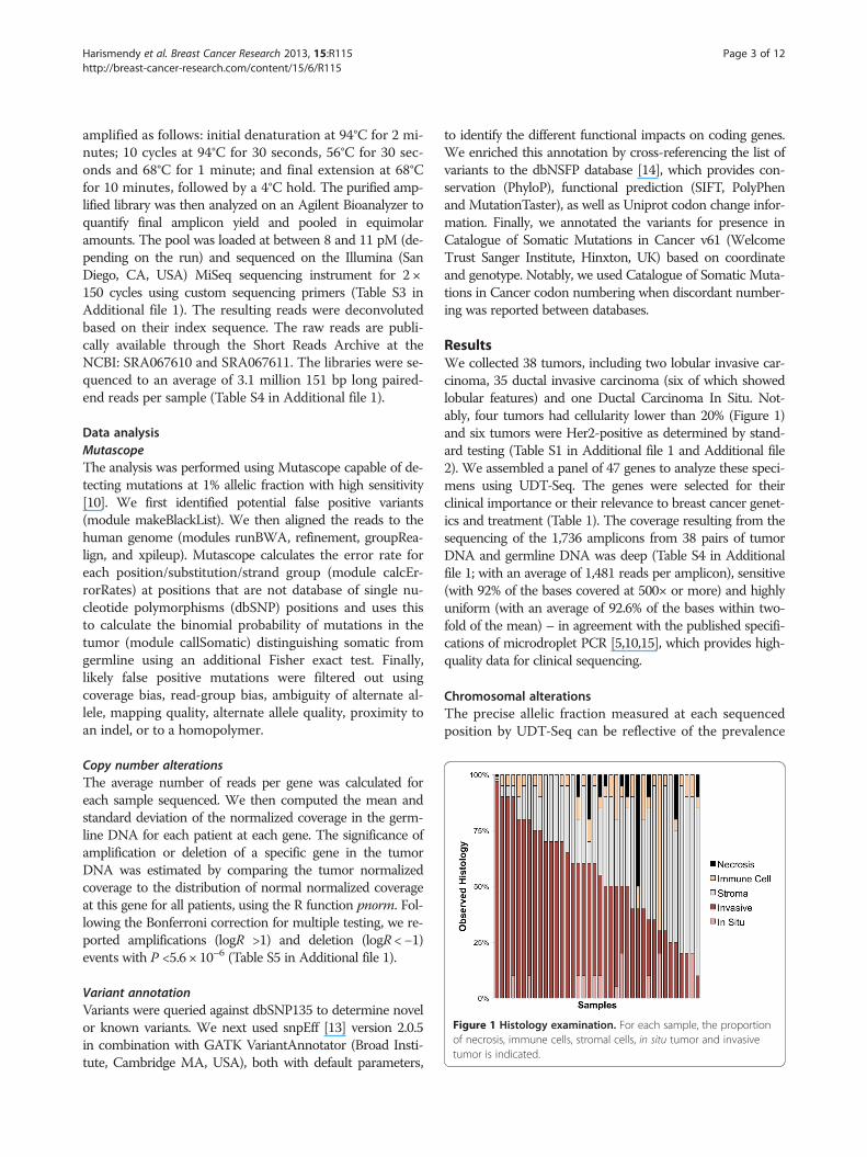
Figure 1 Histology examination. For each sample, the proportionof necrosis, immune cells, stromal cells, in situ tumor and invasivetumor is indicated.
Harismendy et al. Breast Cancer Research 2013, 15:R115 Page 3 of 12http://breast-cancer-research.com/content/15/6/R115
amplified as follows: initial denaturation at 94°C for 2 mi-nutes; 10 cycles at 94°C for 30 seconds, 56°C for 30 sec-onds and 68°C for 1 minute; and final extension at 68°Cfor 10 minutes, followed by a 4°C hold. The purified amp-lified library was then analyzed on an Agilent Bioanalyzer toquantify final amplicon yield and pooled in equimolaramounts. The pool was loaded at between 8 and 11 pM (de-pending on the run) and sequenced on the Illumina (SanDiego, CA, USA) MiSeq sequencing instrument for 2 ×150 cycles using custom sequencing primers (Table S3 inAdditional file 1). The resulting reads were deconvolutedbased on their index sequence. The raw reads are publi-cally available through the Short Reads Archive at theNCBI: SRA067610 and SRA067611. The libraries were se-quenced to an average of 3.1 million 151 bp long paired-end reads per sample (Table S4 in Additional file 1).
Data analysisMutascopeThe analysis was performed using Mutascope capable of de-tecting mutations at 1% allelic fraction with high sensitivity[10]. We first identified potential false positive variants(module makeBlackList). We then aligned the reads to thehuman genome (modules runBWA, refinement, groupRea-lign, and xpileup). Mutascope calculates the error rate foreach position/substitution/strand group (module calcEr-rorRates) at positions that are not database of single nu-cleotide polymorphisms (dbSNP) positions and uses thisto calculate the binomial probability of mutations in thetumor (module callSomatic) distinguishing somatic fromgermline using an additional Fisher exact test. Finally,likely false positive mutations were filtered out usingcoverage bias, read-group bias, ambiguity of alternate al-lele, mapping quality, alternate allele quality, proximity toan indel, or to a homopolymer.
Copy number alterationsThe average number of reads per gene was calculated foreach sample sequenced. We then computed the mean andstandard deviation of the normalized coverage in the germ-line DNA for each patient at each gene. The significance ofamplification or deletion of a specific gene in the tumorDNA was estimated by comparing the tumor normalizedcoverage to the distribution of normal normalized coverageat this gene for all patients, using the R function pnorm. Fol-lowing the Bonferroni correction for multiple testing, we re-ported amplifications (logR >1) and deletion (logR <−1)events with P <5.6 × 10⁻⁶ (Table S5 in Additional file 1).
Variant annotationVariants were queried against dbSNP135 to determine novelor known variants. We next used snpEff [13] version 2.0.5in combination with GATK VariantAnnotator (Broad Insti-tute, Cambridge MA, USA), both with default parameters,
to identify the different functional impacts on coding genes.We enriched this annotation by cross-referencing the list ofvariants to the dbNSFP database [14], which provides con-servation (PhyloP), functional prediction (SIFT, PolyPhenand MutationTaster), as well as Uniprot codon change infor-mation. Finally, we annotated the variants for presence inCatalogue of Somatic Mutations in Cancer v61 (WelcomeTrust Sanger Institute, Hinxton, UK) based on coordinateand genotype. Notably, we used Catalogue of Somatic Muta-tions in Cancer codon numbering when discordant number-ing was reported between databases.
ResultsWe collected 38 tumors, including two lobular invasive car-cinoma, 35 ductal invasive carcinoma (six of which showedlobular features) and one Ductal Carcinoma In Situ. Not-ably, four tumors had cellularity lower than 20% (Figure 1)and six tumors were Her2-positive as determined by stand-ard testing (Table S1 in Additional file 1 and Additional file2). We assembled a panel of 47 genes to analyze these speci-mens using UDT-Seq. The genes were selected for theirclinical importance or their relevance to breast cancer genet-ics and treatment (Table 1). The coverage resulting from thesequencing of the 1,736 amplicons from 38 pairs of tumorDNA and germline DNA was deep (Table S4 in Additionalfile 1; with an average of 1,481 reads per amplicon), sensitive(with 92% of the bases covered at 500× or more) and highlyuniform (with an average of 92.6% of the bases within two-fold of the mean) – in agreement with the published specifi-cations of microdroplet PCR [5,10,15], which provides high-quality data for clinical sequencing.
Chromosomal alterationsThe precise allelic fraction measured at each sequencedposition by UDT-Seq can be reflective of the prevalence
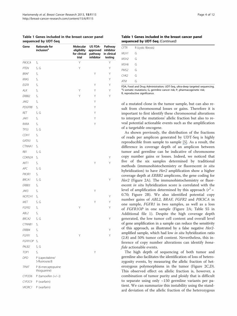
Table 1 Genes included in the breast cancer panelsequenced by UDT-Seq
Gene Rationale forinclusiona
Moleculareligibilityfor clinical
trial
US FDA-approvedpathwayinhibitor
Pathwayinhibitorin clinicaltesting
PIK3CA S, Y Y
PTEN S, G Y Y
BRAF S, Y Y
KRAS S, Y
EGFR S, Y Y
ALK S, Y Y Y
ERBB2 S, Y Y Y
JAK2 S, Y
PDGFRB S, Y
RET S, G Y
JAK1 S, Y
RARA S, Y
TP53 S, G
CDH1 S,
GATA3 S,
CTNNA1 S,
RB1 S, G
CDKN2A S, Y
AKT1 S, Y Y
APC S, G
PIK3R1 S,
BRCA1 S, G Y
ERBB3 S,
JAK3 S, Y
NOTCH1 S, Y
MET S, G Y Y
FGFR2 S, Y Y
ABL2 S,
BRCA2 S, G Y
CTNNB1 S,
ERBB4 S, Y
FGFR1 S, Y Y
FGFR1OP S,
PALB2 S, G
TOP1 S, Y
DPD P (capecitabine/5-fluorouracil)
TPMT P (6-mercaptopurinethioguanine)
CYP2D6 P (tamoxifen (+/−))
CYP2C9 P (warfarin)
VKORC1 P (warfarin)
Table 1 Genes included in the breast cancer panelsequenced by UDT-Seq (Continued)
CFTR R (cystic fibrosis)
MLH1 G
MSH2 G
MSH6 G
PMS2 G
CHK2 G
ATM G
FDA, Food and Drug Administration; UDT-Seq, ultra-deep targeted sequencing.aS somatic mutations; G, germline cancer risk; P, pharmacogenetic risk;R, reproductive significance.
Harismendy et al. Breast Cancer Research 2013, 15:R115 Page 4 of 12http://breast-cancer-research.com/content/15/6/R115
of a mutated clone in the tumor sample, but can also re-sult from chromosomal losses or gains. Therefore it isimportant to first identify these chromosomal alterationsto interpret the mutations’ allelic fraction but also to re-veal potential actionable events such as the amplificationof a targetable oncogene.As shown previously, the distribution of the fractions
of reads per amplicon generated by UDT-Seq is highlyreproducible from sample to sample [5]. As a result, thedifference in coverage depth of an amplicon betweentumor and germline can be indicative of chromosomecopy number gains or losses. Indeed, we noticed thatfive of the six samples determined by traditionalmethods (immunohistochemistry or fluorescent in situhybridization) to have Her2 amplification show a highercoverage depth at ERBB2 amplicons, the gene coding forHer2 (Figure 2A). The immunohistochemistry or fluor-escent in situ hybridization score is correlated with thelevel of amplification determined by this approach (r2 =0.70; Figure 2B). We also identified potential copynumber gains of ABL2, BRAF, FGFR2 and PIK3CA inone sample, FGFR1 in two samples, as well as a lossof FGFR1OP in one sample (Figure 2A; Table S5 inAdditional file 1). Despite the high coverage depthgenerated, the low tumor cell content and overall levelof gene amplification in a sample can reduce the sensitivityof this approach, as illustrated by a false negative Her2-amplified sample, which had low in situ hybridization ratio(2.8) and 50% tumor cell content. Nevertheless, this in-ference of copy number alterations can identify bona-fide actionable events.The high depth of sequencing of both tumor and
germline also facilitates the identification of loss of hetero-zygosity events, by measuring the allelic fraction of het-erozygous polymorphisms in the tumor (Figure 2C,D).This observed effect on allelic fraction is, however, acombination of tumor purity and ploidy that is difficultto separate using only ~150 germline variants per pa-tient. We can summarize this instability using the stand-ard deviation of the allelic fraction of the heterozygous
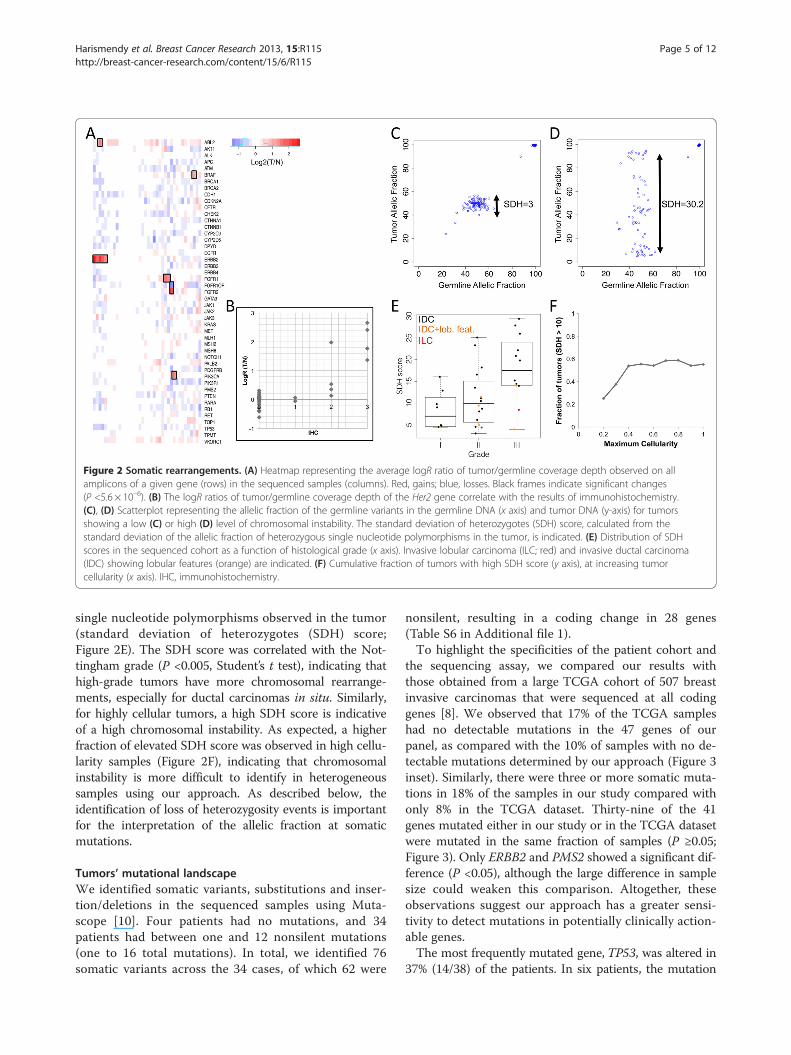
Figure 2 Somatic rearrangements. (A) Heatmap representing the average logR ratio of tumor/germline coverage depth observed on allamplicons of a given gene (rows) in the sequenced samples (columns). Red, gains; blue, losses. Black frames indicate significant changes(P <5.6 × 10⁻⁶). (B) The logR ratios of tumor/germline coverage depth of the Her2 gene correlate with the results of immunohistochemistry.(C), (D) Scatterplot representing the allelic fraction of the germline variants in the germline DNA (x axis) and tumor DNA (y-axis) for tumorsshowing a low (C) or high (D) level of chromosomal instability. The standard deviation of heterozygotes (SDH) score, calculated from thestandard deviation of the allelic fraction of heterozygous single nucleotide polymorphisms in the tumor, is indicated. (E) Distribution of SDHscores in the sequenced cohort as a function of histological grade (x axis). Invasive lobular carcinoma (ILC; red) and invasive ductal carcinoma(IDC) showing lobular features (orange) are indicated. (F) Cumulative fraction of tumors with high SDH score (y axis), at increasing tumorcellularity (x axis). IHC, immunohistochemistry.
Harismendy et al. Breast Cancer Research 2013, 15:R115 Page 5 of 12http://breast-cancer-research.com/content/15/6/R115
single nucleotide polymorphisms observed in the tumor(standard deviation of heterozygotes (SDH) score;Figure 2E). The SDH score was correlated with the Not-tingham grade (P <0.005, Student’s t test), indicating thathigh-grade tumors have more chromosomal rearrange-ments, especially for ductal carcinomas in situ. Similarly,for highly cellular tumors, a high SDH score is indicativeof a high chromosomal instability. As expected, a higherfraction of elevated SDH score was observed in high cellu-larity samples (Figure 2F), indicating that chromosomalinstability is more difficult to identify in heterogeneoussamples using our approach. As described below, theidentification of loss of heterozygosity events is importantfor the interpretation of the allelic fraction at somaticmutations.
Tumors’ mutational landscapeWe identified somatic variants, substitutions and inser-tion/deletions in the sequenced samples using Muta-scope [10]. Four patients had no mutations, and 34patients had between one and 12 nonsilent mutations(one to 16 total mutations). In total, we identified 76somatic variants across the 34 cases, of which 62 were
nonsilent, resulting in a coding change in 28 genes(Table S6 in Additional file 1).To highlight the specificities of the patient cohort and
the sequencing assay, we compared our results withthose obtained from a large TCGA cohort of 507 breastinvasive carcinomas that were sequenced at all codinggenes [8]. We observed that 17% of the TCGA sampleshad no detectable mutations in the 47 genes of ourpanel, as compared with the 10% of samples with no de-tectable mutations determined by our approach (Figure 3inset). Similarly, there were three or more somatic muta-tions in 18% of the samples in our study compared withonly 8% in the TCGA dataset. Thirty-nine of the 41genes mutated either in our study or in the TCGA datasetwere mutated in the same fraction of samples (P ≥0.05;Figure 3). Only ERBB2 and PMS2 showed a significant dif-ference (P <0.05), although the large difference in samplesize could weaken this comparison. Altogether, theseobservations suggest our approach has a greater sensi-tivity to detect mutations in potentially clinically action-able genes.The most frequently mutated gene, TP53, was altered in
37% (14/38) of the patients. In six patients, the mutation
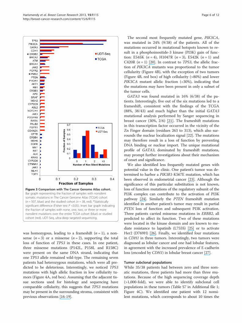
Figure 3 Comparison with The Cancer Genome Atlas cohort.Bar graph representing the fraction of samples with nonsilentsomatic mutations in The Cancer Genome Atlas (TCGA) cohort(n = 507, blue) and the studied cohort (n = 38, red). *Statisticallysignificant difference (Fisher test P <0.05). Inset: bar graph indicatingthe fraction of samples with none, one, two, or three or morenonsilent mutations over the entire TCGA cohort (blue) or studiedcohort (red). UDT-Seq, ultra-deep targeted sequencing.
Harismendy et al. Breast Cancer Research 2013, 15:R115 Page 6 of 12http://breast-cancer-research.com/content/15/6/R115
was homozygous, leading to a frameshift (n = 1), a non-sense (n = 3) or a missense (n = 2), supporting the totalloss of function of TP53 in these cases. In one patient,three missense mutations (P142L, P158L and R158C)were present on the same DNA strand, indicating thatone TP53 allele remained wild-type. The remaining sevenpatients had heterozygous mutations, which were all pre-dicted to be deleterious. Interestingly, we noticed TP53mutations with high allelic fraction in low cellularity tu-mors (Figure 4A, red box). Assuming that the adjacent tis-sue sections used for histology and sequencing havecomparable cellularity, this suggests that TP53 mutationsmay be present in the surrounding stroma, consistent withprevious observations [16-19].
The second most frequently mutated gene, PIK3CA,was mutated in 24% (9/38) of the patients. All of themutations occurred in mutational hotspots known to re-sult in a phosphoinositide-3 kinase (PI3K) gain of func-tion: E545K (n = 4), H1047R (n = 3), E542K (n = 1) andC420R (n = 1) [20]. In contrast to TP53, the allelic frac-tion of PIK3CA mutants was proportional to the tumorcellularity (Figure 4B), with the exception of two tumors(Figure 4B, red box) of high cellularity (>80%) and lowerPIK3CA mutant allelic fraction (<30%), indicating thatthe mutations may have been present in only a subset ofthe tumor cells.GATA3 was found mutated in 16% (6/38) of the pa-
tients. Interestingly, five out of the six mutations led to aframeshift, consistent with the findings of the TCGA(88%, 38/43) and much higher than the initial GATA3mutational analysis performed by Sanger sequencing inbreast cancer (30%, 2/6) [21]. The frameshift mutationsin this transcription factor occurred in the vicinity of theZn Finger domain (residues 263 to 313), which also sur-rounds the nuclear localization signal [22]. The mutationsmay therefore result in a loss of function by preventingDNA binding or nuclear import. The unique mutationalprofile of GATA3, dominated by frameshift mutations,may prompt further investigations about their mechanismof onset and significance.We also identified less frequently mutated genes with
potential value in the clinic. One patient’s tumor was de-termined to harbor a PIK3R1-K567E mutation, which hasbeen observed in endometrial cancer [23]. Although thesignificance of this particular substitution is not known,loss of function mutations of the regulatory subunit of thePI3K complex can contribute to the activation of PI3Kpathway [24]. Similarly the PTEN frameshift mutationidentified in another patient’s tumor may result in partialPTEN loss of function and subsequent PI3K activation.Three patients carried missense mutations in ERBB2, allpredicted to affect its function. Two of these mutationswere located in the kinase domain and are known to me-diate resistance to lapatinib (L755S) [25] or to activateHer2 (D769H) [26]. Finally, we identified four mutationsin CDH1 in three tumors. Interestingly, two tumors werediagnosed as lobular cancer and one had lobular features,in agreement with the increased prevalence of E-cadherinloss (encoded by CDH1) in lobular breast cancer [27].
Tumor subclonal populationsWhile 35/38 patients had between zero and three som-atic mutations, three patients had more than three mu-tations. Because of the high sequencing coverage depth(>1,000-fold), we were able to identify subclonal cellpopulations in these tumors (Table S7 in Additional file 1;Figure 4C). We identified one patient with 12 nonsi-lent mutations, which corresponds to about 10 times the
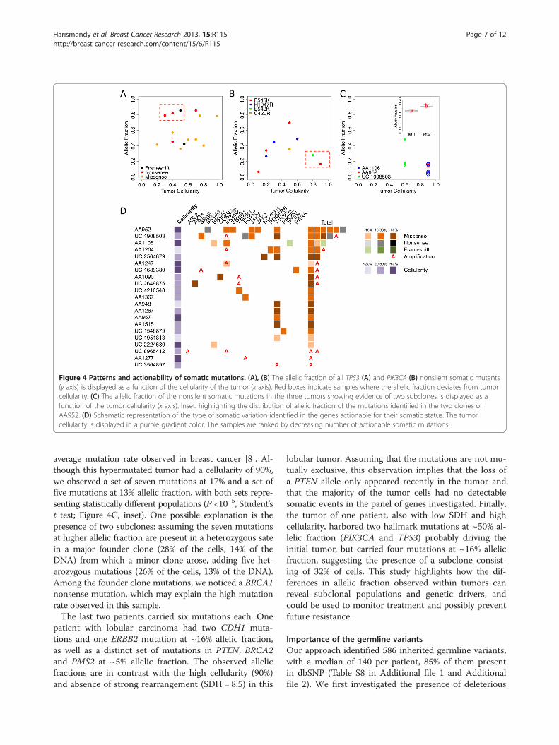
Figure 4 Patterns and actionability of somatic mutations. (A), (B) The allelic fraction of all TP53 (A) and PIK3CA (B) nonsilent somatic mutants(y axis) is displayed as a function of the cellularity of the tumor (x axis). Red boxes indicate samples where the allelic fraction deviates from tumorcellularity. (C) The allelic fraction of the nonsilent somatic mutations in the three tumors showing evidence of two subclones is displayed as afunction of the tumor cellularity (x axis). Inset: highlighting the distribution of allelic fraction of the mutations identified in the two clones ofAA952. (D) Schematic representation of the type of somatic variation identified in the genes actionable for their somatic status. The tumorcellularity is displayed in a purple gradient color. The samples are ranked by decreasing number of actionable somatic mutations.
Harismendy et al. Breast Cancer Research 2013, 15:R115 Page 7 of 12http://breast-cancer-research.com/content/15/6/R115
average mutation rate observed in breast cancer [8]. Al-though this hypermutated tumor had a cellularity of 90%,we observed a set of seven mutations at 17% and a set offive mutations at 13% allelic fraction, with both sets repre-senting statistically different populations (P <10–5, Student’st test; Figure 4C, inset). One possible explanation is thepresence of two subclones: assuming the seven mutationsat higher allelic fraction are present in a heterozygous satein a major founder clone (28% of the cells, 14% of theDNA) from which a minor clone arose, adding five het-erozygous mutations (26% of the cells, 13% of the DNA).Among the founder clone mutations, we noticed a BRCA1nonsense mutation, which may explain the high mutationrate observed in this sample.The last two patients carried six mutations each. One
patient with lobular carcinoma had two CDH1 muta-tions and one ERBB2 mutation at ~16% allelic fraction,as well as a distinct set of mutations in PTEN, BRCA2and PMS2 at ~5% allelic fraction. The observed allelicfractions are in contrast with the high cellularity (90%)and absence of strong rearrangement (SDH = 8.5) in this
lobular tumor. Assuming that the mutations are not mu-tually exclusive, this observation implies that the loss ofa PTEN allele only appeared recently in the tumor andthat the majority of the tumor cells had no detectablesomatic events in the panel of genes investigated. Finally,the tumor of one patient, also with low SDH and highcellularity, harbored two hallmark mutations at ~50% al-lelic fraction (PIK3CA and TP53) probably driving theinitial tumor, but carried four mutations at ~16% allelicfraction, suggesting the presence of a subclone consist-ing of 32% of cells. This study highlights how the dif-ferences in allelic fraction observed within tumors canreveal subclonal populations and genetic drivers, andcould be used to monitor treatment and possibly preventfuture resistance.
Importance of the germline variantsOur approach identified 586 inherited germline variants,with a median of 140 per patient, 85% of them presentin dbSNP (Table S8 in Additional file 1 and Additionalfile 2). We first investigated the presence of deleterious
Harismendy et al. Breast Cancer Research 2013, 15:R115 Page 8 of 12http://breast-cancer-research.com/content/15/6/R115
variants in BRCA1/2, which are the most actionablegenes in the clinical setting. We identified three patientswith a predicted deleterious mutation in one of thesegenes, of which only one seems truly deleterious [28-30](Table S9 in Additional file 1). The BRCA1-Q1355_E1356fsframeshift mutation is a previously reported deleteriousmutation [30] and is clinically actionable. Interestingly,the mutant allele was selected for in the tumor (allelicfraction 94%), indicating a selective advantage. This germ-line finding was later confirmed by a Clinical LaboratoryImprovement Amendments-approved assay after the pa-tient consulted with a clinical genetic counselor.Inherited variants in DPYD have been associated with
toxicity to 5-fluorouracil or capecitabine chemotherapy[12], which is commonly used in breast cancer treat-ment. We identified six patients carrying three variantsin DPYD with predicted deleterious effects. Three pa-tients were heterozygous for rs1801160 (Minor AlleleFrequency = 0.04). This single nucleotide polymorphismdefines the DPYD*6 haplotype, which has been associ-ated with increased toxicity [31]. Two novel missensevariants (K259E and V944A) identified in three patientshave an unknown significance. Interestingly, a recentstudy indicates that variants in DPYD can actually in-crease its metabolic activity, therefore protecting againsttoxicity and decreasing drug efficiency [32]. Until morefunctional experiments are performed, it will be challen-ging to unambiguously determine the clinical relevanceof most inherited DPYD variants. We also identified twopatients carrying one inactive allele of the gene(CYP2D6*6). However, it is not clear whether this particu-lar allele, in a heterozygous state, is associated with a re-duced metabolism of tamoxifen; therefore, a change indrug dosage is not justified.More generally, our approach identified many inher-
ited variants of unknown significance, which should becautiously interpreted. Importantly, in the absence of amatched germline sample, some of these variants mighthave been misidentified as tumor-specific events poten-tially confounding the rationale for targeted therapy,therefore highlighting the importance of sequencingmatched germline DNA.
Clinical implicationsOut of the 47 genes sequenced, 24 are classified as ac-tionable based on their somatic status (Table 1). Thesegenes or the pathway they belong to could be targetedby a specific inhibitor, commercially available or underinvestigation (PIK3CA, ERBB2), or are predictive bio-markers for targeted therapies that are approved or inclinical trials (BRCA1/2, PTEN). There were 21 patientswhose tumors carried nonsilent mutations or copy num-ber alterations in 17 of these 24 genes (Figure 4D). Im-portantly, three of the patients had tumors with less
than 20% cellularity and in four patients we identifiedmutations at an allelic fraction of 10% or lower. We canestablish the added benefit of our strategy in such cases:if we had limited our analysis to the samples with cellular-ity higher than 60% (19 samples), which is the inclusioncriteria used by the TCGA, we would have identified mu-tations in only six patients for an overall sensitivity of only31% (6/19 cases). However, by using the UDT-Seq ap-proach, we identified mutations in actionable genes in 21of the 38 patients studied for an overall sensitivity of 55%(21/38 cases), combining the benefits of less stringent in-clusion criteria and higher assay sensitivity.Based on these molecular findings, we then summarized
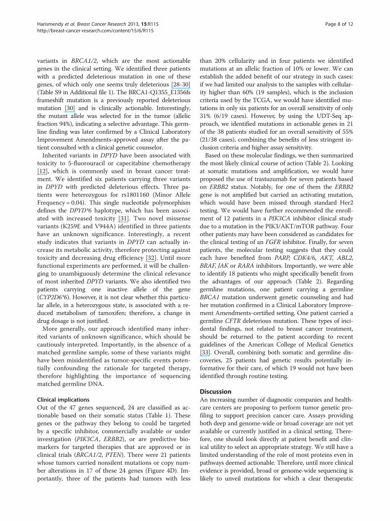
the most likely clinical course of action (Table 2). Lookingat somatic mutations and amplification, we would haveproposed the use of trastuzumab for seven patients basedon ERBB2 status. Notably, for one of them the ERBB2gene is not amplified but carried an activating mutation,which would have been missed through standard Her2testing. We would have further recommended the enroll-ment of 12 patients in a PIK3CA inhibitor clinical studydue to a mutation in the PIK3/AKT/mTOR pathway. Fourother patients may have been considered as candidates forthe clinical testing of an FGFR inhibitor. Finally, for sevenpatients, the molecular testing suggests that they couldeach have benefited from PARP, CDK4/6, AKT, ABL2,BRAF, JAK or RARA inhibitors. Importantly, we were ableto identify 18 patients who might specifically benefit fromthe advantages of our approach (Table 2). Regardinggermline mutations, one patient carrying a germlineBRCA1 mutation underwent genetic counseling and hadher mutation confirmed in a Clinical Laboratory Improve-ment Amendments-certified setting. One patient carried agermline CFTR deleterious mutation. These types of inci-dental findings, not related to breast cancer treatment,should be returned to the patient according to recentguidelines of the American College of Medical Genetics[33]. Overall, combining both somatic and germline dis-coveries, 25 patients had genetic results potentially in-formative for their care, of which 19 would not have beenidentified through routine testing.
DiscussionAn increasing number of diagnostic companies and health-care centers are proposing to perform tumor genetic pro-filing to support precision cancer care. Assays providingboth deep and genome-wide or broad coverage are not yetavailable or currently justified in a clinical setting. There-fore, one should look directly at patient benefit and clin-ical utility to select an appropriate strategy. We still have alimited understanding of the role of most proteins even inpathways deemed actionable. Therefore, until more clinicalevidence is provided, broad or genome-wide sequencing islikely to unveil mutations for which a clear therapeutic
Table 2 Summary of the primary course of action likely to result from the molecular testing
Patient SNP or mutation (allelic fraction) Proposed action UDT-Seq advantagesa
AA1025 rs113993959 (Het) CFTR genetic counseling Germline
AA1090 CDKN2A-A85D (66%) CDK4/6 inhibitor
FGFR1 amplification FGFR1/2 inhibitor CNA
AA1106 ERBB2-L755S (17%) Trastuzumab
PTEN-frameshift (5%) PIK3CA inhibitor Depth
BRCA2-I1418T (4%) PARP inhibitor Depth
AA1204b PIK3CA-H1047R (26%) PIK3CA inhibitor Sensitivity
Her2 amplification Trastuzumab CNA
AA1222b Her2 amplification Trastuzumab CNA
AA1247b Her2 amplification Trastuzumab CNA
ERBB2-D769H (5%) Depth
AA1267 PIK3CA-H1047R (45%) PIK3CA inhibitor
AA1277 rs80357508 (Het) BRCA1 genetic counseling Germline
FGFR2 amplification FGFR1/2 inhibitor CNA
AA948 PIK3CA-E545K (34%) PIK3CA inhibitor Sensitivity
AA952 PIK3CA-E545K (16%) PIK3CA inhibitor
BRCA1-W306* (18%) PARP inhibitor
BRCA1-E550K (13%)
JAK2-S131L (16%) JAK inhibitor
JAK3-I386M (13%)
rs1801160 (Het) 5-FU toxicity Germline
AA957 PIK3CA-E542K (28%) PIK3CA inhibitor
AA1515 PIK3CA-E545K (70%) PIK3CA inhibitor
UCI1546879 PIK3R1-K204E (30%) PIK3CA inhibitor
UCI1689380 RARA-337 T (14%) RARA inhibitor
BRAF amplification Vemurafenib
UCI1908503b PIK3CA-H1047R (40%) PIK3CA inhibitor
Her2 amplification Trastuzumab CNA
UCI1951813 PIK3CA-E545K (7%) PIK3CA inhibitor Sensitivity
UCI2076630b Her2 amplification Trastuzumab CNA
UCI2224680 BRCA2-L1829F (2%) PARP inhibitor Depth
UCI2564879 PIK3R1-K204E (30%) PIK3CA inhibitor
UCI2649875 AKT1-L52R (63%) AKT inhibitor
FGFR1 amplification FGFR1/2 inhibitor CNA
UCI4216548 FGFR1-D683H (13%) FGFR1/2 inhibitor
UCI8965412b Her2 amplification Trastuzumab CNA
ABL2 amplification Imatinib CNA
UCI1804937 rs1801160 (Het) 5-FU toxicity Germline
UCI2008866 rs1801160 (Het) 5-FU toxicity Germline
UCI3564897 PIK3CA amplification PIK3CA inhibitor CNA
5-FU, 5-fluorouracil; SNP, single nucleotide polymorphism. aDepth, accurate calls at low allelic fraction (<10%); sensitivity, accurate calls in heterogeneous samples;CNA, inference of copy number alterations; germline, inclusion of a matched germline DNA. bHer2-positive determined through standard of care.
Harismendy et al. Breast Cancer Research 2013, 15:R115 Page 9 of 12http://breast-cancer-research.com/content/15/6/R115
Harismendy et al. Breast Cancer Research 2013, 15:R115 Page 10 of 12http://breast-cancer-research.com/content/15/6/R115
rationale is not yet available or misunderstood. In con-trast, the use of deep sequencing of a restricted panel ofgenes increases the sensitivity to detect well-known andactionable mutations, which can have a greater impact inthe clinic. For these reasons, deep sequencing of a re-stricted gene panel is likely to benefit the greatest numberof patients today. Using our UDT-Seq approach, we iden-tified potentially actionable mutations in 14/19 patientswhose tumor samples had less than 60% cellularity anddiscovered actionable mutations present at 10% allelicfraction or less in four patients, some of whom had tu-mors with high malignant cellularity. UDT-Seq offers avery quantitative measurement of the allelic fraction ofthe mutations providing information about the biology ofthe tumor. For example, we observed a field effect in tu-mors harboring TP53 mutations and the presence of sub-clonal PIK3CA mutations or of multiple mutated clonesin three tumors, probably resulting from their evolution.Clinical utility of these new data will require specific trialsto show that targeting resistant subclones or field effectsis likely to improve outcomes in both the curative and pal-liative setting.Traditionally, tumor-specific markers are investigated
in the tumor specimen only. While this may be sufficientfor protein markers, a DNA mutation is identified as amismatch to the reference human genome and couldcorrespond either to an inherited variant or somaticallyacquired mutation in the tumor. Only the sequencing ofmatched germline DNA can confirm that the variant issomatic, providing a better rationale for the use of tar-geted therapy, or inherited, providing important infor-mation for the care of the patient and their relatives.Finally, the use of matched germline DNA sequencingfacilitates the detection of mutations at low allelic frac-tion [10,34], which, as discussed above, is likely to be ex-tremely important for optimal implementation in clinicalcare. It is typically feasible to obtain a blood or buccalsample along with the tumor or biopsy sample being in-vestigated, without excessive burden.Importantly, the adoption of such transformative diag-
nostic assays in the clinic needs to include physician educa-tion and training and be associated with the establishmentof molecular tumor boards in academic centers. These mo-lecular tumor boards are not focused on a particular can-cer by site of origin, but rather on the molecular markersidentified. The presence of basic scientists with expertise inthe altered pathways also improves the clinical interpret-ation. Indeed, the role and clinical significance of muta-tions located in less commonly mutated exons, genes orin the noncoding portions of the genome [35] remain tobe established. Interpreting these variants of unknown sig-nificance, whether inherited or somatic, is the most con-troversial and difficult aspect of clinical sequencing.Despite attempts to consolidate variants, mutations, and
clinical information in public databases, molecular tumorboard members must currently perform extensive litera-ture searches to predict the impact of a mutation. In ourstudy, missense mutations in ERBB2 were reported as ac-tivating by only a few published studies, suggesting theirrelevance for trastuzumab or lapatinib treatment. A simi-lar challenge exists for the interpretation of polymor-phisms in drug metabolizing genes, which will benefitfrom the efforts of the pharmacogenomics research net-work [36]. Finally, such precision medicine strategy issensible only if it benefits the patients. For inherited vari-ants, access to clinical genetic counseling is critical to in-terpret the results in the context of a complete familyhistory. Similarly, targeting genes with somatic mutationsusing an investigational drug, requires access to a clinicaltrial or reimbursement for off-label use of targeted drugswith clinical outcome captured in a clinical registry study.
ConclusionOur study evaluates the potential benefits of the UDT-Seq of 47 selected genes for breast cancer care. We showthat our assay identifies actionable findings, both inher-ited variants and somatic mutations, in 25 out of 38samples. In particular, the specificities of our assay – in-clusion of germline DNA, identification of copy numbervariants, high coverage depth and sensitivity to identifysomatic mutations at low allelic fraction – would havebeen directly beneficial to 18 patients. As high-throughputsequencing starts to be used in clinical care, its establish-ment as a routine diagnostic assay will require progress onmany fronts: demonstration of technical validity and clin-ical utility, education of physicians and trainees, and co-operation with pharmaceutical and insurance companiesto increase drug accessibility.
Additional files
Additional file 1: Table S1. Presenting the source and histopathologicdescription of the 38 specimens studied, Table S2. Presenting the list ofprimers used in the study, Table S3. Presenting the universal PCRprimers, Table S4. Presenting the significant copy number gains andlosses, Table S5. Presenting the read alignment and coverage statistics,Table S6. Presenting the list of nonsilent somatic mutations, Table S7.Presenting the list of mutations that segregate into two significantlydifferent groups of allelic fractions in the tumors of three patients, Table S8.Presenting the list of nonsilent germline variants, and Table S9. Presentingthe list of predicted deleterious inherited variants in BRCA1 or BRCA2.
Additional file 2: Supplemental text.
AbbreviationsCFTR: Cysitc Fibrosis Transmembrane Conductance Regulator;dbSNP: Database of single nucleotide polymorphisms; JAK: Janus kinase;PARP: Poly-ADP ribose polymerase; PCR: Polymerase chain reaction;PI3K: Phosphoinositide-3 kinase; SDH: Standard deviation of heterozygotes;TCGA: The Cancer Genome Atlas; UDT-Seq: Ultra-deep targeted sequencing.
Competing interestThe authors declare that they have no competing interests.
Harismendy et al. Breast Cancer Research 2013, 15:R115 Page 11 of 12http://breast-cancer-research.com/content/15/6/R115
Authors’ contributionsOH, SEY, HM and LM analyzed the data. HA and KJ generated the data. RBS, FH,AMW, HLP and PMC collected and analyzed the samples and clinicalinformation. OH, KAF, HA-C, BP and RBS designed the study. OH, LM, RBS andKAF wrote the manuscript. All authors read and approved the final manuscript.
AcknowledgementsThe authors thank Jimmy Salinas and Peter Chase for technical assistance.This work was supported by grants from the National Cancer Institute(1R21CA155615-01A1 and 1R21CA152613-01) to OH and KAF, and from theNational Center for Advancing Translational Sciences (UL1RR031980) toDr Firestein. HA is supported by a fellowship from the German Center Aid.
Author details1Division of Genome Information Sciences, Department of Pediatrics andRady Children’s Hospital, University of California San Diego, 9500 GilmanDrive, La Jolla CA 92093, USA. 2Moores UCSD Cancer Center, School ofMedicine, University of California San Diego, 3855 Health Science Drive, LaJolla CA 92093, USA. 3Clinical and Translational Science Institute, University ofCalifornia San Diego, 9500 Gilman Drive, La Jolla CA 92093, USA.4Department of Medicine, School of Medicine, University of California SanDiego, 9500 Gilman Drive, La Jolla CA 92093, USA. 5Department ofPathology, School of Medicine, University of California San Diego, 9500Gilman Drive, La Jolla CA 92093, USA. 6Department of Surgery, School ofMedicine, University of California San Diego, 9500 Gilman Drive, La Jolla CA92093, USA. 7Bioinformatics Graduate Program, University of California SanDiego, 9500 Gilman Drive, La Jolla CA 92093, USA. 8Department of Familyand Preventive Medicine, School of Medicine, University of California SanDiego, La Jolla CA, USA. 9Institute for Genomic Medicine, University ofCalifornia San Diego, 9500 Gilman Drive, La Jolla CA 92093, USA.10Department of Epidemiology, School of Medicine, University of CaliforniaIrvine, 252 Irvine Hall, Irvine CA 92697, USA. 11Department of Pathology andLaboratory Medicine, School of Medicine, University of California Irvine, 252Irvine Hall, Irvine CA 92697, USA. 12Department of General, Visceral andCancer Surgery, University of Cologne, Frangenheimstraße 4, 50931, KölnGermany.
Received: 27 June 2013 Accepted: 6 December 2013Published: 10 December 2013
References1. Tsimberidou AM, Iskander NG, Hong DS, Wheler JJ, Falchook GS, Fu S,
Piha-Paul SA, Naing A, Janku F, Luthra R, Ye Y, Wen S, Berry DA, KurzrockR: Personalized medicine in a phase I clinical trials program: the MDanderson cancer center initiative. Clin Cancer Res 2012, 18:6373–6383.
2. MacConaill LE, Campbell CD, Kehoe SM, Bass AJ, Hatton C, Niu L, Davis M,Yao K, Hanna M, Mondal C, Luongo L, Emery CM, Baker AC, Philips J, GoffDJ, Fiorentino M, Rubin MA, Polyak K, Chan J, Wang Y, Fletcher JA,Santagata S, Corso G, Roviello F, Shivdasani R, Kieran MW, Ligon KL, StilesCD, Hahn WC, Meyerson ML, et al: Profiling critical cancer gene mutationsin clinical tumor samples. PloS One 2009, 4:e7887.
3. Li M, Diehl F, Dressman D, Vogelstein B, Kinzler KW: BEAMing up fordetection and quantification of rare sequence variants. Nat Methods2006, 3:95–97.
4. Wagle N, Berger MF, Davis MJ, Blumenstiel B, DeFelice M, Pochanard P,Ducar M, Van Hummelen P, MacConaill LE, Hahn WC, Meyerson M, Gabriel SB,Garraway LA: High-throughput detection of actionable genomic alterationsin clinical tumor samples by targeted, massively parallel sequencing. CancerDiscov 2012, 2:82–93.
5. Harismendy O, Schwab RB, Bao L, Olson J, Rozenzhak S, Kotsopoulos SK,Pond S, Crain B, Chee MS, Messer K, Link DR, Frazer KA: Detection of lowprevalence somatic mutations in solid tumors with ultra-deep targetedsequencing. Genome Biol 2011, 12:R124.
6. Beltran H, Yelensky R, Frampton GM, Park K, Downing SR, MacDonald TY,Jarosz M, Lipson D, Tagawa ST, Nanus DM, Stephens PJ, Mosquera JM,Cronin MT, Rubin MA: Targeted next-generation sequencing of advancedprostate cancer identifies potential therapeutic targets and diseaseheterogeneity. Eur Urol 2013, 63:920–926.
7. Beadling C, Neff TL, Heinrich MC, Rhodes K, Thornton M, Leamon J,Andersen M, Corless CL: Combining highly multiplexed PCR with
semiconductor-based sequencing for rapid cancer genotyping. J MolDiagn 2013, 15:171–176.
8. Curtis C, Shah SP, Chin S-F, Turashvili G, Rueda OM, Dunning MJ, Speed D,Lynch AG, Samarajiwa S, Yuan Y, Graf S, Ha G, Haffari G, Bashashati A, RussellR, McKinney S, Langerod A, Green A, Provenzano E, Wishart G, Pinder S,Watson P, Markowetz F, Murphy L, Ellis I, Purushotham A, Borresen-Dale A-L,Brenton JD, Tavare S, Caldas C, et al: Comprehensive molecular portraits ofhuman breast tumours. Nature 2012, 490:61–70.
9. Ross JS, Wang K, Sheehan CE, Boguniewicz AB, Otto G, Downing SR, Sun J,He J, Curran JA, Ali S, Yelensky R, Lipson D, Palmer G, Miller VA, Stephens PJ:Relapsed classic E-Cadherin (CDH1)-mutated invasive lobular breast cancershows a high frequency of HER2 (ERBB2) gene mutations. Clin Cancer Res2013, 19:2668–2676.
10. Yost SE, Alakus H, Matsui H, Schwab RB, Jepsen K, Frazer KA, Harismendy O:Mutascope: sensitive detection of somatic mutations from deepamplicon sequencing. Bioinformatics 2013, 29:1908–1909.
11. Jung H, Bleazard T, Lee J, Hong D: Systematic investigation ofcancer-associated somatic point mutations in SNP databases. Nat Biotech2013, 31:787–789.
12. Bosch T, Meijerman I, Beijnen J, Schellens JM: Genetic polymorphisms ofdrug-metabolising enzymes and drug transporters in the chemotherapeutictreatment of cancer. Clin Pharmacokinetics 2006, 45:253–285.
13. Cingolani P, Platts A, Wang LL, Coon M, Nguyen T, Wang L, Land SJ,Lu X, Ruden DM: A program for annotating and predicting the effects ofsingle nucleotide polymorphisms, SnpEff: SNPs in the genome ofDrosophila melanogaster strain w1118; iso-2; iso-3. Fly (Austin) 2012, 6:80–92.
14. Liu X, Jian X, Boerwinkle E: dbNSFP: a lightweight database of humannonsynonymous SNPs and their functional predictions. Hum Mutat 2011,32:894–899.
15. Tewhey R, Warner JB, Nakano M, Libby B, Medkova M, David PH,Kotsopoulos SK, Samuels ML, Hutchison JB, Larson JW, Topol EJ, Weiner MP,Harismendy O, Olson J, Link DR, Frazer KA: Microdroplet-based PCRenrichment for large-scale targeted sequencing. Nat Biotechnol 2009,27:1025–1031.
16. Kurose K, Gilley K, Matsumoto S, Watson PH, Zhou X-P, Eng C: Frequentsomatic mutations in PTEN and TP53 are mutually exclusive in thestroma of breast carcinomas. Nat Genet 2002, 32:355–357.
17. Patocs A, Zhang L, Xu Y, Weber F, Caldes T, Mutter GL, Platzer P, Eng C:Breast-cancer stromal cells with TP53 mutations and nodal metastases.N Engl J Med 2007, 357:2543–2551.
18. Hill R, Song Y, Cardiff RD, Van Dyke T: Selective evolution of stromalmesenchyme with p53 loss in response to epithelial tumorigenesis.Cell 2005, 123:1001–1011.
19. Lafkas D, Trimis G, Papavassiliou AG, Kiaris H: P53 mutations in stromalfibroblasts sensitize tumors against chemotherapy. Int J Cancer 2008,123:967–971.
20. Gymnopoulos M, Elsliger M-A, Vogt PK: Rare cancer-specific mutationsin PIK3CA show gain of function. Proc Natl Acad Sci U S A 2007,104:5569–5574.
21. Usary J, Llaca V, Karaca G, Presswala S, Karaca M, He X, Langerod A, KaresenR, Oh DS, Dressler LG, Lonning PE, Strausberg RL, Chanock S, Borresen-DaleA-L, Perou CM: Mutation of GATA3 in human breast tumors. Oncogene2004, 23:7669–7678.
22. Yang Z, Gu L, Romeo PH, Bories D, Motohashi H, Yamamoto M, Engel JD:Human GATA-3 trans-activation, DNA-binding, and nuclear localizationactivities are organized into distinct structural domains. Mole Cell Biol1994, 14:2201–2212.
23. Forbes SA, Bindal N, Bamford S, Cole C, Kok CY, Beare D, Jia M, Shepherd R,Leung K, Menzies A, Teague JW, Campbell PJ, Stratton MR, Futreal PA:COSMIC: mining complete cancer genomes in the catalogue of somaticmutations in cancer. Nucleic Acids Res 2011, 39:D945–D950.
24. Cheung LWT, Hennessy BT, Li J, Yu S, Myers AP, Djordjevic B, Lu Y, Stemke-Hale K,Dyer MD, Zhang F, Ju Z, Cantley LC, Scherer SE, Liang H, Lu KH, Broaddus RR,Mills GB: High frequency of PIK3R1 and PIK3R2 mutations in endometrialcancer elucidates a novel mechanism for regulation of PTEN proteinstability. Cancer Discov 2011, 1:170–185.
25. Trowe T, Boukouvala S, Calkins K, Cutler RE, Fong R, Funke R, Gendreau SB,Kim YD, Miller N, Woolfrey JR, Vysotskaia V, Yang JP, Gerritsen ME, Matthews DJ,Lamb P, Heuer TS: EXEL-7647 inhibits mutant forms of ErbB2 associatedwith lapatinib resistance and neoplastic transformation. Clin Cancer Res2008, 14:2465–2475.
Harismendy et al. Breast Cancer Research 2013, 15:R115 Page 12 of 12http://breast-cancer-research.com/content/15/6/R115
26. Bose R, Kavuri SM, Searleman AC, Shen W, Shen D, Koboldt DC, Monsey J,Goel N, Aronson AB, Li S, Ma CX, Ding L, Mardis ER, Ellis MJ: ActivatingHER2 mutations in HER2 gene amplification negative breast cancer.Cancer Discov 2013, 3:224–237.
27. Acs G, Lawton TJ, Rebbeck TR, LiVolsi VA, Zhang PJ: Differential expressionof E-cadherin in lobular and ductal neoplasms of the breast and itsbiologic and diagnostic implications. Am J Clin Pathol 2001, 115:85–98.
28. Borg Å, Haile RW, Malone KE, Capanu M, Diep A, Törngren T, Teraoka S,Begg CB, Thomas DC, Concannon P, Mellemkjaer L, Bernstein L, Tellhed L,Xue S, Olson ER, Liang X, Dolle J, Børresen-Dale A-L, Bernstein JL:Characterization of BRCA1 and BRCA2 deleterious mutations and variantsof unknown clinical significance in unilateral and bilateral breast cancer:the WECARE study. Hum Mutat 2010, 31:E1200–E1240.
29. Karchin R, Monteiro ANA, Tavtigian SV, Carvalho MA, Sali A: Functionalimpact of missense variants in BRCA1 predicted by supervised learning.PLoS Comput Biol 2007, 3:e26.
30. Evans DGR, Neuhausen SL, Bulman M, Young K, Gokhale D, Lalloo F:Haplotype and cancer risk analysis of two common mutations, BRCA14184del4 and BRCA2 2157delG, in high risk northwest England breast/ovarian families. J Med Genet 2004, 41:e21.
31. Kleibl Z, Fidlerova J, Kleiblova P, Kormunda S, Bilek M, Bouskova K, Sevcik J,Novotny J: Influence of dihydropyrimidine dehydrogenase gene (DPYD)coding sequence variants on the development of fluoropyrimidine-relatedtoxicity in patients with high-grade toxicity and patients with excellenttolerance of fluoropyrimidine-based chemotherapy. Neoplasma 2009,56:303–316.
32. Offer SM, Wegner NJ, Fossum C, Wang K, Diasio RB: Phenotypic profiling ofDPYD variations relevant to 5-fluorouracil sensitivity using real-timecellular analysis and in vitro measurement of enzyme activity. Cancer Res2013, 73:1958–1968.
33. Green RC, Berg JS, Grody WW, Kalia SS, Korf BR, Martin CL, McGuire AL,Nussbaum RL, O’Daniel JM, Ormond KE, Rehm HL, Watson MS, Williams MS,Biesecker LG: ACMG recommendations for reporting of incidentalfindings in clinical exome and genome sequencing. Genet Med 2013,15:565–574.
34. Koboldt DC, Zhang Q, Larson DE, Shen D, McLellan MD, Lin L, Miller CA,Mardis ER, Ding L, Wilson RK: VarScan 2: somatic mutation and copynumber alteration discovery in cancer by exome sequencing. GenomeRes 2012, 22:568–576.
35. Huang FW, Hodis E, Xu MJ, Kryukov GV, Chin L, Garraway LA: Highlyrecurrent TERT promoter mutations in human melanoma. Science 2013,339:957–959.
36. Relling MV, Klein TE: CPIC: clinical pharmacogenetics implementationconsortium of the pharmacogenomics research network. Clin PharmacolTher 2011, 89:464–467.
doi:10.1186/bcr3584Cite this article as: Harismendy et al.: Evaluation of ultra-deep targetedsequencing for personalized breast cancer care. Breast Cancer Research2013 15:R115.
Submit your next manuscript to BioMed Centraland take full advantage of:
• Convenient online submission
• Thorough peer review
• No space constraints or color figure charges
• Immediate publication on acceptance
• Inclusion in PubMed, CAS, Scopus and Google Scholar
• Research which is freely available for redistribution
Submit your manuscript at www.biomedcentral.com/submit
Copyright © 2022 FDOKUMEN