Clinical Targeted Panel Sequencing Analysis in ... - MDPI
14
Citation: Hu, C.; He, L.; Li, H.; Ding, Y.; Zhang, K.; Li, D.; Zhu, G.; Wu, B.; Xu, X.; Xu, Q. Clinical Targeted Panel Sequencing Analysis in Clinical Evaluation of Children with Autism Spectrum Disorder in China. Genes 2022, 13, 1010. https://doi.org/ 10.3390/genes13061010 Academic Editors: Jinchen Li, Chao Chen, Mingbang Wang and Stefano Lonardi Received: 1 May 2022 Accepted: 28 May 2022 Published: 2 June 2022 Publisher’s Note: MDPI stays neutral with regard to jurisdictional claims in published maps and institutional affil- iations. Copyright: © 2022 by the authors. Licensee MDPI, Basel, Switzerland. This article is an open access article distributed under the terms and conditions of the Creative Commons Attribution (CC BY) license (https:// creativecommons.org/licenses/by/ 4.0/). genes G C A T T A C G G C A T Article Clinical Targeted Panel Sequencing Analysis in Clinical Evaluation of Children with Autism Spectrum Disorder in China Chunchun Hu 1 , Linlin He 2 , Huiping Li 1 , Yanhua Ding 1 , Kaifeng Zhang 1 , Dongyun Li 1 , Guoqing Zhu 3 , Bingbing Wu 4 , Xiu Xu 1 and Qiong Xu 1, * 1 Department of Child Health Care, Children’s Hospital of Fudan University, Shanghai 201102, China; [email protected] (C.H.); [email protected] (H.L.); [email protected] (Y.D.); [email protected] (K.Z.); [email protected] (D.L.); [email protected] (X.X.) 2 Pediatric Department, Suining Central Hospital, Suining 629000, China; [email protected] 3 Pediatric Department, Binzhou Peoples’ Hospital, Binzhou 256600, China; [email protected] 4 Clinical Genetic Center, Children’s Hospital of Fudan University, Shanghai 201102, China; [email protected] * Correspondence: [email protected] Abstract: Autism spectrum disorder (ASD) is an early-onset neurodevelopmental disorder in which genetics play a major role. Molecular diagnosis may lead to a more accurate prognosis, improved clinical management, and potential treatment of the condition. Both copy number variations (CNVs) and single nucleotide variations (SNVs) have been reported to contribute to the genetic etiology of ASD. The effectiveness and validity of clinical targeted panel sequencing (CTPS) designed to analyze both CNVs and SNVs can be evaluated in different ASD cohorts. CTPS was performed on 573 patients with the diagnosis of ASD. Medical records of positive CTPS cases were further reviewed and analyzed. Additional medical examinations were performed for a group of selective cases. Positive molecular findings were confirmed by orthogonal methods. The overall positive rate was 19.16% (109/569) in our cohort. About 13.89% (79/569) and 4.40% (25/569) of cases had SNVs only and CNVs only findings, respectively, while 0.9% (5/569) of cases had both SNV and CNV findings. For cases with SNVs findings, the SHANK3 gene has the greatest number of reportable variants, followed by gene MYT1L. Patients with MYT1L variants share common and specific clinical characteristics. We found a child with compound heterozygous SLC26A4 variants had an enlarged vestibular aqueduct syndrome and autistic phenotype. Our results showed that CTPS is an effective molecular diagnostic tool for ASD. Thorough clinical and genetic evaluation of ASD can lead to more accurate diagnosis and better management of the condition. Keywords: autism spectrum disorder; targeted panel sequencing; genetic variants; MYT1L; SLC26A4 1. Introduction Autism spectrum disorder (ASD) is an early-onset neurodevelopmental disorder characterized by social communication deficits and repetitive sensory-motor behaviors that affect about 1% of children worldwide [1]. The reported sex ratio ranges from 2:1–5:1 (approximately 4:1) between males and females [2,3]. Genetic variants have been considered to be a major cause of pathogenesis. 64–93% ASD risk is heritable [4,5]. Studies of ASD siblings showed that 7–20% of subsequent children suffered ASD diagnosis after their elder brother/sister [6]. Risk also increased in families with a higher diagnostic rate of ASD. Identifying genetic etiologies of ASD provides useful information for clinicians and families. Genetic testing keeps marching forward owing to advances in sequencing tech- nologies. It is currently considered that ASD genetic variants are highly heterogeneous and individualized. Over 1000 genes have been reported to be associated with ASD [7]. Genes 2022, 13, 1010. https://doi.org/10.3390/genes13061010 https://www.mdpi.com/journal/genes
-
Upload
khangminh22 -
Category
Documents
-
view
1 -
download
0
Transcript of Clinical Targeted Panel Sequencing Analysis in ... - MDPI

Citation: Hu, C.; He, L.; Li, H.; Ding,
Y.; Zhang, K.; Li, D.; Zhu, G.; Wu, B.;
Xu, X.; Xu, Q. Clinical Targeted Panel
Sequencing Analysis in Clinical
Evaluation of Children with Autism
Spectrum Disorder in China. Genes
2022, 13, 1010. https://doi.org/
10.3390/genes13061010
Academic Editors: Jinchen Li,
Chao Chen, Mingbang Wang and
Stefano Lonardi
Received: 1 May 2022
Accepted: 28 May 2022
Published: 2 June 2022
Publisher’s Note: MDPI stays neutral
with regard to jurisdictional claims in
published maps and institutional affil-
iations.
Copyright: © 2022 by the authors.
Licensee MDPI, Basel, Switzerland.
This article is an open access article
distributed under the terms and
conditions of the Creative Commons
Attribution (CC BY) license (https://
creativecommons.org/licenses/by/
4.0/).
genesG C A T
T A C G
G C A T
Article
Clinical Targeted Panel Sequencing Analysis in ClinicalEvaluation of Children with Autism Spectrum Disorderin ChinaChunchun Hu 1, Linlin He 2, Huiping Li 1, Yanhua Ding 1, Kaifeng Zhang 1, Dongyun Li 1, Guoqing Zhu 3,Bingbing Wu 4, Xiu Xu 1 and Qiong Xu 1,*
1 Department of Child Health Care, Children’s Hospital of Fudan University, Shanghai 201102, China;[email protected] (C.H.); [email protected] (H.L.); [email protected] (Y.D.);[email protected] (K.Z.); [email protected] (D.L.); [email protected] (X.X.)
2 Pediatric Department, Suining Central Hospital, Suining 629000, China; [email protected] Pediatric Department, Binzhou Peoples’ Hospital, Binzhou 256600, China; [email protected] Clinical Genetic Center, Children’s Hospital of Fudan University, Shanghai 201102, China;
[email protected]* Correspondence: [email protected]
Abstract: Autism spectrum disorder (ASD) is an early-onset neurodevelopmental disorder in whichgenetics play a major role. Molecular diagnosis may lead to a more accurate prognosis, improvedclinical management, and potential treatment of the condition. Both copy number variations (CNVs)and single nucleotide variations (SNVs) have been reported to contribute to the genetic etiologyof ASD. The effectiveness and validity of clinical targeted panel sequencing (CTPS) designed toanalyze both CNVs and SNVs can be evaluated in different ASD cohorts. CTPS was performedon 573 patients with the diagnosis of ASD. Medical records of positive CTPS cases were furtherreviewed and analyzed. Additional medical examinations were performed for a group of selectivecases. Positive molecular findings were confirmed by orthogonal methods. The overall positive ratewas 19.16% (109/569) in our cohort. About 13.89% (79/569) and 4.40% (25/569) of cases had SNVsonly and CNVs only findings, respectively, while 0.9% (5/569) of cases had both SNV and CNVfindings. For cases with SNVs findings, the SHANK3 gene has the greatest number of reportablevariants, followed by gene MYT1L. Patients with MYT1L variants share common and specific clinicalcharacteristics. We found a child with compound heterozygous SLC26A4 variants had an enlargedvestibular aqueduct syndrome and autistic phenotype. Our results showed that CTPS is an effectivemolecular diagnostic tool for ASD. Thorough clinical and genetic evaluation of ASD can lead to moreaccurate diagnosis and better management of the condition.
Keywords: autism spectrum disorder; targeted panel sequencing; genetic variants; MYT1L; SLC26A4
1. Introduction
Autism spectrum disorder (ASD) is an early-onset neurodevelopmental disordercharacterized by social communication deficits and repetitive sensory-motor behaviorsthat affect about 1% of children worldwide [1]. The reported sex ratio ranges from 2:1–5:1(approximately 4:1) between males and females [2,3]. Genetic variants have been consideredto be a major cause of pathogenesis. 64–93% ASD risk is heritable [4,5]. Studies of ASDsiblings showed that 7–20% of subsequent children suffered ASD diagnosis after their elderbrother/sister [6]. Risk also increased in families with a higher diagnostic rate of ASD.
Identifying genetic etiologies of ASD provides useful information for clinicians andfamilies. Genetic testing keeps marching forward owing to advances in sequencing tech-nologies. It is currently considered that ASD genetic variants are highly heterogeneousand individualized. Over 1000 genes have been reported to be associated with ASD [7].
Genes 2022, 13, 1010. https://doi.org/10.3390/genes13061010 https://www.mdpi.com/journal/genes

Genes 2022, 13, 1010 2 of 14
Chromosomal aberrations, copy number variations (CNVs), and single nucleotide vari-ations (SNVs) both play a role in the pathology of ASD and have led to progress in theunderstanding of the complex genetic background of the disease. Chromosomal microarray(CMA) analysis for CNV detection is recommended as the most appropriate initial test forthe etiologic evaluation of ASD patients [8–10]. In recent years, whole-exome sequencing(WES) based on next-generation sequencing (NGS) technology has been applied for furtheretiological evaluation of patients without CMA findings. It allows for the identificationof SNVs, including pathogenic substitutions, insertions, or deletions, which have beenassociated with ASD [8,11,12]. Whole-genome sequencing (WGS) has also added valueas a diagnostic test for ASD [13]. Clinical heterogeneity is much more widely recognizedin ASD children sharing core features [14]. Improving the skills of distinguishing keyclinical features may increase the rate of recognition for patients harboring relevant geneticvariants. In addition, children with certain syndromes may exhibit autism-like behaviors.Genetic testing can help diagnosis and evaluate prognosis.
Overall, finding a diagnostic etiology helps patients and families obtain more informa-tion about co-occurring medical problems and prognosis, acquire effective interventionsand connect families to specific support groups. Therefore, a study using NGS containingCNVs and SNVs analysis for ASD cohort is ideal for clinical and research practice. In thisstudy, we report the effectiveness and validity of clinical targeted panel sequencing (CTPS)comprising 2742 genes. Additionally, we report the yield and specific founding of CTPScases and understand the correlations between genotype and phenotype of ASD.
2. Materials and Methods2.1. Study Participants and Case Review
Patients receiving a diagnosis of ASD in the Department of Child Health Care, Chil-dren’s Hospital of Fudan University, were included consecutively from January 2019 toDecember 2020. The inclusion criteria were as follows: all the patients met the criteriaof ASD diagnosed by developmental-behavioral pediatricians using the Diagnostic andStatistical Manual of Mental Disorders, fifth edition (DSM-V)) [15], medical records ofpositive CTPS results were reviewed. Additional medical examinations were performedfor the selected cases.
2.2. Clinical Targeted Panel Sequencing, Data Processing, and Variant Classification
Genomic DNA of every patient was extracted from peripheral blood samples in EDTA-coated Vacutainers according to standard procedure. CTPS was applied for enrolled ASDpatients using the Agilent ClearSeq Inherited Disease panel kit (Santa Clara, CA, USA)for enrichment based on NGS [16,17]. The CTPS included 2742 genes. Sequencing wasperformed on an Illumina HiSeq X10 (Illumina, San Diego, CA, USA) with 150 bp pair-endsequencing. The average on-target sequencing depth was 200× for CTPS and average readsmapping rate was 99.8%. The fraction of the targeted region with at least 10× and 20×were 99.1% and 97.2%.
Details of the variant calling, filtration, and annotation can be found in our previ-ously published papers [16]. Briefly, for SNV and small insertion/deletion calling, GATKbest practice pipeline was applied, including sequence alignment to the hg19 referencegenome by BWA (V.05.9-r16), sorting by Samtools (v.1.8) and the duplication removedby Picard (v.2.20.1), with default settings. Variants were annotated by VEP (v.104.2) [18]and ANNOVAR (v.2019-10-24) [19] with basic gene-based annotation (RefSeq, Ensembl),damaging prediction (SIFT, PolyPhen2), function annotation (OMIM), and pathogenicityannotation (ClinVar, HGMD). Then, variants were filtered according to the following crite-ria: (1) variants out of the capture region (exon region extended by 15 bp); (2) high allelefrequency in public gnomAD databases; (3) zygosity not match; (4) variants from geneswith AD inheritance model not inherited from healthy parents; (5) low-quality variantsexcept reported pathogenic variants; (6) clinical phenotype matching by a computationalphenotype filtering process.
Genes 2022, 13, 1010 3 of 14
For CNV calling, two read-depth-based algorithms, CANOES and HMZDelFinder,were applied at the exon level and combined at the region-level. The PICNIC (Pipeline forclinical NGS-involved CNV detection) and AnnotSV (online version) were used for thefollowing CNV filtration and annotation [20]. Variants were annotated with gene-basedannotation (RefSeq), region-based annotation (DGV) and function annotation (OMIM).Then, variants were filtered, mainly considering the frequency of deletions or duplicationsin the internal samples, the region size and also the matching of clinical phenotypes.
For the combination of SNV and CNV, an SCI strategy [16] was additionally appliedto consider the complicated compound heterozygous condition. Finally, our internalautomatic variant processing pipelines can leave an average of approximately a dozengenes (median 40) per sample for further manual review.
We made the diagnosis considering both CNVs and SNVs based on the ACMG guide-lines but with some adjustments, as published studies have described variant classificationcriteria [16,17]. The criteria of “diagnostic rate/positive rate” is the proportion of caseswith positive findings, where we can find disease-causing SNVs or CNVs that can explainthe patient’s phenotype with matched inheritance model. The “overall positive rate” isthe proportion of cases finding both SNVs and CNVs. For the diagnostic SNVs, Sangersequencing was used for variant confirmation, qPCR/MLPA was used for the diagnosticCNVs, and Mutation Surveyor software (SoftGenetics version5.0) was used to analyze thedata for both patient samples and their parents.
3. Results3.1. Demographics and Clinical Files of Patients
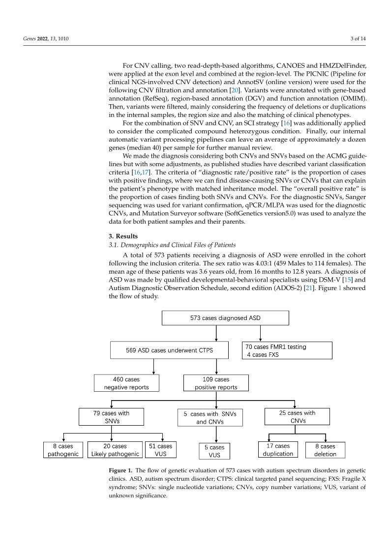
A total of 573 patients receiving a diagnosis of ASD were enrolled in the cohortfollowing the inclusion criteria. The sex ratio was 4.03:1 (459 Males to 114 females). Themean age of these patients was 3.6 years old, from 16 months to 12.8 years. A diagnosis ofASD was made by qualified developmental-behavioral specialists using DSM-V [15] andAutism Diagnostic Observation Schedule, second edition (ADOS-2) [21]. Figure 1 showedthe flow of study.
Genes 2022, 13, x FOR PEER REVIEW 4 of 17
Figure 1. The flow of genetic evaluation of 573 cases with autism spectrum disorders in genetic
clinics. ASD, autism spectrum disorder; CTPS: clinical targeted panel sequencing; FXS: Fragile X
syndrome; SNVs: single nucleotide variations; CNVs, copy number variations; VUS, variant of un-
known significance.
Figure 2. (A) The detection yield of SNV and CNV of CTPS in 569 patients. (B) The percentage of
SNVs diagnostic variations between males and females. SNVs, single nucleotide variations; CNVs,
copy number variations; P, pathogenic; LP, likely pathogenic; VUS, variant of unknown signifi-
cance.
Figure 1. The flow of genetic evaluation of 573 cases with autism spectrum disorders in geneticclinics. ASD, autism spectrum disorder; CTPS: clinical targeted panel sequencing; FXS: Fragile Xsyndrome; SNVs: single nucleotide variations; CNVs, copy number variations; VUS, variant ofunknown significance.

Genes 2022, 13, 1010 4 of 14
Seventy patients underwent extra FMR1 testing in addition to CTPS, who had promi-nent clinical features especially the facial characteristics such as long face, prominent ears,and prominent jaw, and 4 out of 70 (5.71%) obtained positive results and were lately diag-nosed with Fragile X syndrome. Analyzing SNVs and CNVs simultaneously, an overalldiagnostic yield of 19.16% (109/569) was reached (Tables 1 and 2). SNVs alone were de-tected in 13.89% of the cases (79/569), 25 patients with CNVs alone accounted for 4.40%(25/569) of the detection rate, and the remaining 0.88% (5/569) had both SNV and CNVfindings (Figure 2A).
Genes 2022, 13, x FOR PEER REVIEW 4 of 17
Figure 1. The flow of genetic evaluation of 573 cases with autism spectrum disorders in genetic
clinics. ASD, autism spectrum disorder; CTPS: clinical targeted panel sequencing; FXS: Fragile X
syndrome; SNVs: single nucleotide variations; CNVs, copy number variations; VUS, variant of un-
known significance.
Figure 2. (A) The detection yield of SNV and CNV of CTPS in 569 patients. (B) The percentage of
SNVs diagnostic variations between males and females. SNVs, single nucleotide variations; CNVs,
copy number variations; P, pathogenic; LP, likely pathogenic; VUS, variant of unknown signifi-
cance.
Figure 2. (A) The detection yield of SNV and CNV of CTPS in 569 patients. (B) The percentage ofSNVs diagnostic variations between males and females. SNVs, single nucleotide variations; CNVs,copy number variations; P, pathogenic; LP, likely pathogenic; VUS, variant of unknown significance.
For SNVs, 62 patients had missense mutations, 10 patients had frameshift mutations,1 patient had a nonframeshift mutation, 4 had splicing mutations, and 7 had more thanone kind of variant. We found that 5 patients had SHANK3 variants, which was thegreatest number of reportable variants, leading to the diagnostic yield of 0.88% (5/569).Four children had MYT1L variants. MECP2, DIP2B, DYRK1A, FOXP1, and PHIP variantswere found in 3 patients, respectively (Table 1). For CNVs, 18 were duplication variants(interestingly, we found that 6 patients had 15q11-13 duplications in 20% of CNV cases),and 12 were deletion variants (all were heterozygous deletions), which are shown in Table 2(for gene impacted and frequency of CNVs detailed in Table S1).
Of 109 patients who had molecular abnormalities, 84 were males and 25 were females.For diagnostic SNVs in males, a total of 67 cases were included, of which “pathogenic”SNVs accounted for 8.96% (6/67), “likely pathogenic” SNVs accounted for 22.39% (15/67),and the remaining 68.66% (46/67) were “variants of unknown significance (VUS)”. CNVswere found in 21 male patients (including 4 patients with SNVs), of which 8 patients werealso validated by CMA. In 25 females with genetic abnormalities, SNVs were found in17 females and CNVs could be found in 9 females (5 had CMA validation), with one havingboth CNV and SNV. Meanwhile, the percentage of pathogenic SNVs in females was 11.76%(2/17), “likely pathogenic” was 29.41% (5/17), and “VUS” was 58.82% (10/17). There wereno significant differences between the proportions of SNVs in males and females (Fisher’sexact p = 0.63) (Figure 2B). Thirty-four cases also conducted parental tests (both fatherand mother) by Sanger. Fourteen SNVs were de novo (41.18%, 14/34), 17 were inherited(50.00%, 17/34), and 3 had variants that were both de novo and inherited (8.82%, 3/34),leaving 50 unavailable to be tested.

Genes 2022, 13, 1010 5 of 14
Table 1. SNVs identified from ASD patients from CTPS.
Case Sex Classification Gene Position Mutation Zygosity Inherited/de Novo
InheritancePattern
PopulationFrequency
Pathogenic1 M P SHANK3 chr22:51159988 NM_033517:exon21:c.3727C>T(p.Q1243X) Het AD De novo NA2 M P TRIP12 chr2:230744756 NM_004238:exon2:c.40C>T(p.R14X) Het AD De novo NA3 M P NF1 chr17:29552152 NM_000267:exon17:c.1885G>A(p.G629R) Het AD De novo 3.98 × 10⁻⁶
4 M PRAB39B/NDST1/NDST1
chrX:154493531/chr5:149907553/chr5:149929268
NM_171998:exon1:c.43G>C(p.G15R)/NM_001543:exon3:c.701C>T(p.T234I)/
NM_001543:exon13:c.2345G>T(p.R782L)
Hemi/Het/Het
X-linked/AR/AR
NANA/NA/
4.24 × 10⁻⁵
5 M P TCF20 chr22:42608728 NM_005650:exon1:c.2582_2583del Het AD De novo NA
6 M P SLC26A4/SLC26A4
chr7:107330645/chr7:107350577
NM_000441:exon10:c.1226G>A(p.R409H)/NM_0004411:exon19:c.2168A>G(p.H723R)
Het/Het
AR/AR
Paternal/Maternal
9.57 × 10⁻⁵/1.13 × 10−4
7 F P SHANK3 chr22:51158733 NM_033517:exon21:c.2475dupC Het AD De novo NA8 F P MECP2 chrX:153296399 NM_004992:exon4:c.880C>T(p.R294X) Het XLD/XLR NA NA/
Likely pathogenic9 M LP BRAF chr7:140453146 NM_004333:exon15:c.1789C>G(p.L597V) Het AD NA NA
10 M LP DIP2B chr12:51128855 NM_173602:exon34:c.4044-1G>A Het AD NA NA
11 M LP KDM6B chr17:7752387_7752400 NM_001080424:exon11:c.2781_2794del14;p.(Val928Hisfs*2) Het AD NA NA
12 M LP SCN2A chr2:166201068 NM_021007:exon16:c.2566C>T(p.R856X) Het AD NA NA13 M LP STXBP1 chr9:130432185 NM_003165:exon11:c.913dupC Het AD/AR Maternal NA14 M LP CHD8 chr14:21871817 NM_001170629:exon16:c.3312delT Het AD De novo NA15 M LP DEAF1 chr11:687941 NM_021008:exon4:c.634G>A(p.G212S) Het AD/AR Maternal NA16 M LP AP1S2 chrX:15870644 NM_003916:exon2:c.4C>T(p.Q2X) Hemi XLR Maternal NA
17 M LP EHMT1/FGFR3
chr9:140678599/chr4:1807288
NM_001145527:exon16:c.2424G>A(p.W808X)/NM_000142:exon12:c.1537G>A(p.D513N)
Het/Het
AD/AD/AR
Maternal/Maternal
NA/4.61 × 10−5
18 M LP ABCD1 chrX:152991143 NM_000033:exon1:c.422C>T(p.A141V) Hemi XLR NA NA19 M LP MYT1L chr2:1890310 NM_015025:exon18:c.2711dupA Het AD De novo NA20 M LP DYRK1A chr21:38865398 NM_001396:exon7:c.1031_1037del Het AD NA NA21 M LP MYT1L chr2:1915808 NM_015025:exon12:c.1687A>G(p.R563G) Het AD De novo NA22 M LP BCL11A chr2:60689252 NM_022893:exon4:c.793_794delCT Het AD De novo NA23 M LP ARID2 chr12:46244889 NM_152641:exon15:c.2983C>T(p.Q995X) Het AD De novo NA24* F LP MECP2 chrX:153296399 NM_004992:exon4:c.880C>T(p.R294X) Het XLD/XLR De novo 1.64 × 10⁻⁵
25 F LP MYT1L chr2:1915838 NM_015025:exon12:c.1657T>C(p.C553R) Het AD De novo NA26 F LP MYT1L chr2:1891259 NM_015025:exon17:c.2636+1G>A Het AD De novo NA27 F LP SHANK3 chr22:51159684 NM_033517:exon21:c.3424_3425delCT Het AD NA NA28 F LP SLC2A1 chr1:43395422 NM_006516:exon6:c.709G>A(p.V237M) Het AD/AR NA 7.95 × 10⁻⁶
Variant of unknown significance (VUS)29 M VUS TRIO chr5:14508475 NM_007118:exon57:c.9238C>T(p.R3080X) Het AD Paternal NA30 M VUS TRIO chr5:14474205 NM_007118:exon40:c.6082G>A(p.D2028N) Het AD NA 3.98 × 10⁻⁶
31 M VUSDCLRE1C/DCLRE1C/
KANK1/KIF1A
chr10:14976777/chr10:14981850/
chr9:712617/chr2:241702608
NM_001033855:exon7:c.465-3C>T/NM_001033855:exon4:c.265A>G(p.T89A)/
NM_015158:exon3:c.1852del/NM_004321:exon19:c.1897G>A(p.D633N)
Het/Het/Het/Het
AR/AR/AD/
AD/ARNA
NA/2.85 × 10−5/
NA/NA
32 M VUS SCN8A chr12:52080904 NM_014191:exon5:c.515A>T(p.E172V) Het AD Maternal NA33 M VUS KIF7 chr15:90172648 NM_198525:exon17:c.3472_3474delAAG Hom AR NA 8.13 × 10⁻⁶
34 M VUS SMARCA4 chr19:11105513 NM_001128849:exon9:c.1429A>G(p.N477D) Het AD De novo NA35 M VUS MED13L chr12:116418709 NM_015335:exon23:c.5210A>G(p.K1737R) Het AD NA NA

Genes 2022, 13, 1010 6 of 14
Table 1. Cont.
Case Sex Classification Gene Position Mutation Zygosity Inherited/de Novo
InheritancePattern
PopulationFrequency
36 M VUSSKI/
WDR81/WDR81
chr1:2161179/chr17:1633670/chr17:1636940
NM_003036:exon1:c.969+5G>C/NM_001163809:exon2:c.3668-4G>A/
NM_001163809:exon7:c.4609G>A(p.G1537S)
Het/Het/Het
AD/AR/AR
De novo/Maternal/Paternal
NA/4.34 × 10−5/4.14 × 10−5
37 M VUS MAOA chrX:43552577 NM_000240:exon3:c.208G>A(p.V70M) Hemi XLR Maternal NA
38 M VUS NCAPD3/NCAPD3 chr11:134022951/chr11:134051019
NM_015261:exon35:c.4389-4C>G/NM_015261:exon20:c.2512A>G(p.I838V)
Het/Het
AR/AR
Paternal/Maternal
NA/1.59 × 10−5
39 M VUS IQSEC2 chrX:53285098 NM_001111125:exon3:c.883C>T(p.R295W) Hemi XLD NA NA40 M VUS DYRK1A chr21:38884272 NM_001396:exon11:c.1730T>A(p.V577D) Het AD NA NA
41 M VUS PIGV/PIGV
chr1:27121024/chr1:27121133
NM_017837:exon3:c.499G>A(p.G167S)/NM_017837:exon3:c.608G>T(p.R203L)
Het/Het
AR/AR NA 3.54 × 10⁻⁵/
NA42 M VUS FOXP2 chr7:114174736 NM_014491:exon3:c.233G>C(p.S78T) Het AD NA NA43 M VUS HCFC1 chrX:153219744 NM_005334:exon17:c.4106T>C(p.M1369T) Hemi XLR Maternal NA44 M VUS DYRK1A chr21:38850489 NM_001396:exon3:c.214C>G(p.P72A) Het AD NA NA45 M VUS PTEN chr10:89717672 NM_000314:exon7:c.697C>T(p.R233X) Het AD/AR NA NA46 M VUS SLITRK1 chr13:84455351 NM_052910:exon1:c.292G>A(p.V98I) Het AD NA NA
47 M VUS PLA2G6/PLA2G6
chr22:38516880/chr22:38528924
NM_003560:exon12:c.1628G>A(p.R543H)/NM_003560:exon7:c.991G>T(p.D331Y)
Het/Het
AR/AR NA 2.83 × 10⁻⁵/
3.57 × 10⁻⁵
48 M VUS PHIP chr6:79727249 NM_017934:exon11:c.1046T>A(p.F349Y) Het AD Paternal NA
49 M VUSCTNNB1/HCFC1/
SOS1
chr3:41266085/chrX:153217162/
chr2:39216457
NM_001904:exon3:c.82C>G(p.Q28E)/NM_005334:exon21:c.5261-4C>T/NM_005633:exon21:c.3347-2A>G
Het/Hemi/
Het
AD/XLR/AD
NANA/
1.85 × 10⁻⁵/NA
50 M VUS GRIA3 chrX:122318409 NM_000828:exon1:c.22G>A(p.G8R) Hemi XLR NA NA51 M VUS COL4A3BP chr5:74712810 NM_001130105:exon8:c.1112G>A(p.G371E) Het AD NA NA52 M VUS DPP6 chr7:154585802 NM_001936:exon11:c.964A>G(P.T322A) Het AD NA NA
53 M VUS PHIP/ACVR1
chr6:79672916/chr2:158626989
NM_017934:exon30:c.3433A>G(p.R1145G)/NM_001105:exon7:c.681G>A(p.W227X)
Het/Het
AD/AD NA NA/
NA54 M VUS SETBP1 chr18:42531731 NM_015559:exon4:c.2426A>G(p.Q809R) Het AD Paternal NA55 M VUS FOXP1 chr3:71015071 NM_032682:exon20:c.1859G>A(p.S620N) Het AD NA NA56 M VUS PHIP chr6:79665392 NM_017934:exon33:c.3790A>G(p.T1264A) Het AD Paternal NA
57 M VUS DDX3X/DLG3
chrX:41196685/chrX:69719742
NM_001193416:exon2:c.70T>G(p.S24A)/NM_021120:exon16:c.1988G>A(p.R663Q)
Hemi/Hemi
XL/XL
Maternal/Maternal
NA/NA
58 M VUS GNAI3/USP27X
chr1:110121866/chrX:49645815
NM_006496:exon4:c.344A>G(p.E115G)/NM_001145073:exon1:c.905T>C(p.L302S)
Het/Hemi
AD/XL NA NA/
NA59 M VUS TMLHE chrX:154743783 NM_018196:exon4:c.502C>T(p.Q168X) Hemi XLR NA NA60 M VUS BRWD3 chrX:80064545 NM_153252:exon3:c.91-4T>C Hemi XLR NA NA61 M VUS USP9X chrX:41029747 NM_001039590:exon20:c.2902A>C(p.I968L) Hemi dominant/XLR Maternal NA62 M VUS FOXP1 chr3:71037180 NM_032682:exon14:c.1111G>A(p.V371M) Het AD NA NA63 M VUS DIP2B chr12:51074491 NM_173602:exon9:c.1151C>T(p.T384I) Het AD NA 2.12 × 10⁻⁵
64 M VUS L1CAM chrX:153133875 NM_000425:exon13:c.1585G>A(p.E529K) Hemi XLR NA NA65 M VUS SHANK3 chr22:51169394 NM_033517:exon22:c.4850C>T(p.P1617L) Het AD NA NA66 M VUS DIP2B chr12:51068356 NM_173602:exon6:c.740T>C(p.I247T) Het AD NA 3.18 × 10⁻⁵
67 M VUS GRIA3 chrX:122551611 NM_000828:exon11:c.1859G>C(p.G620A) Hemi XLR NA NA
68 M VUS AFF2/TRIO
chrX:147743835/chr5:14387875
NM_002025:exon3:c.587T>C(p.F196S)/NM_007118:exon23:c.3800G>A(p.S1267N)
Hemi/Het XLR/AD NA NA/
3.98 × 10⁻⁶
69 M VUS FOXP2 chr7:114303569 NM_014491:exon15:c.1834T>A(p.L612M) Het AD NA NA
70 M VUS ARID1B/CHD7
chr6:157521844/chr8:61654295
NM_020732:exon18:c.4116C>A(p.Y1372X)/NM_017780:exon2:c.304C>T(p.H102Y)
Het/Het
AD/AD
De novo/Paternal
NA/NA

Genes 2022, 13, 1010 7 of 14
Table 1. Cont.
Case Sex Classification Gene Position Mutation Zygosity Inherited/de Novo
InheritancePattern
PopulationFrequency
71 * M VUS CTNNB1/KLHL15
chr3:41279547/chrX:24006703
NM_001904:exon14:c.2117C>A(p.P706H)/NM_030624:exon4:c.1150G>A(p.V384I)
Het/Hemi
AD/XLR NA NA/
5.48 × 10⁻⁶
72 * M VUS RPS6KA3 chrX:20284690 NM_004586:exon1:c.61A>G(p.S21G) Hemi XLD NA NA
73 * M VUS KIF1A/ZC4H2
chr2:241700653/chrX:64137775
NM_004321:exon22:c.2231A>G(p.K744R)/NM_018684:exon5:c.563C>T(p.A188V)
Het/Hemi
AD/AR/XLR
De novo/Maternal
NA/NA
74 * M VUS FBN2/SPTAN1
chr5:127625581/chr9:131389713
NM_001999:exon51:c.6503delC/NM_001130483:exon50:c.6625G>A(p.D2209N)
Het/Het
AD/AD NA NA/
6.34 × 10⁻⁶
75 F VUS SHANK3 chr22:51142293 NM_033517:exon13:c.1618C>T(p.R540W) Het AD NA NA
76 F VUS GPR98/GPR98
chr5:89954046/chr5:90074281
NM_032119:exon21:c.4703G>A(p.S1568N)/NM_032119:exon63:c.12704A>G(p.Y4235C)
Het/Het
AD/AR/Digenic/AD/AR/Digenic NA 5.46 × 10⁻⁵/
1.61 × 10⁻⁴
77 F VUS PPP2R5D chr6:42974971 NM_006245:exon5:c.560C>T(p.S187L) Het AD NA NA78 F VUS MECP2 chrX:153296071 NM_004992:exon4:c.1158_1201del Het XLD/XLR NA NA79 F VUS SETBP1 chr18:42529856 NM_015559:exon4:c.551G>T(p.R184M) Het AD NA NA80 F VUS SETD5 chr3:9512347 NM_001080517:exon19:c.2929T>A(p.F977I) Het AD Paternal NA
81 F VUSHUWE1/
LRP2/LRP2
chrX:53574690/chr2:170030607/chr2:170081950
NM_031407:exon68:c.10580T>C(p.V3527A)/NM_004525:exon56:c.10836G>T(p.Q3612H)/
NM_004525:exon33:c.5406_5407del
Het/Het/Het
XL/AR/AR
NANA/NANA
82 F VUSERCC2/ERCC2/ASXL3
chr19:45868096-45868099/
chr19:45856520/chr18:31322948
NM_000400:exon7:c.591_594del/NM_000400:exon18:c.1738G>A(p.A508T)/NM_030632:exon12:c.3136G>A(p.G1046R)
Het/Het/Het
AR/AR/AD
Paternal/Maternal/Maternal
1.20 × 10⁻⁵/1.59 × 10−5/1.61 × 10−5
83 F VUS FOXP1 chr3:71247424 NM_032682:exon6:c.109T>C(p.S37P) Het AD NA NA84 * F VUS DCX chrX:110574270 NM_178153:exon5:c.809-1G>C Het XL NA NA
SNV, single nucleotide variations; M, male; F, female; Hom, homozygous; Het, heterozygous; Hemi, hemizygous; AD, autosomal dominant; AR, autosomal recessive; XL, X-linked; XLD,X-linked dominant; XLR, X-linked recessive; P, pathogenic; LP, likely pathogenic; VUS, variant of unknown significance. *, patients with dual SNV and CNV.

Genes 2022, 13, 1010 8 of 14
Table 2. CNVs identified from ASD patients from CTPS.
Case Sex Band Chr Start(hg19) Stop(hg19) Size(kb) Deletion/Duplication
1 * M 15q13.2–15q13.3 chr15 30,653,442 32,464,722 1,811,280 deletion2 * M 20p12.1–20p13 chr20 740,723 13,799,067 13,058,344 duplication3 * M 16p11.2 chr16 29,802,039 30,200,397 398,358 deletion4 * M Xq28 chrX 153,576,898 153,780,404 203,506 duplication5 * F 15q11.2–15q13.1 chr15 23,043,276 28,327,041 5,283,765 duplication6 M 7p13–7p14.1 chr7 41,724,711 44,748,665 3,023,954 duplication7 M 15q13.3 chr15 32,064,983 32,443,563 378,580 duplication8 M 15q11.2–15q13.1 chr15 23,043,276 28,327,041 5,283,765 duplication9 M 2q24.3–2q25.1 chr2 9,628,275 16,087,129 6,458,854 duplication
10 M 15q11.2–15q13.1 chr15 23,043,276 28,327,041 5,283,765 duplication11 M 3q29 chr3 196,195,653 197,024,106 828,453 deletion12 M 1p21.2–1p21.3 chr1 97,543,298 100,715,390 3,172,092 duplication13 M 15q13.2–15q13.3 chr15 30,659,620 32,464,722 1,805,102 duplication14 M 7q36.1–7q36.3 chr7 150,642,048 157,210,133 6,568,085 deletion15 M Xp21.1 chrX 32,235,032 32,235,180 148 deletion16 M 19p13.2–q13.3 chr19 43,370,615 43,530,621 160,006 deletion17 M 10q22.3–10q23.2 chr10 81,697,495 88,854,623 7,157,128 duplication18 M 4q35.1–q 35.2 chr4 186,421,813 190,873,442 4,451,629 deletion19 M 6q16.1–6q16.3 chr6 97,337,188 105,307,794 7,970,606 deletion20 M 15q11.2–15q13.1 chr15 23,043,276 28,327,041 5,283,765 duplication21 M 3q29 chr3 195,776,154 197,300,194 1,524,040 deletion22 M 1p34.3 chr1 36,974,539 38,129,928 1,155,389 duplication23 F 22q13.31–22q13.33 chr22 45,680,862 51,171,726 5,490,864 deletion24 F 14q21.1 chr14 39,559,493 39,665,452 105,959 duplication25 F 2q37.3 chr2 240,016,194 242,708,226 2,692,032 deletion26 F 17p11.2 chr17 16,664,738 20,370,848 3,706,110 duplication27 F 10q22.3–q23.2 chr10 81,697,495 88,854,623 7,157,128 duplication28 F 22q11.21 chr22 18,900,293 21,245,506 2,345,213 duplication29 F 2q37.12q37.3 chr2 234,408,524 242,844,702 8,436,178 deletion30 F p21.1 chrX 32,305,645 32,632,570 326,925 duplication
*: patients with dual SNV and CNV (case 1, 2, 3, 4, and 5 were case 71, 72, 73, 74, and 84 in Table 1); CNV, copynumber variations; Chr, chromosome; M, male; F, female.
3.2. Representative Cases from CTPS3.2.1. Summarized Cases: Analysis of Clinical Characteristics Classify Genetic Problems
Among ASD patients who had identified SNVs, SHANK3 was the most common vari-ant (n = 5). The second on the list was MYT1L, a myelin transcription factor, a transcriptionfactor enabling fibroblast-to-neuron conversions. The inheritance of Cases 19/21/25/26was de novo (Figure 3A). Tracing the developmental milestones, they all had obviousdelayed motor developments. The independent walking for 4 children ranged from 20 to30 months. Additionally, all children had significant language delay, most of them couldspeak less than 5 words even though they were over 3 years old. Abnormal sensory pro-cessing, such as biting, touching, or smelling objects was common in these children. Threepatients (Case 19/21/25) were overweight, and ptyalism still existed owing to hypotonia.Case 21 also had strabismus. It was specifically observed that Cases 21/25/26 with MYT1Lvariants had apparent stereotypic hand movements. Analysis of characteristics in thesepatients can better screen and classify them in the clinic.
3.2.2. Renewed Case: Genetic Diagnosis Should Also Focus on Clinical Manifestations
Case 6 was a boy with compound heterozygous variants of SLC26A4 (Figure 3B). Thepatient failed postnatal hearing screening and was inaudible to high-pitched sounds. Hewas diagnosed with bilateral profound sensorineural hearing loss and enlarged vestibu-lar aqueduct syndrome soon afterwards. Right cochlear implants were performed whenthe child was 1 year old. He has been undertaking language training ever since. Thepatient was referred to our clinic at 4 years old because he behaved abnormally and undis-

Genes 2022, 13, 1010 9 of 14
ciplined in kindergarten. His mother reported her child had a short attention span andhyperactivity. The Wechsler Preschool and Primary Scale of Intelligence (WPPSI) [22]showed that the child had developmental delay both in verbal and nonverbal intelligence(his IQS of Verbal Scale was 48, and IQS of Performance Scales was 62, producing a FullScale IQ of 50). Compound heterozygous variants of SLC26A4 (c.1226G>A (p. R409H),c.2168A>G (p. H723R)) were detected in this patient by CTPS. The parents were val-idated by Sanger sequence. The results showed that his missense variants were bothinherited by his parents (paternal: c.1226G>A (p. R409H) in exon 10, maternal: c.2168A>G(p. H723R) in exon 19). The patient had poor social communication and was suspectedautistic, however, no reports have shown the correlations between the SLC26A4 gene andautism. Neither did he had interventions related to ASD, nor regularly followed up atthe clinic. Atomoxetine hydrochloride was taken irregularly until school age. He had are-examination at 7 years old. Maintaining Back-and-forth conversation was hard for him.He displayed poor social reciprocity and exhibited repetitive patterns of behavior (runningback and forth repeatedly and watching traffic lights consistently) and sensory perceptionproblems such as counting numbers and biting or smelling objects. His ADOS score wasabove the cutoff (social affect 5, repetitive score 3, severity 4). He was eventually diagnosedwith ASD. The WES of the core family and CMA also performed according to his parents’requirements but did not reveal any other causative variants.
Genes 2022, 13, x FOR PEER REVIEW 11 of 17
were inherited (50.00%, 17/34), and 3 had variants that were both de novo and inherited
(8.82%, 3/34), leaving 50 unavailable to be tested.
3.2. Representative Cases from CTPS
3.2.1. Summarized Cases: Analysis of Clinical Characteristics Classify Genetic Problems
Among ASD patients who had identified SNVs, SHANK3 was the most common var-
iant (n = 5). The second on the list was MYT1L, a myelin transcription factor, a transcrip-
tion factor enabling fibroblast-to-neuron conversions. The inheritance of Cases 19/21/25/26
was de novo (Figure 3A). Tracing the developmental milestones, they all had obvious de-
layed motor developments. The independent walking for 4 children ranged from 20 to 30
months. Additionally, all children had significant language delay, most of them could
speak less than 5 words even though they were over 3 years old. Abnormal sensory pro-
cessing, such as biting, touching, or smelling objects was common in these children. Three
patients (Case 19/21/25) were overweight, and ptyalism still existed owing to hypotonia.
Case 21 also had strabismus. It was specifically observed that Cases 21/25/26 with MYT1L
variants had apparent stereotypic hand movements. Analysis of characteristics in these
patients can better screen and classify them in the clinic.
Figure 3. (A) The locations of variants of patients in the study. Representation based on Protein
Paint (Wisteria color: zf-C2HC domain, Turquoise color: MYT1 domain, Orange color: Smc domain)
(https://proteinpaint.stjude.org/, accessed on 1 March 2022). The cases correspond with Table 1. (B)
(1) The dysmorphic features of case 6 (shown for 1). (2) The Pedigree of family 1. 2 was the father of
the patient; 3 was the mother of the patient. The black arrow showed the patient of case 6. (3)
SLC26A4 sequence results of patient and his parents. Heterozygous variants of SLC26A4 were iden-
tified in the proband (red arrows in patient 1). Both his parents were in a heterozygous state for the
variant (paternal: red arrow of 2 for c.1226G>A (p. R409H) in exon 10, maternal: red arrow of 3 for
c.2168A>G(p. H723R) in exon 19).
3.2.2. Renewed Case: Genetic Diagnosis Should Also Focus on Clinical Manifestations
Case 6 was a boy with compound heterozygous variants of SLC26A4 (Figure 3B). The
patient failed postnatal hearing screening and was inaudible to high-pitched sounds. He
was diagnosed with bilateral profound sensorineural hearing loss and enlarged vestibular
aqueduct syndrome soon afterwards. Right cochlear implants were performed when the
child was 1 year old. He has been undertaking language training ever since. The patient
was referred to our clinic at 4 years old because he behaved abnormally and undisciplined
in kindergarten. His mother reported her child had a short attention span and hyperac-
tivity. The Wechsler Preschool and Primary Scale of Intelligence (WPPSI) [22] showed that
Figure 3. (A) The locations of variants of patients in the study. Representation based on ProteinPaint (Wisteria color: zf-C2HC domain, Turquoise color: MYT1 domain, Orange color: Smc domain)(https://proteinpaint.stjude.org/, accessed on 1 March 2022). The cases correspond with Table 1.(B) (1) The dysmorphic features of case 6 (shown for 1). (2) The Pedigree of family 1. 2 was thefather of the patient; 3 was the mother of the patient. The black arrow showed the patient of case 6.(3) SLC26A4 sequence results of patient and his parents. Heterozygous variants of SLC26A4 wereidentified in the proband (red arrows in patient 1). Both his parents were in a heterozygous state forthe variant (paternal: red arrow of 2 for c.1226G>A (p. R409H) in exon 10, maternal: red arrow of 3for c.2168A>G(p. H723R) in exon 19).
4. Discussion4.1. Clinical Benefits and Limits of CTPS
Genetics have a large contribution to ASD. Identifying a genetic etiology improvingaccuracy of counseling for patients and their families. Information about prognosis andrecurrence risk is the most important benefit of genetic testing. The benefits also includepreventing co-occurring medical conditions and avoiding unnecessary tests and harmful

Genes 2022, 13, 1010 10 of 14
treatments [8]. It is necessary to advise patients and their families to undergo geneticevaluation.
Both rare inherited and de novo variations are relevant to ASD during early neurode-velopment. Kim et al. [23] provided evidence that rare inherited variations have a functionalrelationship with ASD in the developing brain. Sanders et al. [24] detected 2591 familiesfrom SSC and revealed that de novo variants were strongly associated with ASD, leadingto a total of 6 risk loci (1q21.1, 3q29, 7q11.23, 16p11.2, 15q11.2-13, 22q11.2), and 65 ASD riskgenes (additional 2 loci: NRXN1 (2p16.3), SHANK3(22q13.3) were included in the list ofrisk genes). The CNVs detected in our study overlapped with their observation in 3q29,16p11.2, 15q11.2-13, 22q11.2, and 22q13.3. These CNVs had a high risk of developmentaldelay [25]. Satterstrom et al.‘s work [26] undertook the largest exome sequencing, identified102 ASD risk genes, most of which had effects on the regulation of gene expression orneuronal communication. Of these genes, 19 of them overlapped with our study (ARID1B,ASXL3, BCL11A, CHD8, CTNNB1, DEAF1, FOXP1, FOXP2, KDM6B, MED13L, PPPP2R5D,PTEN, SCN2A, SETD5, SHANK2, SHANK3, SKI, STXBP1, TCF20), whereas they had notanalyzed de novo mutations on chromosome X. For high-confidence risk genes reported inChoi’s study [27], we had 16 genes overlapped (ASXL3, BCL11A, CHD8, CTNNB1, DDX3X,DEAF1, FOXP1, FOXP2, KDM6B, MYT1L, PTEN, SCN2A, SETD5, SHANK3, SKI, TCF20).It’s worth noting that the contribution of de novo non-coding variants may not be as highas that of coding regions [28].
For now, CMA and Fragile X testing are recommended as the first tier for ASDpatients. CMA tests abnormalities of chromosomal structure and duplications or deletionsin chromosomal regions. The diagnostic yield of CNV is 5.4–14% (median 9%) in ASDpatients [29,30]. When CMA does not find an etiology, the next step recommended foretiologic evaluation of ASD is WES. WES identified SNVs that have been confirmed asASD risk genes. The reported diagnostic yield is 8–25.8% [29,31]. This process takes muchexpenditure and is a waste of time. CTPS, comprising 2742 genes, is able to analyze SNVsand CNVs simultaneously to provide an etiological diagnosis for children and patients.The potential of CTPS is that it can discover variants effectively and costs less. Reducingthe cost of time and price is much needed in developing countries, especially in China.Families will easily accept and be willing to undergo genetic testing and finally benefitfrom it. As research progresses, genetic testing may contribute to identifying effectiveinterventions related to specific etiologies.
Based on the results of CTPS in this study, we had an overall diagnostic yield of19.16% for 569 ASD patients, including both SNVs and CNVs. A meta-analysis in 2020identified 14 ASD studies across 1530 patients using targeted gene panel sequencing orWES, and the diagnostic yield was 17.1% (95% CI, 11–25%) [32]. Rossi et al. [31] recruited163 ASD/autistic patients, all of whom had additional clinical features such as intellectualdisabilities (ID)/developmental delays (DD) (92.6%) and epilepsy/seizures (38.7%). Theyfound that the diagnostic rate of their ASD cohort was 25.8% using WES (42 of 163).Aspromonte et al. [33] developed a next-generation sequencing gene panel of 74 selectedgenes. They analyzed 150 individuals with ID and/or ASD, and a confident diagnosis wasreached in 41 patients (27%). In our study, the diagnostic yield was slightly lower thanreported previously. The possible reason is that it was a single-center analysis, which mayhave selection bias in the sample. The children were referred to our department with fewercomorbidities, such as epilepsy or multiple other malformations. CTPS detected selectedgenes, not including the whole exome/genome. Additionally, a small number of CNVswere not covered in the panel [16]. The CTPS was designed for children with syndrome anddevelopmental abnormalities, not a targeted panel specialized for ASD children. However,it had comprehensive candidate genes other than ASD genes to avoid missing suspecteddisease-causing genes. The restriction could explain the lower rate of our study.
Clinicians should focus more on the clinical features of children and find more valuableclues to the cause of the disease. CTPS offers a better choice for patients and clinicians asan effective molecular diagnostic tool.

Genes 2022, 13, 1010 11 of 14
4.2. Implications for Representative Cases from CTPS
Myelin transcription factor 1-like (MYT1L), mapping to a region of human chromo-some 2, encodes the MYT1L protein, which contains 6 zinc fingers (an N-terminal zincfinger, 2 tandem central zinc fingers, and 3 C-terminal zinc fingers) [34]. The MYT1L geneis co-expressed with other ASD-related genes, including NRXN1, TCF4, and BCL11A in theprefrontal cortex during mid-fetal development, higher in prenatal expression [35], andtranscription may persist in the brains of children. It plays a crucial role in neurogenesis,helps neural stem cells transform into neurons, and is expressed in oligodendrocyte linagecells during myelination and remyelination [36]. We found 4 children with de novo MYT1Lvariants. Coursimault et al. [37] reported 40 patients with MYT1L-associated neurodevelop-mental disorder and reviewed 22 patients in published data. In their reports, many patientswith MYT1L variants had language delay (95%), ID (70%), ASD (43%), motor delay (78%),and hypotonia (47%). Consistent with the previous literature, all of our 4 patients with ASDhad developmental delays, overweight/obesity were common in these patients. Table 3shows the clinical symptoms of our patients and public literature. All of our patients hadabnormal sensory processing, whereas few studies mentioned it. Notably, 3 of our patientshad stereotypic hand movements, which were not detailed in Juliette et al.’s work. DeRocker et al. [38] showed 3 patients with rigid hand movements, which was also manifestedin our patients. These children had special hand phenotypes (typical repetitive, purposelesshand movements such as rubbing, tapping, wringing, or clapping) without rapid regressionof acquired skills and stagnation. Therefore, a detailed history is particularly important.Our patients had obvious sensory abnormalities and stereotypic hand movements, whichmay contribute to the phenotype of MYT1L.
Table 3. Clinical symptoms of MYT1L patients and published literature.
AutismSpectrumDisorder
LanguageDelay
MotorDelay
DevelopmentalDisorder/
IntellectualDisability
StereotypicHand
Movements
AbnormalSensory
ProcessingHypotonia Overweight/
Obesity
Our study 100% (4/4) 100% (4/4) 100% (4/4) 100% (4/4) 75% (3/4) 100% (4/4) 75% (3/4) 75% (3/4)Coursimaultet al.‘s [ [37] 43% (17/40) 95% (38/40) 78% (31/40) 70% (21/30) - - 47% (18/38) 58% (23/40)De Rockeret al.’s [38] 32% (7/22) 100% (22/22) - 100% (22/22) 14% (3/22) - - 74% (14/19)
Windheuseret al.’s [39] 22% (2/9) - 87% (7/8) 100% (8/8) - Mentioned in
1 patient 78% (7/9) 33% (3/9)Blanchet
et al.’s [40] 44% (4/9) 100% (9/9) 100% (8/8) - - - Mentioned in2 patients 66.7% (6/9)
Carvalhoet al.’s [41] 0 100% (1/1) - 100% (1/1) - - - 100% (1/1)Loid et al.’s
[42] 0 100% (1/1) 100% (1/1) 100% (1/1) - - - 100% (1/1)Al Tuwaijriet al.’s [43] 100% (1/1) 100% (1/1) 100% (1/1) 100% (1/1) - - 100% (1/1) 100% (1/1)
Solute carrier family 26 member 4 (SLC26A4), encoding pendrin (an anion trans-porter) [44], causes Pendred syndrome and an enlarged vestibular aqueduct. It is expressedmostly in the thyroid and is also expressed at some levels in the prostate, kidney, urinarybladder, and brain [45]. Pedred Syndrome is a common disorder with hereditary hearingloss, abnormal development of the cochlea, and diffuse thyroid enlargement. The patientin our study had SLC26A4 heterozygous variants. Due to the lack of literature connectingSLC26A4 with autism, CMA and WES were performed. The patient had typical autisticfeatures but found no cause other than SLC26A4 variants. He had a complex phenotypebeyond Pendred syndrome presentation, hence, our study reported an ASD child withSLC26A4 variants. It is hard for us to explain his sensory perception abnormalities andrepetitive behaviors with hearing problems. SLC26A4 variants may cause his autistic pheno-type, while early social competence may be overlooked by deafness. Nevertheless, furtherstudies and case reports are required to show the relationships between the SLC26A4gene and phenotype with ASD. After all, some of the variants of uncertain significancemay be determined as pathogenic in the future [8]. It is hoped that more animal research

Genes 2022, 13, 1010 12 of 14
will be conducted to examine the effect of scl26a4 on autism and on the gene-phenotypeassociations in neurodevelopment.
5. Conclusions
In summary, CTPS is an effective tool for genetic testing of ASD patients, a valuablestrategy suitable for patients with neurodevelopmental disorders in China. It is importantto identify the key clinical features of patients diagnosed with ASD that allow more accurategenetic diagnosis and harbor relevant genetic variants. It is expected that genetic researchwill allow the development of better treatments and interventions for ASD children withinthe next decade, thus guiding further family planning and social support.
Supplementary Materials: The following supporting information can be downloaded at: https://www.mdpi.com/article/10.3390/genes13061010/s1, Table S1: title Detailed CNVs of ASD patientsfrom CTPS.
Author Contributions: Q.X. and C.H. conceived and designed the study; L.H. and G.Z. carried outsample collections, C.H., Y.D., H.L., K.Z., D.L., B.W., X.X., and Q.X. identified patients; C.H. wrotethe manuscript, Q.X. and X.X. modified the manuscript. All authors have read and agreed to thepublished version of the manuscript.
Funding: This research was funded in part by the National Natural Science Foundation of China(NSFC, No. 82171540), the Key Subject Construction Project of Shanghai Municipal Health Commis-sion (No. shslczdzk02903) and the Young Clinical Scientist Program of Children’s Hospital of Fudanuniversity (No. 2022LCKXJ03).
Institutional Review Board Statement: The study was conducted according to the guidelines of theDeclaration of Helsinki and approved by the Ethics Committee of the Children’s Hospital of FudanUniversity (Approval number: 2016-77, approval date: 25 February 2016).
Informed Consent Statement: Written informed consent was obtained from patients or parentsgiving consent on behalf of their minor children.
Data Availability Statement: The data presented in this study are available on request from thecorresponding author. The confidential patient data will not be shared.
Acknowledgments: We thank our colleagues at the Child Health Care of Children’s Hospital ofFudan University for their help. We thank Xia Wang from Ailife Diagnostics for his comments andXinran Dong from Clinical Genetic Center, Children’s Hospital of Fudan University for her help onthe writing of the Material and Methods Section.
Conflicts of Interest: The authors declare no conflict of interest.
References1. Lord, C.; Elsabbagh, M.; Baird, G.; Veenstra-Vanderweele, J. Autism Spectrum Disorder. Lancet 2018, 392, 508–520. [CrossRef]2. Brugha, T.S.; Spiers, N.; Bankart, J.; Cooper, S.A.; McManus, S.; Scott, F.J.; Smith, J.; Tyrer, F. Epidemiology of Autism in Adults
across Age Groups and Ability Levels. Br. J. Psychiatry 2016, 209, 498–503. [CrossRef]3. Lord, C.; Brugha, T.S.; Charman, T.; Cusack, J.; Dumas, G.; Frazier, T.; Jones, E.J.H.; Jones, R.M.; Pickles, A.; State, M.W.; et al.
Autism Spectrum Disorder. Nat. Rev. Dis. Primers 2020, 6, 5. [CrossRef] [PubMed]4. Tick, B.; Bolton, P.; Happe, F.; Rutter, M.; Rijsdijk, F. Heritability of Autism Spectrum Disorders: A Meta-Analysis of Twin Studies.
J. Child Psychol. Psychiatry 2016, 57, 585–595. [CrossRef] [PubMed]5. Woodbury-Smith, M.; Scherer, S.W. Progress in the Genetics of Autism Spectrum Disorder. Dev. Med. Child Neurol. 2018, 60,
445–451. [CrossRef] [PubMed]6. Sandin, S.; Lichtenstein, P.; Kuja-Halkola, R.; Larsson, H.; Hultman, C.M.; Reichenberg, A. The Familial Risk of Autism. JAMA
2014, 311, 1770–1777. [CrossRef]7. He, X.; Sanders, S.J.; Liu, L.; de Rubeis, S.; Lim, E.T.; Sutcliffe, J.S.; Schellenberg, G.D.; Gibbs, R.A.; Daly, M.J.; Buxbaum, J.D.; et al.
Integrated Model of De Novo and Inherited Genetic Variants Yields Greater Power to Identify Risk Genes. PLoS Genet. 2013, 9,e1003671. [CrossRef] [PubMed]
8. Hyman, S.L.; Levy, S.E.; Myers, S.M.; Section On Developmental Council On Children With Disabilities, and Pediatrics Behavioral.Identification, Evaluation, and Management of Children with Autism Spectrum Disorder. Pediatrics 2020, 145. [CrossRef]

Genes 2022, 13, 1010 13 of 14
9. Volkmar, F.; Siegel, M.; Woodbury-Smith, M.; King, B.; McCracken, J.; State, M. Child American Academy of, and IssuesAdolescent Psychiatry Committee on Quality. Practice Parameter for the Assessment and Treatment of Children and Adolescentswith Autism Spectrum Disorder. J. Am. Acad. Child Adolesc. Psychiatry 2014, 53, 237–257. [CrossRef]
10. Schaefer, G.B.; Mendelsohn, N.J. Professional Practice and Guidelines Committee. Clinical Genetics Evaluation in Identifying theEtiology of Autism Spectrum Disorders: 2013 Guideline Revisions. Genet. Med. 2013, 15, 399–407. [CrossRef]
11. O’Roak, B.J.; Vives, L.; Fu, W.; Egertson, J.D.; Stanaway, I.B.; Phelps, I.G.; Carvill, G.; Kumar, A.; Lee, C.; Ankenman, K.; et al.Multiplex Targeted Sequencing Identifies Recurrently Mutated Genes in Autism Spectrum Disorders. Science 2012, 338, 1619–1622.[CrossRef] [PubMed]
12. Srivastava, S.; Love-Nichols, J.A.; Dies, K.A.; Ledbetter, D.H.; Martin, C.L.; Chung, W.K.; Firth, H.V.; Frazier, T.; Hansen, R.L.;Prock, L.; et al. Correction: Meta-Analysis and Multidisciplinary Consensus Statement: Exome Sequencing Is a First-Tier ClinicalDiagnostic Test for Individuals with Neurodevelopmental Disorders. Genet. Med. 2020, 22, 1731–1732. [CrossRef] [PubMed]
13. Lowther, C.; Valkanas, E.; Giordano, J.L.; Wang, H.Z.; Currall, B.B.; O’Keefe, K.; Collins, R.L.; Zhao, X.; Austin-Tse, C.A.;Evangelista, E.; et al. Systematic Evaluation of Genome Sequencing as a First-Tier Diagnostic Test for Prenatal and PediatricDisorders. bioRxiv 2020. [CrossRef]
14. Coury, D. Medical Treatment of Autism Spectrum Disorders. Curr. Opin. Neurol. 2010, 23, 131–136. [CrossRef] [PubMed]15. APAd American Psychiatric Association. Diagnostic and Statistical Manual of Mental Disorders, 5th ed.; American Psychiatric
Association: Washington, DC, USA, 2013.16. Dong, X.; Liu, B.; Yang, L.; Wang, H.; Wu, B.; Liu, R.; Chen, H.; Chen, X.; Yu, S.; Chen, B.; et al. Clinical Exome Sequencing as the
First-Tier Test for Diagnosing Developmental Disorders Covering Both Cnv and Snv: A Chinese Cohort. J. Med. Genet. 2020, 57,558–566. [CrossRef]
17. Yang, L.; Kong, Y.; Dong, X.; Hu, L.; Lin, Y.; Chen, X.; Ni, Q.; Lu, Y.; Wu, B.; Wang, H.; et al. Clinical and Genetic Spectrum of aLarge Cohort of Children with Epilepsy in China. Genet. Med. 2019, 21, 564–571. [CrossRef]
18. McLaren, W.; Pritchard, B.; Rios, D.; Chen, Y.; Flicek, P.; Cunningham, F. Deriving the Consequences of Genomic Variants with theEnsembl Api and Snp Effect Predictor. Bioinformatics 2010, 26, 2069–2070. [CrossRef]
19. Wang, K.; Li, M.; Hakonarson, H. Annovar: Functional Annotation of Genetic Variants from High-Throughput Sequencing Data.Nucleic Acids Res. 2010, 38, e164. [CrossRef]
20. Qian, Q.I.N.; Bo, L.I.U.; Lin, Y.A.N.G.; Bing-bing, W.U.; Hui-jun, W.A.N.G.; Xin-Ran, D.O.N.G.; Yu-lan, L.U.; Wen-Hao, Z.H.O.U.Application of Copy Number Variation Screening Analysis Process Based on High Throughput Sequencing Technology. Chin. J.Evid. Based Pediatrics 2018, 13, 275–279.
21. Lord, C.M.; Rutter, P.D.; Labore, S.; Risi, K.; Gotham, B.S. Autism Diagnostic Observation Schedule, (Ados-2), 2nd ed.; WesternPschological Services: Los Angeles, CA, USA, 2012.
22. David, W. Wechsler Preschool and Primary Scale of Intelligence, 4th ed.; Pearson: London, UK, 2012.23. Kim, Y.; An, J.Y. Spatio-Temporal Roles of Asd-Associated Variants in Human Brain Development. Genes 2020, 11, 535. [CrossRef]24. Sanders, S.J.; He, X.; Willsey, A.J.; Ercan-Sencicek, A.G.; Samocha, K.E.; Cicek, A.E.; Murtha, M.T.; Bal, V.H.; Bishop, S.L.; Dong, S.;
et al. Insights into Autism Spectrum Disorder Genomic Architecture and Biology from 71 Risk Loci. Neuron 2015, 87, 1215–1233.[CrossRef] [PubMed]
25. An, J.Y.; Sanders, S.J. Appreciating the Population-Wide Impact of Copy Number Variants on Cognition. Biol. Psychiatry 2017, 82,78–80. [CrossRef] [PubMed]
26. Satterstrom, F.K.; Kosmicki, J.A.; Wang, J.; Breen, M.S.; De Rubeis, S.; An, J.Y.; Peng, M.; Collins, R.; Grove, J.; Klei, L.; et al.Large-Scale Exome Sequencing Study Implicates Both Developmental and Functional Changes in the Neurobiology of Autism.Cell 2020, 180, 568–584.e23. [CrossRef]
27. Choi, L.; An, J.Y. Genetic Architecture of Autism Spectrum Disorder: Lessons from Large-Scale Genomic Studies. Neurosci.Biobehav. Rev. 2021, 128, 244–257. [CrossRef]
28. Werling, D.M.; Brand, H.; An, J.Y.; Stone, M.R.; Zhu, L.; Glessner, J.T.; Collins, R.L.; Dong, S.; Layer, R.M.; Markenscoff-Papadimitriou, E.; et al. An Analytical Framework for Whole-Genome Sequence Association Studies and Its Implications forAutism Spectrum Disorder. Nat. Genet. 2018, 50, 727–736. [CrossRef]
29. Tammimies, K.; Marshall, C.R.; Walker, S.; Kaur, G.; Thiruvahindrapuram, B.; Lionel, A.C.; Yuen, R.; Uddin, M.; Roberts, W.;Weksberg, R.; et al. Molecular Diagnostic Yield of Chromosomal Microarray Analysis and Whole-Exome Sequencing in Childrenwith Autism Spectrum Disorder. JAMA 2015, 314, 895–903. [CrossRef]
30. Ho, K.S.; Wassman, E.R.; Baxter, A.L.; Hensel, C.H.; Martin, M.M.; Prasad, A.; Twede, H.; Vanzo, R.J.; Butler, M.G. ChromosomalMicroarray Analysis of Consecutive Individuals with Autism Spectrum Disorders Using an Ultra-High Resolution ChromosomalMicroarray Optimized for Neurodevelopmental Disorders. Int. J. Mol. Sci. 2016, 17, 2070. [CrossRef]
31. Rossi, M.; El-Khechen, D.; Black, M.H.; Hagman, K.D.F.; Tang, S.; Powis, Z. Outcomes of Diagnostic Exome Sequencing in Patientswith Diagnosed or Suspected Autism Spectrum Disorders. Pediatr. Neurol. 2017, 70, 34–43.e2. [CrossRef]
32. Stefanski, A.; Calle-López, Y.; Leu, C.; Pérez-Palma, E.; Pestana-Knight, E.; Lal, D. Clinical Sequencing Yield in Epilepsy, AutismSpectrum Disorder, and Intellectual Disability: A Systematic Review and Meta-Analysis. Epilepsia 2021, 62, 143–151. [CrossRef]
33. Aspromonte, M.C.; Bellini, M.; Gasparini, A.; Carraro, M.; Bettella, E.; Polli, R.; Cesca, F.; Bigoni, S.; Boni, S.; Carlet, O.; et al.Characterization of Intellectual Disability and Autism Comorbidity through Gene Panel Sequencing. Hum. Mutat. 2020, 41, 1183.[CrossRef]
Genes 2022, 13, 1010 14 of 14
34. Kim, J.G.; Armstrong, R.C.; Agoston, D.V.; Robinsky, A.; Wiese, C.; Nagle, J.; Hudson, L.D. Myelin Transcription Factor 1 (Myt1)of the Oligodendrocyte Lineage, Along with a Closely Related Cchc Zinc Finger, Is Expressed in Developing Neurons in theMammalian Central Nervous System. J. Neurosci. Res. 1997, 50, 272–290. [CrossRef]
35. Werling, D.M.; Pochareddy, S.; Choi, J.; An, J.-Y.; Sheppard, B.; Peng, M.; Li, Z.; Dastmalchi, C.; Santpere, G.; Sousa, A.M.; et al.Whole-Genome and Rna Sequencing Reveal Variation and Transcriptomic Coordination in the Developing Human PrefrontalCortex. Cell Rep. 2020, 31, 107489. [CrossRef] [PubMed]
36. Shi, Y.; Shao, Q.; Li, Z.; Gonzalez, G.A.; Lu, F.; Wang, D.; Pu, Y.; Huang, A.; Zhao, C.; He, C.; et al. Myt1l Promotes Differentiationof Oligodendrocyte Precursor Cells and Is Necessary for Remyelination after Lysolecithin-Induced Demyelination. Neurosci. Bull.2018, 34, 247–260. [CrossRef] [PubMed]
37. Coursimault, J.; Guerrot, A.M.; Morrow, M.M.; Schramm, C.; Zamora, F.M.; Shanmugham, A.; Liu, S.; Zou, F.; Bilan, F.; LeGuyader, G.; et al. Myt1l-Associated Neurodevelopmental Disorder: Description of 40 New Cases and Literature Review ofClinical and Molecular Aspects. Hum. Genet. 2021, 141, 65–80. [CrossRef] [PubMed]
38. De Rocker, N.; Vergult, S.; Koolen, D.; Jacobs, E.; Hoischen, A.; Zeesman, S.; Bang, B.; Béna, F.; Bockaert, N.; Bongers, E.M.; et al.Refinement of the Critical 2p25.3 Deletion Region: The Role of Myt1l in Intellectual Disability and Obesity. Genet. Med. 2015, 17,460–466. [CrossRef]
39. Windheuser, I.C.; Becker, J.; Cremer, K.; Hundertmark, H.; Yates, L.M.; Mangold, E.; Peters, S.; Degenhardt, F.; Ludwig, K.U.;Zink, A.M.; et al. Nine Newly Identified Individuals Refine the Phenotype Associated with Myt1l Mutations. Am. J. Med. Genet.A 2020, 182, 1021–1031. [CrossRef]
40. Blanchet, P.; Bebin, M.; Bruet, S.; Cooper, G.M.; Thompson, M.L.; Duban-Bedu, B.; Gerard, B.; Piton, A.; Suckno, S.; Deshpande, C.;et al. Myt1l Mutations Cause Intellectual Disability and Variable Obesity by Dysregulating Gene Expression and Development ofthe Neuroendocrine Hypothalamus. PLoS Genet. 2017, 13, e1006957. [CrossRef]
41. Carvalho, L.M.; D’Angelo, C.S.; Mustacchi, Z.; da Silva, I.T.; Krepischi, A.C.V.; Koiffmann, C.P.; Rosenberg, C. A Novel Myt1lMutation in a Boy with Syndromic Obesity: Case Report and Literature Review. Obes. Res. Clin. Pract. 2021, 15, 124–132.[CrossRef]
42. Loid, P.; Makitie, R.; Costantini, A.; Viljakainen, H.; Pekkinen, M.; Makitie, O. A Novel Myt1l Mutation in a Patient with SevereEarly-Onset Obesity and Intellectual Disability. Am. J. Med. Genet. A 2018, 176, 1972–1975. [CrossRef]
43. Al Tuwaijri, A.; Alfadhel, M. Myt1l Mutation in a Patient Causes Intellectual Disability and Early Onset of Obesity: A Case Reportand Review of the Literature. J. Pediatr. Endocrinol. Metab. 2019, 32, 409–413. [CrossRef]
44. Everett, L.A.; Glaser, B.; Beck, J.C.; Idol, J.R.; Buchs, A.; Heyman, M.A.; Adawi, F.; Hazani, E.; Nassir, E.; Baxevanis, A.D.;et al. Pendred Syndrome Is Caused by Mutations in a Putative Sulphate Transporter Gene (Pds). Nat. Genet. 1997, 17, 411–422.[CrossRef] [PubMed]
45. Fagerberg, L.; Hallström, B.M.; Oksvold, P.; Kampf, C.; Djureinovic, D.; Odeberg, J.; Habuka, M.; Tahmasebpoor, S.; Danielsson,A.; Edlund, K.; et al. Analysis of the Human Tissue-Specific Expression by Genome-Wide Integration of Transcriptomics andAntibody-Based Proteomics. Mol. Cell. Proteom. 2014, 13, 397–406. [CrossRef] [PubMed]