Evaluation of the Distribution and Impacts of Parasites, Pathogens, and Pesticides on Honey Bee...
11
Evaluation of the Distribution and Impacts of Parasites, Pathogens, and Pesticides on Honey Bee (Apis mellifera) Populations in East Africa Elliud Muli 1,2. , Harland Patch 3 * . , Maryann Frazier 3. , James Frazier 3 , Baldwyn Torto 1 , Tracey Baumgarten 3 , Joseph Kilonzo 1 , James Ng’ang’a Kimani 1 , Fiona Mumoki 1 , Daniel Masiga 1 , James Tumlinson 3 , Christina Grozinger 3 1 The International Centre of Insect Physiology and Ecology (icipe), Nairobi, Kenya, 2 Department of Biological Sciences, South Eastern Kenya University (SEKU), Kitui, Kenya, 3 Department of Entomology, Center for Pollinator Research, Pennsylvania State University, University Park, Pennsylvania, United States of America Abstract In East Africa, honey bees (Apis mellifera) provide critical pollination services and income for small-holder farmers and rural families. While honey bee populations in North America and Europe are in decline, little is known about the status of honey bee populations in Africa. We initiated a nationwide survey encompassing 24 locations across Kenya in 2010 to evaluate the numbers and sizes of honey bee colonies, assess the presence of parasites (Varroa mites and Nosema microsporidia) and viruses, identify and quantify pesticide contaminants in hives, and assay for levels of hygienic behavior. Varroa mites were present throughout Kenya, except in the remote north. Levels of Varroa were positively correlated with elevation, suggesting that environmental factors may play a role in honey bee host-parasite interactions. Levels of Varroa were negatively correlated with levels of hygienic behavior: however, while Varroa infestation dramatically reduces honey bee colony survival in the US and Europe, in Kenya Varroa presence alone does not appear to impact colony size. Nosema apis was found at three sites along the coast and one interior site. Only a small number of pesticides at low concentrations were found. Of the seven common US/European honey bee viruses, only three were identified but, like Varroa, were absent from northern Kenya. The number of viruses present was positively correlated with Varroa levels, but was not correlated with colony size or hygienic behavior. Our results suggest that Varroa, the three viruses, and Nosema have been relatively recently introduced into Kenya, but these factors do not yet appear to be impacting Kenyan bee populations. Thus chemical control for Varroa and Nosema are not necessary for Kenyan bees at this time. This study provides baseline data for future analyses of the possible mechanisms underlying resistance to and the long-term impacts of these factors on African bee populations. Citation: Muli E, Patch H, Frazier M, Frazier J, Torto B, et al. (2014) Evaluation of the Distribution and Impacts of Parasites, Pathogens, and Pesticides on Honey Bee (Apis mellifera) Populations in East Africa. PLoS ONE 9(4): e94459. doi:10.1371/journal.pone.0094459 Editor: Glenn Francis Browning, The University of Melbourne, Australia Received June 13, 2013; Accepted March 17, 2014; Published April 16, 2014 Copyright: ß 2014 Muli et al. This is an open-access article distributed under the terms of the Creative Commons Attribution License, which permits unrestricted use, distribution, and reproduction in any medium, provided the original author and source are credited. Funding: This study was funded by an NSF-BREAD grant (0965441)(http://www.nsf.gov/bio/bread/index.jsp) to J. Tumlinson, M. Frazier, J. Frazier, C. Grozinger, D. Masiga, E. Muli, H. Patch. The funders had no role in study design, data collection and analysis, decision to publish, or preparation of the manuscript. Competing Interests: The authors have declared that no competing interests exist. * E-mail: [email protected] . These authors contributed equally to this work. Introduction Pollinators are essential contributors to global nutrition and food security. An estimated three-quarters of major global food crops benefit from pollinators [1]. Fruits, vegetables, and nuts, which provide key vitamins, minerals, fats and other micronutri- ents are particularly dependent on pollinators [2], and thus pollinators form a crucial line of defense against micronutrient deficiencies in developing countries. Furthermore, the productivity of many high value crops grown in the developing world, such as cacao, coffee, and cashews, is strongly tied to pollination services [3–6]. Indeed, the amount of animal pollinated crops grown globally has increased significantly in the last fifty years [7], making both developed- and developing world countries increas- ingly dependent on pollinator populations for food security and production of economically important crops. Globally, pollination services amount to $212 billion, corre- sponding to ,9.5% of the total value of world agriculture production for human consumption in 2005 [3]. Honey bees (Apis mellifera) are one of the most important pollinators worldwide, contributing $14.6 billion in pollination services to the US in 2000 [8] and $3.2 billion to the South African economy in 1998 [9]. However, honey bee populations have been in decline in North America and Europe over the last ,30 years, with beekeepers routinely losing 30% of their managed colonies every winter during the last 7 years [10]. Several factors have been shown to negatively impact the longevity of honey bee colonies, including parasites (primarily Varroa mites [11] and Nosema microsporidia [12]), pathogens (22 different viruses have been identified [13,14], along with several bacterial and fungal brood pathogens [15,16]), pesticide exposure [17], poor nutrition [18], reduced genetic diversity [19], and management practices [20]. Large-scale surveys of managed honey bee populations in the US and Europe have PLOS ONE | www.plosone.org 1 April 2014 | Volume 9 | Issue 4 | e94459
-
Upload
independent -
Category
Documents
-
view
0 -
download
0
Transcript of Evaluation of the Distribution and Impacts of Parasites, Pathogens, and Pesticides on Honey Bee...

Evaluation of the Distribution and Impacts of Parasites,Pathogens, and Pesticides on Honey Bee (Apis mellifera)Populations in East AfricaElliud Muli1,2., Harland Patch3*., Maryann Frazier3., James Frazier3, Baldwyn Torto1,
Tracey Baumgarten3, Joseph Kilonzo1, James Ng’ang’a Kimani1, Fiona Mumoki1, Daniel Masiga1,
James Tumlinson3, Christina Grozinger3
1 The International Centre of Insect Physiology and Ecology (icipe), Nairobi, Kenya, 2 Department of Biological Sciences, South Eastern Kenya University (SEKU), Kitui,
Kenya, 3 Department of Entomology, Center for Pollinator Research, Pennsylvania State University, University Park, Pennsylvania, United States of America
Abstract
In East Africa, honey bees (Apis mellifera) provide critical pollination services and income for small-holder farmers and ruralfamilies. While honey bee populations in North America and Europe are in decline, little is known about the status of honeybee populations in Africa. We initiated a nationwide survey encompassing 24 locations across Kenya in 2010 to evaluate thenumbers and sizes of honey bee colonies, assess the presence of parasites (Varroa mites and Nosema microsporidia) andviruses, identify and quantify pesticide contaminants in hives, and assay for levels of hygienic behavior. Varroa mites werepresent throughout Kenya, except in the remote north. Levels of Varroa were positively correlated with elevation,suggesting that environmental factors may play a role in honey bee host-parasite interactions. Levels of Varroa werenegatively correlated with levels of hygienic behavior: however, while Varroa infestation dramatically reduces honey beecolony survival in the US and Europe, in Kenya Varroa presence alone does not appear to impact colony size. Nosema apiswas found at three sites along the coast and one interior site. Only a small number of pesticides at low concentrations werefound. Of the seven common US/European honey bee viruses, only three were identified but, like Varroa, were absent fromnorthern Kenya. The number of viruses present was positively correlated with Varroa levels, but was not correlated withcolony size or hygienic behavior. Our results suggest that Varroa, the three viruses, and Nosema have been relativelyrecently introduced into Kenya, but these factors do not yet appear to be impacting Kenyan bee populations. Thus chemicalcontrol for Varroa and Nosema are not necessary for Kenyan bees at this time. This study provides baseline data for futureanalyses of the possible mechanisms underlying resistance to and the long-term impacts of these factors on African beepopulations.
Citation: Muli E, Patch H, Frazier M, Frazier J, Torto B, et al. (2014) Evaluation of the Distribution and Impacts of Parasites, Pathogens, and Pesticides on Honey Bee(Apis mellifera) Populations in East Africa. PLoS ONE 9(4): e94459. doi:10.1371/journal.pone.0094459
Editor: Glenn Francis Browning, The University of Melbourne, Australia
Received June 13, 2013; Accepted March 17, 2014; Published April 16, 2014
Copyright: � 2014 Muli et al. This is an open-access article distributed under the terms of the Creative Commons Attribution License, which permits unrestricteduse, distribution, and reproduction in any medium, provided the original author and source are credited.
Funding: This study was funded by an NSF-BREAD grant (0965441)(http://www.nsf.gov/bio/bread/index.jsp) to J. Tumlinson, M. Frazier, J. Frazier, C. Grozinger, D.Masiga, E. Muli, H. Patch. The funders had no role in study design, data collection and analysis, decision to publish, or preparation of the manuscript.
Competing Interests: The authors have declared that no competing interests exist.
* E-mail: [email protected]
. These authors contributed equally to this work.
Introduction
Pollinators are essential contributors to global nutrition and
food security. An estimated three-quarters of major global food
crops benefit from pollinators [1]. Fruits, vegetables, and nuts,
which provide key vitamins, minerals, fats and other micronutri-
ents are particularly dependent on pollinators [2], and thus
pollinators form a crucial line of defense against micronutrient
deficiencies in developing countries. Furthermore, the productivity
of many high value crops grown in the developing world, such as
cacao, coffee, and cashews, is strongly tied to pollination services
[3–6]. Indeed, the amount of animal pollinated crops grown
globally has increased significantly in the last fifty years [7],
making both developed- and developing world countries increas-
ingly dependent on pollinator populations for food security and
production of economically important crops.
Globally, pollination services amount to $212 billion, corre-
sponding to ,9.5% of the total value of world agriculture
production for human consumption in 2005 [3]. Honey bees (Apis
mellifera) are one of the most important pollinators worldwide,
contributing $14.6 billion in pollination services to the US in 2000
[8] and $3.2 billion to the South African economy in 1998 [9].
However, honey bee populations have been in decline in North
America and Europe over the last ,30 years, with beekeepers
routinely losing 30% of their managed colonies every winter
during the last 7 years [10]. Several factors have been shown to
negatively impact the longevity of honey bee colonies, including
parasites (primarily Varroa mites [11] and Nosema microsporidia
[12]), pathogens (22 different viruses have been identified [13,14],
along with several bacterial and fungal brood pathogens [15,16]),
pesticide exposure [17], poor nutrition [18], reduced genetic
diversity [19], and management practices [20]. Large-scale surveys
of managed honey bee populations in the US and Europe have
PLOS ONE | www.plosone.org 1 April 2014 | Volume 9 | Issue 4 | e94459

failed to identify a single factor that is consistently strongly
correlated with colony losses, leading researchers to believe a
combination of factors acts synergistically to reduce survival [21–
25].
In East Africa, honey bees provide critical pollination services,
nutrition, and income for small-holder farmers and rural families.
There is considerable genetic diversity in Apis mellifera populations
in this region: indeed, five distinct Apis mellifera subspecies, each
adapted to a specific ecological niche, have been identified in
Kenya and in the surrounding region [26–29]. These bee
populations are unmanaged: typically beekeepers set out empty
receptacles (traditionally, hollowed-out logs), and bee swarms will
occupy them as they migrate into the area [30,31]. In western
Kenya, pollinators provide USD $3.2 million in ecosystems
services to 8 crops (beans, cowpeas, butternuts, sunflower,
monkeynut, tomatoes, capsicum and passion fruit, [32]). Further-
more, the honey collected from these colonies serves as an
important source of nutrition and income for families. Currently,
Kenya is a net importer of honey (over 10 metric tons in 2005,
[33]), and thus honey and potentially beeswax production could be
improved upon as a viable source of income for many rural
communities.
Discussions with beekeepers in 2010, data from the Kenyan
National Beekeeping Station, and the personal experience and
observations of the Kenyan authors indicate that over the past five
to seven years there has been a significant decline in the number of
hives that are being colonized, reduction in the size of migratory
swarms, and decrease in honey production [34]. Beekeepers noted
that in the past, empty hives were colonized in a matter of weeks
during the swarming season, whereas now it could take months
and many hives remain uncolonized. Recently beekeepers
complained about fist-size swarms that they chase out of hives
knowing these will not result in productive colonies. According to
nation-wide data collected by the National Beekeeping Station in
2005, hives in Kenya numbered 1,356,534 and average honey
production was 20.28 kg/hive. In 2006 hives numbered 1,241,604
but honey production dropped to 15 kg/hive and in 2007, hive
numbers increased to 1,575,978 but honey production dropped to
9.3 kg/colony [34]. This reduced performance may be disease
related because in 2009, we identified Varroa mites in honey bee
colonies in East Africa for the first time [35]. A previous survey
conducted between 1996 and 1998 ([36]; Shi Wei, personal
communication, 2011) did not detect Varroa and it thus appears to
be a relatively recent introduction. The introduction of Varroa
mites to South Africa in 1997 was associated with large losses of
managed honey bee colonies [9]. Furthermore, Varroa mites have
been shown to vector several honey bee viruses [37,38], which can
also negatively impact honey bee health [13].
In 2010, we initiated a nationwide survey to obtain compre-
hensive information about the distribution of parasites, pathogens
and pesticides in honey bee populations throughout Kenya, and
determine if these are correlated with honey bee health, as
measured in terms of colony size. In 24 locations across the
country we assessed the numbers and sizes of honey bee colonies,
the presence of Varroa, Nosema, and viruses, identified and
quantified pesticide contaminants in hives, and assayed for levels
of hygienic behavior (removal of dead pupae by adult bees, which
is a measure of resistance to brood diseases and parasites such as
Varroa) in colonies (see Figure 1 for apiary locations). We further
analyzed this data set to determine if there were associations
between Varroa loads, viral diversity, hygienic behavior, subspecies,
location (which is correlated with elevation), and colony size.
Given the relatively recent introduction of Varroa into this region,
the large degree of genetic and ecological diversity, and the lack of
confounding factors introduced by intensive management prac-
tices, this survey provides an unprecedented opportunity to
examine the factors affecting honey bee health, lays the
groundwork for long-term monitoring of bee populations in this
region, and provides important information for the conservation of
populations of this key pollinator species in East Africa.
Materials and Methods
General survey informationWe surveyed and collected samples from 24 apiaries, compris-
ing 81 colonies total, across Kenya (Figure 1). Compiled detailed
information and results related to each apiary and colony are
available in Table S1 and summaries of the types of data obtained
from each apiary are provided in Table S2. All samples were
collected at maintained apiaries and consent was given by the
collaborating authority. In all cases the owner, in the case of
private land, or relevant authority, in the case of public land, gave
permission for collections. See Form S1 for a list of the apiaries
and owners. Apiaries 1–15 were surveyed in June 2010, and
apiaries 16–24 were surveyed in July - September 2010. For each
colony, foragers returning to the hive entrance with pollen were
collected in RNAlater (Qiagen, Valencia, CA) or 95% ethanol.
Abdomens of the RNAlater-stored bees were pierced with sterile
scalpel blades to expose soft tissues to the RNAlater preservative.
These samples were used for viral detection. Bees collected in
EtOH were used for Nosema detection and for subspecies
identification. Bees were collected into individual 2 ml cryogenic
vials (VWR, Radnor, PA). Samples were collected on ice, stored at
220uC during field collections, and then shipped to Penn State
University (University Park, PA) within a month. At Penn State,
RNAlater samples were stored at 280uC and all EtOH samples
were stored at 4uC. When brood nests were accessible (for
example, the interiors of colonies housed in traditional log hives
could not be sampled without destroying the colony) wax and
stored pollen samples were collected into sterile Whirl-Pak bags
(Nasco, Fort Atkinson, WI) for pesticide analysis. When possible,
colonies and apiaries were surveyed for population health
(percentage of occupied hives in an apiary), colony size (number
of frames of adult and immature bees, hygienic behavior (removal
of dead pupae), and numbers of Varroa mites. Since it was not
possible to obtain information on all parameters from each
colony/apiary in the survey, it was necessary to use different
subsets of colonies/apiaries to examine and statistically analyze
correlations among these parameters. Details of the specific assays,
collections, and associated statistical analyses are below.
Survey of Varroa mitesThe presence and quantity of Varroa mites were assessed using a
standard sugar roll assay described in [39], using a half-cup
measuring cup to collect approximately 350 bees. This assay was
performed on colonies from 19 apiaries; apiaries 2, 16, 19, 22 and
24 were not assessed.
Colonies at twelve apiaries (sites 1, 3, 4, 10, 11, 12, 13, 14, 15,
20, 21, 23) were used to examine the correlation between Varroa
presence and elevation. These apiaries were selected because
Varroa had been sampled and was present (note that Varroa was
absent from apiaries 17 and 18, but this was likely because these
regions are geographically quite distant and isolated and Varroa
may not yet have been introduced), and the majority had five
sampled colonies/apiary (sites 10, 14 and 15 had 3, 2, and 4
colonies, respectively). In order to obtain a normal distribution of
the data, Varroa counts were converted to logarithmic scale using
the following equation: log number of Varroa = log10 (Varroa count
Honey Bee Health in East Africa
PLOS ONE | www.plosone.org 2 April 2014 | Volume 9 | Issue 4 | e94459

+1). All subsequent statistical analyses for Varroa counts were
performed using logarithmic scale. The log number of Varroa in
each colony was correlated with elevation using a correlation
analysis in JMP 9.0.2 (SAS, Cary, NC).
Eleven apiaries (sites 1, 3, 4, 10, 11, 12, 13, 14, 15, 20, 21) were
used to examine the correlation between Varroa presence and
colony size. Colony size was measured as the number of frames of
bees. The log number of Varroa/colony was correlated with the
number of frames of bees/colony using a correlation analysis in
JMP 9.0.2 (SAS, Cary, NC).
Survey of NosemaSixteen apiaries (sites 1, 3, 4, 10, 11, 12, 13, 14, 15, 16, 17, 18,
20, 21, 22, 23) were screened for the presence of Nosema
microsporidia. DNA was extracted from pools of 5 foragers/
colony (collected in 95% ethanol) using a CTAB buffer (100 mM
Tris HCl, pH 8.0; 20 mM EDTA, pH 8.0; 1.4 M NaCl; 2% (w/v)
cetyltrimethylammonium bromide; 0.2% (v/v) 2-mercaptoetha-
nol) plus proteinase K overnight incubation at 55uC followed by a
phenol/chloroform/isoamyl alcohol (25:24:1) extraction. The
species of Nosema present in the samples was confirmed using a
PCR-RFLP of partial small subunit (SSU) rRNA gene as in [40],
see Table 1 for primer sequences. The SSU fragment was
amplified in a 25 ml PCR reaction containing 16PCR Buffer,
2.5 mM MgCL2, 200 mM of each dNTP, 0.5 mM of forward and
reverse primer, 2.5 units of platinum Taq polymerase (Invitrogen,
Grand Island, NY), and 1000 ng of template DNA. The PCR
conditions were as follows: 95uC for 4 minutes, followed by 45
cycles of 95uC (60 s), 48uC (60 s), and 72uC (60 s), and a final
extension step at 72uC for 4 minutes. The PCR products were
separated on a 1% agarose gel and visualized with ethidium
bromide. The 400 bp PCR amplicon was subjected to two double
digest RFLP reactions with the restriction enzymes MspI and
either NdeI or PacI (New England Biolabs, Ipswich, MA) at 37uCfor 3 hours. The resulting fragments were separated on a 2%
agarose gel and visualized with ethidium bromide to determine
their sizes. Furthermore, the 400 bp fragments were also gel
purified and extracted with the Qiaquick gel extraction kit
(Qiagen, Valencia, CA) and sequenced at the Penn State
Genomics Core Facility (University Park, PA). In order to increase
our ability to detect Nosema infections, we monitored infections in
foragers (levels and prevalence of Nosema are highest in foragers)
and used a molecular approach, which is not only more sensitive
than screening for spores using light microscopy, but can also
detect the vegetative forms of Nosema [41,65]. While several studies
have successfully used molecular detection of Nosema in pools of 5
bees/colony for large-scale colony screening of Nosema infections
[40,42], larger pools (25–30 bees) or repeated measurements may
have increased the sensitivity of this screen at the individual colony
level, though likely not at the apiary level.
Figure 1. Geographic location of surveyed apiaries. Twenty-four apiaries were surveyed throughout Kenya with an additional three apiaries(25–27), see supplemenatry material, surveyed for ecological effects on colony health. The location and numerical designation of the apiaries isindicated on the map.doi:10.1371/journal.pone.0094459.g001
Honey Bee Health in East Africa
PLOS ONE | www.plosone.org 3 April 2014 | Volume 9 | Issue 4 | e94459

Survey of VirusesPools of 5 foragers/colony were collected into RNAlater
(Qiagen, Valencia, CA); all 81 colonies in the survey were assayed.
RNA was extracted from these pooled samples using Tri-Reagent
(Sigma-Aldrich, St. Louis, MO). cDNA was synthesized using
150 ng of RNA for each pooled samples. The presence of seven
common honey bee viruses was identified using PCR with
previously published primers specific for each virus [37,43–46].
See Table 1 for a listing of the viruses, primers used, and
references for the primers. PCR conditions were as follows: 2
minutes at 95uC, followed by forty cycles of 95uC (30 s), 55uC(60 s), and 68uC (120 s) and a final extension at 68uC for 7
minutes. PCR products were separated on a 1% agarose gel and
visualized with ethidium bromide. Bees collected from apiaries at
Penn State were used as positive controls. A previous large-scale
survey of viral infection dynamics in colonies found that molecular
analysis of pools of five foragers/colony provided equivalent
detection sensitivities as individual analyses of 10–15 bees per
colony [14]; however, as in the case of the survey for Nosema above,
larger pools (25–30 bees) or repeated measurements may have
increased the sensitivity of this screen at the individual colony
level, though likely not at the apiary level.
Associations between viral diversity (the number of viruses
present in a colony) and colony size (measured by the number of
frames of adult bees) were assessed in all colonies where colony size
measurements were available. Fifteen apiaries (sites 1, 2, 3, 4, 5, 6,
9, 10, 11, 12, 13, 14, 15, 20, and 21) with 58 colonies included in
the analysis. A Kruskal-Wallis test was used to determine if there
were significant differences in colony size among colonies with 0, 1
or 2 viruses using JMP 9.0.2 (SAS, Cary, NC).
Associations between viral diversity and Varroa levels were
measured in the 19 apiaries (see above) in which Varroa
measurements were available; 66 colonies were used. A Kruskal-
Wallis test was used to determine if there were significant
differences in Varroa loads among colonies with 0, 1, or 2 viruses,
followed by a pairwise comparisons using nonparametric Wil-
coxon pairwise tests, using JMP 9.0.2 (SAS, Cary, NC).
Because it was not possible to determine which variable (viral
diversity versus number of frames or number of Varroa) is
dependent and which is independent, we also performed a
correlation analysis in JMP 9.0.2 (SAS, Cary, NC). To obtain a
normal distribution of the data, viral diversity were converted to
logarithmic scale using the following equation: log number of
viruses = log10 (number of viruses +1).
Survey of Hygienic BehaviorWhen possible, a colony’s hygienic behavior (the removal of
freeze-killed pupae) was assessed as in [47]. A 3-inch (7.62 cm)
diameter PVC cylinder was pressed into a frame of capped brood
containing purple-eyed pupae; this area corresponds to approx-
imately 207 cells of naturally drawn African honey bee comb. The
percent hygienic behavior for a colony was calculated by taking
the final number of fully and partially removed pupae/(207 –
number originally uncapped or empty cells) *100 (Table S1).
Hygienic behavior was assessed for 10 apiaries, at sites 1, 3, 4,
11, 12, 13, 14, 15, 20, and 21. In total, 36 colonies were assayed.
Associations between hygienic behavior and elevation, colony size,
and log Varroa counts were determined using a correlation analysis
with JMP 9.0.2 (SAS, Cary, NC). A Kruskal-Wallis test was used
to determine if there were differences in levels of hygienic behavior
among colonies with 0, 1 or 2 viruses, followed by nonparametric
Wilcoxon pairwise tests. A correlation analysis was also performed
between the log number of viruses and hygienic behavior.
Subspecies IdentificationHeads of foragers were dissected and homogenized with a
Fastprep instrument (Thermo Fisher, Waltham, MA) for three
cycles at maximum time and speed. DNA was extracted using the
DNeasy Blood and Tissue Kit (Qiagen, Valencia, CA) according
to manufacturer’s instructions. Mitochondrial DNA including the
tRNA ILE and part of the ND2 gene were amplified as described
Table 1. Primers used for molecular analysis for identification of bee populations, pathogens and parasites.
Primer Forward Sequence (59-39) Reverse Sequence (59-39)ProductSize (bp) Reference
ABPV TTATGTGTCCAGAGACTGTATCCA GCTCCTATTGCTCGGTTTTTCGGT 900 Benjeddou et al.2001
BQCV TGGTCAGCTCCCACTACCTTAAAC GCAACAAGAAGAAACGTAAACCAC 700 Benjeddou et al.2001
CBPV AGTTGTCATGGTTAACAGGATACGAG TCTAATCTTAGCACGAAAGCCGAG 455 Ribiere et al.2002
DWV ATCAGCGCTTAGTGGAGGAA TCGACAATTTTCGGACATCA 701 Chen et. al. 2005
IAPV GCGGAGAATATAAGGCTCAG CTTGCAAGATAAGAAAGGGGG 586 Di Prisco et. al.2011
KBV GATGAACGTCGACCTATTGA TGTGGGTTGGCTATGAGTCA 417 Stoltz et al 1995
SBV GCTGAGGTAGGATCTTTGCGT TCATCATCTTCACCATCCGA 824 Chen et. al. 2005
Nosema SSU GCCTGACGTAGACGCTATTC GTATTACCGCGGCTGCTGG 400 Klee, Besana et.al. 2007
Mitochon.Markers
TGATAAAAGAAATATTTTGA GAATCTAATTAATAAAAAA 688 Arias andSheppard 1996
Abbreviations: Israeli acute paralysis virus (IAPV), acute bee paralysis virus (ABPV), black queen cell virus, (BQCV), chronic bee paralysis virus (CBPV), deformed wing virus(DWV), kashmir bee virus (KBV), and sacbrood virus (SBV). References: Arias MC and WS Sheppard WS (1996) Molecular Phylogenetics and Evolution 5: 557–566;Benjeddou et al. (2001) Applied and Environmental Microbiology 67:2384–2387; Chen et al. (2005) Applied and Environmental Microbiology 71(1):436–441; Di Prisco et. al.(2011) Journal of General Virology 92: 151–15; Klee et al. (2007). Journal of Invertabrate Pathology 96: 1–10. Ribiere et al. (2002) Apidologie 33: 339–351; Stoltz et al. (1995)Journal of Apicultural Research 34: 153–160.doi:10.1371/journal.pone.0094459.t001
Honey Bee Health in East Africa
PLOS ONE | www.plosone.org 4 April 2014 | Volume 9 | Issue 4 | e94459

in [48], see Table 1 for primer sequences. PCR was performed on
a Mastercycler Pro (Eppendorf, Hauppauge, NY) using 25 ml
reactions consisting of 2.5 units of platinum Taq DNA Polymer-
ase, PCR buffer minus magnesium at a concentration of 1X,
0.2 mM dNTP mix, 1.25 mM MgCl2, 5% DMSO, 0.2 mM
primers and 10 ng of extracted DNA; reagents were purchased
from Invitrogen (Carlsbad, CA). The PCR was carried out using
the thermal profile of 1 minute at 94uC, followed by 40 cycles of
94uC (40 s), 42uC (80 s) and 62uC (120 s) and a final extension at
72uC for 4 minutes. No-template controls were performed with
each PCR run. Products were visualized on 1.0% agarose gels,
excised and extracted with the QIAquick PCR Purification Kit
(Qiagen, Valencia, CA) and submitted for sequencing. Sequencing
was preformed at the Genomics Core Facility at Pennsylvania
State University. Sequences were aligned using ClustalW [49]
package in BioEdit 7.6 [47]. There were 24 unique haplotypes
from the 109 individuals sequenced (see Table S3 for the
haplotype designation for each individual). These 24 unique
haplotypes were used to construct a neighbor-joining tree [50]
along with haplotypes corresponding to the subspecies described
from [48](see Figure S1 for the tree). The ND2 region variable
sites are described in Table S4.
Survey of PesticidesWhen possible, brood nest wax and bee bread (stored,
fermented pollen mixed with nectar) were collected for pesticide
analysis. Approximately 3–10 grams of each matrix was collected
into individual sterile, 50 ml centrifuge tubes for each colony.
Samples were shipped to Penn State University within three weeks
of collection and stored at 280uC. Pesticide analysis was
performed on wax (45 colonies) and pollen (25 colonies) samples
obtained from 15 (sites 1–15) and 13 (sites 1, 2, 4, 5, 6, 8, 9, 10, 11,
12, 13, 14, 15) of the apiaries respectively. One to five wax or
pollen samples were pooled by site to provide a single wax and
single pollen sample per site for analysis, averaging 9.45 and 7.1
grams/pooled sample respectively. Samples were shipped to the
USDA-AMS-NSL lab in Gastonia, NC for extraction and were
screened for the presence of 171 pesticides and toxic metabolites
by LC-MS-MS and GC-MS according to methods described in
[51].
Results
VarroaOf the 19 apiaries assayed for Varroa mites, 17 (89.5%) had mites
present (Table S1). The two assayed apiaries (sites 17 and 18) that
did not have Varroa were in far northeastern corner of Kenya near
the border with Somalia and Ethiopia. In total, 66 colonies were
assayed for Varroa, and Varroa was found in 55 (83%) of them. The
levels of Varroa were highly variable across colonies and apiaries.
Twenty-four (corresponding to 36%) colonies had 5 or fewer mites
in samples of ,350 bees, 16 colonies had 6–10 mites, 11 colonies
had 11–20 mites, 6 colonies had 21–30 mites, while 9 colonies had
more than 30 mites. In the US, it is recommended that colonies
are treated for mites when 5–20 mites are found in samples of
,300 bees in the fall [39,52]. Varroa levels were positively
correlated with elevation (r(53) = 0.44, p = 0.001; Figure 2A) and
with colony size (r(48) = 0.35, p = 0.013, Figure 2B). There was no
correlation between colony size and elevation (r(48) = 20.02,
p = 0.87, data not shown)
Since Varroa levels can change seasonally, we repeated this
analysis using only colonies sampled in June 2010 (from apiaries 1,
3, 4, 10, 11, 12, 13, 14, 15,). Again, colonies at higher elevations
had significantly higher Varroa loads (r(38) = 0.39, p = 0.014; data
not shown), and there was a trend for a positive correlation
between Varroa levels and colony size (r(38) = 0.31, p = 0.054, data
not shown).
NosemaSixteen apiaries were assessed for the presence of Nosema ceranae
and Nosema apis. Nosema was identified in 4/5 colonies at site 12, in
5/5 colonies at site 13, in 4/5 colonies at site 15, and in 2/3
colonies at site 22 (Table S1). An RFLP approach was used to
determine the presence and subspecies of Nosema [40]. Interest-
ingly, the fragmentation pattern was not consistent with either
species. The 400 bp 16S rRNA gene amplicon from these samples
showed an alternate cleavage pattern when double digested with
restriction enzymes MspI and NdeI. A digestion pattern of 3
fragments (one at 225 bp, one at 100 bp and one at 75 bp) was
observed. The predicted digestion pattern for Nosema apis of three
fragments (at 175, 136, and 91 bp) was observed for the N. apis
control. The predicted digestion pattern for Nosema ceranae of 2
fragments (175 and 225 bp) was observed for the N. ceranae control.
Sequencing of the 400 bp Nosema 16S rRNA gene region used for
the RFLP analysis revealed two recombination events. An
inversion of TAC from CAT at position 151 removed the
predicted NdeI cleavage site from the reference and the N. apis
control sequences. An insertion of a thymine into the sequence
CATAG produced an alternate NdeI cleavage site of CATATG at
position 341. The MspI enzyme cleaved the sequence CCGG as
expected at position 242. The amplicons were not cleaved by the
Figure 2. Association of Varroa infestation with elevation andcolony size. A. Levels of Varroa mites were positively correlated withelevation, with colonies at higher elevations having significantly higheraverage numbers of Varroa (r(53) = 0.44, p = 0.001). B. Levels were alsopositively correlated with colony size ((48) = 0.35, p = 0.013). Varroacounts were converted to logarithmic scale.doi:10.1371/journal.pone.0094459.g002
Honey Bee Health in East Africa
PLOS ONE | www.plosone.org 5 April 2014 | Volume 9 | Issue 4 | e94459
enzyme PacI which exploits a unique digestion site for Nosema
ceranae.
VirusesAll colonies in all 24 apiaries were assessed for the presence of
seven viruses commonly found in honey bees in North America
and Europe: Israeli acute paralysis virus (IAPV), acute bee
paralysis virus (ABPV), black queen cell virus, (BQCV), chronic
bee paralysis virus (CBPV), Deformed wing virus (DWV), Kashmir
bee virus (KBV), and sacbrood virus (SBV). Only DWV, BQCV,
and ABPV were detected in Kenyan bee populations. Viruses
were found in 20 apiaries; no viruses were detected in apiaries at
sites 15, 16, 17, and 18 (Table S1). Sites 16, 17, and 18 are in
northeastern Kenya (which were also free of Varroa), while site 15 is
near the southeastern coast. DWV was found at 12 sites in 36%
(29 out of 81) of the colonies, BQCV was found at 18 sites in 60%
(49 out of 81) of the colonies, and ABPV was found in one colony
at Site 13 (this colony also had DWV and Nosema infections).
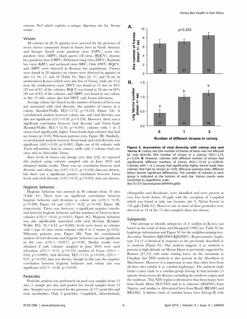
Average colony size (based on the number of frames of bees) was
not associated with viral diversity (the number of viruses in a
colony; Kruskal-Wallis: H(2) = 2.74, p = 0.254, Figure 3A). A
correlational analysis between colony size and viral diversity was
also not significant (r(57) = 0.20, p = 0.128). However, there was a
significant correlation between viral diversity and Varroa loads
(Kruskal-Wallis: H(2) = 13.10; p = 0.001); colonies with 1 or 2
viruses had significantly higher Varroa loads than colonies that had
no viruses (p,0.05, Wilcoxon pairwise tests, Figure 3B). Similarly,
a correlational analysis between Varroa loads and viral diversity was
significant (r(65) = 0.49, p,0.001). Eight out of 66 colonies with
Varroa infestations had no viruses, while only 2 colonies had one
virus and no detectable Varroa.
Since levels of viruses can change over time [14], we repeated
this analysis using colonies sampled only in June 2010 and
obtained similar results. There was no correlation between viral
diversity and colony size (r(47) = 0.11, p = 0.456, data not shown),
but there was a significant positive correlation between Varroa
levels and viral diversity (r(47) = 0.33, p = 0.023, data not shown).
Hygienic behaviorHygienic behavior was assessed in 36 colonies from 10 sites
(Table S1). There were no significant correlations between
hygienic behavior and elevation or colony size (r(35) = 20.18,
p = 0.289, Figure 4A and r(35) = 20.22, p = 0.196, Figure 4B,
respectively). There was, however, a significant negative correla-
tion between hygienic behavior and the numbers of Varroa in these
colonies (r(35) = 20.42, p = 0.011, Figure 4C). Hygienic behavior
was also significantly associated with viral diversity (Kruskal-
Wallis: H(2, 34) = 6.43, p = 0.040); levels were lower in colonies
with 1 type of virus versus colonies with 0 or 2 viruses (p,0.05,
Wilcoxon pairwise tests, Figure 4D). Note the correlational
analysis of viral diversity and hygienic behavior was not significant
in this case (r(35) = 20.0277, p = 0.90). Similar results were
obtained if only colonies sampled in June 2010 were used
(elevation: r(25) = 20.31, p = 0.122; number of Varroa: r(25) = 2
0.63, p = 0.0005; viral diversity: H(2) = 5.55, p = 0.076, r(25) = 2
0.21, p = 0.292; data not shown), though in this case the negative
correlation between hygienic behavior and colony size was
significant (r(25) = 20.46, p = 0.019).
PesticidesPesticide analysis was performed on pool wax samples from 15
sites (1 sample per site) and pooled bee bread samples from 13
sites. Samples were screened for the presence of 171 pesticides and
toxic metabolites. Only 4 pesticides, 1-naphthol, chlorothalonil,
chlorpyrifos and fluvalinate, were identified and were present at
very low levels (below 50 ppb) with the exception of 1-napthol
which was found at only one location (site 9, Nji-ini Forest) at
116 ppb (Table S1). However one or more of these pesticides were
detected at 14 of the 15 sites sampled (data not shown).
SubspeciesOur attempt to identify subspecies of A. mellifera in Kenya was
based on the work of Arias and Sheppard (1996) (see Table S1 for
haplotype information and Figure S1 for the neighbor-joining tree.
Accession Numbers KJ628964-KJ628987). Representative haplo-
type 3.4.11 is identical in sequence to the previously described A.
m. monticola (Figure S1). Our analysis suggests A. m. monticola is
present at high altitude on Mount Elgon as previously suggested by
Ruttner [27,53] with some mixing lower on the mountain at
Chepkui, but ND2 monticola is also present in the Aberdares at
Marchorwe. However it is important to note that other bees from
all three sites exhibit A. m. scutellata haplotypes. The monticola clade
forms a sister clade to a scutellata group (Group A) that includes 15
apiaries from across the Kenya, excluding the northern region and
the southeast. This ND2 region is identical to that from honey bees
from South Africa (SCUTE2) and A. m. adansonii (ADANS1) from
Nigeria, and similar to Africanized bees from Brazil (BRASI1 and
BRASI2). A distinct clade of scutellata honey bees (Group B) with
Figure 3. Association of viral diversity with colony size andVarroa. A. Colony size (the number of frames of bees) was not affectedby viral diversity (the number of viruses in a colony), H(2) = 2.74,p = 0.254. B. However, colonies with different number of viruses hadsignificantly different numbers of Varroa (H(2) = 13.10; p = 0.0014).Colonies with 1 or 2 viruses had significantly higher Varroa loads thancolonies that had no viruses (p,0.05, Wilcoxon pairwise tests, differentletters denote significant differences). The number of colonies in eachgroup is indicated at the bottom of each bar. Varroa counts wereconverted to logarithmic scale.doi:10.1371/journal.pone.0094459.g003
Honey Bee Health in East Africa
PLOS ONE | www.plosone.org 6 April 2014 | Volume 9 | Issue 4 | e94459

the dominant haplotype of 2.4.11 (identical to SCUTE1 from
Kenya) is found predominantly in the central and eastern portion
of the country. The individuals in the haplotype group identified
by 12.1.12 are from the coast and the far north of Kenya (identical
ND2 regions are found at sites 12, 13, 16, 17). Apiary 12 is in the
Arabuko Sokoke Forest Reserve, a remnant of the coastal tropical
forest that supports a number of unique endemic species. Although
no molecular comparison is available from other studies, we
speculate that this could be A. m. litorea given the close relationship
with A. m. lamarckii (LAMARC) and the honey bees in the northern
apiaries at Mandera [27]. Individuals from one colony at apiary
18, called Mandera West, were distinct from the other Kenyan
bees, although another colony at the same apiary groups with
scutellata A. These could represent either A. m. yemenitica or the
recently described A. m. simensis [27,28], neither of which have
been described with molecular makers. There were 14 unique
individual haplotypes whereas many apiaries had multiple ND2
haplotypes (Table S3). We found no correlation between genotype,
as measured the ND2 region, and honey bee health although more
data will be require to clearly distinguish populations or
subspecies.
Discussion
Our survey suggests that several new parasites and pathogens
(Varroa, Nosema, DWV, BQCV, and ABPV) have recently invaded
honey bee populations in East Africa (see below for further
discussion). However, none of these factors are correlated with
colony size, implying that several factors thought to critically
undermine bee populations in the US and Europe (Varroa, Nosema,
and pesticide use) are not yet directly impacting Kenyan bee
populations in terms of this metric. However, since there may be a
time-lag before newly introduced parasites and pathogens cause
substantial negative effects, continuous monitoring of these
populations should be conducted to evaluate the long-term
dynamics of these host-pathogen interactions. Our data also
suggests that chemical control methods for Varroa and Nosema,
which are heavily used by US beekeepers, may be unnecessary at
this time for Kenyan honey bees, and indeed, these bees may
possess novel resistance mechanisms. Furthermore, we found an
intriguing correlation between elevation and Varroa levels,
suggesting that environmental factors (climate, landscape ecology)
may play a key role in mediating this host-parasite interaction, and
perhaps honey bee health in general, though the effect of these
environmental factors needs to be explored in greater detail in
larger scale studies. We did not find any association with
subspecies genotype and honey bee health. However, based on
the molecular marker that we used (the ND2 region of the
mitochondria) there was considerable gene flow between popula-
tions, and thus more sensitive markers may be needed to fully
characterize subspecies and population differences.
Varroa alone does not appear to strongly impact honey bee
colonies in Kenya. Based on recommendations for US beekeepers,
approximately 2/3 of the surveyed colonies had Varroa levels high
enough to warrant treatment to control mite populations [39,52].
However, Varroa levels in Kenyan colonies are not correlated with
decreases in colony strength; in fact, there is a positive correlation
between Varroa numbers and colony size. Colonies with higher
Figure 4. Association of hygienic behavior with colony location, size, parasite and pathogen loads. A. There were no significantcorrelations between hygienic behavior and elevation (r(35) = 20.18, p = 0.289). B. Hygienic behavior was not correlated with colony size (r(35) = 20.22, p = 0.196). C. There was, however, a significant negative correlation between hygienic behavior and the numbers of Varroa in these colonies(r(35) = 20.42, p = 0.011; Varroa counts were converted to logarithmic scale). D. Hygienic behavior was also significantly associated with viral diversity(H(2, 34) = 6.43, p = 0.040); levels were lower in colonies with 1 type of virus versus colonies with 0 or 2 viruses (p,0.05, Wilcoxon pairwise tests,different letter denote significant differences). The number of colonies in each group is indicated at the bottom of each bar.doi:10.1371/journal.pone.0094459.g004
Honey Bee Health in East Africa
PLOS ONE | www.plosone.org 7 April 2014 | Volume 9 | Issue 4 | e94459

levels of hygienic behavior did have lower levels of Varroa, as
expected [54], but hygienic behavior was quite variable across the
surveyed colonies. Varroa levels were strongly positively correlated
with elevation. Based on a limited preliminary study of the impacts
of geographic location on colony weight and Varroa levels (see
Figure S2) this is possibly due to differences in climate or floral
resources rather than due to differences in honey bee genetic
background, though the association between nutrition, climate
and pathogen loads in honey bee populations needs to be more
thoroughly assessed. Varroa was recently found in honey bee
colonies in Nigeria as well, at comparable levels (approximately
80% of colonies infested, with 2–55 mites found on 100 bees), but
again no negative impacts on Varroa infestation on colony health
and productivity were reported [55]. It is unclear what factors
contribute to reduce the impacts of Varroa on African bees relative
to European bees. Previous studies have indicated that Africanized
bees in South America have higher levels of hygienic behavior,
higher levels of grooming mites off of adult bees, lower levels of
mite reproduction on pupae, and are less attractive to Varroa mites
than European bees [56]. African bee subspecies also tend to
abscond (abandoning hives and sometimes migrating) and swarm
(where a large fraction of the colony leaves with the queen to form
a new colony) more readily than European bees [27], thereby
causing breaks in brood rearing that may reduce Varroa loads [57].
Indeed, breaking the brood cycle is recommended as a method to
reduce Varroa loads in European honey bees [58], and removal of
drone brood significantly reduces Varroa levels [59]. However, all
of these factors presumably would lead to lower overall Varroa
levels in colonies, rather than a reduced impact on worker
mortality and colony size with equivalent levels. Thus, other
parameters, perhaps physiological or behavioral, may contribute
to the higher levels of tolerance of African bees to Varroa
infestation.
Kenyan bee populations also displayed infections with DWV,
BQCV, and ABPV, and Varroa numbers are strongly positively
correlated with viral diversity (number of viruses present). Previous
studies have demonstrated that Varroa can vector DWV and IAPV
[37,38], and the introduction of Varroa to a naıve population of
honey bees in Hawaii was correlated with reduction in sequence
diversity of DWV [60], suggesting Varroa mites vector a specific
viral strain. Thus, Varroa may have introduced these three viruses
to the Kenyan honey bee populations, or is altering the population
structure of these viruses. Alternatively, the presence of these
viruses may be weakening bees’ defenses to Varroa; this possibility
has not yet been examined. Interestingly, a 2010 study of honey
bee colonies in Uganda (which borders western Kenya and was
thought to be Varroa-free at the time of the study) found BQCV in
30–40% of the colonies, but DWV and ABPV were not detected
[61], suggesting that these two viruses may have been recently
introduced to Kenya, perhaps by Varroa. Indeed, the viruses were
not present in the geographically distant apiaries in far northeast-
ern Kenya, suggesting that they, and Varroa, have not yet spread to
this region. It should be noted that while the number of individual
bees assayed for viruses in each colonies was small (5 bees), the
number of colonies assayed throughout Kenya (81 colonies) and
those assessed for correlation between viral diversity and Varroa
levels (66 colonies) was fairly substantial. Thus, while the results of
the individual colonies should be interpreted with caution (see Pirk
et al [62] for more information on pathogen sampling in bee
populations), the overall data set suggests that only three common
European viruses are circulating, at this point, in the Kenyan
populations at detectable levels, and that these are associated with
Varroa parasitization levels.
As is the case with Varroa, DWV and BQCV do not appear to
negatively impact Kenyan bee colonies, since viral infection is not
correlated with colony size. Kenyan colonies are overall relatively
small (7 frames of honey bees on average, compared to US
colonies which can easily reach 20+ frames) and thus may not be
at their limits of productivity. Better measures of the impacts of
viruses, Varroa, and Nosema on bee health would include the
longevity of infected and uninfected colonies, their ability to
reproduce and successfully establish new colonies. Indeed, other
studies have indicated that colonies may be particularly sensitive to
the impacts of viruses during specific stressful periods (such as
colony founding or migration) which could result in reduced
populations. In European honey bees, high DWV levels in the fall
are associated with reduced overwintering survival [63,64]. While
there was an effect of viral diversity on hygienic behavior, this was
not consistent – colonies with no viruses were as hygienic as
colonies with two viruses. Previous studies have suggested that bees
more readily remove larvae that are parasitized by Varroa with
high viral titers [65], but our results suggest that hygienic behavior
is not likely functioning as a major mechanism to reduce viral
loads in Kenyan colonies.
The gut microsporidia Nosema ceranae was linked to colony losses
in Spain [66], and high levels of Nosema bombi have been associated
with declining bumble bee populations in North America [67].
However, other studies have not found a strong correlation with
Nosema presence and colony declines [14,24], suggesting that
Nosema may act in concert with other factors, such as pesticides
[68], to reduce bee health. While Nosema was previously found in
Zimbabwe and South Africa [69,70], it was not detected in a
previous survey in 1996–1998 in Kenya ([36]; Shi Wei, personal
communication, 2011). In our 2010 survey, Nosema was only
identified in two apiaries at the coast [site 12 and 13], at site 15
near the Tanzania border and, surprisingly, at high elevation
interior site on Mt. Elgon (site 22). Using PCR-based detection
methods we did not detect Nosema in other locations. As in the case
of the viral analyses, though the sample sizes for individual
colonies was limited (5 bees/colony), our analysis at the apiary
level should be quite robust in detecting Nosema if it is present.
Notably, colonies in coastal Nosema-infected apiaries were similar
in size to those in non-infected apiaries, and Varroa levels were
substantially lower than other regions in the country. Thus, Nosema
does not appear to be affecting honey bee populations in Kenya.
In the US, .90% of honey bee colonies contain pesticide
residues [51]. Over 129 different pesticide-related chemicals have
been found in US bee colonies, with an average of 6 chemicals per
colony. Pesticide exposure has been linked to honey bee
population declines [10,71] reduced survival and impaired
development of brood [72], impaired cognitive function, [73–
75], altered expression of immune genes [76], and increased
Nosema loads [68,77]. In the Kenyan colonies surveyed in this
study we found only four pesticides, and most were at very low
levels compared to pesticide levels in North America. The most
commonly found pesticide was the fungicide chlorothalonil (12 of
15 sites), while chlorpyrifos, an organophosphate insecticide, was
found in five of the apiaries tested. Interestingly, fluvalinate was
found in one apiary: fluvalinate is commonly used to control Varroa
mites in the North America and Europe; however this broad-
spectrum pyrethroid is also used to control mosquitos and
horticultural pests such as aphids, whiteflies and thrips. The low
levels of pesticides in hives from across Kenya, particularly when
compared to levels in developed countries, suggests pesticide
residues play only a limited role in honey bee health in Kenya at
this time.
Honey Bee Health in East Africa
PLOS ONE | www.plosone.org 8 April 2014 | Volume 9 | Issue 4 | e94459

Conclusions
Honey bees provide critical pollination services to agriculture
and natural landscapes, and the honey and wax produced by
honey bees represent a potential source of income for families in
East Africa and across the world. Our survey suggests that several
new parasites and pathogens (Varroa, Nosema, DWV, BQCV, and
ABPV) have recently invaded honey bee populations in East
Africa. Our results indicate that these parasites and pathogens are
not yet impacting honey bee health in Kenya, at least in terms of
colony size. Interestingly, levels of Varroa are strongly impacted by
elevation/geographic region, suggesting that environmental fac-
tors modulate Varroa infestation rates. Finally, our phylogenetic
analyses suggest there is considerable mixing of honey bee
populations in Kenya, and thus newly introduced parasites and
pathogens can likely move easily throughout the region. Our
studies suggest that honey bee populations in East Africa appear to
be largely resistant or tolerant of the parasites and pathogens that
threatened honey bee populations in other parts of the world, and
are not yet significantly impacted by other stressors, such as
exposure to environmental toxins. However, since there may be a
time lag before these newly introduced pathogens and parasites
significantly impact honey bee populations, additional long-term
monitoring is necessary. Finally, our results also highlight the
importance of environmental factors in buffering honey bee
populations from these stressors, and with increasing habitat
fragmentation and destruction and environmental extremes
brought on by global climate change, populations of this keystone
species, in Africa and throughout the world, will be under
increasing pressures.
Supporting Information
Figure S1 Neighbor-joining tree (Saitou and Nei, 1987)comparing representative Kenyan honeybee haplotypes(see Table S2) with subspecies described in Arias andSheppard 1996 (in capitals). The European subspecies Apis
mellifera mellifera (MELLI1) is used as the outgroup. The percentage
of replicate trees in which the associated taxa clustered together in
the bootstrap test (2000 replicates) are shown next to the branches
for values greater than 30% (1). Branch length indicates number of
SNP differences. The analysis involved 39 nucleotide sequences
from the ND2 mitochondrial region. Twenty-four of the 39 are
representative unique haplotypes. For a list of all individuals
represented by these haplotypes see Table S2. There were a total
of 579 nucleotide positions in the final dataset. Phylogenetic
analysis was conducted in MEGA5 (2). This analysis suggests as
many as 7 clades of A. mellifera within Kenya. Whether this is
evidence of more subspecies than previously described will require
more sampling. Moreover, the weak statistical support suggests a
need to expand beyond the ND2 region to describe East African
subspecies of A. mellifera. Accession Numbers KJ628964-
KJ628987.
(DOCX)
Figure S2 Effect of location on colony weight and Varroanumbers. Colonies of from upland site 1 (icipe) were assayed in
June 2010, immediately prior and after moving upland colonies
from site 1 to site 25 on the coast. Coastal colonies were assayed at
nearby apiaries (sites 26 and 27). There were no significant
differences in colony weight (A, F(3,26 = 1.43), p = 0.256), but
there were significantly fewer Varroa in colonies moved to site 25
(B, F(3,25) = 6.63, p = 0.0019). In August 2010, upland colonies at
sites 1 and 25 and coastal colonies at site 25 were assayed. There
were again no significant differences in weight (C, F(2, 16) = 1.29,
p = 0.301), but both upland and coastal colonies had significantly
fewer Varroa at site 25 (D, F(2,16) = 6.96, p = 0.0067). The number
of colonies in each group at each timepoint is indicated at the
bottom of each bar in B and D. Letters represent groups that were
significantly different with a Tukey HSD post-hoc pairwise
comparison, at p,0.05. While the graphs show the actual average
numbers of Varroa, counts were converted to logarithmic scale for
statistical analysis.
(DOCX)
Table S1 Apiary colony list indicating location, Varroaload, hygienic behavior, presence of virus, Nosema,pesticide and subspecies identification. Each apiary was
given a site number and apiary name. Each colony has a unique
identification number with the apiary number in the first position
and the colony number in the second. Colony size is indicated by
numbers of frames with bees. Total Varroa counts are based on
standard sugar roll assay described in Ellis and Macedo, 2001 (1).
The percent hygienic behavior (2) was calculated by taking the
final number of fully and partially removed pupae/(207 – number
originally uncapped or empty cells) *100. Boxes with UD are
undetermined indicating measurements were not taken for these
colonies. Positive for virus or Nosema apis detection per colony is
indicated by an ‘‘X’’. The presence of pesticide detected in wax or
bee bread is indicated in parts per billion (ppb). Only four
pesticides were detected: CP (chloropyrios), CT (chlorothalonil), N
(1-naphtol), F (fluvalinate). ND2 subspecies identification is based
on the analysis presented in Figure S1. Individuals from most of
the colonies grouped with the scutellata A or B clades. Some were
identical in sequence to A. mellifera monticola described by Arias and
Sheppard, 1996 (3). Some individuals from colonies at sites 12, 13,
16 and 17 were most closely related to A. mellifera lamarckii and are
thus called ‘‘lamarckii-like’’. Colonies at site 18 were unique and
perhaps represent a distinct subspecies (A. m. simensis or yemenitica).
A few individuals did not group with any previously described
subspecies and are thus indicated as UD. Interestingly three
colonies (2.1, 2.2, and 16.1) had individuals from multiple
mitochondrial lineages.
(DOCX)
Table S2 Type of data collected at each apiary. An ‘‘X’’
indicates the data type at the top of the column was collected in at
least one colony in the corresponding apiary. See Materials and
Methods for a full description of the type of data collected and the
analyses that were performed.
(DOCX)
Table S3 A list of ND2 haplotypes used for comparison.Individuals were sequenced for the ND2 region (as described in 1)
from 24 apiaries across Kenya. There were 24 unique haplotypes
from 109 individuals sequenced. Each column is headed by the
representative haplotype (in bold) used for analysis. Haplotypes
identical to the representative type follow in each column. The
columns are the far right is a list of unique single haplotypes.
Numeric designations for each individual follow the following
scheme: apiary.colony.individual.
(DOCX)
Table S4 ND2 region variable sites from Kenya honey-bee haplotypes compared to the subspecies A. m.adansonii (ADANS2) (1). The nucleotide positions starting
from the ND2 ATC (isoleucine) are indicated in the top row with
corresponding position numbers from the complete honeybee
mitochondrial genome (2). The codon position for each SNP is
indicated at the bottom of the figure. Over the 579 bp of the ND2
coding region 85% of SNPs were in the third codon position.
Honey Bee Health in East Africa
PLOS ONE | www.plosone.org 9 April 2014 | Volume 9 | Issue 4 | e94459

Three SNPs were in the second codon position. The first is a
transversion at position 53 (T«A) that results in an amino acid
change of isoleucine « asparagine. A second codon position
change is a transition at nucleotide position 161 (C«T) resulting
in an amino acid change of threonine « isoleucine. The third
second codon change (position 458) is also a transition (C«T)
resulting in threonine « isoleucine. The Kenya honeybee
population also shows a first codon transition (position 412;
G«A) that results in an amino acid difference (valine «isoleucine) when compared to the reference sequence.
(DOCX)
Form S1 Collaborating authories.(DOCX)
Acknowledgments
We would like to thank Diana Sammatro for help in early specimen
collections, Justin Malloy for assistance with molecular work associated
with Nosema survey and Daisy Salifu and Elina Lastro Nino for providing
advice for the statistical analyses.
Author Contributions
Conceived and designed the experiments: EM HP MF JF JT CG.
Performed the experiments: EM HP MF BT TB JK JNK FM DM.
Analyzed the data: HP MF TB CG. Wrote the paper: EM HP MF JF BT
TB DM JT CG.
References
1. Klein AM, Vaissiere BE, Cane JH, Steffan-Dewenter I, Cunningham SA, et al.(2007) Importance of pollinators in changing landscapes for world crops.
Proceedings of the Royal Society B-Biological Sciences 274: 303–313.
2. Eilers EJ, Kremen C, Greenleaf SS, Garber AK, Klein AM (2011) Contributionof Pollinator-Mediated Crops to Nutrients in the Human Food Supply. Plos One
6.
3. Gallai N, Salles JM, Settele J, Vaissiere BE (2009) Economic valuation of the
vulnerability of world agriculture confronted with pollinator decline. EcologicalEconomics 68: 810–821.
4. Klein AM, Steffan-Dewenter I, Tscharntke T (2003) Fruit set of highland coffee
increases with the diversity of pollinating bees. Proceedings of the Royal Societyof London Series B-Biological Sciences 270: 955–961.
5. Bhattacharya A (2004) Flower visitor and fruitset of Anacardium occidentole.
Annales Botanici Fennici 41: 385–392.
6. Roubik DW (2002) The value of bees to the coffee harvest. Nature 417: 708.
7. Aizen MA, Harder LD (2009) The Global Stock of Domesticated Honey Bees Is
Growing Slower Than Agricultural Demand for Pollination. Current Biology 19:915–918.
8. Morse RA, Calderone NW (2000) The value of honey bees as pollinators of U.S.
crops in 2000. Bee Culture 128: 1–15.
9. Allsopp M (2004) Cape honeybee (Apis mellifera capensis Eshscholtz) and varroa
mite (Varroa destructor Anderson & Trueman) threats to honeybees andbeekeeping in Africa. International Journal of Tropical Insect Science 24: 87–94.
10. vanEngelsdorp D, Meixner MD (2010) A historical review of managed honey
bee populations in Europe and the United States and the factors that may affectthem. Journal of Invertebrate Pathology 103: S80–S95.
11. Sammataro D, Gerson U, Needham G (2000) Parasitic mites of honey bees: life
history, implications, and impact. Annu Rev Entomol 45: 519–548.
12. Chen YP, Huang ZY (2010) Nosema ceranae, a newly identified pathogen of
Apis mellifera in the USA and Asia. Apidologie 41: 364–374.
13. Chen YP, Siede R (2007) Honey Bee Viruses. In: Maramorosh K, Shatkin AJ,Murphy FA, editors. Advances in Virus Research: Elsevier Academic Press. pp.
34–80.
14. Runckel C, Flenniken ML, Engel JC, Ruby JG, Ganem D, et al. (2011)Temporal Analysis of the Honey Bee Microbiome Reveals Four Novel Viruses
and Seasonal Prevalence of Known Viruses, Nosema, and Crithidia. Plos One 6.
15. Aronstein KA, Murray KD (2010) Chalkbrood disease in honey bees. J Invertebr
Pathol 103 Suppl 1: S20–29.
16. Genersch E (2008) Paenibacillus larvae and American Foulbrood - long sinceknown and still surprising. Journal fur Verbrauchershutz und Lebensmittelsi-
cherheit 3: 429–434.
17. Desneux N, Decourtye A, Delpuech JM (2007) The sublethal effects of pesticides
on beneficial arthropods. Annu Rev Entomol 52: 81–106.
18. Brodschneider R, Crailsheim K (2010) Nutrition and health in honey bees.Apidologie 41: 278–294.
19. Mattila HR, Seeley TD (2007) Genetic diversity in honey bee colonies enhances
productivity and fitness. Science 317: 362–364.
20. VanEngelsdorp D, Caron D, Hayes J, Underwood R, Henson M, et al. (2012) Anational survey of managed honey bee 2010–11 winter colony losses in the USA:
results from the Bee Informed Partnership. Journal of Apicultural Research 51:
115–124.
21. Chauzat MP, Carpentier P, Madec F, Bougeard S, Cougoule N, et al. (2010)The role of infectious agents and parasites in the health of honey bee colonies in
France. Journal of Apicultural Research 49: 31–39.
22. Cox-Foster DL, Conlan S, Holmes EC, Palacios G, Evans JD, et al. (2007) Ametagenomic survey of microbes in honey bee colony collapse disorder. Science
318: 283–287.
23. Nguyen BK, Ribiere M, vanEngelsdorp D, Snoeck C, Saegerman C, et al.
(2011) Effects of honey bee virus prevalence, Varroa destructor load and queencondition on honey bee colony survival over the winter in Belgium. Journal of
Apicultural Research 50: 195–202.
24. vanEngelsdorp D, Evans JD, Saegerman C, Mullin C, Haubruge E, et al. (2009)Colony collapse disorder: a descriptive study. Plos One 4: e6481.
25. vanEngelsdorp D, Speybroeck N, Evans JD, Nguyen BK, Mullin C, et al. (2010)
Weighing risk factors associated with bee colony collapse disorder by
classification and regression tree analysis. J Econ Entomol 103: 1517–1523.
26. Hepburn RH, Radloff SE (1998) Honeybees of Africa. New York: Springer. 386
p.
27. Ruttner F (1987) Biogeography and Taxonomy of Honeybees: Springer. 284 p.
28. Meixner MD, Leta MA, Koeniger N, Fuchs S (2011) The honey bees of Ethiopia
represent a new subspecies of Apis mellifera-Apis mellifera simensis n. ssp.
Apidologie 42: 425–437.
29. Whitfield CW, Behura SK, Berlocher SH, Clark AG, Johnston JS, et al. (2006)
Thrice out of Africa: ancient and recent expansions of the honey bee, Apis
mellifera. Science 314: 642–645.
30. Crane E (1999) The World History of Bee Keeping and Honey Hunting New
York: Routledge. 720 p.
31. Mbae RM (1999) Overview of beekeeping development in Kenya. In: Raina
SK, Kioka EN, Mwanycky SW, editors. The Conservation and Utilization of
Commercial Insects. Nairobi: ICIPE Science Press. pp. 103–105.
32. Kasina M, Kraemer M, Martius C, Wittmann D (2009) Diversity and Activity
Density of Bees Visiting Crop Flowers in Kakamega, Western Kenya. Journal of
Apicultural Research 48: 134–139.
33. UNCTAD/WTO ITC (2004) Kenya: Supply Suvery on Apicultural and
Horticultural Products. Geneva, Switzerland: International Trade Centre.
34. National Beekeeping Station (2007) Hive Population and Production in Kenya
(2005, 2006, and 2007) Provincial Summaries. Nairobi, Kenya: Ministry of
Livestock.
35. Frazier M, Muli E, Conklin T, Schmehl D, Torto B, et al. (2010) A scientific
note on Varroa destructor found in East Africa; threat or opportunity?
Apidologie 41: 463–465.
36. Wei S (2001) Genetic Variation and Colony Development of Honey Bees Apis
mellifera in Kenya. Uppsala: Swedish University of Agricultrual Sciences. 22 p.
37. Di Prisco G, Pennacchio F, Caprio E, Boncristiani HF, Jr., Evans JD, et al.
(2011) Varroa destructor is an effective vector of Israeli acute paralysis virus in
the honeybee, Apis mellifera. J Gen Virol 92: 151–155.
38. Gisder S, Aumeier P, Genersch E (2009) Deformed wing virus: replication and
viral load in mites (Varroa destructor). J Gen Virol 90: 463–467.
39. Ellis MD, Macedo PA (2001) G01-1430 Using the Sugar Roll Technique to
Detect Varroa Mites in Honey Bee Colonies. Lincoln, NE: University of
Nebraska. 4 p.
40. Klee J, Besana AM, Genersch E, Gisder S, Nanetti A, et al. (2007) Widespread
dispersal of the microsporidian Nosema ceranae, an emergent pathogen of the
western honey bee, Apis mellifera. J Invertebr Pathol 96: 1–10.
41. Fries I, Chauzat MP, Chen YP, Doublet V, Genersch E, Gisder S, Higes M,
McMahon DP, Martin-Hernandez R, Natsopoulou M, Paxton RJ, Tanner G,
Webster TC, Williams GR (2013) Standard methods for Nosema research.
Journal of Apicultural Research 51(5): http://dx.doi.org/10.3896/IBRA.1.52.1.
14.
42. Traver BE, Fell RD (2011) ‘‘Prevalence and infection intensity of Nosema in
honey bee (Apis mellifera L.) colonies in Virginia.’’ Journal of Invertebrate
Pathology 107(1): 43–49.
43. Benjeddou M, Leat N, Allsopp M, Davison S (2001) Detection of acute bee
paralysis virus and black queen cell virus from honeybees by reverse
transcriptase pcr. Appl Environ Microbiol 67: 2384–2387.
44. Chen YP, Higgins JA, Feldlaufer MF (2005) Quantitative real-time reverse
transcription-PCR analysis of deformed wing virus infection in the honeybee
(Apis mellifera L.). Appl Environ Microbiol 71: 436–441.
45. Ribiere M, Triboulot C, Mathieu L, Aurieres C, Faucon JP, et al. (2002)
Molecular diagnosis of chronic bee paralysis virus infection. Apidologie 33: 339–
351.
46. Stoltz D, Shen XR, Boggis C, Sisson G (1995) Molecular diagnosis of Kashmir
bee virus infection. Journal of Apicultural Research 34: 153–160.
47. Spivak M, Reuter GS (1998) Honey bee hygienic behavior. American Bee
Journal 138: 283–286.
Honey Bee Health in East Africa
PLOS ONE | www.plosone.org 10 April 2014 | Volume 9 | Issue 4 | e94459

48. Arias MC, Sheppard WS (1996) Molecular phylogenetics of honey bee
subspecies (Apis mellifera L.) inferred from mitochondrial DNA sequence.Mol Phylogenet Evol 5: 557–566.
49. Thompson JD, Higgins DG, Gibson TJ (1994) CLUSTAL W: improving the
sensitivity of progressive multiple sequence alignment through sequenceweighting, position-specific gap penalties and weight matrix choice. Nucleic
Acids Res 22: 4673–4680.50. Saitou N, Nei M (1987) The neighbor-joining method: a new method for
reconstructing phylogenetic trees. Mol Biol Evol 4: 406–425.
51. Mullin CA, Frazier M, Frazier JL, Ashcraft S, Simonds R, et al. (2010) Highlevels of miticides and agrochemicals in North American apiaries: implications
for honey bee health. Plos One 5: e9754.52. Delaplane KS, Hood WM (1997) Effects of delayed acaricide treatment in honey
bee colonies parasitized by Varroa jacobsoni and a late season treatmentthreshold for the southeastern USA. Journal of Apicultural Research 36: 125–
132.
53. Ruttner F (1992) Naturgeschichte der Honigbienen: Kosmos Verlags-GmbH.54. Spivak M, Reuter GS (2001) Varroa destructor infestation in untreated honey
bee (Hymenoptera: Apidae) colonies selected for hygienic behavior. Journal ofEconomic Entomology 94: 326–331.
55. Akinwade KL, Badejo MA, Ogbogu SS (2012) Incidence of the Korea haplotype
of Varroa destructor in southwest Nigeria. Journal of Apicultural Research 15: 369–370.
56. Guzman-Novoa E, Vandame Rm, Arechavaleta M, E. (1999) Susceptibility ofEuropean and Africanized honey bees (Apis mellifera L.) to Varroa jacobsoni
Oud. in Mexico. Apidologie 30: 173–182.57. Fries I, Hansenb H, Imdorfc A, Rosenkranz P (2003) Swarming In Honey Bees
(Apis Mellifera) And Varroa Destructor Population Development In Sweden
Apidologie 34: 389–397.58. National Bee Unit (2010) FAQ 17: Queen trapping. York: The Food and
Environmental Research Agency. 1–2 p.59. Calderone NW (2005) Evaluation of drone brood removal for management of
Varroa destructor (Acari: Varroidae) in colonies of Apis mellifera (Hymenoptera:
Apidae) in the northeastern United States. J Econ Entomol 98: 645–650.60. Martin SJ, Highfield AC, Brettell L, Villalobos EM, Budge GE, et al. (2012)
Global honey bee viral landscape altered by a parasitic mite. Science 336: 1304–1306.
61. Kajobe R, Marris G, Budge G, Laurenson L, Cordoni G, et al. (2010) Firstmolecular detection of a viral pathogen in Ugandan honey bees. J Invertebr
Pathol 104: 153–156.
62. Pirk CWW, de Miranda JR, Kramer M, Murray TE, Nazzi F, et al (2013)‘‘Statistical guidelines for Apis mellifera research.’’ Journal of Apicultural
Research 52(4).
63. Highfield AC, El Nagar A, Mackinder LC, Noel LM, Hall MJ, et al. (2009)
Deformed wing virus implicated in overwintering honeybee colony losses. ApplEnviron Microbiol 75: 7212–7220.
64. Dainat B, Evans JD, Chen YP, Gauthier L, Neumann P (2012) Predictive
markers of honey bee colony collapse. Plos One 7: e32151.65. Schoning C, Gisder S, Geiselhardt S, Kretschmann I, Bienefeld K, et al. (2012)
Evidence for damage-dependent hygienic behaviour towards Varroa destructor-parasitised brood in the western honey bee, Apis mellifera. J Exp Biol 215: 264–
271.
66. Higes M, Martin-Hernandez R, Botias C, Bailon EG, Gonzalez-Porto AV, et al.(2008) How natural infection by Nosema ceranae causes honeybee colony
collapse. Environ Microbiol 10: 2659–2669.67. Cameron SA, Lozier JD, Strange JP, Koch JB, Cordes N, et al. (2012) Patterns
of widespread decline in North American bumble bees. Proc Natl Acad Sci U S A108: 662–667.
68. Pettis JS, vanEngelsdorp D, Johnson J, Dively G (2012) Pesticide exposure in
honey bees results in increased levels of the gut pathogen Nosema.Naturwissenschaften 99: 153–158.
69. Fries I, Slamenda SB, Da Silva A, Pieniazek NJ (2003) African honey bees (Apismellifera scutellata) and Nosema (Nosema apis) infections. Journal of Apicultural
Research 42: 13–15.
70. Swart DJ (2003) The occurence of Nosema apis (Zander), Acarapis woodi(Rennie), and the Cape problem bee in the summer rainfall region of South
Africa Rhodes University. 43 p.71. Faucon J-P, Mathieu L, Ribiere M, Martel A-C, Drajnudel P, et al. (2002)
Honey bee winter mortality in France in 1999 and 2000. Bee World 83 14–23.72. Zhu W, Schmehl DR, Mullin CA, Frazier JL (submitted) Chronic oral toxicity of
four commone pesticides and their mixtures to honey bee larvae.
73. Bortolotti L, Montanari R, Marcelino J, Medrzycki P, Maini S, et al. (2003)Effects of sub-lethal imidacloprid doses on the homing rate and foraging activity
of honey bees. Bulletin of Insectology 56: 63–67.74. Ciarlo TJ, Mullin CA, Frazier JL, Schmehl DR (2012) Learning impairment in
honey bees caused by agricultural spray adjuvants. Plos One 7: e40848.
75. Decourtye A, Devillers J, Cluzeau S, Charreton M, Pham-Delegue MH (2004)Effects of imidacloprid and deltamethrin on associative learning in honeybees
under semi-field and laboratory conditions. Ecotoxicol Environ Saf 57: 410–419.76. Gregorc A, Evans JD, Scharf M, Ellis JD (2012) Gene expression in honey bee
(Apis mellifera) larvae exposed to pesticides and Varroa mites (Varroadestructor). J Insect Physiol 58: 1042–1049.
77. Wu JY, Smart MD, Anelli CM, Sheppard WS (2012) Honey bees (Apis
mellifera) reared in brood combs containing high levels of pesticide residuesexhibit increased susceptibility to Nosema (Microsporidia) infection. J Invertebr
Pathol 109: 326–329.
Honey Bee Health in East Africa
PLOS ONE | www.plosone.org 11 April 2014 | Volume 9 | Issue 4 | e94459