Evaluation of modified adaptive k-means segmentation ...
15
Computational Visual Media https://doi.org/10.1007/s41095-019-0151-2 Vol. 5, No. 4, December 2019, 347–361 Research Article Evaluation of modified adaptive k-means segmentation algorithm Taye Girma Debelee 1,2 ( ), Friedhelm Schwenker 1 , Samuel Rahimeto 2 , and Dereje Yohannes 2 c The Author(s) 2019. Abstract Segmentation is the act of partitioning an image into different regions by creating boundaries between regions. k-means image segmentation is the simplest prevalent approach. However, the segmentation quality is contingent on the initial parameters (the cluster centers and their number). In this paper, a convolution-based modified adaptive k-means (MAKM) approach is proposed and evaluated using images collected from different sources (MATLAB, Berkeley image database, VOC2012, BGH, MIAS, and MRI). The evaluation shows that the proposed algorithm is superior to k-means++, fuzzy c-means, histogram- based k-means, and subtractive k-means algorithms in terms of image segmentation quality (Q-value), computational cost, and RMSE. The proposed algorithm was also compared to state-of-the-art learning-based methods in terms of IoU and MIoU; it achieved a higher MIoU value. Keywords clustering; modified adaptive k-means (MAKM); segmentation; Q-value 1 Introduction 1.1 Overview Segmentation is the act of partitioning an image into different regions by creating boundaries that keep regions apart. It is one of the most used steps in zoning pixels of an image [1]. After segmentation, pixels belonging to the same partition have higher 1 Institute of Neural Information Processing, Ulm University, 89081Ulm, Germany. E-mail: T. G. Debelee, [email protected] ( ); F. Schwenker, [email protected]. 2 Addis Ababa Science and Technology University, Addis Ababa, 120611, Ethiopia. E-mail: S. Rahimeto, [email protected]; D. Yohannes, [email protected]. Manuscript received: 2019-04-01; accepted: 2019-06-29 similarity values, but higher dissimilarity with pixels in other partitions. Segmentation is a technique used in many fields including health care, image processing, traffic image, pattern recognition, etc. According to the review in Ref. [1], image segmentation techniques can be categorized into two types: layered-based segmentation and block-based segmentation. In layered-based segmentation, the image is divided into layers such as background, foreground, and mask layers. Reconstruction of the final image is decided using the mask layer [2]. This method is not widely applicable to medical image segmentation. Block- based segmentation divides the image into unequal blocks using attributes such as color, histogram, pixels, wavelet coefficients, texture, and gradient [1, 2]. Block-based segmentation can be further grouped into methods based on discontinuity or similarity in the image. It can also be further grouped into three categories: region-based, edge- or boundary-based, and hybrid techniques [1, 2]. 1.2 Edge-based segmentation The discontinuous nature of pixels characterizes all algorithms in the edge-based segmentation family [2]. In this type of image segmentation, images are segmented into partitions based on unanticipated changes in gray intensity in the image. In most cases, edge-based segmentation techniques can identify corners, edges, points, and lines in the image. However, pixel miscategorization errors are the main limitation of the edge-based segmentation category. The edge detection technique is an example of this class of segmentation method [2]. 1.3 Region-based segmentation Edge-based segmentation techniques use the discontinuous nature of pixels. However, region- based techniques use similarity of pixels in the image. Edges, lines, and points are attributes that decide the 347
-
Upload
khangminh22 -
Category
Documents
-
view
2 -
download
0
Transcript of Evaluation of modified adaptive k-means segmentation ...

Computational Visual Mediahttps://doi.org/10.1007/s41095-019-0151-2 Vol. 5, No. 4, December 2019, 347–361
Research Article
Evaluation of modified adaptive k-means segmentation algorithm
Taye Girma Debelee1,2 (�), Friedhelm Schwenker1, Samuel Rahimeto2, and Dereje Yohannes2
c© The Author(s) 2019.
Abstract Segmentation is the act of partitioningan image into different regions by creating boundariesbetween regions. k-means image segmentation is thesimplest prevalent approach. However, the segmentationquality is contingent on the initial parameters (thecluster centers and their number). In this paper, aconvolution-based modified adaptive k-means (MAKM)approach is proposed and evaluated using imagescollected from different sources (MATLAB, Berkeleyimage database, VOC2012, BGH, MIAS, and MRI).The evaluation shows that the proposed algorithmis superior to k-means++, fuzzy c-means, histogram-based k-means, and subtractive k-means algorithmsin terms of image segmentation quality (Q-value),computational cost, and RMSE. The proposedalgorithm was also compared to state-of-the-artlearning-based methods in terms of IoU and MIoU; itachieved a higher MIoU value.
Keywords clustering; modified adaptive k-means(MAKM); segmentation; Q-value
1 Introduction1.1 Overview
Segmentation is the act of partitioning an image intodifferent regions by creating boundaries that keepregions apart. It is one of the most used steps inzoning pixels of an image [1]. After segmentation,pixels belonging to the same partition have higher
1 Institute of Neural Information Processing, UlmUniversity, 89081Ulm, Germany. E-mail: T. G.Debelee, [email protected] (�); F. Schwenker,[email protected].
2 Addis Ababa Science and Technology University,Addis Ababa, 120611, Ethiopia. E-mail: S.Rahimeto, [email protected]; D. Yohannes,[email protected].
Manuscript received: 2019-04-01; accepted: 2019-06-29
similarity values, but higher dissimilarity with pixelsin other partitions. Segmentation is a technique usedin many fields including health care, image processing,traffic image, pattern recognition, etc. According tothe review in Ref. [1], image segmentation techniquescan be categorized into two types: layered-basedsegmentation and block-based segmentation. Inlayered-based segmentation, the image is divided intolayers such as background, foreground, and masklayers. Reconstruction of the final image is decidedusing the mask layer [2]. This method is not widelyapplicable to medical image segmentation. Block-based segmentation divides the image into unequalblocks using attributes such as color, histogram,pixels, wavelet coefficients, texture, and gradient [1, 2].Block-based segmentation can be further grouped intomethods based on discontinuity or similarity in theimage. It can also be further grouped into threecategories: region-based, edge- or boundary-based,and hybrid techniques [1, 2].
1.2 Edge-based segmentation
The discontinuous nature of pixels characterizes allalgorithms in the edge-based segmentation family[2]. In this type of image segmentation, images aresegmented into partitions based on unanticipatedchanges in gray intensity in the image. In most cases,edge-based segmentation techniques can identifycorners, edges, points, and lines in the image.However, pixel miscategorization errors are the mainlimitation of the edge-based segmentation category.The edge detection technique is an example of thisclass of segmentation method [2].
1.3 Region-based segmentation
Edge-based segmentation techniques use thediscontinuous nature of pixels. However, region-based techniques use similarity of pixels in the image.Edges, lines, and points are attributes that decide the
347

348 T. G. Debelee, F. Schwenker, S. Rahimeto, et al.
effectiveness of region-based techniques. Algorithmslike clustering, splitting and merging, normalizedcuts, region growing, and thresholding belong to theregion-based segmentation family [1, 2]. Our maininterest is in clustering algorithms. Schwenker andTrentin [3] presented traditional machine learningas supervised and unsupervised learning: supervisedlearning associates every observation of the sampleswith a target label whereas this is not the case inunsupervised learning. Clustering algorithms are veryimportant, especially for unlabeled larger datasetclassification [3]; they belong to the unsupervisedcategory. However, there is another machine learningapproach, partially supervised machine learning,which lies between unsupervised and supervisedmachine learning. A detailed review is given inRef. [3].
1.4 Learning-based segmentation
Deep learning models are well known in objectdetection, feature extraction, and classification. Inaddition, semantic image segmentation or imagelabeling is also an area in which deep learning hasbeen applied. Semantic segmentation is a techniquein which semantic labels (like “cat” or “bike”) areassigned to every pixel in the image [4]. The mostcommon models that have been applied to semanticimage segmentation include FCN-8s [5], DeepLab [4],DeepLab-Msc [4], MSRA-CFM [6], TTI-Zoomout-16 [7], DeepLab-CRF [4], DeepLab-MSc-CRF [4],DeepLab-CRF-7x7 [4], DeepLab-MSc-LargeFOV [4],DeepLab-MSc-CRF-LargeFOV [4], and Front-EndModules [8]. Consecutive application of pooling indeep convolutional neural networks (DCNNs) reducesfeature resolution and allows DCNNs to learn abstractrepresentations of objects [9].
2 Related work2.1 k-means segmentation
There has been much research on image segmentationfor different application areas, using varioustechniques from conventional and learning-basedmethods. Among many segmentation algorithms,k-means is one of the simplest for generating a regionof interest [10–12]. It has a time complexity of O(n)for n samples [13]. However, it is sensitive to outliersand initialization parameters [14]. As a result, itgives different clustering results with different cluster
numbers and initial centroid values [12]. Muchresearch has considered how to initialize the centersfor k-means with the intention of maximizing theefficiency of the algorithm. In the k-means clusteringalgorithm, each pixel belongs to only one clusterand center, so it is a hard clustering algorithm[10, 11]. Some recent works in clustering methods ofsegmentation and deep learning based segmentationare addressed in Sections 2.2 and 2.3 respectively.
2.2 Clustering methods for segmentation
In Ref. [15], adaptive k-means clustering is introducedto ameliorate the performance of k-means. Here, theinitialization parameters remain consistent for severaliterations. However, the initial seed point is computedsimply by taking the usual mean of all data values inthe input image, making it a simple post-processingoperation for good quality image segmentation.A first attempt to ameliorate the deficiencies of
k-means clustering with respect to outliers occurredthree decades ago. Bezdek [16] came up with anew algorithm named fuzzy c-means (FCM) in 1981.This algorithm is a membership-based soft clusteringalgorithm.Faußer and Schwenker [17] proposed an algorithm
that divides the samples into subsets to performclustering in parallel, and merges the outputrepeatedly. In their proposed approach they usedmany kernel-based FCM clustering algorithms. Twodatasets (the Breast Cancer database from the UCIrepository and Enron Emails) were used to evaluatetheir algorithm. The experimental analysis provedthat the algorithm has high accuracy and workswell for large real-life datasets. Benaichouche etal. [18] brought in a region-based image segmentationalgorithm using enhanced spatial fuzzy FCM. Lei etal. [19] explained that traditional FCM is susceptibleto noise, and describes improvements based onthe addition of local spatial information. Thissolves the robustness problem but greatly increasesthe computational complexity. First, they usedmorphological reconstruction to smooth images toenhance robustness and then applied FCM. Theyalso modified FCM by using faster membershipfiltering instead of the slower distance computationbetween pixels within local spatial neighborhoodsand their cluster centers. The gray-level histogramof the morphologically reconstructed image is usedfor clustering. The median filter is employed to avoid

Evaluation of modified adaptive k-means segmentation algorithm 349
noise from the fuzzy membership matrix generatedusing the histogram. The paper demonstratedthat the proposed algorithm is faster and moreefficient when compared to FCM and other typesof modification.Arthur and Vassilvitskii [20] introduced a new
algorithm called k-means++ that improves uponthe initial selection of centroids. The selection forinitial clusters is started by selecting one initial centerrandomly. The other cluster centers are then selectedto satisfy specific probabilities determined by “D2
weighting”. The probabilities are defined based onthe squared distance of each point to the alreadychosen centers. The paper claims that k-means++outperforms the original k-means method in achievinglower intra-cluster separation and in speed. Thenumber of clusters is still chosen by the user. Butthe algorithm is faster and more effective and evenprovided as a library in MATLAB.Zhang et al. [21] used Mahalanobis distance
instead of Euclidean distance to allocate every datapoint to the nearest cluster. Using their newclustering algorithm, PCM clustering, they got bettersegmentation results. However, their algorithm alsohas high computational cost and the challenge ofinitializing parameters.Purohit and Joshi [22] presented a new approach
to improve k-means with aim of reducing the meansquare error of the final cluster and attainingminimum computation time. Yedla et al. [23] alsointroduced an enhanced k-means clustering algorithmwith better initial centers. They achieved an effectiveway to associate data points with appropriate clusterswith reduced computation time compared to standardk-means.Dhanachandra et al. [12] initialized k-means
clustering using a subtractive clustering approachwhich attempted to find optimal centers based ondata point density. The first center is chosen to havethe highest density value in the data points. Afterselecting the first center, the potential of the datapoints near this center decreases. The algorithm thentries to find other centers based on the potentialvalue until the potential of all grid points fallsbelow some threshold. The algorithm is effectiveat finding centers but the computational complexityincreases exponentially as the number of data points
increases. The standard k-means algorithm is theninitialized with these centers. Since the aim of thepaper was the segmentation of medical images, whichsuffer from poor contrast, a partial spatial starchingcontrast enhancement technique was applied. Aftersegmentation, filtering is applied to avoid unwantedregions and noise. The paper attempted to illustratethe out-performance of subtractive clustering basedk-means over normal k-means. However, it failedto compare it to other methods. Subtractiveclustering has a higher computational time than otherclustering methods, which is the main drawback ofthis technique.Kucukkulahlı et al. [24] tried to initialize k-means
by finding both k and centroid locations. First,they used histogram values to determine peaks andpits. Then, by calculating the distances betweenadjacent peaks and pits, a vertical sweep is doneto find the highest peak within some thresholddistance. Horizontal sweeping is followed to grouppeaks that are close to each other, replacing themwith a representative by calculating the mean distanceand choosing peaks which are above the mean. Oncek and centroids have been obtained, the standardk-means method is used for clustering. Even thoughthe approach is dependent on human involvement forassigning the threshold for the vertical sweep, thealgorithm automated the k-means method.
2.3 Deep learning in image segmentation
Recently a number of deep learning models haveshown astounding results in semantic segmentation[4, 25, 26].According to Ref. [26], deep learning has
shown its success in handwritten digit recognition,speech recognition, image categorization, and objectdetection in images. It has also been appliedto screen content image segmentation, and provenits application to semantic pixel-wise labeling [25].Badrinarayanan et al. [26] proposed SegNet, a methodfor semantic pixel-wise segmentation of road scenes,and tested their algorithm using the CamVid roadscenes dataset. They used three popular performanceevaluation parameters: global accuracy, class averageaccuracy, and mean intersection over union (MIoU)over all classes.Minaee and Wang [25] introduced an algorithm
for segmentation of screen content images into two

350 T. G. Debelee, F. Schwenker, S. Rahimeto, et al.
layers (foreground and background). The foregroundlayer mainly consists of text and lines and thebackground layer consists of smoothly varying regions.They compared their results with two algorithms(hierarchical k-means clustering in DjVu, and a shapeprimitive extraction and coding (SPEC) method) interms of precision and recall values using five testimages. The proposed approach scored 91.47% forprecision and 87.73% for recall.Chen et al. [4] proposed a technique that embeds
multiscale features in a fully connected convolutionalneural network to perform pixel-based semanticsegmentation through pixel-level classification. Theyintroduced an attention model to softly determinethe weight of multi-scale features at each pixellocation. They trained the FCN with multiscalefeatures obtained from multiple resized images usinga shared deep network. The attention model playedthe role of average pooling and max-pooling. Besidesfeature reduction, the attention model overcame oneof the challenges of deep learning: it enabled theauthors to visualize the features at different positionsalong with their level of importance. They proved theeffectiveness of their approach using three datasets:PASCAL-Person-Part, VOC 2012, and MS-COCO2014.As reviewed by Minaee and Wang [27], various
algorithms are in use to separate text from itsbackground. Approaches include clustering-basedalgorithms, sparse decomposition based methods,and morphological operations. In their paper,they proposed an alternating direction method ofLagrange multipliers (ADMM) for this problem. Theyadopted the proposed algorithm to separate movingobjects from the background. In a comparison madewith the hierarchical k-means approach and sparsedecomposition, their proposed method scored higherprecision (95%), recall (92.5%), and F1 (93.7%)values. The sparse decomposition approach wasproposed by themselves in Ref. [28].
3 Materials and methods3.1 Dataset
Images used in this paper are from different sources.Some are from the MATLAB image databaseand some from the Berkeley segmentationdatasets (BSD) [29]. The same images were used in
Refs. [12] and [30] to evaluate their segmentationalgorithms. The images used for the first experimentare MRI (mri.tif), Bag (bag.png), Cameraman(cameraman.tif), Coins (coins.png), Moon (moon.tif),Pout (pout.tif), and Glass (glass.png). We ranthe second experiment using AT (AT3 1m4 01.tif),Lena (lena.png), Valley (valley.jpg), Airplane(airplane.jpg), Mountain (mountain.jpg), and Breast(bet06.jpg) images [31]. Further experimentalanalysis was done to measure the effectiveness ofour proposed segmentation algorithm using theVOC2012 challenge datasets [32].
3.2 Proposed approach
Our proposed segmentation approach is convolutionbased, as indicated in Fig. 1. First, the histogram
Fig. 1 Flowchart of our convolution-based segmentation algorithm.First, histograms of the grayscale image of the original image aregenerated. Second, amplitude thresholds, T p, are computed usingthe histogram levels. Third, the dynamic window size is computedusing an amplitude threshold for each image. This is followed by a2D convolution operation. Finally, the mean of the convolution is setas the initial seed to generate other new seed values that can be usedas the centers of clusters, which are then used to perform clustering.

Evaluation of modified adaptive k-means segmentation algorithm 351
distribution (ali) of the grayscale image is generatedfor each image. Second, out of the generatedhistogram values we select only those with ali � 1.Third, we computed the ratio of the sum of theselected histograms to the number of occurrences(l0) of such histogram values to obtain the amplitudethreshold (Tp). Fourth, we added the histogramvalues � Tp and divided by α to get a windowsize. See Algorithm 1; α is computed as indicatedat the end of Algorithm 1. Finally, the convolutionoperation is performed and the result is convertedto an array to compute the mean value used as theinitial seed point of the convolution-based modifiedadaptive k-means (MAKM) segmentation algorithm.The parameters used in the proposed algorithm areconstant for each image and the segmentation resultis consistent for a number of iterations which is nottrue for the other clustering algorithms (FCM, HBK,SC, and k-means++) used for comparative analysis.The pseudocode of the proposed algorithm is givenin Algorithm 2.
Algorithm 1 Pseudocode for window size generationGet input image and convert to gray imageImage = readimg()
if (channels(Image)� 3) thengrayimage = rgb2gray (Image)
elsegrayimage = Image
end ifCalculate the Amplitude Histogram ThresholdHist = histogram (grayimage)
for i = 0:255 doif (ali � 1) then
l0 = l0 + 1Sumali = Sumali + ali
end ifend forT p = Sumali /l0
Window size determination, w
for i = 0 : 255 doif (ali � T p) then
Sumali�T p = Sumali�T p + ali
end ifend forw = Sumali�T p /α
Determine α:hist = imhist(img, k), k = 256hist2 = max(hist−T p + 1, 0)
nonzeros2 = find(hist2)lt = length(nonzeros2)α = 1+floor(min(9,lt/2))
Algorithm 2 Pseudocode for modified adaptive k-meansInput: window size w and gray imagePerform Convolution operationgray=conv2(gray,ones(w)/(w2),’same’)
Place the convolution result in an arrayarray = gray(:)
Initialize iteration Counters: i = 0, j = 0while true do
Initialize seed point, seed = mean(array)Increment counter for each iteration, i = i + 1while true do
Initialize counter for each iteration, j = j + 1Compute distance between seed and gray value
dist = sqrt((array−seed)2)Compute bandwidth for cluster center
distth= sqrt(sum((array−seed)2)/numel(array))Check values are in selected bandwidth or not
qualified = dist<distthUpdate mean
newseed = mean(array(qualified))condition for termination
if (seed = newseed or j>10) thenj = 0Remove values assigned to a clusterStore center of cluster
center(i) = newseedbreak
end ifUpdate seed: seed = newseed
check maximum number of clustersif (isempty(array) or i > 10) then
Reset counter: i = 0break
end ifend whileSort centersCompute distances between adjacent centersFind minimum distance between centersDiscard cluster centers less than distanceMake a clustered image using these centers
end while
3.3 Evaluation
In this paper we use the Q-value criterion [30],computational cost [30], root mean squared error(RMSE) [33, 34], standard deviation, mean absoluteerror (MAE), intersection over union (IoU) [35], meanintersection over union (MIoU) [35], entropy (E),and peak signal to noise ratio (PSNR) to assess ourproposed, convolution-based, modified adaptive k-means segmentation algorithm.The Q-value measures image segmentation quality
taking into consideration both small and large regions

352 T. G. Debelee, F. Schwenker, S. Rahimeto, et al.
in the final segmented images. The Q value evaluationfunction used in this paper is given by
Q =√
R
(NM)
R∑i=1
[e2
i
1 + logAi+
(R(Ai)
Ai
)2](1)
where N and M are the numbers of rows and columnsin the image respectively, R is the total numberof regions in the segmented image, ei is the colordifference between the original and segmented image,Ai is the area of region i, and R(Ai) is the number ofregions with the same area as Ai. The area of eachregion Ai is the number of pixels constituting thatregion; empty regions and regions with 1 pixel are leftunconsidered. Smaller values of Q represent bettersegmentation results whereas higher values indicatehigher color errors due to either under-segmentationor over-segmentation. ei is given by
ei =√((S(xsi, ysi) − O(xoi, yoi))2, i = 1 : R (2)
where S and O are points in 2D Euclidean space withcoordinates S(xsi, ysi) for the segmented image andO(xoi, yoi) for the original (raw) image.RMSE measures how much the output image
deviates from the input image. Mean squared error(MSE) is given by
MSE =1
nxmy
nx−1∑x=0
my−1∑y=0
[r(x, y) − t(x, y)]2 (3)
where r(x, y) and t(x, y) are grayscale values atposition x, y in the raw and segmented images. RMSEis the square root of the MSE [33, 34].
RMSE =√MSE (4)
A smaller value means higher image segmentationquality.For the VOC2012 challenge datasets, popular
performance evaluation parameters include globalaccuracy, class average accuracy, and meanintersection over union (MIoU) for all classes [26].Global accuracy is the percentage of pixels correctlyclassified in the dataset whereas the mean of thepredictive accuracy over all classes is class averageaccuracy. In this paper, we use MIoU to comparethe performance of our algorithm with learning-basedsegmentation methods. MIoU is defined as
MIoU =∑
IoU/N (5)where intersection over union (IoU) is defined inEq. (6) and N is the number of objects consideredfrom the dataset for a particular experiment.
IoU =Area(overlap)Area(union)
(6)
Algorithm 3 Pseudocode for mean Q values and standarddeviation
Initialize Q = 0Perform 10 iterations
for j = 1:10 doIdentify the unique label from the indexed imageCalculate the number of unique labels and initialize tonumber of regions (R)Convert the indexed images to gray-scale usingmat2grayCompute color error (ei) and area (Ai) for each regionfor i = 1:R do
Find number of regions with the same area as Ai andinitialize it to R(Ai)Compute Qi for each regionQ = Q + Qi
end forend forCompute mean of Q and standard deviation (σ)
Algorithm 4 Pseudocode for computation of RMSEInitialize Squared Error, E = 0Perform 10 iterationsfor j = 1:10 do
Get labeled images with their respective centersfor i = 1: R do
Compute Squared Error (SE) for each regionend forfor i = 1:R do
Compute sum of E,E = E + Ei
end forCompute RMSE for each iteration
end forCompute mean RMSE and standard deviation (σ)
where area of overlap is the area between thepredicted bounding box and the ground-truthbounding box, and area of union is the area coveredby both the predicted bounding box and the ground-truth bounding box.
4 Results and discussionThe outcomes of the experiments we conductedusing the proposed technique show that gray imagesegmentation task can be carried out efficiently whileinitialization of parameters is done automatically. Allexperiments were performed in MATLAB version2016b and run on a 3.00 GHz Intel Core i7-4601M CPU, under the Microsoft Windows 10operating system. The performance of the proposedsegmentation algorithm was evaluated using RMSE,

Evaluation of modified adaptive k-means segmentation algorithm 353
Q-value, computation time, MAE, E, PSNR, IoU,and MIoU. RMSE and MAE were used for standardquality measurement of the segmented outputimage. It tells us the degree of deviation betweenthe output image and the input image. Thesame evaluation parameters were used for otherselected clustering segmentation algorithms forcomparative analysis, but for the deep learning basedsegmentation algorithm, only IoU and MIoU wereused for comparison with the proposed segmentationalgorithm. Some performance aspects of the proposedmethod are discussed in this section.To evaluate the proposed image segmentation
approach, we used images that were also used inRefs. [12] and [30]. In the first experiment, weused MRI (mri.tif), Bag (bag.png), Cameraman(cameraman.tif), Coins (coins.png), Moon (moon.tif),Pout (pout.tif), and Glass (glass.png). The resultsobtained are indicated in Figs. 2–6 and Tables 2–4.In Table 1, we list images with their respective sizes
and number of cluster for every clustering algorithmconsidered in this paper. In Table 2, we compare otherclustering algorithms with our proposed algorithmin terms of segmentation quality. Since the clustercenters varying for K++, FCM, and HBK, the Q-value in Table 2, RMSE in Table 3, and computationcost in Table 4 are computed 10 times and theirmean value and standard deviation are determined.
However, in the proposed modified adaptive k-means clustering method, the cluster centers areconsistent for any number of iterations. Comparedto other clustering algorithm, histogram-based k-means (HBK) had the lowest segmentation qualityfor moon.tif, as indicated in Table 2. ComparingK++ and FCM with HBK shows that K++ andFCM had lower Q score. However, adaptive k-meansand modified adaptive k-means methods outperformthese three algorithms, even if the adaptive k-meansalgorithm needs post-processing image to find theregion of interest. In some cases, MAKM performsbetter than AKM, for example for images glass.png,pout.tif, and mri.tif.In terms of RMSE, FCM had highest score for
pout.tif which shows that it is the worst performingclustering algorithm: see Table 3. However, ourproposed approach had the minimum RMSE valuefor all images used in the experiment.To evaluate the attainment of the proposed
technique in image segmentation for other grayimages, further experiments were conducted on somecommonly used images: AT3 1m4 01.tif, lena.png,valley.jpg, airplane.jpg, mountain.jpg, and bet06.jpg.The subtractive k-means algorithm has the highestcomputation cost for all images, although in somecases it shows better segmentation quality than K++,HBK, and FCM.
Fig. 2 MRI-labeled image segmented using various approaches.
Fig. 3 Bag-labeled image segmented using various approaches.

354 T. G. Debelee, F. Schwenker, S. Rahimeto, et al.
Fig. 4 Cameraman-labeled image segmented using various approaches.
Fig. 5 Coins-labeled image segmented using various approaches.
Fig. 6 Moon-labeled image segmented using various approaches.

Evaluation of modified adaptive k-means segmentation algorithm 355
Table 1 Number of clusters used in each algorithm for each respective images
Image Size K++ HBK FCM SC AKM MAKM (proposed)MRI 128×128 3 3 4 2 3 3Bag 250×189 3 3 4 4 5 4Cameraman 256×256 3 3 4 3 4 4
Experiment-1 Coins 246×300 3 3 4 2 4 3Moon 537×358 3 3 4 2 4 4Pout 291×240 3 3 4 2 3 4Glass 181×282 3 3 4 5 3 3
AT3 1m4 01 (AT) 480×640 3 3 4 1 5 5Lena 512×512 3 3 4 4 3 3
Experiment-2 Valley 321×481 3 3 4 5 4 4Airplane 321×481 3 3 4 2 4 4Mountain 321×481 3 3 4 2 4 3Breast 384×512 3 3 4 2 6 5
Table 2 Comparison of algorithms in terms of mean Q-value and standard deviation (Q-value, σ)
Image Size K++ HBK FCM SC AKM MAKM (proposed)MRI 128×128 0.40, 0.18 1.06, 0.60 0.87, 0.64 0.31, — 0.21, 0.00 0.20, 0.00Bag 250×189 0.44, 0.35 0.50, 0.43 0.39, 0.32 0.13, — 0.07, 0.00 0.12, 0.00Cameraman 256×256 0.39, 0.21 0.44, 0.28 0.48, 0.25 0.70, — 0.02, 0.00 0.05, 0.00Coins 246×300 0.26, 0.23 0.54, 0.37 0.42, 0.18 0.13, — 0.13, 0.00 0.13, 0.00Moon 537×358 0.25, 0.36 0.87, 0.57 0.60, 0.51 0.06, — 0.01, 0.00 0.01, 0.00Pout 291×240 0.28, 0.04 0.35, 0.14 0.40, 0.13 0.29, — 0.20, 0.00 0.03, 0.00Glass 181×282 0.55, 0.24 0.46, 0.41 0.44, 0.29 0.62, — 0.15, 0.00 0.14, 0.00
Table 3 Comparison of algorithms in terms of mean RMSE and standard deviation (RMSE, σ)
Image Size K++ HBK FCM SC AKM MAKM (proposed)MRI 128×128 1.14, 0.00 1.14, 0.004 0.88, 0.006 0.91, — 1.03, 0.00 1.03, 0.00Bag 250×189 1.27, 0.004 1.27, 0.01 1.13, 0.001 1.36, — 1.29, 0.00 0.90, 0.00Cameraman 256×256 1.63, 0.001 1.62, 0.005 1.44, 0.01 1.63, — 1.47, 0.00 1.10, 0.00Coins 246×300 1.54, 0.002 1.54, 0.01 1.47, 0.004 1.50, — 1.61, 0.00 1.31, 0.00Moon 537×358 0.86, 0.001 0.86, 0.004 0.91, 0.001 0.76, — 0.88, 0.00 0.86, 0.00Pout 291×240 1.81, 0.04 1.79, 0.02 1.91, 0.005 1.77, — 1.50, 0.00 1.48, 0.00Glass 181×282 1.51, 0.00 1.51, 0.001 1.62, 0.005 1.77, — 1.44, 0.00 1.37, 0.00
Table 4 Comparison of algorithms in terms of mean computation time and standard deviation (time (s), σ)
Image Size K++ HBK FCM SC AKM MAKM (proposed)MRI 128×128 0.01, 0.005 0.02, 0.006 0.02, 0.009 5.00, 0.50 0.003, 0.001 0.004, 0.001Bag 250×189 0.08, 0.07 0.09, 0.03 0.06, 0.02 33.70, 1.02 0.012, 0.002 0.018, 0.011Cameraman 256×256 0.04, 0.02 0.09, 0.005 0.08, 0.01 62.77, 1.25 0.0116, 0.002 0.016, 0.004Coins 246×300 0.09, 0.07 0.11, 0.005 0.08, 0.01 75.90, 1.16 0.015, 0.004 0.022, 0.024Moon 537×358 0.20, 0.11 0.26, 0.01 0.20, 0.007 1025.01, 5.69 0.041, 0.003 0.042, 0.005Pout 291×240 0.02, 0.008 0.09, 0.005 0.06, 0.01 74.03, 0.75 0.015, 0.004 0.015, 0.004Glass 181×282 0.02, 0.006 0.07, 0.004 0.05, 0.02 39.44, 0.05 0.012, 0.008 0.011, 0.004
Table 5 Comparison of proposed algorithm with K++, HBK, FCM, and SC in terms of mean Q-value for AT, LE, VA, AI, MT, and Breastimages
Image Size K++ HBK FCM SC MAKM (proposed)AT3 1m4 01 (AT) 480×640 0.53 0.36 0.49 0.50 0.04Lena (LE) 512×512 0.42 0.35 0.48 0.39 0.06Valley (VA) 321×481 0.45 0.45 0.55 0.45 0.15Airplane (AI) 321×481 0.40 0.29 0.36 0.41 0.10Mountain (MT) 321×481 0.34 0.50 0.53 0.64 0.19Breast 384×512 0.46 0.39 0.73 0.72 0.01

356 T. G. Debelee, F. Schwenker, S. Rahimeto, et al.
Table 6 Comparison of proposed algorithm with K++, HBK, FCM, and SC in terms of mean RMSE for AT, LE, VA, AI, MT, and Breastimages
Image Size K++ HBK FCM SC MAKM (proposed)AT3 1m4 01 (AT) 480×640 0.83 0.83 0.82 0.74 0.69Lena (LE) 512×512 1.00 1.00 1.10 1.12 0.84Valley (VA) 321×481 1.23 1.23 1.20 1.42 1.06Airplane (AI) 321×481 1.45 1.45 1.51 1.28 1.22Mountain (MT) 321×481 0.95 0.95 0.88 0.78 0.72Breast 384×512 0.56 0.58 0.59 0.50 0.56
Table 7 Comparison of proposed algorithm with K++, HBK, FCM, and SC in terms of mean computation cost (s) for AT, LE, VA, AI, MT,and Breast images
Image Size K++ HBK FCM SC MAKM (proposed)AT3 1m4 01 (AT) 480×640 0.34 0.42 0.33 2846.45 0.14Lena (LE) 512×512 0.21 0.34 0.27 2074.85 0.12Valley (VA) 321×481 0.20 0.27 0.18 693.66 0.06Airplane (AI) 321×481 0.12 0.23 0.14 648.50 0.05Mountain (MT) 321×481 0.09 0.24 0.17 658.37 0.07Breast 384×512 0.10 0.20 0.17 1092.93 0.05
From the data analysis, we observe that thetime taken by subtractive k-means becomes veryexpensive for some images. The three image sampleswith highest time cost are AT3 1m4 01.tif (2846 s),lena.png (2075 s), and moon.tif (1565 s).In the experimental results presented in Table 2,
K++ shows better performance than FCM andHBK, except for some image samples: glass.pngfor both HBK and FCM, and bag.png for FCM.However, in the second experiment using imageslike AT3 1m4 01.tif, lena.png, valley.jpg, airplane.jpg,mountain.jpg, and bet06.jpg, HBK proved to providethe best image segmentation quality except formountain.jpg. The modified adaptive k-meansalgorithm has better image segmentation quality, andminimum RMSE for all cases discussed. It scored wellfor the breast image compared to other images. Thelow computation cost of our proposed approach makesit more suitable for image segmentation. The sampleindexed images in the second experiment are given inFig. 7. The final results of the experiment show that
the overall achievement of the proposed modifiedadaptive k-means is superior to other clusteringalgorithms in terms of image segmentation quality(Q-value), computational cost, and RMSE.For further analysis, we considered additional
images from the VOC2012 challenge dataset andmammography images from Bethezatha GeneralHospital (BGH) and MIAS.Four randomly selected images (dog, airplane,
plant, and person) from VOC2012 were used tocompare the proposed algorithm to three clusteringalgorithms (AKM, FCM, K++) in terms of Q, time,MAE, E, and PSNR. For all images our proposedalgorithm scored better for Q, computation time,and PSNR compared to other clustering algorithms,but not for MAE and entropy: see Table 8. In thecase of the “person” image, our proposed algorithmscored minimum MAE compared to other algorithms,indicating good performance for this particular image.Comparative performance of the proposed
algorithm for two randomly selected MRI images is
Fig. 7 Lena-labeled image segmented using various approaches.

Evaluation of modified adaptive k-means segmentation algorithm 357
Table 8 Comparison of proposed method with clustering algorithms in terms of Q, computation time, MAE, entropy, PSNR, precision (P ),recall (R), and F -score (F 1) using VOC2012 dataset
Image Algorithm Q Time (s) MAE Entropy PSNR P R F 1
Dog
AKM 0.042 0.106 6.07 1.53 47.25 89.3 61.7 72.9FCM 0.431 0.345 3.22 1.83 46.98 86.9 92.2 89.5k-means++ 0.209 0.248 2.76 1.94 46.64 84.8 99.5 91.6Proposed 0.040 0.052 3.89 1.79 47.28 90.3 80.4 85.1
Plane
AKM 0.031 0.041 0.407 1.54 43.55 95.0 89.6 92.2FCM 0.743 0.212 0.302 1.71 43.11 91.2 98.7 94.8k-means++ 1.00 0.120 0.252 1.63 43.16 94.2 96.6 95.4Proposed 0.030 0.036 0.337 1.58 43.59 95.3 92.4 93.8
Plant
AKM 0.012 0.054 0.331 1.63 45.78 71.1 81.7 76.0FCM 0.041 0.286 40.60 1.64 45.73 0.0 1.3 0.0k-means++ 0.621 0.107 43.30 1.64 45.65 0.0 0.0 0.0Proposed 0.011 0.061 0.332 1.63 45.84 72.1 82.8 77.2
Person
AKM 0.095 0.11 12.72 1.92 48.67 83.6 88.8 86.1FCM 0.139 0.58 16.30 1.62 47.72 92.2 68.1 78.3k-means++ 0.35 0.65 14.09 1.72 47.34 92.9 72.8 81.6Proposed 0.094 0.050 5.11 2.06 48.71 84.9 99.9 91.8
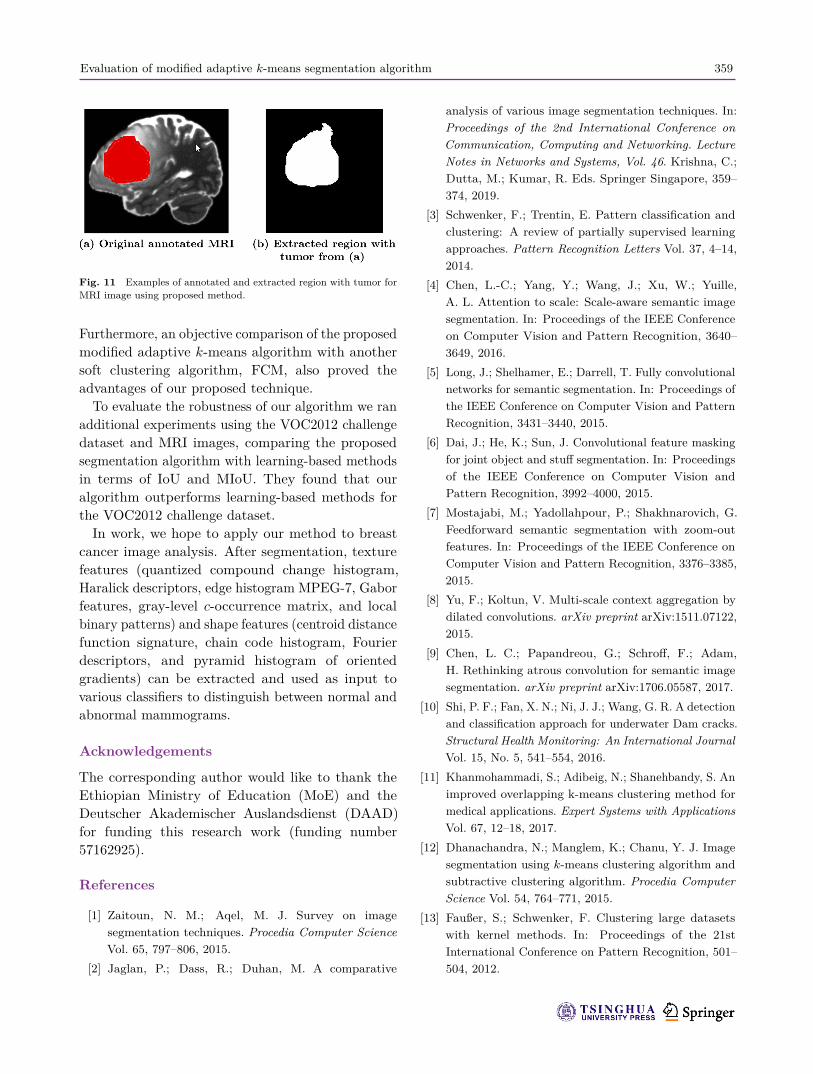
given in Table 9. The proposed algorithm performsbetter in terms of MSE, Q, and computation timefor both MRI images. However, the second MRIrecorded a higher IoU value than the first image, as
Table 9 Comparison of proposed algorithm with clustering imagesegmentation algorithm in terms of MSE, Time, and Q for two MRIimages
Image Method MSE Time Q
MRI 1
AKM 0.67 0.03 0.009FCM 0.74 0.16 0.238k-means++ 0.77 0.37 0.053Proposed 0.66 0.02 0.008
MRI 2
AKM 0.74 0.03 0.020FCM 0.85 0.16 1.585k-means++ 0.91 0.30 1.492Proposed 0.73 0.02 0.019
indicated in Table 10. Segmentation results for thesecond MRI image are given in Fig. 11.A comparison of the proposed algorithm with
learning-based and clustering algorithms is presentedin Table 11. The comparison terms of IoU and MIoUindicate that the proposed algorithm scored higherIoU and MIoU for plant and person images, butfor the dog and plane images, k-means++ scored
Table 10 Comparison of proposed algorithm with clusteringalgorithm in terms of IoU and MIoU for two MRI images
Method MRI 1 MRI 2 MIoUAKM 63.19 58.19 60.69FCM 79.21 62.93 71.07k-means++ 77.90 83.96 80.93Proposed 64.22 86.33 75.28
Fig. 8 Examples of annotated and extracted region with cancer for breast mammographic images from BGH and MIAS datasets usingproposed method.

358 T. G. Debelee, F. Schwenker, S. Rahimeto, et al.
Fig. 9 Annotated and respective segmentation result for dog fromVOC2012 challenge datasets using proposed method.
higher values of IoU and MIoU. Figures 8–10 presentsegmentation results for the proposed algorithm forimages randomly selected from VOC2012, BGH, andMIAS datasets.
5 ConclusionsIn this study, we presented a convolution-basedmodified adaptive k-means algorithm, to get the bestout of the normal k-means method during imagesegmentation. Firstly, an automatic window sizegeneration approach was designed to perform theconvolution process to get the central value for everyconvolution step, and the mean of these values isassigned as the initial seed point. Then, using thisseed point, the cluster centers and number of clustersare determined as initial parameters and given tothe adaptive k-means algorithm. A comparativeanalysis of the proposed modified adaptive k-meanswith K++, HBK, and SC methods was made in termsof image segmentation quality (Q), RMSE, and time.The results obtained confirmed the advantages ofour proposed modified adaptive k-means algorithm.
Fig. 10 Annotated and respective segmentation result for plane and person from VOC2012 challenge datasets using proposed method.
Table 11 Related works from learning-based methods and clustering algorithms for comparison with proposed method in terms of IoU andMIoU for selected images from VOC2012 dataset
Author(s), year, and citation Method Dog Plane Plant Person MIoULong et al., 2015 [5] FCN-8s 71.8 76.8 45.2 73.9 66.92Chen et al., 2016 [4] DeepLab 68.7 72 50.8 73.6 66.28Chen et al., 2016 [4] DeepLab-Msc 68.4 74.9 51.7 75.0 67.5Dai et al., 2015 [6] MSRA-CFM 69.1 75.7 50.4 67.5 65.68Mostajabi et al., 2015 [7] TTI-Zoomout-16 74.0 81.9 68.8 44.3 67.25Chen et al., 2016 [4] DeepLab-CRF 75.2 78.4 54.7 77.6 71.48Chen et al., 2016 [4] DeepLab-MSc-CRF 74.3 80.4 56.9 79.0 72.65Chen et al., 2016 [4] DeepLab-CRF-7x7 78.9 83.9 60.3 80.6 75.93Chen et al., 2016 [4] DeepLab-MSc-LargeFOV 78.5 83.5 58.2 79.7 74.98Chen et al., 2016 [4] DeepLab-MSc-CRF-LargeFOV 79.0 84.4 59.7 80.8 75.98Liu et al., 2015 [36] ParseNet Baseline 73.1 82.6 51.9 78.6 71.58Liu et al., 2015 [36] ParseNet 77.1 84.1 52.6 78.2 73.0Reproduced by Ref. [36] Hypercolumn 72.1 68.7 52.6 72.9 66.58Yu and Koltun, 2015 [8] Front-End Module 73.1 82.2 56.6 79.1 72.75Reproduced AKM 57.4 85.6 61.30 75.6 69.98Reproduced FCM 81.0 90.1 0.0 64.3 58.85Reproduced k-means++ 84.5 91.2 0.0 68.9 61.15Proposed method MAKM 74.0 88.16 61.32 85.0 77.12
Evaluation of modified adaptive k-means segmentation algorithm 359
Fig. 11 Examples of annotated and extracted region with tumor forMRI image using proposed method.
Furthermore, an objective comparison of the proposedmodified adaptive k-means algorithm with anothersoft clustering algorithm, FCM, also proved theadvantages of our proposed technique.To evaluate the robustness of our algorithm we ran
additional experiments using the VOC2012 challengedataset and MRI images, comparing the proposedsegmentation algorithm with learning-based methodsin terms of IoU and MIoU. They found that ouralgorithm outperforms learning-based methods forthe VOC2012 challenge dataset.In work, we hope to apply our method to breast
cancer image analysis. After segmentation, texturefeatures (quantized compound change histogram,Haralick descriptors, edge histogram MPEG-7, Gaborfeatures, gray-level c-occurrence matrix, and localbinary patterns) and shape features (centroid distancefunction signature, chain code histogram, Fourierdescriptors, and pyramid histogram of orientedgradients) can be extracted and used as input tovarious classifiers to distinguish between normal andabnormal mammograms.
Acknowledgements
The corresponding author would like to thank theEthiopian Ministry of Education (MoE) and theDeutscher Akademischer Auslandsdienst (DAAD)for funding this research work (funding number57162925).
References
[1] Zaitoun, N. M.; Aqel, M. J. Survey on imagesegmentation techniques. Procedia Computer ScienceVol. 65, 797–806, 2015.
[2] Jaglan, P.; Dass, R.; Duhan, M. A comparative
analysis of various image segmentation techniques. In:Proceedings of the 2nd International Conference onCommunication, Computing and Networking. LectureNotes in Networks and Systems, Vol. 46. Krishna, C.;Dutta, M.; Kumar, R. Eds. Springer Singapore, 359–374, 2019.
[3] Schwenker, F.; Trentin, E. Pattern classification andclustering: A review of partially supervised learningapproaches. Pattern Recognition Letters Vol. 37, 4–14,2014.
[4] Chen, L.-C.; Yang, Y.; Wang, J.; Xu, W.; Yuille,A. L. Attention to scale: Scale-aware semantic imagesegmentation. In: Proceedings of the IEEE Conferenceon Computer Vision and Pattern Recognition, 3640–3649, 2016.
[5] Long, J.; Shelhamer, E.; Darrell, T. Fully convolutionalnetworks for semantic segmentation. In: Proceedings ofthe IEEE Conference on Computer Vision and PatternRecognition, 3431–3440, 2015.
[6] Dai, J.; He, K.; Sun, J. Convolutional feature maskingfor joint object and stuff segmentation. In: Proceedingsof the IEEE Conference on Computer Vision andPattern Recognition, 3992–4000, 2015.
[7] Mostajabi, M.; Yadollahpour, P.; Shakhnarovich, G.Feedforward semantic segmentation with zoom-outfeatures. In: Proceedings of the IEEE Conference onComputer Vision and Pattern Recognition, 3376–3385,2015.
[8] Yu, F.; Koltun, V. Multi-scale context aggregation bydilated convolutions. arXiv preprint arXiv:1511.07122,2015.
[9] Chen, L. C.; Papandreou, G.; Schroff, F.; Adam,H. Rethinking atrous convolution for semantic imagesegmentation. arXiv preprint arXiv:1706.05587, 2017.
[10] Shi, P. F.; Fan, X. N.; Ni, J. J.; Wang, G. R. A detectionand classification approach for underwater Dam cracks.Structural Health Monitoring: An International JournalVol. 15, No. 5, 541–554, 2016.
[11] Khanmohammadi, S.; Adibeig, N.; Shanehbandy, S. Animproved overlapping k-means clustering method formedical applications. Expert Systems with ApplicationsVol. 67, 12–18, 2017.
[12] Dhanachandra, N.; Manglem, K.; Chanu, Y. J. Imagesegmentation using k-means clustering algorithm andsubtractive clustering algorithm. Procedia ComputerScience Vol. 54, 764–771, 2015.
[13] Faußer, S.; Schwenker, F. Clustering large datasetswith kernel methods. In: Proceedings of the 21stInternational Conference on Pattern Recognition, 501–504, 2012.

360 T. G. Debelee, F. Schwenker, S. Rahimeto, et al.
[14] Razavi Zadegan, S. M.; Mirzaie, M.; Sadoughi, F.Ranked k-medoids: A fast and accurate rank-basedpartitioning algorithm for clustering large datasets.Knowledge-Based Systems Vol. 39, 133–143, 2013.
[15] Dixit, A. Adaptive kmeans clustering for color and grayimage. 2014. Available at https://www.mathworks.com/matlabcentral/fileexchange/45057-adaptive-kmeans-clustering-for-color-and-gray-image.
[16] Bezdek, J. C. Modified objective function algorithms.In: Pattern Recognition with Fuzzy Objective FunctionAlgorithms. Advanced Applications in Pattern Recogni-tion. Springer Boston MA, 155–201, 1981.
[17] Faußer, S.; Schwenker, F. Parallelized kernel patchclustering. In: Artificial Neural Networks in PatternRecognition. Lecture Notes in Computer Science, Vol.5998. Schwenker, F.; El Gayar, N. Eds. Springer BerlinHeidelberg, 131–140, 2010.
[18] Benaichouche, A. N.; Oulhadj, H.; Siarry, P. Improvedspatial fuzzy c-means clustering for image segmentationusing PSO initialization, Mahalanobis distance andpost-segmentation correction. Digital Signal ProcessingVol. 23, No. 5, 1390–1400, 2013.
[19] Lei, T.; Jia, X. H.; Zhang, Y. N.; He, L. F.; Meng, H.Y.; Nandi, A. K. Significantly fast and robust fuzzy c-means clustering algorithm based on morphologicalreconstruction and membership filtering. IEEETransactions on Fuzzy Systems Vol. 26, No. 5, 3027–3041, 2018.
[20] Arthur, D.; Vassilvitskii, S. k-means++: The advantagesof careful seeding. In: Proceedings of the 18th AnnualACM-SIAM Symposium on Discrete Algorithms, 1027–1035, 2007.
[21] Zhang, Y.; Huang, D.; Ji, M.; Xie, F. D. Imagesegmentation using PSO and PCM with Mahalanobisdistance. Expert Systems with Applications Vol. 38, No.7, 9036–9040, 2011.
[22] Purohit, P.; Joshi, R. A new efficient approach towardsk-means clustering algorithm. International Journal ofComputer Applications Vol. 65, No. 11, 7–10 2013.
[23] Yedla, M.; Pathakota, S. R.; Srinivasa, T. M.Enhanced k-means clustering algorithm with improvedinitial center. International Journal of Science andInformation Technologies Vol. 1, No. 2, 121–125, 2010.
[24] Kucukkulahlı, E.; Erdogmus P.; Polat, K. Histogram-based automatic segmentation of images. Neural Com-puting and Applications Vol. 27, No. 5, 1445–1450, 2016.
[25] Minaee, S.; Wang, Y. Screen content imagesegmentation using least absolute deviation fitting. In:Proceedings of the IEEE International Conference onImage Processing, 3295–3299, 2015.
[26] Badrinarayanan, V.; Kendall, A.; Cipolla, R. SegNet:A deep convolutional encoder–decoder architecture forimage segmentation. IEEE Transactions on Pattern
Analysis and Machine Intelligence Vol. 39, No. 12, 2481–2495, 2017.
[27] Minaee, S.; Wang, Y. An ADMM approach to maskedsignal decomposition using subspace representation.IEEE Transactions on Image Processing Vol. 28, No.7, 3192–3204, 2019.
[28] Minaee, S.; Wang, Y. Screen content image segmenta-tion using robust regression and sparse decomposition.IEEE Journal on Emerging and Selected Topics inCircuits and Systems Vol. 6, No. 4, 573–584, 2016.
[29] Martin, D.; Fowlkes, C.; Tal, D.; Malik, J. A databaseof human segmented natural images and its applicationto evaluating segmentation algorithms and measuringecological statistics. In: Proceedings of the 8th IEEEInternational Conference on Computer Vision, 416–423,2001.
[30] Khan, Z.; Ni, J.; Fan, X.; Shi, P. An improvedk-means clustering algorithm based on an adaptiveinitial parameter estimation procedure for imagesegmentation. International Journal of InnovativeComputing, Information and Control Vol. 13, No. 5,1509–1525, 2017.
[31] Suckling, J. P. The mammographic image analysissociety digital mammogram database exerpta medica.Digital Mammo 375–386, 1994.
[32] Everingham, M.; Van Gool, L.; Williams, C. K. I.;Winn, J.; Zisserman, A. The Pascal visual object classes(VOC) challenge. International Journal of ComputerVision Vol. 88, No. 2, 303–338, 2010.
[33] Saffor, R.; Ramli, A. R.; Ng, K-H. A comparativestudy of image compression between JPEG and wavelet.Malaysian Journal of Computer Science Vol. 14, No. 1,39–45, 2001.
[34] Gonzalez, R. C.; Woods, R. E. Digital Image ProcessingNew York: Addison-Wesley, 1992.
[35] Kamran, S. A.; Sabbir, A. S. Efficient yet deepconvolutional neural networks for semantic segmenta-tion. In: Proceedings of the International Symposiumon Advanced Intelligent Informatics, 123–130, 2018.
[36] Liu, W.; Rabinovich, A.; Berg, A. C. Parsenet: Lookingwider to see better. arXiv preprint arXiv:1506.04579,2015.
Taye Girma Debelee is a sandwichprogram Ph.D. student in UlmUniversity and Addis Ababa Scienceand Technology University. He holds anM.Sc. degree in computer engineeringfrom Addis Ababa University, Ethiopia.His research interests are in digitalimaging processing, pattern recognition,
data mining and deep learning.

Evaluation of modified adaptive k-means segmentation algorithm 361
Friedhelm Schwenker is a seniorlecturer and researcher at the Instituteof Neural Information Processing, UlmUniversity, Germany. His research in-terests are in artificial neural networks,pattern recognition, data mining, andaffective computing.
Samuel Rahimeto is an M.Sc. studentat Addis Ababa Science and TechnologyUniversity, Ethiopia. His research in-terests are in digital image processingand machine learning.
Dereje Yohannes is a director of theArtificial Intelligence Excellence Center.He holds M.Sc. and Ph.D. degrees incomputer engineering. His main researchinterests are in network security andwireless. He is also working in the areaof data mining and machine learning.
Open Access This article is licensed under a CreativeCommons Attribution 4.0 International License, whichpermits use, sharing, adaptation, distribution and reproduc-tion in any medium or format, as long as you give appropriatecredit to the original author(s) and the source, provide a linkto the Creative Commons licence, and indicate if changeswere made.
The images or other third party material in thisarticle are included in the article’s Creative Commonslicence, unless indicated otherwise in a credit line to thematerial. If material is not included in the article’sCreative Commons licence and your intended use is notpermitted by statutory regulation or exceeds the permitteduse, you will need to obtain permission directly from thecopyright holder.
To view a copy of this licence, visit http://creativecommons.org/licenses/by/4.0/.
Other papers from this open access journal are availablefree of charge from http://www.springer.com/journal/41095.To submit a manuscript, please go to https://www.editorialmanager.com/cvmj.