Evaluation of Dopamine D-2 Receptor Occupancy by Clozapine, Risperidone, and Haloperidol In Vivo in...
13
NEUROPSYCHOPHARMACOLOGY 2001–VOL. 25, NO. 4 © 2001 American College of Neuropsychopharmacology Published by Elsevier Science Inc. 0893-133X/01/$–see front matter 655 Avenue of the Americas, New York, NY 10010 PII S0893-133X(01)00251-2 Evaluation of Dopamine D-2 Receptor Occupancy by Clozapine, Risperidone, and Haloperidol In Vivo in the Rodent and Nonhuman Primate Brain Using 18 F-Fallypride Jogeshwar Mukherjee, Ph.D., Bradley T. Christian, Ph.D., Tanjore K. Narayanan, Ph.D., Bingzhi Shi, Ph.D., and Joseph Mantil, M.D., Ph.D. We have used the high-affinity dopamine D-2 receptor radioligand, 18 F-fallypride for evaluating receptor occupancy by the antipsychotic drugs, clozapine, risperidone, and haloperidol in rodents and nonhuman primates. In rodents, clozapine (0.1 mg/kg to 100 mg/kg) competed with 18 F-fallypride at all the doses administered. At doses over 40 mg/kg, clozapine was able to displace all the administered 18 F-fallypride. A pseudobiphasic profile of receptor occupancy by clozapine was observed. This behavior was compared with such other neuroleptics as risperidone and haloperidol that exhibited over 90% receptor occupancy at doses over 0.1 mg/kg and did not exhibit a biphasic nature. Dopamine D-2 receptor occupancy in the monkeys was studied using positron emission tomography (PET) after acute subcutaneous doses of the various drugs. At therapeutically relevant doses, clozapine, risperidone, and haloperidol were able to compete significantly with the binding of 18 F-fallypride in all brain regions in rhesus monkeys, and our analyses indicate that these drugs (clozapine, risperidone, and haloperidol) do not discriminate between the striatal (caudate and putamen) and the extrastriatal (thalamus and cortical regions) dopamine receptors. The following extent of D-2 receptor occupancies were measured in the monkey brain using PET: clozapine ≈70% (dose of 9.7 mg/kg), risperidone ≈75% (0.05 mg/kg), and haloperidol ≈90% (0.05 mg/kg). [Neuropsychopharmacology 25:476–488, 2001] © 2001 American College of Neuropsychopharmacology. Published by Elsevier Science Inc. KEY WORDS: Dopamine D-2 Receptors; Occupancy; Neuroleptics; 18 F-Fallypride; PET Clozapine, an atypical neuroleptic, has attracted partic- ular attention because of its unique therapeutic proper- ties (Schwartz and Brotman 1992). A number of studies comparing the efficacy of clozapine with other drugs have indicated the usefulness of this atypical drug in both positive and negative symptoms of schizophrenia (Safferman et al. 1991; Remington and Kapur 2000). The absence of extrapyramidal side effects (EPS) has been a significant factor in its efficacy (Rosenheck et al. 1997). This unique profile of clozapine has resulted in an on- going effort to develop drugs that have the “clozapine pharmacological profile” (Schaus and Bymaster 1998). Dopamine D-2 receptor blockade has been a primary mode of action for the typical and atypical neuroleptics (Sunahara et al. 1993; Seeman et al. 1997). A number of studies have investigated the extent of occupancy of the From the Department of Internal Medicine/Nuclear Medicine, Kettering Medical Center, Wright State University, Dayton, Ohio, USA. Address correspondence to: Jogeshwar Mukherjee, Ph.D., Brain Imaging Center, 163 Irvine Hall, University of California-Irvine, Irvine, CA 92697. Tel: (949) 824-8040; Fax: (949) 824-7873. E-mail: [email protected] Received August 31, 2000; revised February 5, 2001; accepted March 1, 2001. Online publication: 3/6/01 at www.acnp.org/citations/Npp
-
Upload
independent -
Category
Documents
-
view
5 -
download
0
Transcript of Evaluation of Dopamine D-2 Receptor Occupancy by Clozapine, Risperidone, and Haloperidol In Vivo in...

N
EUROPSYCHOPHARMACOLOGY
2001
–
VOL
.
25
,
NO
.
4
© 2001 American College of NeuropsychopharmacologyPublished by Elsevier Science Inc. 0893-133X/01/$–see front matter655 Avenue of the Americas, New York, NY 10010 PII S0893-133X(01)00251-2
Evaluation of Dopamine D-2 Receptor Occupancy by Clozapine, Risperidone, and Haloperidol In Vivo in the Rodent and Nonhuman Primate Brain Using
18
F-Fallypride
Jogeshwar Mukherjee, Ph.D., Bradley T. Christian, Ph.D., Tanjore K. Narayanan, Ph.D.,
Bingzhi Shi, Ph.D., and Joseph Mantil, M.D., Ph.D.
We have used the high-affinity dopamine D-2 receptor radioligand,
18
F-fallypride for evaluating receptor occupancy by the antipsychotic drugs, clozapine, risperidone, and haloperidol in rodents and nonhuman primates. In rodents, clozapine (0.1 mg/kg to 100 mg/kg) competed with
18
F-fallypride at all the doses administered. At doses over 40 mg/kg, clozapine was able to displace all the administered
18
F-fallypride. A pseudobiphasic profile of receptor occupancy by clozapine was observed. This behavior was compared with such other neuroleptics as risperidone and haloperidol that exhibited over 90% receptor occupancy at doses over 0.1 mg/kg and did not exhibit a biphasic nature. Dopamine D-2 receptor occupancy in the monkeys was studied using positron emission tomography (PET) after acute subcutaneous doses
of the various drugs. At therapeutically relevant doses, clozapine, risperidone, and haloperidol were able to compete significantly with the binding of
18
F-fallypride in all brain regions in rhesus monkeys, and our analyses indicate that these drugs (clozapine, risperidone, and haloperidol) do not discriminate between the striatal (caudate and putamen) and the extrastriatal (thalamus and cortical regions) dopamine receptors. The following extent of D-2 receptor occupancies were measured in the monkey brain using PET: clozapine
≈
70% (dose of 9.7 mg/kg), risperidone
≈
75% (0.05 mg/kg), and haloperidol
≈
90% (0.05 mg/kg).
[Neuropsychopharmacology 25:476–488, 2001]
© 2001 American College of Neuropsychopharmacology. Published by Elsevier Science Inc.
KEY
WORDS
:
Dopamine D-2 Receptors; Occupancy; Neuroleptics;
18
F-Fallypride; PET
Clozapine, an atypical neuroleptic, has attracted partic-ular attention because of its unique therapeutic proper-
ties (Schwartz and Brotman 1992). A number of studiescomparing the efficacy of clozapine with other drugshave indicated the usefulness of this atypical drug inboth positive and negative symptoms of schizophrenia(Safferman et al. 1991; Remington and Kapur 2000). Theabsence of extrapyramidal side effects (EPS) has been asignificant factor in its efficacy (Rosenheck et al. 1997).This unique profile of clozapine has resulted in an on-going effort to develop drugs that have the “clozapinepharmacological profile” (Schaus and Bymaster 1998).
Dopamine D-2 receptor blockade has been a primarymode of action for the typical and atypical neuroleptics(Sunahara et al. 1993; Seeman et al. 1997). A number ofstudies have investigated the extent of occupancy of the
From the Department of Internal Medicine/Nuclear Medicine,Kettering Medical Center, Wright State University, Dayton, Ohio,USA.
Address correspondence to: Jogeshwar Mukherjee, Ph.D., BrainImaging Center, 163 Irvine Hall, University of California-Irvine,Irvine, CA 92697. Tel: (949) 824-8040; Fax: (949) 824-7873. E-mail:[email protected]
Received August 31, 2000; revised February 5, 2001; acceptedMarch 1, 2001.
Online publication: 3/6/01 at www.acnp.org/citations/Npp

N
EUROPSYCHOPHARMACOLOGY
2001
–
VOL
.
25
,
NO
.
4
Dopamine D-2 Receptor Occupancy by Neuroleptics
477
dopamine D-2 receptors in vivo in the striata (caudateand putamen) by various neuroleptics in patients un-dergoing treatment (summarized recently in Seemanand Tallerico 1999). These studies have been carried outusing positron emission tomography (PET with
11
C-raclopride and
11
C-FLB 457 as the radioligand; for ex-ample, Farde et al. 1994, 1997; Kapur et al. 1999) as wellas single photon emission tomography (SPECT using
123
I-IBZM and
123
I-epidepride; for example Kufferle etal. 1997; Pickar et al. 1996; Pilowsky et al. 1997). Allstudies report a high degree of dopamine D-2 receptoroccupancy (60 to 90%) by various typical and atypicaldrugs (Seeman and Tallerico 1999). Two exceptions tothe high degree of striatal occupancy are clozapine andquetiapine, which show occupancy levels generally notexceeding 60% (Seeman and Tallerico 1999; Kapur et al.2000; Kufferle et al. 1997).
Clozapine has significant affinities for such other re-ceptor systems as dopamine D-4 receptor, serotonergicsystem (particularly the 5HT-2a receptor), cholinergicand muscarinic systems, its effects on the glutamatesystem, which have all attracted much attention in anattempt to understand the therapeutic differences be-tween clozapine and typical neuroleptics (Meltzer 1994).There is also the hypothesis that clozapine interactsdifferently at the nigrostriatal pathway versus the me-solimbic pathway and that this selective mesolimbic ac-tion accounts for the absence of the EPS effects (re-viewed in Meltzer 1991). Results from in vivo imagingstudies note that clozapine does not seem to increaseoccupancy of striatal D-2 receptors once it reaches aplateau of approximately 60%, unlike the
�
80% occu-pancy observed with the other high-affinity neurolep-tics (e.g., in Kapur et al. 1999; Seeman and Tallerico1999). In one report, a greater degree (
�
80%) of corti-cal D-2 receptor occupancy by clozapine has been re-ported using
123
I-epidepride and SPECT (Pilowsky et al.1997).
Our goal in this work was to address two issues.First, how does the in vivo occupancy of D-2 receptorsvary with increasing doses of clozapine. Second, arethere differential effects of clozapine in the striata (cau-date and putamen) versus that in the extrastriata (thala-mus, cortex, and other regions). For purposes of com-parison, we also studied these two parameters for thenewer atypical neuroleptic, risperidone, as well as theolder typical neuroleptic haloperidol, both of which ex-hibit higher D-2 receptor occupancy as compared toclozapine (Kapur et al. 1999). We used rodents to studythe dose-occupancy relationship and monkeys and PETto study striatal and extrastriatal occupancy. The high-affinity radioligand,
18
F-fallypride, which has beenshown to localize selectively at the D-2 receptors, bothin the striata and extrastriata of rats and rhesus mon-keys, was used to make these measurements (Mukher-jee et al. 1999; Christian et al. 2000).
MATERIALS AND METHODS
Clozapine, risperidone, and haloperidol were pur-chased from Research Biochemicals Int. (Natick, MA).Production of
18
F-fallypride, ((
S
)-
N
-[(1-allyl-2-pyrrolidi-nyl)methyl]-5-(3
�
-
18
F-fluoropropyl)-2,3-dimethoxyben-zamide) was carried out in the computer-controlledprocessor unit of the CTI RDS-112 cyclotron using modifi-cations of our previously reported methods in specificactivities of 2000 Ci/mmol (Mukherjee et al. 1999). Flu-orine-18 radioactivity was counted in a Capintec dosecalibrator, and low-level counting was carried out in awell counter (Auto-Gamma 5000, Packard InstrumentsCo.). Autoradiograms were read and analyzed usingthe Cyclone Storage Phosphor System (Packard Instru-ments Co.). Monkey PET studies were carried out usinga high-resolution Siemens-ECAT HR
�
scanner. All ani-mal studies were approved by the Laboratory for Ani-mal Care and Use Committee of the Wright State Uni-versity.
Autoradiographic Studies
Male Sprague–Dawley rats (150–250 g) were anesthe-tized with diethyl ether and sacrificed; the brains wereremoved and frozen in isopentane previously cooled to
�
20
�
C. Coronal tissue sections (10
�
m) of the brainwere cut on a Lieca Cryocut 1850 cryostat and mountedon microscope slides previously cleaned in chromicacid and coated with gelatin. The tissue sections werethen stored at
�
20
�
C until required.Brain tissue sections were removed from storage and
allowed to come to room temperature (22–25
�
C) over aperiod of 15 to 30 min. The tissue sections were placedin 50 mM Tris HCl buffer, pH 7.4, containing 120 mMNaCl, 5 mM KCl, 2 mM CaCl
2
, 1 mM MgCl
2
, 1 mMNaEDTA, and 0.1 mM sodium ascorbate, and werepreincubated for 15 min at 25
�
C. In the case of experi-ments with 5
�
-guanylylimidophosphate (Gpp(NH)p),which is known to convert the high-affinity (HA) sitesto low-affinity (LA) sites (Grigoriadis and Seeman1985), brain slices were preincubated for 15 min at 25
�
Cwith the above-mentioned buffer containing 50
�
Mof (Gpp(NH)p). The slices were then incubated with
18
F-fallypride at concentrations of approximately 0.1nM for 60 min at 25
�
C. Nonspecific binding was definedas the binding remaining in the presence of 10
�
M(
S
)-sulpiride. Following incubation, tissue sectionswere washed twice for 0.5-min period each with cold50 mM Tris HCl buffer, pH 7.4, followed by a quickrinse in cold deionized water. After washing, tissuesections were dried under a cool stream of air. Tissuesections were then apposed to storage phosphorscreens (Packard Instruments Co.), and exposed for 2 h,and analyzed using the Cyclone Storage PhosphorSystem.
478
J. Mukherjee et al. N
EUROPSYCHOPHARMACOLOGY
2001
–
VOL
.
25
,
NO
.
4
In Vivo Rodent Studies
Male Sprague–Dawley rats (200–250 g) were adminis-tered intraperitoneally with clozapine (0.1 mg/kg to100 mg/kg), risperidone (0.001 mg/kg to 10 mg/kg),haloperidol (0.001 mg/kg to 10 mg/kg), or saline (forcontrols) 15 min before the administration of
18
F-fallyp-ride. Rats were then administered
18
F-fallypride (200
�
Ci in 0.1 ml) by intracardiac injection under anesthesia(by exposure to vapors of diethyl ether). The rats werethen allowed to recover from anesthesia and were al-lowed free access to food and water. Three hourspostinjection of
18
F-fallypride, the rats were killed, andthe brains were excised and dissected. Striata and cere-bellum were counted for fluorine-18 radioactivity toevaluate percentage-injected dose per gram. To ensureappropriate delivery of
18
F-fallypride to the brain, inone group of rats,
18
F-fallypride was administered 20min before the administration of various doses of cloza-pine and killed at 3 h post-
18
F-fallypride injection.In one set of experiments with different doses of
clozapine, the striatal tissue concentration of clozapinewas evaluated using reported methods (Baldessarini etal. 1993). Briefly, experiments were carried out as de-scribed above (i.e., groups of rats were administered 0,10, 20, 40, 60, and 80 mg/kg of clozapine followed by
18
F-fallypride and sacrificed 3 h postinjection of
18
F-fal-lypride) and after counting for the amount of
18
F-fallyp-ride, the tissues were frozen for analysis. To measureextraction efficiency, 1 and 10
�
g of clozapine wereadded to the control striata. All samples were homoge-nized with 1 cc of 0.5 M citric acid in a Potter–Elvejnehomogenizer. This homogenate was then centrifuged at10,000 rpm for 30 min in a Beckman Microfuge. The su-pernatant (0.6 cc) was mixed with 0.2 cc of 5 N sodiumhydroxide and extracted with ethyl acetate (3
�
0.5 cc).The ethyl acetate was blown dry with nitrogen, and thesample was taken up in 0.1 cc of HPLC eluting buffer(68% acetonitrile and 0.25% triethylamine in water).Analysis of clozapine using a UV detector (at 254 nm)was carried out on a reverse-phase 5
�
m Microsorb-MVC-18 column (250
�
4.6 mm) at a flow rate of 1 ml/min(retention time of clozapine was 6.8 min).
Occupancy of the D-2 receptor by the drug in eachset of experiments was calculated as follows: ((St-Ce)
C
-(St-Ce)
D
)/(St-Ce)
C
x100, where St is
18
F-fallypride bind-ing in striata, Ce is
18
F-fallypride binding in cerebellum,(St-Ce)
C
represents specific binding of
18
F-fallypride inthe control striata, and (St-Ce)
D
represents specificbinding of
18
F-fallypride in the striata of rats with thedrug challenge.
Monkey PET Studies
The male rhesus monkeys (8–10 kg) were anesthetizedusing ketamine (10 mg/kg) and were subsequently
maintained on 0.5 to 1.5% isoflurane. The head of themonkey was placed in the gantry of a Siemens ECATHR
�
scanner and positioned in place with adhesivetape. After initial positioning, the animal was notmoved for the duration of the scan. A transmission scanusing a Ge-68/Ga-68 rod source was acquired beforeadministration of the radiopharmaceutical. Image slicesof the whole brain (in-plane spatial resolution of 4.5mm at full width half maximum (FWHM)) parallel tothe canthomeatal plane were acquired in three-dimen-sional (3-D) mode. Typically, a dynamic sequence ofscans for a total of approximately 180 min were ac-quired immediately after intravenous administration ofapproximately 2 to 2.5 mCi of
18
F-fallypride (methodsdescribed in detail in Christian et al. 2000). Arterialblood samples were drawn to provide input functionfor the studies. Several control studies were carried outby administering
18
F-fallypride and graphic methods ofquantitation of
18
F-fallypride were applied as previ-ously reported (Christian et al. 2000). To evaluate the ef-fect of the various drugs in the monkeys, clozapine 9.7mg/kg (29.7
�
moles), risperidone 0.05 mg/kg (0.12
�
moles), and haloperidol 0.05 mg/kg (0.13
�
moles)were administered subcutaneously, 18 to 20 min beforethe injection of
18
F-fallypride. The monkeys were im-aged for a period of 3 h subsequent to
18
F-fallypride in-jection, and the dynamic data were analyzed as de-scribed below.
PET Data Analysis
Receptor occupancy by the drug in each set of experi-ments was calculated by measuring the change in a spe-cific binding index. This index was measured by usingthe distribution volume method (DV) of
18
F-fallypridein each brain region (Logan et al. 1990) as well as thedistribution volume ratio method (DVR) using the cere-bellum as a reference region (Logan et al. 1996) and aredescribed below.
Method A: Distribution Volume (DV) (Logan et al.1990).
This is a multiple time graphic method of anal-ysis that yields a parameter representing the total (free,nonspecific, and specifically bound
18
F-fallypride) dis-tribution volume (DV) of the radioligand in a given re-gion and is equal to K
1
/k
2
(1
�
NS
�
B
�
max
/K
D
), where K
1
and k
2
are the plasma-to-tissue and tissue-to-plasmarate constants, B
�
max
is the concentration of available re-ceptor sites, NS is the ratio of binding constants for non-specific binding of the radiotracer, and K
D
is the equi-librium dissociation rate constant (Logan et al. 1990;Volkow et al. 1999). Using a region such as the cerebel-lum and applying the method as outlined by Volkow etal. 1999, the percentage occupancy is calculated as (DV-
cont
/DV
cer
�
1)-(DV
drug
/DV
cer
�
1)/(DV
cont
/DV
cer
�
1)
�
100 where DV
cont
and DV
drug
are the distribution vol-

N
EUROPSYCHOPHARMACOLOGY
2001
–
VOL
.
25
,
NO
. 4 Dopamine D-2 Receptor Occupancy by Neuroleptics 479
umes in a region-of-interest (caudate, putamen, thala-mus, frontal cortex, or temporal cortex) in the controland drug challenge study, and DVcer is a predeterminedestimate of the free plus nonspecific distribution vol-ume, K1/k2(1�NS) calculated from compartmentalmodeling of the cerebellum in the control studies; avalue of 0.97 for DVcer was used.
Method B: Distribution Volume Ratio (DVR) (Logan etal. 1996). As with the DV method, this is also a multi-ple time graphic method that yields a parameter, DVR,that is proportional to specific binding. The DVRmethod differs from the DV method in that the refer-ence region is incorporated rather than the arterialblood radioligand concentration (Logan et al. 1996). Theimplementation of this method to 18F-fallypride waspreviously described. This method assumes the exist-ence of a reference region, such as the cerebellum,which is devoid of specific binding and has been usedpreviously to measure transporter occupancies (Fowleret al. 1998). The drug occupancy using the DVR is calcu-lated as: percentage occupancy (1� [DVRdrug�1/(DVRcont�1]) � 100, where DVRcont is the distributionvolume ratio of 18F-fallypride in the various brain re-gions of the control study, and DVRdrug is the distribu-tion volume ratio of 18F-fallypride in the various brainregions of the drug study. The two methods describedabove were implemented to study the effect of possiblesmall amounts of specific binding in the cerebellum. Al-though both methods use the cerebellum as a source forthe reference distribution volume; for method A, theterms representing possible specific binding in the cere-bellum have been removed by using compartmentmodeling to obtain K1/k2(1�NS).
RESULTS
In Vivo Rodent Studies
Various doses of clozapine were administered intra-peritoneally to the rats (doses range from 0.1 mg/kg to100 mg/kg). The rats on the lower doses did well; how-ever, the rats at doses 60 mg/kg exhibited signs ofcatalepsy and some rats showed signs of convulsions athigher doses (80 mg/kg). More than 90% of the ratssurvived the high doses (60 mg/kg) of clozapine. Inthe rats 18F-fallypride was taken up rapidly in the brainand was consistent with our previously reported results(uptake in the striata of control rats in several experi-ments ranged between 1.5–2.0% injected dose/g of tis-sue; Mukherjee et al. 1999). As can be seen in Figure 1a,clozapine increasingly occupied the D-2 receptor sitesin the striata. However, the rise in occupancy seemed toshow two different plateaus. The first plateau was be-tween doses 0.1 to 40 mg/kg and a subsequent sharprise to the second plateau at doses between 40 to 100
Figure 1. Plots showing extent of dopamine D-2 receptoroccupancy measured using 18F-fallypride in the striata ofmale Sprague–Dawley rats treated with various doses ofdrugs: (a) clozapine; (b) striatal concentration of clozapine; (c)risperidone; and (d) haloperidol. Rats were injected intrap-eritoneally with various doses of the drugs 15 min before theintracardiac administration of 18F-fallypride. Three hourspost-18F-fallypride administration, rats were sacrificed, andD-2 receptor occupancy was determined in the straita.

480 J. Mukherjee et al. NEUROPSYCHOPHARMACOLOGY 2001–VOL. 25, NO. 4
mg/kg, which indicates that clozapine is able to almostcompletely occupy the D-2 receptors. This increase inoccupancy of the D-2 receptors is attributable to the in-crease in brain concentrations of clozapine, as seen inthe Figure 1b. This dose-dependent increase in brainclozapine levels has been previously reported (Baldes-sarini et al. 1993).
In all clozapine experiments, doses of clozapine wereadministered first followed by the administration of18F-fallypride. However, in one set of clozapine experi-ments, to ascertain appropriate delivery of 18F-fally-pride to the brain at higher doses, the series of experi-ments were carried out by first administering 18F-fallypride followed by the subsequent administrationof clozapine. Results of this experiment were similar tothe one described earlier, where clozapine was able todisplace all the bound 18F-fallypride in the striata athigher doses.
The findings of clozapine are distinctly differentfrom such drugs as risperidone and haloperidol, asshown in Figure 1c and d. At very low doses, both ris-peridone and haloperidol rapidly occupied the D-2 re-ceptors and exhibited a steep rise, which is contrary towhat was observed with clozapine. At doses of 0.1 mg/kg, risperidone and haloperidol exhibited occupancy inexcess of 80%. Increases in doses beyond 0.1 mg/kgshowed only a marginal change in occupancy in thecase of risperidone; whereas, haloperidol showed al-most a complete blockade of the receptors at higherdoses. These findings are consistent with the higher af-finity of risperidone (0.30 nM) and haloperidol (0.35nM) for the D-2 receptors as compared to that of cloza-pine (44 nM) (Seeman et al. 1997).
In Vitro Autoradiographic Studies
Coronal sections of the rat brain exhibited a high degreeof selective D-2 receptor binding of 18F-fallypride (Fig-ure 2). Although 18F-fallypride has a significant affinity
for the D-3 receptor subtype as well, no attempt was madein this study to decipher changes in the D-3 receptorbinding of 18F-fallypride. Clozapine inhibited the bind-ing of 18F-fallypride substantially at 100 nM (greater than80% of 18F-fallypride was displaced), and at 10 nM cloza-pine, approximately 35 to 40% inhibition was observed;whereas, little inhibition was observed at 1 nM. To eval-uate any potential effects on shifting the high-affinitystate of the D-2 receptor to the low-affinity state compe-tition experiments were carried out with clozapine inthe presence of Gpp(NH)p. Total binding of 18F-fally-pride was found to be insensitive to the effects ofGpp(NH)p. Extent of inhibition of 18F-fallypride byclozapine at various concentrations in the presence ofGpp(NH)p was found to be very similar to that mea-sured in the absence of Gpp(NH)p.
Monkey PET Studies
The PET studies in monkeys exhibited localization of18F-fallypride in striatal and extrastriatal regions, asshown in Figure 3. The top row includes coronal slicesshowing caudate and putamen during the same timepoints (summed images between 115–175 min) for thedifferent studies. As is evident from the images, signifi-cant reduction in the binding of 18F-fallypride occursunder drug challenge conditions. The lower row is atransaxial slice showing thalamus as well as corticalbinding during the same time period, which is dis-placed upon pretreatment with neuroleptics. Figure 4shows the time activity curves of the various regions(caudate, putamen, thalamus, frontal cortex, temporalcortex, and cerebellum) in the control monkey brain.Quantitation of the binding of 18F-fallypride in caudate,putamen, thalamus, frontal cortex, temporal cortex, andcerebellum in terms of distribution volumes and distri-bution volume ratios and are shown in Tables 1 and 2(using previously described methods, Christian et al.2000; Volkow et al. 1999). In the control monkey, puta-
Figure 2. In vitro autoradiographsof rat brain slices showing: (a) total18F-fallypride binding and competi-tion with 1, 10, and 100 nM clozapine;(b) total 18F-fallypride in the presenceof 50 �M Gpp(NH)p and competitionwith 1, 10, and 100 nM clozapine inthe presence of 50 �M Gpp(NH)p.

NEUROPSYCHOPHARMACOLOGY 2001–VOL. 25, NO. 4 Dopamine D-2 Receptor Occupancy by Neuroleptics 481
men exhibited the highest degree of 18F-fallypride bind-ing (DV 48.1; DVR 33.2) followed by the caudate(DV 45; DVR 30.8). The DV for thalamus was ap-proximately 10% of that found in the caudate putamenand the cortex, temporal, and frontal, were about 5% ofthat found in the caudate. These findings are consistentwith the dopamine receptor concentrations located ex-trastriatally with respect to that found in the striata(Hall et al. 1994; Kessler et al. 1993).
Occupancy Calculated Using the DV Method
The time-activity curves for the clozapine, risperidone,and haloperidol experiments are shown in Figures 5–7.Clozapine, at a therapeutically equivalent dose (9.7mg/kg, administered subcutaneously, equivalent to ap-proximately 700 mg/day), risperidone (0.05 mg/kg,subcutaneously, which is a therapeutically equivalent
dose of approximately 3.5 mg/day), and haloperidol(0.05 mg/kg, subcutaneously, which is a therapeuti-cally equivalent dose of approximately 3.5 mg/day) ex-hibited substantial degree of occupancy as measured by18F-fallypride binding. Distribution volumes were eval-uated by using the arterial input function to avoid thepossible confounds of specific binding seen in the refer-ence region (cerebellum). Occupancy evaluated usingthe distribution volumes are listed in Table 1. Clozapineexhibited 74% occupancy in caudate and 72% occu-pancy in putamen, and the extrastriatal regions weresomewhat lower, exhibiting 65% for thalamus, 58% forthe frontal cortex, 65% for the temporal cortex, and 79%for the cerebellum. For risperidone, occupancies wereslightly higher: caudate and putamen exhibited 77 and75%, respectively; whereas, thalamus exhibited 66%,and frontal cortex, temporal cortex, and cerebellum ex-
Figure 3. Monkey PET image slices (summed between 115 and 175 min) showing 18F-fallypride binding under control anddrug challenge (clozapine, risperidone, and haloperidol) conditions. The top row shows brain coronal slices passing throughcaudate-putamen in control, clozapine, risperidone, and haloperidol studies. The bottom row shows transaxial slice passingthrough thalamus in the control, clozapine, risperidone, and haloperidol studies.
Figure 4. Time-activity curves mea-sured using PET in various monkeybrain regions after IV administrationof 18F-fallypride. Regions of interestshowing maximal binding weredrawn on putamen, caudate, thala-mus, frontal cortex, temporal cortex,and cerebellum.

482 J. Mukherjee et al. NEUROPSYCHOPHARMACOLOGY 2001–VOL. 25, NO. 4
hibited 56, 51, and 42%, respectively. Haloperidol ex-hibited highest occupancies, caudate and putamenshowed 91 and 90%, respectively, and thalamus, cortex,and cerebellum ranged between 80% to being totally oc-cupied.
Occupancy Calculated Using the DVR Method
The percentage dopamine receptor occupancy wasevaluated from the DVR of control monkeys (DVRcont)and DVR of drug treated monkeys (DVRdrug) using theequation, (1�[DVRdrug�1/(DVRcont�1])� 100 and areshown in Table 2. In the case of clozapine, receptor oc-cupancy in the striatal regions, caudate, and putamenwere the highest, 63 and 65%, respectively. The extras-triatal regions were significantly lower, exhibiting 54%for thalamus and approximately 40% for the cortex. Inthe case of risperidone, caudate and putamen exhibited73 and 70%; whereas, thalamus, frontal cortex, and tem-poral cortex exhibited 66, 61, and 42%, respectively. Inthe case of haloperidol, caudate and putamen showed86 and 85%, respectively, and thalamus and cortexshowed 88 and approximately 75% occupancy. In all
cases, occupancy in the extrastriatal regions was foundto be lower than the striatal regions.
DISCUSSION
Clozapine is an atypical neuroleptic with a moderate af-finity for the D-2 receptors (Ki of 44 nM, Seeman et al.1997; range of 60–152 nM, Wilson et al. 1998). Unlikethe typical neuroleptics, which have a higher affinityfor the D-2 receptors (�10 nM) therapeutic doses cloza-pine have been known to displace only up to approxi-mately 50–60% of such bound radioligands as 11C-raclo-pride and 123I-IBZM (Seeman and Tallerico 1999). Ourresults, both in the rats and monkey, are in generalagreement with the previous occupancy studies withclozapine at therapeutically relevant doses (i.e., up toapproximately 10 mg/kg). At higher doses (i.e., �40mg/kg), our results indicate that clozapine is able to oc-cupy the D-2 receptors almost completely (�90%). Atdose levels of 60 and 80 mg/kg, Figure 1b shows theconcentration of clozapine in the striata to be signifi-cantly higher and is consistent with previous reports(Baldessarini et al. 1993). In the monkey PET study, we
Table 1. Distribution Volumes (DVb) and Calculated Percent Dopamine Receptor Occupancy (Occc) by Neuroleptics in Monkey Brain Regions Measured using 18F-fallypridea
DrugBrain region
ControlDV
Clozapine Risperidone Haloperidol
DV Occ DV Occ DV Occ
Putamen 48.1 14.1 72% 13 75% 5.82 90%Caudate 45.0 12.4 74% 10.9 77% 5.18 91%Thalamus 4.77 2.32 65% 2.25 66% 1.30 91%Frontal cortex 2.22 1.50 58% 1.52 56% 1.17 84%Temporal cortex 2.39 1.47 65% 1.67 51% 1.21 83%Cerebellum 1.35 1.05 79% 1.19 42% 0.97 100%
aClozapine, 9.7 mg/kg administered SC, 20 min before 18F-fallypride; risperidone, 0.05 mg/kg adminis-tered SC, 18 min before 18F-fallypride; haloperidol, 0.05 mg/kg administered SC, 18 min before 18F-fallypride.
bLogan DV measured using the arterial input function of the control experiment.cPercentage occupancy was measured as (DVcont/DVcer -1)-(DVdrug/DVcer -1)/(DVcont/DVcer -1)�100 and
DVcer reflecting free plus nonspecific binding was taken to be 0.97 for all calculations.
Table 2. Distribution Volumes Ratios (DVRb) and Calculated Percent Dopamine Receptor Occupancy (Occc) by Neuroleptics in Monkey Brain Regions Measured Using 18F-fallypridea
DrugBrain Region
ControlDVR
Clozapine Risperidone Haloperidol
DVR Occ DVR Occ DVR Occ
Putamen 33.2 13.1 63% 10.8 70% 5.78 85%Caudate 30.8 11.5 65% 9.06 73% 5.06 86%Thalamus 3.51 2.15 54% 1.85 66% 1.30 88%Frontal cortex 1.66 1.40 39% 1.26 61% 1.17 74%Temporal cortex 1.72 1.41 43% 1.42 42% 1.17 76%
aClozapine, 9.7 mg/kg administered SC, 20 min before 18F-fallypride; risperidone, 0.05 mg/kg adminis-tered SC, 18 min before 18F-fallypride; haloperidol, 0.05 mg/kg administered SC, 18 min before 18F-fallypride.
bLogan DVR values of each study were obtained by using its cerebellum as the reference tissue.cPercent occupancy was measured as (1-[DVRdrug-1/DVRcont-1])�100.

NEUROPSYCHOPHARMACOLOGY 2001–VOL. 25, NO. 4 Dopamine D-2 Receptor Occupancy by Neuroleptics 483
observed an occupancy of 72–74% (63 to 65%, using theDVR method) in the caudate and putamen. This is simi-lar to what has been observed with 11C-raclopride and123I-IBZM, but somewhat higher. It must be noted thatour studies were carried out immediately after an acutedose; whereas, occupancies reported in patients aregenerally carried out at steady state after a few hours ofthe last dose. Quetiapine, a close analog of clozapine,was shown to exhibit significantly higher occupancies(58–64%) when the PET study was carried out 2 to 3 hafter a single dose that declined to levels of 0 to 27% in12 h (Kapur et al. 2000). A dose-dependent increase inoccupancy of up to 87 to 89% with an IV dose of 20 mg/kg clozapine has also recently been reported in mon-keys (Chou et al. 2000). Thus, it is evident that clozapinecan occupy all D-2 receptors in the striatum in a dose-dependent manner in vivo. In human PET studies,however, because of limitations of amount of clozapine
that may be administered, total occupancy in excess of70% has not been possible (Kapur et al. 1999). Our PETfindings on clozapine (72–74% occupancy for a dose ofapproximately 700 mg for a 70 kg subject) are in closeproximity with the suggested 70–80% occupancy for aclozapine dose of 800 to 1,300 mg/day (Nyberg andFarde 2000).
In the case of risperidone, in vivo rat studies indicatea high degree of occupancy at fairly low doses. We ob-served a 50% occupancy of striatal D-2 receptors at adose of �0.1 mg/kg. At a dose of 0.1 mg/kg risperi-done was able to occupy in excess of 80% of the D-2 re-ceptors. A previous study on the degree of occupancyof D-2 receptors in rats by risperidone has been re-ported using 125I-iodosulpiride (Leysen et al. 1993). Thisstudy found a significantly lower occupancy by risperi-done as compared to our findings and reported a 50%occupancy at risperidone doses of 1.0 mg/kg adminis-
Figure 5. Time-activity curvesmeasured using PET in a clozapinechallenge study. Clozapine (9.7 mg/kg) injected SC, 20 min before 18F-fallypride was administered IV.Various monkey brain regions(putamen, caudate, thalamus, fron-tal cortex, temporal cortex, and cere-bellum) were evaluated.
Figure 6. Time-activity curvesmeasured using PET in a risperi-done challenge study. Risperidone(0.05 mg/kg) injected SC, 18 minbefore 18F-fallypride was adminis-tered IV. Various monkey brainregions (putamen, caudate, thala-mus, frontal cortex, temporal cortex,and cerebellum) were evaluated.
484 J. Mukherjee et al. NEUROPSYCHOPHARMACOLOGY 2001–VOL. 25, NO. 4
tered subcutaneously in rodents. In PET studies withrisperidone at a dose of 0.05 mg/kg, receptor occu-pancy calculated from DV and DVR values (Tables 1and 2) show a 70 to 77% occupancy in the caudate andputamen. This observation of high striatal receptor oc-cupancy is in agreement with the recent data on risperi-done, which occupies greater than 70–80% of the D-2receptors (Seeman and Tallerico 1999; Kapur et al. 1999;Kufferle et al. 1997). Our results also give further sup-port to the recommendation of a risperidone dose of 2.5to 3 mg/day to maintain adequate therapeutic D-2 oc-cupancy (Nyberg and Farde 2000; Remington andKapur 2000).
Such typical neuroleptics as haloperidol have beenshown to occupy in excess of 80 to 90% of the D-2 recep-tors in human studies. In rat studies, low doses of halo-peridol (0.05–0.1 mg/kg) were able to occupy a signifi-cant proportion of the D-2 receptors and were similar tothat of risperidone. The PET study using a low dose (0.05mg/kg) exhibited a 85 to 90% occupancy in the caudateand putamen. The high affinity of both haloperidol andrisperidone for the D-2 receptors pose a significant chal-lenge to maintain occupancy in the narrow window of70 to 75%. These findings are consistent with the recom-mendations of clinical doses of haloperidol in the rangeof 2 to 3 mg/day to maintain occupancy below the 75 to80% threshold level for EPS (Nyberg and Farde 2000;Remington and Kapur 2000).
Occupancy of Extrastriatal D-2 Receptors
The ability of 18F-fallypride to provide good extrastri-atal localization in such areas as the thalamus, cortex,and other brain regions results from its high affinityand selectivity for the D-2 receptors (Mukherjee et al.1999; Christian et al. 2000). Imaging methods for the re-lated high-affinity substituted benzamides, 123I-epide-
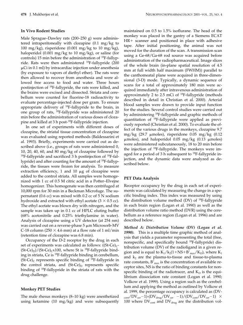
pride and 11C-FLB 457 have also been developed tostudy extrastriatal binding at the D-2 receptors (Fujitaet al. 1999; Delforge et al. 1999). It must be noted thatthese radiotracers also have a significant affinity for theless abundant D-3 receptor subtypes. In this report, wehave not made any attempts to delineate D-3 receptorbinding of 18F-fallypride. Figure 3 shows the selectivelocalization of 18F-fallypride in the thalamus, and thetime-activity curves in Figure 4 show the uptake andclearance of 18F-fallypride from the thalamus and corti-cal areas. This extrastriatal localization of 18F-fallyprideprovides us with an ability to evaluate the extent of D-2receptor occupancy by clozapine, risperidone, and ha-loperidol.
The use of a reference region is frequently employed inthe measurement of D-2 receptor occupancy in brain re-gions using PET and SPECT. For equilibrium studies, thisregion represents the free plus nonspecific radioligandconcentration when measuring the bound to free ratios(Farde et al. 1997). For kinetic studies, a reference regionis used to calculate an index of specific binding, such asbinding potential (BP) or distribution volume ratio (DVR)(Fowler et al. 1998; Volkow et al. 1999). For this work, weinvestigated the use of the cerebellum as a reference re-gion on the calculation of the receptor occupancy.
In employing reference region methods, such as theDVR, the measured occupancy in the high receptor re-gions of the striata were in close agreement with the DVmeasured values reported in Tables 1 and 2. However,in the extrastriatal regions of low receptor density, boththe DV and DVR measured occupancies were found tobe lower than striatal regions. A small, but significant,specific cerebellar binding in rats and our current stud-ies have suggested drug-induced displacement of spe-cific binding in the monkey cerebellum (Mukherjee etal. 1999). This has also been reported for such otherhigh-affinity D-2 antagonists as 11C-FLB 457 (Delforge
Figure 7. Time-activity curvesmeasured using PET in a risperi-done challenge study. Haloperidol(0.05 mg/kg) injected SC, 18 minbefore 18F-fallypride was adminis-tered IV. Various monkey brainregions (putamen, caudate, thala-mus, frontal cortex, temporal cortex,and cerebellum) were evaluated.

NEUROPSYCHOPHARMACOLOGY 2001–VOL. 25, NO. 4 Dopamine D-2 Receptor Occupancy by Neuroleptics 485
et al. 1999) and 125I-epidepride (Hall et al. 1996). There-fore, using the cerebellum as a reference tissue mighthave to be studied carefully to evaluate the extent of oc-cupancy in regions of low receptor concentrations. TheDVR approach would be suitable if a region within thecerebellum can be identified that contains a minimalamount of dopamine receptors and, thus, minimizespecific 18F-fallypride binding in the reference region.
To avoid the confounds of small specific binding inthe cerebellum, the receptor occupancy for this workwas calculated by the use of distribution volumes (DV;Logan et al. 1990; Volkow et al. 1999) as well as the DVRmethod (Logan et al. 1996). Occupancy values obtainedfrom the DV method (which minimizes the specificbinding in the cerebellum) were found to be generallyhigher for all regions for the three drugs. As seen in Ta-bles 1 and 2, using both the methods, occupancy valuesin the extrastriatal regions were similar or lower thanthe striatal regions for the three drugs. Therefore, thiswould suggest that in nonhuman primates at a fairlyhigh dose, clozapine interacts with D-2 receptors in allbrain regions to a similar extent. Using 11C-FLB 457, asimilar striatal and thalamic occupancy level was re-ported by Farde et al. (1997).
Using the ratio analysis similar to that reported byPilowsky et al. (1997), occupancy in the extrastriatal re-gions was found to be higher than striatal regions in thecase of all the drugs, particularly for clozapine and ris-peridone. This finding is similar to the higher temporalcortex D-2 occupancy (90%) compared to that in thestriata (58%) reported in patients on clozapine and mea-sured using a similar ratio method with 123I-epideprideand SPECT (Pilowsky et al. 1997). Measurement of re-ceptor occupancy by the ratio method is confounded byat least two factors: (1) because of the different kineticprofile of 18F-fallypride in the high versus low receptorconcentration regions, the time at which pseudoequilib-rium is achieved is different; and (2) the radiotracerlikely reaches pseudoequilibrium in the drug challengeexperiments at different times as compared to the con-trol experiment and, therefore, measurement of occu-pancy using single time points may be difficult.
Can Endogenous Dopamine Affect D-2 Occupancy by Clozapine?
A significant finding in our studies relates to the differ-ence between clozapine to that of risperidone and halo-peridol in terms of occupying D-2 receptors. Risperi-done and haloperidol being high-affinity dopamine D-2receptor antagonists (Ki 0.3 nM and 0.35 nM, respec-tively, Seeman et al. 1997), exhibited a rather steep in-crease in occupancy with escalating doses. However,clozapine, which has a moderate affinity for D-2 recep-tors (Ki 44 nM, Seeman et al. 1997), exhibited an in-crease in occupancy but did not exhibit a steep rise and
almost exhibited a tendency to plateau at approxi-mately 40 to 60% occupancy before it went up to 90%occupancy with increasing doses. Clozapine has beenshown to exhibit high brain concentrations and is sig-nificantly greater than in the plasma. Also, extractionfraction of clozapine from plasma seems to increasemarginally at higher clozapine concentrations (Baldes-sarini et al. 1993). Our results indicate the increase inoccupancy by clozapine is attributable to the increase inconcentration of clozapine in the brain with increasingdoses and that, at higher doses, there is a significantlygreater concentration of the drug (Figure 1b).
It has been previously suggested that relatively highconcentrations of dopamine in the striata may competewith such moderate affinity compounds as clozapinemore so than with such higher-affinity compounds asrisperidone (Seeman and Tallerico 1999; Wilson et al.1998). Dopamine D-2 receptors exist in two intercon-vertible conformational states, depending on theirG-protein associations, and these states are differentiatedby the affinity with which they bind dopamine and are,thus, referred to as the high-affinity (HA) or the low-affinity state (LA) (Grigoriadis and Seeman 1985). For theHA-state, dopamine has an affinity of approximately 10nM; whereas, for the LA-state, dopamine has an affinityof 5 �M (Seeman et al. 1985). Thus, competition of thevarious drugs with dopamine for the high-affinity statecan be a significant issue. It has been suggested that20% of the D-2 receptors are occupied by dopamine and30% of the receptor sites are vulnerable to increases indopamine (Laruelle 2000).
We have previously shown that binding of 18F-fal-lypride is not significantly altered in reserpinized ratscompared to control rats, suggesting that basal levels ofdopamine has little affect on 18F-fallypride binding toD-2 receptors (Mukherjee et al. 1997). However, whenthe concentration of dopamine is increased by pharma-cological challenges of d-amphetamine, displacement of18F-fallypride is observed. Clozapine on the other hand,having a significantly lower affinity and higher dissoci-ation rate for the D-2 receptors (as compared to risperi-done, haloperidol, and fallypride) is more susceptibleto competition by basal dopamine levels. Thus, al-though both 18F-fallypride and clozapine bind to both,HA- and LA-state of the receptors, the difference intheir dissociation rates allows dopamine to competewith clozapine at the HA-state more efficiently (Seemanand Tallerico 1999). This competition of dopamine withclozapine at the HA-state of the receptors may result inthe transient occupancy nature of clozapine, as sug-gested by Kapur and Seeman (2000).
Because of this dopamine competition, clozapinemay preferentially bind at the LA-state and then pro-gressively occupy the HA-state as the concentration ofclozapine is increased and at higher doses of clozapine,all D-2 receptors are occupied. Thus, it is possible that

486 J. Mukherjee et al. NEUROPSYCHOPHARMACOLOGY 2001–VOL. 25, NO. 4
dopamine, because of its differential affinities for theHA- and LA-state, imposes a pseudobiphasic character-istic on clozapine. Thus, in PET studies with moderatedoses of clozapine an occupancy higher than 60 to 70%is seldom seen. Thus, clozapine may differ from suchhigher affinity drugs as risperidone and haloperidol, interms of sparingly occupying the high-affinity site ascompared to the low-affinity sites of D-2 receptors.
D-2 Occupancy and Extrapyramidal Side Effects
It is generally believed that when neuroleptics exceedapproximately 80% occupancy of the D-2 receptors,propensity of extrapyramidal side effects (EPS) in-crease. Clozapine has been shown not to cause EPS.Previous work from several laboratories has foundclozapine to occupy approximately 60% of the D-2 re-ceptors. It has been suggested that clozapine may ex-hibit “loose binding,” which may be attributable to thecompetition with the radioligand being used as well aswith dopamine, and this may account for the low D-2receptor occupancy seen with clozapine (Seeman et al.1997; Seeman and Tallerico 1999; Wilson et al. 1998).Our findings in PET studies are in agreement withthese occupancy levels. Based on our hypothesis ofclozapine; that is, “high LA state and low HA state oc-cupancy,” is it possible that EPS arises primarily fromhigh occupancy of the HA state of the D-2 receptors? Amore direct evidence of the extent of HA state occu-pancy will be worthwhile to further prove or disprovethis hypothesis.
Limbic Selectivity
One unique feature of clozapine is its limbic selectivity;that is, a more selective inhibition of limbic versus stri-atal dopamine activity. Two major prevailing hypothe-ses include: (1) a differential inhibition of striatal andlimbic dopamine D-2 receptors; and (2) a correctly bal-anced inhibition of several neurotransmitter receptorsfor obtaining limbic selectivity (reviewed in Arnt andSkarsfeldt 1998).
Our work has studied the first hypothesis of differ-ential occupancy of D-2 receptors in the striatal andlimbic areas. In this limited nonhuman primate occu-pancy study with clozapine, a similar extent of occu-pancy in striatal as well as the extrastriatal areas hasbeen observed. Our analyses of the striatal areas (cau-date and putamen) included, in part, the ventral stria-tum, which has been suggested to include the limbic re-gions in the nonhuman primate brain (Heimer et al.1997; Haber and McFarland 1999). Our results of D-2 re-ceptor occupancy in the various brain regions wouldsuggest that the hypothesis of differential occupancy ofD-2 receptors may not account for the limbic selectivity
of clozapine in nonhuman primates. Although the dopa-mine D-2 receptor system has been shown to exhibit ahigh degree of homology between nonhuman primatesand humans, PET studies using 18F-fallypride in humansubjects undergoing treatment with clozapine will beuseful in shedding further light on the issue of differen-tial occupancy in D-2 receptors between striatal and lim-bic areas to account for the limbic selectivity.
CONCLUSIONS
Among the many questions about the unique aspects ofclozapine’s behavior as an atypical neuroleptic drug,we believe our results suggest the following. First, cloz-apine can compete with the binding of 18F-fallypride todopamine D-2 receptors in the striata in rodents andcan occupy all D-2 receptors in vivo. Second, at thera-peutically relevant doses, clozapine can significantlycompete with the binding of 18F-fallypride in all brainregions in the rhesus monkey. Preliminary analysis in-dicates a similar receptor occupancy by clozapine in thestriata (caudate and putamen) and the extrastriata (thal-amus and cortical regions) in the monkey brain. Third,it is postulated that high-affinity drugs, such as halo-peridol and risperidone, bind to similar extent at thevarious conformational states of the receptor. However,moderate affinity drugs, such as clozapine may have areduced occupancy at the high-affinity conformation ofthe receptor because of competition with endogenousdopamine. Because the high-affinity conformation isthe functional state of the receptor, excessive occupancyof this conformation may lead to EPS effects.
ACKNOWLEDGMENTS
Financial support was provided by the U.S. Department ofEnergy DE-FG02-98ER62540. We thank Dr. Harold Stills forhelpful discussions and Kelly Dunigan and Marilyn Brackneyfor technical assistance. We also acknowledge the Wallace-Kettering Neuroscience Institute and the Air Force ResearchLaboratory under Cooperative Agreement No. F33615-98-2-6002 for use of the PET scanner. Presented in part at the29th Annual Meeting of the Society for Neuroscience, Miami,Florida, October 23–28, 1999 and Neuroreceptor MappingMeeting, New York, June 9–11, 2000.
REFERENCES
Arnt J, Skarsfeldt T (1998): Do novel antipsychotics havesimilar pharmacological characteristics? A review ofevidence. Neuropsychopharmacology 18:6–101
Baldessarini RJ, Centorrino F, Flood JG, Volpicelli SA, Hus-ton-Lyons D, Cohen BM (1993): Tissue concentrations ofclozapine and its metabolites in the rat. Neuropsychop-harmacology 9:117–124

NEUROPSYCHOPHARMACOLOGY 2001–VOL. 25, NO. 4 Dopamine D-2 Receptor Occupancy by Neuroleptics 487
Chou Y, Nyberg S, Halldin C (2000): High occupancy of D-2dopamine receptor by high doses of clozapine in theprimate brain. J Nucl Med 41:205 pp
Christian BT, Shi B, Narayanan TK, Mukherjee J (2000):Quantitation of striatal and extrastriatal dopamine D-2receptors using PET imaging of F-18 fallypride in non-human primates. Synapse 38:71–79
Delforge J, Bottlaender M, Loc’h C, Guenther I, Fuseau C,Bendriem B, Syrota A, Maziere B (1999): Quantitation ofextrastriatal D2 receptors using a very high-affinityligand (FLB 457) and the multi-injection approach. JCereb Blood Flow Metab 19:533–546
Farde L, Nordstrom A-L, Nyberg S, Halldin S, Sedvall G(1994): D-1, D-2, and 5HT-2 receptor occupancy in cloz-apine-treated patients. J Clin Psychiat 55:67–69
Farde L, Suhara T, Nyberg S, Karlsson P, Nakashima Y,Hietala J, Halldin, C (1997): A PET study of 11C-FLB 457binding to extrastriatal D-2 dopamine receptors inhealthy subjects and antipsychotic drug-relatedpatients. Psychopharmacology 133:396–404
Fowler JS, Volkow ND, Logan J, Gatley SJ, Pappas N, KingP, Ding Y-S, Wang G-J (1998): Measuring dopaminetransporter occupancy by cocaine in vivo: Radiotracerconsiderations. Synapse 28:111–116
Fujita M, Seibyl JP, Verhoeff NPLG, Ichise M, Baldwin RM,Zoghbi SS, Burger C, Staley JK, Rajeevan N, CharneyDS, Innis RB (1999): Kinetic and equilibrium analyses of123I-epidepride binding to striatal and extrastriataldopamine D2 receptors. Synapse 34:290–304
Grigoriadis D, Seeman P (1985): Complete conversion ofbrain D-2 dopamine receptors from the high- to low-affinity state for dopamine agonists, using sodium ionsand guanine nucleotide. J Neurochem 44:1925–1935
Haber SN, McFarland NR (1999): The concept of the ventralstriatum in nonhuman primates. Ann NY Acad Sci877:33–48
Hall H, Farde L, Halldin C, Hurd YL, Pauli S, Sedvall G(1996): Autoradiographic localization of extrastriatalD2-dopamine receptors in the human brain using 125I-epidepride. Synapse 23:115–123
Hall H, Sedvall G, Magnusson O, Kopp J, Halldin C, Farde L(1994): Distribution of D-1 and D-2 dopamine receptorsand dopamine and its metabolites in the human brain.Neuropsychopharmacology 11:245–256
Heimer L, Alheid GF, de Olmos JS, Groenewegen HJ, HaberSN, Harlan RE, Zahm DS (1997): The accumbens:Beyond the core-shell dichotomy. J Neuropsych ClinNeurosci 9:354–381
Kapur S, Seeman P (2000): Transient occupancy at the D2receptor—A new hypothesis for atypical antipsychotics.Biol Psychiat 47:69S
Kapur S, Zipursky R, Jones C, Shammi CS, Remington G,Seeman P (2000): A positron emission tomographystudy of quetiapine in schizophrenia. Arch Gen Psy-chiat 57:553–559
Kapur S, Zipursky RB, Remington G (1999): Clinical and the-oretical implications of 5HT2 and D2 receptor occu-pancy of clozapine, risperidone, and olanzapine inschizophrenia. Am J Psychiat 156:286–293
Kessler RM, Ansari S, Votaw JR, de Paulis T, Clanton JA,Schmidt DE, Mason NS, Manning, RG (1993): Identifica-
tion of extrastriatal dopamine D-2 receptors in post-mortem human brain with 125I-epidepride. Brain Res609:237–243
Kufferle B, Tauscher J, Asenbaum S, Vesely C, Podreka I,Bruke T, Kasper S (1997): IBZM SPECT imaging of stri-atal dopamine-2 receptors in psychotic patients treatedwith the novel antipsychotic substance quetiapine incomparison to clozapine and haloperidol. Psychophar-macology (Berl) 133:323–328
Laruelle M (2000): Imaging synaptic neurotransmission within vivo binding competition techniques: A criticalreview. J Cereb Blood Flow Metab 20:423–451
Leysen JE, Janssen PMF, Schotte A, Luyten WHML, MegensAAHP (1993): Interaction of antipsychotic drugs withneurotransmitter receptor sites in vitro and in vivo inrelation to pharmacological and clinical effects: Role of5HT2 receptors. Psychopharmacology 112:S40–S54
Logan J, Fowler JS, Volkow ND, Wolf AP, Dewey SL,Schlyer DJ, MacGregor RR, Hizemann R, Bendriem B,Gatley SJ, Christman DR (1990): Graphical analysis ofreversible radioligand binding from time-activity mea-surement applied to [N-11C-methyl]-(-)cocaine PETstudies in human subjects. J Cereb Blood Flow Metab10:740–747
Logan J, Fowler JS, Volkow ND, Wang GJ, Ding YS, AlexoffDL (1996): Distribution volume ratios without bloodsampling from graphical analysis and PET data. J CerebBlood Flow Metab 16:834–840
Meltzer HY (1991): The mechanism of action of novel anti-psychotic drugs. Schizophr Bull 17:263–287
Meltzer HY (1994): An overview of the mechanism of actionof clozapine. J Clin Psychiat 55:47–52
Mukherjee J, Yang Z-Y, Lew R, Brown T, Kronmal S, CooperM, Seiden LS (1997): Evaluation of d-amphetamineeffects on the binding of dopamine D-2 receptor radioli-gand, 18F-fallypride in non-human primates usingpositron emission tomography. Synapse 27:1–13
Mukherjee J, Yang Z-Y, Brown T, Lew R, Wernick M, Ouy-ang X, Yasillo N, Chen C-T, Mintzer R, Cooper M(1999): Preliminary assessment of extrastriatal dopa-mine D-2 receptor binding in the rodent and nonhumanprimate brains using the high affinity radioligand, 18F-fallypride. Nucl Med Biol 26:519–527
Nyberg S, Farde L (2000): Nonequipotent doses partlyexplain differences among antipsychotics—Implica-tions of PET studies. Psychopharmacology 148:22–23
Pickar D, Su TP, Weinberger DR, Coppola R, Malhotra AK,Knable MB, Lee KS, Gorey J, Bartko MB, Breier A, HsiaoJ (1996): Individual variation in D-2 dopamine receptoroccupancy in clozapine-treated patients. Am J Psychiat153:1571–1578
Pilowsky LS, Mulligan RS, Acton PD, Ell PJ, Costa DC, Ker-win RW (1997): Limbic selectivity of clozapine. Lancet350:490–491
Remington G, Kapur S (2000): Atypical antipsychotics: Aresome more atypical than others? Psychopharmacology148:3–15
Rosenheck R, Cramer J, Xu W, Thomas J, Henderson W,Frisman L, Fye C, Charney D (1997): A comparison ofclozapine and haloperidol in hospitalized patients withrefractory schizophrenia. N Engl J Med 337:809–815
488 J. Mukherjee et al. NEUROPSYCHOPHARMACOLOGY 2001–VOL. 25, NO. 4
Safferman A, Lieberman JA, Kane JM, Szymanski S, Kinon B(1991): Update on the clinical efficacy and side effects ofclozapine. Schizophr Bull 17:247–261
Schaus JM, Bymaster FP (1998): Dopaminergic approachesto antipsychotic agents. In Robertson DW (ed), AnnualReports in Medicinal Chemistry, San Diego: AcademicPress, pp 1–10
Schwartz JT, Brotman AW (1992): A clinical guide to antip-sychotics. Drugs 44:981–992
Seeman P, Corbett R, Van Tol HM (1997): Atypical neurolep-tics have low affinity for dopamine D-2 receptors or areselective for D-4 receptors. Neuropscyhopharmacology16:93–110
Seeman P, Watanabe M, Grigoriadis D, Tedesco JL, GeorgeSR, Svensson U, Nilsson JLG, Neumeyer JL (1985):Dopamine D-2 receptor binding sites for agonists: A tet-rahedral model. Mol Pharmacol 28:391–399
Seeman P, Tallerico T (1999): Rapid release of antipsychoticdrugs from dopamine D-2 receptors: An explanation forlow receptor occupancy and early clinical relapse uponwithdrawal of clozapine or quetiapine. Am J Psychiat156: 876–884
Sunahara RK, Seeman P, Van Tol HHM, Niznik HB (1993):Dopamine receptors and antipsychotic drug response.Br J Psychiat 163:31–38
Volkow ND, Wang G-J, Fowler JS, Logan J, Gatley SJ, WongC, Hitzemann R, Pappas NR (1999): Reinforcing effectsof psychostimulants in humans are associated withincreases in brain dopamine and occupancy of D2receptors. J Pharm Exp Therapeut 291:409–415
Wilson JM, Sanyal S, Van Tol HHM (1998): Dopamine D-2and D-4 receptor ligands: Relation to antipsychoticaction. Euro J Pharmacol 351:273–286