Estudos Interdisciplinares em Ciências Sociais e da Saúde
173
-
Upload
khangminh22 -
Category
Documents
-
view
0 -
download
0
Transcript of Estudos Interdisciplinares em Ciências Sociais e da Saúde


2
Interdisciplinary Studies in Social Sciences and Health, Vitória-ES, v.1, n.1, p 1-173, jul-aug-sept./2020
Estudos Interdisciplinares em Ciências Sociais
e da Saúde Interdisciplinary Studies in Social Sciences and Health
v. 1 n. 1 jul.aug.sept./2020

3
Interdisciplinary Studies in Social Sciences and Health, Vitória-ES, v.1, n.1, p 1-173, jul-aug-sept./2020
ESTUDOS INTERDISCIPLINARES EM CIÊNCIAS SOCIAIS E DA SAÚDE Interdisciplinary Studies in Social Sciences and Health
Holy House of Mercy of Vitória Brotherhood
PROVOST
Maria da Penha Rodrigues d’Avila
VICE-PROVOST
Cláudio Medina da Fonseca
BOARD OF DIRECTORS (Incumbent):
Nilo Fernando Rezende Vieira
Antônio Chambô Filho
Sérgio Rubens de Aguiar
Deuber Erly Pretti
BOARD OF DIRECTORS (Substitute):
Joelmar César de Almeida
Elzalina Ramos Barbosa
Décio Sesquim
FISCAL COUNCIL (Incumbent):
Boris Castro
José Antônio Pecorari
Leocarlos Dias da Silva
FISCAL COUNCIL (Substitute):
José Carlos Lyrio Rocha
Maria Bernadete Bringhenti Feu Rosa
Escola Superior de Ciências da Santa Casa de Misericórdia de Vitória – EMESCAM
DIRECTOR
Cláudio Medina da Fonseca
RESEARCH AND GRADUATE COORDINATOR STRICTO SENSU:
Valmin Ramos da Silva

4
Interdisciplinary Studies in Social Sciences and Health, Vitória-ES, v.1, n.1, p 1-173, jul-aug-sept./2020
ESTUDOS INTERDISCIPLINARES EM CIÊNCIAS SOCIAIS E DA SAÚDE Interdisciplinary Studies in Social Sciences and Health
EDITORIAL BOARD
Editor in-chief
• Valmin Ramos da Silva – EMESCAM – Vitória/ES
Associate Editors
• Chárbel Jacob Junior – EMESCAM – Vitória/ES
• Cristina Ribeiro Macedo – EMESCAM – Vitória/ES
• Danilo Nagib Salomão Paulo – EMESCAM – Vitória/ES
• Gissele Carraro – EMESCAM – Vitória/ES
• Janine Pereira da Silva– EMESCAM – Vitória/ES
• Marcela Souza Lima Paulo – EMESCAM – Vitória/ES
• Renato Lirio Morelato – EMESCAM – Vitória/ES
• Roberta Ribeiro Batista – EMESCAM – Vitória/ES
• Roberto Ramos Barbosa – EMESCAM – Vitória/ES
• Rubens José Loureiro – EMESCAM – Vitória/ES
National Reviewers Council
• Ademar Vieira de Barros – EMESCAM – Vitória/ES
• Adércio João Marquezini – EMESCAM – Vitória/ES
• Afrânio Cogo Destefani – EMESCAM – Vitória/ES
• Alessandra Tieppo – EMESCAM – Vitória/ES
• Álvaro Armando Carvalho de Morais – EMESCAM – Vitória/ES
• Alvino Jorge Guerra – EMESCAM – Vitória/ES
• Andre Carnevali da Silva – EMESCAM – Vitória/ES
• Andy Petroianu – UFMG – Belo Horizonte/MG
• Angela Caulyt Santos da Silva – EMESCAM – Vitória/ES
• Antônio Chambô Filho – EMESCAM – Vitória/ES
• César Albenes de Mendonça Cruz – EMESCAM – Vitória/ES
• Chárbel Jacob Junior – EMESCAM – Vitória/ES
• Cláudia Gomes Rossoni – EMESCAM – Vitória/ES
• Carlo Eduardo Montaño Barreto – UFRJ – Rio de janeiro/RJ
• Dalton Valentim Vassallo – EMESCAM/UFES – Vitória/ES
• Danilo Nagib Salomão Paulo - EMESCAM/UFES – Vitória/ES
• Faradiba Sarquis Serpa – EMESCAM – Vitória/ES
• Giovana Machado Souza Simões – EMESCAM – Vitória/ES
• Gissele Carraro – EMESCAM – Vitória/ES
• Gustavo Carreiro Pinasco – EMESCAM – Vitória/ES
• Gustavo Peixoto Soares Miguel – UFES – Vitória/ES
• Gustavo Rocha Leite – UFES – Vitória/ES
• Haydêe Fagundes Moreira Silva de Mendonça – EMESCAM – Vitória/ES
• Hebert Wilson Santos Cabral – EMESCAM – Vitória/ES - UFF – Rio de Janeiro/RJ
• Janice Gusmão Ferreira de Trindade – EMESCAM – Vitória/ES
• Janine Pereira da Silva – Emescam – Vitória/ES
• José Carlos Novaes – EMESCAM – Vitória/ES

5
Interdisciplinary Studies in Social Sciences and Health, Vitória-ES, v.1, n.1, p 1-173, jul-aug-sept./2020
• José Guilherme Pinheiro Pires – EMESCAM – Vitória/ES
• Lauro Ferreira da Silva Pinto Neto – EMESCAM – Vitória/ES
• Luciana Carrupt Machado Sogame – EMESCAM – Vitória/ES
• Lúcia Helena Sagrillo Pimassoni
• Luiz Renato da Silveira Costa – EMESCAM – Vitória/ES
• Maria Carlota de Rezende Coelho – EMESCAM – Vitória-ES
• Marcela Souza Lima Paulo – EMESCAM – Vitória/ES
• Maria da Graça Silva Mattede – EMESCAM – Vitória/ES
• Maria de Fátima dos Santos Nacari – EMESCAM – Vitória/ES
• Maristela Dalbello de Araujo – EMESCAM – Vitória/ES
• Mitre Kalil – EMESCAM – Vitória/ES
• Paulo Roberto Savassi Rocha – UFMG – Belo Horizonte/MG
• Patrícia Casagrande Dias de Almeida – EMESCAM – Vitória/ES
• Renato Lirio Morelato – EMESCAM – Vitória/ES
• Roberta Ribeiro Batista Barbosa – EMESCAM – Vitória/ES
• Silvia Moreira Trugilho – EMESCAM – Vitória/ES
• Silvio Roberto Foletto – EMESCAM – Vitória/ES
• Solange Rodrigues da Costa – EMESCAM – Vitória/ES
• Tarcizo Afonso Nunes – UFMG – Belo Horizonte/MG
• Vinícius Gomes da Silveira – UFRJ – Rio de Janeiro/RJ
International Reviewers Council
Carmelo Loinaz Segurola – ANECA – San Sebastián (Guipúzcoa) – Spain
Emilio Vicente López – Zaragoza University – Spain
Anelisa Dazzi Chequer de Souza – Clinical Lecturer at University of Tasmania School of
Medicine – Australia
Nick Cooling – University of Tasmania – Austrália
Fátima Helena Sert Kuniyoshi – Mayo Clinic College of Medicine e clinical research
associate – Philips Respironics – EUA
Biostatistician
• Lúcia Helena Sagrillo Pimassoni
Grammatical Review of the Portuguese Version
• Loise Cristina Passos Drumond
Page designer
• Rosângela Marques de Almeida Ribeiro
Executive Secretary
• Erica Nascimento de Vitória
Periodicity of publication
• Quartely
Languages in which papers will be accepted
• Portuguese and English
Corporate Author • Escola Superior de Ciências da Santa Casa de Misericórdia – EMESCAM
Avenida Nossa Senhora da Penha, 2190, Santa Lúcia, Vitória – ES
6
Interdisciplinary Studies in Social Sciences and Health, Vitória-ES, v.1, n.1, p 1-173, jul-aug-sept./2020
SUMMARY
Lack of association SNP+45T>G with cardiovascular diseases in obese patients ................. 9
Gabriela Souza do Nascimento, Iara Almeida Pinto, Simone Rodrigues da Fonseca, Perseu
Seixas de Carvalho, Ariele Spanhol Rosseto, Janine Baptista Coimbra, Josivany Valério de
Freitas, Flavia Imbroisi Valle Errera.
The Mannheim Peritonitis Index to predict the outcome of post-surgery of peritonitis....... 20
Laís dos Santos Gueiros, Claudio Medina da Fonseca, Nathalia Maria Dias Moraes Duarte,
Olívia Souza Antunes.
Therapeutic benefits of dry needling in adult patients with crhonic low back ..................... 35
Luana Moraes Leal, Jeanne Florence Soares Santos, Monique Almeida Fernandes, Pitiguara
de Freitas Coelho e Francisco Rodrigues Brioschi.
Influence of medular injury and wheelchair time in respiratory and tosse muscle force. ... 42
Taynara Ribeiro Batalha, Morghana Ferreira Ambrosim, Roberta Ribeiro Batista Barbosa e
Christiane Lourenço Mota
Relations from blood count parameters with mortality and time of hospitalization in critical
patients ..................................................................................................................................... 51
Fabio dos Santos Baptista, Filipe Lemos Bellote, Frederico Ferreira, Carla Tardin Alves
Bellon, João Pedro de Oliveira e Oliveira, Maria das Graças Silva Mattede e Alvaro Armando
Carvalho de Morais
Analysis of bacterial vaginal microbiota in postmenopausal women who have not
undergone hormone therapy ................................................................................................... 64
Fabio dos Santos Baptista, Filipe Lemos Bellote, Frederico Ferreira, Carla Tardin Alves
Bellon, João Pedro de Oliveira e Oliveira, Maria das Graças Silva Mattede e Alvaro Armando
Carvalho de Morais
Association between quality of life and mortality in patients with heart failure .................. 74
Roberto Ramos Barbosa, Luíza Dias Torres, Mayara da Silva, Carla Campos Miranda, Ingrid
Ardisson Colodete, Andressa Corteletti, Tiago de Melo Jacques, Renato Giestas Serpa, Osmar
Araujo Calil e Luiz Fernando Machado Barbosa
The influence of risk of falling on functional dependence of homebound elderly ............... 81
Morghana Ferreira Ambrosim, Taynara Ribeiro Batalha, Fabiola dos Santos Dornellas
Oliveira, Christiane Lourenço Mota, Vanezia Gonçalves da Silva

7
Interdisciplinary Studies in Social Sciences and Health, Vitória-ES, v.1, n.1, p 1-173, jul-aug-sept./2020
Analyses of the hepatotoxic effects related to MTX use in psoriasis treatment .................... 94
Izabelle Venturini Signorelli, João Basilio Espíndola de Souza, Maysa Gavassoni Noemann,
Paula Rodrigues Villela da Motta
Cadmium exposure effects on the blood pressure and myocardial contractility................. 108
Cindy Medici Toscano, Ingridy Reinholz Graffiti Schereider, Priscila Rossi de Batista, Dalton
Valentim Vassallo
Refractory digestive hemorragy by use of direct anticoagulant in octagenary patient with
chronic atrial fibrillation: case report .................................................................................. 117
Renato Lirio Morelato, Ana Clara de Barros Pretti, Bruno Pignaton Ruschi de Aragão, Luíza
Moulin Rezende, Cristiano Ventorim de Barros, Flavio Takemi Kataoka, Luciene Lage da
Motta, Alessandra Tieppo.
Exposure to cadmium as a public health problem and its effect on resistance vessels ...... 123
Priscila Rossi de Batista, Ingridy Reinholz Grafites Schereider, Cindy Medici Toscano,
Dalton Valentim Vassallo.
Acute and chronic cadmium effects on conductance vessels............................................... 137
Ingridy Reinholz Grafites Schereider, Cindy Medici Toscano, Priscila Rossi de Batista,
Maylla Ronacher Simões, Dalton Valentim Vassallo
Pilot study of Ricasa project: late follow-up of patients hospitalized with dispensed heart
failure ..................................................................................................................................... 145
Vítor Lorencini Belloti, Rodolfo Dazzi Bobbio, Pietro Dall’Orto Lima, Renato Giestas Serpa,
Osmar Araujo Calil Luiz Fernando Machado Barbosa, Roberto Ramos Barbosa
Mild neurocognitive disorder: comparison of two cognitive evaluation tests ..................... 152
Lorena Aparecida Bolelli Tatagiba, Tabata Cristina de Oliveira, Daniel Alves Loureiro, Pedro
Trés Vieira Gomes, Raphael Benevenut, Livia Terezinha Devens, Alessandra Tieppo, Renato
Lirio Morelato
Comparative study involving Dolutegravir and Efavirenz in HIV antyretroviral therapy
schemes: drug adherence, efficacy and adverse effects ....................................................... 160
Sara Sary Eldim Campanati, Marina Reis Thiebaut Pereira, Larissa Alvim Werner, Lauro
Ferreira da Silva Pinto Neto

8
Interdisciplinary Studies in Social Sciences and Health, Vitória-ES, v.1, n.1, p 1-173, jul-aug-sept./2020
Presentation Letter
SALUS: Journal of Health Sciences - ISSN 2447-7826, Qualis Capes B4, held its first issue
published on October 15, 2015. From the seventh issue, the journal will be issued as a Journal
of Interdisciplinary Studies in Sciences Social and Health (REICSS) by virtue of the
identification of the company holding the name used.
REICSS will keep on being the vehicle for scientific communication at the Santa Casa de
Misericórdia de Vitória College of Sciences (EMESCAM), which features an undergraduate
course in Medicine, for more than half a century, apart from Nursing, Physiotherapy, Social
Work courses and Postgraduate Course stricto sensu, Academic Master's in Public Policy and
Local Development, for over 10 years.
EMESCAM is sponsored by the Brotherhood of Santa Casa de Misericórdia in Vitória
Hospital (HSCMV), ES, Brazil, a philanthropic institution. The hospital was founded in the
16th century and it is located in downtown Vitória - ES.
The journal observes the same standards, ethical and scientific rigor as journals with the most
national and international academic impact, with the purpose of disseminating the production
of interdisciplinary scientific knowledge in the areas of Health Sciences, Applied Social
Sciences and Public Policies.
Editorial policy and administrative procedures will be supervised by Editora Emescam,
affiliated to the Brazilian Association of University Publishers from the second half of 2019.
We desire to rely on the scientific community support, granting us prestige by providing the
opportunity to divulge the results of quality research. The purpose is to completely abolish the
endogeny inevitably noticed in this first issue.
Success for all of us!
Valmin Ramos da Silva, MD, PhD
Editor

9
Interdisciplinary Studies in Social Sciences and Health, Vitória-ES, v.1, n.1, p 1-173, jul-aug-sept./2020
DOI https://dx.doi.org/10.5935/2675-7397.20200004
Original Article
LACK OF ASSOCIATION SNP+45T>G WITH CARDIOVASCULAR DISEASES IN
OBESE PATIENTS
Gabriela Souza do Nascimento1, Iara Almeida Pinto2, Simone Rodrigues da Fonseca3, Perseu
Seixas de Carvalho4, Ariele Spanhol Rosseto5, Janine Baptista Coimbra6, Josivany Valério de
Freitas7, Flavia Imbroisi Valle Errera8
1 College of Science of Santa Casa de Misericórdia de Vitória (EMESCAM). Fellow by
FAPES. (Medicine Undergraduate Student).
2 Bachelor of Pharmacy from College of Science of Santa Casa de Misericórdia de Vitória
(EMESCAM). (M.S.Student by Postgraduate Program in Biotechnology at Federal
University of Espírito Santo (UFES) e Fellow by Industrial Technological Development –
CNPq – Level 3).
3 Pharmaceutical. Expert in Clinical Analyses. (Master´s Degree Program from Federal
University of Espírito Santo – UFES).
4 Medical Doctor. Expert in Endocrinology from Federal University of Espírito Santo
(UFES) – (Medical Doctor Expert in Endocrinology).
5 Biologist from FAESA. Ph.D. in Medical Genetics and Oncology from University of
Siena. Postdoctoral Degree in Molecular Biology and Bioinformatics in Ematology,
CREO, from University of Perugia. (M.S. Student in Biomolecular Sciences from
University of Perugia and Biologist at Foligno Nursing School, University of Perugia,
Italy. Collaborator at University of Freiburg in 2010).
6 Pharmaceutical. Medical Doctor. (Civil Police – Espírito Santo State)
7 Biologist. M.S. and Ph.D. in Biotechnology. Northeast Network of Biotechnology/
Federal University of Espírito Santo – UFES – (Professor).
8 B.S. in Biology from Federal University of Espírito Santo (1999) and Phd.D. in
Biological Sciences (Genetics) from University of São Paulo (2006). (Adjunct Professor
of Genetic disciplines at College of Science of Santa Casa de Misericórdia de Vitória –
EMESCAM. (coordinator of Institutional and International Relations).
Submitted in April 04, 2016; Approved in September 15, 2017
Institution: EMESCAM
Av. Nossa Sra. da Penha, 2190 – Santa Luíza, Vitória - ES, 29045-402.
ESTUDOS INTERDISCIPLINARES EM
CIÊNCIAS SOCIAIS E DA SAÚDE INTERDISCIPLINARY STUDIES IN SOCIAL SCIENCES AND HEALTH
v.1, n.1, 2020

10
Interdisciplinary Studies in Social Sciences and Health, Vitória-ES, v.1, n.1, p 1-173, jul-aug-sept./2020
Correspondence Author: Iara Almeida Pinto.
Escola Superior de Ciências da Santa Casa de Misericórdia de Vitória, ES.
Av. Nossa Sra. da Penha, 2190 – Santa Luíza, Vitória - ES, 29045-402.
Email: [email protected]
Keywords Abstract
Adiponectin;
Obesity;
Polymorphism;
Single Nucleotide;
Cardiovascular Diseases.
In order to assess whether the single nucleotide
polymorphism (SNP) rs2241766 (+ 45T> G) in the
adiponectin gene (ADIPOQ) is related to the presence
of cardiovascular diseases (CVD) in overweight
patients, it was carried out a survey of CVDs in
patients (IMC ≥ 25 kg / m2) from the clinical
database of the Molecular Genetics Laboratory of
EMESCAM. It took place the extraction of genomic
DNA from peripheral blood, amplified by the
Polymerase Chain Reaction technique, followed by
enzyme digestion and polyacrylamide gel
electrophoresis and staining. The genotypic and
allelic frequencies were compared by the chi-square
test, with p G value of the ADIPOQ gene in
overweight patients, in none of the genetic
inheritance models. Nevertheless, the presence of
familial history had a significant association with the
severity of CVD manifestation.
INTRODUCTION
Obesity is a global epidemic that brings
serious health complications1 and it is
related to an increase in mortality2. Its
prevalence has increased in recent years
and it is estimated that about 58% of adults
worldwide will be overweight or obese by
20303. Its progression is inherent in
increased risk for various diseases, mainly
cardiovascular diseases (CVD) 2-5.
CVD is a group of diseases of the heart and
blood vessels which are the major cause of
death in the world. The Ministry of Health
Data (2011) reveal that CVD represent
29.4% of all deaths recorded in Brazil,
symbolizing that more than 308,000 people
died mostly of heart attack and stroke.
Therefore, the country is placed among the
ten countries with higher CVD deaths. In
the state of Espírito Santo, an analysis of
listed deaths using the International Code
of Diseases (ICD-10) chapter reveals that
diseases of the circulatory system,
including CVDs, are the leading causes of
death, representing a total of 29.5% 7.
Atherosclerosis, the central characteristic
of CVD, is a chronic inflammatory disease
of multifactorial origin that arises in
response to endothelial aggression,
affecting medium- and large-caliber
arteries8. Another important aspect is the
deficiency in the release of nitric oxide
(NO) from vascular endothelium and the
decrease of blood flow to target tissues of
insulin, contributing to insulin resistance,
also known as endothelial dysfunction1.
Endothelial function is controlled by
various molecules, including adipokines or
adipocytokines, cytokines secreted by

11
Interdisciplinary Studies in Social Sciences and Health, Vitória-ES, v.1, n.1, p 1-173, jul-aug-sept./2020
adipocytes2. Leptin, monocyte chemotactic
protein-1 (MCP-1), plasminogen activator
inhibitor -1 (PAI-1), tumor necrosis factor
alpha (TNFα), interleukin 6 (IL-6), resistin
and adiponectin are some of these
adipocytokines associated with endothelial
dysfunction9.
Adiponectin, the most plentiful secreted
adipocytokine, has antiatherogenic, anti-
inflammatory and antidiabetic properties10.
The change in adipokine profile in the
serum of obese people, such as levels of
adiponectin and proinflammatory
cytokines, is a contributing factor in the
development of cardiometabolic alterations3.
Low circulating levels of adiponectin
(hypoadiponectinemia) are considered an
independent risk factor for endothelial
dysfunction,4,1 due to an interaction of
environmental factors and genetic factors
such as single nucleotide polymorphisms
(SNP) in the adiponectin gene (ADIPOQ).
Besides, this gene has been related to
various characteristics of metabolic
syndrome, DM2 and DCV11-15.
Taking into consideration that obesity is
characterized by a chronic, pro-
inflammatory state causing hyperplasia and
fat cell hypertrophy, leading to an
imbalance in adipokine release16 and also
the important regulatory role that
adipokines play, in particular adiponectin,
the aim of the present study was to verify if
the SNP rs2241766 (+ 45T> G), present in
the ADIPOQ gene, is related to the
presence of cardiovascular diseases in
patients with obesity as well as if the
family history influences the association of
the polymorphism.
METHOD
This study is part of a larger project titled
"Adiponectin Gene Polymorphism and
Type 2 Diabetes Risk in Obese Patients
from the population of Vitória-ES”, based
on a sample composed of overweight
patients (BMI ≥ 25 Kg / m2), for which a
survey regarding CVDs was performed.
Among them, are hypertension, angina,
infarction and stroke (stroke). Patients with
excess weight and with at least one CVD
were considered as cases, and patients with
excess weight and with no CVD were
considered non-cases. The survey of CVD
was carried out based on the sample and on
the frequency of familial history. Taking
into account that all cases presented
hypertension, we considered as severe
cases patients who had some other CVD in
addition to hypertension, and with less
severity those who only had hypertension.
A positive family history was considered
when more than one member of the family
with CVD was reported. The information
was acquired through medical records of
each patient.
Genomic DNA was extracted from
peripheral blood leukocytes as stated by
the protocol of Miller et al. (1988) and
intensified by the PCR-RFLP (polymerase
chain reaction-restriction fragment length
polymorphism) technique. PCR conditions
were initial denaturation for 5 minutes at
95 ° C, followed by 35 cycles of 1 minute
at 95 ° C, 1 minute at 55 ° C and 40
seconds at 72 ° C; final extension for 10
minutes at 72 ° C, followed by digestion
with the BspHI enzyme of 0.1 U
concentration in the dry bath at 37 ° C for
8 hours. The products of the PCR reaction
were analyzed by polyacrylamide gel
electrophoresis (8%), visualized by 0.1%
silver nitrate staining18.
The cases (associated with the presence of
CVDs) and the non-cases, which presented
genotype for the SNP + 45T> G, were
examined using the chi-square test. For the
purpose of determining if genotype
distributions were in Hardy-Weinberg
equilibrium, genotypic and allelic
frequencies were compared between cases
and non-cases using chi-square; a p value
<0.05 was considered significant.

12
Interdisciplinary Studies in Social Sciences and Health, Vitória-ES, v.1, n.1, p 1-173, jul-aug-sept./2020
This work was approved by the Research
Ethics Committee of College of Sciences
of Santa Casa de Misericórdia
(EMESCAM) of Vitória - ES under nº
076/2007.
RESULTS
The genotypes were acquired for 112
patients. Most of them were women
(85.7%) obese (94.6%) and with a mean
age of 41.61 ± 13.12 years. With respect to
the type of CVD, 80.61% (70/79) of the
cases presented hypertension, 8.86% (7/79)
angina, 1.26% (1/79) infarction and 1.26%
(1/79) strokes. Out of the total, 70.5%
(79/112) presented CVD and were
considered cases (table 1). The genotype
frequency in the patients was TT 24.1%
(19 cases), GG 15.2% (12 cases), and TG
60.7% (48 cases). The allelic frequency
was 54.4% for the T allele and 45.6% for
the allele G. The allelic and genotypic
frequencies were not in Hardy-Weinberg
equilibrium (p = 0.0457). There was no
relation of polymorphism to any
cardiovascular disease in any of the genetic
inheritance models (Table 2). Examining
genotypes (p = 0.188) and genetic models
(Dominant p = 0.128, Recessive p = 0.177
and Codominant p = 0.734), there was no
significant association of severity with
polymorphism (Table 4).
The family history of CVDs was related to
severe and less severe forms (p = 0.003)
(Table 3). On account of this result, we
analyzed the cases and non-cases with less
severity that presented positive family
history, in search of knowing the influence
of polymorphism on hypertension.
However, there was no significant
difference (p = 0.317).
Table 1- Frequency of Cardiovascular Diseases and genotypes between the cases.
SNP+45T>G
Hypertension Angina Infarction CA Stroke
n
TT 19 15 (78.9%) 3 (15.8%) 1 (5.3%) 0 (0.0%)
TG 48 43 (89.6%) 4 (8.3%) 0 (0.0%) 1 (2.1%)
GG 12 12(100.0%) 0 (0.0%) 0 (0.0%) 0 (0.0%)
CA = Cerebrovascular Accident (Stroke); SNP = Single Nucleotide Polymorphisms
Resource: author

13
Interdisciplinary Studies in Social Sciences and Health, Vitória-ES, v.1, n.1, p 1-173, jul-aug-sept./2020
Table 2- Gentotypic and allelic distribution in genetic models between cases and non-cases.
Cases Non-Cases p
Genotypic Distribution
TT 19 (24.1%) 6 (18.2%)
GG 12 (15.2%) 2 (6.1%)
TG 48 (60.7%) 25 (75.7%)
Allelic Distribution
T 86 (54.4%) 37 (56.0%) 0.88
G 72 (45.6%) 29 (44.0%)
Dominant Model
TT 19 (17.0%) 6 (5.4%) 0.49
TG + GG 60 (53.6%) 27 (24.1%)
Recessive Model
GG 12 (10.7%) 2 (1.8%) 0.18
TG +TT 67 (59.8%) 31 (27.7%)
Condominant Model
TG 48 (42.9%) 25 (22.3%) 0.13
TT+GG 31 (27.7%) 8 (7.1%)
Resource: author.
Table 3 – Familiar History of Cardiovascular Diseases between cases and non-cases.
Cases Non-Cases
p
FH positive 76 (96.20%) 26 (78.79%)
FH negative 3 (3.80%) 7 (21.21%) 0.003
Total
79 33
FH = Familiar History
Resource: author

14
Interdisciplinary Studies in Social Sciences and Health, Vitória-ES, v.1, n.1, p 1-173, jul-aug-sept./2020
Table 4 - Genotypic Distribution of SNT+45T>G in patients with severe
and non-severe DCV
Severe DVC
n (%)
Non-severe DVC
n (%)
p
Genotype
TT 4 (5.1%) 15 (19.0%)
GG 0 (0.0%) 12 (5.2%) 0.188
TG 5 (6.3%) 43 (54.4%)
Dominant Model
TT 4 (5.1%) 15 (19.0%) 0.128
TG + GG 5 (6.3%) 55 (69.6%)
Recessive Model
GG 9 (11.4%) 58 (73.4%) 0.177
TG +TT 0 (0.0%) 12 (15.2%)
Condominant Model
TG 4 (5.1%) 27 (34.2%) 0.734
TT+ GG 5 (6.3%) 43 (54.4%)
SNP = Single Nucleotide Polymorphisms; CVD = Cardiovascular Diseases.
Resource: autohor.
DISCUSSION
Some studies indicate the association of
ADIPOQ polymorphisms with CVD 13,21,23,33. Yet, since this association is still
controversial, it is important to check the
prevalence of alleles in order to confirm
the increased risk in different populations
and to accompany risk groups to better
understand the evolution and clinical
management of CVD. The major purpose
of this study was to analyze whether the +
45T> G polymorphism in the ADIPOQ
gene is related to CVD and whether the
presence of family history influences this
relation.
Around 30% of all global deaths are from
CVDs, with coronary heart disease at 7.4
million, and stroke at 6.7 million deaths19.
The CVD mortality rate in Brazil is 214
per 100,000 inhabitants20. In the state of
Espírito Santo, diseases in the circulatory
system, including CVDs, are the leading
causes of death, representing a total of
30.2% 7. In the current study, 80.6% of the
patients had some type of CVD, with
hypertension being the most frequent,
corroborating their increased prevalence in
overweight patients.
In this research, the three genotypes (GG,
GT, TT) were observed in subjects with
hypertension, and the groups of
heterozygotes and GG homozygotes
corresponded to the majority of the
individuals studied. We also noted that the
G (+ 45T> G) allele was present in most
patients with CVD, predominantly in
hypertensive individuals. Nevertheless, no
association was perceived between
genotypes and CVD.
Oliveira et al. (2005) associated variants of
the ADIPOQ gene in diabetic and non-
diabetic Brazilian patients, independent of
circulating adiponectin, BMI and glycemic
levels. They described an association of

15
Interdisciplinary Studies in Social Sciences and Health, Vitória-ES, v.1, n.1, p 1-173, jul-aug-sept./2020
variants in the adiponectin gene with CVD
in patients with type 2 diabetes mellitus
DM2). Zhou et al. (2014) in a meta-
analysis, found out the distribution of cases
and controls genotypes for the + 45T> G in
relation to coronary artery disease (CAD),
stated associations of variants in the
adiponectin gene with the risk of
developing the disease in the study group.
Ferrarezi et al. (2007) in
A review of evidence reported
associations of allelic variants of the
adiponectin gene with CVD in patients
with Type 2 Diabetes Mellitus (DM2).
These authors cite that GG homozygotes
and TG heterozygotes when compared to
TT homozygotes, are at increased risk for
CAD. Another study achieved with French
and Swiss patients with DM2 and CAD
also noticed an association of SNP + 45T>
G with CAD. Besides, susceptibility to
CAD, due to SNP + 45T> G, was
independent of cardiovascular risk factors
classics23.
Some studies suggest that the G allele is at
risk. However, Oliveira et al. (2005)
reported that adiponectin plasma levels and
the interaction of diseases such as obesity,
insulin resistance, DM2, and CAD showed
high mRNA expression in adipose tissue,
as well as total circulating adiponectin
levels higher in patients with the G allele
and hypothesized that this would be a
protective factor.
Witberg et al. (2016) report that high levels
of adiponectin were associated with
increased mortality and morbidity in a
young multiethnic population with CVD.
Lindberg et al. (2015) eventually followed
random individuals from a community and
concluded that the increase in plasma
adiponectin was associated with decreased
risk of DM2 and subsequent cardiovascular
events. Ferrarezi et al. (2007),
concentrating studies of the association of
ADIPOQ in patients with diabetes and
CVD, notify that genotype-related effects
on serum adiponectin levels are not
consistently observed and explain that this
is resulting from, among others, over time,
with age, the effect of diabetes on this
variation and adequate quantification of the
globular multimers that present this
adipokine, thus suggesting further
investigations.
Our study observed the absence of
association of SNP + 45T> G with CVD,
thus corroborating the studies of Jung Jung
et al. (2006), who analyzed subjects
submitted to coronary angiography with
chest pain as well as those of Zhang et al.
(2012), in meta-analysis with eleven
studies covering 4303 Chinese patients. In
a meta-analysis with different populations,
Zhou et al. (2014) found that the G allele
of SNP + 45T> G has a low penetrating
risk factor for the development of CVD in
a Caucasian group.
Queiroz (2012), argues that studies in
mixed populations, as in Brazil, reveal that
the prevalence of obesity and related
diseases, such as type 2 diabetes and
hypertension, may vary in line with the
ethnic group. Thus, it is important to
segregate individuals by genetic ancestry.
Consequently, even though there is
evidence of absence of association in other
studies, sample size and miscegenation
may have been limiting factors for the
present study, since the population of
Espírito Santo is composed of the sum of
native Indians, foreign immigration of
Africans and European migration from
neighboring states. Saletto (2000), in his
survey of the Capixaba ethnic composition,
illustrates this miscegenation as a "racial
cauldron".
Hardy Weinberg's balance is a significant
principle of population genetics. Deviation
from this equilibrium has been considered
as an indicator that the alleles have not
been independently segregated, which may
denote a non-random cross, small sample
number, or a recent mutation that has not

16
Interdisciplinary Studies in Social Sciences and Health, Vitória-ES, v.1, n.1, p 1-173, jul-aug-sept./2020
yet reached equilibrium, as identified by
Namvaran et al., (2011). In the current
study, the imbalance can be explained by
the small sample size and by the ethnic
diversity of the population.
The presence of familial history in our
study population was an important risk
factor for CVD, as was observed by
Schildkraut et al. (1989) three decades ago.
We attribute this fact to the strong
contribution of several genetic
polymorphisms linked to the familial
history, when compared to the contribution
of only one SNP (+ 45T> G).
Stumvoll et al., (2002) found that
adiponectin polymorphism increased the
risk of obesity and the insulin resistance in
individuals without familial history for
T2DM and also justified the absence of
these associations in individuals with a
family history, declaring that family
predisposition would represent a genetic
load much stronger than the presence of
the PNS alone. Thus, in individuals who
already have other genetic factors, the
small effect of SNP on the phenotype may
be difficult to detect.
CONCLUSION
In the analyzed patients there was no
association of CVD with the SNP + 45T>
G of the ADIPOQ gene in any of the
genetic inheritance models. The presence
of familial history is an important marker
of risk for CVD.
.
REFERENCES
1. Adya R, Tan BK, Randeva HS. Differential effects of leptin and adiponectin in endothelial
angiogenesis. J Diabetes Res [online]. 2015 [cited 2016 Febr 04]; 2015:648239. Available
from: http://www.ncbi.nlm.nih.gov/pubmed/25650072
2. Molica F, Morel S, Kwak BR, Rohner-Jeanrenaud F, Steffens S. Adipokines at the
crossroad between obesity and cardiovascular disease. Thromb Haemost [online]. 2015 Mar
[cited 2016 Jan 22]; 113(3):553-66. Available from:
http://www.ncbi.nlm.nih.gov/pubmed/25338625
3. Balsan GA, Vieira JLC, Oliveira AM, Portal VL. Relationship between adiponectin,
obesity and insulin resistance. Rev. Assoc. Med. Bras. [online]. 2015 Feb [cited 2015 June
11] ; 61( 1 ):72-80. Available from: http://dx.doi.org/10.1590/1806-9282.61.01.072
4. Gomes F, Telo DF, Souza HP, Nicolau JC, Halpern A, Serrano Jr CV. Obesidade e doença
arterial coronariana: papel da inflamação vascular. Arq. Bras. Cardiol. [online]. 2010 Feb
[cited 2016 Apr 11] ; 94( 2 ): 273-279. Available from:
http://www.scielo.br/scielo.php?script=sci_arttext&pid=S0066-
782X2010000200021&lng=en.
5. Pérez RC. Current mapping of obesity. Nutr Hosp [online]. 2013 Sep [cited 2016 March
12]; 28 Suppl 5:21-31. Available from:http://www.ncbi.nlm.nih.gov/pubmed/24010741.
6. BRASIL. MINISTÉRIO DA SAÚDE. Doenças cardiovasculares causam quase 30% das
mortes no País. 2011 [Cited 2016 Jan 20]. Available from:
http://www.brasil.gov.br/saude/2011/09/doencas-cardiovasculares-causam-quase-30-das-
mortes-no-pais

17
Interdisciplinary Studies in Social Sciences and Health, Vitória-ES, v.1, n.1, p 1-173, jul-aug-sept./2020
7. INSTITUTO JONES DOS SANTOS NEVES. Síntese dos indicadores sociais do Espírito
Santo - 2015. 2013 [cited 2016 Apr 08]. Available from:
http://www.ijsn.es.gov.br/artigos/4298-sintese-dos-indicadores-sociais-do-espirito-santo-
2015.
8. ROSS, R. Rous-Whipple Award Lecture. Atherosclerosis: a defense mechanism gone awry.
The American journal of pathology, v. 143, n. 4, p. 987–1002, 1993.
9. Kim JA, Montagnani M, Koh KK, Quon MJ. Reciprocal relationships between insulin
resistance and endothelial dysfunction: molecular and pathophysiological mechanisms.
Circulation [online]. 2006 Apr 18 [cited 2016 Febr 03]; 113(15):1888-904. Available from:
http://www.ncbi.nlm.nih.gov/pubmed/?term=PMID%3A+16618833
10. Silva LR, Stefanello JMF, Pizzi J, Timossi LS, Leite N. Aterosclerose subclínica e
marcadores inflamatórios em crianças e adolescentes obesos e não obesos. Rev. bras.
epidemiol. [online]. 2012 Dec [cited 2016 Apr 11]; 15( 4 ): 804-816. Available from:
http://www.scielo.br/scielo.php?script=sci_arttext&pid=S1415-
790X2012000400012&lng=en.
11. Han SH, Quon MJ, Kim JA, Koh KK. Adiponectin and cardiovascular disease: response
to therapeutic interventions. J Am Coll Cardiol [online]. 2007 Feb [cited 2015 Nov 17];
49(5):531-8. Available from: http://www.ncbi.nlm.nih.gov/pubmed/17276175
12. Sun K, Li Y, Wei C, Tong Y, Zheng H, Guo Y. Recessive protective effect of ADIPOQ
rs1501299 on cardiovascular diseases with type 2 diabetes: a meta-analysis. Mol Cell
Endocrinol [online]. 2012 Feb 26 [cited 2015 Nov 26]; 349(2):162-9. Available
from:http://www.sciencedirect.com/science/article/pii/S0303720711005879
13. Oliveira CS, Saddi-Rosa P, Crispim F, Canani LH, Gerchman F, Giuffrida FM et al.
Association of ADIPOQ variants, total and high molecular weight adiponectin levels with
coronary artery disease in diabetic and non-diabetic Brazilian subjects. J Diabetes
Complications [online]. 2012 Mar-Apr [cited 2016 Febr 06]; 26(2):94-8. Available from:
http://www.ncbi.nlm.nih.gov/pubmed/22459242
14. de Faria AP, Modolo R, Sabbatini AR, Barbaro NR, Corrêa NB, Brunelli V et al.
Adiponectin -11377C/G and +276G/T polymorphisms affect adiponectin levels but do not
modify responsiveness to therapy in resistant hypertension. Basic Clin Pharmacol Toxicol
[online]. 2015 Jul [cited 2015 Oct 17]; 117(1):65-72. Available from:
http://www.ncbi.nlm.nih.gov/pubmed/25546819
15. Motawi T, Salman T, Shaker O, Abdelhamid A. Association of polymorphism in
adiponectin (+45 T/G) and leptin (-2548 G/A) genes with type 2 diabetes mellitus in male
Egyptians. Arch Med Sci [online]. 2015 Oct 12 [cited 2016 Febr 03]; 11(5):937-44. Available
from: http://www.ncbi.nlm.nih.gov/pubmed/26528333
16. Mattu HS, Randeva HS. Role of adipokines in cardiovascular disease. Journal of
Endocrinology [online]. 2013 [cited 2015 Dec 01]; 216, T17–T36. Available from:
https://scholar.google.com.br/scholar?hl=pt-
BR&q=Journal+of+Endocrinology+%282013%29+216%2C+T17%E2%80%93T36&btnG=
&lr=
17. Miller SA, Dykes DD, Polesky HA. A simple salting out procedure for extracting DNA
from human nucleated cells. Nucleic Acids Research. v. 16, n. 3, p. 1215, 1988.

18
Interdisciplinary Studies in Social Sciences and Health, Vitória-ES, v.1, n.1, p 1-173, jul-aug-sept./2020
18. Sanguinetti CJ, Dias Neto E, Simpson AJ. Rapid silver staining and recovery of PCR
products separated on polyacrylamide gels. Biotechniques [online]. 1994 Nov [cited 2016
Mar 03]; 17(5):914-21. Available from: http://www.ncbi.nlm.nih.gov/pubmed/7840973.
19. World Health Organization. Cardiovascular diseases (CVDs). 2015 [cited 2016 Apr 11].
Available from: http://www.who.int/mediacentre/factsheets/fs317/en/
20. ___________. Cardiovascular diseases mortality: age-standardized death rate per
100 000 population, 2000-2012. 2014 [cited 2016 Apr 11]. Available from:
http://gamapserver.who.int/gho/interactive_charts/ncd/mortality/cvd/atlas.html
21. Zhou D, Jin Y, Yao F, Duan Z, Wang Q, Liu J. Association between the adiponectin
+45T>G genotype and risk of cardiovascular disease: a meta-analysis. Heart Lung Circ
[online]. 2014 Feb [cited 2016 Jan 29]; 23(2):159-65. Available from:
http://www.ncbi.nlm.nih.gov/pubmed/23972466
22. Ferrarezi DAF, Cheurfa N, Reis AF, Fumeron F, Velho G. Adiponectin gene and
cardiovascular risk in type 2 diabetic patients: a review of evidences. Arq Bras Endocrinol
Metab [online]. 2007 Mar [cited 2016 Apr 11] ; 51( 2 ): 153-159. Available from:
http://www.scielo.br/scielo.php?script=sci_arttext&pid=S0004-27302007000200003&lng=en.
23. Lacquemant C, Froguel P, Lobbens S, Izzo P, Dina C, Ruiz J. The adiponectin gene
SNP+45 is associated with coronary artery disease in Type 2 (non-insulin-dependent) diabetes
mellitus. Diabet Med [online]. 2004 Jul [cited 2015 Oct 21]; 21(7):776-81. Available from:
http://www.ncbi.nlm.nih.gov/pubmed/15209773
24. Witberg G, Ayers CR, Turer AT, Lev E, Kornowski R, de Lemos J et al. Relation of
Adiponectin to All-Cause Mortality, Cardiovascular Mortality, and Major Adverse
Cardiovascular Events (from the Dallas Heart Study). Am J Cardiol [online] 2016 Feb 15
[cited 2016 Apr 12]; 117(4):574-9. Available from:
http://www.ncbi.nlm.nih.gov/pubmed/26800774
25. Oliveira CSV, Giuffrida FMA, Crispim F, Saddi-Rosa P, Reis AF. ADIPOQ and
adiponectin: the common ground of hyperglycemia and coronary artery disease?. Arq Bras
Endocrinol Metab [online]. 2011 Oct [cited 2016 Apr 11] ; 55( 7 ): 446-454. Available from:
http://www.scielo.br/scielo.php?script=sci_arttext&pid=S0004-27302011000700003&lng=en.
26. Jung CH, Rhee EJ, Kim SY, Shin HS, Kim BJ, Sung KC et al. Associations between two
single nucleotide polymorphisms of adiponectin gene and coronary artery diseases. Endocr J
[online]. 2006 Oct [cited 2015 July 07]; 53(5):671-7. Available from:
http://www.ncbi.nlm.nih.gov/pubmed/16926524
27. Zhang BC, Li WM, Xu YW. A meta-analysis of the association of adiponectin gene
polymorphisms with coronary heart disease in Chinese Han population. Clin Endocrinol (Oxf)
[online]. 2012 Mar [cited 2015 Mar 15]; 76(3):358-64. Available from:
http://www.ncbi.nlm.nih.gov/pubmed/21726267
28. Zhou D, Jin Y, Yao F, Duan Z, Wang Q, Liu J. Association between the adiponectin
+45T>G genotype and risk of cardiovascular disease: a meta-analysis. Heart Lung Circ
[online]. 2014 Feb [cited 2016 Jan 28]; 23(2):159-65. Available from:
http://www.ncbi.nlm.nih.gov/pubmed/23972466

19
Interdisciplinary Studies in Social Sciences and Health, Vitória-ES, v.1, n.1, p 1-173, jul-aug-sept./2020
29. Lindberg S, Jensen JS, Bjerre M, Pedersen SH, Frystyk J, Flyvbjerg A et al. Adiponectin,
type 2 diabetes and cardiovascular risk.
Eur J Prev Cardiol [online]. 2015 Mar [cited 2016 Apr 06]; 22(3):276-83. Available form:
http://www.ncbi.nlm.nih.gov/pubmed/24265290.
30. de Queiroz EM. Fenótipo da obesidade, ancestralidade genética e polimorfismos em genes
candidatos em escolares de uma população miscigenada [Tese de doutorado]. Ouro Preto:
Universidade Federal de Ouro Preto, 2012. 152p.
31. Saletto N. Sobre a composição étnica da população capixaba. Dimensões [Online] Vol. 11
- Jul/Dez 2000. págs 99-109. Available from:
http://www.periodicos.ufes.br/dimensoes/article/view/2329/1825
32. Namvaran F, Azarpira N, Geramizadeh B, Rahimi-Moghaddam P. Distribution and
genotype frequency of adiponectin (+45 T/G) and adiponectin receptor2 (+795 G/A) single
nucleotide polymorphisms in Iranian population. Gene [online]. 2011 Oct 15 [cited 2016 jun
05]; 486(1-2):97-103. Available from: http://www.ncbi.nlm.nih.gov/pubmed/21810455
33. Stumvoll M, Tschritter O, Fritsche A, Staiger H, Renn W, Weisser M et al. Association of
the T-G polymorphism in adiponectin (exon 2) with obesity and insulin sensitivity: interaction
with family history of type 2 diabetes. Diabetes [online]. 2002 Jan [cited 2016 Jan 12];
51(1):37-41. Available from: http://www.ncbi.nlm.nih.gov/pubmed/11756320
34. Schildkraut JM, Myers RH, Cupples LA, Kiely DK, Kannel WB. Coronary risk associated
with age and sex of parental heart disease in the Framingham Study. Am J Cardiol [online].
1989 Sep 15 [cited 2015 Dec 06]; 64(10):555-9. Available from:
http://www.ncbi.nlm.nih.gov/pubmed/2782245
No images were uploaded by the author.

20
Interdisciplinary Studies in Social Sciences and Health, Vitória-ES, v.1, n.1, p 1-173, jul-aug-sept./2020
DOI https://dx.doi.org/10.5935/2675-7397.20200013
Original Article
THE MANNHEIM PERITONITIS INDEX TO PREDICT THE OUTCOME OF POST-
SURGERY OF PERITONITIS.
Laís dos Santos Gueiros1, Claudio Medina da Fonseca2, Nathalia Maria Dias Moraes Duarte3,
Olívia Souza Antunes4.
1 Medical Doctor graduated from College of Sciences of Santa Casa de Misericórdia de
Vitória. (Medical Doctor).
2 College of Sciences of Santa Casa de Misericórdia de Vitória – EMESCAM. (Professor
and surgeon at Santa Casa de Misericórdia Hospital).
3 College of Sciences of Santa Casa de Misericórdia de Vitória – EMESCAM. (Medical
Doctor graduated from College of Sciences of Santa Casa de Misericórdia de Vitória).
4 College of Sciences of Santa Casa de Misericórdia de Vitória – EMESCAM. (Medical
Doctor graduated from College of Sciences of Santa Casa de Misericórdia de Vitória).
Submitted in May 24, 2016; Approved in August 08, 2017.
Institution: EMESCAM
Av. Nossa Sra. Da Penha, 2190 – Santa Luíza, Vitória - ES, 29045-402.
Correspondence Author: Laís dos Santos Gueiros.
Escola Superior de Ciências da Santa Casa de Misericórdia de Vitória, ES.
Av. Nossa Sra. da Penha, 2190 – Santa Luíza, Vitória - ES, 29045-402.
Email: [email protected]
Keywords
Abstract
Peritonitis;
Sepsis;
Prognosis;
Hospital Mortality.
Objective: To evaluate the effectiveness of the Manhein
peritonitis index (MPI) in order to predict mortality in
patients with peritonitis at Hospital Santa Casa de
Misericórdia de Vitória (HSCMV). Method: Retrospective
longitudinal cohort with a sample of 75 patients diagnosed
with peritonitis from January 2010 to December 2, 2015 at
the HSCMV, with all the necessary criteria for the
calculation of MPI. Results: A profile of the patients was
found. Among them, 33 females and 42 males, mean age 42
years, 11 deaths and 14.67% mortality rate. Comparing the
ESTUDOS INTERDISCIPLINARES EM
CIÊNCIAS SOCIAIS E DA SAÚDE INTERDISCIPLINARY STUDIES IN SOCIAL SCIENCES AND HEALTH
v.1, n.1, 2020

21
Interdisciplinary Studies in Social Sciences and Health, Vitória-ES, v.1, n.1, p 1-173, jul-aug-sept./2020
MPI variables in two groups (surviving and deceased), it
was found that age greater than 50 years, presence of
malignancy and patients with organ dysfunction had
statistical significance for mortality, with p <0.05. The IPM
varied between 4 and 41 points, with an average of 21.2
points. Nevertheless, among the deceased the score ranged
from 23 to 41 points, with an average of 32.8. As a result,
the cut-off point of 27 points was established through the
evaluation of the best value of the Kappa Index of
agreement, and through it were calculated: sensitivity of
90.90% and specificity of 78.13% through the ROC curve.
Conclusion: According to these results, it was noticed that
MPI was efficient in estimating the risk of death, which was
identified when the index reached values ≥ 27 points.
Classifying patients in different risk groups is important to
determine a better prognosis and to define the operative
risk, which contributes to the choice of the nature of the
operative procedure.
INTRODUCTION
Peritonitis is an inflammatory process of
the peritoneum caused by any agents, such
as bacteria, fungi, viruses, drugs, digestive
secretions, granulomas, and foreign bodies.
The clinical spectrum of peritonitis may
also be categorized in accordance with the
pathogenesis as primary, secondary or
tertiary peritonitis.1
Besides, Peritonitis is one of the most
significant infectious problems for the
surgeon. In spite of the progress in
antimicrobial agents and of the intensive
care treatment, the peritonitis mortality rate
exceeds 10 to 20%, which remains a high
figure.2
Consequently, it is required to reproduce
scoring systems that permit to determine
the intra-abdominal infection severity, to
ratify the effectiveness of the treatment in
order to assist in the calculation of an
individual risk of selecting patients who
may need a more aggressive approach and,
also, to get sufficient data for a prognosis.
Acute Physiology and Chronic Health
Disease Classification System II
(APACHEII) is widely used in patients in
the emergency room and it considers many
clinical parameters. The literature
describes a good correlation of this score
with mortality in perforated peritonitis,
including indicators such as the type of
peritonitis and causes of perforation.
Yet, its outcome is complex and it can only
be calculated after 24 hours in the intensive
care unit. In the meantime, MPI has
demonstrated similar efficacy in a similar
series of patients with good precision, as
well as offering a very simple way of
handling the clinical parameters required,
since they are routinely requested and
registered in the surgical.2,3
Simplified Acute Physiology Score
(SAPSII), Multiple Organ Dysfunction
Score (MODS), Sepsis-Related Organ
Failure Assessment Score (SOFA),
Multiple Organ Failure Score (MOF) are
scores suitable to predict mortality in
patients with peritonitis who are unable to

22
Interdisciplinary Studies in Social Sciences and Health, Vitória-ES, v.1, n.1, p 1-173, jul-aug-sept./2020
forecast" ongoing infection that require a
relaparotomy. The SAPS II score is
determined from 12 physiological
variables and 3 disease-related variables
and ranges from 0 to 163 points. MODS
score is calculated by using a score ranging
from 0 to 4 for each of the respiratory,
hematological, hepatic, cardiovascular,
renal parameters as well as by the Glasgow
Coma Scale, with a final score varying
from 0 to 24 points.
SOFA is also calculated by using a score
ranging from 0 to 4 for each of the
respiratory, cardiovascular, hepatic,
hematological, renal and Glasgow Coma
Scale parameters, with a final score
varying from 0 to 24 points. MOF Index is
calculated by using a score ranging from 0
to 2 points (0: normal function, 1: organ
dysfunction, 2: organ failure) for each of
the respiratory, cardiovascular, renal,
hepatic, hematologic, gastrointestinal and
central nervous system, with a final score
varying from 0 to 14 points. Higher scores
are related to an increased risk of
morbidity and mortality.4
In 1983, Wacha and Linder developed the
Mannheim Peritonitis Index (MPI) in a
retrospective study of 1253 patients with
peritonitis, in which 20 possible risk
factors were considered. Out of these
factors, only eight proved to be of
prognostic importance and were placed in
the IPM, classified according to their
predictive power. IPM had as aim to
classify the severity of peritonitis or intra-
abdominal infections and to identify
patients requiring rapid intervention and
aggressive treatment, using easily
collectible parameters through clinical
examination and surgical exploration. IPM
score takes into account: age; sex; organ
dysfunction; presence of malignancy;
source; evolution time> 24h; and
peritoneal exudate characteristics, and for
each parameter different values were
assigned, with a final score varying from 0
to 47 (Annex A). Patients with a score
greater than 26 were defined as having a
high mortality rate for severe peritonitis
with good specificity (79%), sensitivity
(84%) and accuracy (81%).
The use of MPI is effective in reducing
complications as well as in improving the
success of the individualized therapeutic
approach. Thus, the current study was
performed to evaluate the efficacy of MPI
in the prognosis of patients with peritonitis
in HSCMV, taking into account that there
are few published studies to evaluate the
validity of this prognostic index at the
national level.
This study aims to evaluate the
effectiveness of MPI to predict mortality in
patients with peritonitis in HSCMV.
Specific objectives are to verify the
prevalence of risk factors for mortality of
patients with peritonitis; to identify the
peritonitis mortality rate; to relate
hospitalization time to the patients score in
the score and to evaluate the IPM score of
patients with peritonitis who died or
survived.
METHOD
A retrospective cohort study was
conducted after the approval of the Ethics
Committee in Research (CEP) with human
beings of EMESCAM, under the number
50831415.0.0000.5065. Patients of both
sexes, over 15 years of age, admitted to the
HSCMV from January 2010 to December
2, 2015, were selected to perform
procedures: exploratory laparotomy (code
0407040161), appendectomy (code
0407020039), videolaparoscopic
appendectomy code 0407020047), surgical
treatment of diverticulum of the digestive
tract (code 0407010289), partial
gastrectomy with or without vagotomy
(code 0407010130).
The online medical record system MV2000
was deployed at the HSCMV in 2010, but

23
Interdisciplinary Studies in Social Sciences and Health, Vitória-ES, v.1, n.1, p 1-173, jul-aug-sept./2020
the search for medical records in this
system is only allowed from codes of
procedures performed, not through
diagnosis. Thus, a number of 1285 of
patients was obtained through the
aforementioned codes research, however,
only 75 fit the diagnosis of peritonitis with
all the following inclusion criteria
contained in the collection form as in
Annex B. The inclusion criteria are: male
and female older than 15 years; a surgical
description documented in the chart
confirming the diagnosis of peritonitis and
with characteristic of the exudate; origin of
non-colonic sepsis; extension of
peritonitis; presence or absence of
malignancy; presence or absence of organ
dysfunction (bowel obstruction / paralysis
≥24 h or complete mechanical obstruction,
oliguria <20 mL / h, creatinine> 177 μmol
/ L or 2.32 mg / dL, urea> 167 mmol / L or
467.8 mg / dL, hypodinamic or
hyperdynamic shock, pO2 <50 mmHg,
pCO2> 50 mmHg) and time of evolution>
24h.
After analyzing all inclusion parameters,
merely 32 patients met all the criteria. By
virtue of the small numbers obtained, it
was necessary to include patients who did
not obtain arterial blood gas analysis at
admission, however, were stable at
physical examination and with other
laboratory tests within normal limits,
excluding a possible organ dysfunction.
Patients with peritonitis who did not
present all the data required to calculate
MPI in the medical records, as those who
died within the first 24 hours of
hospitalization were excluded from the
study.
The analyzed data were stored in an Excel
2013 worksheet in which are included the
following data: patient´s care code, age,
sex, urea, creatinine, oliguria, pO2, pCO2,
presence of shock, bowel obstruction >
24h, presence or absence of malignancy,
non-colonic origin, extension of peritonitis
(localized or diffuse), characteristics of
exudate (clear, purulent or fecal), status
(discharge or death), date of admission and
hospital discharge / death, days of stay and
IPM score.
The smallest possible score for MPI is 0
when there are no risk factors, and the
highest is 47 when there are all risk factors.
Regarding the MPI score, the patients were
divided into two groups, based on the cut-
off point obtained in which there was a
greater significance in predicting mortality
by the HSCMV profile.
The Chi-square or Fisher's exact test (when
at least one score is expected to be less
than 5) was used to verify association
between qualitative variables. However,
the Spearman correlation coefficient was
used to verify association between
quantitative variables. The comparison
between groups was achieved by the non-
parametric Mann-Whitney test. In order to
calculate Specificity and Sensibility, a
ROC curve was performed.
After the analyses of the scores with
mortality, specificity and sensitivity, the
best cutoff point was chosen by comparing
all scores. Statistical analysis was achieved
with SPSS software version 23, with a
significance level of 5%.
RESULTS
Out of 75 patients diagnosed with
peritonitis enrolled in the medical chart,
and filling in the inclusion factors, were
selected for this study. The ages of these
patients ranged from 15 to 86 years, with a
mean age of approximately 42 years, with
a standard deviation of 18.9 years. It was
observed that 42 patients were male,
corresponding to 56%, and 33 females,
corresponding to 44%. Of this total of
patients, 11 were deaths, showing a
mortality rate of 14.67%.

24
Interdisciplinary Studies in Social Sciences and Health, Vitória-ES, v.1, n.1, p 1-173, jul-aug-sept./2020
As stated by Graph 1, it is possible to
identify a linear and positive relationship
between age and the score, that is, for
higher age values there are higher values of
the score with a correlation coefficient of
0.418 and p-value = 0.000.
Graph 1 – Age dispersion in relation to IPM
Resource: author.
When observing the causes, Acute
Inflammatory Abdomen was the most
prevalent with 58 cases (77.33%). Among
them, patients with acute appendicitis were
found in degrees III, IV and V, with 9
cases (12.00%), 44 cases (58.67%), 2 cases
(2.67%), and 1 case of acute cholecystitis
and 1 case of pelvic inflammatory disease
(DIPA) with 1.33% each. The second most
prevalent cause was Acute Perforation
Abdomen with 14 cases (18.67%), 1 case
of Perforated Diverticulitis (1.33%), 8
cases of postoperative peritonitis (10.67%),
1 case of rupture (1.33%), 1 case of
firearm trauma (FAP) (1.33%) and 3 cases
of perforated ulcer (4.00%). The other
causes are displayed in Table 1 and 2. The
main death factor was postoperative
peritonitis with five deaths, corresponding
to 45.5% of the total deceased

25
Interdisciplinary Studies in Social Sciences and Health, Vitória-ES, v.1, n.1, p 1-173, jul-aug-sept./2020
Table 1 – Distribution of peritonitis causes
Table 2 –
Distribution of peritonitis specific causes
Causes Frequency %
Acute Inflamatory Abdomen
Appendicitis grade III
9 12,00
Appendicitis grade IV
44 58,67
Appendicitis grade V
2 2,67
Acute Cholecystitis 2 2,67
DIPA 1 1,33
Acute Hemorrhagic Abdomen
Ruptured ovarian cyst 1 1,33
Obstractive Acute Abdomen
Neoplasia 1 1,33
Acute Abdomen Drilling
Perforated diverticulitis 1 1,33
Postoperative 8 10,67
Ruptured bladder 1 1,33
Perforated ulcer 3 4,00
Trauma 1 1,33
Post-puncture contamination
Peritoneal dialysis 1 1,33
Causes
Frequency
%
Acute Inflammatory Abdomen
58 77,33
Acute Hemorrhagic Abdomen 1 1,33
Obstractive Acute Abdomen 1 1,33
Acute Abdomen Drilling 14 18,67
Post-Puncture Contamination
1 1,33
Total 75 100%
Resource: author.

26
Interdisciplinary Studies in Social Sciences and Health, Vitória-ES, v.1, n.1, p 1-173, jul-aug-sept./2020
Total 75 100,00
Resource: author.
Preoperative duration was greater than 24
hours in 61 cases (81.30%). Purulent
exudate was the most frequent,
corresponding to 58 cases (77.30%). The
extent of peritonitis, commonly found in
the current study, was diffuse with 48
cases (64.00%). In only 9 cases (12%),
peritonitis was of non-colonic origin. With
regard to the organic dysfunction, 31 cases
(41.33%) and only 7 cases (9.30%) of
oncological patients prevailed, conforming
Table 3.
Table 3- Distribution of IPM variables among patients who died and those who survived.
Comparando as variáveis do IPM nos dois
grupos (sobreviventes e falecidos)
constatou-se que idade maior do que 50
anos, presença de malignidade e pacientes
com disfunção de órgãos tiveram
significância estatística, com P < 0,05.
O IPM variou entre os 75 pacientes de 4 a
41 pontos, com média de 21,2 pontos,
mediana de 21. No entanto, entre os 11
(14,67%) pacientes que foram a óbito o
escore variou de 23 a 41, com média de
32,8, mediana de 33. Entre os 64 (85,33%)
pacientes que sobreviveram o IPM variou
de 4 a 39, com média de 19,2 e mediana 19
(Tabela 4 e Gráfico 2).
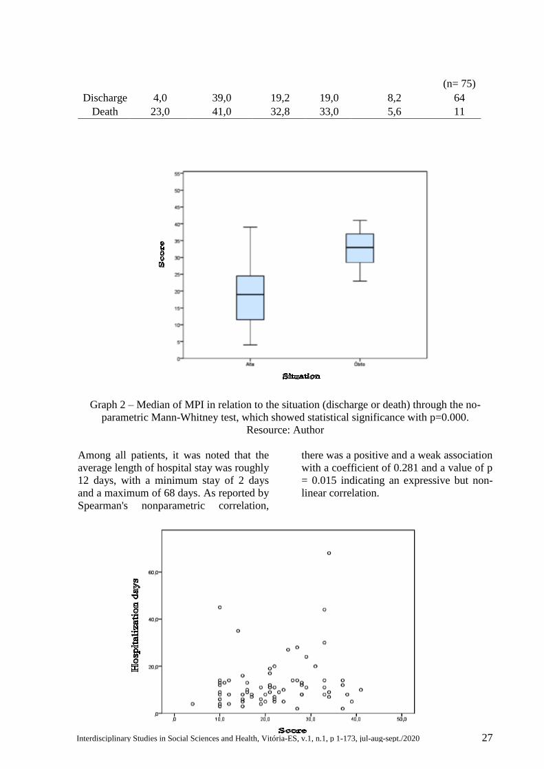
Table 4- MPI Variation in relation to the situation (discharge or death)
Risk Factor Total (n = 75) Total (%) Discharge (%) Death (%) p
Age > 50 years 22 29,33 59,1 40,9 0,000
Female 33 44 81,8 18,2 0,520
Dysfunction of Organs 31 41,33 35,5 64,5 0,000
Malignancy 7 9,30 14,3 85,7 0,000
Duration > 24h 61 81,30 83,6 16,4 0,678
Non-colonic Origin 9 12,00 66,7 33,3 0,121
Difuse peritonitis Exudate 48 64,00 79,2 20,8 0,085
Clear 1 1,30 100 0
0,876 Purulent 58 77,30 84,5 15,5
Fecal 16 21,30 87,5 12,5
Resource: author
Score
Minimum Maximum Average Mean Standard
Deviation Padrão
Total
27
Interdisciplinary Studies in Social Sciences and Health, Vitória-ES, v.1, n.1, p 1-173, jul-aug-sept./2020
Graph 2 – Median of MPI in relation to the situation (discharge or death) through the no-
parametric Mann-Whitney test, which showed statistical significance with p=0.000.
Resource: Author
Among all patients, it was noted that the
average length of hospital stay was roughly
12 days, with a minimum stay of 2 days
and a maximum of 68 days. As reported by
Spearman's nonparametric correlation,
there was a positive and a weak association
with a coefficient of 0.281 and a value of p
= 0.015 indicating an expressive but non-
linear correlation.
(n= 75)
Discharge 4,0 39,0 19,2 19,0 8,2 64
Death 23,0 41,0 32,8 33,0 5,6 11

28
Interdisciplinary Studies in Social Sciences and Health, Vitória-ES, v.1, n.1, p 1-173, jul-aug-sept./2020
Graph 3 – Dispersion of hospitalization days in relation to MPI through Spearman´s
nonparametric correlation, which showed statistical significance with p = 0.015.
Resource: Author.
The outcome agrees with the score (<27
and ≥ 27) on a regular basis, the Kappa’s
value index is 0.464, considerably greater
than zero, (p-value = 0.000). The cut-off
point of 27 points was acquired by
evaluating the Kappa Index of
concordance. By means of this best cut-off
point, we calculated: sensitivity of 90.90%
and specificity of 78.13% through the ROC
curve (Graph 4). The Positive Predictive
Value of the IPM was 71.40%, that is, this
is the probability of an individual
evaluated with a score ≥27 presenting a
death situation. The Negative Predictive
Value of 98.00%, that is, the probability of
an individual with a score <27 to be high is
98.00%.
The percentage of mortality of patients with a score below 27 wa 9.10% ando f patients with
a score ≥ 27 was 90.90% (Table 5 and Graph 5).

29
Interdisciplinary Studies in Social Sciences and Health, Vitória-ES, v.1, n.1, p 1-173, jul-aug-sept./2020
Table 5 – IPM Scores Distribution in relation to the situation (discharge or death)
Score Situation
Discharge Death Total
< 27 n 50 1 51
% 78,1% 9,1% 68,0%
≥ 27 n 14 10 24
% 21,9% 90,9% 32,0%
Total
n 64 11 75
% 100,0% 100,0% 100,0%
Resource: author.
Graph 5 – IPM Scores in relation to the situation (discharge or death)
Resource: author
DISCUSSION
Multicenter studies have corroborated that
in-hospital mortality of peritonitis
continues to be high, nearly 19.5% 7
although some studies reach 60% .8,9,10
Many factors influence the prognosis and
outcome of peritonitis, possibly ranging
from a specific disease to patient-related
factors and therapeutic interventions.
Nevertheless, the outcome is difficult to
predict in most of these patients.
The precocious classification of peritonitis
severity may help to determine surgical
and medical conduct. Yet, scoring systems,
such as the Mannheim Peritonitis Index,
are required to aid in risk stratification and
evaluation of new diagnostic modalities
Discharged Died

30
Interdisciplinary Studies in Social Sciences and Health, Vitória-ES, v.1, n.1, p 1-173, jul-aug-sept./2020
and therapeutic advances, as in comparing
treatment outcomes from different clinics.
When assessing all risk factors in patients
with peritonitis in the HSCMV from 2010
to 2015, it was possible to delineate a
profile indicating higher mortality. By
observing the age risk factor, it was
calculated that the mean age among the
deceased in this study was roughly 64
years. This result was consistent with the
average of 60 years of other studies.11,12
For the patients who survived, the WPI of
this study ranged from 4 to 39 points, with
an average of 19.2, higher than the average
of previous studies.2,11,12 Yet, it was
equivalent to another reference.9
Comparing this work with other
literature,2,9,11,12 an equivalence was noted
in the MPI values of patients who died.
The current study observed that these
values ranged from 23 to 41 points, with an
average of 32.8 points. When analyzing the
mortality rate of the studies, it presented
14.67% of mortality, similar to the rate of
11.70% noted in another article11.
Nonetheless, it was divergent from others,
since these studies9,10 used samples with a
higher risk profile for mortality, selecting
cancer patients and the Intensive Care
Unit, respectively.
Age greater than 50 years and presence of
malignancy and organ dysfunction were
statistically meaningful, all with p = 0.000.
Of those who died, 81.8% were aged> 50
years old with a 40.9% mortality rate. As a
result, mortality was proportional to the
increase in age.
In relation to malignancy and to mortality,
the statistical significance was also
verified. Of those 11 deaths in this study,
54.5% were cancer patients. This
relationship had already been found in
other studies.2,12
The presence of organ dysfunction
increases the risk of death in patients with
peritonitis (p = 0.000), considering that in
this study all patients who died had organ
dysfunction. This result is similar to the
one found in other literature.2,12,10
Mortality was not affected by sex and
duration of peritonitis, which was longer
than 24 hours. All patients who had a
disease duration greater than 24h died,
which was found in other studies.2,12
The risk of death in patients with non-
colonic origin peritonitis is higher than in
those with colonic origin, conforming
another reference.2 The non-colonic origin
is a risk factor for IPM. However, in this
study, this relationship has not been
proven, as in other previous studies.10,12
It was possible to observe that 48 patients
had diffuse peritonitis, of which 10 died
and 38 were discharged. However, of all
11 deaths, 90.9% had diffuse peritonitis.
Yet, there was no significant difference
between extension and mortality, as in
another article.2 In contrast, other
studies10,12 presented a meaningful relation.
In the current study, 77.30% of patients
presented purulent exudate, found in
81.8% of those who died. Other surveys
also presented this profile.2,10
The mean length of hospital stay for
patients who were discharged was
approximately 11 days, for those who died,
approximately 14 days, which disagreed
with another study that analyzed only cases
of patients in intensive care.9 The total
mean value was approximately 12 days.
There was a statistical significance
between days of hospitalization and score,
as stated by another literature.9 After
analysis of Graph 2, it was noted that this
association was not linear, but positive.
Probably, the higher the score, the greater
the severity of the patient, the more likely
he or she will be to die, to stay less
hospitalized or to stay longer on account of
complications.

31
Interdisciplinary Studies in Social Sciences and Health, Vitória-ES, v.1, n.1, p 1-173, jul-aug-sept./2020
After comparing the data for each number
of points of the score, one verified that the
patients who died, 90.90% scored higher
than or equal to 27 for the MPI. Of those
who did not die, merely 21.90% presented
this score. Nevertheless, one infers that the
cut-off point for predicting mortality with
MPI, found in the profile of patients who
were under observation in the HSCMV, in
the period from 2010 to 2015, is 27 points.
The sensitivity for mortality is 90.90% and
the specificity is 78.13%, similar to those
found in another study, 2 with sensitivity of
95.90% and specificity of 80%. Reference
literature7 found sensitivity of 86.00%,
specificity of 74% and accuracy of 76%. In
the study10 conducted at the National
Cancer Institute (INCA), the cut-off point
found was 21, which evaluated the
oncology patients whose risk of mortality
was highest. Their sensitivity was 87.30%
and their specificity was 41.20%.
The cut-off point 27, demonstrated in the
current study, revealed a statistical
significance. Nonetheless, the use of it for
prognostic evaluation in HSCMV patients
is recommended.
CONCLUSION
Based on this study, it is possible to
presume that the main causes of peritonitis
were of non-colonic origin. One may note
that the most frequent specific diagnosis in
HSCMV patients was acute appendicitis,
but it was not the main factor of death. The
main cause of death was postoperative
peritonitis. Of the risk factors assessed, age
greater than 50 years, organ dysfunction
and presence of malignancy were
statistically significant. There was a
positive and non-linear correlation with the
patients' score, regarding hospitalization
time, making this time a risk factor.
However, there is no relation directly
proportional to the score. It was also
possible to indicate a percentage of
peritonitis mortality in the HSCMV,
consistent with other literature.
The expressive majority of confirmed
deaths had MPI ≥ 27 points is in
accordance with other literature, indicating
that MPI was efficient in estimating the
risk of death and in categorizing patients
into risk groups. This stratification assists
in the determination of a prognosis, in the
selection of patients for intensive care and
in the definition of operative risk,
contributing, this way, to the choice of the
nature of the operative procedure, such as
damage control or definitive procedure.
Nevertheless, when this scoring range is
reached, a more aggressive and fast
approach must be established, possible
complications must be observed, and an
individualized and precise behavior must
be adopted.
32
Interdisciplinary Studies in Social Sciences and Health, Vitória-ES, v.1, n.1, p 1-173, jul-aug-sept./2020
REFERENCES
1. Wittmann DH, Schein M, Condon RE. Management of secondary peritonitis. Ann Surg.
1996; 224(1): 10-8.
2. Melgarejo EB,Castro MR, Luque GB, Trujillo NN. Valor Predictivo de Mortalidad del
Indice de Peritonitis de Mannheim. Rev Gastroenterol. 2010; 30(3):219-23.
3. Neri A, Marrelli D, Scheiterle M, Di Mare G, Sforza S, Roviello F. Re-evaluation of
Mannheim prognostic index in perforative peritonitis: Prognostic role of advancedage. A
prospective cohort study. Int J Surg. 2015;13:54-9.
4. Nag DS. Assessing the risk: Scoring systems for outcome prediction in emergency
laparotomies. Biomedicine. 2015; 5(4):7-16.
5. Muralidhar VA, Madhu CP, Sudhir S, Madhu S. Efficacy of Mannheim Peritonitis Index
(MPI) Score in Patients with Secondary Peritonitis. J Clin Diagn Res. 2014; 8(12):1-3.
6. Basnet RB, Sharma VK. Evaluation of predictive power of Mannheim Peritonitis Index.
Postgrad Med J. 2010; 10(2):10-13.
7. Biling A, Frolich D, Schildberg F. Prediction of outcome using the Mannheim peritonitis
index in 2003 patients. Br J Surg. 1994; 81:209-13.
8. Burger JA, Schöffel U, Sach M, Jacobs E, Kownatzki E, Von Specht BU, et al. Effects of
peritonitis exudates on chemotaxis and phagocytosis of human neutrophils. Eur J Surg. 1995;
161(9):647-53.
9. Rodriguez H, García RP, Morales MP, Brambila VR, García VC. Factores pronósticos
asociados a mortalidad en pacientes com sepsis intrabdominal tratados em la unidad de terapia
intensiva. Cir Ciruj. 1999; 67(6):205-7.
10. Correia MM, Thuler LCS, Vidal EM, Schanaider A. Prediction of death using the
Mannheim Peritonitis Index in oncologic patients. Rev Bras de Cancerol. 2001; 47(1):63-68.
11. Teleanu G, Iordache F, Beuran M. The Predictive Value of Mannheim Score in Patients
with colon related peritonitis. Acta Medica Marisiensis. 2012; 58(3):175-177.
12. Bracho-Riquelme RL, Melero-Vela A, Torres-RamírezA. Mannheim Peritonitis Index
Validation study at the Hospital General de Durango (Mexico). Cir Ciruj. 2002; 70(4):217-25.
Conflict of Interest: None.

33
Interdisciplinary Studies in Social Sciences and Health, Vitória-ES, v.1, n.1, p 1-173, jul-aug-sept./2020
ANNEXES
ANNEX A – The score system used to calculate the Mannheim Peritonitis Index (MPI)
FACTORS ADVERSE POINTS FAVORABLE POINTS
Age >50 5 <50 0
Sexo male 5 female 0
Organic Dysfunction* present 7 absent 0
Malignancy present 4 absent 0
Time of evolution > 24 horas 4 < 24 horas 0
Origin non-colonic 4 colonic 0
Extension of peritonitis generalized 6 localized 0
Peritoneal Exudate fecal 12 clear 0
purulent 6
Resource: Correia, 2001.
ORGANIC DYSFUNCTION*
KIDNEY Creatinine > 177umol/L
Urea > 167mmol/L
Oliguria < 20mL/h
LUNG PO2<50mmHg
PCO2<mmHg
SHOCK Hypodynamic or Hyperdynamic
BOWEL OBSTRUCTION Paralysis >24 hours or complete mechanical obstruction
Resource: Correia, 2001.

34
Interdisciplinary Studies in Social Sciences and Health, Vitória-ES, v.1, n.1, p 1-173, jul-aug-sept./2020
ANNEX B – Data sheet used to collect data from patients selected in the study.
No images were not uploaded by the author.
Patient Age Sex Cr Ur Oliguria pO2 pCO2 Schok
Bowel
Obstruction Oncolo-gical < 24h
Origin
Non-colonic
Extension
Exsudato
1- clear
2- purulent
3- fecal
Situation
1- discharge
2- death
Date of
admission
Departure
date

35
Interdisciplinary Studies in Social Sciences and Health, Vitória-ES, v.1, n.1, p 1-173, jul-aug-sept./2020
DOI https://dx.doi.org/10.5935/2675-7397.20200005
Original Article
THERAPEUTIC BENEFITS OF DRY NEEDLING IN ADULT PATIENTS WITH
CHRONIC LOW BACK PAIN
Luana Moraes Leal1, Jeanne Florence Soares Santos2, Monique Almeida Fernandes3,
Pitiguara de Freitas Coelho4, Francisco Rodrigues Brioschi5.
1, 2, 3, 4 College of Sciences of Casa de Misericórdia de Vitória EMESCAM -
(Physiotherapist).
5 University of Vila Velha (UVV) – (Physiotherapist).
Submitted in November 11, 2016; Approved in September 15, 2017
Institution: EMESCAM
Av. Nossa Sra. Da Penha, 2190 – Santa Luíza, Vitória - ES, 29045-402.
Correspondence Author: Luana Morares Leal
Escola Superior de Ciências da Santa Casa de Misericórdia de Vitória, ES.
Av. Nossa Sra. da Penha, 2190 – Santa Luíza, Vitória - ES, 29045-402.
Email: [email protected]
Keywords Abstract
Spine;
Low Back Pain;
Chronic Pain;
Trigger Points;
Microwaves.
Objective: To evaluate the therapeutic benefits of dry
needling in adult patients with chronic low back pain.
Methods: A pilot study of a randomized clinical trial with
blind evaluator was conducted in a convenience sample
composed of 10 patients who underwent an initial
questionnaire and the application of scales by the blind
evaluator through the following instruments: Initial
questionnaire, Roland Morris Questionnaire and Visual
Analogue Pain Scale - EVA. After application of the scale
and questionnaires, the patients were randomly divided
into two groups: one group being submitted to the protocol
involving dry needling and another group that underwent
manual myofascial release therapy. Results: The majority
of the individuals were female (60%), with a mean age of
ESTUDOS INTERDISCIPLINARES EM
CIÊNCIAS SOCIAIS E DA SAÚDE INTERDISCIPLINARY STUDIES IN SOCIAL SCIENCES AND HEALTH
v.1, n.1, 2020

36
Interdisciplinary Studies in Social Sciences and Health, Vitória-ES, v.1, n.1, p 1-173, jul-aug-sept./2020
44.90 ± 12.26 years. There was a considerable
improvement in pain evaluated in the visual analogue
scale (VAS), before and after the study, merely in the
group treated with dry needling p = 0.041. There was no
significance in the Manual Myofascial Release group test
for pain assessment. The functional incapacity of the
patients in the dry needling group and in the control group
did not present important results. Conclusion: Although
further studies would be necessary on the therapeutic
benefits of dry needling, it is plausible that this technique
provides a considerable reduction of pain in patients with
chronic low back pain. Although not showing any
meaning regarding functional incapacity, we observed a
trend in favor of dry needling.
INTRODUCTION
The sensation of pain is essential for
survival, since it is the first indicator of any
tissue injury5. By the 1950s and 1960s, the
concepts of pain underwent deep
modifications, 14 converting itself from a
simple neurophysiological signal to a
psychophysiological phenomenon. This
change occurred in new studies, indicating
that the severity of the pain is probably
related to specific physiological symptoms,
added to the effect of one or more
psychological variables6, the same way a
chronic low back pain has been associated
with psychophysiological factors due to its
duration, affecting the social life of man. 3,7
Lumbar pain, known as a disability by the
WHO, translates itself into a disadvantage
that limits or impedes the full performance
of physical activities11. The International
Association for Pain Study (IASP), 14
considers chronic low back pain, when it
lasts for more than three months.
Considering that chronic low back pain
does not come from specific diseases, but
from a set of elements, it is important to
take into account the profile of the
individual with chronic low back pain. The
psychological and socio-demographic
components (age, sex, income and
schooling) play a meaningful role in the
onset of low back pain, contributing for a
good response to treatment 2,7,16.
Patients who have low back pain spend a
great deal of time and resource searching
for a treatment. Physical therapy is a
crucial resource in the rehabilitation of the
patient with chronic low back pain and its
main objective is to allow him to return to
work and daily activities. Thus, functional
restoration should be started as early as
possible in order to minimize the effects of
immobilization and to protect the lumbar
spine from problems 3,6.
Several painful problems, including
chronic low back pain, resulting from
abnormal stresses that the fascia supports,
are commonly not so severe pains and
generally bearable. However, its duration,
persistence, and frequent recurrences
rapidly become intolerable1. Trigger points
are located in these areas of tension, which
are a common source of pain, and they
have been commonly related to chronic
low back pain15. Consequently, dry
needling comes as an essential resource in
the treatment of chronic low back pain, as
it is narrowly defined as an "intramuscular"
procedure involving the isolated treatment
of "trigger points" 10.

37
Interdisciplinary Studies in Social Sciences and Health, Vitória-ES, v.1, n.1, p 1-173, jul-aug-sept./2020
Dry needling involves the introduction of
needles into areas of the muscle that have
motor alterations in an effort to relieve
pain, decrease muscle tension, and restore
normal muscle function2. The dry needling
technique has been associated with
intramuscular electrical stimulation, and it
is essential to observe that this association
with intramuscular electrostimulation
permits the electrostimulation to reach
target tissues in a deeper way because of
the needles used4.
This theme was developed because of the
increase in patients with chronic low back
pain in search of a faster and more
effective treatment. This study is a
descriptive and transversal applied
research, performed in two moments: first,
with a bibliographical review and then a
field research, through a case series study.
Accordingly, we sought to verify the
therapeutic benefits of dry needling in
adult patients with chronic low back pain.
METHODS
A pilot study of a randomized clinical trial
with blind evaluator was conducted at the
Clinic of Physiotherapy School of the
Santa Casa de Misericórdia School of
Science (EMESCAM) from June 2016 to
December 2016, in a sample of
convenience constituted by 10 patients.
Initially, it was applied a questionnaire
through an interview with blind evaluator,
who did not participate in the choice of
patients. The following were included:
patients with complaints of mechanical
postural low back pain; with pain in the
region of the iliocostal muscles and in the
longest of the chest in the chronic period
greater than 12 weeks; patients of both
genders and those aged between 18 and 65
years, who agreed to sign the EHIC.
Patients diagnosed with chronic low back
pain or who had a diagnosis of
lumbosacatalgia; patients with fear of
insurmountable needles; with allergy to
nickel (needles are made of this metal),
with coagulation problems (even if you are
on anticoagulant therapy, there is risk of
bleeding) and people with lymph nodes
removed (because of the risk of
lymphedema) were excluded.
Those who fulfilled the inclusion criteria
submitted themselves to an initial,
questionnaire and to a scale application by
the blind evaluator through the following
instruments: Initial Questionnaire, Roland
Morris Questionnaire and Visual Analog
Pain Scale - EVA. The interviews were
individual and the study supervisor
previously trained the researchers for the
application of the instruments.
An initial questionnaire was utilized with
the purpose of tracing the patients' profile.
Data such as name, telephone, age, sex,
marital status, occupation, socioeconomic /
cultural history and life habits were
obtained. This questionnaire was applied
on their first day just before treatment was
started.
The functional disability of the patients
was corroborated through the Roland-
Morris questionnaire, a specific instrument
that allows the disability evaluation in
individuals with low back pain. It consists
of 24 items related to daily life activities
and the questions have a dichotomous
answer (yes or no). Its score was calculated
through the total number of questions,
ranging from zero (absence of disability) to
24 (severe disability). The Roland Morris
questionnaire was applied through the
blind assessor on the first and last day of
treatment and the initial score was
compared to the final score.
The Visual Analogue Scale (VAS) utilized
to assess patients' pain, provided a measure
of pain intensity in the patient. This
instrument proved to be important to verify
if the treatment was effective as well as to
know which procedures have given better
results, according to the degree of

38
Interdisciplinary Studies in Social Sciences and Health, Vitória-ES, v.1, n.1, p 1-173, jul-aug-sept./2020
improvement or worsening of pain. To
assess the patient's degree of pain, such
questions as, "Do you have pain? How do
you rate your pain?" were asked. There
was an account of the observations about
what they said. In order to the patient's
pain intensity, a score of 0 to 10 was used,
distributed as follows: 0 to 2 mild pain, 3
to 7 moderate pain, 8 to 10 intense pain
and 10 is the maximum pain level that can
be tolerated by the patient.
After the application of the scale and the
questionnaires, the patients were randomly
divided into two groups with selection
using envelopes numbered from 1 to 10.
Group A (5 patients), who was submitted
to the protocol involving dry needling, and
group B (5 patients) who underwent
manual myofascial release therapy. The
treatment was carried out in a reserved
environment resulting from the exposure of
the patient's chest.
In the Treatment Protocol of group A, the
patients were submitted to the technique of
dry needling of the iliocostal and very long
muscles of the thorax bilaterally at trigger
points, using needles (DONGBANG ®) of
0.3 x 0.6 mm. After locating an active
trigger point, the overlapping skin was
cleaned with 70% alcohol, and the needle
was subsequently inserted, penetrating skin
and muscle to a depth of nearly 20 and 25
mm at the trigger point. The patient was
always in the prone position. Once inserted
at the trigger point, the needle was moved
in multiple directions until the first local
contraction response was obtained. The
needles were coupled to electrostimulation
through the SIKURO® device with a
frequency of 4HZ, for 10 minutes, dosed in
intensity according to the patient's
perception. Patients were advised to report
pain or any increase in their symptoms
after the intervention. Subsequent to the
application of the technique, the needles,
as soon as they were withdrawn from the
patient, were discarded in the Descarpack,
a safe and resistant picker of sharp
materials, guaranteeing, this way, the total
protection against perforations and leaks of
contaminated fluids. The treatment of
patients took place in the morning, twice a
week - lasting 30 minutes each, for four
weeks, with the supervision of two
therapists.
The patients in the B group were submitted
to the manual myofascial release technique
in the region of the iliochalost muscles and
very long of the thorax bilaterally, for 10
minutes. Subsequently, deep heat was
performed with microwaves in the lumbar
region, for 10 minutes with a frequency of
60%. The treatment of these patients
occurred in the morning, twice a week -
lasting 30 minutes each, for four weeks,
along with group A, with the supervision
of two therapists.
The descriptive analysis of data collected
was achieved taking into account
frequencies and percentage for qualitative
variables and data summary measures as
mean, median and standard deviation for
the quantitative variables. The Wilcoxon
test was used to verify the association
between initial and final VAS, and Roland
Morris initial and final in an intragroup
way as well as between the myofascial
release and dry needling groups. The
Mann-Whitney test was used to compare
age in both groups. The values were
considered statistically expressive when
the value was p <0.05. The research was
approved by the Ethics Committee (CEP)
of the Superior School of Sciences of the
Santa Casa de Misericórdia de Vitória
(EMESCAM), under the number of
opinion 1,612,115 on June 28, 2016.
RESULTS
The sample of this study consisted of 10
patients from the physical therapy school
of EMESCAM, where 6 were female
(60%) and 4 male (40%), with a mean age
of ± 12,26 years.

.
Table 1. Mean age and sex distribution between groups of patients treated with Dry Needle
and Manual Myofascial Release
The initial pain data did not present
different values. This shows that the
groups had the same pain index at the
beginning of the study. When compared
intra-group, we observed that there was an
improvement in the indicator evaluated in
the visual analogue scale (VAS) before and
after the study only in the group treated
with dry needling, according to table 2.
Table 2. Mean of the initial and final Visual Analog Scale between groups of patients treated
with Dry Needling and Manual Myofascial Release
Dry Needling
Group Value p
Myofascial
Release Group Value p
Initial VAS 8±0 *0,041
7±2 0,888
Final VAS 6±1 7±0
*p<0,05 considered significant in the Wilcoxon test
Initial data from the patients' disability
assessment did not present different values,
which shows that the groups had the same
disability index at the beginning of the
study. When compared intra-group, there
was no improvement in the indicator
evaluated in the Roland Morris
Questionnaire before and after the study, as
stated in table 3.
Table 3. Mean of the initial and final Roland Morris Questionnaire between groups of patients
treated with Dry Needling and Manual Myofascial Release
Needling Dry
Group Value p
Myofascial
Release Value p
Inicial Roland 14±5 0,068
14±4 0,465
Final Roland 10±4 11±7
*p<0,05 considered significant in the Wilcoxon test.
Age 44,90 ± 12,26
Sex 6(F) / 4(M)
39 Interdisciplinary Studies in Social Sciences and Health, Vitória-ES, v.1, n.1, p 1-173, jul-aug-sept./2020

40
Interdisciplinary Studies in Social Sciences and Health, Vitória-ES, v.1, n.1, p 1-173, jul-aug-sept./2020
DISCUSSION
Women present a higher prevalence of
chronic low back pain than men, due to
anatomic-functional peculiarities added to
other aspects that potentiate this risk16,8,11.
These data corroborate with those found in
this study, where we verified a frequency
of 60%. The degenerative changes and
overloads in the work explain these
findings16. In relation to age, there was a
higher frequency of individuals over the
age of 40 years, similar to the study by
Masiero et al.8 where the mean age was
48.1 years.
Previous studies have investigated the
effect of dry needling in patients with low
back pain and have shown that it is
possible that dry needling is effective in
some patients2,9. Our results showed that
only in the dry needling group there was
significant improvement, before and after
the study, and for a short period of
treatment. The results of the present study
are consistent with the data found in a
study conducted by Kietris et al.14 that in
4 weeks of treatment dry needling had a
strong positive effect when compared to
other treatments. The Manual Myofascial
Release control group showed no
difference in the visual analogue scale
(VAS) before and after the studies.
When related to the function, there was no
significant difference in both the dry
needling group and in the Manual
Myofascial Release therapy group. Even
so, there was a tendency for a better result
in the dry needling group with the p-value
reaching close to the significance,
compared to the control group. These data
go according to the results found in a study
conducted by RAYNEI.C. E, et al.4, where
patients were able to resume their
functionality and return to their
recreational and professional activities
after dry needling therapy without pain.
CONCLUSION
There was an improvement in pain in the
patients treated with dry needling.
Even though not significant, the data found
tended to have a clinical relevance of
significant improvement in aspects of
functional disability in favor of dry
needling.
Despite the fact that a larger and more in-
depth study is needed on the therapeutic
benefits of dry needling, it is plausible that
this technique provides a considerable
reduction of pain in patients with chronic
low back pain, emphasizing the technique
of manual myofascial release therapy.
REFERENCES
1. BIENFAIT, M. “Fáscias e Pompages Estudo e tratamento do esqueleto fibro ”21ª ed. São
Paulo: Summus, 1999.
2. KOPPENHAVER.S.L.et al.”Baseline Examination Factors Associated With Clinical
Improvement After Dry Needling in Individuals With Low Back Pain: Journal of Orthopaedic
& Sports Physical Therapy” - Journal Of Orthopaedic & Sports Physical Therapy - v.45, n.8,
p. 604-612 – Agost. 2015.
3. BRIGANÓ, J. U; MACEDO, C. S. G; “Terapia Manual e Cinesioterapia na Dor,
incapacidade e qualidade de vida de Indivíduos com Lombalgia” – Rev. Espaço para Saúde –
v.10, n 2, p. 1-6 – Londrina, jun. 2009.

41
Interdisciplinary Studies in Social Sciences and Health, Vitória-ES, v.1, n.1, p 1-173, jul-aug-sept./2020
4. RAINEY.C.E. et al. “The use of trigger point dry needling and intramuscular electrical
stimulation for a subject with chronic low back pain: a case report”- The International Journal
of Sports Physical Therapy – v.8, n 2, p. 145-161 – Abril, 2013.
5. SILVA.J.A; FILHO.N.P.R; “A dor como um problema psicofísico” - Revista Dor. v.12,
n.2, p.138-5 - São Paulo, abr/jun, 2011.
6. IMAMURA, S. T; KAZYAMA, H. H. S; IMAMURA.M. “Lombalgia”. Rev. Med. -v. 80
(ed. esp. pt.2), p 90-375 - São Paulo, 2001.
7. HOFFMANN.M. “A Prevalência de Doenças Lombares em Pacientes de Terceira Idade na
Cidade de Concórdia” – Rev. Ágora: R. Divulg. Cient – v. 17, n. 1, p.62-70 – Santa Catarina,
2010.
8. MASIERO.D. et al. “Evaluation of pain level and function on low back pain patients
treated with Back School program” - Rev. Acta Fisiatrica - v.12, n.1, p.11-14, 2005.
9. KALICHMAN.L.et al. “Dry Needling in the management of musculoskeletal pain” –
Journal American Board of Family Medicine – v.23, n.5, p. 640-646 – set\ out, 2010.
10.DUNNING.J. et al. “Dry needling: a literature review with implications for clinical
practice guidelines”- Rev Physical Therapy - v.19, n 4, p. 252-265 – AL, USA - ago, 2014.
11.KRELING.M.C.G.D; PIMMENTA.C.A.M; CRUZ.D.A.L.M. “Prevalência de dor crônica
em adultos” - Revista Brasileira de Enfermagem – v. 59, n. 4, p. 509-13 - jul/ago, 2006.
12.SANTOS.L.S; MASCARENHASC.H.M; “Avaliação da dor e da capacidade funcional em
indivíduos com lombalgia crônica” - Rev. O Journal of the Health Sciences Institute (JHSI) -
v.29, n.3, p.205-208 – Jequié,BA, 2011.
13.JUNIOR.M; GOLDENFUM.M.A; SIENA.C.“Lombalgia Ocupacional” – Rev. Da
Associação Médica Brasileira – v.56, n.5, p.583-589, 2010.
14.PIMENTA.C. A.M; KOIZUMI.M. S; TEIXEIRA.M.J. “Dor Crônica E Depressão: Estudo
Em 92 Doentes” - Rev. Esc. Enf. - v.34, n.1, p. 76-83 - março, 2000.
15.KIETRIS.D.Met al. “Effectiveness of Dry Needling for Upper-Quarter Myofascial Pain: A
Systematic Review and Meta-analysis” - Journal of Orthopaedic & Sports Physical Therapy –
v.43, n.9, p.620-634, set. 2013.
16.SILVA.M. C; FASSA.A. G; VALLE.N.C.J. “Dor lombar crônica em uma população
adulta do Sul do Brasil: prevalência e fatores associados” - Caderno de Saúde Pública – v.20,
n.2, p.377-385 - Rio de Janeiro, mar/abr. 2004.
17.WIBELINGER.L.M; et al. “Abordagem Fisioterápica Da Dor Lombar Crônica No Idoso”-
Rev. Brasileira de Ciências da Saúde – v.8, n.25, p. 56-51 - jul/set , 2010.
No images were uploaded by the author.

42
Interdisciplinary Studies in Social Sciences and Health, Vitória-ES, v.1, n.1, p 1-173, jul-aug-sept./2020
DOI https://dx.doi.org/10.5935/2675-7397.20200012
Original Article
INFLUENCE OF MEDULAR INJURY AND WHEELCHAIR TIME IN
RESPIRATORY AND TOSSE MUSCLE FORCE
Taynara Ribeiro Batalha1, Morghana Ferreria Ambrosim2, Roberta Ribeiro Batista3, Christine
Lourenço4.
1 2 Physiotherapist (graduated from EMESCAM) - (Physiotherapist).
3 Physiotherapist - (Coordinator and professor Ms. of Physiotherapy Undergraduate
Course EMESCAM).
4 Physiotherapist - (Professor PhD of Physiotherapy Undergraduate Course
EMESCAM).
Submitted in September 09, 2017; Approved in January 01, 2018.
Institution: EMESCAM
Av. Nossa Sra. Da Penha, 2190 – Santa Luíza, Vitória - ES, 29045-402.
Correspondence Author: Taynara Ribeiro Batalha.
Escola Superior de Ciências da Santa Casa de Misericórdia de Vitória, ES.
Av. Nossa Sra. da Penha, 2190 – Santa Luíza, Vitória - ES, 29045-402.
Email: [email protected]
Keywords Abstract
Spinal Cord Injuries;
Maximal Respiratory Pressures;
Cough
Objective: To evaluate respiratory muscle strength and
peak cough flow in patients with spinal cord injury and
correlate muscle strength and peak cough flow over
time in a wheelchair and the level and severity of spinal
cord injury. METHODS: This is a case series cross-
sectional study of 23 participants. The profile data of
the individuals were collected through a questionnaire
developed by the researchers, the spinal cord injury was
assessed by the American Spinal Injury Association
(ASIA) scale, respiratory muscle strength was
measured by a manovacuometer and peak cough flow
by a Peak Flow. Results: All participants had weakness
of the expiratory musculature and the data suggest that
ESTUDOS INTERDISCIPLINARES EM
CIÊNCIAS SOCIAIS E DA SAÚDE INTERDISCIPLINARY STUDIES IN SOCIAL SCIENCES AND HEALTH
v.1, n.1, 2020

43
Interdisciplinary Studies in Social Sciences and Health, Vitória-ES, v.1, n.1, p 1-173, jul-aug-sept./2020
the higher the level of the lesion, the greater the
weakness of the inspiratory musculature. The
quadriplegic patients had reduced inspiratory muscle
strength and cough when compared to paraplegics. In
addition, the relationship between wheelchair time and
peak cough flow was negative and statistically
significant (r = -0.45, p = 0.01). Conclusion: The higher
the level of the spinal cord lesion and wheelchair time,
the greater the impact on the respiratory muscles and
the efficacy of the cough. Therefore, physical therapy
acts in an essential way in rehabilitation, reducing
complications and consequently improving the quality
of life.
INTRODUCTION
It is estimated that there are worldwide
nine in fifty cases of spinal cord injury per
million inhabitants1,3 per year, and that,
according to Vall et al3, in Brazil, alone,
there are more than 130,000 people with
such injury.
The importance of the medulla is due to
the conduction of information between the
periphery and the brain. An injury to this
structure may determine changes in
sensitivity, motor function and respiratory
muscles, with resulting impairment of lung
function. As well the respiratory changes
that is possible to be observed according to
the injured spinal cord level, the patient
usually uses the wheelchair, which does
not allow him to maintain an auspicious
posture for respiratory mechanics4-8.
The area of lung capacity deficits will be
determined by the level and extent of
injury, and the dissimilar structures of the
respiratory tract affected. The greater the
extent of the injury, the greater will be the
reduction in lung volume and capacity, and
cough strength, as a consequence of
respiratory muscle fragility9-12.
The leading sources of morbidity and
mortality, either in the acute or chronic
phase of spinal cord injury, are the
respiratory complications. In relation to the
general population, the life expectancy of
these patients can be lowered by up to 3
times5,13,14.
Physical therapy in assisting individuals
with spinal cord injury is important
because it holds resources that can promote
progress in respiratory function, thus
reducing complications and
hospitalizations, improving the quality of
life15. The objective was to assess
respiratory muscle strength (MIP and
MEP) and peak cough flow (PFT) in
patients with chronic spinal cord injury.
There are not enough studies in the state
and, therefore, it is understood that the
expansion of knowledge and possible
scientific evidence may contribute to the
creation of new strategies for monitoring
this population.
METHOD
The current study is an observational and
cross-sectional research. It was marked by
a case series study, endorsed by the Ethics
Committee (CEP) of Santa Casa de
Misericórdia de Vitória School of Sciences
(EMESCAM), within the opinion number
1.310.183, on November 5, 2015. The
research was held in individuals who were
under monitoring at the Espírito Santo
Physical Rehabilitation Center (CREFES)

44
Interdisciplinary Studies in Social Sciences and Health, Vitória-ES, v.1, n.1, p 1-173, jul-aug-sept./2020
in 2015, with a diagnosis of spinal cord
injury, with an exception to those with
associated cardiopulmonary disease,
tracheostomy, cardiopulmonar, decompen-
sation, surgical intervention or
hospitalization, past three months, not
located or not agreeing to engage in the
survey.
For the purpose of characterizing the
sample through a questionnaire created by
the researchers, data regarding age, gender,
race, smoking, wheelchair time and injury
time were collected. Measurement of
inspiratory and expiratory muscle strength
was conducted by measuring MIP and
MEP, respectively, with nasal occlusion
using a commercial manometer. The MIP
was measured from the residual volume,
requesting the subject to exhale as much as
possible and then to inhale through the
mouthpiece. PEmax was measured from
total lung capacity, requiring the individual
to inhale as much as possible and then
exhale through the mouthpiece16, in
agreement with Souza17 reference values,
which use the following formulas as a
basis: MIP = 104 + (0.51x age) MIP = 170
+ (1.03 x age).
During unaided spontaneous cough with
nasal occlusion using a Respironics® Peak
Flow, cough effectiveness was measured
by cough flow (PFT). Measurements of
respiratory muscle strength and cough
effectiveness were held in the sitting and
supported posture, with three consecutive
measurements and considered the best
value obtained. As reported by Barros and
Medeiros, it was considered an ineffective
cough, values lower than 160 liters /
minute, a difficulty in eliminating
secretions, values between 160 and 270
liters / minute and, above 270 liters /
minute, an effective cough18.
Concerning the level and extent of the
lesion, the classification of the spinal cord
injury was settled by neurological
examination in accordance with the criteria
of the American Spinal Injury Association
(ASIA). Motor function was evaluated by
assessing muscle strength, graded from 0
to 5, of the key muscles, and sensory,
tactile and painful function was evaluated
through the 28 bilateral dermatomes
employing a cotton and two-pin distinct
ends19.
Following sensory and motor assessment,
the level of the lesion was established,
where the neurological level is the region
with bilateral sensibility and bilateral
muscle strength equal to or greater than
three. The participants who were classified
as level C4 to C8 were named as
quadriplegic, and the ones as level T1 to
L3 as paraplegic. In order to categorize the
extent of the lesion, a digital touch was
carried out in the anal sphincter region as
well as the perception of the touch by the
evaluator and / or a contraction of the anal
sphincter 19.
In line with each variable, analyzes were
held with mean and standard deviation,
median (minimum and maximum) or
frequency and percentage. For the purpose
of verifying the association of wheelchair
time with respiratory muscle strength and
peak cough flow, the Pearson correlation
test was applied. Simple linear regression
analysis to verify the influence of spinal
cord injury level for each of the dependent
variables: peak cough flow, maximal
inspiratory pressure, and maximal
expiratory pressure. So as to compare the
different classifications of peak cough flow
and respiratory muscle strength between
quadriplegic and paraplegic individuals, a
chi-square test was performed. A
significance level of 5% for all inferential
analyzes was taken. The program applied
was “SPSS version 23”.
RESULTS
Thirty patients were selected for the
research. Out of them 4 were not located

45
Interdisciplinary Studies in Social Sciences and Health, Vitória-ES, v.1, n.1, p 1-173, jul-aug-sept./2020
and 3 did not consent to participate as a
result of transport difficulties. Among the
23 individuals with spinal cord injury
participating in the study, 13 were
categorized as quadriplegic and 10 as
paraplegic. Merely 6 participants agreed
with the assessment of the extent of the
lesion. A 3 were classified as a complete
lesion - ASIA A and B and 3 as an
incomplete lesion - ASIA D. (table 1)
Table 1 – Sample data on participants respiratory muscle strength in this study as well as
their cough.
N = 23
Age (Average ± SD) 32.4 (8.1)
Male sex N (%) 18 (78.3%)
Female sex N (%) 5 (21.7%)
Race N(%)
White 3 (13%)
Brown 7 (30.4%)
Black 13 (55.5%)
Yellow 0 (0%)
Smoking N(%)
Never smoke 16 (66.2%)
Former smoker 7 (30.4%)
Smoker 1 (4.3%)
Wheelchair time - median (min, max)* in months 102 (1.367)
PFT – median (min, max) 400 (160, 900)
Ineffective (<160) 1 (4.3%)
Effective / Weak (160-270) 3 (13%)
Effective / Strong (>270) 19 (82.6%)
Real PEmax l (average ± SD) 97.9 (36.5)
Forecast PEmax (average ± SD) 170.9 (0.2)
Real Plmax (average ± SD) - 107.7 (28.58)
Forecast Plmax (average ± SD) - 120.5 (4.1)
Lesion level N(%)
C4-C5 7 (30.4%)
C6-C8 6 (26.1%)
T1-T6 5 (21.7%)
T7-L3 5 (21.7%)
Lesion extension N (%) N=6
A - Complete 2 (8.7%)
B - Complete 1 (4.3%)
C - Complete 0 (0%)
D - Incomplete 3 (13%)

No significant association (p = 0.32) was
revealed when correlating wheelchair time
with respiratory muscle strength. It was
possible to note a statistically significant
negative, moderate correlation was noted
between wheelchair time and peak cough
flow (r = 0.45, p = 0.014), indicating that
peak cough flow diminishes with as chair
time enhances.
By comparing muscle strength between the
different levels of injury, quadriplegic or
paraplegic, it was noticed that quadriplegic
patients had a mean MIP of -96.15mmhg
(28.4), a MEP of 87.31mmhg (37.6) and a
peak flow median of 390L / min (160.770).
For paraplegics, the mean MIP was -
119mmhg (26), the MEP 110mmhg (30.5)
and the median Peak Flow 495L / min
(240,900). These results indicate that
quadriplegic patients are liable to have a
maximal inspiratory pressure and a lower
cough flow peak than paraplegic patients
(p = 0.06 and p = 0.07 respectively).
Regarding linear regression, it did not
provide statistical significance. Despite
that, the MIP result implies that as the
injury level decreases, the maximal
inspiratory force increases. Nevertheless,
the same was not observed for PFT and
PEmax. (Table 2).
Table 2 - Linear regression analysis to verify the influence of spinal cord injury level on peak
cough flow, maximum inspiratory pressure and maximum expiratory pressure.
ß sig
Constant 380.0
PFT 153.0 L/min 0.73
Constant -96.15 mmhg
Pimax -22.84 0.06
Constant 87.31 mmhg
Pemax 22.69 0.14
The injury level explains 14% of the cough flow variance, 16% of the
maximum inspiratory pressure and 10% of the maximum expiratory pressure.
Looking at Table 3, it can be seen that
there is no significant difference in PFT
between the different injury level
classifications. Most paraplegics and
quadriplegics had a strong and effective
cough. Nonetheless, out of the 13
quadriplegic participants, 84.6% had lower
than predicted MIP, 100% had lower than
predicted MEP values. Sixty percent of
paraplegics had lower than expected MIP
and 100% lower than expected MIP, even
though not statistically significant. (Table
4).
Interdisciplinary Studies in Social Sciences and Health, Vitória-ES, v.1, n.1, p 1-173, jul-aug-sept./2020
46

47
Interdisciplinary Studies in Social Sciences and Health, Vitória-ES, v.1, n.1, p 1-173, jul-aug-sept./2020
Table 3 - Comparison between tetraplegics and paraplegics according to different
classifications of Peak Cough Flow (PFT).
PFT
Quadriplegics Paraplegics Total Tests
≤ 160 L/min
Yes 1 (7.6%) 0 (0.0%) 1 (4.4%)
No 12 (92.3%) 10 (100%) 22 (95.6%) x2=1
p=0.80
total 13 (100%) 10 (100%) 23 (100%)
160 -| 270 L/min
Yes 1 (7.6%) 1 (10%) 3 (13%)
No 12 (92.3%) 9 (90%) 20 (87%) x2=0.04
p=0.84
total 13 (100%) 10 (100%) 23 (100%)
>270 L/min
Yes 11 (84.6%) 9 (90%) 19 (82.6%)
No 2 (15.4%) 1 (10%) 4 (17.4%) x2=0.67
p=0.41
total 13 (100%) 10 (100%) 23 (100%)
Table 4 - Comparison between quadriplegics and paraplegics with values greater or less than
the ideal value for minimum and maximum inspiratory pressure.
Quadriplegics Paraplegics Total Tests
Plmax
>than the forecast 2 (15.4%) 4 (40%) 6 (26.1%) x2=1.77
p=0.18
< than the forecast 11 (84.6%) 6 (60%) 17 (73.9%)
total 13 (100%) 10 (100%) 23 (100%)
PEmax
> than the forecast 0 (0.0%) 0 (0.0%) 0 (0.0%)
< than the forecast 13 (100%) 10 (100%) 23 (100%) x2=0.04
p=0.84
total 13 (100%) 10 (100%) 23 (100%)

48
Interdisciplinary Studies in Social Sciences and Health, Vitória-ES, v.1, n.1, p 1-173, jul-aug-sept./2020
DISCUSSION
The results of this study indicate that PFT
decreases as wheelchair time increases,
and further indicates that quadriplegic
patients tend to present lower MIP and
PFT than paraplegic patients. Thereby, the
lower the lesion, the greater the inspiratory
muscle strength. With regard to expiratory
muscle strength, 100% of quadriplegic and
paraplegic individuals had a lower than
expected MEP.
Literature shows that spinal cord injuries
generally occur in productive men age
from 18 to 35 years2,20,21. Consequently, as
in the current study, among all the 23
participants with spinal cord injury, 78.3%
are male with a mean age of 32.4 (8.1)
years.
Studies demonstrate that, after spinal cord
injury, there is an increase in mortality rate
resulting from loss of respiratory muscle
activation innervated by spinal cord
segments below the lesion, and consequent
decrease in inspiratory and expiratory
muscle strength22,23. In line with these
results, this study revealed that all of
participants had reduced inspiratory and
expiratory muscle strength, and among of
all 23 participants, 17 had below-predicted
MIP and all had below-predicted MEP
values.
Moreover, it was also found that 84.6% of
quadriplegic patients had lower than
expected MIP, while 60% of paraplegic
patients had lower MIP, supporting the
studies by Schilero et al and Barros;
Medeiros8,16 which reveal that the higher
the level of the injury, the greater the
impairment of the respiratory muscles.
With reference to expiratory muscles, all
quadriplegic and paraplegic presented
values below expectations, denoting
weakness of expiratory muscles.
Fonseca, Machado and Ferraz24 claim that
coughing involves four phases, and for the
purpose of being effective, it is crucial that
all structures in charge of it are intact,
essentially the respiratory muscles. The
debility of these muscles compromises
respiratory mechanics, such that, on
inspiration, the lungs do not expand to full
capacity and, on exhalation, the power is
not sufficient to produce lung compression
and eliminate maximum air, compromising
efficacy coughing25,26.
Due to this respiratory muscles debility,
patients with spinal cord injury use the
wheelchair as their main means of
movement, which contributes to an
unfavorable position for the
diaphragm7,8,26. The flexion of the legs
presses the abdominal organs superiorly,
causing the diaphragm not to descend
efficiently during inspiration, suggesting a
direct relationship between cough strength
and wheelchair use13,25, which corroborates
the results of this study demonstrating that
the longer the wheelchair time the lower
the PFT.
The study by Cooper et al.14 revealed a
relationship between respiratory muscle
strength and wheelchair use, besides the
relationship between wheelchair use and
PFT reduction. However, this was not
noted in this study.
This study demonstrated reduced
expiratory muscle strength in all
individuals, and suggested that
quadriplegics are inclined to have lower
MIP and PFT than paraplegic patients,
corroborating the study by Boaventura et
al.22.
Neurological and orthopedic physiotherapy
foments adequate global muscle training,
sensory and proprioceptive stimulation,
adaptation to decubitus changes and
transfers, with the intention of providing
greater stimulation for mobility and
independence. Over respiratory
rehabilitation, specific exercises and

49
Interdisciplinary Studies in Social Sciences and Health, Vitória-ES, v.1, n.1, p 1-173, jul-aug-sept./2020
pulmonary resources supply to better
pulmonary ventilation and elimination of
tracheobronchial tree secretions,
preventing respiratory complications,
reduced pulmonary function and prolonged
hospitalizations, with consequent
improvement in quality of life15.
It was possible to note that these
individuals need regular follow-up and
that, despite the high rate of cases of spinal
cord injury in the state, the limitation of
health services access is yet evident,
resulting from the lack of awareness and
transportation difficulties, which impacted
on sample reduction of the current study.
Given this, the scientific research is
stressed in order to emphasize the needs of
this population for innovation and
incentive to develop strategies for better
care.
CONCLUSION
Spinal cord injury may promote direct
changes in respiratory muscle strength and
peak cough flow values as stated in the
level affected and possible secondary
repercussions thanks to the use of a
wheelchair. The study implies that all
individuals had expiratory muscle
weakness, and quadriplegics are liable to
have lower MIP and PFT than paraplegics.
With regard to wheelchair use, the longer
the use, the lower the cough effectiveness.
REFERENCES
1. Brito LMO, Chein MBC, Marinho SC, Duarte TB. Epidemiological evaluation of victims
of spinal cord injury. Rev Col Bras Cir. 2011;38(5):304-9.
2. Ministério da Saúde. Diretrizes de Atenção à Pessoa com Lesão Medular. Brasília (DF)
2013;6-10.
3. Vall J,Costa CMC, Pereira LF, Friesen TT. Application of International Classification of
Functioning, Disability and Health (ICF) in individuals with spinal cord injury. Arq
Neuropsiquiatr. 2011;69(3):513-8.
4. Medola FO, Castello GLM, Freitas LNF, Busto RM. Avaliação do alcance funcional de
indivíduos com lesão medular espinhal usuários de cadeira de rodas. Revista Movimenta.
2009;2(1):12-16.
5. Fechio MB, Pacheco KMB, Kaihami HN, Alves VLR. A repercussão da lesão medular na
identidade do sujeito. Acta Fisiatr. 2009;16(1):38-42.
6. Darcy AU. Lesão medular traumática. In: Atrice MB, Morrison BS, Mcdowell SL,
Shandalov B. Reabilitação neurológica. Manole Ltda, editor. 4a ed.São Paulo. 2004;p.506-
560.
7. Piai FM. Relação entre capacidade vital forçada e o ponto de corte em lesões medulares.
Faculdade de Educação Física da Universidade Estadual de Campinas. 2007.
8. Schilero GJ, SpungenaAM, Baumana WA, Radulovic M, Lesser M. Pulmonary function
and spinal cord injury. Respir Physi Neurobio, New York. 2009;166(3):129-41.
9. Ginis KM, Hicks AL, Latimer AE, Warburton DER, Bourne C, Ditor DS, et al. The
development of evidence-informed physical activity guidelines for adults with spinal cord
injury. Spinal Cord. 2011;49:1088-96.

50
Interdisciplinary Studies in Social Sciences and Health, Vitória-ES, v.1, n.1, p 1-173, jul-aug-sept./2020
10. Baydur A, Rodney HA, Joseph MM. Lung mechanics in individuals with spinal cord
injury: effects of injury level and posture. J Appl Physiol. 2001;90:405-11.
11. Bernard PL, Préfaut C, Varray A. Influence of lesion level on the cardioventilatory
adaptations in paraplegic wheelchair athletes during muscular exercise. Spinal Cord.
2000;38:16–25.
12. Cooper RA, Baldini FD, Langbein WE, Robertson RN, Bennett P, Monica lS. Prediction
of pulmonary function in wheelchair users. Paraplegia. 1993; 31:560-570.
13. Roth EJ, Stenson KW, Powley S, Oken J, Primack S, Nussbaum SB, Berkowitz M.
Expiratory Muscle Training in Spinal Cord Injury: A Randomized Controlled Trial. Arch
Phys Med Rehabil, Chicago. 2010;91:357-61.
14. Paleville DGLT, Sayenko DG, Aslanb SC, Folzc RJ, McKayd WB, Ovechkinb AV.
Respiratory motor function in seated and supine positions individuals with chronic spinal cord
injury. Respir Physi Neurobio, New York. 2014;203:9-14.
15.BerlowitzDJ, Wadsworth B, Ross J. Respiratory problems and management in people with
spinal cord injury. Breathe 2016; 12: 328–340.
16. Parreira VF, França DC, Zampa CC, Fonseca MM, Tomich GM, Britto RR. Pressões
respiratórias máximas: valores encontrados e preditos em indivíduos saudáveis. Rev. bras.
fisioter., São Carlos, v. 11, n. 5, p. 361-368, set./out. 2007.
17. Souza RB. Pressões respiratórias estáticas máximas. Jornal de pneumologia: São
Paulo,2002;28(3):155-165.
18. Barros LS, Medeiros EB. Revisão do Tratamento Fisioterápico na Tosse Ineficaz: Uso da
Tosse Mecanicamente Assistida (CoughtAssist). Fisio. Brasil, Belo Horizonte,
2008;12(90):13-23.
19. American thoracic society AT. Lung function testing: Selection of reference values and
interpretative strategies. Am Rev Respir Dis. 1991;144:1202–18.
20. Bruni DS, Strazzieri KC, Gumieiro MN, Giovanazzi R, Sá VG, Faro ACM. Aspectos
fisiopatológicos e assistenciais de enfermagem na reabilitação da pessoa com lesão medular.
Rev Esc Enferm USP. 2004;38(1):71-9.
21. Colman ML, Beraldo PC. Estudo das variações de pressão inspiratória máxima em
tetraplégicos, tratados por meio de incentivador respiratório, em regime ambulatorial. Fisioter
em Mov. 2010;23(3):439-49.
22. Boaventura CM, Silveira JM, Santos PR, Gastaldi AC. Força da musculatura respiratória
de pacientes tetraplégicos sentados e em supino. Rev Fisio e pesquisa. 2004;11(2):70-6.
23. Brown R, DiMarco AF, Hoit JD, Garshick E. Respiratory dysfunction and management in
spinal cord injury. Respir Care. 2006;51(8):853-70.
24. Fonseca JG, Machado MJF, Ferraz CLMS. Distrofia muscular de duchenne: complicações
respiratórias e seu tratamento. Rev. Cienc. Med. Campinas, 2007;16(2):109-120.
25. Kang SW, Bach JR. Maximum Insufflation Capacity Chest, San Francisco,
2000;118(1):61-65.
26. Silver JR, Moulton A. The Physiological and Pathological Sequelae of Paralysis of the
Intercostal and Abdominal Muscles in Tetraplegic Patients. Parapleg. 1968;131-141.

51
Interdisciplinary Studies in Social Sciences and Health, Vitória-ES, v.1, n.1, p 1-173, jul-aug-sept./2020
DOI https://dx.doi.org/10.5935/2675-7397.20200014
Original Article
RELATIONS FROM BLOOD COUNT PARAMETERS WITH MORTALITY AND
TIME OF HOSPITALIZATION IN CRITICAL PATIENTS
Fabio dos Santos Baptista1, Filipe Lemos Bellote2, Frederico Ferreria3, Carla Tardin Alves
Bellon4, João Pedro de Oliveira e Oliveira5, Maria das Graças Silva Mattede6, Alvaro
Armando Carvalho de Morais7.
1 2 3 4 5 Academic of College of Science of Santa Casa de Misericórdia de Vitória –
(Academic)
6 Doutora – Doutor/ Universidad Autónoma de Asunción (UAA) - (Professor).
7 Mestre – M.S. Federal University of Minas Gerais UFMG - (Professor).
Submitted in October 19, 2017; Approved in October 27, 2017
Institution: EMESCAM
Correspondence: Av. Nossa Sra. Da Penha, 2190 – Santa Luíza, Vitória - ES, 29045-402.
Correspondence Author: Maria das Graças Silva Mattede.
Escola Superior de Ciências da Santa Casa de Misericórdia de Vitória, ES.
Av. Nossa Sra. da Penha, 2190 – Santa Luíza, Vitória - ES, 29045-402.
Email: [email protected]
Keywords Abstract
Critical illness;
Systemic inflammation;
Biomarker;
Prognosis.
Objective: to evaluate the association of three indices
neutrophil / lymphocyte ratio (NLR), platelet /
lymphocyte ratio (PLR) and lymphocyte / monocyte ratio
(LMR) with mortality and length of hospital stay of
critical patients, and to compare them with the prognostic
indexes Simplified Acute Physiology Score (SAPS III),
Acute Physiology and Chronic Health Evaluation
(APACHE II) and Sequential Organ Failure Assessment
(SOFA). Method: retrospective, longitudinal study,
evaluating patients hospitalized, during one year, in the
general Intensive Care Unit of Hospital Unimed Vtória.
Patients with diseases or conditions that induce immune
ESTUDOS INTERDISCIPLINARES EM
CIÊNCIAS SOCIAIS E DA SAÚDE INTERDISCIPLINARY STUDIES IN SOCIAL SCIENCES AND HEALTH
v.1, n.1, 2020

52
Interdisciplinary Studies in Social Sciences and Health, Vitória-ES, v.1, n.1, p 1-173, jul-aug-sept./2020
depression were excluded. The hematological and
traditional indexes were evaluated in the first 24 hours of
hospitalization. Results: The evaluation consisted of 447
patients, 196 men (43.8%), ranging in age from 16 to 193
years, mean of 68.4 ± 19.3 years. The length of stay in the
intensive care unit ranged from 1 to 112 days, with an
average of 8.4 ± 10.3 days; The hospitalization length
ranged from 1 to 706 days, with an average of 23.5 ± 13
days. Thirty-three patients (7.4%) died in the intensive
care unit and 64 (14.3%) died in the hospital.
Conclusion: There was an association between the
traditional prognostic indexes, but not the inflammatory
indexes derived from the blood count, with the length of
hospital stay and mortality of critically ill patients.
INTRODUCTION
Intensive Care Units (ICUs) are costly and
serve populations with very varied
demographic characteristics. As a result, it
is important to identify at-risk patients,
which will help provide high-quality
individualized care. Several indices
involving laboratory and / or clinical
parameters have been described. Some
scores identify the presence, intensity, and
characteristics of the systemic
inflammatory response; indices using
available parameters in the blood count
have been studied extensively: neutrophil /
lymphocyte ratio (NLR), platelet /
lymphocyte ratio (PLR) and, more
recently, lymphocyte / monocyte ratio
(LMR). Retrospective surveys comparing
the value of the index to hospitalization
with the evolution of the patient, including
hospitalization time, complications and
mortality prevail.
The increase in the value of these
relationships, especially NLR, is related to
the prognosis in several acute conditions,
including cardiovascular events1, acute
appendicitis and other forms of acute
abdomen2, trauma3 and various critical
diseases.4,5
Hemogram is a procedure routinely done
in intensive care units and often repeated
periodically according to clinical evolution
and it is easy to access and inexpensive.
Many publications suggest the evaluation
of these indices in varied populations, as
well as the identification of cutoff points
for specific clinical conditions. They also
suggest the investigation of the accuracy of
these indices, repeated periodically, to
guide the clinical evolution, indicating
improvement or worsening of the
condition.
The main objective was to evaluate if there
was an association between the NLR, PLR
and LMR indices evaluated in the first 24
hours of ICU admission, with mortality
and length of hospital stay in the ICU and
in the hospital, in addition to comparing
them with traditional prognostic indexes.
METHOD
A retrospective, longitudinal study
performed at the Hospital Unimed Vitória,
in consecutive patients hospitalized at the
ICU from 01/01/2015 to 12/31/2015, with
care in general, which serves adults of both
sexes; patients with cardiovascular
diseases are treated in a specific unit. The

53
Interdisciplinary Studies in Social Sciences and Health, Vitória-ES, v.1, n.1, p 1-173, jul-aug-sept./2020
Research Ethics Committee of Hospital
Unimed Vitória approved this study under
the advice of number
65509717.9.0000.5061.
Patients were identified in EPIMED
Monitor, a system for performance and
quality management in intensive care units.
This system has been available at Unimed
Vitória Hospital for several years. There is
a senior professional (nurse) dedicated
exclusively to putting data into the
program. Currently, this nurse works
together with routine physicians who
report details of the patients' clinical
evolution, which are not identifiable in the
medical records. Some of the parameters
evaluated in the research are not available
in this system, since they were obtained in
another hospital management system, the
MV2000i, in the module Unit Management
System (PAGU).
The parameters were related to age,
gender, origin (residence, first aid, other
sectors of the hospital itself and other
hospitals), main and secondary diagnoses,
treatment characteristics (clinical, elective
surgery, urgent surgery), medications and
treatments prior to hospitalization. They
were also related to complications such as
infection, sepsis / septic shock, organ
failure, prognostic indexes SAPS 3,
APACHE II and SOFA, complications
during hospitalization in the ICU such as
pneumonia associated with mechanical
ventilation, other infections, sepsis / septic
shock, organ failure, bleeding and others.
Hematocrit, hemoglobin, leukocytes,
neutrophils, rods and segmented,
lymphocytes, monocytes, eosinophils,
basophils and platelets were assessed in the
ICU admission. The outcomes analyzed
were mortality and length of hospital stay.
Initially, all patients hospitalized in the
programmed period were analyzed,
corresponding to 691 patients, initially
excluded those patients who died or did not
have a hemogram performed within the
first 24 hours of hospitalization at the unit,
ICU readmissions during the same
hospitalization and patients transferred
from the ICU of other hospitals. There
were 586 patients remaining. In addition to
patients with diseases or treatment
inducing immune depression or capable of
interfering directly in the inflammatory
response, also patients with non-neoplastic
hematological diseases, hematologic
cancer, antineoplastic chemotherapy and
chronic glucocorticoid use, totaling 447
patients.
Primary outcomes: 1 - evaluate the
association of NLR, PLR and LMP indices
with mortality and length of stay in the
ICU and hospital; 2 - evaluate the cutoff
points of each of these indices to predict
mortality; 3 – compare the SAPS 3,
APACHE II and SOFA indices with the
NLR, PLR and LMR indices to predict
ICU and hospital mortality.
The SPSS program (Statistical Package for
the Social Sciences, version 17.0; SPSS,
Inc., Chicago, IL, USA) was used in the
analysis. In order to evaluate the
association between the NLR, PLR and
LMR indices with the prognosis of
critically ill patients, the values were also
divided into quartiles, a method used in
equivalent searches.
The normal distribution of the studied
variables was not confirmed by the
Kolmogorov-Smirnov test. Thus, non-
parametric tests were used in all analyzes.
The Pearson test was used for the study of
correlations and in the predictive capacity
of mortality of inflammatory indexes, the
ROC (Receiver Operating Characteristics)
curve. A significance level of 5% was
considered in all tests.
RESULT
A total of 447 patients were included in the
initial sample of 586 patients, along
with196 men (43.8%) and 251 women

54
Interdisciplinary Studies in Social Sciences and Health, Vitória-ES, v.1, n.1, p 1-173, jul-aug-sept./2020
(56.2%); the age ranged from 16 to 103
years, with a median of 72 years, mean and
standard deviation of 68.4 ± 19.3 years; 77
patients (17.2%) had cancer; 75 (18.1%)
hospitalized with infection and 71 (17.1%)
with sepsis. The length of ICU stay ranged
from 1 to 112 days, with a median of 6
days, and mean and standard deviation of
8.4 ± 10.3 days; 28 patients (6.3%) were
hospitalized in the ICU for 21 days or
more, and they were classified as
chronically critical. The length of hospital
stay ranged from 1 to 706 days, with a
median of 13 days, and mean and standard
deviation of 23.5 ± 13 days. The treatment
modality is related in Table 1.
Table 1. Modality of treatment of critical patients.
Treatment Modality Number of Patients %
Clinical 218 48,8
Elective Surgery 121 27,1
Urgent Surgery 108 24,1
Total 447 100,0
Some characteristics of hospitalized patients are in Table 1.
Table 2. Values of some parameters during hospitalization
Parameters Values
Minimum Maximum Median Mean DP N valid
SAPS 3 16,0 96,0 43,0 44,8 16,0 447
APACHE II 0,0 39,0 12,0 12,9 7,5 447
SOFA 0,0 19,0 4,0 3,9 3,6 447
Leucocytes 1394,0 59050,0 11900,0 13109,0 6913,1 447
Bats 0,0 11052,0 458,1 896,5 1322,9 447
Neutrophils 1086,0 46059,0 9450,0 10715,7 6213,4 447
Lynphocytes 139,4 13349,7 1402,2 1682,1 1314,9 447
Monocytes 0,0 2362,0 517,8 587,0 367,8 447
Platelets 0,0 779000,0 225000,0 243776,3 112203,6 447
RNL 0,6 48,0 6,8 8,6 7,0 447
RPL 0,0 1054,5 162,9 200,5 151,4 447
RLM 0,4 25,0 2,8 3,6 3,1 446
SD: Standard Deviation
SAPS 3: Simplified Acute Physiology Score
APACHE II: Acute Physiology and Chronic Health Evaluation
SOFA: Sequencial Organ Failure Assessment
NLR: Neutrophil/lynphocyte ratio

55
Interdisciplinary Studies in Social Sciences and Health, Vitória-ES, v.1, n.1, p 1-173, jul-aug-sept./2020
PLR: Platet/ lynphocyte ratio
LMR: lynphocyte/monocyte ratio
Out of 447 patients studied, 33 (7.4%) died in the ICU and 64 (14.3%) died in the
hospital.
Out of 447 patients studied, 33 (7m4%) died in the ICU and 64 (14,3%) died in the hospital
The final evolution (survival or death) in the ICU and hospital according to the modality of
treatment are shown in Tables 3 and 4, respectively.
Table 3. Final evolution in the Intensive Treatment Unit according to the treatment modality
Treatment Modality Number of
Patients
Survival Death
N % N %
Clinical 218 196 47,3 22 66,7
Elective Surgery 121 118 28,5 3 9,1
Urgent Surgery 108 100 24,2 8 24,2
Total 447 414 100,0 33 100,0
Value -p = 0,037
Table 4. Final evolution in the Hospital according to the modality of treatment
Treatment Modality Number of
Patients
Survival Death
N % N %
Clinical 218 179 46,7 39 60,9
Elective Surgery 121 114 29,8 7 10,9
Urgent Surgery 108 90 23,5 18 28,1
Total 447 383 100,0 64 100,0
Value -p = 0,007
The association of parameters to hospitalization according to ICU mortality (prognostic
indexes and components of the hemogram) is in Table 5

56
Interdisciplinary Studies in Social Sciences and Health, Vitória-ES, v.1, n.1, p 1-173, jul-aug-sept./2020
Table 5. Association of some parameters to hospitalization with mortality in the Intensive
Care Unit
Parameters Mortality in
the ICU Minimum Maximum Median Mean
Standard
Deviation N valid p
SAPS 3 No 16,0 90,0 41,0 43,2 14,9 414
0,000 Yes 32,0 96,0 64,0 64,8 16,0 33
APACHE II No 0,0 35,0 11,0 12,1 6,8 414
0,000 Yes 6,0 39,0 23,0 23,6 7,5 33
SOFA No 0,0 19,0 4,0 3,5 3,3 414
0,000 Yes 0,0 16,0 9,0 9,2 3,9 33
LEUKOCYTES No 1394,0 59050,0 11850,0 13177,9 6988,2 414
0,733 Yes 2000,0 22700,0 12000,0 12245,1 5914,7 33
NEUTROPHILS No 1086,0 46059,0 9494,3 10774,3 6277,5 414
0,627 Yes 1600,0 21565,0 8489,2 9980,8 5373,8 33
LYNPHOCYTES No 139,4 13349,7 1403,1 1686,3 1287,1 414
0,390 Yes 177,5 9600,0 1393,2 1629,2 1647,0 33
MONOCYTES No 0,0 2362,0 525,9 590,3 366,2 414
0,299 Yes 137,4 2140,0 462,5 544,8 391,1 33
RNL No 0,8 48,0 6,8 8,4 6,6 414
0,750 Yes 0,6 47,5 6,8 10,1 10,7 33
RPL No 0,0 1054,5 162,7 196,9 143,0 414
0,601 Yes 20,9 1025,4 163,4 245,9 230,8 33
RLM No 0,4 25,0 2,8 3,6 3,1 413
0,553 Yes 0,7 15,0 2,5 3,4 2,8 33
SAPS 3: Simplified Acute Physiology Score
APACHE II: Acute Physiology and Chronic Health Evaluation
SOFA: Sequencial Organ Failure Assessment
NLR: Neutrophil/lynphocyte ratio
PLR: Platet/ lynphocyte ratio
LMR: lynphocyte/monocyte ratio
The association of parameters to hospitalization according to Hospital mortality (prognostic
indices and components of the hemogram) is in table 6

57
Interdisciplinary Studies in Social Sciences and Health, Vitória-ES, v.1, n.1, p 1-173, jul-aug-sept./2020
Table 6. Association of some parameters with mortality in the hospital
Parameters Hospital
Mortality minimum Maximum Median Mean
Standard
deviation N valid p
SAPS 3 No 16,0 90,0 40,0 42,3 14,7 383
0,000 Yes 31,0 96,0 59,0 59,9 15,3 64
APACHE II No 0,0 35,0 11,0 11,8 6,9 383
0,000 Yes 6,0 39,0 19,0 19,8 7,7 64
SOFA No 0,0 16,0 3,0 3,4 3,2 383
0,000 Yes 0,0 19,0 7,0 7,3 4,4 64
LEUKOCYTES No 1394,0 59050,0 12100,0 13133,7 6837,4 383
0,621 Yes 2000,0 38680,0 11385,0 12961,4 7405,5 64
NEUTROPHILS No 1086,0 46059,0 9576,0 10750,1 6126,4 383
0,449 Yes 1600,0 35198,8 8492,6 10509,7 6758,3 64
LYNPHOCYTES No 139,4 13349,7 1400,4 1670,2 1284,6 383
0,922 Yes 177,5 9600,0 1438,1 1752,8 1492,5 64
MONOCYTES No 0,0 2362,0 518,4 582,8 357,6 383
0,989 Yes 137,4 2298,0 498,9 611,9 426,5 64
NLR No 0,8 48,0 6,8 8,5 6,7 383
0,644 Yes 0,6 47,5 6,4 8,9 8,8 64
PLR No 0,0 1054,5 162,9 196,9 142,5 383
0,758 Yes 20,9 1025,4 158,1 222,2 196,4 64
LMR No 0,4 25,0 2,8 3,6 3,1 382
0,471 Yes 0,7 15,0 2,6 3,4 2,7 64
SAPS 3: Simplified Acute Physiology Score
APACHE II: Acute Physiology and Chronic Health Evaluation
SOFA: Sequencial Organ Failure Assessment
NLR: Neutrophil/lynphocyte ratio
PLR: Platet/ lynphocyte ratio
LMR: Lynphocyte/monocyte ratio
The correlation of prognostic indexes and components of the hemogram with the length of
hospital stay in the ICU and Hospital are described in Tables 7 and 8, respectively.

58
Interdisciplinary Studies in Social Sciences and Health, Vitória-ES, v.1, n.1, p 1-173, jul-aug-sept./2020
Table 7. Correlation of ICU sty with some parameters
Parameters Correlation coefficient Spearman p
SAPS 3
APACHE II
SOFA
Leukocytes
Neutrophil/lynphocyte ratio
Platet/lynphocyte ratio
Lynphocyte/momocyte ratio
0,441
0,377
0,356
0,034
-0,003
0,014
0,033
0,000
0,000
0,000
0,469
0,954
0,764
0,491
SAPS 3: Simplified Acute Physiology Score
APACHE II: Acute Physiology and Chronic Health Evaluation
SOFA: Sequencial Organ Failure Assessment
Table 8. Correlation of Hospital stay time with some patients
Parâmetros Coeficiente de correlação Spearman p
SAPS 3
APACHE II
SOFA
Leukocytes
Neutrophil/lynphocyte ratio
Platet/lynphocyte ratio
Lynphocyte/momocyte ratio
0,427
0,320
0,309
0,020
-0,039
0,016
0,043
0,000
0,000
0,000
0,411
0,954
0,742
0,370
SAPS 3: Simplified Acute Physiology Score
APACHE II: Acute Physiology and Chronic Health Evaluation
SOFA: Sequencial Organ Failure Assessment
As there was no association of the parameters NLR, PLR and LMR, with or without
distribution in quartiles, both with mortality and with the length of hospital stay, it was
impossible to analyze the predictive capacity and cutoff points of each of these indexes.

59
Interdisciplinary Studies in Social Sciences and Health, Vitória-ES, v.1, n.1, p 1-173, jul-aug-sept./2020
DISCUSSION
The number of patients admitted to ICUs is
growing, corresponding to more than five
million people each year in the United
States.5 Reflecting what happens to the
general population, it receives more and
more elderly patients with multiple
comorbidities, with acute chronic
conditions that increase severity and
worsen prognosis. The most salient
outcome in research on this topic is
mortality, which can occur in the hospital
or at home. There are scores traditionally
indicated to evaluate the prognosis of
critical patients. Among these scores,
APACHE II6, SAPS 37, 8 and SOFA9 stand
out. As for the long-term outcomes beyond
survival, the recovery of functional
capacity and the return to normal activities
are increasingly valued.10
Diseases treated in the units, including
sepsis, have systemic inflammation as an
important component of their
pathophysiology. It is the result of the
action of several mediators, with emphasis
on cytokines (tumor necrosis factors,
interleukins and interferons) and
eicosanoids (prostaglandins, thromboxanes
and leukotrienes). Several biomarkers,
including acute phase proteins (PCR,
amyloid A) and cytokines have been used
to assess the presence, characteristics and
intensity of the inflammatory response and,
by extension, the prognosis of critically ill
patients; the most efficient markers are
expensive and little available in clinical
practice.
Leukocytes play a key role in the
pathogenesis of local and systemic
inflammation. There is a tendency to
increase the number of neutrophils and
reduce the number of peripheral blood
lymphocytes in acute aggressions.
Neutrophils are present for the first 6 to 24
hours post-injury. They persist for 7 to 10
hours in the circulation and undergo
apoptosis within 24 hours.12 Activated
platelets produce inflammatory mediators
that can initiate or modulate the
inflammatory response, in addition to
interacting with system cells immune
cells.13 Monocytes exit the circulating
blood and into connective tissue, they turn
into macrophages, important producers of
cytokines.
The hypothesis that NLR is associated with
clinical outcomes is based on the
physiological relationship between
neutrophilia and lymphopenia with stress
and systemic inflammation.14 NLR may be
indicative of the patient's response to an
inflammatory insult; there is an increase in
neutrophils and, when it is very high, it
induces apoptosis of the lymphocytes.
Studies have identified, for example, the
associated presence of neutrophilia and
lymphopenia in trauma victims and
patients meeting the criteria for systemic
inflammatory response syndrome.5
Lymphocytes are important for the
regulation of an appropriate inflammatory
response and its decrease, either by
apoptosis, cellular exhaustion or down-
regulation can perpetuate a chronic
inflammatory state. The increase in NLR
can identify patients who have smaller
physiological reserves, which makes them
with reduced capacity to survive an
inflammatory insult.5
Some indexes that include components of
the blood count have been of increasing
interest in assessing the prognosis in
different clinical settings. Chronic diseases
include cancer (digestive, lung, ovary and
breast) 15, cardiovascular diseases1, 16, 16
and metabolic syndrome18. Several studies
have evaluated inflammatory indexes in
critically ill patients. Higher NLR scores
were found in critically ill patients than in
non-critically ones.5 NLR was associated
with mortality in acute coronary syndrome
and pulmonary embolism.1 High PLR was
associated with incidence of atrial
fibrillation and post-operative period,

60
Interdisciplinary Studies in Social Sciences and Health, Vitória-ES, v.1, n.1, p 1-173, jul-aug-sept./2020
incidence of reoperations due to sternal
dehiscence, length of hospital stay and
mortality in patients undergoing
myocardial revascularization surgery.19
NLR has been associated with the results
of acute pancreatitis and was identified as a
predictor of the need for ICU admission
and prolonged hospitalization5. Out of the
patients hospitalized for sepsis, most due to
abdominal origin, the LNR was
significantly higher in non-survivors. The
progressive increase in their value in the
first five days of hospitalization was
related to late mortality20. NLR values are
grouped in quartiles according to many
studies.
By using this method (group in quartis),
the authors found a positive relationship
with an increase in mortality in 28 days in
the general population as well as in the
non-septic one. However, it was not
possible to predict septic mortality5.
Another study reported that APACHE II
and NLR were independent indicators of
hospital mortality at one and six months4.
In patients with diabetic ketoacidosis, it
has been shown that a PLR higher than
267.67 represents a high risk for mortality
and readmission in 90 days21.
Regardless the high availability and low
cost of the blood count and the initial
success of using these indices, there are
restrictions that deserve alertness. The
studies are usually retrospective,
performed in isolated centers with a
limited number of patients. Moreover,
there is no cut-off point indicating a
universally accepted prognostic change. As
an example, in order to predict the
evolution of the acute coronary syndrome,
the RNL is considered elevated when
superior from 1.4 to 6 in the different
works (3 is the value considered by most
authors) 1. The cutoff point should vary
with the situation (acute, chronic), with the
diagnosis (acute cardiovascular event,
acute appendicitis, etc.) and with the
outcome studied. It may vary in different
populations. Thus, the authors suggest that
surveys be repeated in several hospitals.
The present study is therefore justified as a
complement to the existing literature, since
there are points to be explored.
In the current study we chose to exclude
patients with diseases or conditions that
induce immune suppression or exaggerated
inflammatory response, such as HIV,
autoimmune diseases, hematologic cancer,
antineoplastic chemotherapy, chronic
steroid use (patients receiving
glucocorticoids during ICU treatment were
not excluded). Riché et al. used a similar
criterion to evaluate the NLR in patients
with septic shock 20.
Examining Table 2, we highlight the large
range of values of laboratory tests and
prognostic indices. Platelets, for example,
ranged from 0 to 779,000, with mean and
standard deviation of 243,776 ± 112,203;
the NLR ranged from 0.6 to 48.0, with
mean and standard deviation of 8.6 ± 7.0.
These findings are characteristic of critical
patients because of the variability of
clinical settings, which makes statistical
calculations difficult and requires very
large samples for conclusive results.
Another limiting factor also occurred in
this study: a normal distribution of the
analyzed parameters was not demonstrated
through the Kolmogorov-Smirnov test,
which justified the use of non-parametric
tests.
Mortality in the ICU (7.4%) and Hospital
(14.3%) was relatively low and showed
that half of the deaths of critically ill
patients occur after discharge from the
ICU, a result described by other authors 22,
23. It is noteworthy that this is not the total
mortality, since many high-risk patients
were excluded, as previously justified.
With respect to the association of some
general parameters (gender, age, presence
of infection or cancer, chronically critical

61
Interdisciplinary Studies in Social Sciences and Health, Vitória-ES, v.1, n.1, p 1-173, jul-aug-sept./2020
patients and treatment modality) with
mortality, statistical significance was much
more comprehensive when considering in-
hospital mortality than mortality in ICU.
As regards to the association of mortality
with the traditional prognostic indexes
(SAPS 3, APACHE II and SOFA), to the
components of the hemogram (leukocytes,
neutrophils, lymphocytes, monocytes and
platelets) and to the relationships
originated from components of the
hemogram (NLR, PLR and LMR), both in
the ICU and Hospital, it was statistically
significant only with the traditional
prognostic indexes.
With reference to the hospitalization time,
there was an association with almost all the
general parameters, both in the ICU and in
the hospital. There was correlation with the
traditional prognostic indexes, but not with
the parameters and relations derived from
the hemogram.
In sum, the indices investigated in this
study, NLR, PLR and LMR, were not
associated with length of hospital stay and
mortality in both ICU and Hospital. It is
worth mentioning that important studies
have shown that inflammatory indicators
on admission may not guide the prognosis.
For example, to create a score capable of
assessing the nutritional risk of critical
patients, called NUTRIC score, a Canadian
group tested several markers at the start of
ICU treatment. The multivariate analysis
showed that PCR and procalcitonin were
not able to guide the prognosis; but the IL-
6 was capable to do so.24
The study was repeated by analyzing the
NLR, PLR and LMR indices distributed in
quartiles, as used in another research,5 but
the results were equivalent, that is, they
remained without statistical significance. It
was therefore impossible to compare these
indices with SAPS 3, APACHE II and
SOFA as well as to identify the cut-off
point for each relation.
The research presented some limitations as
it was retrospective, performed in a single
center and with a relatively small number
of participants; the population was very
heterogeneous, as all patients admitted to a
general ICU were evaluated.
Nonparametric tests were used because
there was no normal distribution of the
analyzed variables. As there was no
association of inflammatory indices with
mortality and length of hospital stay, it was
not possible to analyze the predictive
capacity and cut-off point for each index.
The great advantage of NLR, PLR and
LMR lies in the possibility of
implementing these simple parameters by
the use of laboratory tests already available
at no additional cost.
CONCLUSION
There was an association between the
traditional indexes SAPS 3, APACHE II
and SOFA, but not the inflammatory
indexes NLR, PLR and LMR, evaluated at
admission, with length of hospital stay and
mortality of critically ill patients.
The outcome of the inflammatory indexes
was maintained when analyzed through
quartile distribution.
As there was no association of the
inflammatory indexes NLR, PLR and
LMR, with mortality, it was not possible to
compare them with traditional indexes or
to identify a cutoff point.

62
Interdisciplinary Studies in Social Sciences and Health, Vitória-ES, v.1, n.1, p 1-173, jul-aug-sept./2020
REFERENCES
1. Koza Y. What is the clinical benefit of neutrophil-to-lymphocyte ratio in cardiovascular
patients? J Cardiovasc Thorac Res 2014; 6(2):131-2.
2. Beecher SM, Hogan J, O’Leary DP, McLaughlin R. Appraisal of inflammatory markers in
distinguishing acute uncomplicated and complicated appendicitis. Dig Surg 2016; 33(3):177-
81.
3. Dilektasli E, Inaba K, Haltmeier T, Wong MD, Clark D, Benjamin ER, Lam L,
Demetriades D. The prognostic value of neutrophil-to-lymphocyte ratio on mortality in
critically ill trauma patients. J Trauma Acute Care Surg 2016; 81(5):882-8.
4. Akilli NB, Yortanli M, Mutlu H, Günaydin YK, Koylu R, Akca HS, Akinci E, Dundar ZD,
Cander B. Prognostic importance of neutrophil-to-lymphocyte ratio in critically ill patients:
short-and long-term outcomes. Am J Emerg Med 2014; 32)12):1476-80.
5. Salciccioli JD, Marshall DC, Pimentel M, Santos MD, Pollard T, Celi L, Shalhoub J. The
association between the neutrophil-to-lymphocyte ratio and mortality in criticl illness: an
observational study. Crit Care 2015; 19(1):13-19.
6. Knaus WA, Draper EA, Wagner DP, Zimmeraman JE. APACHE II: A severity of disease
classification system. Crit Care Med 1985; 13:818-29.
7. Gall JR, Loirat P, Alperovitch A et al. - A simplified acute physiology score for ICU
patients. Crit Care Med 1984; 12:975-977.
8. Le Gall JR, Lemeshow S, Saulnier F. - A new simplified acute physiology score (SAPS II)
based on a European/North American multicenter study. JAMA 1993; 270:2957-2963.
9. Vincent JL, Moreno R, Takala J, Willatts S, de Mendonça A, Bruining H, Reinhart CK,
Suter PM, Thijs LG. The SOFA (Sepsis-related Organ Failure Assessment) score to describe
organ dysfunction/failure. On behalf of the Working Group on Sepsis-Related Problems of the
European Society of Intensive Care Medicine. Intensive Care Med 1996 Jul;22(7):707-10.
10. Moraes RS, Fonseca JML, di Leoni CBR. Mortalidade em UTI. Fatores associados e
avaliação do estado funcional após a alta hospitalar. RBTI 2005; 17(2):80-4
11. Lord JM, Midwinter MJ, Chen YF, Belli A, Brohi K, Kovacs EJ, Koenderman L, Kubes P,
Liford RJ. The systemic imune response to trauma: an overview of pathophysiology and
treatment. Lancet 2014;384(9952):1455-65).
12. Francischetti I, Moreno JB, Scholz M, Yoshida WB. Os leucócitos e a resposta
inflamatória na lesão de isquemia-reperfusão. Rev Bras Cir Cardiovasc 2010; 25(4):575-84.
13. Oliveira I, Girão MJBC, Sampaio UM, Oliva MLV, Andrade Ss. Plaquetas: papeis
tradicionais e não tradicionais na hemostasia, na inflamação e no câncer. ABCS Health Sci
2013; 38(3):153-61.
14. Zahorec R. Ratio of neutrophil to lymphocyte counts—rapid and simple parameter of
systemic inflammation and stress in critically ill. Bratisl Lek Listy. 2001; 102:5–14.
15. McMillan DC. Systemic inflammation, nutritional status and survival in patients with
cancer. Curr Opin Clin Nutr Metab Care 2009;12(3):233-6.
16. Prado Jr PP, Faria FR, Faria ER, Franceschini SCC, Priore SE. Leucócitos como
marcadores de risco de doenças cardiovasculares na adolescência: associação com

63
Interdisciplinary Studies in Social Sciences and Health, Vitória-ES, v.1, n.1, p 1-173, jul-aug-sept./2020
características de nascimento, situação nutricional e exames bioquímicos. Rev Paul Pediatr
2016;34(1):38-46;
17. Durmus E, Kivrak T, Gerin F, Sunbul M, Sari I, Erdogan O. Relações neutrófilo-linfócito
e plaqueta-linfócito como preditores de insuficiência cardíaca. Arq Bras Cardiol 2015;
105(6):606-13.
18. Akboga MK, Canpolar U, Yusel M, Yayla C, Yilmaz S, Turak S, Ozeke O, Topologlu S,
Aras D. Platelet to lymphocyte ratio as a novel indicator of inflammation is correlated with
severity of metabolic syndrome: A single center large-scale study. Platelets 2015 Jul; 21:1-6.
19. Saskin H, Düzyol Ç, Özean KS, Aksoy R, Idiz M. Preoperative platelet to lymphocyte
ratio is associated with early morbidity and mortality after coronary artery bypass grafting.
Heart Surg Forum 2015; 18(6):E255-62.
20. Riché F, Gayat E, Barthélémy R, Le Dorze M, Matéo J, Payen D. Reversal of neutrophil-
to-lymphocyte count ratio in early versus late death from septic shock. Crit Care 2015;
19:349-54.
21. Liu WY, Lin SG, Wang LR, Lin YQ, Braddock M, Zhu GQ, Zhang Z, Zheng MH, Shen
FX. Platelet-to-lymphocyte ratio: a novel prognostic fator for prediction of 90-day outcomes
in critically ill patients with diabetic ketoacidosis. Medicine 2016; 95(4):e2596.
22. Kaukonen KM, Bailey M, Suzuki S, Pilcher D, Bellomo R. Mortality related to severe
sepsis and septic shock among critically ill patients in Australia and New Zeland, 2000-2012.
JAMA 2014; 311:1308-16.
23. Weycker D, Akhras KS, Edelsnerg J, Angus DC, Oster G. Long-term mortality and
medical care charges in patients with severe sepsis. Crit Care Med 2001; 31(9):2316-23.
24. Heyland DK, Dhaliwal R, Jiang X, Day AG. Identifying critically ill patients who benefit
the most from nutrition therapy: the development and initial validation of a novel risk
assessment tool. Crit Care 2011; 15:R268.
25. Casaer MP, Mesotten D, Hermans G, Wouters PJ, Schetz M, Meyfroidt G, Van
Cromphaut S et al. Early versus late parenteral nutrition in critically ill adults. N Eng J Med
2011; 365(6):506-17.
26. Guinat M, Vincent JL. Thrombocytopenia in the critically ill patient. Rev Med Brux
2011;32(6):513-22.
Conflicts of interest: the authors declare no conflicts of interest.

64
Interdisciplinary Studies in Social Sciences and Health, Vitória-ES, v.1, n.1, p 1-173, jul-aug-sept./2020
DOI https://dx.doi.org/10.5935/2675-7397.20200001
Original Article
ANALYSIS OF BACTERIAL VAGINAL MICROBIOTA IN POSTMENOPAUSAL
WOMEN WHO HAVE NOT UNDERGONE HORMONE THERAPY
Bruna Gustavo Rodrigues1, Isabella Bermudes Modenese2, Maria das Graças Silva Mattede3,
Antônio Chambô Filho4.
1 2 Academic of Medicine/College of Science of Santa Casa de Misericórdia de Vitória
(EMESCAM) – (Graduate Student)
3 Ph.D./Universidad Autónoma de Asunción (UAA) - (Professor).
4 Ph.D./Federal University of Minas Gerais (UFMG) - (Professor).
Submitted in October 19, 2017; Approved in October 27, 2017
Institution: EMESCAM
Av. Nossa Sra. Da Penha, 2190 – Santa Luíza, Vitória - ES, 29045-402.
Correspondence Author: Maria das Graças Silva Mattede.
Escola Superior de Ciências da Santa Casa de Misericórdia de Vitória, ES.
Av. Nossa Sra. da Penha, 2190 – Santa Luíza, Vitória - ES, 29045-402.
Email: [email protected]
Keywords Abstract
Microbiota;
Postmenopause;
Vaginosis, Bacterial
Objective: To investigate the microorganism imbalance of
the bacterial vaginal microbiota in postmenopausal women
who have not undergone hormonal therapy. Method:
Prospective analysis, semi-quantitative of vaginal
secretion in menopausal women attended at the
gynecology service of the Santa Casa de Misericórdia de
Vitória Hospital. Out of 150 patients underwent specular
examination and collection of secretion of the middle third
of the vaginal wall with a spatula and subsequent staining
by the Gram method to identify the various
microorganisms of the vaginal microbiota in terms of
morphology and coloration. Of these, were considered for
analysis only 83 blades stained by the Gram method,
ESTUDOS INTERDISCIPLINARES EM
CIÊNCIAS SOCIAIS E DA SAÚDE INTERDISCIPLINARY STUDIES IN SOCIAL SCIENCES AND HEALTH
v.1, n.1, 2020

65
Interdisciplinary Studies in Social Sciences and Health, Vitória-ES, v.1, n.1, p 1-173, jul-aug-sept./2020
standardized on 2 centimeters of smears. Results: The
presence of Lactobacillus acidophilus was found in
60.24% of the patients, Mobiluncus sp. in 13.25%,
Atopobium vaginae in 8.43% and Gardnerella vaginalis in
7.22%. Of those 4.81% positive for bacterial vaginosis
according to the Nugent criteria. Conclusion: It is possible
to find in postmenopausal women the presence of
Lactobacillus spp., the presence of bacterial vaginosis,
requiring future studies related to the microbioma and
biological factors of women.
INTRODUCTION
The normal vaginal microbiota consists of
a great diversity of microorganisms,
composed of aerobic, microaerophilic,
facultative anaerobic bacteria, and strict
anaerobes, which remain in equilibrium.
Lactobacillus spp., also known as
Döderlein bacilli, whose vaginal
colonization may be influenced by the
estrogenic action in this tissue, and its
concentration is related to different stages
of woman's life.1
Among the microorganisms present in the
lower female genital tract are the
Lactobacillus, Group B hemolytic
Streptococcus, S. viridans, Enterococcus
spp. Peptoestreptococcus, Prevotella spp.,
Clostridium spp., Bacteroides spp.,
Mobiluncus spp., Gardnerella vaginalis,
Ureaplasma urealyticum, Atopobium
vaginae, Bifidobacterium, Enterobacteriaceae
microorganisms (Gram negative rodent
enteric fermenters), various forms of
bacteria (coccus, rods and spirals), fungi,
including the genus Candida and viruses2.
The decrease of Lactobacillus spp., due to
different factors in the mucosa, may favor
the increase of certain microorganisms
causing infections in this environment.
Therefore, this ecosystem becomes
complex with more than 200 bacterial
species influenced by genes, ethnic origins,
environmental and behavioral factors, and
normal and abnormal vaginal microbioma
can undergo changes in the dependence of
adverse factors.2
As a rule, in this type of analysis, vaginal
bacterial cultures do not provide what can
be judged as the cultural identification of
microorganisms considered pathogenic by
microbiota imbalance, mostly due to the
difficulty of cultivating strict anaerobic
bacteria. On that account, microscopic
technologies are not based on culture, but
on direct microscopy of the vaginal
material revealing a complex and dynamic
system, dominated largely by Lactobacillus
spp., which have the capacity to form a
natural biofilm covering the entire mucosa.
Accordingly, they inhibit the adhesion,
growth and proliferation of other
microorganisms foreign to the vaginal
environment.
Besides, some species of Lactobacillus
produce chemical substances considered in
this place, such as microbial toxins, such as
hydrogen peroxide, because it inhibits the
growth of strict anaerobic pathogenic
microorganisms by making a selection of
facultative anaerobic and strict anaerobic
species that are adhered to the vaginal
epithelium
The acidogenic capacity of Lactobacillus
causes the vaginal pH to remain acidic,
which also functions as a protective effect
in view of the maintenance of which limits
the growth of Streptococcus spp.,
Gardnerella vaginalis, Atopobiumvaginae,
Mobiluncus sp. Synergism modifications
66
Interdisciplinary Studies in Social Sciences and Health, Vitória-ES, v.1, n.1, p 1-173, jul-aug-sept./2020
can lead to vaginal pathologies, including
bacterial vaginosis (BV) 2, 3. There are
species of Lactobacillus that dominate the
healthy vaginal mucosa, because they
establish a defense system and favor the
production of antibacterial substances,
cytokines, defensins related to pathogenic
bacteria. During menacing, the vaginal
microbiota consists of 85 to 90% of
Lactobacillus spp. In postmenopausal
women, the vaginal microbiota usually has
its balance maintained with about 60% of
these bacilli.4,5 In menopause, due to
hypoestrogenism, a reduction in the
epithelial layers as well as the vaginal
mucosa also occurs, predisposing to
acquisition of fissures and abrasions,
which can serve as a gateway to fungi and
bacteria.6
When the woman reaches 40, 50 years of
age, the menstrual cycles become irregular
and anovulatory, and they can cease after
months or years. The time at which the
cycles discontinue and the female sex
hormones decrease is called menopause.
During the reproductive life of the woman,
about 400 primordial follicles grow,
nevertheless, thousands of oocytes
degenerate. At the age of 45, only a few
primordial follicles remain to be stimulated
by the Follicle Stimulating (FSH) and
Luteinizing (LH) hormones.7
Estrogen production by the ovaries
decreases as the concentration of
primordial follicles decreases. When this
production is below a critical level, the
estrogen can no longer perform the
negative feedback in the production of
FSH and LH, and, therefore, the
gonadotrophins become elevated, and the
increase of FSH is greater than that of the
hormone LH 7.8.
In consequence of hypoestrogenism,
weakness of the vaginal epithelium occurs
and epithelial cells are deficient in
glycogen. The pH becomes more alkaline
and the mucosa less thick.8 Other related
symptoms are vulvar pruritus, dry vagina
and dyspareunia.9 Hence, there is a change
in the vaginal microbiota of the patients,
which facilitates infections such as BV.10
Nevertheless, an exception to
hypoestrogenism in postmenopausal
women is those with a Body Mass Index
(BMI) greater than or equal to 25, because
in obese women there is an increase in the
activity of the aromatase enzyme,
increasing the level of androstenedione
peripheral conversion in estrone by
elevating circulating estrogen.11
The prevailing cause of pathological
vaginal discharge in women of
childbearing age is imbalance. Still, the
prevalence is underestimated, as 50% of
women are asymptomatic. Infection in VB
is mainly associated with the reduction of
Lactobacillus spp., producers of hydrogen
peroxide in the vaginal microbiota and the
growth of obligatory or optional anaerobic
microorganisms, considering that
Gardnerella vaginalis is present in more
than 95% of the cases.2
Risk factors include pregnancy, vaginal pH
(which occurs in ejaculation or in the use
of showers), high number of sexual
partners, use of intrauterine device (IUD),
use of spermicides, broad spectrum
antibiotics, poor hygiene habits, vaginal
douche use, coitus frequency, decreased
vaginal immune response, and menopause.
According to Table 1, the evaluation of the
vaginal microbiota can be based on the
Nugent score, which considers the amount
of morphotypes present in the vagina.12

67
Interdisciplinary Studies in Social Sciences and Health, Vitória-ES, v.1, n.1, p 1-173, jul-aug-sept./2020
Table 1. Score for each of the morphotypes
1+ = presence of at least 1 morphotype in the immersion field
2+ = presence of 1 to 4 morphotypes in the immersion field
3+ = presence of 5 to 30 morphotypes in the immersion field
4+ = presence of more than 30 morphotypes in the immersion field
Source: Gilbert, GG, Donders, MD. Definition and classification abnormal vaginal flora.
Best Practice and Research Clinical Obstetrics and Gynaecology. Belgium, volume 21,
issue 3, june 2007, pages 355-373. 12
The Nugent criteria, created in 1991, point
to identify Gram-positive bacilli suggestive
of Lactobacillus, Gram-labile short bacilli
indicative of Gardnerella vaginallis or
gram- negative bacilli suggestive of
Mobiluncus sp., as presented in Table 2.
Table 2. Nugent Diagnostic Criteria for Bacterial Vaginosis
A. Lactobacillus acidophillus (Gram-positive bacilli)
B. Gardnerella vaginalis e espécies bacteroides (Gram-short variable bacilli)
C. Mobiluncus sp. (Gram-negative or variable bacilli)
The total score is the sum of the weight of the amount of the three bacterial morphotypes
Score for each one of the morfotypes:
Zero = no morphotypes in the immersion field (1000x)
1+ = less than one morphotype in the immersion field (1000x)
2+ = one to four morphotypes in the immersion field (1000x)
3+ = five to thirty morphotypes in the immersion field (1000x)
4+ = more than thirty morphotypes in the immersion field (1000x)
A+ B + C = 0 a 3 4 to 6 Major 7
Normal Intermediate Bacterial Vaginosis
Source: CAMPOS, Ana Aurélia Salles et al. Estudo comparativo entre o teste do pH e do
KOH versus escore de Nugent para diagnóstico da vaginose bacteriana em gestantes. Rev.
Bras. Ginecol. Obstet. [online]. 2012, vol.34, n.5 [cited 2017-10-19], pp.209-214.13
The hormonal profile of postmenopausal
women is characterized by hypoestrogenism, a
factor that triggers a weakness of the
vaginal epithelium, glycogen deficiency in
the epithelial cells, an alkalinization of the
vaginal pH and a decrease in the thickness
of the vaginal mucosa. Accordingly, the
patients usually present symptoms as
vulvar pruritus, lubrication deficiency of
the vagina, dyspaurenia and decreased
libido.
Furthermore, there is a change in the
vaginal microbiota of these

68
Interdisciplinary Studies in Social Sciences and Health, Vitória-ES, v.1, n.1, p 1-173, jul-aug-sept./2020
postmenopausal women due to
hypoestrogenism, with a decrease in
Lactobacillus spp. and proliferation of
pathogenic potential bacteria, facilitating
infections such as BV. Consequently, the
importance of the current study is to supply
a better quality of life for patients, reduce
morbidity and mortality and prevent
surgical complications, increase
knowledge about women's health, and
collaborate to reduce spending on
medicines used in the hospital. 14
Bacteria of the vaginal microbiota,
considered or not pathogenic, and those
collaborating for balance can participate in
the various stages of a woman's life and
directly or indirectly influence the
development of BV. Hence, the
quantitative presence of Lactobacillus is
considered important. 15-17
METHOD
This study, a prospective, semi-
quantitative analysis, investigated the
vaginal microbiota of 83 postmenopausal
patients without hormonal therapy, who
came from the spontaneous demand of the
Gynecology Outpatient Clinic of the Santa
Casa de Misericórdia Hospital of Vitória
within a period of 6 months after approval
in the committee ethics and research. The
procedure took place according to the
demand of the general gynecology clinic.
After explaining the reasons and objectives
of the research, the patient had the right not
to participate in the study. The research did
not jeopardize the patients' health, once the
procedure performed is part of the routine
of the Gynecology Outpatient Clinic of the
Santa Casa de Misericórdia Hospital in
Vitória.
All patients who underwent amenorrhea
for more than one year, without antibiotics
and specific hormones for topical
(intravaginal) or systemic menopause in
the last months of the study were included
as inclusion criteria as well as
hysterectomized patients at a menopause
compatible age.
Patients who had sexual intercourse up to
two days prior to collection, those who had
pathological vaginal secretion,
hysterectomized patients at a non-
menopausal age were excluded from the
sample. Women with a BMI greater than
or equal to 25, for in obese women occurs
an increase of the aromatase enzyme
activity, increasing, thus, the level of
peripheral conversion of androstenedione
to estrone raising the circulating estrogen.
The smear was obtained by specular
examination, from which a sample of
secretion of the middle third of the vagina
was collected with a spatula. A
unidirectional longitudinal smear on glass
slide for microscopy was done in a space
of 2 cm in diameter, fixed to dry, by means
of heat and later staining by the method of
Gram for identification of the vaginal
microbiota, totaling 150 exams.
Applied the exclusion criteria, we obtained
83 exams.
Gram-stained discharge was analyzed in
the laboratory of clinical analysis of the
Santa Casa de Misericórdia Hospital in
Vitória, using an optical microscope with a
100-fold objective (immersion in 1000-
fold increase), and was classified
according to the score for each of the
morphotypes (TABLE 1), derived
primarily from the diagnostic criteria for
bacterial vaginosis as stated by Nugent.
The technique of analysis of the data of the
research was made by using hypothesis
tests and level of statistical significance.
Probabilities smaller than 5% (p <0.05)
will be considered significant and when the
probability (p) of the null hypothesis is
low, it will be rejected.

69
Interdisciplinary Studies in Social Sciences and Health, Vitória-ES, v.1, n.1, p 1-173, jul-aug-sept./2020
RESULT
Lactobacillusacidophillus Gardnerella
vaginalis, Bacteroides and Mobiluncus sp
were searched for Nugent criteria and
found Lactobacillusacidophillus in 60.24%
of patients; Gardnerella vaginalis in 7.22%,
bacteroides in 8.43% and Mobiluncus sp in
13.25%. Lactobacillus acidophillus -the
most frequent ones (FIGURE 1) and
Mobiluncus sp. (TABLE 3). The presence
of leukocytes (24.09%) and epithelial cells
(42.16%) and the frequency of other
microorganisms such as Gram-positive
cocci (46.98%), Gram-negative coccus
(14.45%), Gram-negative rods (20.48%),
Atopobium vaginae (33.73%), Yeasts
(2.40%) and no protozoa were observed
(TABLE 4)
Figure 1. Lactobacillus spp.
Table 3. Frequency of Microorganisms found
Micro-organisms Ocorrence (nº) (%)
Lactobacillusacidophillus 50 60,24%
Gardnerella vaginalis 6 7,22%
Bacteroides 7 8,43%
Mobiluncus sp 11 13,25%
Table 4. Frequency of other microorganisms
Microorganisms Ocorrence (nº) (%)
Gram-positive Cocos 39 46,98%
Gram-negative Cocos 12 14,45%
Gram-negative Anaerobic rods.
Atopobium vaginae 28 33,73%
Yeasts 2 2,40%

70
Interdisciplinary Studies in Social Sciences and Health, Vitória-ES, v.1, n.1, p 1-173, jul-aug-sept./2020
When using the Nugent criteria to the
results of the 83 bacterioscopies analyzed
by the Gram method, 4 women with
bacterial vaginosis (FIGURE 2)
representing 4.82% of the sample studied
were found, 32 of which were considered
intermediate (38.55%), 44 normal
(53.01%) and 3 impaired (3.61%) due to
the suggestive infection of Chlamydia
trachomatis (TABLE 5).
Figure 2. “Clue Cells” de Gardnerella vaginalis.
Table 5. Prevalence of Bacterial Vaginosis according to Nugent Criteria
Prevalence Classification (%)
Normal (0 a 3) 53,01%
Intermediate (4 a 6) 38,55%
Bacterial vaginosis (>=7) 4,81%
DISCUSSION
The bacterial microbiota of the 83 women
in the current study revealed a wide variety
of microorganisms, with slight variations
as compared to other studies on bacterial
vaginal microbiota.
There are high levels of estrogen that
increase glycogen levels of epithelial cells
in menacing. The metabolism of this
species by some type of Lactobacillus spp.
produces lactic acid, resulting in normal
vaginal pH of 3.8 to 4.2, difficult range of
Gardnerella vaginalis and anaerobes.
Bacterial vaginosis occurs when there is a
reduction in the number of Lactobacillus
spp., decreasing the production of lactic
acid, resulting in increased pH. This
increase favors the growth of anaerobes
and Gardnerella vaginalis, Mobiluncus sp.,
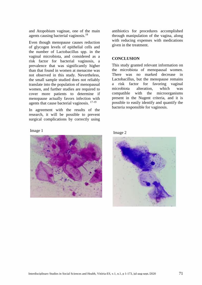
71
Interdisciplinary Studies in Social Sciences and Health, Vitória-ES, v.1, n.1, p 1-173, jul-aug-sept./2020
and Atopobium vaginae, one of the main
agents causing bacterial vaginosis.18
Even though menopause causes reduction
of glycogen levels of epithelial cells and
the number of Lactobacillus spp. in the
vaginal microbiota, and considered as a
risk factor for bacterial vaginosis, a
prevalence that was significantly higher
than that found in women at menacme was
not observed in this study. Nevertheless,
the small sample studied does not reliably
translate into the population of menopausal
women, and further studies are required to
cover more patients to determine if
menopause actually favors infection with
agents that cause bacterial vaginosis. 17-19
In agreement with the results of the
research, it will be possible to prevent
surgical complications by correctly using
antibiotics for procedures accomplished
through manipulation of the vagina, along
with reducing expenses with medications
given in the treatment.
CONCLUSION
This study granted relevant information on
the microbiota of menopausal women.
There was no marked decrease in
Lactobacillus, but the menopause remains
a risk factor for favoring vaginal
microbiota alteration, which was
compatible with the microorganisms
present in the Nugent criteria, and it is
possible to easily identify and quantify the
bacteria responsible for vaginosis.
Image 1
Image 2

72
Interdisciplinary Studies in Social Sciences and Health, Vitória-ES, v.1, n.1, p 1-173, jul-aug-sept./2020
REFERENCES
1. Junior JE, Cavalcante DIM. Contagem de morfotipos de Mobiluncus sp e concentração de
leucócitos em esfregaços vaginais de pacientes com vaginose bacteriana. Revista Brasileira de
Ginecologia e Obstetrícia. 2004. Rio de Janeiro, v.26 (3), 221-225.
2. Mota DA, Monteiro CA, Monteiro SG, Figueiredo PMS. Prevalência de vaginose
bacteriana em pacientes que realizaram bacterioscopia de secreção vaginal em laboratório de
saúde pública. Rev Bras Clin Med. 2012. São Paulo, v. 10 (1), 15-8.
3. Kurimori HY, Lima SMRR, Tamura1 KY, Yamada SS, Navarini A, Ueda SMY.
Microbiota vaginal de mulheres após a menopausa, assintomáticas, portadoras e não
portadoras de Diabetes Mellitus tipo 2. Arquivo Médico dos Hospitais e Faculdade de
Ciências Médicas da Santa Casa São Paulo. 2013. São Paulo, v.58, 59-63.
4. Cauci S, Driussi S, De Santo D. Prevalence of bacterial vaginosis and vaginal microbiota
changes in peri and postmenopausal women. 2002. J. Clin Microbiol. 40:2147-2152.
5. Pabish WL, Fihn SD, Stamm VE. Prevalence and determinants of vaginal microbiota
alterations in postmenopausal women. 2003. J Infect Dis. 188: 1054-8.
6. Gonçalves AKS. Mecanismos de defesa vaginal. In: Martins NV, Ribalta JCL (eds).
Patologia do trato genital inferior: diagnóstico e tratamento. 2005. São Paulo: Roca; p. 107-
221.
7. Guyton AC, Hall JE. Tratado de Fisiologia Médica. 12. ed. Rio de Janeiro: Guanabara
Koogan. 2011. p. 868-895.
8. Baract EC.; Lima GR. Guias de medicina ambulatorial e hospitalar de ginecologia. 2005.
São Paulo; Manole.
9. Lustosa AB. et al. Citologia hormonal do trato urinário baixo e da vagina de mulheres na
pós-menopausa, antes e durante estrogenioterapia oral e transdérmica, Revista Brasileira de
Ginecologia e Obstetrícia. 2002. Rio de Janeiro, v.24 (9) 573-577.
10. Wanderley M da S et al. Vaginose Bacteriana em mulheres com infertilidade e em
menopausadas. Revista Brasileira de Ginecologia e Obstetrícia. 2001. Rio de Janeiro, v.23
(10) 641-646.
11. Wajchenberg LB. Tecido adiposo como glândula endócrina. Arquivos Brasileiros de
Endocrinologia & Metabologia. 2000. São Paulo, v. 44 (1).
12. Gilbert GG, Donders MD. Definition anda classification abnormal vaginal flora. Best
Practice and Research Clinical Obstetrics and Gynaecology. 2007. Belgium, v.21 (3) 355-373.
13. Campos, AAS. Estudo comparativo entre o teste do pH e do KOH versus escore de
Nugent para diagnóstico da vaginose bacteriana em gestantes. Rev. Bras. Ginecol. Obstet.
[online]. 2012, vol.34, n.5, pp.209-214.
14. Gompel C, Koss LG. Citologia ginecológica e suas bases anatomoclínicas. 1997. São
Paulo, Manole.
15. Guerreiro HMN. et al. Flora vaginal e correlação com aspectos citológicos. Revista de
Saúde Pública. 1986. São Paulo, v.20 (6) 415-420.
16. Datcu R. Characterization of the vaginal microflora in health and disease . 2013. Dan Med
J 2014;61(4): B483 Tese phD.

73
Interdisciplinary Studies in Social Sciences and Health, Vitória-ES, v.1, n.1, p 1-173, jul-aug-sept./2020
17. Cerca, NMA. Influence of Biofilm Formation by Gardnerella vaginalis and Other
Anaerobes on Bacterial Vaginosis. 2015.The Journal of Infectious Diseases, v. 212 (12),
1856–1861.
18. Hay P. Bacterial Vaginosis. F1000Research 6. 2017. 1761. PMC. Web. 19.
19. Vespero EC, Azevedo EMM, Pellsson M, Perugini MRE. Correlação entre critérios
clínicos e critérios laboratoriais no diagnóstico de vaginose bacteriana. Semina: Ci. Biol.
Saúde. Londrina, v. 20/21, n. 2, p. 57-66, jun. 1999/2000.

74
Interdisciplinary Studies in Social Sciences and Health, Vitória-ES, v.1, n.1, p 1-173, jul-aug-sept./2020
DOI https://dx.doi.org/10.5935/2675-7397.20200003
Original Article
ASSOCIATION BETWEEN QUALITY OF LIFE AND MORTALITY IN PATIENTS
WITH HEART FAILURE
Roberto Ramos Barbosa1, Luiza Dias Torres2, Mayara da Silva3, Carla Campos Miranda4,
Ingrid Ardisson Colodete5, Andressa Croteletti6, Tiago de Melo Jacques7, Renato Giestas
Serpa8, Osmar Araujo Calil9, Luiz Fernando Machado Barbosa10
1 M.S./ College of Sciences of Santa Casa de Misericórdia de Vitória - (Professor).
2 Santa Casa de Misericórdia de Vitória Hospital - (Medical Doctor).
3 4 5 College of Sciences of Santa Casa de Misericórdia de Vitória - (Academic of
Medicine).
6 College of Sciences of Santa Casa de Misericórdia de Vitória Hospital - (Nurse).
7 Especialista/Hospital da Santa Casa de Misericórdia de Vitória - (Medical Doctor).
8 Expert/ College of Sciences of Santa Casa de Misericórdia de Vitória - (Professor).
Submitted in October 27, 2017; Approved in January 02, 2018.
Institution: EMESCAM
Av. Nossa Sra. Da Penha, 2190 – Santa Luíza, Vitória - ES, 29045-402.
Correspondence Author: Roberto Ramos Barbosa.
Escola Superior de Ciências da Santa Casa de Misericórdia de Vitória, ES.
Av. Nossa Sra. da Penha, 2190 – Santa Luíza, Vitória - ES, 29045-402.
E-mail: [email protected]
ESTUDOS INTERDISCIPLINARES EM
CIÊNCIAS SOCIAIS E DA SAÚDE INTERDISCIPLINARY STUDIES IN SOCIAL SCIENCES AND HEALTH
v.1, n.1, 2020

75
Interdisciplinary Studies in Social Sciences and Health, Vitória-ES, v.1, n.1, p 1-173, jul-aug-sept./2020
Keywords Abstract
Heart Failure;
Mortality;
Quality of Life
Objective: The central focus of this study was to estimate
the association between quality of life and mid-term
mortality in patients with heart failure (HF). Methods:
Prospective study in which the Minnesota Living with
Heart Failure Questionnaire (MLHFQ) was applied to
adults assisted in a specialized center, with voluntary and
secretive filling between October 2012 and March 2013,
in a consecutive inclusion. After a three-year clinical
follow-up, the group of patients who died was compared
to the survivor ones, according to baseline clinical
characteristics and initial quality of life. Results: From 76
patients initially approached and interviewed, 74 (97.4%)
answered the questionnaire and had completed a 3.5 year
follow-up. Comparison between groups death (n=8;
10.8%) and survivors (n=66; 89.2%) showed no difference
concerning age, sex, diabetes, smoking, atrial fibrillation,
body mass index, ejection fraction by echocardiogram,
functional class, use of beta-blocker and use of
angiotensin-converting enzyme inhibitor or angiotensin II
receptor antagonist. The average index studied score was
40.2 ± 24 in the death group and 40.9 ± 21 in the survivors
group (p=0.94). Conclusion: There was no association
between quality of life and mid-term mortality among HF
patients. The influence of quality of life over this
prognosis may be complex, and the ideal measurement
method for quality of life remains undetermined.
INTRODUCTION
Heart failure (HF) is a disease that results
from functional myocardial changes,
taking into account that with the
progressive increase in life expectancy, the
incidence and prevalence of this disease
has been increasing.¹ Nearly 2.3% in the
general population² presents a high
mortality, and may reach up to 50% of this
population within five years after the
diagnosis3.
An estimated 300,000 US deaths each year
have HF as the cause². Several factors such
as gender, lower ejection fraction, renal
dysfunction, diabetes, depression and the
need for hospitalization due to
decompensation are associated with higher
mortality4.
HF generally imposes great limitations on
daily activities, whether due to physical or
psychological symptoms, 5 which is
associated with worsening quality of life.6
It is defined as the patient's beliefs of the
influence of the disease in his or her life.³
There are many specific instruments to
measure quality of life in HF, among them
the Minnesota Living with Heart Failure
Questionnaire (MLHFQ), one of the most
frequent used for both research and daily
clinical practice.7,8 Besides trying to
estimate the quality of life in patients, this

76
Interdisciplinary Studies in Social Sciences and Health, Vitória-ES, v.1, n.1, p 1-173, jul-aug-sept./2020
questionnaire can also be used to estimate
mortality and the need for
hospitalizations.5,7 There is an evidence
that MLHFQ is an adequate and a valid
instrument for measuring quality of life in
HF patients, even in their different versions
translated into several countries, including
Brazil.5,9
The objective of the study was to assess
the relationship between quality of life and
mortality in the medium term in patients
with HF with reduced ejection fraction
attended by outpatients.
METHOD
This prospective cohort study was
performed in adult patients with reduced
ejection fraction HF who completed the
MLHFQ at the time of inclusion.
Patients with HF with ejection fraction of
less than 50% were transthoracic
echocardiograms of both sexes, regularly
followed at the HF clinic of the Santa Casa
de Misericórdia Hospital de Vitória - ES.
Those under 18 years of age, those who did
not accept to complete the questionnaire or
who did not sign the Free and Informed
Consent Term (TCLE) were excluded.
MLHFQ is a questionnaire comprised of
21 questions, reflecting the physical,
emotional and socioeconomic aspects of
patients. Responses range from zero to five
points. The zero score shows that this
aspect did not influence life in the last
month, while the 5 score shows that a
certain circumstance had maximum
limitation. The highest score in the
questionnaire is 105 points, considering
that the higher the score, the worse the
quality of life.
The MLHFQ version validated for
Portuguese language9 was completed in a
voluntary and confidential manner between
October 2012 and March 2013, in
consecutive inclusion. After three-year
clinical follow-up, patients who died were
compared to survivors for baseline clinical
features and initial quality of life.
SPSS software version 18.0 was used for
statistical analysis of the data. The
Pearson's chi-square test, the Fisher's test
and the student's t-test were used with a
significance level of 0.05. The categorical
variables were expressed in frequencies
and percentages, and the continuous
variables, in means and standard
deviations.
Research Ethics Committee of the
institution approved the current study, and
all participants in it completed the TCLE.
RESULT
Out of 76 patients initially treated 74
(97.4%) fulfilled the inclusion criteria in
the study and answered the questionnaire
completely and then with median 3.5 years
of follow-up to 3.5 years (mean 3.1 ± 0.9
years). At the end of the follow-up, eight
patients had died (10.8%) and 66 survived
(89.2%). The final outcome is shown in
figure 1.

77
Interdisciplinary Studies in Social Sciences and Health, Vitória-ES, v.1, n.1, p 1-173, jul-aug-sept./2020
By comparing the groups, there was no
difference in baseline clinical
characteristics or the rate of use of
angiotensin converting enzyme (ACE) beta
blockers and inhibitors or angiotensin
receptor blockers (ARBs), as shown in
table 1.
Table 1. Baseline clinical characteristics according to the group
Characteristics Deaths Survivors p
Age (years), mean ± SD 54,1 ± 10 59,7 ± 12 0,16
Male gender, n (%) 3 (37,5) 39 (59,1) 0,15
Diabetes mellitus, n (%) 3 (37,5) 25 (37,8%) 0,29
Smoking, n (%) 3 (37,5) 12 (18,2%) 0,15
Atrial fibrilation, n (%) 2 (25) 22 (33,3%) 0,30
BMI mean ± SD 25,6 ± 4,5 27,5 ± 6,3 0,32
Ejection fraction, mean ± SD 39% ± 6 37% ± 8 0,41
Funcional class, mean ± SD 1,4 ± 0,7 1,6 ± 0,9 0,72
Use of ACE inhibitors or BRA, n (%) 8 (100) 65 (98,5) 0,89
Use of beta-blocker, n (%) 8 (100) 65 (98,5) 0,89
SD = standard deviation; ARB = angiotensin receptor blockers; ACEI = angiotensin
converting enzyme inhibitors; BMI = body mass index.
The MLHFQ score ranged from 3 to 100, and the mean score obtained in the baseline
questionnaire of the total sample was 40.4 ± 21.8, similar between the death and survivor
groups (Figure 1).
Subtitle
Figure 1 - Quality of life at the time index according to clinical evolution at follow-up (death
or survival) – means of Minnesota Living with Heart Failure Questionnaire (MLHFQ) socres,
in which a higher score indicates lower quality of life.
Score average of basal MLHFQ
Deaths
Survivors

DISCUSSION
Heart failure is a rising disease in our
environment today, which is mainly due to
population aging and, therefore, is no
longer restricted to developed countries.
Since is a chronic-degenerative disease,
government spending on treatments
increases exponentially, costing
medications, hospitalizations, devices,
surgeries and work incapacity; improving
the quality of life of these patients may
reduce mortality and the need for
hospitalization has been demonstrated in
other populations that.10
Different quality of life measurement tools
may present different prognostic
stratification powers in HF. Consequently,
the ideal method of measuring this variable
remains controversial, and literature data
may differ, with general and disease-
specific questionnaires available. For
MLHFQ, the physical component domain
seems to be related to increased long-term
mortality, but adjusted analysis does not
place the overall score as an independent
predictor.11
By comparing patients who died and
survivors at three years of follow-up, this
study observed no difference in the mean
MLHFQ scores applied at the time of
inclusion. Moreover, baseline clinical
characteristics were also statistically
similar, as well as the rate of use of beta-
blockers and ACE inhibitors / ARBs. Other
prognostic factors of the studied population
such as other diseases, socioeconomic
conditions or unfavorable clinical
evolution may have played a decisive role.
The significance of the association
between quality of life and survival can be
compared to other known predictors of HF
mortality, such as diabetes or previous
hospitalizations.10 Frequently, the quality
of life, underestimated and deprecated in
relation to echocardiographic markers or
laboratories, may inform valuable data to
health teams, possibly in subgroups or
populations yet to be defined.12
We identified a wide variation in the
MLHFQ score, initially applied in the HF
population. The mean score found in our
study was similar to that of other
studies.4,11,13 The lower mortality rate in
comparison with other studies was
probably due to a lower risk profile with a
mean ejection fraction above 30%
Spanish study found total mortality of
69.7% in seven years follow-up of HF 433
patients. Survivors had a mean MLHFQ
score of 46 points, while those who died,
51 points (adjusted risk ratio = 1.26; 95%
confidence interval (CI) 95% .97 - 1.64).
The Netherlands conducted analysis of 152
patients with HF with follow-up of 18
months, observing mortality of 34%. In
this, the mean MLHFQ score was 42
among the survivors, and 52 among those
who died (p = 0.02), with a score> 37
presenting a relative risk of 3.24 (95% CI
1.38 - 7.62) for mortality.4
Another Dutch study, with 661 patients
from 17 centers, observed a mortality rate
of 42% in three years, with a MLHFQ
mean of 44 at the time of inclusion.
Comparison between surviving and non-
surviving groups showed a significant
difference for the MLHFQ score (42 ± 22
vs. 47 ± 19, p = 0.010). An increase of 10
points in the score was associated with a
7% increase in mortality.13 Alla et al.
evaluated 108 patients with HF in France,
showing mortality of 24% in one year and
associated a worsening of 10 points in the
MLHFQ score to a 23% higher risk of
death (p = 0.03). However, after the
adjusted analysis, this finding was non-
significant, which was attributed to the
small sample size.14
In the United States, an analysis of 313
patients with HF had shown that worse
MLHFQ scores were associated with
younger, less educated patients with lower
78 Interdisciplinary Studies in Social Sciences and Health, Vitória-ES, v.1, n.1, p 1-173, jul-aug-sept./2020

79
Interdisciplinary Studies in Social Sciences and Health, Vitória-ES, v.1, n.1, p 1-173, jul-aug-sept./2020
functional capacity. In this study, for each
additional point in the MLHFQ, the risk of
a cardiac event at follow-up increased to
1.6% .15
A Brazilian study included 101 patients
with HF who were followed up in public
health services and obtained a mean score
of HFHR of 37.5.2 Another recent study
observed a non-specific questionnaire with
a low quality of life in patients with HF,
when compared to the population with no
HF.16
The prognosis in severe and symptomatic
HF is poor, with high morbidity and
mortality. Quality of life is one of the
notorious aspects, since new therapies
present potential for increased survival.
Improving symptoms and quality of life of
patients with HF is a goal of treatment,
which should be in association with the
reduction of arrhythmic or mechanical
mortality.
Although relevant and unprecedented in
the local population, this study has
limitations, such as the reduced sample
size and the fact that the questionnaire was
not applied periodically to evaluate
evolutionarily the quality of life.
CONCLUSION
There was no association between quality
of life by MLHFQ and mortality in the
medium term among patients with HF.
Heart Failure is a complex disease, with
important systemic repercussions, and the
best method to measure the quality of life
of these patients remains uncertain. Larger
studies are essential to evaluate the
importance of quality of life on the
prognosis and complications of HF
patients.
Potential Conflict of Interest
The authors declare not to have conflicts of
interest.
Financing Source
The present study had no external sources
of financing.
Academic Linkage
The current study is not tied to Graduate
Programs.
REFERENCES
1. Escobar A, García-Pérez L, Navarro G, Bilbao A, Quiros R; CACE-HF Score group. A
one-year mortality clinical prediction rule forpatients with heart failure. Eur J Intern Med.
2017; S0953-6205(17): 30257-1.
2. Santos JJA, Plewka JEA, Brofman PRS. Qualidade de vida e indicadores clínicos na
insuficiência cardíaca: análise multivariada. Arq Bras Cardiol. 2009; 93 (2): 159-66.
3. Erceg P, Despotovic N, Milosevic DP, Soldatovic I, Zdravkovic S, Tomic S, et al. Health-
related quality of life in elderly patients hospitalized with chronic heart failure. Clin Interv
Aging. 2013; 8: 1539-46.
4. Bouvy ML, Heerdink ER, Leufkens HG, Hoes AW. Predicting mortality in patients with
heart failure: a pragmatic approach. Heart. 2003; 89 (6): 605-9.
5. Garin O, Ferrer M, Pont À, Wiklund I, Van Ganse E, Vilagut G, et al. Evidence on the
global measurement model of the Minnesota Living with Heart Failure Questionnaire. Qual
Life Res. 2013; 22 (10): 2675-84.

80
Interdisciplinary Studies in Social Sciences and Health, Vitória-ES, v.1, n.1, p 1-173, jul-aug-sept./2020
6. Cruz FD, Issa VS, Ayub-Ferreira SM, Chizzola PR, Souza GE, Moreira LF, et al. Effect of
a sequential education and monitoring programme on quality-of-life components in heart
failure. Eur J Heart Fail. 2010; 12 (9): 1009-15.
7. Garin O, Herdman M, Vilagut G, Ferrer M, Ribera A, Rajmil L, et al. Assessing health-
related quality of life in patients with heart failure: a systematic, standardized comparison of
available measures. Heart Fail Rev. 2014; 19 (3): 359-67.
8. Bilbao A, Escobar A, García-Perez L, Navarro G, Quirós R. The Minnesota living with
heart failure questionnaire: comparison of different factor structures. Health Qual Life
Outcomes. 2016; 14:23.
9. Carvalho VO, Guimarães GV, Carrara D, Bacal F, Bocchi EA. Validação da versão em
português do Minnesota Living with Heart Failure Questionnaire. Arq Bras Cardiol.
2009;93(1):39-44.
10. Rodríguez-Artalejo F, Guallar-Castillón P, Pascual CR, Otero CM, Montes AO, García
AN, et al. Health-related quality of life as a predictor of hospital readmission and death
among patients with heart failure. Arch Intern Med. 2005; 165 (11): 1274-9.
11. Zuluaga MC, Guallar-Castillón P, López-García E, Banegas JR, Conde-Herrera M, Olcoz-
Chiva M, et al. Generic and disease-specific quality of life as a predictor of long-term
mortality in heart failure. Eur J Heart Fail. 2010; 12 (12): 1372-8.
12. Barbosa RR, Franklin RV, Stefenoni AV, Moraes VD, Jacques TM, Serpa RG, et al.
Análise da qualidade de vida em homens e mulheres portadores de insuficiência cardíaca. Rev
Bras Cardiol. 2014;27(2):593-99
13. Hoekstra T, Jaarsma T, Van Veldhuisen DJ, Hillege HL, Sanderman R, Lesman-Leegte I.
Quality of life and survival in patients with heart failure. Eur J Heart Fail. 2013; 15 (1): 94-
102.
14. Alla F, Briançon S, Guillemin F, Jullière Y, Mertès PM, Villemont JP, et al. Self-rating of
quality of life provides additional prognostic information in heart failure. Insights into the
EPICAL study. Eur J Heart Fail. 2002; 4 (3): 337-43.
15. Wu JR, Lennie TA, Frazier SK, Moser DK. Health-related quality of life, functional status
and cardiac event-free survival in patients with heart failure. J Cardiovasc Nurs. 2016; 31 (3):
236-44.
16. Jorge AJL, Rosa MLG, Correia DMS, Martins WA, Ceron DMM, Coelho LCF, et al.
Avaliação da qualidade de vida em pacientes com e sem insuficiência cardíaca na atenção
primária. Arq Bras Cardiol. 2017; 109 (3): 248-252.

81
Interdisciplinary Studies in Social Sciences and Health, Vitória-ES, v.1, n.1, p 1-173, jul-aug-sept./2020
DOI https://dx.doi.org/10.5935/2675-7397.20200015
Original Article
THE INFLUENCE OF RISK OF FALLING ON FUNCTIONAL DEPENDENCE OF
HOMEBOUND ELDERLY
Morghana Ferreira Ambrosim1, Taynara Ribeiro Batalha2, Fabiola dos Santos Dornellas
Oliveira3, Christiane Boaventura Lourenço4, Vanezia Gonçalves da Silva5
1 Physiotherapist from EMESCAM, Master student in Physical Education–Exercise
Physiology from UFES - (Physiotherapist)
2 Physiotherapist from EMESCAM, resident in the oncological physiotherapy sector
atUNIFESP - Federal University of São Paulo. SP. - (Physiotherapist)
3 Physiotherapist from EMESCAM, Master student in Public Policy from EMESCAM. -
(Physiotherapistand Professor).
4 Fisioterapeuta formada pela EMESCAM, Master Degree from State University of
campinas-SP e PhD. from Mcgill University Montreal, Canadá - (Physiotherapist and
Professor).
5 Nurse from EMESCAM e Master Degree in Public Policy from EMESCAM - (Nurseand
Professor).
Submitted in November 10, 2017; Approved in January 10, 2019.
Institution: EMESCAM
Av. Nossa Sra. da Penha, 2190 – Santa Luíza, Vitória - ES, 29045-402
Correspondence Author: Morghana Ferreira Ambrosim
Escola Superior de Ciências da Santa Casa de Misericórdia de Vitória, ES.
Av. Nossa Sra. da Penha, 2190 – Santa Luíza, Vitória - ES, 29045-402.
Email: [email protected]
ESTUDOS INTERDISCIPLINARES EM
CIÊNCIAS SOCIAIS E DA SAÚDE INTERDISCIPLINARY STUDIES IN SOCIAL SCIENCES AND HEALTH
v.1, n.1, 2020

82
Interdisciplinary Studies in Social Sciences and Health, Vitória-ES, v.1, n.1, p 1-173, jul-aug-sept./2020
Keywords Abstract
Aged Rights;
Homebound Persons;
Personal Autonomy;
Postural Balance
Objective: Verify the influence of sociodemographic
factors, caregivers and risk of falling on the functional
dependency of homebound elderly. Method:
Crossectional descriptive study during the participation
of the PRO PET Saúde - EMESCAM. Functional
dependency was assessed by the Functional
independency measure, risk of falling by the TINETTI,
sociodemographic and presence of caregiver by
questionnaire developed by the authors. For statistical
analysis were used descriptive statistics, chi square and
logistic regression. Results: A total of 155 elderly
participated in the present study. The results of the
logistic regression suggests that a high score in the
TINETTI decreases the chances of being functional
dependent (OR 0,83, p
INTRODUCTION
The increase in population survival in
developed countries is due to the fall in
mortality, progresses in medicine, the
adequate urbanization of cities, nutritional
improvement, the rise in personal and
environmental hygiene levels and
technological advances. In developing
countries, such as Brazil, this increase is
associated with the fall in the fertility rate,
and the increase in life expectancy,
indicated by improvements in health,
coupled with technological advances1-2.
Aging is a dynamic and progressive
process in which morphological,
biochemical, functional changes arise³ and
also changes in the central nervous system
(CNS), compromising the neuromuscular
function, given by changes in balance,
reduced reflex response, loss of muscle
fibers, change in the percentage of
contractile muscle tissue, deficit in muscle
innervation and decrease in muscle
strength and function, with resultant
muscle atrophy, therefore increasing the
loss of bone mineral density (osteoporosis
and sarcopenia)4-5. These changes cause
functional disabilities, restricting the
performance of activities of daily living
(ADLs), namely walking, standing,
maintaining postural balance and
preventing imminent falls4.
In Brazil, about 30% of the elderly fall at a
minimum once a year and the incidence of
falls increases with advancing age,
transpiring in more than a third of people
aged 60 or over. A study carried out in
2015 showed that out of 8,142,342 deaths
in Brazil, between 1997 and 2010, of
people over 60 years of age, 0.61% were
deaths resulting from falls, and 50.6%
occurred among the population of 80 years
or more5-6-7. The outcome is the greater
demand of the elderly for health services,
fostering more frequent hospitalizations,
longer bed occupancy, when as against
other age groups and a higher number of
deaths among the elderly. Consequently,
falls in the elderly represent an essential
public health problem and produce health
expenditures three to seven times higher
than the average cost of the population,
and the social assistance system. Falls can
promote or emphasize morbid states, also
considering their biopsychosocial
consequences, in particular difficulty in

83
Interdisciplinary Studies in Social Sciences and Health, Vitória-ES, v.1, n.1, p 1-173, jul-aug-sept./2020
walking, fear of recurrent falls, limitations
in instrumental activities of daily living,
restriction at home, decreased
independence, autonomy and quality of
life6-7-8, which may encourage to functional
dependence. These functional disabilities
and limitations in activities and
participations contribute the restriction of
the elderly to the domestic environment, in
order to require constant help from a
caregiver who helps in their activities,
which is defined as functional
dependence9. This functional dependence
provides a progressive social confinement,
which can lead to sedentary lifestyle,
cognitive deficit, loss of self-esteem and
abandonment of self-care10 and,
consequently, to the development and / or
worsening of diseases and health
conditions. Based on IBGE, in 2010, 12%
of the inhabitants of Vitória, Espírito
Santo, were 60 years old or older.
Pampolim et al.12, in 2017 noted that the
prevalence of functional dependence (DF),
influenced by sociodemographic and
economic factors in elderly people
homebound elderly, was 48%, but that
there are other factors that can influence
the DF of the elderly homebound elderly,
and there is no evidence of how much the
deficiency of balance and risk of falls
influences the disability and DF of these
elderly12, which makes this knowledge
significant for both health professionals
and public policies, considering the need to
develop prevention and education
promotion programs attached to falls.
As a result, the purpose of this study is to
determine the influence of
sociodemographic factors, the presence of
the caregiver and the risk of falls in the
functional dependence of elderly people
who are restricted to their home.
METHODS
This is a secondary analysis of the study
database called “Socio-demographic and
health profile of the homebound and
bedridden elderly at a family health unit in
the city of Vitória-ES”, conducted by
PRO-PET Saúde ( National Program for
the Reorientation of Professional Training
in Health and the Program of Education
through Work for Health), related to
EMESCAM (Higher School of Sciences of
Santa Casa de Misericórdia de Vitória) and
the State Health Secretariat of Espírito
Santo and the Municipal Health Secretariat
of Vitória. It is characterized by a cross-
sectional study with a quantitative
approach with a convenience sample and
data collection performed from April to
November 2014. This study comprised
individuals aged 60 years or older,
homebound elderly(not bedridden, which
characterizes as restricted to bed),
registered in the territory of the Family
Health Unit Dr. José Moysés and who
accepted engage in the research 155
elderly people, by signing (of the elderly or
responsible) the free and informed consent
term.
The study excluded: elderly people who
were unable to perform the TINETTI test;
those who did not have a suitable caregiver
for this purpose; who showed refusal or
family restriction to participate in the
study; the cases of death and / or migration
to another region during data collection.
This study was approved by the Research
Ethics Committee of EMESCAM under
protocol number 567,990.
The data were collected via face-to-face
interviews, performed at the residence of
the elderly by students of EMESCAM's
physiotherapy, nursing and medicine
courses, formerly trained by the main
researcher, and monitored by ACS. All
participants underwent a semi-structured
interview in order to gather information
concerning the socio-demographic profile,
namely age, sex, race and socioeconomic

84
Interdisciplinary Studies in Social Sciences and Health, Vitória-ES, v.1, n.1, p 1-173, jul-aug-sept./2020
status, and afterwards the elderly were
subjected to the appraisal of functional
capacity and the risk of falling.
Functional capacity was evaluated by the
Functional Independence Measure (FIM), a
scale created in the 1980s with the
purposeof developing an instrument
eligible of measuring the degree of request
for third-party care that patients with
disabilities require to accomplish motor
and cognitive, and hence toestablish what
care is required to be submitted for the
patient to carry out ADLs13.
The purpose of MIF scale is to evaluate
physical and cognitive functionality using
18 items, with regard to six dimensions:
self-care, sphincter control, transfers,
locomotion, communication and social
cognition. For each dimension, the elderly
person is assessed with a score from 0 to 7,
with a value of 0 according to total
dependence and a value of 7 corresponding
to performing tasks independently. The
MIF score has total scores ranging from 18
to 126 points. Once the assessment was
performed in the elderly's own home, there
was no reason to use any special resource
to simulate daily living conditions,
nevertheless it is appropriate mentioning
that, as instructed by the Ministry of
Health, when an item could not be
evaluated / answered, a score of 1 was
given. The total FIM result set the presence
or absence of DF14-15-16. Individuals with
scores ≤ 103 points were assumed
dependent, where lower scores indicate
functional dependence13-16.
The risk of falls was assessed by the
TINETTI test, which has been applied to
assess balance and gait abnormalities. The
test is composed of 16 items, 9 of which
are for body balance and 7 for walking.
The TINETTI Test categorizes aspects of
walking such as speed, stride distance,
symmetry and balance while standing,
turning and also changes with eyes closed.
The score for each exercise ranges from 0
to 1 or 0 to 2, with a lower score denoting
poorer physical ability. The total score is
the sum of the body balance and gait
scores. The maximum score is 12 points
for walking, 16 for body balance and 28
for total16-17.
Data analysis was accomplished using the
statistics program (IBM, SPSS 20.0). The
descriptive characteristics of the elderly
were presented as mean and standard
deviation for continuous variables and as
frequency and percentage for ordinal and
nominal variables. In order to determine
the influence of sociodemographic factors,
presence of caregiver, risk of falling, and
gait in functional dependence, an ordinal
regression analysis was implemented,
where the level of functional dependence
(0 independent or 1 dependent) is the
dependent variable and the variables
sociodemographic (age, sex, race, elderly
income, family income) risk of falling
(low, moderate and high), gait capacity and
caregiver presence as independent
variables. A significance level of 5% was
selected for all analysis.
RESULTS
The total elderly population recorded at the
Dr. José Moyses Health Unit, in February
2014, was 4,832, among them 212
participated in the data collection, of which
167 were restricted to their home. After
analyzing the inclusion criteria, the sample
of the current study is 155 elderly people
restricted to their home. Table 1 illustrates
the sample's sociodemographic
characteristics. It can be detectable that
54.8% of the sample is aged between 81
and 90 years old, with a statistically
significant correlation with the risk of falls,
78.6% being female, and predominantly
white with 72, 2%. Of the 155 participants,
79.6% had a caregiver and 35.1% of the
elderly participants lived with only one
person. Concerning the risk of falls, 27.7%

85
Interdisciplinary Studies in Social Sciences and Health, Vitória-ES, v.1, n.1, p 1-173, jul-aug-sept./2020
had a moderate to high risk of falls with a
score between 19 and 22 or 0 and 18,
respectively. The elderly was classified as
reported by the functional independence
scale (FIM) in dependents, portraying
30.32% of the sample and independent,
representing 67.09%.
Table 1 - Sociodemographic characteristics of elderly people restricted to the home assigned
to USF José Moysés related to TINETTI and MIF scale.
Total Sample
(n=155)
Independent
(n=104)
Dependent
(n=47)
Value of p
n (%)
Age range
60-70 11 (7,1) 6 (5,7) 5 (10,2)
0,085 71-80 43 (27,7) 26 (24,5) 17 (34,7)
81-90 85 (54,8) 63 (59,4) 22 (44,9)
>90 16 (10,3) 11 (10,4) 5 (10,2)
Fourth age
Yes 101 (65,2) 74 (69,8) 27 (55,1) 0.055*
No 54 (34,8) 32 (30,2) 22 (44,9)
Sex
Female 121 (78,6) 90 (84,9) 31 (64,6) 0,005*
Male 33 (21,4) 16 (15,1) 17 (35,4)
Race
Brown 28 (18,5) 18 (17,3) 10 (21,3)
0,33 White 109 (72,2) 75 (72,1) 34 (72,3)
Black 11 (7,3) 8 (7,7) 3 6,4)
Yellow 3 (2) 3 (2,9) 0 (0)
Education
Illiterate 36 (25,9) 25 (26) 11 (25,6)
0,28
Up to 4 years 43 (30.9) 30 (31,3) 13 (30,2)
From 5 to 8 years 35 (25,2) 26 (27,1) 9 (20,9)
From 9 to 11 years 16 (11,5) 10 (10,4) 6 (14)
Over 11 years 9 (6,5) 5 (5,2) 4 (9,3)
Marital status
Married 39 (25,2) 24 (22,6) 15 (30,6) 0,07

86
Interdisciplinary Studies in Social Sciences and Health, Vitória-ES, v.1, n.1, p 1-173, jul-aug-sept./2020
Single 15 (9,7) 12 (11,3) 3 (6,1)
Widower / widow 94 (60,6) 66 (62,3) 28 (57,1)
divorced /Separated 7 (4,5) 4 (3,8) 3 (6,1)
Presence of children
Yes 137 (88,4) 95 (89,6) 42 (85,7) 0,32
No 18 (11,6) 11(10,4) 7 (14,3)
Family income
Up to 1 minimum wage 14 (13,5) 13 (18,8) 1 (2,9) 0,010*
From 1 to 4 min. wages 51 (49) 32 (46,4) 19 (54,3)
Entre 4 e 10 min. wages 28 (26,9) 14 (20,3) 14 (40)
Above 10 min. wages 11 (10,6) 10 (14,5) 1 (2,9)
Elderly income
No income 2 (1,6) 1 (1,2) 1 (2,6)
0,44 Up to 1 minimum wage 64 (51,2) 47 (54,7) 17 (43,6)
Above 4 min. wages 59 (47,2) 38 (44,2) 21 (53,8)
Residents
Alone 18 (11,7) 14 (13,3) 4 (8,2)
0,6 1 person 54 (35,1) 35 (33,3) 19 (38,8)
Above 2 people 82 9 (53,2) 56 (53,3) 26 (53,1)
Elderly caregiver
Yes 117 (79,6) 72 (72,7) 45 (93,8) 0,002*
No 30 (20,4) 27 (27,3) 3 (6,3)
Risk of falls
Low 28 (18,1) 23 (21,7) 5 (10,2) <0,001*
Moderate/High 43 (27,7) 37 (34,9) 6 (12,2)

87
Interdisciplinary Studies in Social Sciences and Health, Vitória-ES, v.1, n.1, p 1-173, jul-aug-sept./2020
Table 2 – Sociodemographic characteristics of elderly people restricted to the home assigned
to USF José Moysés related to TINETTI and MIF scale.
Variables Total Sample Independent Dependent Value of p
(n=155) (n=104) (n=47)
Median (min,max)
Age 82.9 ± 7.5 83 (64,100) 83 (63, 103) 0,28
Tinetti Balance 10.26 ± 3.5 11 (2,16) 7 (1,16)
Walking 6.88 ± 3.37 8 (2,12) 4 (0,12) < 0,001*
Total 17.2 ± 6.4 20 (7,28) 12 (1,28)
MIF
Cognitive 28.33 ± 8.69 34 (18,35) 20 (5, 35)
Motor 74.44 ± 19.39 85 (69, 91) 56 (13, 88) < 0,001*
Total 102.77 ± 25.43 116 (104, 126) 79 (18,102)
*p<0,05
USF = Family Health Unit
In table 3, when the results of the Binary
Logistic Regression analysis were verified,
we observed that the factors that most
influence the functional dependence of the
elderly are the elderly's own income (p
0.039), Tinetti's gait domain (p 0.037), the
presence of caregiver (p 0.019). In
contrast, the other independent variables
such as gender, age, race, children,
education, marital status, number of
residents in the household, family income
and Tinetti balance did not exhibit
statistically significant influence.

88
Interdisciplinary Studies in Social Sciences and Health, Vitória-ES, v.1, n.1, p 1-173, jul-aug-sept./2020
Table 3 - Binary Logistic Regression between socio-demographic and economic variables and
the presence of functional dependence in elderly people restricted to the home, assigned to the
Family Health Unit.
Independent Variables OR IC95% p
Age 0.83 0,68 – 1,01 0,063
Sex 0,13 0,01 – 1,53 0.106
Race
White (Reference)
Brown 3,33 0,50 – 22,08 0,210
Black 0,47 0,01 – 13,45 0,661
Other 0,00 0,00 – 0,00 1
Children 0,22 0,00 – 7,17 0,39
Education
Illiterate (Reference)
Up to 4 years 0,38 0,05 – 3,26 0,38
From 5 to 8 years 0,70 0,06 – 7,89 0,77
From 9 to 11 years 0,026 0,00 – 2,08 0,10
Above 11 yeares 0,06 0,00 – 2,82 0,15
Marital Status
Married (Reference)
Widower 2,24 0,22 – 22,51 0,494
Single 0,44 0,00 – 47,7 0,734
Other 2,58 0,04 – 153,50 0,649
Number of residents in the
Household 0,70 0,17 – 2,92 0,63
Elderly Income 8,52 1,11 – 65,30 0,039
Family Income 3,07 0,75 – 12,59 0,119
Presence of a caregiver 95,0 2,10 – 4298,90 0,019
Tinetti gait 0.55 0,31 – 0,96 0,037
Tinetti Balance
Low risk of falls (Reference)
Moderate risk of falls 1,14 0,02 – 58,20 0,94
High risk of falls 0,33 0,00 – 50,98 0,67
OR – OddsRatio; IC – Confidence Interval; SM = Minimum Wage * p < 0,05

89
Interdisciplinary Studies in Social Sciences and Health, Vitória-ES, v.1, n.1, p 1-173, jul-aug-sept./2020
DISCUSSION
Usually, the profile of the elderly in this
study composed of: elderly between 81 and
90 years old, female, self-declared white,
with low education, widowed, with
children and caregiver, with low income
and living in houses with less than two
people. Those characteristics reiterate what
has already been evidenced in the
literature, underscoring the increase in
longevity and the feminization of aging18-
19.
Nevertheless, we identified that the vast
majority (78.6%) of the elderly studied
were female, corroborating with the studies
by Lopes et al., 201419 and Clares et al.,
201120. When comparing the groups, the
elderly women were stillthe majority.
Though, we noted a decrease in the
frequency of women in the dependent
elderly group, and this difference was
statistically significant (p = 0.005). Gomes,
Nascimento and Araújo 200720 present that
women take better care of themselves,
carry out almost all domestic activities,
take care of their children and are more
active, which may be contributing to a
more active, healthy and independent
aging.
With respect to age, it was noted that the
fourth age (≥ 80 years) elderly had
considerably greater functional dependence
(p = 0.055), a result previously expected,
since the aging process represents an
overburden for all systems, which may be
leaving the elderly vulnerable to falls and
other functional impairments21. It was also
observed that the gradual decline in
functional capacity is concerned to the
progressive enhance in age. Studies by
Alves, Leite and Machado 201223 showed
that the probability of elderly present
impairment of functional capacity
increases considerably with advancing age.
The study of Mello, Engstrom and Alves
201424 characterized functional
dependence with the fact that a person
requires help or assistance to execute an
activity, or if a person is unable to perform
a task. Changes pertinent to the aging
process appear as a result of advancing age
and, occasionally the presence of the
caregiver becomes important24.
Nevertheless, in some cases, the presence
of the caregiver can hustle the natural
functional decline to the aging process,
considering that the elderly can be
accommodated, gradually leading them to
interrupt their activities, as stated by
Pampolim et al., 201712. Macedo et al.,
2012 declare that, in some situations, the
dependence of the elderly is associated
with the ability to perform a task, as well
as the expectations of other people in
relation to what the elderly person is
capable of doing, besides the opportunities
given to these elderly. In this sense, a very
protective environment, which does not
encourage the independence of the elderly,
and where family members / caregivers
end up performing tasks that the elderly
would still be able to execute alone or with
little assistance, helps in their functional
decline.
This study identified that the presence of
the caregiver in the group of dependent
elderly people was significantly (p =
0.002) higher. Amendola, Oliveira and
Alvarenga 201125 highlighted the
importance of the caregiver knowing how
to identify and deal with the degree of
dependence of the elderly, in order to help
only in what is really necessary and avoid
overprotection and increased dependence
on the elderly25. According to Ursine;
Lamb; Moraes 2011,26 the restriction to the
home - which can be promoted by family
overprotection and the presence of the
caregiver - is an insidious and dynamic
process, which can be modified or
prevented, if there is adaptation of the
environment and adequate intervention in
its risk factors. The restriction of the

90
Interdisciplinary Studies in Social Sciences and Health, Vitória-ES, v.1, n.1, p 1-173, jul-aug-sept./2020
elderly to the domestic environment
combines functional dependence with an
increased risk of comorbidities and loss of
autonomy26.
Recognizing that falls may lead to worsen
health in the elderly, contributing to
increased morbidity and mortality, family
members decide for the presence of the
caregiver in order to prevent
complications. In agreement to Machado et
al. 200927 and Soares et al. 201128, falls
may affect the functional capacity,
autonomy and the level of dependency of
the elderly, as it is related to anatomical
changes based onthe natural aging process
and on several pathologies that may head
the elderly to need help. Thus, the
prevention of falls is an essential item for
the health of the elderly, and also a
situation that involves caregivers, family
members and health professionals, taking
into account that this situation can directly
influence the functionality of the elderly27-
28-29.
Ribeiro et al. 20155 stated that the
assessment of the of the elderly
functionality is asignificant parameter in
the practice of gerontology, as it offers
relevant information about their health and
the need for help from third parties to
perform the activities of daily living.
Functional assessment can be implemented
using a variety of instruments, including
the Functional Independence Measure
(FIM). We identified that 47 elderly people
were classified as dependent. Based on the
FIM, 93.8% have a caregiver, manifesting
that dependence can be a predictor for the
presence of a caregiver, corroborating the
studies carried out by Alves et al. 200730
and Colomé et al. 201131 who demonstrate
a strong influence of chronic diseases
concerned the elderly on functional
capacity, and that when they compromise
autonomy, the presence of a caregiver may
be necessary to provide assistance5-30-31.
The Logistic Regression analysis results
derived from this study demonstrate that
the most influential factors in the
functional dependence of the elderly are:
the elderly's income (p = 0.039), Tinetti's
gait domain (p = 0.037), the presence of a
caregiver (p = 0.019) the work of
Pampolim et al, 2017, who found an
influence of 48% of the sociodemographic
factors of the elderly's functional
dependence on the presence of the
caregiver, which is a strong and significant
predictor of this outcome. In the current
study, we observed that furthermore these
sociodemographic factors, the risk of falls
also favors functional dependence by an
additional 27.7% 12.
Functionality means the integrity of
structures, the individual's physical and
mental functions, enabling them to
accomplish activities and take part in a
social context in anautonomous way. The
disability of the elderly has significant
implications for the family, the
community, the health system as well as
their own life, considering that it leads to
greater vulnerability and dependency,
besides the reduction of the well-being,
and the quality of life of the elderly32
After checking the relationship between
functional capacity and income, we
identified that when the elderly person has
a higher income, is much unlikely that he
will present a poor functional
capacity.Education, in contrast, presented
itself as a protective factor. This fact is
justified by the principle that elderly
individuals with a higher educational level
are less likely to be exposed to risk factors
for diseases and to submit to inadequate
working conditions.
Thus, the dependence of the elderly may
take place due to the natural aging process,
the great family protection, the presence of
a caregiver and mainly due to the increased
risk of falls. Aging, dependence and the
presence of the caregiver request new

91
Interdisciplinary Studies in Social Sciences and Health, Vitória-ES, v.1, n.1, p 1-173, jul-aug-sept./2020
forms of assistance from health
professionals. It is understood that the risk
for falls increases proportionally with the
number of risk factors, as it is an event that
is hardly the result of an isolated factor.
Hence the importance of Physiotherapy
acting preventively in relation to falls and
consequently contributing to the
independence of the elderly, seeking to act
on the greatest possible number of these
factors33.
CONCLUSION
The sociodemographic factors and the
presence of the caregiver enhance the
functional dependence of the elderly
homebound elderly and the risk of falls
further increases this situation.
The population growth has been occurring
through a continuous process of
automation of daily practices and
technological advances, where the release
of muscle groups is increasingly observed
in basic daily activities, such as remote
control, elevators, access the help of third
parties, among others.It demonstrates that
there is a gap in scientific knowledge, avid
for new research that intends to emphasize
and rectify the main negative interventions
that may interfere in the active, healthy and
vital aging process.
The rehabilitation operates by performing
muscle strengthening, gait training - on
stable and unstable ground, as well as
promoting improved posture and the
proprioceptive adaptive response. When
associated with other pathological
conditions, it is crucial that the intervention
is geared towards the patient's cognitive,
motor and sensory readjustment. Other
significant strategies implicate improving
flexibility and preventing deformities.
REFERENCES
1. MÁRCIA R.S.S; BARBOSA M; GUSMÃO J.L; FARO A.C.M; LEITE R.C.B. A situação
social do idoso no Brasil: uma breve consideração. Acta. Paul. Enferm. 2005;18(4):422-6.
2. CASTIGLIONI A.H. Envelhecimento da população em Vitória, Espírito Santo (Brasil). III
Congresso da Associação Latino Americana de População, ALAP, realizado em Córdoba.
Argentina, de 24 a 26 de setembro de 2008.
3. FILHO E.T.C. Fisiologia do envelhecimento. In: Netto M.P. Gerontologia: A velhece e o
envelhecimento em visão globalizada. São Paulo: Atheneu; 2005.
4. DAVINI R.; NUNES C. V. Alterações no sistema neuromuscular decorrentes do
envelhecimento e o papel do exercício físico na manutenção da força muscular em indivíduos
idosos. RevBrasFisioter. 2003;7(3):201–7.
5. RIBEIRO D. K. M.N; et al. Contributory factors for the functional independence of oldest
old. RevEscEnferm USP. 2015;49(1):87-93.
6. MATSUDO S. M.; MATSUDO V. K. R.; NETO T. L. D. B. Impacto do envelhecimento
nas variáveis antropométricas, neuromotoras e metabólicas da aptidão física. RevBrasCienc e
Mov. 2000;8(4):21–32.
7. MACIEL A. C. C.; GUERRA R. O. Fatores associados à alteração da mobilidade em
idosos residentes na comunidade. RevBrasFisioter. 2005;9(1):17-23.
92
Interdisciplinary Studies in Social Sciences and Health, Vitória-ES, v.1, n.1, p 1-173, jul-aug-sept./2020
8. KAUFFMAN, T.L. et al. Manual de Reabilitação Geriátrica. Rio de Janeiro: Guanabara
Koogan, 2001.
9. STUBBS, B. et al. Pain and the Risk for Falls in Community-Dwelling Older Adults:
Systematic Review and Meta-Analysis. Archives of Physical Medicine and Rehabilitation.
2014;95(1):175-87.
10. JAHANA, K.O; DIOGO M. J.D. Saúde coletiva- Quedas em idosos: Principais causas e
consequências. Editora Bolina-SP; 2007;17(4):148-153.
11. IBGE. Censo demográfico 2010: resultados da amostra características da população. 2010.
[serial on the Internet]. Available from:
<.http://cidades.ibge.gov.br/xtras/temas.php?lang=&codmun=320530&idtema=90&search=es
pirito-santo|vitoria|censo-demografico-2010:-resultados-da-amostra-caracteristicas-da-
populacao>.
12. PAMPOLIM, G.; LOURENÇO, C.; SILVA, V. G.; COELHO, M. C. R.; SOGAME, L. C.
M. Prevalência e fatores associados a dependência funcional em idosos restritos ao lar de uma
Unidade de Saúde da Família em Vitória-ES. Revista Brasileira de Crescimento e
Desenvolvimento Humano. No prelo 2017.
13. RIBERTO, M. et al. Reprodutibilidade da versão brasileira da Medida de Independência
Funcional. ActaFisiátrica, 2001;1(8):45-52.
14. ALVES LC, LEITE IDC, MACHADO CJ. Factors associated with functional disability of
elderly in Brazil: a multilevel analysis. Rev SaúdePúbl. 2010;44(3):468–78.
15. RIBERTO M, MIYAZAKI MH, JUCÁ SSH, SAKAMOTO H, POTIGUARA P.
Validation of the Brazilian version of Functional Independence Measure. Acta Fisiatr.
2004;11(2):3–7.
16. MACÊDO AML, CERQUIARI EAN, ALVARENGA MRM, FACCENDA O,
OLIVEIRA MAC. Functional assessment of elderly with cognitive deficit. Acta Paul Enferm.
2012;25(3):358–63.
17. SILVA J. M. N et al. Correlação entre o risco de queda e autonomia funcional em idosos
institucionalizados. Rev. Bras. Geriatr. Gerontol., Rio de Janeiro, 2013;16(2):337-346.
18. MENDES, MRSSB; GUSMÃO, JL.; FARO ACM, LEITE RCBO. A situação social do
idoso no Brasil: uma breve consideração. Acta Paul Enferm. 2005;18(4):422-6.
19. LOPES, FAM; MONTANHOLI, LL; SILVA, JML, OLIVEIRA, FA. Perfil
epidemiológico em idosos assistidos pela estratégia saúde da família. REAS [Internet].
2014;3(1):84-94.
20. CLARES, JWB; FREITAS, MC; ALMEIDA, PC; GALIZA, FT; QUEIROZ, TA. Perfil
de idosos cadastrados numa unidade básica de saúde da família de fortaleza-ce. Rev Rene,
Fortaleza, 2011;12:988-94.
21. ROMEU GOMES, R; NASCIMENTO, EF; ARAÚJO, FC. Por que os homens buscam
menos os serviços de saúde do que as mulheres? As explicações de homens com baixa
escolaridade e homens com ensino superior. Rio de Janeiro, Cad. Saúde Pública,
2007;23(3):565-574.

93
Interdisciplinary Studies in Social Sciences and Health, Vitória-ES, v.1, n.1, p 1-173, jul-aug-sept./2020
22. FERREIRA, OGL; MACIL, SC; COSTA, SMG; SILVA, AO; MOREIRA, MASP.
Envelhecimento ativo e sua relação com a independência funcional. Florianópolis, Texto
Contexto Enferm, 2012;21(3):513-8.
23. ALVES, LC; LEITE, IC; MACHADO, CJ. Fatores associados à incapacidade funcional
dos idosos no Brasil: análise multinível. Rev Saúde Pública. 2010;44(3).
24. MELLO A.C; ENGSTROM E.M; ALVES L.C. Health-related and socio-demographic
factors associated with frailty in the elderly: a systematic literature review. Cad. Saúde
Pública, Rio de Janeiro; 2014;30(6):1143-1168.
25. AMENDOLA F; OLIVEIRA M.A.C, ALVARENGAM.R.M. Influence of social support
on the quality of life of family caregivers while caring for people with dependence.
RevEscEnferm USP; 2011;45(4):880-5.
26. URSINE P.G.S; CORDEIRO H.A; MORAES C.L. Prevalência de idosos restritos em
domicílio em região metropolitana de Belo horizonte (Minas Gerais, Brasil). Cienc. Saud.
Colet. 2011;16:2953-2962.
27. MACHADO T. R. et al. Avaliação da presença de risco para queda em idosos. Rev. Eletr.
Enf. 2009;11(1):32-8.
28. SOARES, A.G. Quedas em idosos: uma abordagem multifatorial. BIUS. 2011.2(2).
29. MEIRELES, AE; PEREIRA,LMS; OLIVEIRA,TG; CHRISTOFOLETTI,G; FONSECA,
AL. RevNeurocienc. 2010;18(1):103-108.
30. ALVES L.C; et al. A influência das doenças crônicas na capacidade funcional dos idosos
do Município de São Paulo, Brasil. Cad. Saúde Pública.Rio de Janeiro. 2007;23(8):1924-
1930.
31. COLOMÉ I.C.S et al. Cuidar de idosos institucionalizados: características e dificuldades
dos cuidadores. Ver. Eletr. Enf. 2011;13(2):306-12.
32. ANDRESSA DA SILVA, A.; ALMEIDA, G. J. M.; CASSILHAS, R. C.; COHEN, M.;
PECCIN, M. S.; TUFIK, S.; MELLO, M. T.M. Equilíbrio, Coordenação e Agilidade de
Idosos Submetidos à Prática de Exercícios Físicos Resistidos. RevBrasMed Esporte.
2008;14(2):88 - 93.
33. RAMOS L.R. Fatores determinantes do envelhecimento saudável em idosos residentes em
centro urbano: Projeto Epidoso, São Paulo. Cad. Saúde Pública, Rio de Janeiro,
2003;19(3):793-798.

94
Interdisciplinary Studies in Social Sciences and Health, Vitória-ES, v.1, n.1, p 1-173, jul-aug-sept./2020
DOI https://dx.doi.org/10.5935/2675-7397.20200002
Original Article
ANALYSES OF THE HEPATOTOXIC EFFECTS RELATED TO MTX USE IN
PSORIASIS TREATMENT
Izabelle Venturini Signorelli1, João Basilio Espíndola de Souza2, Maysa Gavassoni
Noemann3, Paula Rodrigues Villela da Motta4
1 M.S. in Medicine from Federal University of Espírito Santo - UFES. - (Medical doctor,
Professor at Santa Casa de Misericórdia de Vitória College of Sciences, Gastroenterology
Assistent Professor at Federal University of Espírito Santo).
2 Postgraduate Degree in Dermatology from Santa Casa de Misericórdia de Vitória College
of Sciences - EMESCAM. – (Medical doctor and Assistent Professor of undergraduated
and medical residency in Dermatology at Santa Casa de Misericórdia de Vitória College
of Sciences - EMESCAM).
3 Graduated in Medicine from Santa Casa de Misericórdia de Vitória College of Sciences -
EMESCAM - (Medical doctor).
4 Graduated in Medicine from Santa Casa de Misericórdia de Vitória College of Sciences -
EMESCAM - (Medical doctor).
Submitted in: March 03, 2018; Approved in May 04, 2018.
Institution: EMESCAM
Av. Nossa Sra. da Penha, 2190 – Santa Luíza, Vitória - ES, 29045-402
Correspondence Author: Paula Rodrigues Villela da Motta.
Escola Superior de Ciências da Santa Casa de Misericórdia de Vitória, ES.
Av. Nossa Sra. da Penha, 2190 – Santa Luíza, Vitória - ES, 29045-402.
E-mail: [email protected]
ESTUDOS INTERDISCIPLINARES EM
CIÊNCIAS SOCIAIS E DA SAÚDE INTERDISCIPLINARY STUDIES IN SOCIAL SCIENCES AND HEALTH
v.1, n.1, 2020

95
Interdisciplinary Studies in Social Sciences and Health, Vitória-ES, v.1, n.1, p 1-173, jul-aug-sept./2020
Keywords Abstract
Psoriasis;
Methotrexate;
Liver;
Transaminases.
Objective: Analyze the use of methotrexate and its
possible hepatotoxic effects in psoriasis patients at the
outpatient dermatology clinic of the Santa Casa de
Misericórdia de Vitória Hospital. Method: The present
study is cross-sectional, retrospective, descriptive,
elucidative and quantitative. All data were collected by
analyzing medical records of patients with moderate to
severe psoriasis who used methotrexate from 2006 to 2016
at the outpatient dermatology clinic of the Santa Casa de
Misericórdia de Vitória Hospital. Results: Of a 50 patients
sample, 13 (26%) developed deranged transaminases, and
methotrexate withdrawal was necessary in only 6 (12%)
patients, which presented transaminases value greater than
twice the upper limit of the normal range. There was no
development of hepatic cirrhosis related to drug use in any
case. Conclusion: Methotrexate can be used with safety
and effectiveness as a long-term therapy as long as there is
a careful selection of patients prior to therapy initiation
and regular laboratory follow-up to assess the
development of liver abnormalities.
INTRODUCTION
Psoriasis is a chronic inflammatory disease
that affects the skin and joints. It is
immune-mediated, of genetic base, with
great polymorphism of clinical expression.
Current studies propose that the onset of
psoriasis is multifactorial in origin,
alleging as factors firmly concerned the
onset or exacerbation of the pathology
infections, metabolic changes, emotional
exacerbation, physical trauma, drugs,
smoking and alcoholism. The disease has a
universal occurrence between sexes and
age, with peaks of incidence in the second
and fifth decades of life. It is a common
dermatosis in clinical practice and it is
usually manifested by well-defined
erythematous-scaly plaques, occasionally
pruritic, in areas of constant trauma to the
skin, such as elbows, knees, pre-tibial
region, scalp and region sacred. The size
and number of plaques are variable, with
the possibility of affecting the entire skin,
and in 8% of moderate to severe psoriasis,
joint manifestations may occur. Among the
cutaneous forms it can mentioned: guttate
psoriasis, psoriasis vulgaris or plaque,
palmoplantar psoriasis, nail psoriasis,
pustular psoriasis and erythrodermic
psoriasis.1,2
Psoriasis was previously considered an
exclusive skin disease, and healing of skin
lesions was considered the primary goal of
treatment. Nevertheless, the knowledge of
dermatological disease has advanced a lot
in the last decades, and it is now
recognized as a multisystemic disease,
strongly related to inflammatory bowel
disease, uveitis, psychiatric disorders, and,
more recently, osteoporosis / osteopenia,
chronic obstructive pulmonary disease and
apnea obstructive sleep. In recent years,
metabolic syndrome and its isolated
components (hypertension, type 2 diabetes
mellitus, obesity and dyslipidemia) have
also been related to psoriasis. The
Understanding of these associations in

96
Interdisciplinary Studies in Social Sciences and Health, Vitória-ES, v.1, n.1, p 1-173, jul-aug-sept./2020
clinical practice implies a new attitude of
the dermatologist in face of this disease.1,3
The diagnosis of psoriasis is clinical,
confirmed through histopathology (skin
biopsy) in less typical cases. Considering
that it is an immune-mediated, genetic-
based disease with an increase in
inflammatory mediators, particularly the
tumor necrosis factor (TNF) -alpha,
psoriasis has no cure, however it has good
control in most cases. The treatment of the
disease relies on the clinical form, age, sex,
severity and extent of the lesions, the
patient's clinical condition and
socioeconomic status. It is important to
consider the representation of psoriasis in
the impairment of the patient's quality of
life, as the disease can be understood as
stigmatizing by the individual, in a very
different way from non-dermatological
diseases.1,2
Consequently, among the topical control
medications are: corticosteroids, coaltar
(variable concentration of 1 to 5% of tar in
the form of pastes, ointments, gels, lotions
and shampoos, in the latter, in the form of
liquor carbonis detergens ), Goeckerman's
method (association of coaltar with
ultraviolet B radiation), anthralin or
dithranol and analogues of vitamin D.
Topical corticosteroid is considered the
gold standard medicine in the treatment of
thicker lesions, with clobetazole propionate
being the most used. The topical-systemic
medication used when the lesions cannot
be controlled by topical medication or
when the extent / severity of the condition
so requires, is the association of psoralen
and ultraviolet A radiation (PUVA), highly
effective in psoriasis and, obtaining
whitening of the lesions, can be used as
maintenance therapy. Systemic
medications include methotrexate, acitretin,
cyclosporine A and immunobiological drugs.2
Methotrexate is a derivative of aminopterin
used for the first time by Farber, in 1948,
in children with acute lymphocytic
leukemia. In 1951, Gubner et. al published
the first evidence on the action of the
precursor drug methotrexate, considering it
effective in the treatment of psoriasis. The
FDA approved methotrexate for the
treatment of psoriasis in 1972. The drug
has an antiproliferative, anti-inflammatory
and immunosuppressive mechanism of
action. The compound is structurally
similar to folic acid, and binds
competitively and irreversibly to
dihydrofolate reductase. This connection
inhibits the conversion of dihydrofolate to
tetrahydrofolate, which is the cofactor
necessary for the transfer of carbon atoms,
which are fundamental to the formation of
the nucleotides purine and thymidine for
the synthesis of DNA and RNA. MTX
(methotrexate) also acts by increasing the
concentration of adenosine, mediating the
secretion of cytokines in macrophages and
neutrophils and in the expression of
adhesion molecules (L sectin, beta2
integrin and CD11b)
Methotrexate is the drug of choice for
systemic psoriasis therapy. Its use is
indicated in erythrodermic psoriasis,
psoriatic arthritis, moderate to severe,
acute pustular psoriasis (generalized or
localized) and severe or disabling plaque
psoriasis. The drug is especially used in
patients who fail to improve after topical
therapy and phototherapy, or in cases
where these therapies are contraindicated.
Methotrexate is also indicated as an
element of combination therapy with other
immunosuppressive drugs, mainly in
combination with immunobiological drugs.
The drug is available as 2.5 mg tablets or
as a 2 ml solution for injection (50 mg
MTX), for intramuscular (IM) or
subcutaneous (SC) use. Taking the
medication can be prescribed in a single
weekly dose or subdivided into three
doses, with an interval of 12 hours.4

97
Interdisciplinary Studies in Social Sciences and Health, Vitória-ES, v.1, n.1, p 1-173, jul-aug-sept./2020
The incidence of adverse reactions
involved in MTX connected with psoriasis
is calculated to be nearly 78%. Adverse
symptoms arise with variable severity,
with a pronounced tendency to decrease
after dose reduction or with
discontinuation of treatment. The most
serious complication concerned MTX
therapy is myelosuppression. Leukopenia
and thrombocytopenia can emerge at any
stage of treatment, though, they usually
occur between the 7th and 10th day.
Gastrointestinal intolerance to MTX,
characterized by nausea, vomiting,
diarrhea and anorexia is the most common
effect. The adverse liver effects are
roughly due to the extended use of the
drug, which can scope from a simple
elevation of liver enzymes to steatosis,
fibrosis and liver cirrhosis. Recently, liver
damage induced by methotrexate is
clinically and histologically similar to
hepatic steatosis, and there is an increased
risk of progression to non-alcoholic
steatohepatitis (NASH) with higher
cumulative doses or in the presence of risk.
Evidence demonstrates that folic acid
supplementation is eligible to reduce
hematological and gastrointestinal side
effects, and also to encourage reduction in
the absolute risk of hepatotoxicity without,
however, decreasing the effectiveness of
the drug.4-7
An essential element related to MTX
therapy is the problem with the drug's
interactions with other drugs, and periodic
scrutiny of this possibility should be
exercised, particularly in the elderly. Drugs
which have the same target organ as
methotrexate, such as systemic retinoids
and alcohol, can synergistically increase
hepatotoxicity.4
The selection of patients for the use of
MTX must consider the criteria of absolute
and relative contraindication, listed in
Table 1. The relative contraindications
should be carefully considered about the
risk-benefit context and discussed with the
patient. Besides a careful history and
clinical examination, the candidate for
MTX therapy must undergo the following
pre-treatment laboratory evaluation:
complete blood count, including platelet
count; urea and creatinine, type I urine
(and in the elderly, creatinine clearance);
alanine aminotransferase (ALT), aspartate
aminotransferase (AST), alkaline
phosphatase, total proteins and fractions,
gammaglutamyltransferase (gamma-GT),
beta- HCG (women). Based on patient's
epidemiological history, serology for
hepatitis A, B and C and determination of
antibodies to the acquired
immunodeficiency virus (HIV) may be
requested. While some experts defend the
need to order serology for hepatitis in all
patients prior to methotrexate therapy,
others do not obtain viral titers unless there
is further evidence of viral hepatitis, such
as elevated liver function tests.4,8
Understanding concerned liver disease in
psoriasis has risen. The presence of other
possible mechanisms that promote the
development of steatosis, fibrosis and
cirrhosis in psoriatic patients has been
revealed. Obesity, arterial hypertension,
dyslipidemia and type 2 diabetes mellitus
(usually related to metabolic syndrome) are
common in individuals with psoriasis and
are known to be precursors of NASH, a
precursor of fibrosis. Alcohol abuse,
another hepatotoxin, was also noted in
individuals with psoriasis, enhancing the
development of liver fibrosis.7
Liver biopsy is the gold standard test for
the diagnosis of liver fibrosis and, for this
reason, it is important for drawing causal
conclusions. The clinical scenario of each
patient must rule its need. The updated
guidelines indicate that patients considered
for methotrexate therapy should be divided
into two groups on the bases of their risk
factors for liver injury (Table 2).8

98
Interdisciplinary Studies in Social Sciences and Health, Vitória-ES, v.1, n.1, p 1-173, jul-aug-sept./2020
Patients with no risk factors for liver injury
are probably to have a low risk of
hepatotoxicity and progression to fibrosis.
Hence, the follow-up criteria for MTX as
reported by the guidelines of the American
College of Rheumatology (ACR) and the
British Society for Rheumatology (BSR),
demonstrated in Table 3, can be applied to
these patients. This approach was validated
and exhibited a safe reduction in the
number of biopsies conducted.8
There is a consensus that patients with one
or more risk factors for hepatotoxicity
should follow the stricter guidelines
previously published, presented in Table
4.8
In patients selected for a liver biopsy, on
the bases of the conditions considered
above, the relative decision to continue or
discontinue methotrexate is made after
analyzing the biopsy results. The
recommendations are in accordance with
liver abnormalities using the Roenigk scale
and are shown in Table 5.8
It is important to note that this procedure
has real morbidity and occasional
mortality, and cannot be indicated, or
should be postponed, if the risks of
performing it exceed the benefits for the
patient. In recent years, the dosage of the
pro-collagen III aminoterminal peptide
(PIIINP), which is still poorly available in
Brazil, has been used as a potential marker
for detecting liver fibrosis. A recent study
compared the guidelines of the American
Academy of Dermatology (AAD) of 1998
and the guidelines developed in
Manchester for the use of PIIINP and
showed seven times less biopsies in this
last group. Another study revealed that
liver biopsies could be avoided entirely if
the use of PIIINP was consolidated. Most
dermatologists in the UK use this test to
monitor liver fibrosis.8
It is recommended that patients using
MTX be monitored in the laboratory
throughout the treatment period. The
complete blood count with platelet count
should be performed 7 to 14 days after
starting treatment or after increasing the
dose of MTX, and every 2 to 4 weeks in
the first months, then approximately every
1 to 3 months, depending on the number of
leukocytes and the stability of the patient.
Patients with risk factors for hematological
toxicity need closer monitoring,
particularly at the beginning of therapy and
after increasing doses. Liver assessment
should be performed every one or two
months, with measurement of liver
enzymes and albumin levels, every six
months.4,8
This study was conducted in order to
analyze the use of MTX, and its possible
hepatoxic effects, in patients with
moderate to severe psoriasis, at the
dermatology outpatient clinic of Santa
Casa de Misericórdia de Vitória Hospital.
METHODS
This study is cross-sectional, retrospective,
descriptive, elucidative and quantitative.
At first, a literature review was conducted
and the sources used for bibliographic
research were articles from Pubmed,
Medline and Scielo. The following
keywords were used: “psoriasis”,
“methotrexate”, “liver”, “transaminases”,
published between 2000 and 2016, in
English and Portuguese.
Second stage, a retrospective cohort study
was carried out, composed of a simple
random sample of 50 patients undergoing
psoriasis using MTX. Inclusion criteria
were patients using MTX and patients of
both sexes. Exclusion criteria were defined
as patients who had laboratory-proven liver
changes prior to starting treatment with the
drug.

99
Interdisciplinary Studies in Social Sciences and Health, Vitória-ES, v.1, n.1, p 1-173, jul-aug-sept./2020
Data were collected via analyzing medical
records of patients with moderate to severe
psoriasis that used MTX from 2006 to
2016 (duration of 10 years), from the
dermatology outpatient clinic of Hospital
Santa Casa de Misericórdia de Vitória,
accomplished by the medical team of
Dermatology. Individual assessment of
patients and details of changes in
biochemistry and liver function
(transaminases, bilirubins, alkaline
phosphatase, gamma glutamyltransferase,
albumin and prothrombin activity time),
blood count and serology of hepatitis A, B
and C viruses and ultrasound of abdomen
when possible, in addition to excluding the
use of other hepatotoxic drugs.
Data analysis was conducted in the SPSS
version 23 program, using descriptive
statistics methods to characterize the
profile of patients using MTX, percentage
analysis of the sample concerning the route
of medication administration, initial dose
of MTX, ultrasound of abdomen, serology,
skin biopsy and presence of comorbidities.
The prevalence of laboratory liver
disorders in patients using MTX was
calculated, the mean and standard
deviations of: age, age at diagnosis of
psoriasis and initial MTX dose, in addition
to the mean, standard deviation, median
and minimum and maximum dose values
cumulative MTX to hepatotoxicity.
RESULTS
The results are on the basis of the analysis
of medical records of 50 psoriasis patients
using MTX, over a 10-year period (2006 to
2016). Demographic data and clinical
characteristics of the patients are
demonstrated in Table 6. Only 2 (4%)
patients in the sample underwent skin
biopsy for the purpose of corroborating the
diagnosis of psoriasis, the remaining
patients were clinically diagnosed.
The initial dose of MTX varied from 5 mg
to 15 mg weekly when oral (VO) and from
20 mg to 25 mg weekly when SC, with an
average of 9.7 ± 4.1 mg. In the sample, 3
patients (6%) used the SC medication, 45
(90%) patients used MTX VO and 2 (4%)
patients used both presentations.
Out of 50 patients in the sample, 13 (26%)
developed changes in transaminases. AST
normal values between 12-38 U / L and
ALT between 7-41 U / L were considered
as reference. Among patients with changes
in transaminases, 7 (54%) had an alteration
less than or equal to twice the ULN (upper
limit of the normal range) (values found
for AST between 52-76 and ALT between
48-79), 4 (31%) had an alteration greater
than twice and less than five times the
ULN (values found for AST between 80-
122 and ALT between 96-134) and 2
(15%) presented changes in transaminases
greater than 5 times the ULN (values AST
between 244-415 and ALT 258). Thus, it
was stablished that MTX withdrawal was
necessary only in patients with
transaminase changes greater than twice
the ULN, that is, in 6 (12%) patients. For
the remaining 7, it was possible to continue
treatment at the same dose or at a reduced
dose value, with regular monitoring of
liver function and biochemical tests. The
average cumulative dose of MTX until
reaching hepatotoxicity was 781.9 ± 753.9
mg, with a median of 710 mg and
minimum and maximum values 45mg and
2615mg, respectively. Serologies for the
investigation of viral hepatitis were
performed in 19 (38%) patients, all of
whom had non-reactive serologies. Among
these, 8 patients had hepatoxicity, thus
excluding, in these patients, changes in
transaminases concerned viral hepatitis.
Among the 13 patients with transaminase
disorders, in our sample, 5 (38%) had
comorbidities such as type 2 diabetes
mellitus, arterial hypertension,
dyslipidemia or obesity. Eight (16%)

100
Interdisciplinary Studies in Social Sciences and Health, Vitória-ES, v.1, n.1, p 1-173, jul-aug-sept./2020
patients in the study had undergone
abdominal ultrasound. Among them, 2
presented USG (ultrasonography) of the
abdomen with hepatic steatosis, without
transaminase alterations and 3 presented
USG with hepatic steatosis and
transaminase alteration. It should be
pointed out that out of the 5 patients with
abdominal ultrasound with a pattern of
hepatic steatosis, 3 were patients with a
metabolic syndrome profile, with 2 of
these showing alteration of associated liver
biochemistry. No patient underwent liver
biopsy.
DISCUSSION
Psoriasis is a quiet common, chronic,
immune-mediated skin disease of
unfamiliar cause, predisposed to
genetically determined disease. Such
predisposition was proven through
research in monozygotic twins, where 95%
of psoriasis was demonstrated in both. At
present, studies indicate that the onset of
psoriasis is of multifactorial origin. Factors
firmly concerned the onset or exacerbation
of the disease are bacterial and / or viral
infections (streptococcal infection and
HIV), metabolic changes (puberty,
pregnancy and menopause), emotional
stress, physical trauma, excessive sun
exposure, smoking, alcoholism and use of
some drugs such as lithium, beta blockers,
antimalarials, non-steroidal anti-
inflammatory drugs and even the abrupt
withdrawal of systemic corticosteroids. It
is noteworthy that environmental,
geographic and ethnic aspects can interfere
with its incidence, nevertheless, in Brazil,
there are no studies on its prevalence,
considering that 1% of the population is
affected. Psoriasis can occur in any age
group, with peaks of incidence in the
second and fifth decades of life.1,2 The
average age at diagnosis of psoriasis in our
sample was 41.2 ± 16.9 years of age. In
our sample, there was a preponderance of
males, with a male-to-female ratio of 1.38:
1.00, which is very close when in
comparison with the study by Ng, Lee,
Lee, Wong9, in which a total of 66
psoriasis patients who received MTX
during the study period, with a male to
female ratio of 1.44: 1.00.
In recent years, progress has been made in
identifying the inflammatory mechanisms
involved in the pathogenesis of psoriasis.
A large number of inflammatory molecules
are produced in skin lesions of patients
with psoriasis, such as TNF, IL
(interleukin) -1, IL-6, IL-8, IL-17, IL-22,
IL-23, growth factor vascular endothelial,
interferon-γ. It is understood that they are
released into the systemic circulation based
on the severity and extent of skin lesions.11
Experimental and epidemiological studies
have correlated certain interleukins,
cytokines and hormones (adipokines) with
cardiovascular disease, metabolic
syndrome, obesity and type 2 diabetes
mellitus, making psoriasis a risk factor in
the systemic development of these
comorbidities. Furthermore inflammation,
other factors should be taken into account
to explain this association, such as
common risk factors (e.g. smoking and
alcohol consumption), treatment (e.g. use
of immunosuppressive agents or drugs that
altered the lipid profile) or genetic
inheritance of susceptibility loci.10 A
recent meta-analysis revealed that in
psoriasis patients have a higher prevalence
of metabolic syndrome compared to the
general population, and in patients with
more severe psoriasis, the chances are even
greater.11
In a large population-based study in the
United Kingdom, Langan et al12 found a
22% increase in the chances of developing
the metabolic syndrome in people with
mild psoriasis, a 56% increase in those
with moderate disease, and a 98% increase
in patients with severe psoriasis. Insulin
resistance was found in non-obese patients

101
Interdisciplinary Studies in Social Sciences and Health, Vitória-ES, v.1, n.1, p 1-173, jul-aug-sept./2020
with psoriasis and correlated with the
severity of the disease, increasing the risk
of developing diabetes. In vitro evidence
shows a common inflammatory
relationship between psoriasis and
dyslipidemia. Cytokines such as IL-1, IL-6
and TNF-α, which mediate psoriasis, can
alter the function of hepatocytes and
arterial smooth muscle cells, leading to the
development of arterial plaques. Besides,
these interleukins increase lipid levels.
Psoriasis is related to increased oxidative
stress, with oxidized LDL elevated in
severe psoriasis.9
Accordingly, it should be pointed out that
in our study 14 patients had comorbidities,
6 (12%) with type 2 diabetes mellitus, 11
(22%) with systemic arterial hypertension,
6 (12%) with dyslipidemia, 2 (4%) with
metabolic syndrome and 2 (4%) with
obesity. Nevertheless, we were unable to
establish when such comorbidities were
diagnosed with regard to the diagnosis of
psoriasis, by virtue of the lack of prior
medical monitoring by these patients. The
recommendation is that all patients with
psoriasis undergo a detailed medical
evaluation and receive specific treatment
for different comorbidities. Physicians
should be aware of the increased potential
risk of psoriasis, in particular, of moderate
to severe forms, for the development of
metabolic syndrome and therefore
cardiovascular disease and increased risk
of mortality.10
The approval of methotrexate for the
treatment of psoriasis by the FDA and the
publication of the initial guidelines in 1972
happened simultaneously. The Brazilian
Consensus on Psoriasis recommends the
prescription of MTX in a single weekly
dose or subdivided into three doses, with
an interval of 12 hours. As a result of the
greater toxicity, the dose should not be
divided into daily doses. It recommends an
initial dose of 5.0 to 7.5 mg, with a gradual
weekly increase (2.5 to 5.0 mg, every
week), in accordance with the results of the
control tests or starting with a dose of 15
mg / week besides performing weekly
controls until dose security is achieved in
that particular patient. For patients who do
not respond or need doses greater than 15
mg, a change from VO to SC or IM should
be an alternative. The total weekly dose
should not exceed 25 mg.1
The approval of methotrexate for the
treatment of psoriasis by the FDA and the
publication of the initial guidelines in 1972
occurred simultaneously. The Brazilian
Consensus on Psoriasis recommends the
prescription of MTX in a single weekly
dose or subdivided into three doses, with
an interval of 12 hours. As a result of the
greater toxicity, the dose should not be
divided into daily doses. It recommends an
initial dose of 5.0 to 7.5 mg, with a gradual
weekly increase (2.5 to 5.0 mg, every
week), in accordance with the results of the
control tests or starting with a dose of 15
mg / week besides performing weekly
controls until dose security is achieved in
that particular patient. For patients who do
not respond or need doses greater than 15
mg, a change from VO to SC or IM should
be an alternative. The total weekly dose
should not exceed 25 mg.1
Out of 50 patients in our sample, 13 (26%)
developed changes in transaminases.
Nonetheless, merely 6 patients (12%) had
MTX discontinuation, since they had an
elevation greater than 2 times the ULN.
This result is very similar if compared to
the study by Ng, Lee, Lee, Wong9, in
which of the 66 patients in the sample,
removal of MTX as a result of severe
hepatotoxicity was necessary in only 6
(9%) patients. Thirty-two (48.5%) patients
developed moderate liver enzyme changes
and they were able to continue treatment
with the same dose or with a reduced dose
value, which is against our study, in which
only 7 (14%) patients had moderate

102
Interdisciplinary Studies in Social Sciences and Health, Vitória-ES, v.1, n.1, p 1-173, jul-aug-sept./2020
changes and, therefore, it was not required
to withdraw the medication.
In that same study, the initial dose of MTX
ranged from 5 mg to 10 mg weekly, with
an average of 8.3 ± 3.02 mg, while in our
study the initial dose had an average of 9.7
± 4.1 mg, quite approximate data. The
cumulative dose of MTX related to altered
transaminases in the study by Ng, Lee,
Lee, Wong9 was 552.3 ± 596.1 mg, while
ours was slightly higher, with a value of
781.9 ± 753.9 mg. Conversely, Heydendael
et al13 performed a study with a total of 43
patients with moderate to severe psoriasis
selected at random, receiving treatment for
16 weeks with methotrexate (initial dose
15 mg per week). The treatment had to be
stopped in 12 (27.9%) of the patients as a
result of the increased levels of liver
enzymes (the highest level found was ALT
198 U / L). These laboratory changes were
transient and the values reverted to normal
within four to eight weeks after stopping
treatment.
In the current study, folic acid
supplementation was not conducted in all
patients, contrary the patients in our
sample, in which all of them received folic
acid by mouth. There are no evident
recommendations on the optimal dose of
folic acid that should be administered with
MTX. The main purpose of
supplementation is to prevent adverse
reactions from the hematopoietic system
and decrease hepatotoxicity. The broadly
recommended dose of folic acid is 15 mg
per week, administered at least 12 hours
(usually 24-48 hours) after the last dose of
MTX. Another option is to use 1-5 mg of
folic acid per day (except on the days of
MTX use).8
Another study by Warren et al,14 consisted
of a sample of 330 psoriasis patients who
had completed at least 3 months of
methotrexate therapy, revealed that 65
(19.7%) of the patients using MTX
developed hepatotoxicity. Haustein and
Rytter15 carried out a study with 157
patients with the forms of plaque psoriasis:
disseminated, erythrodermic, pustular and
psoriatic arthritis. Patients were treated
with a low dose of methotrexate (15-20 mg
maximum weekly dose, most for long
periods). It was demonstrated that 40
(25%) of the patients developed an
increase in liver enzyme levels or signs
were identified by ultrasound or liver
biopsy of hepatotoxicity. Nonetheless, in
only 22 cases (14%) the medication needed
to be discontinued. There was no case of
liver cirrhosis concerned the MTX
treatment, as in our study. The study by
Conway, Low, Coughlan, O'Donnell,
Careyl16 showed that the use of
methotrexate is actually connected with na
increased risk of liver enzyme changes in
patients with rheumatoid arthritis,
psoriasis, psoriatic arthritis and
inflammatory bowel disease. Even though
the majority of transaminase increases
were minimal (7.9%), the most significant
increases occurred in 3.3%. However, this
did not translate into an increased risk of
developing fibrosis, cirrhosis, liver failure
or death related to liver disease.
The histopathological characteristics of
methotrexate-induced liver toxicity are
similar to NASH, a liver histological
pattern noted in a large proportion of
obese, dyslipidemic or diabetic people with
non-alcoholic fatty liver disease. It is
common knowledge that patients with
psoriasis have a higher incidence of these
comorbidities, whose presence is
considered to be a risk factor for the
development of liver disorders with a
lower cumulative dose of MTX, and
probable worsening of pre-existing NASH
with the use of methotrexate.8 In in our
sample, 5 patients who underwent
abdominal USG had a pattern of mild
hepatic steatosis, supporting the data that
MTX can be considered a steatogenic drug.
Out of these 5 patients, 3 had a metabolic

103
Interdisciplinary Studies in Social Sciences and Health, Vitória-ES, v.1, n.1, p 1-173, jul-aug-sept./2020
syndrome profile, and 2 of them also
developed liver biochemical changes.
Liver biopsy is the gold standard procedure
for diagnosing liver fibrosis, however it is
not an innocuous procedure. The risk of
advanced fibrosis must be balanced with
the risk of liver biopsy complications.
hopefully, in patients with psoriasis the
risks tend to be lower than in patients with
other diseases. These risks include
subcapsular hemorrhage, perforation of the
gallbladder, pneumothorax and
hemoperitoneum. Most harmful events
emerge in patients with changes concerned
other diseases.8 In this study, no patient
underwent liver biopsy, for economic
reasons, since the study was performed in a
public hospital, and because the risk was
considered higher that the benefit in cases
that evolved with hepatotoxicity with MTX
use.
Psoriasis patients have a higher rate of
alcohol consumption, depression and
suicide than the general population. The
disease can present a considerable impact
on social relationships, self-image and self-
esteem. It is crucial that psychosocial
aspects are always considered when
evaluating therapeutic options.1 In our
group of patients, 1 (2%) were alcohol
abuse and 3 (6%) had symptoms of
depression, nervousness or anxiety.
CONCLUSION
Psoriasis is a relevant disease and,
therefore, the number of patients using
methotrexate, a hepatotoxic drug, is
significant. In our sample, we found 26%
of patients with transaminase changes after
using methotrexate, concluding that the
event of hepatotoxicity, involved in the use
of medication, is a relatively ordinary
event. Regardless these results,
discontinuation of the medication was
required in only 12% of the patients, who
had a transaminase value twice as high as
the ULN. These data corroborate the
current literature, which illustrates that
most cases of hepatotoxicity related to the
use of MTX do not translate into an
increased risk of developing fibrosis,
cirrhosis, liver failure or death concerned
liver disease.
Consequently, we are aware that MTX,
used as a systemic therapy for the
treatment of psoriasis, is an effective and
relatively safe medication, which can cause
liver damage, overall, from mild to
moderate degree. Cautious selection of the
candidate patient for MTX treatment is
crucial. The criteria of absolute and
relative contraindication concerned the use
of the drug must be taken into account, and
the relative contraindications must be
considered in the risk-benefit context and
discussed with the patient. All patients
taking the medication should receive
regular laboratory follow-up to evaluate
the development of liver disorders,
particularly those with associated risk
factors, in which abdominal USG may be
required throughout the treatment for better
assessment and liver biopsy.
Accordingly, the current study
demonstrates that the treatment of a
chronic disease will always be a challenge
for the physician, since administering
drugs for continuous use is frequently an
uncomfortable situation. For the purpose of
being successful in MTX therapy, it is
highly important to follow this patient
closely, maintaining a good doctor-patient
relationship to ensure adherence to
treatment, always advising on possible, but
avoidable liver damage or other adverse
events concerned damn it.
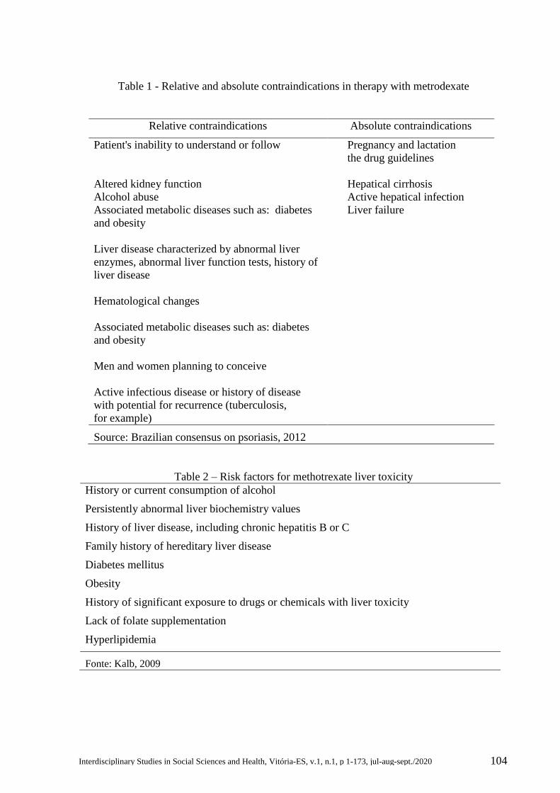
104
Interdisciplinary Studies in Social Sciences and Health, Vitória-ES, v.1, n.1, p 1-173, jul-aug-sept./2020
Table 1 - Relative and absolute contraindications in therapy with metrodexate
Table 2 – Risk factors for methotrexate liver toxicity
History or current consumption of alcohol
Persistently abnormal liver biochemistry values
History of liver disease, including chronic hepatitis B or C
Family history of hereditary liver disease
Diabetes mellitus
Obesity
History of significant exposure to drugs or chemicals with liver toxicity
Lack of folate supplementation
Hyperlipidemia
Fonte: Kalb, 2009
Relative contraindications Absolute contraindications
Patient's inability to understand or follow
Pregnancy and lactation
the drug guidelines
Altered kidney function Hepatical cirrhosis
Alcohol abuse Active hepatical infection
Associated metabolic diseases such as: diabetes
and obesity
Liver failure
Liver disease characterized by abnormal liver
enzymes, abnormal liver function tests, history of
liver disease
Hematological changes
Associated metabolic diseases such as: diabetes
and obesity
Men and women planning to conceive
Active infectious disease or history of disease
with potential for recurrence (tuberculosis,
for example)
Source: Brazilian consensus on psoriasis, 2012

105
Interdisciplinary Studies in Social Sciences and Health, Vitória-ES, v.1, n.1, p 1-173, jul-aug-sept./2020
Table 3 – Monitoring of hepatotoxicity in low-risk patients
Without baseline liver biopsy
Monitor liver function tests monthly for the first 6 months, and then every 1 to 2 months.
For small elevations (<2 times the LSN), repeat in 2 to 4 weeks.
For moderate elevations (> 2 times, but <3 times the ULN), monitor more closely, repeat in
2-4 weeks, and dose reduction if necessary.
For persistent elevations in 5 of 9 AST levels over a 12-month period or if there is a decline
in serum albumin (in the context of normal nutritional status) below the normal range in a
well-controlled disease scenario, liver biopsy should be performed.
Consider continuing follow-up according to the ACR guidelines above without biopsy.
Or
Consider liver biopsy after a total cumulative dose of 3.5-4.0 g.
Or
Consider switching to another agent or discontinuing therapy after a total cumulative dose
of 3.5-4.0 g.
LSN, Upper limit of the normal range; AST, Aspartate Aminotransferase;
ACR, American College of Rheumatology
Source: Kalb, 2009
Table 4 – Monitoring of hepatotoxicity in high-risk patients
Consider using a different systemic agent.
Consider late baseline liver biopsy (2 to 6 months after starting therapy to determine
medication effectiveness and tolerability).
Repeat liver biopsies after approximately 1.0 to 1.5 g of cumulative doses of MTX.
Source: Kalb, 2009
Tabela 5 – Recommendations based on liver biopsy results regarding the decision to decision
to continue or discontinue methotrexate
Patients with grade I or II of alteration can continue to receive methotrexate.
Patients with grade III-A changes may continue to receive methotrexate therapy, but liver
biopsy should be repeated after approximately 6 months of methotrexate therapy.
Alternative to systemic therapy should be considered.
Patients with grade III-B changes should be discontinued on methotrexate. Exceptional
circumstances, however, may require continued therapy with methotrexate, with minimal
follow-up through liver biopsy.
Grade I - Normal liver tissue, or mild fatty infiltration and mild inflammation of the portal space;
Grade II – Moderate to severe fatty infiltration; moderate to severe inflammation of the portal space;
Grade IIIA - Presence of mild fibrosis;
Grade IIIB - Presence of moderate to severe fibrosis;
Grade IV - Presence of liver cirrhosis;
Source: Kalb, 2009

106
Interdisciplinary Studies in Social Sciences and Health, Vitória-ES, v.1, n.1, p 1-173, jul-aug-sept./2020
Table 6 - Demographic data and clinical characteristics of patients
with psoriasis using methotrexate.
Variables All cases (n=50)
Age (years)
Average ±SD
Variation
52,8 ± 15,7
13-80
Sex n(%)
Female
Male
21 (42%)
29 (58%)
Diagnostic age (years)
Average ±SD
Variation
41,2 ± 16,9
13-80
Comorbidities n (%)
Type 2 diabetes mellitus
Arterial hypertension
Dyspipidemia
Metabolic syndrome
Obesity
Alcoholism
Crohn's disease
Idiopathic Ulcerative Retocolitis
Depression / anxiety / nervousness
6 (12%)
11 22%)
6 (12%)
2 (4%)
2 (4%)
1 (2%)
1 (2%)
1 (2%)
3 (6%)
N, número; DP, desvio padrão.
Fonte: Próprios autors.
REFERENCES
1. Consenso de Psoríase 2012 - Guias de avaliação e tratamento. 2 ed. Rio de Janeiro:
Sociedade Brasileira de Dermatologia, 2012. p. 11-26.
2. Takahashi MDF. Erupções eritemato escamosas: Psoríase. In: Rivitti EA. Manual de
Dermatologia Clínica de Sampaio e Rivitti. 4 ed. São Paulo: Artes Médicas, 2014. p. 86-92.
3. JM Pinto, NC Bavoso, MS Diniz. Psoríase: novas comorbidades. An Bras Dermatol.
2016;91(1):8-14.
4. Consenso de Psoríase 2012 - Guias de avaliação e tratamento. 2 ed. Rio de Janeiro:
Sociedade Brasileira de Dermatologia, 2012. p. 67-73.
5. Czarnecka-Operacz M, Sadowska-Przytocka A. The possibilities and principles of
methotrexate treatment of psoriasis – the updated knowledge. Postep Derm Alergol.
2014;31(6):392-400.
6. Bishnoi P, Kumari R, Thappa DM. Monitoring methotrexate hepatotoxicity in psoriasis.
Indian J Dermatol Venereol Leprol. 2011;77(5):545-548.

107
Interdisciplinary Studies in Social Sciences and Health, Vitória-ES, v.1, n.1, p 1-173, jul-aug-sept./2020
7. Maybury CM, Jabbar-Lopez ZK, Wong T, Dhillon AP, Barker JN, Smith CH.
Methotrexate and liver fibrosis in people with psoriasis: a systematic review of observational
studies. Br J Dermatol. 2014;171(1):17–29.
8. Kalb R E, Strober B, Weinstein G, Lebwohl M. Methotrexate and psoriasis: 2009 National
Psoriasis Foundation consensus conference. J Am Academy Dermatol. 2009;60(5):824-837.
9. Ng LC, Lee YY, Lee CK, Wong SM. A retrospective review of methotrexate‐induced
hepatotoxicity among patients with psoriasis in a tertiary dermatology center in Malaysia. Int
J Dermatol. 2013;52(1):102-105.
10. Voiculescu VM, Lupu M, Papagheorghe L, Giurcaneanu C, Micu E. Psoriasis and
Metabolic Syndrome – scientific evidence and therapeutic implications. J Med Life.
2014;7(4):468-471.
11. Armstrong AW, Harskamp CT, Armstrong EJ. Psoriasis and metabolic syndrome: A
systematic review and meta-analysis of observational studies. J Am Acad Dermatol.
2013;68(4):654-662.
12. Langan SM, Seminara NM, Shin DB, Troxel AB, Kimmel SE, Mehta NN, et al.
Prevalence of metabolic syndrome in patients with psoriasis: A population-based study in the
United Kingdom. J Invest Dermatol. 2012;132(3 Pt 1):556-562.
13. Heydendael VMR, Spuls PI, Opmeer BC, Borgie CAJM, Reitsma JB, Goldschmidt VFM,
et al. Methotrexate versus cyclosporine in moderate-to-severe chronic plaque psoriasis. N
Engl J Med. 2003;349(7):658-665.
14. Warren RB, Smith RL, Campalani E, Eyre S, Smith CH, Barker JNWN, et al. Outcomes
of methotrexate therapy for psoriasis and relationship to genetic polymorphisms. Br J
Dermatol. 2008;160(2):438–441.
15. Haustein UF, Rytter M. Methotrexate in psoriasis: 26 years’ experience with low‐dose
long‐term treatment. J Eur Acad Dermatol Venereol. 2000;14(5):382-388.
16. Conway R, Low C, Coughlan RJ, O’Donnell MJ, Carey JJ. Risk of liver injury among
methotrexate users: A meta-analysis of randomised controlled trials. Semin Arthritis Rheum.
2015;45(2):156-162.

108
Interdisciplinary Studies in Social Sciences and Health, Vitória-ES, v.1, n.1, p 1-173, jul-aug-sept./2020
DOI https://dx.doi.org/10.5935/2675-7397.20200008
Original Article
CADMIUM EXPOSURE EFFECTS ON THE BLOOD PRESSURE AND
MYOCARDIAL CONTRACTILITY.
Cindy Medici Toscano1, Ingridy Reinholz Grafite Schereider3, Priscila Rossi de Batista2,
Dalton Valentim Vassallo4
1 M.S. in Physiological Sciences from PPGCF-UFES; PhD. student at PPGCF-UFES
2 Scientific Initiation Scholarship Program – Graduate Program in Physiological
Sciences from Federal University of Espírito Santo (UFES), PPGCF-UFES; Nursing
Senior Student at UFES
3 PhD. in Physiological Sciences from PPGCF-UFES; Postdoctoral Researcher at
PPGCF-UFES
4 Graduated in Medicine at the Federal University of Espírito Santo; Master in
Biological Sciences (Biophysics) from the Federal University of Rio de Janeiro; PhD
in Biological Sciences from the Federal University of Rio de Janeiro; Professor at the
School of Medicine of Santa Casa de Misericórdia de Vitória - EMESCAM
Submitted in: December 10, 2016; Approved: June 06, 2017.
Institution: EMESCAM
Av. Nossa Sra. da Penha, 2190 – Santa Luíza, Vitória - ES, 29045-402
Correspondence Author: Cindy Medici Toscano
Graduate Program in Physiological Sciences, Center for Health Sciences - Federal University of
Espírito Santo (UFES). Av. Marechal Campos, 1468, Maruípe, 29040-090, Vitoria, ES, Brasil.
Tel: 55-27-33357340/33357339; FAX: 55-27-33357339.
E-mail: [email protected]
ESTUDOS INTERDISCIPLINARES EM
CIÊNCIAS SOCIAIS E DA SAÚDE INTERDISCIPLINARY STUDIES IN SOCIAL SCIENCES AND HEALTH
v.1, n.1, 2020

109
Interdisciplinary Studies in Social Sciences and Health, Vitória-ES, v.1, n.1, p 1-173, jul-aug-sept./2020
Keywords Abstract
Cadmium;
Blood pressure;
Heart.
Objective: To develop a literature review on the effects of
cadmium exposure in the heart and blood pressure.
Methods: The search approach was based on a narrative
review for selection of studies and interpretation of
information. To this end, a review study was conducted in
books, theses, dissertations, magazines and papers
acquired from databases: LILACS, PUBMED, MEDLINE
and SCIELO. Results: The studies suggest that cadmium
exposure is involved in the development of hypertension,
associating the deleterious effects of cadmium on vascular
endothelium as the main cause in the genesis of
hypertension. Conclusion: There is a need for more
studies in order to elucidate the mechanisms involved in
these changes promoted by cadmium, particularly in the
heart. Nevertheless, it is important to recognize the risks
of intoxication by this metal because it has been associated
with increased mortality from cardiovascular diseases.
INTRODUCTION
Cadmium (Cd) is a metal extremely toxic
to man, even in low concentrations, and
not essential to human metabolism. There
are a number of different modes of
environmental contamination by
Cadmium, but they include contamination
from the use of pesticides and agricultural
fertilizers, as well as the disposal of
industrial wastes derived from the
production of polyvinyl chloride (PVC),
plastics, glass and batteries1. With this
widespread use of the metal, vegetables,
seeds and tobacco leaves accumulate high
levels of cadmium from the soil1. Thus,
tobacco smoking has become the main
form of non-occupational exposure to
cadmium2.
A large number of scientific evidences
relates this metal to the increased blood
pressure in humans and the development of
hypertension, atherosclerosis and other
cardiovascular diseases3,4,5.
Smoking is considered a source of
exposure to cadmium, considering that the
presence of this and other heavy metals in
cigarettes has been identified as a causative
agent of cardiovascular disease induced by
smoking6,7,8,9.
It is important to note that hypertension is
an important risk factor for various
cardiovascular diseases and that, since the
1960s, these diseases represent the leading
cause of death in Brazil10. Therefore, this
article aimed to build a review of the blood
pressure and cardiac effects of exposure to
cadmium, based on a collection of data
from the scientific literature
METHODOLOGY
For the selection of studies and
interpretation of information on the toxic
effects of Cadmium on blood pressure and
the heart, a narrative review was adopted.
Consequently, for the construction of
knowledge, a descriptive-discursive type
survey of scientific production data was
performed.
This review was based on studies that
pointed to the toxic effects of cadmium on
the heart and blood pressure. Based on the

110
Interdisciplinary Studies in Social Sciences and Health, Vitória-ES, v.1, n.1, p 1-173, jul-aug-sept./2020
data presented, a critical discourse on the
exposed theme will be presented.
The analysis of this study was carried out
through the search of electronic
bibliographic data of available literature in
theses, dissertations, journals and scientific
articles acquired from the LILACS (Latin
American and Caribbean Literature) and
MEDLINE (National Library of Medicine,
U.S). The electronic search was done from
the combination of the descriptors:
cadmium; arterial pressure; heart.
The articles were separated according to
importance and insertion in the context of
the topic addressed, based on a critical
analysis performed from the considerations
presented by reading the researched
content.
RESULTS/DISCUSSION
Even though it is known that heavy metals
are harmful to the functioning of the
organism and to the maintenance of health,
it is not yet fully known what are exactly
the specific mechanisms involved by
which metals produce their effects.
However, there is evidence that many of
these effects may result from interactions
of cardiovascular system components.
3.1 Exposure to cadmium and
hypertension
The World Health Organization (WHO)
considers cardiovascular diseases as the
central cause of morbidity and mortality in
the world. In addition, the appearance or
aggravation of these diseases has been
related to the exposure of heavy metals
considered toxic to the organism, such as
cadmium11. Considering that about 17
million people died as a result of
cardiovascular diseases in the world in
2011, the WHO estimates that in 2030
more than 23 million individuals will die
from cardiovascular causes11.
Hypertension is considered one of the main
risk factors for the development of
cardiovascular diseases. It is estimated that
20% of the world population is
hypertensive and that in Brazil 40% of the
Brazilian population can become
hypertensive12.
Since the twentieth century, cadmium has
been associated with cardiovascular
effects, when Asberg and Schwertze
proposed changing coloration in the
testicles of animals exposed to this metal.
Subsequently, they attributed this effect of
cadmium to the rupture of the junctions
between endothelial cells and testicular
capillaries, promoting edema, hemorrhage
and necrosis14,15.
According to Téllez-Plaza et al.5, there is
an association between peripheral arterial
disease and the presence of cadmium in the
urine, by means of the ankle-brachial index
evaluation capable of detecting variations
in flow in the arterial circuit caused by
stenosis in the vascular bed. This study
observed that individuals with a higher
level of urinary cadmium were at higher
risk for peripheral arterial disease.
Houtman16 noted that hypertensive
individuals had higher amounts of
cadmium in the urine and also noted that
populations living in areas of higher
cadmium contamination had higher
incidences of atherosclerosis.
An increase in blood pressure was
observed in animals exposed to cadmium
in the diet for 3, 5, 10 and 14 months,
where systolic blood pressure was 15 to 20
mmHg higher than the control group in all
models of exposure17. Other studies have
associated increased blood pressure,
reduced body mass gain, and increased
levels of malondialdehyde on exposure to
cadmium18,19,20. Taking into account that
malondialdehyde is a marker of lipid
peroxidation, it may be the factor involved
in the reduction in body mass gain and also
can be viewed as a pro-atherosclerotic

111
Interdisciplinary Studies in Social Sciences and Health, Vitória-ES, v.1, n.1, p 1-173, jul-aug-sept./2020
factor in a model of cadmium
intoxication20.
3.2 Exposure to cadmium and
endothelial dysfunction
It is believed that the mechanisms involved
in the pathogenesis of cadmium-induced
hypertension are multifactorial, even
though they have not been clarified yet.
Some authors suggest that cadmium-
induced glutathione depletion leads to
oxidative stress and lipid peroxidation,
which are hypertensive factors21, while
others suggest interactions with calcium
channels22, sympathetic stimulation23 and
reduction of the release of vasodilatory
agents24.
It is suggested that endothelial dysfunction
is involved with hypertension, considering
that it may contribute to the increase of
vascular resistance, favoring the onset or
the aggravation of the hypertensive
process25,26,27. Overall, some studies claim
that endothelium is the primary target of
cadmium intoxication with respect to the
cardiovascular system, and that the
interaction between the metal and the
endothelium may result in imbalance of the
production of vasodilator and
vasoconstrictor substances and
consequently, increased vascular
tone28,29,30.
Reducing the bioavailability of nitric oxide
(NO) is considered one of the most
important factors associated to vascular
disease.31,32,33 One of the main mechanisms
involved in this process would be the
increase in the production of superoxide
anion and the consequent reduction of the
bioavailability of NO and the production of
peroxynitrite, a potent oxidizing
agent.31,32,33
A recent study performed in our laboratory
by ALMENARA and collaborators34
demonstrated in an experimental model of
exposure for 30 days in drinking water that
cadmium promotes increased blood
pressure from the first week of exposure.
In this study, the endothelial dysfunction
caused by the metal occurs due to the
increase of superoxide anion production
and, consequently, decrease of the
bioavailability of nitric oxide. In addition,
the metal promotes an increased reactivity
to phenylephrine in isolated aortic rings.34
In another metal exposure model, 0.5 mg /
kg / day for 120 days i.m., Tzotzes and
collaborators35 did not observe a change in
the contractile response to phenylephrine.
Nevertheless, in mice exposed to cadmium
for 8 weeks via drinking water 100 mg / L,
there was a reduction of the reactivity to
phenylephrine.19,30 The different results
reported suggest that the effects of
cadmium on vascular reactivity depends on
the time of exposure as well as on the
concentration of the metal.34
Cadmium, likewise other heavy metals,
appears to be involved in an increased
oxidative stress. This increase has been
associated with an increased NADPH
oxidase activity, increased expression of
NOX-2 and the production of hydrogen
peroxide, with consequent increase in the
production of reactive oxygen species.34
Other studies have also reported increased
bioavailability of superoxide anion in
animals exposed to cadmium.19,30
Changes in SOD expression reveal an
important mechanism of action of
oxidative stress, considering that this
enzyme plays the leading role in cellular
antioxidant defense. Using COS-7 culture
cells exposed to cadmium, Obara et al.36
demonstrated reduced expression of the
extracellular isoform of SOD, and Ozturk
et al.37 observed an increase in SOD
enzyme activity in rats treated with 15 mg /
kg / day by gavage during 60 days.

112
Interdisciplinary Studies in Social Sciences and Health, Vitória-ES, v.1, n.1, p 1-173, jul-aug-sept./2020
3.3 Exposure to cadmium and cardiac
contractility
An association between the exposure of
some populations to dietary cadmium and
mortality rates from cardiovascular disease
has been described38. Alterations such as
cardiac conduction system disorders,
changes in blood pressure and heart rate
were observed in a population of elderly
women exposed to cadmium through
food39. Besides, Wronska-Nofer et al40
revealed rhythmic changes, such as
ventricular fibrillation, in an individual
after ingestion of 25 mg / kg of cadmium.
The cytotoxic effects of cadmium
apparently affect the heart, because when
exposed to low doses it has caused
accumulation of metal in cardiac tissue,
reduction of myocardial contractility41,
damage to the intercalated discs, leading to
structural changes of the heart muscle42.
Furthermore, exposure to cadmium impairs
mitochondrial respiration of
cardiomyocytes, inhibiting the transfer of
electrons, resulting in an increase in the
formation of reactive oxygen species43,44.
Turckan et al.,45 utilizing an animal
exposure model of 100 mg L-1 of CdCl2
via drinking water, established the
presence of fibrosis in the myocardium
only in animals that received a lipid-rich
diet, and indicated that cadmium induces
apoptosis after 96 hours of exposure. They
also observed that cadmium intoxication
associated with moderate
hypercholesterolemia or normal cholesterol
levels induces the development of cardiac
fibrosis and, consequently, the reduction of
cardiac contractility and, thus, the
deposition of cadmium in cardiac tissue in
animals seems to be dependent on high
levels of cholesterol45.
Not only does cadmium affect tissue
structure and cardiac muscle integrity, but
also it interferes with the cardiac
conduction system46. The metal blocks the
L-type calcium channels and changes the
potassium current in the ventricular
myocytes, causing changes in the
physiological and biochemical
processes47,48.
Using an experimental model of exposure
to cadmium for 30 days in drinking water
plus 100 mg L-1 of CdCl2, Vescovi49
demonstrated that the metal was involved
in increasing peripheral vascular resistance
by increasing sympathetic activity,
resulting in an elevated blood pressure and
cardiac inotropism. Even so, by in vitro
evaluation after 30 days of exposure, he
observed that cadmium did not promote
increased contraction force of isolated
papillary muscles, nor did it modify
transarcolemal calcium influx, nor did it
promote changes in β-adrenergic
stimulation, thus suggesting the
intervention of neurohumoral regulation in
the mechanisms involved49.
Cadmium is a transition metal and it is
commonly associated with zinc, lead and
copper ore in the environment. 50 Like
lead, cadmium is a bivalent, toxic metal
and has no physiological function in the
body. 2,51
When investigating the effects of lead on
myocardial contractility, Vassalo et al52
reported a reduction in strength developed
at concentrations above 30 μmol L-1. They
attributed the reduction in strength
promoted by acute exposure to lead to
lower transarcolemal calcium influx and
reduction in the activity of the myosin
ATPase enzyme, without altering the
activity of the sarcoplasmic reticulum39.
Since lead, like cadmium, is a bivalent
heavy metal, the results demonstrated may
contribute to the clarification of the
mechanisms involved in the alterations of
the "cardiac contractile machinery" in the
exposure to cadmium.

113
Interdisciplinary Studies in Social Sciences and Health, Vitória-ES, v.1, n.1, p 1-173, jul-aug-sept./2020
FINAL CONSIDERATIONS
Cadmium is a metal toxic to the body and
it plays an important role in vascular
system changes, such as the onset or
impairment of hypertension, peripheral
arterial disease and other cardiovascular
diseases.
It is known that exposure to cadmium is
related to the development of arterial
hypertension, increasing the mortality rate
due to cardiovascular diseases. It is worth
noting that the evidence suggests the
deleterious effects of cadmium on the
vascular endothelium as the main cause in
the genesis of hypertension promoted by
this metal as a result of imbalance in the
production of vasodilator and
vasoconstrictor substances. The endothelial
dysfunction caused by the metal can occur
due to the increase in the production of
superoxide anions and consequently the
decrease of the bioavailability of the nitric
oxide, besides causing structural changes
in the cardiac tissue.
Although the effects of cadmium on the
heart are already known, little is
comprehended about these effects, and
much research is still needed to elucidate
the mechanisms involved in these changes.
Thus, it is necessary to recognize the risks
of cadmium poisoning and to certify a
reassessment by competent authorities of
the effects of exposure to cadmium in the
population, especially the occupationally
exposed population, and in smokers.
REFERENCES
1. World Health Organization (WHO). (1992) Cadmium. Environmental Health Criteria 134.
International Programme on Chemical Safety (IPCS), Geneva, Switzerland. 148pp.
2. Agency for Toxic Substances and Disease Registry (ATSDR). Public Health Statement for
Cadmium. 2008.
3. Varoni MV, Palomba D, Gianorso S, Anania V. Cadmium as an environmental factor of
hypertension in animals: new perspectives on mechanisms. Vet. Res. Commun. 2003
Sep;27(1):807–10.
4. Meijer GW, Beems RB, Janssen GB, Vaessen HA, Speijers GJ. Cadmium and
atherosclerosis in the rabbit: reduced atherogenesis by superseding of iron. Food Chem
Toxicol. 1996 Aug;34(7)611–21.
5. Téllez-Plaza M,Navas-AciénA,Guallar E. Cadmium as a novel cardiovascular risk fator:
supportive evidence and future directions. Nature Reviews. 2010 Jul;7(7):41-6.
6. Hoffmann D, Hoffmann I, El Bayoumy K. The less harmful cigarette: a controversial issue:
a tribute to Ernst L. Wynder. Chem Res Toxicol. 2001 Jul;14(7):767–90.
7. Loh HS. Cigarette smoking and the pathogenesis of atherosclerosis: a hypothesis. Ir J Med
Sci. 1973 Dec; 142:174–8.
8. Sharrett AR, Coady SA, Folsom AR, Couper DJ, Heiss G, ARIC study. Smoking and
diabetes differ in their associations with subclinical atherosclerosis and coronary heart
disease: the ARIC Study. Atherosclerosis. 2004 Jan;172(1):143–9.
9. Gordon T, Kannel WB. Predisposition to atherosclerosis in the head, heart, and legs: the
Framingham study. JAMA. 1972 Aug;221(7):661–6.

114
Interdisciplinary Studies in Social Sciences and Health, Vitória-ES, v.1, n.1, p 1-173, jul-aug-sept./2020
10. Lotufo PA. Epidemiology of heart disease in Brazil: history, current situation and
proposed theoretical model. Rev Soc Cardiol Estado de São Paulo. 1996; 6:541-7.
11. World Health Organization (WHO). (2013) Cardiovascular diseases (CVDs). World
health organization. Disponívelem: www.who.int/entity/cardiovascular_diseases/en/. Acesso
em: 12/jul/2015.
12. VI Diretrizes Brasileiras de Hipertensão (DBH VI). Revista BrasHipertens. 2010;17(1):4.
13.Alsberg CL, Schwartze, EW. Phamacologial action of cadmium. J Pharmacol Exp Ther.
1919; 13:504-5.
14. Aoki A, Hoffer AP. Reexamination of the lesions in rat testis caused by cadmium. Biol
Reprod. 1978 May;18(4):579-91.
15. Fende PL, Niewenhuis RJ. An electron microscopic study of the effects ofcadmium
chloride on cryptorchid testes of the rat. Biol Reprod. 1977 Apr;16(3):298-305.
16. Houtman JP. Prolonged low-level cadmium intake and atherosclerosis. Sci Total Environ.
1993 Sep;138(1-3):31-6.
17. Perry HM,ErlangerMW. Sodium retention in rats with cadmium-induced hypertension.
Sci Total Environ. 1981 Dec;22(1):31-8.
18. Gökalp O, Ozdem S, Donmez S, Dogan M, Demerin H, Kara HY, Sutcu R, Cicek E, Ozer
MK, Delibas N. Impairment of endothelium-dependent vasorelaxation in cadmium-
hypertensive rats. Toxicol Ind Health. 2009 Jul;25(7):447-53.
19. Sompamit K, Kukongviriyapan U, Donpunha W, Nakmareong S, Kukongviriyapan V.
Reversal of cadmium-induced vascular dysfunction and oxidative stress by meso-
2,3dimercaptosuccinic acid in mice. Toxicol Lett. 2010 Apr;198(1):77-82.
20. Almenara CACP. Efeitos da exposição crônica ao cloreto de cádmio sobre a reatividade
vascular e pressão arterial de ratos. Dissertação de mestrado em Ciências Fisiológicas.
Departamento de Ciências Fisiológicas da universidade Federal do espírito Santo, Vitória,
2013. 95 pp.
21. Yiin SJ, Chern CL, Sheu JY, Lin TH. Cadmium induced lipid peroxidation in rat testes
and protection by selenium. Biometals. 1999 Dec;12(4):353-9.
22. BalaramanR, GulatiOD, BhattJD, RathodSP, Hemavathi KG. Cadmium-induced
hypertension in rats. Pharmacology. 1989;38(4):226-60.
23. FadlounZ,Leach GD. The effects of cadmium ions on blood pressure, dopamine-
betahydrozylase activity and the responsiveness of in vivo preparations to sympathetic nerve
stimulation, noradrelaline and tyramine. J Pharm Phamacol.1981;33(10):600-4.
24. SchoczynskaA,Martynowicz H. The impact of subchronic cadmium poisoning on the
vascular effect of nitric oxide in rats. Hum Exp Toxicol. 2005 Jul;24(7), 353-61.
25. Cannon III RO. Role of nitric oxide in cardiovascular disease: focus on the Endothelium.
Clin Chem.1998 Aug;44(8-2):1809-19.
26. Triggle CR, Hollenberg M, Anderson TJ, Ding H, Jiang Y, Ceroni L, Wiehler WB, Ella
SMN, Ellis A, Andrews K, McGuire JJ, Pannirselvam M. The endothelium in health and
disease – A Target for therapeutic intervention. J Smooth Muscle R. 2003 Dec;39(6):249-67.

115
Interdisciplinary Studies in Social Sciences and Health, Vitória-ES, v.1, n.1, p 1-173, jul-aug-sept./2020
27. Kolluru GK, Siamwala JH, Chatterjee S. eNOS phosphorylation in health and disease.
Biochimie. 2010 Apr;92(9):1189-98.
28. Martynowicz H, Skoczynska A, Wojakowska A, Turczyn B. Serum vasoactive agents in
rats poisoned with cadmium. Int J Occup Med Environ Health. 2004;17(4):479-85.
29. Kolluru GK, Tamilarasan KP, Geetha Priya S, Durgha NP, Chatterjee S. Cadmium
induced endothelial dysfunction: consequence of defective migratory pattern of endothelial
cells in association with poor nitric oxide availability under cadmium challenge. Cell Biol Int.
2006 May;30(5):427-38.
30. Donpunha W, Konungviriyapan U, Sompamit K, Pakdeechote P, Konungviriyapan V,
Pannangpetch P. Protective effect of ascorbic acid on cadmium-induced hypertension and
vascular dysfunction in mice. Biometals. 2011 Feb;24(1):105-15.
31. Stroes E, Hijmering M, Zandvoort M, Wever R, Rabelink TJ, Faassen EE. Origin of
superoxide production by endothelial nitric oxide syntase. FEBS Lett.1998 Nov;438(3):161-4.
32. Förstermann U, Munzel T. Endothelial nitric oxide syntase in vascular disease: From
Marvel to menace. Circulation. 2006 Apr;113(13):1708-14.
33. Takaya T, Hirata K, Yamashita T, Shinohara M, Sasaki N, Inoue N, Yada T, Goto M,
Fukatsu A, Hayashi T, Alp NJ, Channon MK, Yokoyama M, Kawashima S. A specific role
for eNOS-derived reactive oxygen species in atherosclerosis progression.
ArteriosclerThrombVasc Biol. 2007 Jul;27(7):1632-7.
34. Almenara CCP, Broseghuini-Filho GB, Vescovi MVA, Angeli JK, Faria TO, Stefanon I,
Vassallo DV, Padilha AS. Chronic cadmium treatmente promotes oxidative stress and
endothelial damage in isolated rat aorta. PlosOne. 2013 Jul;8(7):e68418.
35. Tzotzes V, Tzilalis V, Giannakakis S, Saranteas T, Papas A, Mourouzis I, MourouzisC,
Zarros A, Pantos C, Cokkinos D, Carageorgiou H. Effects of acute and chroniccadmium
administration on the vascular reactivity of rat aorta. Biometals. 2007 Feb;20(1):83-91.
36. Obara A, Kamiya T, Izumi M, Hara H, Yamada H, Adachi T. Extracellularsuperoxide
dismutase expression in COS7 cells exposed to cadmium chloride. Biol Pharm Bull.
2011;34(9):1443-7.
37. Ozturk IM, Buyukakilli B, Balli E, Cimen B, Gunes S, Erdogan S. Determination of acute
and chronic effects of cadmium on the cardiovascular system of rats. Toxicol Mech Methods.
2009 May;19(4):308-317.
38. Shigematsu I. The epidemiological approach to cadmium pollution in Japan. Ann Acad
Med Singap. 1984;13(2):231-36.
39. KagamimoriS,WatanabeM,NakagawaH,OkumaraY,Kawano S. Case-control study on
cardiovascular function in females with a history of heavy exposure to cadmium. Bull
Environ ContamToxicol. 1986 Apr;36(4):484-90.
40. Wronska-NoferT,Wisniewska-Knpl J, Wyszynska K. Prooxidative and genotoxic effect of
transition metals (cadmium, nickel, chromium, and vanadium) in mice. Trace Elem Electroly.
1999 Jun;15(2):87-92.
41. Kopp, S. J., Fischer, V. W., Erlanger, M., Perry, E. F., and Perry, H. M., Jr.
Electrocardiographical, biochemical and morphological effects of chronic low-level cadmium
feeding on the rat heart. Proc Soc Exp Biol Med. 1978 Dec;159(3):339–345.

116
Interdisciplinary Studies in Social Sciences and Health, Vitória-ES, v.1, n.1, p 1-173, jul-aug-sept./2020
42. Kolakowski, J., Baranski, B., and Opalska, B. Effect of long-term inhalation exposure to
cadmium oxide fumes on cardiac muscle ultrastructure in rats. Toxicol Lett. 1983
Dec;19(3):273–278.
43. Kisling, G. M., Kopp, S. J., Paulson, D. J., Hawley, P. L., and Tow, J. P. Inhibition of rat
heart mitochondrial respiration by cadmium chloride. Toxicol App Pharmacol. 1987
Jul;89(3):295–304.
44. Wang, Y., Fang, J., Leonard, S. S., and Rao, K. M. Cadmium inhibits the electron transfer
chain and induces reactive oxygen species. Free Radic Biol Med. 2004 Jun;36(11):1434–
1443.
45. Turkcan A, Scharinger B, Grabmann G, Keppler BK, Laufer G, Bernhard D, Messner B.
Combination of Cadmium and High Cholesterol Levels as a Risk Factor for Heart Fibrosis.
Toxicol Sci. 2015 Jun;145(2):360–371.
46. Prentice RC, Hawley PL, Glonek T, et al. Calcium-dependent effects of cadmium on
energy metabolism and function of perfused rat heart. Toxicol App Pharmacol.1984 Sep
15;75(2):198–210.
47. Shen JB, Jiang B, Pappano AJ. Comparison of L-type calcium channel blockade by
nifedipine and/or cadmium in guinea pig ventricular myocytes. J Pharmacol Exp Ther. 2000
Aug;294(2):562–70.
48. Follmer CH, Lodge NJ, Cullinan CA, et al. Modulation of the delayed rectifier, IK, by
cadmium in cat ventricular myocytes. Am J Physiol. 1992 Jan;262(1 Pt 1):C75–83.
49. Vescovi MVA. Efeitos da exposição ao CdCl2 em ratos: um estudo de deposição tecidual
e uma visão cardiovascular. Dissertação de mestrado em química. Departamento de Química
da Universidade Federal do Espírito Santo, Vitória, 2013. 138 pp.
50. Mendham, N.J.; Denney, R.C.; Barnes, J.D.; Thomas, M.J.K. VOGEL. Análise Química
Quantitativa; 6ª edição, LTC; 2011.
51. Paoliello, M.M.; Gutierrez, P.R.; Turini, C.A.; Matsuo, T.; Mezzaroba, L.; Barbosa, D.S.;
Carvalho, S.R.; Alvarenga, A.L.; Rezende, M.I.; Figueiroa, G.A.; Leite, V.G.; Gutierrez,
A.C.; Lobo, B.C.; Cascales, R.A. Referencevalues for lead in blood in urbanpopulation in
southernBrazil. Rev PanamSalud Publica. 2001 May;9(5):315-9.
52. Vassallo DV, Lebarch EC, Moreira CM, Wiggers GA, Stefanon I. Lead reduces tension
development and the myosin ATPase activity of the rat right ventricular myocardium. Braz J
Med Biol Res. 2008 Sept;41(9):789-95.

117
Interdisciplinary Studies in Social Sciences and Health, Vitória-ES, v.1, n.1, p 1-173, jul-aug-sept./2020
DOI https://dx.doi.org/10.5935/2675-7397.20200011
Original Article
REFRACTORY DIGESTIVE HEMORRAGY BY USE OF DIRECT
ANTICOAGULANT IN OCTAGENARY PATIENT WITH CHRONIC ATRIAL
FIBRILLATION: CASE REPORT
Autores: Renato Lirio Morelato1,2, Ana Clara de Barros Pretti2, Bruno PignatonRuschi de
Aragão2, Luíza Moulin Rezende2, Cristiano Ventorim de Barros1,2, Flavio Takemi Kataoka1,2,
Luciene Lage da Motta1,2, Alessandra Tieppo1,2
1 Santa Casa de Misericórdia de Vitória Hospital.
2 Santa Casa de Misericórdia de Vitória College of Sciences.
3 Final Course Work by Santa Casa de Misericórdia de Vitória College of Sciences -
EMESCAM.
Submitted in: Setember 10, 2019; Approved: September 25, 2019.
Institution: EMESCAM
Av. Nossa Sra. da Penha, 2190 – Santa Luíza, Vitória - ES, 29045-402
Correspondence Author: Renato Lirio Morelato.
Escola Superior de Ciências da Santa Casa de Misericórdia de Vitória, ES. Programa de
residência médica de Geriatria do Hospital Santa Casa de Misericórdia de Vitória – Rua Dr.
João dos Santos Neves 143 – Vila Rubim – 29025-023 Vitória ES – Brasil.
E-mail: [email protected]
Keywords Abstract
Anticoagulants;
Atrial fibrillation;
Hemorrhage;
Elderly aged 80 years or
more.
Fibrilação atrial é a arritmia cardíaca sustentada mais
prevalente do mundo, cuja incidência aumenta
progressivamente com o envelhecimento humano,
apresenta elevada morbimortalidade, especialmente o
acidente vascular cerebral, muito comum na população
idosa. A terapia preconizada para reduzir eventos
tromboembólicos são os dos anticoagulantes orais;
atualmente a varfarina vem sendo substituída pelos novos
anticoagulantes orais (NACOs), pela facilidade de manejo
terapêutico. Apresentamos o caso de um senhor de 88 anos
de idade, portador de insuficiência coronariana crônica
com histórico recente de fibrilação atrial sustentada, que
ESTUDOS INTERDISCIPLINARES EM
CIÊNCIAS SOCIAIS E DA SAÚDE INTERDISCIPLINARY STUDIES IN SOCIAL SCIENCES AND HEALTH
v.1, n.1, 2020

118
Interdisciplinary Studies in Social Sciences and Health, Vitória-ES, v.1, n.1, p 1-173, jul-aug-sept./2020
foi introduzido um inibidor direto da trombina, a
dabigratana 110mg duas vezes ao dia, como estratégica
preventiva. Ele apresentou um quadro de hemorragia
digestiva volumosa após seis meses de uso do NACO.
Internou com necessidade de várias transfusões sanguíneas
e evoluiu por duas semanas sem estabilizar o quadro
clínico. Não foi detectado o local do sangramento pela
endoscopia digestiva alta e a colonoscopia demonstrava
um sangramento ativo procedente do intestino delgado.
Através de angiotomografia abdominal foi detectado, em
topografia em terminal de intestino delgado, o possível
local de sangramento. Foi indicada intervenção cirúrgica
no 16º dia de internação, a qual, por meio do método de
transluscência de íleo terminal, confirmou o local de
sangramento, com enterectomia do segmento
comprometido. O exame histopatológico demonstrou
epitélio atrófico com vasos congestos.
INTRODUCTION
Atrial fibrillation (AF) is the most
prevalent sustained cardiac arrhythmia in
the world, and its significant management
and knowledge is mostly a result of the
great connection with high morbidity and
mortality events, namely stroke, especially
ischemic stroke. Accordingly, therapy
should be established to diminish the risk
of thromboembolic event. In the past 5
years, the guidelines have replaced
warfarin with Direct Oral Anticoagulants
(DOACs) for anticoagulation in cases of
non-valve AF, in response to a broad
therapeutic window; onset of rapid action;
no need for regular laboratory monitoring
(no control with INR); pharmacokinetics
and predictable pharmacodynamics; short
half-life; little food and medication
interaction (no restrictions on some foods);
more favorable dosage; among other
benefits.3
DOACs class has different action
mechanisms between their drugs. The
Dabigatran is an anticoagulant that acts as
direct thrombin inhibitor. Studies have
indicates that Dabigatran, at a dosage of
110 mg, had rates of stroke and systemic
embolism similar to warfarin, besides
lower rates of severe bleeding. The 150mg
dosage was relatable with lower
occurrences of stroke and systemic
embolism, but similar to severe bleeding in
comparison with warfarin.1 Other study
reported that, in patients under the age of
75, the doses of dabigatran, in contrast to
warfarin, have a lower risk of intracranial
and extracranial bleeding. In patients aged
75 years or older, the risk of intracranial
bleeding is lower, but the risk of
extracranial bleeding is similar or even
greater when comparing the doses of
dabigatran with warfarin.2
Hemorrhage is a common complication to
the use of Dabigatran, nevertheless, it is
barely required to resort to surgical
intervention to contain the condition. Small
bleeding has a short half-life and,
therefore, can be resolved only by
discontinuing the medication. More severe
cases submit a good response to
hemodialysis, given that the binding to
plasma proteins is weak and has
predominantly renal metabolism. Other
ways of conduction of hemorrhage are the
use of activated charcoal, although it is
contraindicated in situations of digestive

119
Interdisciplinary Studies in Social Sciences and Health, Vitória-ES, v.1, n.1, p 1-173, jul-aug-sept./2020
hemorrhage, administration of Fresh
Frozen Plasma and Concentrate of
Prothrombin Complexes, especially in
situations of severe hemorrhage. Since
they are not currently available in the
Unified Health System (SUS), there are
reversers of the action of DOACs, with
Idarucizumab being specific for
Dabigatran. Surgery is indicated for
specific cases, as exemplified in this
report.4
CASE REPORT
Male patient, 88 years old, with
paroxysmal atrial fibrillation (FAP),
CHADVASC 4 (risk of high
thromboembolism), HASBLED 3 (risk of
moderate bleeding), coronary artery
disease (CAD) and acute myocardial
infarction event (AMI) with implantation
of 2 stents 17 years ago, besides heart
failure with an ejection fraction of 40%.
Smoker for 50 years, with ½ pack a day.
He is in use of omeprazole, enalapril,
simvastatin, carvedilol, buffered acetyl
salicylic acid and dabigatran 110mg every
12 hours.
Regularly monitored in the cardiology and
geriatrics sector at Hospital Santa Casa de
Misericórdia de Vitória (HSCMV). He was
admitted to the emergency room of the
institution with digestive hemorrhage, and
referred to symptoms of asthenia, vertigo,
postural hypotension and melena, the latter
being present for 1 week. On admission, he
presented Hb 4.8, TAP 30%, negative
urine culture and mild leukocytosis. A
diagnostic hypothesis of an adverse event
was performed using dabigatran. By virtue
of significant anemia, transfusion of 2 red
blood cell concentrates, 3 fresh frozen
plasmas, and suspension of ASA and
dabigatran was prescribed as an initial
approach.
The patient was hospitalized for 28 days,
and the medication suspension was
continued for the entire time, in addition to
the specific oral diet for CKD. On the
second day of hospitalization, an upper
digestive endoscopy (EDA) with biopsy
was conducted, showing mild atrophic
pangastritis. The melena episodes
continued; the TAP continued broad. On
the sixth day, colonoscopy was performed
(complete examination until terminal
ileum), resulting in a large amount of clots
and indication of probable digestive
hemorrhage in a supracolic site (jejunum
or ileum), which cannot be identified by
the method. In the first week of
hospitalization, a total of 4 transfusions of
packed red blood cells and 3 fresh frozen
plasmas were conducted, with no clinical
improvement.
In the second week of hospitalization,
examinations of abdominal
angiotomography, chest CT and abdominal
CT were performed. In the abdominal CT,
an area of hyper-enhancement in the
arterial phase was identified, seen next to
the small intestine loop wall, at the
entrance of the pelvis, on the right,
apparently in projection of the ileum,
measuring about 2cm x 1,6cm x 0,5cm
(respectively in their largest longitudinal x
anteroposterior x transverse diameters),
without causing blurring of the
surrounding myoadipose planes, and the
image is iso / hypodense in the non-
contrast phase and has reduced
enhancement, nonetheless, still in the late
phase, there is an indeterminate aspect.
Mesenteric angiography was also carried
out and no bleeding was identified by the
method.
The fact that the patient has kept an
extended TAP, an evaluation of
hematology was demanded. TAP
collection with a 50% mixture was
proposed to define factor deficiency or
inhibitor presence. Episodes of bleeding

120
Interdisciplinary Studies in Social Sciences and Health, Vitória-ES, v.1, n.1, p 1-173, jul-aug-sept./2020
emerged on the 8th and 14th day and 4
more red blood cell transfusions were
performed. The gastroenterology team
introduced the use of thalidomide to
stabilize the condition. Exploratory
laparotomy and red cell scintigraphy were
the two approaches suggested for the case.
Exploratory laparotomy was indicated, as
the previous measures were not successful
in stopping bleeding, maintaining
significant anemia and a constant need for
transfusions. Scintigraphy was not
indicated, as it would not be a curative test.
The patient's surgical risk was resoluted,
which determined low operative risk (Lee's
Class 3) and the preparation for surgery
started with transfusion of 2 red blood cell
concentrates. On the 16th day of
hospitalization, a laparotomy was
performed, being identified by means of
the perioperative transillumination method,
an intestinal loop, a vascular lesion 15 cm
away from the ileocecal valve and a
bloody-looking content inside the loops of
ileum and colon, resulting in ileal segment
enterectomy. The histopathological
analysis of the specimen determined recent
superficial hemorrhage, with bright and
intense blood in the light and without
hemosiderin pigment. There were no signs
of necrosis or thrombosis (absence of
fibrin, neovascularization, inflammatory
process and elements of digested blood).
The mucosa was preserved, nevertheless,
swollen and with congested vessels. An
area of atrophy of the epithelium was
evidenced, but without signs of complete
reparative lesion, and the mucosa region
close to the altered area is unchanged,
suggesting a local mechanical trauma.
The postoperative period was conducted in
the Intensive Care Unit (ICU). Changes in
renal function, hyponatremia, metabolic
acidosis, episodes of melena and atrial
fibrillation were presented, and measures
indicated for control were carried out. The
patient was discharged from the ICU,
returning to the infirmary, where he stayed
for another 4 days, using furosemide by
reason of congestion with bronchospasm
and vitamin K He was discharged from the
hospital, being referred to the HSCMV
cardiology outpatient clinic, so that the
anticoagulant drug management and the
follow-up with the Geriatrics team could
be determined. He also received referral to
the hematologist, with the purpose of
investigating coagulopathy.
Acknowledgments
"We sincerely thank the real character of the clinical case who was another fatal victim, along
with his wife, from COVID-19 (in memorian)"

121
Interdisciplinary Studies in Social Sciences and Health, Vitória-ES, v.1, n.1, p 1-173, jul-aug-sept./2020
IMAGES - SLIDES, ABDOMEN AND SURGERY CT
a. Atrophic epithelium, close to the region of preserved mucosa. Intestinal lumen with living
blood. It shows an intense concentration of red blood cells. Note also the congested vessels.
b. Tomography images of the abdomen (without and with contrast) showing the bleeding site
Without contrast
Late
Arterial
Without contrast
Arterial
Late

122
Interdisciplinary Studies in Social Sciences and Health, Vitória-ES, v.1, n.1, p 1-173, jul-aug-sept./2020
c. Translucence technique surgery
Vascular injury
REFERENCES
1. Connolly SJ, Ezekowitz MD, Yusuf S, Eikelboom J, Oldgren J, Parekh A, et al. Dabigatran
versus warfarin in patients with atrial fibrillation. N Engl J Med. 2009; 361(12):1139-51
2. Eikelboom JW, Wallentin L, Connolly SJ, Ezekowitz M, Healey JS, Oldgren J, et al. Risk
of bleeding with 2 doses of dabigatran compared with warfarin in older and younger patients
with atrial fibrillation: an analysis of the randomized evaluation of long-term anticoagulant
therapy (RE-LY) trial. Circulation. 2011;123(21):2363-72.
3. Marques Marcos Areas. Os novos anticoagulantes orais no Brasil. J. vasc. bras. 2013 Sep ;
12( 3 ): 185-186.
4. Magalhães LP, Figueiredo MJO, Cintra FD, Saad EB, Kuniyoshi RR, Teixeira RA, et al. II
Diretrizes Brasileiras de Fibrilação Atrial. Sociedade Brasileira de Cardiologia. Abril 2016
Volume 106, Nº 4, Supl. 2.

123
Interdisciplinary Studies in Social Sciences and Health, Vitória-ES, v.1, n.1, p 1-173, jul-aug-sept./2020
DOI https://dx.doi.org/10.5935/2675-7397.20200010
Original Article
EXPOSURE TO CADMIUM AS A PUBLIC HEALTH PROBLEM AND ITS EFFECT
ON RESISTANCE VESSELS
Priscila Rossi de Batista1, Ingridy Reinholz Graffiti Schereider3, Cindy Medici Toscano2,
Dalton Valentim Vassallo4
1 PhD. in Physiological Sciences by the Postgraduated Program in Physiological
Sciences (PPGCF) from Federal University of Espírito Santo (UFES); Postdoctoral
student at PPGCF-UFES.
2 M.S. in Physiological Sciences from PPGCF-UFES; PhD. student in Physiological
Sciences at PPGCF-UFES.
3 Scientific Initiation Scholarship Program – Graduate Program in Physiological
Sciences from Federal University of Espírito Santo (UFES), PPGCF-UFES; Nursing
Senior Student at UFES.
4 M.S. in Biological Sciences (Biophysics) from the Federal University of Rio de
Janeiro; PhD in Biological Sciences from the Federal University of Rio de Janeiro;
Professor at the School of Medicine of Santa Casa de Misericórdia de Vitória -
EMESCAM.
Submitted ind: December 12, 2016; Approved in: June 14, 2017.
Institution: EMESCAM
Av. Nossa Sra. da Penha, 2190 – Santa Luíza, Vitória - ES, 29045-402
Corresponding author: Priscila Rossi de Batista
Post-Graduation Program in Physiological Sciences, Health Sciences Center, Federal
University of Espírito Santo. Av. Marechal Campos, 1468, Maruípe, 29040-091, Vitoria, ES,
Brazil. Tel: 55-27-33357329; FAX: 55-27-3357329.
E-mail: [email protected]
Keywords Abstract
Cadmium exposure;
Public health;
Resistance artery.
Objective: To elaborate a bibliographic review of cadmium
exposure as a public health problem and the effect of this
exposure on the vascular system and resistance vessels.
Methods: This is a structured narrative review performed in
three phases: 1) elaboration of guiding questions: "How is
the exposure to cadmium a public health problem?" And
ESTUDOS INTERDISCIPLINARES EM
CIÊNCIAS SOCIAIS E DA SAÚDE INTERDISCIPLINARY STUDIES IN SOCIAL SCIENCES AND HEALTH
v.1, n.1, 2020

124
Interdisciplinary Studies in Social Sciences and Health, Vitória-ES, v.1, n.1, p 1-173, jul-aug-sept./2020
"What are the effects of exposure to cadmium on resistance
vessels?"; 2) sampling in the literature and selection of
studies; 3) Critical analysis of selected studies. Results: a
review was made about the history, characteristics, levels
of cadmium acetate currently used, sources of exposure,
use, kinetics and distribution of cadmium in the body; as
well as the effects of cadmium on the vascular system and
resistance vessels. CONCLUSION: Studies show that the
effects of cadmium exposure are already known to impair
the cardiovascular system, as well as the function of the
vascular system and resistance vessels, and constitute a
public health problem. However, further studies should be
performed to elucidate the mechanisms by which cadmium
can participate in the pathophysiology of cardiovascular
diseases.
INTRODUCTION
Cadmium metal is particularly employed
as a pigment to create colors such as
yellow, orange, red and brown dyes,
besides being used in the composition of
paints, plastics and ceramics. Cadmium is
also used in the production of nickel-
cadmium batteries and in the electroplating
process as an anticorrosive. It can be found
in electrical conductors, polyvinyl chloride
(PVC), photocells, tires, car radiators,
electronic components, among others.
The use of cadmium in the production of
pigments, PVC stabilizers and
electroplating has been reduced and, in
some countries, it has even been banned.
Yet, in the European Union such purposes
still account for a large share of total
cadmium consumption. The growing
production of nickel-cadmium batteries in
the world market contributes to
approximately 78% of the total
consumption of the metal (NCM, 2003).
Although the widespread idea that heavy
metals (in particular, lead, mercury,
cadmium and arsenic) are considered risk
factors for health, including for
cardiovascular system (Alissa & Ferns,
2011), the mechanisms by which metals
produce their adverse effects are not
completely clear yet.
It is noted that exposure to cadmium
results in the damage of different organs
and systems in the body. Depending on the
dose, route and duration of exposure,
cadmium may damage several organs,
including kidneys, lungs, liver and testes
(Friberg et al., 1986; Jarup et al., 1998).
Cadmium also has teratogenic and
carcinogenic activity (Degraeve, 1981;
IARC, 1993). Furthermore, evidence
suggests that many of the effects of
cadmium in the body involve actions on
the cardiovascular system, including
vascular endothelium (Nolan & Shaikh,
1986, Navas-Acien et al., 2004 and 2005,
Woods et al. Chen et al., 2016;
Kukongviriyapanet al., 2016) .The
endothelium is able to release
metabolically active substances, which
modulate important functions in the body
such as vasomotor tone control, vascular
diameter, blood flow and also to participate
in the Control of inflammatory and
immunological responses (Rubanyi, 1993).
In this context, considering that vascular
tone of resistance arteries is the main
determinant of peripheral vascular
resistance, and that this resistance is
important in regulating blood pressure and

125
Interdisciplinary Studies in Social Sciences and Health, Vitória-ES, v.1, n.1, p 1-173, jul-aug-sept./2020
blood flow distribution between and within
the tissues and organs of the body, the
objective of this review was to search the
literature for scientific evidences related to
the impact of cadmium exposure on public
health, focusing on its effects on resistance
vessels.
METHODOLOGY
This is a narrative review associated with
the effects of cadmium exposure on the
public health and, specifically, on
resistance vessels. This type of literature
review authorizes the incorporation of
evidence for convenience, by an expertise
in the subject, for the purpose of building
knowledge about a certain topic of
scientific relevance.
The review process was systematized into
three distinct phases, the first of which was
the development of guiding questions:
"How is the exposure to cadmium a public
health problem?" And "What are the
effects of exposure to cadmium on
resistance vessels? The second phase
corresponded to sampling in the literature,
which included the largest possible variety
of identified products and ensured the
variety and amplitude of results.
To that end, an electronic selection was
performed in the LILACS (Latin American
and Caribbean Literature), MEDLINE
(National Library of Medicine, USA) and
PUB MED (National Library of Medicine /
National Institutes of Health) databases.
The electronic search was carried out
through the following combinations of
Descriptors in Health Sciences (DeCS):
"cadmium exposure; blood cadmium
concentrations; Population exposure;
Effects of cadmium; Vascular reactivity;
Resistances arteries ", and was based on
the adoption of the following inclusion
criteria: the indexing of studies in the
respective databases in Portuguese, English
and Spanish. Exclusion criteria were
defined as: productions without availability
of the text in full or with a central theme of
the study not related to the current theme.
The third phase of this review consisted of
a critical analysis of the selected studies.
Decision making regarding inclusion or
rejection of the studies was based on
reading the titles of the selected studies,
followed by critical analysis of the
abstracts. The studies with a central theme
unrelated to the theme proposed for the
review were rejected. In a second analysis,
the contents altogether were verified,
guided by the thematic analysis technique
to identify the central ideas presented. In a
second analysis, the content was verified in
full, guided by the thematic analysis
technique to identify the central ideas
presented.
RESULTS / DISCUSSION
3.1 Characteristics of cadmium
Cadmium is a transition metal, in the
periodic classification it is located in group
IIB, it is silvery white, malleable, ductile, it
has chemical and mechanical resistance.
The oxidation states of cadmium vary
based on the compounds, and may be +1,
or +2. The oxidation state +1 is very rare,
and most of the times it occurs in the
oxidation state +2 (Russel, 2004). This
metal was found out in the year 1817 by
the German chemist Friendrich Stromeyer,
when heating the calamite (zinc
carbonate). When heating the ore in its
impure state he observed a different
coloration during the experiment,
conjecturing if it were another metal,
unknown until then. The name of the
element comes from the Latin "cadmia"
which means Calamite, because the
cadmium is present in this ore (Russel,
2004).
Cadmium is not found in its elemental
form in nature, but combined with other
elements, such as oxygen, chlorine, or
sulfur. It is industrially obtained as a

126
Interdisciplinary Studies in Social Sciences and Health, Vitória-ES, v.1, n.1, p 1-173, jul-aug-sept./2020
byproduct of electrolytic purification of
copper, lead and zinc or by reduction of
cadmium sulfide (CdS) (Mendham et al.,
2002).The use of cadmium in the
production of pigments, PVC stabilizers
and galvanizing has been reduced and in
some countries has even been
extinguished. Nevertheless, in the
European Union, such uses still account for
a large portion of total cadmium
consumption (NCM, 2003). The
production of nickel-cadmium batteries has
been gaining considerable space in the
world market, contributing to about 78% of
the total consumption of the metal (NCM,
2003).
3.2. Cadmium Levels
The World Health Organization assumes as
the safety limit for the cadmium intake 7
μg / week / kg of body weight. This value
was rooted on the critical renal
concentration of cadmium from 100 to 200
μg / g dry weight, which corresponds to the
urinary cadmium concentration of 5 to 10
μg / g creatinine (WHO, 1993). The main
markers of exposure to cadmium are
cadmium concentrations in urine and in
blood. The concentration of cadmium in
the 24-hour urine is used as a marker for
long-term exposure (Nordberg et al.,
2007). Furthermore, the blood
concentration of cadmium reflects a more
recent exposure higher than that of
cadmium in the blood resulting from the
last months (Nordberg et al., 2007) and has
a biological tolerance limit of 5 µg / L of
cadmium in the blood (ACGIH, 2007).
3.3. Sources of exposure and use
Natural sources of cadmium in the
atmosphere are volcanic activity, erosion
of sedimentary and phosphate rocks, and
forest fires. Cadmium is present in a
smaller amount in most soils, rocks and
waters, while it is found in abundance
associated with lead, zinc and some
phosphate rock ores, which are a rich
source of phosphate for the production of
fertilizers (Cercla 1992, WHO, 1992).
Cadmium is found throughout the Earth's
crust at a concentration of 0.1-0.5 ppm. It
is also a natural constituent of ocean waters
with mean levels between <5 and 110 ng /
L and is generally concentrated near
coastal areas in the form of marine
phosphates (ATSDR, 2008). In 1983,
about 140-1500 tons of cadmium were
released into the atmosphere by volcanic
eruptions (Nriagu, 1989). In the same year,
Nriagu and Pacyna (1988) estimated the
atmospheric contamination by
anthropogenic sources in 7600 tons of
cadmium.
Anthropogenic sources include the
activities of mining, production,
consumption and disposal of products
using cadmium and sources considered
"inadvertent" where cadmium is a natural
constituent of the material being processed
or consumed: non-ferrous metals, zinc
alloys, Lead and copper, emissions from
iron and steel industries, fossil fuels (FIT,
2012). The reported concentrations of
cadmium in the blood of exposed workers
are generally between 5 and 50 μg / L, but
extreme exposures have resulted in levels
between 100 and 300 μg / L (Roels et al.,
1982; Hassler et al. 1983).
Anthropogenic sources are the central
sources of contamination of the population
with cadmium, mainly industries,
responsible for the contamination of
population in their surroundings areas. A
classic example of cadmium contamination
was widely reported: in 1910, the postwar
period, after the accident on the banks of
the Jintsu River in the FunchuMachi region
of Japan, rice planters and fishermen were
suffering from rheumatic pains and
myalgias. These workers were victims of
the industrial dumping of zinc and lead
deposit called Kamioka and the respective
zinc and lead processing plant located 50

127
Interdisciplinary Studies in Social Sciences and Health, Vitória-ES, v.1, n.1, p 1-173, jul-aug-sept./2020
km from the river banks. The disease,
caused by cadmium, a natural
contaminating element of this process,
became known in the medical science as
Itai-Itai. It acquired this name because the
victims moaned a lot and itai-itai means
the groaning of "ai" pain. It is
characterized by sudden pain in the lower
back, back and joints. Even with protests
from the population against the
contamination of rice fields, in order to
meet the demand of the war industry,
increased production of lead and zinc. This
increased the concentration of cadmium in
the effluents of the river, which resulted in
a worsening of the contamination of the
population (Friberg et al., 1974).
In 1996 in Itaguaí, Rio de Janeiro, Brazil,
an electrolytic zinc mill was denounced for
dumping more than 50 million liters of
water and mud containing zinc and
cadmium into the region, contaminating
mangroves that contained crabs, oysters
and mussels, consumed by local
populations and the city of Rio de Janeiro
(Gonçalves et al., 1996).
By virtue of its non-biodegradable
properties, cadmium persists in the
environment and bio accumulates in plants
and animals including humans (Satarug et
al., 2003; Clemens, 2006). Certain plants
(eg tobacco, peanuts, sunflower seeds,
linseed seeds) and animals (eg, oyster
filters) may bioaccumulate high levels of
cadmium and are referred to as hyper
accumulators (Kruzynski, 2004; Whyte et
al., 2009; Bendell, 2010). The cadmium
content of Pacific oysters reaches 13.5
mg/kg dry weight, while the amount of
cadmium observed in New Zealand coast
oysters is twice as high due to
contamination of this area (Copes et al.
Al., 2008).
Food and smoking are the major sources of
exposure for most people. High body
cadmium content was related to frequent
consumption of mollusks and crustaceans
(Haswell-Elkins et al., 2007; Copes et al.,
2008; Satarug et al., 2010) and smoking
(El-Agha & Gökmen 2002, Marano et al.
Al, 2012). Therefore, the largest source of
human cadmium uptake is cigarette
smoking (a cigarette contains
approximately 1 to 2 μg, and there is a
daily intake of approximately 1 to 3 μg per
pack of smoked cigarettes) and for
nonsmokers, food is the largest source
(daily intake, approximately 30 μg, uptake
of about 1 to 3 μg) (ATSDR, 2008).
Abu-Hayyeh et al. (2001) demonstrated
that the cadmium content in human aortas
increases in direct proportion to the years /
pack of smoked cigarettes, with the
selective accumulation in the middle layer.
The mean cadmium concentration in the
middle aorta layer of humans was 7 μmol /
L. Cadmium exposure in smokers has been
associated with increased risk of
hypertension and cardiovascular diseases
(Afridi et al., 2010) which can be also
explained in the study of Colacino et al.,
2014, performed with the US population
through NHANES (National Health and
Nutrition Survey 2003-2010). They
demonstrated that smoking was a strong
pro-atherosclerotic and pro-inflammatory
factor.
Another form of increasing exposure,
especially for children, is by colored
pigments containing this metal. The
concentration of cadmium has been a
leading cause of recall in different
children's products such as jewelry, toys,
and paints, among others. In 2010, pre-
adolescent companies including Claire's
Stores, Walmart and Dress Barn recalled
products such as necklaces, earrings and
bracelets after finding that these products
contained substantial levels of cadmium. In
the same year, the company McDonald's
recalled 12 million cups of the children's
character Shrek by the same motive (Mead,
2010). Cadmium was used in these

128
Interdisciplinary Studies in Social Sciences and Health, Vitória-ES, v.1, n.1, p 1-173, jul-aug-sept./2020
products to produce colors such as intense
red and yellow.
3.4. Cadmium kinetics and distribution
in the body
As mentioned above, tobacco smoking is
the primary form of non-occupational
exposure to cadmium. But this is not only
on account of the fact that tobacco leaves
accumulate cadmium from the soil, since
oily seeds, such as sunflower seed, peanut
and flaxseed, also have accumulating
properties of cadmium, similar to tobacco
leaves. However, about 50% of the inhaled
cadmium is absorbed, whereas the oral
route allows the absorption of only 5% of
the dose of the metal (WHO, 1992).
Cadmium accumulates in many organs,
mainly in the kidneys and liver, presenting
half-life around 10 to 35 years in humans
(WHO, 1992). Its daily renal excretion is
only 0.007% of the body content of
cadmium, presenting great variations
between individuals (WHO, 1992).
Cadmium does not present an essential
function for organisms, and consequently
does not present specific mechanisms for
its absorption, transport and entry into
cells. In order to do so, it uses mechanisms
developed for essential metals, especially
iron, zinc, magnesium and calcium, such as
the divalent metals carrier 1 (DMT1). As a
result, the nutritional status of the
individual, such as its iron content, will
influence the intestinal absorption of
cadmium due to the competition of iron
and cadmium by the carriers. The iron
deficient diet increases the expression of
DMT1, which carries iron but also
cadmium, increasing the absorption of
cadmium (Klaassen et al., 2009). In a study
of Thai people, researchers observed that
women with iron depletion had cadmium
content 3 to 4 times higher than that
observed in women of same age and with
normal iron stores (Satarug and Moore,
2004).
After absorption, cadmium (Cd2 +) gains
movement where it can circulate as free
ion or bind to plasma proteins such as
albumin and metallothionein (MT), a low
affinity cysteine-rich protein with high
affinity for metals (Bressler et al., 2004;
Nordberg, 2004). The binding to this
protein prevents the actions of cadmium on
other macromolecules, besides hindering
the renal excretion of this metal (Klaassen
et al., 2009). Therefore, cadmium is
absorbed by the cells of the target organs
(liver, kidneys and testicles) through solute
transporters and channels of calcium,
manganese, iron (Bressler et al., 2004;
Dalton et al., 2005; Levesque et al.).
3.5. Effects of exposure to cadmium on
the vascular system and resistance
vessels
The vascular system functions as a
complex interaction between the vascular
endothelium, the vascular smooth muscle,
the immune system, the nervous system,
and even local chemical and metabolic
factors in organs and tissues (Gibbins et
al., 2003; Hill et al. , Triggle et al., 2003,
Villar et al., 2006). Damage of these
functions, such as that caused by the
toxicity of cadmium, may contribute to a
variety of pathological conditions,
including atherosclerosis and hypertension
(Schroeder, 1967, Navas-Acien et al.,
2004, Prozialeck et al.) There are several
evidences of the association between high
levels of cadmium and hypertension,
stroke and cardiac arrest (Navas-Acien et
al., 2005; Bhatnagar, 2006; Tellez-Plaza et
al., 2008).
The hypothesis that vascular endothelium
is an important target in cadmium toxicity
was first reported in the twentieth century,
when Alsberg & Schwartze (1919)
demonstrated that acute exposure to
cadmium administered subcutaneously in
rats caused purplish staining in the testes of
these animals. In the 1950s and 1960s,

129
Interdisciplinary Studies in Social Sciences and Health, Vitória-ES, v.1, n.1, p 1-173, jul-aug-sept./2020
other researchers reported that cadmium
caused testicular hemorrhage in a wide
variety of species (Parizek & Zahor, 1956,
Kar & Das, 1960, Chiquoine, 1964 and
Hoey, 1966). Further studies attributed this
vascular effect of cadmium to rupture of
the junctions between capillary endothelial
cells and testicular venules, resulting in
increased testicular permeability, followed
by edema, hemorrhage, and necrosis (Gunn
& Gould, 1970; Gabbiani et al. Fende &
Niewenhuis, 1977; Aoki & Hoffer, 1978;
Priest & Cavicchia, 1983).
In vitro experiments with endothelial cells
have shown that a low concentration of
cadmium induces cell death and leads to
increased permeability of these cells
(Messner et al., 2009). Furthermore,
Szuster-Ciesielska et al. (2000) reported
that cadmium induces the expression of
pro-atherogenic adhesion molecules in the
surface of endothelial cells, such as
intercellular adhesion molecule (ICAM-1)
and vascular cell adhesion molecule
(VCAM -1) facilitating the adhesion and
migration of cells from the immune system
to the vessel wall. Other researchers have
shown that cadmium can directly inhibit
the migration of endothelial cells,
associating these effects with reduced
production of nitric oxide by endothelial
cells (Kolluruet et al., 2006).
Evidence is growing that certain toxic
metals, especially cadmium and arsenic,
may have important effects on
angiogenesis. Prozialeck et al. (2008)
published a review highlighting the
possible effects of cadmium on the
angiogenic process. Nai et al. (2015) have
described that endothelial cells and smooth
muscle cells are capable of absorbing
cadmium and can transport it into the
subendothelial space, initiating the
proliferation of these cells and leading to
thickening of the middle layer of the vessel
wall.
It is worth noting the relationship between
levels of cadmium and hypertension,
suggested by several studies (Schroeder,
1967, Vivoli et al., 1989, Luoma et al.,
1995; Satarug et al., 2003). In a clinical
study, Houtman (1993) noted that patients
with hypertension had higher
concentrations of cadmium in the urine
and, in addition, in areas contaminated
with cadmium, there was a higher
incidence of atherosclerosis.
Even if the association between cadmium
exposure and hypertension is not fully
understood in humans, the induction of
hypertension through exposure to cadmium
is perceived in several animal models.
There are reports of induction of proximal
renal tubule damage, salt retention, and
increased blood volume as a possible cause
of cadmium-induced hypertension (Satarug
et al., 2005). Other authors associate it
with glutathione depletion (Valko et al.,
2005) leading to oxidative stress and lipid
peroxidation as potential prohypertensive
actions induced by cadmium (Yiin et al.,
1999).
Supporting hypotheses for the mechanism
of hypertension development following
exposure to cadmium include interaction
with Ca2+ channels (Balaraman et al.,
1989) and stimulation of the sympathetic
nervous system (Fadloun & Leach, 1980).
Prozialeck et al. (2006) also investigated
some mechanisms by which metals, such
as cadmium and lead, may contribute to
the development of hypertension. In the
case of cadmium, considerable evidence
suggests that hypertensive effects result
from complex actions on the vascular
endothelium and vascular smooth muscle
(Almenara et al., 2013, Balaraman et al.,
1989, Angeli et al., 2013, Varoni et al.
2010).
It is admitted that endothelial dysfunction
contributes to the maintenance of increased
vascular resistance, favoring the
hypertensive process (Cannon III, 1998;

130
Interdisciplinary Studies in Social Sciences and Health, Vitória-ES, v.1, n.1, p 1-173, jul-aug-sept./2020
Kolluru et al., 2010; Triggle et al., 2003).
In addition, evidences demonstrate that the
Vascular endothelium is considered the
main target of cadmium intoxication,
leading to the imbalance of the
bioavailability of vasodilator and
vasoconstricting substances, causing an
increase in vascular tone (Martinowiczet et
al., 2004; Kolluru et al., 2006).
Given that, research with different
experimental models revealed that
exposure to cadmium promotes increased
oxidative stress and endothelial
dysfunction, possibly promoted by the
reduction of the bioavailability of nitric
oxide (NO) due to the increase of the
reactive oxygen species (Donpunha et al.,
2011 Siow et al., 1995; Sompamit et al.,
2010). Recently, Angeli et al. (2013) and
Almenara et al. (2013) confirmed these
findings in the aorta artery, a vessel of
conductance, where they demonstrated an
increase in vascular reactivity due to the
increase of reactive oxygen species and
consequent reduction of NO
bioavailability. On the other hand, the
mechanisms involving the intoxication of
cadmium on resistance vessels are poorly
understood, although it is essential to know
them by reason of the contribution of these
vessels to the genesis and / or maintenance
of arterial hypertension.
Skozynska & Martynowicz (2005), when
studying mesenteric arteries of rats,
observed a reduction in the serum nitric
oxide concentration before cadmium
poisoning at doses considered to be
hypertensive (with both 50 and 200 ppm of
cadmium in drinking water for three
months). In the present study, these
researchers described that the cadmium-
poisoned rats showed a reduction in the
vascular response to the e-NOS blocker, N-
omega-nitro-L-arginine (L-NOARG),
under basal conditions as well as after
stimulation by Acetylcholine.
Taking into account that increased renin-
angiotensin system activation is generally
observed in hypertension and associated
vascular changes, another study by
Skoczyńska (1997) demonstrated a greater
response to angiotensin II-produced
norepinephrine in mesenteric arteries
isolated from cadmium-poisoned rats (20
mg Cd / kg of body weight). It was
suggested that this occurred as a result of
the influence of cadmium on calcium
homeostasis.
Corroborating the idea that cadmium may
lead to a hypertensive effect through its
influence on the renin-angiotensin system,
Wróbel & Skoczyńska (2002) observed in
the mesenteric arteries that the contribution
of the endothelial renin-angiotensin system
to the regulation of vascular resistance was
higher in rats poisoned with Cadmium
when compared to those poisoned with
lead. The authors also suggested that the
effect of angiotensin converting enzyme
(ACE) inhibitors and angiotensin II
receptor blockers may be slightly greater in
individuals with arterial hypertension
exposed to cadmium than in non-exposed
patients.
In view of the above mentioned, it is
possible to verify that the effects of
cadmium on the population health and in
the vascular system, especially on vascular
endothelium, in both humans and rats,
result in alterations involving several
mechanisms that occur with endothelial
dysfunction and increase in pressure
mainly because of the increase in
peripheral vascular resistance.
FINAL CONSIDERATIONS
Considering existing literature related to
the toxic effects of cadmium on the
cardiovascular system, it is evident that
exposure to the metal is a public health
problem.

131
Interdisciplinary Studies in Social Sciences and Health, Vitória-ES, v.1, n.1, p 1-173, jul-aug-sept./2020
Concerning the effects of cadmium on
resistance arteries, it is worth noting that,
despite the limited research, there is
already evidence that the metal causes
impairment in the function of these vessels.
Therefore, it is necessary to perform new
studies on the mechanisms by which
cadmium impairs the vascular function of
resistance arteries, whose role in
maintaining vascular resistance is
determinant for the hypertensive disease,
among other diseases that affect the
cardiovascular system.
It is expected that studies presented to date
will encourage the continuity of this line of
research in order to promote greater
knowledge about the participation of
cadmium in the pathophysiology of
cardiovascular diseases, contributing to the
development of new preventive, diagnostic
and therapeutic strategies. Regarding
prevention, it is worth emphasizing the
importance of the reassessment of levels
considered as safe for exposed and non-
exposed persons, in order to reduce the
impact of this metal on the population
health.
REFERENCES
1. Alissa EM; Ferns GA. Heavy metal poisoning and cardiovascular disease. J Toxicol,
2011:870125; 2011.
2. NCM. Nordic Council of Ministers. Cadmium nitric oxide synthase and cyclooxygenase-2
by chronic cadmium exposure in mouse peritoneal macrophages. Toxicol. Lett., 145: 121-132;
2003.
3. Friberg, L.; Elinder, C.G.; Kjellström, T.; Nordberg, G.F. Cadmium and health, a
toxicological and epidemiological appraisal. Vol. II. Effects and response, Cleveland, Ohio,
CRC Press. 303; 1986.
4. Jarup, L.; Berglund, M.; Elinder, C.G.; Nordberg, G.; Vahter, M. Health effects of
cadmium exposure--a review of the literature and a risk estimate. Scand. J. Work
Environ. Health., 24 (Suppl 1):1-51; 1998.
5. Degraeve, N. Carcinogenic, teratogenic and mutagenic effects of cadmium. Mutat. Res.,
86:115-135; 1981.
6. IARC. Cadmium and cadmium compounds. Beryllium, cadmium, mercury, and exposures
in the glass manufacturing industry. IARC Monographs on the Evaluation of Carcinogenic
Risks to Humans, 58:119-237; 1993.
7. Nolan, C.V.; Shaikh, Z.A. The vascular endothelium as a target tissue in acute cadmium
toxicity. Life Sci. 39: 1403–1409; 1986.
8. Navas-Acien, A.; Selvin, E.; Sharrett, A.R.; Calderon-Aranda, E.; Silbergeld, E.; Guallar,
E. Lead, cadmium, smoking, and increased risk of peripheral arterial disease.
Circulation.,109(25):3196-201; 2004.
9. Navas-Acien, A.; Silbergeld, E.K.; Sharrett, R.; Calderon-Aranda, E.; Selvin, E.; Guallar,
E. Metals in urine and peripheral arterial disease. Environ. Health Perspect.; 113:164 –169;
2005.

132
Interdisciplinary Studies in Social Sciences and Health, Vitória-ES, v.1, n.1, p 1-173, jul-aug-sept./2020
10. Woods, J.M.; Leone, M.; Klosowska, K.; Lamar, P.C.; Shaknovsky, T.J.; Prozialeck,
W.C. Direct antiangiogenic actions of cadmium on human vascular endothelial cells. Toxicol.
In. Vitro., 22(3):643-51; 2008.
11. Prozialeck, W.C.; Edwards, J.R.; Nebert, D.W.; Woods, J.M.; Barchowsky, A.; Atchison,
W.D. The vascular system as a target of metal toxicity. Toxicol. Sci. 102(2):207-18; 2008.
12. Chen H et al. Cadmium induces NLRP3 inflammasome-dependent pyroptosys in vascular
endothelium cells. Toxicol Lett. 30;246-7; 2016.
13. Kukongviritapan U, et al. Oxidative Stress and Cardiovascular Dysfunction Associated
with Cadmium Exposure: Beneficial Effects of Curcumin and Tetrahydrocurcumin. Tohoku, J
Exp Med. 239(1):25-38; 2016.
14. Rubanyi, G.M. The role endothelium in cardiovascular homeostasis and disease. J.
Cardiovasc. Pharmacol., 22 (4): 1-14; 1993.
15. Mendham, N.J.; Denney, R.C.; Barnes, J.D.; Thomas, M.J.K. VOGEL. Análise Química
Quantitativa; 6ª edição, LTC; 2011.
16. World Health Organization (WHO). Evaluation of certain food additives and
contaminants. Forty-first Report of the Joint FAO/WHO Expert Committee on Food
Additives. Geneva. (Technical Report Series 837); 1993.
17. Nordberg, G.F.; Nogawa, K.; Nordberg, M.; Friedmann, J.M. Cadmium. In: Handbook on
the Toxicology of Metals. Amsterdam: Elsevier, 445-486; 2007.
18. ACGIH (American Conference of Governmental Industrial Hygienists). Threshold limit
values for chemical substances and physical agents and biological exposure indices.
Cincinnati: American Conference of Governmental Industrial Hygienists, 2007.
19. Cercla. Priority List of Hazardous Substances. Agency for Toxic Substances and Disease
Registry (ATSDR). International Program on Chemical Safety (IPCS). Cadmium:
Environmental Health Criteria 134, World Health Organization, Geneva, 1992.
20. World Health Organization (WHO). Environmental Health Criteria 134 Cadmium.
International Programme on Chemical Safety (IPCS) Monograph. Geneva, Switzerland:
World Health Organization; 1992.
21. ATSDR. Agency for Toxic Substances and Disease Registry. Toxicological Profile for
Cadmium. U.S Department of Health and Human Services. Public Health Service; 2008.
22. Nriagu, J.O. A global assessment of natural sources of atmospheric trace metals. Nature,
338: 47-48; 1989.
23. Nriagu, J.; Pacyna, J.M. Quantitative assessment of worldwide contamination of air, water
and soils by trace metals. Nature, 333:134-139; 1988.
24. FIT. Ficha de informação toxicologia. Cádmio e seus Compostos, CETESB; janeiro de
2012.
25. Roels, H.; Djubgang. J.; Buchet, J.P.; Bernard, A.; Lauwerys, R.R. Evolution of cadmium-
induced renal dysfunction in workers removed from exposure. Scand. J. Work Environ.
Health. 8: 191-200; 1982.
26. Hassler, E.; Lind, B.; Piscator, M. Cadmium in blood and urine related to present and past
exposure. A study of workers in an alkaline battery factory. Br. J. Ind. Med., 40: 420-425;
1983.
133
Interdisciplinary Studies in Social Sciences and Health, Vitória-ES, v.1, n.1, p 1-173, jul-aug-sept./2020
27. Friberg. L.; Piscator. M.; Nordberg. M. B. Cadmium In The Environment. Ohi: C. R. C
Press, 248;1974.
28. Gonçalves, L.; Alves, M.E.; Infrator, S. "FEEMA apura vazamento na Baia de Sepetiba",
O Globo, Rio de Janeiro, 25/02/96, p.34; 1996.
29. Satarug, S.; Baker, J.R.; Urbenjapol, S.; Haswell-Elkins, M.; Reilly, P.E.B.; Williams,
D.J.; Moore, M.R. A global perspective on cadmium pollution and toxicity in non-
occupationally exposed population. Toxicol. Lett., 137 (1-2):, 65-83; 2003.
30. Clemens, S. Toxic metal accumulation, responses to exposure and mechanisms of
tolerance in plants. Biochimie., 88 (11): 1707-1719; 2006.
31. Kruzynski, G.M. Cadmium in oysters and scallops: the BC experience. Toxicol. Lett.,148:
159-169; 2004.
32. Whyte, A.L.; Hook, G.R.; Greening, G.E.; Gibbs-Smith, E.; Gardner, J.P. Human dietary
exposure to heavy metals via the consumption of greensell mussel (Perns canaliculus Gmelin
1791) from the Bay of Islands, northern New Zealand. Sci. Total Environ., 407(14): 4348-
4355; 2009.
33. Bendell, L.I. Cadmium in shellfish: The British Columbia, Canada experience-A mini-
review. Toxicol. Lett., 198: 7-12; 2010.
34. Copes, R.; Clark, N.A.; Rideout, K.; Palaty, J.; Teschke, K. Uptake of cadmium from
Pacific oysters (Crassostrea gigas) in British Columbia oyster growers. Environ. Res.,107 (2):
160-169; 2008.
35. Haswell-Elkins, M.; McGrath, V.; Moore, M.; Satarug, S.; Walmby, M.; Ng, J. Exploring
potential dietary contributions including traditional seafood and other determinants of urinary
cadmium levels among indigenous women of a Torres Strait Island (Australia). J. Expos. Sci.
Environ. Epidemiol., 17(3): 298-306; 2007.
36. Satarug, S.; Garrett, S. H.; Sens, M.A.; Sens, D. A. Cadmium, environmental exposure,
and health outcomes. Environ. Health Perspect. Feb; 118(2):182-90; 2010.
37. El-Agha, O.; Gökmen, I.G. Smoking habits and cadmium intake in Turkey. Biol. Trace
Elem. Res. Jul; 88(1): 31-43; 2002.
38. Marano, K.M.; Naufal, Z.S.; Kathman, S.J.; Bodnar, J.A.; Borgerding, M.F.; Garner,
C.D.; Wilson, C.L. Cadmium exposure and tobacco consumption: Biomarkers and risk
assessment. Regul. Toxicol. Pharmacol., Nov; 64(2): 243-52; 2012.
39. Afridi, H.I.; Kazi, T.G.; Kazi, N.G.; Jamali, M.K.; Arain, M.B.; Sirajuddin; Baig, J.A.;
Kandhro, G.A.; Wadhwa, S.K.; Shah, A.Q. Evaluation of cadmium, lead, nickel and zinc
status in biological samples of smokers and non smokers hypertensive patients. J. Human
Hypertension, 24: 34-43, 2010.
40. Abu-Hayyeh, S.; Sian, M.; Jones, K.G.; Manuel, A.; Powell, J.T. Cadmium accumulation
in aortas of smokers. Arterioscler. Thromb. Vasc. Biol., 21: 863-867; 2001.
41. Mead, M.N. Cadmium confusion: Do consumers need protection? Environ. Health
Perspect, 118(12): a528-34; 2010.
42. Klaassen, C.D.; Liu, J.; Diwan, B.A. Metallotionein Protection of cadmium toxicity.
Toxicol. Appl. Pharmacol., 238(3): 215-220; 2009.

134
Interdisciplinary Studies in Social Sciences and Health, Vitória-ES, v.1, n.1, p 1-173, jul-aug-sept./2020
43. Satarug S, Moore MR. Adverse health effects of chronic exposure to low-level cadmium
in foodstuffs and cigarette smoke. Environmental Health Perspectives. 2004; 112: 1099-1103.
44. Nordberg, G.F. Cadmium and health in the 21st century--historical remarks and trends for
the future.Biometals.,17(5):485-9; 2004.
45. Bressler, J.P.; Olivi, L.; Cheong, J.H.; Kim, Y.; Bannona, D. Divalent metal transporter 1
in lead and cadmium transport. Ann. N. Y. Acad. Sci. 1012:142-152; 2004.
46. Dalton, T.P.; He, L.; Wang, B.; Miller, M.L.; Jin. L.; Stringer, K.F.; Chang, X.; Baxter,
C.S.; Nebert, D.W. Identification of mouse SLC39A8 as the transporter responsible for
cadmium-induced toxicity in the testis. Proc. Natl. Acad. Sci. U.S.A. 102:3401-3406; 2005.
47. Levesque, M.; Martineau, C.; Jumarie, C.; Moreau, R. Characterization of cadmium
uptake and cytotoxicity in human osteoblast-like MG-63 cells. Toxicol. Appl. Pharmacol.,
231:308-317, 2008.
48. Gibbins, I.L.; Jobling, P.; Morris, J.L. Functional organization of peripheral vasomotor
pathways. Acta Physiol. Scand., 177:237-245; 2003.
49. Hill, C.E.; Phillips, J.K.; Sandow, S.L. Heterogeneous control of blood flow amongst
different vascular beds. Med. Res. Rev., 21:1-60; 2001.
50. Triggle, C.R.; Hollenberg, M.; Anderseon, T.J.; Ding, H.; Jiang, Y.; Ceroni, L.; Wiehler,
W.B.; Ng, E.S.; Ellis, A.; Andrews, K.; McGuire, J.J.; Pannirselvam, M. The endothelium
health and disease - a targed for therapeutic intervention. J. Smooth. Muscle. Res., 39: 249-
267; 2003.
51. Villar, I.C.; Francis, S.; Webb, A.; Hobbs, A.J.; Ahluwalia, A. Novel aspects of
endothelium-dependent regulation of vascular tone. Kidney Int. 70:840-853; 2006.
52. Schroeder, H.A. Cadmium, chromium, and cardiovascular disease. Circulation; 35(3):570-
82; 1967.
53. Prozialeck, W.C.; Edwards, J.R.; Woods, J.M. The vascular endothelium as a target of
cadmium toxicity. Life Sci., 79:1493-1506; 2006.
54. Bhatnagar, A. Environmental cardiology: studying mechanistic links between pollution
and heart disease. Circ Res. 99:692-705; 2006.
55. Tellez-Plaza, M.; Navas-Acien, A.; Crainiceanu, C.M.; Guallar, E.; Cadmium exposure
and hypertension in the 1999 –2004 National Health and Nutrition Examination Survey
(NHANES). Environ. Health Perspect. 116:51-56; 2008.
56. Colacino J.A.; Arthur A.E.; Ferquson K.K.; Rozek L.S.; Dietary antioxidant and anti-
inflammatory intake modifies tha effect of cadmium exposure on markers of systemic
inflammation and oxidative stress. Environ Res. 131: 6-12, 2014.
57. Alsberg, C.L.; Schwartze, E.W. Phamacologial action of cadmium. J. Pharmacol Exp.
Ther. 13: 504-505, 1919.
58. Parizek, J.; Zahor, K. Effect of cadmium salts on testicular tissue. Nature,177:1036; 1956.
59. Kar, A.B.; Das, R.P. Testicular changes in rats after treatment with cadmium chloride.
Acta Biol. Med. Ger. 5:153-173; 1960.
60. Chiquoine, A.D. Observations on the early events of cadmium necrosis of the testis. Anat.
Rec., 149:23-35; 1964.

135
Interdisciplinary Studies in Social Sciences and Health, Vitória-ES, v.1, n.1, p 1-173, jul-aug-sept./2020
61. Hoey, M.J. The effects of metallic salts on the histology and functioning of the rat testis.
J. Reprod. Fertil., 12:461-472; 1966.
62. Gunn, S.A.; Gould, T.C. Cadmium and other mineral elements. In: Gomes, W.R.; Van
Demark, N.L., editors. The Testis. Academic Press; New York: p. 377-481; 1970.
63. Gabbiani, G.; Badonnel, M.C.; Mathewson, S.M.; Ryan, G.B. Acute cadmium
intoxication. Early selective lesions of endothelial clefts. Lab. Invest. 30:686–695; 1974.
64. Fende, P.L.; Niewenhuis, R.J. An electron microscopic study of the effects of cadmium
chloride on cryptorchid testes of the rat. Biol. Reprod. 16(3):298-305; 1977.
65. Aoki, A.; Hoffer, A.P. Reexamination of the lesions in rat testis caused by cadmium. Biol.
Reprod. 18(4):579-91; 1978.
66. Sacerdote, L.; Cavicchia, J.C. Ultrastructural effects of cadmium on the rat epididymis.
Int. J. Androl. 6:533-540; 1983.
67. Messner, B.; Knoflach, M.; Seubert, A.; Ritsch, A.; Pfaller, K.; Henderson. B, et al.
Cadmium is a novel and independent risk factor for early atherosclerosis mechanisms and in
vivo relevance. Arterioscler. Thromb.Vasc. Biol. 29:1392-1398; 2009.
68. Szuster-Ciesielska, A.; Lokaj, I.; Kandefer-Szerszen, M. The influence of cadmium and
zinc ions on the interferon and tumor necrosis factor production in bovine aorta endothelial
cells. Toxicology., 145:135–145; 2000.
69. Kolluru, G.K.; Tamilarasan, K.P.; Geetha Priya S.; Durgha, N.P.; Chatterjee, S.
Cadmium induced endothelial dysfunction: consequence of defective migratory pattern of
endothelial cells in association with poor nitric oxide availability under cadmium challenge.
Cell. Biol. Int., 30(5):427-38. 2006.
70. Nai G.A.; Golghetto J.J.; Estrella M.P.; Garcia L.A.; Ph dependence of cadmium-
contaminated drinking water on the development of cardiovascular injury in wistar rats.
165(1):81-5, 2015.
71. Vivoli, G.; Bergomi, M.; Borella, P.; Fantuzzi, G.; Caselgrandi, E. Cadmium in blood,
urine and hair related to human hypertension. J Trace Elem. Electrolytes Health Dis. 3:139-
145; 1989.
72. Luoma, P.V.; Nayha, S.; Pyy, L.; Hassi, J. Association of blood cadmium to the area of
residence and hypertensive disease in Arctic Finland. Sci. Total Environ., 160-161:571-575;
1995.
73. Satarug, S.; Nishijo, M.; Ujjin, P.; Vanavanitkun, Y.; Moore, M.R. Cadmium induced
nephropathy in the development of high blood pressure. Toxicol. Lett. 157:57–68; 2005.
74. Houtman, J.P. Prolonged low-level cadmium intake and atherosclerosis. Sci. Total
Environ., 138:31-6; 1993.
76. Almenara C.C.; Broseghini-Filho G.B.; Vescovi M.V.; Angeli J.K.; Faria Tde O.;
Stefanon I.; Vassallo D.V.; Padilha A. S. Chronic cadmium treatment promotes oxidative
stress and endothelial damage in isolated rat aorta. PLoS One. 12:8(7); 2013.
77. Balaraman, R.; Gulati, O.D.; Bhatt, J.D.; Rathod, S.P.; Hemavathi, K.G. Cadmium-
induced hypertension in rats. Pharmacology. 38(4):226-34; 1989.
136
Interdisciplinary Studies in Social Sciences and Health, Vitória-ES, v.1, n.1, p 1-173, jul-aug-sept./2020
78. Angeli J.K.; Cruz Pereira C.A.; de Oliveira Faria T.; Stefanon I.; Padilha A. S.; Vassallo
D. V. Cadmium exposure induces vascular injury due to endotelial oxidative stress: the role of
local angiotensin II and COX-2. Free Radic Biol Med. 65:838-48; 2013.
79. Varoni, M.V.; Palomba, D.; Macciotta, N.P.; Antuofermo, E.; Deiana, G.; Baralla, E.;
Anania, V.; Demontis, M.P. Brain renin–angiotensin system modifies the blood pressure
response to intracerebro ventricular cadmium in rats. Drug Chem. Toxicol., 33:302–9; 2010
80. Valko, M.; Morris, H.; Cronin, M.T. Metals, toxicity and oxidative stress. Curr. Med.
Chem.,12(10):1161-208; 2005.
81. Yiin, S.J.; Chern, C.L.; Sheu, J.Y.; Lin, T.H. Cadmium induced lipid peroxidation in rat
testes and protection by selenium. Biometals., 12(4):353-9; 1999.
82. Fadloun, Z.; Leach, G.D. The effects of Cd2+ on the myogenic activity and the
responsiveness of the rat portal vein to perimural stimulation, noradrenaline and potassium
ions [proceedings]. Br. J. Pharmacol., 68(1):181-182; 1980.
83. Cannon III, R.O. Role of nitric oxide in cardiovascular disease: focus on the Endothelium.
Clin. Chem. 44(8): 1809-1819; 1998.
84. Kolluru, G.K.; Siamwala, J.H.; Chatterjee. eNOS phosphorylation in health and disease.
Biochimie. 30:1-13; 2010.
85. Martynowicz, H., Skoczyńska, A., Wojakowska, A., Turczyn, B. Serum vasoactive agents
in rats poisoned with cadmium.Int. J. Occup. Med. Environ. Health., 17(4):479-85; 2004.
86.. Donpunha, W.; Kukongviriyapan, U.; Sompamit, K.; Pakdeechote, P.; Kukongviriyapan,
V.; Pannangpetch, P. Protective effect of ascorbic acid on cadmium-induced hypertension and
vascular dysfunction in mice. Biometals., 24(1):105-15; 2011.
87. Siow, R.C.; Ishii, T.; Sato, H.; Taketani, S.; Leake, D.S.; Sweiry, J.H.; Pearson, J.D.;
Bannai, S.; Mann, G.E. Induction of the antioxidant stress proteins heme oxygenase-1 and
MSP23 by stress agents and oxidized LDL in cultured vascular smooth muscle cells. Febs.
Lett. 17;368(2):239-42; 1995.
88. Sompamit, K.; Kukongviriyapan, U.; Donpunha, W.; Nakmareong, S.; Kukongviriyapan,
V. Reversal of cadmium-induced vascular dysfunction and oxidative stress by meso-2,3-
dimercaptosuccinic acid in mice. Toxicol. Lett., 198(1):77-82; 2010.
89. Skoczynska, A.; Martynowicz, H.The impact of subchronic cadmium poisoning on the
vascular effect of nitric oxide in rats.Hum. Exp. Toxicol., 24(7):353-61; 2005.
90. Skoczyska, A. Effect of angiotensin II on the reactivity of isolated mesenteric vessels to
norepinephrine in rats poisoned with cadmium. Int. J. Occup. Med. Environ. Health. 10(1):67-
77, 1997.
91. Wróbel J; Skoczyska A. The activity of angiotensin converting enzyme in vascular
mesenteric bed of rats poisoned with lead and cadmium. Med Pr. 53(2):131-6; 2002.

137
Interdisciplinary Studies in Social Sciences and Health, Vitória-ES, v.1, n.1, p 1-173, jul-aug-sept./2020
DOI https://dx.doi.org/10.5935/2675-7397.20200006
Original Article
ACUTE AND CHRONIC CADMIUM EFFECTS ON CONDUCTANCE VESSELS
Ingridy Reinholz Grafites Schereider1; Cindy Medici Toscano2; Priscila Rossi de Batista3;
Maylla Ronacher Simões3, Dalton Valentim Vassallo4
1 Scientific Initiation Scholarship Program – Graduate Program in Physiological
Sciences from Federal University of Espírito Santo (UFES), PPGCF-UFES; Nursing
Senior Student at UFES.
2 M.S. in Physiological Sciences from PPGCF-UFES; PhD. student at PPGCF-UFES
3 PhD. in Physiological Sciences from PPGCF-UFES; Postdoctoral Researcher at
PPGCF-UFES
4 M.S. in Biological Sciences (Biophysics) from the Federal University of Rio de
Janeiro; PhD in Biological Sciences from the Federal University of Rio de Janeiro;
Professor at the School of Medicine of Santa Casa de Misericórdia de Vitória -
EMESCAM.
Submitted in December 12, 2016; Approved in June 14, 2017.
Institution: EMESCAM
Av. Nossa Sra. da Penha, 2190 – Santa Luíza, Vitória - ES, 29045-402
Corresponding author: Ingridy Reinholz Grafites Schereider
Graduate Program in Physiological Sciences, Center for Health Sciences - Federal University of
Espírito Santo (UFES). Av. Marechal Campos, 1468, Maruípe, 29040-091, Vitoria, ES, Brazil.
Tel: 55-27-3335-7329; FAX: 55-27-335-7329.
E-mail: [email protected]
Keywords Abstract
Cadmium;
Vascular reactivity;
Hypertension.
This study aims to demonstrate the main effects of
cadmium exposure on the vascular system emphasizing
conductance vessels. For this, a narrative review was
realized with two main axes: 1) Guiding question: What
are the effects of acute and chronic cadmium exposure on
conductance vessels in humans and experimental
animals? 2) Literature review and selection of studies
relevant to the theme of this study. The studies presented
ESTUDOS INTERDISCIPLINARES EM
CIÊNCIAS SOCIAIS E DA SAÚDE INTERDISCIPLINARY STUDIES IN SOCIAL SCIENCES AND HEALTH
v.1, n.1, 2020

138
Interdisciplinary Studies in Social Sciences and Health, Vitória-ES, v.1, n.1, p 1-173, jul-aug-sept./2020
in this review showed that cadmium promotes vascular
changes such as the development of arterial hypertension
and atherosclerosis, depending on the dose, route and
time of cadmium exposure.
INTRODUCTION
Cadmium alters different organs,
especially kidneys, lungs, liver, testis1 and
vascular endothelium in a harmful way.2
By virtue of its non-biodegradable
properties cadmium persists in the
environment and bioaccumulates in plants
and animals including humans.3,4
Humans and animals are exposed to
cadmium by many ways, including
industrial contamination, food sources and
tobacco.5 As reported by the World Health
Organization (2010)6, human exposure
occurs primarily through the consumption
of contaminated food, active and passive
inhalation of cigarette smoke and
occupational contamination, available for
distribution by different ways: oral,
inhalation and skin. Nevertheless, about
50% of the inhaled cadmium is absorbed,
whereas the oral way allows the absorption
of only 5% of the ingested dose of the
metal.7
Studies show that the smoking population
has approximately three times the blood
concentration of cadmium found in the
non-smoking population (1.58μg/L for
smokers and 0.47μg/L for non-smokers),
which makes tobacco smoke the main form
of non-occupational exposure to
cadmium.8
This exposure of cadmium in smokers has
been associated with increased risk of
hypertension and cardiovascular diseases9
which can also be explained in the study
by Colacino et al. (2014)10 conducted with
the US population through NHANES
(National Health Survey And Nutrition
2003-2010) in which they demonstrate
smoking as a strong pro-atherosclerotic
and pro-inflammatory factor.
In accordance with these studies, Abu-
Hayyeh et al. (2001)11 demonstrated that
the cadmium content in human aorta
increases in direct proportion to the pack-
years of cigarettes smoked, with the
selective accumulation in the medial layer.
The mean cadmium concentration in the
human aortic mid layer was 7μmol/L. This
cadmium concentration was sufficient to
significantly reduce collagen synthesis in
these cells.
One should notice that when analyzing the
effects of cadmium on the vascular system,
factors such as: dose, time and route of
exposure must be considered,12 since all of
them influence in a different way the
results provoked by this metal.
Taking into account the effects of
cadmium on vasculature and damage to the
cardiovascular system, the toxic effects of
cadmium on conductance vessels will be
highlighted in this review. We will present
studies in humans and experimental
animals that expressed the relationship
between acute and chronic exposure to this
metal with the development of
cardiovascular diseases.
ACUTE AND CHRONIC EFFECTS OF
CADMIUM IN HUMANS AND
EXPERIMENTAL ANIMALS ON
CONDUCTANCE ARTERIES
Epidemiologically, there is a great
association between high concentrations of
cadmium in the blood and urine with
hypertension, stroke, cardiac arrest13,14,15
and development of atherosclerosis,16 in

139
Interdisciplinary Studies in Social Sciences and Health, Vitória-ES, v.1, n.1, p 1-173, jul-aug-sept./2020
addition to having teratogenic and
carcinogenic activity.17 A clinical study,
carried out in a population of areas with
contaminated soil and water, showed that
patients with hypertension had higher
concentrations of cadmium in their urine
and a higher incidence of atherosclerosis.18
Studies by Knoflach et al. (2009)16
evaluated histologically the aorta of Apo-
Eknowout mice exposed to 100 mg/L of
CdCl2 for twelve weeks. It was observed
that cadmium affects the integrity of
endothelial cells by exposing
subendothelial structures to the
bloodstream, which may facilitate the
influx of atherogenic serum contents and
migration of inflammatory cells. This
migration of inflammatory cells can occur
through the increase of pro-atherogenic
adhesion molecules such as ICAM-1 and
VCAM1, triggering a systemic
inflammatory response.
In parallel, Nai et al. (1998)19 showed that
exposure to CdCl2 (400mg/L) in drinking
water for 6 months caused the
development of fatty streaks in those
animals aorta, through the absorption of
cadmium that interrupts the endothelial
intercellular connections, thus facilitating
their diffusion and allowing lipids and
immune cells to access the intimate layer.
The accumulation of smooth muscle cells
in the intima, intimal invasion by
macrophages and death of endothelial cells
promotes a vascular inflammatory process
leading to tissue remodeling, deposition of
extracellular matrix and free lipids.
The induction of hypertension was
observed in several experimental models of
acute and chronic exposure to cadmium,
suggesting that the effects result from
complex actions on vascular endothelium
and vascular smooth muscle.20,21,22,23
Corroborating these results, Nai et al.
(2015)19 evidenced that endothelial cells
and smooth muscle cells absorb cadmium
and can transport it into the subendothelial
space, initiating the proliferation of these
cells, leading to thickening of the medial
layer of the vessel wall. Furthermore,
cadmium concentrations similar to those
found in occupationally exposed
populations induce increased systolic
blood pressure and endothelial dysfunction
in the aorta of rats treated with cadmium
for 4 weeks.23
Vascular endothelium is considered the
main target of cadmium intoxication,
leading to imbalance of vasodilating and
vasoconstrictive substances that participate
in the control of vascular tone, resulting in
endothelial dysfunction and favoring the
hypertensive process. These substances
produced by the endothelium were named
as endothelium-derived relaxing factors
(EDRFs),25,26 which included nitric oxide
(NO), prostacyclin (PGI2)27,28,29 and
Endothelium-Derived Hyperpolarizing
Factors (EDHF),30 and endothelium-
derived contracting factors (EDCFs)
including angiotensin II,31 Endothelin-1,32
superoxide anion33 and products derived
from the metabolism of arachidonic acid
such as thromboxane A2 and
prostaglandins.28,34
Tzotzes et al. (2007)34 revealed that the
endothelium-dependent vasodilator
response after acute administration of
cadmium (2 mg/kg) in rat aorta was not
altered. In contrast, Gökalp et al. (2009)36
demonstrated that intraperitoneal
administration of 1mg/kg/day of cadmium
over 15 days made the response to
acetylcholine remarkably attenuated in rat
aortic rings.
Angeli and collaborators (2013)21
demonstrated that endothelial dysfunction
caused by exposure to cadmium is
characterized by increased free radical
production, primarily superoxide anion by
NADPH oxidase and increased production
of vasopressor cyclooxygenase-2
prostanoids (COX-2) such as thromboxane
A2 and prostaglandin E2, contributing to
140
Interdisciplinary Studies in Social Sciences and Health, Vitória-ES, v.1, n.1, p 1-173, jul-aug-sept./2020
the increase of free radicals and
consequently to the decrease of NO
bioavailability.
Studies with different experimental models
have revealed that exposure to cadmium
promotes an increased oxidative stress and
an endothelial dysfunction, possibly
promoted by the reduction of nitric oxide
(NO) bioavailability due to the action of an
increased reactive oxygen species.25,37,38
Recently, Angeli et al. (2013)21 and
Almenara et al. (2013)23 confirmed these
findings in the aorta artery, where they
showed an increase in vascular reactivity
by the increase of reactive oxygen species
(ROS) with consequent reduction in NO
bioavailability.
Reduction in the production or
bioavailability of NO may be one of the
mechanisms that contribute to the increase
of vascular resistance and to the
development of hypertension in different
experimental models, as well as in
human.39 The reaction of NO with ROS,
mainly superoxide anion, produces toxic
substances such as peroxynitrite (ONOO-),
which reduces the bioavailability of NO,
consequently leading to the reduction of
endothelium-dependent vasodilation, thus
becoming a vasoconstrictor factor.40 In this
respect, Wolf and Baynes (2007)40 have
suggested that cadmium induces oxidative
stress by increasing ROS through the
depletion of antioxidant enzymes leading
to severe endothelial dysfunction.
The elevated blood pressure observed on
exposure to cadmium20,23 may also be
related to the action of angiotensin II on
vascular smooth muscle.42 Thus, another
possible factor associated with pressure
elevation is the interaction of cadmium
with the renin-angiotensin system which is
part of a complex hormonal system and it
plays an important role in regulating blood
pressure and homeostasis of body
fluids.43,44,45 Angiotensin II synthesized in
vascular endothelium acts on AT1
receptors on the vascular smooth muscle
membrane, activating pathways mediated
by G-Protein and intracellular kinases. The
G-Protein-mediated pathway activates
phospholipases C (PLC), A2 (PLA2), D
(PLD) and lipogenases. Activation of
phospholipase C (PLC) forms
diacylglycerol (DAG) and inositol
triphosphate (IP3), which leads to vascular
smooth muscle contraction.46,47
Angiotensin II also changes the vascular
reactivity playing an important pro-
inflammatory action on the vascular wall,
through the production of reactive oxygen
species, inflammatory cytokines and
adhesion molecules.48,49 As a result,
alterations in the renin-angiotensin system
are associated with development of
diseases such as atherosclerosis,
hypertension and renal and cardiac
insufficiency.50,51
Angeli et al. (2013)21 investigated the
involvement of the renin-angiotensin
system in vascular reactivity in the
presence of angiotensin converting enzyme
inhibitor, enalapril and also the AT1
receptor antagonist, losartan, and they
observed that both reduced the maximal
contractile response to phenylephrine in
segments of the aorta artery exposed
acutely to cadmium.
Additional hypotheses for the mechanism
of hypertension development following
exposure to cadmium include interaction
with Ca2++ channels and stimulation of the
sympathetic nervous system.52
Furthermore, cadmium may have an effect
on angiogenesis by inhibiting endothelial
cell migration, proliferation and the
vascular tube formation process.2,53 Recent
studies demonstrate that these
microvascular effects of cadmium on
angiogenesis may involve changes in the
function of the molecule of cell adhesion,
VE-cadherin, which is involved in the cell-
cell adhesion of the vascular endothelium
as well as the intercellular formation of the

141
Interdisciplinary Studies in Social Sciences and Health, Vitória-ES, v.1, n.1, p 1-173, jul-aug-sept./2020
lumen.54 Evidence suggests that in the
presence of cadmium vascular quiescence
predominates.53
As a result, it is observed that the effects of
cadmium on the cardiovascular system,
both in humans and animals, result in
alterations involving several mechanisms
that leads to a vascular dysfunction and to
an increase of blood pressure, suggesting
that the exposure to cadmium is a high
factor risk to the population.
FINAL CONSIDERATIONS
As stated by the studies presented in this
article, it was possible to observe that the
acute and chronic exposure to cadmium
promotes influence on endothelial function
and a probable association with the
development of hypertension and
atherosclerosis, a process that occurs in
large caliber arteries such as aorta. It is
worth noticing that the effects of cadmium
on the cardiovascular system, in both
humans and experimental animals, result in
alterations involving several mechanisms
that leads to a vascular dysfunction and to
an increase in blood pressure. This
reinforces the hypothesis that cadmium
should be considered as a risk factor in the
development of cardiovascular diseases,
since its exposure is involved in the
development of hypertension, and
increased mortality rate.
REFERENCES
1. Jarup L, Berglund M, Elinder CG, Nordberg G, Vahter M. Health effects of cadmium
exposure - a review of the literature and a risk estimate. Scand J Work Environ Health.
1998;24(Suppl 1):1-51.
2. Prozialeck WC, Edwards JR, Nebert DW, Woods JM, Barchowsky A, Atchison WD. The
vascular system as a target of metal toxicity. Toxicol. Sci. 2008;102(2):207-18.
3. Satarug S, Baker JR, Urbenjapol S, Haswell-Elkins M, Reilly PEB, Williams DJ et al. A
global perspective on cadmium pollution and toxicity in non-occupationally exposed
population. Toxicol Lett. 2003;137(1-2):65-83.
4. Clemens S. Toxic metal accumulation, responses to exposure and mechanisms of tolerance
in plants. Biochimie. 2006;88(11):1707-19.
5. Paoliello MM, Gutierrez PR, Turini CA, Matsuo T, Mezzaroba L, Barbosa DS et al.
Reference values for lead in blood in urban population in southern Brazil. Rev Panam Salud
Publica. 2001;9(5):315-19.
6. Organização Mundial da Saúde (WHO). Preventing Disease through Healthy
Environments: Action Is Needed on Chemicals of Major Public Health Concern. EUA: World
Health Organization; 2010.
7. Organização Mundial da Saúde (WHO). IPCS. Environmental Health Criteria 134
Cadmium. Geneva: WHO; 1992.
8. Agency for Toxic Substances and Disease Registry (ATSDR), 2008 [homepage da internet,
search: cadmium]. [Acesso em 26 nov de 2016]. Disponível em: https://www.atsdr.cdc.gov/.

142
Interdisciplinary Studies in Social Sciences and Health, Vitória-ES, v.1, n.1, p 1-173, jul-aug-sept./2020
9. Afridi HI, Kazi TG, Kazi NG, Jamali MK, Arain MB, Sirajuddin Baig J et al. Evaluation of
cadmium, lead, nickel and zinc status in biological samples of smokers and non smokers
hypertensive patients. J Human Hypertension. 2010;24(1):34-43.
10. Colacino JA, Arthur AE, Ferquson KK, Rozek LS. Dietary antioxidant and anti-
inflammatory intake modifies the effect of cadmium exposure on markers of systemic
inflammation and oxidative stress. Environ Res. 2014;131:6-12.
11. Abu-Hayyeh S, Sian M, Jones KG, Manuel A, Powell JT. Cadmium accumulation in
aortas of smokers. Arterioscler Thromb Vasc Biol. 2001;21(5):863-67.
12. Friberg L, Elinder CG, Kjellström T, Nordberg GF. Cadmium and health, a toxicological
and epidemiological appraisal. Vol. II. Effects and response, Cleveland, Ohio: CRC
Press.303; 1986.
13. Navas-Acien A, Silbergeld EK, Sharrett R, Calderon-Aranda E, Selvin E, Guallar E.
Metals in urine and peripheral arterial disease. Environ Health Perspect. 2005;113(2):164-69.
14. Bhatnagar A. Environmental cardiology: studying mechanistic links between pollution
and heart disease. Circ Res. 2006;99(7):692-705.
15. Tellez-Plaza M, Navas-Acien A, Crainiceanu CM, Guallar E. Cadmium exposure and
hypertension in the 1999 –2004. National Health and Nutrition Examination Survey
(NHANES). Environ Health Perspect. 2008;116(1):51-56.
16. Knoflach M, Messner B, Shen YH, Frotschnig S, Liu G, Pfaller K, Wang X, et al. Non-
toxic cadmium concentrations induce vascular inflammation and promote atherosclerosis.
Circulation J. 2009; 75(10):2491-95.
17. Degraeve N. Carcinogenic, teratogenic and mutagenic effects of cadmium. Mutat Res.
1981;86(1):115-35.
18. Houtman JP. Prolonged low-level cadmium intake and atherosclerosis. Sci Total Environ.
1993;138(1-3):31-36.
19. Nai GA, Golghetto JJ, Estrella MP, Alves JA, Garcia LA. pH dependence of cadmium-
contaminated drinking water on the development of cardiovascular injury in wistar rats. Biol
Trace Elem Res. 2015;165(1):81-5.
20. Balaraman R, Gulati OD, Bhatt JD, Rathod SP, Hemavathi KG. Cadmium-induced
hypertension in rats. Pharmacology. 1989;38(4):226-34.
21. Angeli JK, Cruz Pereira CA, de Oliveira Faria T, Stefanon I, Padilha AS, Vassallo DV.
Cadmium exposure induces vascular injury due to endothelial oxidative stress: the role of
local angiotensin II and COX-2. Free Radic Biol Med. 2013;65:838-48.
22. Varoni MV, Palomba D, Macciotta NP, Antuofermo E, Deiana G, Baralla E, et al. Brain
renin–angiotensin system modifies the blood pressure response to intracerebroventricular
cadmium in rats. Drug Chem Toxicol. 2010;33(3):302-09.
23. Almenara CC, Broseghini-Filho GB, Vescovi MV, Angeli JK, Faria T de O, Stefanon I, et
al. Chronic cadmium treatment promotes oxidative stress and endothelial damage in isolated
rat aorta. PLoS One 2013;8(7).
24. Kolluru GK, Tamilarasan KP, Geetha Priya S, Durgha NP, Chatterjee S. Cadmium
induced endothelial dysfunction: consequence of defective migratory pattern of endothelial

143
Interdisciplinary Studies in Social Sciences and Health, Vitória-ES, v.1, n.1, p 1-173, jul-aug-sept./2020
cells in association with poor nitric oxide availability under cadmium challenge. Cell Biol Int.
2006;30(5):427-38.
25. Donpunha W, Kukongviriyapan U, Sompamit K, Pakdeechote P, Kukongviriyapan V,
Pannangpetch P. Protective effect of ascorbic acid on cadmium-induced hypertension and
vascular dysfunction in mice. Biometals. 2011;24(1):105-15.
26. Bunting, S, Gryglewski R, Moncada S, Vane JR. Arterial walls generate from
prostaglandin endoperoxides a substance (prostaglandin X) which relaxes strips of mesenteric
and coeliac arteries and inhibits platelet aggregation. Prostaglandins. 1976;12(6):897-913.
27. Moncada S, Herman AG, Higgs EA, Vane JR. Differential formation of prostacyclin
(PGX or PGI2) by layers of the arterial wall. An explanation for the anti-thrombotic
properties of vascular endothelium. Thromb Res. 1977;11(3):323-44.
28. Vanhoutte PM, Shimokawa H. Endothelium-derived relaxing factor and coronary
vasospasm. Circulation. 1989;80(1):1-9.
29. Taylor SG, Weston AH. Endothelium-derived hyperpolaring factor: a new endogenous
inhibitor from the vascular endothelium. Trends Pharmacol Sci. 1988;9(8):272-74.
30. Kifor I, Dzau VJ. Endothelial renin-angiotensin pathway: evidence for intracellular
synthesis and secretion of angiotensins. Circ Res. 1987;60(3):422-28.
31. Yanagisawa M, Kurihara H, Kimura S, Tomobe Y, Kobayashi M, Mitsui Y, et al. A novel
potent vasoconstrictor peptide produced by vascular endothelial cells. Nature. 1988;331:32.
32. Furchgott RF. Role of endothelium in responses of vascular smooth muscle. Circ Res.
1983;53(5):557-72.
33. Frölish JC, Förstermann U. Role of eicosanoids in regulation of vascular resistance. Adv
Prostaglandin Thromboxane Leukot Res. 1989;19:211-5.
34. Tzotzes V, Tzilalis V, Giannakakis S, Saranteas T, Papas A, Mourouzis I, et al. Effects of
acute and chronic cadmium administration on the vascular reactivity of rat aorta. Biometals.
2007;20(1),83-91.
35. Gökalp O, Ozdem S, Dönmez S, Dogan M, Demirin H, Kara HY, et al. Impairment of
endothelium-dependent vasorelaxation in cadmium-hypertensive rats. Toxicol Ind Health.
2009;25(7):447-53.
36. Siow RC, Ishii T, Sato H, Taketani S, Leake DS, Sweiry JH, et al. Induction of the
antioxidant stress proteins heme oxygenase-1 and MSP23 by stress agents and oxidised LDL
in cultured vascular smooth muscle cells. Febs Lett. 1995;368(2):239-42.
37. Sompamit K, Kukongviriyapan U, Donpunha W, Nakmareong S, Kukongviriyapan V.
Reversal of cadmium-induced vascular dysfunction and oxidative stress by meso-2,3-
dimercaptosuccinic acid in mice. Toxicol Lett. 2010;198(1):77-82.
38. McIntyre M, Bohr DF, Dominiczak AF. Endothelial function in hypertension: The role of
superoxide anion. Hypertension. 1999;34:539-45.
39. Kerr S, Brosnan MJ, McIntyre M, Reid JL, Dominiczak AF, Hamilton CA. Superoxide
anion production is increased in a model of genetic hypertension: Role of the endothelium.
Hypertension. 1999; 33(6):1353-58.

144
Interdisciplinary Studies in Social Sciences and Health, Vitória-ES, v.1, n.1, p 1-173, jul-aug-sept./2020
40. Wolf MB, Baynes JW. Cadmium and mercury cause an oxidative stress-induced
endothelial dysfunction. Biometals. 2007;20(1):73-81.
41. Persson PB, Skalweit A, Thiele BJ. Controlling the release and production of renin. Acta
Physiol Scand 2004;181(4):375–81.
42. Dzau VJ. Multiple pathways of angiotensin production in the blood vessel wall: evidence,
possibilities and hypotheses. J Hypertens.1989;7(12):933-36.
43. Danser AH, Schalekamp MA. Is there an internal cardiac renin-angiotensin system?
Heart. 1996;76(3):28-32.
44. Bader M, Peters J, Baltatu O, Müller DN, Luft FC, Ganten D. Tissue renin-angiotensin
systems: new insights from experimental animal models in hypertension research. J Mol Med.
2001;79:76-102.
45. Griedling KK, Lassegue B, Alexander RW. Angiotensin receptors and their therapeutic
implications. Annu Rev Pharmacol Toxicol. 1996;36:281-306.
46. Touyz RM, Schiffrin EL. Signal transduction mechanisms mediating the physiological
and pathophysiological actions of angiotensin II in vascular smooth muscle cells. Pharmacol
Rev. 2000;52(4):639-72.
47. Schiffrin EL. Vascular endothelin in hypertension. Vascul Pharmacol. 2005;43(1):19-29.
48. Pauletto P, Rattazzi M. Inflammation and hypertension: the search for a link. Nephrol Dial
Transplant. 2006;21(4):850-53.
49. Rajagopalan S, Kurz S, Münzel T, Tarpey M, Freeman BA, Griendling KK, et al.
Angiotensin II-mediated hypertension in the rat increases vascular superoxide production via
membrane NADH/NADPH oxidase activation. Contribution to alterations of vasomotor tone.
J Clin Invest. 1996;97(8):1916-23.
50. Ohtsu H, Frank GD, Utsunomiya H, Eguchi S. Redox-dependent protein kinase regulation
by angiotensin II: mechanistic insights and its pathophysiology. Antioxid Redox Signal.
2005;7(9-10):1315-26.
51. Fadloun Z, Leach GD. The effects of Cd2+ on the myogenic activity and the
responsiveness of the rat portal vein to perimural stimulation, noradrenaline and potassium
ions [proceedings]. Br J Pharmacol. 1980;68(1):181-82.
52. Kishimoto T, Oguri T, Yamabe S, Tada M. Effect of cadmium injury on growth and
migration of cultured human vascular endothelial cells. Hum Cell. 1996;9(1):43-48.
53. Yang S, Graham J, Kahn JW, Schwartz EA, Gerritsen ME. Functional roles for PECAM-1
(CD31) and VE-cadherin (CD144) in tube assembly and lumen formation in three-
dimensional collagen gels. Am J Pathol. 1999;155(3):887-95.

145
Interdisciplinary Studies in Social Sciences and Health, Vitória-ES, v.1, n.1, p 1-173, jul-aug-sept./2020
DOI https://dx.doi.org/10.5935/2675-7397.20200009
Original Article
PILOT STUDY OF RICASA PROJECT: LATE FOLLOW-UP OF PATIENTS
HOSPITALIZED WITH DISPENSED HEART FAILURE
Vítor Lorencini Belloti1, Rodolfo Dazzi Bobbio1, Pietro Dall’Orto Lima2, Renato Giestas
Serpa3, Osmar Araujo Calil3, Luiz Fernando Machado Barbosa3, Roberto Ramos Barbosa4
1 Undergraduate student of Medical School at Santa Casa de Misericórdia de Vitória
College of Sciences (ES) – EMESCAM
2 Cardiology Physician resident at Santa Casa de Misericórdia de Vitória (ES)
3 Undergraduate Professor of Medical School at EMESCAM
Submitted in: December 01, 2019; Approved in: June 05, 2020.
Institution: EMESCAM
Av. Nossa Sra. da Penha, 2190 – Santa Luíza, Vitória - ES, 29045-402
Correspondence Author: Roberto Ramos Barbosa
Escola Superior de Ciências da Santa Casa de Misericórdia de Vitória, ES.
Av. Nossa Sra. da Penha, 2190 – Santa Luíza, Vitória - ES, 29045-402.
E-mail: [email protected]
Keywords Abstract
Heart failure;
Hospitalization;
Adherence to Treatment;
Mortality.
Introduction: Heart failure (HF) is a significant public
health disorder, as of today a disease of high prevalence
and mortality, in particular with the tendency to
population aging. Moreover, it is a chronic condition with
a high-priced treatment and a high rate of readmissions,
largely as a result of a lack of clinical follow-up or low
medication adherence. Objective: The current study
targeted to assess the relationship between clinical
characteristics and clinical follow-up with the outcome of
death, in patients one year after discharge from
decompensated HF. Methods: Prospective observational
study that evaluated adult patients hospitalized for
decompensated HF, included between February and
ESTUDOS INTERDISCIPLINARES EM
CIÊNCIAS SOCIAIS E DA SAÚDE INTERDISCIPLINARY STUDIES IN SOCIAL SCIENCES AND HEALTH
v.1, n.1, 2020

146
Interdisciplinary Studies in Social Sciences and Health, Vitória-ES, v.1, n.1, p 1-173, jul-aug-sept./2020
August 2017. Baseline clinical characteristics were
acquired from medical records. The upshot of death and
clinical follow-up, which was determined as at least one
consultation with a cardiologist were surveyed by
telephone contact, up to one year after hospital discharge.
Chi-square test, Fisher's test and Student's t test were
employed, adopting a significance level of 0.05. Results:
Out of total 40 patients, six died in-hospital, 14 lost
follow-up, and 20 patients were contacted within one
year. Five of the 20 who were contacted died within a
year. The mean age of the death group was 69.2 ± 14 vs
63.8 ± 14 of the non-death group (p = 0.44). The
prevalence of males was 20.0% in the death group and
53.3% in the non-death group (p = 0.19). The only
examined variable that was related to the mortality was
clinical follow-up, which was lacking in all patients who
died, whereas 60% of surviving patients maintained the
post-discharge follow-up (p = 0.02). Conclusions: Post-
discharge clinical follow-up was considerably resulting
from a lower risk of death in one year in patients
hospitalized for decompensated HF.
INTRODUCTION
Heart failure (HF) is viewed as a final
route of the majority heart diseases and is
repeatedly in consequence of an initial
change in the structure and / or function of
the heart. It is a clinical syndrome of fine
relevance not only in the medical
environment but also in the socioeconomic
context, since it is a condition that, besides
the impacts on morbidity and mortality, it
also engenders a high cost of infrastructure
to provide care to these patients, taking
into account that it is a chronic disease,
with pricey maintenance treatment and a
high rate of readmissions.1-2
Decompensated HF is a potentially fatal
condition, in which occurs a sudden onset
or deterioration of the signs and symptoms
of HF. It constitutes the main cause of
hospitalization in people over 65 years of
age, not merely in the USA but also the
United Kingdom. It is coreferent with a
high mortality, at approximately about
13% in the hospital, and about 30% in the
first year. At present, intravenous loop
diuretics are the mainstay of the treatment
of pulmonary congestion in
decompensated patients.3 Apart from this
class of drugs, after stabilizing the patient
clinical monitoring of the disease with a
specialist is advised who should employ
the planned chronic treatment guidelines
for the treatment of HF, evading additional
decompensations.4
Furthermore, the high in-hospital
mortality, the dilemma in maintaining
acceptable clinical follow-up is an
additional problem encountered in the
management of these patients, notably in
the public health service. The problem in
scheduling appointments, delay in service,
overcrowding of services, inadequacy of
self-care, low socioeconomic status,
absence of understanding about
comorbidity and its risks are some of the
impasses that excuse this non-medical
monitoring.

147
Interdisciplinary Studies in Social Sciences and Health, Vitória-ES, v.1, n.1, p 1-173, jul-aug-sept./2020
Following a HF decompensation event
demanding hospitalization, strict clinical
follow-up is of chief relevance, mainly in
the first year after discharge, a vulnerable
period in which patients require regular
and specialized care. Accordingly, the
current study purports to evaluate the
association of clinical characteristics and
post-discharge clinical follow-up with the
outcome of death up to one year after
hospital discharge, in patients hospitalized
for decompensated HF.
METHODS
This is a prospective observational study
that assessed adult patients hospitalized for
decompensated HF, comprised between
February and August 2017, who were
discharged from the hospital and followed
for one year after discharge.
The inclusion criteria were patients older
than 18 years of age, who were admitted
with the diagnosis of acute /
decompensated HF. Patients who did not
have HF as their primary diagnostic
hypothesis, were excluded. There was not
differentiation between systolic or diastolic
dysfunction, namely the normal ejection
fraction was not applied as an exclusion
criterion.
The data collection was conducted through
the evaluation of baseline clinical
characteristics in the medical record,
comprising: sex, age, systemic arterial
hypertension, diabetes mellitus,
dyslipidemia, smoking, previous
percutaneous coronary intervention,
previous myocardial revascularization
surgery, chronic renal failure and
permanent / persistent atrial fibrillation.
After one year of hospital discharge,
telephone contact was made with the
patient and / or family members to
question whether there was re-
hospitalization or death during this period.
Moreover, it was queried about the
appropriate outpatient follow-up of the
disease, considered as at the very least one
consultation with a cardiologist. As the
primary outcome measure, death was
evaluated by comparing the death and non-
death groups with regard to baseline
clinical characteristics and post-discharge
clinical follow-up.
Statistical Analysis
Through the SPSS 23.0 program, chi-
square test, Fisher test and student t test
were used, adopting a significance level of
0.05.
Ethical Aspects
This study was approved by the Research
Ethics Committee (CEP) / EMESCAM by
opinion No. 2,618,469.
RESULTS
From the 40 patients with HF included at
the beginning of the follow-up, six died in
hospital. Out of the 34 remaining patients,
20 were contacted with a mean post-
discharge time of 12.6 ± 1.2 months and
there was a loss of follow-up of 14 (Figure
1). Five deaths (25.0%) were observed in
the follow-up, one of them (5.0%) in the
first 30 days after discharge.

148
Interdisciplinary Studies in Social Sciences and Health, Vitória-ES, v.1, n.1, p 1-173, jul-aug-sept./2020
Figure 1. Study flowchart
Source: own authorship
Substantial differences in comorbidities
and clinical risk factors in the death and
non-death groups were not noticed. The
baseline clinical characteristics observed at
the time of inclusion in the study are
described in Table 1, as stated in the
occurrence or not of death in the clinical
follow-up.
Table1. Baseline clinical characteristics and comparison between patients who died and
survivors after one year of hospital discharge.
DEATH NON-DEATH P
Total (n) 5 (25%) 15 (75%)
Male, n (%) 1 (20%) 8 (53,3%) 0,19
Age (years) 69,2 ± 14 63,8 ± 14 0,44
HAS, n (%) 3 (60%) 12 (80%) 0,29
DM, n (%) 1 (20%) 10 (66,7%) 0,08
Dislipidemia, n (%) 0 4 (26,7%) 0,28
Smoking, n (%) 1 (20%) 2 (13,3%) 0,46
ICP previous, n (%) 0 0 NA
RM previous, n (%) 1 (20%) 5 (33,3%) 0,38
IRC, n (%) 1 (20%) 2 (13,3%) 0,46
FA, n (%) 1 (20%) 3 (20%) 0,47
SAH: Systemic Arterial Hypertension; DM: Diabetes Mellitus; PCI: Percutaneous Coronary
Intervention; MRI: Myocardial revascularization; CRF: Chronic Renal Insufficiency; AF: Atrial
Fibrillation.

149
Interdisciplinary Studies in Social Sciences and Health, Vitória-ES, v.1, n.1, p 1-173, jul-aug-sept./2020
With regard to the outcome, there was an association between lack of adequate clinical
follow-up and death at the end of the one-year follow-up (Table 2).
Table 2. Comparison between death and non-death groups with regard to adequate post-
discharge clinical follow-up.
DEATH NON-DEATH p
Total, n (%) 5 (25%) 15 (75%)
Clinical folllow-up, n (%) 0 (0%) 9 (60%) 0,02
DISCUSSION
Out of the patients hospitalized for
decompensated HF, who were discharged,
25% of them died within a year,
considered the vulnerable period after
hospitalization as a consequence of HF. In
the post-discharge follow-up, the absence
of clinical follow-up evidenced to be a
predictor of mortality in patients with
decompensated HF. Baseline clinical
characteristics, namely age, arterial
hypertension or diabetes mellitus, did not
reveal a statistically considerable
relationship with the outcome of death.
The health system in our country presents
lags, making it substantially more difficult
to provide adequate services to each
patient. Even though these lags are well
known, it is considered that investing in
the promotion of more rigorous clinical
follow-up would save long-term expenses
in HF. In this respect, recent scientific
evidence establishes the association
between clinical monitoring and reduction
of unwanted clinical outcomes, suchlike
death and readmission.5,6
Different studies that assessed post-
discharge clinical follow-up warrant this
understanding by displaying a reduction in
readmission rates, in spite of whether the
first consultation was conducted within
seven days or within 30 days.7,8
Nevertheless, it is essential to notice that
early follow-up ( within the first seven
days) significantly reduced readmissions
even in the first 30 days, which leads us to
consider that the earlier the contact, the
better the post-discharge prognosis.
Furthermore, there was a proportional
relationship between the number of
consultations and the reduction in the risk
of readmission.9
Moreover, the decrease in the readmission
rate, mortality was also reduced with post-
discharge clinical follow-up. A study held
in China terminated that simple telephone
contact via text messages via smartphone
was capable of reducing the occurrence of
this outcome.5 The current study obtained
mortality in the 9.9% post-discharge HF
scenario,5 lower rate when in comparison
with this study (25.0%). This can be
elucidated by the fact that all patients in
the proposed study had some clinical
follow-up, either by telephone contact or
outpatient follow-up, different from this
study. The same happened in a study
carried out in Wales, however, in this one,
the follow-up was through at least one
face-to-face consultation with the
cardiologist.6 This last study was the one
that most resembled the data of this
research, no matter the view that used the
same criterion for clinical follow-up and
the same death outcome one year after
hospital discharge. Another similar finding
alluded to mortality in patients who were
not referred for follow-up with a
cardiologist, which in this study was 32%.

150
Interdisciplinary Studies in Social Sciences and Health, Vitória-ES, v.1, n.1, p 1-173, jul-aug-sept./2020
As a central difference, this study included
only patients with systolic HF and an
ejection fraction below 40%.
Individualization of treatment and follow-
up based on the comorbidities and clinical
characteristics of patients are crucial in this
syndrome, even despite the absence of
association between different clinical
variables and mortality after discharge, in
this study. The systolic blood pressure
measured after discharge,10 the “time at
home” 11 (which can be translated as time
outside any health institution) and the level
of BNP or NT-proBNP, admission, can
serve, for example, as a prognostic value.12
Consequently, it is necessary to assess all
these and other patient's individualities for
the prognosis and promotion of due care,
in the critical post-discharge period of
decompensated HF.
The current study presented limitations,
such as reduced sample size and great loss
of follow-up of patients in one year, taking
into account that the contact was made by
telephone. The justification for this is by
virtue of the studied population low
socioeconomic and cultural level, since the
major part did not possess their own
telephone however, they provided
telephone numbers of close relatives.
CONCLUSIONS
The absence of clinical follow-up after
hospital discharge was associated with
higher mortality in cases of decompensated
HF. It would be important to carry out
further studies in different countries and
regions, with their specificities, and with a
larger sample size for the purpose of
corroborating these results. It is understood
the need for additional studies in different
countries and regions, with their
particularities, and with a larger sample
size, to corroborate these results.
Furthermore, it is worth emphasizing the
importance of making investments in
health and public policies, raising
awareness of the impact that clinical
follow-up has on mortality and costly
readmissions of patients with
decompensated HF. Besides, doctors who
guide these patients throughout
hospitalization could as well guide them on
the chronicity of their disease, non-
pharmacological measures that can
enhance survival, signs of decompensation
and the importance of regular clinical
follow-up.
REFERENCES
1. Kurmani S, Squire I. Acute Heart Failure: Definition, Classification and Epidemiology.
Curr Heart Fail Rep. 2017;14(5):385–92.
2. Ponikowski P, Voors AA, Anker SD, Bueno H, Cleland JGF, Coats AJS, et al. 2016 ESC
Guidelines for the diagnosis and treatment of acute and chronic heart failure: The Task Force
for the diagnosis and treatment of acute and chronic heart failure of the European Society of
Cardiology (ESC) Developed with the special contribution of the Heart Failure Association
(HFA) of the ESC. Eur Heart J. 2016 May 20;37(27):2129–200.
3. Shah P, Pellicori P, Cuthbert J, Clark AL. Pharmacological and Non-pharmacological
Treatment for Decompensated Heart Failure: What Is New? Curr Heart Fail Rep. 2017
Jun;14(3):147–57.
4. Ali D, Banerjee P. Inpatient Monitoring of Decompensated Heart Failure: What Is Needed?
Curr Heart Fail Rep [Internet]. 2017;14(5):393–7.

151
Interdisciplinary Studies in Social Sciences and Health, Vitória-ES, v.1, n.1, p 1-173, jul-aug-sept./2020
5. Chen C, Li X, Sun L, Cao S, Kang Y, Hong L, et al. Post-discharge short message service
improves short-term clinical outcome and self-care behaviour in chronic heart failure. ESC
Hear Fail. 2019;6(1):164–73.
6. Emdin CA, Hsiao AJ, Kiran A, Conrad N, Salimi-Khorshidi G, Woodward M, et al.
Referral for Specialist Follow-up and Its Association With Post-discharge Mortality Among
Patients With Systolic Heart Failure (from the National Heart Failure Audit for England and
Wales). Am J Cardiol. 2017;119(3):440—444.
7. Lingala SM, Ghany MGMMhs. Post-Discharge Follow-up Characteristics Associated with
30- Day Readmission After Heart Failure Hospitalization. 2016;25(3):289–313.
8. DeLia D, Tong J, Gaboda D, Casalino L. Post-Discharge Follow-Up Visits and Hospital
Utilization by Medicare Patients 2007–2010. Medicare Medcaid Res Rev. 2014;4(2):E1–10.
9. Wadhera RK, Maddox KEJ, Kazi DS, Shen C, Yeh RW. Hospital revisits within 30 days
after discharge for medical conditions targeted by the Hospital Readmissions Reduction
Program in the United States: national retrospective analysis. 2009;
10. Chen C, Li X, Sun L, Cao S, Kang Y, Hong L, et al. Post-discharge short message service
improves short-term clinical outcome and self-care behaviour in chronic heart failure. ESC
Hear Fail. 2019;6(1):164–73.
11. Emdin CA, Hsiao AJ, Kiran A, Conrad N, Salimi-Khorshidi G, Woodward M, et al.
Referral for Specialist Follow-up and Its Association With Post-discharge Mortality Among
Patients With Systolic Heart Failure (from the National Heart Failure Audit for England and
Wales). Am J Cardiol. 2017;119(3):440—444.
12. Study C. Relation of Systolic Blood Pressure on the Following Day with Post-Discharge
Mortality in Hospitalized Heart Failure Patients with Preserved Ejection Fraction. 2019;876–
85.
13. Turner SJ, Harm BP, Yancy CW, Ms C, Hernandez AF, Curtis LH, et al. Home-Time
After Discharge Among Patients Hospitalized With Heart Failure. 2018;71(23).
14. Shiraishi Y, Nagai T, Kohsaka S, Goda A, Nagatomo Y, Mizuno A. Outcome of
hospitalised heart failure in Japan and the United Kingdom stratified by plasma N-terminal
pro-B-type natriuretic peptide. Clin Res Cardiol [Internet]. 2018;0(0):

152
Interdisciplinary Studies in Social Sciences and Health, Vitória-ES, v.1, n.1, p 1-173, jul-aug-sept./2020
DOI https://dx.doi.org/10.5935/2675-7397.20200016
Original Article
MILD NEUROCOGNITIVE DISORDER: COMPARISON OF TWO COGNITIVE
EVALUATION TESTS
Lorena Aparecerica Bolelli Tatagiba1, Tabata Cristina de Oliveira1, Daniel Alves Loureiro2;
Pedro Trés Vieira Gomes3; Raphael Benevenut3, Livia Terezinha Devens4, Alessandra
Tieppo5, Renato Lirio Morelato6
1 Postgraduated student in Geriatrics at Santa Casa de Misericórdia de Vitória College of
Sciences, ES, Brazil.
2 Scientific Initiation Medicine Student – PIBIQ/CNPQ (Scientific Initiation Program) at
Santa Casa de Misericórdia de Vitória Hospital, Santa Casa de Misericórdia de Vitória
College of Sciences, ES, Brazil.
3 Scientific Initiation Medicine Student – PIBIQ/CNPQ (Scientific Initiation Program) at
Santa Casa de Misericórdia de Vitória Hospital, Santa Casa de Misericórdia de Vitória
College of Sciences, ES, Brazil.
4 Assistant Professor of Geriatrics at Santa Casa de Misericórdia de Vitória Hospital, Santa
Casa de Misericórdia de Vitória College of Sciences, ES, Brasil.
5 Assistant Professor of Geriatrics at Santa Casa de Misericórdia de Vitória Hospital, Santa
Casa de Misericórdia de Vitória College of Sciences, ES, Brasil.
6 PhD in Physiological Sciences, Adjunct Professor and Supervisor of the Geriatrics
program at Santa Casa de Misericórdia de Vitória Hospital, at Santa Casa de Misericórdia
de Vitória College of Sciences, ES, Brasil.
Submitted and Approved in: June 05, 2020
Instituição: EMESCAM
Av. Nossa Sra. da Penha, 2190 – Santa Luíza, Vitória - ES, 29045-402
Correspondence Author: Renato Lirio Morelato.
Escola Superior de Ciências da Santa Casa de Misericórdia de Vitória College of Sciences,
ES. Geriatrics Medical Residency of Santa Casa de Misericórdia de Vitória Hospital – Rua
Dr. João dos Santos Neves 143 – Vila Rubim – 29025-023 Vitória ES – Brazil.
E-mail: renato.morelato@ emescam.br
ESTUDOS INTERDISCIPLINARES EM
CIÊNCIAS SOCIAIS E DA SAÚDE INTERDISCIPLINARY STUDIES IN SOCIAL SCIENCES AND HEALTH
v.1, n.1, 2020

153
Interdisciplinary Studies in Social Sciences and Health, Vitória-ES, v.1, n.1, p 1-173, jul-aug-sept./2020
Keywords Abstract
Tracking;
Cognitive function;
Elderly;
Schooling;
Mild Cognitive Impairment.
Introduction: Mild Neurocognitive Disorder (TNCL)
was aimed to conceptualize elderly individuals with no
dementia, yet with mild cognitive impairment
characterized by changes in memory or other cognitive
functions (DSMV-2013). It displays a prevalence of 12 to
18% in people over 65 and the annual rates of progression
to Alzheimer's disease from 10 to 15%. OBJECTIVES:
To compare the cognitive screening instruments Mini-
Mental State Examination (MMSE) and Montreal
Cognitive Assessment (MoCA) in people of low level of
education in patients with TNCL. Methods: patients aged
70 years or older underwent MEEM and MoCA
instruments. The sample was composed of individuals
with no cognitive complaints and patients with amnestic
TNCL. Accuracy between tests was evaluated using the
ROC curve (ACR). RESULTS: Seventeen normal patients
(51.5%) were compared with sixteen patients with TNCL
(48.5%). Patients with normal cognition and with TNL
presented a similar distribution by sex, age, comorbidities
(arterial hypertension, diabetes mellitus, dyslipidemia)
and functionality / autonomy. The MMSE had a total
score of 22 points (p = 0.04), with a ROC curve area
(ARC 0.71; p = 0.03), with no significant change in the
cognitive domains evaluated. MoCA a score of 16 (p =
0.004), with ROC curve area (ACR 0.78; p = 0.005) with
difference in the visuospatial / executive domains (p =
0.02) and late evocation (p = 0.01). Conclusions: Both
tests were good tools for screening neurocognitive
disorder in elderly people with low education, with a
slight MoCA superiority over MMSE.
INTRODUTION
Mild Neurocognitive Disorder (NCD) is a
transient stage between normal aging and
dementia, which results in mild cognitive
impairment with changes in memory or
other cognitive functions (1). Its standard
comprises memory complaint, as indicated
by the patient or a relative, and a slightly
lowered score on cognitive assessment
instruments (1.5 standard deviations) with
regard to individuals with the same age and
education or 0.5 CDR (2).
The prevalence of TNCL is 12 to 18% in
people over 65 and the annual rates of
progression to Alzheimer's disease are 10
to 15% (3,4).
Among the instruments used for its
tracking, the most common are: the MMSE
(Mini-Mental State Examination) and the
MoCA (Montreal Cognitive Assessment);
validated for individuals with high
schooling. In a recent systematic review
study, a small number of studies were
encountered with tracking instruments in

154
Interdisciplinary Studies in Social Sciences and Health, Vitória-ES, v.1, n.1, p 1-173, jul-aug-sept./2020
elderly people with low levels of schooling
(5).
This study aimed to compare the MoCA
(Montreal Cognitive Assessment)
instruments with the Mini Mental State
Examination (MMSE) in patients with
mild cognitive impairment and normal
individuals, with low education.
METHODS
This is an observational, cross-sectional
study of a convenience sample in a
geriatric outpatient clinic at Santa Casa de
Misericórdia de Vitória Hospital -ES. The
elderly of both sexes agreed to participate
in the study, which only occurred after
reading and signing the informed consent
form, with a description of its objectives.
TNCL patients were in accordance with
the criteria presented in the “Internatinal
Working Group Mild Cognitive
Impairment” (6): a) Cognitive complaint
reported by the patient and / or informant;
b) Report of cognitive decline in relation to
the previous year; c) Change in cognition
(memory or other domains) when
compared to normal elderly people of the
same age and education and evidenced in
the clinical evaluation; d) Absence of
difficulties with activities of daily living,
preserved general cognitive functioning; e)
Absence of dementia.
TNCL is categorized into multiple
subtypes, with their own designations:
amnestic; multiple altered cognitive
domains, comprising memory; and single
cognitive impairment, except memory (7).
Patients with amnestic TNCL were
included in this study merely, as it presents
a greater risk of progression to Alzheimer's
disease.
Patients with schooling up to 8 years old
(elementary school), older than 70 years
old and both sexes were included, and
patients with low visual and auditory
acuity and patients with severe chronic
disease were excluded.
Two cognitive tracking instruments were
applied, the Montreal Cognitive
Assessment (MoCA), validated to evaluate
elderly people with four or more years of
schooling, with eight cognitive domains,
with a total score of 30 points, validated in
Brazil, with a cutoff point indicated 24/25,
with sensitivity and specificity of 81 and
77%, respectively (8) and MINI MENTAL
EXAM (MEEM) with six cognitive
domains, with cut-off points of: 18/19, for
illiterate and 23/24 for individuals with
more than 1 year of schooling (9).
With the firm purpose of evaluating an
independent and active life in the
community, we used the LAWTON Scale
(10). This scale is corresponding to the
performance of more complex tasks,
namely, cleaning the house, calling,
traveling, shopping, preparing food,
controlling and taking medicine and
managing finances.
MoCA (11) was designed as a concise
screening tool for mild cognitive
impairment. This instrument evaluates
different cognitive domains: executive
function; visuospatial capacity; memory;
attention, concentration and working
memory; language; temporal and spatial
orientation. The administration time is
roughly 10 to 15 minutes. The maximum
score is 30 (points). The test was validated
for Portuguese by Memoria et al. (8).
The MMSE (12) is presumably the most
used instrument worldwide, with versions
in several languages, validated for the
Brazilian population (13). It offers
information about different cognitive
parameters. It comprises questions
classified into seven categories, each
aimed at evaluating "functions" specific
cognitive and temporal orientation (5
points), spatial orientation (5pontos), three-
word record (3 points), attention and

155
Interdisciplinary Studies in Social Sciences and Health, Vitória-ES, v.1, n.1, p 1-173, jul-aug-sept./2020
calculation (5 points), recall of the three
words (3 points), language (8 points) and
visual constructive capacity (1 point). The
MMSE score can vary from a minimum of
zero points, which suggests the highest
degree of cognitive impairment of
individuals up to 30 points, which, in turn,
corresponds to the best cognitive ability.
The administration time takes about 10
minutes.
Variables were depicted by percentage and
average (standard deviation). For statistical
inference, Student's t test for independent
samples or Mann Whitneye chi-square was
employed, when continuous or categorical
variables, respectively. The ROC curve
was applied via analysis of occupation of
the area of the curve (ACR), with the
purpose of assessing the best accuracy
between the two screening tests. Values ≤
0.05 were considered significant, the SPSS
25.0 software was used. Project approved
at CEP-EMESCAM under number
1.469.472, CAAE: 52932816.4.0000.5065.
RESULTS
Seventeen patients with normal cognition
(51.5%) were compared with sixteen
patients with amnestic TNCL (48.5%).
Patients with normal cognition and with
TNL presented a similar distribution by
sex, age, comorbidities (arterial
hypertension, diabetes mellitus,
dyslipidemia), smoking, body mass index
and functionality (LAWTON). The
patients with normal cognition presented a
higher level of education (F = 1.332; p
0.03).
When comparing the normal and TNCL
groups, the MINI EXAME (p = 0.04)
demonstrated no difference in the domains
assessed, with a total average score of 22
points. MoCA (p = 0.004) revealed a
difference in the visuospatial / executive
domains (p = 0.02) and late evocation (p =
0.01) with a total mean score of 16 points.
The MoCA test engaged a larger area of
the ROC curve (078; p = 0.005) in
comparison with the MINI EXAME (071;
p = 0.03). The variables are delineated in
TABLES 1 and 2 and FIGURE portraying
the ROC curve.
DISCUSSION
The sample was paired, in order to prevent
interference, by sex, age, comorbidities,
Nevertheless, patients with no cognitive
complaints, presented a higher level of
education.
There are about 758 million illiterates in
the world and around 13 million in Brazil
(14).
Illiteracy and low educational level are
resulting from an increased risk of mild
neurocognitive disorder and dementia (15).
In contrast, the rates of cognitive
impairment and dementia can be
overestimated in populations with low
education as a consequence of a low
performance in cognitive screening tests
that demand some degree of literacy (16).
In a recent systematic review (2019), the
authors noticed that MMSE (86%) and
MoCA (27%) were the most broadly
employed cognitive screening tools with
low education people (5).
MoCA revealed a difference in two
domains, visuospatial and late evocation,
not detected in the MMSE, which assesses
them as well. The majority of the studies
performed, described in a recent meta-
analysis, were comparing patients with
dementia with TNCL. This study was
related to patients without cognitive
symptoms (17) without the influence of the
factors: sex, age, functionality and
comorbidity, formerly paired.
MoCA and MMSE presented a lower
total score in patients with cognitive
history, compatible with LMNT, however,
with greater significance for the first test.

156
Interdisciplinary Studies in Social Sciences and Health, Vitória-ES, v.1, n.1, p 1-173, jul-aug-sept./2020
The cutoff point was 16 for MoCA and 22
for MMSE in patients with
TNCLamnestic. Studies published in
Brazil demonstrated a cutoff point of 15
for MoCA, nonetheless with low accuracy
in a population with low education level,
yet in patients with a lower age (18).
Another study revealed a cutoff point of 20
in the MMSE (19).
With respect to the area on the ROC curve
(ACR), to evaluates the accuracy of the
tests, the MoCA presented (ACR 0.78; p =
0.005) with regard to the MINI EXAME
(0.71; p = 0.03). Matias-Guiu et al. (2017),
in a cross-sectional study comparing
various cognitive screening instruments in
92 patients with TNCL with 68 healthy
people with high education, noticed that
the MMSE occupied 0.86 and MoCA 0.85
of the ROC curve.
This current study presented some
limitations, namely the small sample size,
difficulty in conducting interviews in the
period preceding the consultation and
willingness to participate in the control
group (cognitively normal).
All things considered, both tests
demonstrated good accuracy for cognitive
screening with low education, with MoCA
superiority over MMSE.
Table 1– Distribution of functionality, habits, age and risk factors
NORMAIS TNCL
Ssxo (M/F) % 41,8 / 58,8 50 / 50 0,73
Smorking* 29,4% 31,2% 0,57
Arterial Hipert. 58,8% 62,4% 0,52
Diabetes Mellitus 28,8% 37,4% 0,29
Dislipidemia 35,3% 37,5% 0,59
Katz (0-6) 0,73 ± 1,55 0,86 ± 0,29 0,80
Lawton (0-21) 16 ± 5,3 14,60 ± 3,62 0,51
IMC 25,9 ± 4,6 25,9 ± 6,3 0,99
Schooling
Age
4,75 ±2,5
80 ± 5
2,63 ±2,80
80± 5
0,03
0,71
* history of smoking. T-test for independent samples (continuous variables) and chi-
square for categorical variables. significant p ≤ 0.05.

157
Interdisciplinary Studies in Social Sciences and Health, Vitória-ES, v.1, n.1, p 1-173, jul-aug-sept./2020
Table 2 – Cognitive evaluation of normal patients with amnestic TNCL
NORMAL (17) TNCL (16) p
MINI MENTAL (30) 23,88 ± 2,80 22,06 ± 2,0 0,04*
Register (3) 2,94 ± 0,24 2,75 ± 0,73 0,34
Calculation (5) 1,94 ± 1,79 1,56 ± 1,56 0,50
Evocation (3) 1,76 ± 1,24 1,00 ± 1,15 0,06
Language (9) 7,94 ± 1,24 7,81 ± 1,16 0, 76
MoCA (30) 19,94 ± 3,99 15,94 ± 3,47 0,004**
Visuospatial (5) 3,29 ± 1,10 2,31 ± 1,35 0, 02*
Appointment (3) 1,12 ± 0,78 1,69 ± 0,87 0,14
Atention(6) 3,47 ±1,62 3,25 ± 1,52 0,69
Language (3) 2,53 ± 0,71 2,00 ± 0,96 0,08
Abstraction (2) 1,53 ± 0,80 1,00 ± 0,73 0,056
Late evocation (5) 1,65 ± 1,56 0,44 ± 1,03 0,01*
Guidance (6) 5,35 ± 0,93 5,25 ± 0,93 0,75
Teste t de studenteMann Whitney p significante ≤ 0,05*
Roc curve figure: mini mental examination and MOCA
ACR (curve area ROC): MEEM 0,71 (0,53-0,89); p 0,03 MoCA 0,78 (0,62-0,94); p = 0,005
158
Interdisciplinary Studies in Social Sciences and Health, Vitória-ES, v.1, n.1, p 1-173, jul-aug-sept./2020
REFERENCES
1. American Psychiatric Association (APA). Diagnostic and statistical manual of mental
disorders. 5. ed. Washington, DC: APA, 2013.
2. Hughes CP, Berg L, Danziger WL, Coben LA, Martin RL. A new clinical scale for the
staging of dementia. Brit J Psychiatr. 1982; 45:31-2.
3. Ding D, Zhao Q, Guo Q, Meng H, Wang B, Luo J, et al. Prevalence of mild cognitive
impairment in an urban community in china: a cross-sectional analysis of the shanghai aging
study. Alzheimers Dement. 2015; 11(3):300–9.e2. 10.1016/j.jalz.2013.11.002.
4. Petersen RC, Lopez O, Armstrong MJ, Getchius TSD, Ganguli M, Gloss D, et al. Practice
guideline update summary: mild cognitive impairment Report of the Guideline Development,
Dissemination, and Implementation, Subcommittee of the American Academy of
Neurology. Neurol. 2018; 90(3):126–35.
5. Tavares-Júnior JWL, Souza ACC, Alves GS, Bonfadini JC, Siqueira-Neto JI, Braga-Neto
P. Cognitive Assessment Tools for Screening Older Adults With Low Levels of Education: A
Critical Review. Front. Psychiatry. 2019; 10 (878): 1-12.doi.org/10.3389/fpsyt.2019.00878
6. WINBLAD B, PALMER K, KIVIPELTO V, et al. Mild cognitive impairment – beyond
controversies, towards a consensus: report of the International Working Group on Mild
Cognitive Impairment. Journal of Internal Medicine 2004; 256: 240–246.
7. Petersen RC, Doody R, Kurz A, Mohs RC, Morris JC, Rabius PV et al. Current concepts in
mild cognitive impairment. Arch Neurol. 2001; 58: 1985-92.
8. Memória, CM, Yassuda, MS, Nakano, Ey, Forlenzaov. Brief screening for mild cognitive
impairment: validation of the Brazilian version of the Montreal cognitive assessment. Int J
GeriatrPsychiatry. 2013; 28: 34–40.
9. Lourenço RA, Veras RP. Mini-Exame do Estado Mental: características psicométricas em
idosos ambulatoriais [Mini-MentalStateExamination: psychometriccharacteristics in
elderlyoutpatients]. Rev Saúde Pública. 2006; 40:712-9.
10. Lawton MP; Brody EM. Assessment of older people: self-maintaining and instrumental
activities of daily living. Gerontologist. 1969; 9:179-86.
11. Nasreddine ZS, Phillips NA, B_edirian V et al. The Montreal Cognitive Assessment,
MoCA: A brief screening tool for mild cognitive impairment. J AmGeriatr Soc. 2005;53:695–
699.
12. Folstein MF, Folstein SE, McHug, PR. Mini-Mental State. A pratical method for grading
the cognitive state of patients for the Clinician. JournalofPsychiatricResearch. 1975; 12:189-
98.
13. Bertolucci PHF, Brucki SMD, Campacci SR, Juliano Y. O miniexame do estado mental
em uma população geral. Arquivos de Neuropsiquiatria. 1994; 52:1-7.
14. 3º GLOBAL REPORT ON ADULT LEARNING AND EDUCATION.
https://unesdoc.unesco.org/ark:/48223/pf0000245917
15. Meng, X. and D’Arcy, C. Education and dementia in the context of the cognitive reserve
hypothesis: a systematic review with meta-analyses and qualitative analyses. PLoSONE
2012; 7:38268.

159
Interdisciplinary Studies in Social Sciences and Health, Vitória-ES, v.1, n.1, p 1-173, jul-aug-sept./2020
16. Paddick, SM, Dotchin C, Gray WK, Kissoli A et al. Utility of the mini-mental state
examination (MMSE) for identification of dementia in a low-literacy setting in rural
Tanzania. Age andAgeing. 2015; 44 ii25–ii25.doi: 10.1093/ageing/afv116.
17. Paddick SM, Gray WK,McGuire J, Richardson J,DotchinC,Walker RW. Cognitive
screening tools for identification of dementia in illiterate and low-educated older adults, a
systematic review and meta-analysis. International Psychogeriatrics.2017,29:6,897–929. -
doi:10.1017/S1041610216001976
18. CESAR KG, YASSUDA MS, PORTO FHG, BRUCKI SMD, NITRINI R. MoCA Test:
normative and diagnostic accuracy data for seniors with heterogeneous educational levels in
Brazil. ArqNeuropsiquiatr. 2019;77(11):775-781.
19. Brucki SMD, Nitrini R, Caramelli P, Bertolucci PHF, Okamoto IH. Sugestões para o uso
do mini-exame do estado mental no Brasil. Arq Neuro-psiquiatr. 2003; 61(3B):777–81. doi:
10.1590/S0004-282X2003000500014

160
Interdisciplinary Studies in Social Sciences and Health, Vitória-ES, v.1, n.1, p 1-173, jul-aug-sept./2020
DOI https://dx.doi.org/10.5935/2675-7397.20200007
Original Article
COMPARATIVE STUDY INVOLVING DOLUTEGRAVIR AND EFAVIRENZ IN
HIV ANTYRETROVIRAL THERAPY SCHEMES: DRUG ADHERENCE,
EFFICACY AND ADVERSE EFFECTS
Sara Sary Eldim Campanati1, Marina Reis Thiebaut Pereira2, Larissa Alvim Werner3, Lauro
Ferreira da Silva Pinto Neto4
1, 2, 3 Medical doctor (EMESCAM – Escola Superior de Ciências da Santa Casa de
Misericórdia de Vitória
4 Medical doctor (UFES), M,S and Ph.D. in Infectious Diseases (UFES)
Submitted in May 05, 2020; Approved in June 06, 2020.
Institution: EMESCAM
Av. Nossa Sra. da Penha, 2190 – Santa Luíza, Vitória - ES, 29045-402
Correspondence Author: Sara Sary Eldim Campanati.
Rua Vitalino dos Santos Valadares, nº 290, Apto
E-mail: [email protected]
Keywords Abstract
HIV;
Integrase de HIV;
Efavirenz; Eficácia;
Efeitos Colaterais a
Medicamentos
Introduction: This study has compared therapies in
HIV/AIDS treatment with Dolutegravir (DTG) or
Efavirenz (EFV) analyzing drug adherence, efficacy, and
clinical and laboratory adverse effects. Aim: As since
2017, HIV treatment has included Dolutegravir integrase
inhibitor (DTG), involving a significantly larger number
of patients, further studies on its use in real life are
needed. Methods: This cohort analytical study was
conducted from 2017 to 2019 at an Infectious Disease
Outpatient Reference Center in Vitória, ES. 100 patients
were studied using DTG by means of a prospective
cohort and 100 patients using EFV by retrospective
analysis of medical records for a minimum of six months
and a maximum of one year since the beginning of
ESTUDOS INTERDISCIPLINARES EM
CIÊNCIAS SOCIAIS E DA SAÚDE INTERDISCIPLINARY STUDIES IN SOCIAL SCIENCES AND HEALTH
v.1, n.1, 2020

161
Interdisciplinary Studies in Social Sciences and Health, Vitória-ES, v.1, n.1, p 1-173, jul-aug-sept./2020
therapy. Chi-square or Fischer tests were used to
compare groups with SPSS 25 and BioEstat. Results:
Among 200 patients, 56% were male, 43.2% white and
59.6% acquired the disease with heterosexual intercourse.
The median age in the DTG group was 46 years (IQR 34-
56) and in the EFV group was 38 years (IQR 30-45),
with significant difference (p = 0.000). Adherence
observed in the DTG Group was 89% and in the EFV
Group was 86%; the efficacy in the DTG group was 85%
and in the EFV group 81%, with no statistical
significance of both these variables. A total of 87 adverse
effects antiretroviral therapy related were recorded in 69
patients in total, the most common being
neuropsychiatric events (27.6%) and lipid profile
alteration (21.8%). Total cholesterol levels decreased in
the DTG group, with an average of 15 mg / dl (SD ± 38;
p = 0.000), with a predominance of a reduction in serum
LDL levels and an increase in the EFV group with an
average of 21 mg / dl (SD ± 32; p = 0.000), with a
predominance of serum LDL levels. The mean increase
in serum creatinine was 0.16mg / dL (SD ± 0.65; p =
0.000) in the DTG group, but without alteration of renal
function. The average weight gain with the use of DTG
was 1.51kg (SD ± 3.38; p = 0.009) and only 10% were in
initial treatment. In the EFV group, it was 4.66kg (SD ±
6.86; p = 0.009) with more than 80% in initial treatment.
Antiretroviral therapy replacement as a result of adverse
effects occurred in 4.5% of patients in the DTG group,
compared with 30.2% in the EFV group. Conclusion:
The study showed that the new treatment had similar
adherence, did not change efficacy during the study
period, improved lipid profile, did not change the weight
significantly in those who switched to DTG and resulted
in less need for antiretroviral therapy replacement after
adverse effects when compared to EFV.
INTRODUCTION
AIDS pandemic has turned out to be a
topic of major issues, such as human
rights, quality of life and drug policies1.
Ministry of Health (MS) data reveal that
the HIV / AIDS epidemic in Brazil has
been increasing in number of cases and
even in mortality, despite this situation
substantially differs, based on the region
and with specific subpopulations².
As reported by the 2019 UNAIDS
Information Report, there are 37.9 million
people with HIV / AIDS worldwide, with
a rate of 1.7 million new infections. Out
of these people, only 23.3 million gain
access to antiretroviral therapy (ART) and
770 thousand people, adults and children,
died of AIDS-related causes that year,
with a decrease of more than 55% in
comparison with 2004, when the peak
disease mortality took place³.

162
Interdisciplinary Studies in Social Sciences and Health, Vitória-ES, v.1, n.1, p 1-173, jul-aug-sept./2020
For the purpose of reaching the end of the
AIDS epidemic, the United Nations (UN)
has as its strategy the Goal 90/90/90,
adhered by Brazil, which proposal is that
90% of people living with HIV are
diagnosed by 2020 and that, among these
people, 90% are under treatment and,
further, that among these people under
treatment, 90% have an undetectable viral
load⁴.
In Brazil, among people living with HIV, it
is estimated that 84% are diagnosed. As
stated by the 2017 MS data, 72% of the
total is being treated and 91% of all people
living with HIV have suppressed viral
load, which suggests that Brazil has
already exceeded the 90/90/90 target on
this last point⁵.
A growing number of antiretroviral drugs
have been developed with the purpose of
controlling HIV given that treatment is
viewed as way to control of the epidemic,
as it diminishes the risk of transmission.
Dolutegravir, the latest integrase
inhibitor, may have a positive effect on
reaching the goal established by the UN,
as it fosters better adherence to treatment,
and therefore it enhances the rate of
people in viral suppression.
In Brazil, HIV treatment observes the
recommendations of the Clinical Protocol
and Therapeutic Guidelines for the
management of HIV infection in Adults
(PCDT), and until 2016 the first line
therapy composed of Tenofovir,
Lamivudine and Efavirenz (TDF + 3TC +
EFV) is advisable. ⁶ On the basis of the
update of this protocol by the MS in
2017, the first-line therapeutic regimen
became Tenofovir, Lamivudine and
Dolutegravir (TDF + 3TC + DTG) ⁷.
By virtue of a growing concern about the
adverse effects of EFV, a non-nucleoside
reverse transcriptase inhibitor was
occasionally replaced by other drugs, as it
presented neurological and psychiatric
reactions namely nightmares, dizziness,
insomnia, nervousness and lack of
concentration, in addition to more severe
symptoms, such as depression, suicidal
ideation or even psychosis⁷.
Dolutegravir is an antiretroviral agent that
is equally effective for both untreated
patients and patients with HIV infection
already being treated. This drug
prognosticates few pharmacological
interactions; reduced toxicity; high
genetic barrier to resistance; single daily
administration and reduced cost⁷˒⁸˒⁹.
The data of 2018 reveal that, in Brazil,
76,713 people use DTG as first-line
therapy and another 45,645 have switched
to DTG. Simultaneously, there are more
than 122 thousand Brazilians living with
HIV using Dolutegravir in their
antiretroviral treatment schemes,
portraying 19% of the total of 572
thousand Brazilians who receive
antiretroviral treatment pro bono, via
SUS. It should also be noted that 87% of
people who started treatment in 2018
started with DTG¹º.
On account of PCDT update, taking into
account a greater number of users who
will benefit from this therapy, additional
clarification on Dolutegravir adherence,
efficacy and adverse effects is required.
The main goal of the current study is to
compare the therapies responses with
Dolutegravir and Efavirenz in
antiretroviral therapy regimens in the
population living with HIV-AIDS
monitored in a reference center of
Hospital Santa Casa de Misericórdia de
Vitoria (HSCMV).
This study has as objectives: a) compare
the responses of therapies with
Dolutegravir and Efavirenz in
antiretroviral therapy regimens in the
population living with HIV-AIDS
monitored in a reference center of
Hospital Santa Casa de Misericórdia de
Vitoria (HSCMV); b) to evaluate

163
Interdisciplinary Studies in Social Sciences and Health, Vitória-ES, v.1, n.1, p 1-173, jul-aug-sept./2020
medication adherence, c) to analyze the
efficacy by viral suppression and d) to
compare adverse effects, both clinical and
laboratory, between the two schemes
earlier referred.
MATERIALS AND METHODS
This is a historical cohort-type analytical
study, undertaken in two distinct phases,
by monitoring medical records and files
displayed in the appendices of this work.
The study participants were divided into
two groups: the first composed of ART-
naïve patients who started their regimen
with Dolutegravir and in patients who
were on ART before, and swapped from
their previous regimen to Dolutegravir
(DTG Group); and the second group with
patients on current or previous use of
ART with Efavirenz (EFV Group).
The research was held with HIV / AIDS
patients from the Outpatient Service of
Infectious Diseases at HSCMV with 200
patients - 100 within each group
mentioned above. Patients using ART for
a minimum of six months and a
maximum of one year were included.
The sample of patients in the DTG group
was selected from a list with names of
patients who removed this medication at
the pharmacy of this service, which was
updated per month in the interest of
reaching the final number of participants
in that first group. The sample of patients
in the EFV group was selected at random
by coin toss. The medical records aimed
for describing therapeutic regimens that
included EFV when added in the research.
The difference in selection between the
groups was required owing to the fact that
many patients are in the process of
switching to the currently recommended
regimen.
Patients were identified by codes in order
to protect the confidentiality of their
medical record data and authorized by
them after signing the Free and Informed
Consent Form (ICF). This study was
approved by the Research Ethics
Committee of EMESCAM-CAAE:
68564417.9.0000.5065.
Data collection was conducted on the
days of operation of the Outpatient
Service of Infectology at HSCMV, by
three researchers formerly trained and
aided by the advisor, who is also part of
the team of infectologists responsible for
outpatient care. The information was
collected from handwritten and digital
medical records retrospectively for the
EFV group and prospective for the DTG
group.
The analyzed variables included:
effectiveness, adherence and adverse
events. Suppression of viral load (CV)
within a period of six months to one year
after the beginning of the medication was
defined as efficacy, and absence of
rebound (detectable CV in individuals
who had reached viral suppression under
treatment). Participants who did not
fulfill the above criteria were portrayed
as virological failure.
Adherence was evaluated in regard to the
participants' report of the regular use of
ART during the consultation and recorded
in the medical records, apart from
checking the list of medication
withdrawals from the pharmacy of this
service. A weak adherence was deemed in
those patients who either interrupted or
made irregular use of medications at a
given time.
Adverse effects (AE) were evaluated by
the arisen of clinical and laboratory
alterations over the treatment interval
recommended by the research. Clinical
AEs were ranked into the following
groups: neuropsychiatric, gastrointestinal,
skin rash and other events. Data
collection was founded on the report and
perception of patients and on the

164
Interdisciplinary Studies in Social Sciences and Health, Vitória-ES, v.1, n.1, p 1-173, jul-aug-sept./2020
collection of laboratory data with respect
to renal, liver, lipid and glycemic profiles.
The change in renal function was
determined by serum creatinine> 1.3 mg /
dL in patients who had previous values ≤
1.3 mg / dL, and / or glomerular filtration
rate (GFR) <60 mL / min / 1.73m² using
as reference MDRD (Modification of Diet
in Renal Desease) calculation, in patients
who previously had values ≥ 60 mL / min
/ 1.73m². ¹¹ The change in liver function
was defined by transaminase values (TGP
and / or TGO)> 40 U / L, which
corresponds to the upper limit of normal
based on the HSCMV laboratory. The
change in the lipid profile was
characterized by total cholesterol (TC)
values> 190 mg / dL in patients who had
previous values ≤ 190 mg / dL; HDL <50
mg / dL in women and <40 mg / dL in
men who had previous values ≥ 50 mg /
dL and ≥ 40 mg / dL respectively; LDL
levels> 130 mg / dL in patients who had
previous values ≤ 130 mg / dL.¹² The
change in glycemic profile was defined as
blood glucose values ≥ 100 mg / dL in
patients who had previous values <100
mg / dL.¹³
The AE outcomes were also evaluated,
and there may have been an alteration or
not in the ART emplyoed by the patient.
Patients in the DTG group were in
comparison to patients in the EFV group
at the infectious disease clinic.
Equivalent collaborative studies are being
performed in other services that are part
of the Brazilian AIDS Cohort with the
purpose of increasing the sample size and
assessing the performance of the new
protocol of the Ministry of Health in the
reality of the assistance services of the
Unified Health System.
The sample size calculation was held in
G-Power given the comparison between
the group with patients using current or
previous ART with EFV, and the other
group with patients using DTG, for the
two main variables: CV and count of CD4
cells. A significance level of 5%, test
power of 80%, an average effect size of
0.4 was contemplated. The result
demands a sample size of 100 individuals
in each group.
The data of participants of both assessed
groups were registered in a standardized
instrument and organized in spreadsheets
in Excel software and arranged in SPSS
version 25 and BioStat software. Data
analysis was held by using descriptive
statistics, for instance frequencies and
percentages for categorical variables.
Quantitative variables were ordered as
mean standard deviation, median and
interquartile range. The association
between variables carried out using the
Chi-square test or Fisher's exact test in the
case of expected frequencies less than 5.
The quantitative variables between the
two groups were compared using the
Student's t-test for independent samples
or by the Mann-Whitney when the data
did not put forward a normal probability
distribution. The normality of the data
was confirmed by the Kolmogorov-
Smirnov test. A significance level of 5%
was considered.
RESULTS
A total of 200 forms were collected, of
which 100 correspond to the EFV group
and 100 to the DTG group. In the service
analyzed, a higher prevalence of treatment
was observed in males (56%), in whites
(43.2%), and through heterosexual
transmission (59.6%) in relation to all
patients. The prevalence of these
epidemiological data was similar between
the DTG and EFV groups, with no
statistical significance between them (Table
1).

Table 1 - Epidemiological data from the comparative study between Dolutegravir and
Efavirenz in antiretroviral therapy regimens in the treatment of HIV (n = 200).
Variable of interest Total Groups
DTG (n%) EFV (n%)
SEX
Male
Female
112 (56.0%)
88 (44.0%)
55 (55%)
45 (45%)
57 (57.0%)
43 (43%)
RACE
White
Brown
Black
64 (43.2%)
53 (35.8%)
31 (20.9%)
41 (44.6%)
31 (33.7%)
20 (21.7%)
23 (41.1%)
22 (39.3%)
11 (19.6%)
TRANSMISSION
Homossexual
Heterossexual
Transfusion
UDEV
Vertical
37 (26.2%)
84 (59.6%)
3 (2.1%)
2 (1.4%)
15 (10.6%)
21 (25.0%)
50 (59.5%)
2 (2.4%)
2 (2.4%)
9 (10.7%)
16 (28.1%)
34 (59.6%)
1 (1.8%)
0
6 (10.5%)
p Sex= 0.776; p Race= 0.789; p Transmission= 0.819.
Source: data collected by the authors.
The median age of patients studied in the
DTG group was 46 years, with an
interquartile range varying from 34 to 75
years. In the EFV group, the median age
was 38 years, with an interquartile range
ranging from 30 to 75 years. The DTG
group was significantly older than the EFV
group (p <0.001). According to data
analysis, 87.5% of all patients had
adherence to drug treatment, whereas in
the DTG group it was 89% and in the EFV
group it was 86%. The overall
effectiveness observed in the study showed
that 83% of patients achieved suppression
of viral load. In the DTG group this
number was 85% and in the EFV group it
corresponded to 81%, with no significant
difference between the results of both
variables (Table 2).
Interdisciplinary Studies in Social Sciences and Health, Vitória-ES, v.1, n.1, p 1-173, jul-aug-sept./2020
165

166
Interdisciplinary Studies in Social Sciences and Health, Vitória-ES, v.1, n.1, p 1-173, jul-aug-sept./2020
Table 2 - Information related to age, adherence, efficacy and diagnosis related to the
comparison between Dolutegravir and Efavirenz in antiretroviral therapy regimens in the
treatment of HIV (n = 200).
Variable of interest Dolutegravir Efavirenz
AGE (p=0.000)
Median
Percentile 25
Percentile 50
46
34
56
38
30
46
ADHERENCE (p=0,521)
Yes (n=175/87.5%)
No (n=25/12.5%)
89 (89.0%)
11 (11.0%)
86 (86.0%)
14 (14.0%)
EFICACY (p=0.451)
Viral supression (166/83.0%
Virological speech (34/17.0%)
85 (85.0%)
81 (81.0%)
DIAGNOSIS (p=0,0234)
Clinical (n=47 (18,0%)
Laboratory (87 (44,0%)
TOTAL
18 (40.9%)
26 (59.1%)
44
29 (67.4%)
14 (32.6%)
43
Source: data collected by the authors.
A total of 87 adverse events related to
HAART were counted in 69 patients, out
of the total 200 studied, demonstrating that
some patients had more than one event,
clinical or laboratory. The maximum
number of events in the same patient was
four. The proportion of adverse clinical and
laboratory events. The neuropsychiatric event
was the most incident in total, with a value
of 27.6% among all events. In the DTG
group, this value was 25% against 30.2%
in the EFV group. Alteration of the lipid
profile was the second most incident
finding, which corresponds to 21.8% of the
total, being a beneficial finding seen in the
group of patients who used Dolutegravir.
In this group, this value was 29.5%, with a
predominance of reduced serum levels of
TC and LDL. In the EFV group, this value
was 14%, with a predominance of
increased serum levels of TC and LDL,
with both variables showing statistical
significance (p <0.001). The mean CT
variation in the DTG group corresponds to
a 15 mg / dL reduction in serum levels,
while in the EFV group it corresponds to
an increase of 21 mg / dL, within the
period evaluated by the study. Regarding
serum LDL levels, the mean of its
variation in the DTG group was a
reduction of 7.86 mg / dL, in contrast to
the EFV group in which an increase of
14.63 mg / dl was observed. Table 3
exhibits the detailed results.

167
Interdisciplinary Studies in Social Sciences and Health, Vitória-ES, v.1, n.1, p 1-173, jul-aug-sept./2020
Table 3 - Information related to adverse events and laboratory changes related to the
comparison between Dolutegravir and Efavirenz in antiretroviral therapy regimens in the
treatment of HIV (n = 200).
Variable of interest Dolutegravir Efavirenz
Adverse events (p<0,001)
Neuropsychic (n=24/)
Gastrointestinal (n=14)
Rash/alergy (n=6)
Renal (n=7)
Hepatic (n=5)
Alteration of the lipid profile (n=10)
Altered fasting glucose (n=8)
Others (n=4)
11(25.0%)
7 (15.9%)
0
7 (15.9%)
1 (2.3%)
13 (29.5%)
5 (11.4%)
0
13 (30.2%)
7(16.3%)
6 (14.0%)
0
4 (9.3%)
6 (14.0%)
3 (7.0%)
4 (9.3%)
Source: data collected by the authors.
Laboratory changes with mean and standard deviation values and p values are exhibited in Table 4.
Table 4 - Description of mean and standard deviation values of laboratory changes observed
in comparison between Dolutegravir and Efavirenz in antiretroviral therapy regimens in the
treatment of HIV (n = 200).
Variable of interest Dolutegravir Efavirenz p Value
Mean±SD Mean±SD
Nadir 281±258 253±190 0.941
PRE-TARV CV 54332±178896 535336±2023081 0.000
PRE-TARV CD4 459±353 312±244 0.007
POST TARV CD4 505±266 489±336 0.364
∆¹ CD4 109±179 163±208 0.153
∆ TOTAL CHOLESTEROL -15±38 21±32 0.000
∆ HDL CHOLESTEROL -3.46±11.55 -2.00±14.77 0.680
∆ LDL CHOLESTEROL -7.86±35.96 14.63±28.95 0.036
∆ FAST GLUCEMIA 2.6±30.1 -0.8±15.6 1.000
POST TARV TGO 24.00±10.92 23.43±8.78 0.912
POST TARV TGP 29.7±23.2 27.0±17.1 0.957
POST TARV CREATININE 1.21±0.99 1.12±1.41 0.000
∆ CREATININE 0.16±0.65 -0.24±0.60 0.000
POST TARV TFG² 72.09±18.48 107.07±35.01 0.000
∆ TFG -10.35±18.89 12.68±20.75 0.003
PRE-TARV WEIGHT 69.85±14.47 61.62±10.23 0.005
POST TARV WEIGHT 71.63±14.49 66.32±9.96 0.029
∆ WEIGHT 1.51±3.38 4.66±6.86 0.009

168
Interdisciplinary Studies in Social Sciences and Health, Vitória-ES, v.1, n.1, p 1-173, jul-aug-sept./2020
¹∆: Pre-TARV and post TARV values variation;
² TFG: Glomerular Filtration Rate (calculated by MDRD).
Source: data collected by the authors.
In the outcome of adverse events when comparing Dolutegravir to Efavirenz, differences were
found between the Dolutegravir and Efavirenz groups (p = 0.0039), as shown in Table 5.
Table 5 - Description of adverse events in the comparison study between Dolutegravir and
Efavirenz in antiretroviral therapy regimens in the treatment of HIV (n = 200).
Variable of interest Dolutegravir Efavirenz
TARV changes (n=15) 2 (4.5%) 13 (30.2%)
No change in TARV (n=72) 42 (95.5%) 30 (69.8%)
Source: data collected by the authors.
DISCUSSION
The epidemiology observed in the study
indicated a higher prevalence of treatment
among males, whites and heterosexual
transmission, which concurs with the
profile of patients living with HIV / AIDS
in Brazil in agreement with the 2018
Epidemiological Bulletin published by the
Secretariat of Health Surveillance - MS.¹⁴
The much older age in the DTG group is a
result of the fact that most patients in this
group had a recent switch to this drug, with
older age. It is important to mention that
the majority of patients used Efavirenz
eventually during the treatment, as this
medication was already part of the chosen
therapeutic regimen. Accordingly, the
lower average age presented in the EFV
group in comparison with the DTG group
is warranted.
Same adherence was noticed between the
groups, regardless conflicting with data in
the literature that show unfavorable
adherence to the use of Efavirenz as a
consequence of its higher incidence of side
effects.8 This was not observed in this
study, given that they are patients followed
up at a reference service which emphasizes
the importance of regular use of
medication, which reflects the similarity
between groups. The data of this research
corroborate the findings of Venter et al.
regarding the efficacy of both drugs, which
demonstrated that the effectiveness of
treatment with Dolutegravir was not
inferior to the standard treatment with
Efavirenz.¹⁵ It is worth pointing that the
virological failures shown in table 3 were
secondary to poor adherence, with no
resistance to medication being detected.
One may observe that the large amount of
AEs in the DTG group, especially
laboratory ones, arose on account of the
greater ability of obtaining information,
given that they are recent data and gathered
prospectively. Conversely, some difficulty
was encountered in obtaining trustworthy
data in the EFV group, owing to the fact
that the collection was hampered by the
handwritten report of the medical records
and for the reason that it was performed
retrospectively. The scarcity of data in this
group overestimated the proportion of
laboratory AEs in the DTG group when
compared. Predictably, there was an
association of clinical AEs with the use of
Efavirenz in this study.

169
Interdisciplinary Studies in Social Sciences and Health, Vitória-ES, v.1, n.1, p 1-173, jul-aug-sept./2020
About neuropsychiatric AEs, the EFV
group evidenced greater quantity and
severity of this type of manifestation, like:
depression, sleep disturbance, nightmares
and vivid dreams. The DTG group,
nevertheless, further having less
neuropsychiatric events, were milder,
namely: headache, sleep disturbance and
dizziness. Accordingly, the findings of this
study corroborate those described by
Kendal et al. and Curtis et al. that present
mild adverse events that are not entirely
concerned the medication in question. ¹⁶˒¹⁷
The alterations in the lipid profile
encountered in this research were the ones
described by Curtis et al., who reported
that, unlike other antiretrovirals,
Dolutegravir does not cause elevation of
total cholesterol and LDL. ¹⁷ In the current
study, it was also revealed that there was an
association between the use of this drug
and an improvement in lipid rates, causing
a reduction in serum levels of total and
LDL cholesterol.
One observed an increase in serum
creatinine and a reduction in GFR with the
use of Dolutegravir, a result in contrast to
that observed in the EFV group. Although
creatinine dosage is usually used to study
glomerular function, some situations can
specifically alter creatinine excretion
without impacting other aspects of kidney
function, like Dolutegravir, in line with
studies. This happens by reason of a
specific competition for the tubular
excretion site of creatinine, a phenomenon
analogous to what occurs with other drugs,
such as Rilpivirine (RPV), Cimetidine
(CMT) or Trimethoprim (TMP). Despite
that, this change is not very significant: an
increase of up to 0.12 mg / dL in serum
creatinine. ¹¹ In this study, an average
increase in serum creatinine of 0.16 mg /
dL was detected, a value close to that
observed in the study by Koteff et. al.¹⁸
Above all, both in the studies used as a
reference and in the present study, no
patient stopped using DTG by virtue of
kidney injury. In fact, integrase inhibitors,
as a class, appear to be a relevant option for
patients with nephropathy.
Regarding the analysis of weight, there was
a greater average gain in the EFV group
[4.66 kg (SD ± 6.86)] asw against the DTG
group [1.51 kg (SD ± 3.38)]. This
difference is by dint of the fact that more
than 80% of patients in the first group were
ART-naïve, having started their treatment
with schemes including Efavirenz. Thus,
this weight gain generates a confusing
factor given that it can be explained either
by the clinical improvement related to the
treatment of the disease or by the adverse
effect of the drug. Nevertheless, there are
no data in the literature that evidence
significant weight gain with the use of
Efavirenz as part of their AEs, reinforcing
the hypothesis that weight gain is
vindicated by the natural course of HIV
treatment.¹⁹
With regard to DTG group, the patients
presented a higher mean weight prior to the
use of Dolutegravir, given that 90% of
these patients were previously on treatment
with another ART regimen, having
switched to this medication. Even though
the patients in this group had a lower
average weight gain, considering that they
are already on treatment, there is a
considerable chance that this effect is
vindicated by the use of this drug.
Nonetheless, for the subgroup of patients
who underwent switch to Dolutegravir,
weight gain was not important, taking into
account that there was a small weight
increase in at least 6 months of follow-up.
More current studies also disclose a small
weight gain concerned the use of
Dolutegravir, as in the cohort conducted by
SEARCH (The Thai Red Cross AIDS
Research Center) in which an average
weight gain of 2 kg was observed after 48
weeks of follow-up.²º Thereby, more
studies are required in order to compare the
effect of the drug on weight gain when
used in initial versus switch therapy.

170
Interdisciplinary Studies in Social Sciences and Health, Vitória-ES, v.1, n.1, p 1-173, jul-aug-sept./2020
This study showed the association of
allergic reactions / widespread skin rash
with the use of Efavirenz, a well-known
manifestation of the drug and one of the
biggest causes of change in ART including
this medication.
By the AEs outcome analysis, one may get
to the conclusion that, regardless the
similar number of events between the
groups, the EFV group revealed a higher
incidence of change in ART, about 6 times
greater than the DTG group. It may be
explained by the clinical relevance of the
manifestations of adverse events caused by
the use of Efavirenz. The primary reasons
for exchanging ART with this drug in the
study were vertigo, depression, suicidal
ideation, sleep disturbance and allergic
reactions / disseminated skin rash. In the
DTG group, the exchange of medication
secondary to its use happened in only 2
patients out of 100 analyzed, both by virtue
of headache, and with later resolution of
the condition.
CONCLUSION
The study pointed out that treatment with
Dolutegravir in our service revealed similar
adherence and did not significantly alter
efficacy. Moreover, it improved the
patients' lipid profile and resulted in less
need to change the TARV after adverse
events, when compared to the use of
efavirenz. it is important to emphasize that
the monitoring of the glomerular filtration
rate in patients using Dolutegravir starts to
contemplate other alternative methods that
are not only by the measurement of serum
creatinine. It is perceived the need for more
research to elucidate the effects that this
drug generates in relation to weight and
how these changes can affect the course of
treatment and the disease.
Similar studies to this one have been
broadly used in decision-making regarding
new drugs, considering that they are carried
out in a real-world environment, whose
participating populations are much more
representative of reality than those selected
for clinical trials, these being held in
controlled environments and with criteria
stricter criteria for patient inclusion.
REFERENCES
1. Pinto ACS, Pinheiro PNC, Vieira NFC, Alves MDS. Compreensão da pandemia da aids
nos últimos 25 anos. DST - J bras Doenças Sex Transm. 2007;19(1):45-50.
2. Ministério da Saúde. Secretaria de Vigilância em Saúde - Departamento de DST, Aids e
Hepatites Virais. Boletim Epidemiológico - Aids e DST. Brasília (DF); 2015.
3. Relatório Informativo - Atualização Global da AIDS 2019 [publicação online];
Unaids.org.br. 2019 [acesso em 22 ago 2019]. Disponível em https://unaids.org.br/wp-
content/uploads/2019/07/2019_UNAIDS_GR2019_FactSheet_pt_final.pdf.
4. 90-90-90 Uma meta ambiciosa de tratamento para contribuir para o fim da epidemia de
AIDS [publicação online]; Unaids.org.br. 2019 [acesso em 22 ago 2019]. Disponível em
https://unaids.org.br/wp-
content/uploads/2015/11/2015_11_20_UNAIDS_TRATAMENTO_META_PT_v4_GB.pdf.

171
Interdisciplinary Studies in Social Sciences and Health, Vitória-ES, v.1, n.1, p 1-173, jul-aug-sept./2020
5. Brasil avança na meta 90/90/90 para limitar novas infecções por HIV. [Publicação online].
BONDE. 2019 [acesso em 22 ago 2019].
Disponível em https://www.bonde.com.br/saude/hiv-36-anos/brasil-avanca-na-meta-90-90-
90-para-limitar-novas-infeccoes-por-hiv-454073.html.
6. Ministério da Saúde. Secretaria de Vigilância em Saúde. Protocolo clínico e diretrizes
terapêuticas para manejo da infecção pelo HIV em adultos. Brasília (DF); 2013.
7. Ministério da Saúde. Secretaria de Vigilância em Saúde. Nota informativa nº 007/2017-
DDAHV/SVS/MS. Brasília (DF); 2017.
8. Kandel CE, Walmsley SL. Dolutegravir – a review of the pharmacology, efficacy, and
safety in the treatment of HIV. PMC. 2015 Jul 7; 9: 3547–3555. doi: 10.2147/DDDT.S84850.
PMCID: PMC4500604.
9. Apostolova N, Funes HA, Blas GA. Efavirenz and the CNS: what we already know and
questions that need to be answered. PMC. Oct; 70 (10): 2693-708. Doi: 10.1093. PMID:
26203180.
10. Estudo brasileiro demonstra maior eficácia do medicamento dolutegravir [publicação
online]. Departamento de Doenças de Condições Crônicas e Infecções Sexualmente
Transmissíveis. 2019 [acesso em 3 September 2019]. Disponível em
http://www.aids.gov.br/pt-br/noticias/estudo-brasileiro-demonstra-maior-eficacia-do-
medicamento-dolutegravir.
11. Portela Nunes E. Terapia antirretroviral e função renal [publicação online]. Bjid.org.br.
2019 [acesso em 22 ago 2019]. Disponível em http://www.bjid.org.br/en-terapia-
antirretroviral-e-funcao-renal-articulo-X217751171655969X.
12. Faludi A, Izar M, Saraiva J, Chacra A, Bianco H, Afiune Neto A et al. ATUALIZAÇÃO
DA DIRETRIZ BRASILEIRA DE DISLIPIDEMIAS E PREVENÇÃO DA
ATEROSCLEROSE - 2017. Arquivos Brasileiros de Cardiologia. 2017;109(1):7-9,13-15.
13. Diretrizes Sociedade Brasileira de Diabetes 2017-2018 [publicação online].
Diabetes.org.br. 2019 [acesso em 22 ago 2019].
Disponível em https://www.diabetes.org.br/profissionais/images/2017/diretrizes/diretrizes-
sbd-2017-2018.pdf.
14. 9. HIV AIDS 2018 [publicação online]. 49th ed. Brasília: Editora Científica; 2018
[acesso em 22 ago 2019]. Disponível em http://www.aids.gov.br/pt-br/pub/2018/boletim-
epidemiologico-hivaids-2018.
15. Venter W, Moorhouse M, Sokhela S, Fairlie L, Mashabane N, Masenya M et al.
Dolutegravir plus Two Different Prodrugs of Tenofovir to Treat HIV. New England Journal
of Medicine. 2019.
16. Kandel CE, Walmsley SL. Dolutegravir – a review of the pharmacology, efficacy, and
safety in the treatment of HIV. PMC. 2015 Jul 7; 9: 3547–3555. doi: 10.2147/DDDT.S84850.
PMCID: PMC4500604.
17. L. Curtis, G. Nichols, C. Stainsby, J. Lim, A. Aylott, B. Wynne, A. Clark, M Bloch, G.
Maechler, L. Martin-Carpenter, F. Raffi & S. Min (2014) Dolutegravir: Clinical and
Laboratory Safety in Integrase Inhibitor–Naive Patients, HIV Clinical Trials, 15:5, 199-
208, DOI: 10.1310/hct1505-199.

172
Interdisciplinary Studies in Social Sciences and Health, Vitória-ES, v.1, n.1, p 1-173, jul-aug-sept./2020
18. Koteff J, Borland J, Chen S, Song I, Peppercorn A, Koshiba T et al. A phase 1 study to
evaluate the effect of dolutegravir on renal function via measurement of iohexol and para-
aminohippurate clearance in healthy subjects. British Journal of Clinical Pharmacology.
2013;75(4):990-996.
19. Kryst J, Kawalec P, Pilc A. Efavirenz-Based Regimens in Antiretroviral-Naive HIV-
Infected Patients: A Systematic Review and Meta-Analysis of Randomized Controlled Trials.
PLOS ONE. 2015;10(5):e0124279.
20. Goh O, Colby D, Pinyakorn S, Sacdalan C, Kroon E, Chan P et al. Switch to dolutegravir
is well tolerated in Thais with HIV infection. Journal of the International AIDS Society.
2019;22(7).

173
Interdisciplinary Studies in Social Sciences and Health, Vitória-ES, v.1, n.1, p 1-173, jul-aug-sept./2020