Estrogenicity of resin-based composites and sealants used in dentistry
8
Estrogenicity of Resin-based Composites and Sealants Used in Dentistry Nicolas Olea,I Rosa Pulgar,2 Pilar Pdrez,1 FAtima Olea-Serrano,3 Ana Rivas,3 Arantzazu Novillo-Fertrell,3 Pedraza,1 Ana M. Soto, and Carlos Sonnenschein4 'Laboratory of Medical Investigation, Department of Radiology, School of Medicine, 2Department Dentistry, and 3Department of Nutrition, School of Pharmacy, University of Granada, 18071 Granada, 4Department and Cellular Biology, Tufts University, School of Medicine, Boston, MA 02111 USA A. ... .... . ... .Z. *~~~~~~~~~~. .Z. .. .. .. ... .. .., - , , ,, ,, .;~~~~~~~~~~~~~~~~~~~~~~~~~~~~~~~~~~~~~~~~~~~~~~~~~~~~~~~~~~~~~~~. . . * - ,,*..;...*..W .^.. f.i j S . ' s ''~~~~~~~~~~~~~~~~~~~~~~~~~~~~~~~~~~~~~~~~~~~~....... ....... .....~~ ~~~~~~~~~~~~~~~~~. .. . .. .... ~~~~~~~~~~~~k.<.:..... .. .. ......... :..,... ............ . .., . .~~~~~~~~~~~~~~~~~~~~~~~~. . 4 ..............~~~~~~~~~~~~~~~~~~~~~~~~~~~~~~~~~~~~~~~~~~~......... ... ... .. ... .... .. .. ... ... .~~~ ~~~~~~~~~~~~~~~~~~~~~~~~~~~~~~~~~~~~~~~~~~~~~~~~~~~~~~~~~~~~~~~~~~~~~~~~~~~~~~~:.: The impact of certain estrogenic xenobi- otics on the development, health, and reproductive systems of wildlife has been clearly documented (1). As data accumu- late, environmental xenobiotics are also being implicated in human infertility, geni- tal tract malformations, and increased can- cer rates in estrogen target tissues (2,3). In 1936, Dodds and Lawson reported the estrogenicity of some diphenyl compounds containing two hydroxyl groups in para positions (4). Reid and Wilson (5) subse- quently confirmed the estrogenicity of 4,4'- dihyroxydiphenylmethane derivatives. One such derivative, bearing two methyl groups and known as bisphenol-A, is a major com- ponent of epoxy resins. Bisphenol-A was found to leach from autoclavable polycar- bonate laboratory flasks (6). Recently, we demonstrated that food packed in lacquer- coated cans was active in a bioassay for estrogenicity; bisphenol-A released from the epoxy resin lining of the cans was identified as the estrogenic contaminant (7). Bisphenol-A is a common ingredient in restorative materials used in dentistry. Since the 1960s, when bisphenol-A diglycidyl methacrylate (bis-GMA)-based restorative materials were first used in odontology, many studies have assessed the effects of resins on pulpal injury (8) and their cyto- toxic properties (9-11). Little attention was paid, however, to the systemic health effects of these chemicals or their monomers (12,13). Some studies focused on the alky- lating properties of the glycidaldehyde por- tion of bisphenol-A diglycidylether (BADGE)(14). For example, the formation of glycidaldehyde adducts in adenine residues was demonstrated in mice after cutaneous treatment with BADGE (15). rn: 1ut : .... ...: Other studies examined the mutagenic an carcinogenic properties of epoxy resi monomers, with contradictory results (15, Resin-based composite restoratih materials used in dentistry ("composites' consist of two major components: a organic resin matrix and an inorganic fille Composites without inorganic fillers a. known as "sealants." Tooth-colore restorative materials are increasingly use for preventive purposes, to replace missir tooth structures and to modify tooth cob and contour. The resin matrix is initial present as a fluid monomer that is conver ed into a rigid polymer by a free radica initiated reaction of addition. The pot merization reaction (curing) can be chenm cally initiated (autocuring) or photoinitia ed using ultraviolet or visible light in tI presence of a photoinitiator. Because of the low degree of polyme. ization required by the monomers in con posites, concerns have been expresse about the leaching of chemicals froi unpolymerized material, which is rapid' released after curing (16). In vitro studii have shown that most of the unpolyme ized monomers of bis-GMA had leache 24 hr after serting. In addition, cured con posites placed in the oral cavity ai attacked mechanically and chemicall, Enzymatic hydrolysis of methacrylate together with mechanical forces, coi tribute to the breakdown of composii resins (17), which are slowly and persi tently degraded (11]). A significant portion of the uncured decomposed material that is swallowed co be absorbed by the intestine. Climie et (19,20) studied the metabolic degradatic of 14C-BADGE in mice after the or administration of a single dose and found that 90% of the radioactivity was eliminat- ed in feces and urine during the first 3 days of the experiment. Interestingly, a small k amount of BADGE (- 5%) underwent F oxidative dealkylation to yield glycidalde- hyde (which has alkylating properties) and V bisphenol-A, among other by-products (20). The systemic behavior of the dipheno- lic derivative is poorly known and needs further investigation. The purpose of this research was to determine whether bis-GMA-based restora- tive resins and their components have estro- Ld genic activity in an in culture assay. We in demonstrate here that the sealant and resin ). components bisphenol-A and bisphenol-A ve dimethacrylate are estrogenic and may rep- 1)) resent an additional source of xenoestrogen In exposure in humans. ,r. re Methods d Cell line and culture conditions. MCF7 Id human breast cancer cells originally estab- ig lished by Soule and colleagues (21) were at or passages 100-106 postcloning at the time ly of study. For routine maintenance, cells rt- were grown in Dulbecco's modification of 1- Eagle's medium (DME) supplemented with y- 5% fetal bovine serum (FBS; PAA Labor li- und Forschungs Ges, MBH, Linz, Austria) Lt- in an atmosphere of 5% C02/95% air under he saturating humidity at 37°C. Plasma-derived human serum and r- removal of sex steroids. Plasma-derived n- human serum was prepared from expired 4d plasma by adding calcium chloride to a final m concentration of 30 mM to facilitate clot [ly formation. We removed sex steroids from es serum by charcoal-dextran stripping (22). r- Briefly, a suspension of 5% charcoal (Norit n- re y. a-S ~te is- or an al. -al Address correspondence to N. Olea. Department of Radiology, School of Medicine, Universidad de Granada, 18071 Granada, Spain. This work was reported in part at the SETAC- Europe meeting held in Copenhagen, Denmark, 25-28 June 1995. We thank Karen Shashok for improving the English of the manuscript. This work was supported by grant 94/1551 from the Fondo de Investigaciones Sanitarias (FIS), the Spanish Ministry of Health and the Consejeria de Salud, Junta de Andalucia (to N.O.), National Institutes of Health grant CA13410 (to C.S.), CA 55574, and NSF-DCB-9105594 (to A.M.S.). Received 9 August 1995; accepted 21 November 1995. Volume 104, Number 3, March 1996 * Environmental Health Perspectives *:.: :.e : .: :: : *:: *;: .::. ....... .; ;; ;.; ;; .; :: N. .. .., .:.. .- .: ,... Im... m. ..; ;;. .;;;;.;;;. ;;.. ;-..;;;. - - m.. m... mm..; :. .: :.; :. - -:: :,.: "..- ..... :,:: "- :. .: .. .:- ... ......:. ....: :. :. .:: ..::: .:. :: :f. .:: .:: .::- -:: -::.. ;:.:.:.... t::::u:" !..:..:::. ::.. ..::"::.. ...:. :.: .:. :... :.:' .--: ... .:. .:. :,.:' ... .. ::- .: .:: .:. -:-. ..:"...:. ... -:...:. .i: ::- ... ... ... ... .-:- .---...... :.. ......:. .... .: .. .:..::- :: .." :. :. ":.: ...: -:-- N. :-- :.. .......f...... ...:. .::-... .::: ..:..... .:,:. -:."..... ... .:: ..: .. .: .: ..: .. .:: :: ::: ::: .:: ::- ::: :- .. :.. ..s:::. .: :. .. '.. .:.. ... .. .:.. :. .:' .. .:. .:.., :, '. -'.. :. ::, ..... :: ...... --:' ::- .. --:. .. 298
Transcript of Estrogenicity of resin-based composites and sealants used in dentistry

Estrogenicity of Resin-based Composites and Sealants Used in Dentistry
Nicolas Olea,I Rosa Pulgar,2 Pilar Pdrez,1 FAtima Olea-Serrano,3 Ana Rivas,3 Arantzazu Novillo-Fertrell,3
Pedraza,1 Ana M. Soto, and Carlos Sonnenschein4
'Laboratory of Medical Investigation, Department of Radiology, School of Medicine, 2Department
Dentistry, and 3Department of Nutrition, School of Pharmacy, University of Granada, 18071 Granada, 4Department
and Cellular Biology, Tufts University, School of Medicine, Boston, MA 02111 USA
A.... .... . ... .Z.*~~~~~~~~~~. .Z. .. .. .. ...
.. ..,- , , ,, , , .;~~~~~~~~~~~~~~~~~~~~~~~~~~~~~~~~~~~~~~~~~~~~~~~~~~~~~~~~~~~~~~~. . .
* - ,,*..;...*..W .^.. f.i j S . ' s ''~~~~~~~~~~~~~~~~~~~~~~~~~~~~~~~~~~~~~~~~~~~~....... .......
.....~~~~~~~~~~~~~~~~~~~. .. . .. ....~~~~~~~~~~~~k.<.:..... .. .. ......... :..,... ...............,..~~~~~~~~~~~~~~~~~~~~~~~~..4..............~~~~~~~~~~~~~~~~~~~~~~~~~~~~~~~~~~~~~~~~~~~......... ...
... .. ... .... .. .. ... ...
.~~~~~~~~~~~~~~~~~~~~~~~~~~~~~~~~~~~~~~~~~~~~~~~~~~~~~~~~~~~~~~~~~~~~~~~~~~~~~~~~~~:.:
The impact of certain estrogenic xenobi-otics on the development, health, andreproductive systems of wildlife has beenclearly documented (1). As data accumu-
late, environmental xenobiotics are alsobeing implicated in human infertility, geni-tal tract malformations, and increased can-
cer rates in estrogen target tissues (2,3). In1936, Dodds and Lawson reported theestrogenicity of some diphenyl compoundscontaining two hydroxyl groups in para
positions (4). Reid and Wilson (5) subse-quently confirmed the estrogenicity of 4,4'-dihyroxydiphenylmethane derivatives. Onesuch derivative, bearing two methyl groups
and known as bisphenol-A, is a major com-
ponent of epoxy resins. Bisphenol-A was
found to leach from autoclavable polycar-bonate laboratory flasks (6). Recently, we
demonstrated that food packed in lacquer-coated cans was active in a bioassay forestrogenicity; bisphenol-A released from theepoxy resin lining of the cans was identifiedas the estrogenic contaminant(7).
Bisphenol-A is a common ingredient inrestorative materials used in dentistry. Sincethe 1960s, when bisphenol-A diglycidylmethacrylate (bis-GMA)-based restorativematerials were first used in odontology,many studies have assessed the effects ofresins on pulpal injury (8) and their cyto-
toxic properties (9-11). Little attention was
paid, however, to the systemic health effectsof these chemicals or their monomers
(12,13). Some studies focused on the alky-lating properties of the glycidaldehyde por-
tion of bisphenol-A diglycidylether(BADGE)(14). For example, the formationof glycidaldehyde adducts in adenineresidues was demonstrated in mice aftercutaneous treatment with BADGE (15).
rn:1ut
: .... ...:
Other studies examined the mutagenic ancarcinogenic properties of epoxy resimonomers, with contradictory results (15,
Resin-based composite restoratihmaterials used in dentistry ("composites'consist of two major components: aorganic resin matrix and an inorganic filleComposites without inorganic fillers a.known as "sealants." Tooth-colorerestorative materials are increasingly usefor preventive purposes, to replace missirtooth structures and to modify tooth coband contour. The resin matrix is initialpresent as a fluid monomer that is convered into a rigid polymer by a free radicainitiated reaction of addition. The potmerization reaction (curing) can be chenmcally initiated (autocuring) or photoinitiaed using ultraviolet or visible light in tIpresence of a photoinitiator.
Because of the low degree of polyme.ization required by the monomers in conposites, concerns have been expresseabout the leaching of chemicals froiunpolymerized material, which is rapid'released after curing (16). In vitro studiihave shown that most of the unpolymeized monomers of bis-GMA had leache24 hr after serting. In addition, cured conposites placed in the oral cavity aiattacked mechanically and chemicall,Enzymatic hydrolysis of methacrylatetogether with mechanical forces, coitribute to the breakdown of composiiresins (17), which are slowly and persitently degraded (11]).
A significant portion of the uncureddecomposed material that is swallowed co
be absorbed by the intestine. Climie et(19,20) studied the metabolic degradaticof 14C-BADGE in mice after the or
administration of a single dose and foundthat 90% of the radioactivity was eliminat-ed in feces and urine during the first 3 daysof the experiment. Interestingly, a small
k amount of BADGE (- 5%) underwentF oxidative dealkylation to yield glycidalde-
hyde (which has alkylating properties) andV bisphenol-A, among other by-products
(20). The systemic behavior of the dipheno-lic derivative is poorly known and needsfurther investigation.
The purpose of this research was todetermine whether bis-GMA-based restora-tive resins and their components have estro-
Ld genic activity in an in culture assay. Wein demonstrate here that the sealant and resin). components bisphenol-A and bisphenol-Ave dimethacrylate are estrogenic and may rep-1)) resent an additional source of xenoestrogenIn exposure in humans.,r.re Methodsd Cell line and culture conditions. MCF7Id human breast cancer cells originally estab-ig lished by Soule and colleagues (21) were ator passages 100-106 postcloning at the timely of study. For routine maintenance, cellsrt- were grown in Dulbecco's modification of1- Eagle's medium (DME) supplemented withy- 5% fetal bovine serum (FBS; PAA Laborli- und Forschungs Ges, MBH, Linz, Austria)Lt- in an atmosphere of 5% C02/95% air underhe saturating humidity at 37°C.
Plasma-derived human serum andr- removal of sex steroids. Plasma-derivedn- human serum was prepared from expired4d plasma by adding calcium chloride to a finalm concentration of 30 mM to facilitate clot[ly formation. We removed sex steroids fromes serum by charcoal-dextran stripping (22).r- Briefly, a suspension of 5% charcoal (Norit
n-rey.
a-S~teis-
oranal.
-al
Address correspondence to N. Olea. Department ofRadiology, School of Medicine, Universidad deGranada, 18071 Granada, Spain.This work was reported in part at the SETAC-Europe meeting held in Copenhagen, Denmark,25-28 June 1995. We thank Karen Shashok forimproving the English of the manuscript. This workwas supported by grant 94/1551 from the Fondo deInvestigaciones Sanitarias (FIS), the SpanishMinistry of Health and the Consejeria de Salud,Junta de Andalucia (to N.O.), National Institutes ofHealth grant CA13410 (to C.S.), CA 55574, andNSF-DCB-9105594 (to A.M.S.).Received 9 August 1995; accepted 21 November1995.
Volume 104, Number 3, March 1996* Environmental Health Perspectives
*:.:
:.e:
.:
::
:
*::*;:
.::.
....... .; ;; ;.; ;; .; ::N... .., .:.. .- .: , ... Im... m. ..; ;;. .;;;;.;;;. ;;.. ;-..;;;. - - m.. m... mm..; :. .: :.; :. - -:::,.:"..-..... :,:: "- :. .: .. .:- .........:.....:
:. :. .:: ..::: .:. :: :f. .:: .:: .::- -:: -::.. ;:.:.: .... t::::u:" !..:..:::. ::.. ..::"::.. ...:. :.: .:. :...:.:' .--: ... .:. .:. :,.:' ... .. ::- .: .:: .:. -:-...:".. .:....-:...:. .i:::-... ... ... ... .-:-.---...... :.. ......:. .... .: .. .:..::- :: .." :. :. ":.: ...: -:-- N. :-- :.. .......f...... ... :. .::-... .::: ..:......:,:. -:.".....
... .:: ..:...: .:..: .. .:: :: ::: ::: .:: ::- ::: :- .. :.. ..s:::. .: :. .. '.. .: .. ..... .:.. :. .:' .. .:. .:.., :, '. -'.. :.::, ..... :: ...... --:' ::- .. --:. ..
298
Articles - Estrogenic composites and sealants
A, Sigma Chemical Co., St. Louis,Missouri) with 0.5% dextran T-70(Pharmacia-LKB, Uppsala, Sweden) wasprepared. Aliquots of the charcoal-dextransuspension of a volume similar to the serumaliquot to be processed were centrifuged at1iOOg for 10 min. Supernatants were aspi-rated, and serum aliquots were mixed withthe charcoal pellets. This charcoal-serummixture was maintained in suspension byrolling at 6 cycles/min at 37°C for 1 hr.The suspension was centrifuged at 10OOgfor 20 min, and the supernatant was thenfiltered through a 0.20-pm filter (GelmanSciences, Ann Arbor, Michigan). Charcoaldextran-treated human serum (CDHuS)was stored at -20°C until needed.
Cellproliferation experiments. We usedMCF7 cells in the E-screen test of estro-genicity according to a technique slightlymodified (23) from that originallydescribed by Soto et al. (24). Briefly, cellswere trypsinized and plated in 24-wellplates (Limbro, McLean, Virginia) at initialconcentrations of 10,000 cells per well in5% FBS in DME. Cells were allowed toattach for 24 hr, then the seeding mediumwas replaced with 10% CDHuS-supple-mented phenol red-free DME. Differentconcentrations of the test compound wereadded, and the assay was stopped after 144hr by removing medium from wells, fixingthe cells, and staining them with sulforho-damine-B (SRB). The staining techniquewas modified from that described bySkehan et al. (25). Briefly, cells were treatedwith cold 10% trichloracetic acid and incu-bated at 4°C for 30 min, washed five timeswith tap water, and left to dry.Trichloroacetic-fixed cells were stained for10 min with 0.4% (wt:vol) SRB dissolvedin 1% acetic acid. Wells were rinsed with1% acetic acid and air dried. Bound dyewas solubilized with 10 mM Tris base (pH10.5) in a shaker for 20 min. Finally,aliquots were transferred to a 96-well plateand read in a Titertek Multiscan apparatus(Flow, Irvine, California) at 492 nm. Weverified the linearity of the SRB assay bycell number before cell growth experiments.
Results are expressed as means ± SDs.We normalized mean cell numbers fromeach experiment to the steroid-free controlcultures to correct for differences in the ini-tial seeding density. Differences betweenthe xenoestrogen and estradiol-17L; groupswere assessed by analysis of variance andthe a posteriori Scheffe's test. A p -value <0.05 was regarded as significant.
Estrogen andprogesterone receptor mea-surements. MCF7 cells were seeded in T-25 flasks in 5% FBS-supplemented DME.On the following day, the medium waschanged to 10% CDHuS-supplemented
phenol red-free DME medium, and estradi-ol-17l3 or the chemicals to be tested wereadded. One group of cells received vehiclealone. After 72 hr, the culture medium wasdiscarded and cells were frozen in liquidN2. To extract receptor molecules, we incu-bated cells at 40C for 30 min with 1 mlextraction buffer (0.5 M KCl, 10 mMpotassium phosphate, 1.5 mM EDTA, and1 mM monothioglycerol, pH 7.4), accord-ing to a technique previously described indetail (26). After centrifugation to pellet thecell debris, estrogen and progesterone recep-tors were measured in a 100-jil aliquot ofthe supernatant by enzyme immunoassayusing the Abbott ER and PgR enzymeimmunoassay monoclonal kits (AbbottDiagnostic, Wiesbaden, Germany) accord-ing to the manufacturer's instructions.
Estrogen-induced cell type-specific pro-teins. We measured cathepsin-D and pS2in the culture medium of MCF7 cells withthe ELSA-CATH-D and ELSA-PS2immunoradiometric assays (CIS BioInternational, Gif-sur-Yvette, France). Theculture medium was centrifuged at 1,200gfor 10 min to eliminate floating anddetached cells. Samples were kept frozen at-800C until the assays were conducted.
Competitive binding assays. Cytosolfrom immature, female rat uteri was pre-pared at a protein concentration of approx-imately 2 mg/ml in phosphate buffer.Aliquots of this 105,000g supernatant werethen incubated with various concentrationsof bisphenol-A, bisphenol-A dimethacry-late, BADGE, bis-GMA, and 3 nM[3H]estradiol for 16 hr at 0-40C. The freeand bound fractions were separated withthe charcoal-dextran technique. We calcu-lated the relative binding ability (RBA) ofeach competitor as the ratio of the concen-tration of radioinert estradiol/competitorrequired to inhibit 50% of the specific[3H]estradiol binding, with the affinity ofestradiol set at 100%.
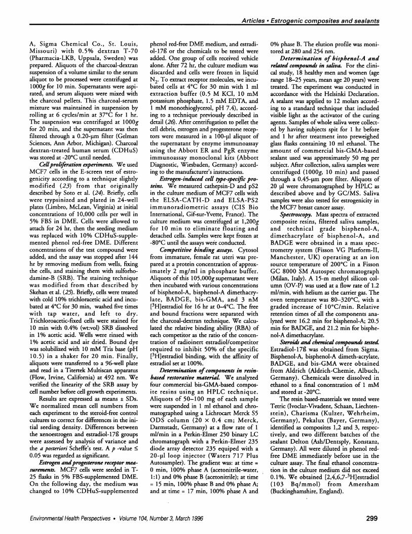
Determination ofcomponents in resin-based restorative material We analyzedfour commercial bis-GMA-based compos-ite resins using an HPLC technique.Aliquots of 50-100 mg of each samplewere suspended in 1 ml ethanol and chro-matographed using a Lichrocart Merck S5ODS column (20 x 0.4 cm; Merck,Darmstadt, Germany) at a flow rate of 1ml/min in a Perkin-Elmer 250 binary LCchromatograph with a Perkin-Elmer 235diode array detector 235 equiped with a20-pl loop injector (Waters 717 PlusAutosampler). The gradient was: at time =0 min, 100% phase A (acetonitrile-water,1:1) and 0% phase B (acetonitrile); at time= 15 min, 100% phase B and 0% phase A;and at time = 17 min, 100% phase A and
0% phase B. The elution profile was moni-tored at 280 and 254 nm.
Determination of bisphenol-A andrelated compounds in saliva. For the dini-cal study, 18 healthy men and women (agerange 18-25 years, mean age 20 years) weretreated. The experiment was conducted inaccordance with the Helsinki Declaration.A sealant was applied to 12 molars accord-ing to a standard technique that includedvisible light as the activator of the curingagents. Samples ofwhole saliva were collect-ed by having subjects spit for 1 hr beforeand 1 hr after treatment into preweighedglass flasks containing 10 ml ethanol. Theamount of commercial bis-GMA-basedsealant used was approximately 50 mg persubject. After collection, saliva samples werecentrifuged (1OOOg, 10 min) and passedthrough a 0.45-pm pore filter. Aliquots of20 pl were chromatographed by HPLC asdescribed above and by GC/MS. Salivasamples were also tested for estrogenicity inthe MCF7 breast cancer assay.
Spectroscopy. Mass spectra of extractedcomposite resins, filtered saliva samples,and technical grade bisphenol-A,dimethacrylate of bisphenol-A, andBADGE were obtained in a mass spec-trometry system (Fisson VG Platform-II,Manchester, UK) operating at an ionsource temperature of 200°C in a FissonGC 8000 SM Autospec chromatograph(Milan, Italy). A 15-m methyl silicon col-umn (OV-P) was used at a flow rate of 1.2ml/min, with helium as the carrier gas. Theoven temperature was 80-320°C, with agraded increase of 10°C/min. Relativeretention times of all the components ana-lyzed were 16.2 min for bisphenol-A; 20.5min for BADGE, and 21.2 min for bisphe-nol-A dimethacrylate.
Steroids and chemical compounds testedEstradiol-17g was obtained from Sigma.Bisphenol-A, bisphenol-A dimeth-acrylate,BADGE, and bis-GMA were obtainedfrom Aldrich (Aldrich-Chemie, Albuch,Germany). Chemicals were dissolved inethanol to a final concentration of 1 mMand stored at -20°C.
The resin based-materials we tested wereTetric (Ivoclar-Vivadent, Schaan, Liechten-stein), Charisma (Kulzer, Wehrheim,Germany), Pekalux (Bayer, Germany),identified as composites 1,2 and 3, respec-tively, and two different batches of thesealant Delton (Ash/Dentsply, Konstanz,Germany). All were diluted in phenol red-free DME immediately before use in theculture assay. The final ethanol concentra-tion in the culture medium did not exceed0.1%. We obtained [2,4,6,7-3H]estradiol(103 Bq/mmol) from Amersham(Buckinghamshire, England).
Environmental Health Perspectives * Volume 104, Number 3, March 1996 299

Articles - Olea et al.
ResultsEstrogenic Activity of Resin-basedCompositesThe addition of estradiol-17g to CDHuS-supplemented medium increased the num-bers of MCF7 cells in culture. Maximumproliferative effect was obtained at estradi-ol-17f concentrations of 10 pM and higher(Fig. IA). Cell yields were sixfold greater
than in control cultures after 6 days (mean± SD, 6.67 ± 1.21; n = 15 experiments). Inthe absence of estradiol-17l3, cells prolifer-ated minimally.
Four bis-GMA-based composite resinswere assayed before polymerization usingthe MCF7 proliferation test. Compositesand sealants were prepared in ethanol at a
concentration of 100 and 50 mg/ml,respectively, and assayed at a range of dilu-tions (1/100 to 1/106). The cell yieldobtained with 5 pg/ml sealant sample was
sixfold greater than in control cultures. Themagnitude of this proliferative effect was
similar to that obtained with estradiol-1713.Cell toxicity was observed at concentra-
tions of 50 pg/ml and higher (Fig. 1B).In contrast to the sealant sample, the
three resin-based composites assayed didnot induce MCF7 cell proliferation at a
maximal concentration of 1 mg/ml. Thesecommercial formulations contain a largeproportion of inorganic filler particles(50-85% by weight of the composite).Stock suspensions were prepared withoutdiscarding the inorganic portion.
0
U
;0
0
LI.
Identification of EstrogenicSubstances from Composite Resins
Figure 2 shows the HPLC profile of anunpolymerized sealant that was positive inthe proliferation test. Relative retentiontimes of the components identified were: t =2.35 min or bisphenol-A; t = 7.35 min forBADGE; t= 8.55 min for bis-GMA; and t =11.85 min for dimethacrylate of bisphenol-A. These components were quantified bycalibration curves made after eluting knownamounts (1 liM to 1 mM) of the pure sub-stance [bisphenol-A, y = (2.92 x 10¹¹)x +(9.075 x 10⁶), r = 0.998; bis-GMA, y =(1.04 x 10¹¹)x + (0.670 x 10⁶), r = 0.999;BADGE, y = (1.12 x 10¹¹)x + (0.470 x10⁶), r = 0.999, and bisphenol-Adimethacrylate, y = (2.18 x 10'1)x + (8.782x10⁶), r = 0.995]. Mass spectrometric analy-sis confirmed the presence of bisphenol-Aand dimethacrylate of bisphenol-A in allsamples for which HPLC chromatogramsshowed the corresponding peaks (Fig. 3).
To determine whether the monomerbisphenol-A can be cleaved from compos-ites and from their main oligomers(dimethacrylate of bisphenol-A, bis-GMA,and BADGE), these were hydrolyzed inalkaline (pH = 13) and acidic media (pH =1) after heating (100'C for 30 min), andthen chromatographed as indicated. Table1 summarizes the components identified inthe chromatograms. Extensive breakdownof the oligomers into smaller compoundssuch as bisphenol-A was observed.
I0-
Sa0b0
10aL
1013 1o-llConcentration (M)
Estrogenic Activity ofSomeComponents of Resin-basedRestorative Composites
Bisphenol-A and its dimethacrylate wereestrogenic when assayed in the breast can-cer cell proliferation assay. The concentra-tions required to produce maximum prolif-eration of MCF7 cells were 10,000-foldhigher than those of estradiol-17f; (Fig. 4).MCF7 cells were grown in the presence ofbisphenol-A or bisphenol-A dimethacrylateand the estrogen antagonist hydroxytamox-ifen; the proliferative effect of 0.1 pMbisphenol-A and bisphenol-A dimethacry-late disappeared when cells were grown inthe presence of a 10-fold higher concentra-tion of the antagonist (data not shown).
Bis-GMA was negative in the estro-genicity test. A proliferative effect ofBADGE was observed at a concentrationof 10 pM (Fig. 5). Oligomers from bothcompounds became active in the prolifera-tive test after hydrolysis (100°C for 30min, in alkaline or acidic medium).Bisphenol-A and bisphenol-A dimethacry-late were identified in chromatogramswhen oligomers were found to be estro-genic after hydrolysis.
Progesterone Receptor InductionMCF7 cells have receptors for estradiol-17f (estrogen receptor; ER) and proges-terone (PgR). The basal concentration of
3
0.05 0.5 5 50 500Concentration (gg/ml)
Figure 1. Cell proliferation of MCF7 cells. Cells growing in 10% charcoal dextran-treated human serum-supplemented medium were exposed for 144 hr to (A) estradiol-17B and (B) bis-GMA-based sealant,sample no. 1. Points represent quadruplicate cultures; brackets indicate SDs. Ordinates represent cellnumbers (experimental/control); abscissas represent concentration of the test compound. Estradiol-171was effective at >1 pM (0.272 pg/ml). *Values significantly different from control, p < 0.001.
Figure 2. Chromatogram of sealant sample no. 1,which was estrogenic in the breast cancer prolif-eration bioassay shown in Figure 1. Peaks atretention times 2.3, 7.3, 8.6, and 11.9 min wereidentified as 1) bisphenol-A, 2) bis-GMA, 3)BADGE, and 4) bisphenol-A dimethacrylate.
Volume 104, Number 3, March 1996 * Environmental Health Perspectives
109
300

Articles - Estrogenic composites and sealants
ER in these cells was 183 ± 29 fmol/mg ofextracted protein (Fig. 6A). Estradiol-171decreased the concentration ofER by 50%and increased that of PgR The basal PgRvalue was 15.5 ± 4.3 fmol/mg protein,close to the lower limit of detection of themonoclonal antibody assay (Fig. 6B).Estradiol-17l (1 nM) significantlyincreased PgR nearly 15-fold over the con-trol value.
Treatment ofMCF7 cells with concen-trations of 0.1 pM and higher of bisphe-nol-A dimethacrylate resulted in a signifi-cant increase in PgR levels. Bisphenol-Atreatment also increased PgR levels (Fig.6B). However, no reduction of ER levelswas observed when bisphenol-A or bisphe-nol-A dimethacrylate induced PgR
Secretion ofpS2 and Cathepsin-DCathepsin-D and pS2 accumulation in theculture medium reflected increases in cellnumber during the experiments. The secre-tion of pS2 by MCF7 cells was significant-ly increased by >0.1 nM estradiol-17g (-3.5-fold increase over controls). The basalconcentration of pS2 (55.4 ± 15.2 ngIl6cells) increased to 202.2 ± 19.1 ng/10 cellsafter treatment with 10 pM bisphenol-Adimethacrylate; this was similar to theeffect of estradiol- 1 7L on pS2 secretion(Fig. 7A).
The basal concentration of cathepsin-Dsecreted by MCF7 cells was 8.9 ± 2.9pmol/106 cells. Estradiol-1781 treatment (1and 10 nM) resulted in a modest 1.7-foldincrease in the accumulation of cathepsin-D in the culture medium. Bisphenol-Adimethacrylate (1 and 10 pM) increasedcathepsin-D secretion only 1.6-fold (Fig.7B). These changes were not significant.
Binding Affinity of Composite ResinCompounds for the EstrogenReceptorThe binding affinity of bisphenol-A,bisphenol-A dimethacrylate, BADGE, andbis-GMA for the cytosol ER was deter-mined by competitive binding analysis.The receptor relative binding affinities(RBA) were 0.012 and 0.0033 for bisphe-nol-A and bisphenol-A dimethacrylate,respectively, versus 100 for estradiol- 1711.Bis-GMA and BADGE did not competewith estradiol for binding to the ER, evenat concentrations a million-fold higherthan estradiol-17f (Fig. 8).
Analysis and Quantification ofEstrogenic Substances in SalivaEighteen saliva samples taken before andafter sealant treatment were chromato-graphed using the technique described inMethods. Figure 9 shows the chromato-
100
100 r
50
0D U 70 W0 90 IlUU I IU 120 130 1IW IU 1W 17U 1WU 190 200 21U 22U 2d3 240 2W
Figure 3. Mass spectra of (A) pure grade bisphenol-A and (B) bisphenol-A from a sealant sample.
Table 1. Identification of the components in the composites and sealants"
pg/100 mg commercial productProduct Bisphenol-Aand pH Bis-GMA BADGE dimethacrylate Bisphenol-ASealant 17 45.8 75.0 29.2 7.413 32.8 117.0 4.6 84.41 49.4 164.8 6.4 18.2
Composite 17 105.0 19.6 ND 67.713 16.7 1.7 186.0 53.11 0.4 ND 1.8 49.9
Composite 27 18.1 10.0 ND 0.513 53.2 24.2 878.4 ND1 46.8 140.0 ND ND
Composite 37 22.0 3.2 ND 4.213 1809.4 38.6 ND N D1 1802.0 5.2 3.6 11.4
Abbreviations: bis-GMA, bisphenol-A digicidylether methacrylate; BADGE, bisphenol-A diglcidylether; ND,not detectable.8 Samples were hydrolyzed in alkaline (pH = 13) and acidic media (pH = 1) after heating (100'C for 30 min)and then chromatographed as indicated in Methods. The contents before hydrolysis are indicated as pH = 7.
Environmental Health Perspectives * Volume 104, Number 3, March 1996 301

Articles - Olea et al.
25(
U~~~~~~~~~~4
3U
0 ~~~~~~~~~~~~~~~~0l0-10 lo's 108 100 100
Concentration (M) Concentration (M)
Figure 4. Cell proliferation in MCF7 cells. Cells 'growing in 10% charcoal dextran-treated human serum-supplemented medium were exposed for 144 hr to (A) bisphenol-A and (B) bisphenol-A dimethacrylate.Points represent quadruplicate cultures; brackets indicate SDs. *Values significantly different from con-trol, p< 0.001.
3.5 3.5
pH_1 pH ___1__
.5 2.5 5 .5
0~~~~~~~~~~~
2.0 20
o 0_g __
LE 1.5 LE 1.5
1.0 1.0
0.5 0.5
0.0 0.010-9 10.7 105 109 10.7 10,5
Concentration (M) Concentration (M)
Figure 5. Cell proliferation in MCF7 cells. Cells growing in 10% charcoal dextran-treated human serum-supplemented medium were exposed for 144 hr to (A) bis-GMA and (B) BADGE and to products obtainedby hydrolytic treatments of these compounds in alkaline (pH = 13) and acidic medium (pH = 1). Points rep-resent quadruplicate cultures; brackets indicate SDs. *Values significantly different from control, p <0.001.
graphic profile of a representative salivasample from a patient selected at randomafter treatment with 50 mg of the bis-GMA-based sealant. Peaks in the chro-matogram corresponded to Bis-GMA,BADGE, bisphenol-A, and dimethacrylateof bisphenol-A. Variations in the chro-matographic profiles were seen betweensealant batches and between patients whoreceived sealant from the same batch.Monomers of composite components were
measured by HPLC in saliva collected 1 hrafter treatment (Table 2). After treatment,all saliva samples contained variableamounts of bisphenol-A ranging from 90to 931 pg; this was confirmed by massspectrometry.
Subjects were treated with a fixedamount of approximately 50 mg of sealant,and the proportion of free, unpolymerizedmaterial collected during 1 hr after treat-ment never exceeded 2% of the total
_ 200
0
i E,=oo 15(
101
5(
251
'I'S 2010
*0.CDE 151
i-
nM gM gM
Concentration
1 10 1 10 1 10nM gM gtM
Concentration
Figure 6. (A) Estrogen (ER) and (B) progesterone(PgR) receptors in MCF7 cells. Cells in T25 flaskswere incubated in 10% charcoal dextran-treatedhuman serum for 72 hr with 1 and 10 nM of estra-diol-17B. A parallel set of flasks was exposed to 1and 10 pM bisphenol-A or bisphenol-Adimethacrylate. Controls received the vehiclealone. At the end of the experiment, the mediumwas aspirated and flasks were kept in liquid N2until assayed. ER and PgR were measured inextracted cells with a monoclonal antibody tech-nique as indicated in Methods. Results areexpressed as femtomole per milligram of extract-ed protein ± SD. *Values significantly differentfrom control, p < 0.001.
amount of sealant applied. Compositecomponents were not observed in any ofthe saliva samples collected before treat-ment. A subject initially selected for treat-ment had been treated with tooth sealant 2years earlier; chromatograms demonstratedthe presence of bisphenol-A (66.4 fog) andbisphenol-A dimethacrylate (49.2 jig) in
Volume 104, Number 3, March 1996 * Environmental Health Perspectives302

200
150D0co
a.100
50
0
co
E
-a
._
56
15
10
1 10 i 10 1 10 1 10 1 10 1 10nM PiM igM nM JiM JIM
Concentration Concentration
Figure 7. Accumulation of (A) pS2 and (B) cathepsin-D in culture medium. MCF7 cells were grown in 10%charcoal dextran-treated human serum-supplemented medium and exposed for 144 hr to 1 and 10 nMestradiol-17B. Parallel cultures were exposed to 1 and 10 pM bisphenol-A or bisphenol-A dimethacrylate.Controls received the vehicle alone. *Values significantly different from control, p < 0.001.
her saliva before the second treatment. Theresults from this subject were excludedfrom analysis.
Samples containing the highestamounts of bisphenol-A and bisphenol-Adimethacrylate were estrogenic in the pro-
liferation test. Figure 10 shows the prolifer-ative pattern of a sample containing 231 jigbisphenol-A in 27 ml of collected salivaplus 10 ml of ethanol. An aliquot of 100 piof this sample diluted in 1 ml of the culturemedium was positive in the estrogenicity
assay. The estimated amount of bisphenol-A assayed was 0.62 pg/ml. Because thepresence of endogenous estrogens couldnot be ruled out, saliva samples takenbefore the sealant was applied were alsoassayed; none showed estrogenicity.
DiscussionEpoxy resins of the bisphenol-A type havemany applications such as lacquer coatingin food cans, in dental, surgical, and pros-
thetic devices, and as additives in a varietyof other plastic materials (27). Here we
demonstrate that bisphenol-A and bisphe-nol-A dimethacrylate, components of com-mercial resin-based composites and sealantsused in dentistry, are estrogenic and may
represent additional sources of humanexposure to xenoestrogens.
Commercial smooth-surface compos-
ites vary in monomer composition andthus have different physical properties andbiological behaviors. We found that theamount of bisphenol-A and bisphenol-Adimethacrylate varied between commercial
* Estradiol-173 A Bisphenol-A* BADGE dimethacrylate* Bisphsnol-A U Bis-GMA
100
m
c
a.U)
Figure 9. Chromatogram of a saliva sample withestrogenic activity. Peaks detected at retentiontimes 2.3, 7.4, 8.5, and 11.9 min were identified as 1)bisphenol-A, 2) BADGE, 3) bis-GMA, and 4) bisphe-nol-A dimethacrylate, respectively.
s0
40
20
Concentration (M)
Figure 8. Competitive displacement of [3H1-estradi-ol from the estrogen receptor by estradiol-17 andbisphenol-A, bisphenol-A dimethacrylate, BADGE,and bis-GMA. The relative binding affinities ofbisphenol-A and bisphenol-A dimethacrylate were0.012 and 0.0033, respectively. [3H]Estradiol con-centration was 3 nM, and the total binding was 73fmol/mg protein; Kd = 8.5 x10¹¹ M.
composites and batches. Bisphenol-A was
identified in all the composite samples andbatches analyzed, whereas methacrylate ofbisphenol-A was identified only in thesealant. The oligomers BADGE and bis-GMA were found in all three compositesand in the sealant. The proportion of thesecompounds varied widely between com-
posites and batches. Vankerckhoven et al.(28) studied 12 commercial restorative bis-
Table 2. Quantitative evaluation of bisphenol-Aidentified in saliva obtained 1 hr after the applica-tion of 50 mg of sealant
Sample. Bisphenol-Aano Sex Volume (ml) (pg) (pg/ml)
1 F 29 320.6 11.12 F 31 931.0 30.03 F 35 174.6 4.94 F 33 865.6 26.25 M 26 91.2 3.56 M 31 249.6 8.17 F 27 165.7 6.18 F 38 263.6 6.99 M 32 105.3 3.310 M 27 230.5 8.511 M 36 181.4 5.012 M 35 324.2 9.313 M 29 353.0 12.214 M 39 810.8 20.815 M 32 230.8 7.216 M 27 130.4 4.817 M 27 89.8 3.318 F 29 112.4 3.9
"Saliva samples were collected in glass flaskscontaining 10 ml ethanol. Values of bisphenol-Aconcentration representthose of undiluted saliva.
Environmental Health Perspectives * Volume 104, Number 3, March 1996
Articles * Estrogenic composites and sealants
17 ....11'.~ Ssi.
303

Articles - Olea et al.
7
6
5
4
0
*0
1/10, 1/10, ~~~~1/10Dilution factor
Figure 10. Proliferative effect of a saliva sampleobtained 1 hr after treatment with sealant no 1.Points represent quadruplicate cultures; bracketsindicate SDs. *Values significantly different fromcontrol, p < 0.001.
GMA-based resins and found bis-GMA inall samples at concentrations ranging from19 to 51% of the total composite weight.Iso-bis-GMA was also present and repre-sented 40% of the total percentage of bis-GMA. Bisphenol-A dimethacrylate wasfound in four commercial mixtures inamounts that ranged from 6 to 10% of thetotal composite weight. In addition tothese monomers, other compounds such aslight activators, phthalates, and benzoates,as well as a large amount of inorganicfillers, are commom ingredients of compos-ites (28).
Once applied to tooth cavities, compos-ites and sealants are polymerized in situ.The degree of conversion of oligomers intopolymers varies depending on the composi-tion of the resin and its distance from thetooth surface. Conversion of 60-75% isexpected with most common composites(16), although levels of curing as low as30% are found in the bottom of fillings.Lower levels of conversion are thought tobe associated with greater elution of freecomponents from the composites (16). Astrong inverse relationship has been foundbetween the leaching of resin from bis-GMA-based composites and monomerconversion. Monomer leaching reached aplateau at conversion levels of 60% (29). Innine commercial resins, the amount ofresidual bis-GMA measured at the end ofthe setting time was in the range of0.4-1.2% of the original weight after cur-ing. Amounts of the residual monomers ofup to one-tenth the initial release wereeluted into water over a period of 14 days
(29). Higher values were described byFerracane and Condon (16), who founddifferences in percentage elution in relationto curing and double illumination. Ourdata confirm these figures: residual bisphe-nol-A in saliva after curing ranged from 0.1to 2% of the 50 mg initially applied to thetooth surface in samples taken during 1 hrafter treatment.
In addition to bisphenol-A, present in100% of the saliva samples, its methacry-late derivative was present in 3 out of 18samples. Both compounds were identifiedby chromatography and estrogenic activityas measured in the MCF7 breast cancer cellproliferation test, the E-screen assay.
The oligomer bis-GMA was not estro-genic in the E-screen bioassay. HydrolyzedBADGE and bis-GMA in alkaline or acidicmedia were estrogenic. Chromatographicanalyses of hydrolyzed samples detected thepresence of bisphenol-A and bisphenol-Adimethacrylate after treatment. The meta-bolic routes of systemic degradation ofsome composite resins have been studied inmice (15,19,20). For example, BADGE ismetabolized by oxidative dealkylation togive a phenol diol derivative. Much atten-tion has been given to the smaller fragmentof the cleavage process because of themutagenic properties of the glycidylalde-hyde portion. However, the estrogenicactivity of these derivatives has not beenevaluated; their chemical similarity to somebisphenol-A derivatives with this propertysuggests the need for closer examination.Moreover, little information is availableabout the potential estrogenicity of by-products of the environmental breakdownof biphenolic compounds. Lobos et al. (30)isolated a novel bacterium from the sludgeof a wastewater treatment plant of a plasticsmanufacturing facility; the organism wasable to degrade bisphenol-A to 4-hydroxy-benzoic acid and 4-hydroxyacetophenone(both monophenolic by-products) and to2,2-bis(4-hydroxyphenyl)-1-propanol, andfinally to 2,3-bis(4-hydroxyphenyl)-1,2-propanediol. So far, the estrogenicity ofthese bisphenol-A derivatives has not beenexplored, although the similarity of the lat-ter compound to some stilbenes suggeststhat it may display considerable activity.
Bisphenol-A is toxic to fish and inverte-brates at concentrations of 1.1-10 mg/l ( >4 pmol/l) (31). In a mouse model, bisphe-nol-A induced photoallergic contact der-matitis (32). If administered duringorganogenesis, bisphenol-A is fetotoxicwhen administered to pregnant mothers. Astudy of the effects of bisphenol-A onreproduction and fertility showed a signifi-cant reduction in seminal vesicle weightand sperm motility in treated male mice
(33). These effects were present in parentsand in a second generation when offspringwere maintained at the same exposure levelas their parents (4.37-17.5 mg/kg bodyweight). In addition, bisphenol-A treat-ment resulted in a significant reduction inthe number of litters per pair and in livepups per litter.
Composites were discovered to be toxicin humans because lesions developed inunlined cavities (34,35). The concentra-tions to which cells and tissues are exposedin humans are unknown (11), but it wasestimated that seven components of com-posites, including bisphenol-A, were toxicin the 10-100 pmol/l concentration rangein an in culture assay (11). The sealant wetested was toxic to MCF7 breast cancercells at concentrations > 50 jig/ml ( -200pmol/l of bisphenol-A). Lower concentra-tions were not toxic but were fully estro-genic in the bioassay (Fig. 1B).
Human exposure to bisphenol-A anddimethacrylate of bisphenol-A hasincreased during the last 30 years becauseof the extensive use of bisphenol-A-basedresins. The use of sealants as an effectivetherapy and preventive treatment for dentalpits and fissures in adults and children hasbeen endorsed by European and U.S. agen-cies (18,36,34). In view of the documentedexposure to bis-GMA-based compositesand sealants used in dental treatments foradults and children, the use of these xenoe-strogens should be reevaluated.
REFERENCES
1. Colborn T, Clement C, eds. Chemicallyinduced alterations in sexual and functionaldevelopment: the wildlife/human connection.Princeton, NJ:Princeton Scientific Publishing,1992.
2. Sharpe R, Skakkebaek N. Are estrogensinvolved in falling sperm counts and disordersof the male reproductive tract? Lancet341:1392-1395 (1993).
3. Wolff MP, Toniolo P, Lee E, Rivera M, DubinN. Blood levels of organochlorine residues andrisk of breast cancer. J Natl Cancer Inst85:648-652 (1993).
4. Dodds EC, Lawson W. Synthetic estrogenicagents without the phenanthrene nucleus.Nature 137:996 (1936).
5. Reid EE, Wilson E. The relation of estrogenicactivity to structure in some 4,4'-dihydroxyde-phenylmethanes. J Am Chem Soc 66:967-969(1944).
6. Krishnan AV, Starhis P, Permuth SF, Tokes L,Feldman D. Bisphenol-A: an estrogenic sub-stance is released from polycarbonate flasksduring autoclaving. Endocrinology132:2279-2286 (1993).
7. Brotons JA, Olea-Serrano MF, Villalobos M,Pedraza V, Olea N. Xenoestrogens releasedfrom lacquer coating in food cans. EnvironHealth Perspect 103:608-612 (1995).
8. Ranks CT, Craig RG, Diehi ML, Pashley DH.
304 Volume 104, Number 3, March 1996 * Environmental Health Perspectives

Articles - Estrogenic composites and sealants
Cytotoxicity in dental composites and othermaterials in a new in vitro device. J Oral Pathol17:396-403 (1988).
9. Hensten-Pettersen A, Helgeland K Sensitivityof different human cell lines in the biologicevaluation of dental resin-based restorativematerials. Scand J Dent Res 89:102-107(1981).
10. Terakado M, Yamazaki M, Tsujimoto Y,Kawashima T, Nagashima K, Ogawa J, FujitaY, Sugiya H, Sakai T, Furuyama S. Lipid per-oxidation as a possible cause of benzoyl perox-ide toxicity in rabbit dental pulp-a microso-mal lipid peroxidation in vitro. J Dent Res63:901-905 (1984).
11. Hanks CT, Strawn SE, Wataha JC, Craig RG.Cytotoxic effects of resin components on cul-tured mammalian fibroblasts. J Dent Res70:1450-1455 (1991).
12. Bourne LB, Milner FJM, Alberman KB. Healthproblems of epoxy resins and amine curingagents Br J Ind Med 16:81-97 (1959).
13. Morrissey RE, George JD, Price CJ, Tyl RW,Marr MC, Kimmel CA. The developmentaltoxicity of bisphenol A in rats and in mice.Fundam Appl Toxicol 8:571-582 (1987).
14. Bentley P, Bieri P, Kuster H, Muakkassah-Kelly S, Sagelsdorff P, Staubli W, Waechter F.Hydrolysis of bisphenol A diglycidylether byepoxide hydro-lases in cytosolic and microso-mal fractions of mouse liver and skin: inhibi-tion by bis epoxycyclopentylether and theeffects upon the covalent binding to mouse skinDNA. Carcinogenesis 10:321-327 (1989).
15. Steiner S., Honger, Sagelsdorff P. Moleculardosimetry ofDNA adducts in C3H mice treat-ed with bisphenol A diglycidylether.Carcinogenesis 13:969-972 (1992).
16. Ferracane JL, Condon JR. Rate of elution ofleachable components from composite. DentMater 6:282-287 (1990).
17. Freund M, Munksgaard EC. Enzymatic degra-dation of BISGMAITEGDMA polymers caus-ing decreased microhardness and greater wearin vitro. Scan J Dent Res 98:351-355 (1990).
18. Ripa LW. Sealants revisited: an update of the
effectiveness of pit-and-fissure sealants. CariesRes 27:77-82 (1993).
19. Climie IJG, Hutson DH, Stoydin G.Metabolism of the epoxy resin component 2,2-bis[4-(2,3-epoxypropoxy)phenyl]propane, thediglycidyl ether of bisphenol A (DGEBPA) inthe mouse. Part I. A comparison of the fate of asingle dermal application and of a single oraldose of 14C-DGEBPA in the mouse.Xenobiotica 11:391-399 (1981).
20. Climie IJG, Hutson DH, Stoydin G.Metabolism of the epoxy resin component 2,2-bis[4-(2,3-epoxypropoxy)phenyl]propane, thediglycidyl ether of bisphenol A (DGEBPA) inthe mouse. Part II. Identification of metabolitesin urine and feces following a single oral dose of14C-DGEBPA. Xenobiotica 11:401-424(1981).
21. Soule HD, Vazquez J, Long A, Alberts S,Brennan MJ. A human cell line from a pleuraleffusion derived from a breast carcinoma. JNad Cancer Inst 51:1409-1413 (1973).
22. Soto AM, Sonnenschein C. The role of estro-gen on the proliferation of human breast tumorcells (MCF-7). J Steroid Biochem 23:87-94(1985).
23. Villalobos M, Olea N, Brotons JA, Olea MF,Ruiz de Almod6var JM, Pedraza V. The E-screen assay: comparison among differentMCF7 cell stocks. Environ Health Perspect103:844-850 (1995).
24. Soto AM, Lin TM, Justicia H, Silvia RM,Sonnenschein C. An "in culture" bioassay toassess the estrogenicity of xenobiotics (E-SCREEN). In: Chemically induced alterationsin sexual and functional development; thewildlife/human connection. (Colborn T,Clement C, eds). Princenton, NJ: PrincentonScientific Publishing, 1992;295-309.
25. Skehan P, Storeng R, Scudiero D, Monks A,Mcmahon J, Vistica D, Warren JT, Bokesch H,Kenney S, Boyd MR. New colorimetric cyto-toxicity assay for anticancer-drug screening. JNatl Cancer Inst 82:1107-1112 (1990).
26. Maddedu L, Legros N, Devleeschouwer N,Bosman C, Piccart M, Leclercq G. Estrogen
receptor status and estradiol sensitivity ofMCF-7 cells in exponential growth phase. EurJ Cancer Clin Oncol 24:385-391 (1988).
27. Paseiro Losada P, Simal Lozano J, Paz Abuin S,Lopez Mahia P, Simal Gandara J. Kinetics ofthe hydrolysis of bisphenol A diglycidyl ether(BADGE) on water-based food simulants. FresJ Anal Chem 345:527-532 (1993).
28. Vankerckhoven H, Lambrechts P, Van BeylenM, Vanherle G. Characterization of compositeresins by NMR and TEM. J Dent Res60:1957-1065 (1981).
29. Rueggeberg FA, Craing RG. Correlation ofparameters used to estimate monomer conver-sion in a light-cured composite. J Dent Res67:932-937 (1988).
30. Lobos JH, Leib TK, Su T-M. Biodegradationof bisphenol A and other bisphenols by a gram-negative aerobic bacterium. Appl EnvironMicrobiol 58:1823-1831 (1992).
31. Alexander HC, Dill DC, Smith LA, GuineyPA, Dorn P. Bisphenol A: acute aquatic toxici-ty. Environ Toxicol Chem 7:19-26 (1988).
32. Maguire MC. Experimental photoallergic con-tact dermatitis to bisphenol A. Acta DermVenereol 68:402-412 (1988).
33. Dutch Expert Committee on OccupationalStandards. Health-based recommended occupa-tional exposure limit for bisphenol-A and itsdiglycidylether. Den Haag, TheNetherlands:Committee of the Health Councilof the Netherlands, 1995
34. Stanley HR, Bowen RL, Folio J. Compatibilityof various materials with oral tissues. II Pulpresponses to composite ingredients. J Dent Res58:1507-1517 (1979).
35. Inoue K, Hayashi I. Residual monomer (Bis-GMA) of composite resins. J Oral Rahabilit9:493-497 (1982).
36. NIH. Seal out dental decay. Publication no.91-489. Washington, DC:National Institutesof Health, 1991.
37. Weintraub JA. The effectiveness of pit and fis-sure sealants. J Public Health Dent 49:317-330(1989)
Environmental Health Perspectives * Volume 104, Number 3, March 1996 305