Estradiol promotes proliferation of dopaminergic precursors resulting in a higher proportion of...
8
Estradiol promotes proliferation of dopaminergic precursors resulting in a higher proportion of dopamine neurons derived from mouse embryonic stem cells Ne ´ stor F. Dı ´az a,b , Ne ´ stor E. Dı ´az-Martı ´nez a , Ignacio Camacho-Arroyo b , Iva ´ n Velasco a, * a Departamento de Neurociencias, Instituto de Fisiologı´a Celular, Universidad Nacional Auto ´noma de Me ´xico, Me ´xico D.F. 04510, Mexico b Departamento de Biologı´a, Facultad de Quı´mica, Universidad Nacional Auto ´noma de Me ´xico, Me ´xico D.F. 04510, Mexico 1. Introduction There is evidence indicating that estrogens can induce the development and protection of nigrostriatal dopamine (DA) neurons in vivo (Kipp et al., 2006; Liu and Dluzen, 2007). Parkinson’s disease (PD) is a neurodegenerative disorder char- acterized by a progressive and selective loss of midbrain DA neurons of the substantia nigra pars compacta. This decrease of DA neurons results in a dopamine depletion in the striatum, which impairs normal motor functions. Epidemiological studies show that PD is 1.5 to 2 times more common in men than in women (Baldereschi et al., 2000; Czlonkowska et al., 2006; Haaxma et al., 2007); recently, delayed onset and more benevolent progress have been reported for women when compared to men patients with PD, and these differences could be due to estrogens action (Haaxma et al., 2007). Furthermore, in experimental animal models, numerous studies demonstrated that estrogens adminis- tration protects against neurotoxins such as 1-methyl-4-phenyl- 1,2,3,6-tetrahydropyridine (reviewed in Morissette et al., 2008), methamphetamine (Dluzen and McDermott, 2002) and 6- hydroxy-DA (reviewed in Dluzen, 1997) that selectively destroy DA neurons of the substantia nigra. All these findings imply that estrogens play a protective role for DA neurons. In addition to this well-described action, in recent years a proliferative action of estradiol (E 2 ) has been reported for neural precursor cells (Brannvall et al., 2002; Martinez-Cerdeno et al., 2006; Wang et al., 2008). Embryonic stem cells (ESC) are pluripotent cell lines derived from the inner cell mass of pre-implantation embryos. Because of their ability to self-renew, proliferate undifferentiated in vitro, while maintaining the potential to differentiate into most cell types, ESC are useful to dissect critical steps of cell commitment. Furthermore, they also represent a potential source for cell replacement therapy (McKay, 2000). In this context, it has been shown that midbrain DA neurons can be induced in vitro from mouse ESC with the 5-stage method and that these DA neurons have improved parkinsonism in rodents for over 32 weeks after grafting (Diaz et al., 2007; Kim et al., 2002; Rodriguez-Gomez et al., 2007). We have previously reported the expression of sex steroid hormone receptors during this protocol. The last two phases of this procedure produce nestin-positive cells (stage 4, neural precur- sors) that respond to the application of Fibroblast growth factor Int. J. Devl Neuroscience 27 (2009) 493–500 ARTICLE INFO Article history: Received 22 February 2009 Received in revised form 7 April 2009 Accepted 8 April 2009 Keywords: Dopamine neurons Embryonic stem cells Estradiol Lmx1a Tyrosine hydroxylase ABSTRACT Estradiol protects dopamine neurons of the substantia nigra from toxic insults. Such neurons succumb in Parkinson’s disease; one strategy for restoring dopamine deficiency is cell therapy with neurons differentiated from embryonic stem cells. We investigated the effects of 17b-estradiol on dopaminergic induction of embryonic stem cells using the 5-stage protocol. Cells were incubated with different steroid concentrations during the proliferation (stage 4) or differentiation (stage 5) phases. Estradiol added at nM concentrations only during stage 4 increases the proliferation of dopaminergic precursors expressing Lmx1a, inducing a higher proportion of dopamine neurons at stage 5. These actions were mediated by activation of estrogen receptors, because co-incubation of cells with estradiol and ICI 182,780 completely abolished the positive effect on both proliferation of committed precursors, and subsequent differentiation to dopaminergic neurons. Our results suggest that estradiol should be useful in producing higher proportions of dopamine neurons from embryonic stem cells aimed for treating Parkinson’s disease. ß 2009 ISDN. Published by Elsevier Ltd. All rights reserved. Abbreviations: DA, dopamine; E 2 , estradiol; ER, estrogen receptor; ESC, embryonic stem cells; FGF, Fibroblast growth factor; GAPDH, Glyceraldehyde phosphate dehydrogenase; PD, Parkinson’s disease; SHH, Sonic hedgehog; TH, tyrosine hydroxylase. * Corresponding author at: Instituto de Fisiologı ´a Celular, UNAM, AP 70-253, Me ´ xico D.F. 04510, Mexico. Tel.: +52 555 622 5649; fax: +52 555 622 5607. E-mail address: [email protected] (I. Velasco). Contents lists available at ScienceDirect International Journal of Developmental Neuroscience journal homepage: www.elsevier.com/locate/ijdevneu 0736-5748/$36.00 ß 2009 ISDN. Published by Elsevier Ltd. All rights reserved. doi:10.1016/j.ijdevneu.2009.04.002
-
Upload
independent -
Category
Documents
-
view
0 -
download
0
Transcript of Estradiol promotes proliferation of dopaminergic precursors resulting in a higher proportion of...
Int. J. Devl Neuroscience 27 (2009) 493–500
Estradiol promotes proliferation of dopaminergic precursors resulting in a higherproportion of dopamine neurons derived from mouse embryonic stem cells
Nestor F. Dıaz a,b, Nestor E. Dıaz-Martınez a, Ignacio Camacho-Arroyo b, Ivan Velasco a,*a Departamento de Neurociencias, Instituto de Fisiologıa Celular, Universidad Nacional Autonoma de Mexico, Mexico D.F. 04510, Mexicob Departamento de Biologıa, Facultad de Quımica, Universidad Nacional Autonoma de Mexico, Mexico D.F. 04510, Mexico
A R T I C L E I N F O
Article history:
Received 22 February 2009
Received in revised form 7 April 2009
Accepted 8 April 2009
Keywords:
Dopamine neurons
Embryonic stem cells
Estradiol
Lmx1a
Tyrosine hydroxylase
A B S T R A C T
Estradiol protects dopamine neurons of the substantia nigra from toxic insults. Such neurons succumb in
Parkinson’s disease; one strategy for restoring dopamine deficiency is cell therapy with neurons
differentiated from embryonic stem cells. We investigated the effects of 17b-estradiol on dopaminergic
induction of embryonic stem cells using the 5-stage protocol. Cells were incubated with different steroid
concentrations during the proliferation (stage 4) or differentiation (stage 5) phases. Estradiol added at
nM concentrations only during stage 4 increases the proliferation of dopaminergic precursors expressing
Lmx1a, inducing a higher proportion of dopamine neurons at stage 5. These actions were mediated by
activation of estrogen receptors, because co-incubation of cells with estradiol and ICI 182,780
completely abolished the positive effect on both proliferation of committed precursors, and subsequent
differentiation to dopaminergic neurons. Our results suggest that estradiol should be useful in producing
higher proportions of dopamine neurons from embryonic stem cells aimed for treating Parkinson’s
disease.
� 2009 ISDN. Published by Elsevier Ltd. All rights reserved.
Contents lists available at ScienceDirect
International Journal of Developmental Neuroscience
journal homepage: www.e lsev ier .com/ locate / i jdevneu
1. Introduction
There is evidence indicating that estrogens can induce thedevelopment and protection of nigrostriatal dopamine (DA)neurons in vivo (Kipp et al., 2006; Liu and Dluzen, 2007).Parkinson’s disease (PD) is a neurodegenerative disorder char-acterized by a progressive and selective loss of midbrain DAneurons of the substantia nigra pars compacta. This decrease of DAneurons results in a dopamine depletion in the striatum, whichimpairs normal motor functions. Epidemiological studies showthat PD is 1.5 to 2 times more common in men than in women(Baldereschi et al., 2000; Czlonkowska et al., 2006; Haaxma et al.,2007); recently, delayed onset and more benevolent progress havebeen reported for women when compared to men patients withPD, and these differences could be due to estrogens action(Haaxma et al., 2007). Furthermore, in experimental animalmodels, numerous studies demonstrated that estrogens adminis-
Abbreviations: DA, dopamine; E2, estradiol; ER, estrogen receptor; ESC, embryonic
stem cells; FGF, Fibroblast growth factor; GAPDH, Glyceraldehyde phosphate
dehydrogenase; PD, Parkinson’s disease; SHH, Sonic hedgehog; TH, tyrosine
hydroxylase.* Corresponding author at: Instituto de Fisiologıa Celular, UNAM, AP 70-253,
Mexico D.F. 04510, Mexico. Tel.: +52 555 622 5649; fax: +52 555 622 5607.
E-mail address: [email protected] (I. Velasco).
0736-5748/$36.00 � 2009 ISDN. Published by Elsevier Ltd. All rights reserved.
doi:10.1016/j.ijdevneu.2009.04.002
tration protects against neurotoxins such as 1-methyl-4-phenyl-1,2,3,6-tetrahydropyridine (reviewed in Morissette et al., 2008),methamphetamine (Dluzen and McDermott, 2002) and 6-hydroxy-DA (reviewed in Dluzen, 1997) that selectively destroyDA neurons of the substantia nigra. All these findings imply thatestrogens play a protective role for DA neurons. In addition to thiswell-described action, in recent years a proliferative action ofestradiol (E2) has been reported for neural precursor cells(Brannvall et al., 2002; Martinez-Cerdeno et al., 2006; Wanget al., 2008).
Embryonic stem cells (ESC) are pluripotent cell lines derivedfrom the inner cell mass of pre-implantation embryos. Because oftheir ability to self-renew, proliferate undifferentiated in vitro,
while maintaining the potential to differentiate into most celltypes, ESC are useful to dissect critical steps of cell commitment.Furthermore, they also represent a potential source for cellreplacement therapy (McKay, 2000). In this context, it has beenshown that midbrain DA neurons can be induced in vitro frommouse ESC with the 5-stage method and that these DA neuronshave improved parkinsonism in rodents for over 32 weeks aftergrafting (Diaz et al., 2007; Kim et al., 2002; Rodriguez-Gomez et al.,2007). We have previously reported the expression of sex steroidhormone receptors during this protocol. The last two phases of thisprocedure produce nestin-positive cells (stage 4, neural precur-sors) that respond to the application of Fibroblast growth factor
N.F. Dıaz et al. / Int. J. Devl Neuroscience 27 (2009) 493–500494
(FGF) 2, FGF 8 and Sonic hedgehog (SHH) to produce terminallydifferentiated DA neurons (stage 5). We found (Diaz et al., 2007)that both pluripotent ESC and DA neurons (stage 1 and stage 5 cells,respectively), expressed estrogen receptor a (ER-a). Nineteenpercent of DA neurons co-expressed ER-a and tyrosine hydro-xylase (TH, the rate-limiting enzyme for DA biosynthesis). Theseresults suggest that the activation of these receptors by E2 mightparticipate in the induction of dopaminergic cells, and that thishormone could be used to modulate DA neuron production fromESC.
2. Experimental procedures
2.1. ESC differentiation to DA neurons
We used male R1 mouse ESC from Dr. Nagy’s laboratory (Nagy et al., 1993),
which have been proved to produce DA neurons (Diaz et al., 2007; Kim et al., 2002;
Rodriguez-Gomez et al., 2007). The differentiation procedure was performed as
reported (Diaz et al., 2007). Briefly, undifferentiated ESC (stage 1) were grown on
gelatin-coated tissue culture plates in the presence of 1000 U/ml of Leukemia
inhibitory factor (Chemicon, USA) in medium supplemented with ESC-tested fetal
calf serum (Wisent, Canada). To induce formation of floating embryoid bodies
(stage 2), cells were dissociated into cell suspension with trypsin and plated onto
bacterial dishes at a density of 2 � 106 cells/57 cm2 in the presence of Leukemia
inhibitory factor. Embryoid bodies were cultured for 4 days and then plated onto
adhesive tissue culture surface. Enrichment of nestin-positive cells (stage 3) was
initiated in serum-free insulin/transferrin/selenite/fibronectin (ITSFn) medium.
After 9–11 days of culture, cells were dissociated with trypsin and plated at a
concentration of 1.5 � 105 cell/cm2 in serum-free N2 medium (DMEM/F12
supplemented with 25 mg/ml insulin, 100 mg/ml apotransferrin, 100 mM putres-
cine, 30 nM selenite and 20 nM progesterone) with or without phenol red. These
neural stem cells were plated on dishes pre-coated with poly-L-ornithine and 1 mg/
ml mouse laminin (Becton Dickinson, USA), treated with 10 ng/ml FGF 2, 100 ng/ml
FGF 8b and 100 ng/ml of human SHH (growth factors from R&D Systems and
Peprotech, USA) for 4 days to expand/instruct DA precursors (stage 4).
Differentiation (stage 5) was induced by growth factors withdrawal and feeding
with the corresponding N2 medium with 200 mM ascorbic acid for 6 days. This
differentiation protocol produces only DA neurons and not other catecholaminergic
neurons (Lee et al., 2000).
2.2. Estradiol and pharmacological treatments
To determine the effects of E2 on final stages of the protocol to differentiate ESC
to DA neurons, cells were treated with different steroid concentrations at the period
of proliferation (stage 4) or differentiation (stage 5). Water soluble 17b-E2
(cyclodextrin-encapsulated; Sigma, St. Louis, MO, USA) was used at several
concentrations ranging from 1 nM to 10 mM. During proliferation, E2 was applied
on the first and third days of this stage in N2 medium. For the experiments in the
phase of differentiation, E2 was added every other day, starting at day 1 until day 5.
Controls received 30 nM water soluble cyclodextrin (Sigma), which is equivalent to
the amount applied with micromolar E2 treatments.
In other series of experiments, we employed the ER antagonist ICI 182,780 at
2 mM (Tocris Cookson Inc., Ellisville, MO) to determine whether the effect of E2 was
mediated through their nuclear receptors. The following treatments of hormone
and its antagonist were applied in the stage 4 of the protocol in N2 medium in the
presence of phenol red: (1) control (cyclodextrin); (2) 2 mM ICI; (3) 10 nM E2; (4)
10 nM E2 + 2 mM ICI; (5) 10 mM E2; (6) 10 mM E2 + 2 mM ICI. For N2 medium
without phenol red, we repeated conditions 1–4 described above.
2.3. Immunofluorescence
Immunofluorescence procedures were carried out using described standard
protocols (Diaz et al., 2007; Molina-Hernandez and Velasco, 2008; Velasco et al.,
2003). After fixing cells with4% paraformaldehyde, primary antibodies were applied as
follows: rabbit anti-TH antibody, 1:1000 (Pel-Freeze, USA), mouse anti-b Tubulin III (a
neuronal marker) monoclonal antibody, 1:1000 (Covance, USA), rabbit anti-Lmx1a (a
DA precursor marker; a kind donation from Dr. Michael German), 1:2000, and mouse
anti-Ki67 (a nuclear antigen expressed in all proliferating cells; Novocastra, U.K.),
1:200. Appropriate fluorescently labeled secondary antibodies (Molecular Probes,
USA) were used. Nuclei were stained with 1 ng/ml of Hoechst 33258 (Sigma). Negative
controls consisted of cells in which the primary antibody was omitted. These
experiments did not produce any staining (data not shown). As previously reported, all
TH-positive cells also expressed b Tubulin III (Diaz et al., 2007).
2.4. RNA extraction and RT-PCR
Assays were performed as described (Molina-Hernandez and Velasco, 2008). Total
RNA was isolated from differentiated (stage 5) cultures, using TRIZOL (Invitrogen).RNA
(1 mg) was reverse transcribed with random hexamers and 2 ml from the reaction
were used in PCR containing 2 U of Taq polymerase (Invitrogen), 20 pmol of specific
primers (Sigma), 500 m; deoxynucleoside triphosphates and 1.5 mM MgCl2. For
cDNA amplification, the following reported (Barberi et al., 2003; Kim et al., 2002)
forward (F) and reverse (R) primer sequences were used: DA transporter (DAT), F: 50-
GGACCAATGTCTTCAGTGGTGGC-30 , R: 50-GGATCCATGGGAGGTCCATgg-30; aromatic
L-amino acid decarboxylase (AADC), F: 50-CCTACTGGCTGCTCGGACTAA-30 , R:
50-GCGTACCAGTGACTCAAACTC-30; Engrailed-1 (En-1), F: 50-TCAAGACTGACTCACAG-
CAACCCC-30 , R: 50-CTTTGTCCTGAACCGTGGTGGTAG-30; Lmx1b, F: 50-CCTCAGCGT-
GCGTGTGGTC-30 , R: 50-AGCAGTCGCTGAGGCTGGTG-30; Nurr 1, F: 50-TGAAGAGAGCG-
GAGAAGGAGATC-30 , R: 50-TCTGGAGTTAAGAAATCGGAGCTG; Ptx 3, F: 50-
AGGACGGCTCTCTGAAGAA-30, R: 50-TTGACCGAGTTGAAGGCGAA-30; c-RET, F: 50-
GCGCCCCGAGTGTGAGGAATGTGG-30 , R: 50-GCTGATGCAATGGGCGGCTTGTGC-30; bTubulin III, F: 50-TCAGCGATGAGCACGGCATA-30 , R: 50-CACTCTTTCCGCACGACATC-30
and Glyceraldehyde phosphate dehydrogenase (GADPH), F: 50-ATCACCATCTTCCAG-
GAGCG-30, R: 50-CCTGCTTCACCACCTTCTTG-30 . Cycling parameters were as follows:
denaturalization at 94 8C for 1 min, annealing at 57–62 8C for 1 min depending on the
primer, and elongation at 72 8C for 1 min. The number of cycles varied between 25 and
35, depending on the gene of interest. Final extension at 74 8C for 10 min was
terminated by rapid cooling at 4 8C. PCR products were analyzed in 2% agarose gel
electrophoresis, and the size of products was determined by comparison with
molecular weight standards after eithidium bromide staining. As a negative control for
PCR amplification, reactions with RNA in the absence of retrotranscription were
included.
2.5. Cell counting
At the final stage of differentiation, cell counts from immunofluorescence
experiments were performed from pictures taken with a Nikon (Tokyo, Japan)
digital camera DMX1200 F and the Nikon ACT-1 imaging software. Analysis of
cultures was performed by counting in duplicate the number of TH+ and b Tubulin
III+ cells in eight random fields taken at 400�, from three to six independent
experiments. In the case of staining for Lmx1a and Ki67, cells were fixed at the end
of stage 4. In these cultures, we quantified the total number of cells by counting the
Hoechst-stained nuclei. Some images were acquired in a FV1000 confocal
microscope (Olympus, Tokyo, Japan) using a super apochromat objective (N.A.
0.40, Olympus), to detect Alexa 488, Alexa 568 and Hoechst fluorescence in a
sequential fashion, by exciting with different lasers. To establish co-expression of
the used markers, merged images were generated. The examiner was unaware of
the treatment condition of cells.
2.6. Electrophoresis and western blot
Assays were performed as described (Diaz et al., 2007). Briefly, cells of each stage of
differentiation were homogenized in lysis buffer (10 mM Tris–HCl, 1 mM dithio-
threitol, 30% glycerol, 1% Triton X-100, 15 mM sodium azide, 1 mM EDTA, 4 mg/ml
leupeptin, 2 mg/ml aprotinin, 1 mM phenylmethylsulfonyl fluoride). Proteins were
obtained by centrifugation at 15,000 rpm at 4 8C for 15 min, and supernatant was
quantified by a modified Bradford assay (BioRad, Germany). Proteins (30 mg) were
resolved on 8% SDS-PAGE and transferred to nitrocellulose membranes (Amersham
Bioscience, USA) which were blocked with 5% non-fat dry milk and incubated
overnight with primary antibodies (diluted 1:1000). Pre-stained markers (BioRad)
were included for size determination. The following antibodies were used:rabbit anti-
TH (Pel-Freez, USA) and mouse anti-GAPDH monoclonal antibody (Santa Cruz
Biotechnology). Membranes were washed and incubated with horseradish perox-
idase-coupled secondary antibodies (Santa Cruz Biotechnology; diluted 1:5000).
Immunoreactive bands were detected using a semi-quantitative enhanced chemi-
luminescence method (Millipore). To correct for differences in the amount of total
protein loaded in each lane, TH protein content was normalized to that of GAPDH.
After TH detection, blots were stripped with glycine (0.1 M, pH 2.5, 0.5% SDS) at 37 8Cfor 30 min and re-probed with anti-GAPDH. The intensity of the bands was quantified
by densitometry using a Hewlett-Packard scanner for capturing data and the
Chemiimager Image Analysis software for densitometric comparisons.
2.7. Statistical analysis
Data are expressed as mean � SEM of at least three independent experiments
performed by duplicate. Statistical analysis was made by ANOVA followed by Fisher’s
test. GB-STAT Version 7.0 program (Dynamic Microsystem, USA) was used for
calculating probability values. P < 0.05 was considered statistically significant.
3. Results
3.1. Effects of 17b-E2 during stages 4 or 5 on DA neuron
differentiation of ESC
To establish the effects of 17b-E2 during the proliferation stageof the method, we performed a set of experiments with several
N.F. Dıaz et al. / Int. J. Devl Neuroscience 27 (2009) 493–500 495
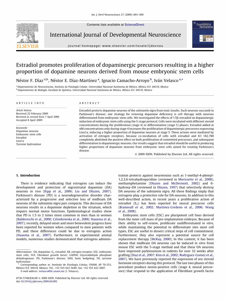
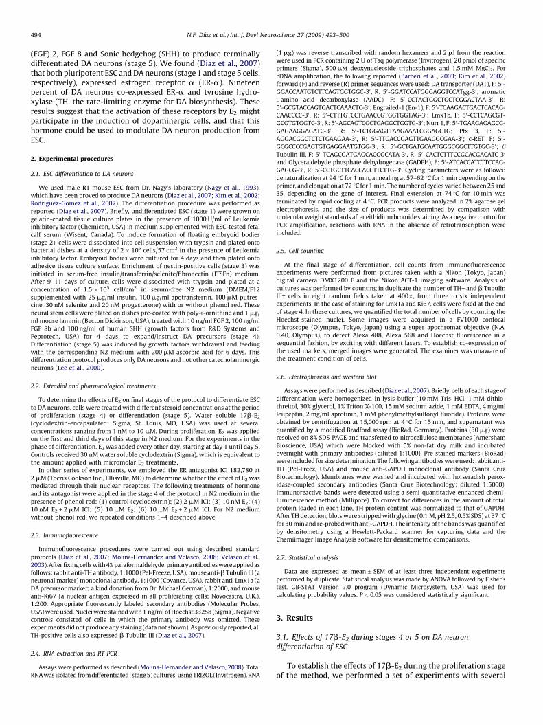
hormone concentrations in N2 medium together with FGF 2, FGF 8and SHH. All cultures started at the same initial density, wereincubated for 4 days in the presence of cytokines, with or without17b-E2, and then allowed to differentiate for 4–6 days in N2medium with ascorbic acid. Representative pictures of differen-tiated (day 6 of stage 5) cultures treated with 17b-E2 only duringstage 4 are shown to document that this hormone at nMconcentration is beneficial, and at mM doses deleterious, fordopaminergic induction (Fig. 1). We quantified the number ofneurons (b Tubulin III-positive cells) and DA (TH-positive) neuronsby performing double immunodetection of these markers incontrol and hormone-treated cultures. We found a significant 43%increase in the number of b Tubulin III-positive cells with the doseof 10 nM of 17b-E2 as compared with control (Fig. 2A). We alsoobserved statistically significant increases (76–96%) in the numberof TH-positive neurons with 1, 10 and 100 nM 17b-E2, relative tocontrol (Fig. 2A). When we used 10 mM 17b-E2, a significant 66%decrease in the number of TH+ neurons was observed (Fig. 2A) inabsence of apparent variations in total cell number after nucleistaining with Hoechst (first column of Fig. 1). Accordingly, wefound statistically significant increases in the percentage of TH-positive neurons (i.e., # of TH-positive/# b Tubulin III-positive cellsX 100) with 10 (46 � 4%) and 100 nM (47 � 2%) E2, and a decreasewith 10 mM (15 � 2%) E2, in comparison with control conditions(34 � 3%; Fig. 2C). Cells treated during stage 5 with 10 nM 17b-E2
showed augmented numbers of b Tubulin III-positive neurons(Fig. 2B). However, no significant differences were found in TH-positive cell number nor the percentage of TH-positive cells with thetested concentrations of 17b-E2 at this stage (Fig. 2B and D).
3.2. Estrogen receptor antagonism inhibits the increase in DA neurons
measured at stage 5 in N2 medium with or without phenol red
Once we established the effects of E2 in the number of DAneurons differentiated from ESC, we wanted to find out if such
Fig. 1. Effects of 17b-E2, added only in the phase of proliferation (stage 4) in N2 medi
Representative confocal images of immunofluorescence detection for b-Tubulin III (gree
total number of nuclei is shown in blue. Ten nM 17b-E2 caused an increase and 10 mM 1
with cyclodextrin. Scale bar applies to all pictures and represents 100 mm.
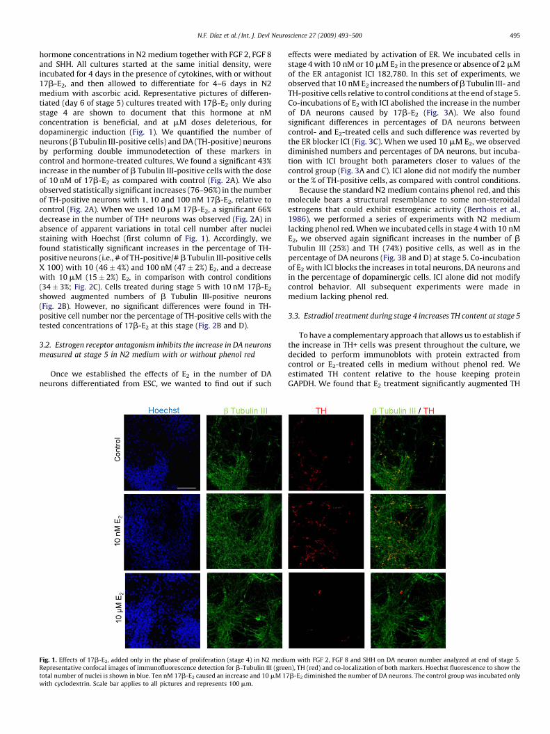
effects were mediated by activation of ER. We incubated cells instage 4 with 10 nM or 10 mM E2 in the presence or absence of 2 mMof the ER antagonist ICI 182,780. In this set of experiments, weobserved that 10 nM E2 increased the numbers of b Tubulin III- andTH-positive cells relative to control conditions at the end of stage 5.Co-incubations of E2 with ICI abolished the increase in the numberof DA neurons caused by 17b-E2 (Fig. 3A). We also foundsignificant differences in percentages of DA neurons betweencontrol- and E2-treated cells and such difference was reverted bythe ER blocker ICI (Fig. 3C). When we used 10 mM E2, we observeddiminished numbers and percentages of DA neurons, but incuba-tion with ICI brought both parameters closer to values of thecontrol group (Fig. 3A and C). ICI alone did not modify the numberor the % of TH-positive cells, as compared with control conditions.
Because the standard N2 medium contains phenol red, and thismolecule bears a structural resemblance to some non-steroidalestrogens that could exhibit estrogenic activity (Berthois et al.,1986), we performed a series of experiments with N2 mediumlacking phenol red. When we incubated cells in stage 4 with 10 nME2, we observed again significant increases in the number of bTubulin III (25%) and TH (74%) positive cells, as well as in thepercentage of DA neurons (Fig. 3B and D) at stage 5. Co-incubationof E2 with ICI blocks the increases in total neurons, DA neurons andin the percentage of dopaminergic cells. ICI alone did not modifycontrol behavior. All subsequent experiments were made inmedium lacking phenol red.
3.3. Estradiol treatment during stage 4 increases TH content at stage 5
To have a complementary approach that allows us to establish ifthe increase in TH+ cells was present throughout the culture, wedecided to perform immunoblots with protein extracted fromcontrol or E2-treated cells in medium without phenol red. Weestimated TH content relative to the house keeping proteinGAPDH. We found that E2 treatment significantly augmented TH
um with FGF 2, FGF 8 and SHH on DA neuron number analyzed at end of stage 5.
n), TH (red) and co-localization of both markers. Hoechst fluorescence to show the
7b-E2 diminished the number of DA neurons. The control group was incubated only
Fig. 2. Effects of 17b-E2 added during stage 4 (proliferation) in N2 medium with FGF 2, FGF 8 and SHH or stage 5 (differentiation) on DA neuron numbers analyzed at end of
stage 5. Quantification of the total number (A and B) of b-Tubulin III and TH-positive cells and the percentage of TH-positive neurons (C and D) from eight fields from three to
six independent experiments made in duplicate. Cells were incubated in N2 medium, with different E2 concentrations during proliferation (A and C) or differentiation stages
(B and D). Results are expressed as mean � SEM. *P < 0.05 and **P < 0.001 as compared with corresponding (cyclodextrin) group determined by ANOVA followed by Fisher’s test
for 3–6 independent experiments.
Fig. 3. Effects of 17b-E2 added at stage 4, in N2 medium in the presence of FGF 2, FGF 8 and SHH with or without phenol red, on DA neuron number, and its antagonism by ICI
182,780. Quantification of the total number (A and B) of b-Tubulin III and TH-positive cells and the percentage of TH-positive neurons (C and D) from eight fields from three to
five independent experiments made in duplicate. Cells were incubated in N2 medium with (A and C) or without (B and D) phenol red, at the indicated E2 concentrations during
proliferation stage. ICI 182,780 was added at 2 mM where indicated. Results are expressed as mean � SEM. *P < 0.05 and **P < 0.001 as compared with control (cyclodextrin)
group. &P < 0.05 relative to 10 nM E2 determined by ANOVA followed by Fisher’s test for 3–6 independent experiments.
N.F. Dıaz et al. / Int. J. Devl Neuroscience 27 (2009) 493–500496
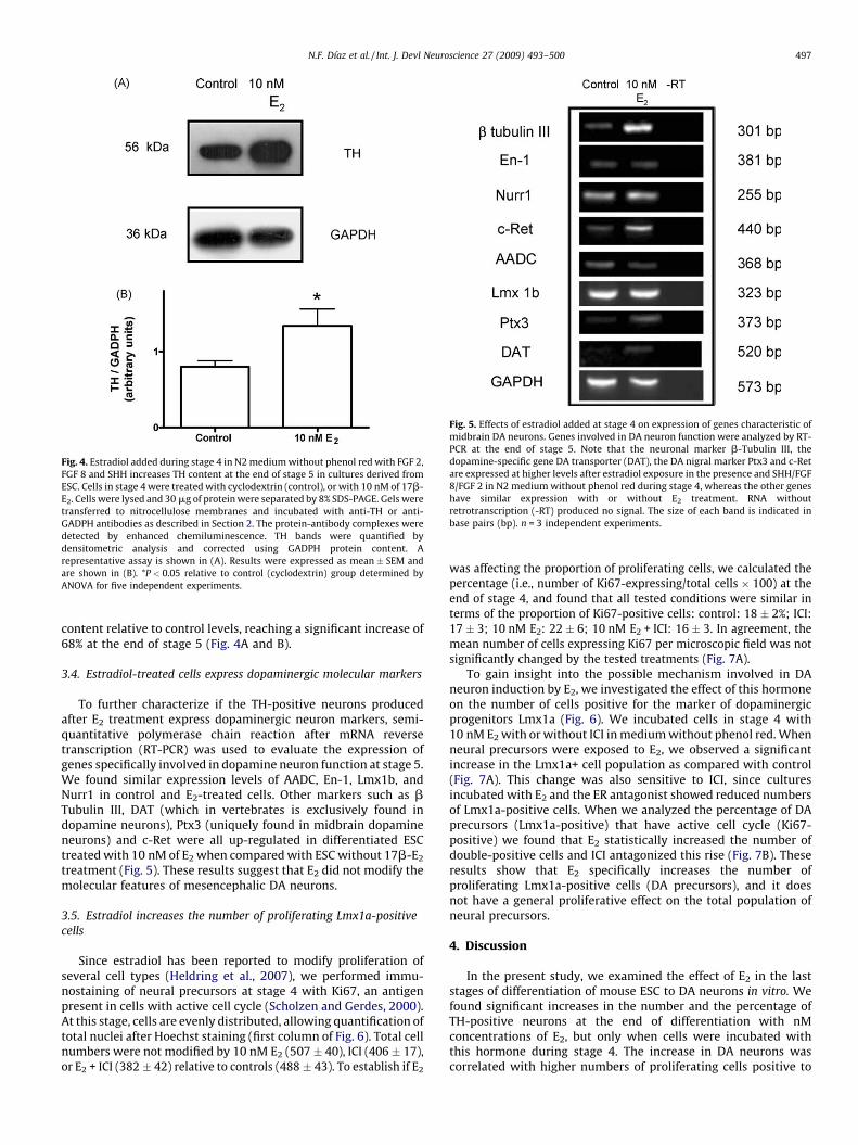
Fig. 4. Estradiol added during stage 4 in N2 medium without phenol red with FGF 2,
FGF 8 and SHH increases TH content at the end of stage 5 in cultures derived from
ESC. Cells in stage 4 were treated with cyclodextrin (control), or with 10 nM of 17b-
E2. Cells were lysed and 30 mg of protein were separated by 8% SDS-PAGE. Gels were
transferred to nitrocellulose membranes and incubated with anti-TH or anti-
GADPH antibodies as described in Section 2. The protein-antibody complexes were
detected by enhanced chemiluminescence. TH bands were quantified by
densitometric analysis and corrected using GADPH protein content. A
representative assay is shown in (A). Results were expressed as mean � SEM and
are shown in (B). *P < 0.05 relative to control (cyclodextrin) group determined by
ANOVA for five independent experiments.
Fig. 5. Effects of estradiol added at stage 4 on expression of genes characteristic of
midbrain DA neurons. Genes involved in DA neuron function were analyzed by RT-
PCR at the end of stage 5. Note that the neuronal marker b-Tubulin III, the
dopamine-specific gene DA transporter (DAT), the DA nigral marker Ptx3 and c-Ret
are expressed at higher levels after estradiol exposure in the presence and SHH/FGF
8/FGF 2 in N2 medium without phenol red during stage 4, whereas the other genes
have similar expression with or without E2 treatment. RNA without
retrotranscription (-RT) produced no signal. The size of each band is indicated in
base pairs (bp). n = 3 independent experiments.
N.F. Dıaz et al. / Int. J. Devl Neuroscience 27 (2009) 493–500 497
content relative to control levels, reaching a significant increase of68% at the end of stage 5 (Fig. 4A and B).
3.4. Estradiol-treated cells express dopaminergic molecular markers
To further characterize if the TH-positive neurons producedafter E2 treatment express dopaminergic neuron markers, semi-quantitative polymerase chain reaction after mRNA reversetranscription (RT-PCR) was used to evaluate the expression ofgenes specifically involved in dopamine neuron function at stage 5.We found similar expression levels of AADC, En-1, Lmx1b, andNurr1 in control and E2-treated cells. Other markers such as bTubulin III, DAT (which in vertebrates is exclusively found indopamine neurons), Ptx3 (uniquely found in midbrain dopamineneurons) and c-Ret were all up-regulated in differentiated ESCtreated with 10 nM of E2 when compared with ESC without 17b-E2
treatment (Fig. 5). These results suggest that E2 did not modify themolecular features of mesencephalic DA neurons.
3.5. Estradiol increases the number of proliferating Lmx1a-positive
cells
Since estradiol has been reported to modify proliferation ofseveral cell types (Heldring et al., 2007), we performed immu-nostaining of neural precursors at stage 4 with Ki67, an antigenpresent in cells with active cell cycle (Scholzen and Gerdes, 2000).At this stage, cells are evenly distributed, allowing quantification oftotal nuclei after Hoechst staining (first column of Fig. 6). Total cellnumbers were not modified by 10 nM E2 (507 � 40), ICI (406 � 17),or E2 + ICI (382 � 42) relative to controls (488 � 43). To establish if E2
was affecting the proportion of proliferating cells, we calculated thepercentage (i.e., number of Ki67-expressing/total cells � 100) at theend of stage 4, and found that all tested conditions were similar interms of the proportion of Ki67-positive cells: control: 18 � 2%; ICI:17 � 3; 10 nM E2: 22 � 6; 10 nM E2 + ICI: 16 � 3. In agreement, themean number of cells expressing Ki67 per microscopic field was notsignificantly changed by the tested treatments (Fig. 7A).
To gain insight into the possible mechanism involved in DAneuron induction by E2, we investigated the effect of this hormoneon the number of cells positive for the marker of dopaminergicprogenitors Lmx1a (Fig. 6). We incubated cells in stage 4 with10 nM E2 with or without ICI in medium without phenol red. Whenneural precursors were exposed to E2, we observed a significantincrease in the Lmx1a+ cell population as compared with control(Fig. 7A). This change was also sensitive to ICI, since culturesincubated with E2 and the ER antagonist showed reduced numbersof Lmx1a-positive cells. When we analyzed the percentage of DAprecursors (Lmx1a-positive) that have active cell cycle (Ki67-positive) we found that E2 statistically increased the number ofdouble-positive cells and ICI antagonized this rise (Fig. 7B). Theseresults show that E2 specifically increases the number ofproliferating Lmx1a-positive cells (DA precursors), and it doesnot have a general proliferative effect on the total population ofneural precursors.
4. Discussion
In the present study, we examined the effect of E2 in the laststages of differentiation of mouse ESC to DA neurons in vitro. Wefound significant increases in the number and the percentage ofTH-positive neurons at the end of differentiation with nMconcentrations of E2, but only when cells were incubated withthis hormone during stage 4. The increase in DA neurons wascorrelated with higher numbers of proliferating cells positive to
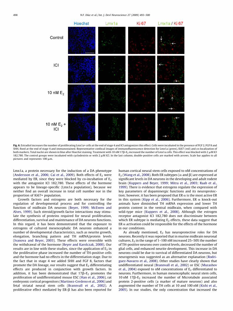
Fig. 6. Estradiol increases the number of proliferating Lmx1a+ cells at the end of stage 4 and ICI antagonizes this effect. Cells were incubated in the presence of FGF 2, FGF 8 and
SHH, fixed at the end of stage 4 and immunostained. Representative confocal images of immunofluorescence detection for Lmx1a (green), Ki67 (red) and co-localization of
both markers. Total nuclei are shown in blue after Hoechst staining. Treatment with 10 nM 17b-E2 increased the number of Lmx1a cells. This effect was blocked with 2 mM ICI
182,780. The control groups were incubated with cyclodextrin or with 2 mM ICI. In the last column, double-positive cells are marked with arrows. Scale bar applies to all
pictures and represents 100 mm.
N.F. Dıaz et al. / Int. J. Devl Neuroscience 27 (2009) 493–500498
Lmx1a, a protein necessary for the induction of a DA phenotype(Andersson et al., 2006; Cai et al., 2009). Both effects of E2 weremediated by ER, since they were blocked by co-incubation of E2
with the antagonist ICI 182,780. These effects of the hormoneappears to be lineage-specific (Lmx1a population), because weneither find an overall increase in total cell number nor in theproportion of Ki67+ population.
Growth factors and estrogens are both necessary for theregulation of developmental process and for controlling thefunction of midbrain DA neurons (Beyer, 1999; McEwen andAlves, 1999). Such steroid/growth factor interactions may stimu-late the synthesis of proteins required for neural proliferation,differentiation, survival and maintenance of DA neurons functions.In this regard, it has been demonstrated that the exposure toestrogens of cultured mesencephalic DA neurons enhanced anumber of developmental characteristics, such as neurite growth,elongation, branching pattern and TH mRNA/protein levels(Ivanova and Beyer, 2003). These effects were reversible withthe withdrawal of the hormone (Beyer and Karolczak, 2000). Ourresults are in line with these studies, since the application of E2 inthe proliferative phase increased the number of TH-positive cellsand the hormone had no effects in the differentiation stage. Due tothe fact that in stage 4 we added SHH and FGF 8, factors thatpromote the DA lineage, our results suggest that E2 differentiatingeffects are produced in conjunction with growth factors. Inaddition, it has been demonstrated that 17b-E2 promotes theproliferation of undifferentiated mouse ESC (Han et al., 2006), ratembryonic cortical progenitors (Martinez-Cerdeno et al., 2006) andfetal striatal neural stem cells (Brannvall et al., 2002). Aproliferative effect mediated by ER-b has also been reported for
human cortical neural stem cells exposed to nM concentrations ofE2 (Wang et al., 2008). Both ER subtypes (a and b) are expressed atsignificant levels in DA neurons in the developing and adult rodentbrain (Kuppers and Beyer, 1999; Mitra et al., 2003; Raab et al.,1999). There is evidence that estrogens regulate the expression ofkey parameters of dopaminergic functions and its neuroprotec-tion; however, it has been proposed that ER a is the most active ERin this system (Kipp et al., 2006). Furthermore, ER a knock-outanimals have diminished TH mRNA expression and lower THprotein content in the ventral midbrain, when compared withwild-type mice (Kuppers et al., 2008). Although the estrogenreceptor antagonist ICI 182,780 does not discriminate betweenwhich ER subtype is mediating E2 effects, these data suggest thatER a activation could be responsible for the effects of the hormonein our conditions.
As already mentioned, E2 has neuroprotective roles for DAneurons. Recently it was reported that in murine midbrain neuronalcultures, E2 in the range of 1–100 nM increased 25–50% the numberof TH-positive neurons over control levels, decreased the number ofglial cells, and enhanced neurite development. This increase in DAneurons could be due to survival of differentiated DA neurons, butneurogenesis was suggested as an alternative explanation (Rodri-guez-Navarro et al., 2008). Other studies have clearly shown thatundifferentiated neural (Brannvall et al., 2002) or ESC (Murashovet al., 2004) exposed to nM concentrations of E2 differentiated toneurons. Furthermore, in human mesencephalic neural stem cells,10 nM 17b-E2 increased the number of Microtubule associatedprotein 2-positive cells (a marker of mature neurons) and alsoaugmented the number of TH cells at 10 and 100 nM (Kishi et al.,2005). In our studies, the only concentration that increased the
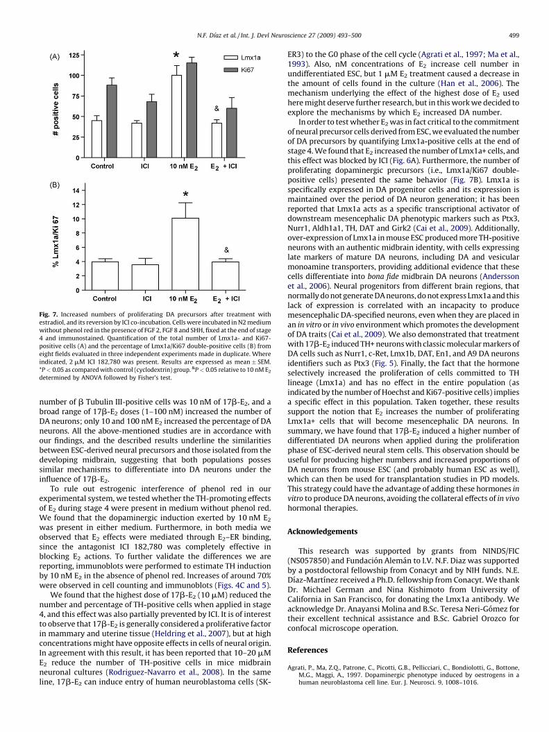
Fig. 7. Increased numbers of proliferating DA precursors after treatment with
estradiol, and its reversion by ICI co-incubation. Cells were incubated in N2 medium
without phenol red in the presence of FGF 2, FGF 8 and SHH, fixed at the end of stage
4 and immunostained. Quantification of the total number of Lmx1a- and Ki67-
positive cells (A) and the percentage of Lmx1a/Ki67 double-positive cells (B) from
eight fields evaluated in three independent experiments made in duplicate. Where
indicated, 2 mM ICI 182,780 was present. Results are expressed as mean � SEM.
*P < 0.05 as compared with control (cyclodextrin) group. &P < 0.05 relative to 10 nM E2
determined by ANOVA followed by Fisher’s test.
N.F. Dıaz et al. / Int. J. Devl Neuroscience 27 (2009) 493–500 499
number of b Tubulin III-positive cells was 10 nM of 17b-E2, and abroad range of 17b-E2 doses (1–100 nM) increased the number ofDA neurons; only 10 and 100 nM E2 increased the percentage of DAneurons. All the above-mentioned studies are in accordance withour findings, and the described results underline the similaritiesbetween ESC-derived neural precursors and those isolated from thedeveloping midbrain, suggesting that both populations possessimilar mechanisms to differentiate into DA neurons under theinfluence of 17b-E2.
To rule out estrogenic interference of phenol red in ourexperimental system, we tested whether the TH-promoting effectsof E2 during stage 4 were present in medium without phenol red.We found that the dopaminergic induction exerted by 10 nM E2
was present in either medium. Furthermore, in both media weobserved that E2 effects were mediated through E2–ER binding,since the antagonist ICI 182,780 was completely effective inblocking E2 actions. To further validate the differences we arereporting, immunoblots were performed to estimate TH inductionby 10 nM E2 in the absence of phenol red. Increases of around 70%were observed in cell counting and immunoblots (Figs. 4C and 5).
We found that the highest dose of 17b-E2 (10 mM) reduced thenumber and percentage of TH-positive cells when applied in stage4, and this effect was also partially prevented by ICI. It is of interestto observe that 17b-E2 is generally considered a proliferative factorin mammary and uterine tissue (Heldring et al., 2007), but at highconcentrations might have opposite effects in cells of neural origin.In agreement with this result, it has been reported that 10–20 mME2 reduce the number of TH-positive cells in mice midbrainneuronal cultures (Rodriguez-Navarro et al., 2008). In the sameline, 17b-E2 can induce entry of human neuroblastoma cells (SK-
ER3) to the G0 phase of the cell cycle (Agrati et al., 1997; Ma et al.,1993). Also, nM concentrations of E2 increase cell number inundifferentiated ESC, but 1 mM E2 treatment caused a decrease inthe amount of cells found in the culture (Han et al., 2006). Themechanism underlying the effect of the highest dose of E2 usedhere might deserve further research, but in this work we decided toexplore the mechanisms by which E2 increased DA number.
In order to test whether E2 was in fact critical to the commitmentof neural precursor cells derived from ESC, we evaluated the numberof DA precursors by quantifying Lmx1a-positive cells at the end ofstage 4. We found that E2 increased the number of Lmx1a+ cells, andthis effect was blocked by ICI (Fig. 6A). Furthermore, the number ofproliferating dopaminergic precursors (i.e., Lmx1a/Ki67 double-positive cells) presented the same behavior (Fig. 7B). Lmx1a isspecifically expressed in DA progenitor cells and its expression ismaintained over the period of DA neuron generation; it has beenreported that Lmx1a acts as a specific transcriptional activator ofdownstream mesencephalic DA phenotypic markers such as Ptx3,Nurr1, Aldh1a1, TH, DAT and Girk2 (Cai et al., 2009). Additionally,over-expression of Lmx1a in mouse ESC produced more TH-positiveneurons with an authentic midbrain identity, with cells expressinglate markers of mature DA neurons, including DA and vesicularmonoamine transporters, providing additional evidence that thesecells differentiate into bona fide midbrain DA neurons (Anderssonet al., 2006). Neural progenitors from different brain regions, thatnormally do not generate DA neurons, do not express Lmx1a and thislack of expression is correlated with an incapacity to producemesencephalic DA-specified neurons, even when they are placed inan in vitro or in vivo environment which promotes the developmentof DA traits (Cai et al., 2009). We also demonstrated that treatmentwith 17b-E2 induced TH+ neurons with classic molecular markers ofDA cells such as Nurr1, c-Ret, Lmx1b, DAT, En1, and A9 DA neuronsidentifiers such as Ptx3 (Fig. 5). Finally, the fact that the hormoneselectively increased the proliferation of cells committed to THlineage (Lmx1a) and has no effect in the entire population (asindicated by the number of Hoechst and Ki67-positive cells) impliesa specific effect in this population. Taken together, these resultssupport the notion that E2 increases the number of proliferatingLmx1a+ cells that will become mesencephalic DA neurons. Insummary, we have found that 17b-E2 induced a higher number ofdifferentiated DA neurons when applied during the proliferationphase of ESC-derived neural stem cells. This observation should beuseful for producing higher numbers and increased proportions ofDA neurons from mouse ESC (and probably human ESC as well),which can then be used for transplantation studies in PD models.This strategy could have the advantage of adding these hormones in
vitro to produce DA neurons, avoiding the collateral effects of in vivo
hormonal therapies.
Acknowledgements
This research was supported by grants from NINDS/FIC(NS057850) and Fundacion Aleman to I.V. N.F. Diaz was supportedby a postdoctoral fellowship from Conacyt and by NIH funds. N.E.Dıaz-Martınez received a Ph.D. fellowship from Conacyt. We thankDr. Michael German and Nina Kishimoto from University ofCalifornia in San Francisco, for donating the Lmx1a antibody. Weacknowledge Dr. Anayansi Molina and B.Sc. Teresa Neri-Gomez fortheir excellent technical assistance and B.Sc. Gabriel Orozco forconfocal microscope operation.
References
Agrati, P., Ma, Z.Q., Patrone, C., Picotti, G.B., Pellicciari, C., Bondiolotti, G., Bottone,M.G., Maggi, A., 1997. Dopaminergic phenotype induced by oestrogens in ahuman neuroblastoma cell line. Eur. J. Neurosci. 9, 1008–1016.
N.F. Dıaz et al. / Int. J. Devl Neuroscience 27 (2009) 493–500500
Andersson, E., Tryggvason, U., Deng, Q., Friling, S., Alekseenko, Z., Robert, B.,Perlmann, T., Ericson, J., 2006. Identification of intrinsic determinants of mid-brain dopamine neurons. Cell 124, 393–405.
Baldereschi, M., Di Carlo, A., Rocca, W.A., Vanni, P., Maggi, S., Perissinotto, E.,Grigoletto, F., Amaducci, L., Inzitari, D., 2000. Parkinson’s disease and parkin-sonism in a longitudinal study: two-fold higher incidence in men ILSA WorkingGroup. Italian Longitudinal Study on Aging. Neurology 55, 1358–1363.
Barberi, T., Klivenyi, P., Calingasan, N.Y., Lee, H., Kawamata, H., Loonam, K., Perrier,A.L., Bruses, J., Rubio, M.E., Topf, N., Tabar, V., Harrison, N.L., Beal, M.F., Moore,M.A., Studer, L., 2003. Neural subtype specification of fertilization and nucleartransfer embryonic stem cells and application in parkinsonian mice. Nat.Biotechnol. 21, 1200–1207.
Berthois, Y., Katzenellenbogen, J.A., Katzenellenbogen, B.S., 1986. Phenol red intissue culture media is a weak estrogen: implications concerning the study ofestrogen-responsive cells in culture. Proc. Natl. Acad. Sci. U.S.A. 83, 2496–2500.
Beyer, C., 1999. Estrogen and the developing mammalian brain. Anat. Embryol.(Berl) 199, 379–390.
Beyer, C., Karolczak, M., 2000. Estrogenic stimulation of neurite growth in midbraindopaminergic neurons depends on cAMP/protein kinase A signalling. J. Neu-rosci. Res. 59, 107–116.
Brannvall, K., Korhonen, L., Lindholm, D., 2002. Estrogen-receptor-dependent reg-ulation of neural stem cell proliferation and differentiation. Mol. Cell. Neurosci.21, 512–520.
Cai, J., Donaldson, A., Yang, M., German, M.S., Enikolopov, G., Iacovitti, L., 2009. Therole of Lmx1a in the differentiation of human embryonic stem cells intomidbrain dopamine neurons in culture and after transplantation into a Par-kinson’s disease model. Stem Cells 27, 220–229.
Czlonkowska, A., Ciesielska, A., Gromadzka, G., Kurkowska-Jastrzebska, I., 2006.Gender differences in neurological disease: role of estrogens and cytokines.Endocrine 29, 243–256.
Diaz, N.F., Guerra-Arraiza, C., Diaz-Martinez, N.E., Salazar, P., Molina-Hernandez, A.,Camacho-Arroyo, I., Velasco, I., 2007. Changes in the content of estrogen alphaand progesterone receptors during differentiation of mouse embryonic stemcells to dopamine neurons. Brain Res. Bull. 73, 75–80.
Dluzen, D., 1997. Estrogen decreases corpus striatal neurotoxicity in response to 6-hydroxydopamine. Brain Res. 767, 340–344.
Dluzen, D.E., McDermott, J.L., 2002. Estrogen, anti-estrogen, and gender: differencesin methamphetamine neurotoxicity. Ann. N.Y. Acad. Sci. 965, 136–156.
Haaxma, C.A., Bloem, B.R., Borm, G.F., Oyen, W.J., Leenders, K.L., Eshuis, S., Booij, J.,Dluzen, D.E., Horstink, M.W., 2007. Gender differences in Parkinson’s disease. J.Neurol. Neurosurg. Psychiatry 78, 819–824.
Han, H.J., Heo, J.S., Lee, Y.J., 2006. Estradiol-17beta stimulates proliferation of mouseembryonic stem cells: involvement of MAPKs and CDKs as well as protoonco-genes. Am. J. Physiol. Cell. Physiol. 290, C1067–1075.
Heldring, N., Pike, A., Andersson, S., Matthews, J., Cheng, G., Hartman, J., Tujague, M.,Strom, A., Treuter, E., Warner, M., Gustafsson, J.A., 2007. Estrogen receptors:how do they signal and what are their targets. Physiol. Rev. 87, 905–931.
Ivanova, T., Beyer, C., 2003. Estrogen regulates tyrosine hydroxylase expression inthe neonate mouse midbrain. J. Neurobiol. 54, 638–647.
Kim, J.H., Auerbach, J.M., Rodriguez-Gomez, J.A., Velasco, I., Gavin, D., Lumelsky, N.,Lee, S.H., Nguyen, J., Sanchez-Pernaute, R., Bankiewicz, K., McKay, R., 2002.Dopamine neurons derived from embryonic stem cells function in an animalmodel of Parkinson’s disease. Nature 418, 50–56.
Kipp, M., Karakaya, S., Pawlak, J., Araujo-Wright, G., Arnold, S., Beyer, C., 2006.Estrogen and the development and protection of nigrostriatal dopaminergicneurons: concerted action of a multitude of signals, protective molecules, andgrowth factors. Front. Neuroendocrinol. 27, 376–390.
Kishi, Y., Takahashi, J., Koyanagi, M., Morizane, A., Okamoto, Y., Horiguchi, S.,Tashiro, K., Honjo, T., Fujii, S., Hashimoto, N., 2005. Estrogen promotes differ-
entiation and survival of dopaminergic neurons derived from human neuralstem cells. J. Neurosci. Res. 79, 279–286.
Kuppers, E., Beyer, C., 1999. Expression of estrogen receptor-alpha and beta mRNAin the developing and adult mouse striatum. Neurosci. Lett. 276, 95–98.
Kuppers, E., Krust, A., Chambon, P., Beyer, C., 2008. Functional alterations of thenigrostriatal dopamine system in estrogen receptor-alpha knockout (ERKO)mice. Psychoneuroendocrinology 33, 832–838.
Lee, S.H., Lumelsky, N., Studer, L., Auerbach, J.M., McKay, R.D., 2000. Efficientgeneration of midbrain and hindbrain neurons from mouse embryonic stemcells. Nat. Biotechnol. 18, 675–679.
Liu, B., Dluzen, D.E., 2007. Oestrogen and nigrostriatal dopaminergic neurodegen-eration: animal models and clinical reports of Parkinson’s disease. Clin. Exp.Pharmacol. Physiol. 34, 555–565.
Ma, Z.Q., Spreafico, E., Pollio, G., Santagati, S., Conti, E., Cattaneo, E., Maggi, A., 1993.Activated estrogen receptor mediates growth arrest and differentiation of aneuroblastoma cell line. Proc. Natl. Acad. Sci. U.S.A. 90, 3740–3744.
Martinez-Cerdeno, V., Noctor, S.C., Kriegstein, A.R., 2006. Estradiol stimulatesprogenitor cell division in the ventricular and subventricular zones of theembryonic neocortex. Eur. J. Neurosci. 24, 3475–3488.
McEwen, B.S., Alves, S.E., 1999. Estrogen actions in the central nervous system.Endocr. Rev. 20, 279–307.
McKay, R., 2000. Stem cells—hype and hope. Nature 406, 361–364.Mitra, S.W., Hoskin, E., Yudkovitz, J., Pear, L., Wilkinson, H.A., Hayashi, S., Pfaff, D.W.,
Ogawa, S., Rohrer, S.P., Schaeffer, J.M., McEwen, B.S., Alves, S.E., 2003. Immu-nolocalization of estrogen receptor beta in the mouse brain: comparison withestrogen receptor alpha. Endocrinology 144, 2055–2067.
Molina-Hernandez, A., Velasco, I., 2008. Histamine induces neural stem cell pro-liferation and neuronal differentiation by activation of distinct histaminereceptors. J Neurochem 106, 706–717.
Morissette, M., Al Sweidi, S., Callier, S., Di Paolo, T., 2008. Estrogen and SERMneuroprotection in animal models of Parkinson’s disease. Mol. Cell. Endocrinol.290, 60–69.
Murashov, A.K., Pak, E.S., Hendricks, W.A., Tatko, L.M., 2004. 17beta-Estradiolenhances neuronal differentiation of mouse embryonic stem cells. FEBS Lett.569, 165–168.
Nagy, A., Rossant, J., Nagy, R., Abramow-Newerly, W., Roder, J.C., 1993. Derivation ofcompletely cell culture-derived mice from early-passage embryonic stem cells.Proc. Natl. Acad. Sci. U.S.A. 90, 8424–8428.
Raab, H., Karolczak, M., Reisert, I., Beyer, C., 1999. Ontogenetic expression andsplicing of estrogen receptor-alpha and beta mRNA in the rat midbrain. Neu-rosci. Lett. 275, 21–24.
Rodriguez-Gomez, J.A., Lu, J.Q., Velasco, I., Rivera, S., Zoghbi, S.S., Liow, J.S., Musa-chio, J.L., Chin, F.T., Toyama, H., Seidel, J., Green, M.V., Thanos, P.K., Ichise, M.,Pike, V.W., Innis, R.B., McKay, R.D., 2007. Persistent dopamine functions ofneurons derived from embryonic stem cells in a rodent model of Parkinsondisease. Stem Cells 25, 918–928.
Rodriguez-Navarro, J.A., Solano, R.M., Casarejos, M.J., Gomez, A., Perucho, J., deYebenes, J.G., Mena, M.A., 2008. Gender differences and estrogen effects inparkin null mice. J. Neurochem. 106, 2143–2157.
Scholzen, T., Gerdes, J., 2000. The Ki-67 protein: from the known and the unknown.J. Cell. Physiol. 182, 311–322.
Velasco, I., Velasco-Velazquez, M.A., Salazar, P., Lajud, N., Tapia, R., 2003. Influenceof serum-free medium on the expression of glutamate transporters and thesusceptibility to glutamate toxicity in cultured cortical neurons. J. Neurosci. Res.71, 811–818.
Wang, J.M., Liu, L., Brinton, R.D., 2008. Estradiol-17beta-induced human neuralprogenitor cell proliferation is mediated by an estrogen receptor beta-phos-phorylated extracellularly regulated kinase pathway. Endocrinology 149,208–218.