Estimation of leaf area in selected Hypericum species
11
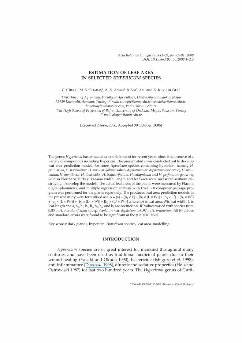
Acta Botanica Hungarica 50(1–2), pp. 81–91, 2008 DOI: 10.1556/ABot.50.2008.1–2.5 ESTIMATION OF LEAF AREA IN SELECTED HYPERICUM SPECIES C. ÇÝRAK 1 , M. S. ODABAŞ 1 , A. K. AYAN 2 , B. SAÐLAM 1 and K. KEVSEROÐLU 1 1 Department of Agronomy, Faculty of Agriculture, University of Ondokuz Mayýs 55139 Kurupelit, Samsun, Turkey; E-mail: [email protected]; [email protected]; [email protected]; [email protected] 2 The High School of Profession of Bafra, University of Ondokuz Mayýs, Samsun, Turkey E-mail: [email protected] (Received 2 June, 2006; Accepted 30 October, 2006) The genus Hypericum has attracted scientific interest for recent years, since it is a source of a variety of compounds including hypericin. The present study was conducted out to develop leaf area prediction models for some Hypericum species containing hypericin, namely H. pruinatum, H. perfoliatum, H. aviculariifolium subsp. depilatum var. depilatum (endemic), H. mon- tanum, H. montbretii, H. linarioides, H. triquetrifolium, H. bithynicum and H. perforatum growing wild in Northern Turkey. Lamina width, length and leaf area were measured without de- stroying to develop the models. The actual leaf areas of the plants were measured by Placom digital planimeter, and multiple regression analysis with Excel 7.0 computer package pro- gram was performed for the plants separately. The produced leaf area prediction models in the present study were formulised as LA = (a) + (b 1 × L) + [b 2 × (L × W)] + (b 3 ×L 2 ) + (b 4 ×W 2 ) + [b 5 × (L × W 2 )] + [b 6 × (L 2 × W)] + [b 7 × (L 2 +W 2 )] where LA is leaf area, Wis leaf width, L is leaf length and a, b 1 ,b 2 ,b 3 ,b 4 ,b 5 ,b 6 , and b 7 are coefficients. R 2 values varied with species from 0.80 in H. aviculariifolium subsp. depilatum var. depilatum to 0.97 in H. pruinatum. All R 2 values and standard errors were found to be significant at the p < 0.001 level. Key words: dark glands, hypericin, Hypericum species, leaf area, modelling INTRODUCTION Hypericum species are of great interest for mankind throughout many centuries and have been used as traditional medicinal plants due to their wound-healing (Yazaki and Okuda 1990), bactericide (Ishiguro et al. 1998), anti-inflammatory (Dias et al. 1998), diuretic and sedative properties (Holz and Ostrowiski 1987) for last two hundred years. The Hypericum genus of Gutti- 0236–6495/$ 20.00 © 2008 Akadémiai Kiadó, Budapest
-
Upload
xn--om-yka -
Category
Documents
-
view
3 -
download
0
Transcript of Estimation of leaf area in selected Hypericum species
Acta Botanica Hungarica 50(1–2), pp. 81–91, 2008DOI: 10.1556/ABot.50.2008.1–2.5
ESTIMATION OF LEAF AREAIN SELECTED HYPERICUM SPECIES
C. ÇÝRAK1, M. S. ODABAŞ1, A. K. AYAN2, B. SAÐLAM1 and K. KEVSEROÐLU1
1Department of Agronomy, Faculty of Agriculture, University of Ondokuz Mayýs55139 Kurupelit, Samsun, Turkey; E-mail: [email protected]; [email protected];
[email protected]; [email protected] High School of Profession of Bafra, University of Ondokuz Mayýs, Samsun, Turkey
E-mail: [email protected]
(Received 2 June, 2006; Accepted 30 October, 2006)
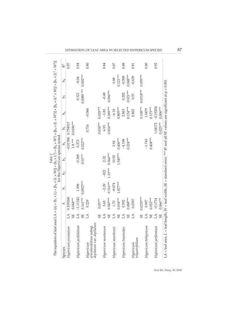
The genus Hypericum has attracted scientific interest for recent years, since it is a source of avariety of compounds including hypericin. The present study was conducted out to developleaf area prediction models for some Hypericum species containing hypericin, namely H.pruinatum, H. perfoliatum, H. aviculariifolium subsp. depilatum var. depilatum (endemic), H. mon-tanum, H. montbretii, H. linarioides, H. triquetrifolium, H. bithynicum and H. perforatum growingwild in Northern Turkey. Lamina width, length and leaf area were measured without de-stroying to develop the models. The actual leaf areas of the plants were measured by Placomdigital planimeter, and multiple regression analysis with Excel 7.0 computer package pro-gram was performed for the plants separately. The produced leaf area prediction models inthe present study were formulised as LA = (a) + (b1 × L) + [b2 × (L × W)] + (b3 × L2) + (b4 × W2)+ [b5 × (L × W2)] + [b6 × (L2 × W)] + [b7 × (L2 + W2)] where LA is leaf area, Wis leaf width, L isleaf length and a, b1, b2, b3, b4, b5, b6, and b7 are coefficients. R2 values varied with species from0.80 in H. aviculariifolium subsp. depilatum var. depilatum to 0.97 in H. pruinatum. All R2 valuesand standard errors were found to be significant at the p < 0.001 level.
Key words: dark glands, hypericin, Hypericum species, leaf area, modelling
INTRODUCTION
Hypericum species are of great interest for mankind throughout manycenturies and have been used as traditional medicinal plants due to theirwound-healing (Yazaki and Okuda 1990), bactericide (Ishiguro et al. 1998),anti-inflammatory (Dias et al. 1998), diuretic and sedative properties (Holz andOstrowiski 1987) for last two hundred years. The Hypericum genus of Gutti-
0236–6495/$ 20.00 © 2008 Akadémiai Kiadó, Budapest
ferae is represented in Turkey by 89 species of which 43 are endemic. This ge-nus is widespread in Turkey. The most abundant and well-known species is H.perforatum L. (Davis 1988). Use of the crude extract of this plant as especially anantidepressant is very popular today (Biffignandi and Bilia 2000).
The biological activities of the genus Hypericum derived from mainly theirhypericin contents (Sakar and Tamer 1990, Diwu 1995, Kubin et al. 1996, Gree-son et al. 2001, Agostinis et al. 2002) and total hypericin content of products hasbeen used for standardisation purposes within the botanical industry (Nahr-stedt and Butterweck 1997). Hypericin is found in dark glands of leaves themost intensively (Ciccarelli et al. 2001). That is why; leaves are the most impor-tant organs of these plants. Of all Hypericum, only a few have been reported tocontain hypericin to date (Gerassim and Kitanov 2001, Ferraz et al. 2002). AllHypericum species evaluated in the present study namely, H. pruinatum Boiss.et Bal., H. perfoliatum L., H. aviculariifolium Jaup. et Spach subsp. depilatum (Freynet Bornm.) Robson var. depilatum (endemic), H. montanum L., H. montbretiiSpach, H. linarioides Bosse, H. triquetrifolium Turra, H. bithynicum Boiss. and H.perforatum L. contain hypericin in various levels (Kitanov 2001, Ayan et al.2004).
Leaf area has been measured in experiments concerning some physiolog-ical phenomenon such as light, photosynthesis, respiration, plant water con-sumption and transpiration (Rieger and Duemmel 1992, Horsley and Gott-schalk 1993, Gottschalk 1994, Kersteins and Hawes 1994, Picchioni and Wein-baum 1995, Uzun 1996, Centritto et al. 2000). In addition, leaf number and areaof a plant have an important role in some cultural practices such as training,pruning, irrigation, fertilisation, etc. The leaf area estimation models aiming topredict leaf area non-destructively can provide researches with many advan-tages in agricultural experiments. Moreover, these kinds of models enable re-searchers to carry out leaf area measurements on the same plants over thecourse of a study, resulting in reduced experimental variability (Gamiely et al.1991, NeSmith 1991, 1992). Leaf area can be measured by using expensive in-struments and/or predictive models. Recently, new instruments, tools andmachines such as hand scanners and laser optical apparatuses have been de-veloped for leaf and fruit measurements. These are very expensive and com-plex devices for both basic and simple studies. Furthermore, non-destructiveestimation of leaf area saves time as compared with geometrical measure-ments (Robbins and Pharr 1987). For this reason, several leaf area predictionmodels were produced for some plant species in previous studies. But, to theauthors’ knowledge, there are no published reports concerning leaf area pre-diction model for Hypericum plants. Due to the lack of such information, in thepresent study, we aimed to develop reliable equations that allow for the non-
84 ÇÝRAK, C., ODABAÞ, M. S., AYAN, A. K., SAÐLAM, B. AND KEVSEROÐLU, K.
Acta Bot. Hung. 50, 2008
destructive estimation of leaf area through linear measurements of the afore-said species of Hypericum.
MATERIALS AND METHODS
Brief description of plant materials
H. pruinatum: Stems 15–35 cm, erect or ascending from a rooting and branch-ing base, pruinose. Leaves on main stems 10–35 mm, oblong to elliptic,pruinose. H. perfoliatum: Stems 15–80 cm, erect or decumbent. Leaves 13–60mm, ovate to triangular-lanceolate or rarely linear, the upper most sometimesblack-glandular-ciliate, usually densely pellucid-dotted, with obscure reticu-late venation. H. aviculariifolium subsp. depilatum var. depilatum: Stems 5–60cm, erect or prostrate, branching and sometimes rooting at the base. Leaves5–35 mm, oblong or linear to elliptic or obovate, with intramarginal blackglands, glabrous or shortly pubescent. H. montanum: Stems 20–80 cm, erect,glabrous. Leaves 20–70 mm, ovate to lanceolate or oblong-elliptic, scabrid be-neath or rarely wholly glabrous. H. montbretii: Stems 15–60 cm, erect or decum-bent, rarely rooting. Leaves 15–55 mm, ovate to oblong or triangular-lanceo-late, the upper most sometimes black-glandular-denticulate. H. linarioides:Stems 5–33 cm, erect or ascending from a rooting and branching base, gla-brous. Leaves 5–30 mm, oblong to elliptic to linear, often revolute and gla-brous. H. triquetrifolium: Stems 15–55 cm, erect or decumbent, with branchesspreading usually forming a pyramid. Leaves 3–20 mm, triangular-lanceolateor rarely narrowly ovate to linear-oblong, amplexicaul, undulate, sometimeswith medium to small pellucid dots. H. bithynicum: Stems 10–60 cm, erect orascending, rooting. Leaves 10–55 mm, ovate or ovate-oblong to suborbicular,entire without pellucid dots or with a few in the upper most pairs, sometimeswith superficial black dots. H. perforatum: Stems 10–110 cm, erect, sometimesrooting. Leaves 5–30 mm, narrowly ovate or lanceolate to elliptic oblong, al-ways with large pellucid dots (Davis 1988).
All plants described above grow wild in Black Sea region of Turkey andthey were identified by Dr Hasan Korkmaz, Department of Biology, Univer-sity of 19 Mayis, Samsun, Turkey.
Experimental procedures
Leaf samples were randomly taken from wild populations of the Hyperi-cum plants tested between March and August 2004 at a five time intervals. Inthis period, 60 leaves were collected for each plant within the first three day of
ESTIMATION OF LEAF AREA IN SELECTED HYPERICUM SPECIES 85
Acta Bot. Hung. 50, 2008
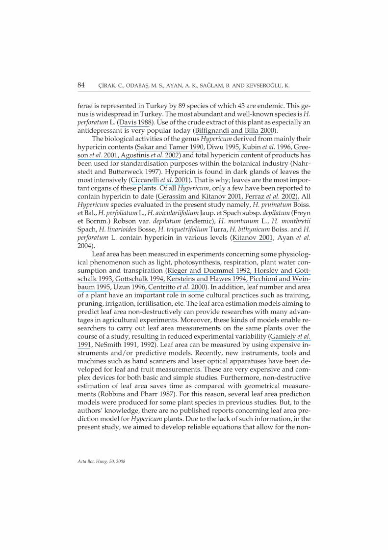
April, May, June, July and August to catch the different phases of leaf morpho-genesis. Thus, 300 leaf samples for each species and a total of 2,700 leaves foreight species were processed at the same day as they were collected in the fol-lowing manner. First, they were placed on the photocopier desktop by holdingflat and secure and copied on A3 sheet (at 1:1 ratio). Second, a Placom digitalplanimeter (Sokkisha Planimeter Inc., Model KP-90) was used to measure ac-tual leaf area of the copy. Selection of leaf dimensions for measurement wasgoverned by variation in leaf characteristics (e.g. size, shape, and symmetry)and practical constraints (e.g. ease and accuracy of measurements under fieldconditions). Given these concerns, maximum leaf width (W) and length (L)were chosen to correlate with leaf area. Leaf width (cm) was measured fromtip to tip at the widest part of the lamina and leaf length (cm) was measuredfrom lamina tip to the point of petiole intersection along the midrib (Fig. 1).The leaf positions were selected with regard to points that could be easily iden-tified and used to facilitate the measurement of leaf length and width.
86 ÇÝRAK, C., ODABAÞ, M. S., AYAN, A. K., SAÐLAM, B. AND KEVSEROÐLU, K.
Acta Bot. Hung. 50, 2008
Fig. 1. Leaf diagram of H. pruinatum (1), H. perfoliatum (2), H. aviculariifolium subsp. depilatumvar. depilatum (3), H. montanum (4), H. montbretii (5), H. linarioides (6), H. triquetrifolium (7), H.bithynicum (8) and H. perforatum (9) showing the position of leaf length (L) and leaf width
(W). Bars seen at the lower right corner of each figure represent 1 cm
ESTIMATION OF LEAF AREA IN SELECTED HYPERICUM SPECIES 87
Acta Bot. Hung. 50, 2008
Tabl
e1
The
equa
tion
ofle
afar
eaLA
=(a
)+(b
1×
L)+
[b2
×(L
×W
)]+
(b3
×L2 )+
(b4
×W
2 )+[b
5×
(L×
W2 )]
+[b
6×
(L2
×W
)]+
[b7
×(L
2+
W2 )]
for
the
Hyp
eric
umsp
ecie
ste
sted
Spec
ies
Ab 1
b 2b 3
b 4b 5
b 6b 7
b 8R
2
Hyp
eric
umpr
uina
tum
LA0.
1092
68–0
.013
060.
7549
170.
97SE
0.04
4***
1.8
***
0.01
06**
*H
yper
icum
perf
olia
tum
LA–1
.113
421.
596
–0.3
690.
373
0.32
1–0
.04
0.94
SE0.
161*
**0.
0927
***
0.01
***
0.02
2***
0.00
89**
*0.
002*
**H
yper
icum
avic
ular
iifol
ium
subs
p.de
pila
tum
var.
depi
latu
m
LA0.
229
0.71
6–0
.066
0.80
SE0.
05**
*0.
034*
**0.
035*
**H
yper
icum
mon
tanu
mLA
3.65
–2.2
9–8
222.
326.
93–1
.83
–0.4
90.
84SE
0.58
7***
0.31
6***
1.33
***
0.56
6***
0.82
4***
0.28
5***
0.09
6***
Hyp
eric
umm
ontb
retii
LA1.
70–8
.074
10.9
22.
94–6
.18
0.88
0.87
SE0.
634*
**1.
877*
**1.
545*
**0.
406*
**0.
805*
**0.
121*
**H
yper
icum
linar
ioid
esLA
0.59
2–4
.198
2.86
30.
202
–0.5
080.
88SE
0.06
8***
0.22
4***
0.15
4***
0.02
1***
0.04
8***
Hyp
eric
umtr
ique
trifo
lium
LA0.
0397
0.86
20.
51–0
.639
0.91
SE0.
0235
***
0.10
8***
0.03
18**
*0.
059*
**H
yper
icum
bith
ynic
umLA
0.90
7–1
.743
1.04
790.
80SE
0.05
2***
0.40
4***
0.17
3***
Hyp
eric
umpe
rfor
atum
LA–0
.171
41.
0417
2–0
.192
040.
92SE
0.06
6***
0.02
5***
0.00
6***
LA=
leaf
area
,L=
leaf
leng
th,W
=le
afw
idth
,SE
=st
anda
rder
ror.
***
R2
and
allS
Eva
lues
are
sign
ific
anta
tp<
0.00
1
88 ÇÝRAK, C., ODABAÞ, M. S., AYAN, A. K., SAÐLAM, B. AND KEVSEROÐLU, K.
Acta Bot. Hung. 50, 2008
R2=
0.7
761
0
0.51
1.52
2.5
00.5
11.5
22.5
R2=
0.9
375
012345678
01
23
45
67
8
R2=
0.9
70
1
0123456789
02
46
81
0
R2=
0.8
76
0
0.51
1.52
2.53
3.5
00
.51
1.5
22
.53
3.5
R2=
0.8
40
8
0
0.51
1.52
2.53
3.5
00
.51
1.5
22
.5
R2=
0.8
748
0.0
0.5
1.0
1.5
2.0
2.5
3.0
3.5
4.0
4.5
5.0
01
23
45
R2
=0
.92
08
00
.511
.52
2.53
3.54
4.55
12
34
5
R2=
0.8
0
0.51
1.52
2.5
00.5
11.5
22.5
R2
=0
.91
29
0
0.2
0.4
0.6
0.81
1.2
1.4
1.6
00.2
0.4
0.6
0.8
11.2
1.4
1.6
Actu
alle
af
are
a(c
m)
2
Predictedleafarea(cm)2
Predictedleafarea(cm)2
Predictedleafarea(cm)2
Actu
alle
af
are
a(c
m)
2A
ctu
alle
af
are
a(c
m)
2
12
3
45
6 98
7
Fig.
2.R
elat
ions
hip
betw
een
actu
alle
afar
ea(c
m2 )a
ndpr
edic
ted
leaf
area
(cm
2 )for
H.p
ruin
atum
(1),
H.p
erfo
liatu
m(2
),H
.avi
cula
riifo
lium
(3),
H.m
onta
num
(4),
H.m
ontb
retii
(5),
H.l
inar
ioid
es(6
),H
.tri
quet
rifo
lium
(7),
H.b
ithyn
icum
(8)a
ndH
.per
fora
tum
(9)
Model constructions
Multiple regression analysis of the data was performed for each plantseparately. A search for the best model for predicting leaf area was conductedwith various subsets of the independent variables, namely, length (L), (L × W),width (W), (L2), (W2), (L × W2) ( L2 × W) and (L2 × W2). The best estimatingequations for the leaf area (LA) of the plants tested were determined with theExcel 7.0 and formulised as LA = (a) + (b1 × L) + [b2 × (L × W)] + (b3 × L2) + (b4 ×W2) + [b5 × (L × W2)] + [b6 × (L2 × W)] + [b7 × (L2 + W2)] where LAis leaf area, Wisleaf width, L is leaf length and a, b1, b2, b3, b4, b5, b6 and b7 are coefficients of theproduced equation. Multiple regression analysis was carried out until the leastsum of square was obtained.
RESULTS
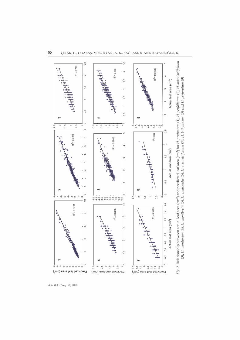
Multiple regression analysis used for determination of the best fittingequation for estimation of leaf area in the medicinal plants evaluated hereshowed that most of the variation in leaf area values was explained by the se-lected parameters (length and width). The variation explained by the parame-ters was 97% for H. pruinatum, 94% for H. perfoliatum, 80% for H. aviculariifo-lium subsp. depilatum var. depilatum, 84% for H. montanum, 87% for H. mont-bretii, 88% for H. linarioides, 91% for H. triquetrifolium, 80% for H. bithynicumand 92% for H. perforatum. The produced leaf area prediction models in thepresent study were LA = 0.109268 + (–0.01306 × W2) + [0.754917 × (L × W)] forH. pruinatum, LA = –1.11342 + (1.596 × L) + (–0.369 × L2) + (0.373 × W2) + [0.321× (L2 × W)] + [–0.04 × (L2 × W2)] for H. perfoliatum, LA = 0.229 + [0.716 × (L × W)]+ [–0.066 × (L × W2)] for H. aviculariifolium, LA = 3.65 + (–2.29 × L) + (–8.22 × W)+ (2.32 × W2) + [6.93 × (L × W)] + [–0.49 × (L2 × W)] + [–1.83 × (L × W2)] for H.montanum, LA = 1.70 + (–8.074 × W) + (10.92 × W2) + [2.94 × (L × W)] + [–6.18 ×(L × W2)] + [0.88 × (L2 × w2)] for H. montbretii, LA = (0.592) + (–4.198 × W2) +[2.863 × (L × W2)] + [–0.508 × (L2 × W2)] + [0.202 × (L2 × W)] for H. linarioides, LA= 0.0397 + [0.51 × (L2 ×] + [0.862 × (L × W2)] + [–0.639 × (L2 × W2)] for H.triquetrifolium, LA = 0.907 + (–1.743 × W2) + [1.0479 × (L × W2)] for H.bithynicum and LA = –0.1714 + [1.04172 × (L × W)] + [–0.19204 × (W2 × L)] for H.perforatum (Table 1).
ESTIMATION OF LEAF AREA IN SELECTED HYPERICUM SPECIES 89
Acta Bot. Hung. 50, 2008
DISCUSSION
Hypericum plants are characterised mainly by the presence of three typesof secretory structures, light glands, dark glands and secretory canals (Cicca-relli et al. 2001). The dark glands on the vegetative organs of many species ofHypericum have been used by botanists as an important distinguishing markfor the classification of this genus (Kitanov 2001). In addition, there is a markedevidence and general agreement about localisation of hypericins in the darkglands (Lu et al. 2001). The presence of dark glands in Hypericum species is ex-pressed as its density in leaves for which it is necessary to calculate the leafarea. Therefore, leaf area has been routinely measured in morphological andtaxonomic studies on Hypericum genus (Walker et al. 2001). For example, in aprevious study we determined a positive and significant relationship betweendark gland density of leaves and both leaf and whole plant contents of hype-ricin in H. perforatum, H. pruinatum and H. aviculariifolium subsp. depilatum var.depilatum. For this purpose, we measured the total area of 50 leaves for eachspecies by using of Placom digital planimeter and it was very time-consumingprocess (Çýrak et al. 2006). Thus, we developed very useful, simple and accu-rate models for estimation leaf area for some hypericin-contained species ofHypericum in the present study.
Similarly, a number of studies have been carried out to estimate leaf areaby linear measurements of leaf width and length in the following plants: soy-bean (Lieth et al. 1986), cucumbers (Robbins and Pharr 1987), orange (Arias etal. 1989, Ramkhelawan and Brathwaite 1992), French bean (Rai et al. 1990), co-conut (Mathes et al. 1990), banana (Potdar and Pawar 1995), grape (Uzun andÇelik 1999), Miscanthus (Vargas et al. 2002), broad bean (Odabaş 2003), cherry(Demirsoy and Demirsoy 2003, Pinto et al. 2004), strawberry (Demirsoy et al.2005), peach (Demirsoy et al. 2004), summer snowflake (Çýrak et al. 2005a), net-tle and lemon balm (Çýrak et al. 2005b). The same authors found that there wereclose relationship between leaf area value, leaf length and leaf width for theseplants (R2 = 0.94 for soybean, R2 = 0.76 to 0.99 for cucumber, R2 = 0.89 to 0.93 fororange, R2 = 0.99 for French bean, R2 = 0.95 to 0.98 for coconut, R2 = 0.96 for ba-nana, R2 = 0.98 for grapes, R2 = 0.91 for Miscanthus, R2 = 0.99 for broad bean,R2 = 0.95 and 0.99 for cherry, R2 = 0.99 for strawberry, R2 = 0.99 for peach, 0.97for summer snowflake, R2 = 0.97 for nettle and R2 = 0.98 for lemon balm). Wefound that there was very close relationship between actual and predicted leafarea for all Hypericum species tested (Fig. 2).
Our results were consistent with those of other studies mentioned abovethat used linear measurements of leaves from different plants for estimatingleaf area. Although coefficients of determination were generally high (R2 >0.85) in both the current and previous studies, it is interesting to note that R2
90 ÇÝRAK, C., ODABAÞ, M. S., AYAN, A. K., SAÐLAM, B. AND KEVSEROÐLU, K.
Acta Bot. Hung. 50, 2008
values for Hypericum species tested varied with species greatly from 0.80 in H.aviculariifolium subsp. depilatum var. depilatum to 0.97 in H. pruinatum. How-ever, the difference is not surprising when the evident difference in size andshape of leaves among the species is taken into consideration (Fig. 1). Apartfrom different species, regression coefficients of leaf area estimation can evenbe different between the cultivars of the same species. For example, using thegrapevine cultivars ‘Niagara’ and ‘DeChaunac’, Williams and Martinson (2003)found that the product of maximum leaf length and width was most highlycorrelated with leaf area, but R2 values of cultivars were different (0.99 for Ni-agara and 0.96 for Dechaunac).
CONCLUSION
Here, we developed leaf area prediction models for nine Hypericum spe-cies, each has potential using fields in medicinal treatments and botanical in-dustry with their well-documented hypericin contents. As the understandingof plant growth and development has been increasing, such mathematicalmodels as this shown in Table 1 will be very useful tools for prediction of leafarea for many plants without using of expensive devices. Also, considering theimportance of expressing the presence of dark glands in vegetative parts ofHypericum plants, prediction of leaf area easily by simple equations instead ofusing expensive and time-consuming devices during the course of experimentis an also important topic for morphological and taxonomic studies on the ge-nus Hypericum. Therefore, the models produced in the present study can beused safely by Hypericum researchers for the species used in this research. Onthe other hand, different models can be developed by researches studying onHypericum species different from those used in the present study.
REFERENCES
Agostinis, P., Annelies, V., Wilfried, M. and Peter A. M. (2002): Hypericin in cancer treat-ment: more light on the way. – Int. J. Biocem. Cell Biol. 34: 221–241.
Arias, E., Fernandez, M. and Telleria, T. (1989): Modified method for determining foliararea in leaf samples of Valencia orange. – Hort. Abst. 59: 9508.
Ayan, A. K., Çýrak, C., Kevseroðlu, K. and Özen, T. (2004): Hypericin in some Hypericumspecies from Turkey. – Asian J. Plant Sci. 3: 200–202.
Biffignandi, P. M. and Bilia, A. R. (2000): The growing knowledge of St. John’s wort (Hype-ricum perforatum L.) drug interactions and their clinical significance. – Curr. Ther.Res. 61: 389–394.
Centritto, M., Loreto, F., Massacci, A., Pietrini, F., Villani, M. C. and Zacchine, M. (2000): Im-proved growth and water use efficiency of cherry saplings under reduced light inten-sity. – Ecol. Res. 15: 385–392.
ESTIMATION OF LEAF AREA IN SELECTED HYPERICUM SPECIES 91
Acta Bot. Hung. 50, 2008
Ciccarelli, D., Andreucci, A. C. and Pagni, A. M. (2001): Translucent glands and secretorycanals in Hypericum perforatum: morphological, anatomical and histochemical stud-ies during the course of onthogenesis. – Ann. Bot. 88: 637–644.
Çýrak, C., Odabaþ, M. S. and Ayan, A. K. (2005a): Leaf area prediction model for summersnowflake (Leucojum aestivum L.). – Int. J. Bot. 1: 12–14.
Çýrak, C., Odabaþ, M. S., Saðlam, B. and Ayan, A. K. (2005b): Relation between leaf area anddimensions of selected medicinal plants. – Res. Agricult. Engineering 51: 13–19.
Çýrak, C., Saðlam, B., Ayan, A. K. and Kevseroðlu, K. (2006): Morphogenetic and diurnalvariation of hypericin in some Hypericum species from Turkey during the course ofontogenesis. – Biochem. Syst. Ecol. 34: 1–13.
Davis, P. H. (1988): Flora of Turkey and the East Aegean Islands. – Edinburgh University Press,Edinburgh.
Demirsoy, H. and Demirsoy, L. (2003): A validated leaf area prediction model for somecherry cultivars in Turkey. – Pakistan J. Bot. 35: 361–367.
Demirsoy, H., Demirsoy, L. and Öztürk, A. (2005): Improved model for the non-destructiveestimation of strawberry leaf area. – Fruits 60: 69–73.
Demirsoy, H., Demirsoy, L., Uzun, S. and Ersoy, B. (2004): Non-destructive leaf area estima-tion in peach. – Eur. J. Hort. Sci. 69: 144–146.
Dias, A. C. P., Francisco, A., Barberan, T., Ferreria, F. and Ferreres, F. (1998): Unusual flavo-noids produced by callus of Hypericum perforatum. – Phytochemistry 48: 1165–1168.
Diwu, Z. (1995): Novel therapeutic and diagnostic applications of hypoccrellins and hype-ricins. – Photochem. Photobiol. 61: 529–532.
Ferraz, A., Bordignon, S., Mans, D., Schmitt, A. and Ravazzolo, A. P. (2002): Screening forthe presence of hypericins in southern Brazilian species of Hypericum (Guttiferae). –Pharm. Biol. 40: 294–297.
Gamiely, S., Randle, W. M., Mills, H. A. and Smittle, D. A. (1991): A rapid and non-destruc-tive method for estimating leaf area of onions. – Hort. Sci. 26: 206.
Gerassim, M. and Kitanov, M. (2001): Hypericin and pseudohypericin in some Hypericumspecies. – Biochem. Syst. Ecol. 29: 171–178.
Gottschalk, K. W. (1994): Shade, leaf growth and crown development of Quercus rubra, Quer-cus velutina, Prunus serotina and Acer rubrum seedlings. – Tree Physiol. 14: 735–749.
Greeson, J., Sanford, B. and Monti, D. A. (2001): St. John’s worth (Hypericum perforatum): areview of the current pharmacological, toxicological and clinical literature. – Psycho-pharmacol. 153: 402–414.
Holz, J. and Ostrowiski, E. (1987): St. John’s wort HPLC analysis of main components andtheir variability in the populations. – Deutschl. Apothekertz. 127: 1227–1230.
Horsley, S. B. and Gottschalk, K. W. (1993): Leaf area and net photosynthesis during devel-opment of prunus serotina seedlings. – Tree Physiol. 12: 55–69.
Ishiguro, K., Nagareya, N. and Fukomoto, H. A. (1998): Phloroglucinol derivative from cellsuspension cultures of Hypericum perforatum. – Phytochemistry 47: 347–369.
Kersteins, G. and Hawes, C. W. (1994): Response of growth and carbon allocation to ele-vated CO2 in young cherry (Prunus avium L) saplings in relation to root environment.– New Phytologist 128: 607–614.
Kitanov, G. M. (2001): Hypericin and pseudohypericin in some Hypericum species. – Bio-chem. Syst. Ecol. 29: 171–178.
Kubin, A., Alth, C., Jindra, R., Jessner, G. and Ebermann, R. (1996): Wavelength-dependentphotoresponse of biological and aqueous model systems using the photodynamicplant pigment hypericin.– J. Photochem. Photobiol. B. 36: 103–108.
92 ÇÝRAK, C., ODABAÞ, M. S., AYAN, A. K., SAÐLAM, B. AND KEVSEROÐLU, K.
Acta Bot. Hung. 50, 2008
Lieth, J. H., James, F. R. and Hugo, H. R. (1986): Estimation of leaf area of soybeans grownunder elevated carbon dioxide levels. – Field Crop Res. 13: 193–203.
Lu, H. F., Shen, Z. G., Li, J. Y. H. and Hu, Z. H. (2001): The patterns of secretory structure andtheir relation to hypericin content in Hypericum. – Acta Bot. Sinica 43: 1085–1088.
Mathes, D., Liyanage, L. V. K. and Randeni, G. (1990): A method for determining leaf area ofone, two and three years old coconut seedlings (var. Cric60). – Hort. Abst. 60: 9366.
Nahrstedt, A. and Butterweck, V. (1997): Biologically active and other chemical constituentsof the herb Hypericum perforatum L. – Pharmacopsychiatry 30: 129–134.
NeSmith, D. S. (1991): Non-destructive leaf area estimation of rabbiteye blueberries. – Hort.Sci. 26: 132.
NeSmith, D. S. (1992): Estimating summer squash leaf area non-destructively. – Hort. Sci.27: 77.
Odabaş, M. S. (2003): The quantitative effect of temperature and light on growth, development andyield of broad bean (Vicia faba L.). – Unpublished Ph.D. thesis, Univ. Ondokuz Mayýs,Turkey.
Picchioni, G. A. and Weinbaum, S. A. (1995): Retention and the kinetics of uptake and ex-port of foliage-applied, labeled boron by apple, pear, prune, and sweet cherry leaves.– J. Am. Soc. Hortic. Sci. 120: 868.
Pinto, A. C. R., Rodrigues, T. J. D., Barbosa, J. C. and Leite, I. C. (2004): Leaf area predictionmodels for Zinnia elegans Jacq., Zinnia haageana Regel and ‘profusion cherry’. – Sci.Agric., Piracicaba, 61: 47–52.
Potdar, P. V. and Pawar, K. R. (1995): Non-destructive leaf area estimation in banana. – Sci.Hortic. 45: 251–254.
Rai, A., Alipit, P. V. and Toledo, M. B. (1990): Estimation of leaf area of French bean (Pha-seolus vulgaris L.) using linear measurements. – Hort. Abst. 60: 3405.
Ramkhelawan, E. and Brathwaite, R. A. I. (1992): Leaf area estimation by non-destructivemethods in sour orange (Citrus aurantium L.). – Hort. Abst. 62: 2557.
Rieger, M. and Duemmel, M. J. (1992): Comparison of drought resistance among Prunusspecies from divergent habitats. – Tree Physiol. 11: 369–380.
Robbins, N. S. and Pharr, D. M. (1987): Leaf area prediction models for cucumber from lin-ear measurements. – Hort. Sci. 22: 1264–1266.
Sakar, M. K. and Tamer, A. U. (1990): Antimicrobial activity of different extracts from someHypericum species. – Fitoterapia 61: 464–465.
Uzun, S. (1996): The quantitative effects of temperature and light environment on the growth, devel-opment and yield of tomato (Lycopersicon esculentum Mill.) and aubergine (Solanummelongena L.). – Thesis, The University of Reading, Reading, UK.
Uzun, S. and Celik, H. (1999): Leaf area prediction models (Uzçelik-I) for different horticul-tural plants. – Turkish J. Agric. Forest. 23: 645–650.
Vargas, L. A., Andersen, M. N., Jensen, C. R. and Jørgensen, U. (2002): Estimation of leafarea index, light interception and biomass accumulation of Miscanthus sinensis ‘Goli-ath’ from radiation measurements. – Biomass and Bioenergy 22: 1–14.
Walker, L., Sirvent, T., Gibson, D. and Vance, N. (2001): Regional differences in hypericinand pseudohypericin concentrations and five morphological traits among Hypericumperforatum plants in the North-western United States. – Can. J. Bot. 79: 1248–1255.
Williams, L. and Martinson, T. E. (2003): Nondestructive leaf area estimation of ‘Niagara’and ‘DeChaunac’ grapevines. – Sci. Hortic. 98: 493–498.
Yazaki, K. and Okuda, T. (1990): Procyanins in callus and multiple shoots of Hypericumerectum. – Planta Med. 56: 490–491.
ESTIMATION OF LEAF AREA IN SELECTED HYPERICUM SPECIES 93
Acta Bot. Hung. 50, 2008