Essential Cell Biology - MCB lab
88
Chapter 15 Intracellular Compartments and Transport Essential Cell Biology Third Edition Copyright © Garland Science 2010 backgrounds
-
Upload
khangminh22 -
Category
Documents
-
view
3 -
download
0
Transcript of Essential Cell Biology - MCB lab

Chapter 15Intracellular Compartments and
Transport
EssentialCell Biology
Third Edition
Copyright © Garland Science 2010
backgrounds

Legend p. 209
Legend p. 209

Figure 15-2 Essential Cell Biology (© Garland Science 2010)

Table 15-2 Essential Cell Biology (© Garland Science 2010)
• Membrane-Enclosed Organelles
• Protein Sorting
• Vesicular Transport
• Secretory Pathways
• Endocytic Pathways
Contents

Membrane-Enclosed Organelles
Figure 15-1 Essential Cell Biology (© Garland Science 2010)

1. Eucaryotic cells contain a basic set of membrane-enclose organelles
2. Membrane-enclose organelles evolved in different ways
Eucaryotic cells contain a basic set of membrane-enclose organelles

Figure 15-2 Essential Cell Biology (© Garland Science 2010)
Table 15-1 Essential Cell Biology (© Garland Science 2010)

Smooth ER
• Steroid hormone synthesis (adrenal gland)
• Organic molecules detoxified (liver cells)
• Ca2+ release and reuptake
Endocrine system
adapted from http://www.clinica-verde.com/

Table 15-2 Essential Cell Biology (© Garland Science 2010)
separating one type of organelle from another
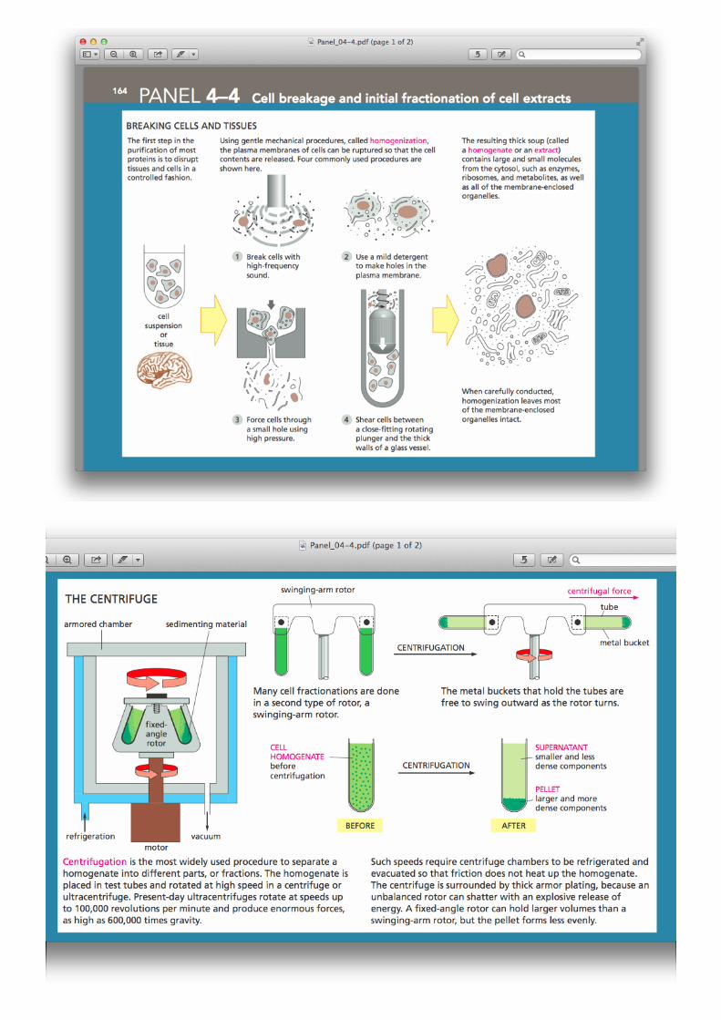

Table 15-1 Essential Cell Biology (© Garland Science 2010)

Membrane-enclose organelles evolved in different ways
The precursors of the first eucaryotic cells
• Simple microorganisms, resembling bacteria?
• Had a plasma membrane but no internal membranes.
• This plasma membrane would have provided all membrane-dependent functions, including ATP synthesis and lipid synthesis.
• However, what about present-day eucaryotic cells?
Text p. 498

1. Invagination of the plasma membrane
Figure 15-3 Essential Cell Biology (© Garland Science 2010)
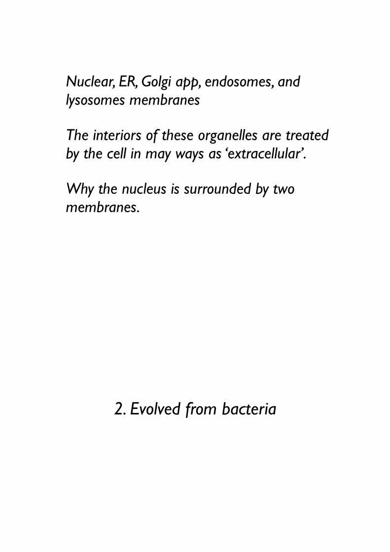
Nuclear, ER, Golgi app, endosomes, and lysosomes membranes
The interiors of these organelles are treated by the cell in may ways as ‘extracellular’.
Why the nucleus is surrounded by two membranes.
2. Evolved from bacteria

Figure 15-4 Essential Cell Biology (© Garland Science 2010)
Mitochondria and chloroplasts.
The similarity of the genomes to those of bacteria.
The close resemblance of some of their proteins to bacterial proteins.
These remain isolated from the extensive vesicular traffic.

• Membrane-Enclosed Organelles
• Protein Sorting
• Vesicular Transport
• Secretory Pathways
• Endocytic Pathways
Contents
Protein Sorting
background
Text p. 500
Proteins are imported into organelles by three mechanisms

Figure 15-5 Essential Cell Biology (© Garland Science 2010)
Signal sequences direct proteins to the correct compartment
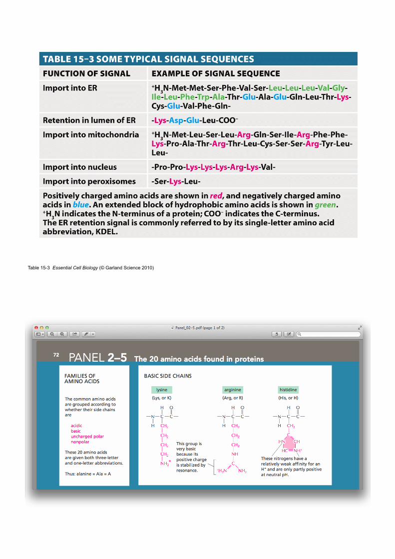
Table 15-3 Essential Cell Biology (© Garland Science 2010)

signal sequence
• typically 15-60 amino acids long
• often removed from the finished protein
• direct a protein to a particular organelle
• physical properties (hydrophobicity, charged amino acids) important than the exact amino acid sequence

Figure 15-6 Essential Cell Biology (© Garland Science 2010)
SnapShot: Ras SignalingMegan Cully and Julian DownwardCancer Research UK London Research Institute, London WC2A 3PX, UK
See online version for legend and references.
1292 C
ell 133, June 27, 2008 ©2008 Elsevier Inc.
DO
I 10.1016/j.cell.2008.06.020

PI 3-kinase-Akt Signaling Pathway
Figure 15-5 Essential Cell Biology (© Garland Science 2010)
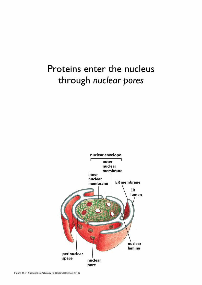
Proteins enter the nucleus through nuclear pores
Figure 15-7 Essential Cell Biology (© Garland Science 2010)

15.1-nuclear_import
Figure 15-8 Essential Cell Biology (© Garland Science 2010)

Figure 15-8a Essential Cell Biology (© Garland Science 2010)
Figure 15-8b Essential Cell Biology (© Garland Science 2010)

Figure 15-8c Essential Cell Biology (© Garland Science 2010)
Köhler, A. & Hurt, E. Exporting RNA from the nucleus to the cytoplasm. Nature Publishing Group 8, 761–773 (2007).

Figure 15-9 Essential Cell Biology (© Garland Science 2010)
Figure 15-10 Essential Cell Biology (© Garland Science 2010)

Proteins unfold to enter mitochondria and chloroplasts
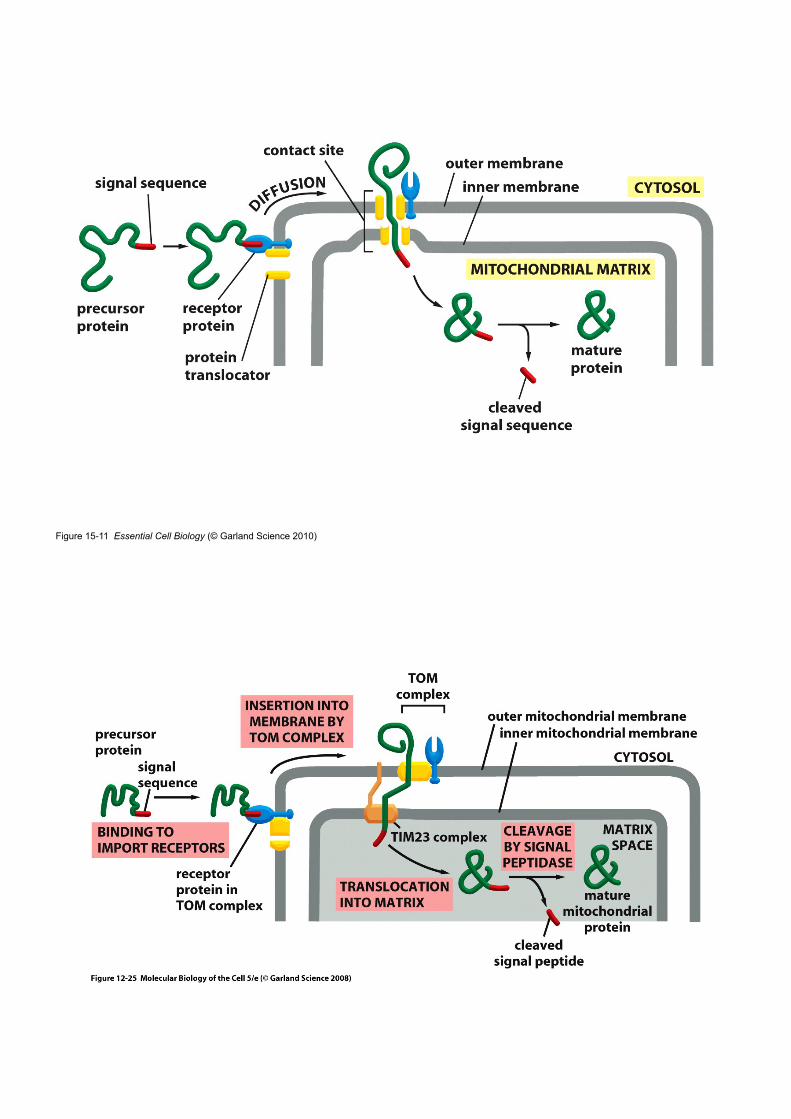
Figure 15-11 Essential Cell Biology (© Garland Science 2010)

15.2-protein_import
Proteins enter the endoplasmic reticulum while being synthesized

Figure 15-12 Essential Cell Biology (© Garland Science 2010)
15.3-ER_tubules

• The most extensive membrane system in an euk cell.
• Serves as an entry point for proteins destined for other organelles.
• Two kinds of proteins transferred from the cytosol to the ER.
• Water-soluble proteins: completely translocated across ER membrane (destination: secretion or lumen of an organelle)
• Transmembrane proteins: only partly translocated and embedded in ER membrane (destination: membrane of ER / other / plasma membrane).
• Proteins enter the endoplasmic reticulum while being synthesized. (see rough ER).
• Membrane-bound ribosomes vs free ribosomes.
Endoplasmic Reticulum
Figure 15-13 Essential Cell Biology (© Garland Science 2010)

Soluble proteins are released into the ER lumen
Figure 15-14 Essential Cell Biology (© Garland Science 2010)
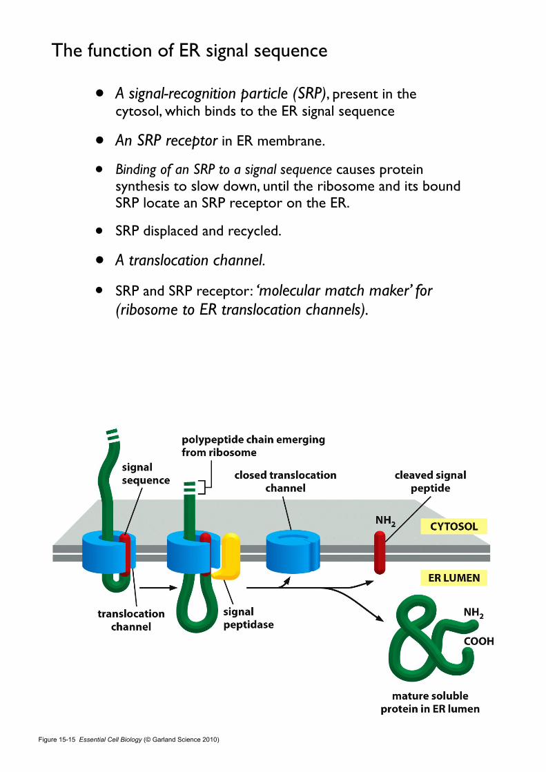
• A signal-recognition particle (SRP), present in the cytosol, which binds to the ER signal sequence
• An SRP receptor in ER membrane.
• Binding of an SRP to a signal sequence causes protein synthesis to slow down, until the ribosome and its bound SRP locate an SRP receptor on the ER.
• SRP displaced and recycled.
• A translocation channel.
• SRP and SRP receptor: ‘molecular match maker’ for (ribosome to ER translocation channels).
The function of ER signal sequence
Figure 15-15 Essential Cell Biology (© Garland Science 2010)
• Open the translocation channel.
• The signal peptide remains bound to the channel.
• The signal peptide is cleaved off by a signal peptidase.
• The signal peptide is then released from the channel and rapidly degraded.
The function of ER signal sequence
Start and stop signals determine the arrangement of a transmembrane protein
in the lipid bilayer

Proteins that enter the ER
• Release into the ER lumen (soluble proteins).
• Remain embedded in the ER membrane (transmembrane proteins) - a complicated process
Figure 15-16 Essential Cell Biology (© Garland Science 2010)

A transmembrane protein with a single membrane-spanning segment
• N-term signal seq initiates translocation.
• is released from the channel into the lipid bilayer and is cleaved off.
• a stop-transfer sequence
• is released form the translocation channel and drift into the plane of the lipid bilayer.
• Anchors the protein in the membrane.
15.4-protein_translocation

Figure 15-17 Essential Cell Biology (© Garland Science 2010)
Vesicular Transport

Transport vesicles carry soluble proteins and membrane between compartments
Figure 15-18 Essential Cell Biology (© Garland Science 2010)

Vesicle budding is driven by the assembly of a protein coat
Figure 15-19a Essential Cell Biology (© Garland Science 2010)

Figure 15-19b Essential Cell Biology (© Garland Science 2010)
Protein coat
• coated vesicles
• shape the membrane into a bud and help to capture molecules for onward transport
• clathrin-coated vesicles
• outward secretory pathway or inward endocytic pathway
• a small GTP-binding protein, dynamin assembles as a ring around the neck, causes the ring to constrict
• adaptins
• secure the clathrin coat to the vesicle membrane and help select cargo molecules
• cargo receptor
• COP (coat protein)-coated vesicles

Figure 15-20 Essential Cell Biology (© Garland Science 2010)
15.5_Clathrin

Table 15-4 Essential Cell Biology (© Garland Science 2010)
Vesicle docking depends on tethers and SNAREs

Figure 15-21 Essential Cell Biology (© Garland Science 2010)
Vesicle docking
• Identification process
• Rab proteins
• on the surface of the vesicle
• recognized by tethering proteins of the target membrane
• molecular marker
• SNAREs
• transmembrane proteins
• v-SNAREs interact with t-SNAREs
• docking the vesicle in place

Figure 15-22 Essential Cell Biology (© Garland Science 2010)
Membrane fusion
• not only delivers the contents of the vesicle into the interior of the target organelle
• also adds the vesicle membrane to the membrane of the organelle
• close approach: within 1.5 nm of each other
• Pairing of v-SNAREs and t-SNAREs draw the two lipid bilayers into close apposition
• The force of the SNAREs winding together squeeze out any water molecules

Secretory pathways
Most proteins are covalently modified in the ER

Figure 15-13 Essential Cell Biology (© Garland Science 2010)
Figure 4-26 Essential Cell Biology (© Garland Science 2010)

Disulfide bonds
• Most proteins that enter the ER are chemically modified.
• Disulfide bonds are formed by the oxidation of paris of cysteine side chains
• catalyzed by an enzyme that resides in the ER lumen
• help to stabilize the structure of those proteins that may encounter changes in pH and degradative enzymes outside the cell
04.6-disulfide_bonds

Figure 15-13 Essential Cell Biology (© Garland Science 2010)
glycosylation
• Many of the proteins that enter the ER lumen or membrane are converted to glycoproteins
• Glycosylation
• carried out by glycosylating enzyme in the ER but not in the cytosol
• a total 14 sugars (attached to dolichol) is transferred to Asn by oligosaccharide protein transferase


Figure 15-23 Essential Cell Biology (© Garland Science 2010)

glycosylation
• Many of the proteins that enter the ER lumen or membrane are converted to glycoproteins
• Glycosylation
• protect from degradation
• hold in the ER until properly folded
• transport signal for packaging the protein into appropriate transport vesicles
• form part of cell surface carbohydrate layer
• recognition function of one cell by another
Exit from the ER is controlled to ensure protein quality

•Some proteins retained in the ER by an ER retention signal.
•Most proteins are packaged into transport vesicles.•Exit from the ER is highly selective.•Proteins that fold incorrectly, and dimeric or multimeric proteins are actively retained in the ER by binding chaperone proteins (cf. Antibody).
•In this way, the ER controls the quality of the proteins that it exports to the Golgi apparatus.
Quality control mechanism

Figure 15-24 Essential Cell Biology (© Garland Science 2010)
cystic fibrosis낭포성섬유증 [cystic fibrosis, 囊胞性纖維症]
호흡기관과 위장관에 진하고 끈적끈적한 점액 분비물이 달라붙어 있는 증상이 나타나는 유전성 대사장애

Molecular Cell Biology, Lodish 5th Ed.
cystic fibrosis
•Cystic fibrosis (CF) is an autosomal recessive genetic disorder that affects most critically the lungs, and also the pancreas, liver, and intestine. It is characterized by abnormal transport of chloride and sodium across an epithelium, leading to thick, viscous secretions.
•The name cystic fibrosis refers to the characteristic scarring (fibrosis) and cyst formation within the pancreas, first recognized in the 1930s. Difficulty breathing is the most serious symptom and results from frequent lung infections that are treated with antibiotics and other medications.
•CF is caused by a mutation in the gene for the protein cystic fibrosis transmembrane conductance regulator (CFTR). This protein is required to regulate the components of sweat, digestive fluids, and mucus.
•CF is most common among Caucasians; 4% of people of European descent carries one allele for CF. The World Health Organization states that "In the European Union, 1 in 2000–3000 newborns is found to be affected by CF".
http://en.wikipedia.org/wiki/Cystic_fibrosis

• Causes severe degeneration of the lung, produces a plasma-membrane transport protein (CFTR) that is slightly misfolded.
• Even though the mutant protein could function normally as a chloride channel if it reached the plasma membrane, it is retained in the ER.

“The devastating disease results not because the mutation inactivates an important protein but because the active protein is discarded by the
cells before it is given an opportunity to function”.
Exit from the ER is controlled to ensure protein quality

The size of the ER is controlled by the amount of protein that flows
through it
Unfolded protein response (UPR)
• When protein synthesis is vigorous, misfolded proteins begin to accumulate.
• A signal to direct the cell to make more ER.
• activating receptors that activate a vast transcriptional program, UPR.
• The UPR program prompts the cell to produce more ER.
• Allows cells to adjust the size of the ER according to need.

Figure 15-25 Essential Cell Biology (© Garland Science 2010)
Even an expanded ER can become overloaded
• The UPR program directs the cell to self-destruct (apoptosis).
• Diabetes
• Adult-onset diabetes
• The tissues of the body gradually become resistant to the effects of insulin.
• ER in the β cells of pancreas reaches a maximum capacity (Further expansion is physiologically impossible).
• β cell elimination leads to other surviving cells destruction.

The size of the ER is controlled by the amount of protein that flows
through it
Proteins are further modified and sorted in the Golgi apparatus

Golgi appratus
• located near the cell nucleus and close to the centrosome.
• consists of a collection of fattened membrane-enclosed sacs (cisternae).
• Each stack contains 3-20 cisternae.
• The cis face, adjacent to the ER and the trans face, pointed toward plasma membrane
• important for protein sorting (How we know).
• ER retention signal? destined for lysosomes or for the cell surface?
Figure 15-26a Essential Cell Biology (© Garland Science 2010)

Figure 15-26b Essential Cell Biology (© Garland Science 2010)
Figure 15-26c Essential Cell Biology (© Garland Science 2010)
A cultured fibroblast
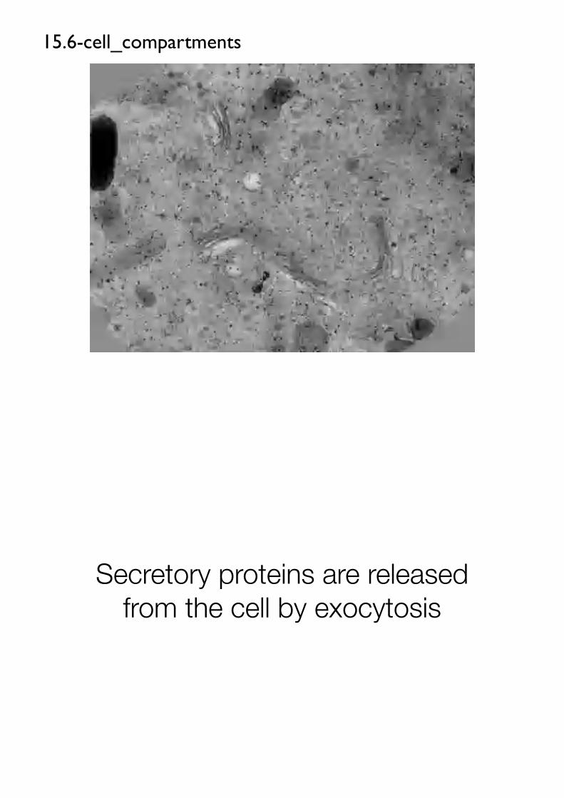
15.6-cell_compartments
Secretory proteins are released from the cell by exocytosis

Figure 15-27 Essential Cell Biology (© Garland Science 2010)
Constitutive exocytosis pathway
• Vesicles buds from the trans Golgi network and fuses with the plasma membrane.
• Operates continually.
• Supplies newly made lipids and proteins.
• Pathway for plasma membrane growth when cells enlarge before dividing.
• Proteins for secretion.
• Nonselective or default pathway (does not require a particular signal sequence).
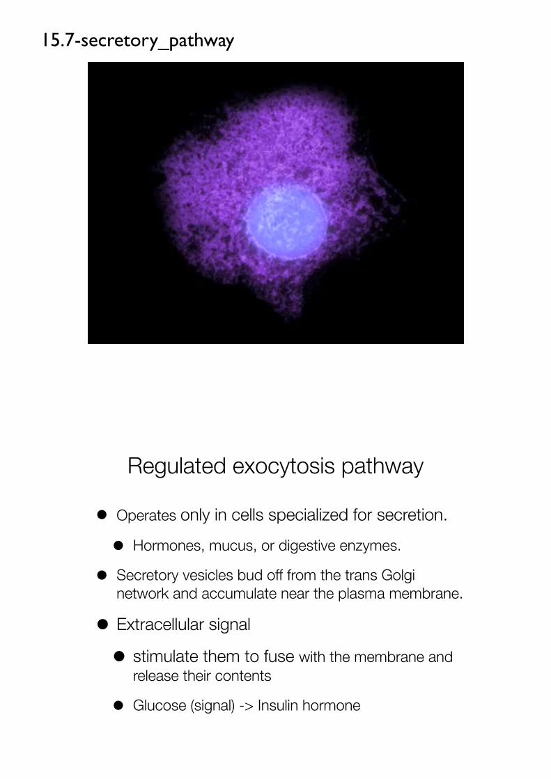
15.7-secretory_pathway
Regulated exocytosis pathway
• Operates only in cells specialized for secretion.
• Hormones, mucus, or digestive enzymes.
• Secretory vesicles bud off from the trans Golgi network and accumulate near the plasma membrane.
• Extracellular signal
• stimulate them to fuse with the membrane and release their contents
• Glucose (signal) -> Insulin hormone

15.8-exocytotic_transport
Regulated exocytosis pathway
• Proteins in this pathway have special surface properties
• aggregate with one another under ionic conditions (acidic pH and high Ca2+).
• Packaged into secretory vesicles and await a signal instructing them to fuse with the plasma membrane ('Selective aggregation')
• Packaged at high concentrations (200-fold).

Figure 15-28 Essential Cell Biology (© Garland Science 2010)
Figure 15-27 Essential Cell Biology (© Garland Science 2010)

Secretory proteins are released from the cell by exocytosis
How we know:Tracking protein and vesicle transport
Experimental techniques for studying how proteins shuttle from one cellular compartment to another

Figure 15-29 Essential Cell Biology (© Garland Science 2010)
Method #1
Figure 15-30 Essential Cell Biology (© Garland Science 2010)
Method #2

Figure 15-31 Essential Cell Biology (© Garland Science 2010)
Method #3
15.1-nuclear_import

15.7-secretory_pathway
15.8-exocytotic_transport
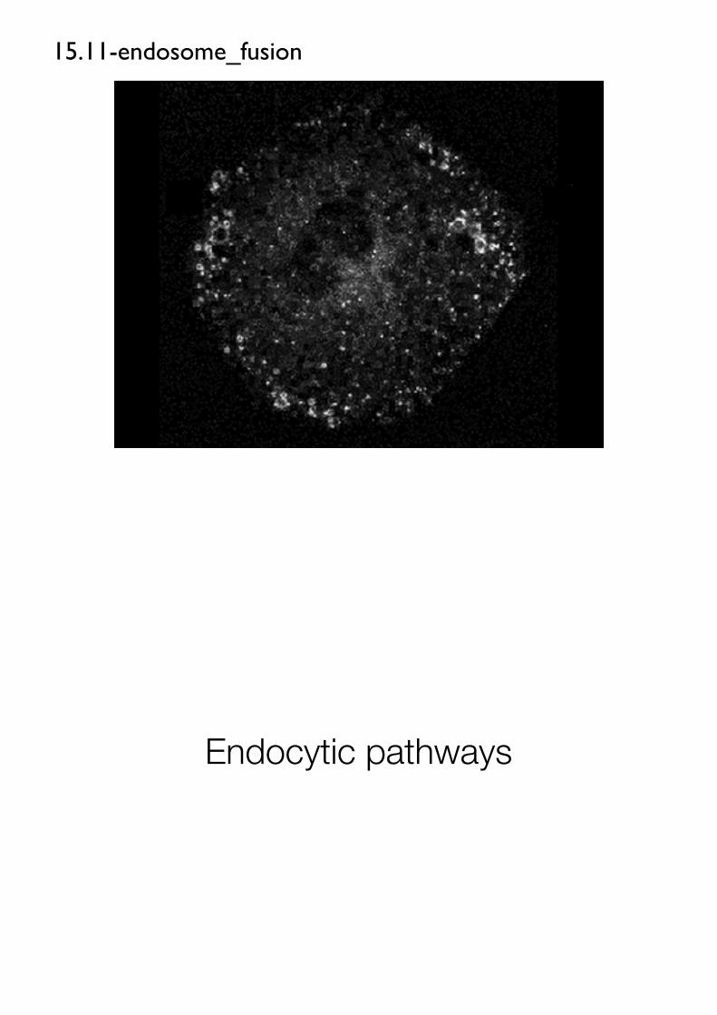
15.11-endosome_fusion
Endocytic pathways

Endocytosis
• Endocytosis: Eucaryotic cells are continually taking up fluid, as well as large and small molecules.
• Phagocytic cells are able to internalize large particles and even other cells.
• Endocytic vesicle and Lysosome
Pinocytosis vs Phagocytosis
• Pinocytosis
• cellular drinking
• ingestion of fluid and molecules via small vesicles (<150 nm)
• all eucaryotic cells
• Phagocytosis
• cellular eating
• ingestion of large particles, such as microorganisms and cell debris via large vesicles, phagosomes (> 250 nm)
• specialized phagocytic cells

Specialized phagocytic cells ingest large particles
In unicellular eucaryotes, phagocytosis is a form of feeding
They ingest even bacteria for nutrition purpose

15.9-phagocytosis
Phagocytic cells
• Macrophage widely distributed in tissues and white blood cells (WBC)
• defend us against infection
• Antibody coated bacteria + cell surface receptor -> phagocytosis
• pseudopods (engulf the bacterium) > phagosome > lysosome

Figure 15-32a Essential Cell Biology (© Garland Science 2010)
Mycobacterium tuberculosis
How the bacterium survives and multiplies within the macrophage?
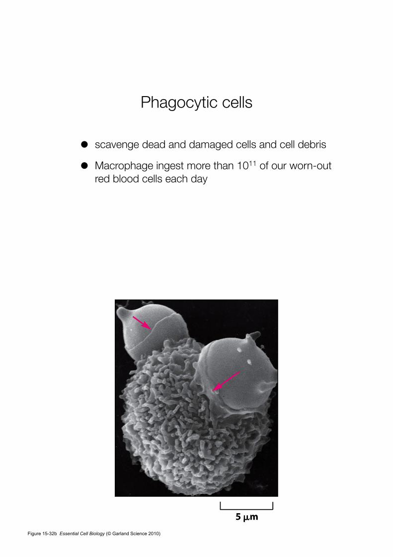
Phagocytic cells
• scavenge dead and damaged cells and cell debris
• Macrophage ingest more than 1011 of our worn-out red blood cells each day
Figure 15-32b Essential Cell Biology (© Garland Science 2010)

Fluid and macromolecules are taken up by pinocytosis
text p.523

PERRY, M. M. & GILBERT, A. B. Yolk transport in the ovarian follicle of the hen (Gallus domesticus): lipoprotein-like particles at the periphery of the oocyte in the rapid growth phase. J Cell Sci 39, 257–272 (1979).


Receptor-mediated endocytosis provides a specific route into animal
cells

Enhanced pinocytosis
• receptor-macromolecule complexes in clathrin-coated vesicles
• receptor-mediated endocytosis
• a selective concentrating mechanism ( > 1000-fold)
• cholesterol (for new membrane)
cholesterol
• extremely insoluble and
• transported in the bloodstream bound to protein (LDL, low-density lipoproteins)
• LDL + cell surface receptor > endocytosis

Figure 15-33 Essential Cell Biology (© Garland Science 2010)
15.10-receptor_endocytosis

atherosclerosisIn individuals who inherit a defective gene encoding the
LDL receptor protein, which molecular pathway is disrupted?
Text p. 524
Endocytosed macromolecules are sorted in endosomes
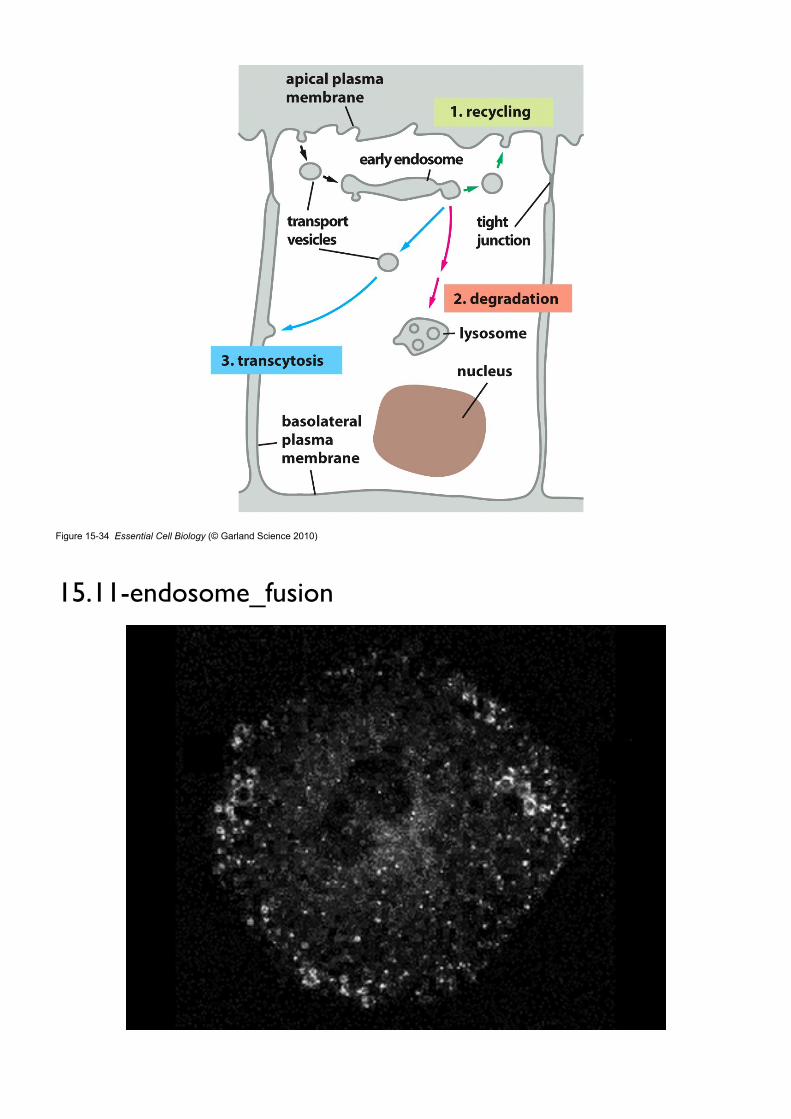
Figure 15-34 Essential Cell Biology (© Garland Science 2010)
15.11-endosome_fusion

Lysosomes are the principal sites of intracellular digestion
Figure 15-36 Essential Cell Biology (© Garland Science 2010)

lysosomes
• membrane sacs of hydrolytic enzymes
• control intracellular digestion of both extracellular materials and worn-out organelles
• contain about 40 types of hydrolytic enzymes
• acidic condition (pH ~5)
• membrane transporter
• metabolite transporter
• ATP-driven H+ pump (acidic pH)
• Phagocytosis, Endocytosis, Autophagy > lysosomes
Figure 15-35 Essential Cell Biology (© Garland Science 2010)


![[Lab Report] PSpice](https://static.fdokumen.com/doc/165x107/631a338ebb40f9952b01e638/lab-report-pspice.jpg)