chapter 17 copy - MCB lab
56
Molecular Biology of the Cell Fifth Edition Chapter 17 The Cell Cycle Copyright © Garland Science 2008 Alberts • Johnson • Lewis • Raff • Roberts • Walter • Overview of the cell cycle • The cell-cycle control system • S phase • Mitosis • Cytokinesis • Control of cell division and cell growth Contents Figure 17-1 Molecular Biology of the Cell (© Garland Science 2008) Cells reproduce by duplicating their contents and dividing in two, a process called the cell cycle “Where a cell arises, there must be a previous cell, just as animals can only arise from animals and plants from plants” Rudolf Virchow in 1858
-
Upload
khangminh22 -
Category
Documents
-
view
0 -
download
0
Transcript of chapter 17 copy - MCB lab
Molecular Biology of the Cell Fifth Edition
Chapter 17 The Cell Cycle
Copyright © Garland Science 2008
Alberts • Johnson • Lewis • Raff • Roberts • Walter
• Overview of the cell cycle
• The cell-cycle control system• S phase• Mitosis• Cytokinesis
• Control of cell division and cell growth
Contents
Figure 17-1 Molecular Biology of the Cell (© Garland Science 2008)
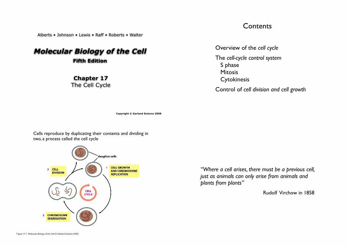
Cells reproduce by duplicating their contents and dividing in two, a process called the cell cycle
“Where a cell arises, there must be a previous cell, just as animals can only arise from animals and plants from plants”
Rudolf Virchow in 1858

1. How do cells duplicate their contents?
2. How do they partition the duplicated contents and split in two?
3. How do they coordinate all the machinery that is required for these two processes?
Major questions
The duration of the cell cycle varies greatly from one cell type to another
Overview
Figure 17-2 Molecular Biology of the Cell (© Garland Science 2008)
1. The eucaryotic cell cycle is divided into four phases

Figure 17-4 Molecular Biology of the Cell (© Garland Science 2008) Figure 17-3 Molecular Biology of the Cell (© Garland Science 2008)
• M phase• Most dramatic events.
• Nucleus divides (mitosis)• Cell splits in two (cytokinesis)• About an hour (only a small fraction of the total cell-cycle time)
• Interphase (period b/w one M phase and the next; it appears as an uneventful interlude during which the cell simply increases in size)
• S phase
• S = Synthesis
• Cell replicates its nuclear DNA
• An essential prerequisite for cell division
• G1 phase (interval between the completion of M phase and the beginning of S phase)
• G2 phase (interval between the end of S phase and the beginning of M phase)
• Monitors the internal and external environments
• Decide whether to proceed to the next phase or pause to allow
more time to prepare.

• Generally continues to transcribe genes, synthesize proteins, and grow in mass.
• Provide additional time for the cell to grow and duplicate its cytoplasmic organelles
• If interphase lasted only long enough for DNA replication → shrink.
• Cleavage division
• The first cell divisions after fertilization serve to divide a giant egg cell into many smaller cells
In addition, in interphase...
• Following DNA replication in S phase
• The two copies of each chromosome remain tightly bound together.
• Progressive condensation (the first visible sign that a cell is about to enter M phase).
• Visible in the light microscope as long threads.
• Easier to segregate to the two forming daughter cells.
2. Cell-cycle control system can be dissected genetically by analysis of yeast mutants
Yeast genetics • Tiny, single-celled fungi. • Cell-cycle system remarkably similar to our own. • Fission yeast Schizosaccharomyces pombe (named after the african beer it
is used to produce) • Budding yeast Saccharomyces cerevisiae (used by both brewers and bakers) • Reproduce almost as rapidly as bacteria. • A genome size less than 1% that of a mammmal. • Many important discoveries about cell-cyle control have come from systemic
search in yeast.
Figure 17-5 Molecular Biology of the Cell (© Garland Science 2008) Figure 17-6 Molecular Biology of the Cell (© Garland Science 2008)
Cell-division-cycle genes (Cdc genes) • Many of these mutations cause cells to arrest at a specific point in the cell
cycle. • A Cdc mutant : cannot complete cell cycle + cannot propagate. • Conditional mutants: Permissive (low) temperature, Restrictive (high)
temperature.

Figure 17-7 Molecular Biology of the Cell (© Garland Science 2008)
The morphology of budding yeast cells arrested by a Cdc mutation (A) In a normal population of proliferating yeast cells, buds vary in size. (B) In a Cdc15 mutant, cells arrest uniformly with large buds which are characteristic of late M phase.
Figure 17-8 Molecular Biology of the Cell (© Garland Science 2008)
3. Cell-cycle control system can be analysed biochemically in animal embryos
• The biochemical features of the cell cycle are readily analysed in the giant fertilized eggs of many animals, which carry large stockpiles of the proteins needed for cell division.
• The egg of the frog Xenopus is over 1 mm in diameter and contains 100,000 times more cytoplasm than an average cell in the human body.
Figure 17-9 Molecular Biology of the Cell (© Garland Science 2008)
Oocyte growth and egg cleavage in Xenopus
Figure 17-10 Molecular Biology of the Cell (© Garland Science 2008)
Studying the cell-cyle in a cell-free system : Cell-cycle control system is operating in the cell-free cytoplasmic extract.
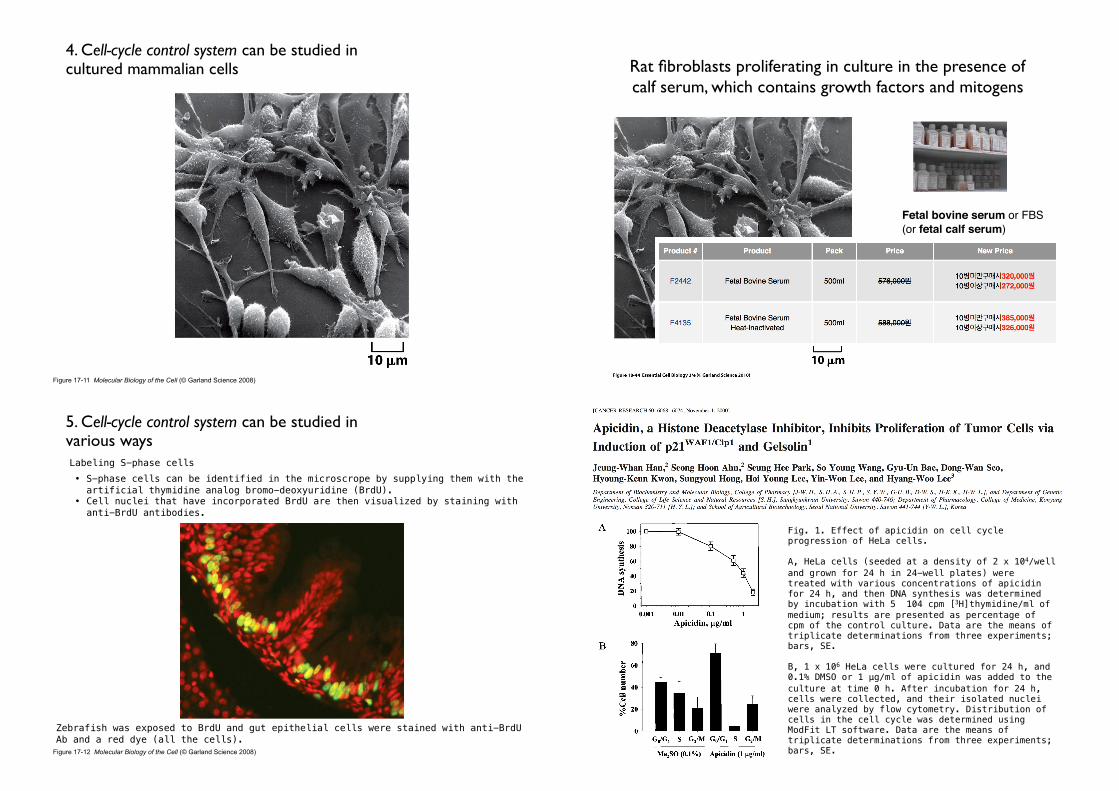
Figure 17-11 Molecular Biology of the Cell (© Garland Science 2008)
4. Cell-cycle control system can be studied in cultured mammalian cells Rat fibroblasts proliferating in culture in the presence of
calf serum, which contains growth factors and mitogens
Fetal bovine serum or FBS (or fetal calf serum)
5. Cell-cycle control system can be studied in various ways
Figure 17-12 Molecular Biology of the Cell (© Garland Science 2008)
Labeling S-phase cells • S-phase cells can be identified in the microscrope by supplying them with the
artificial thymidine analog bromo-deoxyuridine (BrdU). • Cell nuclei that have incorporated BrdU are then visualized by staining with
anti-BrdU antibodies.
Zebrafish was exposed to BrdU and gut epithelial cells were stained with anti-BrdU Ab and a red dye (all the cells).
Fig. 1. Effect of apicidin on cell cycle progression of HeLa cells.
A, HeLa cells (seeded at a density of 2︎ x 104/well and grown for 24 h in 24-well plates) were treated with various concentrations of apicidin for 24 h, and then DNA synthesis was determined by incubation with 5 ︎ 104 cpm [3H]thymidine/ml of medium; results are presented as percentage of cpm of the control culture. Data are the means of triplicate determinations from three experiments; bars, SE.
B, 1 x 10⁶ HeLa cells were cultured for 24 h, and 0.1% DMSO or 1 μ︎g/ml of apicidin was added to the culture at time 0 h. After incubation for 24 h, cells were collected, and their isolated nuclei were analyzed by flow cytometry. Distribution of cells in the cell cycle was determined using ModFit LT software. Data are the means of triplicate determinations from three experiments; bars, SE.
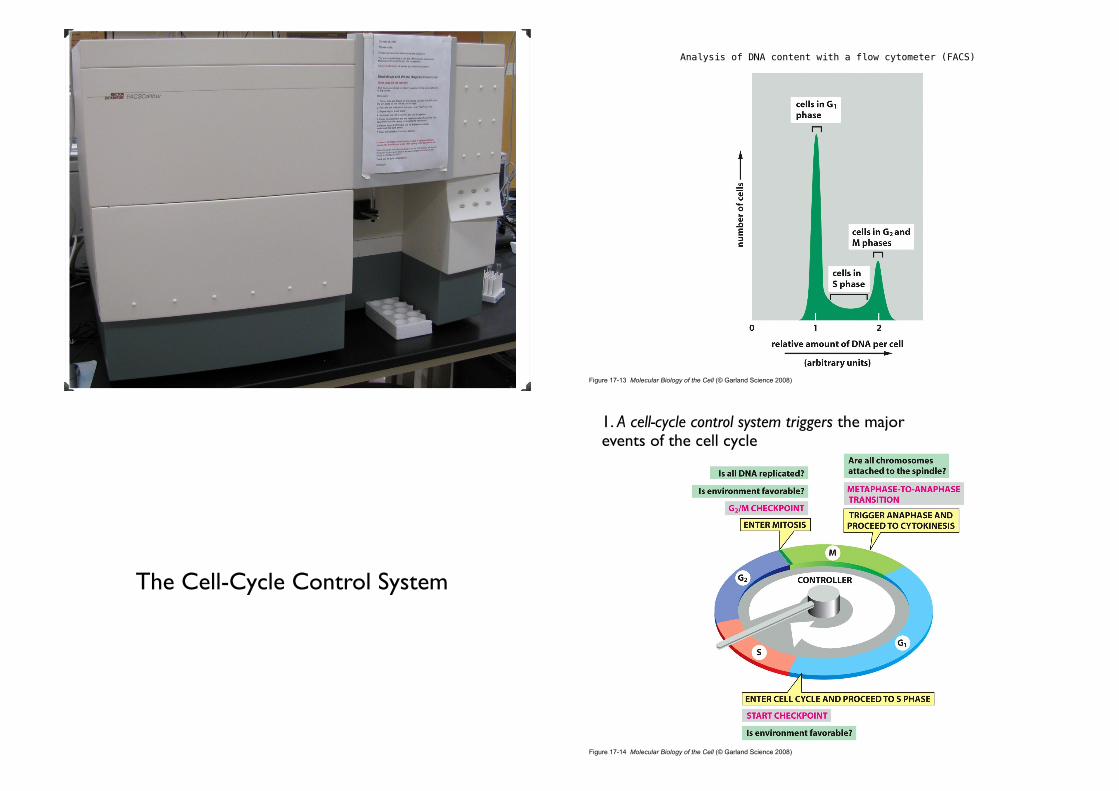
Figure 17-13 Molecular Biology of the Cell (© Garland Science 2008)
Analysis of DNA content with a flow cytometer (FACS)
The Cell-Cycle Control System
Figure 17-14 Molecular Biology of the Cell (© Garland Science 2008)
1. A cell-cycle control system triggers the major events of the cell cycle

• The essential processes of the cell cycle, such as DNA replication, mitosis, and cytokinesis, are triggered by a cell-cycle control system (a complex network of regulatory proteins).
• Arrests the cell cycle at specific checkpoints.
• G1 or G2 checkpoints, checkpoint in mitosis.
2. The cell-cycle control system depends on cyclically activated Cyclin-Dependent Protein Kinases (Cdks)
Figure 17-15 Molecular Biology of the Cell (© Garland Science 2008)
Figure 17-16 Molecular Biology of the Cell (© Garland Science 2008)
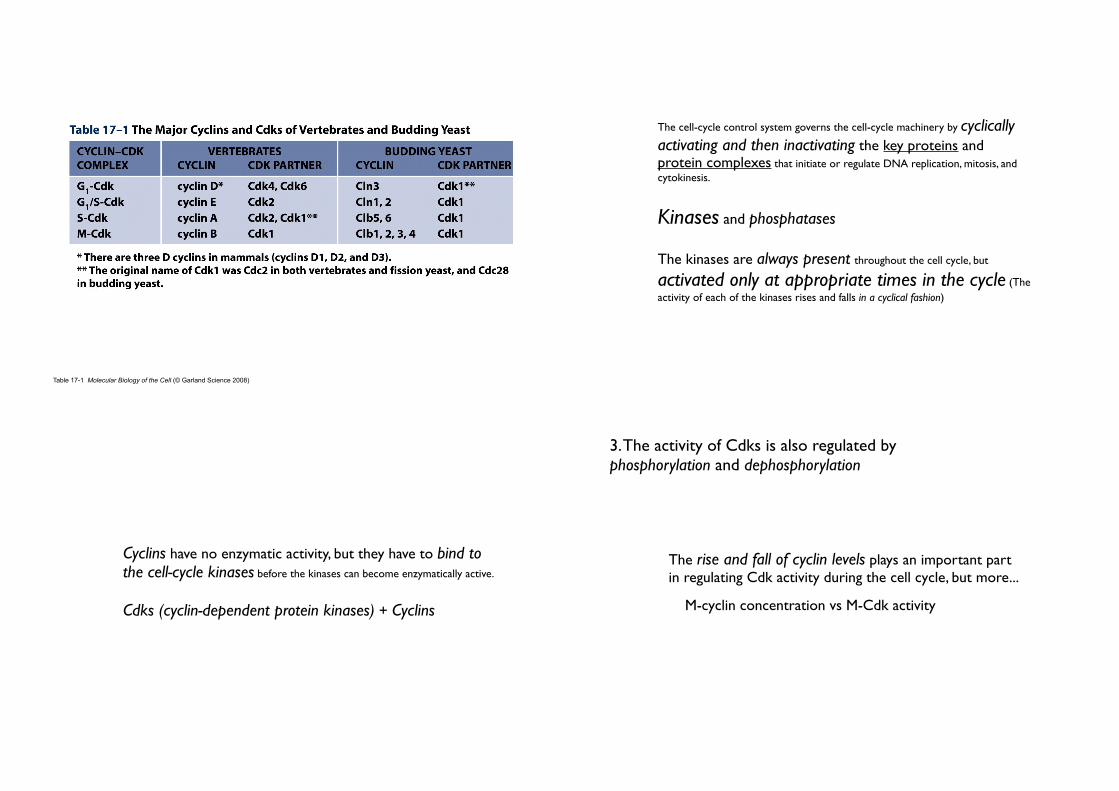
Table 17-1 Molecular Biology of the Cell (© Garland Science 2008)
• The cell-cycle control system governs the cell-cycle machinery by cyclically activating and then inactivating the key proteins and protein complexes that initiate or regulate DNA replication, mitosis, and cytokinesis.
• Kinases and phosphatases
• The kinases are always present throughout the cell cycle, but
activated only at appropriate times in the cycle (The activity of each of the kinases rises and falls in a cyclical fashion)
• Cyclins have no enzymatic activity, but they have to bind to the cell-cycle kinases before the kinases can become enzymatically active.
• Cdks (cyclin-dependent protein kinases) + Cyclins
3. The activity of Cdks is also regulated by phosphorylation and dephosphorylation
• The rise and fall of cyclin levels plays an important part in regulating Cdk activity during the cell cycle, but more...
• M-cyclin concentration vs M-Cdk activity

Figure 17-17 Molecular Biology of the Cell (© Garland Science 2008)
The abrupt activation of M-Cdk.
• For M-Cdk to be maximally active, it has to be phosphorylated at one or more sites by a specific protein kinase, and dephosphorylated at other sites by a specific protein phosphatase.
Figure 17-18 Molecular Biology of the Cell (© Garland Science 2008)
4. Inhibitory phosphorylation and Cdk inhibitory proteins (CKIs) can suppress Cdk activity

Figure 17-16 Molecular Biology of the Cell (© Garland Science 2008) Figure 17-19 Molecular Biology of the Cell (© Garland Science 2008)
• Cdk inhibitor proteins
• Molecular brakes of the cell cycle
• Pause cell-cycle at checkpoints
• Block the assembly or activity of cyclin-Cdk.• Sometimes, help maintain Cdks in an inactive state.
5. Proteins that inhibit Cdks can arrest the cell cycle at specific checkpoints

• G0
• If deprived of mitogens (extracellular signal), the cell will withdraw from the cell cycle - Non-proliferating state, G0.
• Cell can remain for days or weeks or even for the lifetime of the organism.
6. The cell-cycle control system depends on cyclical proteolysis
• [cyclin] : rises gradually but then falls sharply at a specific time.
• This abrupt fall results from the targeted degradation of the cyclin protein.
• This rapid elimination of cyclin returns the Cdk to its inactive state.
• Ubiquitylation.

• Proteolysis by APC/C and SCF during cell cycle
• Anaphase-promoting complex, or cyclosome (APC/C) : ubiquitin ligase
• SCF (after the names of its three subunits) : ubiquitin ligase

Figure 17-21 Molecular Biology of the Cell (© Garland Science 2008)
An overview of the cell-cycle control system: The cell-cycle control system functions as a network of biochemical switches
• Overview of the cell cycle
• The cell-cycle control system• S phase• Mitosis• Cytokinesis
• Control of cell division and cell growth
Contents

Review
1. S-Cdk initiates DNA replication once per cycle
• A cell must solve several problems in controlling the initiation and completion of DNA replication
• DNA replication is initiated at the right time and not more than once per cycle...
• ‘The cell-cycle control system’
Figure 17-22 Molecular Biology of the Cell (© Garland Science 2008)
• Initiating Origin firing by S-Cdk
• Origin recognition complex (ORC)• remains bound to origins of replication throughout the cell cycle.
• serves as a sort of landing pad for additional regulatory proteins (Cdc6, Cdt1. and Mcm) that bind before the start of S phase.
• Cdc6 • present at low levels during most of the cell cycle.• its concentration increases transiently in early G1.
• bind to ORC
• promotes the binding of additional proteins to form a ‘pre-replicative complex’.
• The replication origin is ready to ‘fire’.• The activation of S-Cdk then ‘pulls the trigger’.
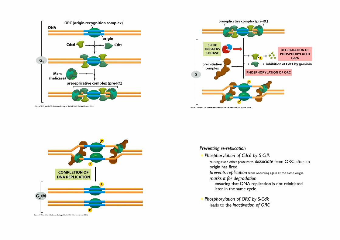
• Preventing re-replication
• Phosphorylation of Cdc6 by S-Cdk• causing it and other proteins to dissociate from ORC after an
origin has fired.
• prevents replication from occurring again at the same origin.
• marks it for degradation• ensuring that DNA replication is not reinitiated
later in the same cycle.
• Phosphorylation of ORC by S-Cdk• leads to the inactivation of ORC

2. Cohesins help hold sister chromatids together
Figure 17-24 Molecular Biology of the Cell (© Garland Science 2008)
• Sister chromatids• After the chromosomes have been duplicated in S phase, the two
copies of each remain tightly bound together.
• held together by protein complex, cohesins.
• assemble along each sister chromatid.
• form protein rings that surround the two sister chromatids, keeping them united.
• crucial for proper chromosome segregation.
• broken completely only in late mitosis to allow the sister chromatid to be pulled apart by the mitotic spindle.

M PHASE


Review
1. M-Cdk drives entry into mitosis
• M-Cdk
• brings about all the diverse and intricate rearrangements in the early stages of mitosis (the most remarkable features)• triggers the condensation of the replicated chromosome
(compact, rod-like structures)• induces the assembly of the mitotic spindle (that separate
and segregate into the two daughter cells)
Figure 17-25 Molecular Biology of the Cell (© Garland Science 2008)
2. Dephosphorylation activates M-Cdk at the onset of mitosis

• M-Cdk activationAccumulation of M cyclin (cyclin B)
• synthesis of cyclin B starts immediately after S phase• rise gradually and helps time the onset of M phase
• leads to a corresponding accumulation of M-Cdk (still inactive)
Sudden activation• triggered by Cdc25 at the end of G2
• Cdc25 phosphatase removes the inhibitory phosphatesPositive feedback loop• Indirectly activates more M-Cdk• Inhibit Wee1(inhibitory kinase), further promoting the activation of M-Cdk
• An explosive increase in M-Cdk activity• drives the cell abruptly from G2 into M phase
Review3. Condensins help configure duplicated chromosomes for separation
Cohesin

Figure 17-27 Molecular Biology of the Cell (© Garland Science 2008)
Condensin
19_03_Cohesins_conde.jpgCondensins
• Five-subunit protein complex that resemble cohesin (four subunits).
• help carry out the chromosome condensation• M-Cdk phosphorylates some of the condensin subunits of five
• M-Cdk triggers the assembly of condensin complexes onto DNA
• makes the mitotic chromosomes more compact
• structurally related to cohesins
• form ring structure
• configure the related chromosomes for mitosis
• assemble on each individual chromatid at the start of M phase and coil up the DNA to condense (cf. cohesins assemble on the DNA in S phase and tie together two parallel DNA)
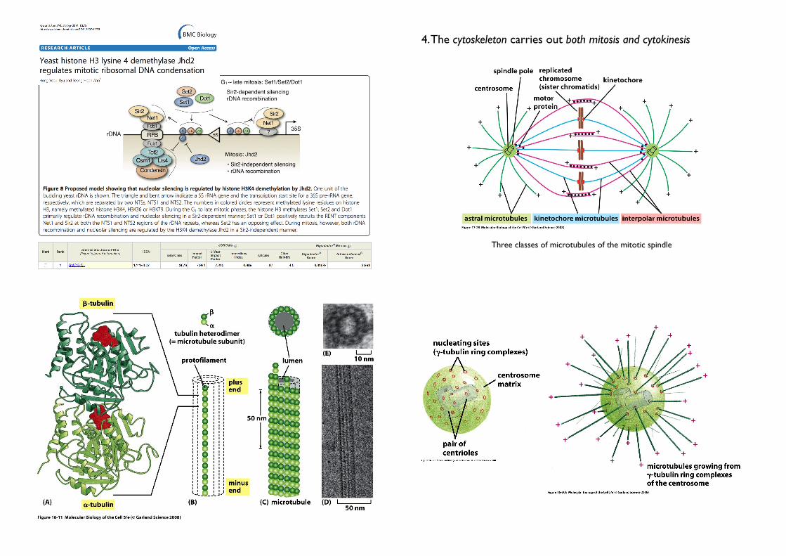
Three classes of microtubules of the mitotic spindle
4. The cytoskeleton carries out both mitosis and cytokinesis

• Mitotic spindle• The mitotic spindle is a microtubule-based machine
• carries out nuclear division (mitosis)
• rapidly disassemble after their tasks
• composed of microtubules + motor proteins
• separates the replicated chromosomes and allocates one copy of each chromosome to each daughter cell (The delicate job!)
17_17globular heads.jpg

Kinesins

Three classes of microtubules of the mitotic spindle
4. The cytoskeleton carries out both mitosis and cytokinesis• The movement are generated by motor protein : bind to actin filaments or
microtubules and use the ATP energy to travel in a single direction.
• Motor proteins also attach to other cell components and thus transport the cargo along the filaments.
Kinesins and Dyneins - Kinesins: move toward plus end, Dyneins: move toward minus end - Two globular ATP-binding head and a tail. - Heads interact with microtubules Tail binds to some cell components.
Major motor proteins of the spindle 1 Kinesin-5: + end, forces the poloes apart. 2 Kinesin-14: - end, pulls the poles together. 3 Kinesin-4, 10: + end, push the attached chromosome away. 4 Dynein: - end, organise microtubuels at various cellular locations.
• Contractile ring
• carries out cytoplasmic division (cytokinesis)• rapidly disassemble after their tasks
• Consists mainly of actin + myosin filaments• Pulls the membrane inward, thereby dividing the cell in two

5. M phase is conventionally divided into six stages
• M phase (6 stages)
• Mitosis (prophase; prometaphase; metaphase; anaphase; and telophase)
• Cytokinesis
• Together ...
• A dynamic sequence in which many independent cycles - involving
the chromosomes, cytoskeleton, and centrosomes - are coordinated to produce two genetically identical daughter cells
• Mitosis (the five stages of mitosis occur in strict sequential order, while cytokinesis begins in anaphase and continues through telophase)
• Prophase: the replicated chromosome condense and the mitotic spindle begins
to assemble outside the nucleus.
• Prometaphase: the nuclear envelop breaks down, allowing the spindle microtubules to contact the chromosomes and bind to them.
• Metaphase: the mitotic spindle gathers all of the chromosomes to the center of the spindle.
• Ananphase: the two sister chromatids in each replicated chromosome synchronously split apart, and the spindle draws them to opposite poles of the
cells.
• Telophase: a nuclear envelop reassembles around each of the two sets of separated chromosomes to form two nuclei.
Mitosis in Cancer Cells

6. Centrosomes duplicate to help form the two poles of the mitotic spindle
• Before M phase begins
• DNA must be fully replicated.
• Centrosome must be duplicated.
• Centrosome
• Microtubule-organizing center• Help form the two poles of the mitotic spindle
• Each daughter cell receive duplicated centrosome• Proteins containing γ-tubulin rings + a pair of centrioles
(each made of short microtubules)
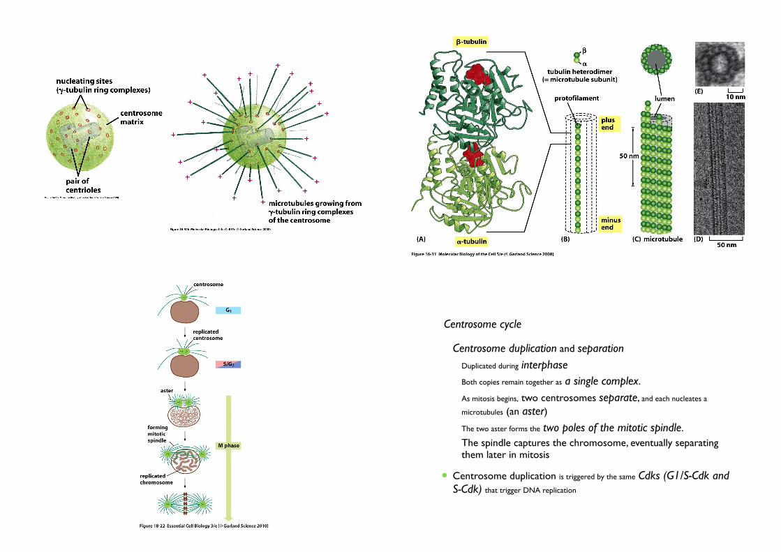
• Centrosome cycle
• Centrosome duplication and separation
• Duplicated during interphase
• Both copies remain together as a single complex.
• As mitosis begins, two centrosomes separate, and each nucleates a
microtubules (an aster)
• The two aster forms the two poles of the mitotic spindle.
• The spindle captures the chromosome, eventually separating them later in mitosis
• Centrosome duplication is triggered by the same Cdks (G1/S-Cdk and S-Cdk) that trigger DNA replication

7. The mitotic spindle starts to assemble in prophase
• Mitotic spindle
• Spindle poles: the two centrosomes that give rise to the microtubules
• Interpolar microtubule
• Two sets of microtubules are joined together to form the basic framework of the mitotic spindle
• This is driven, in part, by motor protein• A bipolar mitotic spindle is formed by the selective stabilization of
interacting microtubules
• Decreases the probability of their depolymerization (shrinking).
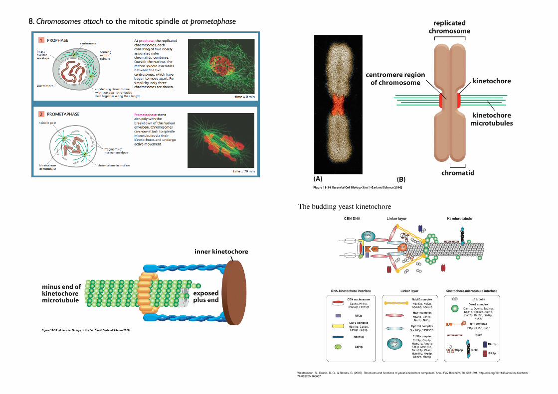
8. Chromosomes attach to the mitotic spindle at prometaphase
The budding yeast kinetochore
Westermann, S., Drubin, D. G., & Barnes, G. (2007). Structures and functions of yeast kinetochore complexes. Annu Rev Biochem, 76, 563–591. http://doi.org/10.1146/annurev.biochem.76.052705.160607

• Kinetochores
• Attach chromosomes to the mitotic spindle
• Assemble into a large protein complex on the condensed chromosomes
during late prophase.• Kinetochore assembly depends on the presence of the centromere DNA
sequence
• Kinetochore microtubules
• The microtubules eventually attached to the kinetochore.• The number of microtubules attached to each kinetochore
varies among species (Human: 20-40; Yeast just one)
• Bi-orientation
• The attachment to opposite poles
• generates tension on the kinetochore
• The cell-cycle control system monitors this tension to ensure
correct chromosome attachment, another important cell-cycle checkpoint
Review

Foley, E. A., & Kapoor, T. M. (2013). Microtubule attachment and spindle assembly checkpoint signalling at the kinetochore. Nature Reviews Molecular Cell Biology, 14(1), 25–37. http://doi.org/10.1038/nrm3494
10. Chromosomes line up at the spindle equator at metaphase
• Metaphase plate
• The beginning of metaphase
• The force that act to bring the chromosome to the equator are not well understood
• Continual growth and shrinkage + Motor proteins (A continuous balanced addition and loss of tubulin subunits)
• The chromosomes at the equator oscillate back and forth (The tug-of-war)
• The chromosomes at the metaphase plate are held there under considerable tension.

11. Proteolysis triggers sister-chromatid separation and the completion of mitosis
Review
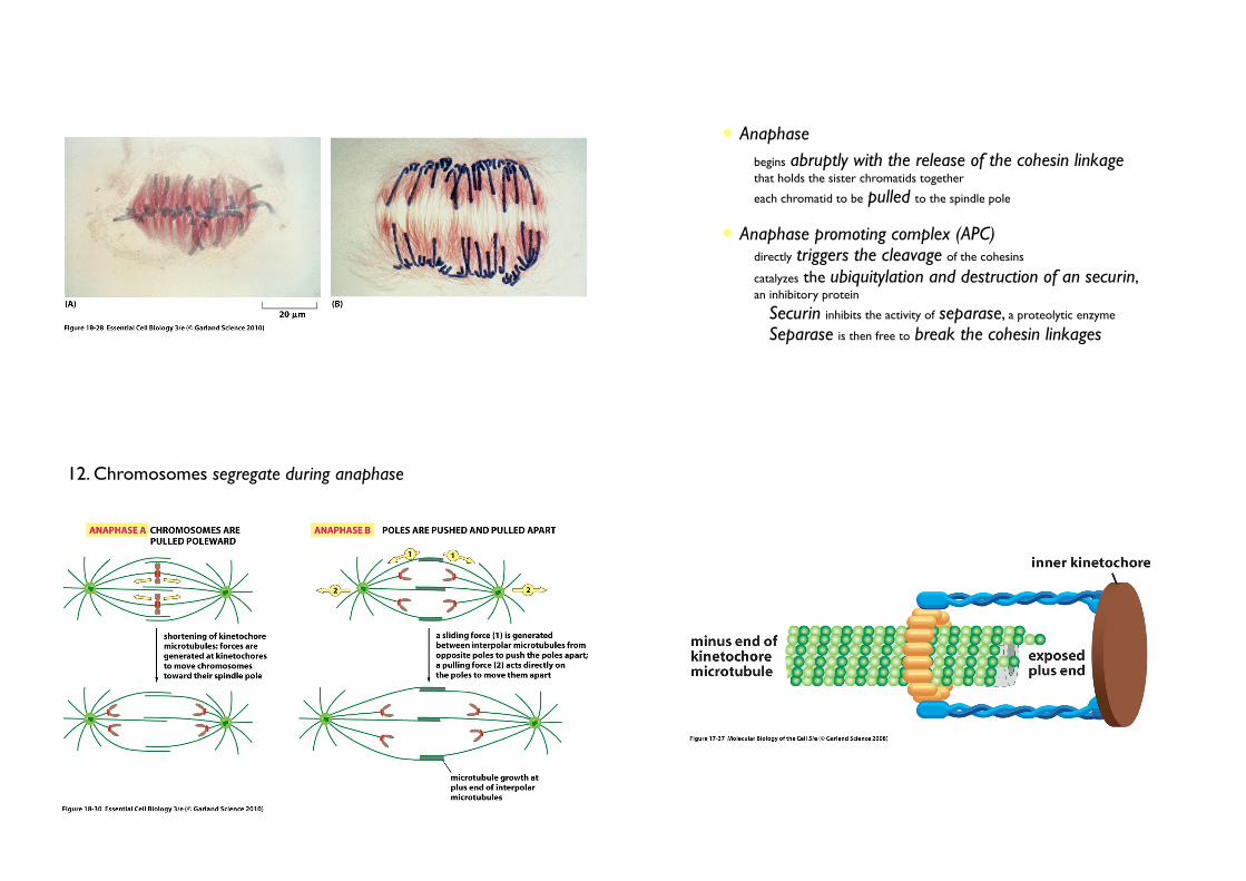
• Anaphase
• begins abruptly with the release of the cohesin linkage that holds the sister chromatids together
• each chromatid to be pulled to the spindle pole
• Anaphase promoting complex (APC)• directly triggers the cleavage of the cohesins
• catalyzes the ubiquitylation and destruction of an securin, an inhibitory protein
• Securin inhibits the activity of separase, a proteolytic enzyme
• Separase is then free to break the cohesin linkages
12. Chromosomes segregate during anaphase

by catastrophin
• Once released, daughter chromosomes move at the same speed about 1 µm per minute
• Anaphse A
• The kinetochore microtubules shorten by depolymerization, the
attached chromosomes move.
• Motor protein + loss of tubulin.
• Loss of tubulin: catastrophin that uses the energy of ATP hydrolysis to remove tubulin subunits from the microtubules
• Anaphse B
• The spindle poles move apart, further contributing to the segregation.
• The overlapping interpolar microtubules elongate and slide past each other
• Kinesin + dynein
Some cell types by microtubule flux (the addiiton of new tubulin at the + end = the loss at - end)

13. Unattached chromosomes block sister-chromatid separation
• If unattached chromosomes are separated...
• Spindle assembly checkpoint
• monitor chromosome attachment
• send a negative signal (a ‘STOP’ signal to the cell-cycle control system)
• blocks the activation of the APC
Bi-orientation is achieved by trial and error (Alternative forms of chromosome attachment)
14. The nuclear envelope re-forms at telophase

• Telophase : a nuclear envelope reassembles to form the two daughter nuclei
• The final stage of mitosis
• Mitotic spindle disassemble
• The nuclear pores reassemble.
• The nuclear lamins are dephosphorylated and re-form the nuclear lamina.
• The pores pump in nuclear proteins, the nucleus expands, the
condensed mitotic chromosomes decondense into their interphase state.
• Gene transcription is able to resume
• A new nucleus has been created, and mitosis is complete
CYTOKINESIS
1. What determines the plane of cytoplasmic cleavage?
Questions
2. What triggers assembly and contraction of molecules involved in cytokinesis?
3. What are the differences in cytokinesis in plants and animals?
4. How are the membrane-enclosed organelles distributed to daughter cells?
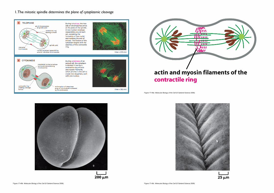
1. The mitotic spindle determines the plane of cytoplasmic cleavage
Figure 17-49a Molecular Biology of the Cell (© Garland Science 2008)
Figure 17-49b Molecular Biology of the Cell (© Garland Science 2008)
A cleaving frog egg
Figure 17-49c Molecular Biology of the Cell (© Garland Science 2008)

• Cleavage furrow
• Puckering and furrowing of the plasma membrane during anaphase
• occurs in a plane that runs perpendicular to the long axis of the mitotic spindle
• ensures that each daughter cell receives an identical and complete set of chromosomes (If the mitotic spindle is deliberately displaced...)
• How the mitotic spindle dictates the position of the cleavage furrow remains a mystery
Figure 17-50a Molecular Biology of the Cell (© Garland Science 2008)
2. The contractile ring of animal cells is made of actin and myosin
Figure 17-50b Molecular Biology of the Cell (© Garland Science 2008)
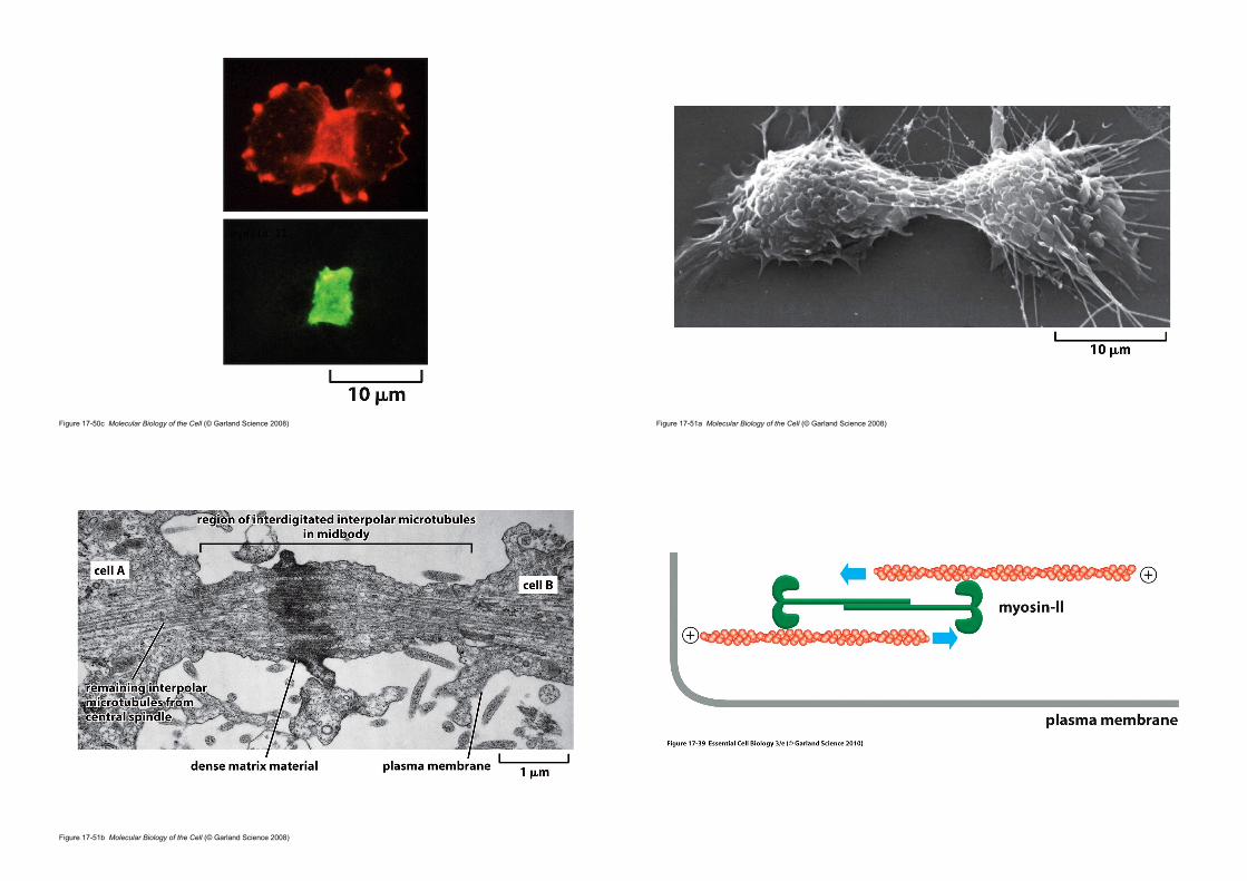
Figure 17-50c Molecular Biology of the Cell (© Garland Science 2008)
actin
myosin II
Figure 17-51a Molecular Biology of the Cell (© Garland Science 2008)
Figure 17-51b Molecular Biology of the Cell (© Garland Science 2008)

• Contractile ring
• actin filaments + myosin• is capable of exerting a force strong enough to bend a fine glass needle
inserted into the cell before cytokinesis.
• The sliding of the actin filaments against the myosin filaments generates the force.
• Transient structure (disassembles completely once the cell is cleaved in two ; vs. contractile apparatus in muscle)
3. The microtubules of the mitotic spindle determine the plane of animal cell division
Three current models of how the microtubules of the anaphase spindle generate signals that influence the positioning of the contractile ring.
1. Astral stimulation: astral microtubules carry furrow-inducing signals to the cell cortex (from embryonic cells).
2. Central spindle stimulation: the spindle mid zone generates a furrow-inducing signal that specifies the site of furrow formation at the cell cortex. The overlapping interpolar microtubules associate with numerous singling proteins, including proteins (such as Cyk4) that may simulate RhoA 3. Astral relaxation: the astral microtubules promote the local relaxation of actin-myosin bundles at the cell cortex. The cortical relaxation is minimal at the spindle equator, thus promoting cortical contraction at that site.
At the beginning of cytokinesis in human cells: RhoA (red), Cyk4 (green)
Cross-section in the plane of the contractile ring

Raf
Mek
Erk
Review
Regulation of the contractile ring by the GTPase RhoA
Extracellular signals control the arrangement of actin filaments
• For the actin cytoskeleton, the structural rearrangements are triggered by activation of a variety of receptor proteins.
• Activation of GTP-binding proteins has a dramatic effect on the organization of actin filaments in fibroblasts.
17_39_fibroblasts.jpg(A)Unstimulated fibroblast have actin filaments primarily in the cortex.(B)Microinjection of an activated form of Rac causes the formation of an
enormous lamellipodium(C)Microinjection of an activated form of cdc42 causes the protrusion of
many long filopodia
*Rho protein family: Cdc42, Rac and Rhomonomeric GTPases and act as molecular switch to control cellular process

• RhoA
• A small GTPase of the Ras superfamily• Controls the assembly and function of the contractile ring at the site of cleavage
• Promotes actin filament formation by activating formins
• Promotes myosin II assembly by activating the kinases, including Rock
• Rock phosphorylates the regulatory myosin light chain (RMLC), stimulating bipolar myosin II filament formation and motor activity.• The local activation of RhoA is dependent on RhoGEF• The microtubules of the anaphase spindle seem to be involved in
RhoGEF activation
4. Animal cells change shape during M phase
• Mammalian fibroblasts in culture• Interphase: spread out flat - strong adhesive contacts
with the surface (substratum)
• M phase: round up - Integrins become phosphorylated and
weaken their grip
• Cycle of attachment and detachment• allows the cells to rearrange their contacts with neighboring cells
and with the extracellular matrix
• the new cells can be accommodated within the tissue
Cell division is accompanied by large changes in cell shape and a decrease in the adherence of the cell to the extracellular matrix: reorganization of actin and myosin filaments in the cell cortex
Figure 17-57 Molecular Biology of the Cell (© Garland Science 2008)
5. Cytokinesis in plant cells involves the formation of a new cell wall

Figure 17-56 Molecular Biology of the Cell (© Garland Science 2008)
• Phragmoplast
• The two daughter cells are separated by the construction of a new wall inside the cell
• formed by the remains of the interpolar microtubules
• Small membrane-enclosed vesicles (from Golgi) are transported to the equator of the phragmoplast
• filled with polysaccharides and glycoproteins• They fuse to form a membrane-enclosed structure
• Question: When a eukaryotic cell divides, each daughter cells must inherit all of
the other essential cell components, including mitochondria, endoplasmic reticulum (ER), or Golgi apparatus. How are these organelles segregated when the cell divides?
• Problems
• Mitochondria (or chloroplast) cannot assemble spontaneously from their individual components.
• They arise only from the growth and division of the preexisting organelles.
• New ER or Golgi apparatus needs some part of it.
6. Membrane-enclosed organelles must be distributed to daughter cells when a cell divides
1 Mitochondria (Chloroplast): are usually present in large enough numbers to be safely inherited if, on average, their numbers rougly doulbe one each cycle.
2 ER: remains largely intact and is cut in two during cytokinesis. 3 Golgi apparatus: is reorganised and fragmented during mitosis. Golgi
fragments associate with the spindle poles and are thereby distributed to opposite ends of the spindle.

• Overview of the cell cycle
• The cell-cycle control system• S phase• Mitosis• Cytokinesis
• Control of cell division and cell growth
Contents
Figure 17-61 Molecular Biology of the Cell (© Garland Science 2008)
1. Mitogen stimulates cell division
Platetelets are miniature cells without a nucleus. They circulate in the blood and help stimulate blood clotting at the sites of tissue damage, thereby preventing excessive bleeding. They also release various factors that stimulate healing. Secretory vesicles contain platelet-derived growth factor (PDGF).
• PDGF
• one of the first mitogens identified in culture
• PDGF (platelet-derived growth factor)
• involved in blood clotting
• When blood clots form (in a wound), blood platelets incorporated in the clots are triggered to release PDGF
• binds to receptor tyrosine kinase in surviving cells at the wound site
• stimulates proliferation and help heal the wound
J Thromb Haemost. 2009 February ; 7(2): 241–246. doi:10.1111/j.1538-7836.2008.03211.x.

• Hepatocyte growth factor
• If part of the liver is lost through surgery or acute injury, cell in the liver
and elsewhere produce a protein called hepatocyte growth factor
• helps stimulates the surviving liver cell to proliferate
Classes of growth factorsIndividual growth factor proteins tend to occur as members of larger families of structurally and evolutionarily related proteins. There are many families, which are listed below:This list is incomplete; you can help by expanding it.
• Adrenomedullin (AM)• Angiopoietin (Ang)• Autocrine motility factor• Bone morphogenetic proteins (BMPs)• Brain-derived neurotrophic factor (BDNF)• Epidermal growth factor (EGF)• Erythropoietin (EPO)• Fibroblast growth factor (FGF)• Glial cell line-derived neurotrophic factor (GDNF)• Granulocyte colony-stimulating factor (G-CSF)• Granulocyte macrophage colony-stimulating factor (GM-CSF)• Growth differentiation factor-9 (GDF9)• Hepatocyte growth factor (HGF)• Hepatoma-derived growth factor (HDGF)• Insulin-like growth factor (IGF)• Migration-stimulating factor• Myostatin (GDF-8)• Nerve growth factor (NGF) and other neurotrophins• Platelet-derived growth factor (PDGF)• Thrombopoietin (TPO)• Transforming growth factor alpha(TGF-α)• Transforming growth factor beta(TGF-β)• Tumor_necrosis_factor-alpha(TNF-α)• Vascular endothelial growth factor (VEGF)• Wnt Signaling Pathway• placental growth factor (PlGF)• [(Foetal Bovine Somatotrophin)] (FBS)• IL-1- Cofactor for IL-3 and IL-6. Activates T cells.• IL-2- T-cell growth factor. Stimulates IL-1 synthesis. Activates B-cells and NK cells.• IL-3- Stimulates production of all non-lymphoid cells.• IL-4- Growth factor for activated B cells, resting T cells, and mast cells.• IL-5- Induces differentiation of activated B cells and eosinophils.• IL-6- Stimulates Ig synthesis. Growth factor for plasma cells.• IL-7- Growth factor for pre-B cells.
Adapted from Wikipedia
• Mitogen
• secreted signal proteins
• bind to cell-surface receptors
• activate intracellular signaling pathways
• release the molecular brakes (Rb) that block the G1-S transition
• stimulate cell division
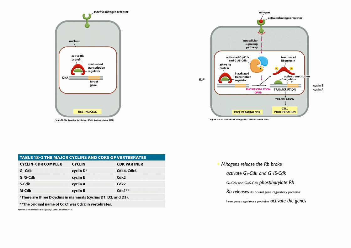
•Rb protein
• molecular brake
• Rb (Retinoblastoma) protein
• Rb was first identified through a rare childhood eye tumor, retinoblastoma (망막아세포종 [網膜芽細胞腫]), in which the Rb protein is missing or defective
• abundant in the nucleus of all vertebrate cells
• binds to particular gene regulatory proteins
• prevents them from stimulating transcription of genes.
2. Mitogen stimulate G1-Cdk and G1/S-Cdk activities
cyclin E
E2F
cyclin A
• Mitogens release the Rb brake
• activate G1-Cdk and G1/S-Cdk
• G1-Cdk and G1/S-Cdk phosphorylate Rb
• Rb releases its bound gene regulatory proteins
• Free gene regulatory proteins activate the genes
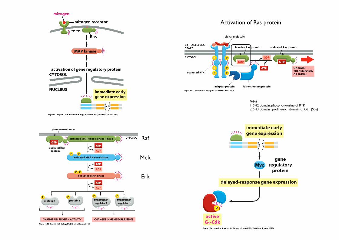
Grb-21. SH2 domain: phosphotyrosine of RTK2. SH3 domain : proline-rich domain of GEF (Sos)
Activation of Ras protein
Raf
Mek
Erk

Figure 17-62 Molecular Biology of the Cell (© Garland Science 2008)
Figure 17-63 Molecular Biology of the Cell (© Garland Science 2008)
3. DNA damage blocks cell division: The DNA damage response
Figure 17-63 (part 1 of 2) Molecular Biology of the Cell (© Garland Science 2008)

Figure 17-63 (part 2 of 2) Molecular Biology of the Cell (© Garland Science 2008)
• Especially well understood
• ATM/ATR kinase are recruited to the site of damaged and initiate a signaling pathway that causes cell-cycle arrest.
• Chk1 and Chk2 are then recruited and activated, resulting in the
phosphorylation of p53.
• MDM2 normally binds to p53 and promotes its ubiquitylation and destruction in proteasomes
• Phosphorylation of p53 blocks its binding to Mdm2
• p53 accumulates to high levels
• stimulates transcription of CKI protein, p21.
• p21 binds and inactivates G1/S-Cdk and S-Cdk complexes
• arrests the cell in G1
When DNA is damaged... (G1 checkpoint)
• G1 arrest gives the cell time to repair the damaged DNA before replicating it.
• If the DNA damage is too severe to be repaired, p53 can induce the cell
to kill itself by undergoing apoptosis.
• If p53 is missing or defective, DNA may lead to mutation and cancer.
• Mutation in the p53 gene are found in about half of all human cancers.
Meaning... (p53-mediated G1 arrest)
Once DNA replication has begun, another type of checkpoint mechanism operates to prevent a cell centering M Phase with damaged or incompletely replicated DNA.
When DNA is damaged... (G2 checkpoint)

• The activating protein phosphatase is itself inhibited
• M-Cdk remains inactive and M phase cannot be initiated until DNA replication is complete and any DNA damage is repaired.
When DNA is damaged... (G2 checkpoint)
p53
4. Abnormal proliferation signals cause cell-cycle arrest or apoptosis, except in cancer cells

• Unicellular organism (bacteria and yeast)
• proliferation depends on the nutrients.
• Multicellular organism
• Nutrients are not enough.
• must receive chemical signals from other cells
• Extracellular Signals for cell division
• Survival factors
• Mitogens
• Growth factors
5. Animal cells require extracellular signals to survive, grow, and divide • Cell growth
• depends on signals from other cells
• does not depend on the cell-cycle control system
• Nerve cells and most muscle cells grow after they have become specialised and permanently stopped dividing
• Growth Factors
• bind to cell surface receptors• activate various intracellular signaling pathways• lead to the accumulation of proteins and other macromolecules• increase the rate of synthesis and decrease the rate of degradation
6. Growth factors stimulate cells to grow
• Compared cell division, there has been surprisingly little study of how cell size is controlled in animals.
• It remains a mystery how different cell types in the same animal grow to be so different in size.

‘One way in which growth factors promote cell growth’
Figure 17-65 Molecular Biology of the Cell (© Garland Science 2008)
7. Proliferating cells usually coordinate their growth and division
Inhibitor of eIF4E
• Rapamycin: a macrolide antibiotic; a immunosuppressant used in kidney transplantation
• Rapamycin interacts with FKBP12 (FK506-binding protein, 12 kDa)
• Rapamycin-FKBP12 complex inhibits mTOR (mammalian target of rapamycin)
• mTOR controls p70S6K and eIF-4E (eukaryotic translation initiation factor 4E)
p70 S6 kinase participates in regulation of transcription as well as translation of mRNA
Figure 17-66 Molecular Biology of the Cell (© Garland Science 2008)
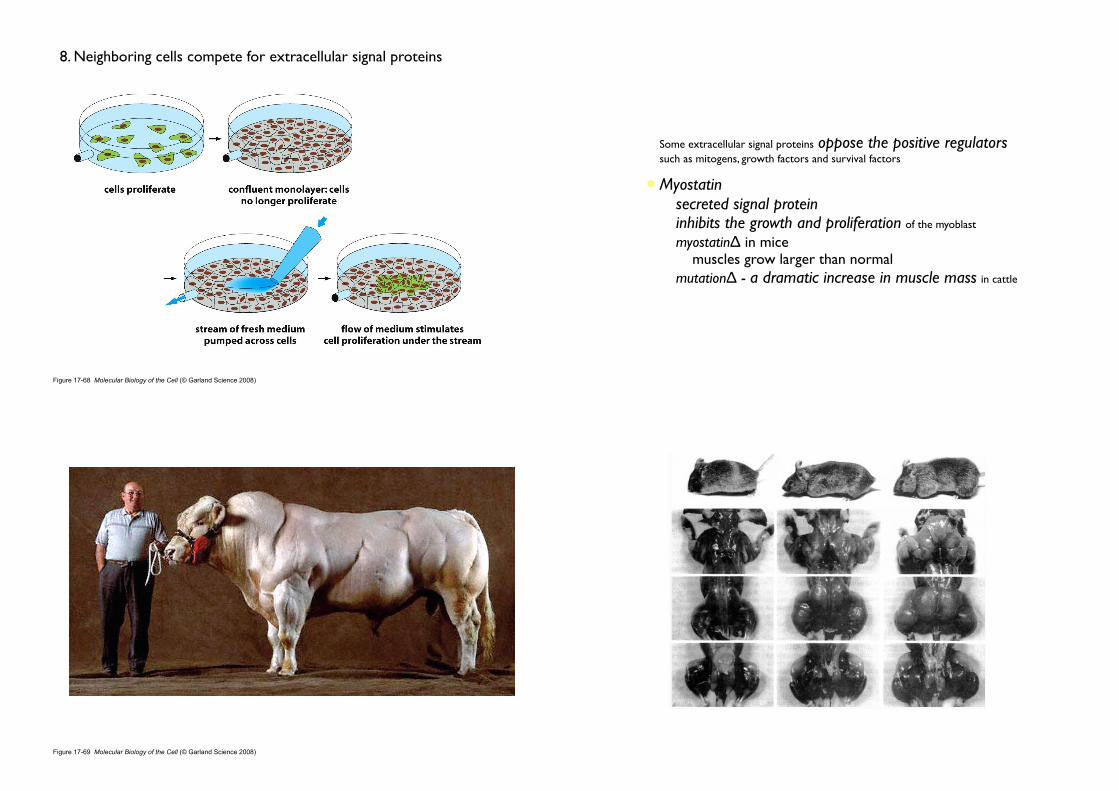
Figure 17-68 Molecular Biology of the Cell (© Garland Science 2008)
8. Neighboring cells compete for extracellular signal proteins
• Some extracellular signal proteins oppose the positive regulators such as mitogens, growth factors and survival factors
• Myostatin• secreted signal protein• inhibits the growth and proliferation of the myoblast
• myostatinΔ in mice • muscles grow larger than normal
• mutationΔ - a dramatic increase in muscle mass in cattle
Figure 17-69 Molecular Biology of the Cell (© Garland Science 2008)

Regulation of muscle size by myostatin. (A) A normal mouse compared with a mutant mouse deficient in myostatin. (B) Leg of a normal and (C) of a myostatin-deficient mouse, with skin removed to show the massive enlargement of the musculature in the mutant. (From S.J. Lee and A.C. McPherron, Curr. Opin. Genet. Devel. 9:604–607, 1999
“Myostatin and the associated gene were discovered in 1997 by geneticists Alexandra McPherron, Se-Jin Lee and Ravi Kambadur, who also produced a strain of mutant mice that lack the gene and thereby have approximately twice as much muscle as normal mice”
McPherron AC, Lawler AM, Lee SJ (May 1997). "Regulation of skeletal muscle mass in mice by a new TGF-beta superfamily member". Nature 387 (6628): 83–90
Figure 17-70 Molecular Biology of the Cell (© Garland Science 2008)
9. Animals control total cell mass by unknown mechanisms
• The size of an animal or one of its organs depends on the number and size of the cells it contains (Total cell mass).
• Animals can somehow assess the total cell mass in a tissue or organ and regulate it. • If cell size is increased or decreased, cell numbers adjust to maintain a normal organ size.
• Individual cells in a pentploid salamander are about five times the volume of those in a haploid salamander (도룡뇽).